Note: Descriptions are shown in the official language in which they were submitted.
CA 02843146 2014-01-24
WO 2013/016094
PCT/US2012/047148
PATENT
ACC5039W0PCT
IMPROVED DEVICE AND METHOD FOR DILATING AN AIRWAY STENOSIS
FIELD OF THE INVENTION
The present invention relates, in general, to medical devices and, in
particular,
to medical devices and related methods for treating a stenosis in an airway of
a
patient.
BACKGROUND OF THE INVENTION
Airway stenosis (or "airway narrowing") is a medical condition that occurs
when some portion of a patient's airway becomes narrowed or constricted, thus
making breathing difficult. A stenosis may occur in any part of the airway,
i.e. larynx,
trachea, bronchi or a combination (laryngotracheal or tracheobroncial
stenosis) in
adults or children and due to any of several different causes. By far the most
common airway stenoses (approximately 95%) are acquired, meaning the patient
is
not born with the condition, and the most common cause of airway stenosis is
trauma caused by intubation (a tube placed in the airway for
ventilation/breathing
assistance in a patient who cannot breathe). lntubation for prolonged periods
of time
may traumatize the airway, causing scar tissue formation that forms the
stenosis.
Sometimes the cause of stenosis is unknown, such as in idiopathic subglottic
stenosis. Managing airway stenosis is one of the most challenging problems for
an
ENT (ear, nose and throat) surgeon.
Subglottic stenosis is one form of airway stenosis that occurs in the larynx,
below the glottis (the area of the larynx around the vocal chords). The
disorder can
either be congenital or acquired and can affect both adults and children.
Acquired
subglottic stenosis is the most commonly acquired anomaly of the larynx in
children
and the most common abnormality requiring tracheotomy in children younger than
one year. To correct subglottic stenosis, the lumen of the cricoids area is
expanded
to increase airflow during breathing. Surgical correction of subglottic
correction of
subglottic stenosis has been performed with various techniques over the years.
Therapies for treating airway stenosis range from endoscopic treatments,
such as dilation and laser resection, to open procedures such as
laryngotracheal
reconstruction. In one technique, a series of rigid dilators of increasing
diameter are
pushed down the airway, gradually expanding the constriction but also applying
1
CA 02843146 2014-01-24
WO 2013/016094
PCT/US2012/047148
PATENT
ACC5039W0PCT
unwanted shear forces to the airway. More recently, balloon catheters have
been
used to perform airway dilation. Such a balloon procedure is described, for
example,
in US Patent Publication No. 2010/0168511 which is incorporated herein by
reference in its entirety. The system described in that patent application is
configured for use in an airway and describes a system for dilating a stenotic
region
with a catheter shaft having an overall length of less than 70 cm, an
inflatable
balloon disposed along a distal portion of the catheter shaft, and a stylet.
The
method for dilating a stenotic region in an airway includes advancing a
balloon
catheter through the airway of a patient to position an inflatable balloon of
the
catheter within at least a portion of the stenotic region, maintaining a
position of the
catheter relative to the patient and inflating the balloon to dilate the
stenotic region.
Methods and devices for improved patient comfort would allow for patient
ventilation during dilation of the stenotic region in the airway and increased
flexibility
for the physician with regard to duration of dilation and number of inflation
and
deflation cycles. These objectives are addressed by the embodiments described
in
this application.
SUMMARY OF THE INVENTION
Accordingly, in one aspect the invention is directed to medical device for
dilating an airway stenosis. The device comprises a proximal end, a distal end
and a
shaft system. The shaft system has an inflation lumen and a ventilating lumen
between the proximal and distal ends of the device. The shaft system has a
proximal shaft section and a distal shaft section with an inflatable balloon
on the
distal shaft section, proximal to the distal end of the medical device. The
distal shaft
section further has a ventilating tip distal to the inflatable balloon, the
ventilating tip
having a tip opening and one or more radially facing openings.
In one embodiment, the medical device of has four radially facing openings.
In another embodiment the radially facing openings have a diameter of between
1
mm and 2 mm and may be spaced 90 degrees apart.
In other embodiments, the inflation and ventilating lumens are adjacent
lumens. In still other embodiments the medical device has an atraumatic tip
portion,
and may incorporate direct visualization markers and/or one or more
radiographic
markers. In some embodiments, the markers are located on the shaft system and
in
2
CA 02843146 2014-01-24
WO 2013/016094
PCT/US2012/047148
PATENT
ACC5039W0PCT
other embodiments the markers are located on the balloon. In some embodiments,
the ventilating tip comprises a soft and atraumatic tip portion, and in other
embodiments the soft and atraumatic tip portion is a slanted soft and
atraumatic tip
portion.
In another aspect, the invention is directed to a connector for connecting a
medical device to a ventilation source and an inflation source. The connector
has a
ventilation port and an inflation port. The ventilation port and the inflation
port are
either ports of different sizes, ports of different shapes or ports of
different
connection types. The inflation source is water, saline or contrast agent and
the
ventilation source is oxygen or air.
In one embodiment of the connector, the inflation port has a threaded
connector and the ventilation port has a -non-threaded connector or in another
embodiment, the inflation port has a non-threaded connector and the
ventilation port
has a threaded connector. In other embodiments, the inflation port has a right-
handed threaded connector and the ventilation port has a left-handed threaded
connector or the inflation port has a left-handed threaded connector and the
ventilation port has a right-handed threaded connector. In another embodiment
of
the connector, the ventilation port is larger in diameter than the inflation
port
In another aspect, the invention is directed to a packaged kit for treating an
airway stenosis. The kit contains a medical device having an inflation lumen,
a
ventilating lumen, an inflatable balloon and a ventilating tip, the inflation
lumen and
the ventilating lumen being adjacent lumens and the ventilating tip comprising
at
least one radially facing opening, an optional balloon insertion stylet for
insertion of
the medical device into the anatomy, and ventilating tubing for connecting the
medical device to a ventilation source. In another embodiment, the packaged
kit
contains a medical device having an inflation lumen, a ventilating lumen, an
inflatable balloon and a ventilating tip, the inflation lumen and the
ventilating lumen
being adjacent lumens and the ventilating tip comprising at least one radially
facing
opening and a balloon insertion stylet for insertion of the medical device
into the
anatomy
In a further aspect, the invention is directed to a method for treating a
stenotic
region in the airway of a human patient. The method comprises providing a
medical
3
CA 02843146 2014-01-24
WO 2013/016094
PCT/US2012/047148
PATENT
ACC5039W0PCT
device having an inflation lumen, a ventilating lumen, an inflatable balloon
and a
ventilating tip, the inflation lumen and the ventilating lumen being adjacent
lumens
and the ventilating tip comprising a tip opening and at least one radially
facing
opening, inserting the medical device into an airway, positioning the medical
device
in the airway at the stenosis, inflating the balloon to dilate the airway,
deflating the
balloon, and optionally repeating the inflating and deflating steps and
withdrawing
the medical device from the airway. The oxygen is delivered through the
ventilating
lumen before, during or after the inflating step.
In another embodiment, the method comprises providing a medical device
having an inflation lumen, a ventilating lumen, an inflatable balloon and a
ventilating
tip, the inflation lumen and the ventilating lumen being adjacent lumens and
the
ventilating tip comprising a tip opening and at least one radially facing
opening,
inserting the medical device into an airway, positioning the medical device in
the
airway at the stenosis, inflating the balloon to dilate the airway, deflating
the balloon,
and optionally repeating the inflating and deflating steps and withdrawing the
medical
device from the airway. Air is inspired through the ventilating lumen before,
during or
after the inflating step.
In a further embodiment, the stenotic region is in the airway portion selected
from the group consisting of larynx, trachea and bronchi.
BRIEF DESCRIPTION OF THE DRAWINGS
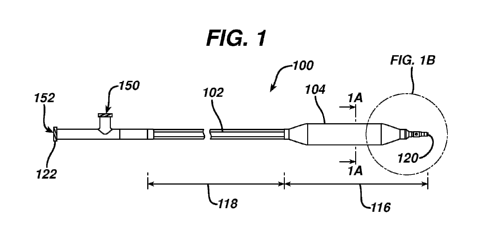
FIG. 1 is a simplified side view of a medical device according to an
embodiment of the present invention.
FIG. 1A is a cross section view through line 1A-1A of FIG. 1.
FIG. 1B is an enlarged side view of the distal end of the medical device of
FIG. 1.
FIG. 2 is a perspective view of a second embodiment of the medical device of
the present invention.
FIG. 2A is an enlarged top view of the distal end of the medical device of
FIG.
2.
FIG. 2B is an enlarged side view of the distal end of the medical device of
FIG. 2.
4
CA 02843146 2014-01-24
WO 2013/016094
PCT/US2012/047148
PATENT
ACC5039W0PCT
FIG. 2C is an enlarged side view of the connector of the medical device of
FIG. 2.
DETAILED DESCRIPTION
The following detailed description should be read with reference to the
drawings, in which like elements in different drawings are identically
numbered. The
drawings, which are not necessarily to scale, depict exemplary embodiments for
the
purpose of explanation only and are not intended to limit the scope of the
invention.
The detailed description illustrates by way of example, not by way of
limitation, the
principles of the invention. This description will clearly enable one skilled
in the art to
make and use the invention, and describes several embodiments, adaptations,
variations, alternatives and uses of the invention, including what is
presently believed
to be the best mode of carrying out the invention.
As used herein, the terms "about" or "approximately" for any numerical values
or ranges indicate a suitable dimensional tolerance that allows the part or
collection
of components to function for its intended purpose as described herein.
Medical devices according to embodiments of the present invention are
beneficial in that, for example, their configuration provides for a
particularly efficient
preparation and treatment of a patient's airway and is mechanically simple.
Moreover, the simplicity of the medical devices provides for them to be
manufactured
in a cost effective manner. In addition, the medical device according to
embodiments of the present invention is sufficiently stiff that it can be
beneficially
employed to access the airway with or without the additional use of a stylet.
FIG. 1 is a simplified side view of a medical device 100 for the treatment of
an
airway stenosis according to an embodiment of the present invention. The
medical
device 100 is an airway dilation and ventilating catheter with an integrated
shaft
system 102. The shaft system 102 has a distal shaft portion 116 and a proximal
shaft portion 118 and the medical device has a distal end 120 and a proximal
end
122. The distal shaft portion 116 is surrounded by a high pressure balloon 104
located near the ventilating tip 106. The shaft system 102 contains adjacent
dual
lumen tubing (see FIG. 1A). By adjacent dual lumen tubing is intended that the
lumens are next to each other and are spaced apart, one from the other. The
inflation lumen 108 is used for inflation of the balloon with water, contrast
medium or
CA 02843146 2014-01-24
WO 2013/016094
PCT/US2012/047148
PATENT
ACC5039W0PCT
saline through inflation port 150 located near the proximal end 122 of medical
device
100, and the ventilating lumen 110 permits passage of oxygen from the
ventilation
port located near the proximal end 122 of medical device 100 to facilitate
ventilation
of the patient and prevent negative pressure pulmonary edema due to attempted
breathing during the dilation procedure and the resultant airway blockage. The
inner diameter of the ventilation lumen is between about 2 mm and about 4 mm,
and
is often about 4 mm. The ventilation lumen is patent during inflation of the
balloon,
that is, the shaft may be made of pebax 72D or nylon 12 or similar non-
collapsing
materials to ensure that the ventilation lumen does not collapse during
balloon
inflation. In an alternative embodiment, a third lumen may be included as a
separate
stylet insertion lumen, such that the shaft system comprises an inflation
lumen, a
ventilating lumen, and a stylet insertion lumen. Alternative designs wherein
the
inflation lumen and the ventilating lumen are coaxial lumens, or all three
lumens are
coaxial lumens are also contemplated herein.
The medical device 100 has a ventilating tip 106 with both a forward facing
tip
opening 114 and radially facing openings 112a, 112b, 112c and 112d to
facilitate
oxygen flow through the ventilating lumen 110. The medical device 100 is
intended
to dilate an airway stenosis and to provide a means to ventilate the airway
during the
dilation procedure. The medical device 100 is designed to ventilate through
the tip
opening 114 and four radially facing openings 112a, 112b, 112c and 112d in the
ventilating tip 106, by delivering oxygen via the ventilating lumen 110 for
delivery
before, during, or after dilation of the airway stenosis. By radially facing
openings is
intended that the flow through the openings may be at 90 degrees from the flow
through the tip opening, but is may also be at 30, 45 or 60 degrees or other
angles
between 0 and 90 degrees, and the openings may be round or non-round such as
oval or slot-shaped. The ventilating tip 106 is located on the distal shaft
section 116,
distal to the distal end of the balloon 104.
The balloon 104 is designed to be non-compliant or semi-compliant, but in
certain embodiments may also be compliant. The diameter of the non-compliant
balloon does not vary significantly with inflation pressure and that of the
semi-
compliant balloon will vary only to the extent that it will "hourglass" or
"dog-bone"
about a target region. The balloon itself may be any shape such as round,
6
CA 02843146 2014-01-24
WO 2013/016094
PCT/US2012/047148
PATENT
ACC5039W0PCT
triangular, oval or square. In the embodiment shown in FIG. 1, the balloon is
round
and semi-compliant.
In some embodiments, direct visualization markers and/or radiographic
markers may be disposed along the integrated shaft system 102. Generally,
"direct
visualization markers" refers to markers that may be viewed during use with
the
naked eye or by use of an endoscope, while "radiographic markers" include
radiopaque material and are viewed using a radiographic device such as intra-
operative fluoroscopy. Direct visualization markers can be positioned in a
number of
locations along the integrated shaft system 102, including the segment of the
shaft
system inside the balloon and may also be incorporated onto the balloon
itself. A
shaft system 102 may have a dark color, such as black, dark blue, dark grey or
the
like, and markers may have a light color, such as white, yellow, green, red or
the like.
In some embodiments, markers may have different colors and/or different widths
to
facilitate distinguishing the markers from one another during use. This
contrast in
colors may facilitate viewing the markers in a darkened operation room and/or
when
using an endoscope inside a patient in the presence of blood. The endoscope
may
be inserted into the ventilation lumen at any time before, during, or after
the
procedure to aid in visualization of the airway and of the stenosis and/or to
aid in
insertion of the medical device. Radiographic markers are often used to ensure
proper alignment of the balloon with the stenosis.
The medical device 100 may be packaged with a balloon insertion stylet and
ventilation tubing. The insertion stylet assists with insertion of the medical
device
100 into the airway and is removed from the device 100 prior to inflation of
the
balloon. The ventilation tubing incorporates standard connectors on each end
and is
used to attach a source of oxygen to the ventilation port 152 of the medical
device
100 for airway ventilation. The medical device 100 may also be packaged with
an
insertion stylet alone where the ventilation source is the ambient air.
Airway access is achieved by inserting the medical device 100 into the airway,
advancing the medical device and positioning the balloon 104 at the site of
the
stenosis. The medical device 100 is then inflated to dilate the airway.
Following
dilation, the balloon is deflated. The process of inflation and deflation may
be
repeated 2, 3, 4 or more times. An oxygen source is connected to the
ventilation
7
CA 02843146 2014-01-24
WO 2013/016094
PCT/US2012/047148
PATENT
ACC5039W0PCT
port 152 of the medical device 100. Oxygen is delivered to the ventilation
lumen
through the ventilation tip 106 via the distal tip opening 114 and four
radially facing
openings 112a, 112b, 112c and 112d of the medical device 100, each side port
having a diameter of 0.157 inches (4 mm). Oxygen may be delivered before,
during
or after inflation of the balloon. Alternatively, the ventilation source may
be the
ambient air, and the ventilation port 152 may be open to the atmosphere. Upon
completion, the medical device 100 is removed from the anatomy. Ventilation of
the
patient during the procedure allows for prolonged duration of balloon
inflation, and
the ability to repeat the inflation, deflation procedure multiple times while
maintaining
oxygen saturation of the patient. While the procedure may be done in the
operating
suite of a hospital, it may also be done in an out-patient surgery center or a
doctor's
office.
The medical device 100 may have any number of suitable sizes, shapes and
configurations. For example, the balloon 104 may have different lengths and
diameters in different embodiments, to accommodate different patient
anatomies.
The overall catheter length and diameter may also vary. In some embodiments,
for
example, the overall length of the medical device 100 from the proximal end
122 to
the distal end 120 is about 35-70 cm, often less than or equal to about 50 cm,
and
often about 45 cm.
The working length of the balloon 104 may be about 40 mm. By "working
length" it is meant the length between the two tapered portions of the balloon
104
may range from between about 10 mm to about 60 mm and often from about 16 mm
to about 45 mm. A variety of lengths may be provided, including about 16 mm,
24
mm and 40 mm. The outer diameter of the fully inflated working length of the
balloon
104 may also vary. The balloon may have inflated diameter in the range of
about 3
mm to about 24 mm and often about 5mm to about 20 mm. In one embodiment, a
variety of diameters may be provided, including about 5 mm, about 7 mm, about
10
mm, about 14 mm, about 20 mm and about 24 mm. For example, a combination of
balloon sizes and lengths may be provided, such that a physician may choose an
appropriate size for an adult or pediatric patient. In one example, the
following
combinations may be provided (first dimension is diameter, second is length):
5 mm
x 24 mm; 7 mm x.24 mm; 8 mm x 24 mm, 8.5 mm x 24 mm, 8.5 mm x 40 mm, 10
8
CA 02843146 2014-01-24
WO 2013/016094
PCT/US2012/047148
PATENT
ACC5039W0PCT
mm x 40 mm; and 14 mm x40 mm. Of course, any of a number of other
combinations of sizes of balloons 104 may be provided.
The balloon 104 is made of any suitable material known in the art for
inflation
balloons and may be constructed of semi-compliant or non-compliant materials
such
as nylon (semi-compliant) and polyethylene terepththalate (PET) (non-
compliant).
The atraumatic tip portion 106 is made of nylon with 20% barium sulfate and is
approximately 10 mm in length (it may be between about 5 mm and 20 mm in
length)
and may contain a radiopaque marker for fluoroscopic visualization in the
patient
anatomy. The combination of materials (the nylon balloon and the adjacent dual
lumen design) provides for ease of insertion of the medical device into and
removal
from the airway. The soft and atraumatic nature of the tip further prevents
injury of
the airway during deployment of the medical device 100 and allows for collapse
and
low profile of the tip during insertion of the medical device 100.
Referring now to FIG. 1, in one embodiment, medical device100 may include
a forward facing tip opening 114 and four radially facing openings 112 a,
112b, 112c
and 112d, on irrigation tip 106 spaced 90 degrees apart, with the inner
diameter of
the forward facing tip opening being 0.157 inches (4mm) and each of the side
openings having a inner diameter of between about 1 and 2 mm and the outer
diameter of the integrated shaft system 102 being about 0.236 inches (6 mm).
Alternative embodiments may include any suitable alternative number of side
openings (1 to 4, 5, 6 or more) distributed in any suitable pattern such as a
helical
pattern. Each side opening may have any suitable diameter in various
alternative
embodiments. For example, in one embodiment, each side opening may have a
diameter of between about 0.5 mm and about 3 mm and often between about 1 and
2 mm.
Referring now to FIG. 2, in a second embodiment, medical device 200 is an
airway dilation and ventilating catheter and may include an integrated shaft
system
202, a balloon 204 and a ventilating tip 206. The integrated shaft system 202
includes a distal shaft portion 216 and a proximal shaft portion 218 and the
medical
device has a distal end 220 and a proximal end 222. The distal shaft portion
216 is
surrounded by a high pressure balloon 204 located near the ventilating tip
206. The
ventilating tip 206 is soft and atraumatic for easy navigation to the site of
the airway
9
CA 02843146 2014-01-24
WO 2013/016094
PCT/US2012/047148
PATENT
ACC5039W0PCT
stenosis and protection of the airway from damage during insertion of the
catheter.
The shaft system 202 contains adjacent dual lumen tubing as described earlier
with
regard to FIG. 1A. Referring now to FIG. 2A as well as to FIG. 2, the
inflation lumen
208 is used for inflation of the balloon with water, contrast medium or saline
through
inflation port 250 located near the proximal end 222 of medical device 200,
and the
ventilating lumen 210 permits passage of oxygen or air from the ventilation
port 252
located near the proximal end 222 of medical device 200 to facilitate
ventilation of
the patient and prevent negative pressure pulmonary edema due to attempted
breathing during the dilation procedure and the resultant airway blockage. The
medical device 200 has a ventilating tip 206 with a slanted distal end 220 (in
this
case a 45 degree slant, but may be slanted between about 15 and 75 degrees and
often between about 25 and 65 degrees) , a forward facing tip opening 214
(with a
diameter of between about 2 mm and 5 mm, often between about 3 mm, and 4 mm
and in this case about 4 mm) and a radially facing opening 212 with a diameter
of
between about 2 mm and 6 mm, often between about 3 mm, and 5 mm and in this
case about 4 mm) to facilitate air or oxygen flow through the ventilating
lumen 210
for delivery before, during, or after dilation of the airway stenosis. The
ventilating tip
206 is located on the distal shaft section 216, distal to the distal end of
the balloon
204.
In the embodiment shown in FIGs 2, 2A and 2B, direct visualization markers
and/or radiographic markers may be disposed along the integrated shaft system
202
and in this case are disposed on the portion of the shaft that is surrounded
by the
balloon. The first shaft marker 260 is located at the mid-point of the balloon
and may
be positioned at the stenosis. The second shaft marker 262 is located in the
proximal taper 264 of the balloon 202 and may be located proximally of the
stenosis
prior to inflation of the balloon and dilation of the stenosis. Any number of
shaft
markers may be located along the integrated shaft system inside or outside of
the
balloon and may be of the same or different lengths, and may be a single
integral
marker or may be single and double or even triple markers with the same or
different
colors to differentiate one from the other. In addition, the balloon may be
marked or
colored in order to more clearly visualize the position of the balloon in the
patient's
airway.
CA 02843146 2014-01-24
WO 2013/016094
PCT/US2012/047148
PATENT
ACC5039W0PCT
The connector 270 of the device of FIG. 2 is shown in an enlarged view in
FIG. 2C. The connector 270 has an inflation port 250 and a ventilation port
252. In
order to ensure that the inflation medium (water, contrast medium or saline)
is
connected to the inflation port 250 and the ventilation port 252 is connected
to the
ventilation source (oxygen or air), the inflation port 250 and the ventilation
port 252
are of different size, shape or type of connection. For example the inflation
port 250
may be a threaded connector and the ventilation port 252 may be a non-threaded
connector, or vise versa. One of the connectors may be a right-handed threaded
connector, and the other may be a left-handed threaded connector. As shown in
FIG. 2C, the inflation port 250 is much smaller in diameter (approximately 6
mm
outer diameter) than the ventilation port 252 (approximately 20 mm outer
diameter)
and therefore could not be connected incorrectly. The difference in size of
the
different ports is that one port is at least about 10% larger than the other
port, often
at least about 50% larger and often about 100% larger than the other port.
The invention has been described with reference to certain examples or
embodiments of the invention, but various additions, deletions, alterations
and
modifications may be made to those examples and embodiments without departing
from the intended spirit and scope of the invention. For example, any element
or
attribute of one embodiment or example may be incorporated into or used with
another embodiment or example, unless otherwise specified or if to do so would
render the embodiment or example unsuitable for its intended use. Also, where
the
steps of a method or process have been described or listed in a particular
order, the
order of such steps may be changed unless otherwise specified or unless doing
so
would render the method or process unworkable for its intended purpose. All
reasonable additions, deletions, modifications and alterations are to be
considered
equivalents of the described examples and embodiments and are to be included
within the scope of the following claims.
11