Note: Descriptions are shown in the official language in which they were submitted.
CA2843445
RAPID, MASSIVELY PARALLEL SINGLE-CELL DRUG RESPONSE
MEASUREMENTS VIA LIVE CELL INTERFEROMETRY
STATEMENT OF GOVERNMENT SUPPORT
This invention was made with U.S. Government support of Grant Nos. CA090571,
CA107300, GM074509 awarded by the National Institutes of Health. The U.S.
Government
has certain rights in this invention.
CROSS REFERENCE TO RELATED APPLICATIONS
This application claims the benefit under 35 U.S.C. Section 119(e) of co-
pending U.S.
Provisional Patent Application Serial No. 61/514,353, filed on August 2, 2011,
entitled
"RAPID, MASSIVELY PARALLEL SINGLE-CELL DRUG RESPONSE
MEASUREMENTS VIA LIVE CELL INTERFEROMETRY". This application is related to
United States Patent Application No. 12/436,702 filed May 6, 2009.
Technical Field
The present disclosure relates to interferometric systems, materials, and
techniques that
can be used to examine one or more cells.
Background of Invention
Interference microscopy provides an interesting biophysical approach to
measuring the spatial distribution of material inside cells and other
transparent objects. It has
been previously shown that an adaptation of this technique, Live Cell
1nterferometry (LCI), can
sensitively detect and track the nanomechanical properties of hundreds of
cells simultaneously
(1). LCI can also be used to monitor the dynamic flow of the cytoplasm inside
single cells as
small indentions are made by highly magnetic probes on the surface of a cell
(2). Studies
showed that an almost instantaneous redistribution of cell material resulted
from indentation of
the cell surface, which was beyond the detection limit of conventional optical
microscopy.
1
CA 2843445 2018-12-05
CA2843445
How individual cells regulate their size is poorly understood, as is the
relationship
between cell mass and well characterized biochemical pathways. While
quantitative mass
measurements of single live cells began in the 1950s, (3, 4) only recently
have newer
approaches to increase the speed, precision, and practicality of cellular mass
measurements
become available. There is a need for new ways to rapidly and simultaneously,
measure the
masses of one or more cells either alone or clustered within large populations
of cells (7, 8).
Embodiments disclosed herein meet this as well as other needs.
SUMMARY
Embodiments of the disclosure include, for example, interferometry systems,
methods
and materials that can be used to determine one or more characteristics or
properties of one or
more cells. Such properties include for example, cell mass, cell volume,
optical cell thickness
(cell density), and the like. Embodiments of the disclosure involve observing
one or more
characteristics or properties of a cell with an interferometer and then using
these observations
to characterize cellular physiology.
An illustrative embodiment is a method for observing a mass of a cell using
live cell
interferometry. Typically, such interferometry methods include the steps of
placing the cell in
an observation chamber of an interference microscope adapted to measure a
fractional phase
shift between a test beam of light and a reference beam of light; exposing the
cell to a test beam
of light at an illumination wavelength; and then measuring the fractional
phase shift between
the test beam of light propagating through the cell and a reference/control
beam of light. Such
measurements can then be used to derive the mass of the cell.
In common embodiments, the mass of the cell is observed using an equation:
1
m = ¨a f vA dA
wherein in is the mass of the cell, a is a constant describing a relationship
between the phase
shift and cell mass, go is the measured fractional phase shift, A. is the
illumination wavelength,
and integration is performed across an entire cell area, A. In certain
embodiments, a = 1.8 x 10-
3 m3kg-I. Optionally, the mass of the cell is observed a plurality of times so
as to observe how
the mass of the cell changes over a period of time. In some embodiments, the
mass of the cell
2
CA 2843445 2017-07-20
CA2843445
is quantified in real-time. In certain embodiments, the method is used to
quantify the masses of
a plurality of cells.
In typical embodiments, the method is performed using a live cell
interferometry system
that comprises a detector operatively coupled to the microscope, a sample
assembly comprising
an observation chamber adapted to contain the cell, a reference assembly
comprising a
reference chamber adapted to contain the reference cell, and a beam splitter
for splitting a light
beam from a light source into a test beam and a reference beam. In certain
embodiments, the
observation chamber comprises at least one perfusion conduit adapted to
circulate a cell
medium within the chamber. In some embodiments, the live cell interferometry
system
comprises a processor element and a memory storage element adapted to process
and store one
or more images of the cell(s). In certain embodiments, the mass property of
the cell is observed
to quantify a cell's response to a therapeutic agent. Optionally, the one or
more cell is obtained
from an individual suffering from a cancer and the therapeutic agent is one
typically used to
treat the cancer (e.g. in methods designed to assess the sensitivity of the
cancer cell to the
therapeutic agent).
Yet another embodiment, is a method for observing a cellular response to a
specific
environment or set of environmental conditions, for example a cell culture
comprising a test
composition, for example a therapeutic agent such as HERCEPTIN. In such
methods of the
invention, the cell is placed in a first environment (e.g. a first observation
chamber) and a mass
property of the cell in the first environment is then observed using a process
comprising live
cell interferometry. In methods of the disclosure, the mass property observed
in the first
environment is compared with the mass property of the cell observed in a
second environment
using a process comprising live cell interferometry. In this way, cellular
responses to the first
environment can be observed. In typical methods, the first environment
comprises a test
composition and the second environment does not comprise the test composition.
In these
methods, the physiology of the cell is transformed by the test composition
when the cell is
exposed to the test composition in the first environment and this
transformation is then
observed by the methods of the disclosure (and typically compared with control
cells that are
not exposed to the test composition and therefore not transformed by the
composition).
Optionally, the test composition comprises an antibiotic, an antibody, an
alkylating agent, an
3
CA 2843445 2017-07-20
CA2843445
antimetabolite, a cell cycle inhibitor, a topoisomerase inhibitor, or a cell.
In some
embodiments, the test composition functions intracellularly and comprises, for
example, an
exogenous polynucleotide such as an siRNA.
In certain embodiments, the cell in which a mass property is observed is
present in the
first environment as an isolated single cell. Alternatively, the cell in which
a mass property can
be observed is present in the first environment in a cluster or clump of
cells. In some
embodiments, mass properties of a plurality of cells present in the first
environment are
observed. In embodiments of the disclosure, the mass property of one or more
cells in the first
environment can be observed a plurality of times so as to observe how the mass
property of the
one or more cells changes over a period of time. Optionally, for example,
changes in the mass
property of the cell are observed over time to observe a temporal mass
profile. Certain
embodiments include the steps of comparing an observed temporal mass profile
to a database
of temporal mass profiles, wherein the database of temporal mass profiles is
selected to include
temporal mass profiles that are characteristic of cellular sensitivity to the
test composition (e.g.
in situations where the growth of the cell is inhibited in the presence of a
test composition) and
temporal mass profiles that are characteristic of cellular resistance to the
test composition (e.g.
in situations where the growth of the cell is not inhibited in the presence of
a test composition).
Other illustrative embodiments can include, for example, systems and methods
for
quantifying the mass of a cell and/or observing how the mass of one or more
cells changes in
response to environmental stimuli. Some embodiments include a method that
comprises the
steps of: placing one or more cells in an observation chamber of an
interference microscope
capable of generating and measuring a fractional phase shift between a test
beam and a
reference beam; exposing the cells to a test beam at an illumination
wavelength; measuring the
fractional phase shift between the test beam propagating through the cell and
a reference beam
propagating through a reference cell; and determining the mass of one or more
cells with an
equation:
in = if q91 dA
4
CA 2843445 2017-07-20
CA2843445
wherein m is the mass of the cell, a is a constant describing a relationship
between the phase
shift and cell mass, tp is the measured fractional phase shift, X is the
illumination wavelength,
and integration is performed across an entire cell area, A.
Methodological embodiments for observing other cellular properties are also
contemplated. For example, in certain embodiments, the method can be used to
observe an
optical thickness of a live cell in an aqueous medium. Alternatively, the
method can be used to
observe a population of live cells simultaneously, for example to identify,
monitor, and
measure resting and dynamic cell responses to stimuli in a population of live
cells. Typically in
these methods, the property is observed in response to the cell's exposure to
a stimulus such as
therapeutic drugs. In certain embodiments, the methods can be conducted in a
highly parallel
fashion to profile the differential response of cells in a population to
internal or external stimuli.
Optionally, the methods further comprise removing the cell from the
observation chamber and
manipulating the cell for a further analysis. In certain embodiments, the
method is used to
obtain information comprising a cell specific profile of a live cell in an
aqueous medium and to
then store this information in a memory storage element.
A variety of methodological embodiments are contemplated. For example, certain
methodological embodiments can be performed using a system comprising: a
microscope
having a Michelson interference objective; a detector such as a camera (e.g. a
still camera, a
video camera, charge coupled devices (CCD) and the like) operatively coupled
to the
microscope; a sample assembly comprising an observation chamber adapted to
contain the cell;
and a reference assembly comprising a reference chamber. Further, such a
system may
additionally include a memory storage element adapted to store one or more
images of the cell
and a processor element adapted to process one or more images of the cell. In
other
embodiments, the microscope is an interference microscope capable of observing
interference
fringes through a fluid medium. Alternatively, the system is capable of
observing interference
patterns at multiple phase shifts and then correlating the observed
interference patterns to an
optical thickness profile of the cell. Such general embodiments are non-
limiting as the systems
disclosed herein can adopt a variety of configurations. Other embodiments
include a real-time
and non-invasive marker of cellular fitness and a method of observing cellular
fitness. Such
systems and techniques can rely on the observation of changes in cell mass.
CA 2843445 2017-07-20
CA2843445
cellular fitness. Such systems and techniques can rely on the observation of
changes in cell
mass.
Various embodiments of the claimed invention pertain to a method for observing
a
cellular response to a test agent, said method comprising: (a) placing at
least one cell in a first
environment containing said test agent; (b) performing live cell
interferometry to determine a
measurement proportional to the mass of a cell in said first environment,
wherein said live cell
interferometry comprises measuring the fractional phase shift between a test
beam of light
propagating through the cell and a reference beam of light, and integrating
said fractional phase
shift over the area of said cell to provide said measurement; (c) performing
live cell
interferometry to determine a measurement proportional to the mass of a cell
in a second
environment lacking said test agent, wherein said live cell interferometry
comprises measuring
the fractional phase shift between a test beam of light propagating through
the cell and a
reference beam of light and integrating said fractional phase shift over the
area of said cell to
provide said measurement; and (d) comparing the measurement determined in (b)
with the
measurement determined in (c) where the difference between the measurement
determined
in (b) and the measurement determined in (c) indicates the cellular response
to the test
agent, and wherein said method provides a sensitivity sufficient to detect
said cellular
response to the test agent by the fourth hour of treatment.
Various embodiments of the claimed invention also pertain to a method for
observing a
mass of a cell, the method comprising: (a) placing the cell in an observation
chamber of an
interference microscope adapted to measure a fractional phase shift between a
test beam of
light and a reference beam of light; (b) exposing the cell to a test beam of
light at an
illumination wavelength; (c) measuring the fractional phase shift between the
test beam of light
propagating through the cell and the reference beam of light; and (d) using a
measurement
obtained in (c) to observe the mass of the cell.
Various embodiments of the claimed invention pertain to a live cell
interferometry
system for observing a cellular response to a test composition, the system
comprising: at least
one cell observation chamber adapted to contain live cells; a camera adapted
to capture and
store an interference phase image of the live cells in a memory storage
element; and a processor
element coupled with the memory storage element and configured to: obtain,
from the camera
6
CA 2843445 2018-12-05
=
CA2843445
via the memory storage element, a first interference phase image of unexposed
live cells
lacking exposure to the test composition and determine a first mass
measurement of the
unexposed live cells by integrating a first fractional phase shift of the
first interference phase
image over the surface areas of the unexposed live cells; obtain, from the
camera via the
memory storage element, a second interference phase image of exposed live
cells exposed to
the test composition and determine a second mass measurement of the exposed
live cells by
integrating a second fractional phase shift of the second phase image over the
surface areas of
the exposed live cells; and detect a cellular response of the live cells to
the test composition
based on at least a mass difference between the first and second mass
measurements.
Other objects, features and advantages of the present disclosure will become
apparent to
those skilled in the art from the following detailed description including the
Appendices. It is
to be understood, however, that the detailed description and specific
examples, while indicating
some embodiments of the present invention are given by way of illustration and
not limitation.
Many changes and modifications within the scope of the present invention may
be made
without departing from the spirit thereof, and the invention includes all such
modifications.
6a
CA 2843445 2018-12-05
CA 02843445 2014-01-28
WO 2013/019984 PCT/US2012/049388
BRIEF DESCRIPTION OF THE DRAWINGS
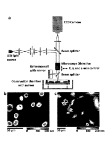
Figure 1 illustrates a schematic of an embodiment of a live cell
interferometer
(LCI). The LCI (a) is a Michelson-type interference microscope that compares
the
optical thickness of a reference cell to the optical thickness of samples
placed in the
observation chamber. Suspended in the observation chamber is a mirrored
substrate,
allowing the LCI to make measurements of optical thickness on transparent
cells. The
relative position of the microscope objective and observation chamber is
controlled by
computer and translatable in three-dimensions allowing for rapid, automated
image
acquisition. Throughout data collection, cells in the observation chamber are
maintained in standard cell culture conditions (e.g., pH 7.4, 37 C, 5% CO2).
The live
cell interferometer is capable of measuring the mass of both adherent and non-
adherent cells. Frame (b) shows several non-adherent H929 cells attached to
the
observation chamber substrate after coating the substrate with Poly-L-Lysine
solution,
while frame (c) shows adherent female Indian Muntjac (9) cells cultured
directly on
the substrate. The color maps show optical thickness measurements with blue
being a
low optical thickness relative to background and red being a high optical
thickness.
Figure 2 illustrates high-throughput and longitudinal measurements of cell
mass with LCI. Four sample images of H929 multiple myeloma cells (a) from the
LCI show optical thickness profiles of cells over six hours of monitoring.
Color
.. indicates the phase shift in nm, with dark blue indicating low thickness
and white/red
indicating high thickness. These sample images are composites of 25 successive
CCD captures taken every 7 minutes. The inset shows a measurement of the phase
shift across a single cell. Integrated phase shift across a cell is directly
proportional to
cell dry mass. (b) Hundreds of individual cells (outlined in red) are
identified at
unique positions in each frame and (c) the mass of each individual cell is
determined,
enabling high-throughput, population-level mass profiling over time. (d) The
mass of
individual cells is tracked longitudinally over time to examine single cell
growth
dynamics. Measurements are shown as open symbols with a linear least squares
best
fit line. The measured growth rate in this case is 6.5 (se +/- 0.72) pg/hr.
The variation
7
CA 02843445 2014-01-28
WO 2013/019984 PCT/US2012/049388
about the linear trend, taken as the standard deviation of the residual error,
is 5.0 pg or
1.17% of the median cell mass. The maximum peak-to-peak residual error is 11
pg at
102 minutes or 2.61% of the median mass at that time point.
Figure 3 illustrates a drug response of H929 multiple myeloma cells as
profiled by single-cell mass accumulation. Results of LCI longitudinal mass
measurements on populations of H929 multiple myeloma cells, comparing the mass
accumulation of DMSO-treated controls with Tunicamycin-treated (10 g/m1)
cells.
Data are taken over five hours. The treated cells grow more slowly than do the
controls. Experiments #2 and #3 were conducted at 32 C, versus 37 C for #1,
which
accounts for the slightly lower overall growth rates observed. The scatter
plots (a-b)
depict the growth of individual cells at five hours versus their initial mass
(normalized
by initial mass). Error bars represent +/- 2% CV, an estimate of the
measurement
error. Error bars apply to all data, but are omitted for the majority of
points in the plot
for clarity. In the box plots of normalized mass versus time (c-d), circles
indicate the
sample median, and triangles indicate the 95% confidence interval for the
median.
Solid boxes indicate limits of the 25 and 75 percentiles, and whiskers
represent two
standard deviations from the mean.
Figure 4 illustrates a molecular profile of H929 response to Tunicamycin.
The divergence in growth rates between the treated and untreated populations
occurs
synchronously with the up-regulation of the transcription factor CHOP (a) and
the
alternative splicing of transcription factor XBP1 (b) in the treated
population. CHOP
and XBP1-s activate a host of genes responsible for mitigating the effects of
protein
mis-folding in the endoplasmic reticulum. This is consistent with the known
mechanism of TM action, an inhibitor of protein glycosylation. (c) Cell cycle
data
show a rapid reduction in the G2/M phase population and a corresponding
increase in
the G1/G0 population, consistent with cell cycle arrest. This shift becomes
pronounced after three hours of treatment, leaving 50% of cells in G1/G0 by
the end
of five hours of treatment.
Figure 5 illustrates mass dynamics of cell division. Twenty eight division
8
CA 02843445 2014-01-28
WO 2013/019984 PCT/US2012/049388
events were recorded from the treated and untreated populations in all
experiments
(from a total of ¨600 cells). (a) The mass range of dividing cells was
determined by
observing individual divisions and measuring the mass of the parent and
daughter
cells directly. Panel (a) compares the mass distribution of all cells measured
(both
treated and untreated; dashed line) with the masses of those cells which
divided
during the experiment (red before the division, blue after division). (b)
Surprisingly, a
number of cell divisions were highly asymmetric, with ¨55%, or more, of the
total
parent cell mass remaining in the smaller of the two daughter cells. (c) Two
examples
of highly asymmetric division are shown over the five hour time course. The
smaller
of the daughter cells in these divisions (indicated by an asterisk) contained
35% and
40% respectively, of the parent cell mass. These division events are indicated
by red-
filled circles in (b). Error bars represent +/- 2% CV, an estimate of the
measurement
error (see Methods).
Figure 6 illustrates live cell interferometer measurements of the mass of
adherent cells. The frame shows several mouse fibroblast cells cultured
directly on a
polished silicon substrate. The color map indicates optical thickness
measurements
with blue being a low optical thickness relative to background and red being a
high
optical thickness. To the left are mass measurements of the four cells, as
indicated,
taken every two seconds for 200 minutes. The smaller optical thickness of the
adherent cells vs. non-adherent cells is easily measured by the LCI.
Figure 7 illustrates scatter plots of treated and untreated data sets. The red
trend line represents a linear least-squares fit to the data. The p value,
indicating the
probability that there is no correlation between growth rate and cell mass, is
given for
each fit. There is a trend toward a slower growth as cell mass increases in
the
untreated controls, although this is not statistically significant to 95%
confidence.
The treated sets Tm2 and Tm3 show no correlation between growth rate and mass,
while the negative slope is significant (p=0.02) for Tml . The norm of the
residuals
for each linear fit provides an estimate of growth variation within each mass
segment.
Error bars represent +1- 2% CV, an estimate of the measurement error.
9
CA 02843445 2014-01-28
WO 2013/019984 PCT/US2012/049388
Figure 8 illustrates the optical path and mass measurement stability of LCI.
Gold islands vapor deposited onto silicon (a, lower panel), ¨35 nm in height,
were
measured repeatedly for 140 minutes to test the stability of the
interferometer. Shown
in the top panel, the mean height of three representative islands shows no
meaningful
drift during this period and the measurement repeatability, given as the
standard
deviation of the height, is ¨1.2 angstroms or 0.35% of the total height.
Similarly,
stability of the mass measurement for transparent objects is estimated by
repeatedly
measuring partially-melted 10 um diameter polystyrene spheres (b) over 80+
minutes.
In the top panel, trace #1 represents three spheres melted into a cluster, and
trace #2
.. represents two spheres melted into a cluster. The other three traces are
from single
spheres. The coefficient of variation of the mass measurement for the
polystyrene
spheres is similar to that obtained for the mean height of the gold islands,
<0.4%, with
negligible drift over the measurement period. Data were collected in the LCI
observation chamber under conditions identical to those used to measure live
cells,
with the exception that water replaced the culture medium.
Figure 9 illustrates high precision mass measurements with LCI. Four
individual cells (a through d) were selected and tracked over 98 successive
measurements made at roughly 12 s intervals. Over this time period (20 min
total) the
observed cells showed small changes in mass (see inset plots of mass vs.
time),
allowing assessment of measurement repeatability. Cells in c and d show >one
picogram average mass change over this period, so histograms in c and d
present
measured mass minus the linear component of the least-squares fit line (shown
in red
in the inset) to remove any additional variance due to growth. Histograms of
measurements for each cell were fitted to a Gaussian distribution by nonlinear
least-
squares fitting in Matlab. Mean ( ) and standard deviation (a) are reported
for each
distribution. All standard deviations are <1% of the distribution mean
indicating that
mass measurements with the LCI are highly repeatable.
Figure 10 illustrates the mass of a population of partially melted 6 Jim
diameter polystyrene spheres (Flow Check, Polysciences Inc.) that were
measured by
CA 02843445 2014-01-28
WO 2013/019984 PCT/US2012/049388
LCI in water (a). During preparation the spheres infrequently aggregate and
upon
heating dimers and trimers coalesce into single, conical clusters. Peaks for
the
population of monomers (110.7 pg), dimers (213.7 pg) and trimers (308.1 pg)
can be
distinguished in the histogram. The LCI-measured standard deviation of the
monomer
population mass is 7.5 pg, or 6.8% of the mean, which exceeds the
manufacturers'
stated specification of 15%. The mass distribution of populations of four
different
mammalian cells types and the 6 um polystyrene spheres are plotted together as
histograms for comparison (b). The mean mass of the mouse red blood cell (RBC)
population determined with LCI can be compared to published values determined
by
other techniques (see, e.g. Nie, Z., et al. Analytical Chemistry, 2007. 79: p.
7401-
7407; Vaysse, J., et al. Mechanisms of Ageing and Development, 1988. 44(3): p.
265-
276; Wirth-Dzieciolowska, E., et al. Animal Science Papers and Reports, 2009.
27(1):
p. 69-77; Magnani, M., et al. Mechanisms gfAgeing and Development, 1988.
42(1): p.
37-47), and by another group using microinterferometry (see, e.g. Mysliwski,
A., et
al. Mechanisms gfAgeing and Development, 1985. 29(2): p. 107-110) (c). Nie et
al
measured the cell mass of mouse RBCs using a novel mass spectrometric method,
and
also the mean corpuscular hemoglobin mass (MCH) by traditional photochemical
techniques (see, e.g. Nie, Z., et al. Analytical Chemistry, 2007. 79: p. 7401-
7407).
The MCH typically represents a large fraction of the total cell mass of
mammalian red
blood cells. The range of MCH values for various mouse strains are well
established
(see, e.g. Magnani, M., et al. Mechanisms of Ageing and Development, 1988.
42(1): p.
37-47; Mysliwski, A. et al. Mechanisms of Ageing and Development, 1985. 29(2):
p.
107-110; Wirth-Dzieciolowska, E., et al. Animal Science Papers and Reports,
2009.
27(1): p. 69-77). Using the ratio of MCH-to-total mass given by Nie et al. as
an
estimate, the established values for mouse RBC MCH can be compared to the
average
mouse RBC mass measured. The estimated values are displayed in red.
Figure 11 illustrates the mass distributions of populations of four different
live or freshly prepared cell types were measured by LCI: (a) mouse WEHI-231 B
lymphoma cells (b) red blood cells (RBCs) from a 15 week-old female C57BL/6
11
CA 02843445 2014-01-28
WO 2013/019984
PCT/US2012/049388
mouse, (c) human H929 multiple myeloma cells, and (d) a mixture of primary
bone
marrow and acute myeloid leukemia (AML) cells established in a C57BL/6 mouse
by
retroviral transduction and adoptive cell transfer using standard techniques.
Figure 12 illustrates mass vs. time plots (a) for representative cells from
the
data given in Figure 9a. The dashed lines represent an exponential growth fit
to the
data. To estimate the scale of measurement variation in the live cell
experiments, all
single-cell mass vs. time data, from all three Tm-treated and DMSO control
runs
(indicated D1-D3 and Tm1-Tm3, respectively), were fitted to a simple
exponential
growth model (mass(t)=mo*Ct , where the constant C is close to unity), and
residual
error calculated as the percent difference between the trend and the actual
data at each
time point. In the box plot, the central line indicates the sample median, and
triangles
indicate the 95% confidence interval for the median. Solid boxes indicate
limits of
the 25 and 75 percentiles, and whiskers represent two standard deviations from
the
mean. The residuals are symmetrically distributed about zero, and the range
between
the 25% and 75% quartile (IQR) varies from 0.0126 (D2) to 0.027 (D3). The mean
IQR is 0.02.
Figure 13 illustrates various single cells, cell clusters, and dense colonies
imaged and mass quantified by aLCI. (a) Confocal images of MDA-MB-231, MCF-
7, SK-BR-3, and BT-474 breast cancer cell lines. SK-BR-3 and MDA-MB-231 cell
lines grow as single cells or in loose clusters, whereas the BT-474 and MCF-7
cell
lines grow as dense multi-cellular colonies. Red is Alexa 568 Phalloidin actin
stain
and blue is a DAPI nuclear stain. (b) Phase images of mass distributions for
each cell
line. Single cells, loose clusters, and dense colonies are reproducibly
quantified in
real-time with aLCI. Color scale denotes mass density in pg/um2. Scale bars
are
50um.
Figure 14 illustrates simultaneous imaging of single cells and large colonies
with aLCI. (a) The phase image shows a single MCF-7 cell (555 pg) alongside a
multi-cellular MCF-7 colony (21,660 pg) consisting of approximately 52 cells.
Color
scale denotes mass density in pg/um2. (b) Composite scatter plot of growth
rate vs.
12
CA 02843445 2014-01-28
WO 2013/019984
PCT/US2012/049388
initial mass for all cell lines. MDA-MB-231 (red), MCF-7 (blue), SK-BR-3
(cyan),
and BT-474 (black) are overlaid to show the range of cell and colony sizes and
growth rates of the different lines. Colony forming lines (MCF-7 and BT-474)
span
the range from a single cell to large, multi-cellular colonies.
Figure 15 illustrates breast cancer cell growth inhibition due to trastuzumab
reproducibly quantified with aLCI within 3-5h. (a) Population mean normalized
mass
versus time plots for each cell line with 20ug/m1trastuzumab treatment (error
bars
indicate standard error). (b) Growth inhibition due to trastuzumab treatment
becomes
highly significant (p < 0.001) for the SKBR-3 and BT-474 lines by 7h. Hourly
growth rates are calculated from a linear fit to mass accumulation data.
Figure 16 provides a table that illustrates trastuzumab (Herceptin)
sensitivity
determined by a 7-day proliferation assay (column 3) being shown alongside
aLCI
mass accumulation profiling data collected at six hours (column 4). HER2
status was
determined by O'Brien et al. (see, e.g. Molecular Cancer Therapeutics. 2010,
9, 1489-
502).
DETAILED DESCRIPTION OF THE INVENTION
Unless otherwise defined, all terms of art, notations and other scientific
terms
or terminology used herein are intended to have the meanings commonly
understood
by those of skill in the art to which this invention pertains. In some cases,
terms with
commonly understood meanings may be defined herein for clarity and/or for
ready
reference, and the inclusion of such definitions herein should not necessarily
be
construed to represent a substantial difference over what is generally
understood in
the art. Many of the techniques and procedures described or referenced herein
are
well understood and commonly employed using conventional methodology by those
skilled in the art. As appropriate, procedures involving the use of
commercially
available kits and reagents are generally carried out in accordance with
manufacturer
defined protocols and/or parameters unless otherwise noted. A number of terms
are
defined below.
13
CA2843445
Before the present invention is further described, it is to be understood that
this
invention is not limited to particular embodiments described, as such may, of
course, vary. It is
also to be understood that the terminology used herein is for the purpose of
describing
particular embodiments only, and is not intended to be limiting, since the
scope of the present
invention will be limited only by the appended claims. It must also be noted
that as used herein
and in the appended claims, the singular forms "a", "and", and "the" include
plural referents
unless the context clearly dictates otherwise. Thus, for example, reference to
''a test
composition" includes a plurality of such test compositions and so forth. All
numbers recited
in the specification and associated claims that refer to values that can be
numerically
characterized with a value other than a whole number (e.g. the concentration
of a compound in
a solution) are understood to be modified by the term "about". Where a range
of values is
provided, it is understood that each intervening value, to the tenth of the
unit of the lower limit
unless the context clearly dictates otherwise, between the upper and lower
limit of that range
and any other stated or intervening value in that stated range, is encompassed
within the
invention. The upper and lower limits of these smaller ranges may
independently be included in
the smaller ranges, and are also encompassed within the invention, subject to
any specifically
excluded limit in the stated range. Where the stated range includes one or
both of the limits,
ranges excluding either or both of those included limits are also included in
the invention.
Publications cited herein are cited for their disclosure prior to the filing
date of the
present application. Nothing here is to be construed as an admission that the
inventors are not
entitled to antedate the publications by virtue of an earlier priority date or
prior date of
invention. Further the actual publication dates may be different from those
shown and require
independent verification.
The invention disclosed herein has a number of embodiments. Embodiments of the
invention provide methods, materials and systems for observing and/or
14
CA 2843445 2017-07-20
CA 02843445 2014-01-28
WO 2013/019984 PCT/US2012/049388
characterizing one or more properties of a cell, for example its mass and/or
how the
mass of a cell changes in response to various environmental conditions.
Illustrative
embodiments of the invention comprise using interferometry to obtain
information on
the mass of a cell and/or characterize one or more cellular properties that
are related
to the mass of a cell. Illustrative cellular properties that can be observed
by
embodiments of the invention can include for example cytoskeletal remodeling
behavior in response to a stimulus, for example a stimulus comprising exposure
to a
drug or other biologically active agent as well as a variety of other factors.
In some
embodiments of the invention, the phenomena that is observed is one
corresponding
to, or associated with, a pathological condition such as aberrant cell
division, such as
that occurring in precancerous and cancerous cells. In some embodiments of the
invention, the cell membrane in which movement is observed is a membrane of a
single cell. In other embodiments of the invention, the membrane properties of
a
plurality of cells are observed. In certain embodiments, the membrane is a
membrane
of a cell in a tissue. In other embodiments, the membrane is a membrane of a
cell
within a colony of cells (e.g. an in vitro cell culture of primary cells taken
from a
patient or an established cell line). In typical embodiments of the invention,
the cell is
a eukaryotic (e.g. mammalian) cell.
In typical interferometric embodiments of the invention, an interferometer
uses, for example, a Michelson configuration. In addition, methods and
elements
associated with interferometric technologies including spectrally resolved
interferometry, wavelength scanning interferometry, digital holography and the
like
can be used in embodiments of the invention. While many interferometric
microscopy systems and methods can be adapted for use with embodiments of the
invention, other embodiments of the invention can use scanning optical
microscopes,
confocal microscopes and the like. An illustrative and non-limiting list of
publications that describe optical profiling methods and materials that can be
adapted
for use with embodiments of the invention are disclosed for example in U.S.
Patent
Application Nos. 20100284016; 20050248770; 20050225769; 20050200856;
CA2843445
20050195405; 20050122527; 20050088663; 20040252310; 20050117165; 20030234936;
20040066520; 20080018966 and 20050167578.
Embodiments of the invention use optical profilometry techniques to provide
for
example methods of height measurement, shape measurement, as well as measures
of other
modulations in the shapes of cell membranes and other properties that can
relate to the mass of
a cell. Depending on the shape, size etc. of a test cell or a population off
cells, these techniques
typically use structured light, focusing properties of optics, interference of
light, etc., to
optimize results in an economical and practical way. Moire' techniques, ESPI
(electronic
speckle-pattern interferometry), laser scanning, photogrammetry, and
interferometry are
illustrative techniques developed for conducting three-dimensional shape
measurements. The
technique of white-light vertical scanning interferometry (VSI), also commonly
referred to as
white-light interferometry or coherence radar, is used for imaging small
objects, typically those
with roughness that does not exceed a few micrometers. VSI methodology is
based on
detection of the coherence peak created by two interfering, polychromatic
wavefronts. It has
many advantages such as absolute depth discrimination, fast measurement cycle,
and high
vertical resolution. One advantage of VSI is the ease with which it can be
combined with
other measurement techniques, such as phase-shifting interferometry (PSI),
which are superior
in accuracy but may lack the scanning depth of VSI. PSI is typically used for
measurements of
smooth surfaces with small changes in profile (see K. Creath, "Temporal Phase
Measurement
Methods," Interferogram Analysis, Institute of Physics Publishing Ltd.,
Bristol, 1993, pp. 94-
140). VSI is generally used to measure smooth and/or rough surfaces with large
interpixel
height ranges (K. G. Larkin, "Efficient Nonlinear Algorithm for Envelope
Detection in White
Light Interferometry," J. Opt. Soc. Am., A/Vol. 13, 832-843 (1996). The
combination of VSI
and PSI has been used, for example, to measure large steps with PSI precision
(C. Ai, U.S. Pat.
No. 5,471,303). The PSIOTF technique, which is a particular case of VSI and
PSI
combination, improves measurements of smooth surfaces in the larger height
range (see, e.g.
Harasaki et al., "Improved Vertical Scanning Interferometry," Appl. Opt. 39,
2107-2115, 2000).
Typical VSI and PSI systems and methods are disclosed for example in U.S. Pat.
Nos,
5,133,601, 5,471,303 and U.S. Pat. No. 6,449,048, and U.S. Patent Application
No.
20020196450.
16
CA 2843445 2017-07-20
CA2843445
As noted above, embodiments of the invention include methods for observing a
mass
property of a cell using the systems disclosed herein. Embodiments include for
example
observations of membranes where membrane motion is observed with real-time
phase
measurements of factors such as optical cell thickness (cell density), cell
volume and the like.
One such method is a method for observing a property of a cell (and/or a
population of cells),
the method comprising placing the cell in a cell observation chamber of an
optical microscope
having a Michelson interference objective; and using this Michelson
interference objective to
observe the cell. Typically in such embodiments, a mass property can be
correlated to an
observable property of the cell such as cell density and/or cell volume and
the like, and in this
way the methods allow a mass property of the cell to be observed.
One illustrative embodiment of the invention is method for observing a mass of
a cell
using live cell interferometry. Typically, such methods include the steps of:
placing the cell in
an observation chamber of an interference microscope adapted to measure a
fractional phase
shift between a test beam of light and a reference beam of light; exposing the
cell to a test beam
of light at an illumination wavelength; and then measuring the fractional
phase shift between
the test beam of light propagating through the cell and the reference beam of
light (e.g. one
propagating through a control or reference cell). In some embodiments of the
invention,
artisans can use the microscope to measure the fractional phase shift between
the test beam
propagating through the cell and the reference beam propagating through the
reference cell,
wherein the fractional phase shift correlates to a property of the cell. Such
measurements can
then be used to derive the mass of the cell.
In common embodiments of the invention, the mass of the cell is
observed/derived
using an equation:
m = ¨1 f dA
a
wherein m is the mass of the cell, a is a constant describing a relationship
between the phase
shift and cell mass, yo is the measured fractional phase shift, A is the
illumination wavelength,
and integration is performed across an entire cell area, A. In certain
embodiments of the
invention, a = 1.8 x 10⁻³ m3kg-1. Optionally, the mass of the cell is observed
a plurality of
17
CA 2843445 2017-07-20
CA2843445
times so as to observe how the mass of the cell changes over a period of time.
In some
embodiments of the invention, the mass of the cell is quantified in real-time.
In certain
embodiments of the invention, the method is used to quantify the masses of a
plurality of cells.
In typical embodiments of the invention, the method is performed using a live
cell
interferometry system that comprises a detector operatively coupled to the
microscope, a
sample assembly comprising an observation chamber adapted to contain the cell,
a reference
assembly comprising a reference chamber adapted to contain the reference cell,
and a beam
splitter for splitting a light beam from a light source into a test beam and a
reference beam. In
certain embodiments of the invention, the observation chamber comprises at
least one perfusion
conduit adapted to circulate a cell media within the chamber. In some
embodiments of the
invention, the live cell interferometry system comprises a processor element
and a memory
storage element adapted to process and store one or more images of the cell.
In certain
embodiments of the invention, the mass of the cell property (e.g. the mass of
the cell, the
weight of the cell, the volume of a cell etc.) is observed to quantify a
cell's response to a
therapeutic agent. Optionally for example, the cell is obtained from an
individual suffering
from a cancer and the therapeutic agent is used to treat the cancer.
Yet another embodiment of the invention is a method for observing a cellular
response
to a specific environment, for example one comprising a therapeutic agent
18
CA 2843445 2017-07-20
CA 02843445 2014-01-28
WO 2013/019984 PCT/US2012/049388
such as HERCEPTIN. In such methods of the invention, the cell is placed in a
first
environment and a mass property of the cell in the first environment is then
observed
using a process comprising live cell interferometry. Typically this comparison
comprises observing cells of the same lineage (e.g. a cancer lineage) derived
from the
patient in the first and second environments. In this way, cellular responses
to the
first environment and/or second environment can be observed. Typically in
these
methods, the first environment comprises a test composition and the second
environment does not comprise the test composition. Optionally, the test
composition
comprises an antibiotic, an antibody, an alkylating agent, an antimetabolite,
a cell
cycle inhibitor, a topoisomerase inhibitor, an siRNA or a cell (e.g. a human
immune
cell such as an antigen presenting cell). In these methods, the mass property
observed
in the first environment is compared with the mass property of the cell
observed in a
second environment using a process comprising live cell interferometry.
In certain embodiments of the invention, the cell in which a mass property is
observed is present in the first environment as an isolated single cell.
Alternatively,
the cell in which a mass property can be observed is present in the first
environment
in a cluster or clump of cells (e.g. an aggregation of at least 5, 10, 15, 20,
25, 30, 35,
40, 45 or 50 cells). In some embodiments of the invention, mass properties of
a
plurality of cells present in the first environment are observed. In
embodiments of the
invention, the mass property of one or more cells in the first environment can
be
observed a plurality of times so as to observe how the mass property of the
one or
more cells changes over a period of time. Optionally, for example, changes in
the
mass property of the cell are observed over time to observe a temporal mass
profile
(e.g. the specific way in which the cell's mass changes over a period of
time). Certain
embodiments of the invention include the steps of comparing an observed
temporal
mass profile to a database of temporal mass profiles, wherein the database of
temporal
mass profiles is selected to include temporal mass profiles that are
characteristic of
cellular sensitivity to the test composition and temporal mass profiles that
are
characteristic of cellular resistance to the test composition.
19
CA2843445
Other illustrative embodiments of the invention can include, for example,
systems and
methods for quantifying the mass of a cell and/or observing how the mass of
one or more cells
changes in response to environmental stimuli. Some embodiments include a
method that
comprises the steps of: placing one or more cells in an observation chamber of
an interference
microscope capable of generating and measuring a fractional phase shift
between a test beam
and a reference beam; exposing the cells to a test beam at an illumination
wavelength;
measuring the fractional phase shift between the test beam propagating through
the cell and a
reference beam propagating through a reference cell; and determining the mass
of one or more
cells with an equation:
m = ¨1 f dA (1)
a
wherein m is the mass of the cell, a is a constant describing a relationship
between the
phase shift and cell mass, y is the measured fractional phase shift, A, is the
illumination
wavelength, and integration is performed across an entire cell area, A.
Methodological embodiments for observing mass and/or other cellular properties
are
contemplated. For example, in certain embodiments of the invention, the method
can be used
to observe an optical thickness of a live cell in an aqueous medium.
Alternatively, the method
can be used to observe a population of live cells simultaneously, for example
to identify,
monitor, and measure resting and dynamic cell responses to stimuli in a
population of live cells.
Typically in these methods, the property is observed in response to the cell's
exposure to a
stimulus such as therapeutic drugs. In certain embodiments, the methods of the
invention can
be conducted in a highly parallel fashion to profile the differential response
of cells in a
population to internal or external stimuli. Optionally, the methods further
comprise removing
the cell from the observation chamber and manipulating the cell for a further
analysis. In
certain embodiments of the invention, the method is used to obtain information
comprising a
cell specific profile of a live cell in an aqueous medium and to then store
this information in a
memory storage element.
CA 2843445 2017-07-20
CA 02843445 2014-01-28
WO 2013/019984
PCT/US2012/049388
A variety of methodological embodiments are contemplated. For example,
certain methodological embodiments of the invention can be performed using a
system comprising: a microscope having a Michelson interference objective; a
detector such as a camera (e.g. a still camera, a video camera, charge coupled
devices
(CCD) and the like) operatively coupled to the microscope; a sample assembly
comprising an observation chamber adapted to contain the cell; and a reference
assembly comprising a reference chamber. Further, such a system may
additionally
include a memory storage element adapted to store one or more images of the
cell and
a processor element adapted to process one or more images of the cell. In
other
embodiments, the microscope is an interference microscope capable of observing
interference fringes through a fluid medium. Alternatively, the system is
capable of
observing interference patterns at multiple phase shifts and then correlating
the
observed interference patterns to an optical thickness profile of the cell.
Such general
embodiments are non-limiting as the systems disclosed herein can adopt a
variety of
configurations. Other embodiments of the invention include a real-time and non-
invasive marker of cellular fitness and a method of observing cellular
fitness.
Another embodiment of the invention is a system for observing a property of a
cell's
mass comprising: a microscope; a detector such as a point detector, a line
detector, a
microbolometer or a camera (e.g. a still camera, a video camera, charge
coupled
devices (CCD) other image capture devices used microscopy) operatively coupled
to
the microscope; a sample assembly comprising an observation chamber adapted to
contain the cell; and a reference assembly comprising a reference chamber.
Such
systems and techniques rely on the observation of changes in cell mass.
Other embodiments of the invention include a system for obtaining one or
more images of a cell comprising: an interference microscope capable of
extracting
information from interferometric fringes; a detector operatively coupled to
the
interference microscope; a sample assembly comprising an observation chamber
adapted to contain the cell, and a reference assembly adapted to substantially
match
an optical path length of the sample assembly. One typical embodiment of the
21
CA 02843445 2014-01-28
WO 2013/019984 PCT/US2012/049388
invention is a system for obtaining an image of a cell comprising: a
microscope
having a Michelson interference objective; a camera operatively coupled to the
microscope; a sample assembly comprising an observation chamber adapted to
contain the cell; and a reference assembly comprising a reference chamber
adapted to
contain a fluid (e.g. the media disposed in the observation chamber, RPMI,
PBS,
water or the like).
Embodiments of the system are adapted to use a variety elements and methods
known in the art and/or described herein. For example, while the sample and/or
reference chambers typically include a fluid, other embodiments that do not
need a
.. fluid cell, e.g. a transmissive media (TTM) objective (e.g. by using a
salt) can also be
used in embodiments of the invention. Moreover, in certain embodiments of the
invention, the sample chamber that holds the cell is closed while in other
embodiments the cell chamber can be open on top (i.e. does not need a lid).
Embodiments of the invention can include matching the optical path difference
between the arms of an interferometric system, typically by controlling the
sizes and
architecture of the elements that make up the sample and reference assemblies.
For
example, in certain embodiments of the invention, the reference assembly
further
comprises: a first optical window; a first housing element adapted to hold the
first
optical window; a second optical window; a second housing element adapted to
hold
the second optical window; and a plurality of spherical spacer elements
disposable
between the first optical window and the second optical window and adapted to
separate the first and second optical windows to a defined distance. This is
merely an
illustrative and non-limiting example of one way of accomplishing this goal,
and there
are a variety of other ways to match the optical path difference between the
arms etc.
(e.g. in an embodiment where just one plate that matches the cell chamber, two
wedges can be shifted with respect to each other so that the optical path is
varied,
different types of spacers can be used instead of spherical spacer elements,
etc.).
Another embodiment of the invention includes the steps of providing an
interferometer comprising a beam splitter, reference mirror and compensating
fluid
22
CA2843445
cell, wherein said fluid cell is used to adjust for optical path differences
induced by fluid
surrounding the specimen. Such methods can comprise using a piezoelectric
translator to
decrease the light path a small amount causing a phase shift between the test
and reference
beams. Such methods can comprise determining the variation in phase imparted
to light
propagating through a transparent cell body. Such methods can comprise
determining the cell
mass in relation to the measured phase retardation with the formula:
= ¨1 f (pA dA
a
Wherein in is the mass of the cell, a is a constant describing the
relationship between phase
shift and cell mass, co is the measured fractional phase shift, .1 is the
illumination wavelength,
and integration is performed across the entire cell area, A
Embodiments of the invention include a variety of permutations of these
systems. For example, in certain embodiments, the observation chamber
comprises at least one
perfusion conduit adapted to circulate a cell media within the chamber. Some
embodiments of
the invention further comprise a processor element and a memory storage
element adapted to
process and store one or more images of the cell. In certain embodiments of
the invention, the
cell is labelled with another marker/probe known in the art such as a
fluorescent marker (e.g.
green fluorescent protein) and the system includes optical elements adapted to
image these
labelled cells. Some embodiments of the invention include additional elements
used to observe
cellular properties such as devices and processes (e.g. software based
processes) used in FT
infrared spectroscopy, Raman spectroscopy and the like.
The methods of the invention can be used to obtain a wide variety of
information relating to one or more cellular properties. For example, in
certain embodiments of
the invention, the method can be used for example to observe an optical
thickness of a live cell
in an aqueous medium. Embodiments of the invention can be used to measure the
optical
thickness of a live cell in liquid (i.e. culture medium) to 1 nm vertical
resolution with an image
capture rate of 1 every 11 sees (can
23
CA 2843445 2017-07-20
CA 02843445 2014-01-28
WO 2013/019984 PCT/US2012/049388
be increased to 1 in 1/1000th of a second with modifications) for all cells in
the field
of view. This observation provides useful information and comprises, for
example, a
measure of the proteins, nucleic acids and other molecules in the cell that
retard the
return of the interferometer light back to the CCD detector camera on a pixel-
by-pixel
basis across the horizontal axis of a cell body within the field of view.
Alternatively, the method can be used to observe a cell mass property of a
live
cell in an aqueous medium. For example, cell mass in liquid can be calculated
for
each cell from observations obtained from embodiments of the systems disclosed
herein. By collecting such calculations over a period of time, adaptive and/or
maladaptive changes in cell optical thickness (mass) can be evaluated in
response to
environmental (i.e. interactions with other cells such as antigen presenting
cells,
interactions with agents such as HERCEPTIN or the like). From this
information, one
can then, for example, derive biophysical parameters for each cell in the
field, such as
viscoelasticity (typically using calculations known in the art). In this way,
artisans
can observe cell mass properties under changing conditions over time. In yet
another
embodiment of the invention cell mass "signatures" can be derived for each
individual
cell in a population at rest or in response to some environmental condition.
In another
embodiment of the invention, individual live cells with unique properties can
be
isolated and recovered from the field of view because their position(s) are
identified
in the interferometer field of view. Further manipulations such as recovering
an
observed cell for additional analyses are contemplated. Recovery can be with a
suction pipette, for example, to allow further studies (i.e. adoptive transfer
into small
animals, further testing in a variety of settings, such as single cell
microarray gene
expression profiling etc.).
As noted above, in some embodiments of the invention, the method is used to
observe a mass property of a live cell in an aqueous medium. Optionally, the
method
is used to observe a population of live cells, for example to observe resting
and
dynamic responses to stimuli in a population of live cells. In certain
embodiments of
the invention, resting and/or dynamic responses of a plurality of cells in a
population
24
CA 02843445 2014-01-28
WO 2013/019984
PCT/US2012/049388
of live cells can be measured simultaneously. Typically in these methods, the
property is observed in response to the cell's exposure to a stimulus such as
a
composition introduced into the cell's media. Optionally the methods further
comprise removing the cell from the observation chamber and manipulating the
cell
for a further analysis. In certain embodiments of the invention, the method is
used to
obtain information comprising a cell specific profile of a live cell in an
aqueous
medium and to then store this information in the memory storage element. In
some
embodiments of the invention, cells can be arrayed for more uniform, higher
density,
and higher throughput analysis (e.g. by photoresist deposition processes known
in the
.. art) with "holes" (e.g. nanowells or microwells) of an appropriate size.
Embodiments of the invention useful for identifying a characteristic of a test
cell can be coupled to computer systems and databases. Methods for identifying
a
characteristic of a test cell generally involve determining a cell
characteristic profile
of the test cell to generate a test profile, and comparing the test profile
with a
reference profile in a subject database. Such methods further include the
generation
of a library of profiles (e.g. one grouped according to specific physiological
conditions associated with various profiles such as cellular sensitivity or
resistance to
a therapeutic agent) as well as comparisons of a test profile to profiles in a
library of
profiles. Such comparisons can use software processes known in the art to
provide
the best match, e.g., to identify a reference profile that is substantially
identical to the
test profile. The reference profile can then be used to correlate one or more
characteristics of the test cell.
The cell characteristic profiles can be compiled in a database, as described
above, and the information in the database is used to compare the profile of a
test cell
to a reference profile in the database. The comparison can be made by trained
personnel (e.g., a clinician, a technician, etc.), or can be made by a
computer or other
machine. The subject diagnostic assays are useful for identifying any type of
abnormal cell, for example, diagnostic assays of the invention are useful for
CA2843445
identifying cancerous cells in a biological sample, e.g., a biopsy, as well as
in an individual in
vivo.
The data obtained from analysis of various cell types under various
physiological
conditions and in various physiological states can be compiled in a database
in order to, for
example, train neural networks for independent detection of cell types and
physiological status
of cells. The cell characteristic profiles are obtained as described above,
and the neural
network is trained to recognize cells of various cell types, cells in various
physiological states,
and cells responding to various stimuli. The neural network is useful for
identifying cancerous
cells, pre-cancerous cells, and cells in other pathological conditions.
General methods and materials that can be adapted for use with embodiments of
the
invention are disclosed in U.S. Patent Application Nos. 20100284016. Further
aspects,
elements, and processes associated with embodiments of the invention are
disclosed below.
As noted above, embodiments of the invention relate to Live Cell
Interferometry
techniques. Such techniques are known in the art. An illustrative and non-
limiting list of
publications that describe live cell interferometry (LCI) systems, methods,
and materials are
disclosed for example in Teitell et al., U.S. Patent App. Pub. No.
2010/0284016; Popescu et al.,
U.S. Patent App. Pub. No. 2009/0290156; Reed, J. et al., ACS Nano. 2008,2, 841-
6; Reed, J. et
al. Nanotechnology 2008, 19, 235101; Reed, J. et al. Biophys .1 2011, 101,
1025-31.
The physical principal underlying LCI is as follows: The variation in phase
imparted to
coherent or semi-coherent light propagating through a transparent cell body is
linearly
proportional to the material density of the cell (9-11). Interference
microscopy can measure
these changes in phase, for micron-sized objects, to a precision exceeding
1/1000 of a
wavelength, or better than 0.5 nm for visible light. Cell mass can then be
related to the
measured phase retardation of each cell as: (9)
m = ¨1 f 9A dA
a
Where in is the mass of the cell, a is a constant describing the relationship
between
phase shift and cell mass, yo is the measured fractional phase shift, A is the
illumination
wavelength, and integration is performed across the entire cell area, A. Here,
a = 1.8x10⁻³ m3kg-
1, consistent with Ross (9) as an average value taking into account the usual
contents of a cell.
The exact value of a is not known, however, based on prior, independent
measurements, it is
26
CA 2843445 2017-07-20
CA2843445
assumed that: (1) a remains constant across a wide range of concentrations and
(2) a is not
likely to vary more than approximately 5% due to changes in cellular
content.(11-13)
Nevertheless, the specific value of a will not affect the accuracy of
measurements of
comparative growth rates (c.f. Fig. 2, 3) and relative daughter cell masses
after cell division
(c.f. Fig. 5). Figure 1 shows a schematic of the LCI, and typical optical
thickness images of
adherent and non-adherent cells.
Because it is a wide-field imaging technique, LCI provides simultaneous mass
measurements of hundreds of cells (Fig. 2). Throughout the data collection,
cells can be
maintained in standard culture dishes in physiological conditions (e.g., pH
7.4, 37 C, 5% CO2)
enabling periodic, longitudinal measurements for 6 hours or longer (Fig. 2a).
With an
automated image processing algorithm, hundreds of cells can be identified and
mass-profiled in
each image in rapid succession (Fig. 2b). In these conditions, the single-cell
mass
measurements are highly repeatable (<3% CV; see Methods: Measurement Errors).
At each
time point, therefore, the population-wide distribution of cell mass can be
determined (Fig. 2c).
Furthermore, individual cells can be tracked over long times to yield growth
rate curves (non-
aqueous cell mass changes), as in Fig. 2d.
The following examples are provided so that the invention might be more fully
understood. These examples are illustrative only and should not be construed
as limiting the
invention in any way.
27
CA 2843445 2017-07-20
CA 02843445 2014-01-28
WO 2013/019984 PCT/US2012/049388
EXAMPLE 1: ILLUSTRATIVE METHODS AND MATERIALS USEFUL
WITH EMBODIMENTS OF THE INVENTION
.. Interferometer
The live cell interferometer has been described in detail previously (1).
Briefly, the system is an optical microscope, based on a modified Veeco NT9300
optical profiler, with a 20X 0.28NA Michelson interference objective that
allows for
the observation of not only lateral features with typical optical resolution
(1.16ium for
.. the 20x objective) but also height dimensions of reflective objects below
the scale of
one nanometer. The Michelson interferometer is composed of a beam splitter,
reference mirror and compensating fluid cell to adjust for optical path
differences
induced by fluid surrounding the specimen. The phase shifting interferometry
(PSI)
(14) method was used to capture phase images of the cell bodies in situ.
During
.. measurement, a piezoelectric translator decreases the light path a small
amount
causing a phase shift between the test and reference beams. The system records
the
irradiance of the resulting interference pattern at many different phase
shifts and then
converts the irradiance to phase wavefront data by integrating the irradiance
data
using a PSI algorithm. As currently implemented, the autofocus and PSI
measurement cycle takes 12 seconds. The PSI measurement itself takes 1-2
seconds,
and is limited by the camera frame rate (60 fps). In this experiment, one set
of 25
images, containing 400-1,000 cells, was captured every 7 minutes. Each set of
25
images contained hundreds of cells, with data from the first five images
presented
here, and therefore each run includes ¨80 cells. All cells within each of the
selected
images were measured.
Data analysis
The software native to the Veeco NT9300 allows automated optical thickness
measurements of cells selected manually from the phase image. The optical
thickness
.. is converted to mass as described in the text, using the conversion
constant, a =
28
CA 02843445 2014-01-28
WO 2013/019984 PCT/US2012/049388
1.8x10⁻³m3kg-1, consistent with Ross.(9) The boundary of each cell was
automatically
selected by an algorithm that partitions objects from the background using a
threshold
determined from the histogram of pixel heights.(15) Conversion of the raw
phase
image into optical thickness uses a series of well established 'phase
unwrapping'
routines.(16) Occasionally, this conversion from phase to optical thickness is
incorrect by a factor of negative one wavelength (530 nm), which causes
contiguous
regions with the cell to have an apparent optical thickness one wavelength
less than
the true value. This error is easily detected as a non-physical discontinuity
in optical
thickness, and corrected by adding back one wavelength of optical thickness to
the
affected pixels. This process is not fully automated at present.
Quantification of measurement errors
The accuracy of interference microscopy for cell mass measurements is firmly
established in electromagnetic theory, (17, 18) and by a variety of reference
techniques that include ultracentrifugation, (3, 4, 10-12, 19-21)
refractometry of
protein solutions, hydrogels and transparent films, (22-24) x-ray
densitometry, (25)
and electron microcopy. (26-30) To characterize the accuracy and stability of
the LCI
system, we conducted several benchmark experiments, the detail for which is
given in
Figures 8-11. The lower limit of coefficient of variation (CV) for LCI mass
measurements, which is a function of the temporal stability of the
interferometric
optical path (1.2 angstroms; Fig. 8a) was determined to be ¨0.35%. Similar CVs
were
determined for serial measurements of partially melted polystyrene beads,
which
simulated cells (CV < 0.4%; Fig. 8b), and for short repeated measurements of
actual
live cells (CV < 1%; Fig. 9). We measured populations of 6 1.1m diameter
polystyrene
spheres (Fig. 10a) normally used as calibration standards in flow cytometry
(Flow
Check, Polysciences Inc), and for which a population mean volume and standard
deviation are provided by the manufacturer; the population mass CV determined
by
LCI (6.8%) was considerably smaller than that determined by the manufacturer
(15%). We also measured a population of red blood cells (RBCs) freshly
obtained
29
CA 02843445 2014-01-28
WO 2013/019984 PCT/US2012/049388
from a 15 week old female C57BL/6 mouse (Fig. 10b-c). Mouse RBCs serve as an
informative independent standard because there exits an established range of
values
for average cell mass (determined by photochemical and other methods). Our LCI-
determined value of mean RBC cell mass, 19.4 pg, is in excellent agreement
with the
range of published values at 15-21 pg. (9-12, 31) Finally, for comparison we
measured the masses of populations of a variety of mammalian cell types (Figs.
10b,
11). These are plotted together with the mouse RBC and polystyrene sphere data
in
Fig. 9b. To estimate the scale of measurement variation in multi-hour live
cell
experiments, all single-cell mass vs. time data, (representing ¨480 cells)
were fitted to
.. a simple exponential growth model (mass(t)=mo*C, where the constant C is
close to
unity) and residual error calculated as the percent difference between the
trend and the
actual data at each time point (Fig. 12a). The residuals are symmetrically
distributed
about zero (Fig. 12b) and the range between the 25% and 75% quartile (IQR)
varies
from 0.0126 (c2) to 0.027 (c3). The mean IQR was 0.02. Taken together, these
.. results indicate a lower bound of measurement repeatability on the order of
0.5-1.0%
and an outer bound of 2.0-3.0%. The main difference between short- and long-
term
measurements of live cells is the shape change which occurs over the scale of
hours.
This can cause added variation in the integrated optical thickness from: (1)
small
errors in partitioning the cell boundaries, (2) optical 'averaging' of closely
spaced
.. fringes present at the edge of 'rounded' cells, and (3) a potential change
in the value
of a, the mass-to-optical thickness constant, although previous work suggests
this
error would be relatively small. (3) It is established that (1) a is
unaffected by
changes in concentration, even up to the limit of crystallized protein
solutions (9), (2)
a reflects the mass interacting with light at a specific location (9-12, 31)
and is,
.. therefore, not affected by how much area the cell occupies within the field
of view as
it grows, and (3) the value of a remains close to 0.0018 over a wide range of
materials
found in cells. (32)
Cell lines and tissue culture
CA 02843445 2014-01-28
WO 2013/019984 PCT/US2012/049388
H929 human multiple myeloma cells were maintained at 37 C in 5% CO2 in
RPMI 1640 growth media supplemented with 10% defined fetal bovine serum
(HyClone) and antibiotics. The observation chamber was 4.5 cm in diameter and
1.5
cm deep with a 2x2 cm silicon substrate placed on top of a plastic shelf such
that the
silicon was near the top of the fluid surface. The imaging cell was completed
by a
piece of optical glass (BK7 glass, Quartz Plus, Inc., Brookline, NH) separated
from
the silicon surface by resting on top of three 600 i_tm stainless steel beads
(Salem
Specialty Ball Company, Canton, CT) to create a uniform thickness sample
chamber.
Media bubbled with 5% CO2 air was continuously flowed through the incubation
chamber using a peristaltic perfusion pump at a rate of 0.5 mL/min. The 530 nm
wavelength LED illumination (Luxeon Star LED, Brantford, Ontario) incident on
the
sample chamber had a power of 15iLtW spread over a 1.2 mm diameter
illumination
spot. Cell responses to external stimuli were measured for as long as seven
hours in
this configuration, and observed unperturbed cultures for up to twelve hours,
although
the upper limit of experiment duration has not been determined.
Drug treatment, cell cycle analysis, and nucleic acid isolation
H929 cells were seeded in 6-well culture plates at a density of
lx106cells/well.
Before plating cells in the LCI's observation chamber, either 1 lat of
Tunicamycin
(T7765; Sigma-Aldrich) in DMSO, or DMSO alone were added to the media at a
concentration of 10 mg/ml, DMSO/media (1:1000 dilution). Mass measurements
commenced one hour after the cells were plated in the observation chamber in
order
to allow the experimental system to stabilize, i.e.; culture acclimation,
temperature
stabilization, etc. For cell cycle analysis, cells from each time point were
collected
and incubated with a hypotonic DNA-staining buffer containing propidium iodide
and
later analyzed by flow cytometry. RNA for each time point was extracted using
the
Trizol reagent (Invitrogen).
Reverse transcription, RT-PCR, and quantitative RT-PCR
31
CA 02843445 2014-01-28
WO 2013/019984 PCT/US2012/049388
CDNA was synthesized from 3 jig of total RNA with oligo(dT) primers using
the Superscript III first strand cDNA synthesis kit (Invitrogen). RT-PCR for
XBP1
spliced and unspliced isoforms were performed using Platinum Taq (Invitrogen)
at an
annealing temperature of 58 C for 25 cycles. Quantitative RT-PCR for CHOP
(DDIT3) mRNA was performed using the SYBR green real time PCR kit (Diagenode)
and an Applied Biosystems (Foster City, CA, USA) 7700 sequence detector as
described (33). Samples were analyzed for 36b4 expression as a normalization
control. Primer sequences are available on request.
Results and Discussion
Mass accumulation dynamics have not been previously reported on a cell-by-
cell basis over long time scales (several hours) for an entire population of
¨100 cells
measured simultaneously. To test the hypothesis that LCI mass profiling can
rapidly
determine a response to external cell stimuli, such as a drug response, we
exposed
.. H929 multiple myeloma cells to the drug tunicamycin (TM), a protein
glycosylation
inhibitor, (34) and compared the growth profiles of TM-treated to untreated
control
cells by measuring mass continuously over five hours.
We determined that the initial distribution of H929 cell masses is
approximately log normal, with a range of 200 to 700 pg. The majority of cells
had
mass >200 pg and <400 pg, while a small fraction (36%) are much larger than
average, with masses above 500 pg. Both the treated and untreated populations
exhibited growth, but the mass accumulation rate was much lower in the treated
cells
(Fig. 3). The growth profiles of both populations are clearly heterogeneous
(Fig. 3 a-
b), and in both, a minority of cells exhibited either a vigorous increase in
mass (+15%
growth), or little to no mass accumulation (<5% growth). The suppression of
growth
of the treated population appears within two hours, and is readily apparent by
the
fourth hour (Fig. 3 c-d). Thus, whole population detection and quantification
of cell
drug responses were attained within several hours of treatment. The variation
in
growth rates within the treated and untreated groups (Fig. 3 c-d) at five
hours
32
CA 02843445 2014-01-28
WO 2013/019984 PCT/US2012/049388
approached the magnitude of the variation between treated and untreated
cultures at
the same time point. These experiments were conducted on separate days, with
distinct subcultures taken from a master stock. Therefore, they are
'biological' not
'technical' replicates, and the difference in behavior likely reflects
biological
variation. We used technical replicates on controlled samples to estimate the
measurement error to be <3% CV. Nonetheless, we note that the differences in
normalized final mass (final/intitial) between each treated sample and each
untreated
sample are statistically significant with p <0.05 (Fig. 3 c-d). This provides
evidence
that the LCI is capable of detecting differences in growth rates between
treated and
untreated populations of cells.
At the single cell level, the growth rate of individual cells is largely
independent of cell mass, within experimental error, for both treated and
untreated
cells (Fig. 7). An exception is treated population Tml, which showed a
statistically
significant linear trend toward slower growth in its larger cell
subpopulation. The
reason for this difference is unclear. Interestingly, the spread in growth
rates within
any particular mass fraction cannot be explained entirely by measurement
error,
suggesting a biological origin of this variation as well. This variation,
taken as the
norm of the residuals of a linear least squares fit to the growth versus mass
data,
ranges from 3.15.8% (Fig. 12), while we estimate mass measurement error is <3%
CV
(see discussion of errors in Methods).
To link the kinetics of mass accumulation with biochemical signaling, we
profiled molecular markers with PCR and conducted a cell cycle analysis on the
treated population. The divergence in growth rates between the treated and
untreated
populations occurs synchronously with the up-regulation of transcription
factors
CHOP and the spliced form of XBP1 (ABP1-s'), in the treated population (Fig. 4
a-
b). CHOP and XBP1-s activate a host of genes responsible for mitigating the
effects
of protein mis-folding in the endoplasmic reticulum, through increased
production of
molecular chaperones to aid protein folding, and accelerated degradation of
mis-
folded proteins (the so-called unfolded protein response, UPR, and the ER-
associated
33
CA 02843445 2014-01-28
WO 2013/019984 PCT/US2012/049388
protein degradation, ERAD, pathway (35). This is consistent with the known
mechanism of TM action. (34) Both the UPR and ERAD molecular pathways are
emerging targets for therapeutic intervention in a wide range of diseases,
including
multiple myeloma.
XBP1 is a context dependent positive or negative regulator of cell growth and
differentiation in multiple myeloma cells. (34) The molecular dynamics of its
bipolar
transcriptional potential is not well understood. In the context of our
experiments,
induction of XBP1 mRNA splicing is associated with slowing mass accumulation,
but
not cell shrinkage or apoptosis. This time-resolved, non-destructive
measurement of
cell mass greatly helps interpretations of conflicting pro- and anti-
proliferate
molecular signals, assayed through traditional techniques, including
immunohistochemistry or qPCR. Cell cycle data show a rapid reduction in the
G2/M
phase population and a corresponding increase in the Gl/G0 population,
consistent
with cell cycle arrest (Fig. 4 c). This shift becomes pronounced after three
hours of
TM exposure, leaving 50% of the cells in GI/GO by the end of five hours of
treatment. This is also consistent with observations that activation of the
UPR
pathway leads to cell cycle arrest. (35, 36)
We determined the mass range of dividing cells by observing individual
divisions and measuring the mass of the parent and daughter cells directly.
Twenty
eight cell divisions were observed across all experiments, out of a total of
¨600 cells.
The number of divisions was skewed in favor of the untreated population 18:11.
This
is consistent with the observed higher growth rates in that population. The
mass at
which a cell divides was tightly regulated, and similar in both treated and
untreated
populations (Fig. 5a). The median mass at division was 515 pg (+/- 75 pg),
with the
two resulting daughter cells each having a median mass of 250 pg (+/- 40pg).
This
results enable us to infer via mass values which individual cells in the
population are
likely to be in early-, mid- and late-phases of the cell cycle. While the mass
fraction
for the daughter cells was ¨50/50 in most instances, a minority of cell
divisions were
highly asymmetric, with the smaller of the two daughter cells retaining less
than 45%
34
CA 02843445 2014-01-28
WO 2013/019984
PCT/US2012/049388
of the parent's cell mass (Fig. 5 b). Mass maps of two cells undergoing
asymmetric
cell division are shown in Fig. 5 c.
There are clear advantages of LCI over other established and emerging
methods for single-cell mass measurements. Unlike hollow cantilever MEMS mass
measurement devices, (5, 6) which require non-adherent cells, LCI is equally
compatible with adherent or non-adherent cells (Fig. 6). The ability to work
with
adherent cells is absolutely critical for probing the relationship between
mass
accumulation/distribution and cell :substrate interactions, and for assessing
epithelial
or stromal cell types, which form the bulk of human malignancies. LCI is also
an
excellent approach for linking mass profiling with a whole class of cell
migration,
motility, and tissue invasiveness assays commonly used in drug discovery. The
interferometric microscope permits full optical access to the specimen,
meaning high
resolution light micrographs and fluorescent images are easily obtained. This
enables
the combined use of mass profiling and the extensive armamentarium of
fluorescent
reporter assays used in cell biology, for simultaneous assessments.
Furthermore, LCI
demonstrates quantification tracking and quantification of individual cell
masses
throughout, and following, cell division. This will directly enable, for the
first time,
broad spectrum profiling of mass partitioning in stem cells, for example.
LCI is high-throughput and allows longitudinal measurements of the same
cells over time; it is also massively parallel, enabling hundreds of
longitudinal
measurements simultaneously and reducing inter-experiment error due to varying
conditions. However, occasionally the conversion from phase to optical
thickness is
incorrect by a factor of negative one wavelength (530 nm), due to the
ambiguity in
phase shifts greater than 27r. This situation causes contiguous regions with
the cell to
have an apparent optical thickness one wavelength less than the true value.
This error
is easily detected as a nonphysical discontinuity in optical thickness, and
corrected by
adding back one wavelength of optical thickness to the affected pixels. This
correction process is not fully automated at present, although a substantial
body of
work addressing this issue exists in the literature (16).
CA 02843445 2014-01-28
WO 2013/019984
PCT/US2012/049388
We have measured cell responses to external stimuli for as long as seven
hours, and observed unperturbed cultures up to twelve hours. In principle,
measurements can continue for much longer durations since the cells remain
viable
for days under tightly controlled culture conditions. One limitation, common
to LCI
and alternative approaches (5, 7, 8), is the time required for the system to
stabilize
after cells are introduced into the observation chamber, or after media with a
different
temperature or density is introduced. In the present experiments, we have
conservatively allowed one hour of settling time, although if required this
settling
time could be reduced by at least a factor of two.
In summary, high throughput LCI mass profiling is a sensitive and precise
mechanism for quantifying single-cell, population based responses to
environmental
perturbations, such as medically-relevant drug responses.
A variety of methods and materials known in the art can be adapted to make
and/or use embodiments of the invention, for example those disclosed in the
following references:
REFERENCES IDENTIFIED IN ABOVE TEXT IN PARENTHESIS
(1) Reed, J., M. Frank, J.J. Troke, J. Schmit, S. Han, M.A. Teitell, and
J.K.
Gimzewski, High throughput cell nanomechanics with mechanical imaging
interferometry. Nanotechnology, 2008. 19(23).
(2) Reed, J., J.J. Troke, J. Schmit, S. Han, M.A. Teitell, and J.K.
Gimzewski, Live
cell interferometry reveals cellular dynamism during force propagation. ACS
Nano, 2008. 2(5): p. 841-846.
(3) Barer, R. and S. Joseph, Refractometry of Living Cells .1. Basic
Principles.
Quarterly Journal of Microscopical Science, 1954. 95(4): p. 399-423.
(4) Davies, H.G., M.H.F. Wilkins, J. Chayen, and L.F. Lacour, The Use of
the
Interference Microscope to Determine Dry Mass in Living Cells and as a
Quantitative Cytochemical Method. Quarterly Journal of Microscopical
Science, 1954. 95(3): p. 271-&
36
CA 02843445 2014-01-28
WO 2013/019984 PCT/US2012/049388
(5) Godin, M., F.F. Delgado, S.M. Son, W.H. Grover, A.K. Bryan, A. Tzur,
P.
Jorgensen, K. Payer, A.D. Grossman, M.W. Kirschner, and S.R. Manalis,
Using buoyant mass to measure the growth of single cells. Nature Methods,
2010. 7(5): p. 387-U70.
(6) Bryan, A.K., A. Goranov, A. Amon, and S.R. Manalis, Measurement of
mass,
density, and volume during the cell cycle of yeast. Proceedings of the
National
Academy of Sciences of the United States of America, 2010. 107(3): p. 999-
1004.
(7) Park, Y.K., M. DiezSilva, G. Popescu, G. Lykotrafitis, W.S. Choi, M.S.
Feld,
and S. Suresh, Refractive index maps and membrane dynamics of human red
blood cells parasitized by Plasmodium falciparum. Proceedings of the
National Academy of Sciences of the United States of America, 2008.
105(37): p. 13730-13735.
(8) Popescu, G., Y. Park, N. Lue, C. BestPopescu, L. Deflores, R.R. Dasari,
M.S.
Feld, and K. Badizadegan, Optical imaging of cell mass and growth dynamics.
American Journal of PhysiologyCell Physiology, 2008. 295(2): p. C538-0544.
(9) Ross, K.F.A., Phase Contrast and Interference Microscopy of Cell
Biologists.
1967, London: Edward Arnold Ltd.
(10) Barer, R., Interference Microscopy and Mass Determination. Nature, 1952.
169(4296): p. 366-367.
(11) Davies, H.G. and M.H.F. Wilkins, Interference Microscopy and Mass
Determination. Nature, 1952. 169(4300): p. 541-541.
(12) Barer, R., K.F.A. Ross, and S. Tkaczyk, Refractonzetry of Living Cells.
Nature,
1953. 171(4356): p. 720-724.
(13) Ross, K.F.A., Phase Contrast and Interference Microscopy for Cell
Biologists.
1967: Edward Arnold Ltd.
(14) Schmit, J. and K. Creath, Window function influence on phase error in
phase-
shifting algorithms. Applied Optics, 1996. 35(28): p. 5642-5649.
37
CA 02843445 2014-01-28
WO 2013/019984
PCT/US2012/049388
(15) Otsu, N., A Threshold Selection Method fi-om GrayLevel Histograms. IEEE
Transactions on Systems, Man, and Cybernetics, 1979. 9(1): p. 62-66.
(16) Ghiglia, D. and M. Pritt, Two-Dimensional Phase Unwrapping: Theory,
Algorithms, and Software. 1998: John Wiley & Sons.
(17) Adair, G.S., A.G. Ogston, and J.P. Johnston, Osmotic Pressures and
Sedimentation Velocity of Ga.slrophilus Methaemoglobin. Biochemical
Journal, 1946. 40(56): p. 867-869. (18) Pedersen, K.O., Ultracentrifugal
and electrophoretic studies on the milk proteins. II. The lacto globulin of
Palmer. Biochemical Journal, 1936. 30: p. 961-970.
(19) Adair, G.S. and M.E. Robinson, The specific refraction increments of
serum-
albumin and serum-globulin. Biochemical Journal, 1930. 24(4): p. 993-1011.
(20) Davies, H.G., On Microscope interferometry and the Specific Refraction
Increment of a Crystalline Protein. Journal of Histochemistry &
Cytochemistry, 1958. 6(6): p. 393-393.
(21) Barer, R. and S. Joseph, Rqfractometry of Living Cells .3. Technical and
Optical Methods. Quarterly Journal of Microscopical Science, 1955. 96(4): p.
423-447.
(22) Grampp, W., 0. Hallen, and B. Rosengren, Mass Determination by
Interference Microscopy and X-Ray Microscopy - a Comparative Study.
Experimental Cell Research, 1960. 19(3): p. 437-442.
(23) Gamble, C.N. and D. Glick, Determination of the Total Dry Mass of Human
Erythrocytes by Interference Microscopy and X-Ray Microradiography.
Journal of Histochemistry & Cytochemistry, 1960. 8(5): p. 332-333.
(24) Otto son, R., K. Kahn, and D. Glick, Studies in Histochemistry .48. Thy
Mass
of Mast Cells Measured by Interference Microscopy and X-Ray Absorption.
Experimental Cell Research, 1958. 14(3): p. 567-574.
(25) Ruch, F. and G.F. Bahr, Dry Mass Determination by Interference Microscopy
Agreement with Quantitative Electron Microscopy. Experimental Cell
Research, 1970. 60(3): p. 470-&.
38
CA 02843445 2014-01-28
WO 2013/019984 PCT/US2012/049388
(26) Magnani, M., L. Rossi, V. Stocchi, L. Cucchiarini, G. Piacentini, and G.
Fomaini, Effect of Age on Some Properties of Mice Erythrocytes.
Mechanisms of Ageing and Development, 1988. 42(1): p. 37-47.
(27) Mysliwski, A. and P. Lass, Increase of Size and Dry Mass of Mouse
Erythrocytes Depending on Age of Donors. Mechanisms of Ageing and
Development, 1985. 29(2): p. 107-110.
(28) Nie, Z., F. Cui, Y.K. Tzeng, H.C. Chang, M. Chu, H.C. Lin, C.H. Chen,
H.H.
Lin, and A.L. Yu, High-speed mass analysis of whole erythrocytes by charge--
detection quadrupole ion trap mass Spectrometry. Analytical Chemistry,
2007. 79: p. 7401-7407.
(29) Vaysse, J., R. Vassy, V. Eclache, D. Bladier, L. Gattegno, and P.
Pilardeau,
Does Red Blood-Cell Size Correlate with Red Blood-Cell Age in Mouse.
Mechanisms of Ageing and Development, 1988. 44(3): p. 265-276.
(30) Wirth-Dzieciolowska, E., J. Karaszewska, K. Pysniak, M. Smolinska, and M.
Gajewska, Selected peripheral blood cell parameters in twelve inbred strains
of laboratory mice. Animal Science Papers and Reports, 2009. 27(1): p. 69-77.
(31) Barer, R., Rqfractometry and Interferometry of Living Cells. Journal of
the Optical Society of America, 1957. 47(6): p. 545-556.
(32) Dawson, D.W., J.S. Hong, R.R. Shen, S.W. French, J.J. Troke, Y.Z. Wu,
S.S.
Chen, D. Gui, M. Regelson, Y. Marahrens, H.C. Morse, J. Said, C. Plass, and
M.A. Teitell, Global DNA methylation profiling reveals silencing of a secreted
form of Epha7 in mouse and human germinal center B-cell lymphomas.
Oncogene, 2007. 26(29): p. 4243-4252.
(33) Lee, J.Y., M. Koi, E.J. Stanbridge, M. Oshimura, A.T. Kumamoto, and A.P.
Feinberg, Simple Purification of Human-Chromosomes to Homogeneity Using
Muntjac Hybrid-Cells. Nature Genetics, 1994. 7(1): p. 29-33.
(34) Shaffer, A.L., M. ShapiroShelef, N.N. Iwakoshi, A.H. Lee, S.B. Qian, H.
Zhao, X. Yu, L.M. Yang, B.K. Tan, A. Rosenwald, E.M. Hurt, E. Petroulakis,
N. Sonenberg, J.W. Yewdell, K. Calame, L.H. Glimcher, and L.M. Staudt,
39
CA 02843445 2014-01-28
WO 2013/019984 PCT/US2012/049388
XBP1, downstream of Blimp-1, expands the secretory apparatus and other
organelles, and increases protein synthesis in plasma cell differentiation.
Immunity, 2004. 21(1): p. 81-93.
(35) Brewer, J.W. and J.A. Diehl, PERK mediates cell-cycle exit during the
mammalian unfolded protein response. Proceedings of the National Academy
of Sciences of the United States of America, 2000. 97(23): p. 12625-12630.
(36) Brewer, J.W., L.M. Hendershot, C.J. Shen, and J.A. Diehl, Mammalian
unfolded protein response inhibits cyclin D1 translation and cell-cycle
progression. Proceedings of the National Academy of Sciences of the United
States of America, 1999. 96(15): p. 8505-8510.
EXAMPLE 2: QUANTIFYING REAL-TIME DRUG SENSITIVITY OF
SINGLE AND CLUSTERED BREAST CANCER CELLS BY MASS
PROFILING
As discussed above, live cell mass profiling is a promising new approach for
rapidly quantifying responses to therapeutic agents through picogram-scale
changes in
cell mass over time. A significant barrier in mass profiling is the inability
of existing
methods to handle pleomorphic cellular clusters and clumps, which are more
commonly present in patient-derived samples or tissue cultures than are
isolated
single cells. Here, evidence is provided of automated Live Cell Interferometry
(aLCI)
as a rapid and accurate quantifier of the sensitivity of single cell and
colony-forming
human breast cancer cell lines to the HER2-directed monoclonal antibody,
trastuzumab (Herceptin). Relative sensitivities were determined tens-to-
hundreds of
times faster than possible with traditional proliferation assays. These aLCI
advances
in clustered sample assessment and speed may be used for therapeutic response
testing of patient-derived solid tumor samples, which are viable only for
short periods
ex vivo and likely to be in the form of cell aggregates and clusters.
In the United States in 2011, 230,480 women were diagnosed with breast
cancer and 39,520 women died from the disease (see, e.g. R. Siegel, et al. CA
Cancer
CA 02843445 2014-01-28
WO 2013/019984 PCT/US2012/049388
J Clin. 2011, 61, 212-36). The clinical course and outcome for this common
malignancy remains variable despite therapies that are usually guided by a
combination of clinical assessments, including tumor subtype, clinical grade
and
stage, and the expression of estrogen (ER), progesterone (PR), and amplified
HER2
cell surface receptors (see, e.g. M. Ignatiadis, et al. Clin Cancer Res. 2009,
15,1848-
52; M. Ignatiadis, et al. Nat Rev Clin Oncol. 2012, 9, 12-4). Unfortunately,
breast
cancers expressing ER, PR, and/or amplified HER2 surface receptors do not
always
respond to therapies that target these receptor-linked pathways, making the
analysis of
expression of these biomarkers alone insufficient for treatment decisions. For
example, breast cancers with amplified HER2 expression frequently do not
respond to
the humanized monoclonal antibody trastuzumab (Herceptin) (see, e.g. JA.
Wilken, et
al. Primary trastuzumab resistance: new tricks for an old drug. In: Braaten D,
editor.
Toward Personalized Medicine .for Cancer 2010. p. 53-65). Furthermore,
initially
responsive, receptor-positive tumors may become refractory to targeted
therapies over
time, which occurs for HER2-amplified breast cancers (see, e.g. R. Nahta, et
al.
Breast Cancer Research. 2006, 8) and many other types of cancer as well.
A common feature, and failure, of current biomarker approaches in breast and
other cancers is their typically static, snapshot-in-time surrogate nature
that does not
directly evaluate tumor cell responses to particular agents for specific
patients. A
superior approach, if one was available, could be to rapidly determine by real-
time
monitoring how a tumor responds to a battery of candidate therapies and then
to pick
the agent(s) that are most efficacious for that particular patient's disease.
Real-time
mass profiling of living cells is a new and reproducible biophysical
measurement
modality that may provide a superior approach. Live cell mass profiling is
accomplished primarily using optical methods (see, e.g. G. Popescu, et al.
American
Journal of Physiology-Cell Physiology. 2008, 295, C538-C44; B. Rappaz, et al.
Optics Express. 2005, 13, 9361-73; J. Reed, et al. Biophys J. 2011, 101,1025-
31; J.
Reed, et al. ACS Nano . 2008, 2, 841-6) or micro-fabricated sensors (see, e.g.
M.
Godin, et al. Applied Physics Letters. 2007;91; K. Park, et al. Proc Nati Acad
Sci US
41
CA 02843445 2014-01-28
WO 2013/019984 PCT/US2012/049388
A. 2010, 107, 20691-6) and can yield rapid, continuous quantification of
single-cell
dry mass changes in a population of cells exposed to a changing external
environment, including the detection of cellular responses to growth-
inhibiting or
cytotoxic agents (see, e.g. J. Reed, etal. Biophys J. 2011, 101,1025-31).
Unfortunately, due to technical limitations, live cell mass profiling has been
constrained to cell types that exist as spatially isolated single cells, such
as bacteria,
yeasts, and lymphocytes. This is a substantial roadblock to the effective use
of mass
profiling for solid tumor therapeutic response testing, such as in breast
cancer. In
general, dissected solid tumor samples even when mechanically disaggregated
exist as
a combination of small and large multi-cellular clumps, sheets, or spheres,
rather than
as purely single cells. Also, the agitation required to separate solid tumors
into single
cells may damage the cells and disrupts the cell-cell and cell-matrix
interactions
which may be crucial to maintenance of the malignant phenotype and required to
assess agent responsiveness (see, e.g. BE. Miller, et al. Cancer Res. 1981,
41, 4378-
81; MS. Wicha, et al. Proc Natl Acad Sci USA. 1982, 79, 3213-7).
Using a mass profiling approach as disclosed herein, termed automated Live
Cell Interferometry (aLCI), this inhibitory barrier has been overcome. With
aLCI one
has profiled the therapeutic response kinetics of breast cancers that grow in
culture as
both single cells and as large colonies or clusters. These organized colonies
were up
to 50 cells in size and much larger colonies may also be accurately measured.
One
has quantified the growth dynamics of populations of cells or colonies from
four
breast cancer cell lines exposed to trastuzumab over the course of six hours.
In the
study, aLCI was performed without prior knowledge of which breast cancer lines
expressed amplified HER2 surface receptor or at what level. Trastuzumab-
sensitive
and resistant tumors were rapidly differentiated by quantifying single-
cell/single-
colony mass accumulation with very high precision. Notably, aLCI identified
sensitive and resistant cells and colonies about a log-order more quickly than
possible
using traditional techniques, such as cell proliferation assays. This
improvement in
speed and sensitivity allows for assessing sensitive versus refractory HER2-
amplified
42
CA 02843445 2014-01-28
WO 2013/019984 PCT/US2012/049388
breast cancer responses. It also allows for translation to the clinic, where
often
fragile, patient derived cells are viable for short periods only and samples
are most
likely to be in the form of a heterogeneous mixture of single cells and
aggregated
clumps for many if not all solid tumor types.
Mass response profiling of four human breast cancer cell lines was performed
in real-time using aLCI with co-incubation of 20ug/m1 clinical grade
trastuzumab.
For each cell type, a control well containing only culture media and a treated
well
containing trastuzumab were measured simultaneously. Two of the lines, BT-474
and
SK-BR-3, have amplified HER2 with high level surface receptor expression and
are
.. differentially sensitive to trastuzumab in vitro as assessed by 5 ¨ 7 day
proliferation
assays, whereas the other two lines, MCF-7 and MDA-MB-231, express the HER2
receptor at normal levels and are trastuzumab resistant (see, e.g. NA.
O'Brien, et al.
Molecular Cancer Therapeutics. 2010, 9, 1489-502). Importantly, these cell
lines
grow with very different morphologies. MDA-MB-231 and SK-BR-3 lines grow as
single cells or in loose disaggregated clusters, whereas MCF-7 and BT-474
lines grow
as dense multi-cellular colonies (Fig. 13). The relative scale between a
single MCF-7
cell (mass ¨5 x 10² pg) and a large colony (-22 x 103 pg) covers a 44-fold
difference
in mass (Fig. 14).
The masses of hundreds of individual cells and colonies were quantified
continuously over 7h. Mean mass accumulation rates for each cell line at 30
min
intervals were plotted to characterize the whole population response in the
treated and
control groups (Fig. 15a). Treated and control samples of HER2 normal
expression
lines, MCF-7 and MDA-MB-231, exhibited an identical increase in mass over
time.
In contrast, the growth rates of treated and control samples for HER2
amplified high-
expressing lines, BT-474 and SK-BR-3, began to diverge at ¨4h of treatment.
Trastuzumab sensitive lines, SK-BR-3 and BT-474, showed a highly significant
difference (p< 0.001) in growth rates whereas insensitive lines, MCF-7 and MDA-
MB-231, showed no significant difference (Fig. 15b). The BT-474 line was more
responsive to trastuzumab than was the SK-BR-3 line, with control-to-treated
mass
43
CA 02843445 2014-01-28
WO 2013/019984 PCT/US2012/049388
fold changes of 1.70 +/- 0.39 and 1.24 +/- 0.10 respectively after 6h (mean +/-
standard error; Fig. 16). Of the two sensitive lines, SK-BR-3 exists primarily
as
isolated single cells whereas BT-474 grows in small colonies, indicating that
colony
formation is not predictive or required for trastuzumab sensitivity or
resistance. One
also compared the aLCI-measured response to trastuzumab to that determined
with
traditional multi-day, cell counting growth-inhibition assays. In all four
cases, the
trastuzumab sensitivity measured by aLCI over 6h was concordant with that
measured
by cell counting over 3-7 days (Fig. 16).
These results show that live cell mass quantification via aLCI can rapidly and
sensitively detect the biologic response to trastuzumab in breast cancer
regardless of
the physical configuration or association of the cells being examined. Other
recently-
developed live cell mass profiling methods, such as MEMS micro-resonators, can
measure the mass of single cells instantaneously with high accuracy, depending
on the
configuration of the micro-resonator (see, e.g. M. Godin, et al. Applied
Physics
Letters. 2007;91; K. Park, et al. Proc Nati Acad Sci USA. 2010, 107, 20691-6).
A
drawback to that approach is that in order to achieve sufficient sensitivity,
the active
area of the resonator must be on the order of microns or smaller, which makes
the
continuous measurement of mixtures of single cells and larger multi-cellular
colonies,
as occurs for most solid tumor types, very difficult if not impossible.
In general, quantitative phase optical microscopy approaches, including aLCI,
possesses mass measurement precision and accuracy on par with MEMS-based
approaches, (see, e.g. G. Popescu, et al. American Journal of Physiology-Cell
Physiology. 2008, 295, C538-C44; J. Reed, et al. Biophys J. 2011, 101,1025-31)
and
their application to the study of cell clusters and clumps has been limited by
practical
difficulties associated with high throughput phase imaging, rather than by
fundamental physical limitations. Converting a raw phase image into mass
information can be computationally challenging, particularly in the case of
clusters of
cells and objects with complex internal structures and optical thicknesses
that are
large compared to the illumination wavelength (see, e.g. D. Ghiglia, et al.
Two-
44
CA 02843445 2014-01-28
WO 2013/019984
PCT/US2012/049388
Dimensional Phase Unwrapping: Theory, Algorithms, and Software: John Wiley &
Sons; 1998). The increased speed of analysis and quantification of therapeutic
responses for aggregated cell clumps, sheets, and spheres provides exciting
new
opportunities for agent selection and prognosis in solid tumor therapy.
Materials & Methods
Cell lines and Culture
BT-474, SK-BR-3, MDA-MB-231, and MCF-7 breast cancer cell lines were
obtained from American Type Culture Collection (Rockville, MD). All lines were
maintained in RPM1 1640 (Cellgro; Manassas, VA) growth media supplemented with
10% fetal bovine serum (Omega; Tarzana, CA) and 1% penicillin, streptomycin,
and
L-glutamine.
Drug Treatment
Clinical grade trastuzumab (Herceptin) (Genentech; South San Francisco, CA)
was used at 20ug/ml.
Proliferation Assays
5 x 10⁴ cells were seeded into 12-well plates and allowed to adhere and grow
for 2 days before beginning treatment. On days 0, 3, 5, and 7 of treatment
with
20ug/m1 Herceptin, cells were trypsinized and counted. To calculate the fold
change,
the doubling time was determined (DT = t*(log(2)/log(Nt/No))) for control and
drug
treated samples and the fold change taken as DTdrog/DTcol. DT = doubling time,
t =
time, Nt = number or mass of cells at time t, No = number or mass of cells at
time t =
0.
Confocal Imaging
Cells were seeded onto chambered coverglass and allowed to adhere
overnight. Cells were fixed with 3.7% formaldehyde in 1 x PBS, pH 7.4, and
CA 02843445 2014-01-28
WO 2013/019984
PCT/US2012/049388
permeablized in 0.1% Triton-X. Samples were then incubated with Alexa 568-
Phalloidin actin stain (Invitrogen; Grand Island, NY) and DAPI. Confocal
images
were taken with a Zeiss LSM 780 CCD camera using Zen 2010 software.
Interferometer
The live cell interferometer has been described previously (see, e.g. J. Reed,
et
al. Biophys J. 2011, 101, 1025-31; J. Reed, etal. ACS Nano. 2008,2, 841-6; J.
Reed,
et al. Nanotechnology. 2008, 19). The system consists of a modified Bruker
NT9300
optical profiler (Bruker; Tucson, AZ) with a 20X 0.28NA Michelson interference
objective. The Michelson interferometer contains a beam splitter, reference
mirror,
and compensating fluid cell to account for the optical path differences
induced by the
fluid surrounding the sample. The phase shifting (PSI) method was used to
capture
phase images of the cell samples. To enable multi-sample imaging, aLCI employs
a
small motor to adjust the interferometer reference mirror for small
differences in
cover glass optical path length at each sample well.
Data Analysis
Image analysis was performed using a custom, multi-step program written in
Matlab (Mathworks Inc., Natick, MA). The first step was a phase-unwrapping
step to
remove phase-errors (integer wavelength errors due to the ambiguity inherent
in
quantitative phase imaging) which remained after processing by the Goldstein
phase
unwrapping algorithm employed by Bruker Vision software (Bruker Nano Inc.,
Tuscon, AZ). This algorithm uses multiple random walks away from each pixel to
remove integer wavelength jumps and non-physical excursions below background
level. The second step is to segment each image into cell or colony objects
using a
combination of a local adaptive median filter and a watershed transform.
Finally,
objects identified by image segmentation were tracked using the particle
tracking
code adapted for Matlab by Daniel Blair and Eric Dufresne, based on an IDL
particle
46
CA 02843445 2014-01-28
WO 2013/019984
PCT/US2012/049388
tracking code (see, e.g. JC. Crocker, et al. Journal of Colloid and Interface
Science.
1996, 179, 298-310).
47