Note: Descriptions are shown in the official language in which they were submitted.
CA 02844141 2014-02-04
WO 2013/026832
PCT/EP2012/066214
ANTI-MCSP ANTIBODIES
CROSS REFERENCE TO RELATED APPLICATIONS
This application claims priority to European Patent Application No. EP
11178393.2, filed on August
23, 2011, the disclosure of which is incorporated herein by reference in its
entirety.
FIELD OF THE INVENTION
The present invention relates to anti-MCSP antibodies and methods of using the
same in the treatment
and diagnosis of diseases.
BACKGROUND
MCSP
Melanoma chondroitin sulfate proteoglycan (MCSP) is a large transmembrane
proteoglycan that is
expressed in the majority of melanoma cancers. MCSP is also expressed on other
cancers, including
glioblastomas, osteosarcomsa, chondrosarcomas, some types of ALL and AML, and
in basel cell carcinomas.
It serves as an early cell surface melanoma progression marker and is involved
in stimulating tumor cell
proliferation, metastasis, migration, invasion, and angiogenesis. Staube, E.
et al., FEBS Letters, 527: 114-118
The oligosaccharide component can significantly affect properties relevant to
the efficacy of a
therapeutic glycoprotein, including physical stability, resistance to protease
attack, interactions with the
IgG1 type antibodies, the most commonly used antibodies in cancer
immunotherapy, are glycoproteins
that have a conserved N-linked glycosylation site at Asn 297 in each CH2
domain. The two complex
biantennary oligosaccharides attached to Asn 297 are buried between the CH2
domains, forming extensive
contacts with the polypeptide backbone, and their presence is essential for
the antibody to mediate effector
1
CA 02844141 2014-02-04
WO 2013/026832
PCT/EP2012/066214
functions such as antibody dependent cell-mediated cytotoxicity (ADCC) (Lifely
et al., Glycobiology 5, 813-
822 (1995); Jefferis et al., Immunol Rev 163, 59-76 (1998); Wright and
Morrison, Trends Biotechnol 15, 26-
32 (1997)).
Cell-mediated effector functions of monoclonal antibodies can be enhanced by
engineering their
oligosaccharide component as described in Umana et al., Nat Biotechnol 17, 176-
180 (1999) and U.S. Pat. No.
6,602,684 (WO 99/54342). Umana et al. showed that overexpression of
.beta.(1,4)-N-
acetylglucosaminyltransferase III (GnTIII), a glycosyltransferase catalyzing
the formation of bisected
oligosaccharides, in Chinese hamster ovary (CHO) cells significantly increases
the in vitro ADCC activity of
antibodies produced in those cells. Alterations in the composition of the Asn
297 carbohydrate or its elimination
also affect binding of the antibody Fc-domain to Fc.gamma.R and Clq protein
(Umana et al., Nat Biotechnol
17, 176-180 (1999); Davies et al., Biotechnol Bioeng 74, 288-294 (2001);
Mimura et al., J Biol Chem 276,
45539-45547 (2001); Radaev et al., J Biol Chem 276, 16478-16483 (2001);
Shields et al., J Biol Chem 276,
6591-6604 (2001); Shields et al., J Biol Chem 277, 26733-26740 (2002); Simmons
et al., J Immunol Methods
263, 133-147 (2002)).
SUMMARY
The invention provides anti-MCSP antibodies and methods of using the same. One
aspect of the
invention provides for an isolated antibody that binds to a membrane proximal
epitope of human MCSP
wherein the antibody has been glycoengineered to modify the oligosaccharides
in the Fc region and wherein the
antibody has increased ADCC effector function as compared to an non-
glycoerigineered antibody. In one
embodiment, the membrane proximal epitope of human MCSP comprises comprising a
CSPG repeat-
containing domain. In one embodiment, the CSPG repeat-containing domain
comprises CSPG repeat 14 (SEQ
ID NO: 3). In one embodiment, the Fc region of the antibody has a reduced
number of fucose residues as
compared to the nonglycoengineered antibody. In one embodiment, the antibody
has an increased ratio of
GlcNAc residues to fucose residues in the Fc region compared to the non-
glycoerigineered antibody. In one
embodiment, the Fc region of the antibody has an increased proportion of
bisected oligosaccharides as
compared to the non-glycoengineered antibody. In certain embodiments, the
antibody is a monoclonal
antibody. In certain embodiments, the antibody is a human, humanized, or
chimeric antibody. In certain
embodiments, the antibody is a full-length IgG class antibody.
In one embodiment, the anti-MCSP antibody comprises an HVR-H1 comprising the
amino acid
sequence of SEQ ID NO: 14, an HVR-H2 comprising the amino acid sequence of SEQ
ID NO: 15, and an
HVR-H3 comprising the amino acid sequence of SEQ ID NO: 16. In one embodiment,
the anti-MCSP
2
CA 02844141 2014-02-04
WO 2013/026832
PCT/EP2012/066214
antibody comprises an HVR-L1 comprising the amino acid sequence of SEQ ID NO:
10; an HVR-L2
comprising the amino acid sequence of SEQ ID NO: 11; and an HVR-L3 comprising
the amino acid sequence
of SEQ ID NO: 12. In one embodiment, the anti-MCSP antibody comprises an HVR-
H1 comprising the amino
acid sequence of SEQ ID NO: 14; an HVR-H2 comprising the amino acid sequence
of SEQ ID NO: 15; an
HVR-H3 comprising the amino acid sequence of SEQ ID NO: 16; an HVR-L1
comprising the amino acid
sequence of SEQ ID NO: 10; an HVR-L2 comprising the amino acid sequence of SEQ
ID NO: 11; and an
HVR-L3 comprising the amino acid sequence of SEQ ID NO: 12.
In one embodiment, the anti-MCSP antibody comprises an HVR-H1 comprising the
amino acid
sequence of SEQ ID NO: 17, an HVR-H2 comprising the amino acid sequence of SEQ
ID NO: 18, and an
HVR-H3 comprising the amino acid sequence of SEQ ID NO: 16. In one embodiment,
the anti-MCSP
antibody comprises an HVR-L1 comprising the amino acid sequence of SEQ ID NO:
13; an HVR-L2
comprising the amino acid sequence of SEQ ID NO: 11; and an HVR-L3 comprising
the amino acid sequence
of SEQ ID NO: 12. In one embodiment, the anti-MCSP antibody comprises an HVR-
H1 comprising the amino
acid sequence of SEQ ID NO: 17; an HVR-H2 comprising the amino acid sequence
of SEQ ID NO: 18; an
HVR-H3 comprising the amino acid sequence of SEQ ID NO: 16; an HVR-L1
comprising the amino acid
sequence of SEQ ID NO: 13; an HVR-L2 comprising the amino acid sequence of SEQ
ID NO: 11; and an
HVR-L3 comprising the amino acid sequence of SEQ ID NO: 12.
In one embodiment, the anti-MCSP antibody comprises a VH sequence haying at
least 95% sequence
identity to the amino acid sequence of SEQ ID NO: 29; a VL sequence haying at
least 95% sequence identity
to the amino acid sequence of SEQ ID NO: 28; or a VH sequence haying at least
95% sequence identity to the
amino acid sequence of SEQ ID NO: 29 and a VL sequence haying at least 95%
sequence identity to the amino
acid sequence of SEQ ID NO: 28
In one embodiment, the anti-MCSP antibody comprises a VH sequence of SEQ ID
NO: 29; a VL
sequence of SEQ ID NO: 28. In one embodiment, the anti-MCSP antibody comprises
a VH sequence of SEQ
ID NO: 29 and a VL sequence of SEQ ID NO: 28
In one embodiment, the anti-MCSP antibody comprises a VH sequence haying at
least 95% sequence
identity to the amino acid sequence of SEQ ID NO: 32; a VL sequence haying at
least 95% sequence identity
to the amino acid sequence of SEQ ID NO: 31; or a VH sequence haying at least
95% sequence identity to the
amino acid sequence of SEQ ID NO: 32 and a VL sequence haying at least 95%
sequence identity to the amino
acid sequence of SEQ ID NO: 32
3
CA 02844141 2014-02-04
WO 2013/026832
PCT/EP2012/066214
In one embodiment, the anti-MCSP antibody comprises a VH sequence of SEQ ID
NO: 29; a VL
sequence of SEQ ID NO: 28. In one embodiment, the anti-MCSP antibody comprises
a VH sequence of SEQ
ID NO: 29 and a VL sequence of SEQ ID NO: 28
Another aspect of the invention provides for an isolated nucleic acid encoding
an anti-MCSP antibody
Another aspect of the invention provides for an immunoconjugate comprising an
anti-MCSP antibody
as described above and a cytotoxic agent. Another aspect of the invention
provides for an immunoconjugate
Another aspect of the invention provides for an immunoconjugate comprising an
anti-MCSP antibody
as described above for use as a medicament. Another aspect of the invention
provides for an anti-MCSP
antibody as described above or an immunoconjugate thereof for treating a
cancer, in particular those cancers
that express MCSP, including skin cancer (including melanoma and basel cell
carcinomas), gliomas (including
Another aspect of the invention provides for use of an anti-MCSP antibody as
described above for
inducing cell lysis. Another aspect of the invention provides for use of an
anti-MCSP antibody as described
above or immunoconjugate thereof in the manufacture of a medicament, such as a
medicament for treatment of
cancer, or for inducing cell lysis.
20 Another aspect of the invention provides for a method of treating an
individual having cancer
comprising administering to the individual an effective amount of an anti-MCSP
antibody as described above
or immunoconjugate thereof. The cancer is, for example, a cancer that
expresses MCSP, such as skin cancer
(including melanoma and basel cell carcinomas), gliomas (including
glioblastomas), bone cancer (such as
osteosarcomas), and leukemia (including ALL and AML).
25 Another aspect of the invention provides for a method of inducing cell
lysis in an individual comprising
administering to the individual an effective amount of an anti-MCSP antibody
as described above or
immunoconjugate thereof to induce cell lysis.
Another aspect of the invention provides for MCSP immunohistochemical assay
comprising contacting
a sample with an anti-MCSP antibody as described above under conditions
permissive for formation of an
4
CA 02844141 2014-02-04
WO 2013/026832
PCT/EP2012/066214
BRIEF DESCRIPTION OF THE FIGURES
Figure 1 is a graph depicting the results of a FACs assay showing binding
affinity of chimeric antibody
LC007 for surface MCSP in Co1o38 cells.
Figure 2 is a graph depicting the results of a FACs assay showing binding
affinity of chimeric antibody
LC007 for surface MCSP in A2058 and A375 cancer cells.
Figure 3 is a schematic of the CSPG repeat containing structure of MCSP.
Figure 4 is a graph showing binding specificity of LC007 for MCSP CSPG repeat
constructs.
Figure 5 is a graph depicting the results of a FACs assay showing that
antibody LC007 binds with
similar affinity to the cynomolgus construct as to the corresponding human
expression construct.
Figure 6 is a graph showing the ADCC effect of both the non-glycoengineered
and glycoengineered
LC007 antibody.
Figure 7 is a graph showing that the ADCC effect of the glycoengineered LC007
antibody is observed
in the human U86MG glioblastoma cell-line.
Figure 8 is a graph showing the binding properties of several humanized
variants of the LC007
antibody.
Figure 9 is a graph showing that the humanized variants of LC007 retain the
ADCC activity of the
parent glycoengineered LC007 antibody.
Figure 10 is a graph showing that the humanized variants of LC007 retain the
ADCC activity of the
parent glycoengineered LC007 antibody.
Figure 11 depicts a survival curve showing that a humanized glyco-engineered
anti-MCSP antibody
significantly increases survival time in FcgR3A transgenic SCID mice harboring
a MV3 tumor cell line as
compared to the vehicle control.
Figure 12 depicts a survival curve showing that a chimeric glyco-engineered
anti-MCSP antibody
significantly increases survival time in FcgR3A transgenic SCID mice harboring
a MDA-MB-435 tumor cell
line as compared to the vehicle control.
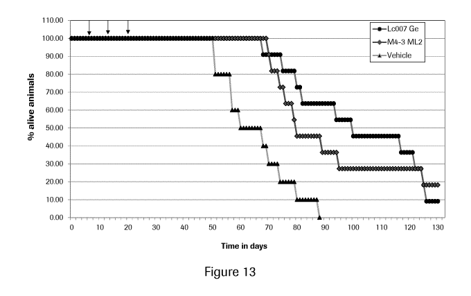
Figure 13 depicts a survival curve showing that both the chimeric glyco-
engineered anti-MCSP
antibody and humanized variant thereof, M4-3 ML2, significantly increase
survival time in FcgR3A transgenic
SCID mice harboring a MDA-MB-435 tumor cell line as compared to the vehicle
control.
5
CA 02844141 2014-02-04
WO 2013/026832
PCT/EP2012/066214
DETAILED DESCRIPTION OF EMBODIMENTS OF THE INVENTION
I. DEFINITIONS
An "acceptor human framework" for the purposes herein is a framework
comprising the amino acid
sequence of a light chain variable domain (VL) framework or a heavy chain
variable domain (VH) framework
derived from a human immunoglobulin framework or a human consensus framework,
as defined below. An
acceptor human framework "derived from" a human immunoglobulin framework or a
human consensus
framework may comprise the same amino acid sequence thereof, or it may contain
amino acid sequence
changes. In some embodiments, the number of amino acid changes are 10 or less,
9 or less, 8 or less, 7 or less,
6 or less, 5 or less, 4 or less, 3 or less, or 2 or less. In some embodiments,
the VL acceptor human framework
is identical in sequence to the VL human immunoglobulin framework sequence or
human consensus framework
sequence.
"Affinity" refers to the strength of the sum total of noncovalent interactions
between a single binding
site of a molecule (e.g., an antibody) and its binding partner (e.g., an
antigen). Unless indicated otherwise, as
used herein, "binding affinity" refers to intrinsic binding affinity which
reflects a 1:1 interaction between
members of a binding pair (e.g., antibody and antigen). The affinity of a
molecule X for its partner Y can
generally be represented by the dissociation constant (Kd). Affinity can be
measured by common methods
known in the art, including those described herein. Specific illustrative and
exemplary embodiments for
measuring binding affinity are described in the following.
An "affinity matured" antibody refers to an antibody with one or more
alterations in one or more
hypervariable regions (HVRs), compared to a parent antibody which does not
possess such alterations, such
alterations resulting in an improvement in the affinity of the antibody for
antigen
An "angiogenic disorder" refers to any dysregulation of angiogenesis,
including both non-neoplastic
and neoplastic conditions. Neoplastic conditions include but are not limited
those described below. Non-
neoplastic disorders include but are not limited to undesired or aberrant
hypertrophy, arthritis, rheumatoid
arthritis (RA), psoriasis, psoriatic plaques, sarcoidosis, atherosclerosis,
atherosclerotic plaques, diabetic and
other proliferative retinopathies including retinopathy of prematurity,
retrolental fibroplasia, neovascular
glaucoma, age-related macular degeneration, diabetic macular edema, corneal
neovascularization, corneal graft
neovascularization, corneal graft rejection, retinal/choroidal
neovascularization, neovascularization of the angle
(rubeosis), ocular neovascular disease, vascular restenosis, arteriovenous
malformations (AVM), meningioma,
hemangioma, angiofibroma, thyroid hyperplasias (including Grave's disease),
corneal and other tissue
transplantation, chronic inflammation, lung inflammation, acute lung
injury/ARDS, sepsis, primary pulmonary
hypertension, malignant pulmonary effusions, cerebral edema (e.g., associated
with acute stroke/ closed head
6
CA 02844141 2014-02-04
WO 2013/026832
PCT/EP2012/066214
injury/ trauma), synovial inflammation, pannus formation in RA, myositis
ossificans, hypertropic bone
formation, osteoarthritis (OA), refractory ascites, polycystic ovarian
disease, endometriosis, 3rd spacing of
fluid diseases (pancreatitis, compartment syndrome, burns, bowel disease),
uterine fibroids, premature labor,
chronic inflammation such as IBD (Crohn's disease and ulcerative colitis),
renal allograft rejection,
inflammatory bowel disease, nephrotic syndrome, undesired or aberrant tissue
mass growth (non-cancer),
hemophilic joints, hypertrophic scars, inhibition of hair growth, Osler-Weber
syndrome, pyogenic granuloma
retrolental fibroplasias, scleroderma, trachoma, vascular adhesions,
synovitis, dermatitis, preeclampsia, ascites,
pericardial effusion (such as that associated with pericarditis), and pleural
effusion.
The terms "anti-MCSP antibody" and "an antibody that binds to MCSP" refer to
an antibody that is
capable of binding MC SP with sufficient affinity such that the antibody is
useful as a diagnostic and/or
therapeutic agent in targeting MCSP. In one embodiment, the extent of binding
of an anti-MCSP antibody to
an unrelated, non-MCSP protein is less than about 10% of the binding of the
antibody to MCSP as measured,
e.g., by a radioimmunoassay (RIA). In certain embodiments, an antibody that
binds to MCSP has a
dissociation constant (Kd) of < 1 M, < 100 nM, < 10 nM, < 1 nM, < 0.1 nM, <
0.01 nM, or < 0.001 nM (e.g.
10-8M or less, e.g. from 10-8M to 10-13M, e.g., from 10-9M to 10-13 M). In
certain embodiments, an anti-
MCSP antibody binds to an epitope of MCSP that is conserved among MCSP from
different species.
The term "antibody" herein is used in the broadest sense and encompasses
various antibody structures,
including but not limited to monoclonal antibodies, polyclonal antibodies,
multispecific antibodies (e.g.,
bispecific antibodies), and antibody fragments so long as they exhibit the
desired antigen-binding activity.
An "antibody fragment" refers to a molecule other than an intact antibody that
comprises a portion of
an intact antibody that binds the antigen to which the intact antibody binds.
Examples of antibody fragments
include but are not limited to Fv, Fab, Fab', Fab'-SH, F(ab1)2; diabodies;
linear antibodies; single-chain
antibody molecules (e.g. scFv); and multispecific antibodies formed from
antibody fragments.
An "antibody that binds to the same epitope" as a reference antibody refers to
an antibody that blocks
binding of the reference antibody to its antigen in a competition assay by 50%
or more, and conversely, the
reference antibody blocks binding of the antibody to its antigen in a
competition assay by 50% or more. An
exemplary competition assay is provided herein.
The terms "cancer" and "cancerous" refer to or describe the physiological
condition in mammals that is
typically characterized by unregulated cell growth/proliferation. Examples of
cancer include, but are not
limited to, carcinoma, lymphoma (e.g., Hodgkin's and non-Hodgkin's lymphoma),
blastoma, sarcoma, and
leukemia. More particular examples of such cancers include squamous cell
cancer, small-cell lung cancer, non-
small cell lung cancer, adenocarcinoma of the lung, squamous carcinoma of the
lung, cancer of the peritoneum,
7
CA 02844141 2014-02-04
WO 2013/026832
PCT/EP2012/066214
hepatocellular cancer, cancer of the bone (e.g. osteosarcomas, chondrosarcoma,
Ewing's sarcoma),
gastrointestinal cancer, pancreatic cancer, glioma, cervical cancer, ovarian
cancer, liver cancer, bladder cancer,
hepatoma, breast cancer, colon cancer, colorectal cancer, endometrial or
uterine carcinoma, salivary gland
carcinoma, kidney cancer, liver cancer, prostate cancer, skin cancer (e.g.
melanoma and basel cell carcinoma),
vulval cancer, thyroid cancer, hepatic carcinoma, leukemia and other
lymphoproliferative disorders, and
various types of head and neck cancer.
The terms "cell proliferative disorder" and "proliferative disorder" refer to
disorders that are associated
with some degree of abnormal cell proliferation. In one embodiment, the cell
proliferative disorder is cancer.
The term "chimeric" antibody refers to an antibody in which a portion of the
heavy and/or light chain is
derived from a particular source or species, while the remainder of the heavy
and/or light chain is derived from
a different source or species.
The "class" of an antibody refers to the type of constant domain or constant
region possessed by its
heavy chain. There are five major classes of antibodies: IgA, IgD, IgE, IgG,
and IgM, and several of these may
be further divided into subclasses (isotypes), e.g., IgGi, IgG2, IgG3, IgG4,
IgAi, and IgA2. The heavy chain
constant domains that correspond to the different classes of immunoglobulins
are called a, 6, E, 7, and n,
respectively.
The term "cytotoxic agent" as used herein refers to a substance that inhibits
or prevents a cellular
function and/or causes cell death or destruction. Cytotoxic agents include,
but are not limited to, radioactive
isotopes (e.g., At211, 1131, 1125, y90, Re186, Re188, sm153, Bi212, 1332, p+0
212
and radioactive isotopes of Lu);
chemotherapeutic agents or drugs (e.g., methotrexate, adriamicin, vinca
alkaloids (vincristine, vinblastine,
etoposide), doxorubicin, melphalan, mitomycin C, chlorambucil, daunorubicin or
other intercalating agents);
growth inhibitory agents; enzymes and fragments thereof such as nucleolytic
enzymes; antibiotics; toxins such
as small molecule toxins or enzymatically active toxins of bacterial, fungal,
plant or animal origin, including
fragments and/or variants thereof; and the various antitumor or anticancer
agents disclosed below.
"Effector functions" refer to those biological activities attributable to the
Fc region of an antibody,
which vary with the antibody isotype. Examples of antibody effector functions
include: Clq binding and
complement dependent cytotoxicity (CDC); Fc receptor binding; antibody-
dependent cell-mediated cytotoxicity
(ADCC); phagocytosis; down regulation of cell surface receptors (e.g. B cell
receptor); and B cell activation.
An "effective amount" of an agent, e.g., a pharmaceutical formulation, refers
to an amount effective, at
dosages and for periods of time necessary, to achieve the desired therapeutic
or prophylactic result.
The term "Fc region" herein is used to define a C-terminal region of an
immunoglobulin heavy chain
that contains at least a portion of the constant region. The term includes
native sequence Fc regions and variant
8
CA 02844141 2014-02-04
WO 2013/026832
PCT/EP2012/066214
Fc regions. In one embodiment, a human IgG heavy chain Fc region extends from
Cys226, or from Pro230, to
the carboxyl-terminus of the heavy chain. However, the C-terminal lysine
(Lys447) of the Fc region may or
may not be present. Unless otherwise specified herein, numbering of amino acid
residues in the Fc region or
constant region is according to the EU numbering system, also called the EU
index, as described in Kabat et al.,
Sequences of Proteins of Immunological Interest, 5th Ed. Public Health
Service, National Institutes of Health,
Bethesda, MD, 1991.
"Framework" or "FR" refers to variable domain residues other than
hypervariable region (HVR)
residues. The FR of a variable domain generally consists of four FR domains:
FR1, FR2, FR3, and FR4.
Accordingly, the HVR and FR sequences generally appear in the following
sequence in VH (or VL): FR1-
Hl(L1)-FR2-H2(L2)-FR3-H3(L3)-FR4.
The terms "full length antibody," "intact antibody," and "whole antibody" are
used herein
interchangeably to refer to an antibody having a structure substantially
similar to a native antibody structure or
having heavy chains that contain an Fc region as defined herein.
The terms "host cell," "host cell line," and "host cell culture" are used
interchangeably and refer to cells
into which exogenous nucleic acid has been introduced, including the progeny
of such cells. Host cells include
"transformants" and "transformed cells," which include the primary transformed
cell and progeny derived
therefrom without regard to the number of passages. Progeny may not be
completely identical in nucleic acid
content to a parent cell, but may contain mutations. Mutant progeny that have
the same function or biological
activity as screened or selected for in the originally transformed cell are
included herein.
A "human antibody" is one which possesses an amino acid sequence which
corresponds to that of an
antibody produced by a human or a human cell or derived from a non-human
source that utilizes human
antibody repertoires or other human antibody-encoding sequences. This
definition of a human antibody
specifically excludes a humanized antibody comprising non-human antigen-
binding residues.
A "human consensus framework" is a framework which represents the most
commonly occurring
amino acid residues in a selection of human immunoglobulin VL or VH framework
sequences. Generally, the
selection of human immunoglobulin VL or VH sequences is from a subgroup of
variable domain sequences.
Generally, the subgroup of sequences is a subgroup as in Kabat et al.,
Sequences of Proteins of Immunological
Interest, Fifth Edition, NIH Publication 91-3242, Bethesda MD (1991), vols. 1-
3. In one embodiment, for the
VL, the subgroup is subgroup kappa I as in Kabat et al., supra. In one
embodiment, for the VH, the subgroup
is subgroup III as in Kabat et al., supra.
A "humanized" antibody refers to a chimeric antibody comprising amino acid
residues from non-
human HVRs and amino acid residues from human FRs. In certain embodiments, a
humanized antibody will
9
CA 02844141 2014-02-04
WO 2013/026832
PCT/EP2012/066214
comprise substantially all of at least one, and typically two, variable
domains, in which all or substantially all
of the HVRs (e.g., CDRs) correspond to those of a non-human antibody, and all
or substantially all of the FRs
correspond to those of a human antibody. A humanized antibody optionally may
comprise at least a portion of
an antibody constant region derived from a human antibody. A "humanized form"
of an antibody, e.g., a non-
The term "hypervariable region" or "HVR," as used herein, refers to each of
the regions of an antibody
variable domain which are hypervariable in sequence and/or form structurally
defined loops ("hypervariable
loops"). Generally, native four-chain antibodies comprise six HVRs; three in
the VH (H1, H2, H3), and three
in the VL (L1, L2, L3). HVRs generally comprise amino acid residues from the
hypervariable loops and/or
An "immunoconjugate" is an antibody conjugated to one or more heterologous
molecule(s), including
An "individual" or "subject" is a mammal. Mammals include, but are not limited
to, domesticated
animals (e.g., cows, sheep, cats, dogs, and horses), primates (e.g., humans
and non-human primates such as
monkeys), rabbits, and rodents (e.g., mice and rats). In certain embodiments,
the individual or subject is a
human.
30 An "isolated" antibody is one which has been separated from a component
of its natural environment.
In some embodiments, an antibody is purified to greater than 95% or 99% purity
as determined by, for
example, electrophoretic (e.g., SDS-PAGE, isoelectric focusing (IEF),
capillary electrophoresis) or
CA 02844141 2014-02-04
WO 2013/026832
PCT/EP2012/066214
chromatographic (e.g., ion exchange or reverse phase HPLC). For review of
methods for assessment of
antibody purity, see, e.g., Flatman et al., I Chromatogr. B 848:79-87 (2007).
An "isolated" nucleic acid refers to a nucleic acid molecule that has been
separated from a component
of its natural environment. An isolated nucleic acid includes a nucleic acid
molecule contained in cells that
ordinarily contain the nucleic acid molecule, but the nucleic acid molecule is
present extrachromosomally or at
a chromosomal location that is different from its natural chromosomal
location.
"Isolated nucleic acid encoding an anti-MCSP antibody" refers to one or more
nucleic acid molecules
encoding antibody heavy and light chains (or fragments thereof), including
such nucleic acid molecule(s) in a
single vector or separate vectors, and such nucleic acid molecule(s) present
at one or more locations in a host
cell.
The term "monoclonal antibody" as used herein refers to an antibody obtained
from a population of
substantially homogeneous antibodies, i.e., the individual antibodies
comprising the population are identical
and/or bind the same epitope, except for possible variant antibodies, e.g.,
containing naturally occurring
mutations or arising during production of a monoclonal antibody preparation,
such variants generally being
present in minor amounts. In contrast to polyclonal antibody preparations,
which typically include different
antibodies directed against different determinants (epitopes), each monoclonal
antibody of a monoclonal
antibody preparation is directed against a single determinant on an antigen.
Thus, the modifier "monoclonal"
indicates the character of the antibody as being obtained from a substantially
homogeneous population of
antibodies, and is not to be construed as requiring production of the antibody
by any particular method. For
example, the monoclonal antibodies to be used in accordance with the present
invention may be made by a
variety of techniques, including but not limited to the hybridoma method,
recombinant DNA methods, phage-
display methods, and methods utilizing transgenic animals containing all or
part of the human immunoglobulin
loci, such methods and other exemplary methods for making monoclonal
antibodies being described herein.
A "naked antibody" refers to an antibody that is not conjugated to a
heterologous moiety (e.g., a
cytotoxic moiety) or radiolabel. The naked antibody may be present in a
pharmaceutical formulation.
"Native antibodies" refer to naturally occurring immunoglobulin molecules with
varying structures.
For example, native IgG antibodies are heterotetrameric glycoproteins of about
150,000 daltons, composed of
two identical light chains and two identical heavy chains that are disulfide-
bonded. From N- to C-terminus,
each heavy chain has a variable region (VH), also called a variable heavy
domain or a heavy chain variable
domain, followed by three constant domains (CHL CH2, and CH3). Similarly, from
N- to C-terminus, each
light chain has a variable region (VL), also called a variable light domain or
a light chain variable domain,
11
CA 02844141 2014-02-04
WO 2013/026832
PCT/EP2012/066214
followed by a constant light (CL) domain. The light chain of an antibody may
be assigned to one of two types,
called kappa (ic) and lambda (X), based on the amino acid sequence of its
constant domain.
The term "package insert" is used to refer to instructions customarily
included in commercial packages
of therapeutic products, that contain information about the indications,
usage, dosage, administration,
combination therapy, contraindications and/or warnings concerning the use of
such therapeutic products.
"Percent (%) amino acid sequence identity" with respect to a reference
polypeptide sequence is defined
as the percentage of amino acid residues in a candidate sequence that are
identical with the amino acid residues
in the reference polypeptide sequence, after aligning the sequences and
introducing gaps, if necessary, to
achieve the maximum percent sequence identity, and not considering any
conservative substitutions as part of
the sequence identity. Alignment for purposes of determining percent amino
acid sequence identity can be
achieved in various ways that are within the skill in the art, for instance,
using publicly available computer
software such as BLAST, BLAST-2, ALIGN or Megalign (DNASTAR) software. Those
skilled in the art can
determine appropriate parameters for aligning sequences, including any
algorithms needed to achieve maximal
alignment over the full length of the sequences being compared. For purposes
herein, however, % amino acid
sequence identity values are generated using the sequence comparison computer
program ALIGN-2. The
ALIGN-2 sequence comparison computer program was authored by Genentech, Inc.,
and the source code has
been filed with user documentation in the U.S. Copyright Office, Washington
D.C., 20559, where it is
registered under U.S. Copyright Registration No. TXU510087. The ALIGN-2
program is publicly available
from Genentech, Inc., South San Francisco, California, or may be compiled from
the source code. The
ALIGN-2 program should be compiled for use on a UNIX operating system,
including digital UNIX V4.0D.
All sequence comparison parameters are set by the ALIGN-2 program and do not
vary.
In situations where ALIGN-2 is employed for amino acid sequence comparisons,
the % amino acid
sequence identity of a given amino acid sequence A to, with, or against a
given amino acid sequence B (which
can alternatively be phrased as a given amino acid sequence A that has or
comprises a certain % amino acid
sequence identity to, with, or against a given amino acid sequence B) is
calculated as follows:
100 times the fraction X/Y
where X is the number of amino acid residues scored as identical matches by
the sequence alignment program
ALIGN-2 in that program's alignment of A and B, and where Y is the total
number of amino acid residues in
B. It will be appreciated that where the length of amino acid sequence A is
not equal to the length of amino
acid sequence B, the % amino acid sequence identity of A to B will not equal
the % amino acid sequence
12
CA 02844141 2014-02-04
WO 2013/026832
PCT/EP2012/066214
identity of B to A. Unless specifically stated otherwise, all % amino acid
sequence identity values used herein
are obtained as described in the immediately preceding paragraph using the
ALIGN-2 computer program.
The term "pharmaceutical formulation" refers to a preparation which is in such
form as to permit the
biological activity of an active ingredient contained therein to be effective,
and which contains no additional
components which are unacceptably toxic to a subject to which the formulation
would be administered.
A "pharmaceutically acceptable carrier" refers to an ingredient in a
pharmaceutical formulation, other
than an active ingredient, which is nontoxic to a subject., A pharmaceutically
acceptable carrier includes, but
is not limited to, a buffer, excipient, stabilizer, or preservative.
The term "MCSP," as used herein, refers to any native MCSP (Melanoma
Chondroitin Sulfate
Proteoglycan) from any vertebrate source, including mammals such as primates
(e.g. humans) and rodents
(e.g., mice and rats), unless otherwise indicated. The term encompasses "full-
length," unprocessed MCSP as
well as any form of MCSP that results from processing in the cell. The term
also encompasses naturally
occurring variants of MCSP, e.g., splice variants or allelic variants. MCSP is
also known as chondroitin sulfate
proteoglycan 4 (CSPG4), chondroitin sulfate proteoglycan NG2, high molecular
weight-melanoma associated
antigen (HMW-MAA), and melanoma chondroitin sulfate proteoglycan. The amino
acid sequence of an
exemplary human MCSP is shown in SEQ ID NO: 1. See also Pluschke G., et al.,
Molecular cloning of a
human melanoma-associated chondroitin sulfate proteoglycan, Proc. Natl. Acad.
Sci. U.S.A. 93:9710-
9715(1996), Staub E., et al., A novel repeat in the melanoma-associated
chondroitin sulfate proteoglycan
defines a new protein family, FEB S Left. 527:114-118(2002); Genbank
AccessionNo: NP 001888.
As used herein, "treatment" (and grammatical variations thereof such as
"treat" or "treating") refers to
clinical intervention in an attempt to alter the natural course of the
individual being treated, and can be
performed either for prophylaxis or during the course of clinical pathology.
Desirable effects of treatment
include, but are not limited to, preventing occurrence or recurrence of
disease, alleviation of symptoms,
diminishment of any direct or indirect pathological consequences of the
disease, preventing metastasis,
decreasing the rate of disease progression, amelioration or palliation of the
disease state, and remission or
improved prognosis. In some embodiments, antibodies of the invention are used
to delay development of a
disease or to slow the progression of a disease.
The term "variable region" or "variable domain" refers to the domain of an
antibody heavy or light
chain that is involved in binding the antibody to antigen. The variable
domains of the heavy chain and light
chain (VH and VL, respectively) of a native antibody generally have similar
structures, with each domain
comprising four conserved framework regions (FRs) and three hypervariable
regions (HVRs). (See, e.g., Kindt
et al. Kuby Immunology, 6th ed., W.H. Freeman and Co., page 91 (2007).) A
single VH or VL domain may be
13
CA 02844141 2014-02-04
WO 2013/026832
PCT/EP2012/066214
sufficient to confer antigen-binding specificity. Furthermore, antibodies that
bind a particular antigen may be
isolated using a VH or VL domain from an antibody that binds the antigen to
screen a library of complementary
VL or VH domains, respectively. See, e.g., Portolano et al., I Immunol.
150:880-887 (1993); Clarkson et al.,
Nature 352:624-628 (1991).
The term "vector," as used herein, refers to a nucleic acid molecule capable
of propagating another
nucleic acid to which it is linked. The term includes the vector as a self-
replicating nucleic acid structure as
well as the vector incorporated into the genome of a host cell into which it
has been introduced. Certain vectors
are capable of directing the expression of nucleic acids to which they are
operatively linked. Such vectors are
referred to herein as "expression vectors."
II. COMPOSITIONS AND METHODS
The invention provides anti-MCSP antibodies that find use in treating and/or
diagnosing cell
proliferative diseases, such as cancer. In certain embodiments, antibodies
that bind to the membrane proximal
epitope of MCSP are provided. In certain embodiments, antibodies with enhanced
effector function that bind to
MCSP are provided.
A. Exemplary Anti-MCSP Antibodies
In one aspect, the invention provides isolated antibodies that bind to MCSP.
In particular, the anti-
MCSP antibodies provided for in the invention bind to a membrane proximal
epitope of human MCSP. As
discussed in Staub E., et al., FEBS Left. 527:114-118(2002), the membrane
proximal region of MCSP is
comprised of multiple novel repeated domains, referred to as CSPG repeat
domains. Figure 3. The anti-MCSP
antibodies of the invention bind to an epitope present in the membrane
proximal domain of human MCSP
comprising a CSPG repeat-containing domain. In one embodiment, the CSPG repeat-
containing domain
comprises CSPG repeat 14, which corresponds to amino acids amino acids 1937-
2043 of human MCSP. In
one embodiment, the CSPG repeat 14 domain has the amino acid sequence shown in
SEQ ID NO: 3. In
another embodiment, the CSPG repeat-containing domain comprises CSPG repeat 14
and at least a portion of
CSPG repeat 15. The CSPG repeat 15 domain corresponds to amino acids 2044-2246
of human MCSP. In
one embodiment, the CSPG repeat-15 domain has the amino acid sequence of SEQ
ID NO: 4. In one
embodiment, the CSPG repeat-containing domain comprises the amino acid
sequence of SEQ ID NO: 5. In
one embodiment, the CSPG repeat-containing domain comprises the amino acid
sequence of SEQ ID NO: 5
without the native transmembrane domain. In one embodiment, the CSPG repeat-
containing domain comprises
CSPG repeat 13-15. In one embodiment, the CSPG repeat-containing domain
comprises the amino acid
sequence of SEQ ID NO: 6. In one embodiment, the CSPG repeat-containing domain
comprises the amino acid
14
CA 02844141 2014-02-04
WO 2013/026832
PCT/EP2012/066214
sequence of SEQ ID NO: 6 without the native transmembrane domain. In one
embodiment, the CSPG repeat-
containing domain comprises CSPG repeat 12-15. In one embodiment, the CSPG
repeat-containing domain
comprises the amino acid sequence of SEQ ID NO: 7. In one embodiment, the CSPG
repeat-containing domain
comprises the amino acid sequence of SEQ ID NO: 7 without the native
transmembrane domain. In certain
embodiments, the native transmembrane domain is VIIPMC LVLLLLALIL PLLFY
(UniProt entry Q6UVK1)
(SEQ ID NO: 44).
In one embodiment, the anti-MCSP antibodies induce lysis of cells expressing
MCSP. Lysis can be
induced by any mechanism, such as by mediating an effector function, such as
Clq binding and complement
dependent cytotoxicity (CDC); Fc receptor binding; antibody-dependent cell-
mediated cytotoxicity (ADCC);
phagocytosis; down regulation of cell surface receptors (e.g. B cell
receptor); and B cell activation, or by
directly inducing apoptosis of the cells.
In one embodiment, the anti-MCSP antibody is glycoengineered to have at least
one increase in effector
function as compared to the non-glycoengineered parent anti-MCSP antibody. The
increase in effector function
is increased binding affinity is to an Fc receptor, increased antibody-
dependent cellular cytotoxicity (ADCC);
increased binding to NK cells; increased binding to macrophages; increased
binding to polymorphonuclear
cells; increased binding to monocytes; direct signaling inducing apoptosis;
increased dendritic cell maturation;
or increased T cell priming. The glycoengineered anti-MCSP antibodies provide
a survival benefit in subjects
suffering from cancers which express MCSP as compared to non-glycoengineered
antibodies directed to the
same epitope of MCSP.
In one aspect, the invention provides an anti-MCSP antibody comprising at
least one, two, three, four,
five, or six HVRs selected from (a) HVR-H1 comprising the amino acid sequence
of SEQ ID NO: 14; (b)
HVR-H2 comprising the amino acid sequence of SEQ ID NO: 15; (c) HVR-H3
comprising the amino acid
sequence of SEQ ID NO: 16; (d) HVR-L1 comprising the amino acid sequence of
SEQ ID NO: 10; (e) HVR-
L2 comprising the amino acid sequence of SEQ ID NO: 11; and (f) HVR-L3
comprising the amino acid
sequence of SEQ ID NO: 12.
In one aspect, the invention provides an anti-MCSP antibody comprising at
least one, at least two, or
all three VH HVR sequences selected from (a) HVR-H1 comprising the amino acid
sequence of SEQ ID NO:
14; (b) HVR-H2 comprising the amino acid sequence of SEQ ID NO: 15; and (c)
HVR-H3 comprising the
amino acid sequence of SEQ ID NO: 16. In a further embodiment, the antibody
comprises (a) HVR-H1
comprising the amino acid sequence of SEQ ID NO: 14; (b) HVR-H2 comprising the
amino acid sequence of
SEQ ID NO: 15; and (c) HVR-H3 comprising the amino acid sequence of SEQ ID NO:
16.
CA 02844141 2014-02-04
WO 2013/026832
PCT/EP2012/066214
In one aspect, the invention provides an anti-MCSP antibody comprising at
least one, at least two, or
all three VL HVR sequences selected from (a) HVR-L1 comprising the amino acid
sequence of SEQ ID NO:
10; (b) HVR-L2 comprising the amino acid sequence of SEQ ID NO: 11 and (c) HVR-
L3 comprising the
amino acid sequence of SEQ ID NO: 12. In one embodiment, the antibody
comprises (a) HVR-L1 comprising
the amino acid sequence of SEQ ID NO: 10; (b) HVR-L2 comprising the amino acid
sequence of SEQ ID NO:
11; and (c) HVR-L3 comprising the amino acid sequence of SEQ ID NO: 12.
In another aspect, an anti-MCSP antibody of the invention comprises (a) a VH
domain comprising at
least one, at least two, or all three VH HVR sequences selected from (i) HVR-
H1 comprising the amino acid
sequence of SEQ ID NO: 14, (ii) HVR-H2 comprising the amino acid sequence of
SEQ ID NO: 15, and (iii)
HVR-H3 comprising an amino acid sequence selected from SEQ ID NO: 16; and (b)
a VL domain comprising
at least one, at least two, or all three VL HVR sequences selected from (i)
HVR-L1 comprising the amino acid
sequence of SEQ ID NO: 10, (ii) HVR-L2 comprising the amino acid sequence of
SEQ ID NO: 11, and (c)
HVR-L3 comprising the amino acid sequence of SEQ ID NO: 12.
In another aspect, the invention provides an anti-MCSP antibody comprising (a)
HVR-H1 comprising
the amino acid sequence of SEQ ID NO: 14; (b) HVR-H2 comprising the amino acid
sequence of SEQ ID NO:
15; (c) HVR-H3 comprising the amino acid sequence of SEQ ID NO: 16; (d) HVR-L1
comprising the amino
acid sequence of SEQ ID NO: 10; (e) HVR-L2 comprising the amino acid sequence
of SEQ ID NO: 11; and (f)
HVR-L3 comprising an amino acid sequence selected from SEQ ID NO: 12.
In one aspect, the invention provides an anti-MCSP antibody comprising at
least one, two, three, four,
five, or six HVRs selected from (a) HVR-H1 comprising the amino acid sequence
of SEQ ID NO: 17; (b)
HVR-H2 comprising the amino acid sequence of SEQ ID NO: 18; (c) HVR-H3
comprising the amino acid
sequence of SEQ ID NO: 16; (d) HVR-L1 comprising the amino acid sequence of
SEQ ID NO: 13; (e) HVR-
L2 comprising the amino acid sequence of SEQ ID NO: 11; and (f) HVR-L3
comprising the amino acid
sequence of SEQ ID NO: 12.
In one aspect, the invention provides an anti-MCSP antibody comprising at
least one, at least two, or
all three VH HVR sequences selected from (a) HVR-H1 comprising the amino acid
sequence of SEQ ID NO:
17; (b) HVR-H2 comprising the amino acid sequence of SEQ ID NO: 18; and (c)
HVR-H3 comprising the
amino acid sequence of SEQ ID NO: 16. In a further embodiment, the antibody
comprises (a) HVR-H1
comprising the amino acid sequence of SEQ ID NO: 17; (b) HVR-H2 comprising the
amino acid sequence of
SEQ ID NO: 18; and (c) HVR-H3 comprising the amino acid sequence of SEQ ID NO:
16.
In one aspect, the invention provides an anti-MCSP antibody comprising at
least one, at least two, or
all three VL HVR sequences selected from (a) HVR-L1 comprising the amino acid
sequence of SEQ ID NO:
16
CA 02844141 2014-02-04
WO 2013/026832
PCT/EP2012/066214
13; (b) HVR-L2 comprising the amino acid sequence of SEQ ID NO: 11; and (c)
HVR-L3 comprising the
amino acid sequence of SEQ ID NO: 12. In one embodiment, the antibody
comprises (a) HVR-L1 comprising
the amino acid sequence of SEQ ID NO: 13; (b) HVR-L2 comprising the amino acid
sequence of SEQ ID NO:
11; and (c) HVR-L3 comprising the amino acid sequence of SEQ ID NO: 12.
In another aspect, an anti-MCSP antibody of the invention comprises (a) a VH
domain comprising at
least one, at least two, or all three VH HVR sequences selected from (i) HVR-
H1 comprising the amino acid
sequence of SEQ ID NO: 17, (ii) HVR-H2 comprising the amino acid sequence of
SEQ ID NO: 18, and (iii)
HVR-H3 comprising an amino acid sequence selected from SEQ ID NO: 16; and (b)
a VL domain comprising
at least one, at least two, or all three VL HVR sequences selected from (i)
HVR-L1 comprising the amino acid
sequence of SEQ ID NO: 13, (ii) HVR-L2 comprising the amino acid sequence of
SEQ ID NO: 11, and (c)
HVR-L3 comprising the amino acid sequence of SEQ ID NO: 12.
In another aspect, the invention provides an anti-MCSP antibody comprising (a)
HVR-H1 comprising
the amino acid sequence of SEQ ID NO: 17; (b) HVR-H2 comprising the amino acid
sequence of SEQ ID NO:
18; (c) HVR-H3 comprising the amino acid sequence of SEQ ID NO: 16; (d) HVR-L1
comprising the amino
acid sequence of SEQ ID NO: 13; (e) HVR-L2 comprising the amino acid sequence
of SEQ ID NO: 11; and (f)
HVR-L3 comprising an amino acid sequence selected from SEQ ID NO: 12.
In another aspect, the invention provides an anti-MCSP antibody comprising (a)
HVR-H1 comprising
the amino acid sequence of SEQ ID NO: 17; (b) HVR-H2 comprising the amino acid
sequence of SEQ ID NO:
18; (c) HVR-H3 comprising the amino acid sequence of SEQ ID NO: 16; (d) HVR-L1
comprising the amino
acid sequence of SEQ ID NO: 10; (e) HVR-L2 comprising the amino acid sequence
of SEQ ID NO: 11; and (f)
HVR-L3 comprising an amino acid sequence selected from SEQ ID NO: 12.
In another aspect, the invention provides an anti-MCSP antibody comprising (a)
HVR-H1 comprising
the amino acid sequence of SEQ ID NO: 14; (b) HVR-H2 comprising the amino acid
sequence of SEQ ID NO:
18; (c) HVR-H3 comprising the amino acid sequence of SEQ ID NO: 16; (d) HVR-L1
comprising the amino
acid sequence of SEQ ID NO: 13; (e) HVR-L2 comprising the amino acid sequence
of SEQ ID NO: 11; and (f)
HVR-L3 comprising an amino acid sequence selected from SEQ ID NO: 12.
17
CA 02844141 2014-02-04
WO 2013/026832
PCT/EP2012/066214
In one aspect, an anti-MCSP antibody comprises a VH sequence having at least
90%, 91%, 92%,
93%, 94%, 95%, 96%, 97%, 98%, 99%, or 100% sequence identity to the amino acid
sequence of SEQ ID
NO: 27. In certain embodiments, a VH sequence having at least 90%, 91%, 92%,
93%, 94%, 95%, 96%,
97%, 98%, or 99% identity contains substitutions (e.g., conservative
substitutions), insertions, or deletions
relative to the reference sequence, but an anti-MCSP antibody comprising that
sequence retains the ability to
bind to MCSP. In certain embodiments, a total of 1 to 10 amino acids have been
substituted, inserted and/or
deleted in SEQ ID NO: 27. In certain embodiments, substitutions, insertions,
or deletions occur in regions
outside the HVRs (i.e., in the FRs). Optionally, the anti-MCSP antibody
comprises the VH sequence of SEQ
ID NO: 27, including post-translational modifications of that sequence. In a
particular embodiment, the VH
comprises one, two or three HVRs selected from: (a) HVR-Hl comprising the
amino acid sequence of SEQ ID
NO: 14, (b) HVR-H2 comprising the amino acid sequence of SEQ ID NO: 15, and
(c) HVR-H3 comprising the
amino acid sequence of SEQ ID NO: 16.
In another aspect, an anti-MCSP antibody is provided, wherein the antibody
comprises a light chain
variable domain (VL) having at least 90%, 91%, 92%, 93%, 94%, 95%, 96%, 97%,
98%, 99%, or 100%
sequence identity to the amino acid sequence of SEQ ID NO: 26. In certain
embodiments, a VL sequence
having at least 90%, 91%, 92%, 93%, 94%, 95%, 96%, 97%, 98%, or 99% identity
contains substitutions
(e.g., conservative substitutions), insertions, or deletions relative to the
reference sequence, but an anti-MCSP
antibody comprising that sequence retains the ability to bind to MCSP. In
certain embodiments, a total of 1 to
10 amino acids have been substituted, inserted and/or deleted in SEQ ID NO:
26. In certain embodiments, the
substitutions, insertions, or deletions occur in regions outside the HVRs
(i.e., in the FRs). Optionally, the anti-
MCSP antibody comprises the VL sequence of SEQ ID NO: 26, including post-
translational modifications of
that sequence. In a particular embodiment, the VL comprises one, two or three
HVRs selected from (a) HVR-
Ll comprising the amino acid sequence of SEQ ID NO: 10; (b) HVR-L2 comprising
the amino acid sequence
of SEQ ID NO: 11; and (c) HVR-L3 comprising the amino acid sequence of SEQ ID
NO: 12.
In another aspect, an anti-MCSP antibody is provided, wherein the antibody
comprises a VH as in any
of the embodiments provided above, and a VL as in any of the embodiments
provided above. In one
embodiment, the antibody comprises a VH comprising the amino acid sequence of
SEQ ID NO: 27 and a VL
sequence in SEQ ID NO: 26, including post-translational modifications of those
sequences.
In another aspect, an anti-MCSP antibody comprises a VH sequence having at
least 90%, 91%, 92%,
93%, 94%, 95%, 96%, 97%, 98%, 99%, or 100% sequence identity to the amino acid
sequence of SEQ ID
NO: 32. In certain embodiments, a VH sequence having at least 90%, 91%, 92%,
93%, 94%, 95%, 96%,
97%, 98%, or 99% identity contains substitutions (e.g., conservative
substitutions), insertions, or deletions
18
CA 02844141 2014-02-04
WO 2013/026832
PCT/EP2012/066214
relative to the reference sequence, but an anti-MCSP antibody comprising that
sequence retains the ability to
bind to MCSP. In certain embodiments, a total of 1 to 10 amino acids have been
substituted, inserted and/or
deleted in SEQ ID NO: 32. In certain embodiments, substitutions, insertions,
or deletions occur in regions
outside the HVRs (i.e., in the FRs). Optionally, the anti-MCSP antibody
comprises the VH sequence of SEQ
ID NO: 32, including post-translational modifications of that sequence. In a
particular embodiment, the VH
comprises one, two or three HVRs selected from: (a) HVR-Hl comprising the
amino acid sequence of SEQ ID
NO: 17, (b) HVR-H2 comprising the amino acid sequence of SEQ ID NO: 18, and
(c) HVR-H3 comprising the
amino acid sequence of SEQ ID NO: 16.
In another aspect, an anti-MCSP antibody comprises a VL sequence having at
least 90%, 91%, 92%,
93%, 94%, 95%, 96%, 97%, 98%, 99%, or 100% sequence identity to the amino acid
sequence of SEQ ID
NO: 31 In certain embodiments, a VL sequence having at least 90%, 91%, 92%,
93%, 94%, 95%, 96%, 97%,
98%, or 99% identity contains substitutions (e.g., conservative
substitutions), insertions, or deletions relative to
the reference sequence, but an anti-MCSP antibody comprising that sequence
retains the ability to bind to
MCSP. In certain embodiments, a total of 1 to 10 amino acids have been
substituted, inserted and/or deleted in
SEQ ID NO: 31. In certain embodiments, substitutions, insertions, or deletions
occur in regions outside the
HVRs (i.e., in the FRs). Optionally, the anti-MCSP antibody comprises the VL
sequence in SEQ ID NO: 31,
including post-translational modifications of that sequence. In a particular
embodiment, the VL comprises one,
two or three HVRs selected from: (a) HVR-Li comprising the amino acid sequence
of SEQ ID NO: 13, (b)
HVR-L2 comprising the amino acid sequence of SEQ ID NO: 11, and (c) HVR-L3
comprising the amino acid
sequence of SEQ ID NO: 12.
In another aspect, an anti-MCSP antibody is provided, wherein the antibody
comprises a VH as in any
of the embodiments provided above, and a VL as in any of the embodiments
provided above. In one
embodiment, the antibody comprises the VH comprising the amino acid sequence
of SEQ ID NO: 32 and a VL
comprising the amino acid sequence of SEQ ID NO: 31, including post-
translational modifications of those
sequences.
In another aspect, an anti-MCSP antibody comprises a VH sequence having at
least 90%, 91%, 92%,
93%, 94%, 95%, 96%, 97%, 98%, 99%, or 100% sequence identity to the amino acid
sequence of SEQ ID
NO: 29. In certain embodiments, a VH sequence having at least 90%, 91%, 92%,
93%, 94%, 95%, 96%,
97%, 98%, or 99% identity contains substitutions (e.g., conservative
substitutions), insertions, or deletions
relative to the reference sequence, but an anti-MCSP antibody comprising that
sequence retains the ability to
bind to MCSP. In certain embodiments, a total of 1 to 10 amino acids have been
substituted, inserted and/or
deleted in SEQ ID NO: 29. In certain embodiments, substitutions, insertions,
or deletions occur in regions
19
CA 02844141 2014-02-04
WO 2013/026832
PCT/EP2012/066214
outside the HVRs (i.e., in the FRs). Optionally, the anti-MCSP antibody
comprises the VH sequence of SEQ
ID NO: 29, including post-translational modifications of that sequence. In a
particular embodiment, the VH
comprises one, two or three HVRs selected from: (a) HVR-H1 comprising the
amino acid sequence of SEQ ID
NO: 14, (b) HVR-H2 comprising the amino acid sequence of SEQ ID NO: 18, and
(c) HVR-H3 comprising the
In another aspect, an anti-MCSP antibody comprises a VL sequence having at
least 90%, 91%, 92%,
93%, 94%, 95%, 96%, 97%, 98%, 99%, or 100% sequence identity to the amino acid
sequence of SEQ ID
NO: 28. In certain embodiments, a VL sequence having at least 90%, 91%, 92%,
93%, 94%, 95%, 96%, 97%,
98%, or 99% identity contains substitutions (e.g., conservative
substitutions), insertions, or deletions relative to
In another aspect, an anti-MCSP antibody is provided, wherein the antibody
comprises a VH as in any
of the embodiments provided above, and a VL as in any of the embodiments
provided above. In one
comprising the amino acid sequence of SEQ ID NO: 28, including post-
translational modifications of those
sequences.
In another aspect, an anti-MCSP antibody comprises a heavy chain sequence
having at least 90%,
91%, 92%, 93%, 94%, 95%, 96%, 97%, 98%, 99%, or 100% sequence identity to the
amino acid sequence of
CA 02844141 2014-02-04
WO 2013/026832
PCT/EP2012/066214
In another aspect, an anti-MCSP antibody is provided, wherein the antibody
comprises a light chain
having at least 90%, 91%, 92%, 93%, 94%, 95%, 96%, 97%, 98%, 99%, or 100%
sequence identity to the
amino acid sequence of SEQ ID NO: 34. In certain embodiments, a light chain
sequence having at least 90%,
91%, 92%, 93%, 94%, 95%, 96%, 97%, 98%, or 99% identity contains substitutions
(e.g., conservative
substitutions), insertions, or deletions relative to the reference sequence,
but an anti-MCSP antibody comprising
that sequence retains the ability to bind to MCSP. In certain embodiments, a
total of 1 to 10 amino acids have
been substituted, inserted and/or deleted in SEQ ID NO: 34. In certain
embodiments, the substitutions,
insertions, or deletions occur in regions outside the HVRs (i.e., in the FRs).
Optionally, the anti-MCSP
antibody comprises the light chain sequence of SEQ ID NO: 34, including post-
translational modifications of
that sequence.
In another aspect, an anti-MCSP antibody is provided, wherein the antibody
comprises a heavy chain
as in any of the embodiments provided above, and a light chain in any of the
embodiments provided above. In
one embodiment, the antibody comprises a heavy chain comprising the amino acid
sequence of SEQ ID NO:
35 and a light chain sequence comprising the amino acide sequence of SEQ ID
NO: 34, including post-
translational modifications of those sequences.
In another aspect, an anti-MCSP antibody comprises a heavy chain sequence
having at least 90%,
91%, 92%, 93%, 94%, 95%, 96%, 97%, 98%, 99%, or 100% sequence identity to the
amino acid sequence of
SEQ ID NO: 37. In certain embodiments, a heavy chain sequence having at least
90%, 91%, 92%, 93%, 94%,
95%, 96%, 97%, 98%, or 99% identity contains substitutions (e.g., conservative
substitutions), insertions, or
deletions relative to the reference sequence, but an anti-MCSP antibody
comprising that sequence retains the
ability to bind to MCSP. In certain embodiments, a total of 1 to 10 amino
acids have been substituted, inserted
and/or deleted in SEQ ID NO: 37. In certain embodiments, substitutions,
insertions, or deletions occur in
regions outside the HVRs (i.e., in the FRs). Optionally, the anti-MCSP
antibody comprises the heavy chain
sequence of SEQ ID NO: 37, including post-translational modifications of that
sequence.
In another aspect, an anti-MCSP antibody is provided, wherein the antibody
comprises a light chain
having at least 90%, 91%, 92%, 93%, 94%, 95%, 96%, 97%, 98%, 99%, or 100%
sequence identity to the
amino acid sequence of SEQ ID NO: 36. In certain embodiments, a light chain
sequence having at least 90%,
91%, 92%, 93%, 94%, 95%, 96%, 97%, 98%, or 99% identity contains substitutions
(e.g., conservative
substitutions), insertions, or deletions relative to the reference sequence,
but an anti-MCSP antibody comprising
that sequence retains the ability to bind to MCSP. In certain embodiments, a
total of 1 to 10 amino acids have
been substituted, inserted and/or deleted in SEQ ID NO: 36. In certain
embodiments, the substitutions,
insertions, or deletions occur in regions outside the HVRs (i.e., in the FRs).
Optionally, the anti-MCSP
21
CA 02844141 2014-02-04
WO 2013/026832
PCT/EP2012/066214
antibody comprises the light chain sequence of SEQ ID NO: 36, including post-
translational modifications of
that sequence.
In another aspect, an anti-MCSP antibody is provided, wherein the antibody
comprises a heavy chain
as in any of the embodiments provided above, and a light chain in any of the
embodiments provided above. In
one embodiment, the antibody comprises a heavy chain comprising the amino acid
sequence of SEQ ID NO:
37 and a light chain sequence comprising the amino acide sequence of SEQ ID
NO: 36, including post-
translational modifications of those sequencesIn a further aspect, the
invention provides an antibody that binds
to the same epitope or epitopes as an anti-MCSP antibody provided herein.
In one embodiment, an antibody is provided that binds to the same epitope as
an anti-MCSP antibody
having a VH comprising the amino acid sequence of SEQ ID NO: 27 and a VL
comprising the amino acid
sequence of SEQ ID NO: 26 In another embodiment, an antibody is provided that
binds to the same epitope as
an anti-MCSP antibody having a VH comprising the amino acid sequence of SEQ ID
NO: 32 and a VL
comprising the amino acid sequence of SEQ ID NO: 31.
In other embodiments, an antibody is provided that competes for binding to the
same epitope as an anti-
MCSP antibody as described herein.
In one embodiment, the antibody that binds to the same epitope, and/or
competes for binding to the
same epitope as an anti-MCSP antibody exhibits effector function activities,
such as, for example, Fc-mediated
cellular cytotoxicity, , including ADCC activity.
In one embodiment, the anti-MCSP antibody binds to a membrane proximal epitope
of human MCSP.
In one embodiment, the anti-MCSP antibody binds to a membrane proximal epitope
of human
MCSP comprising a CSPG repeat-containing domain. In one embodiment, anti-MCSP
antibody binds to
membrane proximal epitope of human MCSP that is from, within, or overlapping
the amino acid sequence of
SEQ ID NO: 5. In one embodiment, anti-MCSP antibody binds to membrane proximal
epitope of human
MCSP that is from, within, or overlapping the amino acid sequence of SEQ ID
NO: 4. In one embodiment,
anti-MCSP antibody binds to membrane proximal epitope of human MCSP that is
from, within, or overlapping
the amino acid sequence of SEQ ID NO: 3.
In a further aspect of the invention, an anti-MCSP antibody according to any
of the above
embodiments is a monoclonal antibody, including a chimeric, humanized or human
antibody. In one
embodiment, an anti-MCSP antibody is an antibody fragment, e.g., a Fv, Fab,
Fab', scFv, diabody, or F(ab')2
fragment. In another embodiment, the antibody is a full length antibody, e.g.,
an intact IgG1 antibody or other
antibody class or isotype as defined herein.
22
CA 02844141 2014-02-04
WO 2013/026832
PCT/EP2012/066214
In one embodiment, the anti-MCSP antibody is the mouse monoclonal antibody
LC007. The nucleic
acid sequences for the heavy and light chains of this antibody are presented
in SEQ ID NOs: 37 and 36,
respectively. In one embodiment, the anti-MSCP antibody is a chimeric antibody
derived from mouse
monoclonal antibody LC007. In one embodiment, the anti-MSCP antibody is a
humanized antibody derived
from mouse monoclonal antibody LC007. In one embodiment, the anti-MSCP
antibody is a human antibody
derived from mouse monoclonal antibody LC007.
In a further aspect, an anti-MCSP antibody according to any of the above
embodiments may
incorporate any of the features, singly or in combination, as described in
Sections 1-7 below:
1. Antibody Affinity
In certain embodiments, an antibody provided herein has a dissociation
constant (Kd) of < 1 M, < 100
nM, < 10 nM, < 1 nM, < 0.1 nM, < 0.01 nM, or < 0.001 nM (e.g. 10-8M or less,
e.g. from 10-8M to 10-13M,
e.g., from 10-9M to 10-13 M).
In one embodiment, Kd is measured by a radiolabeled antigen binding assay
(RIA) performed with the
Fab version of an antibody of interest and its antigen as described by the
following assay. Solution binding
affinity of Fabs for antigen is measured by equilibrating Fab with a minimal
concentration of (125e-labeled
antigen in the presence of a titration series of unlabeled antigen, then
capturing bound antigen with an anti-Fab
antibody-coated plate (see, e.g., Chen et al., I /1//o/. Biol. 293:865-
881(1999)). To establish conditions for the
assay, MICROTITER multi-well plates (Thermo Scientific) are coated overnight
with 5 Kg/m1 of a capturing
anti-Fab antibody (Cappel Labs) in 50 mM sodium carbonate (pH 9.6), and
subsequently blocked with 2%
(w/v) bovine serum albumin in PBS for two to five hours at room temperature
(approximately 23 C). In a non-
adsorbent plate (Nunc #269620), 100 pM or 26 pM [125I]-antigen are mixed with
serial dilutions of a Fab of
interest (e.g., consistent with assessment of the anti-VEGF antibody, Fab-12,
in Presta et al., Cancer Res.
57:4593-4599 (1997)). The Fab of interest is then incubated overnight;
however, the incubation may continue
for a longer period (e.g., about 65 hours) to ensure that equilibrium is
reached. Thereafter, the mixtures are
transferred to the capture plate for incubation at room temperature (e.g., for
one hour). The solution is then
removed and the plate washed eight times with 0.1% polysorbate 20 (TWEEN-20 )
in PBS. When the plates
have dried, 150 p1/well of scintillant (MICROSCINT-20 TM; Packard) is added,
and the plates are counted on a
TOPCOUNT TM gamma counter (Packard) for ten minutes. Concentrations of each
Fab that give less than or
equal to 20% of maximal binding are chosen for use in competitive binding
assays.
According to another embodiment, Kd is measured using surface plasmon
resonance assays using a
BIACOR0-2000 or a BIACORE -3000 (BIAcore, Inc., Piscataway, NJ) at 25 C with
immobilized antigen
CM5 chips at ¨10 response units (RU). Briefly, carboxymethylated dextran
biosensor chips (CM5,
23
CA 02844141 2014-02-04
WO 2013/026832
PCT/EP2012/066214
BIACORE, Inc.) are activated with N-ethyl-N'- (3-dimethylaminopropy1)-
carbodiimide hydrochloride (EDC)
and N-hydroxysuccinimide (NHS) according to the supplier's instructions.
Antigen is diluted with 10 mM
sodium acetate, pH 4.8, to 5 jig/m1 (-0.2 uM) before injection at a flow rate
of 5 p1/minute to achieve
approximately 10 response units (RU) of coupled protein. Following the
injection of antigen, 1 M
ethanolamine is injected to block unreacted groups. For kinetics measurements,
two-fold serial dilutions of Fab
(0.78 nM to 500 nM) are injected in PBS with 0.05% polysorbate 20 (TWEEN-261m)
surfactant (PBST) at
25 C at a flow rate of approximately 25 ul/min. Association rates ('ion) and
dissociation rates (koff) are
calculated using a simple one-to-one Langmuir binding model (BIACORE
Evaluation Software version 3.2)
by simultaneously fitting the association and dissociation sensorgrams. The
equilibrium dissociation constant
(Kd) is calculated as the ratio koff/kon. See, e.g., Chen et al., I Ha Biol.
293:865-881 (1999). If the on-rate
exceeds 106 M-1 51 by the surface plasmon resonance assay above, then the on-
rate can be determined by
using a fluorescent quenching technique that measures the increase or decrease
in fluorescence emission
intensity (excitation = 295 nm; emission = 340 nm, 16 nm band-pass) at 25 C of
a 20 nM anti-antigen
antibody (Fab form) in PBS, pH 7.2, in the presence of increasing
concentrations of antigen as measured in a
spectrometer, such as a stop-flow equipped spectrophometer (Aviv Instruments)
or a 8000-series SLM-
AMINCO TM spectrophotometer (ThermoSpectronic) with a stirred cuvette.
2. Antibody Fragments
In certain embodiments, an antibody provided herein is an antibody fragment.
Antibody fragments
include, but are not limited to, Fab, Fab', Fab'-SH, F(ab')2, Fv, and scFy
fragments, and other fragments
described below. For a review of certain antibody fragments, see Hudson et al.
Nat. Med. 9:129-134 (2003).
For a review of scFy fragments, see, e.g., Pluckthiin, in The Pharmacology of
Monoclonal Antibodies, vol.
113, Rosenburg and Moore eds., (Springer-Verlag, New York), pp. 269-315
(1994); see also WO 93/16185;
and U.S. Patent Nos. 5,571,894 and 5,587,458. For discussion of Fab and
F(ab1)2 fragments comprising
salvage receptor binding epitope residues and having increased in vivo half-
life, see U.S. Patent No. 5,869,046.
Diabodies are antibody fragments with two antigen-binding sites that may be
bivalent or bispecific.
See, for example, EP 404,097; WO 1993/01161; Hudson et al., Nat. Med. 9:129-
134 (2003); and Hollinger et
al., Proc. Nail. Acad. Sci. USA 90: 6444-6448 (1993). Triabodies and
tetrabodies are also described in
Hudson et al., Nat. Med. 9:129-134 (2003).
Single-domain antibodies are antibody fragments comprising all or a portion of
the heavy chain
variable domain or all or a portion of the light chain variable domain of an
antibody. In certain embodiments, a
single-domain antibody is a human single-domain antibody (Domantis, Inc.,
Waltham, MA; see, e.g., U.S.
Patent No. 6,248,516 B1).
24
CA 02844141 2014-02-04
WO 2013/026832
PCT/EP2012/066214
Antibody fragments can be made by various techniques, including but not
limited to proteolytic
digestion of an intact antibody as well as production by recombinant host
cells (e.g. E. coli or phage), as
described herein.
3. Chimeric and Humanized Antibodies
In certain embodiments, an antibody provided herein is a chimeric antibody.
Certain chimeric
antibodies are described, e.g., in U.S. Patent No. 4,816,567; and Morrison et
al., Proc. Natl. Acad. Sci. USA,
81:6851-6855 (1984)). In one example, a chimeric antibody comprises a non-
human variable region (e.g., a
variable region derived from a mouse, rat, hamster, rabbit, or non-human
primate, such as a monkey) and a
human constant region. In a further example, a chimeric antibody is a "class
switched" antibody in which the
class or subclass has been changed from that of the parent antibody. Chimeric
antibodies include antigen-
binding fragments thereof.
In certain embodiments, a chimeric antibody is a humanized antibody.
Typically, a non-human
antibody is humanized to reduce immunogenicity to humans, while retaining the
specificity and affinity of the
parental non-human antibody. Generally, a humanized antibody comprises one or
more variable domains in
which HVRs, e.g., CDRs, (or portions thereof) are derived from a non-human
antibody, and FRs (or portions
thereof) are derived from human antibody sequences. A humanized antibody
optionally will also comprise at
least a portion of a human constant region. In some embodiments, some FR
residues in a humanized antibody
are substituted with corresponding residues from a non-human antibody (e.g.,
the antibody from which the
HVR residues are derived), e.g., to restore or improve antibody specificity or
affinity.
Humanized antibodies and methods of making them are reviewed, e.g., in Almagro
and Fransson,
Front. Biosci. 13:1619-1633 (2008), and are further described, e.g., in
Riechmann et al., Nature 332:323-329
(1988); Queen et al., Proc. Nat'l Acad. Sci. USA 86:10029-10033 (1989); US
Patent Nos. 5, 821,337,
7,527,791, 6,982,321, and 7,087,409; Kashmiri et al., Methods 36:25-34 (2005)
(describing SDR (a-CDR)
grafting); Padlan, /1//o/. Immunol. 28:489-498 (1991) (describing
"resurfacing"); Dall'Acqua et al., Methods
36:43-60 (2005) (describing "FR shuffling"); and Osbourn et al., Methods 36:61-
68 (2005) and Klimka et al.,
Br. I Cancer, 83:252-260 (2000) (describing the "guided selection" approach to
FR shuffling).
Human framework regions that may be used for humanization include but are not
limited to:
framework regions selected using the "best-fit" method (see, e.g., Sims et al.
I Immunol. 151:2296 (1993));
framework regions derived from the consensus sequence of human antibodies of a
particular subgroup of light
or heavy chain variable regions (see, e.g., Carter et al. Proc. Natl. Acad.
Sci. USA, 89:4285 (1992); and Presta
et al. I Immunol., 151:2623(1993)); human mature (somatically mutated)
framework regions or human
germline framework regions (see, e.g., Almagro and Fransson, Front. Biosci.
13:1619-1633 (2008)); and
CA 02844141 2014-02-04
WO 2013/026832
PCT/EP2012/066214
framework regions derived from screening FR libraries (see, e.g., Baca et al.,
I Biol. Chem. 272:10678-10684
(1997) and Rosok et al., I Biol. Chem. 271:22611-22618 (1996)).
4. Human Antibodies
In certain embodiments, an antibody provided herein is a human antibody. Human
antibodies can be
produced using various techniques known in the art. Human antibodies are
described generally in van Dijk and
van de Winkel, Curr. Opin. Pharmacol. 5: 368-74 (2001) and Lonberg, Curr.
Opin. Immunol. 20:450-459
(2008).
Human antibodies may be prepared by administering an immunogen to a transgenic
animal that has
been modified to produce intact human antibodies or intact antibodies with
human variable regions in response
to antigenic challenge. Such animals typically contain all or a portion of the
human immunoglobulin loci,
which replace the endogenous immunoglobulin loci, or which are present
extrachromosomally or integrated
randomly into the animal's chromosomes. In such transgenic mice, the
endogenous immunoglobulin loci have
generally been inactivated. For review of methods for obtaining human
antibodies from transgenic animals, see
Lonberg, Nat. Biotech. 23:1117-1125 (2005). See also, e.g., U.S. Patent Nos.
6,075,181 and 6,150,584
describing XENOMOUSETm technology; U.S. Patent No. 5,770,429 describing HuMABO
technology; U.S.
Patent No. 7,041,870 describing K-M MOUSE technology, and U.S. Patent
Application Publication No. US
2007/0061900, describing VELociMousE0 technology). Human variable regions from
intact antibodies
generated by such animals may be further modified, e.g., by combining with a
different human constant region.
Human antibodies can also be made by hybridoma-based methods. Human myeloma
and mouse-
human heteromyeloma cell lines for the production of human monoclonal
antibodies have been described. (See,
e.g., Kozbor I Immunol., 133: 3001 (1984); Brodeur et al., Monoclonal Antibody
Production Techniques and
Applications, pp. 51-63 (Marcel Dekker, Inc., New York, 1987); and Boerner et
al., I Immunol., 147: 86
(1991).) Human antibodies generated via human B-cell hybridoma technology are
also described in Li et al.,
PrOC. Nati, Acad. Sci, USA. 103 :3557-3562 (2006). Additional methods include
those described, for example,
in U.S. Patent No. 7,189,826 (describing production of monoclonal human IgM
antibodies from hybridoma cell
lines) and Ni, Xiandai Mianyixue, 26(4):265-268 (2006) (describing human-human
hybridomas). Human
hybridoma technology (Trioma technology) is also described in Vollmers and
Brandlein, Histology and
Histopathology, 20(3):927-937 (2005) and Vollmers and Brandlein, Methods and
Findings in Experimental
and Clinical Pharmacology, 27(3):185-91 (2005).
Human antibodies may also be generated by isolating FAT clone variable domain
sequences selected
from human-derived phage display libraries. Such variable domain sequences may
then be combined with a
26
CA 02844141 2014-02-04
WO 2013/026832
PCT/EP2012/066214
desired human constant domain. Techniques for selecting human antibodies from
antibody libraries are
described below.
5. Library-Derived Antibodies
Antibodies of the invention may be isolated by screening combinatorial
libraries for antibodies with the
desired activity or activities. For example, a variety of methods are known in
the art for generating phage
display libraries and screening such libraries for antibodies possessing the
desired binding characteristics. Such
methods are reviewed, e.g., in Hoogenboom et al. in Methods in Molecular
Biology 178:1-37 (O'Brien et al.,
ed., Human Press, Totowa, NJ, 2001) and further described, e.g., in the
McCafferty et al., Nature 348:552-
554; Clackson et al., Nature 352: 624-628 (1991); Marks et al., I /1//o/.
Biol. 222: 581-597 (1992); Marks and
Bradbury, in Methods in Molecular Biology 248:161-175 (Lo, ed., Human Press,
Totowa, NJ, 2003); Sidhu et
al., I /1//o/. Biol. 338(2): 299-310 (2004); Lee et al., I /1//o/. Biol.
340(5): 1073-1093 (2004); Fellouse, Proc.
Natl. Acad. Sci. USA 101(34): 12467-12472 (2004); and Lee et al., I Immunol.
Methods 284(1-2): 119-
132(2004).
In certain phage display methods, repertoires of VH and VL genes are
separately cloned by polymerase
chain reaction (PCR) and recombined randomly in phage libraries, which can
then be screened for antigen-
binding phage as described in Winter et al., Ann. Rev. Immunol., 12: 433-455
(1994). Phage typically display
antibody fragments, either as single-chain FAT (scFv) fragments or as Fab
fragments. Libraries from immunized
sources provide high-affinity antibodies to the immunogen without the
requirement of constructing hybridomas.
Alternatively, the naive repertoire can be cloned (e.g., from human) to
provide a single source of antibodies to a
wide range of non-self and also self antigens without any immunization as
described by Griffiths et al., EN/B0
J, 12: 725-734 (1993). Finally, naive libraries can also be made synthetically
by cloning unrearranged V-gene
segments from stem cells, and using PCR primers containing random sequence to
encode the highly variable
CDR3 regions and to accomplish rearrangement in vitro, as described by
Hoogenboom and Winter, I ilia
Biol., 227: 381-388 (1992). Patent publications describing human antibody
phage libraries include, for
example: US Patent No. 5,750,373, and US Patent Publication Nos. 2005/0079574,
2005/0119455,
2005/0266000, 2007/0117126, 2007/0160598, 2007/0237764, 2007/0292936, and
2009/0002360.
Antibodies or antibody fragments isolated from human antibody libraries are
considered human
antibodies or human antibody fragments herein.
6. Multispecifie Antibodies
In certain embodiments, an antibody provided herein is a multispecific
antibody, e.g. a bispecific
antibody. Multispecific antibodies are monoclonal antibodies that have binding
specificities for at least two
27
CA 02844141 2014-02-04
WO 2013/026832
PCT/EP2012/066214
different sites. In certain embodiments, one of the binding specificities is
for MCSP and the other is for any
other antigen. In certain embodiments, bispecific antibodies may bind to two
different epitopes of MCSP.
Bispecific antibodies may also be used to localize cytotoxic agents to cells
which express MCSP. Bispecific
antibodies can be prepared as full length antibodies or antibody fragments.
Techniques for making multispecific antibodies include, but are not limited
to, recombinant co-
expression of two immunoglobulin heavy chain-light chain pairs having
different specificities (see Milstein and
Cuello, Nature 305: 537 (1983)), WO 93/08829, and Traunecker et al., EV/B01
10: 3655 (1991)), and
"knob-in-hole" engineering (see, e.g., U.S. Patent No. 5,731,168). Multi-
specific antibodies may also be made
by engineering electrostatic steering effects for making antibody Fc-
heterodimeric molecules
(WO 2009/089004A1); cross-linking two or more antibodies or fragments (see,
e.g., US Patent No. 4,676,980,
and Brennan et al., Science, 229: 81(1985)); using leucine zippers to produce
bi-specific antibodies (see, e.g.,
Kostelny et al., I Immunol., 148(5):1547-1553 (1992)); using "diabody"
technology for making bispecific
antibody fragments (see, e.g., Hollinger et al., Proc. Natl. Acad. Sci. USA,
90:6444-6448 (1993)); and using
single-chain FAT (sFy) dimers (see,e.g. Gruber et al., I Immunol., 152:5368
(1994)); and preparing trispecific
antibodies as described, e.g., in Tuft et al. I Immunol. 147: 60 (1991).
Engineered antibodies with three or more functional antigen binding sites,
including "Octopus
antibodies," are also included herein (see, e.g. US 2006/0025576A1).
The antibody or fragment herein also includes a "Dual Acting FAb" or "DAF"
comprising an antigen
binding site that binds to MCSP as well as another, different antigen (see, US
2008/0069820, for example).
7. Antibody Variants
In certain embodiments, amino acid sequence variants of the antibodies
provided herein are
contemplated. For example, it may be desirable to improve the binding affinity
and/or other biological
properties of the antibody. Amino acid sequence variants of an antibody may be
prepared by introducing
appropriate modifications into the nucleotide sequence encoding the antibody,
or by peptide synthesis. Such
modifications include, for example, deletions from, and/or insertions into
and/or substitutions of residues within
the amino acid sequences of the antibody. Any combination of deletion,
insertion, and substitution can be made
to arrive at the final construct, provided that the final construct possesses
the desired characteristics, e.g.,
antigen-binding.
a) Substitution, Insertion, and Deletion Variants
In certain embodiments, antibody variants having one or more amino acid
substitutions are provided.
Sites of interest for substitutional mutagenesis include the HVRs and FRs.
Conservative substitutions are
28
CA 02844141 2014-02-04
WO 2013/026832
PCT/EP2012/066214
shown in Table 1 under the heading of "conservative substitutions." More
substantial changes are provided in
Table 1 under the heading of "exemplary substitutions," and as further
described below in reference to amino
acid side chain classes. Amino acid substitutions may be introduced into an
antibody of interest and the
products screened for a desired activity, e.g., retained/improved antigen
binding, decreased immunogenicity, or
improved ADCC or CDC.
TABLE 1
Original Exemplary Preferred
Residue Substitutions
Substitutions
Ala (A) Val; Leu; Ile Val
Arg (R) Lys; Gln; Asn Lys
Asn (N) Gln; His; Asp, Lys; Arg Gln
Asp (D) Glu; Asn Glu
Cys (C) Ser; Ala Ser
Gln (Q) Asn; Glu Asn
Glu (E) Asp; Gln Asp
Gly (G) Ala Ala
His (H) Asn; Gln; Lys; Arg Arg
Ile (I) Leu; Val; Met; Ala; Phe; Norleucine Leu
Leu (L) Norleucine; Ile; Val; Met; Ala; Phe Ile
Lys (K) Arg; Gln; Asn Arg
Met (M) Leu; Phe; Ile Leu
Phe (F) Trp; Leu; Val; Ile; Ala; Tyr Tyr
Pro (P) Ala Ala
Ser (S) Thr Thr
Thr (T) Val; Ser Ser
Trp (W) Tyr; Phe Tyr
Tyr (Y) Trp; Phe; Thr; Ser Phe
29
CA 02844141 2014-02-04
WO 2013/026832
PCT/EP2012/066214
Original Exemplary Preferred
Residue Substitutions
Substitutions
Val (V) Ile; Leu; Met; Phe; Ala; Norleucine Leu
Amino acids may be grouped according to common side-chain properties:
(1) hydrophobic: Norleucine, Met, Ala, Val, Leu, Ile;
(2) neutral hydrophilic: Cys, Ser, Thr, Asn, Gin;
(3) acidic: Asp, Glu;
(4) basic: His, Lys, Arg;
(5) residues that influence chain orientation: Gly, Pro;
(6) aromatic: Trp, Tyr, Phe.
Non-conservative substitutions will entail exchanging a member of one of these
classes for another
class.
One type of substitutional variant involves substituting one or more
hypervariable region residues of a
parent antibody (e.g. a humanized or human antibody). Generally, the resulting
variant(s) selected for further
study will have modifications (e.g., improvements) in certain biological
properties (e.g., increased affinity,
reduced immunogenicity) relative to the parent antibody and/or will have
substantially retained certain
biological properties of the parent antibody. An exemplary substitutional
variant is an affinity matured
antibody, which may be conveniently generated, e.g., using phage display-based
affinity maturation techniques
such as those described herein. Briefly, one or more HVR residues are mutated
and the variant antibodies
displayed on phage and screened for a particular biological activity (e.g.
binding affinity).
Alterations (e.g., substitutions) may be made in HVRs, e.g., to improve
antibody affinity. Such
alterations may be made in HVR "hotspots," i.e., residues encoded by codons
that undergo mutation at high
frequency during the somatic maturation process (see, e.g., Chowdhury, Methods
Mol. Biol. 207:179-196
(2008)), and/or SDRs (a-CDRs), with the resulting variant VH or VL being
tested for binding affinity. Affinity
maturation by constructing and reselecting from secondary libraries has been
described, e.g., in Hoogenboom et
al. in Methods in Molecular Biology 178:1-37 (O'Brien et al., ed., Human
Press, Totowa, NJ, (2001).) In
some embodiments of affinity maturation, diversity is introduced into the
variable genes chosen for maturation
by any of a variety of methods (e.g., error-prone PCR, chain shuffling, or
oligonucleotide-directed
mutagenesis). A secondary library is then created. The library is then
screened to identify any antibody
variants with the desired affinity. Another method to introduce diversity
involves HVR-directed approaches, in
which several HVR residues (e.g., 4-6 residues at a time) are randomized. HVR
residues involved in antigen
CA 02844141 2014-02-04
WO 2013/026832
PCT/EP2012/066214
binding may be specifically identified, e.g., using alanine scanning
mutagenesis or modeling. CDR-H3 and
CDR-L3 in particular are often targeted.
In certain embodiments, substitutions, insertions, or deletions may occur
within one or more HVRs so
long as such alterations do not substantially reduce the ability of the
antibody to bind antigen. For example,
conservative alterations (e.g., conservative substitutions as provided herein)
that do not substantially reduce
binding affinity may be made in HVRs. Such alterations may be outside of HVR
"hotspots" or SDRs. In
certain embodiments of the variant VH and VL sequences provided above, each
HVR either is unaltered, or
contains no more than one, two or three amino acid substitutions.
A useful method for identification of residues or regions of an antibody that
may be targeted for
mutagenesis is called "alanine scanning mutagenesis" as described by
Cunningham and Wells (1989) Science,
244:1081-1085. In this method, a residue or group of target residues (e.g.,
charged residues such as arg, asp,
his, lys, and glu) are identified and replaced by a neutral or negatively
charged amino acid (e.g., alanine or
polyalanine) to determine whether the interaction of the antibody with antigen
is affected. Further substitutions
may be introduced at the amino acid locations demonstrating functional
sensitivity to the initial substitutions.
Alternatively, or additionally, a crystal structure of an antigen-antibody
complex to identify contact points
between the antibody and antigen. Such contact residues and neighboring
residues may be targeted or
eliminated as candidates for substitution. Variants may be screened to
determine whether they contain the
desired properties.
Amino acid sequence insertions include amino- and/or carboxyl-terminal fusions
ranging in length from
one residue to polypeptides containing a hundred or more residues, as well as
intrasequence insertions of single
or multiple amino acid residues. Examples of terminal insertions include an
antibody with an N-terminal
methionyl residue. Other insertional variants of the antibody molecule include
the fusion to the N- or C-
terminus of the antibody to an enzyme (e.g. for ADEPT) or a polypeptide which
increases the serum half-life of
the antibody.
b) Glycosylation variants
In certain embodiments, an antibody provided herein is altered to increase or
decrease the extent to
which the antibody is glycosylated. Addition or deletion of glycosylation
sites to an antibody may be
conveniently accomplished by altering the amino acid sequence such that one or
more glycosylation sites is
created or removed.
Where the antibody comprises an Fc region, the carbohydrate attached thereto
may be altered. Native
antibodies produced by mammalian cells typically comprise a branched,
biantennary oligosaccharide that is
generally attached by an N-linkage to Asn297 of the CH2 domain of the Fc
region. See, e.g., Wright et al.
31
CA 02844141 2014-02-04
WO 2013/026832
PCT/EP2012/066214
TIBTECH 15:26-32 (1997). The oligosaccharide may include various
carbohydrates, e.g., mannose, N-acetyl
glucosamine (G1cNAc), galactose, and sialic acid, as well as a fucose attached
to a GlcNAc in the "stem" of the
biantennary oligosaccharide structure. In some embodiments, modifications of
the oligosaccharide in an
antibody of the invention may be made in order to create antibody variants
with certain improved properties.
In one embodiment, antibody variants are provided having a carbohydrate
structure that lacks fucose
attached (directly or indirectly) to an Fc region. For example, the amount of
fucose in such antibody may be
from 1% to 80%, from 1% to 65%, from 5% to 65% or from 20% to 40%. The amount
of fucose is
determined by calculating the average amount of fucose within the sugar chain
at Asn297, relative to the sum
of all glycostructures attached to Asn 297 (e. g. complex, hybrid and high
mannose structures) as measured by
MALDI-TOF mass spectrometry, as described in WO 2008/077546, for example.
Asn297 refers to the
asparagine residue located at about position 297 in the Fc region (Eu
numbering of Fc region residues);
however, Asn297 may also be located about + 3 amino acids upstream or
downstream of position 297, i.e.,
between positions 294 and 300, due to minor sequence variations in antibodies.
Such fucosylation variants
may have improved ADCC function. See, e.g., US Patent Publication Nos. US
2003/0157108 (Presta, L.); US
2004/0093621 (Kyowa Hakko Kogyo Co., Ltd). Examples of publications related to
"defucosylated" or
"fucose-deficient" antibody variants include: US 2003/0157108; WO 2000/61739;
WO 2001/29246; US
2003/0115614; US 2002/0164328; US 2004/0093621; US 2004/0132140; US
2004/0110704; US
2004/0110282; US 2004/0109865; WO 2003/085119; WO 2003/084570; WO 2005/035586;
WO
2005/035778; W02005/053742; W02002/031140; Okazaki et al. I /1//o/. Biol.
336:1239-1249 (2004);
Yamane-Ohnuki et al. Biotech. Bioeng. 87: 614 (2004). Examples of cell lines
capable of producing
defucosylated antibodies include Lec13 CHO cells deficient in protein
fucosylation (Ripka et al. Arch.
Biochem. Biophys. 249:533-545 (1986); US Pat Appl No US 2003/0157108 Al,
Presta, L; and
WO 2004/056312 Al, Adams et al., especially at Example 11), and knockout cell
lines, such as alpha-1,6-
fucosyltransferase gene, FUT8, knockout CHO cells (see, e.g., Yamane-Ohnuki et
al. Biotech. Bioeng. 87: 614
(2004); Kanda, Y. et al., Biotechnol. Bioeng., 94(4):680-688 (2006); and
W02003/085107).
Antibodies variants are further provided with bisected oligosaccharides, e.g.,
in which a biantennary
oligosaccharide attached to the Fc region of the antibody is bisected by
GlcNAc. Such antibody variants may
have reduced fucosylation and/or improved ADCC function. Examples of such
antibody variants are
described, e.g., in WO 2003/011878 (Jean-Mairet et al.); US Patent No.
6,602,684 (Umana et al.); and US
2005/0123546 (Umana et al.). Antibody variants with at least one galactose
residue in the oligosaccharide
attached to the Fc region are also provided. Such antibody variants may have
improved CDC function. Such
32
CA 02844141 2014-02-04
WO 2013/026832
PCT/EP2012/066214
antibody variants are described, e.g., in WO 1997/30087 (Patel etal.); WO
1998/58964 (Raju, S.); and WO
1999/22764 (Raju, S.).
Accordingly, the present invention is further directed to a method for
modifying the glycosylation
profile of the anti-MCSP antibodies of the present invention that are produced
by a host cell, comprising
expressing in said host cell a nucleic acid encoding an anti-MCSP antibody of
the invention and a nucleic acid
encoding a polypeptide with a glycosyltransferase activity, or a vector
comprising such nucleic acids. Genes
with glycosyltransferase activity include [3(1,4)-N-
acetylglucosaminyltransferase III (GnTII), a-mannosidase II
(ManII), [3(1,4)-galactosyltransferase (GalT), [3(1,2)-N-
acetylglucosaminyltransferase I (GnTI), and [3(1,2)-N-
acetylglucosaminyltransferase II (GnTII). In one embodiment, a combination of
genes with glycosyltransferase
activity are expressed in the host cell (e.g., GnTIII and Man II). Likewise,
the method also encompasses
expression of one or more polynucleotide(s) encoding an anti-MCSP antibody in
a host cell in which a
glycosyltransferase gene has been disrupted or otherwise deactivated (e.g., a
host cell in which the activity of
the gene encoding al -6 core fucosyltransferase has been knocked out). In
another embodiment, the anti-MCSP
antibodies of the present invention can be produced in a host cell that
further expresses a polynucleotide
encoding a polypeptide having GnTIII activity to modify the glycosylation
pattern. In a specific embodiment,
the polypeptide having GnTIII activity is a fusion polypeptide comprising the
Golgi localization domain of a
Golgi resident polypeptide. The term Golgi localization domain refers to the
amino acid sequence of a Golgi
resident polypeptide which is responsible for anchoring the polypeptide in
location within the Golgi complex.
Generally, localization domains comprise amino terminal "tails" of an enzyme.
In another preferred
embodiment, the expression of the anti-MCSP antibodies of the present
invention in a host cell that expresses a
polynucleotide encoding a polypeptide having GnTIII activity results in anti-
MCSP antibodies with increased
Fc receptor binding affinity and increased effector function. Accordingly, in
one embodiment, the present
invention is directed to a host cell comprising (a) an isolated nucleic acid
comprising a sequence encoding a
polypeptide having GnTIII activity; and (b) an isolated polynucleotide
encoding an anti-MCSP antibody of the
present invention, such as a chimeric, primatized or humanized antibody that
binds human MCSP. In a
preferred embodiment, the polypeptide having GnTIII activity is a fusion
polypeptide comprising the catalytic
domain of GnTIII and the Golgi localization domain is the localization domain
of mannosidase II. Methods for
generating such fusion polypeptides and using them to produce antibodies with
increased effector functions are
disclosed in U.S. Provisional Pat. App!. No. 60/495,142 and U.S. Pat. App!.
Pub!. No. 2004/0241817, the
entire contents of which are expressly incorporated herein by reference. In a
particular embodiment, the
modified anti-MCSP antibody produced by the host cell has an IgG constant
region or a fragment thereof
33
CA 02844141 2014-02-04
WO 2013/026832
PCT/EP2012/066214
comprising the Fc region. In another particular embodiment the anti-MCSP
antibody is a humanized antibody
or a fragment thereof comprising an Fc region.
Anti-MCSP antibodies with altered glycosylation produced by the host cells of
the invention typically
exhibit increased Fc receptor binding affinity and/or increased effector
function as a result of the modification
of the host cell (e.g., by expression of a glycosyltransferase gene).
Preferably, the increased Fc receptor
binding affinity is increased binding to a Fey activating receptor, such as
the FeyRIIIa receptor. The increased
effector function is preferably an increase in one or more of the following:
increased antibody-dependent
cellular cytotoxicity, increased antibody-dependent cellular phagocytosis
(ADCP), increased cytokine secretion,
increased immune-complex-mediated antigen uptake by antigen-presenting cells,
increased Fc-mediated cellular
cytotoxicity, increased binding to NK cells, increased binding to macrophages,
increased binding to
polymorphonuclear cells (PMNs), increased binding to monocytes, increased
crosslinking of target-bound
antibodies, increased direct signaling inducing apoptosis, increased dendritic
cell maturation, and increased T
cell priming.
In one aspect, the present invention provides glycoforms of an anti-MCSP
antibody (e.g., variant
antibody) having increased effector function as compared to the anti-MCSP
antibody that has not been
glycoengineered, including antibody-dependent cellular cytotoxicity.
Glycosylation engineering of antibodies
has been previously described. See, e.g., U.S. Patent No. 6,602,684,
incorporated herein by reference in its
entirety. Methods of producing anti-MCSP antibodies from host cells that have
altered activity of genes
involved in glyocsylation are also described herein in detail (See, e.g,
preceding section entitled "Expression
Vectors and Host Cells"). Increases in ADCC of the anti-MCSP antibodies of the
present invention is also
achieved by increasing affinity of the antibody for MC SP, for example by
affinity maturation or other methods
of improving affinity (see Tang et al., J. Immunol. 2007, 179:2815-2823).
Combinations of these approaches
are also encompassed by the present invention.
Clinical trials of unconjugated monoclonal antibodies (mAbs) for the treatment
of some types of cancer
have recently yielded encouraging results. Dillman, Cancer Biother. &
Radiopharm. 12:223-25 (1997); Deo et
al., Immunology Today 18:127 (1997). A chimeric, unconjugated IgG1 has been
approved for low-grade or
follicular B-cell non-Hodgkin's lymphoma. Dillman, Cancer Biother. &
Radiopharm. 12:223-25 (1997), while
another unconjugated mAb, a humanized IgG1 targeting solid breast tumors, has
also showed promising results
in phase III clinical trials. Deo et al., Immunology Today 18:127 (1997). The
antigens of these two mAbs are
highly expressed in their respective tumor cells and the antibodies mediate
potent tumor destruction by effector
cells in vitro and in vivo. In contrast, many other unconjugated mAbs with
fine tumor specificities cannot
trigger effector functions of sufficient potency to be clinically useful.
Frost et al., Cancer 80:317-33 (1997);
34
CA 02844141 2014-02-04
WO 2013/026832
PCT/EP2012/066214
Surfus et al., J. Immunother. 19:184-91 (1996). For some of these weaker mAbs,
adjunct cytokine therapy is
currently being tested. Addition of cytokines can stimulate antibody-dependent
cellular cytotoxicity (ADCC)
by increasing the activity and number of circulating lymphocytes. Frost et
al., Cancer 80:317-33 (1997);
Surfus et al., J. Immunother. 19:184-91 (1996). ADCC, a lytic attack on
targeted cells, is triggered upon
binding of leukocyte receptors to the constant region (Fc) of antibodies. Deo
et al., Immunology Today 18:127
(1997).
A different, but complementary, approach to increase ADCC activity of
unconjugated IgGls is to
engineer the Fc region of the antibody. Protein engineering studies have shown
that FcyRs interact with the
lower hinge region of the IgG CH2 domain. Lund et al., J. Immunol. 157:4963-69
(1996). However, FcyR
binding also requires the presence of oligosaccharides covalently attached at
the conserved Asn 297 in the CH2
region. Lund et al., J. Immunol. 157:4963-69 (1996); Wright and Morrison,
Trends Biotech. 15:26-31 (1997),
suggesting that either oligosaccharide and polypeptide both directly
contribute to the interaction site or that the
oligosaccharide is required to maintain an active CH2 polypeptide
conformation. Modification of the
oligosaccharide structure can therefore be explored as a means to increase the
affinity of the interaction.
An IgG molecule carries two N-linked oligosaccharides in its Fc region, one on
each heavy chain. As
any glycoprotein, an antibody is produced as a population of glycoforms which
share the same polypeptide
backbone but have different oligosaccharides attached to the glycosylation
sites. The oligosaccharides normally
found in the Fc region of serum IgG are of complex bi-antennary type (Wormald
et al., Biochemistry 36:130-38
(1997), with a low level of terminal sialic acid and bisecting N-
acetylglucosamine (G1cNAc), and a variable
degree of terminal galactosylation and core fucosylation. Some studies suggest
that the minimal carbohydrate
structure required for FcyR binding lies within the oligosaccharide core. Lund
et al., J. Immunol. 157:4963-69
(1996).
The mouse- or hamster-derived cell lines used in industry and academia for
production of unconjugated
therapeutic mAbs normally attach the required oligosaccharide determinants to
Fc sites. IgGs expressed in
these cell lines lack, however, the bisecting GlcNAc found in low amounts in
serum IgGs. Lifely et al.,
Glycobiology 318:813-22 (1995). In contrast, it was recently observed that a
rat myeloma-produced,
humanized IgG1 (CAMPATH-1H) carried a bisecting GlcNAc in some of its
glycoforms. Lifely et al.,
Glycobiology 318:813-22 (1995). The rat cell-derived antibody reached a
similar maximal in vitro ADCC
activity as CAMPATH-1H antibodies produced in standard cell lines, but at
significantly lower antibody
concentrations.
The CAMPATH antigen is normally present at high levels on lymphoma cells, and
this chimeric mAb
has high ADCC activity in the absence of a bisecting GlcNAc. Lifely et al.,
Glycobiology 318:813-22 (1995).
CA 02844141 2014-02-04
WO 2013/026832
PCT/EP2012/066214
In the N-linked glycosylation pathway, a bisecting GlcNAc is added by GnTIII.
Schachter, Biochem. Cell Biol.
64:163-81 (1986).
Previous studies used a single, antibody-producing CHO cell line that was
previously engineered to
express, in an externally-regulated fashion, different levels of a cloned
GnTIII enzyme gene (Umalia, P., et al.,
Nature Biotechnol. 17:176-180 (1999)). This approach established for the first
time a rigorous correlation
between expression of a glycosyltransferase (e.g., GnTIII) and the ADCC
activity of the modified antibody.
Thus, the invention contemplates an anti-MCSP antibody, comprising an Fc
region or region equivalent to an
Fc region having altered glycosylation resulting from changing the expression
level of a glycosyltransferase
gene in the antibody-producing host cell. In a specific embodiment, the change
in gene expression level is an
increase in GnTIII activity. Increased GnTIII activity results in an increase
in the percentage of bisected
oligosaccharides, as well as a decrease in the percentage of fucose residues,
in the Fc region of the antibody.
This antibody, or fragment thereof, has increased Fc receptor binding affinity
and increased effector function.
The present invention is also directed to a method for producing an anti-MCSP
antibody of the present
invention having modified oligosaccharides, comprising (a) culturing a host
cell engineered to express at least
one nucleic acid encoding a polypeptide having glycosyltransferase activity
under conditions which permit the
production of an anti-MCSP antibody according to the present invention,
wherein said polypeptide having
glycosyltransferase activity is expressed in an amount sufficient to modify
the oligosaccharides in the Fc region
of said anti-MCSP antibody produced by said host cell; and (b) isolating said
anti-MCSP antibody. In one
embodiment, the polypeptide having glycosyltransferase activity is GnTIII. In
another embodiment, there are
two polypeptides having glycosyltransferase activity. In a particular
embodiment, the two peptides having
glycosyltransferase activity are GnTIII and ManII. In another embodiment, the
polypeptide having
glycosltransferase activity is a fusion polypeptide comprising the catalytic
domain of GnTIII. In a more
specific embodiment, the fusion polypeptide further comprises the Golgi
localization domain of a Golgi resident
polypeptide. Preferably, the Golgi localization domain is the localization
domain of mannosidase II or GnTI.
Alternatively, the Golgi localization domain is selected from the group
consisting of: the localization domain of
mannosidase I, the localization domain of GnTII, and the localization domain
of a 1-6 core fucosyltransferase.
The anti-MCSP antibodies produced by the methods of the present invention have
increased Fc receptor
binding affinity and/or increased effector function. Generally, the increased
effector function is one or more of
the following: increased Fc-mediated cellular cytotoxicity (including
increased antibody-dependent cellular
cytotoxicity), increased antibody-dependent cellular phagocytosis (ADCP),
increased cytokine secretion,
increased immune-complex-mediated antigen uptake by antigen-presenting cells,
increased binding to NK cells,
increased binding to macrophages, increased binding to monocytes, increased
binding to polymorphonuclear
36
CA 02844141 2014-02-04
WO 2013/026832
PCT/EP2012/066214
cells, increased direct signaling inducing apoptosis, increased crosslinking
of target-bound antibodies, increased
dendritic cell maturation, or increased T cell priming. The increased Fc
receptor binding affinity is preferably
increased binding to Fc activating receptors such as FcyRIIIa. In a
particularly preferred embodiment the
ABM is a humanized antibody or a fragment thereof.
In one embodiment, the percentage of bisected N-linked oligosaccharides in the
Fc region of the anti-
MCSP antibody is at least about 10% to about 100%, specifically at least about
50%, more specifically, at
least about 60%, at least about 70%, at least about 80%, or at least about 90-
95% of the total oligosaccharides.
In yet another embodiment, the antibody produced by the methods of the
invention has an increased proportion
of nonfucosylated oligosaccharides in the Fc region as a result of the
modification of its oligosaccharides by the
methods of the present invention. In one embodiment, the percentage of
nonfucosylated oligosaccharides is at
least about 20% to about 100%, specifically at least about 50%, at least about
60% to about 70%, and more
specifically, at least about 75%. The nonfucosylated oligosaccharides may be
of the hybrid or complex type.
In yet another embodiment, the antibody produced by the methods of the
invention has an increased proportion
of bisected oligosaccharides in the Fc region as a result of the modification
of its oligosaccharides by the
methods of the present invention. In one embodiment, the percentage of
bisected oligosaccharides is at least
about 20% to about 100%, specifically at least about 50%, at least about 60%
to about 70%, and more
specifically, at least about 75%. In a particularly preferred embodiment, the
anti-MCSP antibody produced by
the host cells and methods of the invention has an increased proportion of
bisected, nonfucosylated
oligosaccharides in the Fc region. The bisected, nonfucosylated
oligosaccharides may be either hybrid or
complex. Specifically, the methods of the present invention may be used to
produce antibodies in which at least
about 10% to about 100%, specifically at least about 15%, more specifically at
least about 20% to about 50%,
more specifically at least about 20% to about 25%, and more specifically at
least about 30% to about 35% of
the oligosaccharides in the Fc region of the antibody are bisected,
nonfucosylated. The anti-MCSP antibodies
of the present invention may also comprise an Fc region in which at least
about 10% to about 100%,
specifically at least about15%, more specifically at least about 20% to about
25%, and more specifically at
least about 30% to about 35% of the oligosaccharides in the Fc region of the
anti-MCSP antibody are bisected
hybrid nonfucosylated.
In another embodiment, the present invention is directed to an anti-MCSP
antibody engineered to have
increased effector function and/or increased Fc receptor binding affinity,
produced by the methods of the
invention. The increased effector function can include, but is not limited to
one or more of the following:
increased Fc-mediated cellular cytotoxicity (including increased antibody-
dependent cellular cytotoxicity),
increased antibody-dependent cellular phagocytosis (ADCP), increased cytokine
secretion, increased immune-
37
CA 02844141 2014-02-04
WO 2013/026832
PCT/EP2012/066214
complex-mediated antigen uptake by antigen-presenting cells, increased binding
to NK cells, increased binding
to macrophages, increased binding to monocytes, increased binding to
polymorphonuclear cells, increased
direct signaling inducing apoptosis, increased crosslinking of target-bound
antibodies, increased dendritic cell
maturation, or increased T cell priming. In a preferred embodiment, the
increased Fc receptor binding affinity
is increased binding to an Fc activating receptor, most preferably FcyRIIIa.
In one embodiment, the antibody is
an intact antibody. In one embodiment, the antibody is an antibody fragment
containing the Fc region, or a
fusion protein that includes a region equivalent to the Fc region of an
immunoglobulin.
The present invention further provides methods for the generation and use of
host cell systems for the
production of glycoforms of the antibodies of the present invention, having
increased Fc receptor binding
affinity, preferably increased binding to Fc activating receptors, and/or
having increased effector functions,
including antibody-dependent cellular cytotoxicity. The glycoengineering
methodology that can be used with
the antibodies of the present invention has been described in greater detail
in U.S. Pat. No. 6,602,684, U.S. Pat.
Appl. Publ. No. 2004/0241817 Al, U.S. Pat. Appl. Publ. No. 2003/0175884 Al,
Provisional U.S. Patent
Application No. 60/441,307 and WO 2004/065540, the entire contents of each of
which is incorporated herein
by reference in its entirety. The antibodies of the present invention can
alternatively be glycoerigineered to have
reduced fucose residues in the Fc region according to the techniques disclosed
in U.S. Pat. Appl. Pub. No.
2003/0157108 (Genentech), or in EP 1 176 195 Al, WO 03/084570, WO 03/085119
and U.S. Pat. Appl.
Pub. Nos. 2003/0115614, 2004/093621, 2004/110282, 2004/110704, 2004/132140
(Kyowa). The contents of
each of these documents are herein incorporated by reference in their
entireties. Glycoengineered antibodies of
the invention may also be produced in expression systems that produce modified
glycoproteins, such as those
taught in U.S. Pat. Appl. Pub. No. 60/344,169 and WO 03/056914 (GlycoFi, Inc.)
or in WO 2004/057002 and
WO 2004/024927 (Greenovation), the contents of each of which are hereby
incorporated by reference in their
entirety.
In another aspect, the present invention provides host cell expression systems
for the generation of the
antibodies of the present invention having modified glycosylation patterns. In
particular, the present invention
provides host cell systems for the generation of glycoforms of the antibodies
of the present invention having an
improved therapeutic value. Therefore, the invention provides host cell
expression systems selected or
engineered to express a polypeptide having a glycosyltransferase activity. In
a specific embodiment, the
glycosyltransferase activity is a GnTIII activity. In one embodiment, the
polypeptide having GnTIII activity is
a fusion polypeptide comprising the Golgi localization domain of a
heterologous Golgi resident polypeptide.
Specifically, such host cell expression systems may be engineered to comprise
a recombinant nucleic acid
38
CA 02844141 2014-02-04
WO 2013/026832
PCT/EP2012/066214
molecule encoding a polypeptide having GnTIII, operatively linked to a
constitutive or regulated promoter
system.
In one specific embodiment, the present invention provides a host cell that
has been engineered to
express at least one nucleic acid encoding a fusion polypeptide having GnTIII
activity and comprising the Golgi
localization domain of a heterologous Golgi resident polypeptide. In one
aspect, the host cell is engineered with
a nucleic acid molecule comprising at least one gene encoding a fusion
polypeptide having GnTIII activity and
comprising the Golgi localization domain of a heterologous Golgi resident
polypeptide.
Generally, any type of cultured cell line, including the cell lines discussed
above, can be used as a
background to engineer the host cell lines of the present invention. In a
preferred embodiment, CHO cells,
BHK cells, NSO cells, SP2/0 cells, YO myeloma cells, P3X63 mouse myeloma
cells, PER cells, PER.C6 cells
or hybridoma cells, other mammalian cells, yeast cells, insect cells, or plant
cells are used as the background
cell line to generate the engineered host cells of the invention.
The invention is contemplated to encompass any engineered host cells
expressing a polypeptide having
glycosyltransferase activity, e.g., GnTIII activity, including a fusion
polypeptide that comprises the Golgi
localization domain of a heterologous Golgi resident polypeptide as defined
herein.
One or several nucleic acids encoding a polypeptide having glycosyltransferase
activity, e.g., GnTIII
activity, may be expressed under the control of a constitutive promoter or,
alternately, a regulated expression
system. Such systems are well known in the art, and include the systems
discussed above. If several different
nucleic acids encoding fusion polypeptides having glycosyltransferase
activity, e.g., GnTIII activity, and
comprising the Golgi localization domain of a heterologous Golgi resident
polypeptide are comprised within the
host cell system, some of them may be expressed under the control of a
constitutive promoter, while others are
expressed under the control of a regulated promoter. Expression levels of the
fusion polypeptides having
glycosyltransferase activity, e.g., GnTIII activity, are determined by methods
generally known in the art,
including Western blot analysis, Northern blot analysis, reporter gene
expression analysis or measurement of
glycosyltransferase activity, e.g., GnTIII activity. Alternatively, a lectin
may be employed which binds to
biosynthetic products of the GnTIII, for example, E4-PHA lectin.
Alternatively, a functional assay which
measures the increased Fc receptor binding or increased effector function
mediated by antibodies produced by
the cells engineered with the nucleic acid encoding a polypeptide with
glycosyltransferase activity, e.g., GnTIII
activity, may be used.
The host cells which contain the coding sequence of an antibody of the
invention and which express the
biologically active gene products may be identified by at least four general
approaches; (a) DNA-DNA or
DNA-RNA hybridization; (b) the presence or absence of "marker" gene functions;
(c) assessing the level of
39
CA 02844141 2014-02-04
WO 2013/026832
PCT/EP2012/066214
transcription as measured by the expression of the respective mRNA transcripts
in the host cell; and (d)
detection of the gene product as measured by immunoassay or by its biological
activity.
In the first approach, the presence of the coding sequence of an anti-MCSP
antibody and/or the coding
sequence of the polypeptide having glycosyltransferase (e.g.,GnTIII) activity
can be detected by DNA-DNA or
DNA-RNA hybridization using probes comprising nucleotide sequences that are
homologous to the respective
coding sequences, respectively, or portions or derivatives thereof.
In the second approach, the recombinant expression vector/host system can be
identified and selected
based upon the presence or absence of certain "marker" gene functions (e.g.,
thymidine kinase activity,
resistance to antibiotics, resistance to methotrexate, transformation
phenotype, occlusion body formation in
baculovirus, etc.). For example, if the coding sequence of the antibody of the
invention, or a fragment thereof,
and/or the coding sequence of the polypeptide having glycosyltransferase
(e.g.,GnTIII) activity are inserted
within a marker gene sequence of the vector, recombinants containing the
respective coding sequences can be
identified by the absence of the marker gene function. Alternatively, a marker
gene can be placed in tandem
with the coding sequences under the control of the same or different promoter
used to control the expression of
the coding sequences. Expression of the marker in response to induction or
selection indicates expression of the
coding sequence of the antibody of the invention and/or the coding sequence of
the polypeptide having
glycosyltransferase (e.g.,GnTIII) activity.
In the third approach, transcriptional activity for the coding region of the
antibody of the invention, or a
fragment thereof, and/or the coding sequence of the polypeptide having
glycosyltransferase (e.g.,GnTIII)
activity can be assessed by hybridization assays. For example, RNA can be
isolated and analyzed by Northern
blot using a probe homologous to the coding sequences of the antibody of the
invention, or a fragment thereof,
and/or the coding sequence of the polypeptide having glycosyltransferase
(e.g.,GnTIII) activity or particular
portions thereof. Alternatively, total nucleic acids of the host cell may be
extracted and assayed for
hybridization to such probes.
In the fourth approach, the expression of the protein products can be assessed
immunologically, for
example by Western blots, immunoassays such as radioimmuno-precipitation,
enzyme-linked immunoassays
and the like. The ultimate test of the success of the expression system,
however, involves the detection of the
biologically active gene products.
c) Fc region variants
In certain embodiments, one or more amino acid modifications may be introduced
into the Fc region of
an antibody provided herein, thereby generating an Fc region variant. The Fc
region variant may comprise a
CA 02844141 2014-02-04
WO 2013/026832
PCT/EP2012/066214
human Fc region sequence (e.g., a human IgGl, IgG2, IgG3 or IgG4 Fc region)
comprising an amino acid
modification (e.g. a substitution) at one or more amino acid positions.
In certain embodiments, the invention contemplates an antibody variant that
possesses some but not all
effector functions, which make it a desirable candidate for applications in
which the half life of the antibody in
vivo is important yet certain effector functions (such as complement and ADCC)
are unnecessary or
deleterious. In vitro and/or in vivo cytotoxicity assays can be conducted to
confirm the reduction/depletion of
CDC and/or ADCC activities. For example, Fc receptor (FcR) binding assays can
be conducted to ensure that
the antibody lacks FcyR binding (hence likely lacking ADCC activity), but
retains FcRn binding ability. The
primary cells for mediating ADCC, NK cells, express FcyRIII only, whereas
monocytes express FcyRI,
FcyRII and FcyRIII. FcR expression on hematopoietic cells is summarized in
Table 3 on page 464 of Ravetch
and Kinet, Annu. Rev. Immunol. 9:457-492 (1991). Non-limiting examples of in
vitro assays to assess ADCC
activity of a molecule of interest is described in U.S. Patent No. 5,500,362
(see, e.g. Hellstrom, I. et al. Proc.
Nat'l Acad. Sci. USA 83:7059-7063 (1986)) and Hellstrom, Jet al., Proc. Nat'l
Acad. Sci. USA 82:1499-1502
(1985); 5,821,337 (see Bruggemann, M. et al., J. Exp. Med. 166:1351-1361
(1987)). Alternatively, non-
radioactive assays methods may be employed (see, for example, ACTITm non-
radioactive cytotoxicity assay for
flow cytometry (CellTechnology, Inc. Mountain View, CA; and CytoTox 96 non-
radioactive cytotoxicity
assay (Promega, Madison, WI). Useful effector cells for such assays include
peripheral blood mononuclear
cells (PBMC) and Natural Killer (NK) cells. Alternatively, or additionally,
ADCC activity of the molecule of
interest may be assessed in vivo, e.g., in a animal model such as that
disclosed in Clynes et al. Proc. Nat'l
Acad. Sci. USA 95:652-656 (1998). Clq binding assays may also be carried out
to confirm that the antibody
is unable to bind Clq and hence lacks CDC activity. See, e.g., Clq and C3c
binding ELISA in
WO 2006/029879 and WO 2005/100402. To assess complement activation, a CDC
assay may be performed
(see, for example, Gazzano-Santoro et al., I Immunol. Methods 202:163 (1996);
Cragg, M.S. et al., Blood
101:1045-1052 (2003); and Cragg, M.S. and M.J. Glennie, Blood 103:2738-2743
(2004)). FcRn binding and
in vivo clearance/half life determinations can also be performed using methods
known in the art (see, e.g.,
Petkova, S.B. et al., Int'l. Immunol. 18(12):1759-1769 (2006)).
One accepted in vitro ADCC assay is as follows:
1) the assay uses target cells that are known to express the
target antigen recognized by
the antigen-binding region of the antibody;
2) the assay uses human peripheral blood mononuclear cells (PBMCs),
isolated from
blood of a randomly chosen healthy donor, as effector cells;
3) the assay is carried out according to following protocol:
41
CA 02844141 2014-02-04
WO 2013/026832
PCT/EP2012/066214
i) the PBMCs are isolated using standard density centrifugation procedures
and are
suspended at 5 x 10⁶ cells/ml in RPMI cell culture medium;
ii) the target cells are grown by standard tissue culture methods,
harvested from the
exponential growth phase with a viability higher than 90%, washed in RPMI cell
culture medium, labeled with
100 micro-Curies of 51Cr, washed twice with cell culture medium, and
resuspended in cell culture medium at a
density of 105 cells/ml;
iii) 100 microliters of the final target cell suspension above are
transferred to each well of
a 96-well microtiter plate;
iv) the antibody is serially-diluted from 4000 ng/ml to 0.04 ng/ml in cell
culture medium
and 50 microliters of the resulting antibody solutions are added to the target
cells in the 96-well microtiter plate,
testing in triplicate various antibody concentrations covering the whole
concentration range above;
v) for the maximum release (MR) controls, 3 additional wells in the plate
containing the
labeled target cells, receive 50 microliters of a 2% (VN) aqueous solution of
non-ionic detergent (Nonidet,
Sigma, St. Louis), instead of the antibody solution (point iv above);
vi) for the spontaneous release (SR) controls, 3 additional wells in the
plate containing the
labeled target cells, receive 50 microliters of RPMI cell culture medium
instead of the antibody solution (point
iv above);
vii) the 96-well microtiter plate is then centrifuged at 50 x
g for 1 minute and incubated for
1 hour at 4oC;
viii) 50 microliters of the PBMC suspension (point i above) are added to
each well to yield
an effector:target cell ratio of 25:1 and the plates are placed in an
incubator under 5% CO2 atmosphere at
37oC for 4 hours;
ix) the cell-free supernatant from each well is harvested and
the experimentally released
radioactivity (ER) is quantified using a gamma counter;
x) the percentage of specific lysis is calculated for each antibody
concentration according
to the formula (ER-MR)/(MR-SR) x 100, where ER is the average radioactivity
quantified (see point ix above)
for that antibody concentration, MR is the average radioactivity quantified
(see point ix above) for the MR
controls (see point v above), and SR is the average radioactivity quantified
(see point ix above) for the SR
controls (see point vi above);
4) "increased ADCC" is defined as either an increase in the maximum
percentage of
specific lysis observed within the antibody concentration range tested above,
and/or a reduction in the
concentration of antibody required to achieve one half of the maximum
percentage of specific lysis observed
42
CA 02844141 2014-02-04
WO 2013/026832
PCT/EP2012/066214
within the antibody concentration range tested above. The increase in ADCC is
relative to the ADCC,
measured with the above assay, mediated by the same antibody, produced by the
same type of host cells, using
the same standard production, purification, formulation and storage methods,
which are known to those skilled
in the art, but that has not been produced by host cells engineered to
overexpress GnTIII.
Antibodies with reduced effector function include those with substitution of
one or more of Fc region
residues 238, 265, 269, 270, 297, 327 and 329 (U.S. Patent No. 6,737,056).
Such Fc mutants include Fc
mutants with substitutions at two or more of amino acid positions 265, 269,
270, 297 and 327, including the
so-called "DANA" Fc mutant with substitution of residues 265 and 297 to
alanine (US Patent No. 7,332,581).
Certain antibody variants with improved or diminished binding to FcRs are
described. (See, e.g., U.S.
Patent No. 6,737,056; WO 2004/056312, and Shields et al., I Biol. Chem. 9(2):
6591-6604 (2001).)
In certain embodiments, an antibody variant comprises an Fc region with one or
more amino acid
substitutions which improve ADCC, e.g., substitutions at positions 298, 333,
and/or 334 of the Fc region (EU
numbering of residues).
In some embodiments, alterations are made in the Fc region that result in
altered (i.e., either improved
or diminished) Clq binding and/or Complement Dependent Cytotoxicity (CDC),
e.g., as described in US Patent
No. 6,194,551, WO 99/51642, and Idusogie et al. I Immunol. 164: 4178-4184
(2000).
Antibodies with increased half lives and improved binding to the neonatal Fc
receptor (FcRn), which is
responsible for the transfer of maternal IgGs to the fetus (Guyer et al., I
Immunol. 117:587 (1976) and Kim et
al., I Immunol. 24:249 (1994)), are described in U52005/0014934A1 (Hinton et
al.). Those antibodies
comprise an Fc region with one or more substitutions therein which improve
binding of the Fc region to FcRn.
Such Fc variants include those with substitutions at one or more of Fc region
residues: 238, 256, 265, 272,
286, 303, 305, 307, 311, 312, 317, 340, 356, 360, 362, 376, 378, 380, 382,
413, 424 or 434, e.g., substitution
of Fc region residue 434 (US Patent No. 7,371,826).
See also Duncan & Winter, Nature 322:738-40 (1988); U.S. Patent No. 5,648,260;
U.S. Patent No.
5,624,821; and WO 94/29351 concerning other examples of Fc region variants.
d) Cysteine engineered antibody variants
In certain embodiments, it may be desirable to create cysteine engineered
antibodies, e.g., "thioMAbs,"
in which one or more residues of an antibody are substituted with cysteine
residues. In particular embodiments,
the substituted residues occur at accessible sites of the antibody. By
substituting those residues with cysteine,
reactive thiol groups are thereby positioned at accessible sites of the
antibody and may be used to conjugate the
antibody to other moieties, such as drug moieties or linker-drug moieties, to
create an immunoconjugate, as
described further herein. In certain embodiments, any one or more of the
following residues may be substituted
43
CA 02844141 2014-02-04
WO 2013/026832
PCT/EP2012/066214
with cysteine: V205 (Kabat numbering) of the light chain; A118 (EU numbering)
of the heavy chain; and S400
(EU numbering) of the heavy chain Fc region. Cysteine engineered antibodies
may be generated as described,
e.g., in U.S. Patent No. 7,521,541.
e) Antibody Derivatives
In certain embodiments, an antibody provided herein may be further modified to
contain additional
nonproteinaceous moieties that are known in the art and readily available. The
moieties suitable for
derivatization of the antibody include but are not limited to water soluble
polymers. Non-limiting examples of
water soluble polymers include, but are not limited to, polyethylene glycol
(PEG), copolymers of ethylene
glycol/propylene glycol, carboxymethylcellulose, dextran, polyvinyl alcohol,
polyvinyl pyrrolidone, poly-1, 3-
dioxolane, poly-1,3,6-trioxane, ethylene/maleic anhydride copolymer,
polyaminoacids (either homopolymers or
random copolymers), and dextran or poly(n-vinyl pyrrolidone)polyethylene
glycol, propropylene glycol
homopolymers, prolypropylene oxide/ethylene oxide co-polymers,
polyoxyethylated polyols (e.g., glycerol),
polyvinyl alcohol, and mixtures thereof. Polyethylene glycol propionaldehyde
may have advantages in
manufacturing due to its stability in water. The polymer may be of any
molecular weight, and may be
branched or unbranched. The number of polymers attached to the antibody may
vary, and if more than one
polymer are attached, they can be the same or different molecules. In general,
the number and/or type of
polymers used for derivatization can be determined based on considerations
including, but not limited to, the
particular properties or functions of the antibody to be improved, whether the
antibody derivative will be used
in a therapy under defined conditions, etc.
In another embodiment, conjugates of an antibody and nonproteinaceous moiety
that may be selectively
heated by exposure to radiation are provided. In one embodiment, the
nonproteinaceous moiety is a carbon
nanotube (Kam et al., Proc. Natl. Acad. Sci. USA 102: 11600-11605 (2005)). The
radiation may be of any
wavelength, and includes, but is not limited to, wavelengths that do not harm
ordinary cells, but which heat the
nonproteinaceous moiety to a temperature at which cells proximal to the
antibody-nonproteinaceous moiety are
killed.
B. Recombinant Methods and Compositions
Antibodies may be produced using recombinant methods and compositions, e.g.,
as described in U.S.
Patent No. 4,816,567. In one embodiment, isolated nucleic acid encoding an
anti-MCSP antibody described
herein is provided. Such nucleic acid may encode an amino acid sequence
comprising the VL and/or an amino
acid sequence comprising the VH of the antibody (e.g., the light and/or heavy
chains of the antibody). In a
further embodiment, one or more vectors (e.g., expression vectors) comprising
such nucleic acid are provided.
44
CA 02844141 2014-02-04
WO 2013/026832
PCT/EP2012/066214
In a further embodiment, a host cell comprising such nucleic acid is provided.
In one such embodiment, a host
cell comprises (e.g., has been transformed with): (1) a vector comprising a
nucleic acid that encodes an amino
acid sequence comprising the VL of the antibody and an amino acid sequence
comprising the VH of the
antibody, or (2) a first vector comprising a nucleic acid that encodes an
amino acid sequence comprising the VL
of the antibody and a second vector comprising a nucleic acid that encodes an
amino acid sequence comprising
the VH of the antibody. In one embodiment, the host cell is eukaryotic, e.g. a
Chinese Hamster Ovary (CHO)
cell or lymphoid cell (e.g., YO, NSO, Sp20 cell). In one embodiment, a method
of making an anti-MCSP
antibody is provided, wherein the method comprises culturing a host cell
comprising a nucleic acid encoding the
antibody, as provided above, under conditions suitable for expression of the
antibody, and optionally recovering
the antibody from the host cell (or host cell culture medium).
For recombinant production of an anti-MCSP antibody, nucleic acid encoding an
antibody, e.g., as
described above, is isolated and inserted into one or more vectors for further
cloning and/or expression in a host
cell. Such nucleic acid may be readily isolated and sequenced using
conventional procedures (e.g., by using
oligonucleotide probes that are capable of binding specifically to genes
encoding the heavy and light chains of
the antibody).
Suitable host cells for cloning or expression of antibody-encoding vectors
include prokaryotic or
eukaryotic cells described herein. For example, antibodies may be produced in
bacteria, in particular when
glycosylation and Fc effector function are not needed. For expression of
antibody fragments and polypeptides
in bacteria, see, e.g., U.S. Patent Nos. 5,648,237, 5,789,199, and 5,840,523.
(See also Charlton, Methods in
Molecular Biology, Vol. 248 (B.K.C. Lo, ed., Humana Press, Totowa, NJ, 2003),
pp. 245-254, describing
expression of antibody fragments in E. coli.) After expression, the antibody
may be isolated from the bacterial
cell paste in a soluble fraction and can be further purified.
In addition to prokaryotes, eukaryotic microbes such as filamentous fungi or
yeast are suitable cloning
or expression hosts for antibody-encoding vectors, including fungi and yeast
strains whose glycosylation
pathways have been "humanized," resulting in the production of an antibody
with a partially or fully human
glycosylation pattern. See Gerngross, Nat. Biotech. 22:1409-1414 (2004), and
Li et al., Nat. Biotech. 24:210-
215 (2006).
Suitable host cells for the expression of glycosylated antibody are also
derived from multicellular
organisms (invertebrates and vertebrates). Examples of invertebrate cells
include plant and insect cells.
Numerous baculoviral strains have been identified which may be used in
conjunction with insect cells,
particularly for transfection of Spodoptera frugiperda cells.
CA 02844141 2014-02-04
WO 2013/026832
PCT/EP2012/066214
Plant cell cultures can also be utilized as hosts. See, e.g., US Patent Nos.
5,959,177, 6,040,498,
6,420,548, 7,125,978, and 6,417,429 (describing PLANTIBODIESTm technology for
producing antibodies in
transgenic plants).
Vertebrate cells may also be used as hosts. For example, mammalian cell lines
that are adapted to grow
in suspension may be useful. Other examples of useful mammalian host cell
lines are monkey kidney CV1 line
transformed by SV40 (COS-7); human embryonic kidney line (293 or 293 cells as
described, e.g., in Graham et
al., I Gen Virol. 36:59 (1977)); baby hamster kidney cells (BHK); mouse
sertoli cells (TM4 cells as described,
e.g., in Mather, Biol. Reprod. 23:243-251 (1980)); monkey kidney cells (CV1);
African green monkey kidney
cells (VERO-76); human cervical carcinoma cells (HELA); canine kidney cells
(MDCK; buffalo rat liver cells
(BRL 3A); human lung cells (W138); human liver cells (Hep G2); mouse mammary
tumor (MMT 060562);
TRI cells, as described, e.g., in Mather et al., Annals N.Y Acad. Sci. 383:44-
68 (1982); MRC 5 cells; and F54
cells. Other useful mammalian host cell lines include Chinese hamster ovary
(CHO) cells, including DHFR-
CHO cells (Urlaub et al., Proc. Natl. Acad. Sci. USA 77:4216 (1980)); and
myeloma cell lines such as YO,
NSO and 5p2/0. For a review of certain mammalian host cell lines suitable for
antibody production, see, e.g.,
Yazaki and Wu, Methods in Molecular Biology, Vol. 248 (B.K.C. Lo, ed., Humana
Press, Totowa, NJ), pp.
255-268 (2003).
C. Assays
Anti-MCSP antibodies provided herein may be identified, screened for, or
characterized for their
physical/chemical properties and/or biological activities by various assays
known in the art.
1. Binding assays and other assays
In one aspect, an antibody of the invention is tested for its antigen binding
activity, e.g., by known
methods such as ELISA, Western blot, etc.
In another aspect, competition assays may be used to identify an antibody that
competes with the anti-
MCSP antibodies described herein for binding to MCSP. In certain embodiments,
such a competing antibody
binds to the same epitope (e.g., a linear or a conformational epitope) that is
bound by the anti-MCSP antibodies
described herein. Detailed exemplary methods for mapping an epitope to which
an antibody binds are provided
in Morris (1996) "Epitope Mapping Protocols," in Methods in Molecular Biology
vol. 66 (Humana Press,
Totowa, NJ).
In an exemplary competition assay, immobilized MCSP is incubated in a solution
comprising a first
labeled antibody that binds to MCSP and a second unlabeled antibody that is
being tested for its ability to
compete with the first antibody for binding to MCSP. The second antibody may
be present in a hybridoma
46
CA 02844141 2014-02-04
WO 2013/026832
PCT/EP2012/066214
supernatant. As a control, immobilized MCSP is incubated in a solution
comprising the first labeled antibody
but not the second unlabeled antibody. After incubation under conditions
permissive for binding of the first
antibody to MCSP, excess unbound antibody is removed, and the amount of label
associated with immobilized
MCSP is measured. If the amount of label associated with immobilized MCSP is
substantially reduced in the
test sample relative to the control sample, then that indicates that the
second antibody is competing with the
first antibody for binding to MCSP. See Harlow and Lane (1988) Antibodies: A
Laboratory Manual ch.14
(Cold Spring Harbor Laboratory, Cold Spring Harbor, NY).
2. Activity assays
In one aspect, assays are provided for identifying anti-MCSP antibodies
thereof having biological
activity. Antibodies having such biological activity in vivo and/or in vitro
are also provided.
In certain embodiments, an antibody of the invention is tested for such
biological activity.
D. Immunoconjugates
The invention also provides immunoconjugates comprising an anti-MCSP antibody
herein conjugated
to one or more cytotoxic agents, such as chemotherapeutic agents or drugs,
growth inhibitory agents, toxins
(e.g., protein toxins, enzymatically active toxins of bacterial, fungal,
plant, or animal origin, or fragments
thereof), or radioactive isotopes.
In one embodiment, an immunoconjugate is an antibody-drug conjugate (ADC) in
which an antibody is
conjugated to one or more drugs, including but not limited to a maytansinoid
(see U.S. Patent Nos. 5,208,020,
5,416,064 and European Patent EP 0 425 235 B1); an auristatin such as
monomethylauristatin drug moieties
DE and DF (MMAE and MMAF) (see U.S. Patent Nos. 5,635,483 and 5,780,588, and
7,498,298); a
dolastatin; a calicheamicin or derivative thereof (see U.S. Patent Nos.
5,712,374, 5,714,586, 5,739,116,
5,767,285, 5,770,701, 5,770,710, 5,773,001, and 5,877,296; Hinman et al.,
Cancer Res. 53:3336-3342
(1993); and Lode et al., Cancer Res. 58:2925-2928 (1998)); an anthracycline
such as daunomycin or
doxorubicin (see Kratz et al., Current Med. Chem. 13:477-523 (2006); Jeffrey
et al., Bioorganic & Med.
Chem. Letters 16:358-362 (2006); Torgov et al., Bioconj. Chem. 16:717-721
(2005); Nagy et al., Proc. Natl.
Acad. Sci. USA 97:829-834 (2000); Dubowchik et al., Bioorg. & Med. Chem.
Letters 12:1529-1532 (2002);
King et al., I Med. Chem. 45:4336-4343 (2002); and U.S. Patent No. 6,630,579);
methotrexate; vindesine; a
taxane such as docetaxel, paclitaxel, larotaxel, tesetaxel, and ortataxel; a
trichothecene; and CC1065.
In another embodiment, an immunoconjugate comprises an antibody as described
herein conjugated to
an enzymatically active toxin or fragment thereof, including but not limited
to diphtheria A chain, nonbinding
active fragments of diphtheria toxin, exotoxin A chain (from Pseudomonas
aeruginosa), ricin A chain, abrin A
47
CA 02844141 2014-02-04
WO 2013/026832
PCT/EP2012/066214
chain, modeccin A chain, alpha-sarcin, Aleurites fordii proteins, dianthin
proteins, Phytolaca americana
proteins (PAPI, PAPII, and PAP-S), momordica charantia inhibitor, curcin,
crotin, sapaonaria officinalis
inhibitor, gelonin, mitogellin, restrictocin, phenomycin, enomycin, and the
tricothecenes.
In another embodiment, an immunoconjugate comprises an antibody as described
herein conjugated to
a radioactive atom to form a radioconjugate. A variety of radioactive isotopes
are available for the production
of radioconjugates. Examples include At211, 1131, 1125, y90, Re186, Re188,
sm153, Bi212, 13 32, F 212
6 and
radioactive
isotopes of Lu. When the radioconjugate is used for detection, it may comprise
a radioactive atom for
scintigraphic studies, for example tc99m or 1123, or a spin label for nuclear
magnetic resonance (NMR)
imaging (also known as magnetic resonance imaging, mri), such as iodine-123
again, iodine-131, indium-111,
fluorine-19, carbon-13, nitrogen-15, oxygen-17, gadolinium, manganese or iron.
Conjugates of an antibody and cytotoxic agent may be made using a variety of
bifunctional protein
coupling agents such as N-succinimidy1-3-(2-pyridyldithio) propionate (SPDP),
succinimidy1-4-(N-
maleimidomethyl) cyclohexane-l-carboxylate (SMCC), iminothiolane (IT),
bifunctional derivatives of
imidoesters (such as dimethyl adipimidate HC1), active esters (such as
disuccinimidyl suberate), aldehydes
(such as glutaraldehyde), bis-azido compounds (such as bis (p-azidobenzoyl)
hexanediamine), bis-diazonium
derivatives (such as bis-(p-diazoniumbenzoye-ethylenediamine), diisocyanates
(such as toluene 2,6-
diisocyanate), and bis-active fluorine compounds (such as 1,5-difluoro-2,4-
dinitrobenzene). For example, a
ricin immunotoxin can be prepared as described in Vitetta et al., Science
238:1098 (1987). Carbon-14-labeled
1-isothiocyanatobenzy1-3-methyldiethylene triaminepentaacetic acid (MX-DTPA)
is an exemplary chelating
agent for conjugation of radionucleotide to the antibody. See W094/11026. The
linker may be a "cleavable
linker" facilitating release of a cytotoxic drug in the cell. For example, an
acid-labile linker, peptidase-sensitive
linker, photolabile linker, dimethyl linker or disulfide-containing linker
(Chari et al., Cancer Res. 52:127-131
(1992); U.S. Patent No. 5,208,020) may be used.
The immunuoconjugates or ADCs herein expressly contemplate, but are not
limited to such conjugates
prepared with cross-linker reagents including, but not limited to, BMPS, EMCS,
GMBS, HBVS, LC-SMCC,
MBS, MPBH, SBAP, SIA, SIAB, SMCC, SMPB, SMPH, sulfo-EMCS, sulfo-GMBS, sulfo-
KMUS, sulfo-
MBS, sulfo-SIAB, sulfo-SMCC, and sulfo-SMPB, and SVSB (succinimidy1-(4-
vinylsulfone)benzoate) which
are commercially available (e.g., from Pierce Biotechnology, Inc., Rockford,
IL., U.S.A).
E. Methods and Compositions for Diagnostics and Detection
In certain embodiments, any of the anti-MCSP antibodies provided herein is
useful for detecting the
presence of MCSP in a biological sample. In particular, the LC007 antibody was
determined to recognize
MCSP on Western blots as well as on fresh frozen and fixed tissue indicating
that this antibody, and varaints
48
CA 02844141 2014-02-04
WO 2013/026832
PCT/EP2012/066214
thereof that recognize the same epitope as LC007, is a suitable antibody for
various technics for detecting the
presence of MCSP.The term "detecting" as used herein encompasses quantitative
or qualitative detection.
In one embodiment, an anti-MCSP antibody for use in a method of diagnosis or
detection is provided.
In a further aspect, a method of detecting the presence of MCSP in a
biological sample is provided. In certain
embodiments, the method comprises contacting the biological sample with an
anti-MCSP antibody as described
herein under conditions permissive for binding of the anti-MCSP antibody to
MCSP, and detecting whether a
complex is formed between the anti-MCSP antibody and MCSP. Such method may be
an in vitro or in vivo
method. In one embodiment, the method is an immunohistochemistry (IHC) based
assay. MCSP IHC assays,
in general, involve contacting an anti-MCSP antibody with a tissue sample
under conditions permissive for
binding of the anti-MCSP antibody to MCSP, and detecting whether a complex is
formed between the anti-
MCSP antibody and MCSP. The presence or absence of the antibody-MCSP antigen
complex can be detected
by any immunodetection method known in the art, including fluorescence,
immunogold, or enzyme-mediated
staining methods. The analysis can be performed on fresh tissue samples or on
samples that have been frozen
or fixed (for example, formalin-fixed, paraffin-embedded tissues (FFPET). See
for example, Miller et al.,
Fixation and epitope retrieval in diagnostic immunohistochemistry: a concise
review with practical
considerations. Appl. Immunohistochem. Mol.Morphol. (2000) 8(3): 228-235.
In one embodiment, an anti-MCSP antibody is used to select subjects eligible
for therapy with an anti-
MCSP antibody, e.g. where MCSP is a biomarker for selection of patients.
Exemplary disorders that may be diagnosed using an antibody of the invention
include disorders
characterized by expression of MCSP, including cell proliferative disorders or
angiogenic disorders. In one
embodiment, the disorder is a cancer, such as a skin cancer (including
melanoma and basel cell carcinomas),
gliomas (including glioblastomas), bone cancer (such as osteosarcomas), and
leukemia (including ALL and
AML).
In certain embodiments, labeled anti-MCSP antibodies are provided. Labels
include, but are not
limited to, labels or moieties that are detected directly (such as
fluorescent, chromophoric, electron-dense,
chemiluminescent, and radioactive labels), as well as moieties, such as
enzymes or ligands, that are detected
indirectly, e.g., through an enzymatic reaction or molecular interaction.
Exemplary labels include, but are not
limited to, the radioisotopes 32P, 14C, 125,,
1 3H, and 1311, fluorophores such as rare earth chelates or fluorescein
and its derivatives, rhodamine and its derivatives, dansyl, umbelliferone,
luceriferases, e.g., firefly luciferase
and bacterial luciferase (U.S. Patent No. 4,737,456), luciferin, 2,3-
dihydrophthalazinediones, horseradish
peroxidase (HRP), alkaline phosphatase, P-galactosidase, glucoamylase,
lysozyme, saccharide oxidases, e.g.,
glucose oxidase, galactose oxidase, and glucose-6-phosphate dehydrogenase,
heterocyclic oxidases such as
49
CA 02844141 2014-02-04
WO 2013/026832
PCT/EP2012/066214
uricase and xanthine oxidase, coupled with an enzyme that employs hydrogen
peroxide to oxidize a dye
precursor such as HRP, lactoperoxidase, or microperoxidase, biotin/avidin,
spin labels, bacteriophage labels,
stable free radicals, and the like.
F. Pharmaceutical Formulations
Pharmaceutical formulations of an anti-MCSP antibody as described herein are
prepared by mixing
such antibody having the desired degree of purity with one or more optional
pharmaceutically acceptable
carriers (Remington 's Pharmaceutical Sciences 16th edition, Osol, A. Ed.
(1980)), in the form of lyophilized
formulations or aqueous solutions. Pharmaceutically acceptable carriers are
generally nontoxic to recipients at
the dosages and concentrations employed, and include, but are not limited to:
buffers such as phosphate, citrate,
and other organic acids; antioxidants including ascorbic acid and methionine;
preservatives (such as
octadecyldimethylbenzyl ammonium chloride; hexamethonium chloride;
benzalkonium chloride; benzethonium
chloride; phenol, butyl or benzyl alcohol; alkyl parabens such as methyl or
propyl paraben; catechol;
resorcinol; cyclohexanol; 3-pentanol; and m-cresol); low molecular weight
(less than about 10 residues)
polypeptides; proteins, such as serum albumin, gelatin, or immunoglobulins;
hydrophilic polymers such as
polyvinylpyrrolidone; amino acids such as glycine, glutamine, asparagine,
histidine, arginine, or lysine;
monosaccharides, disaccharides, and other carbohydrates including glucose,
mannose, or dextrins; chelating
agents such as EDTA; sugars such as sucrose, mannitol, trehalose or sorbitol;
salt-forming counter-ions such
as sodium; metal complexes (e.g. Zn-protein complexes); and/or non-ionic
surfactants such as polyethylene
glycol (PEG). Exemplary pharmaceutically acceptable carriers herein further
include insterstitial drug
dispersion agents such as soluble neutral-active hyaluronidase glycoproteins
(sHASEGP), for example, human
soluble PH-20 hyaluronidase glycoproteins, such as rHuPH20 (HYLENEX , Baxter
International, Inc.).
Certain exemplary sHASEGPs and methods of use, including rHuPH20, are
described in US Patent Publication
Nos. 2005/0260186 and 2006/0104968. In one aspect, a sHASEGP is combined with
one or more additional
glycosaminoglycanases such as chondroitinases.
Exemplary lyophilized antibody formulations are described in US Patent No.
6,267,958. Aqueous
antibody formulations include those described in US Patent No. 6,171,586 and
W02006/044908, the latter
formulations including a histidine-acetate buffer.
The formulation herein may also contain more than one active ingredients as
necessary for the
particular indication being treated, preferably those with complementary
activities that do not adversely affect
each other. Such active ingredients are suitably present in combination in
amounts that are effective for the
purpose intended.
CA 02844141 2014-02-04
WO 2013/026832
PCT/EP2012/066214
Active ingredients may be entrapped in microcapsules prepared, for example, by
coacervation
techniques or by interfacial polymerization, for example,
hydroxymethylcellulose or gelatin-microcapsules and
poly-(methylmethacylate) microcapsules, respectively, in colloidal drug
delivery systems (for example,
liposomes, albumin microspheres, microemulsions, nano-particles and
nanocapsules) or in macroemulsions.
Such techniques are disclosed in Remington 's Pharmaceutical Sciences 16th
edition, Osol, A. Ed. (1980).
Sustained-release preparations may be prepared. Suitable examples of sustained-
release preparations
include semipermeable matrices of solid hydrophobic polymers containing the
antibody, which matrices are in
the form of shaped articles, e.g. films, or microcapsules.
The formulations to be used for in vivo administration are generally sterile.
Sterility may be readily
accomplished, e.g., by filtration through sterile filtration membranes.
G. Therapeutic Methods and Compositions
Any of the anti-MCSP antibodies provided herein may be used in therapeutic
methods.
In one aspect, an anti-MCSP antibody for use as a medicament is provided. In
further aspects, an anti-
MCSP antibody for use in treating cancer is provided. In certain embodiments,
an anti-MCSP antibody for use
in a method of treatment is provided. In certain embodiments, the invention
provides an anti-MC SP antibody
for use in a method of treating an individual having cancer comprising
administering to the individual an
effective amount of the anti-MCSP antibody. In one such embodiment, the method
further comprises
administering to the individual an effective amount of at least one additional
therapeutic agent, e.g., as
described below. In further embodiments, the invention provides an anti-MCSP
antibody for use in treating
melanoma. An "individual" according to any of the above embodiments is
preferably a human.
In a further aspect, the invention provides for the use of an anti-MCSP
antibody in the manufacture or
preparation of a medicament. In one embodiment, the medicament is for
treatment of cancer. In a further
embodiment, the medicament is for use in a method of treating cancer
comprising administering to an individual
having cancer an effective amount of the medicament. In one such embodiment,
the method further comprises
administering to the individual an effective amount of at least one additional
therapeutic agent, e.g., as
described below. An "individual" according to any of the above embodiments may
be a human.
In a further aspect, the invention provides a method for treating cancer. In
one embodiment, the
method comprises administering to an individual having such cancer an
effective amount of an anti-MCSP
antibody. In one such embodiment, the method further comprises administering
to the individual an effective
amount of at least one additional therapeutic agent, as described below. An
"individual" according to any of
the above embodiments may be a human.
51
CA 02844141 2014-02-04
WO 2013/026832
PCT/EP2012/066214
In one embodiment, the cancer in the above aspects, expresses MCSP on the
surface of its constituent
cells. In one embodiment, the cancer in the above aspects is selected from
among skin cancer (including
melanoma and basel cell carcinomas), gliomas (including glioblastomas), bone
cancer (such as osteosarcomas),
and leukemia (including ALL and AML). In one embodiment, the cancer in the
above aspects is melanoma.
In a further aspect, the invention provides pharmaceutical formulations
comprising any of the anti-
MC SP antibodies provided herein, e.g., for use in any of the above
therapeutic methods. In one embodiment, a
pharmaceutical formulation comprises any of the anti-MCSP antibodies provided
herein and a
pharmaceutically acceptable carrier. In another embodiment, a pharmaceutical
formulation comprises any of
the anti-MCSP antibodies provided herein and at least one additional
therapeutic agent, e.g., as described
below.
Antibodies of the invention can be used either alone or in combination with
other agents in a therapy.
For instance, an antibody of the invention may be co-administered with at
least one additional therapeutic
agent.
Such combination therapies noted above encompass combined administration
(where two or more
therapeutic agents are included in the same or separate formulations), and
separate administration, in which
case, administration of the antibody of the invention can occur prior to,
simultaneously, and/or following,
administration of the additional therapeutic agent and/or adjuvant. Antibodies
of the invention can also be used
in combination with radiation therapy.
An antibody of the invention (and any additional therapeutic agent) can be
administered by any suitable
means, including parenteral, intrapulmonary, and intranasal, and, if desired
for local treatment, intralesional
administration. Parenteral infusions include intramuscular, intravenous,
intraarterial, intraperitoneal, or
subcutaneous administration. Dosing can be by any suitable route, e.g. by
injections, such as intravenous or
subcutaneous injections, depending in part on whether the administration is
brief or chronic. Various dosing
schedules including but not limited to single or multiple administrations over
various time-points, bolus
administration, and pulse infusion are contemplated herein.
Antibodies of the invention would be formulated, dosed, and administered in a
fashion consistent with
good medical practice. Factors for consideration in this context include the
particular disorder being treated,
the particular mammal being treated, the clinical condition of the individual
patient, the cause of the disorder,
the site of delivery of the agent, the method of administration, the
scheduling of administration, and other
factors known to medical practitioners. The antibody need not be, but is
optionally formulated with one or
more agents currently used to prevent or treat the disorder in question. The
effective amount of such other
agents depends on the amount of antibody present in the formulation, the type
of disorder or treatment, and
52
CA 02844141 2014-02-04
WO 2013/026832
PCT/EP2012/066214
other factors discussed above. These are generally used in the same dosages
and with administration routes as
described herein, or about from 1 to 99% of the dosages described herein, or
in any dosage and by any route
that is empirically/clinically determined to be appropriate.
For the prevention or treatment of disease, the appropriate dosage of an
antibody of the invention
(when used alone or in combination with one or more other additional
therapeutic agents) will depend on the
type of disease to be treated, the type of antibody, the severity and course
of the disease, whether the antibody
is administered for preventive or therapeutic purposes, previous therapy, the
patient's clinical history and
response to the antibody, and the discretion of the attending physician. The
antibody is suitably administered to
the patient at one time or over a series of treatments. Depending on the type
and severity of the disease, about 1
jig/kg to 15 mg/kg (e.g. 0.1mg/kg-10mg/kg) of antibody can be an initial
candidate dosage for administration to
the patient, whether, for example, by one or more separate administrations, or
by continuous infusion. One
typical daily dosage might range from about 1 jig/kg to 100 mg/kg or more,
depending on the factors mentioned
above. For repeated administrations over several days or longer, depending on
the condition, the treatment
would generally be sustained until a desired suppression of disease symptoms
occurs. One exemplary dosage
of the antibody would be in the range from about 0.05 mg/kg to about 10 mg/kg.
Thus, one or more doses of
about 0.5 mg/kg, 2.0 mg/kg, 4.0 mg/kg or 10 mg/kg (or any combination thereof)
may be administered to the
patient. Such doses may be administered intermittently, e.g. every week or
every three weeks (e.g. such that the
patient receives from about two to about twenty, or e.g. about six doses of
the antibody). An initial higher
loading dose, followed by one or more lower doses may be administered. The
progress of this therapy is easily
monitored by conventional techniques and assays.
It is understood that any of the above formulations or therapeutic methods may
be carried out using an
immunoconjugate of the invention in place of or in addition to an anti-MCSP
antibody.
H. Articles of Manufacture
In another aspect of the invention, an article of manufacture containing
materials useful for the
treatment, prevention and/or diagnosis of the disorders described above is
provided. The article of manufacture
comprises a container and a label or package insert on or associated with the
container. Suitable containers
include, for example, bottles, vials, syringes, IV solution bags, etc. The
containers may be formed from
a variety of materials such as glass or plastic. The container holds a
composition which is by itself or
combined with another composition effective for treating, preventing and/or
diagnosing the condition and may
have a sterile access port (for example the container may be an intravenous
solution bag or a vial having a
stopper pierceable by a hypodermic injection needle). At least one active
agent in the composition is an
antibody of the invention. The label or package insert indicates that the
composition is used for treating the
53
CA 02844141 2014-02-04
WO 2013/026832
PCT/EP2012/066214
condition of choice. Moreover, the article of manufacture may comprise (a) a
first container with a
composition contained therein, wherein the composition comprises an antibody
of the invention; and (b) a
second container with a composition contained therein, wherein the composition
comprises a further cytotoxic
or otherwise therapeutic agent. The article of manufacture in this embodiment
of the invention may further
comprise a package insert indicating that the compositions can be used to
treat a particular condition.
Alternatively, or additionally, the article of manufacture may further
comprise a second (or third) container
comprising a pharmaceutically-acceptable buffer, such as bacteriostatic water
for injection (BWFI), phosphate-
buffered saline, Ringer's solution and dextrose solution. It may further
include other materials desirable from
a commercial and user standpoint, including other buffers, diluents, filters,
needles, and syringes.
It is understood that any of the above articles of manufacture may include an
immunoconjugate of the
invention in place of or in addition to an anti-MCSP antibody.
EXAMPLES
The following are examples of methods and compositions of the invention. It is
understood that
various other embodiments may be practiced, given the general description
provided above.
Example 1 ¨ Generation of anti-MCSP Antibodies
Immunization and hybridoma generation
Balb/c mice were immunized i.p. with a synthetic peptide corresponding to aa
2177-2221 of the human
MCSP sequence coupled to KLH (SVPE AARTEAGKPE SSTPTGEPGPMASSPEPAVA KGGFLSFLEAN
(SEQ ID NO: 2)) every 4 weeks for 4 times followed by two immunizations with
Co1o38 cells (Giacomini P,
Natali P, Ferrone S J Immunol. 1985 Jul;135(1):696-702) expressing MCSP. The
initial immunization was
performed in CFA, all following boosts in IFA.
Serum test bleeds were taken and half-maximal serum titer was determined using
the MCSP peptide
aa2177-2221 coupled to biotin and coated onto Streptavidin ELISA microtiter
plates. Mice with a half-
maximal titer of 1:50,000 were selected for i.v. boost. An i.v. boost on day 4
before fusion was performed
using 20 jig of the MCSP peptide and Co1o38 cells. Three days following the
i.v. boost, splenocytes were
harvested, and fused with Ag8 myeloma cells.
54
CA 02844141 2014-02-04
WO 2013/026832
PCT/EP2012/066214
Screening and hybridoma characterization
Screening for MCSP specific antibodies was started by identifying antibodies
binding to MCSP-biotin
peptide aa 2177-2221 (SEQ ID NO: 2) coated onto streptavidin microtiter
plates. Positive clones binding to
immobilized MCSP peptide were then expanded in serum free medium (Hyclone ADCF-
Mab -Thermo
Scientific, Cat. No. 5H30349.02).
Binding to the native form of MCSP was performed by FACS analysis on Co1o38
cells naturally
overexpressing high levels of human MCSP. The prostate carcinoma line PC3 that
does not express detectable
levels of MCSP was used as negative control. To further characterize the
specificity of the lead antibodies,
double immunocytochemistry analysis was performed on Colo38 cells using an
established commercial anti-
MCSP antibody (Invitrogen Corp., Catalog No. 41-2000, Clone LHM2) for
doublestaining in combination with
chimeric lead antibodies (expressing human Fc). As shown by immunofluorescence
labeling, one antibody,
LC007, strongly stained surface MCSP in Co1o38 cells, but was negative on PC3
cells.
Example 2: Chimerization
mRNA was isolated from the hybridoma cell line expressing antibody clone LC007
and converted into
cDNA using commercial available kits. The cDNA isolates for heavy (SEQ ID NO:
39) and light chain (SEQ
ID NO: 38) were sequenced and each segment was fused to the constant regions
of human IgG1 and kappa.
Sequences were expressed, using signal peptides from human immunoglobulins, in
HEK-EBNA cells,
and purified using conventional proteinA and size exclusion chromatography
(SEC).
Binding activity was determined by the following method. Target cells were
detached from culture flask
with cell dissociation buffer, counted and checked for viability. Cells were
resuspended and adjusted to
1.111x10⁶ (viable) cells/ml in PBS-0.1%BSA. 180 1 of this suspension were
transferred to each well (200,000
cells/well) in a round bottom 96-well-plate, centrifuged for 4 min, at 400g,
and resuspended. 20 1 of antibody
dilutions in PBS-0.1% BSA (from l0ng/m1 to 0.002 g/m1) were added to each
well. The samples were
centrifuged for 4 min, at 400g, and resuspended. Secondary antibody, FITC-
conjugated AffiniPure F(ab')2
fragment goat anti-human IgG Fcg Fragment Specific (Jackson Immuno Research
Lab # 109-096-098)), was
added and the sample centrifuged for 4 min, at 400g, and resuspended.
Fluorescence was measured in flow
cytometer (e.g. FACS Canto II). Results of titration are shown in Figures 1
and 2. Antibody 9.2.27 , described
in Morgan AC Jr, Galloway DR, Reisfeld RA. Hybridoma. 1981;1(1):27-36. ;
GenBank Accession Numbers:
GI:20797193 and GI:20797189 for light and heavy chain respectively, was used
as a reference (Figure 2).
CA 02844141 2014-02-04
WO 2013/026832
PCT/EP2012/066214
Human melanoma cell-lines Co1o38, A2058, and A375 were used. Giacomini et al.
1985 (for Co1o38).
Marquardt H, Todaro GJ. J Biol Chem. 1982 May 10;257(9):5220-5 (for A2058).
Geiser M, Schultz D, Le
Cardinal A, Voshol H, Garcia-Echeverria C. Cancer Res. 1999 Feb 15;59(4):905-
10 (for A375).
Example 3: Determination of binding epitope of LC007 antibody on MCSP antigen
The LC007 antibody showed good binding on melanoma cells, but only weak
binding on the original
immunogen. Therefore, an epitope mapping of antibody LC007 was undertaken in
order to determine the exact
binding site on the antigen. For this several truncated versions of the MCSP
antigen were generated, each
containing varying numbers of the membrane proximal repeat region of human
MCSP, referred to as the CSPG
repeat. Staub E., et al., FEBS Left. 527:114-118(2002).
Construct 1 contained CSPG repeat 15 (SEQ ID NO: 4), Construct 2 contained
CSPG repeat 14-15
(SEQ ID NO: 5), Construct 3 contained CSPG repeat 13-15 (SEQ ID NO: 6), and
Construct 4 contained
CSPG repeat 12-15 (SEQ ID NO: 7). Figure 3 provides a schematic of the CSPG
repeat containing structure
of MCSP. These constructs contained the original transmembrane region and were
expressed on HEK-EBNA
cells for detection of LC007 binding by FACS. Figure 4 shows the outcome of
this experiment. The construct
including only the MCSP repeat 15 and the natural transmembrane domain did not
show any significant
binding. In contrast, all constructs including domains 14 and 15 showed
significant binding. This indicates that
the binding epitope either is within repeat 14, or is only reconstituted when
repeat 14 is present and potentially
includes also parts of repeat 15 or the unstructured region between the CSPG
repeats and the transmembrane
domain.
It was also determined that LC007 recognizes MCSP on Western blots (denatured,
linear epitope) as
well as on fresh frozen and fixed tissue. Western blot analysis also showed
that LC007 recognizes MC SP
fragments and glycosylation variants, but no proteins on MCSP negative cell
lines. As such, the LC007
antibody is a suitable antibody for various techniques for detecting the
presence of MCSP, including
immunohistochemistry (IHC) based analysis, such as formalin-fixed, paraffin-
embedded tissues (FFPET) IHC
analysis.
Example 4: Determination of crossreactivity with human and cynomolgus antigen
An expression construct was generated that included the C-terminal part of the
cynomolgus MCSP
protein, a signal peptide for secretion and a N-terminal FLAG-tag (SEQ ID NO:
8) to test for crossreactivity
towards the cynomolgus antigen. This domain was referred to as the D3 domain
Tillet, F. et. Al, J. Biol. Chem.
272: 10769-10776 (1997). A similar construct was done for the human
counterpart (SEQ ID NO: 9). An
expression plasmid encoding for these two construct was electroporated into
HEK-EBNA cells, and expression
56
CA 02844141 2014-02-04
WO 2013/026832
PCT/EP2012/066214
was confirmed with an anti-FLAG antibody. Binding of LC007 antibody was then
tested by flow cytometry.
Figure 5 shows that antibody LC007 binds with similar affinity to the
cynomolgus construct as to the
corresponding human expression construct.
Example 5: Glycoengineered LC007 antibody
Glycoengineered variants of the LC007 antibody were produced by co-
transfection of the antibody
expression vectors together with a GnT-III glycosyltransferase expression
vector, or together with a GnT-III
expression vector plus a Golgi mannosidase II expression vector.
Example 6 ADCC of glycoengineered LC007 antibody
ADCC assay
Lysis of Colo38 human malignant melanoma cells (target) by human lymphocytes
(effector), at a
target:effector ratio of 1:19, during a 16 h incubation at 37 C in the
presence of different concentrations of the
glycoengineered LC007 antibody and control antibody samples, was measured via
retention of a fluorescent
dye. Kolber et al, 1988, J. Immunol. Methods 108: 255-264. IMR-32 cells were
labeled with the fluorescent
dye Calcein AM for 20 min (final concentration 3.3 iM). The labeled cells
(80,000 cells/well) were incubated
for 1 h with different concentrations of the glycoengineered LC007 antibody
and control antibody samples.
Then, monocyte depleted mononuclear cells were added (1,500,000 cells/well)
and the cell mixture was
incubated for 16 h at 37 C in a 5% CO2 atmosphere. The supernatant was
discarded and the cells were washed
once with HBSS and lysed in Triton X-100 (0.1%). Retention of the fluorescent
dye in Co1o38 cells was
measured with a fluorometer (Perkin Elmer, Luminscence Spectrometer LS 50B,
(Foster City, Calif.) and
specific lysis was calculated relative to a total lysis control, resulting
from exposure of the target to a detergent
instead of exposure to antibody. The signal in the absence of antibody was set
to 0% cytotoxicity. Each
antibody concentration was analyzed by triplicate, and the assay was repeated
three separate times. As shown
in Figure 6, the non-glycoengineered LC007 antibody (LC007 wt) exhibited an
ADCC effect. The
glycoengineered LC007 antibody (LC007 g2) showed increased ADCC as compared to
the non-
glycoengineered LC007. Thus, the non-glycoengineered LC007 antibody per se
shows some ADCC activity,
which can further be enhanced by glycoengineering. In contrast, anti-MCSP
antibody MHLG KV9 G2, which
is a humanized version of antibody 225.28S described in Buraggi G, et al. Int
J Biol Markers. 1986 Jan-
Apr;1(1):47-54), did not show any significant ADCC induction in this assay.
The binding epitope of the
225.28 antibody was determined to be within the N-terminal part, or membrane
distal portion, of the MCSP
antigen. The glycoengineered GA201 antibody that binds to the EGF Receptor,
which is absent on the Co1o38
57
CA 02844141 2014-02-04
WO 2013/026832
PCT/EP2012/066214
cells, was included as a control. Absence of ADCC with this antibody shows
that activation of NK cells must
occur via the target present on the tumor cell.
Figure 7 shows the ADCC of the glycoengineered LC007 antibody is observed also
for the human
U86MG glioblastoma cell-line.
Example 7 Humanization of glycoengineered LC007 antibody
The humanization procedure was done following the classical loop-grafting
procedure (Jones PT, Dear
PH, Foote J, Neuberger MS, Winter G. Nature. 1986 May 29-Jun 4;321(6069):522-
5.
P. Carter et al.; Proc. Natl. Acad. Sci. USA; Vol. 89, pp. 4285-4289, May
1992). In brief, the CDRs (SEQ ID
NOs. 10, 11, 12, 14, 15, and 16) of the murine antibody were grafted onto the
human framework sequences:
IMGT Acc No. IGKV1D-39*01 and IGKJ1 for the light chain, and IMGT Acc No:
IGHV4-31*02 and IGHJ4
for the heavy chain, resulting in an antibody that had a heavy chain
comprising the amino acid sequence of
SEQ ID NO: 29 and a light chain comprising the amino acid sequence of SEQ ID
NO: 28.
The antibody constructs were optimized to retain binding affinity to the
target MCSP antigen. Figure
8 shows the binding properties of the different humanized variants. The human
residues Va171 and Arg94 were
replaced by their corresponding murine counterparts, arginine and aspartic
acid, respectively, as it was
determined that antibody constructs with the human residues exhibited reduced
binding to antigen. As shown in
Figure 8, the constructs M4-2 ML1, having a Arg at position 94 in the heavy
chain (Kabat numbering) (SEQ
ID NO: 30(corresponding to D98R in this sequence)) and M4-6 ML1, having a Val
at position 74 in the heavy
chain (Kabat numbering) (SEQ ID NO: 33 (corresponding to R72V in this
sequence)) showed reduced binding
to the MCSP antigen, indicating the relevance of these residues to the binding
specifity of the antibodies.
Those constructs which had the corresponding murine counterparts, arginine and
aspartic acid, in those
positions respectively, retained binding activity, for example those
antibodies having the heavy chain constructs
of M4-1 (SEQ ID NO: 29) and M4-3 (SEQ ID NO: 32).
The CDR-H1 residue Asn35 was substituted towards the corresponding human germ-
line serine
residue. As shown in Figure 8, construct M4-7 ML1 (SEQ ID NO: 25), which
contains this substitution,
showed a reduction in binding to the target MCSP antigen, indicating that this
residue is also involved in
retaining the antigen binding strength.
Additional constructs indicated the relevance of other residues in the binding
properties of the anti-
MCSP antibodies. Replacing the arginine residue with a serine at position 7 in
HVR-L1 (SEQ ID NO: 21)
resulted in a reduced binding activity for the MCSP antigen. Replacing the
aspartic acid tyrosine with an
58
CA 02844141 2014-02-04
WO 2013/026832
PCT/EP2012/066214
aspartic acid at position 1 and replacing the alanine with threonine at
postion 2 of HVR-L2 SEQ ID NO: 21
also resulted in a reduced binding activity for the MCSP antigen.
The monovalent binding affinities for the chimeric LC007 and the humanized
variant M4-3 ML2 were
determined using a Biacore assay. Briefly, the antibodies were chemically
immobilized on a CM5 chip
(Biacore) via amine-coupling (activation with EDC-NHS, coupling of 5000 RU for
each antibody, deactivation
with ethanolamine). The recombinant D3 domain of MCSP was used as analyte. The
experiment was
performed at 25 and 37 in HBS-EP+ running buffer on a Biacore T100. A 1:2
dilution series of MCSP D3
(50 nM down to 1.56 nM) were injected over the chip surface for 240 s
(association), followed by running
buffer for 300 s (dissociation). The surface was regenerated between
injections with 10 mM glycine pH 2 for
30 s. The sensorgrams were fitted with the 1:1 binding model (with RI=0 and
Rmax=local) to determine the
Kd.
The Kd for the chimeric LC007 antibody was determined to be 9.8 nM at 25 C and
10.8 nM at 37 C.
The Kd for the M4-3 ML2 antibody was determined to be 11.4 nM at 25 C and 16.6
nM at 37 C.
Example 8 ADCC of humanized variants of glycoengineered LC007 antibody
ADCC activity for the humanized variants for the glycoengineered LC007
antibody was measured by
lactate dehydrogenase using Co1o38 cells as the target cells. Human peripheral
blood mononuclear cells
(PBMC) were used as effector cells and were prepared using Histopaque-1077
(Sigma Diagnostics Inc., St.
Louis, Mo. 63178 USA) following essentially the manufacturer's instructions.
In brief, venous blood was taken
with heparinized syringes from healthy volunteers. The blood was diluted
1:0.75-1.3 with PBS (not containing
Ca' H or Mg) and layered on Histopaque-1077. The gradient was centrifuged at
400 x g for 30 min at room
temperature (RT) without breaks. The interphase containing the PBMC was
collected and washed with PBS
(50 ml per cells from two gradients) and harvested by centrifugation at
300×g for 10 minutes at RT. After
resuspension of the pellet with PBS, the PBMC were counted and washed a second
time by centrifugation at
200 x g for 10 minutes at RT. The cells were then resuspended in the
appropriate medium for the subsequent
procedures.
The effector to target ratio used for the ADCC assays was 25:1 and 10:1 for
PBMC and NK cells,
respectively. The effector cells were prepared in AIM-V medium at the
appropriate concentration in order to
add 50 ul per well of round bottom 96 well plates. Target cells were Colo30
cells. Target cells were washed in
PBS, counted and resuspended in AIM-V at 0.3 million per ml in order to add
30,000 cells in 100 ul per
microwell. Antibodies were diluted in AIM-V, added in 50 ul to the pre-plated
target cells and allowed to bind
59
CA 02844141 2014-02-04
WO 2013/026832
PCT/EP2012/066214
to the targets for 10 minutes at RT. Then the effector cells were added and
the plate was incubated for 4 hours
at 37 C in a humidified atmosphere containing 5% CO2. Killing of target cells
was assessed by measurement of
lactate dehydrogenase (LDH) release from damaged cells using the Cytotoxicity
Detection kit (Roche
Diagnostics, Rotkreuz, Switzerland). After the 4-hour incubation the plates
were centrifuged at 800 x g. 100 41
supernatant from each well was transferred to a new transparent flat bottom 96
well plate. 100 41 color
substrate buffer from the kit were added per well. The Vmax values of the
color reaction were determined in an
ELISA reader at 490 nm for at least 10 min using SOFTmax PRO software
(Molecular Devices, Sunnyvale,
Calif. 94089, USA). Spontaneous LDH release was measured from wells containing
only target and effector
cells but no antibodies. Maximal release was determined from wells containing
only target cells and 1% Triton
X-100. Percentage of specific antibody-mediated killing was calculated as
follows: ((x-SR)/(MR-SR)*100,
where x is the mean of Vmax at a specific antibody concentration, SR is the
mean of Vmax of the spontaneous
release and MR is the mean of Vmax of the maximal release.
Figure 9 shows the results of this assay and confirms that the humanized
variants retained the ADCC
activity of the parent glycoengineered LC007 antibody.
The surviving target cells were further quantified by calcein measurement
(Wallac Victor3 1420
Multilabel Counter) after washing and cell lysis using 5 mM borate Buffer
containing 0.1 % Triton X-100
using the assay as described in Example 6. The results of this assay are shown
in Figure 10.
Example 9 Mouse Xenograft Assays
9.1 MV3 cells in FcgR3 transgenic SCID mice
20 FcgR3A tg SCID mice (purchased from Charles River, Lyon, France) were
maintained under IVC
(Isolated Ventilated Cages) conditions with daily cycles of 12 h light /12 h
darkness according to committed
guidelines (GV-Solas; Felasa; TierschG). Experimental study protocol was
reviewed and approved by local
government (P 2005086). After arrival animals were maintained for one week to
get accustomed to new
environment and for observation. Continuous health monitoring was carried out
on regular basis.
MV3 tumor cell lines( van Muijen GN, et al., Int J Cancer. 48(1):85-91
(1991)). were routinely
cultured in DMEM medium (GIBCO, Switzerland) supplemented with 10 % fetal
bovine serum (Invitrogen,
Switzerland) at 37 C in a water-saturated atmosphere at 5 % CO2. Culture
passage was performed with
trypsin / EDTA lx (GIBCO, Switzerland) splitting every third day. At day of
injection, the tumor cells were
harvested using trypsin-EDTA (Gibco, Switzerland) from culture flasks (Greiner
Bio-One) and transferred into
50 ml culture medium, washed once and resuspended in AIM V (Gibco,
Switzerland). After an additional
CA 02844141 2014-02-04
WO 2013/026832
PCT/EP2012/066214
washing with AIM V, cell concentration was determined using a cell counter.
0.2x10⁶ cells in 200 ul of Aim V
medium were injected into tail vein of each FcgR3A tg SCID mice.
Therapy
The xenograph mice were assigned to either a treatment group or a vehicle
control group, each group
consisting of nine mice. The treatment group was administered 25 mg/kg of the
humanized glyco-engineered
anti-MCSP mAb M4-3 ML2intraveriously. The vehicle control group was
intravenously administered the
vehicle only. Both groups received three doses, on day 7, 14, and 21.
Statistical analysis was performed on the data obtained from the therapy using
a log-rank (Mantel-
Cox) Test: p=0.0033 and Gehan-Breslow-Wilcoxon Test: p=0.0039.
Results
As shown in Figure 11, the humanized glyco- engineered anti-MCSP antibody
significantly increases
survival time in this model as compared to the vehicle control.
9.2 MDA-MB-435 cells in FcgR3 transgenic SCID mice
MDA-MB435 cells were originally obtained from ATCC and after expansion
deposited in the Glycart
internal cell bank. MDA-MB435 tumor cell lines were routinely cultured in RPMI
medium (GIBCO,
Switzerland) supplemented with 10 % fetal bovine serum (Invitrogen,
Switzerland) and 1% Glutamax at 37 C
in a water-saturated atmosphere at 5 % CO2. Culture passage was performed with
trypsin / EDTA lx
(GIBCO, Switzerland) splitting every third day.
FcgR3A tg SCID mice (purchased from Charles River, Lyon, France) were
maintained under IVC
(Isolated Ventilated Cages) conditions with daily cycles of 12 h light /12 h
darkness according to committed
guidelines (GV-Solas; Felasa; TierschG). Experimental study protocol was
reviewed and approved by local
government (P 2005086). After arrival animals were maintained for one week to
get accustomed to new
environment and for observation. Continuous health monitoring was carried out
on regular basis.
At day of injection, the tumor cells were harvested using trypsin-EDTA (Gibco,
Switzerland) from
culture flasks (Greiner Bio-One) and transferred into 50 ml culture medium,
washed once and resuspended in
AIM V (Gibco, Switzerland). After an additional washing with AIM V, cell
concentration was determined
using a cell counter. 0.2x10⁶ cells in 200 ul of Aim V medium were injected
into tail vein of each FcgR3A tg
SCID mice.
61
CA 02844141 2014-02-04
WO 2013/026832
PCT/EP2012/066214
Therapy
The xenograph mice were assigned to either a treatment group or a vehicle
control group.. The
treatment group was administered 25 mg/kg oft chimeric glyco-engineered anti-
MCSP mAb intravenously.
The vehicle control group was intravenously administered the vehicle only.
Both groups received three doses,
on day 7, 14, and 21.
Results
As shown in Figure 12, the chimeric glyco-engineered anti-MCSP antibody
significantly increases
survival time in this model as compared to the vehicle control.
9.3 MDA-MB-435 cells in FcgR3 transgenic SCID mice
The same protocol as in Example 9.2 was followed, except that humanized
antibody M4-3 ML2
(comprising the VH of SEQ ID NO: 32 and the VL of SEQ ID NO: 31) was compared
to its parental,
chimeric antibody LC007. Both of these antibodies are glycoengineered.
Results
As shown in Figure 13 both the parental, chimeric antibody LC007 and humanized
glyco-engineered
variant thereof significantly increase survival time in this model as compared
to the vehicle control.
62
CA 02844141 2014-02-04
WO 2013/026832
PCT/EP2012/066214
TABLE A: Sequence Listing Description
SEQ ID Description
SEQ ID NO: 1 Human MCSP
SEQ ID NO: 2 MCSP Peptide (amino acids 2177-2221 of human MCSP)
SEQ ID NO: 3 CSPG repeat 14 (amino acids 1937-2043 of human MCSP)
SEQ ID NO: 4 CSPG repeat 15 (amino acids 2044-2246 of human MCSP)
SEQ ID NO: 5 CSPG repeat 14-15 (amino acids 1937-2246 of human MCSP)
SEQ ID NO: 6 CSPG repeat 13-15 (amino acids 1828-2246 of human MCSP)
SEQ ID NO: 7 CSPG repeat 12-15 (amino acids 1702-2246 of human MCSP)
SEQ ID NO: 8 D3 domain of cynomologus MCSP (extracellular part)
SEQ ID NO: 9 D3 domain of human MCSP (extracellular part)
SEQ ID NO: 10 LC007 chimeric antibody HVR-L1
ML1 HVR-Li
SEQ ID NO: 11 LC007 chimeric antibody HVR-L2
ML1 HVR-L2
ML2 HVR-L2
SEQ ID NO: 12 LC007 chimeric antibody HVR-L3
LC007 humanized antibody ML1 HVR-L3
LC007 humanized antibody ML2 HVR-L3
SEQ ID NO: 13 LC007 humanized antibody ML2 HVR-L1
SEQ ID NO:14 LC007 chimeric antibody HVR-H1
LC007 humanized antibody M4-1 HVR-Hl
SEQ ID NO: 15 LC007 chimeric antibody HVR-H2
SEQ ID NO: 16 LC007 chimeric antibody HVR-H3
LC007 humanized antibody M4-1 HVR-H3
LC007 humanized antibody M4-3 HVR-H3
SEQ ID NO: 17 LC007 humanized antibody M4-3 HVR-H1
SEQ ID NO: 18 LC007 humanized antibody M4-1 HVR-H2
LC007 humanized antibody M4-3 HVR-H2
SEQ ID NO: 19 LC007 humanized antibody ML3 HVR-L1
SEQ ID NO: 20 LC007 humanized antibody L7A HVR-L1
63
CA 02844141 2014-02-04
WO 2013/026832
PCT/EP2012/066214
SEQ ID Description
SEQ ID NO: 21 LC007 humanized antibody L7B HVR-L1
SEQ ID NO: 22 LC007 humanized antibody ML5 HVR-L2
SEQ ID NO: 23 LC007 humanized antibody L7C HVR-L2
SEQ ID NO: 24 LC007 humanized antibody L7D HVR-L2
SEQ ID NO: 25 LC007 humanized antibody M4-7 HVR-H1
SEQ ID NO: 26 LC007 chimeric antibody VL
SEQ ID NO: 27 LC007 chimeric antibody VH
SEQ ID NO: 28 LC007 humanized antibody ML1 VL
SEQ ID NO: 29 LC007 humanized antibody M4-1 VH
SEQ ID NO: 30 LC007 humanized antibody M4-2 VH
SEQ ID NO: 31 LC007 humanized antibody ML2 VL
SEQ ID NO: 32 LC007 humanized antibody M4-3 VH
SEQ ID NO: 33 LC007 humanized antibody M4-6 VH
SEQ ID NO: 34 LC007 chimeric antibody light chain
SEQ ID NO: 35 LC007 chimeric antibody heavy chain
SEQ ID NO: 36 LC007 humanized antibody ML2 light chain
SEQ ID NO: 37 LC007 humanized antibody M4-3 heavy chain
SEQ ID NO: 38 LC007 murine antibody light chain nucleic acid sequence
SEQ ID NO: 39 LC007 murine antibody heavy chain nucleic acid sequence
SEQ ID NO: 40 LC007 chimeric antibody light chain nucleic acid sequence
SEQ ID NO: 41 LC007 chimeric antibody heavy chain nucleic acid sequence
SEQ ID NO: 42 LC007 humanized antibody ML2 light chain nucleic acid
sequence
SEQ ID NO: 43 LC007 humanized antibody M4-3 heavy chain nucleic acid
sequence
SEQ ID NO: 44 MCSP Transmembrane domain
Although the foregoing invention has been described in some detail by way of
illustration and example
for purposes of clarity of understanding, the descriptions and examples should
not be construed as limiting the
scope of the invention. The disclosures of all patent and scientific
literature cited herein are expressly
incorporated in their entirety by reference.
64