Note: Descriptions are shown in the official language in which they were submitted.
HEART OCCLUSION DEVICES
CROSS-REFERENCES TO RELATED APPLICATIONS
(00011 This application claims priority to U.S. Application No. 13/210,198,
filed
August 15, 2011, which is a continuation-in-part of Application No.
12/400,445, filed
March 9, 2009, which claims priority to U.S. Provisional Application Serial
Number
61/034,772, filed March 7, 2008.
FIELD OF THE INVENTION
100021 The present invention is directed to a medical device and particularly
to a
device for closing congenital cardiac defects. The present invention is
specifically
directed to a heart occlusion device with a self-centering mechanism.
DESCRIPTION OF THE PRIOR ART
(0003] Heart occlusion devices for correcting congenital heart defects, such
as atrial
septa! defects ("ASD"), patent foramen ovate (PFO") defects, ventricular
septal
defects (VSD"), and patent ductus arteriosus ("FDA") defects, are known to the
medical field. The following companies manufacture different types of devices:
AGA
Medical, Microvena Corp./EV3 Medical, Velocimed/St. Jude Medical, Occlutech
International, NMT Medical, Cardia, Inc., Solysafe S A, Sideris (Custom
Medical,
Inc.), W L Gore, and Cook, Inc.
100041 A specific example of one such heart defect is a PFO. A PFO,
illustrated in
FIG 1 at 6A, is a persistent, one-way, usually flap-like opening in the wall
between
the right atrium 2 and left atrium 3 of the heart 1. Because left atrial (LA)
pressure is
normally higher than right atrial (RA) pressure, the flap usually stays
closed. Under
certain conditions, however, right atrial pressure can exceed left atrial
pressure,
creating the possibility that blood could pass from the right atrium 2 to the
left atrium
3, and blood clots could enter the systemic circulation. It is desirable that
this
circumstance be eliminated.
1
CA 2844300 2018-12-27
CA 02844300 2014-02-04
WO 2013/025711
PCT/US2012/050785
[0005] The foramen ovale 6A serves a desired purpose when a fetus is gestating
in
utero. Because blood is oxygenated through the umbilical chord and not through
the
developing lungs, the circulatory system of the fetal heart allows the blood
to flow
through the foramen ovale as a physiologic conduit for right-to-left shunting.
After
birth, with the establishment of pulmonary circulation, the increased left
atrial blood
flow and pressure results in functional closure of the foramen ovale. This
functional
closure is subsequently followed by anatomical closure of the two over-lapping
layers
of tissue: septum primum 8 and septum secundum 9. However, a PFO has been
shown
to persist in a number of adults.
[0006] The presence of a PFO defect is generally considered to have no
therapeutic
consequence in otherwise healthy adults. Paradoxical embolism via a PFO defect
is
considered in the diagnosis for patients who have suffered a stroke or
transient
ischemic attack (TIA) in the presence of a PFO and without another identified
cause
of ischemic stroke. While there is currently no definitive proof of a cause-
effect
relationship, many studies have confirmed a strong association between the
presence
of a PFO defect and the risk for paradoxical embolism or stroke. In addition,
there is
significant evidence that patients with a PFO defect who have had a cerebral
vascular
event are at increased risk for future, recurrent cerebrovascular events.
[0007] Accordingly, patients at such an increased risk are considered for
prophylactic
medical therapy to reduce the risk of a recurrent embolic event. These
patients are
commonly treated with oral anticoagulants, which potentially have adverse side
effects, such as hemorrhaging, hematoma, and interactions with a variety of
other
drugs. The use of these drugs can alter a person's recovery and necessitate
adjustments
in a person's daily living pattern.
[0008] In certain cases, such as when anticoagulation is contraindicated,
surgery may
be necessary or desirable to close a PFO defect. The surgery would typically
include
suturing a PFO closed by attaching septum secundum to septum primum. This
sutured
attachment can be accomplished using either an interrupted or a continuous
stitch and
is a common way a surgeon shuts a PFO under direct visualization.
[0009] Umbrella devices and a variety of other similar mechanical closure
devices,
developed initially for percutaneous closure of atrial septal defects (ASDs),
have been
2
CA 02844300 2014-02-04
WO 2013/025711
PCMJS2012/050785
used in some instances to close PF0s. These devices potentially allow patients
to
avoid the side effects often associated with anticoagulation therapies and the
risks of
invasive surgery. However, umbrella devices and the like that are designed for
ASDs
arc not optimally suited for use as PFO closure devices.
[0010] Currently available septal closure devices present drawbacks, including
technically complex implantation procedures. Additionally, there are not
insignificant
complications due to thrombus, fractures of the components, conduction system
disturbances, perforations of heart tissue, and residual leaks. Many devices
have high
septal profile and include large masses of foreign material, which may lead to
unfavorable body adaptation of a device. Given that ASD devices are designed
to
occlude holes, many lack anatomic conformability to the flap-like anatomy of
PF0s.
The flap-like opening of the PFO is complex, and devices with a central post
or
devices that are self-centering may not close the defect completely, an
outcome that is
highly desired when closing a PFO defect. Hence, a device with a waist which
can
conform to the defect will have much higher chance of completely closing the
defect.
Even if an occlusive seal is formed, the device may be deployed in the heart
on an
angle, leaving some components insecurely seated against the septum and,
thereby,
risking thrombus formation due to bemodynamic disturbances. Finally, some
septal
closure devices are complex to manufacture, which may result in inconsistent
product
performance.
[0011] Devices for occluding other heart defects, e.g., ASD, VSD, PDA, also
have
drawbacks. For example, currently available devices tend to be either self-
centering or
non-self-centering and may not properly conform to the intra-cardiac anatomy.
Both
of these characteristics have distinct advantages and disadvantages. The non-
self
centering device may not close the defect completely and may need to be over-
sized
significantly. This type of device is usually not available for larger
defects. Further,
the self-centering device, if not sized properly, may cause injury to the
heart.
[0012] Some have sharp edges, which may damage the heart causing potentially
clinical problems.
3
CA 02844300 2014-02-04
WO 2013/025711
PCMJS2012/050785
[0013] Some devices contain too much nitinol/metal, which may cause untoward
reaction in the patient and hence can be of concern for implanting physicians
and
patients.
[0014] Some currently marketed devices have numerous model numbers (several
available sizes), making it difficult and uneconomical for hospitals and
markets to
invest in starting a congenital and structural heart interventional program.
100151 The present invention is designed to address these and other
deficiencies of
prior art aperture closure devices. Furthermore, other desirable features and
characteristics of the present invention will become apparent from the
subsequent
detailed description and the appended claims, taken in conjunction with the
accompanying drawings and this section.
SUMMARY OF THE INVENTION
[0016] The present invention is directed to a heart occlusion device with a
self-
centering mechanism comprising two separate, uniquely-shaped wires wherein
each
wire is shaped into two semi-circular designs to form two half-discs by the
memory-
shaping capability of the wires, a self-centering waist area formed between
the two
semi-circular designs, and a covering over the each of the two semi-circular
designs,
wherein the covering is a sealant from the heart occlusion.
[0017] More specifically, the present invention is directed to a device for
occluding
an aperture in tissue comprising a first flexible wire and a second flexible
wire,
wherein each of the first and second wires is comprised of a shape memory
properties,
and wherein each of the first and second wires is shaped into first and second
generally semi-circular forms such that the first semicircular form of the
first wire
opposes the first semicircular form of the second wire to form a first disc
and the
second semicircular form of the first wire opposes the second semicircular
form of the
second wire to form a second disc wherein further each of the first and second
discs is
separated by a self-centering waist formed from two sections of the first wire
and two
sections of the second wire; and a sealed covering over each of the first and
second
discs, wherein the covering provides a seal to occlude the aperture.
4
CA 02844300 2014-02-04
WO 2013/025711
PCMJS2012/050785
100181 The present invention is also directed to a device for occluding an
aperture in a
heart tissue comprising a first flexible wire and a second flexible wire. Each
of the
first and second wires is comprised of a shape memory property. Further, each
of the
first and second wires is shaped into first and second generally semi-circular
forms
such that the first semicircular form of the first wire opposes the first
semicircular
form of the second wire to form a first disc and the second semicircular form
of the
first wire opposes the second semicircular form of the second wire to form a
second
disc. Each of the first and second discs is separated by a self-centering
waist formed
from two sections of the first wire and two sections of the second wire, and
wherein
the two sections of the first wire and two sections of the second wire create
an
outward radial force to maintain the self-centering configuration of the
device. Each
of the first and second wires has a first and second end and wherein each of
the first
and second ends of the first and second wires is connected to a hub, wherein
the hub
further comprises a delivery attachment mechanism for attachment to a
deployment
cable. The device also includes a sealed covering over each of the first and
second
discs, wherein the covering provides a seal to occlude the aperture wherein
the
coverings comprise a flexible, biocompatible material capable of promoting
tissue
growth and/or act as a sealant.
100191 The present invention is also directed to a method for inserting the
occluder
device described above into an aperture defect in a heart to prevent the flow
of blood
therethro ugh. The method comprises:
a. attaching the occluder device to a removable deployment cable,
b. placing the occluding device within a flexible delivery catheter having
an open channel,
c. feeding the catheter into a blood vessel and advancing the catheter via
the blood vessel system to the aperture defect in the heart,
d. advancing the catheter through the aperture defect,
e. withdrawing the catheter from the occluder device such that the first
disc of the occluder device expands on one side of the aperture defect,
CA 02844300 2014-02-04
WO 2013/025711
PCMJS2012/050785
f. further withdrawing the catheter from the occluder device such that the
second disc of the occluder device expands of the other side of the aperture
defect,
such that the waist of the occluder device expands by memory retention within
the
aperture defect to self-center the occluder device,
g. further withdrawing the catheter from the blood vessel; and
h. removing the deployment cable from the hub.
Advantages:
100201 The device of the present invention has many advantages:
= Lower Profile: The occluder device of the present invention has a lower
profile than available devices.
= Conformable: The device is flexible and conformable to the patient
anatomy, specifically the hole that is being closed. There are no sharp edges.
The
device is soft and hence less traumatic to the atrial tissue.
= Self-Centering on Demand: Because of the unique way the two discs are
connected, the device has self-centering characteristics. The uniqueness of
this device
is in the self-centering mechanism. The waist of the device is made of four
wires. The
wires will have the capability to conform to the shape and size of the defect
in the
organ--a characteristic not seen in prior art devices. Therefore, the self-
centering of
the device is dependent upon the size and the shape of the defect. The wires
will have
enough radial force to maintain the self-centering configuration but will not
be strong
enough to press against the defect edges in a manner that exacerbates the
defect. The
device is fully repositionable and retrievable after deployment.
= Custom Fit: The device has the further ability to be custom-fit within
the
defect with balloon-expansion of the waist. Because of the self-expanding
nature of
the waist, this will not be needed in most cases. However, in cases in which
custom
expansion is needed (oval defects, tunnel defects), the waist size can be
increased to
conform to the defect by the balloon catheter expansion. A balloon may be
inserted
through a hollow screw attachment on the device's delivery hub and delivery
cable.
6
CA 02844300 2014-02-04
WO 2013/025711
PCMJS2012/050785
The expansion will be possible before the release of the device, which will
increase
the margin of safety.
= Fewer Sizes: The expandable waist requires fewer sizes to close a wider
variety of differently-sized defects. Thus, a single device may offer
physicians the
ability to implant devices in several different sizes.
= The device will be less thrombogenic as the discs will be covered with
ePTFE. The ePTFE has been time-tested and found to be least thrombogenic.
There is
the ability to close defects up to 42 mm with very mild modifications.
= Security: There will be the opportunity to remain tethered to the
implanted device before releasing it, which is an extra security feature.
Uses:
[0021] The device of the present invention should be appropriate for an ASD
(atrial
septal defect), PFO (patent foramen ovale), VSD (ventricular septal defect),
and PDA
(patent ductus arteriosus) with minor modifications. One skilled in the art
would also
recognize the device's application for use as a vascular occluder or plug as
well as an
atrial appendage occluder.
[0022] An important use of the device will also be in closure of an aperture
in a left
atrial appendage. The device can be modified to conform to the atrial
appendage
anatomy. The discs are modified so that the device is not extruded out with
the
heartbeats. Yet, the device is still soft enough to form adequate closure.
[0023] The discs can also be modified so that they become compatible for
closure of
veins and arteries. For this use, the connecting waist will become equivalent
(or near
equivalent) to the diameter of the discs. Other important uses will be in
closure of
coronary artery fistulas, arteriovenous fistulas, arteriovenous malformations,
etc.
100241 The objects and advantages of the invention will appear more fully from
the
following detailed description of the preferred embodiment of the invention
made in
conjunction with the accompanying drawings.
7
CA 02844300 2014-02-04
WO 2013/025711
PCMJS2012/050785
[0025] In accordance with an exemplary embodiment, a device for occluding an
aperture in tissue is provided. The device comprises a first flexible wire and
a second
flexible wire. Each of the first and second wires is comprised of a shape
memory
material. Each of the first and second wires is shaped into first and second
geometric
forms such that the first geometric form of the first wire and the first
geometric form
of the second wire form a first plate in a first plane, and the second
geometric form of
the first wire and the second geometric form of the second wire form a second
plate in
a second plane that is parallel to and remote from the first plane. The first
and second
plates are separated by a waist formed from two portions of the first wire and
two
portions of the second wire.
[0026] In accordance with another exemplary embodiment, a device for occluding
an
aperture in tissue is provided. The device comprises a first flexible wire and
a second
flexible wire. Each of the first and second wires is comprised of a shape
memory
material. Each of the first and second wires is shaped into first and second
geometric
forms such that the first geometric form of the first wire and the first
geometric form
of the second wire form a first plate in a first plane. The first plate has a
center. The
second geometric form of the first wire and the second geometric form of the
second
wire form a second plate in a second plane that is parallel to and remote from
the first
plane. The first and second plates are separated by a waist formed from two
portions
of the first wire and two portions of the second wire. The waist is offset
from the
center of the first plate.
[0027] In accordance with another exemplary embodiment, a device for occluding
an
aperture in tissue is provided. The device comprises a first flexible wire and
a second
flexible wire. Each of the first and second wires is comprised of a shape
memory
material. Each of the first and second wires is shaped into first and second
geometric
forms around an inner region such that the first geometric form of the first
wire and
the first geometric form of the second wire form a first plate in a first
plane, and the
second geometric form of the first wire and the second geometric form of the
second
wire form a second plate in a second plane that is parallel to and remote from
the first
plane. The first and second plates are separated by a waist formed from two
portions
of the first wire and two portions of the second wire. The waist comprises a
first
portion and a second portion. The first portion is connected to the inner
region by a
first segment. The second portion is connected to the inner region by a second
8
CA 02844300 2014-02-04
WO 2013/025711
PCMJS2012/050785
segment. The first segment has a first length, and the second segment has a
second
length that is greater than the first length.
100281 In accordance with another exemplary embodiment, a device for occluding
an
aperture in tissue is provided. The device comprises a first flexible wire and
a second
flexible wire. Each of the first and second wires is comprised of a shape
memory
material. Each of the first and second wires is shaped into first and second
geometric
forms such that the first geometric form of the first wire and the first
geometric form
of the second wire form a first plate in a first plane, and the second
geometric form of
the first wire and the second geometric form of the second wire form a second
plate in
a second plane that is parallel to and remote from the first plane. The first
and second
plates are separated by a waist formed from two portions of the first wire and
two
portions of the second wire. The first and second geometric forms comprise a
first
segment, a second segment, a third segment, and a fourth segment. The first
segment
is formed from a first of the two portions of the first wire. The first
segment has a
first length. The second segment is formed from a first of the two portions of
the
second wire. The second segment has a second length that is substantially
equal to
the first length. The third segment is formed from a second of the two
portions of the
first wire. The third segment generally opposes the first segment. The third
segment
has a third length that is greater than the first length. The fourth segment
is formed
from a second of the two portions of the second wire. The fourth segment
generally
opposes the first segment. The fourth segment has a fourth length that is
substantially
equal to the third length.
100291 In accordance with another exemplary embodiment, a device for occluding
an
aperture in tissue is provided. The device comprises a first flexible wire and
a second
flexible wire. Each of the first and second wires is comprised of a shape
memory
material. Each of the first and second wires is shaped into first and second
geometric
forms such that the first geometric form of the first wire and the first
geometric form
of the second wire form a first plate in a first plane, and the second
geometric form of
the first wire and the second geometric form of the second wire form a second
plate in
a second plane that is parallel to and remote from the first plane. The first
and second
plates are separated by a waist formed from two portions of the first wire and
two
portions of the second wire. The first and second geometric forms comprise a
first
segment and a second segment. The first segment is formed from a first of the
two
9
CA 02844300 2014-02-04
WO 2013/025711
PCMJS2012/050785
portions of the first wire. The first segment has a first arm and a second
arm. The
first arm has a first length, and the second arm has a second length. The
second
length is greater than the first length. The second segment is formed from a
first of
the two portions of the second wire. The second segment has a third arm and a
fourth
arm. The third arm has the first length, and the fourth arm has the second
length.
[0030] In accordance with another exemplary embodiment, a device for occluding
an
aperture in tissue is provided. The device comprises a first flexible wire and
a second
flexible wire. Each of the first and second wires is comprised of a shape
memory
material. Each of the first and second wires is shaped into first and second
geometric
forms such that the first geometric form of the first wire and the first
geometric form
of the second wire form a first plate in a first plane, and the second
geometric form of
the first wire and the second geometric form of the second wire form a second
plate in
a second plane that is parallel to and remote from the first plane. The first
plate has a
first surface area. The second plate has a second surface area that is greater
than the
first surface area. The first and second plates are separated by a waist
formed from
two portions of the first wire and two portions of the second wire.
[0031] In accordance with another exemplary embodiment, a device for occluding
an
aperture in tissue is provided. The device comprises a first flexible wire and
a second
flexible wire. Each of the first and second wires is comprised of a shape
memory
material. Each of the first and second wires is shaped into first and second
geometric
forms such that the first geometric form of the first wire and the first
geometric form
of the second wire form a first plate in a first plane, and the second
geometric form of
the first wire and the second geometric form of the second wire form a second
plate in
a second plane that is parallel to and remote from the first plane. The first
and second
plates are separated by a waist formed from two portions of the first wire and
two
portions of the second wire. The waist has a length that is greater than eight
millimeters.
[0032] In accordance with another exemplary embodiment, a device for occluding
an
aperture in tissue is provided. The device comprises a first flexible wire and
a second
flexible wire. Each of the first and second wires is comprised of a shape
memory
material. Each of the first and second wires is shaped into first and second
geometric
forms such that the first geometric form of the first wire and the first
geometric form
CA 02844300 2014-02-04
WO 2013/025711
PCMJS2012/050785
of the second wire form a first plate in a first plane, and the second
geometric form of
the first wire and the second geometric form of the second wire form a second
plate in
a second plane that is parallel to the first plane and greater than eight
millimeters from
the first plane. The first and second plates are separated by a waist formed
from two
portions of the first wire and two portions of the second wire.
[0033] In accordance with another exemplary embodiment, a device for occluding
an
aperture in tissue is provided. The device comprises a first flexible wire, a
second
flexible wire, and a hook. Each of the first and second wires is comprised of
a shape
memory material. Each of the first and second wires is shaped into first and
second
geometric forms such that the first geometric form of the first wire and the
first
geometric form of the second wire form a first plate in a first plane, and the
second
geometric form of the first wire and the second geometric form of the second
wire
form a second plate in a second plane that is parallel to the first plane. The
first and
second plates are separated by a waist formed from two portions of the first
wire and
two portions of the second wire. The hook is coupled to the first plate, and
is
configured for engagement with a positioning system.
[0034] In accordance with another exemplary embodiment, a device for occluding
an
aperture in tissue is provided. The device comprises a first flexible wire and
a second
flexible wire. Each of the first and second wires is comprised of a shape
memory
material. Each of the first and second wires is shaped into first and second
geometric
forms. The first geometric form of the first wire overlaps with the first
geometric
form of the second wire to form a first plate in a first plane. The second
geometric
form of the first wire overlaps with the second geometric form of the second
wire to
form a second plate in a second plane that is parallel to and remote from the
first
plane. The first and second plates are separated by a waist formed from two
portions
of the first wire and two portions of the second wire.
[0035] In accordance with another exemplary embodiment, a device for occluding
an
aperture in tissue is provided. The device comprises a first flexible wire and
a second
flexible wire. Each of the first and second wires is comprised of a shape
memory
material. Each of the first and second wires is shaped into first and second
geometric
forms separated by a waist formed from two portions of the first wire and two
portions of the second wire. The first geometric form of the first wire and
the first
11
CA 02844300 2014-02-04
WO 2013/025711
PCMJS2012/050785
geometric form of the second wire form a first plate in a first plane. The
second
geometric form of the first wire and the second geometric form of the second
wire
form a second plate in a second plane that is parallel to and remote from the
first
plane. The first plane has a first quadrant, a second quadrant that is
adjacent to the
first quadrant, a third quadrant that is below the first quadrant, and a
fourth quadrant
that is below the second quadrant and adjacent to the third quadrant. The
first
geometric form of the first wire extends through the first, second, and third
quadrants
of the first plane. The first geometric form of the second wire extends
through the
first, third, and fourth quadrants of the first plane.
[0036] In accordance with another exemplary embodiment, a device for occluding
an
aperture in tissue is provided. The device comprises a first flexible wire and
a second
flexible wire. Each of the first and second wires is comprised of a shape
memory
material. Each of the first and second wires is shaped into first and second
geometric
forms. The first geometric form of the first wire and the first geometric form
of the
second wire form a first plate in a first plane, and the second geometric form
of the
first wire and the second geometric form of the second wire form a second
plate in a
second plane that is parallel to and remote from the first plane. The first
and second
plates are separated by a waist formed from two portions of the first wire and
two
portions of the second wire. The first plate and the second plate form a non-
zero
angle with respect to one another.
[0037] In accordance with another exemplary embodiment, a device for occluding
an
aperture in tissue is provided. The device comprises a first flexible wire and
a second
flexible wire. Each of the first and second wires is comprised of a shape
memory
material. Each of the first and second wires is shaped into first and second
geometric
forms. The first geometric form of the first wire and the first geometric form
of the
second wire form a first plate in a first plane, and the second geometric form
of the
first wire and the second geometric form of the second wire form a second
plate in a
second plane that is parallel to and remote from the first plane. The first
and second
plates are not substantially parallel to one another and are separated by a
waist formed
from two portions of the first wire and two portions of the second wire.
[0038] In accordance with another exemplary embodiment, a device for occluding
an
aperture in tissue is provided. The device comprises a first flexible wire and
a second
12
CA 02844300 2014-02-04
WO 2013/025711
PCMJS2012/050785
flexible wire. Each of the first and second wires is comprised of a shape
memory
material. Each of the first and second wires is shaped into first and second
geometric
forms. The first geometric form of the first wire and the first geometric form
of the
second wire form a first plate in a first plane, and the second geometric form
of the
first wire and the second geometric form of the second wire form a second
plate in a
second plane that is parallel to and remote from the first plane. The first
and second
plates are separated by a waist formed from a first waist component of the
first wire
and a second waist component of the second wire, the first and second waist
components being of unequal sizes.
[0039] In accordance with another exemplary embodiment, a device for occluding
an
aperture in tissue is provided. The device comprises a first flexible wire and
a second
flexible wire. Each of the first and second wires is comprised of a shape
memory
material. Each of the first and second wires is shaped into first and second
geometric
forms. The first geometric form of the first wire and the first geometric form
of the
second wire form a first plate in a first plane, and the second geometric form
of the
first wire and the second geometric form of the second wire form a second
plate in a
second plane that is parallel to and remote from the first plane. The first
and second
plates are separated by a waist formed from a first waist component of the
first wire
and a second waist component of the second wire. The first and second waist
components are configured to generate a non-zero angle of curvature for the
waist.
[0040] In accordance with another exemplary embodiment, a device for occluding
an
aperture in tissue is provided. The device comprises a first flexible wire and
a second
flexible wire. Each of the first and second wires is comprised of a shape
memory
material. Each of the first and second wires is shaped into first and second
geometric
forms between their respective first and second ends. The first geometric form
of the
first wire and the first geometric form of the second wire form a first plate
in a first
plane, and the second geometric form of the first wire and the second
geometric form
of the second wire form a second plate in a second plane that is parallel to
and remote
from the first plane. The first and second plates are separated by a waist
formed from
two portions of the first wire and two portions of the second wire. The first
end of the
first wire is disposed at a first hub. At least one of the second end of the
first wire, the
first end of the second wire, and the second end of the second wire is
disposed at a
second hub.
13
CA 02844300 2014-02-04
WO 2013/025711
PCMJS2012/050785
100411 In accordance with another exemplary embodiment, a device for occluding
an
aperture in tissue is provided. The device comprises a first flexible wire, a
second
flexible wire, and a third flexible wire. Each of the first, second, and third
wires is
comprised of a shape memory material. Each of the first, second, and third
wires is
shaped into first and second geometric forms. The first geometric form of the
first
wire, the first geometric form of the second wire, and the first geometric
form of the
third wire form a first plate. The second geometric form of the first wire,
the second
geometric form of the second wire, and the second geometric form of the third
wire
form a second plate. The first and second plates are separated by a waist
formed from
two portions of the first wire, two portions of the second wire, and two
portions of the
third wire.
100421 In accordance with another exemplary embodiment, a method for occluding
an
aperture defect in a heart to prevent the flow of blood therethrough is
provided. The
method comprises the steps of providing an occluder device comprising a first
flexible
wire and a second flexible wire. Each of the first and second wires is
comprised of a
shape memory material. Each of the first and second wires is shaped into first
and
second geometric forms around an inner region such that the first geometric
form of
the first wire and the first geometric form of the second wire form a first
plate in a
first plane, and the second geometric form of the first wire and the second
geometric
form of the second wire form a second plate in a second plane that is parallel
to and
remote from the first plane. The first and second plates are separated by a
waist
formed from two portions of the first wire and two portions of the second
wire. The
occluder device further comprises a sealed covering over at least one of the
first and
second plates, wherein the covering provides a seal for the aperture defect.
Each of
the first and second wires has a first and second end. Each of the first and
second
ends of the first and second wires is connected to a hub. The hub further
comprises a
delivery attachment mechanism for attachment to a removable deployment cable.
The
method further comprises attaching the occluder device to the removable
deployment
cable, placing the occluder device within a flexible delivery catheter having
an open
channel, feeding the catheter into a blood vessel system and advancing the
catheter
via the blood vessel system to the aperture defect in the heart. The catheter
is
advanced through the aperture defect, and is withdrawn from the occluder
device such
that the first plate of the occluder device expands on a first side of the
aperture defect.
14
CA 02844300 2014-02-04
WO 2013/025711
PCMJS2012/050785
The catheter is further withdrawn from the occluder device such that the
second plate
of the occluder device expands on a second side of the aperture defect, such
that the
waist of the occluder device expands by memory retention within the aperture
defect
to self-center the occluder device. The catheter is further withdrawn from the
blood
vessel system, and the deployment cable is removed from the hub.
BRIEF DESCRIPTION OF THE DRAWINGS
100431 FIG. 1 is a schematic representation of a human heart including various
septal
defects.
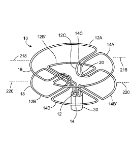
[0044] FIG 2 is a perspective view of the occluder device of the present
invention.
[0045] FIG. 3 is a top plan view of the occluder device of FIG. 2.
[0046] FIG. 4 is a side plan view of the occluder device taken along lines in
FIG 2.
[0047] FIG 5 is a side plan view of the occluder device taken along in FIG. 2.
[0048] FIG. 6 is a perspective view of the occluder device of FIG. 2,
illustrating the
covering 42.
[0049] FIG. 7 is a top plan view of the occluder device of FIG. 6.
100501 FIG. 8 is a perspective view of the occluder device first emerging from
the
catheter.
[0051] FIG. 9 is a perspective view of the occluder device half-way emerged
from the
catheter.
[0052] FIG 10 is a perspective view of the occluder device fully emerged from
the
catheter and separated from the deployment cable.
[0053] FIG 11 is a perspective view of the occluder device of the present
invention
illustrating restriction wires encircling the waist of the occluder device.
100541 FIG 12A is a perspective view of a first alternative embodiment of the
occluder device of the present invention.
CA 02844300 2014-02-04
WO 2013/025711
PCMJS2012/050785
[0055] FIG. 12B is a side plan view of the first alternative embodiment of the
occluder device of the present invention as shown in FIG. 12A.
[0056] FIG. 13 is a side plan view of a second alternative embodiment of the
occluder
device of the present invention.
[0057] FIG. 14 is a side plan view of a third alternative embodiment of the
occluder
device of the present invention.
[0058] FIG. 15 is a side plan view of a fourth alternative embodiment of the
occluder
device of the present invention.
100591 FIG. 16 is a side view of another exemplary alternative embodiment of
the
occluder device.
[0060] FIG. 17 is a side view of another exemplary alternative embodiment of
the
occluder device.
100611 FIG 18 is a perspective view of another exemplary alternative
embodiment of
the occluder device.
[0062] FIG 19 is a plan view of another exemplary alternative embodiment of
the
occluder device, depicted with reference to planar quadrants in FIG. 19A;
100631 FIG. 20 is a side view of another exemplary alternative embodiment of
the
occluder device.
[0064] FIG 21A is a perspective view of another exemplary alternative
embodiment
of the occluder device.
[0065] FIG. 21B is a plan view of another exemplary alternative embodiment of
the
occluder device.
[0066] FIG. 21C is a plan view of another exemplary alternative embodiment of
the
occluder device.
[0067] FIG. 21D is a plan view of another exemplary alternative embodiment of
the
occluder device.
16
CA 02844300 2014-02-04
WO 2013/025711
PCMJS2012/050785
[0068] FIG. 21E is a plan view of another exemplary alternative embodiment of
the
occluder device.
[0069] FIG. 22 is a plan view of another exemplary alternative embodiment of
the
occluder device.
[0070] FIG. 23 is a flowchart of an exemplary embodiment of a method for
occluding
an aperture defect in a heart to prevent the flow of blood therethrough, and
that may
be implemented using the occluder devices of FIGS. 2-22.
DETAILED DESCRIPTION OF THE INVENTION
100711 The following detailed description is merely exemplary in nature and is
not
intended to limit the disclosure or the application and uses of the
disclosure.
Furthermore, there is no intention to be bound by any theory presented in the
preceding background information or the following detailed description.
[0072] The present invention provides a device for occluding an aperture
within body
tissue. One skilled in the art will recognize that the device and methods of
the present
invention may be used to treat other anatomical conditions in addition to
those
specifically discussed herein. As such, the invention should not be considered
limited
in applicability to any particular anatomical condition.
[0073] FIG. 1 illustrates a human heart 1, having a right atrium 2, a left
atrium 3, a
right ventricle 4, and a left ventricle 5. Shown are various anatomical
anomalies 6A,
6B, and 6C. The atrial septum 7 includes septum primum 8 and septum secundum
9.
The anatomy of the septum 7 varies widely within the population. In some
people, the
septum primum 8 extends to and overlaps with the septum secundum 9. The septum
primum 8 may be quite thin. When a PFO is present, blood could travel through
the
passage 6A between septum primum 8 and septum secundum 9 (referred to as "the
PFO tunnel"). Additionally or alternatively, the presence of an A SD could
permit
blood to travel through an aperture in the scptal tissue, such as that
schematically
illustrated by aperture 6B. A VSD is similar to an ASD, except that an
aperture 6C
exists in the septum between the left and right ventricle of the heart.
[0074] PDA results from defects in the ductus arteriosus. The human blood
circulation comprises a systemic circuit and a pulmonary circuit. In the
embryonic
17
CA 02844300 2014-02-04
WO 2013/025711
PCMJS2012/050785
phase of human development, the two circuits are joined to one another by the
ductus
arteriosus. The ductus connects the aorta (circulation to the body) to the
pulmonary
artery (pulmonary circuit). In normal development of an infant, this ductus
closes
after birth. If development is defective, it can happen that the ductus does
not close,
and as a result the two blood circuits are still joined even after birth.
[0075] Unless specifically described otherwise, "aperture" 6 will refer to the
specific
heart defects described above, including PFO 6A, ASD 6B, VSD 6C, and PDA among
others.
[0076] As used herein, "distal" refers to the direction away from a catheter
insertion
location and "proximal" refers to the direction nearer the insertion location.
[0077] As used herein, "memory" or "shape memory" refers to a property of
materials
to resume and maintain an intended shape despite being distorted for periods
of time,
such as during storage or during the process of delivery in vivo.
[0078] Referring now to FIGS. 2-5, the occluder device 10 of the present
invention
comprises two separate uniquely shaped memory wires 12, 14. The wire can be
formed of biocompatible metals or polymers, such as bioresorbable polymers,
shape
memory polymers, shape memory metal alloys, biocompatible metals,
bioresorbable
metals, or combinations thereof. Specific examples include but are not limited
to iron,
magnesium, stainless steel, nitinol, or combinations of these and similar
materials. A
preferred metal for the present invention is a nitinol alloy. Nitinol (an
acronym for
Nickel Titanium Naval Ordnance Laboratory) is a family of intermetallic
materials,
which contain a nearly equal mixture of nickel (55 wt. %) and titanium. Other
elements can be added to adjust or "tune" the material properties. Nitinol
exhibits
unique behavior, specifically, a well defined "shape memory" and super
elasticity. In
general, any biocompatible material with a memory capability can be used with
the
present invention. The thermal shape memory and/or superelastic properties of
shape
memory polymers and alloys permit the occluder 10 to resume and maintain its
intended shape in vivo despite being distorted during the delivery process. In
certain
embodiments, the memory may also assist in pressing an aperture, such as a PFO
tunnel, closed. The diameter or thickness of the wire depends on the size and
type of
the device, i.e., the larger the device, the larger the diameter of the wire.
In general,
18
CA 02844300 2014-02-04
WO 2013/025711
PCMJS2012/050785
wire having a diameter between about 0.2 mm and 0.8 mm can be used. As
described
further below in connection with FIGS. 12A, 12B, and 22, in certain
embodiments
more than two wires may be utilized.
[0079] The first wire 12 forms one or more first geometric forms 12A and one
or
more second geometric forms 12B. "Geometric forms" as used herein comprises
symmetric as well as asymmetric forms. Relative to a delivery attachment
mechanism or hub 30, discussed below in greater detail, the first geometric
form 12A
of the first wire 12 preferably comprises a distal geometric form, and the one
or more
second geometric forms 12B of the first wire preferably each comprise proximal
geometric forms. In the embodiment of FIGS. 2-5, there is a single first, or
distal,
geometric form 12A of the first wire 12. Also in the embodiment of FIGS. 2-5,
there
are two second, or proximal, geometric forms 12B of the first wire 12 (namely,
12B(A) and 12B(B)). However, the number and configuration of the first and/or
second geometric forms 12A, 12B of the first wire 12 may vary.
100801 Similarly, the second wire 14 forms a first geometric form 14A and a
second
geometric form 14B. Relative to the hub 30, the first geometric form 14A of
the
second wire 14 preferably comprises a distal geometric form, and the second
geometric form 14B of the second wire preferably comprises a proximal
geometric
form. In the embodiment of FIGS. 2-5, there is a single first, or distal,
geometric
form 14A of the second wire 14. Also in the embodiment of FIGS. 2-5, there are
two
second, or proximal, geometric forms 14B of the second wire 14 (namely, 14B(A)
and
14B(B)). However, the number and configuration of the first and/or second
geometric
forms 14A, 14B of the second wire 14 may vary.
[0081] The first geometric forms 12A of the first wire 12 and the first
geometric
forms 14A of the second wire 14 form a first plate, such as a disc, or another
otherwise relatively flat surface (hereinafter referred to as a "plate") 16 in
a first plane
218. The second geometric forms 12B of the first wire 12 and the second
geometric
forms 14B of the second wire 14 form a second plate 18 (also referred to as a
"disc"
in certain embodiments) in a second plane 220 that is parallel to and remote
from the
first plane 218. In the embodiment of FIGS. 2-5, the first and second plates
16, 18
each comprise one or more semi-circular discs (as described directly below).
19
CA 02844300 2014-02-04
WO 2013/025711
PCMJS2012/050785
[0082] However, this may vary in other embodiments, for example as described
further below in connection with FIGS. 21A - 21E.
[0083] As shown in FIGS. 2-5, in these embodiments, each wire 12 or 14 forms a
shape which mirrors that of the respective wire 14 or 12. Specifically, each
wire 12,
14 forms a distal semi-circle or half-disc 12A, 14A in addition to two
proximal
quarter-circles or quarter-discs 12B, 12B' or 14B, 14B'. The two proximal
quarter-
circles of each wire together form proximal semi-circles or half-discs 12B,
12B' or
14B, 1413'. The two distal semi-circles of each respective wire 12A, 14A
together
comprise a distal circle or distal disc 16 of the occluder 10. The four
proximal quarter-
circles 12B, 12B', 14B, 14B', which form a "four-leaf clover" configuration,
comprise
a proximal circle or proximal disc 18 of the occluder 10.
[0084] The proximal semi-circle 12B, 12B' or 14B, 14B' of each wire is
connected to
the distal semi-circle 12A or 14A by waist portions (also referred to herein
as waist
components) 12C, 14C. As shown in FIG 2, there are two waist portions 12C, 14C
per wire. The four waist portions (two from each wire) 12C, 14C together
comprise a
restricted area or waist 20 of the occluder device 10. The distance between
the waist
portions, both within the same wire and from wire to wire, determines the size
of the
waist 20. The size of the waist 20 is dependent on the particular application
and the
size of the occluder device 10. The resiliency and memory of the waist
portions 12C,
14C and capacity to expand radially serves as a self-centering mechanism of
the
occluder device 10 in apertures 6.
The Hub 30:
[0085] The two half-discs are not attached or joined to each other except at
the
junction of the delivery attachment mechanism or hub 30. The ends 12D, 14D of
wires 12, 14 will be welded or otherwise connected to the hub 30.
Coverings 24A and 24B:
100861 According to some embodiments of the present invention, the distal disc
16
and/or proximal disc 18 may include membranous coverings 24A and 24B,
illustrated
in FIGS. 6 and 7. The membranous coverings 24A and 24B ensure more complete
coverage of aperture 6 and promote encapsulation and endothelialization of
tissue,
thereby further encouraging anatomical closure of the tissue and improving
closure
rate. The coverings 24A and 24B also help stabilize the occluder device 10.
[0087] The membranous coverings 24A and 24B may be formed of any flexible,
biocompatible material capable of promoting tissue growth and/or act as a
sealant,
including but not limited to DACRON®, polyester fabrics, Teflon-based
materials, ePTFE, polyurethanes, metallic materials, polyvinyl alcohol (PVA),
extracellular matrix (ECM) or other bioengineered materials, synthetic
bioabsorbable
polymeric materials, other natural materials (e.g. collagen), or combinations
of the
foregoing materials. For example, the membranous coverings 24A and 24B may be
formed of a thin metallic film or foil, e.g. a nitinol film or foil, as
described in U.S.
Pat. No. 7,335,426. The
preferred material is Poly(tetrafluoroethene) (ePTFE), as it combines several
important features such as thickness and the ability to stretch. Loops may
also be
stitched to the membranous coverings 24A and 24B to securely fasten the
coverings to
occluder 10. The coverings may alternatively be glued, welded or otherwise
attached
to the occluder 10 via the wires 12, 14.
Size:
[0088] As illustrated in FIGS. 2-7, the diameters of the distal disc 16 and
proximal
disc 18 are generally 5-8 mm larger than the diameter of the connecting waist
20. For
example, if the diameter of the connecting waist 20 is 4 mm, the diameters of
the
discs 16,18 are generally about 9 mm each. Because of the flexibility in the
waist 20,
a 12 mm waist device will be able to be placed in a 6 mm to 12 mm defect. For
larger
waists 20 or larger devices, the diameter of the disc size will increase
proportionately.
[0089] It is within the scope of the present invention to envision occluder
devices
available in 7 or more sizes, specifically waist size having the following
diameters for
different-sized apertures 6: 6 mm, 12 mm, 18 mm, 24 mm, 30 mm, 36 mm, and 42
mm.
Operation:
[0090] In general, the occluder 10 may be inserted into an aperture 6 to
prevent the
flow of blood therethrough. As a non-limiting example, the occluder 10 may
extend
21
CA 2844300 2018-12-27
CA 02844300 2014-02-04
WO 2013/025711
PCMJS2012/050785
through a PFO 6A or an ASD 6B such that the distal disc 16 is located in the
left
atrium 3 and the proximal disc 18 is located in the right atrium 2 (as shown
in the
heart 1 in FIG. 1). The closure of apertures in these and other tissues, as
well as other
types of apertures, will become apparent as described below.
[0091] Referring now to FIGS. 8-10, the occluder device 10 is attached to a
deployment cable 34 which is removably attached to the occluder device 10 at
the hub
30. As illustrated in FIG 10, one method of releasably attaching the
deployment cable
34 to the hub 30 is by threaded engagement utilizing a screw end 36 which
engages
unseen female threads within the hub 30. Other known means of attachment can
be
used to releasably connect the deployment cable 34 to the hub 30.
100921 When the deployment cable 34 is engaged with the hub 30, as illustrated
in
FIGS. 8 and 9, the occluder device 10 is initially housed within a flexible
delivery
catheter 40 having an open channel 42. Reference is made to FIG. 8 which
illustrates
the occluder device 10 in which the distal disc 16 is expanded, due to the
memory
expansion of the wires 12 and 14, and housed within the open channel 42 of the
delivery catheter 40. During the initial stages of placement of the occluder
device 10,
both the distal disc 16 and proximal disc 18, as well as the coverings 24A and
24B,
are housed within the open channel 42 of the delivery catheter 40. In this
manner, the
catheter 40 is fed into the blood vessel through an already placed sheath and
advanced
via the blood vessel system to a defect in the heart.
[0093] Once the delivery catheter 40 traverses the aperture that needs to be
occluded,
e.g., a hole in the heart, the device 10 will be partially advanced from the
catheter 40
as illustrated in FIG 8. As the device 10 leaves the catheter 40, the distal
disc 16,
which includes the covering 24A, begins to expand on the distal side of the
aperture.
Due to the memory capabilities of the wires 12 and 14, the occluder device 10
begins
to return to its normal shape such that the distal disc 16 expands on the
distal side of
the aperture in the heart. Once the distal disc 16 is completely out of the
catheter
opening 42, as shown in FIG. 9, it 16 and the attached covering 24A become
fully
expanded. The catheter 40 is further withdrawn to expose the waist 20 which
then
begins to emerge and expand due to the memory shape of the wires 12 and 14.
Advantageously, the waist 20 is designed to expand such that each of the wires
forming the waist 20 are urged against the aperture in the heart causing a
custom fit
22
CA 02844300 2014-02-04
WO 2013/025711
PCMJS2012/050785
device of the occluder 10 within the aperture. As the catheter 40 is further
withdrawn,
the proximal disc 18 and the covering 24B begin their process of expansion on
the
proximal side of the aperture. When the proximal disc 18 is fully delivered
from the
catheter 40, it will expand and effectively form a seal over the aperture. The
distal
disc 16 and proximal disc 18 are secured in place by the action of the wires
in the
waist 20 urging against the aperture. At this stage, as shown in FIG 10, the
deployment cable 34 is removed from the hub 30 and the catheter 40 and the
deployment cable 34 are removed from the body. The occluder device 10 is left
in the
heart at the region of the aperture. Over several months, skin tissue and
other
membranous structures will bind to the occluder device 10 thereby permanently
locking the occluder device 10 to the specific area in the heart.
100941 The two wires 12, 14 function to form round discs 16, 18 on each side
of the
tissue. The discs 16, 18 maintain the circular shape because of the memory
capability
of the wires 12, 14. The coverings 24A, 24B will stabilize the discs and will
act to
completely occlude the defect.
[0095] The wires 12, 14 at the waist portions 12C, 14C will be separated
enough at
the waist 20 to make the occluder device 10 self-centering. Due to the
conformity of
this design, the occluder device 10 should self-center within commonly (round,
oval)
shaped septal defects, as the waist 20 can adjust to any type of opening.
[0096] If a larger-diameter waist 20 is required, the waist 20 has the
capability to
expand (only if needed) to a larger size with the help of a balloon. In this
manner, a
center channel 50 extends through the deployment cable 34, the hub 30, and the
screw
end 36. A balloon (not shown) is urged through the center channel 50 after the
occluder device has been removed from the catheter 40 and expanded, and
preferably
before the hub 30 has been attached from the deployment cable 34. The balloon
is
placed within the waist 20 and expanded. The waist 20 is dilatable, i.e.,
expandable,
when gentle pressure of the balloon is applied. The dilation will expand the
waist
portions 12C, 14C. Once the desired diameter is reached, the balloon is
deflated and
removed by withdrawal through the center channel 50. Once the occluder device
10
appears stable, the device 10 is separated from the deployment cable 34 as
discussed
above. In the majority of cases, balloon dilation will not be required.
23
CA 02844300 2014-02-04
WO 2013/025711
PCMJS2012/050785
Restriction Wires 60, 62 (FIG 11):
[0097] In order to increase stability in the occluder device 10 and to avoid
significant
crimping of the waist 20 or the proximal or distal discs 18, 16, the waist 20
can be
encircled by one or more restriction wires 60, 62 as illustrated in FIG. 11.
The
restriction wires 60, 62 can be made of the same wire material as the wires 12
and 14,
or they may be of a different material, such as plastic wire, fish line, etc.
The
restriction wires 60, 62 may be welded or otherwise connected to the waist
portions
12C, 14C. The purpose of the restriction wires 60 or 62 is also to restrict
the
circumference of the waist 20 if necessary. Although one restriction wire 60
is
generally suitable, a second restriction wire 62 can also be incorporated to
further
improve stability.
Alternative Embodiments:
[0098] Reference is now made to FIGS. 12-15 for alternative embodiments of the
occluder device 10 of the present invention. Unless otherwise noted, the same
reference numbers will be applied to similar structures in each embodiment.
[0099] Reference is made to FIGS. 12A and 12B for an alternative embodiment of
the
occluder device (labeled as occluder device 100 in FIGS. 12A and 12B). The
occluder
device 100 in this embodiment is designed for PDA procedures. This embodiment
is
similar to previously described embodiments except that it is comprised of
four wires
112, 114, 116, 118 rather than two wires. In this case, each wire forms a
mirror image
of each of its neighboring wires. For example, wire 112 mirrors wire 114 as
well as
wire 118, etc. Each of the four wires 112, 114, 116, 118 forms a proximal
quarter-disc
112B, 114B, 116B, 118B and a distal quarter-disc 112A, 114A, 116A, 118A. The
proximal quarter-discs 112B, 114B, 116B, 118B together form a proximal disc
111 in
a "four-leaf clover" configuration, and the distal quarter-discs 112A, 114A,
116A,
118A together form a distal disc 110 also in a "four-leaf clover"
configuration. This
embodiment also differs from previously-described embodiments in that the
waist 20
is comprised of a single portion of each of the four wires 112, 114, 116, 118.
This
embodiment further differs from previously-described embodiments in that it
comprises a second hub 119 with a screw mechanism. The second hub 119 connects
to the distal disc 110 by distal ends 112E, 114E (116E, 118E behind 112E, 114E
in
24
CA 02844300 2014-02-04
WO 2013/025711
PCT/1JS2012/050785
FIG 12B) of each of the four wires 112, 114, 116, 118, just as proximal ends
112D,
114D (116D, 118D behind 112D, 114D in FIG 12B) connect to the proximal hub 30.
The wires 112, 114, 116, 118 may be connected to the hubs 30, 119 by welding
or
other means known in the art. The length of the waist 20 will be anywhere from
4-8
mm. In addition, the distal disc 110 is typically 4-8 mm larger than the waist
20.
However, the proximal disc 111 is generally 1-3 mm, preferably 2 mm, larger
than the
waist 20 diameter. Hence, the diameter of the distal disc 110 is larger than
the
diameter of the proximal disc 111.
100100] Reference is now made to FIG 13 for a second alternative embodiment
of
the occluder device 120. This embodiment, like the embodiment shown in FIGS.
12A
and 12B, uses four wires 112, 114, 116, 118 and two hubs 30, 119. It is
designed to
close apertures in large arteries and veins. In occluder device 120, the
distal and
proximal discs 122 and 124 are modified so that they are compatible with
closure of
veins and arteries. For this use, the connecting waist 20 is equivalent or
near
equivalent to the diameter of each of the discs 122, 124. The diameter of the
waist 20
will be 1 mm smaller than the discs 122, 124. The length of the waist will be
4-8 mm.
This embodiment can be used in the closure of coronary artery fistulas,
arteriovenous
fistulas, and arteriovenous malformations.
100101] Reference is made to FIG 14 for a third alternative embodiment of
the
occluder device 130. The importance of the occluder device 130 will be in the
closure
of the left atrial appendage. The device 130 is modified to conform to the
atrial
appendage anatomy. The distal disc 132 is modified so that the device 130 is
not
extruded out with the heartbeats. For the left atrial appendage occluder
device 130,
the memory wire structure of the distal disc 132 is woven to form anywhere
from 2 to
8 protuberances or hooks 136. Upon inserting the device 10 in an aperture in
the left
atrial appendage of the heart, the hooks 136 grip the outer portion of the
left atrium
heart tissue and thereby assist in keeping the device 130 from extruding out
of the left
atrial appendage with contraction of the heart. The proximal disc 134 is
typically flat
and similar to the disc formed by the proximal discs 18 in FIGS. 2-7. The
proximal
disc 134 abuts the inner atrial wall of the heart. Typically, the waist 20
will be about
4-8 mm in diameter. The length of the waist may range from 4 to 16 mm.
CA 02844300 2014-02-04
WO 2013/025711
PCMJS2012/050785
[00102] Reference is made to FIG 15 for a fourth alternative embodiment of
the
occluder device 140. Occluder device 140 is intended to occlude perimembranous
ventricular septa] ("PVS") defects. This embodiment, like the embodiment shown
in
FIGS. 12A and 12B, uses four wires 112, 114, 116, 118 and two hubs 30, 119.
The
occluder device 140 is different from other embodiments in that two of the
four wires
form truncated distal-quarter discs, with the effect that the distal disc 142
substantially
misses half of the disc. Therefore, the device 140 has approximately 1.5 discs
as
opposed to two discs. The half distal disc 142 is also significantly longer
than the
proximal disc 144. Typically, the distal disc 142 will be 6-8 mm in diameter.
In
addition, the distal disc 142 converges or curves inwards at 143, i.e., it is
angled to
contact the ventricular septum when the device 140 is inserted in the PVS
defect. (See
below for details.) The lower edge of the proximal disc (opposite to the long
distal
disc) will be 3-4 mm larger than the waist, and the other half of the proximal
disc will
be 2-3 mm larger than the waist. The discs can also be modified to be of
different
shapes in the same device. Alternatively, the disc angle may be created by a
straight
distal disc 142 angled with respect to the plane perpendicular to the waist 20
in a slant
fashion.
[00103] With reference to FIGS. 16-22, various additional exemplary
alternative
embodiments are provided with respect to the occluder device and/or components
thereof. With reference to FIG. 16, certain embodiments of the occluder device
10
may have one or more plates 16, 18 and/or geometric forms 12A, 12B, 14A, 14B
of
different sizes and/or configurations as compared with the embodiment
described
above in connection with FIG 2. For example, the distal (or first) plate 16
and the
proximal (or second) plate 18 may be offset with respect to the hub 30, and/or
one
side of a plate 16, 18 may be relatively higher or farther from the hub 30
than the
other, for example via an oblique shift. In the particular embodiment of FIG.
16, a
center 202 of the hub 30 is not aligned with (and, rather, is offset against)
a center 204
of the first plate 16, but is aligned with a center 206 of the second plate
18. In another
embodiment, the distal plate 16 and the proximal plate 18 are of equal size,
yet off set
from each other via a shift in opposite directions from the hub.
[00104] In certain embodiments, the first and second plates 16, 18 are
configured
such that a first segment formed from a first portion of the first wire 12
(for example,
corresponding to form 12B of FIG. 16) has a first length, a second segment
formed
26
CA 02844300 2014-02-04
WO 2013/025711
PCMJS2012/050785
from a first portion of the second wire 14 (for example, corresponding to form
14A of
FIG 16) has a second length, a third segment formed for a second portion of
the first
wire 12 (for example, corresponding to form 12A of FIG. 16) has a third
length, and a
fourth segment formed for a second portion of the second wire 14 (for example,
corresponding to form 14B of FIG 16) has a fourth length. The second length is
substantially equal to the first length. The third length is greater than the
first length.
The fourth length is substantially equal to the third length.
1001051 The semi-circle or half-disc 12A of the first wire 12 (also
referenced
above as the first geometric form 12A of the first wire 12) may differ in size
(for
example, having a larger radius and therefore a larger surface area) from the
semi-
circle or half-disc 14A of the second wire 14 (also referenced above as the
first
geometric form 14A of the second wire 14). In certain other embodiments, the
semi-
circle or half-disc 12A of the first wire 12 and the semi-circle or half-disc
14A of the
second wire 14 may be of the same size same as one another, but may
collectively
form a distal plate 16 that differs in size from the proximal plate 18. In one
such
embodiment, the distal plate 16 is smaller in surface area than the proximal
plate 18.
1001061 For example, the distal plate 16 may be of the same size as in FIG
2,
while the proximal plate 18 is larger in surface area than depicted in FIG. 2.
This may
occur, by way of example, when certain of the proximal quarter-circles of the
second
geometric forms 12B, 14B are larger in surface area than depicted in FIG. 2.
Certain
proximal quarter-circles of the second geometric forms 12B, 14B may be larger
in
surface area than other, adjacent quarter-circles of the second geometric
forms 12B,
14B. Such differing sizes of the proximal quarter-circles of the second
geometric
forms 12B, 14B may be present regardless of the relative sizes of the distal
and
proximal plates 16, 18.
1001071 FIG. 17 depicts an embodiment of an occluder device contemplated
herein
with a wider waist 20. In one exemplary embodiment, the first plate 16 and the
second plate 18 are disposed further apart as compared with the example of
FIG. 2, so
that a total length 225 of the waist 20 is greater than eight millimeters.
Preferably, in
this embodiment, the length 225 of the waist 20 is greater than eight
millimeters and
less than or equal to ten millimeters. In one such example, a straight-line
distance
between the first plane 218 and the second plane 220 of FIG. 2 is greater than
eight
27
CA 02844300 2014-02-04
WO 2013/025711
PCMJS2012/050785
millimeters, and is preferably also less than or equal to ten millimeters.
[00108] FIG 18 depicts an embodiment of an occluder device contemplated
herein
with a hook engagement system 230. The hook engagement system 230 comprises a
hook 232 and a lanyard 234 coupled thereto. The hook 232 is connected to the
first
plate 16 or the second plate 18 (and to the first and/or second wires 12, 14
thereof)
described above, preferably proximate one of the coverings 24A, 24B. The hook
engagement system 230 is configured for engagement with a positioning system
(not
depicted). In one embodiment, the hook engagement system 230 is used to remove
the occluder device 10 from the heart. In this regard, a loop of the lanyard
234 is
positioned onto the hook 232, and the lanyard 234 is pulled in the direction
away from
the heart, thus pulling the occluder device 10 through the heart aperture and
through
the body. In another embodiment, the positioning system comprises a deployment
system for deploying the occluder device 10, for example by grasping the hook
232
for movement of the occluder device 10 into a human heart in a desired
position
proximate an aperture. In a further embodiment, the positioning system
comprises a
repositioning system for repositioning the occluder device 10, for example by
grasping the hook 232 for adjusting the position of the occluder device 10 for
more
ideal placement of the occluder device 10 proximate an aperture. In certain
embodiments, the lanyard (and/or another connection feature) is part of the
positioning system, and the hook may exist separately from the occluder device
10.
The hook 232 is preferably used in connection with a screw device for further
engagement with the positioning system, such as a screw and nut system used in
conjunction with FIGS. 8-10 described above. For example, the hook 232 may be
positioned internal to a screw and nut system during placement of the device.
Alternatively, the hook 232 may be used in connection with a thread cord
through an
eyelet or an opening, so that the cord would need to be pulled in order to
lose the
connection with the occluder device 10. In addition, such a cord may be used
for
retrieval of the occluder device 10, for example by including multiple lumens,
preferably with an opening or slit, as part of a catheter delivery system.
[00109] With reference to FIGS. 19 and 19A, an embodiment of an occluder
device contemplated herein is depicted with overlapping wires at least at one
plate.
Overlapping wires add additional strength and rigidity to the plate of the
occluder
device. Specifically, the first geometric form 12A of the first wire 12
overlaps at least
28
CA 02844300 2014-02-04
WO 2013/025711
PCMJS2012/050785
a portion of one region (for example, at least a portion of a common spatial
quadrant,
half-plane, and/or quartile) in common with the first geometric form 14A of
the
second wire 14 within the first plate 16. Alternatively, or in addition, the
second
geometric form 12B (not shown) of the first wire 12 overlaps at least a
portion of one
region (for example, at least a portion of a common spatial quadrant, half-
plane,
and/or quartile) in common with the second geometric form 14B (not shown) of
the
second wire 14 within the second plate 18 (not shown).
1001101 In a preferred embodiment, as illustrated in FIG 19, the first
geometric
form 12A of the first wire 12 occupies at least three spatial quadrants 300,
301, and
302, two of which (namely, spatial quadrants 300 and 302) are shared in their
entireties with the first geometric form 14A of the second wire 14. Likewise,
the first
geometric form 14A of the second wire 14 occupies at least three spatial
quadrants
302, 303, and 300, two of which (namely, spatial quadrants 300 and 302) are
shared in
their entireties with the first geometric form 12A of the first wire 12.
Similarly, the
second geometric form 12B of the first wire 12 (not depicted in FIG 19)
occupies at
least three spatial quadrants, two of which are shared in their entireties
with the
second geometric form 14B of the second wire 14 (not depicted in FIG. 19).
Likewise, the second geometric form 14B of the second wire 14 occupies at
least
three spatial quadrants, two of which are shared in their entireties with the
second
geometric form 12B of the first wire 12.
[00111] FIG. 19A depicts an exemplary classification of planar quadrants
for the
first and second planes 218, 220 of FIG 2 for reference with respect to the
embodiment of FIG 19. One skilled in the art will recognize that less or more
than
four quadrants can be utilized. With reference to FIG 19A, the first plane 218
of FIG
2 has a first quadrant 241(A), a second quadrant 242(A) that is adjacent to
the first
quadrant 241(A), a third quadrant 243(A) that is below the first quadrant
241(A), and
a fourth quadrant 244(A) that is below the second quadrant 242(A) and adjacent
to the
third quadrant 243(A). The second plane 220 of FIG 2 has a first quadrant
241(B), a
second quadrant 242(B) that is adjacent to the first quadrant 241(B), a third
quadrant
243(B) that is below the first quadrant 241(B), and a fourth quadrant 244(B)
that is
below the second quadrant 242 (B) and adjacent to the third quadrant 243(B).
The
first quadrant 241(A) of the first plane 218 is closer to the first quadrant
241(B) of the
second plane 220 than to the second, third, or fourth quadrants 242(B),
243(B),
29
CA 02844300 2014-02-04
WO 2013/025711
PCMJS2012/050785
244(B) of the second plane 220. The second quadrant 242(A) of the first plane
218 is
closer to the second quadrant 242(B) of the second plane 220 than to the
first, third, or
fourth quadrants 241(B), 243(B), 244(B) of the second plane 220. The third
quadrant
243(A) of the first plane 218 is closer to the third quadrant 243(B) of the
second plane
220 than to the first, second, or fourth quadrants 241(B), 242(B), 244(B) of
the second
plane 220. The fourth quadrant 244(A) of the first plane 218 is closer to the
fourth
quadrant 244(B) of the second plane 220 than to the first, second, or third
quadrants
241(B), 242(B), 243(B) of the second plane 220.
[00112] With reference to the spatial quadrants set forth in FIG. 19A, in
one
preferred embodiment of FIG. 19, the first geometric form 12A of the first
wire 12
extends through the first, second, and third quadrants 241(A), 242(A), 243(A)
of the
first plane 218. The first geometric form 14A of the second wire 14 extends
through
the first, third, and fourth quadrants 241(A), 243(A), and 244(A) of the first
plane
218. Accordingly, in this embodiment, the first geometric forms 12A, 14A of
the first
and second wires 12, 14 share the first and third quadrants 241(A), 243(A) of
the first
plane 218 in common, for example to provide increased support and/or rigidity
for the
occluder device 10.
[00113] Also in one version of this embodiment of FIG 19, the second
geometric
form 12B of the first wire 12 extends through the first, second, and third
quadrants
241(B), 242(B), 243(B) of the second plane 220. The second geometric form 14B
of
the second wire 14 extends through the first, third, and fourth quadrants
241(B),
243(B), 244(B) of the second plane 220. Accordingly, in this version, the
second
geometric forms 12A, 14A of the first and second wires 12, 14 share the first
and third
quadrants 241(B), 243(B) of the second plane 220 in common, for example to
provide
increased support and/or rigidity for the occluder device 10.
[00114] However, this may vary in other versions or embodiments. For
example,
in another version of the embodiment depicted in FIG. 19, the second geometric
form
12B of the first wire 12 extends through the third, fourth, and first
quadrants 243(B),
244(B), 241(B) of the second plane 220, and the second geometric form 14B of
the
second wire 14 extends through the first, second, and third quadrants 241(B),
242(B),
243(B) of the second plane 220.
CA 02844300 2014-02-04
WO 2013/025711
PCMJS2012/050785
[00115] FIG. 20 depicts an embodiment of an occluder device contemplated
herein
with a clothes-pin shape. In the embodiment of FIG. 20, the first plate 16 and
the
second plate 18 described above are non-parallel, and form a non-zero angle
260 with
respect to one another. The angle 260 is preferably greater than five degrees,
is more
preferably greater than ten degrees, and is most preferably approximately
equal to
twenty degrees.
[00116] Also in the embodiment of FIG. 20, the waist 20 is configured such
that
the above-referenced waist components 12C of the first wire 12 and the waist
components 14C of the second wire 14 are unequal in size. For example, as
shown in
FIG 20, each waist component 12C of the first wire 12 has a first length
indicated by
double arrow 261, and each waist component 14C of the second wire 14 has a
second
length indicated by double arrow 262 that is greater than the first length.
The length
is defined as the distance between the first plate 16 and the second plate 18
taken from
a predetermined distance from the occluder device 10's center point. Each
waist
component 14C of the second wire 14 may also have a greater surface area and
radius
as compared to respective waist components 12C of the first wire 12. In
addition, in
the embodiment of FIG 20, the waist components 12C of the first wire 12 and
the
waist components 14C of the second wire 14 are preferably configured such that
the
waist 20 is curved, with a non-zero angle of curvature. The angle of curvature
of the
waist 20 is preferably greater than five degrees, is more preferably greater
than ten
degrees, and is most preferably greater than twenty degrees.
[00117] FIGS. 21A-21E depict an embodiment of an occluder device
contemplated herein in which one or more of the first and second plates 16, 18
are
non-circular in their geometric shape(s). In one embodiment of FIG. 21A, at
least the
first plate 16 has a generally oval shape. In an embodiment of FIG 21B, at
least the
first plate 16 has a generally rectangular shape. In an embodiment of FIG.
21C, at
least the first plate 16 has a generally triangular shape. In an embodiment of
FIG
21D, at least the first plate 16 has a generally elliptical shape. In an
embodiment of
FIG. 21E, at least the first plate 16 has a generally keyhole shape. In
certain versions,
the first plate 16 and/or the second plate 18 have generally the same
geometric shapes
as one another. In certain other versions, the first plate 16 and/or the
second plate 18
differ from one another. The first plate 16 and the second plate 18 may also
comprise
any number of other different geometric shapes.
31
CA 02844300 2014-02-04
WO 2013/025711
PCMJS2012/050785
[00118] FIG. 22 depicts an embodiment of an occluder device contemplated
herein
that is formed by more than two wires. Specifically, in the embodiment of FIG
22,
the occluder device 10 has three wires, namely: the first wire 12 and the
second wire
14 described above, as well as a third wire 205. In other embodiments, four
wires
may be utilized. In yet other embodiments, six wires may be utilized. In still
other
embodiments, the number of wires may differ further.
[00119] In the particular embodiment of FIG. 22, the three wires 12, 14,
and 205
each form respective, non-overlapping thirds of each plane. Specifically, as
depicted
in FIG 22, the first geometric form 12A of the first wire 12 is disposed
within and
extends through a first region 272 of the first plane 218 described above. The
second
geometric form 12B of the second wire 14 is disposed within and extends
through a
second region 274 of the first plane 218. A first geometric form 207 of the
third wire
205 is disposed within and extends through a third region 276 of the first
plane 218.
The first geometric forms 12A, 14A, 207 of the first, second, and third wires
12, 14,
205 collectively form the first plate 16.
[00120] Within the first plane 218, the first region 272 is adjacent to the
second
region 274, with a common border 277 formed by the first and second wires 12,
14.
The first region 272 is also adjacent to the third region 276, with a common
border
278 formed by the first and third wires 12, 205. In addition, the third region
276 is
also adjacent to the second region 274, with a common border 279 formed by the
second and third wires 14, 205.
[00121] Similarly, the second geometric form 12B of the first wire 12, the
second
geometric form 14B of the second wire 14, and a second geometric form of the
third
wire 205 would likewise be disposed within and extend through three similar
adjacent, non-overlapping regions of the second plane 220, collectively
forming the
second plate 18 (not depicted in FIG. 22). The various first and second
components of
the first, second, and third wires 12, 14, and 205 are preferably curved with
an arch,
such as is shown in FIG 22. Similar combinations of any number of different
amounts of wires can similarly be used to form any number of different forms.
[00122] As mentioned above, in certain embodiments, the occluder device 10
may
include multiple hubs 30, for example as depicted in FIG 15. The number and
32
CA 02844300 2014-02-04
WO 2013/025711
PCMJS2012/050785
configuration of such multiple hubs 30 may vary in different embodiments. In
one
such embodiment, a first end of the first wire 12 is disposed at a first hub
30, and at
least one of the second end of the first wire 12, the first end of the second
wire 14,
and/or the second end of the second wire 14 is disposed at a second hub (such
as hub
119 of FIGS. 12B, 13, and/or 15). In one such exemplary embodiment, the first
and
second ends of the first wire 12 are disposed at the first hub 30, and the
first and
second ends of the second wire 14 are disposed at the second hub (such as the
second
hub 119 of FIG. 15). In another such exemplary embodiment, the first ends of
the first
and second wires 12, 14 are disposed at the first hub 30, and the second ends
of the
first and second wires 12, 14 are disposed at the second hub (such as the hub
119 of
FTGS. 12B, 13, and/or 15), among other possible variations.
1001231 FIG. 23 is a flowchart of an exemplary embodiment of a method 2300
for
occluding an aperture defect in a heart. The method 2300 can be utilized in
connection with the heart 1 of FIG. 1 and the various embodiments of the
occluder
device 10 of FIGS. 2-22. Specifically, the method 2300 preferably utilizes one
or
more embodiments of the occluder devices 10 of FIGS. 2-22 to occlude an
aperture
defect of a heart, such as the aperture defect 6A of the heart 1 depicted in
FIG. 1.
1001241 As depicted in FIG 23, the method 2300 includes the step of
providing an
occluder device (step 2302). In various embodiments, the occluder device
corresponds to the occluder device 10 depicted in any of the embodiments
depicted in
FIGS. 2-22 and/or described above. The occluder device preferably comprises a
first
flexible wire (such as wire 12 described above) and a second flexible wire
(such as
wire 14 described above). Each of the first and second wires is comprised of a
shape
memory material. Each of the first and second wires is shaped into first and
second
geometric forms (such as forms 12A, 12B, 14A, and 14B described above) around
an
inner region such that the first geometric form of the first wire and the
first geometric
form of the second wire form a first plate (such as plate 16 described above)
in a first
plane, and the second geometric form 12B of the first wire 12 and the second
geometric form 14B of the second wire 14 form a second plate (such as plate 18
described above) in a second plane that is parallel to and remote from the
first plane.
The first and second plates are separated by a waist (such as waist 20
described
above) formed from two portions of the first wire and two portions of the
second wire.
A sealed covering (such as covering 24A or 24B described above) is preferably
33
CA 02844300 2014-02-04
WO 2013/025711
PCMJS2012/050785
disposed over at least one of the first and second plates. The covering
provides a seal
for the aperture defect (such as the defect 6A of the heart 1 described
above). Each of
the first and second wires has a first end and a second end. Each of the first
and
second ends of the first and second wires are connected to a hub (such as hub
30
described above). The hub further comprises a delivery attachment mechanism
(for
example, that includes or is used in connection with the catheter 40 described
above)
for attachment to a removable deployment cable (such as deployment cable 34
described above).
[00125] The method 2300 also includes the step of attaching the occluder
device
to the removable deployment cable (step 2304). The occluder device is placed
within
a flexible delivery catheter (such as the catheter 40 described above) having
an open
channel (such as the channel 42 described above) (step 2306). The catheter is
fed into
a blood vessel system (such as a blood vessel system of the heart 1 described
above)
and advanced via the blood vessel system to the aperture defect in the heart
(step
2308). The catheter, with the occluder device disposed within, is similarly
advanced
through the aperture defect (step 2310).
[00126] In certain optional embodiments, a balloon sub-process 2312 is also
utilized in occluding the aperture defect in the heart. In one such
embodiment,
depicted in FIG. 23, a balloon is advanced into the heart through the open
channel
toward the occluder device at the aperture defect (step 2314). The balloon is
also
inserted into the waist of the occluder device (step 2316). The balloon is
then inflated
(step 2318), in order to help position the occluder device proximate the heart
defect.
Once the occluder device is properly positioned, the balloon is deflated (step
2320)
and then removed from the waist of the occluder device (step 2322).
[00127] In other optional embodiments, a hook sub-process 2324 may be
utilized
in occluding the aperture defect in the heart. In one such embodiment,
depicted in
FIG 23, a hook (such as one or more of the hooks 136, 232 described above), is
engaged with the delivery attachment mechanism (such as the catheter) (step
2326),
preferably via a screw system. The hook is manipulated using the delivery
attachment
mechanism and used to reposition the occluder device (step 2328). In certain
embodiments, the hook may also be utilized to retrieve the occluder device by
exerting force on the delivery attachment mechanism in a direction away from
the
34
CA 02844300 2014-02-04
WO 2013/025711
PCMJS2012/050785
heart (step 2330).
[00128] The catheter next is withdrawn from the occluder device (step
2332).
Preferably, the catheter is withdrawn from the occluder device in step 2332 in
a
manner such that the first plate of the occluder device expands on a first
side of the
aperture defect. In addition, the catheter is further withdrawn from the
occluder
device such that the second plate of the occluder device expands on a second
side of
the aperture defect (step 2334). Preferably, the catheter is withdrawn from
the
occluder device in step 2334 in a manner, such that the waist of the occluder
device
expands by memory retention within the aperture defect to self-center the
occluder
device. The catheter is then withdrawn from the blood vessel system (step
2336), and
the deployment cable is removed from the hub of the occluder device (step
2338).
[00129] It will be appreciated that certain steps of the method 2300 may
vary in
certain embodiments. It will also be appreciated that certain steps of the
method 2300
may occur in a different order than is depicted in FIG 23. For example, the
optional
hook sub-process 2324 may be used before the optional balloon sub-process
2312. It
will similarly be appreciated that certain steps of the method 230 may occur
simultaneously with one another.
[00130] Other embodiments may comprise any combinations of the embodiments
described herein and/or described in the drawings. It is understood that the
disclosure
is not confined to the particular construction and arrangement of parts herein
illustrated and described, but embraces such modified forms thereof as come
within
the scope of the following claims. Additionally, it will be appreciated that
various
embodiments may be freely combined together, and/or that various features of
different embodiments may be freely combined together.
[00131] While at least one exemplary embodiment has been presented in the
foregoing detailed description, it should be appreciated that a vast number of
variations exist. It should also be appreciated that the exemplary embodiment
or
exemplary embodiments are only examples, and are not intended to limit the
scope,
applicability, or configuration of the invention in any way. Rather, the
foregoing
detailed description will provide those skilled in the art with a convenient
road map
for implementing an exemplary embodiment of the invention, it being understood
that
CA 02844300 2014-02-04
WO 2013/025711
PCT/1JS2012/050785
various changes may be made in the function and arrangement of elements
described
in an exemplary embodiment without departing from the scope of the invention
as set
forth in the appended claims and their legal equivalents.
36