Note: Descriptions are shown in the official language in which they were submitted.
CA 02844538 2014-02-06
WO 2013/026831 PCT/EP2012/066213
-1-
BISPECIFIC ANTIGEN BINDING MOLECULES
Field of the Invention
The present invention generally relates to bispecific antigen binding
molecules. In addition, the
present invention relates to polynucleotides encoding such bispecific antigen
binding molecules,
and vectors and host cells comprising such polynucleotides. The invention
further relates to
methods for producing the bispecific antigen binding molecules of the
invention, and to methods
of using these bispecific antigen binding molecules in the treatment of
disease.
Background
Bi- or multispecific antibodies capable of binding two or more antigens are
known in the art.
Such multispecific binding proteins can be generated by hybridoma cell fusion,
chemical
.. conjugation or recombinant DNA techniques.
Bispecific antibodies are of great interest for therapeutic applications, as
they allow the
simultaneous binding and inactivation of two or more target antigens, thereby
obviating the need
for combination therapies. Another promising application of bispecific
antibodies is as engagers
of immune effector cells e.g. for cellular cancer immunotherapy. For this
purpose, bispecific
antibodies are designed which bind to a surface antigen on target cells and,
for example, to an
activating component of the T cell receptor (TCR) complex. The simultaneous
binding of such
an antibody to both of its targets will force a temporary interaction between
target cell and T cell,
causing activation of any cytotoxic T lymphocyte (CTL) and subsequent lysis of
the target cell.
Hence, the immune response is re-directed to the target cells, independently
of peptide antigen
presentation by the target cell or the specificity of the T cell as required
for normal MHC-
restricted activation of CTLs. In this context it is important that CTLs are
only activated when a
target cell is presenting the bispecific antibody to them, i.e. when the
immunological synapse is
mimicked, and not simply upon binding of the antibody to the T cell antigen.
A variety of recombinant multispecific antibody formats have been developed in
the recent past,
including, for example, tetravalent IgG-single-chain variable fragment (scFv)
fusions (see e.g.
Coloma & Morrison, Nat Biotechnol 15, 159-163 (1997)), tetravalent IgG-like
dual-variable
CA 02844538 2014-02-06
WO 2013/026831 PCT/EP2012/066213
-2-
domain antibodies (Wu et al., Nat Biotechnol 25, 1290-1297 (2007)), or
bivalent rat/mouse
hybrid bispecific IgGs (see e.g. Lindhofer et al., I Immunol 155, 219-225
(1995)).
Also several bispecific formats wherein the antibody core structure (IgA, IgD,
IgE, IgG or IgM)
is no longer retained have been made. Examples include diabodies (see e.g.
Holliger et al., Proc
Natl Acad Sci USA 90, 6444-6448 (1995)), tandem scFv molecules (see e.g.
Bargou et al.,
Science 321, 974-977 (2008)), and various derivatives thereof.
The multitude of formats that are being developed shows the great potential
attributed to
bispecific antibodies. The task of generating bispecific antibodies suitable
for a particular
purpose is, however, by no means trivial and subject to a number of
considerations. For example.
the valency and geometry of the antibody needs to be appropriately chosen,
depending on the
characteristics of the target antigens and the intended effect. As for all
therapeutic antibodies,
efficacy and toxicity have to be balanced, which requires i.a. minimization of
immunogenicity
and optimization of pharmacokinetic properties of the antibody. Also, the
desirablility of Fe-
mediated effects has to be considered. Furthermore, the production of
bispecific antibody
constructs at a clinically sufficient quantity and purity poses a major
challenge, as the
homodimerization of antibody heavy chains and/or the mispairing of antibody
heavy and light
chains of different specificities upon co-expression decreases the yield of
the correctly
assembled construct and results in a number of non-functional side products
from which the
desired bispecific antibody may be difficult to separate.
Given the increasing number of possible applications of bispecific antibodies,
and the difficulties
and disadvantages associated with currently available bispecific antibodies,
there remains a need
for novel, improved formats of such molecules.
Summary of the Invention
In a first aspect. the invention provides a bispecific antigen binding
molecule, comprising a first
Fab fragment which specifically binds to a first antigen, a second Fab
fragment which
specifically binds to a second antigen, and an Fe domain composed of a first
and a second
subunit capable of stable association; wherein
a) the bispecific antigen binding molecule provides monovalent binding to the
first and/or the
second antigen,
b) the first Fab fragment, the second Fab fragment and the first Fc domain
subunit are fused to
each other, and
CA 02844538 2014-02-06
WO 2013/026831 PCT/EP2012/066213
-3-
c) in the first and/or the second Fab fragment one of the following
replacements is made: (i) the
variable domains VL and VH are replaced by each other, (ii) the constant
domains CL and
CH1 are replaced by each other, or (iii) both the variable and constant
domains VL-CL and
VH-CHI are replaced by each other,
provided that not the same replacement is made in the first and the second Fab
fragment.
In particular embodiments, the first Fab fragment is fused at its C-terminus
to the N-terminus of
the second Fab fragment, which is in turn fused at its C-terminus to the N-
terminus of the first Fe
domain subunit. In a more specific embodiment, the first Fab fragment is fused
at the C-terminus
of its heavy chain to the N-terminus of the heavy chain of the second Fab
fragment, which is in
turn fused at the C-terminus of its heavy chain to the N-terminus of the first
Fc domain subunit.
In other embodiments, the second Fab fragment is fused at its C-terminus to
the N-terminus of
the first Fab fragment, which is in turn fused at its C-terminus to the N-
terminus of the first Fe
domain subunit. In a more specific embodiment, the second Fab fragment is
fused at the C-
terminus of its heavy chain to the N-terminus of the heavy chain of the first
Fab fragment. which
is in turn fused at the C-terminus of its heavy chain to the N-terminus of the
first Fc domain
subunit. In still other embodiments, the second Fab fragment is fused at its C-
terminus to the N-
terminus of the first Fc domain subunit, which is in turn fused at its C-
terminus to the N-
terminus of the first Fab fragment. In a more specific embodiment, the second
Fab fragment is
fused at the C-terminus of its heavy chain to the N-terminus of the first Fc
domain subunit,
which is in turn fused at its C-terminus to the N-terminus of the heavy chain
of the first Fab
fragment.
In embodiments wherein either the first Fab fragment is fused at the C-
terminus of its heavy
chain to the N-terminus of the heavy chain of the second Fab fragment which is
in turn fused at
the C-terminus of its heavy chain to the N-terminus of the first Fc domain
subunit, or the second
Fab fragment is fused at the C-terminus of its heavy chain to the N-terminus
of the heavy chain
of the first Fab fragment which is in turn fused at the C-terminus of its
heavy chain to the N-
terminus of the first Fc domain subunit, additionally the Fab light chain of
the first Fab fragment
and the Fab light chain of the second Fab fragment may be fused to each other,
optionally via a
peptide linker.
In one embodiment, the replacement is made in the first Fab fragment. In some
embodiments,
the replacement is a replacement of the variable domains VL and VH by each
other. In other
embodiments the replacement is a replacement of the constant domains CL and
CH1 by each
other.
CA 02844538 2014-02-06
WO 2013/026831 PCT/EP2012/066213
-4-
In one embodiment the bispecific antigen binding molecule essentially consists
of the first Fab
fragment, the second Fab fragment, the Fc domain, and optionally one or more
peptide linkers.
In particular embodiments, the bispecific antigen binding molecule comprises a
third Fab
fragment which specifically binds to the first or the second antigen. In one
embodiment, the third
Fab fragment is fused to the second Fc domain subunit. In a more specific
embodiment, the third
Fab fragment is fused at its C-terminus to the N-terminus of the second Fc
domain subunit. In en
even more specific embodiment, the third Fab fragment is fused at the C-
terminus of its heavy
chain to the N-terminus of the second Fc domain subunit. In one embodiment,
the third Fab
fragment specifically binds to the second antigen. In some embodiments the the
second Fab
fragment, the third Fab fragment and the Fc domain are part of an
immunoglobulin molecule. In
a specific such embodiment, the immunoglobulin molecule is an IgG class
immunoglobulin
molecule, more specifically an IgG1 or IgG4 subclass immunoglobulin molecule.
In one
embodiment, the immunoglobulin molecule is a human immunoglobulin molecule. In
one
embodiment, the bispecific antigen binding molecule essentially consists of a
first Fab fragment
which specifically binds to the first antigen, an immunoglobulin molecule
which specifically
binds to the second antigen, and optionally one or more peptide linkers.
In one embodiment, the same replacement is made in Fab fragments that
specifically bind to the
same antigen. In a further embodiment, a replacement is made only in the first
Fab fragment. In
one embodiment, the bispecific antigen binding molecule provides monovalent
binding to the
first antigen. In one embodiment, the bispecific antigen binding molecule does
not comprise a
single-chain Fab fragment.
In certain embodiments. the Fc domain comprises a modification promoting the
association of
the first and second Fc domain subunit. In a specific such embodiment, an
amino acid residue in
the CH3 domain of the first subunit of the Fc domain is replaced with an amino
acid residue
having a larger side chain volume, thereby generating a protuberance within
the CH3 domain of
the first subunit which is positionable in a cavity within the CH3 domain of
the second subunit,
and an amino acid residue in the CH3 domain of the second subunit of the Fc
domain is replaced
with an amino acid residue having a smaller side chain volume, thereby
generating a cavity
within the CH3 domain of the second subunit within which the protuberance
within the CH3
domain of the first subunit is positionable. In one embodiment. the Fc domain
is an IgG Fe
domain, specifically an IgGI or IgG4 Fc domain. In one embodiment the Fc
domain is human. In
certain embodiments, the Fc domain is engineered to have altered binding
affinity to an Fe
receptor and/or altered effector function, as compared to a non-engineered Fc
domain.
CA 02844538 2014-02-06
WO 2013/026831 PCT/EP2012/066213
-5-
According to another aspect of the invention there is provided an isolated
polynucleotide
encoding a bispecific antigen binding molecule of the invention or a fragment
thereof. The
invention also encompasses polypeptides encoded by the polynucleotides of the
invention. The
invention further provides an expression vector comprising the isolated
polynucleotide of the
invention, and a host cell comprising the isolated polynucleotide or the
expression vector of the
invention. In some embodiments the host cell is a eukaryotic cell,
particularly a mammalian cell.
In another aspect is provided a method of producing the bispecific antigen
binding molecule of
the invention, comprising the steps of a) culturing the host cell of the
invention under conditions
suitable for the expression of the bispecific antigen binding molecule and b)
recovering the
bispecific antigen binding molecule. The invention also encompasses a
bispecific antigen
binding molecule produced by the method of the invention.
The invention further provides a pharmaceutical composition comprising the
bispecific antigen
binding molecule of the invention and a pharmaceutically acceptable carrier.
Also encompassed by the invention are methods of using the bispecific antigen
binding molecule
and pharmaceutical composition of the invention. In one aspect the invention
provides a
bispecific antigen binding molecule or a pharmaceutical composition of the
invention for use as
a medicament. In one aspect is provided a bispecific antigen binding molecule
or a
pharmaceutical composition according to the invention for use in the treatment
of a disease in an
individual in need thereof. In a specific embodiment the disease is cancer.
Also provided is the use of a bispecific antigen binding molecule of the
invention for the
manufacture of a medicament for the treatment of a disease in an individual in
need thereof; as
well as a method of treating a disease in an individual, comprising
administering to said
individual a therapeutically effective amount of a composition comprising the
bispecific antigen
binding molecule according to the invention in a pharmaceutically acceptable
form. In a specific
embodiment the disease is cancer. In any of the above embodiments the
individual preferably is
a mammal, particularly a human.
Brief Description of the Drawings
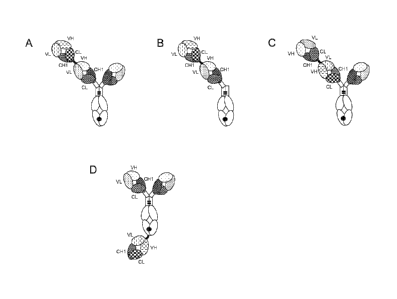
FIGURE 1. Illustration of exemplary formats of the bispecific antigen binding
molecules of the
invention. (A) "2+1" format, with Crossfab fragment of different specificity
fused to N-terminus
of a Fab fragment comprised in an antibody ("2+1 IgG Crossfab (N-terminal)").
(B) "1+1"
format, with Crossfab fragment of different specificity fused to N-terminus of
a Fab fragment
-6-
comprised in an antibody lacking the second Fab fragment ("1+1 IgG Crossfab (N-
terminal)") .
(C) "2+1" format as in (A), wherein the order of the Crossfab fragment, and
the Fab fragment to
which the Crossfab fragment is fused, is inverted ("2+1" IgG Crossfab (N-
terminal), inverted").
(D) "2+1" format, with Crossfab fragment of different specificity fused to C-
terminus of an Fc
.. domain subunit comprised in an antibody ("2+1 IgG Crossfab (C-terminal)").
Black dot: optional
modification in the Fc domain promoting heterodimerization.
FIGURE 2. (A, B) SDS PAGE (4-12% Tris-Acetate (A) or 4-12% Bis/Tris (B),
NuPage
Invitrogen, Coomassie-stained) of "1+1 IgG Crossfab (N-terminal), Fc(hole)
P329G LALA /
Fc(knob) wt" (anti-MCSP/anti-huCD3) (see SEQ ID NOs 1, 2, 3 and 4), non
reduced (A) and
reduced (B). (C) Analytical size exclusion chromatography (SuperdexTM 200
10/300 GL GE
Healthcare; 2 mM MOPS pH 7.3, 150 mM NaCl, 0.02% (w/v) NaCI; 50 pg sample
injected) of
"1+1 IgG Crossfab (N-terminal). Fc(hole) P329G LALA / Fc(knob) wt" (anti-
MCSP/anti-
huCD3).
FIGURE 3. (A, B) SDS PAGE (4-12% Bis/Tris, NuPage Invitrogen, Coomassie-
stained) of "2+1
IgG Crossfab (N-terminal)" (anti-MCSP/anti-huCD3) (see SEQ ID NOs 1, 3, 4 and
5), non
reduced (A) and reduced (B). (C) Analytical size exclusion chromatography
(SuperdexTM 200
10/300 GL GE Healthcare; 2 mM MOPS pH 7.3, 150 mM NaCI, 0.02% (w/v) NaCI; 50
lig
sample injected) of "2+1 IgG Crossfab (N-terminal)" (anti-MCSP/anti-huCD3).
FIGURE 4. (A, B) SDS PAGE (4-12% Bis/Tris, NuPage Invitrogen, Coomassie-
stained) of "2+1
IgG Crossfab (N-terminal), inverted" (anti-CEA/anti-huCD3) (see SEQ ID NOs 3,
8, 9 and 10),
non reduced (A) and reduced (B). (C) Analytical size exclusion chromatography
(SuperdexTM
200 10/300 GL GE Healthcare; 2 mM MOPS pH 7.3, 150 mM NaCI, 0.02% (w/v) NaCI;
50 pg
sample injected) of "2+1 IgG Crossfab (N-terminal), inverted" (anti-CEA/anti-
huCD3).
FIGURE 5. (A, B) Capillary electrophoresis (CE)-SDS gel analysis of "2+1 IgG
Crossfab (C-
terminal)" (anti-c-Met/anti-Her3) (see SEQ ID NOs 11, 12, 13, 14), non reduced
(A) and
reduced (B).
FIGURE 6. Simultaneous binding of bispecific constructs to the D3 domain of
human MCSP
and human CD37(G4S)5CD3E¨AcTev¨Fc(knob)¨Avi/Fc(hole). (A) Biacore assay setup;
(B)
measurement of "2+1 IgG Crossfab (N-terminal)".
FIGURE 7. Levels of different cytokines measured in the supernatant of whole
blood after
treatment with 1 nM of different CD3-MCSP bispecific constructs ("2+1 IgG
Crossfab (N-
terminal)", "(scFv)2") or corresponding control IgGs in the presence (A, B) or
absence (C, D) of
Colo-38 tumor cells for 24 hours.
CA 2844538 2018-10-23
CA 02844538 2014-02-06
WO 2013/026831 PCT/EP2012/066213
-7-
FIGURE 8. Surface expression level of the late activation marker CD25 on
cynomolgus CD8 T
cells from two different animals (cyno Nestor, cyno Nobu) after 43 hours
incubation with the
indicated concentrations of the "2+1 IgG Crossfab (N-terminal)" bispecific
construct (targeting
cynomolgus CD3 and human MCSP), in the presence or absence of human MCSP-
expressing
MV-3 tumor target cells (E:T ratio = 3:1). As controls, the reference IgGs
(anti-cynomolgus CD3
IgG, anti-human MCSP IgG) or the unphysiologic stimulus PHA-M were used.
FIGURE 9. Killing (as measured by LDH release) of MDA-MB-435 tumor cells upon
co-culture
with human pan T cells (E:T ratio = 5:1) and activation for 20 hours by
different concentrations
of the "2+1 IgG Crossfab (N-terminal)" and "(scFv)2" bispecific molecules and
corresponding
IgGs.
FIGURE 10. Killing (as measured by LDH release) of MDA-MB-435 tumor cells upon
co-
culture with human pan T cells (E:T ratio = 5:1), and activation for 21 hours
by different
concentrations of the bispecific constructs and corresponding IgGs. The CD3-
MCSP bispecific
"2+1 IgG Crossfab (N-terminal)" and "1+1 IgG Crossfab (N-terminal)"
constructs, the "(scFv)2"
molecule and corresponding IgGs were compared.
FIGURE 11. Killing (as measured by LDH release) of huMCSP-positive MV-3
melanoma cells
upon co-culture with human PBMCs (E:T ratio = 10:1), treated with different
CD3-MCSP
bispecific constructs ("2+1 IgG Crossfab (N-terminal)" and "(scFv)2") for ¨26
hours.
FIGURE 12. Examplary configurations of bispecific antigen binding molecules of
the invention
having a linked light chain. (A) Illustration of the "2+1 IgG Crossfab (N
terminal), linked light
chain" molecule. (B) Illustration of the "1+1 IgG Crossfab (N-terminal),
linked light chain"
molecule. (C) Illustration of the "2+1 IgG Crossfab (N-terminal), inverted,
linked light chain"
molecule. (D) Illustration of the "1+1 IgG Crossfab (N-terminal), inverted,
linked light chain"
molecule.
FIGURE 13. CE-SDS analyses. Electropherogram shown as SDS PAGE of "2+1 IgG
Crossfab
(N-terminal), linked light chain" (lane 1: reduced, lane 2: non-reduced).
FIGURE 14. Analytical size exclusion chromatography of "2+1 IgG Crossfab (N-
terminal),
linked light chain" (final product). 20 p g sample "2+1 IgG Crossfab (N-
terminal), linked light
chain" were injected.
FIGURE 15. Killing (as measured by LDH release) of MCSP-positive MV-3 tumor
cells upon
co-culture by human PBMCs (E:T ratio = 10:1), treated with different CD3-MCSP
bispecific
constructs for ¨ 44 hours. Human PBMCs were isolated from fresh blood of
healthy volunteers.
CA 02844538 2014-02-06
WO 2013/026831 PCT/EP2012/066213
-8-
FIGURE 16. Killing (as measured by LDH release) of MCSP-positive Colo-38 tumor
cells upon
co-culture by human PBMCs (E:T ratio = 10:1), treated with different CD3-MCSP
bispecific
constructs for ¨22 hours. Human PBMCs were isolated from fresh blood of
healthy volunteers.
FIGURE 17. Killing (as measured by LDH release) of MCSP-positive Colo-38 tumor
cells upon
co-culture by human PBMCs (E:T ratio -= 10:1), treated with different CD3-MCSP
bispecific
constructs for ¨22 hours. Human PBMCs were isolated from fresh blood of
healthy volunteers.
FIGURE 18. Killing (as measured by LDH release) of MCSP-positive WM266-4 cells
upon co-
culture by human PBMCs (E:T ratio = 10:1), treated with different CD3-MCSP
bispecific
constructs for ¨22 hours. Human PBMCs were isolated from fresh blood of
healthy volunteers.
FIGURE 19. Surface expression level of the early activation marker CD69 (A)
and the late
activation marker CD25 (B) on human CD8+ T cells after 22 hours incubation
with 10 nM, 80
pM or 3 pM of different CD3-MCSP bispecific constructs in the presence or
absence of human
MCSP-expressing Colo-38 tumor target cells (E:T ratio = 10:1).
FIGURE 20. CE-SDS analyses. (A) Electropherogram shown as SDS-PAGE of 1+1 IgG
Crossfab (N-terminal); VL/VH exchange (LC007/V9): a) non-reduced, b) reduced.
(B)
Electropherogram shown as SDS-PAGE of 1+1 CrossMab; CL/CH1 exchange
(LC007/V9): a)
reduced, b) non-reduced. (C) Electropherogram shown as SDS-PAGE of 2+1 IgG
Crossfab (N-
terminal), inverted; CL/CH1 exchange (LC007/V9): a) reduced, b) non-reduced.
(D)
Electropherogram shown as SDS-PAGE of 2+1 IgG Crossfab (N-terminal); VL/VH
exchange
(M4-3 ML2/V9): a) reduced, b) non-reduced. (E) Electropherogram shown as SDS-
PAGE of
2+1 IgG Crossfab (N-terminal); CL/CH1 exchange (M4-3 ML2/V9): a) reduced, b)
non-reduced.
(F) Electropherogram shown as SDS-PAGE of 2+1 IgG Crossfab (N-terminal),
inverted;
CL/CH1 exchange (CH1A1A/V9): a) reduced, b) non-reduced.
FIGURE 21. Surface expression level of the early activation marker CD69 (A) or
the late
activation marker CD25 (B) on human CD4+ or CD8+ T cells after 24 hours
incubation with the
indicated concentrations of the CD3/MCSP "1+1 CrossMab", "1+1 IgG Crossfab (N-
terminal)"
and "2+1 IgG Crossfab (N-terminal)" constructs. The assay was performed in the
presence or
absence of MV-3 target cells, as indicated.
FIGURE 22. Killing (as measured by LDH release) of MKN-45 (A) or LS-174T (B)
tumor cells
upon co-culture with human PBMCs (E:T ratio = 10:1) and activation for 28
hours by different
concentrations of the "2+1 IgG Crossfab (N-terminal), inverted (VL/VH)" versus
the "2+1 IgG
Crossfab (N -terminal), inverted (CL/CH1)" construct.
CA 02844538 2014-02-06
WO 2013/026831 PCT/EP2012/066213
-9-
FIGURE 23. Killing (as measured by LDH release) of WM266-4 tumor cells upon co-
culture
with human PBMCs (E:T ratio = 10:1) and activation for 26 hours by different
concentrations of
the "2+1 IgG Crossfab (N-terminal) (VL/VH)" versus the "2+1 IgG Crossfab (N-
terminal)
(CL/CH )" construct.
FIGURE 24. Killing (as measured by LDH release) of MV-3 tumor cells upon co-
culture with
human PBMCs (E:T ratio = 10:1) and activation for 27 hours by different
concentrations of the
"2+1 IgG Crossfab (N-terminal) (VH/VL)" versus the "2+1 IgG Crossfab (N-
terminal)
(CL/CH1)" constructs.
FIGURE 25. Killing (as measured by LDH release) of human MCSP-positive WM266-4
(A) or
MV-3 (B) tumor cells upon co-culture with human PBMCs (E:T ratio = 10:1) and
activation for
21 hours by different concentrations of the "2+1 IgG Crossfab (N-terminal)",
the "1+1
CrossMab", and the "1+1 IgG Crossfab (N-terminal)", as indicated.
FIGURE 26. Binding of bispecific constructs to human CD3, expressed by Jurkat
cells (A), or to
human CEA, expressed by LS-174T cells (B) as determined by FACS. As a control,
the
equivalent maximum concentration of the reference IgGs and the background
staining due to the
labeled 2ndary antibody (goat anti-human FITC-conjugated AffiniPure F(ab')2
Fragment, Fcy
Fragment-specific, Jackson Immuno Research Lab # 109-096-098) were assessed as
well.
FIGURE 27. Binding of bispecific constructs to human CD3, expressed by Jurkat
cells, or to
human MCSP, expressed by WM266-4 tumor cells (B) as determined by FACS.
Detailed Description of the Invention
Definitions
Terms are used herein as generally used in the art, unless otherwise defined
in the following.
As used herein, the term "antigen binding molecule" refers in its broadest
sense to a molecule
that specifically binds an antigenic determinant. Examples of antigen binding
molecules are
immunoglobulins and derivatives, e.g. fragments. thereof.
The term "bispecific" means that the antigen binding molecule is able to
specifically bind to two
distinct antigenic determinants. Typically, a bispecific antigen binding
molecule comprises two
antigen binding sites, each of which is specific for a different antigenic
determinant. In certain
embodiments the bispecific antigen binding molecule is capable of
simultaneously binding two
antigenic determinants, particularly two antigenic determinants expressed on
two distinct cells.
CA 02844538 2014-02-06
WO 2013/026831 PCT/EP2012/066213
-10-
As used herein, the term "antigenic determinant" is synonymous with "antigen"
and "epitope,"
and refers to a site (e.g. a contiguous stretch of amino acids or a
conformational configuration
made up of different regions of non-contiguous amino acids) on a polypeptide
macromolecule to
which an antigen binding moiety binds, forming an antigen binding moiety-
antigen complex.
Useful antigenic determinants can be found, for example, on the surfaces of
tumor cells, on the
sutfaces of virus-infected cells, on the surfaces of other diseased cells, on
the surface of immune
cells, free in blood serum, and/or in the extracellular matrix (ECM). The
proteins referred to as
antigens herein (e.g. MCSP, FAP, CEA, EGFR, CD33, CD3, c-Met, Her3) can be any
native
form the proteins from any vertebrate source, including mammals such as
primates (e.g. humans)
and rodents (e.g. mice and rats), unless otherwise indicated. In a particular
embodiment the
antigen is a human protein. Where reference is made to a specific protein
herein, the term
encompasses the "full-length", unprocessed protein as well as any form of the
protein that results
from processing in the cell. The term also encompasses naturally occurring
variants of the
protein, e.g. splice variants or allelic variants. Exemplary human proteins
useful as antigens
include, but are not limited to: Melanoma-associated Chondroitin Sulfate
Proteoglycan (MCSP),
also known as Chondroitin Sulfate Proteoglycan 4 (UniProt no. Q6UVK1, NCBI
Accession no.
NP_001888); Fibroblast Activation Protein (FAP), also known as Seprase (Uni
Prot nos. Q12884,
Q86Z29, Q99998, NCBI Accession no. NP_004451); Carcinoembroynic antigen (CEA),
also
known as Carcinoembryonic antigen-related cell adhesion molecule 5 (UniProt
no. P06731,
NCBI Accession no. NP_004354); CD33, also known as gp67 or Siglec-3 (UniProt
no. P20138,
NCBI Accession nos. NP_001076087, NP_001171079); Epidermal Growth Factor
Receptor
(EGFR), also known as ErbB-1 or Hen l (UniProt no. P0053, NCBI Accession nos.
NP_958439,
NP_958440), CD3, particularly the epsilon subunit of CD3 (UniProt no. P07766,
NCBI
Accession no. NP_000724); c-Met, also known as Hepatocyte Growth Factor
Receptor (UniProt
no. P08581, NCBI Accession nos. NP_000236, NP_001120972) and Her3, also known
as ErbB-
3 (UniProt no. P21860. NCBI Accession nos. NP_001973, NP_001005915). In
certain
embodiments the bispecific antigen binding molecule of the invention binds to
an epitope of an
first antigen or a second antigen that is conserved among the first antigen or
second antigen from
different species.
By "specific binding" is meant that the binding is selective for the antigen
and can be
discriminated from unwanted or non-specific interactions. The ability of an
antibody to bind to a
specific antigenic determinant can be measured either through an enzyme-linked
immunosorbent
assay (ELISA) or other techniques familiar to one of skill in the art, e.g.
surface plasmon
CA 02844538 2014-02-06
WO 2013/026831 PCT/EP2012/066213
-11 -
resonance (SPR) technique (analyzed on a BIAcore instrument) (Liljeblad et
al., Glyco J 17,
323-329 (2000)), and traditional binding assays (Heeley, Endocr Res 28. 217-
229 (2002)). In one
embodiment, the extent of binding of an antibody to an unrelated protein is
less than about 10%
of the binding of the antibody to the antigen as measured, e.g.. by SPR. In
certain embodiments,
.. an antibody or a fragement thereof that binds to the antigen has a
dissociation constant (KD) of <
1 [tM, < 100 nM, < 10 nM, < 1 nM, < 0.1 nM, < 0.01 nM. or < 0.001 nM (e.g. 10-
8M or less, e.g.
from 10-8M to 1013M, e.g., from 10-9M to 1013 M).
"Affinity" refers to the strength of the sum total of non-covalent
interactions between a single
binding site of a molecule (e.g., a receptor) and its binding partner (e.g., a
ligand). Unless
indicated otherwise, as used herein, "binding affinity" refers to intrinsic
binding affinity which
reflects a 1:1 interaction between members of a binding pair (e.g., an antigen
binding moiety and
an antigen, or a receptor and its ligand). The affinity of a molecule X for
its partner Y can
generally be represented by the dissociation constant (KD), which is the ratio
of dissociation and
association rate constants (koff and kon, respectively). Thus, equivalent
affinities may comprise
different rate constants, as long as the ratio of the rate constants remains
the same. Affinity can
be measured by well established methods known in the art, including those
described herein. A
particular method for measuring affinity is Surface Plasmon Resonance (SPR).
The term "valent" as used herein denotes the presence of a specified number of
antigen binding
sites in an antigen binding molecule. As such, the term "monovalent binding to
an antigen"
.. denotes the presence of one (and not more than one) antigen binding site
specific for the antigen
in the antigen binding molecule.
An "antigen binding site" refers to the site, i.e. one or more amino acid
residues, of an antigen
binding molecule which provides interaction with the antigen. For example, the
antigen binding
site of an antibody comprises amino acid residues from the complementarity
determining regions
(CDRs). A native immunoglobulin molecule typically has two antigen binding
sites, a Fab
fragment typically has a single antigen binding site.
As used herein, the term "antigen binding moiety" refers to a polypeptide
molecule that
specifically binds to an antigenic determinant. Antigen binding moieties
include antibodies and
fragments thereof as further defined herein. Particular antigen binding
moieties include an
antigen binding domain of an antibody, comprising an antibody heavy chain
variable region and
an antibody light chain variable region. In certain embodiments, the antigen
binding moieties
may comprise antibody constant regions as further defined herein and known in
the art. Useful
CA 02844538 2014-02-06
WO 2013/026831 PCT/EP2012/066213
-12-
heavy chain constant regions include any of the five isotypes: a, 6, 8, y, or
t. Useful light chain
constant regions include any of the two isotypes: ic and k.
As used herein, the terms "first" and "second" with respect to Fab fragments
etc., are used for
convenience of distinguishing when there is more than one of each type of
moiety. Use of these
terms is not intended to confer a specific order or orientation of the
bispecific antigen binding
molecule unless explicitly so stated.
As used herein, the term "single-chain" refers to a molecule comprising amino
acid monomers
linearly linked by peptide bonds. By a single-chain Fab fragment is meant a
Fab molecule
wherein the Fab light chain and the Fab heavy chain are connected by a peptide
linker to form a
single peptide chain.
The term "immunoglobulin molecule" refers to a protein having the structure of
a naturally
occurring antibody. For example, immunoglobulins of the IgG class are
heterotetrameric
glycoproteins of about 150,000 daltons, composed of two light chains and two
heavy chains that
are disulfide-bonded. From N- to C-terminus, each heavy chain has a variable
region (VH), also
called a variable heavy domain or a heavy chain variable domain, followed by a
hinge region
(HR) and three constant domains (CHL CH2, and CH3), also called a heavy chain
constant
region. In case of an IgE class immunoglobulin the heavy chain additionally
has a CH4 domain.
Hence, an immunoglobulin heavy chain is a polypeptide consisting in N-terminal
to C-terminal
direction of the following domains: VH-CH1-HR-CH2-CH3-(CH4). Similarly, from N-
to C-
terminus, each light chain has a variable region (VL), also called a variable
light domain or a
light chain variable domain, followed by a constant light (CL) domain, also
called a light chain
constant region. Hence, an immunoglobulin light chain is a polypeptide
consisting in N-terminal
to C-terminal direction of the following domains: VL-CL. The heavy chain of an
immunoglobulin may be assigned to one of five types, called a (IgA), 6 (IaD),
E (IgE), (IgG).
or 11 (IgM), some of which may be further divided into subtypes, e.g. yi
(IgGO, y2 (IgG2),
(IgG3), y4 (IgG4), al (IgAi) and a2 (IgA2). The light chain of an
immunoglobulin may be
assigned to one of two types, called kappa (lc) and lambda (X), based on the
amino acid sequence
of its constant domain. An immunoglobulin essentially consists of two Fab
fragments and an Fe
domain, linked via the immunoglobulin hinge region.
The term "antibody" herein is used in the broadest sense and encompasses
various antibody
structures, including but not limited to monoclonal antibodies, polyclonal
antibodies, and
antibody fragments so long as they exhibit the desired antigen-binding
activity.
CA 02844538 2014-02-06
WO 2013/026831 PCT/EP2012/066213
-13-
An "antibody fragment" refers to a molecule other than an intact antibody that
comprises a
portion of an intact antibody that binds the antigen to which the intact
antibody binds. Examples
of antibody fragments include but are not limited to Fv, Fab, Fab', Fab'-SH,
F(ab')2, diabodies,
linear antibodies, single-chain antibody molecules (e.g. scFv), and single-
domain antibodies. For
a review of certain antibody fragments, see Hudson et al., Nat Med 9, 129-134
(2003). For a
review of scFv fragments, see e.g. Pliickthun, in The Pharmacology of
Monoclonal Antibodies.
vol. 113, Rosenburg and Moore eds., Springer-Verlag, New York, pp. 269-315
(1994); see also
WO 93/16185; and U.S. Patent Nos. 5.571,894 and 5,587,458. For discussion of
Fab and F(ab')2
fragments comprising salvage receptor binding epitope residues and having
increased in vivo
half-life, see U.S. Patent No. 5,869.046. Diabodies are antibody fragments
with two antigen-
binding sites that may be bivalent or bispecific. See, for example, EP
404,097; WO 1993/01161;
Hudson et al., Nat Med 9, 129-134 (2003); and Hollinger et al., Proc Natl Acad
Sci USA 90.
6444-6448 (1993). Triabodies and tetrabodies are also described in Hudson et
al., Nat Med 9,
129-134 (2003). Single-domain antibodies are antibody fragments comprising all
or a portion of
the heavy chain variable domain or all or a portion of the light chain
variable domain of an
antibody. hi certain embodiments, a single-domain antibody is a human single-
domain antibody
(Domantis, Inc., Waltham, MA; see e.g. U.S. Patent No. 6,248,516 B1). Antibody
fragments can
be made by various techniques, including but not limited to proteolytic
digestion of an intact
antibody as well as production by recombinant host cells (e.g. E. coli or
phage), as described
herein.
A "Fab fragment" refers to a protein consisting of the VH and CH1 domain of
the heavy chain
(the "Fab heavy chain") and the VL and CL domain of the light chain (the "Fab
light chain") of
an immunoglobulin. A Fab fragment being fused to another protein is, in its
unmodified form,
fused at its heavy chain C- or N-terminus. Consequently, where the variable
domains VH and
VL are replaced by each other, the Fab fragment is fused at the C-terminus of
the CH1 domain or
the N-terminus of the VL domain. Similarly, where the constant domains CH1 and
CL are
replaced by each other, the Fab fragment is fused at the C-terminus of the CL
domain or the N-
terminus of the VH domain, and where the complete Fab heavy chain (VH-CH1) and
Fab light
chain (VL-CL) are replaced by each other, the Fab fragment is fused at its
light chain C- or N-
terminus.
By "fused" is meant that the components (e.g. a Fab fragment and an Fc domain
subunit) are
linked by peptide bonds, either directly or via one or more peptide linkers.
CA 02844538 2014-02-06
WO 2013/026831 PCT/EP2012/066213
-14-
The term "antigen binding domain" refers to the part of an antibody that
comprises the area
which specifically binds to and is complementary to part or all of an antigen.
An antigen binding
domain may be provided by, for example, one or more antibody variable domains
(also called
antibody variable regions). Particularly, an antigen binding domain comprises
an antibody light
chain variable region (VL) and an antibody heavy chain variable region (VH).
The term "variable region" or "variable domain" refers to the domain of an
antibody heavy or
light chain that is involved in binding the antibody to antigen. The variable
domains of the heavy
chain and light chain (VH and VL, respectively) of a native antibody generally
have similar
structures. with each domain comprising four conserved framework regions (FRs)
and three
hypervariable regions (HVRs). See, e.g., Kindt et al., Kuby Immunology, 6th
ed., W.H. Freeman
and Co., page 91 (2007). A single VH or VL domain may be sufficient to confer
antigen-binding
specificity.
The term "hypervariable region" or "HVR", as used herein, refers to each of
the regions of an
antibody variable domain which are hypervariable in sequence and/or form
structurally defined
loops ("hypervariable loops"). Generally, native four-chain antibodies
comprise six HVRs; three
in the VH (H1, H2, H3), and three in the VL (L1, L2, L3). HVRs generally
comprise amino acid
residues from the hypervariable loops and/or from the complementarity
determining regions
(CDRs), the latter being of highest sequence variability and/or involved in
antigen recognition.
With the exception of CDR1 in VH, CDRs generally comprise the amino acid
residues that form
the hypervariable loops. Hypervariable regions (HVRs) are also referred to as
"complementarity
determining regions" (CDRs), and these terms are used herein interchangeably
in reference to
portions of the variable region that form the antigen binding regions. This
particular region has
been described by Kabat et al., U.S. Dept. of Health and Human Services,
Sequences of Proteins
of Immunological Interest (1983) and by Chothia et al., J Mol Biol 196:901-917
(1987), where
the definitions include overlapping or subsets of amino acid residues when
compared against
each other. Nevertheless, application of either definition to refer to a CDR
of an antibody or
variants thereof is intended to be within the scope of the term as defined and
used herein. The
appropriate amino acid residues which encompass the CDRs as defined by each of
the above
cited references are set forth below in Table 1 as a comparison. The exact
residue numbers which
encompass a particular CDR will vary depending on the sequence and size of the
CDR. Those
skilled in the art can routinely determine which residues comprise a
particular CDR given the
variable region amino acid sequence of the antibody.
CA 02844538 2014-02-06
WO 2013/026831 PCT/EP2012/066213
-15-
TABLE 1. CDR Definitions'
CDR Kabat Chothia AbM2
= CDR1 31-35 26-32 26-35
= CDR2 50-65 52-58 50-58
VH CD R 3 95-102 95-102 95-102
= CDRI 24-34 26-32 24-34
VL CDR2 50-56 50-52 50-56
VL CDR3 89-97 91-96 89-97
1
Numbering of all CDR definitions in Table 1 is according to the numbering
conventions
set forth by Kabat et al. (see below).
2 ,'AbM" with a lowercase "b" as used in Table 1 refers to the CDRs as
defined by Oxford Molecular's "AbM" antibody modeling software.
Kabat et al. also defined a numbering system for variable region sequences
that is applicable to
any antibody. One of ordinary skill in the art can unambiguously assign this
system of "Kabat
numbering" to any variable region sequence, without reliance on any
experimental data beyond
the sequence itself. As used herein, "Kabat numbering" refers to the numbering
system set forth
by Kabat et al., U.S. Dept. of Health and Human Services, "Sequence of
Proteins of
Immunological Interest" (1983). Unless otherwise specified, references to the
numbering of
specific amino acid residue positions in an antibody variable region are
according to the Kabat
numbering system.
The polypeptide sequences of the sequence listing are not numbered according
to the Kabat
numbering system. However, it is well within the ordinary skill of one in the
art to convert the
numbering of the sequences of the Sequence Listing to Kabat numbering.
"Framework" or "1-R" refers to variable domain residues other than
hypervariable region (HVR)
residues. The FR of a variable domain generally consists of four FR domains:
FR1, FR2, FR3,
and FR4. Accordingly, the HVR and FR sequences generally appear in the
following sequence in
VH (or VL): FR -1-H1 (L1)-FR2-H2(L2)-FR3-H3(L3)-FR4.
The "class" of an antibody or irnmunoglobulin refers to the type of constant
domain or constant
region possessed by its heavy chain. There are five major classes of
antibodies: IgA, IgD, IgE,
IgG, and IgM, and several of these may be further divided into subclasses
(isotypes), e.g., IgGi.
IgG2, IgG2, IgG4, IgAi, and IgA2. The heavy chain constant domains that
correspond to the
different classes of immunoglobulins are called a, 6, E, y, and u,
respectively.
The term "Fe domain" or "Fe region" herein is used to define a C-terminal
region of an
immunoblobulin heavy chain that contains at least a portion of the constant
region. The term
includes native sequence Fc regions and variant Fc regions. Although the
boundaries of the Fe
region of an IgG heavy chain might vary slightly, the human IgG heavy chain Fc
region is
CA 02844538 2014-02-06
WO 2013/026831 PCT/EP2012/066213
-16-
usually defined to extend from Cys226, or from Pro230, to the carboxyl-
terminus of the heavy
chain. However, the C-terminal lysine (Lys447) of the Fc region may or may not
be present.
Unless otherwise specified herein, numbering of amino acid residues in the Fe
region or constant
region is according to the EU numbering system, also called the EU index, as
described in Kabat
et al., Sequences of Proteins of Immunological Interest, 5th Ed. Public Health
Service, National
Institutes of Health, Bethesda, MD, 1991. A "subunit" of an Fc domain as used
herein refers to
one of the two polypeptides forming the dimeric Fc domain, i.e. a polypeptide
comprising C-
terminal constant regions of an immunoglobulin heavy chain, capable of stable
self-association.
For example, a subunit of an IgG Fc domain comprises an IgG CH2 and an IgG CH3
constant
region.
A "modification promoting the association of the first and the second subunit
of the Fc domain"
is a manipulation of the peptide backbone or the post-translational
modifications of an Fe
domain subunit that reduces or prevents the association of a polypeptide
comprising the Fe
domain subunit with an identical polypeptide to form a homodimer. A
modification promoting
association as used herein particularly includes separate modifications made
to each of the two
Fe domain subunits desired to associate (i.e. the first and the second subunit
of the Fc domain),
wherein the modifications are complementary to each other so as to promote
association of the
two Fc domain subunits. For example, a modification promoting association may
alter the
structure or charge of one or both of the Fc domain subunits so as to make
their association
sterically or electrostatically favorable, respectively. Thus,
(hetero)dimerization occurs between
a polypeptide comprising the first Fc domain subunit and a polypeptide
comprising the second
Fc domain subunit, which might be non-identical in the sense that further
components fused to
each of the subunits (e.g. Fab fragments) are not the same. In some
embodiments the
modification promoting association comprises an amino acid mutation in the Fc
domain,
specifically an amino acid substitution. In a particular embodiment, the
modification promoting
association comprises a separate amino acid mutation, specifically an amino
acid substitution, in
each of the two subunits of the Fc domain.
The term "effector functions" refers to those biological activities
attributable to the Fc region of
an antibody, which vary with the antibody isotype. Examples of antibody
effector functions
include: Clq binding and complement dependent cytotoxicity (CDC), Fc receptor
binding,
antibody-dependent cell-mediated cytotoxicity (ADCC), antibody-dependent
cellular
phagocytosis (ADCP), cytokine secretion, immune complex-mediated antigen
uptake by antigen
CA 02844538 2014-02-06
WO 2013/026831 PCT/EP2012/066213
-17-
presenting cells, down regulation of cell surface receptors (e.g. B cell
receptor), and B cell
activation.
As used herein, the terms "engineer, engineered, engineering", are considered
to include any
manipulation of the peptide backbone or the post-translational modifications
of a naturally
occurring or recombinant polypeptide or fragment thereof. Engineering includes
modifications of
the amino acid sequence, of the glycosylation pattern, or of the side chain
group of individual
amino acids, as well as combinations of these approaches.
The term "amino acid mutation" as used herein is meant to encompass amino acid
substitutions,
deletions, insertions, and modifications. Any combination of substitution,
deletion, insertion, and
modification can be made to arrive at the final construct, provided that the
final construct
possesses the desired characteristics, e.g., reduced binding to an Fc
receptor, or increased
association with another peptide. Amino acid sequence deletions and insertions
include amino-
and/or carboxy-terminal deletions and insertions of amino acids. Particular
amino acid mutations
are amino acid substitutions. For the purpose of altering e.g. the binding
characteristics of an Fe
region, non-conservative amino acid substitutions, i.e. replacing one amino
acid with another
amino acid having different structural and/or chemical properties, are
particularly preferred.
Amino acid substitutions include replacement by non-naturally occurring amino
acids or by
naturally occurring amino acid derivatives of the twenty standard amino acids
(e.g. 4-
hydroxyproline, 3-methylhistidine, ornithine, homoserine, 5-hydroxylysine).
Amino acid
mutations can be generated using genetic or chemical methods well known in the
art. Genetic
methods may include site-directed mutagenesis, PCR, gene synthesis and the
like. It is
contemplated that methods of altering the side chain group of an amino acid by
methods other
than genetic engineering, such as chemical modification, may also be useful.
Various
designations may be used herein to indicate the same amino acid mutation. For
example, a
substitution from proline at position 329 of the Fc domain to glycine can be
indicated as 329G,
G329, G329, P329G, or Pro329Gly.
As used herein, term "polypeptide" refers to a molecule composed of monomers
(amino acids)
linearly linked by amide bonds (also known as peptide bonds). The term
"polypeptide" refers to
any chain of two or more amino acids, and does not refer to a specific length
of the product.
.. Thus, peptides, dipeptides, tripeptides, oligopeptides, "protein," "amino
acid chain," or any other
term used to refer to a chain of two or more amino acids, are included within
the definition of
"polypeptide," and the term "polypeptide" may be used instead of, or
interchangeably with any
of these terms. The term "polypeptide" is also intended to refer to the
products of post-expression
CA 02844538 2014-02-06
WO 2013/026831 PCT/EP2012/066213
-18-
modifications of the polypeptide, including without limitation glycosylation,
acetylation,
phosphorylation, amidation, derivatization by known protecting/blocking
groups, proteolytic
cleavage, or modification by non-naturally occurring amino acids. A
polypeptide may be derived
from a natural biological source or produced by recombinant technology, but is
not necessarily
translated from a designated nucleic acid sequence. It may be generated in any
manner, including
by chemical synthesis. A polypeptide of the invention may be of a size of
about 3 or more, 5 or
more, 10 or more, 20 or more, 25 or more, 50 or more, 75 or more, 100 or more,
200 or more,
500 or more, 1,000 or more, or 2,000 or more amino acids. Polypeptides may
have a defined
three-dimensional structure. although they do not necessarily have such
structure. Polypeptides
with a defined three-dimensional structure are referred to as folded, and
polypeptides which do
not possess a defined three-dimensional structure, but rather can adopt a
large number of
different conformations, and are referred to as unfolded.
By an "isolated" polypeptide or a variant, or derivative thereof is intended a
polypeptide that is
not in its natural milieu. No particular level of purification is required.
For example, an isolated
polypeptide can be removed from its native or natural environment.
Recombinantly produced
polypeptides and proteins expressed in host cells are considered isolated for
the purpose of the
invention, as are native or recombinant polypeptides which have been
separated, fractionated, or
partially or substantially purified by any suitable technique.
By "isolated" nucleic acid molecule or polynucleotide is intended a nucleic
acid molecule, DNA
or RNA, which has been removed from its native environment. For example, a
recombinant
polynucleotide encoding a polypeptide contained in a vector is considered
isolated for the
purposes of the present invention. Further examples of an isolated
polynucleotide include
recombinant polynucleotides maintained in heterologous host cells or purified
(partially or
substantially) polynucleotides in solution. An isolated polynucleotide
includes a polynucleotide
molecule contained in cells that ordinarily contain the polynucleotide
molecule, but the
polynucleotide molecule is present extrachromosomally or at a chromosomal
location that is
different from its natural chromosomal location. Isolated RNA molecules
include in vivo or in
vitro RNA transcripts of the present invention, as well as positive and
negative strand forms, and
double-stranded forms. Isolated polynucleotides or nucleic acids according to
the present
invention further include such molecules produced synthetically. In addition,
a polynucleotide or
a nucleic acid may be or may include a regulatory element such as a promoter,
ribosome binding
site, or a transcription terminator.
CA 02844538 2014-02-06
WO 2013/026831 PCT/EP2012/066213
-19-
The term "vector" or "expression vector" is synonymous with "expression
construct" and refers
to a DNA molecule that is used to introduce and direct the expression of a
specific gene to which
it is operably associated in a target cell. The term includes the vector as a
self-replicating nucleic
acid structure as well as the vector incorporated into the genome of a host
cell into which it has
been introduced. The expression vector of the present invention comprises an
expression
cassette. Expression vectors allow transcription of large amounts of stable
mRNA. Once the
expression vector is inside the target cell, the ribonucleic acid molecule or
protein that is
encoded by the gene is produced by the cellular transcription and/or
translation machinery. In
one embodiment, the expression vector of the invention comprises an expression
cassette that
comprises polynucleotide sequences that encode bispecific antigen binding
molecules of the
invention or fragments thereof.
The terms "host cell", "host cell line," and "host cell culture" are used
interchangeably and refer
to cells into which exogenous nucleic acid has been introduced, including the
progeny of such
cells. Host cells include "transformants" and "transformed cells," which
include the primary
transformed cell and progeny derived therefrom without regard to the number of
passages.
Progeny may not be completely identical in nucleic acid content to a parent
cell, but may contain
mutations. Mutant progeny that have the same function or biological activity
as screened or
selected for in the originally transformed cell are included herein. A host
cell is any type of
cellular system that can be used to generate the bispecific antigen binding
molecules of the
present invention. Host cells include cultured cells, e.g. mammalian cultured
cells, such as CHO
cells, BHK cells, NSO cells. SP2/0 cells, YO myeloma cells, P3X63 mouse
myeloma cells, PER
cells, PER.C6 cells or hybridoma cells, yeast cells, insect cells, and plant
cells, to name only a
few, but also cells comprised within a transgenic animal, transgenic plant or
cultured plant or
animal tissue.
An "activating Fc receptor" is an Fc receptor that following engagement by an
Fc domain of an
antibody elicits signaling events that stimulate the receptor-bearing cell to
perform effector
functions. Human activating Fc receptors include FcyRIIIa (CD16a). FcyRI
(CD64), FcyRIIa
(CD32), and FcaRI (CD89).
Antibody-dependent cell-mediated cytotoxicity (ADCC) is an immune mechanism
leading to the
lysis of antibody-coated target cells by immune effector cells. The target
cells are cells to which
antibodies or derivatives thereof comprising an Fc region specifically bind,
generally via the
protein part that is N-terminal to the Fc region. As used herein, the term -
reduced (or increased)
ADCC" is defined as either a reduction (increase) in the number of target
cells that are lysed in a
CA 02844538 2014-02-06
WO 2013/026831 PCT/EP2012/066213
-20-
given time, at a given concentration of antibody in the medium surrounding the
target cells, by
the mechanism of ADCC defined above, and/or an increase (reduction) in the
concentration of
antibody in the medium surrounding the target cells, required to achieve the
lysis of a given
number of target cells in a given time, by the mechanism of ADCC. The
reduction (increase) in
ADCC is relative to the ADCC mediated by the same antibody produced by the
same type of
host cells, using the same standard production, purification, formulation and
storage methods
(which are known to those skilled in the art), but that has not been
engineered. For example the
reduction in ADCC mediated by an antibody comprising in its Fc domain an amino
acid
substitution that reduces ADCC, is relative to the ADCC mediated by the same
antibody without
this amino acid substitution in the Fc domain. Suitable assays to measure ADCC
are well known
in the art (see e.g. PCT publication no. WO 2006/082515 or PCT patent
application no.
PCT/EP2012/055393).
An "effective amount" of an agent refers to the amount that is necessary to
result in a
physiological change in the cell or tissue to which it is administered.
A "therapeutically effective amount" of an agent, e.g. a pharmaceutical
composition, refers to an
amount effective, at dosages and for periods of time necessary, to achieve the
desired therapeutic
or prophylactic result. A therapeutically effective amount of an agent for
example eliminates,
decreases, delays, minimizes or prevents adverse effects of a disease.
An "individual" or "subject" is a mammal. Mammals include, but are not limited
to,
domesticated animals (e.g. cows, sheep, cats, dogs, and horses), primates
(e.g. humans and non-
human primates such as monkeys), rabbits, and rodents (e.g. mice and rats).
Particularly, the
individual or subject is a human.
The term "pharmaceutical composition" refers to a preparation which is in such
form as to permit
the biological activity of an active ingredient contained therein to be
effective, and which
contains no additional components which are unacceptably toxic to a subject to
which the
formulation would be administered.
A "pharmaceutically acceptable carrier" refers to an ingredient in a
pharmaceutical composition,
other than an active ingredient, which is nontoxic to a subject. A
pharmaceutically acceptable
carrier includes, but is not limited to, a buffer, excipient, stabilizer, or
preservative.
As used herein, "treatment" (and grammatical variations thereof such as
"treat" or "treating")
refers to clinical intervention in an attempt to alter the natural course of a
disease in the
individual being treated, and can be performed either for prophylaxis or
during the course of
clinical pathology. Desirable effects of treatment include, but are not
limited to, preventing
CA 02844538 2014-02-06
WO 2013/026831 PCT/EP2012/066213
-2] -
occurrence or recurrence of disease, alleviation of symptoms, diminishment of
any direct or
indirect pathological consequences of the disease, preventing metastasis,
decreasing the rate of
disease progression, amelioration or palliation of the disease state, and
remission or improved
prognosis. In some embodiments, bispecific antigen binding molecules of the
invention are used
to delay development of a disease or to slow the progression of a disease.
The term "package insert" is used to refer to instructions customarily
included in commercial
packages of therapeutic products, that contain information about the
indications, usage, dosage,
administration, combination therapy, contraindications and/or warnings
concerning the use of
such therapeutic products.
Detailed Description of the Embodiments
The invention provides a bispecific antigen binding molecule, comprising a
first Fab fragment
which specifically binds to a first antigen, a second Fab fragment which
specifically binds to a
second antigen, and an Fc domain composed of a first and a second subunit
capable of stable
association; wherein
a) the bispecific antigen binding molecule provides monovalent binding to the
first and/or the
second antigen,
b) the first Fab fragment, the second Fab fragment and the first Fc domain
subunit are fused to
each other, and
c) in the first and/or the second Fab fragment one of the following
replacements is made: (i)
the variable domains VL and VH are replaced by each other, (ii) the constant
domains CL
and CH1 are replaced by each other, or (iii) both the variable and constant
domains VL-CL
and VH-CH1 are replaced by each other,
provided that not the same replacement is made in the first and the second Fab
fragment.
Bispecific antigen binding molecule formats
The components of the bispecific antigen binding molecule can be fused to each
other in a
variety of configurations. Exemplary configurations are depicted in Figure 1.
In particular embodiments, the first Fab fragment is fused at its C-terminus
to the N-terminus of
the second Fab fragment, which is in turn fused at its C-terminus to the N-
terminus of the first Fe
domain subunit (see examples in Figure lA and 1B). In one such embodiment. the
second Fab
fragment is fused to the first Fc domain subunit via an immunoglobulin hinge
region. In a further
such embodiment, the first Fab fragment is fused to the second Fab fragment
via a peptide linker.
CA 02844538 2014-02-06
WO 2013/026831 PCT/EP2012/066213
-22-
In one embodiment, the first Fab fragment is fused at the C-terminus of its
heavy chain to the N-
terminus of the heavy chain of the second Fab fragment, which is in turn fused
at the C-terminus
of its heavy chain to the N-terminus of the first Fc domain subunit.
In other embodiments, the second Fab fragment is fused at its C-terminus to
the N-terminus of
the first Fab fragment, which is in turn fused at its C-terminus to the N-
terminus of the first Fe
domain subunit (see example in Figure 1C). In one such embodiment, the first
Fab fragment is
fused to the first Fc domain subunit via an immunoglobulin hinge region. In a
further such
embodiment, the second Fab fragment is fused to the first Fab fragment via a
peptide linker. In
one embodiment, the second Fab fragment is fused at the C-terminus of its
heavy chain to the N-
terminus of the heavy chain of the first Fab fragment, which is in turn fused
at the C-terminus of
its heavy chain to the N-terminus of the first Fc domain subunit.
In some embodiments wherein either the first Fab fragment is fused at the C-
terminus of its
heavy chain to the N-terminus of the heavy chain of the second Fab fragment
which is in turn
fused at the C-terminus of its heavy chain to the N-terminus of the first Fc
domain subunit, or the
second Fab fragment is fused at the C-terminus of its heavy chain to the N-
terminus of the heavy
chain of the first Fab fragment which is in turn fused at the C-terminus of
its heavy chain to the
N-terminus of the first Fc domain subunit, additionally the Fab light chain of
the first Fab
fragment and the Fab light chain of the second Fab fragment are fused to each
other, optionally
via a peptide linker (see examples in Figure 12).
According to these embodiments, two Fab fragments of different specificity are
fused to each
other, one of which is in turn fused to an Fc domain subunit. This
configuration allows for a
geometry (e.g. distance, angle between the Fab fragments) different from the
classical bispecific
immunoglobulin format with the two Fab fragments of the immunoglobulin
molecule having
different specificities. For example, the inventors found that this
configuration is more suitable
than the classical bispecific immunoglobulin format for mimicking an
immunological synapse
between a T cell and a target cell, as required if the bispecific antigen
binding molecule is to be
used for T cell engagement and re-direction (data not shown).
In other embodiments, the second Fab fragment is fused at its C-terminus to
the N-terminus of
the first Fc domain subunit, which is in turn fused at its C-terminus to the N-
terminus of the first
Fab fragment (see example in Figure ID). In one such embodiment, the second
Fab fragment is
fused to the first Fc domain subunit via an immunoglobulin hinge region. In a
further such
embodiment, the first Fab fragment is fused to the first Fc domain subunit via
a peptide linker. In
one embodiment, the second Fab fragment is fused at the C-terminus of its
heavy chain to the N-
-23-
terminus of the first Fe domain subunit, which is in turn fused at its C-
terminus to the N-
terminus of the heavy chain of the first Fab fragment. According to these
embodiments, two Fab
fragments of different specificity are fused to the two termini of an Fe
domain subunit. Again,
this configuration allows for a distinct geometry which might be advantageous
for particular
applications. In one embodiment the bispecific antigen binding molecule
essentially consists of
the first Fab fragment, the second Fab fragment, the Fe domain, and optionally
one or more
peptide linkers.
The bispecific antigen binding molecule according to the invention provides
monovalent binding
to at least one of the two antigens it binds to. Monovalent binding is
important, for example in
cases where internalization of the target antigen is to be expected following
binding of a high
affinity antigen binding molecule. In such cases, the presence of more than
one antigen binding
moiety specific for the target antigen may enhance internalization of the
antigen, thereby
reducing its availablity. Furthermore, monovalent binding is essential where
crosslinking of
target antigen is not desired. For example in bispecific antigen binding
molecules for T cell
engagement and re-direction, bivalent binding to an activating T cell antigen
such as CD3 could
lead to activation of the T cell even in the absence of target cells.
In other cases, however, bivalent binding might be desirable, for example to
increase binding
affinity, optimize targeting to the target site or allow crosslinking of a
target antigen.
Accordingly, in particular embodiments, the bispecific antigen binding
molecule comprises a
third Fab fragment which specifically binds to the first or the second
antigen. In one embodiment,
the third Fab fragment is fused to the second Fe domain subunit. In a more
specific embodiment,
the third Fab fragment is fused at its C-terminus to the N-terminus of the
second Fe domain
subunit. In an even more specific embodiment, the third Fab fragment is fused
at the C-terminus
of its heavy chain to the N-terminus of the second Fe domain subunit. In one
embodiment, the
third Fab fragment is fused to the second Fe domain subunit via an
immunoglobulin hinge region.
In one embodiment, the third Fab fragment specifically binds to the second
antigen.
In some embodiments the second Fab fragment, the third Fab fragment and the Fe
domain are
part of an immunoglobulin molecule. In embodiments where the third Fab
fragment specifically
binds to the second antigen, the immunoglobulin molecule is an immunoglobulin
molecule
which specifically binds to the second antigen. In a specific such embodiment,
the
immunoglobulin molecule is an IgG class immunoglobulin molecule, more
specifically an IgG1
or IgG4 subclass immunoglobulin molecule. In one specific embodiment the
immunoglobulin
molecule is an IgG4 molecule comprising an amino acid substitution at position
S228 (EU
CA 2844538 2018-10-23
CA 02844538 2014-02-06
WO 2013/026831 PCT/EP2012/066213
-24-
numbering), particularly the amino acid substitution S228P. This amino acid
substitution reduces
in vivo Fab arm exchange of Igat antibodies (see Stubenrauch et al., Drug
Metabolism and
Disposition 38, 84-91 (2010)). In one embodiment, the immunoglobulin molecule
is a human
immunoglobulin molecule. In one embodiment, the bispecific antigen binding
molecule
essentially consists of a first Fab fragment which specifically binds to the
first antigen, an
immunoglobulin molecule which specifically binds to the second antigen, and
optionally one or
more peptide linkers.
According to some of the above embodiments, the light chain of the first Fab
fragment and the
light chain of the second Fab fragment are fused to each other, optionally via
a peptide linker.
Depending on the configuration of the first and the second Fab fragment, the
light chain of the
first Fab fragment may be fused at its C-terminus to the N-terminus of the
light chain of the
second Fab fragment, or the light chain of the second Fab fragment may be
fused at its C-
terminus to the N-terminus of the light chain of the first Fab fragment.
Fusion of the light chains
of the first and the second Fab fragment further reduces mispairing of
unmatched Fab heavy and
light chains, and also reduces the number of plasmids needed for expression of
some of the
bispecific antigen binding molecules of the invention.
According to any of the above embodiments, components of the bispecific
antigen binding
molecule (e.g. Fab fragments, Fc domain subunit) may be linked directly or
through various
linkers, particularly peptide linkers comprising one or more amino acids,
typically about 2-20
amino acids, that are described herein or are known in the art. Suitable, non-
immunogenic
peptide linker include, for example, (G4S)11, (SG4)11, (G4S)n or G4(SG4)n
peptide linkers, wherein
n is generally a number between 1 and 10, typically between 2 and 4. A
particularly suitable
peptide linker for fusing the light chains of the first and the second Fab
fragment to each other is
(G4S)2. Additionally, peptide linkers may comprise (a portion of) an
immunoglobulin hinge
region. An exemplary such linker is EPKSC(D)-(G4S)2 (SEQ ID NOs 72 and 73).
Particularly
where a Fab fragment is linked to the N-terminus of an Fc domain subunit, it
may be linked via
an immunoglobulin hinge region or a portion thereof, with or without an
additional peptide
linker.
In certain embodiments the bispecific antigen binding molecule comprises a
polypeptide wherein
a VL region shares a carboxy-terminal peptide bond with a CHI region, which in
turn shares a
carboxy-terminal peptide bond with a peptide linker, which in turn shares a
carboxy-terminal
peptide bond with an immunoglobulin heavy chain (VH-CH1-HR-CH2-CH3-(CH4)). In
some of
these embodiments, the bispecific antigen binding molecule further comprises
an antibody light
CA 02844538 2014-02-06
WO 2013/026831 PCT/EP2012/066213
-25-
chain (VL-CL) and/or a polypeptide wherein a VH region shares a carboxy
terminal peptide
bond with a CL region. In some of these embodiments, the bispecific antigen
binding molecule
further comprises a polypeptide wherein a VH region shares a carboxy-terminal
peptide bond
with a CL region, which in turn shares a carboxy-terminal peptide bond with a
peptide linker,
which in turn shares a carboxy-terminal peptide bond with a Fab light chain
(VL-CL).
In other embodiments the bispecific antigen binding molecule comprises a
polypeptide wherein a
Fab heavy chain (VH-CH1) shares a carboxy-terminal peptide bond with a peptide
linker, which
in turn shares a carboxy-terminal peptide bond with a VL region, which in turn
shares a carboxy-
terminal peptide bond with a CH1 region, which in turn shares a carboxy-
terminal peptide bond
with an Fc domain subunit including an immunoglobulin hinge region (HR-CH2-CH3-
(CH4)).
In some of these embodiments, the bispecific antigen binding molecule further
comprises an
antibody light chain (VL-CL) and/or a polypeptide wherein a VH region shares a
carboxy
terminal peptide bond with a CL region. In some of these embodiments, the
bispecific antigen
binding molecule further comprises a polypeptide wherein a Fab light chain (VL-
CL) shares a
carboxy-terminal peptide bond with a peptide linker, which in turn shares a
carboxy-terminal
peptide bond with a VH region, which in turn shares a carboxy-terminal peptide
bond with a CL
region.
In certain embodiments the bispecific antigen binding molecule comprises a
polypeptide wherein
a VH region shares a carboxy-terminal peptide bond with a CL region, which in
turn shares a
carboxy-terminal peptide bond with a peptide linker, which in turn shares a
carboxy-terminal
peptide bond with an immunoglobulin heavy chain (VH-CH1-HR-CH2-CH3-(CH4)). In
some of
these embodiments, the bispecific antigen binding molecule further comprises
an antibody light
chain (VL-CL) and/or a polypeptide wherein a VL region shares a carboxy
terminal peptide
bond with a CH1 region. In some of these embodiments, the bispecific antigen
binding molecule
further comprises a polypeptide wherein a VL region shares a carboxy-terminal
peptide bond
with a CH1 region, which in turn shares a carboxy-terminal peptide bond with a
peptide linker,
which in turn shares a carboxy-terminal peptide bond with a Fab light chain
(VL-CL).
In other embodiments the bispecific antigen binding molecule comprises a
polypeptide wherein a
Fab heavy chain (VH-CH1) shares a carboxy-terminal peptide bond with a peptide
linker, which
in turn shares a carboxy-terminal peptide bond with a VH region, which in turn
shares a carboxy-
terminal peptide bond with a CL region, which in turn shares a carboxy-
terminal peptide bond
with an Fc domain subunit including an immunoglobulin hinge region (HR-CH2-CH3-
(CH4)).
In some of these embodiments, the bispecific antigen binding molecule further
comprises an
CA 02844538 2014-02-06
WO 2013/026831 PCT/EP2012/066213
-26-
antibody light chain (VL-CL) and/or a polypeptide wherein a VL region shares a
carboxy
terminal peptide bond with a CH1 region. In some of these embodiments, the
bispecific antigen
binding molecule further comprises a polypeptide wherein a Fab light chain (VL-
CL) shares a
carboxy-terminal peptide bond with a peptide linker, which in turn shares a
carboxy-terminal
peptide bond with a VL region, which in turn shares a carboxy-terminal peptide
bond with a
CH1 region.
In still other embodiments, the bispecific antigen binding molecule comprises
a polypeptide
wherein an immunoglobulin heavy chain ((VH-CH1-HR-CH2-CH3-(CH4)) shares a
carboxy-
terminal peptide bond with a peptide linker, which in turn shares a carboxy-
terminal peptide
bond with a VH region, which in turn shares a carboxy-terminal peptide bond
with a CL region.
In one embodiment, the bispecific antigen binding molecule further comprises
an
immunoglobulin heavy chain ((VH-CH1-HR-CH2-CH3-(CH4)). In another embodiment,
the
bispecific antigen binding molecule further comprises an Fc domain subunit,
optionally
including an antibody hinge region ((HR)-CH2-CH3-(CH4)). In some of these
embodiments, the
bispecific antigen binding molecule further comprises an antibody light chain
(VL-CL) and/or a
polypeptide wherein a VL region shares a carboxy terminal peptide bond with a
CH1 region.
Fab fragments
The antigen binding molecule of the invention is bispecific, i.e. it comprises
at least two antigen
binding moieties capable of specific binding to two distinct antigenic
determinants. In a
particular embodiment, the bispecific antigen binding molecule is capable of
simultaneous
binding to two distinct antigenic determinants. According to the invention,
the antigen binding
moieties are Fab fragments (i.e. antigen binding domains composed of a heavy
and a light chain,
each comprising a variable and a constant region). In one embodiment said Fab
fragments are
human. In another embodiment said Fab fragments are humanized. In yet another
embodiment
said Fab fragments comprise human heavy and light chain constant regions.
According to the invention, at least one of the Fab fragments is a "Crossfab"
fragment, wherein
the variable and/or constant domains of the Fab heavy and light chain are
exchanged. Such
modifications prevent mispairing of heavy and light chains from different Fab
fragments, thereby
improving the yield and purity of the bispecific antigen binding molecule of
the invention in
recombinant production. In other words, the problem of heavy and light chain
mispairing in
bispecific antibody production is overcome by the exchange of heavy and light
chain variable
and/or constant domains within one or more Fab fragments of the bispecific
antigen binding
CA 02844538 2014-02-06
WO 2013/026831 PCT/EP2012/066213
-27-
molecule, so that Fab fragments of different specificity do not have identical
domain
arrangement and consequently do not -interchange" light chains.
Possible replacements include the following: (i) the variable domains of the
Fab heavy and light
chain (VH and VL) are replaced by each other; (ii) the constant domains of the
Fab heavy and
light chain (CH1 and CL) are replaced by each other; or (iii) the Fab heavy
and light chain (VH-
CH1 and VL-CL) are replaced by each other.
To achieve the desired result, i.e. prevention of mispairing of heavy and
light chains of different
specificity, not the same replacement must be made in Fab fragments of
different specificity. For
example, in a Fab fragment which specifically binds to a first antigen, the
heavy and light chain
variable domains may be exchanged, while in a Fab fragment which specifically
binds to a
second antigen, the heavy and light chain constant region may be exchanged. As
another
example, in a Fab fragment which specifically binds to a first antigen, no
replacement may be
made. while in a Fab fragment which specifically binds to a second antigen,
the heavy and light
chain variable domains may be exchanged.
In a particular embodiment, the same replacement is made in Fab fragments of
the same
specificity (i.e. in Fab fragments which specifically bind to the same
antigen). A replacement
need not be made in all Fab fragments comprised in the bispecific antigen
binding molecule. For
example in embodiments wherein there are three Fab fragments, it is sufficient
to make a
replacement only in the Fab fragment having a different specificity from the
other two Fab
fragments. Specifically, in embodiments wherein the bispecific antigen binding
molecule
comprises a third Fab fragment which binds to the first antigen, a replacement
is made only in
the second Fab fragment. Similarly, in embodiments wherein the bispecific
antigen binding
molecule comprises a third Fab fragment which binds to the second antigen, a
replacement is
made only in the first Fab fragment.
In particular embodiments, a replacement is made in the first Fab fragment. In
one such
embodiment, no further replacement is made. In some embodiments, the
replacement is a
replacement of the variable domains VL and VH by each other. In other
embodiments the
replacement is a replacement of the constant domains CL and CH1 by each other.
In still other
embodiments, the replacement is a replacement of both the variable and
constant domains VL-
and VH-CH1 by each other.
In a particular embodiment, the bispecific antigen binding molecule provides
monovalent
binding to the first antigen. In one embodiment, the bispecific antigen
binding molecule does not
comprise a single chain Fab fragment.
CA 02844538 2014-02-06
WO 2013/026831 PCT/EP2012/066213
-28-
In a particular aspect, the present invention provides a bispecific antigen
binding molecule
comprising a first Fab fragment which specifically binds to a first antigen, a
second Fab
fragment which specifically binds to a second antigen, and an Fc domain
composed of a first and
a second subunit capable of stable association; wherein
a) the bispecific antigen binding molecule provides monovalent binding to the
first antigen,
b) the first Fab fragment is fused at its C-terminus to the N-terminus of the
second Fab
fragment, which is in turn fused at its C-terminus to the N-terminus of the
first Fc domain
subunit,
c) in the first Fab fragment the constant domains CL and CH1 are replaced
by each other, and
d) the bispecific antigen binding molecule optionally comprises a third Fab
fragment which
specifically binds to the second antigen and is fused at its C-terminus to the
N-terminus of
the second Fc domain subunit.
In another aspect, the present invention provides a bispecific antigen binding
molecule
comprising a first Fab fragment which specifically binds to a first antigen, a
second Fab
fragment which specifically binds to a second antigen, and an Fc domain
composed of a first and
a second subunit capable of stable association; wherein
a) the bispecific antigen binding molecule provides monovalent binding to
the first antigen,
b) the first Fab fragment is fused at its C-terminus to the N-terminus of the
second Fab
fragment, which is in turn fused at its C-terminus to the N-terminus of the
first Fc domain
subunit,
c) in the first Fab fragment the variable domains VL and VH are replaced by
each other, and
d) the bispecific antigen binding molecule optionally comprises a third Fab
fragment which
specifically binds to the second antigen and is fused at its C-terminus to the
N-terminus of
the second Fc domain subunit.
In a further particular aspect, the invention provides a bispecific antigen
binding molecule,
comprising a first Fab fragment which specifically binds to a first antigen, a
second Fab
fragment which specifically binds to a second antigen, and an Fc domain
composed of a first and
a second subunit capable of stable association; wherein
a) the bispecific antigen binding molecule provides monovalent binding to
the first antigen,
b) the second Fab fragment is fused at its C-terminus to the N-terminus of the
first Fab
fragment, which is in turn fused at its C-terminus to the N-terminus of the
first Fc domain
subunit,
c) in the first Fab fragment the constant domains CL and CHI are replaced
by each other, and
CA 02844538 2014-02-06
WO 2013/026831 PCT/EP2012/066213
-29-
d) the bispecific antigen binding molecule optionally comprises a third Fab
fragment which
specifically binds to the second antigen and is fused at its C-terminus to the
N-terminus of
the second Fc domain subunit.
In a further aspect, the invention provides a bispecific antigen binding
molecule, comprising a
first Fab fragment which specifically binds to a first antigen, a second Fab
fragment which
specifically binds to a second antigen, and an Fe domain composed of a first
and a second
subunit capable of stable association; wherein
a) the bispecific antigen binding molecule provides monovalent binding to
the first antigen,
b) the second Fab fragment is fused at its C-terminus to the N-terminus of the
first Fab
fragment, which is in turn fused at its C-terminus to the N-terminus of the
first Fc domain
subunit,
c) in the first Fab fragment the variable domains VL and VH are replaced by
each other, and
d) the bispecific antigen binding molecule optionally comprises a third Fab
fragment which
specifically binds to the second antigen and is fused at its C-terminus to the
N-terminus of
the second Fc domain subunit.
In yet a further aspect, the invention provides a bispecific antigen binding
molecule, comprising
a first Fab fragment which specifically binds to a first antigen, a second Fab
fragment which
specifically binds to a second antigen, and an Fe domain composed of a first
and a second
subunit capable of stable association; wherein
a) the bispecific antigen binding molecule provides monovalent binding to the
first antigen,
b) the second Fab fragment is fused at its C-terminus to the N-terminus of the
first Fc domain
subunit, which is in turn fused at its C-terminus to the N-terminus of the
first Fab fragment,
c) in the first Fab fragment the constant domains CL and CH1 are replaced
by each other, and
d) the bispecific antigen binding molecule optionally comprises a third Fab
fragment which
specifically binds to the second antigen and is fused at its C-terminus to the
N-terminus of
the second Fc domain subunit.
Fc domain
The Fc domain of the bispecific antigen binding molecule consists of a pair of
polypeptide
chains comprising heavy chain domains of an antibody molecule. For example,
the Fc domain of
an immunoglobulin G (IgG) molecule is a dimer, each subunit of which comprises
the CH2 and
CH3 IgG heavy chain constant domains. The two subunits of the Fc domain are
capable of stable
-30-
association with each other. The bispecific antigen binding molecule of the
invention comprises
not more than one Fe domain.
In one embodiment according the invention the Fe domain of the bispecific
antigen binding
molecule is an IgG Fe domain. In a particular embodiment the Fe domain is an
IgGI Fe domain.
-- In another embodiment the Fe domain is an IgG4 Fe domain. In a more
specific embodiment, the
Fe domain is an IgG4 Fc domain comprising an amino acid substitution at
position S228 (EU
numbering), particularly the amino acid substitution S228P. This amino acid
substitution reduces
in vivo Fab arm exchange of IgG4 antibodies (see Stubenrauch et al., Drug
Metabolism and
Disposition 38, 84-91 (2010)). In a further particular embodiment the Fe
domain is human. An
-- exemplary sequence of a human IgGi Fe region is given in SEQ ID NO: 71.
Fe domain modifications promoting heterodimerization
Bispecific antigen binding molecules according to the invention comprise
different Fab
-- fragments, fused to one or the other of the two subunits of the Fe domain,
thus the two subunits
of the Fe domain are typically comprised in two non-identical polypeptide
chains. Recombinant
co-expression of these polypeptides and subsequent dimerization leads to
several possible
combinations of the two polypeptides. To improve the yield and purity of
bispecific antigen
binding molecules in recombinant production, it will thus be advantageous to
introduce in the Fe
domain of the bispecific antigen binding molecule a modification promoting the
association of
the desired polypeptides.
Accordingly, in particular embodiments, the Fe domain comprises a modification
promoting the
association of the first and the second Fe domain subunit. A modification may
be present in the
first Fe domain subunit and/or the second Fe domain subunit.
The site of most extensive protein-protein interaction between the two
subunits of a human IgG
Fe domain is in the CH3 domain of the Fe domain. Thus, in one embodiment said
modification is
in the CH3 domain of the Fe domain.
In a specific embodiment said modification is a so-called "knob-into-hole"
modification,
comprising a "knob" modification in one of the two subunits of the Fe domain
and a "hole"
modification in the other one of the two subunits of the Fe domain.
The knob-into-hole technology is described e.g. in US 5,731,168; US 7,695,936;
Ridgway et al.,
Prot Eng 9, 617-621 (1996) and Carter, J Immunol Meth 248, 7-15 (2001).
Generally, the
method involves introducing a protuberance ("knob") at the interface of a
first polypeptide and a
CA 2844538 2018-10-23
CA 02844538 2014-02-06
WO 2013/026831 PCT/EP2012/066213
-31 -
corresponding cavity ("hole") in the interface of a second polypeptide, such
that the
protuberance can be positioned in the cavity so as to promote heterodimer
formation and hinder
homodimer formation. Protuberances are constructed by replacing small amino
acid side chains
from the interface of the first polypeptide with larger side chains (e.g.
tyrosine or tryptophan).
Compensatory cavities of identical or similar size to the protuberances are
created in the
interface of the second polypeptide by replacing large amino acid side chains
with smaller ones
(e.g. alanine or threonine).
Accordingly, in a particular embodiment, in the CH3 domain of the first Fc
domain subunit of
the bispecific antigen binding molecule an amino acid residue is replaced with
an amino acid
residue having a larger side chain volume, thereby generating a protuberance
within the CH3
domain of the first subunit which is positionable in a cavity within the CH3
domain of the
second subunit, and in the CH3 domain of the second Fc domain subunit an amino
acid residue
is replaced with an amino acid residue having a smaller side chain volume,
thereby generating a
cavity within the CH3 domain of the second subunit within which the
protuberance within the
CH3 domain of the first subunit is positionable.
The protuberance and cavity can be made by altering the nucleic acid encoding
the polypeptides,
e.g. by site-specific mutagenesis, or by peptide synthesis.
In a specific embodiment, in the CH3 domain of the first subunit of the Fc
domain the threonine
residue at position 366 is replaced with a tryptophan residue (T366W), and in
the CH3 domain of
the second subunit of the Fc domain the tyrosine residue at position 407 is
replaced with a valine
residue (Y407V). In one embodiment, in the second subunit of the Fc domain
additionally the
threonine residue at position 366 is replaced with a senile residue (T366S)
and the leucine
residue at position 368 is replaced with an alanine residue (L368A).
In yet a further embodiment, in the first subunit of the Fe domain
additionally the senile residue
at position 354 is replaced with a cysteine residue (S354C), and in the second
subunit of the Fe
domain additionally the tyrosine residue at position 349 is replaced by a
cysteine residue
(Y349C). Introduction of these two cysteine residues results in formation of a
disulfide bridge
between the two subunits of the Fc domain, further stabilizing the dimer
(Carter, J Immunol
Methods 248, 7-15 (2001)).
In an alternative embodiment a modification promoting association of the first
and the second
subunit of the Fc domain comprises a modification mediating electrostatic
steering effects, e.g.
as described in PCT publication WO 2009/089004. Generally, this method
involves replacement
of one or more amino acid residues at the interface of the two Fc domain
subunits by charged
-32-
amino acid residues so that homodimer formation becomes electrostatically
unfavorable but
heterodimerization electrostatically favorable.
Fc domain modifications altering Fc receptor binding and/or effector function
In certain embodiments, the Fc domain is engineered to have altered binding
affinity to an Fc
receptor and/or altered effector function, as compared to a non-engineered Fc
domain.
Binding to Fc receptors can be easily determined e.g. by ELISA, or by Surface
Plasmon
Resonance (SPR) using standard instrumentation such as a BlAcore instrument
(GE Healthcare),
and Fc receptors such as may be obtained by recombinant expression. A suitable
such binding
assay is described herein. Alternatively, binding affinity of Fc domains or
bispecific antigen
binding molecules comprising an Fc domain for Fc receptors may be evaluated
using cell lines
known to express particular Fc receptors, such as NK cells expressing FcyIIIa
receptor.
Effector function of an Fc domain, or a bispecific antigen binding molecule
comprising an Fc
domain, can be measured by methods known in the art. Suitable in vitro assays
to assess ADCC
activity of a molecule of interest are described in PCT publication no. WO
2006/082515 or PCT
patent application no. PCT/EP2012/055393. Useful effector cells for such
assays include
peripheral blood mononuclear cells (PBMC) and Natural Killer (NK) cells.
Alternatively, or
additionally, ADCC activity of the molecule of interest may be assessed in
vivo, e.g. in a animal
model such as that disclosed in Clynes et al., Proc Natl Acad Sci USA 95, 652-
656 (1998).
In some embodiments binding of the Fc domain to a complement component,
specifically to Clq,
is altered. Accordingly, in some embodiments wherein the Fc domain is
engineered to have
altered effector function, said altered effector function includes altered
CDC. Clq binding assays
may be carried out to determine whether the bispecific antigen binding
molecule is able to bind
C lq and hence has CDC activity. See e.g., CI q and C3c binding ELISA in WO
2006/029879
and WO 2005/100402. To assess complement activation, a CDC assay may be
performed (see,
for example, Gazzano-Santoro et al., J Immunol Methods 202, 163 (1996); Cragg
et al., Blood
101, 1045-1052 (2003); and Cragg and Glennie, Blood 103, 2738-2743 (2004)).
a) Decreased Fc receptor binding and/or effector function
The Fc domain confers to the bispecific antigen binding molecule favorable
pharmacokinetic
properties, including a long serum half-life which contributes to good
accumulation in the target
tissue and a favorable tissue-blood distribution ratio. At the same time it
may, however, lead to
undesirable targeting of the bispecific antigen binding molecule to cells
expressing Fc receptors
CA 2844538 2018-10-23
CA 02844538 2014-02-06
WO 2013/026831 PCT/EP2012/066213
-33-
rather than to the preferred antigen-bearing cells. Moreover, the activation
of Fc receptor
signaling pathways may lead to cytokine release and severe side effects upon
systemic
administration.
Accordingly, in particular embodiments, the Fc domain of the bispecific
antigen binding
molecule is engineered to have reduced binding affinity to an Fc receptor
and/or reduced effector
function, as compared to a non-engineered Fc domain. In one such embodiment
the Fc domain
(or the bispecific antigen binding molecule comprising said Fc domain)
exhibits less than 50%,
preferably less than 20%, more preferably less than 10% and most preferably
less than 5% of the
binding affinity to an Fc receptor, as compared to a non-engineered Fc domain
(or a bispecific
antigen binding molecule comprising a non-engineered Fc domain), and/or less
than 50%,
preferably less than 20%, more preferably less than 10% and most preferably
less than 5% of the
effector function, as compared to a non-engineered Fc domain domain (or a
bispecific antigen
binding molecule comprising a non-engineered Fc domain). In one embodiment,
the Fc domain
domain (or the bispecific antigen binding molecule comprising said Fc domain)
does not
substantially bind to an Fc receptor and/or induce effector function. In a
particular embodiment
the Fc receptor is an Fcy receptor. In one embodiment the Fc receptor is a
human Fc receptor. In
one embodiment the Fc receptor is an activating Fc receptor. In a specific
embodiment the Fe
receptor is an activating human Fey receptor, more specifically human
FcyRIIIa, FcyRI or
FeyRIIa, most specifically human FcyRIIIa. In one embodiment the effector
function is one or
more selected from the group of CDC, ADCC, ADCP, and cytokine secretion. In a
particular
embodiment the effector function is ADCC. In one embodiment the Fc domain
domain exhibits
substantially similar binding affinity to neonatal Fc receptor (FcRn), as
compared to a non-
engineered Fc domain. Substantially similar binding to FcRn is achieved when
the Fc domain (or
the bispecific antigen binding molecule comprising said Fc domain) exhibits
greater than about
70%, particularly greater than about 80%, more particularly greater than about
90% of the
binding affinity of a non-engineered Fc domain (or the bispecific antigen
binding molecule
comprising a non-engineered Fc domain) to FcRn.
In certain embodiments, the Fc domain of the bispecific antigen binding
molecule comprises one
or more amino acid mutation that reduces the binding affinity of the Fc domain
to an Fc receptor
and/or effector function. Typically, the same one or more amino acid mutation
is present in each
of the two subunits of the Fc domain. In one embodiment the amino acid
mutation reduces the
binding affinity of the Fc domain to an Fc receptor. In one embodiment the
amino acid mutation
reduces the binding affinity of the Fe domain to an Fc receptor by at least 2-
fold, at least 5-fold.
CA 02844538 2014-02-06
WO 2013/026831 PCT/EP2012/066213
-34-
or at least 10-fold. In embodiments where there is more than one amino acid
mutation that
reduces the binding affinity of the Fc domain to the Fc receptor, the
combination of these amino
acid mutations may reduce the binding affinity of the Fc domain to an Fc
receptor by at least 10-
fold, at least 20-fold, or even at least 50-fold. In a particular embodiment
the Fc receptor is an
Fcy receptor. In some embodiments the Fc receptor is a human Fc receptor. In
some
embodiments the Fc receptor is an activating Fc receptor. In a specific
embodiment the Fe
receptor is an activating human Fcy receptor, more specifically human
FcyRIIIa, FcyRI or
FcyRIIa, most specifically human FcyRIIIa. Preferably, binding to each of
these receptors is
reduced. In some embodiments binding affinity to a complement component,
specifically
binding affinity to Cl q, is also reduced. In one embodiment binding affinity
to neonatal Fe
receptor (FcRn) is not reduced.
In certain embodiments the Fc domain of the bispecific antigen binding
molecule is engineered
to have reduced effector function, as compared to a non-engineered Fc domain.
The reduced
effector function can include, but is not limited to, one or more of the
following: reduced
complement dependent cytotoxicity (CDC), reduced antibody-dependent cell-
mediated
cytotoxicity (ADCC), reduced antibody-dependent cellular phagocytosis (ADCP),
reduced
cytokine secretion, reduced immune complex-mediated antigen uptake by antigen-
presenting
cells, reduced binding to NK cells, reduced binding to macrophages, reduced
binding to
monocytes, reduced binding to polymorphonuclear cells, reduced direct
signaling inducing
apoptosis, reduced crosslinking of target-bound antibodies, reduced dendritic
cell maturation, or
reduced T cell priming. In one embodiment the reduced effector function is one
or more selected
from the group of reduced CDC, reduced ADCC, reduced ADCP, and reduced
cytokine
secretion. In a particular embodiment the reduced effector function is reduced
ADCC. In one
embodiment the reduced ADCC is less than 20% of the ADCC induced by a non-
engineered Fe
domain (or a bispecific antigen binding molecule comprising a non-engineered
Fe domain).
In one embodiment the amino acid mutation that reduces the binding affinity of
the Fc domain to
an Fc receptor and/or effector function is an amino acid substitution. In one
embodiment the Fe
domain comprises an amino acid substitution at a position selected from the
group of E233.
L234, L235, N297, P331 and P329. In a more specific embodiment the Fe domain
comprises an
amino acid substitution at a position selected from the group of L234, L235
and P329. In some
embodiments the Fc domain comprises the amino acid substitutions L234A and
L235A. In one
such embodiment, the Fc domain is an IgGi Fc domain, particularly a human 1gGi
Fc domain. In
one embodiment the Fc domain comprises an amino acid substitution at position
P329. In a more
-35-
specific embodiment the amino acid substitution is P329A or P329G,
particularly P329G. In one
embodiment the Fc domain comprises an amino acid substitution at position P329
and a further
amino acid substitution at a position selected from E233, L234, L235, N297 and
P331. In a more
specific embodiment the further amino acid substitution is E233P, L234A,
L235A, L235E,
N297A, N297D or P33 IS. In particular embodiments the Fc domain comprises
amino acid
substitutions at positions P329, L234 and L235. In more particular embodiments
the Fc domain
comprises the amino acid mutations L234A, L235A and P329G ("P329G LALA"). In
one such
embodiment, the Fc domain is an IgGI Fc domain, particularly a human IgGI Fc
domain. The
"P329G LALA" combination of amino acid substitutions almost completely
abolishes Fcy
receptor binding of a human IgGI Fc domain, as described in PCT patent
application no.
PCT/EP2012/055393. PCT/EP2012/055393 also describes methods of preparing such
mutant Fc
domains and methods for determining its properties such as Fc receptor binding
or effector
functions.
IgG4 antibodies exhibit reduced binding affinity to Fc receptors and reduced
effector functions as
compared to IgGI antibodies. Hence, in some embodiments the Fc domain of the
bispecific
antigen binding molecules of the invention is an 'gat Fc domain, particularly
a human IgG4 Fc
domain. In one embodiment the IgG4 Fc domain comprises amino acid
substitutions at position
S228, specifically the amino acid substitution S228P. To further reduce its
binding affinity to an
Fc receptor and/or its effector function, in one embodiment the IgG4 Fc domain
comprises an
amino acid substitution at position L235, specifically the amino acid
substitution L235E. In
another embodiment, the IgG4 Fc domain comprises an amino acid substitution at
position P329,
specifically the amino acid substitution P329G. In a particular embodiment,
the IgG4 Fc domain
comprises amino acid substitutions at positions S228. L235 and P329,
specifically amino acid
substitutions S228P, L235E and P329G. Such IgG4 Fc domain mutants and their
Fey receptor
binding properties are described in PCT patent application no.
PCT/EP2012/055393.
In a particular embodiment the Fc domain exhibiting reduced binding affinity
to an Fc receptor
and/or reduced effector function, as compared to a native IgGi Fc domain, is a
human IgGi Fc
domain comprising the amino acid substitutions L234A, L235A and optionally
P329G, or a
human IgG4 Fc domain comprising the amino acid substitutions S228P, L235E and
optionally
P329G.
CA 2844538 2018-10-23
CA 02844538 2014-02-06
WO 2013/026831 PCT/EP2012/066213
-36-
In certain embodiments N-glycosylation of the Fc domain has been eliminated.
In one such
embodiment the Fc domain comprises an amino acid mutation at position N297,
particularly an
amino acid substitution replacing asparagine by alanine (N297A) or aspartic
acid (N297D).
In addition to the Fc domains described hereinabove and in PCT patent
application no.
PCT/EP2012/055393, Fc domains with reduced Fc receptor binding and/or effector
function also
include those with substitution of one or more of Fc domain residues 238, 265,
269, 270, 297.
327 and 329 (U.S. Patent No. 6,737,056). Such Fc mutants include Fc mutants
with substitutions
at two or more of amino acid positions 265, 269, 270, 297 and 327, including
the so-called
"DANA" Fc mutant with substitution of residues 265 and 297 to alanine (US
Patent No.
7,332,581).
Mutant Fc domains can be prepared by amino acid deletion, substitution,
insertion or
modification using genetic or chemical methods well known in the art. Genetic
methods may
include site-specific mutagenesis of the encoding DNA sequence, PCR, gene
synthesis, and the
like. The correct nucleotide changes can be verified for example by
sequencing.
b) Increased Fc receptor binding and/or effector function
Conversely, there may be situations where it is desirable to maintain or even
enhance Fc receptor
binding and/or effector functions of the bispecific antigen binding molecules,
for example when
the bispecific antigen binding molecule is targeted to a highly specific tumor
antigen. Hence, in
certain embodiments the Fc domain of the bispecific antigen binding molecules
of the invention
is engineered to have increased binding affinity to an Fc receptor. Increased
binding affinity may
be an increase in the binding affinity of the Fc domain to the Fc receptor by
at least 2-fold, at
least 5-fold, or at least 10-fold. In one embodiment the Fc receptor is an
activating Fc receptor.
In a specific embodiment the Fc receptor is an Fey receptor, particularly a
human Fcy receptor.
.. In one embodiment the Fc receptor is selected from the group of FcyRIIIa,
FcyRI and FcyRIIa.
In a particular embodiment the Fc receptor is FcyRIIIa.
In one such embodiment the Fc domain is engineered to have an altered
oligosaccharide
structure compared to a non-engineered Fc domain. In a particular such
embodiment the Fc
domain comprises an increased proportion of non-fucosylated oligosaccharides,
compared to a
non-engineered Fc domain. In a more specific embodiment, at least about 50%,
more particularly
at least about 70%, of the N-linked oligosaccharides in the Fc domain of the
bispecific antigen
binding molecule are non-fucosylated. The non-fucosylated oligosaccharides may
be of the
hybrid or complex type. In another specific embodiment the Fc domain comprises
an increased
CA 02844538 2014-02-06
WO 2013/026831 PCT/EP2012/066213
-37-
proportion of bisected oligosaccharides, compared to a non-engineered Fc
domain. In a more
specific embodiment, at least about 35%, particularly at least about 50%, more
particularly at
least about 70%, of the N-linked oligosaccharides in the Fc domain of the
bispecific antigen
binding molecule are bisected. The bisected oligosaccharides may be of the
hybrid or complex
type. In yet another specific embodiment the Fc domain comprises an increased
proportion of
bisected, non-fucosylated oligosaccharides, compared to a non-engineered Fc
domain. In a more
specific embodiment, at least about 15%, more particularly at least about 25%,
at least about
35% or at least about 50%, of the N-linked oligosaccharides in the Fe domain
of the bispecific
antigen binding molecule are bisected, non-fucosylated. The bisected, non-
fucosylated
oligosaccharides may be of the hybrid or complex type.
The oligosaccharide structures in the bispecific antigen binding molecule Fc
domain can be
analysed by methods well known in the art, e.g. by MALDI TOF mass spectrometry
as described
in Umana et al., Nat Biotechnol 17, 176-180 (1999) or Ferrara et al., Biotechn
Bioeng 93, 851-
861 (2006). The percentage of non-fucosylated oligosaccharides is the amount
of
oligosaccharides lacking fucose residues, relative to all oligosaccharides
attached to Asn 297
(e.g. complex, hybrid and high mannose structures) and identified in an N-
glycosidase F treated
sample by MALDI TOF MS. Asn 297 refers to the asparagine residue located at
about position
297 in the Fc domain (EU numbering of Fc region residues); however, Asn297 may
also be
located about 3 amino acids upstream or downstream of position 297, i.e.,
between positions
294 and 300, due to minor sequence variations in inamunoglobulins. The
percentage of bisected,
or bisected non-fucosylated, oligosaccharides is determined analogously.
Modification of the glycosylation in the Fc domain of the bispecific antigen
binding molecule
may result from production of the bispecific antigen binding molecule in a
host cell that has been
manipulated to express altered levels of one or more polypeptides having
glycosyltransferase
activity.
In one embodiment the Fe domain of the bispecific antigen binding molecule is
engineered to
have an altered oligosaccharide structure, as compared to a non-engineered Fc
domain, by
producing the bispecific antigen binding molecule in a host cell having
altered activity of one or
more glycosyltransferase. Glyc o s yltransferas es
include for example 13(1,4)-N-
acetylglucosaminyltransferase III (GnTIII), 0(1,4)-galactosyltransferase
(GalT), 0(1,2)-N-
acetylglucosaminyltransferase I (GnTI), 13(1,2)-N-
acetylglucosaminyltransferase II (GnTII) and
a(1,6)-fucosyltransferase. In a specific embodiment the Fc domain of the
bispecific antigen
binding molecule is engineered to comprise an increased proportion of non-
fucosylated
-38-
oligosaccharides, as compared to a non-engineered Fe domain, by producing the
bispecific
antigen binding molecule in a host cell having increased 13(1,4)-N-
acetylglucosaminyltransferase
III (GnTIII) activity. In an even more specific embodiment the host cell
additionally has
increased a-mannosidase II (ManII) activity. The glycoengineering methodology
that can be
used for glycoengineering bispecific antigen binding molecules of the present
invention has been
described in greater detail in Umana et al., Nat Biotechnol 17, 176-180
(1999); Ferrara et al.,
Biotechn Bioeng 93, 851-861 (2006); WO 99/54342 (U.S. Pat. No. 6,602,684; EP
1071700);
WO 2004/065540 (U.S. Pat. Appl. Publ. No. 2004/0241817; EP 1587921), WO
03/011878 (U.S.
Pat. Appl. Publ. No. 2003/0175884).
Generally, any type of cultured cell line, including the cell lines discussed
herein, can be used to
generate cell lines for the production of bispecific antigen binding molecules
with altered
glycosylation pattern. Particular cell lines include CHO cells, BHK cells, NSO
cells, SP2/0 cells,
YO myeloma cells, P3X63 mouse myeloma cells, PER cells, PER.C6 cells or
hybridoma cells,
and other mammalian cells. In certain embodiments, the host cells have been
manipulated to
express increased levels of one or more polypeptides having 13(1,4)-N-
acetylglucosaminyltransferase III (GnTIII) activity. In certain embodiments
the host cells have
been further manipulated to express increased levels of one or more
polypeptides having a-
mannosidase II (ManII) activity. In a specific embodiment, the polypeptide
having GnTIII
activity is a fusion polypeptide comprising the catalytic domain of GnTIII and
the Golgi
localization domain of a heterologous Golgi resident polypeptide.
Particularly, said Golgi
localization domain is the Golgi localization domain of mannosidase II.
Methods for generating
such fusion polypeptides and using them to produce antibodies with increased
effector functions
are disclosed in Ferrara et al., Biotechn Bioeng 93, 851-861 (2006) and WO
2004/065540.
The host cells which contain a coding sequence of a bispecific antigen binding
molecule of the
invention and/or a coding sequence of a polypeptide having glycosyltransferase
activity, and
which express the biologically active gene products, may be identified e.g. by
DNA-DNA or
DNA-RNA hybridization, the presence or absence of "marker" gene functions,
assessing the
level of transcription as measured by the expression of the respective mRNA
transcripts in the
host cell, or detection of the gene product as measured by immunoassay or by
its biological
activity - methods which are well known in the art. GnTIII or Man II activity
can be detected e.g.
by employing a lectin which binds to biosynthesis products of GnTIII or ManII,
respectively. An
CA 2844538 2018-10-23
CA 02844538 2014-02-06
WO 2013/026831 PCT/EP2012/066213
-39-
example for such a lectin is the E4-PHA lectin which binds preferentially to
oligosaccharides
containing bisecting G1cNAc. Biosynthesis products (i.e. specific
oligosaccharide structures) of
polypeptides having GnTIII or ManII activity can also be detected by mass
spectrometric
analysis of oligosaccharides released from glycoproteins produced by cells
expressing said
polypeptides. Alternatively, a functional assay which measures the increased
effector function
and/or increased Fc receptor binding, mediated by bispecific antigen binding
molecules
produced by the cells engineered with the polypeptide having GnTIII or ManII
activity may be
used.
In another embodiment the Fc domain is engineered to comprise an increased
proportion of non-
fucosylated oligosaccharides, as compared to a non-engineered Fc domain, by
producing the
bispecific antigen binding molecule in a host cell having decreased a(1,6)-
fucosyltransferase
activity. A host cell having decreased a(1,6)-fucosyltransferase activity may
be a cell in which
the a(1,6)-fucosyltransferase gene has been disrupted or otherwise
deactivated, e.g. knocked out
(see Yamane-Ohnuki et al., Biotech Bioeng 87, 614 (2004); Kanda et al.,
Biotechnol Bioeng
94(4). 680-688 (2006); Niwa et al.. J Immunol Methods 306, 151-160 (2006)).
Other examples of cell lines capable of producing defucosylated bispecific
antigen binding
molecules include Lec13 CHO cells deficient in protein fucosylation (Ripka et
al., Arch
Biochem Biophys 249, 533-545 (1986); US Pat. Appl. No. US 2003/0157108; and WO
2004/056312, especially at Example ll ). The bispecific antigen binding
molecules of the present
invention can alternatively be glycoengineered to have reduced fucose residues
in the Fc domain
according to the techniques disclosed in EP 1 176 195 Al, WO 03/084570, WO
03/085119 and
U.S. Pat. Appl. Pub. Nos. 2003/0115614, 2004/093621, 2004/110282, 2004/110704,
2004/132140, US Pat. No. 6.946,292 (Kyowa), e.g. by reducing or abolishing the
activity of a
GDP-fucose transporter protein in the host cells used for bispecific antigen
binding molecule
production.
Glycoengineered bispecific antigen binding molecules of the invention may also
be produced in
expression systems that produce modified glycoproteins, such as those taught
in WO
2003/056914 (GlycoFi, Inc.) or in WO 2004/057002 and WO 2004/024927
(Greenovation).
In one embodiment the Fe domain of the bispecific antigen binding molecule is
engineered to
have increased effector function, compared to a non-engineered Fc domain. The
increased
effector function can include, but is not limited to, one or more of the
following: increased
complement dependent cytotoxicity (CDC), increased antibody-dependent cell-
mediated
cytotoxicity (ADCC), increased antibody-dependent cellular phagocytosis
(ADCP), increased
CA 02844538 2014-02-06
WO 2013/026831 PCT/EP2012/066213
-40-
cytokine secretion, increased immune complex-mediated antigen uptake by
antigen-presenting
cells, increased binding to NK cells, increased binding to macrophages,
increased binding to
monocytes, increased binding to polymorphonuclear cells, increased direct
signaling inducing
apoptosis, increased crosslinking of target-bound antibodies, increased
dendritic cell maturation,
.. or increased T cell priming.
In one embodiment the increased effector function is one or more selected from
the group of
increased CDC, increased ADCC, increased ADCP, and increased cytokine
secretion. In a
particular embodiment the increased effector function is increased ADCC. In
one embodiment
ADCC induced by an engineered Fe domain (or a bispecific antigen binding
molecule
comprising an engineered Fc domain) is a least 2-fold increased as compared to
ADCC induced
by a non-engineered Fe domain (or a bispecific antigen binding molecule
comprising a non-
engineered Fc domain).
Antigens
The bispecific antigen binding molecules of the invention may bind to a
variety of antigens. In
certain embodiments the first and/or second antigen is an antigen associated
with a pathological
condition, such as an antigen presented on a tumor cell, on a virus-infected
cell, or at a site of
inflammation. Suitable antigens include cell surface antigens (for example,
but not limited to,
cell surface receptors), antigens free in blood serum, and/or antigens in the
extracellular matrix.
In particular embodiments the antigen is a human antigen.
Non-limiting examples of antigens include tumor antigens such as MAGE. MART-
1/Melan-A,
gp100. Dipeptidyl peptidase IV (DPPIV), adenosine deaminase-binding protein
(ADAbp),
cyclophilin b, Colorectal associated antigen (CRC)-0017-1A/GA733,
Carcinoembryonic
Antigen (CEA) and its immunogenic epitopes CAP-1 and CAP-2, etv6, amll,
Prostate Specific
Antigen (PSA) and its immunogenic epitopes PSA-1, PSA-2, and PSA-3, prostate-
specific
membrane antigen (PSMA), T-cell receptor/CD3-zeta chain. MAGE-family of tumor
antigens
(e.g., MAGE-AL MAGE-A2, MAGE-A3, MAGE-A4, MAGE-A5, MAGE-A6, MAGE-A7,
MAGE-A8, MAGE-A9, MAGE-A10, MAGE-Al 1, MAGE-Al2, MAGE-Xp2 (MAGE-B2),
MAGE-Xp3 (MAGE-B3), MAGE-Xp4 (MAGE-B4), MAGE-C1, MAGE-C2, MAGE-C3,
MAGE-C4, MAGE-05), GAGE-family of tumor antigens (e.g., GAGE-1, GAGE-2, GAGE-
3,
GAGE-4, GAGE-5, GAGE-6, GAGE-7, GAGE-8, GAGE-9), BAGE, RAGE, LAGE-1, NAG,
GnT-V, MUM-1, CDK4, tyrosinase, p53, MUC family, HER2/neu, Her3, p2lras,
RCAS1, a-
fetoprotein, E-cadherin, a-catenin, I3-catenin and y-catenin, p 120ctn, gp100
Pme1117, PRAME.
CA 02844538 2014-02-06
WO 2013/026831 PCT/EP2012/066213
-4] -
NY-ESO-1, cdc27, adenomatous polyposis coli protein (APC), fodrin, Connexin
37, Ig-idiotype,
p15. gp75, GM2 and GD2 gangliosides, Smad family of tumor antigens, Imp-1,
PIA, EBV-
encoded nuclear antigen (EBNA)-1, brain glycogen phosphorylase, SSX-1, SSX-2
(HOM-MEL-
40), SSX-1, SSX-4, SSX-5, SCP-1 and CT-7, and c-erbB-2; ECM antigens such as
syndecan,
heparanase, integrins, osteopontin, link, cadherins, laminin, laminin type
EGF, lectin, fibronectin
and its alternatively spliced domains (e.g. the Extra Domain B), notch,
various forms of tenascin
(e.g. tenascin C) and its alternatively spliced domains (e.g. the Al or A2
domain of tenascin-C),
and matrixin; Fibroblast Activation Protein (FAP), Epidermal Growth Factor
Receptor (EGFR),
CD2 (T-cell surface antigen), CD3 (heteromultimer associated with the TCR),
CD19, CD22 (B-
cell receptor), CD23 (low affinity IgE receptor), CD25 (IL-2 receptor a
chain), CD30 (cytokine
receptor), CD33 (myeloid cell surface antigen), CD40 (tumor necrosis factor
receptor), IL-6R
(IL6 receptor), CD20, Melanoma-associated Chondroitin Sulfate Proteoglycan
(MCSP), Insulin-
like growth factor-1 receptor (IGF-1R), and PDGF13R (13 platelet-derived
growth factor receptor).
In a specific embodiment the first and second antigen are selected from the
group of Fibroblast
Activation Protein (FAP), Melanoma-associated Chondroitin Sulfate Proteoglycan
(MCSP),
Epidermal Growth Factor Receptor (EGER), Her3, c-Met, Carcinoembryonic Antigen
(CEA),
CD33 and CD3.
In a particular embodiment the first antigen is CD3, particularly human or
cynomolgus CD3,
most particularly human CD3. In some embodiments, the first antigen is the
epsilon subunit of
CD3. In one embodiment, the first Fab fragment can compete with monoclonal
antibody H2C
(described in PCT publication no. W02008/119567) for binding an epitope of
CD3. In a
particular embodiment, the first Fab fragment can compete with monoclonal
antibody 5P34
(described in Pessano et al., EMBO J 4, 337-340 (1985)) for binding an epitope
of CD3. In
another embodiment, the first Fab fragment can compete with monoclonal
antibody V9
(described in Rodrigues et al., Int J Cancer Suppl 7, 45-50 (1992) and US
patent no. 6,054,297)
for binding an epitope of CD3. In yet another embodiment, the first Fab
fragment can compete
with monoclonal antibody FN18 (described in Nooij et al., Eur J Immunol 19,
981-984 (1986))
for binding an epitope of CD3. In one embodiment, the first Fab fragment is
specific for CD3
and comprises the heavy chain CDR1 of SEQ ID NO: 77, the heavy chain CDR2 of
SEQ ID NO:
78, the heavy chain CDR3 of SEQ ID NO: 79, the light chain CDR1 of SEQ ID NO:
81, the light
chain CDR2 of SEQ ID NO: 82, and the light chain CDR3 of SEQ ID NO: 83. In a
further
embodiment, the Fab fragment that is specific for CD3 comprises a heavy chain
variable region
sequence that is at least about 80%, 85%, 90%, 95%, 96%, 97%, 98%, 99% or 100%
identical to
-42-
SEQ ID NO: 80 and a light chain variable region sequence that is at least
about 80%, 85%, 90%,
95%, 96%, 97%, 98%, 99% or 100% identical to SEQ ID NO: 84, or variants
thereof that retain
functionality. In another embodiment, the first Fab fragment is specific for
CD3 and comprises
the heavy chain CDR1 of SEQ ID NO: 104, the heavy chain CDR2 of SEQ ID NO:
105, the
heavy chain CDR3 of SEQ ID NO: 106, the light chain CDR1 of SEQ ID NO: 108,
the light
chain CDR2 of SEQ ID NO: 109, and the light chain CDR3 of SEQ ID NO: 110. In a
further
embodiment, the Fab fragment that is specific for CD3 comprises a heavy chain
variable region
sequence that is at least about 80%, 85%, 90%, 95%, 96%, 97%, 98%, 99% or 100%
identical to
SEQ ID NO: 107 and a light chain variable region sequence that is at least
about 80%, 85%,
90%, 95%, 96%, 97%, 98%, 99% or 100% identical to SEQ ID NO: 111, or variants
thereof that
retain functionality.
In a particular embodiment according to the invention, the bispecific antigen
binding molecule is
capable of simultaneous binding to a target cell antigen, particularly a tumor
cell antigen, and
CD3. In one embodiment, the bispecific antigen binding molecule is capable of
crosslinking a T
cell and a target cell by simultaneous binding to a target cell antigen and
CD3. In an even more
particular embodiment, such simultaneous binding results in lysis of the
target cell, particularly a
tumor cell. In one embodiment, such simultaneous binding results in activation
of the T cell. In
other embodiments, such simultaneous binding results in a cellular response of
a T lymphocyte,
particularly a cytotoxic T lymphocyte, selected from the group of:
proliferation, differentiation,
cytokine secretion, cytotoxic effector molecule release, cytotoxic activity,
and expression of
activation markers. In one embodiment, binding of the bispecific antigen
binding molecule to
CD3 without simultaneous binding to the target cell antigen does not result in
T cell activation.
In one embodiment, the bispecific antigen binding molecule is capable of re-
directing cytotoxic
activity of a T cell to a target cell. In a particular embodiment, said re-
direction is independent of
MHC-mediated peptide antigen presentation by the target cell and and/or
specificity of the T cell.
Particularly, a T cell according to any of the embodiments of the invention is
a cytotoxic T cell.
In some embodiments the T cell is a CD4+ or a CD8+ T cell, particularly a CD8+
T cell.
In one embodiment, the first antigen is c-Met, particularly human c-Met. In
one embodiment, the
first Fab fragment can compete with monoclonal antibody 5D5 (described e.g. in
US patent no.
7,476,724) for binding an epitope of c-Met. In one embodiment, the first Fab
fragment is specific
for c-Met and comprises the heavy chain CDR1 of SEQ ID NO: 63, the heavy chain
CDR2 of
SEQ ID NO: 64, the heavy chain CDR3 of SEQ ID NO: 65, the light chain CDR1 of
SEQ ID
NO: 67, the light chain CDR2 of
CA 2844538 2018-10-23
CA 02844538 2014-02-06
WO 2013/026831 PCT/EP2012/066213
-43-
SEQ ID NO: 68, and the light chain CDR3 of SEQ ID NO: 69. In a further
embodiment, the Fab
fragment that is specific for c-Met comprises a heavy chain variable region
sequence that is at
least about 80%, 85%, 90%, 95%, 96%, 97%, 98%, 99% or 100% identical to SEQ ID
NO: 66
and a light chain variable region sequence that is at least about 80%, 85%,
90%, 95%, 96%,
97%, 98%, 99% or 100% identical to SEQ ID NO: 70, or variants thereof that
retain
functionality.
In particular embodiments the second antigen is a tumor-associated antigen,
specficially an
antigen presented on a tumor cell or a cell of the tumor stroma. In one such
embodiment the
second antigen is selected from the group of Fibroblast Activation Protein
(FAP), Melanoma-
associated Chondroitin Sulfate Proteoalycan (MCSP), Epidermal Growth Factor
Receptor
(EGFR), Her3, CD33, and Carcinoembryonic Antigen (CEA).
In one embodiment the second antigen is Melanoma-associated Chondroitin
Sulfate
Proteoglycan (MCSP). In another embodiment second and optionally the third Fab
fragment can
compete with monoclonal antibody LC007 (see SEQ ID NOs 18 and 22) for binding
to an
epitope of MCSP. In one embodiment, the Fab fragment that is specific for MCSP
comprises the
heavy chain CDR1 of SEQ ID NO: 15, the heavy chain CDR2 of SEQ ID NO: 16, the
heavy
chain CDR3 of SEQ ID NO: 17, the light chain CDR1 of SEQ ID NO: 19, the light
chain CDR2
of SEQ ID NO: 20, and the light chain CDR3 of SEQ ID NO: 21. In a further
embodiment, the
Fab fragment that is specific for MCSP comprises a heavy chain variable region
sequence that is
at least about 80%, 85%, 90%, 95%, 96%, 97%, 98%, 99% or 100% identical to SEQ
ID NO: 18
and a light chain variable region sequence that is at least about 80%, 85%,
90%, 95%, 96%,
97%, 98%, 99% or 100% identical to SEQ ID NO: 22, or variants thereof that
retain
functionality. In another embodiment second and optionally the third Fab
fragment can compete
with monoclonal antibody M4-3 ML2 (see SEQ ID NOs 99 and 103) for binding to
an epitope of
MCSP. In one embodiment, the Fab fragment that is specific for MCSP comprises
the heavy
chain CDR1 of SEQ ID NO: 96, the heavy chain CDR2 of SEQ ID NO: 97, the heavy
chain
CDR3 of SEQ ID NO: 98, the light chain CDR1 of SEQ ID NO: 100, the light chain
CDR2 of
SEQ ID NO: 101, and the light chain CDR3 of SEQ ID NO: 102. In a further
embodiment, the
Fab fragment that is specific for MCSP comprises a heavy chain variable region
sequence that is
at least about 80%, 85%, 90%, 95%, 96%, 97%, 98%, 99% or 100% identical to SEQ
ID NO: 99
and a light chain variable region sequence that is at least about 80%, 85%,
90%, 95%, 96%,
97%, 98%, 99% or 100% identical to SEQ ID NO: 103, or variants thereof that
retain
functionality.
-44-
In yet another embodiment the bispecific antigen binding molecule comprises
the polypeptide
sequence of SEQ ID NO: 1, the polypeptide sequence of SEQ ID NO: 2, the
polypeptide
sequence of SEQ ID NO: 3 and the polypeptide sequence of SEQ ID NO: 4, or
variants thereof
that retain functionality. In a further embodiment the bispecific antigen
binding molecule
comprises the polypeptide sequence of SEQ ID NO: I, the polypeptide sequence
of SEQ ID NO:
3, the polypeptide sequence of SEQ ID NO: 4 and the polypeptide sequence of
SEQ ID NO: 5,
or variants thereof that retain functionality. In yet another embodiment the
bispecific antigen
binding molecule comprises the polypeptide sequence of SEQ ID NO: 4, the
polypeptide
sequence of SEQ ID NO: 5, the polypeptide sequence of SEQ ID NO: 6 and the
polypeptide
sequence of SEQ ID NO: 7, or variants thereof that retain functionality. In
still another
embodiment the bispecific antigen binding molecule comprises the polypeptide
sequence of SEQ
ID NO: 4, the polypeptide sequence of SEQ ID NO: 5, the polypeptide sequence
of SEQ ID NO:
1 and the polypeptide sequence of SEQ ID NO: 85, or variants thereof that
retain functionality.
In a further embodiment the bispecific antigen binding molecule comprises the
polypeptide
sequence of SEQ ID NO: 1, the polypeptide sequence of SEQ ID NO: 3, the
polypeptide
sequence of SEQ ID NO: 4 and the polypeptide sequence of SEQ ID NO: 86, or
variants thereof
that retain functionality. In still a further embodiment the bispecific
antigen binding molecule
comprises the polypeptide sequence of SEQ ID NO: 4, the polypeptide sequence
of SEQ ID NO:
87, the polypeptide sequence of SEQ ID NO: 89 and the polypeptide sequence of
SEQ ID NO:
.. 90, or variants thereof that retain functionality. In a further embodiment
the bispecific antigen
binding molecule comprises the polypeptide sequence of SEQ ID NO: 3, the
polypeptide
sequence of SEQ ID NO: 91, the polypeptide sequence of SEQ ID NO: 92 and the
polypeptide
sequence of SEQ ID NO: 93, or variants thereof that retain functionality. In
still another
embodiment the bispecific antigen binding molecule comprises the polypeptide
sequence of SEQ
ID NO: 87, the polypeptide sequence of SEQ ID NO: 91, the polypeptide sequence
of SEQ ID
NO: 93 and the polypeptide sequence of SEQ ID NO: 94, or variants thereof that
retain
functionality.
In one embodiment the second antigen is Carcinoembryonic Antigen (CEA). In
another
embodiment the second and optionally the third Fab fragment can compete with
monoclonal
antibody CH IA1A for binding to an epitope of CEA. See PCT publication WO
2011/023787. In
one embodiment, the Fab fragment that is specific for CEA comprises the heavy
chain CDR1 of
SEQ ID NO: 39, the heavy chain CDR2 of SEQ ID NO: 40, the heavy chain CDR3 of
SEQ ID
NO: 41, the light chain CDR I of SEQ ID
CA 2844538 2018-10-23
-45-
NO: 43, the light chain CDR2 of SEQ ID NO: 44, and the light chain CDR3 of SEQ
ID NO: 45.
In a further embodiment, the Fab fragment that is specific for CEA comprises a
heavy chain
variable region sequence that is at least about 80%, 85%, 90%, 95%, 96%, 97%,
98%, 99% or
100% identical to SEQ ID NO: 42 and a light chain variable region sequence
that is at least
about 80%, 85%, 90%, 95%, 96%, 97%, 98%, 99% or 100% identical to SEQ ID NO:
46, or
variants thereof that retain functionality.
In yet another embodiment the bispecific antigen binding molecule comprises
the polypeptide
sequence of SEQ ID NO: 3, the polypeptide sequence of SEQ ID NO: 8, the
polypeptide
sequence of SEQ ID NO: 9 and the polypeptide sequence of SEQ ID NO: 10, or
variants thereof
that retain functionality. In still another embodiment the bispecific antigen
binding molecule
comprises the polypeptide sequence of SEQ ID NO: 9, the polypeptide sequence
of SEQ ID NO:
10, the polypeptide sequence of SEQ ID NO: 87 and the polypeptide sequence of
SEQ ID NO:
95, or variants thereof that retain functionality.
In one embodiment the second antigen is Her3. In another embodiment the second
and optionally
the third Fab fragment can compete with monoclonal antibody Mab 205.10 for
binding to an
epitope of Her3. See PCT publication no. WO 2011/076683. In one embodiment,
the Fab
fragment that is specific for Her3 comprises the heavy chain CDR1 of SEQ ID
NO: 55, the
heavy chain CDR2 of SEQ ID NO: 56, the heavy chain CDR3 of SEQ ID NO: 57, the
light chain
CDR1 of SEQ ID NO: 59, the light chain CDR2 of SEQ ID NO: 60, and the light
chain CDR3 of
SEQ ID NO: 61. In a further embodiment, the Fab fragment that is specific for
Her3 comprises a
heavy chain variable region sequence that is at least about 80%, 85%, 90%,
95%, 96%, 97%,
98%, 99% or 100% identical to SEQ ID NO: 58 and a light chain variable region
sequence that is
at least about 80%, 85%, 90%, 95%, 96%, 97%, 98%, 99% or 100% identical to SEQ
ID NO:
62, or variants thereof that retain functionality.
In yet another embodiment the bispecific antigen binding molecule comprises
the polypeptide
sequence of SEQ ID NO: 11, the polypeptide sequence of SEQ ID NO: 12, the
polypeptide
sequence of SEQ ID NO: 13 and the polypeptide sequence of SEQ ID NO: 14, or
variants
thereof that retain functionality.
In one embodiment the second antigen is epidermal growth factor receptor
(EGFR). In another
embodiment the second and optionally the third Fab fragment can compete with
monoclonal
antibody GA201 for binding to an epitope of EGER. See PCT publication WO
2006/082515. In
one embodiment, the Fab fragment that is
CA 2844538 2018-10-23
-46-
specific for EGFR comprises the heavy chain CDR1 of SEQ ID NO: 23, the heavy
chain CDR2
of SEQ ID NO: 24, the heavy chain CDR3 of SEQ ID NO: 25, the light chain CDR1
of SEQ ID
NO: 27, the light chain CDR2 of SEQ ID NO: 28, and the light chain CDR3 of SEQ
ID NO: 29.
In a further embodiment, the Fab fragment that is specific for EGFR comprises
a heavy chain
variable region sequence that is at least about 80%, 85%, 90%, 95%, 96%, 97%,
98%, 99% or
100% identical to SEQ ID NO: 26 and a light chain variable region sequence
that is at least
about 80%, 85%, 90%, 95%, 96%, 97%, 98%, 99% or 100% identical to SEQ ID NO:
30, or
variants thereof that retain functionality.
In one embodiment the second antigen is fibroblast activation protein (FAP).
In another
embodiment the second and optionally the third Fab fragment can compete with
monoclonal
antibody 3F2 for binding to an epitope of FAP. See PCT publication WO
2012/020006. In one
embodiment, the Fab fragment that is specific for FAP comprises the heavy
chain CDR1 of SEQ
ID NO: 31, the heavy chain CDR2 of SEQ ID NO: 32, the heavy chain CDR3 of SEQ
ID NO:
33, the light chain CDR1 of SEQ ID NO: 35, the light chain CDR2 of SEQ ID NO:
36, and the
light chain CDR3 of SEQ ID NO: 37. In a further embodiment, the Fab fragment
that is specific
for FAP comprises a heavy chain variable region sequence that is at least
about 80%, 85%, 90%,
95%, 96%, 97%, 98%, 99% or 100% identical to SEQ ID NO: 34 and a light chain
variable
region sequence that is at least about 80%, 85%, 90%, 95%, 96%, 97%, 98%, 99%
or 100%
identical to SEQ ID NO: 38, or variants thereof that retain functionality.
In one embodiment the second antigen is CD33. In one embodiment, the Fab
fragment that is
specific for CD33 comprises the heavy chain CDR1 of SEQ ID NO: 47, the heavy
chain CDR2
of SEQ ID NO: 48, the heavy chain CDR3 of SEQ ID NO: 49, the light chain CDR1
of SEQ ID
NO: 51, the light chain CDR2 of SEQ ID NO: 52, and the light chain CDR3 of SEQ
ID NO: 53.
In a further embodiment, the Fab fragment that is specific for CD33 comprises
a heavy chain
variable region sequence that is at least about 80%, 85%, 90%, 95%, 96%, 97%,
98%, 99% or
100% identical to SEQ ID NO: 50 and a light chain variable region sequence
that is at least
about 80%, 85%, 90%, 95%, 96%, 97%, 98%, 99% or 100% identical to SEQ ID NO:
54, or
variants thereof that retain functionality.
Polynucleotides
The invention further provides isolated polynucleotides encoding a bispecific
antigen binding
molecule as described herein or a fragment thereof. The polynucleotides
encoding bispecific
CA 2844538 2018-10-23
CA 02844538 2014-02-06
WO 2013/026831 PCT/EP2012/066213
-47-
antigen binding molecules of the invention may be expressed as a single
polynucleotide that
encodes the entire bispecific antigen binding molecule or as multiple (e.g.,
two or more)
polynucleotides that are co-expressed. Polypeptides encoded by polynucleotides
that are co-
expressed may associate through, e.g., disulfide bonds or other means to fon-n
a functional
.. bispecific antigen binding molecule. For example, the light chain portion
of a Fab fragment may
be encoded by a separate polynucleotide from the portion of the bispecific
antigen binding
molecule comprising the heavy chain portion of the Fab fragment. an Fe domain
subunit and
optionally (part of) another Fab fragment. When co-expressed, the heavy chain
polypeptides will
associate with the light chain polypeptides to form the Fab fragment. In
another example, the
portion of the bispecific antigen binding molecule comprising one of the two
Fc domain subunits
and optionally (part of) one or more Fab fragments could be encoded by a
separate
polynucleotide from the portion of the bispecific antigen binding molecule
comprising the the
other of the two Fc domain subunits and optionally (part of) a Fab fragment.
When co-expressed,
the Fc domain subunits will associate to form the Fc domain.
In one embodiment, an isolated polynucleotide of the invention encodes the
first Fc domain
subunit, the heavy chain of the second Fab fragment and the heavy chain of the
first Fab
fragment. In a more specific embodiment, the isolated polynucleotide encodes a
polypeptide
wherein a VL region shares a carboxy-terminal peptide bond with a CHI region,
which in turn
shares a carboxy-terminal peptide bond with a peptide linker, which in turn
shares a carboxy-
terminal peptide bond with an immunoglobulin heavy chain (VH-CH1-HR-CH2-CH3-
(CH4)). In
another specific embodiment the isolated polynucleotide encodes a polypeptide
wherein a Fab
heavy chain (VH-CH1) shares a carboxy-terminal peptide bond with a peptide
linker, which in
turn shares a carboxy-terminal peptide bond with a VL region, which in turn
shares a carboxy-
terminal peptide bond with a CH1 region, which in turn shares a carboxy-
terminal peptide bond
with an Fc domain subunit including an immunoglobulin hinge region (HR-CH2-CH3-
(CH4)).
In yet another specific embodiment, the isolated polynucleotide encodes a
polypeptide wherein a
VH region shares a carboxy-terminal peptide bond with a CL region, which in
turn shares a
carboxy-terminal peptide bond with a peptide linker, which in turn shares a
carboxy-terminal
peptide bond with an immunoglobulin heavy chain (VH-CHI-HR-CH2-CH3-(CH4)). In
still
another specific embodiment the isolated polynucleotide encodes a polypeptide
wherein a Fab
heavy chain (VH-CH1) shares a carboxy-terminal peptide bond with a peptide
linker, which in
turn shares a carboxy-terminal peptide bond with a VH region, which in turn
shares a carboxy-
terminal peptide bond with a CL region, which in turn shares a carboxy-
terminal peptide bond
CA 02844538 2014-02-06
WO 2013/026831 PCT/EP2012/066213
-48-
with an Fc domain subunit including an immunoglobulin hinge region (HR-CH2-CH3-
(CH4)).
In yet another specific embodiment, the isolated polynucleotide encodes a
polypeptide wherein
an immunoglobulin heavy chain ((VH-CH1-HR-CH2-CH3-(CH4)) shares a carboxy-
terminal
peptide bond with a peptide linker, which in turn shares a carboxy-terminal
peptide bond with a
VH region, which in turn shares a carboxy-terminal peptide bond with a CL
region.
In further embodiments, an isolated polynucleotide of the invention encodes
the second Fe
domain subunit and optionally the heavy chain of a third Fab fragment. In a
specific
embodiment, the isolated polynucleotide encodes an immunoglobulin heavy chain
((VH-CH1-
HR-CH2-CH3-(CH4)). In another specific embodiment, the isolated polynucleotide
encodes an
Fc domain subunit, optionally including an antibody hinge region ((HR)-CH2-CH3-
(CH4)).
In still further embodiments, an isolated polynucleotide of the invention
encodes one or more
light chain comprised in the bispecific antigen binding molecule. In a
specific embodiment, the
isolated polynucleotide encodes an immunoglobulin light chain (VL-CL). In
another specific
embodiment, the isolated polynucleotide encodes a polypeptide wherein a VL
region shares a
carboxy-terminal peptide bond with a CHI region. In yet another specific
embodiment, the
isolated polynucleotide encodes a polypeptide wherein a VH region shares a
carboxy-terminal
peptide bond with a CL region. In still another specific embodiment, the
isolated polynucleotide
encodes a polypeptide wherein a VH region shares a carboxy-terminal peptide
bond with a CL
region, which in turn shares a carboxy-terminal peptide bond with a Fab light
chain (VL-CL). In
yet another specific embodiment, the isolated polynucleotide encodes a
polypeptide wherein a
Fab light chain (VL-CL) shares a carboxy-terminal peptide bond with a VH
region, which in turn
shares a carboxy-terminal peptide bond with a CL region.
In another embodiment, the present invention is directed to an isolated
polynucleotide encoding
a bispecific antigen binding molecule of the invention or a fragment thereof,
wherein the
polynucleotide comprises a sequence that encodes a variable region sequence as
shown in SEQ
ID NOs 18, 22, 26, 30, 34, 38, 42, 46, 50, 54, 58, 62, 66, 70, 80, 84, 99,
103, 107 and 111. In
another embodiment, the present invention is directed to an isolated
polynucleotide encoding a
bispecific antigen binding molecule or fragment thereof, wherein the
polynucleotide comprises a
sequence that encodes a polypeptide sequence as shown in SEQ ID NOs 1, 2, 3,
4, 5, 6, 7, 8, 9,
10, 11, 12, 13, 14, 85, 86, 87, 88, 89, 90, 91, 92, 93, 94 and 95. In another
embodiment, the
invention is directed to an isolated polynucleotide encoding a bispecific
antigen binding
molecule of the invention or a fragment thereof, wherein the polynucleotide
comprises a
sequence that encodes a variable region sequence that is at least about 80%,
85%, 90%, 95%,
CA 02844538 2014-02-06
WO 2013/026831 PCT/EP2012/066213
-49-
96%, 97%, 98%, or 99% identical to an amino acid sequence in SEQ ID NOs 18,
22, 26, 30, 34,
38, 42, 46, 50, 54, 58, 62, 66, 70, 80, 84, 99, 103, 107 or 111. In another
embodiment, the
invention is directed to an isolated polynucleotide encoding a bispecific
antigen binding
molecule or fragment thereof, wherein the polynucleotide comprises a sequence
that encodes a
polypeptide sequence that is at least 80%, 85%, 90%, 95%, 96%, 97%, 98%, or
99% identical to
an amino acid sequence in SEQ ID NOs 1, 2, 3, 4, 5, 6, 7, 8, 9, 10, 11, 12,
13, 14, 85, 86, 87, 88,
89, 90, 91, 92, 93, 94 or 95. The invention encompasses an isolated
polynucleotide encoding a
bispecific antigen binding molecule of the invention or a fragment thereof,
wherein the
polynucleotide comprises a sequence that encodes the variable region sequence
of SEQ ID NOs
18, 22, 26, 30, 34. 38, 42, 46, 50, 54, 58, 62, 66, 70, 80, 84, 99, 103, 107
or 111 with
conservative amino acid substitutions. The invention also encompasses an
isolated
polynucleotide encoding a bispecific antigen binding molecule or fragment
thereof, wherein the
polynucleotide comprises a sequence that encodes the polypeptide sequence of
SEQ ID NOs 1,
2, 3, 4, 5, 6. 7, 8, 9, 10, 11, 12, 13, 14, 85, 86, 87, 88, 89, 90, 91, 92,
93, 94 or 95 with
conservative amino acid substitutions.
In certain embodiments the polynucleotide or nucleic acid is DNA. In other
embodiments, a
polynucleotide of the present invention is RNA, for example, in the form of
messenger RNA
(mRNA). RNA of the present invention may be single stranded or double
stranded.
Recombinant Methods
Bispecific antigen binding molecules of the invention may be obtained, for
example, by solid-
state peptide synthesis (e.g. Merrifield solid phase synthesis) or recombinant
production. For
recombinant production one or more polynucleotide encoding the bispecific
antigen binding
molecule (fragment), e.g., as described above, is isolated and inserted into
one or more vectors
for further cloning and/or expression in a host cell. Such polynucleotide may
be readily isolated
and sequenced using conventional procedures. In one embodiment a vector,
preferably an
expression vector, comprising one or more of the polynucleotides of the
invention is provided.
Methods which are well known to those skilled in the art can be used to
construct expression
vectors containing the coding sequence of a bispecific antigen binding
molecule (fragment)
along with appropriate transcriptional/translational control signals. These
methods include in
vitro recombinant DNA techniques, synthetic techniques and in vivo
recombination/genetic
recombination. See, for example, the techniques described in Maniatis et al.,
MOLECULAR
CLONING: A LABORATORY MANUAL, Cold Spring Harbor Laboratory, N.Y. (1989); and
Ausubel
CA 02844538 2014-02-06
WO 2013/026831 PCT/EP2012/066213
et al., CURRENT PROTOCOLS IN MOLECULAR BIOLOGY, Greene Publishing Associates
and Wiley
1nterscience, N.Y (1989). The expression vector can be part of a plasmid,
virus, or may be a
nucleic acid fragment. The expression vector includes an expression cassette
into which the
polynucleotide encoding the bispecific antigen binding molecule (fragment)
(i.e. the coding
region) is cloned in operable association with a promoter and/or other
transcription or translation
control elements. As used herein, a "coding region" is a portion of nucleic
acid which consists of
codons translated into amino acids. Although a "stop codon" (TAG, TGA, or TAA)
is not
translated into an amino acid, it may be considered to be part of a coding
region, if present, but
any flanking sequences, for example promoters, ribosome binding sites.
transcriptional
terminators, introns, 5' and 3' untranslated regions, and the like, are not
part of a coding region.
Two or more coding regions can be present in a single polynucleotide
construct, e.g. on a single
vector, or in separate polynucleotide constructs, e.g. on separate (different)
vectors. Furthermore,
any vector may contain a single coding region, or may comprise two or more
coding regions, e.g.
a vector of the present invention may encode one or more polypeptides, which
are post- or co-
translationally separated into the final proteins via proteolytic cleavage. In
addition, a vector,
polynucleotide, or nucleic acid of the invention may encode heterologous
coding regions, either
fused or unfused to a polynucleotide encoding the bispecific antigen binding
molecule
(fragment) of the invention, or variant or derivative thereof. Heterologous
coding regions include
without limitation specialized elements or motifs, such as a secretory signal
peptide or a
heterologous functional domain. An operable association is when a coding
region for a gene
product, e.g. a polypeptide, is associated with one or more regulatory
sequences in such a way as
to place expression of the gene product under the influence or control of the
regulatory
sequence(s). Two DNA fragments (such as a polypeptide coding region and a
promoter
associated therewith) are "operably associated" if induction of promoter
function results in the
transcription of mRNA encoding the desired gene product and if the nature of
the linkage
between the two DNA fragments does not interfere with the ability of the
expression regulatory
sequences to direct the expression of the gene product or interfere with the
ability of the DNA
template to be transcribed. Thus, a promoter region would be operably
associated with a nucleic
acid encoding a polypeptide if the promoter was capable of effecting
transcription of that nucleic
acid. The promoter may be a cell-specific promoter that directs substantial
transcription of the
DNA only in predetermined cells. Other transcription control elements, besides
a promoter, for
example enhancers, operators, repressors, and transcription termination
signals, can be operably
associated with the polynucleotide to direct cell-specific transcription.
Suitable promoters and
CA 02844538 2014-02-06
WO 2013/026831 PCT/EP2012/066213
-51 -
other transcription control regions are disclosed herein. A variety of
transcription control regions
are known to those skilled in the art. These include, without limitation,
transcription control
regions, which function in vertebrate cells, such as, but not limited to,
promoter and enhancer
segments from cytomegaloviruses (e.g. the immediate early promoter, in
conjunction with
intron-A), simian virus 40 (e.g. the early promoter), and retroviruses (such
as, e.g. Rous sarcoma
virus). Other transcription control regions include those derived from
vertebrate genes such as
actin, heat shock protein, bovine growth hormone and rabbit d-globin, as well
as other sequences
capable of controlling gene expression in eukaryotic cells. Additional
suitable transcription
control regions include tissue-specific promoters and enhancers as well as
inducible promoters
(e.g. promoters inducible tetracyclins). Similarly, a variety of translation
control elements are
known to those of ordinary skill in the art. These include, but are not
limited to ribosome binding
sites, translation initiation and termination codons, and elements derived
from viral systems
(particularly an internal ribosome entry site, or IRES, also referred to as a
CITE sequence). The
expression cassette may also include other features such as an origin of
replication, and/or
chromosome integration elements such as retroviral long terminal repeats
(LTRs), or adeno-
associated viral (AAV) inverted terminal repeats (ITRs).
Polynucleotide and nucleic acid coding regions of the present invention may be
associated with
additional coding regions which encode secretory or signal peptides, which
direct the secretion
of a polypeptide encoded by a polynucleotide of the present invention. For
example, if secretion
of the bispecific antigen binding molecule is desired, DNA encoding a signal
sequence may be
placed upstream of the nucleic acid encoding a bispecific antigen binding
molecule of the
invention or a fragment thereof. According to the signal hypothesis, proteins
secreted by
mammalian cells have a signal peptide or secretory leader sequence which is
cleaved from the
mature protein once export of the growing protein chain across the rough
endoplasmic reticulum
has been initiated. Those of ordinary skill in the art are aware that
polypeptides secreted by
vertebrate cells generally have a signal peptide fused to the N-terminus of
the polypeptide, which
is cleaved from the translated polypeptide to produce a secreted or "mature"
form of the
polypeptide. In certain embodiments, the native signal peptide, e.g. an
immunoglobulin heavy
chain or light chain signal peptide is used, or a functional derivative of
that sequence that retains
the ability to direct the secretion of the polypeptide that is operably
associated with it.
Alternatively, a heterologous mammalian signal peptide, or a functional
derivative thereof, may
be used. For example, the wild-type leader sequence may be substituted with
the leader sequence
CA 02844538 2014-02-06
WO 2013/026831 PCT/EP2012/066213
-52-
of human tissue plasminogen activator (TPA) or mouse 13-glucuronidase.
Exemplary amino acid
sequences of secretory signal peptides are given in SEQ ID NOs 74-76.
DNA encoding a short protein sequence that could be used to facilitate later
purification (e.g. a
hi stidine tag) or assist in labeling the bispecific antigen binding molecule
may be included within
or at the ends of the bispecific antigen binding molecule (fragment) encoding
polynucleotide.
In a further embodiment, a host cell comprising one or more polynucleotides of
the invention is
provided. In certain embodiments a host cell comprising one or more vectors of
the invention is
provided. The polynucleotides and vectors may incorporate any of the features,
singly or in
combination, described herein in relation to polynucleotides and vectors,
respectively. In one
such embodiment a host cell comprises (e.g. has been transformed or
transfected with) a vector
comprising a polynucleotide that encodes (part of) a bispecific antigen
binding molecule of the
invention. As used herein, the term "host cell" refers to any kind of cellular
system which can be
engineered to generate the bispecific antigen binding molecules of the
invention or fragments
thereof. Host cells suitable for replicating and for supporting expression of
bispecific antigen
binding molecules are well known in the art. Such cells may be transfected or
transduced as
appropriate with the particular expression vector and large quantities of
vector containing cells
can be grown for seeding large scale fermenters to obtain sufficient
quantities of the bispecific
antigen binding molecule for clinical applications. Suitable host cells
include prokaryotic
microorganisms, such as E. coli, or various eukaryotic cells, such as Chinese
hamster ovary cells
(CHO), insect cells, or the like. For example, polypeptides may be produced in
bacteria in
particular when glycosylation is not needed. After expression, the polypeptide
may be isolated
from the bacterial cell paste in a soluble fraction and can be further
purified. In addition to
prokaryotes, eukaryotic microbes such as filamentous fungi or yeast are
suitable cloning or
expression hosts for polypeptide-encoding vectors, including fungi and yeast
strains whose
glycosylation pathways have been "humanized", resulting in the production of a
polypeptide
with a partially or fully human glycosylation pattern. See Gerngross, Nat
Biotech 22, 1409-1414
(2004), and Li et al.. Nat Biotech 24, 210-215 (2006). Suitable host cells for
the expression of
(glycosylated) polypeptides are also derived from multicellular organisms
(invertebrates and
vertebrates). Examples of invertebrate cells include plant and insect cells.
Numerous baculoviral
strains have been identified which may be used in conjunction with insect
cells, particularly for
transfection of Spodoptera frugiperda cells. Plant cell cultures can also be
utilized as hosts. See
e.g. US Patent Nos. 5,959,177, 6,040,498, 6,420,548, 7,125,978, and 6,417,429
(describing
PLANTIBODIESTivi technology for producing antibodies in transgenic plants).
Vertebrate cells
CA 02844538 2014-02-06
WO 2013/026831 PCT/EP2012/066213
-53-
may also be used as hosts. For example, mammalian cell lines that are adapted
to grow in
suspension may be useful. Other examples of useful mammalian host cell lines
are monkey
kidney CV1 line transformed by SV40 (COS-7); human embryonic kidney line (293
or 293T
cells as described, e.g., in Graham et al., J Gen Virol 36, 59 (1977)), baby
hamster kidney cells
(BHK), mouse sertoli cells (TM4 cells as described, e.g., in Mather, Biol
Reprod 23, 243-251
(1980)), monkey kidney cells (CV1), African green monkey kidney cells (VERO-
76), human
cervical carcinoma cells (HELA), canine kidney cells (MDCK), buffalo rat liver
cells (BRL 3A),
human lung cells (W138), human liver cells (Hep G2), mouse mammary tumor cells
(MMT
060562), TRI cells (as described, e.g., in Mather et al., Annals N.Y. Acad Sci
383, 44-68
(1982)), MRC 5 cells, and FS4 cells. Other useful mammalian host cell lines
include Chinese
hamster ovary (CHO) cells, including dhfr- CHO cells (Urlaub et al., Proc Natl
Acad Sci USA
77, 4216 (1980)); and myeloma cell lines such as YO, NSO, P3X63 and 5p2/0. For
a review of
certain mammalian host cell lines suitable for protein production, see, e.g.,
Yazaki and Wu,
Methods in Molecular Biology, Vol. 248 (B.K.C. Lo, ed., Humana Press, Totowa,
NJ), pp. 255-
268 (2003). Host cells include cultured cells, e.g., mammalian cultured cells,
yeast cells, insect
cells, bacterial cells and plant cells, to name only a few, but also cells
comprised within a
transgenic animal, transgenic plant or cultured plant or animal tissue. In one
embodiment, the
host cell is a eukaryotic cell, preferably a mammalian cell, such as a Chinese
Hamster Ovary
(CHO) cell, a human embryonic kidney (HEK) cell or a lymphoid cell (e.g., YO,
NSO, Sp20 cell).
Standard technologies are known in the art to express foreign genes in these
systems. Cells
expressing a polypeptide comprising either the heavy or the light chain of an
antigen binding
domain such as an antibody, may be engineered so as to also express the other
of the antibody
chains such that the expressed product is an antibody that has both a heavy
and a light chain.
In one embodiment, a method of producing a bispecific antigen binding molecule
according to
the invention is provided, wherein the method comprises culturing a host cell
comprising a
polynucleotide encoding the bispecific antigen binding molecule, as provided
herein, under
conditions suitable for expression of the bispecific antigen binding molecule,
and recovering the
bispecific antigen binding molecule from the host cell (or host cell culture
medium).
The components of the bispecific antigen binding molecule are genetically
fused to each other.
Bispecific antigen binding molecule can be designed such that its components
are fused directly
to each other or indirectly through a linker sequence. The composition and
length of the linker
may be determined in accordance with methods well known in the art and may be
tested for
efficacy. Examples of linker sequences between different components of
bispecific antigen
CA 02844538 2014-02-06
WO 2013/026831 PCT/EP2012/066213
-54-
binding molecules are found in the sequences provided herein. Additional
sequences may also be
included to incorporate a cleavage site to separate the individual components
of the fusion if
desired, for example an endopeptidase recognition sequence.
In certain embodiments the Fab fragments forming part of the bispecific
antigen binding
molecules comprise at least an antibody variable region capable of binding an
antigenic
determinant. Variable regions can form part of and be derived from naturally
or non-naturally
occurring antibodies and fragments thereof. Methods to produce polyclonal
antibodies and
monoclonal antibodies are well known in the art (see e.g. Harlow and Lane,
"Antibodies, a
laboratory manual", Cold Spring Harbor Laboratory, 1988). Non-naturally
occurring antibodies
can be constructed using solid phase-peptide synthesis, can be produced
recombinantly (e.g. as
described in U.S. patent No. 4,186,567) or can be obtained, for example, by
screening
combinatorial libraries comprising variable heavy chains and variable light
chains (see e.g. U.S.
Patent. No. 5,969,108 to McCafferty).
Any animal species of antibody, antibody fragment, antigen binding domain or
variable region
can be used in the bispecific antigen binding molecules of the invention. Non-
limiting
antibodies, antibody fragments, antigen binding domains or variable regions
useful in the present
invention can be of murine, primate, or human origin. If the bispecific
antigen binding molecule
is intended for human use, a chimeric form of antibody may be used wherein the
constant
regions of the antibody are from a human. A humanized or fully human form of
the antibody can
also be prepared in accordance with methods well known in the art (see e. g.
U.S. Patent No.
5,565,332 to Winter). Humanization may be achieved by various methods
including, but not
limited to (a) grafting the non-human (e.g., donor antibody) CDRs onto human
(e.g. recipient
antibody) framework and constant regions with or without retention of critical
framework
residues (e.g. those that are important for retaining good antigen binding
affinity or antibody
functions), (b) grafting only the non-human specificity-determining regions
(SDRs or a-CDRs;
the residues critical for the antibody-antigen interaction) onto human
framework and constant
regions, or (c) transplanting the entire non-human variable domains, but
"cloaking" them with a
human-like section by replacement of surface residues. Humanized antibodies
and methods of
making them are reviewed, e.g., in Almagro and Fransson, Front Biosci 13, 1619-
1633 (2008).
and are further described, e.g., in Riechmann et al., Nature 332, 323-329
(1988); Queen et al.,
Proc Natl Acad Sci USA 86, 10029-10033 (1989); US Patent Nos. 5,821,337,
7,527,791.
6,982,321, and 7,087,409; Jones et al., Nature 321, 522-525 (1986); Morrison
et al., Proc Natl
Acad Sci 81. 6851-6855 (1984); Morrison and 0i, Adv Immunol 44, 65-92 (1988);
Verhoeyen et
-55-
al., Science 239, 1534-1536 (1988); Padlan, Molec Immun 31(3), 169-217 (1994);
Kashmiri et
al., Methods 36, 25-34 (2005) (describing SDR (a-CDR) grafting); PadIan, Mol
Immunol 28,
489-498 (1991) (describing "resurfacing"); Dall'Acqua et al., Methods 36, 43-
60 (2005)
(describing "FR shuffling"); and Osbourn et al., Methods 36, 61-68 (2005) and
Klimka et al., Br
J Cancer 83, 252-260 (2000) (describing the "guided selection" approach to FR
shuffling).
Human antibodies and human variable regions can be produced using various
techniques known
in the art. Human antibodies are described generally in van Dijk and van de
Winkel, Curr Opin
Pharmacol 5, 368-74 (2001) and Lonberg, Curr Opin Immunol 20, 450-459 (2008).
Human
variable regions can form part of and be derived from human monoclonal
antibodies made by the
hybridoma method (see e.g. Monoclonal Antibody Production Techniques and
Applications, pp.
51-63 (Marcel Dekker, Inc., New York, 1987)). Human antibodies and human
variable regions
may also be prepared by administering an immunogen to a transgenic animal that
has been
modified to produce intact human antibodies or intact antibodies with human
variable regions in
response to antigenic challenge (see e.g. Lonberg, Nat Biotech 23, 1117-1125
(2005). Human
antibodies and human variable regions may also be generated by isolating Fv
clone variable
region sequences selected from human-derived phage display libraries (see
e.g., Hoogenboom et
al. in Methods in Molecular Biology 178, 1-37 (O'Brien et al., ed., Human
Press, Totowa, NJ,
2001); and McCafferty et al., Nature 348, 552-554; Clackson et al., Nature
352, 624-628 (1991)).
Phage typically display antibody fragments, either as single-chain Fv (scFv)
fragments or as Fab
fragments.
In certain embodiments, the Fab fragments useful in the present invention are
engineered to have
enhanced binding affinity according to, for example, the methods disclosed in
U.S. Pat. Appl.
Publ. No. 2004/0132066. The ability of the bispecific antigen binding molecule
of the invention
to bind to a specific antigenic determinant can be measured either through an
enzyme-linked
immunosorbent assay (ELISA) or other techniques familiar to one of skill in
the art, e.g. surface
plasmon resonance technique (analyzed on a BIACORE T100 system) (Liljeblad, et
al., Glyco J
17, 323-329 (2000)), and traditional binding assays (Heeley, Endocr Res 28,
217-229 (2002)).
Competition assays may be used to identify an antibody, antibody fragment,
antigen binding
domain or variable domain that competes with a reference antibody for binding
to a particular
antigen. In certain embodiments, such a competing antibody binds to the same
epitope (e.g. a
linear or a conformational epitope) that is bound by the reference antibody.
Detailed exemplary
methods for mapping an epitope to which an antibody binds are provided in
Morris (1996)
"Epitope Mapping Protocols," in
CA 2844538 2018-10-23
CA 02844538 2014-02-06
WO 2013/026831 PCT/EP2012/066213
-56-
Methods in Molecular Biology vol. 66 (Humana Press, Totowa, NJ). In an
exemplary
competition assay, immobilized antigen is incubated in a solution comprising a
first labeled
antibody that binds to the antigen and a second unlabeled antibody that is
being tested for its
ability to compete with the first antibody for binding to the antigen. The
second antibody may be
present in a hybiidoma supernatant. As a control, immobilized antigen is
incubated in a solution
comprising the first labeled antibody but not the second unlabeled antibody.
After incubation
under conditions permissive for binding of the first antibody to the antigen,
excess unbound
antibody is removed, and the amount of label associated with immobilized
antigen is measured.
If the amount of label associated with immobilized antigen is substantially
reduced in the test
sample relative to the control sample, then that indicates that the second
antibody is competing
with the first antibody for binding to the antigen. See Harlow and Lane (1988)
Antibodies: A
Laboratory Manual ch.14 (Cold Spring Harbor Laboratory, Cold Spring Harbor,
NY).
Bispecific antigen binding molecules prepared as described herein may be
purified by art-known
techniques such as high performance liquid chromatography, ion exchange
chromatography, gel
electrophoresis, affinity chromatography, size exclusion chromatography, and
the like. The
actual conditions used to purify a particular protein will depend, in part, on
factors such as net
charge, hydrophobicity, hydrophilicity etc., and will be apparent to those
having skill in the art.
For affinity chromatography purification an antibody, ligand, receptor or
antigen can be used to
which the bispecific antigen binding molecule binds. For example, for affinity
chromatography
purification of bispecific antigen binding molecules of the invention, a
matrix with protein A or
protein G may be used. Sequential Protein A or G affinity chromatography and
size exclusion
chromatography can be used to isolate a bispecific antigen binding molecule
essentially as
described in the Examples. The purity of the bispecific antigen binding
molecule can be
determined by any of a variety of well known analytical methods including gel
electrophoresis.
high pressure liquid chromatography, and the like. For example, the heavy
chain fusion proteins
expressed as described in the Examples were shown to be intact and properly
assembled as
demonstrated by reducing SDS-PAGE (see e.g. Figure 3). Three bands were
resolved at
approximately Mr 25,000, Mr 50,000 and Mr 75,000, corresponding to the
predicted molecular
weights of the bispecific antigen binding molecule light chains, heavy chain
and heavy chain/Fab
heavy chain fusion protein, respectively.
Assays
CA 02844538 2014-02-06
WO 2013/026831 PCT/EP2012/066213
-57-
Bispecific antigen binding molecules provided herein may be identified,
screened for, or
characterized for their physical/chemical properties and/or biological
activities by various assays
known in the art.
Affinity assays
The affinity of the bispecific antigen binding molecule for an Fc receptor or
a target antigen can
be determined in accordance with the methods set forth in the Examples by
surface plasmon
resonance (SPR), using standard instrumentation such as a BIAcore instrument
(GE Healthcare),
and receptors or target proteins such as may be obtained by recombinant
expression.
Alternatively, binding of bispecific antigen binding molecules for different
receptors or target
antigens may be evaluated using cell lines expressing the particular receptor
or target antigen, for
example by flow cytometry (FACS). A specific illustrative and exemplary
embodiment for
measuring binding affinity is described in the following and in the Examples
below.
According to one embodiment, KD is measured by surface plasmon resonance using
a
BIACORE T100 machine (GE Healthcare) at 25 C.
To analyze the interaction between the Fc-portion and Fc receptors, His-tagged
recombinant Fc-
receptor is captured by an anti-Penta His antibody (Qiagen) immobilized on CMS
chips and the
bispecific constructs are used as analytes. Briefly, carboxymethylated dextran
biosensor chips
(CMS, GE Healthcare) are activated with N-ethyl-N'-(3-dimethylaminopropyl)-
carbodiimide
hydrochloride (EDC) and N-hydroxysuccinimide (NHS) according to the supplier's
instructions.
Anti Penta-His antibody is diluted with 10 mM sodium acetate, pH 5.0, to 40
[tg/m1 before
injection at a flow rate of 5 to achieve approximately 6500 response units
(RU) of
coupled protein. Following the injection of the ligand, 1 M ethanolamine is
injected to block
unreacted groups. Subsequently the Fc-receptor is captured for 60 s at 4 or 10
nM. For kinetic
.. measurements, four-fold serial dilutions of the bispecific construct (range
between 500 nM and
4000 nM) are injected in HBS-EP (GE Healthcare, 10 mM HEPES, 150 mM NaCl, 3 mM
EDTA,
0.05 % Surfactant P20. pH 7.4) at 25 C at a flow rate of 30 pl/min for 120 s.
To determine the affinity to the target antigen, bispecific constructs are
captured by an anti
human Fab specific antibody (GE Healthcare) that is immobilized on an
activated CMS-sensor
chip surface as described for the anti Penta-His antibody. The final amount of
coupled protein is
is approximately 12000 RU. The bispecific constructs are captured for 90 s at
300 nM. The
target antigens are passed through the flow cells for 180 s at a concentration
range from 250 to
1000 nM with a flowrate of 30 pl/min. The dissociation is monitored for 180 s.
CA 02844538 2014-02-06
WO 2013/026831 PCT/EP2012/066213
-58-
Bulk refractive index differences are corrected for by subtracting the
response obtained on
reference flow cell. The steady state response was used to derive the
dissociation constant KD by
non-linear curve fitting of the Langmuir binding isotherm. Association rates
(kon) and
dissociation rates (koff) are calculated using a simple one-to-one Langmuir
binding model
(BIACORE T100 Evaluation Software version 1.1.1) by simultaneously fitting
the association
and dissociation sensorgrams. The equilibrium dissociation constant (KD) is
calculated as the
ratio koff/kon. See, e.g., Chen et al., J Mol Biol 293, 865-881 (1999).
Activity assays
Biological activity of the bispecific antigen binding molecules of the
invention can be measured
by various assays known in the art, including those described in the Examples.
Biological
activities may for example include the induction of proliferation of T cells,
the induction of
signaling in T cells, the induction of expression of activation markers in T
cells, the induction of
cytokine secretion by T cells, the inhibition of signaling in target cells
such as tumor cells or
cells of the tumor stroma, the inhibition of proliferation of target cells,
the induction of lysis of
target cells, and the induction of tumor regression and/or the improvement of
survival.
Compositions, Formulations, and Routes of Administration
In a further aspect, the invention provides pharmaceutical compositions
comprising any of the
bispecific antigen binding molecules provided herein, e.g., for use in any of
the below
therapeutic methods. In one embodiment, a pharmaceutical composition comprises
any of the
bispecific antigen binding molecules provided herein and a pharmaceutically
acceptable carrier.
In another embodiment, a pharmaceutical composition comprises any of the
bispecific antigen
binding molecules provided herein and at least one additional therapeutic
agent, e.g., as
described below.
Further provided is a method of producing a bispecific antigen binding
molecule of the invention
in a form suitable for administration in vivo, the method comprising (a)
obtaining a bispecific
antigen binding molecule according to the invention, and (b) formulating the
bispecific antigen
binding molecule with at least one pharmaceutically acceptable carrier,
whereby a preparation of
bispecific antigen binding molecule is formulated for administration in vivo.
Pharmaceutical compositions of the present invention comprise a
therapeutically effective
amount of one or more bispecific antigen binding molecule dissolved or
dispersed in a
pharmaceutically acceptable carrier. The phrases "ph arm aceuti cal or
pharmacologically
-59-
acceptable" refers to molecular entities and compositions that are generally
non-toxic to
recipients at the dosages and concentrations employed, i.e. do not produce an
adverse, allergic or
other untoward reaction when administered to an animal, such as, for example,
a human, as
appropriate. The preparation of a pharmaceutical composition that contains at
least one bispecific
antigen binding molecule and optionally an additional active ingredient will
be known to those
of skill in the art in light of the present disclosure, as exemplified by
Remington's
Pharmaceutical Sciences, 18th Ed. Mack Printing Company, 1990. Moreover, for
animal (e.g.,
human) administration, it will be understood that preparations should meet
sterility,
pyrogenicity, general safety and purity standards as required by FDA Office of
Biological
Standards or corresponding authorities in other countries. Preferred
compositions are lyophilized
formulations or aqueous solutions. As used herein, "pharmaceutically
acceptable carrier"
includes any and all solvents, buffers, dispersion media, coatings,
surfactants, antioxidants,
preservatives (e.g. antibacterial agents, antifungal agents), isotonic agents,
absorption delaying
agents, salts, preservatives, antioxidants, proteins, drugs, drug stabilizers,
polymers, gels,
binders, excipients, disintegration agents, lubricants, sweetening agents,
flavoring agents, dyes,
such like materials and combinations thereof, as would be known to one of
ordinary skill in the
art (see, for example, Remington's Pharmaceutical Sciences, 18th Ed. Mack
Printing Company,
1990, pp. 1289-1329). Except insofar as any conventional carrier is
incompatible with the active
ingredient, its use in the therapeutic or pharmaceutical compositions is
contemplated.
The composition may comprise different types of carriers depending on whether
it is to be
administered in solid, liquid or aerosol form, and whether it need to be
sterile for such routes of
administration as injection. Bispecific antigen binding molecules of the
present invention (and
any additional therapeutic agent) can be administered intravenously,
intradermally,
intraarterially, intraperitoneally, intralesionally, intracranially,
intraarticularly, intraprostatically,
intrasplenically, intrarenally, intrapleurally, intratracheally, intranasally,
intravitreally,
intravaginally, intrarectally, intratumorally, intramuscularly,
intraperitoneally, subcutaneously,
subconjunctivally, intravesicularlly, mucosally,
intrapericardially, intraumbilically,
intraocularally, orally, topically, locally, by inhalation (e.g. aerosol
inhalation), injection,
infusion, continuous infusion, localized perfusion bathing target cells
directly, via a catheter, via
a lavage, in cremes, in lipid compositions (e.g. liposomes), or by other
method or any
combination of the forgoing as would be known to one of ordinary skill in the
art (see, for
example, Remington's Pharmaceutical Sciences, 18th Ed. Mack Printing Company,
1990).
CA 2844538 2018-10-23
-60-
Parenteral administration, in particular intravenous injection, is most
commonly used for
administering polypeptide molecules such as the bispecific antigen binding
molecules of the
invention.
Parenteral compositions include those designed for administration by
injection, e.g.
subcutaneous, intradermal, intralesional, intravenous, intraarterial
intramuscular, intrathecal or
intraperitoneal injection. For injection, the bispecific antigen binding
molecules of the invention
may be formulated in aqueous solutions, preferably in physiologically
compatible buffers such as
Hanks' solution, Ringer's solution, or physiological saline buffer. The
solution may contain
formulatory agents such as suspending, stabilizing and/or dispersing agents.
Alternatively, the
bispecific antigen binding molecules may be in powder form for constitution
with a suitable
vehicle, e.g., sterile pyrogen-free water, before use. Sterile injectable
solutions are prepared by
incorporating the bispecific antigen binding molecules of the invention in the
required amount in
the appropriate solvent with various of the other ingredients enumerated
below, as required.
Sterility may be readily accomplished, e.g., by filtration through sterile
filtration membranes.
Generally, dispersions are prepared by incorporating the various sterilized
active ingredients into
a sterile vehicle which contains the basic dispersion medium and/or the other
ingredients. In the
case of sterile powders for the preparation of sterile injectable solutions,
suspensions or
emulsion, the preferred methods of preparation are vacuum-drying or freeze-
drying techniques
which yield a powder of the active ingredient plus any additional desired
ingredient from a
previously sterile-filtered liquid medium thereof. The liquid medium should be
suitably buffered
if necessary and the liquid diluent first rendered isotonic prior to injection
with sufficient saline
or glucose. The composition must be stable under the conditions of manufacture
and storage, and
preserved against the contaminating action of microorganisms, such as bacteria
and fungi. It will
be appreciated that endotoxin contamination should be kept minimally at a safe
level, for
example, less that 0.5 ng/mg protein. Suitable pharmaceutically acceptable
carriers include, but
are not limited to: buffers such as phosphate, citrate, and other organic
acids; antioxidants
including ascorbic acid and methionine; preservatives (such as
octadecyldimethylbenzyl
ammonium chloride; hexamethonium chloride; benzalkonium chloride; benzethonium
chloride;
phenol, butyl or benzyl alcohol; alkyl parabens such as methyl or propyl
paraben; catechol;
resorcinol; cyclohexanol; 3-pentanol; and m-cresol); low molecular weight
(less than about 10
residues) polypeptides; proteins, such as serum albumin, gelatin, or
immunoglobulins;
hydrophilic polymers such as polyvinylpyrrolidone; amino acids such as
glycine, glutamine,
asparagine, histidine, arginine, or lysine; monosaccharides, disaccharides,
and other
CA 2844538 2018-10-23
CA 02844538 2014-02-06
WO 2013/026831 PCT/EP2012/066213
-61 -
carbohydrates including glucose, mannose, or dextrins; chelating agents such
as EDTA; sugars
such as sucrose, mannitol, trehalose or sorbitol; salt-forming counter-ions
such as sodium; metal
complexes (e.g. Zn-protein complexes); and/or non-ionic surfactants such as
polyethylene glycol
(PEG). Aqueous injection suspensions may contain compounds which increase the
viscosity of
the suspension, such as sodium carboxymethyl cellulose, sorbitol, dextran, or
the like. Optionally,
the suspension may also contain suitable stabilizers or agents which increase
the solubility of the
compounds to allow for the preparation of highly concentrated solutions.
Additionally,
suspensions of the active compounds may be prepared as appropriate oily
injection suspensions.
Suitable lipophilic solvents or vehicles include fatty oils such as sesame
oil, or synthetic fatty
acid esters, such as ethyl cleats or triglycerides, or liposomes.
Active ingredients may be entrapped in microcapsules prepared, for example, by
coacervation
techniques or by interfacial polymerization, for example,
hydroxymethylcellulose or gelatin-
microcapsules and poly-(methylmethacylate) microcapsules, respectively, in
colloidal drug
delivery systems (for example, liposomes, albumin microspheres,
microemulsions, nano-
particles and nanocapsules) or in macroemulsions. Such techniques are
disclosed in Remington's
Pharmaceutical Sciences (18th Ed. Mack Printing Company, 1990). Sustained-
release
preparations may be prepared. Suitable examples of sustained-release
preparations include
semipermeable matrices of solid hydrophobic polymers containing the
polypeptide, which
matrices are in the form of shaped articles, e.g. films, or microcapsules. In
particular
embodiments, prolonged absorption of an injectable composition can be brought
about by the
use in the compositions of agents delaying absorption, such as, for example,
aluminum
monostearate, gelatin or combinations thereof.
In addition to the compositions described previously, the bispecific antigen
binding molecules
may also be formulated as a depot preparation. Such long acting formulations
may be
administered by implantation (for example subcutaneously or intramuscularly)
or by
intramuscular injection. Thus, for example, the bispecific antigen binding
molecules may be
formulated with suitable polymeric or hydrophobic materials (for example as an
emulsion in an
acceptable oil) or ion exchange resins, or as sparingly soluble derivatives,
for example, as a
sparingly soluble salt.
Pharmaceutical compositions comprising the bispecific antigen binding
molecules of the
invention may be manufactured by means of conventional mixing, dissolving,
emulsifying,
encapsulating, entrapping or lyophilizing processes. Pharmaceutical
compositions may be
formulated in conventional manner using one or more physiologically acceptable
carriers,
CA 02844538 2014-02-06
WO 2013/026831 PCT/EP2012/066213
-62-
diluents, excipients or auxiliaries which facilitate processing of the
proteins into preparations that
can be used pharmaceutically. Proper formulation is dependent upon the route
of administration
chosen.
The bispecific antigen binding molecules may be formulated into a composition
in a free acid or
base, neutral or salt form. Pharmaceutically acceptable salts are salts that
substantially retain the
biological activity of the free acid or base. These include the acid addition
salts, e.g., those
formed with the free amino groups of a proteinaceous composition, or which are
formed with
inorganic acids such as for example, hydrochloric or phosphoric acids, or such
organic acids as
acetic, oxalic, tartaric or mandelic acid. Salts formed with the free carboxyl
groups can also be
derived from inorganic bases such as for example, sodium, potassium, ammonium,
calcium or
ferric hydroxides; or such organic bases as isopropylamine, trimethylamine,
histidine or
procaine. Pharmaceutical salts tend to be more soluble in aqueous and other
protic solvents than
are the corresponding free base forms.
Therapeutic Methods and Compositions
Any of the bispecific antigen binding molecules provided herein may be used in
therapeutic
methods. Bispecific antigen binding molecules of the invention can be used for
example in the
treatment of cancers.
For use in therapeutic methods, bispecific antigen binding molecules of the
invention would be
formulated, dosed, and administered in a fashion consistent with good medical
practice. Factors
for consideration in this context include the particular disorder being
treated, the particular
mammal being treated, the clinical condition of the individual patient, the
cause of the disorder,
the site of delivery of the agent, the method of administration, the
scheduling of administration,
and other factors known to medical practitioners.
.. In one aspect, bispecific antigen binding molecules of the invention for
use as a medicament are
provided. In further aspects, bispecific antigen binding molecules of the
invention for use in
treating a disease are provided. In certain embodiments, bispecific antigen
binding molecules of
the invention for use in a method of treatment are provided. In one
embodiment, the invention
provides a bispecific antigen binding molecule as described herein for use in
the treatment of a
disease in an individual in need thereof. In certain embodiments, the
invention provides a
bispecific antigen binding molecule for use in a method of treating an
individual having a disease
comprising administering to the individual a therapeutically effective amount
of the bispecific
antigen binding molecule. In certain embodiments the disease to be treated is
a proliferative
CA 02844538 2014-02-06
WO 2013/026831 PCT/EP2012/066213
-63-
disorder. In a particular embodiment the disease is cancer. In certain
embodiments the method
further comprises administering to the individual a therapeutically effective
amount of at least
one additional therapeutic agent, e.g., an anti-cancer agent if the disease to
be treated is cancer.
An "individual" according to any of the above embodiments is a mammal,
preferably a human.
In a further aspect, the invention provides for the use of a bispecific
antigen binding molecule of
the invention in the manufacture or preparation of a medicament. In one
embodiment the
medicament is for the treatment of a disease in an individual in need thereof.
In a further
embodiment, the medicament is for use in a method of treating a disease
comprising
administering to an individual having the disease a therapeutically effective
amount of the
medicament. In certain embodiments the disease to be treated is a
proliferative disorder. In a
particular embodiment the disease is cancer. In one embodiment, the method
further comprises
administering to the individual a therapeutically effective amount of at least
one additional
therapeutic agent, e.g., an anti-cancer agent if the disease to be treated is
cancer. An "individual"
according to any of the above embodiments may be a mammal, preferably a human.
In a further aspect, the invention provides a method for treating a disease.
In one embodiment,
the method comprises administering to an individual having such disease a
therapeutically
effective amount of a bispecific antigen binding molecule of the invention. In
one embodiment a
composition is administered to said invididual, comprising the bispecific
antigen binding
molecule of the invention in a pharmaceutically acceptable form. In certain
embodiments the
disease to be treated is a proliferative disorder. In a particular embodiment
the disease is cancer.
In certain embodiments the method further comprises administering to the
individual a
therapeutically effective amount of at least one additional therapeutic agent,
e.g., an anti-cancer
agent if the disease to be treated is cancer. An "individual" according to any
of the above
embodiments may be a mammal, preferably a human.
In certain embodiments the disease to be treated is a proliferative disorder,
particularly cancer.
Non-limiting examples of cancers include bladder cancer, brain cancer, head
and neck cancer,
pancreatic cancer, lung cancer, breast cancer, ovarian cancer, uterine cancer,
cervical cancer,
endometrial cancer, esophageal cancer, colon cancer, colorectal cancer, rectal
cancer, gastric
cancer, prostate cancer, blood cancer, skin cancer, squamous cell carcinoma,
bone cancer, and
kidney cancer. Other cell proliferation disorders that can be treated using a
bispecific antigen
binding molecule of the present invention include, but are not limited to
neoplasms located in
the: abdomen, bone, breast, digestive system, liver, pancreas, peritoneum,
endocrine glands
(adrenal, parathyroid, pituitary, testicles, ovary, thymus, thyroid), eye,
head and neck, nervous
CA 02844538 2014-02-06
WO 2013/026831 PCT/EP2012/066213
-64-
system (central and peripheral), lymphatic system, pelvic, skin, soft tissue,
spleen, thoracic
region, and urogenital system. Also included are pre-cancerous conditions or
lesions and cancer
metastases. In certain embodiments the cancer is chosen from the group
consisting of renal cell
cancer, skin cancer, lung cancer, colorectal cancer, breast cancer, brain
cancer, head and neck
cancer. A skilled artisan readily recognizes that in many cases the bispecific
antigen binding
molecule may not provide a cure but may only provide partial benefit. In some
embodiments, a
physiological change having some benefit is also considered therapeutically
beneficial. Thus, in
some embodiments, an amount of bispecific antigen binding molecule that
provides a
physiological change is considered an "effective amount" or a "therapeutically
effective
amount". The subject, patient, or individual in need of treatment is typically
a mammal, more
specifically a human.
In some embodiments, an effective amount of a bispecific antigen binding
molecule of the
invention is administered to a cell. In other embodiments, a therapeutically
effective amount of a
bispecific antigen binding molecule of the invention is administered to an
individual for the
treatment of disease.
For the prevention or treatment of disease, the appropriate dosage of a
bispecific antigen binding
molecule of the invention (when used alone or in combination with one or more
other additional
therapeutic agents) will depend on the type of disease to be treated, the
route of administration,
the body weight of the patient, the type of bispecific antigen binding
molecule, the severity and
course of the disease, whether the bispecific antigen binding molecule is
administered for
preventive or therapeutic purposes, previous or concurrent therapeutic
interventions, the patient's
clinical history and response to the bispecific antigen binding molecule, and
the discretion of the
attending physician. The practitioner responsible for administration will, in
any event, determine
the concentration of active ingredient(s) in a composition and appropriate
dose(s) for the
individual subject. Various dosing schedules including but not limited to
single or multiple
administrations over various time-points, bolus administration, and pulse
infusion are
contemplated herein.
The bispecific antigen binding molecule is suitably administered to the
patient at one time or
over a series of treatments. Depending on the type and severity of the
disease, about 1 p g/kg to
15 mg/kg (e.g. 0.1 mg/kg ¨ 10 mg/kg) of bispecific antigen binding molecule
can be an initial
candidate dosage for administration to the patient, whether, for example, by
one or more separate
administrations, or by continuous infusion. One typical daily dosage might
range from about 1
jig/kg to 100 mg/kg or more, depending on the factors mentioned above. For
repeated
CA 02844538 2014-02-06
WO 2013/026831 PCT/EP2012/066213
-65-
administrations over several days or longer, depending on the condition, the
treatment would
generally be sustained until a desired suppression of disease symptoms occurs.
One exemplary
dosage of the bispecific antigen binding molecule would be in the range from
about 0.005 mg/kg
to about 10 mg/kg. In other non-limiting examples, a dose may also comprise
from about 1
microgram/kg body weight, about 5 microgram/kg body weight, about 10
microgram/kg body
weight, about 50 microgram/kg body weight, about 100 microgram/kg body weight,
about 200
microgram/kg body weight, about 350 microgram/kg body weight, about 500
microgram/kg
body weight, about 1 milligram/kg body weight, about 5 milligram/kg body
weight, about 10
milligram/kg body weight, about 50 milligram/kg body weight, about 100
milligram/kg body
weight, about 200 milligram/kg body weight, about 350 milligram/kg body
weight, about 500
milligram/kg body weight, to about 1000 mg/kg body weight or more per
administration, and
any range derivable therein. In non-limiting examples of a derivable range
from the numbers
listed herein, a range of about 5 mg/kg body weight to about 100 mg/kg body
weight. about 5
microgram/kg body weight to about 500 milligram/kg body weight, etc., can be
administered.
based on the numbers described above. Thus, one or more doses of about 0.5
mg/kg, 2.0 mg/kg,
5.0 mg/kg or 10 mg/kg (or any combination thereof) may be administered to the
patient. Such
doses may be administered intermittently, e.g. every week or every three weeks
(e.g. such that
the patient receives from about two to about twenty, or e.g. about six doses
of the bispecific
antigen binding molecule). An initial higher loading dose, followed by one or
more lower doses
may be administered. However, other dosage regimens may be useful. The
progress of this
therapy is easily monitored by conventional techniques and assays.
The bispecific antigen binding molecules of the invention will generally be
used in an amount
effective to achieve the intended purpose. For use to treat or prevent a
disease condition, the
bispecific antigen binding molecules of the invention, or pharmaceutical
compositions thereof.
are administered or applied in a therapeutically effective amount.
Determination of a
therapeutically effective amount is well within the capabilities of those
skilled in the art,
especially in light of the detailed disclosure provided herein.
For systemic administration, a therapeutically effective dose can be estimated
initially from in
vitro assays, such as cell culture assays. A dose can then be formulated in
animal models to
achieve a circulating concentration range that includes the IC50 as determined
in cell culture.
Such information can be used to more accurately determine useful doses in
humans.
-66-
Initial dosages can also be estimated from in vivo data, e.g., animal models,
using techniques that
are well known in the art. One having ordinary skill in the art could readily
optimize
administration to humans based on animal data.
Dosage amount and interval may be adjusted individually to provide plasma
levels of the
bispecific antigen binding molecules which are sufficient to maintain
therapeutic effect. Usual
patient dosages for administration by injection range from about 0.1 to 50
mg/kg/day, typically
from about 0.5 to 1 mg/kg/day. Therapeutically effective plasma levels may be
achieved by
administering multiple doses each day. Levels in plasma may be measured, for
example, by
HPLC.
In cases of local administration or selective uptake, the effective local
concentration of the
bispecific antigen binding molecules may not be related to plasma
concentration. One having
skill in the art will be able to optimize therapeutically effective local
dosages without undue
experimentation.
A therapeutically effective dose of the bispecific antigen binding molecules
described herein will
generally provide therapeutic benefit without causing substantial toxicity.
Toxicity and
therapeutic efficacy of a bispecific antigen binding molecule can be
determined by standard
pharmaceutical procedures in cell culture or experimental animals. Cell
culture assays and
animal studies can be used to determine the LD50 (the dose lethal to 50% of a
population) and the
ED50 (the dose therapeutically effective in 50% of a population). The dose
ratio between toxic
and therapeutic effects is the therapeutic index, which can be expressed as
the ratio LD5o/EDso.
Bispecific antigen binding molecules that exhibit large therapeutic indices
are preferred. In one
embodiment, the bispecific antigen binding molecule according to the present
invention exhibits
a high therapeutic index. The data obtained from cell culture assays and
animal studies can be
used in formulating a range of dosages suitable for use in humans. The dosage
lies preferably
within a range of circulating concentrations that include the ED50 with little
or no toxicity. The
dosage may vary within this range depending upon a variety of factors, e.g.,
the dosage form
employed, the route of administration utilized, the condition of the subject,
and the like. The
exact formulation, route of administration and dosage can be chosen by the
individual physician
in view of the patient's condition (see, e.g., Fingl et al., 1975, in: The
Pharmacological Basis of
Therapeutics, Ch. 1, p. 1).
The attending physician for patients treated with bispecific antigen binding
molecules of the
invention would know how and when to terminate, interrupt, or adjust
administration due to
toxicity, organ dysfunction, and the like. Conversely, the attending physician
would also know to
CA 2844538 2018-10-23
CA 02844538 2014-02-06
WO 2013/026831 PCT/EP2012/066213
-67-
adjust treatment to higher levels if the clinical response were not adequate
(precluding toxicity).
The magnitude of an administered dose in the management of the disorder of
interest will vary
with the severity of the condition to be treated, with the route of
administration, and the like. The
severity of the condition may, for example, be evaluated, in part, by standard
prognostic
evaluation methods. Further, the dose and perhaps dose frequency will also
vary according to the
age, body weight, and response of the individual patient.
Other Agents and Treatments
The bispecific antigen binding molecules of the invention may be administered
in combination
with one or more other agents in therapy. For instance, a bispecific antigen
binding molecule of
the invention may be co-administered with at least one additional therapeutic
agent. The term
"therapeutic agent" encompasses any agent administered to treat a symptom or
disease in an
individual in need of such treatment. Such additional therapeutic agent may
comprise any active
ingredients suitable for the particular indication being treated, preferably
those with
complementary activities that do not adversely affect each other. In certain
embodiments, an
additional therapeutic agent is an immunomodulatory agent, a cytostatic agent,
an inhibitor of
cell adhesion, a cytotoxic agent, an activator of cell apoptosis, or an agent
that increases the
sensitivity of cells to apoptotic inducers. In a particular embodiment, the
additional therapeutic
agent is an anti-cancer agent, for example a microtubule disruptor, an
antimetabolite, a
topoisomerase inhibitor, a DNA intercalator, an alkylating agent, a hormonal
therapy, a kinase
inhibitor, a receptor antagonist, an activator of tumor cell apoptosis, or an
antiangiogenic agent.
Such other agents are suitably present in combination in amounts that are
effective for the
purpose intended. The effective amount of such other agents depends on the
amount of bispecific
antigen binding molecule used, the type of disorder or treatment, and other
factors discussed
above. The bispecific antigen binding molecules are generally used in the same
dosages and with
administration routes as described herein, or about from 1 to 99% of the
dosages described
herein, or in any dosage and by any route that is empirically/clinically
determined to be
appropriate.
Such combination therapies noted above encompass combined administration
(where two or
more therapeutic agents are included in the same or separate compositions),
and separate
administration, in which case, administration of the bispecific antigen
binding molecule of the
invention can occur prior to, simultaneously, and/or following, administration
of the additional
CA 02844538 2014-02-06
WO 2013/026831 PCT/EP2012/066213
-68-
therapeutic agent and/or adjuvant. Bispecific antigen binding molecules of the
invention can also
be used in combination with radiation therapy.
Articles of Manufacture
In another aspect of the invention, an article of manufacture containing
materials useful for the
treatment, prevention and/or diagnosis of the disorders described above is
provided. The article
of manufacture comprises a container and a label or package insert on or
associated with the
container. Suitable containers include, for example, bottles, vials, syringes,
IV solution bags, etc.
The containers may be formed from a variety of materials such as glass or
plastic. The container
holds a composition which is by itself or combined with another composition
effective for
treating, preventing and/or diagnosing the condition and may have a sterile
access port (for
example the container may be an intravenous solution bag or a vial having a
stopper pierceable
by a hypodermic injection needle). At least one active agent in the
composition is a bispecific
antigen binding molecule of the invention. The label or package insert
indicates that the
composition is used for treating the condition of choice. Moreover, the
article of manufacture
may comprise (a) a first container with a composition contained therein,
wherein the
composition comprises a bispecific antigen binding molecule of the invention;
and (b) a second
container with a composition contained therein, wherein the composition
comprises a further
cytotoxic or otherwise therapeutic agent. The article of manufacture in this
embodiment of the
invention may further comprise a package insert indicating that the
compositions can be used to
treat a particular condition. Alternatively, or additionally, the article of
manufacture may further
comprise a second (or third) container comprising a pharmaceutically-
acceptable buffer, such as
bacteriostatic water for injection (BWFI), phosphate-buffered saline, Ringer's
solution and
dextrose solution. It may further include other materials desirable from a
commercial and user
standpoint, including other buffers, diluents, filters, needles, and syringes.
Examples
The following are examples of methods and compositions of the invention. It is
understood that
various other embodiments may be practiced, given the general description
provided above.
General methods
Recombinant DNA Techniques
-69-
Standard methods were used to manipulate DNA as described in Sambrook et al.,
Molecular
cloning: A laboratory manual; Cold Spring Harbor Laboratory Press, Cold Spring
Harbor, New
York, 1989. The molecular biological reagents were used according to the
manufacturers'
instructions. General information regarding the nucleotide sequences of human
immunoglobulins
light and heavy chains is given in: Kabat, E.A. et at., (1991) Sequences of
Proteins of
Immunological Interest, 5th ed., NIH Publication No. 91-3242.
DNA Sequencing
DNA sequences were determined by double strand sequencing.
Gene Synthesis
Desired gene segments where required were either generated by PCR using
appropriate
templates or were synthesized by Geneart AG (Regensburg, Germany) from
synthetic
oligonucleotides and PCR products by automated gene synthesis. In cases where
no exact gene
sequence was available, oligonucleotide primers were designed based on
sequences from closest
homologues and the genes were isolated by RT-PCR from RNA originating from the
appropriate
tissue. The gene segments flanked by singular restriction endonuclease
cleavage sites were
cloned into standard cloning / sequencing vectors. The plasmid DNA was
purified from
transformed bacteria and concentration determined by UV spectroscopy. The DNA
sequence of
the subcloned gene fragments was confirmed by DNA sequencing. Gene segments
were
designed with suitable restriction sites to allow sub-cloning into the
respective expression
vectors. All constructs were designed with a 5'-end DNA sequence coding for a
leader peptide
which targets proteins for secretion in eukaryotic cells. SEQ ID NOs 74-76
give exemplary
leader peptides.
Isolation of primary human pan T cells from PBMCs
Peripheral blood mononuclear cells (PBMCs) were prepared by HistopaqueTM
density
centrifugation from enriched lymphocyte preparations (buffy coats) obtained
from local blood
banks or from fresh blood from healthy human donors. Briefly, blood was
diluted with sterile
PBS and carefully layered over a l-IistopaqueTM gradient (Sigma, H8889). After
centrifugation
for 30 minutes at 450 x g at room temperature (brake switched off), part of
the plasma above the
PBMC containing interphase was discarded. The PBMCs were transferred into new
50 ml
Falcon tubes and tubes were filled up with PBS to a total volume of 50 ml. The
mixture was
CA 2844538 2018-10-23
-70-
centrifuged at room temperature for 10 minutes at 400 x g (brake switched on).
The supernatant
was discarded and the PBMC pellet washed twice with sterile PBS
(centrifugation steps at 4 C
for 10 minutes at 350 x g). The resulting PBMC population was counted
automatically (ViCell)
and stored in RPMI1640 medium, containing 10% FCS and 1% L-alanyl-L-glutamine
(Biochrom, K0302) at 37 C, 5% CO2 in the incubator until assay start.
T cell enrichment from PBMCs was performed using the Pan T Cell Isolation Kit
II (Miltenyi
Biotec #130-091-156), according to the manufacturer's instructions. Briefly,
the cell pellets were
diluted in 40 I cold buffer per 10 million cells (PBS with 0.5% BSA, 2 mM
EDTA, sterile
filtered) and incubated with 10 I Biotin-Antibody Cocktail per 10 million
cells for 10 min at
4 C. 30 1 cold buffer and 20 I Anti-Biotin magnetic beads per 10 million
cells were added, and
the mixture incubated for another 15 min at 4 C. Cells were washed by adding
10-20x the
current volume and a subsequent centrifugation step at 300 x g for 10 min. Up
to 100 million
cells were resuspended in 500 1 buffer. Magnetic separation of unlabeled
human pan T cells
was performed using LS columns (Miltenyi Biotec #130-042-401) according to the
manufacturer's instructions. The resulting T cell population was counted
automatically (ViCell)
and stored in AIM-V medium at 37 C, 5% CO2 in the incubator until assay start
(not longer than
24h).
Isolation of primary human naive T cells from PBMCs
Peripheral blood mononuclar cells (PBMCs) were prepared by HistopaqueTM
density
centrifugation from enriched lymphocyte preparations (buffy coats) obtained
from local blood
banks or from fresh blood from healthy human donors. T-cell enrichment from
PBMCs was
performed using the Naive CD8+ T cell isolation Kit from Miltenyi Biotec (#130-
093-244),
according to the manufacturer's instructions, but skipping the last isolation
step of CD8+ T cells
(also see description for the isolation of primary human pan T cells).
Isolation of primary cynomolgus PBMCs from heparinized blood
Peripheral blood mononuclar cells (PBMCs) were prepared by density
centrifugation from fresh
blood from healthy cynomolgus donors, as follows: Heparinized blood was
diluted 1:3 with
sterile PBS, and Lymphoprep medium (Axon Lab #1114545) was diluted to 90% with
sterile
PBS. Two volumes of the diluted blood were layered over one volume of the
diluted density
gradient and the PBMC fraction was separated by centrifugation for 30 min at
520 x g, without
brake, at room temperature. The PBMC band was transferred into a fresh 50 ml
Falcon tube and
CA 2844538 2018-10-23
CA 02844538 2014-02-06
WO 2013/026831 PCT/EP2012/066213
-71-
washed with sterile PBS by centrifugation for 10 min at 400 x g at 4 C. One
low-speed
centrifugation was performed to remove the platelets (15 min at 150 x g, 4 C),
and the resulting
PBMC population was automatically counted (ViCell) and immediately used for
further assays.
Target cells
For the assessment of MCSP-targeting bispecific antigen binding molecules, the
following tumor
cell lines were used: the human melanoma cell line WM266-4 (ATCC #CRL-1676),
derived
from a metastatic site of a malignant melanoma and expressing high levels of
human MCSP; and
the human melanoma cell line MV-3 (a kind gift from The Radboud University
Nijmegen
Medical Centre), expressing medium levels of human MCSP.
For the assessment of CEA-targeting bispecific antigen binding molecules, the
following tumor
cell lines were used: the human gastric cancer cell line MKN45 (DSMZ #ACC
409), expressing
very high levels of human CEA; the human female Caucasian colon adenocarcinoma
cell line
LS-174T (ECACC #87060401), expressing medium to low levels of human CEA; the
human
epithelioid pancreatic carcinoma cell line Panc-1 (ATCC #CRL-1469), expressing
(very) low
levels of human CEA; and a murine colon carcinoma cell line MC38-huCEA, that
was
engineered in-house to stably express human CEA.
In addition, a human T cell leukaemia cell line. Jurkat (ATCC #TIB-152), was
used to assess
binding of different bispecific constructs to human CD3 on cells.
Example 1
Preparation, purification and characterization of bispecific antigen binding
molecules
The heavy and light chain variable region DNA sequences were subcloned in
frame with either
the constant heavy chain or the constant light chain pre-inserted into the
respective recipient
mammalian expression vector. The antibody expression was driven by an MPSV
promoter and a
synthetic polyA signal sequence is located at the 3' end of the CDS. In
addition each vector
contained an EBV OriP sequence.
The molecules were produced by co-transfecting HEK293 EBNA cells with the
mammalian
expression vectors. Exponentially growing HEK293 EBNA cells were transfected
using the
calcium phosphate method. Alternatively, HEK293 EBNA cells growing in
suspension were
transfected using polyethylenimine (PEI). For preparation of "1+1 IgG
Crossfab" constructs,
cells were transfected with the corresponding expression vectors in a 1:1:1:1
ratio (-vector
second heavy chain" : "vector first light chain" : "vector light chain
Crossfab" : "vector first
CA 02844538 2014-02-06
WO 2013/026831 PCT/EP2012/066213
-72-
heavy chain-heavy chain Crossfab"). For preparation of "2+1 IgG Crossfab"
constructs cells
were transfected with the corresponding expression vectors in a 1:2:1:1 ratio
(-vector second
heavy chain" : "vector light chain" : "vector first heavy chain-heavy chain
Crossfab" : "vector
light chain Crossfab". For preparation of the "2+l IgG Crossfab (N-terminal),
linked light chain"
construct, cells were transfected with the corresponding expression vectors in
a 1:1:1:1 ratio
("vector heavy chain" : "vector light chain" : "vector heavy chain (CrossFab-
Fab-Fc)" : "vector
linked light chain"). For preparation of the "1+1 CrossMab" construct, cells
were transfected
with the corresponding expression vectors in a 1:1:1:1 ratio ("vector first
heavy chain" : "vector
second heavy chain" : "vector first light chain" : "vector second light
chain").
For transfection using calcium phosphate cells were grown as adherent
monolayer cultures in T-
flasks using DMEM culture medium supplemented with 10 % (v/v) FCS, and
transfected when
they were between 50 and 80 % confluent. For the transfection of a T150 flask,
15 million cells
were seeded 24 hours before transfection in 25 ml DMEM culture medium
supplemented with
FCS (at 10% v/v final), and cells were placed at 37 C in an incubator with a
5% CO2 atmosphere
overnight. For each T150 flask to be transfected, a solution of DNA, CaCl2 and
water was
prepared by mixing 94 ug total plasmid vector DNA divided in the corresponding
ratio, water to
a final volume of 469 pl and 469 pi of a 1 M CaCl2 solution. To this solution,
938 pl of a 50 mM
HEPES, 280 mM NaC1, 1.5 mM Na2HPO4 solution at pH 7.05 were added, mixed
immediately
for 10 s and left to stand at room temperature for 20 s. The suspension was
diluted with 10 ml of
DMEM supplemented with 2 % (v/v) FCS, and added to the T150 in place of the
existing
medium. Subsequently, additional 13 ml of transfection medium were added. The
cells were
incubated at 37 C, 5% CO2 for about 17 to 20 hours, then medium was replaced
with 25 ml
DMEM, 10 % FCS. The conditioned culture medium was harvested approximately 7
days post-
media exchange by centrifugation for 15 mM at 210 x g, sterile filtered (0.22
=na filter).
supplemented with sodium azide to a final concentration of 0.01 % (vv/v), and
kept at 4 C.
For transfection using polyethylenimine (PEI) HEK293 EBNA cells were
cultivated in
suspension in serum free CD CHO culture medium. For the production in 500 ml
shake flasks.
400 million HEK293 EBNA cells were seeded 24 hours before transfection. For
transfection
cells were centrifuged for 5 min at 210 x g, and supernatant was replaced by
20 ml pre-warmed
CD CHO medium. Expression vectors were mixed in 20 ml CD CHO medium to a final
amount
of 200 pa DNA. After addition of 540 pl PEI, the mixture was vortexed for 15 s
and
subsequently incubated for 10 min at room temperature. Afterwards cells were
mixed with the
DNA/PEI solution, transferred to a 500 ml shake flask and incubated for 3
hours at 37 C in an
-73-
incubator with a 5% CO2 atmosphere. After the incubation time 160 ml F17
medium was added
and cells were cultivated for 24 hours. One day after transfection 1 mM
valproic acid and 7%
Feed 1 (Lonza) were added. After a cultivation of 7 days, supernatant was
collected for
purification by centrifugation for 15 min at 210 x g, the solution was sterile
filtered (0.22 gm
filter), supplemented with sodium azide to a final concentration of 0.01 %
w/v, and kept at 4 C.
The secreted proteins were purified from cell culture supernatants by Protein
A affinity
chromatography, followed by a size exclusion chromatography step.
For affinity chromatography supernatant was loaded on a HiTrapTm ProteinA HP
column (CV =
5 ml, GE Healthcare) equilibrated with 25 ml 20 mM sodium phosphate, 20 mM
sodium citrate,
pH 7.5 or 40 ml 20 mM sodium phosphate, 20 mM sodium citrate, 0.5 M sodium
chloride, pH
7.5. Unbound protein was removed by washing with at least ten column volumes
20 mM sodium
phosphate, 20 mM sodium citrate, 0.5 M sodium chloride pH 7.5, followed by an
additional
wash step using six column volumes 10 mM sodium phosphate, 20 mM sodium
citrate, 0.5 M
sodium chloride pH 5.45. Subsequently, the column was washed with 20 ml 10 mM
MES,
100 mM sodium chloride, pH 5.0, and target protein was eluted in six column
volumes 20 mM
sodium citrate, 100 mM sodium chloride, 100 mM glycine, pH 3Ø Alternatively,
target protein
was eluted using a gradient over 20 column volumes from 20 mM sodium citrate,
0.5 M sodium
chloride, pH 7.5 to 20 mM sodium citrate, 0.5 M sodium chloride, pH 2.5. The
protein solution
was neutralized by adding 1/10 of 0.5 M sodium phosphate, pH 8. The target
protein was
concentrated and filtrated prior to loading on a HiLoadTM SuperdexTM 200
column (GE
Healthcare) equilibrated with 25 mM potassium phosphate, 125 mM sodium
chloride, 100 mM
glycine solution of pH 6.7. For the purification of 1+1 IgG Crossfab the
column was equilibrated
with 20 mM histidine, 140 mM sodium chloride solution of pH 6Ø
The protein concentration of purified protein samples was determined by
measuring the optical
density (OD) at 280 nm, using the molar extinction coefficient calculated on
the basis of the
amino acid sequence. Purity and molecular weight of the bispecific constructs
were analyzed by
SDS-PAGE in the presence and absence of a reducing agent (5 mM 1,4-
dithiotreitol) and
staining with Coomassie (SimpleBlueTM SafeStain from Invitrogen), using the
NuPAGES Pre-
Cast gel system (Invitrogen, USA) according to the manufacturer's instructions
(4-12% Tris-
Acetate gels or 4-12% Bis-Tris). Alternatively, purity and molecular weight of
molecules were
analyzed by CE-SDS analyses in the presence and absence of a reducing agent,
using the Caliper
LabChip GXII system (Caliper Lifescience) according to the manufacturer's
instructions.
CA 2844538 2018-10-23
-74-
The aggregate content of the protein samples was analyzed using a SuperdexTM
200 10/300GL
analytical size-exclusion chromatography column (GE Healthcare) in 2 mM MOPS,
150 mM
NaCl, 0.02% (w/v) NaN3, pH 7.3 running buffer at 25 C. Alternatively, the
aggregate content of
antibody samples was analyzed using a TSKgel G3000 SW XL analytical size-
exclusion column
(Tosoh) in 25 mM K2HPO4, 125 mM NaCI, 200 mM L-arginine monohydrocloride,
0.02% (w/v)
NaN3, pH 6.7 running buffer at 25 C.
The "2+1 IgG Crossfab (C-terminal)" construct (see SEQ ID NOs 11, 12, 13 and
14) was
prepared using plasmid vectors having a genomic gene organization, including
intron sequences,
a CMV promoter and a polyadenylation signal from the gene of bovine growth
hormone. The
bispecific construct was transiently expressed in HEK293F cells by
simultaneous transfection of
required plasmids via lipofection using different plasmid ratios. Cell were
grown in F17 medium.
Supernatant was collected 5-7 days after transfection. Harvested cell-culture
supernatant was
sterile filtrated through a 0.2 im pore-size membrane (Millipore) prior to
purification. For
purification, the bispecific molecule was captured on a MabSelectSure resin
(GE Healthcare),
.. washed with lx PBS and eluted with 20 mM sodium-citrate at pH 3Ø The
molecule was further
purified by size exclusion chromatography using a SuperdexTM 200 GL (Amersham
Bioscience)
column equilibrated with 20 mM histidine, 140 mM NaC1, pH 6Ø
Characterization (antibody
integrity assessment) of the bispecific molecule was done using Capillary
electrophoresis (CE-
SDS) analysis, using microfluidic Labchip technology (Caliper). 5 tl of
protein solution was
prepared for CE-SDS analysis using the HT Protein Express Reagent Kit
according to the
manufacturer's instructions and analysed on a LabChip GXII system using a HT
Protein Express
Chip.
Figures 2-5 show the results of the SDS PAGE and analytical size exclusion
chromatography
and Table 2 shows the yields, aggregate content after Protein A and final
monomer content of the
preparations of the different bispecific constructs. Importantly, the
bispecific IgG Crossfab
constructs of the present invention showed a 10 to 20 fold reduced aggregate
content after
Protein A affinity chromatography as compared to corresponding bispecific
constructs
comprising a single chain Fab fragment instead of a Crossfab fragment (data
not shown).
Figure 13 shows the result of the CE-SDS analyses of the anti-CD3/anti-MCSP
bispecific "2+1
IgG Crossfab (N-terminal), linked light chain" construct (see SEQ ID NOs 1, 4,
5 and 85). 2 lig
sample was used for analyses. Figure 14 shows the result of the analytical
size exclusion
chromatography of the final product (20 jig sample injected).
CA 2844538 2018-10-23
CA 02844538 2014-02-06
WO 2013/026831 PCT/EP2012/066213
-75-
Figure 20 shows the results of the CE-SDS analyses of the 1+1 IgG Crossfab (N-
terminal);
VL/VH exchange (LC007/V9), the 1+1 CrossMab; CL/CH1 exchange (LC007/V9), the
2+1 IgG
Crossfab (N-terminal), inverted; CL/CH1 exchange (LC007/V9), the 2+1 IgG
Crossfab (N-
terminal); VL/VH exchange (M4-3 ML2/V9), the 2+1 IgG Crossfab (N-terminal);
CL/CH1
exchange (M4-3 ML2/V9), and the 2+1 IgG Crossfab (N-terminal), inverted;
CL/CH1 exchange
(CH1A1A/V9), and Table 2 shows the yields, aggregate content after Protein A
and final
monomer content of the preparations of the different bispecific constructs.
TABLE 2. Yields, aggregate content after Protein A and final and monomer
content.
Construct Yield Aggregates after HMW LMW Monomer
[mWI] Protein A [%] [%] [%] [(7=9
2+1 IgG Crossfab (N-terminal); 12.8 2.2 0 0 100
VL/VH exchange
(MCSP (LC007)/huCD3)
2+1 IgG Crossfab (N-terminal); 3.2 5.7 0.4 0 99.6
VL/VH exchange
(MCSP (LC007)/cyCD3)
1+1 IgG Crossfab (N-terminal); 9.8 0 0 0 100
VLNH exchange
(MCSP (LC007)/huCD3)
2+1 IgG Crossfab (N-terminal), 0.34 13.04 4.4 0 95.6
inverted; VL/VH exchange
(CEA/huCD3)
2+1 IgG Crossfab (C-terminal); 15 14
CL/CH1 exchange
(c-Met/Her3)
2+1 IgG Crossfab (N-terminal), 0.54 40 1.4 0 98.6
linked light chain; VLNH
exchange
(MCSP (LC007)/huCD3)
1+1 IgG Crossfab (N-terminal); 6.61 8.5 0 0 100
VL/VH exchange
(MCSP (LC007)/huCD3)
1+1 CrossMab; CL/CH1 6.91 10.5 1.3 1.7 97
exchange
(MCSP (LC007)/huCD3)
2+1 IgG Crossfab (N-terminal), 9.45 6.1 0.8 0 99.2
inverted; CL/CH1 exchange
(MCSP (LC007)/huCD3
2+1 IgG Crossfab (N-terminal); 36.6 0 9.5 35.3 55.2
VLNH exchange
(MCSP (M4-3 ML2)/huCD3)
2+1 IgG Crossfab (N-terminal); 2.62 12 2.8 0 97.2
CL/CH1 exchange
(MCSP(M4-3 ML2)/huCD3)
CA 02844538 2014-02-06
WO 2013/026831 PCT/EP2012/066213
-76-
2+1 IgG Crossfab (N-terminal), 12.7 43 0 0 100
inverted; CL/CH1 exchange
(CEA/huCD3)
Example 2
Simultaneous binding of bispecific constructs to both target antigens
Simultaneous binding to of the "2+1 IgG Crossfab (N-terminal)" construct (SEQ
ID NOs 1, 3, 4,
5) to human MCSP and human CD3E was analyzed by surface plasmon resonance
(Figure 6).
All suiface plasmon resonance (SPR) experiments are performed on a Biacore
T100 at 25 C
with HBS-EP as running buffer (0.01 M HEPES pH 7.4, 0.15 M NaC1, 3 mM EDTA,
0.005%
Surfactant P20. Biacore, Freiburg/Germany).
Analysis of simultaneous binding of the bispecific construct to the tumor
antigen and the human
CD3E was performed by direct coupling of 1650 resonance units (RU) of
biotinylated D3 domain
of MCSP on a sensor chip SA using the standard coupling procedure. The assay
setup is shown
in Figure 6A.
The "2+1 IgG Crossfab (N-terminal)" construct was captured for 60 s at 200 nM.
Human
CD3y(G4S)5CD3E¨AcTev¨Fc(knob)¨Avi/Fc (hole) was subsequently passed at a
concentration of
2000 nM and a flow rate of 40 gmin for 60 s. Bulk refractive index differences
were corrected
for by subtracting the response obtained on a reference flow cell where the
recombinant CD3E
was flown over a surface with immobilized D3 domain of MCSP without captured
bispecific
construct.
As shown in Figure 6B, the construct was able to bind the tumor antigen and
the CD3
simultaneously. The binding level (RU) after injection of human CD3E was
higher than the
binding level achieved after injection of the construct alone reflecting that
both tumor antigen
and the human CD3E are bound to the bispecific construct.
Example 3
T cell activation by bispecific constructs in the presence and absence of
target cells
Cytokine release
The purified "2+1 IgG Crossfab (N-terminal)" construct (SEQ ID NOs 1, 3, 4, 5)
and the
"(scFv)2"molecule, both targeting human MCSP and human CD3, were analyzed for
their ability
to induce T cell-mediated de novo secretion of cytokines in the presence or
absence of tumor
target cells.
CA 02844538 2014-02-06
WO 2013/026831 PCT/EP2012/066213
-77-
Briefly. 280 pi whole blood from a healthy donor were plated per well of a
deep-well 96-well
plate. 30 000 Colo-38 tumor target cells, expressing human MCSP, as well as
the two bispecific
constructs and IgG controls were added at 1 nM final concentration. The cells
were incubated for
24 h at 37 C, 5% CO2 and then centrifuged for 5 min at 350 x g. The
supernatant was transferred
into a new deep-well 96-well-plate for the subsequent analysis. The CBA
analysis was
performed according to manufacturer's instructions for FACS CantoII, using the
combination of
the following CBA Flex Sets: human granzyme B (BD #560304), human IFN-y Flex
Set (BD
#558269), human TNF Flex Set (BD #558273), human IL-10 Flex Set (BD #558274),
human IL-
6 Flex Set (BD #558276), human IL-4 Flex Set (BD #558272), human IL-2 Flex Set
(BD
#558270).
Figure 7 shows the levels of the different cytokine measured in the
supernatant. The main
cytokine secreted in the presence of Colo-38 tumor cells was IL-6, followed by
IFN-y. In
addition, also the levels of granzyme B strongly increased upon activation of
T cells in the
presence of target cells. In general, both bispecific constructs induced high
levels of cytokine
secretion in the presence of target cells (Figure 7, A and B), but not in the
absence of target cells
(Figure 7, C and D). There was no significant secretion of Th2 cytokines (IL-
10 and IL-4) upon
activation of T cells in the presence (or absence) of target cells.
Expression of surface activation markers
In another experiment, purified "2+1 IgG Crossfab (N-terminal)" (SEQ ID NOs 4,
5, 6, 7),
targeting cynomolgus CD3 and human MCSP, was analyzed for its potential to up-
regulate the
surface activation marker CD25 on CD8+ T cells in the presence of tumor target
cells. Briefly,
human MCSP-expressing MV-3 tumor target cells were harvested with Cell
Dissociation Buffer,
washed and resuspendend in DMEM containing 2% FCS and 1% GlutaMax. 30 000
cells per
well were plated in a round-bottom 96-well plate and the respective antibody
dilution was added
at the indicated concentrations (Figure 8). The bispecific construct and the
different IgG controls
were adjusted to the same molarity. Cynomolgus PBMC effector cells, isolated
from blood of
two healthy animals, were added to obtain a final E:T ratio of 3:1. After
incubation for 43 h at
37 C, 5% CO2, the cells were centrifuged at 350 x g for 5 min and washed twice
with PBS.
containing 0.1% BSA. Surface staining for CD8 (Miltenyi Biotech #130-080-601)
and CD25
(BD #557138) was performed according to the supplier's suggestions. Cells were
washed twice
with 150 [il/well PBS containing 0.1% BSA and fixed for 15 min at 4 C, using
100 1.11/well
CA 02844538 2014-02-06
WO 2013/026831 PCT/EP2012/066213
-78-
fixation buffer (BD #554655). After centrifugation, the samples were
resuspended in 2001A/well
PBS with 0.1% BSA and analyzed using a FACS Cantoll machine (Software FACS
Diva).
As depicted in Figure 8, the bispecific construct induces concentration-
dependent up-regulation
of CD25 on CD8+ T cells only in the presence of target cells. The anti cyno
CD3 IgG (clone FN-
18) is also able to induce up-regulation of CD25 on CD8+ T cells, without
being crosslinked to
tumor target cells (see data obtained with cyno Nestor). There is no
hyperactivation of cyno T
cells with the maximal concentration of the bispecific construct (in the
absence of target cells).
In another experiment, the CD3-MCSP "2+1 IgG Crossfab (N-terminal), linked
light chain" (see
SEQ ID NOs 1, 4, 5 and 85) was compared to the CD3-MCSP "2+1 IgG Crossfab (N-
terminal)"
(see SEQ ID NOs 1, 3, 4 and 5) for its potential to up-regulate the early
activation marker CD69
or the late activation marker CD25 on CD8'- T cells in the presence of tumor
target cells. Primary
human PBMCs (isolated as described above) were incubated with the indicated
concentrations of
bispecific constructs for at least 22 h in the presence or absence of MCSP-
positive Colo38 target
cells. Briefly. 0.3 million primary human PBMCs were plated per well of a flat-
bottom 96-well
plate, containing the MCSP-positive target cells (or medium). The final
effector to target cell
(E:T) ratio was 10:1. The cells were incubated with the indicated
concentration of the bispecific
constructs and controls for the indicated incubation times at 37 C. 5% CO,.
The effector cells
were stained for CD8, and CD69 or CD25 and analyzed by FACS Canton.
Figure 19 shows the result of this experiment. There were no significant
differences detected for
CD69 (A) or CD25 up-regulation (B) between the two 2+1 IgG Crossfab molecules
(with or
without the linked light chain).
In yet another experiment, the CD3-MCSP "2+1 IgG Crossfab (N-terminal)" (see
SEQ ID NOs 1,
3, 4, 5) and "1+1 IgG Crossfab (N-terminal)" (see SEQ ID NOs 1, 3, 4, 86)
constructs were
compared to a bispecific CD3-MCSP IgG-like construct having one antigen
binding arm
replaced by a Crossfab fragment ("1+1 CrossMab"; see SEQ ID NOs 4, 87, 88, 89)
for their
potential to up-regulate CD69 or CD25 on CDe or CD8+ T cells in the presence
of tumor target
cells. The assay was performed as described above, in the presence of absence
of human MCSP
expressing MV-3 tumor cells, with an incubation time of 24 h.
As shown in Figure 21, the "1+1 IgG Crossfab (N-terminal)" and "2+1 IgG
Crossfab (N-
terminal)" constructs induced more pronounced upregulation of activation
markers than the "1+1
Cros sM ab" molecule.
Example 4
CA 02844538 2014-02-06
WO 2013/026831 PCT/EP2012/066213
-79-
Re-directed T cell cytotoxicity mediated by cross-linked bispecific constructs
targeting CD3 on T cells and MCSP on tumor cells (LDH release assay)
Bispecific constructs were analyzed for their potential to induce T cell-
mediated apoptosis in
tumor target cells upon crosslinkage of the construct via binding of the
antigen binding moieties
to their respective target antigens on cells.
In one experiment purified "2+1 IgG Crossfab (N-terminal)" construct (SEQ ID
NOs 1, 3, 4, 5),
targeting human CD3 and human MCSP, and the corresponding "(scFv)2" molecule
were
compared. Briefly, huMCSP-expressing MDA-MB-435 human melanoma target cells
were
harvested with Cell Dissociation Buffer, washed and resuspendend in AIM-V
medium
(Invitrogen # 12055-091). 30 000 cells per well were plated in a round-bottom
96-well plate and
the respective dilution of the construct was added at the indicated
concentration. All constructs
and controls were adjusted to the same molarity. Human pan T effector cells
were added to
obtain a final E:T ratio of 5:1. As a positive control for the activation of
human pan T cells, 1
ug/m1PHA-M (Sigma #L8902) was used. For normalization, maximal lysis of the
target cells (=
100%) was determined by incubation of the target cells with a final
concentration of 1% Triton
X-100. Minimal lysis (= 0%) refers to target cells co-incubated with effector
cells, but without
any construct or antibody. After an overnight incubation of 20 h at 37 C, 5%
CO,), LDH release
of apoptotianecrotic target cells into the supernatant was measured with the
LDH detection kit
(Roche Applied Science, #11 644 793 001), according to the manufacturer's
instructions.
As depicted in Figure 9. the "2+1 IgG Crossfab (N-terminal)" construct induces
apoptosis in
target cells comparable to the "(scFv)2" molecule.
In a further experiment the purified "2+1 IgG Crossfab (N-terminal)" (SEQ ID
NOs 1, 3, 4, 5),
the "1+1 IgG Crossfab (N-terminal)" (SEQ ID NOs 1, 2, 3, 4) and the "(scFv)2"
molecule were
analyzed for their potential to induce T cell-mediated apoptosis in tumor
target cells upon
crosslinkage of the construct via binding of both target antigens, CD3 and
MCSP, on cells.
huMCSP-expressing MDA-MB-435 human melanoma cells were used as target cells,
the E:T
ratio was 5:1, and the incubation time 20 h. The results are shown in Figure
10. The "2+1 IgG
Crossfab (N-terminal)" construct induces apoptosis in target cells comparably
to the "(scFv)2"
molecule. The comparison of the mono- and bivalent "IgG Crossfab (N-terminal)"
formats
.. shows that the bivalent one is more potent in this assay.
In yet another experiment, purified "2+1 IgG Crossfab (N-terminal)" (SEQ ID
NOs 4, 5, 6, 7)
targeting cynomolgus CD3 and human MCSP, and the corresponding "(scFv)2"
construct were
compared, using MCSP-expressing human melanoma cell line (MV-3) as target
cells. Briefly,
CA 02844538 2014-02-06
WO 2013/026831 PCT/EP2012/066213
-80-
MV-3 cells were harvested with Cell Dissociation Buffer, washed and
resuspendend in DMEM
containing 2% FCS and 1% GlutaMax. 30 000 cells per well were plated in a
round-bottom 96-
well plate and the respective dilution of construct or reference IgG was added
at the
concentrations indicated. The bispecific construct and the different IgG
controls were adjusted to
the same molarity. Cynomolgus PBMC effector cells, isolated from blood of
healthy
cynomolgus, were added to obtain a final E:T ratio of 10:1. After incubation
for 26 h at 37 C.
5% CO2, LDH release of apoptotic/necrotic target cells into the supernatant
was measured with
the LDH detection kit (Roche Applied Science, #11 644 793 001), according to
the
manufacturer's instructions.
As depicted in Figure 11. the "2+1 IgG Crossfab (N-terminal)" construct is
more potent in terms
of EC50 than the "(scFv)," molecule.
In another set of experiments. the CD3-MCSP "2+1 IgG Crossfab (N-terminal),
linked light
chain" (see SEQ ID NOs 1, 4, 5 and 85) was compared to the CD3-MCSP "2+1 IgG
Crossfab
(N-terminal)" (see SEQ ID NOs 1, 3, 4 and 5). Briefly, target cells (human
Colo-38, human MV-
3 or WM266-4 melanoma cells) were harvested with Cell Dissociation Buffer on
the day of the
assay (or with trypsin one day before the assay was started), washed and
resuspended in the
appropriate cell culture medium (RPMI1640, including 2% FCS and 1% Glutamax).
20 000 - 30
000 cells per well were plated in a flat-bottom 96-well plate and the
respective antibody dilution
was added as indicated (triplicates). PBMCs as effector cells were added to
obtain a final
effector-to-target cell (E:T) ratio of 10:1. All constructs and controls were
adjusted to the same
molarity, incubation time was 22 h. Detection of LDH release and normalization
was done as
described above.
Figure 15 to 18 show the result of four assays performed with MV-3 melanoma
cells (Figure 15),
Colo-38 cells (Figure 16 and 17) or WM266-4 cells (Figure 18). As shown in
Figure 15, the
construct with the linked light chain was less potent compared to the one
without the linked light
chain in the assay with MV-3 cells as target cells. As shown in Figure 16 and
17, the construct
with the linked light chain was more potent compared to the one without the
linked light chain in
the assays with high MCSP expressing Colo-38 cells as target cells. Finally,
as shown in Figure
18, there was no significant difference between the two constructs when high
MCSP-expressing
WM266-4 cells were used as target cells.
In another experiment, two CEA-targeting "2+1 IgG Crossfab (N-terminal),
inverted" constructs
were compared, wherein in the Crossfab fragment either the V regions (VL/VH,
see SEQ ID
NOs 3, 8, 9, 10) or the C regions (CL/CHI, see SEQ ID NOs 9, 10, 87, 95) were
exchanged. The
CA 02844538 2014-02-06
WO 2013/026831 PCT/EP2012/066213
assay was performed as described above, using human PBMCs as effector cells
and human
CEA-expressing target cells. Target cells (MKN-45 or LS-174T tumor cells) were
harvested with
trypsin-EDTA (LuBiosciences #25300-096), washed and resuspendend in RPMI1640
(Invitrogen #42404042), including 1% Glutamax (LuBiosciences #35050087) and 2%
FCS. 30
000 cells per well were plated in a round-bottom 96-well plate and the
bispecific constructs were
added at the indicated concentrations. All constructs and controls were
adjusted to the same
molarity. Human PBMC effector cells were added to obtain a final E:T ratio of
10:1, incubation
time was 28 h. EC50 values were calculated using the GraphPad Prism 5
software.
As shown in Figure 22, the construct with the CL/CH1 exchange shows slightly
better activity
on both target cell lines than the construct with the VL/VH exchange.
Calculated EC50 values
were 115 and 243 pM on MKN-45 cells, and 673 and 955 pM on LS-174T cells, for
the
CL/CH1-exchange construct and the VUVH-exchange construct, respectively.
Similarly, two MCSP-targeting "2+1 IgG Crossfab (N-terminal)" constructs were
compared,
wherein in the Crossfab fragment either the V regions (VLNH, see SEQ ID NOs 3,
91, 92, 93)
or the C regions (CL/CH1, see SEQ ID NOs 87, 91, 93, 94) were exchanged. The
assay was
performed as described above, using human PBMCs as effector cells and human
MCSP-
expressing target cells. Target cells (VVM266-4) were harvested with Cell
Dissociation Buffer
(LuBiosciences #13151014), washed and resuspendend in RPMI1640 (Invitrogen
#42404042),
including -1% Glutamax (LuBiosciences #35050087) and 2% FCS. 30 000 cells per
well were
plated in a round-bottom 96-well plate and the constructs were added at the
indicated
concentrations. All constructs and controls were adjusted to the same
molarity. Human PBMC
effector cells were added to obtain a final E:T ratio of 10:1, incubation time
was 26 h. EC50
values were calculated using the GraphPad Prism 5 software.
As depicted in Figure 23, the two constructs show comparable activity, the
construct with the
CL/CH1 exchange having a slightly lower EC50 value (12.9 pM for the CL/CH1-
exchange
construct, compared to 16.8 pM for the VL/VH-exchange construct).
Figure 24 shows the result of a similar assay, performed with human MCSP-
expressing MV-3
target cells. Again, both constructs show comparable activity, the construct
with the CL/CH1
exchange having a slightly lower EC50 value (approximately 11.7 pM for the
CL/CHI-exchange
construct, compared to approximately 82.2 pM for the VL/VH-exchange
construct). Exact EC50
values could not be calculated, since the killing curves did not reach a
plateau at high
concentrations of the compounds.
CA 02844538 2014-02-06
WO 2013/026831 PCT/EP2012/066213
-82-
In a further experiment, the CD3-MCSP "2+1 IgG Crossfab (N-terminal)" (see SEQ
ID NOs 1, 3,
4, 5) and -1+1 IgG Crossfab (N-terminal)" (see SEQ ID NOs 1, 3, 4, 86)
constructs were
compared to the CD3-MCSP "1+1 CrossMab" (see SEQ ID NOs 4, 87, 88, 89). The
assay was
performed as described above, using human PBMCs as effector cells and WM266-4
or MV-3
target cells (E:T ratio -= 10:1) and an incubation time of 21 h.
As shown in Figure 25, the "2+1 IgG Crossfab (N-terminal)" construct is the
most potent
molecule in this assay, followed by the "1+1 IgG Crossfab (N-terminal)" and
the "1+1
CrossMab". This ranking is even more pronounced with MV-3 cells, expressing
medium levels
of MCSP, compared to high MCSP expressing WM266-4 cells. The calculated EC50
values on
MV-3 cells were 9.2, 40.9 and 88.4 pM, on WM266-4 cells 33.1, 28.4 and 53.9
pM, for the "2+1
IgG Crossfab (N-terminal)", the "1+1 IgG Crossfab (N-terminal)" and the "1+1
CrossMab",
respectively.
Example 5
Binding of bispecific constructs to the respective target antigen on cells
Binding of the different bispecific constructs to CD3 on Jurkat (ATCC #TIB-
152) cells, and the
respective tumor antigen MCSP on WM266-4 cells or CEA on LS174-T cells, was
determined
by FACS. Briefly, cells were harvested, counted and checked for viability.
0.15 - 0.2 million
cells per well were plated in a round-bottom 96-well plate and incubated with
the indicated
concentration of the bispecific constructs and controls for 30 min at 4 C. For
a better comparison,
the constructs were normalized to same molarity. Cells were washed with PBS
containing 0.1%
BSA once. After incubation with a FITC-or PE-conjugated secondary antibody for
30 min at 4 C,
bound constructs were detected using a FACSCantoII (Software FACS Diva). A
FITC- or PE-
conjugated AffiniPure F(ab')2 Fragment goat anti-human IgG Fcy Fragment
Specific (Jackson
Immuno Research Lab # 109-096-098 / working solution 1:20, or #109-116-170 /
working
solution 1:80, respectively) was used. Unless otherwise indicated, cells were
fixed with 100
1/well fixation buffer (BD #554655) for 15 min at 4 C in the dark, centrifuged
for 6 min at 400
x g and kept in 200 ul/well PBS containing 0.1% BSA until analysis. EC50
values were
calculated using the GraphPad Prism 5 software.
Figure 26 shows the binding of CD3/CEA "2+1 IgG Crossfab (N-terminal),
inverted" bispecific
constructs with either a VL/VH (see SEQ ID NOs 3, 8, 9, 10) or a CL/CH1
exchange (see SEQ
ID NOs 9, 10, 87, 95) in the Crossfab fragment to human CD3, expressed by
Jurkat cells, or to
human CEA, expressed by LS-174T cells. As a control, the equivalent maximum
concentration
-83-
of the corresponding IgGs and the background staining due to the labeled
2ndary antibody (goat
anti-human FITC-conjugated AffiniPure F(ab')2 Fragment, Fey Fragment-specific,
Jackson
Immuno Research Lab # 109-096-098) were assessed as well. Both constructs show
good
binding to human CEA, as well as to human CD3 on cells. The calculated EC50
values were 4.6
and 3.9 nM (CD3), and 9.3 and 6.7 nM (CEA) for the "2+1 IgG Crossfab (N-
terminal), inverted
(VL/VH)" and the "2+1 IgG Crossfab (N-terminal), inverted (CL/CH1)"
constructs, respectively.
Figure 27 shows the binding of CD3/MCSP "2+1 IgG Crossfab (N-terminal)" (see
SEQ ID NOs
1, 3, 4, 5) and "2+1 IgG Crossfab (N-terminal), inverted" (see SEQ ID NOs 4,
87, 89, 90)
constructs to human CD3, expressed by Jurkat cells, or to human MCSP,
expressed by WM266-
.. 4 cells. While binding of both construct to MCSP on cells was comparably
good, the binding of
the "inverted" construct to CD3 was reduced compared to the other construct.
The calculated
EC50 values were 6.1 and 1.66 nM (CD3), and 0.57 and 0.95 nM (MCSP) for the
"2+1 IgG
Crossfab (N-terminal), inverted" and the "2+1 IgG Crossfab (N-terminal)"
constructs,
respectively.
Although the foregoing invention has been described in some detail by way of
illustration and
example for purposes of clarity of understanding, the descriptions and
examples should not be
construed as limiting the scope of the invention.
CA 2844538 2018-10-23