Note: Descriptions are shown in the official language in which they were submitted.
CA 02844744 2016-08-04
WO 2013/043276
PCT/US2012/050604
DEVICES AND SYSTEMS FOR COUNTERPULSATION AND BLOOD
FLOW CONDUIT CONNECTION
Background
[0001] Counterpulsation is a weil-recognized form ol mechanical
assistance for the heart. It is used in over 100,000 patients worldwide each
year and many patients with short term cardiac dysfunction can be saved.
Almost all of these patients currently undergo treatment with an Imre Aortic
Balloon Pump (IABP) which is composed of a balloon attached to a catheter
that resides in the patient's descending aorta and which is inflated and
deflated
to improve the heart's performance. The balloon catheter is usually inserted
in
a groin artery and the catheter is connected to a console which is placed
beside
the patient's bed. The console shuttles a light gas, such as Helium, through
the
narrow catheter into and out of the balloon. The balloon is timed to empty
very
quickly as the heart beats, which lowers the pressure inside the aorta and
makes it easy for the heart to eject blood. When the heart relaxes, the
balloon
fills and blood is pushed through the arteries of the heart and the rest of
the
body. The combination of reduced work for the heart and !Mproved blood flow
to the heart have a very salutary effeci on cardiac tunction.
[0002] Unfortunately for the patient. the catheter is inserted in the
groin
and he or she must remain supine in bed. This (-.:ondition cannot be
maintained
indefinitely as the patient becomes weak IrOfil immobliity. Also infection
sometimes travels up the catheter and into the blood stream, causing a serious
condition,
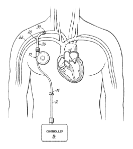
[0003] An alternative form of counterpulsation is shown in Figure 1.
Here
a pump 10 is implanted in a pacemaker pocket on the patient's right side.
Blood fills the pump 10 on one side and air or other fluid fills a sac or
bladder
(not shown) on the other side of the pump 10. An air drive line 12 is tunneled
from the pacemaker pocket to a skin exit site 14. so the entire pump 10 is
under
the skin and can remain there chronically. After the driveline 1.2 exits the
skin, it
is attached to a small air drive unit 16 that controls shuttling of
pressurized air in
and out of the pump 10. A void in the pump 10 may be formed with the sac or
bladder if void fill with air as the heart beats (less cardiac work in
ejecting
blood) and empties to return blood into the circulation (more flow to the
patient).
-1-
CA 02844744 2014-02-07
WO 2013/043276
PCT/US2012/050604
The pump 10 is attached to the circulation with a conduit 20. The conduit 20
shuttles blood between the patient's circulatory system and the pump 10. This
situation allows a patient to have chronic counterpulsation with full
mobility. For
a patient with severe and potentially non-reversible cardiac dysfunction, this
is a
great advantage as it is possible to live a relatively normal life ¨ apart
from the
need to carry a small battery powered drive console 16,
[00041 As described, the blood is shuttled in and out of the pump 10
with
a conduit 20 which is connected to the circulation. There are a number of
considerations related to implantation and use of this conduit 20. First,
almost
every conduit has blood flowing in one direction, but this conduit 20 has
blood
alternating flow direction two times for each heart beat as the pump 10 fills
and
empties with each cardiac cycle. This creates a number of important issues
which will be described. A second potential difficulty with a conduit in this
situation is that it will typically be sewn to the subclavian artery 22 or ax
Vary
artery which is located beneath the clavicle and often quite deep, so it is
technically difficult for a surgeon to suture the end of the conduit 20 to the
artery
22.
[00051 The problem of a conduit with bidirectional flow relates to
the
responses of blood and tissues to the interfaces with synthetic materials and
the response is dependent on the direction of .flood flow. Many medical
devices, such as blood pumps, are connected to the patient's circulation with
artificial graft material such as polyester materials like Dacron or
expanded,
porous Teflon (ePTFE.) that will promote tissue or cell ingrowth. The inside
of
blood pumps are generally smooth and composed of metals or plastics. When
blood flows from a smooth metal or plastic blood pump into a synthetic graft
(such as polyester), the interface where the pump meets the conduit (plastic
or
metal to synthetic graft) is a stable junction and there tends to be i:E,19
problem
when blood flows forward through this junction,
[0006] Unfortunately, experience has shown that when k.)lood instead
flows from a synthetic graft such as polyester into a smooth surfaced blood
pump, a deposit of blood elements including platelets and fibrin tends to
deposit
at the junction of the two materials ¨ principally on the synthetic graft and
overhanging the 'inflow to the pump. These deposits, especially platelets,
tend
-2-
CA 02844744 2015-04-07
to attract more blood elements and large and often fragile deposits occur at
this
junction. These deposits can break free from the junction and enter the blood
pump and be sent through the patient's circulation. These deposits can flow
anywhere, but if they arrive in an artery to the brain, a stroke can result.
For this
reason, many successful blood pumps employ a smooth synthetic conduit
(such as silicone or urethane) for blood inflow into the pump.
[0007] The problem with counterpulsation is that blood is flowing in an
alternating bi-directional manner. One solution would be to use a smooth
silicone or urethane conduit which would create a stable junction between the
pump and the conduit where the blood enters into the pump. This solves the
problem at the inflow to the pump. However, when a silicone material is
anastomosed (sewn) to an artery, the junction develops a heavy deposit of
blood material (fibrin and platelets). So merely replacing the inflow conduit
with
a silicone surface is not satisfactory, it is tempting to merely have a
silicone
conduit and add a fabric extension, but this merely moves the problem that
occurs at the junction of the rough textured surface of the graft and the pump
to
the junction between the graft and the silicone tube or cannula.
Brief Description of the Drawings
[0007a] Figure 1 depicts an alternative form of counterpulsation. A pump
10
is implanted in a pacemaker pocket on the patient's right side.
[0007b] Figures 2 shows an embodiment with an arrangement where the
smooth and rough surfaces are not in direct linear contact. A graft element is
sewn to the artery.
[0007c] Figure 3 shows an embodiment with an arrangement where the
smooth and rough surfaces are not in direct linear contact. It shows
configuration
permitting sewing of element 30 around an opening 22a on the artery 22.
[0007d] Figure 4 shows a junction between the silicone material portion 26
of the conduit 20 and the graft element 30.
-3-
CA 02844744 2015-04-07
[0007e] Figure 5 shows two flanges 32, 34 are sewn together.
[0007f] Figure 6A shows a side cross sectional view ot the two flanges 32,
34 coming together.
[0007g] Figure 6B shows the bubble or enlarged connector 30.
[0007h] Figure 6B shows clips or staples 38 attaching the connector 30 to
the artery 22 and attaching the flanges 32, 34 together.
[00071] Figure 7 shows a bubble or enlarged area 40 constructed by
"splitting" the bubble In the middle of the hemisphere.
[0007j] Figure 8 is a cross-sectional view of an embodiment of the device.
The subclavian artery 22 Is shown at the top of the figure.
[0007k] Figure 9 shows an arrangement of the "bubble" or enlarged area
24a of graft material located away from the anastomosis.
[00071] Figure 10A and 10B show an arrangement at the junction of the
pump 10, with the smooth surfaced junction or tip portion 10a of the pump 10
separated from the rough surface of the enlarged graft material by a bubble
interface 24a.
[0008] Figure 8 shows one potential solution in a cross-sectional view.
The subclavian artery 22 is shown at the top of the figure. A "bubble" or
enlarged area 24 of Dacron , Teflon or other material is sewn to the artery
22. A silicone or other smooth material conduit portion 26 is connected to the
other side of the enlarged area 24. Rather than a direct junction, a special
interface is created. The smooth silicone surface portion 26 extends with a
tip
portion 26a several millimeters inside the enlarged area 24 of fabric or other
material. The walls of the silicone tip portion 26a do not contact the fabric
or
material of the enlarged area or bubble 24. This avoids a silicone-to-fabric
(or
smooth-to-rough) point of contact.
-3a-
CA 02844744 2015-04-07
[0009] Heart valves have been constructed with arrangements to avoid
tissue ingrowth into the valve by creating an elevation - so that there is not
a
continuous connection between the fabric surface and the smooth surface. This
elevation prevents tissue from growing over into junction point and creating a
point where platelets and fibrin are deposited. The use of a small washer of
-3b-
CA 02844744 2014-02-07
WO 2013/043276
PCT/US2012/050604
material may also be of use. Figure 8 shows a small washer 28 around the
base of the tip 26a that may help arrest the attachment of blood elements.
[0010] Figure 9 shows that this arrangement of the "bubble" or
enlarged
area 24a of graft material is located away from the ana.stomosis.
Specifically,
enlarged area 24a is coupled to or includes an extension 24b that is
anastomosed to the artery 22. Other features may be as described pre.viously.
[0011] Figure 10A and 10B show a similar arrangement can be made at
the junction of the pump 10. Here, the plastic, metal or other smooth surfaced
junction or tip portion 10a of the pump 10 is separated from the rough surface
of
the enlarged graft material by a bubble interface 24a. An extension 24b of the
graft material is sewn to the artery 22 (Fig. 9) as previously described.
Another
extension 24c on the opposite end may fa.cilitate connection to the pump
interface or -tip portion 10a, along with a suitable connector 28. The
junction or
interface 10a, which serves as an inlet/outlet port that extends into, but
does
normally not contact, the graft material 24a in use.
Luv
'4*-t,;11
These devices with bubbles or enlargements could be made in
one piece. As described previously, the subclavian artery 22 is located fairly
deep and the incision is smaL So a surgeon who is trying to sew a graft with a
bubble or enlargement on it is working in a deep hole. The bubble or
enlargement on the end of a graft obscures his view of the artery. It would be
useful to avoid this problem and also satisfy the need for maintaining the
arrangement where the smooth and rough surfaces are not n direct linear
contact.
[0013] Such a solution is shown in Figures 2 and 3. Here, a graft
element form from material such as described above is sewn to the artery. The
graft element 30 has a flange 32 at one end. The element 30 is small and easy
to move around, so does not obscure the view of the surgeon. Figure 3 shows
that it is easy to sew this element 30 around an opening 22a on the artery 22.
[0014] Figure 4 shows how a junction between the silicone material
portion 26 of the conduit 20 and the graft element 30 is recreated when a rim
or
flange 34 of sewing material or graft material, for example, of the conduit
CA 02844744 2015-04-07
WO 2013/043276
PCT/US2012/050604
portion 26 is affixed to the flange 32 on the element 30 previously
anastomo.sed
to the artery 22.
[0015] Figure 5 shows how the two flanges 32, 34 are sewn together.
This is a very easy anastomosis to perform.
[0016] It wili be appreciated that these flanges 32, 34 could be
joined not
just by sutures but by staples, clips, glues, clamps etc.
[0017] Figure 6A shows a side cross sectional view pi the two flanges
32,
34 coming together.
[0018] Figure 68 shows how the bubble or enlarged connector 30 does
not have to be fiat it could be beveled. Also the connector 30 does not have
to be a generally spherical bubble as shown elsewhere herein. The key is only
that the enlarged area keeps the silicone and graft surfaces (that is, smooth
and
rough flow surfaces) from direct contact at their junction during use,
[0019] The bubble or enlarged area 36 is quite useful as it allows
the
tip portion to move or "swivel" inside the bubble 36 and still not contact the
wall
of the bubble 36.
[0020] Figure 68 also shows clips or staples 38 attaching the
connector
30 to the artery 22 and attaching the flanges 32, 34 together.
[0021] The conduit portion 26 does not have to be entirely silicone.
it
could have any inner core that presents a compatible surface to the exposed
blood. For example, the inside could be metal, have a metal spiral re-
inforcement, etc. It could also have graft material inside like c.,.PTFE or
other
polyester.
[0022] The smooth surface does not have to be silicone. This is used
as
representative of a smooth surface. The surface could be a metal or plastic
(such as in the pump connection shown in Figures 10A and 108.1
[0023] Figure 7 shows a bubble or enlarged area 40 constructed by
"splitting" the bubble in the middle of the hemisphere, it could be equally
-5-
CA 02844744 2015-04-07
possible to form the junction 42 anywhere in this arrangement; the location at
the
hemisphere is merely an example.
[0024] Alternatively, a more complete bubble could be created and the
silicone cannula could be slipped into a defect at the end to perform the same
function.
[0025] It should be noted that the terms used are basically smooth
(silicone, plastics, metals) and rough or textured surfaces (Dacron, Teflon,
ePTFE). It is also possible to have a tightly woven or knitted material that
is
typically called a textile, but could function as a smooth surface.
[0026] Also, It is possible to create a tightly woven polyester that
behaves
like a smooth surface. It could be possible to bring a tightly woven sewable
graft
into direct contact with a silicone surface without an intervening "bubble" or
step.
[0027] It may also be important to prevent these conduits from collapsing
as they can be located below the skin and could be crushed by a patient lying
on
them. Reinforcement of the conduits with plastic or wire spirals or rings can
be
used here. In addition, extra thicknesses of polymer or plastic could be added
make them stronger.
[0028] The scope of the claims should not be limited to the preferred
embodiments but should be given the broadest interpretation consistent with
the
description as a whole. Additional advantages and modifications will readily
appear to those skilled in the art. The various features discussed herein may
be
used alone or in any combination depending on the needs and preferences of the
user. This has been a description of illustrative aspects and embodiments the
present invention, along with the preferred methods of practicing the present
invention as currently known.
-6-
