Note: Descriptions are shown in the official language in which they were submitted.
CA 02844822 2014-02-10
WO 2013/033629
PCT/US2012/053518
METHODS AND COMPOSITIONS FOR THE TREATMENT AND DIAGNOSIS OF
COLORECTAL CANCER
10001] This application claims priority US Provisional Application No.
61/529,525,
filed August 31, 2011, the entire contents of which are hereby incorporated by
reference.
Field of the Invention
100021 The field of the invention relates to cancer and the diagnosis and
treatment of
cancer.
Background
[0003] Early detection of cancer can impact treatment outcomes and disease
progression. Typically, cancer detection relies on diagnostic information
obtained from
biopsy, x-rays, CAT scans, Nivrtz and the like. These procedures may be
invasive, time
consuming and expensive. Moreover, they have limitations with regard to
sensitivity and
specificity. There is a need in the field of cancer diagnostics for a highly
specific, highly
sensitive, rapid, inexpensive, and relatively non-invasive method of
diagnosing cancer.
Various embodiments of the invention described below meet this need as well as
other needs
existing in the field of diagnosing and treating cancer.
Summary of the Invention
100041 Embodiments of the disclosure provide methods of diagnosis, prognosis
and
treatment of cancer, e.g. colorectal cancer. Other embodiments provide
compositions relating
to the diagnosis, prognosis and treatment of cancer, such as colorectal
cancer.
[0005] In certain embodiments the invention provides a method of detecting
colorectal cancer in a subject comprising a) obtaining a sample from a
subject; b) contacting
the sample obtained from the subject with one or more agents that detect one
or more markers
expressed by a colorectal cancer cell c) contacting a non-cancerous cell with
the one or more
agents from b); and d) comparing the expression level of the marker in the
sample obtained
from the subject with the expression level in the non-cancerous cell, wherein
a higher level of
expression of the marker in the sample compared to the non-cancerous cell
indicates that the
subject has colorectal cancer. =
[0006] In certain embodiments the invention provides a method of detecting
colorectal cancer in a subject comprising a) obtaining a sample from a
subject; b) contacting
the sample obtained from the subject with one or more agents that detect
expression of at
least one of the markers listed in Table 1; c) contacting a non-cancerous
cell, with the one or
more agents from b); and d) comparing the expression level of one or more of
the markers
listed 111 Table I in the sample obtained from the subject with the expression
level of one or
CA 02844822 2014-02-10
WO 2013/033629
PCT/US2012/053518
more of the markers listed in Table 1 in the non-cancerous cell, wherein a
higher level of
expression of one or more of the markers listed in Table lin the sample
obtained from the
subject compared to the non-cancerous cell indicates that the subject has
colorectal cancer.
[0007] In some embodiments the invention provides a method of detecting
colorectal
cancer in a subject comprising a) obtaining a sample from a subject b)
contacting the sample
obtained from the subject with one or more agents that detect expression of
one or more of
the markers encoded by genes chosen from Homo sapiens serine peptidase
inhibitor, Kazal
type 4 (SPINK4), Homo sapiens LINE-1 type transposase domain containing 1
(LITD1),
Homo sapiens solute carrier family 35, member D3 (SLC35D3), Homo sapiens
lymphocyte
antigen 6 complex, locus G6D (LY6G6D), Homo sapiens matrix metallopeptidase 12
(macrophage elastase) (MMP12), Homo sapiens matrix metallopeptidase 12
(macrophage
elastase) (MMP12), Homo sapiens apolipoprotein B mRNA editing enzyme,
catalytic
polypeptide 1 (APOBEC1), Homo sapiens dickkopf homolog 4 (Xenopus laevis)
(DKK4),
Homo sapiens NADPH oxidase 1 (NOX1), Homo sapiens matrix metallopeptidase 11
(stromelysin 3) (MVP I 1), Homo sapiens ring finger protein 43 (RNF43),
AGENCOURT 10229596 N11-1 MGC 141 Homo sapiens cDNA clone IMAGE:6563923 5
(BU536065), Homo sapiens KIAA1199 (KIAA1199), Homo sapiens carcinoembryonic
antigen-related cell adhesion molecule 5 (CEACAM5), Homo sapiens achaete-scute
complex
homolog 2 (Drosophila) (ASCL2), Homo sapiens villin 1 (VIL1), Homo sapiens
naked
cuticle homolog I (Drosophila) (NKD1), PREDICTED: HOMO sapiens hypothetical
L00729669 (L00729669), Homo sapiens mucin 17, cell surface associated (MUC17),
Homo sapiens notum pectinacetylesterase homolog (Drosophila) (NOTUM), Homo
sapiens
collagen, type XI, alpha 1 (COL11A1), Homo sapiens defensin, alpha 5, Paneth
cell-specific
(DEFA5), Homo sapiens notum pectinacetylesterase homolog (Drosophila) (NOTUM),
Homo sapiens phospholipase inhibitor (L00646627), Homo sapiens NADPH oxidase
organizer 1 (NOX01), Homo sapiens lipocalin 15 (LCN15), Homo sapiens chemokine
(C-C
motif) ligand 24 (CCL24), Homo sapiens gastrin-releasing peptide (GRP), Homo
sapiens
pregnancy specific beta-l-glycoprotein 1 (PSG1), Homo sapiens claudin 2
(CLDN2), Homo
sapiens defensin, alpha 6, Paneth cell-specific (DEFA6), Homo sapiens
neuropeptide S
receptor 1 (NPSR1), Homo sapiens cystatin SN (CST1), Homo sapiens keratin 23
(histone
deacetylase inducible) (KRT23), Homo sapiens matrix metallopeptidase 7
(matrilysin,
uterine) (MMP7), Homo sapiens membrane-spanning 4-domains, subfamily A, member
12
(MS4Al2), Homo sapiens keratin 20 (KRT20), or a complement thereof; c)
contacting a non-
cancerous cell with the one or more agents from b); and d) comparing the
expression level of
2
CA 02844822 2014-02-10
WO 2013/033629
PCT/US2012/053518
one or more of the markers encoded by genes chosen from Homo sapiens serine
peptidase
inhibitor, Kazal type 4 (SPINK4), Homo sapiens LINE-I type transposase domain
containing
1 (LI TD1), Homo sapiens solute carrier family 35, member D3 (SLC35D3), Homo
sapiens
lymphocyte antigen 6 complex, locus G6D (LY6G6D), Homo sapiens matrix
metallopeptidase 12 (macrophage elastase) (MMP12), Homo sapiens matrix
metallopeptidase
12 (macrophage elastase) (MMP12), Homo sapiens apolipoprotein B mRNA editing
enzyme,
catalytic polypeptide I (APOBEC1), Homo sapiens dickkopf homolog 4 (Xenopus
laevis)
(DICK4), Homo sapiens NADPH oxidase 1 (NOX I), Homo sapiens matrix
metallopeptidase
11 (stromelysin 3) (MMP11), Homo sapiens ring finger protein 43 (RNF43),
AGENCOURT 10229596 NIH MGC 141 Homo sapiens cDNA clone IMAGE:6563923 5
(BU536065), Homo sapiens KIAA1199 (KIAA1199), Homo sapiens carcinoembryonic
antigen-related cell adhesion molecule 5 (CEACAM5), Homo sapiens achaete-scute
complex
homolog 2 (Drosophila) (ASCL2), Homo sapiens villin 1 (VIL1), Homo sapiens
naked
cuticle homolog 1 (Drosophila) (NKD1), PREDICTED: Homo sapiens hypothetical
L00729669 (L00729669), Homo sapiens mucin 17, cell surface associated (MUC17),
Homo sapiens notum pectinacetylesterase homolog (Drosophila) (NORM), Homo
sapiens
collagen, type XI, alpha 1 (COL11A1), Homo sapiens defensin, alpha 5, Paneth
cell-specific
(DEFA5), Homo sapiens notum pectinacetylesterase homolog (Drosophila) (NOTUM),
Homo sapiens phospholipase inhibitor (L00646627), Homo sapiens NADPH oxidase
organizer I (NOX01), Homo sapiens lipocalin 15 (LCN15), Homo sapiens chemokine
(C-C
motif) ligand 24 (CCL24), Homo sapiens gastrin-releasing peptide (GRP), Homo
sapiens
pregnancy specific beta-1 -glycoprotein I (PSG1), Homo sapiens claudin 2
(CLDN2), Homo
sapiens defensin, alpha 6, Paneth cell-specific (DEFA6), Homo sapiens
neuropeptide S
receptor 1 (NPSR1), Homo sapiens cystatin SN (CST1), Homo sapiens keratin 23
Oilstone
deacetylase inducible) (KRT23), Homo sapiens matrix metallopeptidase 7
(matrilysin,
uterine) (MMP7), Homo sapiens membrane-spanning 4-domains, subfamily A, member
12
(MS4Al2), Homo sapiens keratin 20 (KRT20), or a complement thereof in the non-
cancerous
cell, wherein a higher level of expression of one or more of the markers
encoded by genes
chosen from Homo sapiens serine peptidase inhibitor, Kazal type 4 (SPINK4),
Homo sapiens
LINE-1 type transposase domain containing 1 (LITDI), Homo sapiens solute
carrier family
35, member D3 (SLC35D3), Homo sapiens lymphocyte antigen 6 complex, locus G6D
(LY6G6D), Homo sapiens matrix metallopeptidase 12 (macrophage elastase)
(MMP12),
Homo sapiens matrix metallopeptidase 12 (macrophage elastase) (MMP12), Homo
sapiens
apolipoprotein B inRNA editing enzyme, catalytic polypeptide I (APOBEC1), Homo
sapiens
3
CA 02844822 2014-02-10
WO 2013/033629
PCT/US2012/053518
dickkopf homolog 4 (Xenopus laevis) (DKK4), Homo sapiens NADPH oxidase I
(NOX1),
Homo sapiens matrix metallopeptidase 11 (stromelysin 3) (MMPI1), Homo sapiens
ring
finger protein 43 (RNF43), AGENCOURT_10229596 NIH_MGC_141 Homo sapiens cDNA
clone 1MAGE:6563923 5 (3U536065), Homo sapiens KIAA1199 (KIAA1199), Homo
sapiens carcinoembryonic antigen-related cell adhesion molecule 5 (CEACAM5),
Homo
sapiens achaete-scute complex homolog 2 (Drosophila) (ASCL2), Homo sapiens
villin 1
(VIL1), Homo sapiens naked cuticle homolog 1 (Drosophila) (NKD1), PREDICTED:
Homo
sapiens hypothetical L00729669 (L00729669), Homo sapiens mucin 17, cell
surface
associated (MUC17), Homo sapiens notum pectinacetylesterase homolog
(Drosophila)
(NOTUM), Homo sapiens collagen, type )U, alpha I (COL11A1), Homo sapiens
defensin,
alpha 5, Paneth cell-specific (DEFA5), Homo sapiens notum pectinacetylesterase
homolog
(Drosophila) (NOTUM), Homo sapiens phospholipase inhibitor (L00646627), Homo
sapiens NADPH oxidase organizer I (NOX01), Homo sapiens lipocalin 15 (LCN15),
Homo
sapiens chemokine (C-C motif) ligand 24 (CCL24), Homo sapiens gastrin-
releasing peptide
(GRP), Homo sapiens pregnancy specific beta-1-glycoprotein 1 (PSG1), Homo
sapiens
claudin 2 (CLDN2), Homo sapiens defensin, alpha 6, Paneth cell-specific
(DEFA6), Homo
sapiens neuropeptide S receptor 1 (NPSR1), Homo sapiens cystatin SN (CST1),
Homo
sapiens keratin 23 Oilstone deacetylase inducible) (KRT23), Homo sapiens
matrix
metallopeptidase 7 (matrilysin, uterine) (MMP7), Homo sapiens membrane-
spanning 4-
domains, subfamily A, member 12 (MS4Al2), Homo sapiens keratin 20 (KRT20), or
a
complement thereof in the sample obtained from the subject compared to the non-
cancerous
cell indicates that the subject has colorectal cancer,
[0008] In other embodiments the invention provides a method of detecting
colorectal
cancer in a subject comprising a) obtaining a sample from a subject b)
contacting the sample
obtained from the subject with one or more agents that detect expression of a
panel of
markers encoded by the genes SPINK4, LITDI, LY6G6D, APOBECI, L00729669,
COL1OA, SLC35D 1024, MMP7, MMP12, NMU, WNTIOA or a complement thereof; c)
contacting a non-cancerous cell, with the one or more agents from b); and d)
comparing the
expression level of the panel of markers encoded for by the genes SPINK4,
LITDI,
LY6G6D, APOBECI, L00729669, COL10A, SLC35D 1024, MMP7, MMP12, NMU,
WNTI OA Or a complement thereof in the sample obtained from the subject with
the
expression level of the panel of markers encoded for by the genes SPINK4, L1TD
I,
LY6G6D, APOBEC1, L00729669, COL10A, SLC35D_1024, MMP7, MMP12, NMU,
WNT1OA or a complement thereof in the non-cancerous cell, wherein a higher
level of
4
CA 02844822 2014-02-10
WO 2013/033629
PCT/US2012/053518
expression of the panel of markers encoded for by genes SPINK4, LlTDI, LY6G6D,
APOBECI, L00729669, COL1OA, SLC35D 1024, MMP7, MMP12, NMU, WNTI OA or a
complement thereof in the sample compared to the non-cancerous cell in wherein
a higher
level of expression of the panel of markers encoded by genes SPINK4, L1TD1,
LY6G6D,
APOBEC1, L00729669, COL10A, SLC35D 1024, MMP7, MMP12, NMU, WNT10A or a
complement thereof in the sample compared to the non-cancerous cell indicates
that the
subject has colorectal cancer that the subject has cancer.
[0009] In some embodiments the invention provides a method of detecting
colorectal
cancer in a subject comprising a) obtaining a sample from a subject b)
contacting the sample
obtained from the subject with one or more agents that detect expression of
one or more of
the markers encoded by genes chosen from SPINK4, L ITD I, LY6G6D, APOBEC1,
L00729669, COL1OA, SLC35D _1024, MMP7, MMP12, NMU, WNTIOA or a complement
thereof; c) contacting a non-cancerous cell with the one or more agents from
b); and d)
comparing the expression level of one or more of the markers encoded by genes
chosen from
SPINK4, LlTDI, LY6G6D, APOBECI, L00729669, COLIOA, SLC35D 1024, MMP7,
MMPI2, NMU, WNT10A or a complement thereof in the sample obtained from the
subject
with the expression level of one or more of the markers encoded by genes
chosen from
SPINK4, LlTDI, LY6G6D, APOBEC1, L00729669, COL10A, SLC3513_1024, MMP7,
MMP12, NMU, WNT10A or a complement thereof in the non-cancerous cell, wherein
a
higher level of expression of one or more of the markers encoded by genes
chosen from
SPINK4, L1TD1, LY6G6D, APOBEC1, L00729669, COL10A, SLC3513_1024, MMP7,
MMP12, NMU, WNT10A or a complement thereof in the sample obtained from the
subject
compared to the non-cancerous cell indicates that the subject has colorectal
cancer.
[0010] In further embodiments the invention provides a method of detecting
colorectal cancer cells in a sample comprising a) obtaining a sample b)
contacting the sample
obtained in a) with one or more agents that detect expression of one or more
of the markers
encoded by genes chosen from Homo sapiens serine peptidase inhibitor, Kazal
type 4
(SPINK4), Homo sapiens LINE-1 type transposase domain containing 1 (LITDI),
Homo
sapiens solute carrier family 35, member D3 (SLC35D3), Homo sapiens lymphocyte
antigen
6 complex, locus G6D (LY6G6D), Homo sapiens matrix metallopeptidase 12
(macrophage
elastase) (MMP12), Homo sapiens matrix metallopeptidase 12 (macrophage
elastase)
(MMP12), Homo sapiens apolipoprotein B mRNA editing enzyme, catalytic
polypeptide 1
(APOBECI), Homo sapiens dickkopf homolog 4 (Xenopus laevis) (DKK4), Homo
sapiens
NADPH oxidase 1 (NOXI), Homo sapiens matrix metallopeptidase 11 (stromelysin
3)
CA 02844822 2014-02-10
WO 2013/033629
PCT/US2012/053518
(MMP11), Homo sapiens ring finger protein 43 (RNF43), AGENCOURT_10229596
NIH MGC 141 Homo sapiens cDNA clone IMAGE:6563923 5 (BU536065), Homo sapiens
KIAA1199 (KIAA1199), Homo sapiens carcinoembryonic antigen-related cell
adhesion
molecule 5 (CEACAM5), Homo sapiens achaete-scute complex homolog 2
(Drosophila)
(ASCL2), Homo sapiens villin I (VILI), Homo sapiens naked cuticle homolog I
(Drosophila) (NKD1), PREDICTED: Homo sapiens hypothetical L00729669
(L00729669),
Homo sapiens mein 17, cell surface associated (MUC17), 1-lomo sapiens notum
pectinacetylesterase homolog (Drosophila) (NOTUM), Homo sapiens collagen, type
XI,
alpha I (COL1 1A1), Homo sapiens defensin, alpha 5, Paned' cell-specific
(DEFA5), Homo
sapiens notum pectinacetylesterase homolog (Drosophila) (NOTUM), Homo sapiens
phospholipase inhibitor (L00646627), Homo sapiens NADPH oxidase organizer 1
(NOX01), Homo sapiens lipocalin 15 (LCN15), Homo sapiens chemokine (C-C motif)
ligand 24 (CCL24), Homo sapiens gastrin-releasing peptide (GRP), Homo sapiens
pregnancy
specific beta- -glycoprotein 1 (PSG1), Homo sapiens claudin 2 (CLDN2), Homo
sapiens
defensin, alpha 6, Paneth cell-specific (DEFA6), Homo sapiens neuropeptide S
receptor 1
(NPSR1), Homo sapiens cystatin SN (CST1), Homo sapiens keratin 23 (histone
deacetylase
inducible) (KRT23), Homo sapiens matrix metallopeptidase 7 (matrilysin,
uterine) (MMP7),
Homo sapiens membrane-spanning 4-domains, subfamily A, member 12 (MS4Al2), 1-
lomo
sapiens keratin 20 (KRT20), or a complement thereof; c) contacting a non-
cancerous cell
with the one or more agents from b); and d) comparing the expression level of
one or more of
the markers encoded by genes chosen from Homo sapiens serine peptidase
inhibitor, Kazal
type 4 (SPINK4), Homo sapiens LINE-1 type transposase domain containing 1
(LITDI),
Homo sapiens solute carrier family 35, member D3 (SLC35D3), Homo sapiens
lymphocyte
antigen 6 complex, locus G6D (LY6G6D), Homo sapiens matrix metallopeptidase 12
(macrophage elastase) (MMP12), Homo sapiens matrix inetallopeptidase 12
(macrophage
elastase) (MMP12), Homo sapiens apolipoprotein B inRNA editing enzyme,
catalytic
polypeptide I (APOBEC1), Homo sapiens dickkopf homolog 4 (Xenopus laevis)
(DKK4),
Homo sapiens NADPH oxidase 1 (NOXI), Homo sapiens matrix metallopeptidase 11
(stromelysin 3) (M_MP11), Homo sapiens ring finger protein 43 (RNF43),
AGENCOURT 10229596 NIH MGC 141 Homo sapiens cDNA clone 1MAGE:6563923 5
(BU536065), Homo sapiens KIAA1199 (KIAA1199), Homo sapiens eareinoembryonie
antigen-related cell adhesion molecule 5 (CEACAM5), Homo sapiens achaete-scute
complex
homolog 2 (Drosophila) (ASCL2), Homo sapiens villin I (VILI), Homo sapiens
naked
cuticle homolog 1 (Drosophila) (NKD1), PREDICTED: Homo sapiens hypothetical
6
CA 02844822 2014-02-10
WO 2013/033629
PCT/US2012/053518
L00729669 (L00729669), Homo sapiens tnucin 17, cell surface associated
(MUC17),
Homo sapiens notum pectinacetylesterase homolog (Drosophila) (NOTUM), Homo
sapiens
collagen, type XI, alpha 1 (COL11A1), Homo sapiens defensin, alpha 5, Paneth
cell-specific
(DEFA5), Homo sapiens not= pectinacetylesterase homolog (Drosophila) (NOTUM),
Homo sapiens phospholipase inhibitor (L00646627), Homo sapiens NADPH oxidase
organizer 1 (NOX01), Homo sapiens lipocalin 15 (LCN15), Homo sapiens chemokine
(C-C
motif) ligand 24 (CCL24), Homo sapiens gastrin-releasing peptide (GRP), Homo
sapiens
pregnancy specific beta-l-glycoprotein 1 (PSG1), Homo sapiens claudin 2
(CLDN2), Homo
sapiens defensin, alpha 6, Paneth cell-specific (DEFA6), Homo sapiens
neuropeptide S
receptor 1 (NPSR1), Homo sapiens cystatin SN (CST1), Homo sapiens keratin 23
Oilstone
deacetylase inducible) (KRT23), Homo sapiens matrix metallopeptidase 7
(matrilysin,
uterine) (MMP7), Homo sapiens membrane-spanning 4-domains, subfamily A, member
12
(MS4Al2), Homo sapiens keratin 20 (KRT20), or a complement thereof in the
sample
obtained in a) with the expression level of one or more of the markers encoded
by genes
chosen from Homo sapiens serine peptidase inhibitor, Kazal type 4 (SPINK4),
Homo sapiens
LINE-1 type transposase domain containing 1 (L1TD1), Homo sapiens solute
carrier family
35, member D3 (SLC35D3), Homo sapiens lymphocyte antigen 6 complex, locus G6D
(LY6G6D), Homo sapiens matrix metallopeptidase 12 (macrophage elastase)
(MMP12),
Homo sapiens matrix metallopeptidase 12 (macrophage elastase) (MMPI2), Homo
sapiens
apolipoprotein B mRNA editing enzyme, catalytic polypeptide I (APOBEC1), Homo
sapiens
dickkopf homolog 4 (Xenopus laevis) (DKK4), Homo sapiens NADPH oxidase 1
(NOX1),
Homo sapiens matrix metallopeptidase 11 (stromelysin 3) (MMP I I), Homo
sapiens ring
finger protein 43 (RNF43), AGENCOURT_10229596 NIH_MGC_141 Homo sapiens cDNA
clone IMAGE:6563923 5 (BU536065), Homo sapiens KIAA1199 (K1AA1199), Homo
sapiens carcinoembiyonic antigen-related cell adhesion molecule 5 (CEACAM5),
Homo
sapiens achaete-seute complex homolog 2 (Drosophila) (ASCL2), Homo sapiens
villin 1
(VIL1), Homo sapiens naked cuticle homolog 1 (Drosophila) (NKD1), PREDICTED:
Homo
sapiens hypothetical L00729669 (L00729669), Homo sapiens mucin 17, cell
surface
associated (MUC17), Homo sapiens Mum pectinacetylesterase homolog (Drosophila)
(NOTUM), Homo sapiens collagen, type XI, alpha 1 (COLI1A1), Homo sapiens
defensin,
alpha 5, Paneth cell-specific (DEFA5), Honio sapiens notum
pectinacetylesterase homolog
(Drosophila) (NOTUM), Homo sapiens phospholipase inhibitor (L00646627), Homo
sapiens NADPH oxidase organizer 1 (NOXO I), Homo sapiens lipocal in 15
(LCNI5), Homo
sapiens chemokine (C-C motif) ligand 24 (CCL24), Homo sapiens gastrin-
releasing peptide
7
CA 02844822 2014-02-10
WO 2013/033629
PCT/US2012/053518
(GRP), Homo sapiens pregnancy specific beta-I-glyeoprotein 1 (PSG1), Homo
sapiens
claudin 2 (CLDN2), Homo sapiens defensin, alpha 6, Paneth cell-specific
(DEFA6), Homo
sapiens neuropeptide S receptor 1 (NPSR1), Homo sapiens cystatin SN (CST1),
Homo
sapiens keratin 23 Oilstone deacetylase inducible) (KRT23), Homo sapiens
matrix
metallopeptidase 7 (matrilysin, uterine) (MMP7), Homo sapiens membrane-
spanning 4-
domains, subfamily A, member 12 (MS4Al2), Homo sapiens keratin 20 (KRT20), or
a
complement thereof in the non-cancerous cell, wherein a higher level of
expression of one or
more of the markers encoded by genes chosen from Homo sapiens serine peptidase
inhibitor,
Kazal type 4 (SPINK4), Homo sapiens LINE-I type transposase domain containing
I
(LITD1), Homo sapiens solute carrier family 35, member D3 (SLC35D3), Homo
sapiens
lymphocyte antigen 6 complex, locus G6D (LY6G6D), Homo sapiens matrix
metallopeptidase 12 (macrophage elastase) (MMP12), Homo sapiens matrix
metallopeptidase
12 (macrophage elastase) (MMP12), Homo sapiens apolipoprotein B mRNA editing
enzyme,
catalytic polypeptide 1 (APOBEC I), Homo sapiens dickkopf homolog 4 (Xenopus
Nevis)
(DKK4), Homo sapiens NADPH oxidase I (NOX1), Homo sapiens matrix
metallopeptidase
11 (stromelysin 3) (MMP 11), Homo sapiens ring finger protein 43 (RNF43),
AGENCOURT 10229596 NIH MGC 141 Homo sapiens cDNA clone 1MAGE:6563923 5
(BU536065), Homo sapiens KIAAI199 (KIAA1199), Homo sapiens carcinoembryonic
antigen-related cell adhesion molecule 5 (CEACAM5), Homo sapiens achaete-scute
complex
homolog 2 (Drosophila) (ASCL2), Homo sapiens villin 1 (VIL1), Homo sapiens
naked
cuticle homolog 1 (Drosophila) (NKDI), PREDICTED: Homo sapiens hypothetical
L00729669 (L00729669), Homo sapiens mucin 17, cell surface associated (MUCI7),
Homo sapiens notum pectinacetylesterase homolog (Drosophila) (NOTUM), Homo
sapiens
collagen, type XI, alpha 1 (COL11A1), Homo sapiens defensin, alpha 5, Paneth
cell-specific
(DEFA5), Homo sapiens notum pectinacetylesterase homolog (Drosophila) (NOTUM),
Homo sapiens phospholipase inhibitor (L00646627), Homo sapiens NADPH oxidase
organizer I (NOX01), Homo sapiens lipocalin 15 (LCN15), Homo sapiens chemokine
(C-C
motif) ligand 24 (CCL24), Homo sapiens gastrin-releasing peptide (GRP), Homo
sapiens
pregnancy specific beta- 1 -glycoprotein I (PSG1), Homo sapiens claudin 2
(CLDN2), Homo
sapiens defensin, alpha 6, Paneth cell-specific (DEFA6), Homo sapiens
neuropeptide S
receptor 1 (NPSR I), Homo sapiens cystatin SN (CSTI), Homo sapiens keratin 23
(histone
deacetylase inducible) (KRT23), Homo sapiens matrix metallopeptidase 7
(matrilysin,
uterine) (MMP7), Hoino sapiens membrane-spanning 4-domains, subfamily A,
member 12
(MS4Al2), Homo sapiens keratin 20 (KRT20), or a complement thereof in the
sample
8
CA 02844822 2014-02-10
WO 2013/033629
PCT/US2012/053518
compared to the non-cancerous cell indicates that the sample contains
colorectal cancer cells.
The sample may be an in vitro sample or an in vivo sample, or derived from an
in vivo
sample.
[0011] In certain embodiments the invention provides a method of detecting
colorectal cancer in a sample comprising a) contacting the sample with one or
more agents
that detect expression of at least one of the markers chosen from SPINK4,
L!TDI, LY6G6D,
APOBEC1, L00729669, COL10A, SLC35D 1024, MMP7, MMP12, NMU, WNT1OA; c)
contacting a non-cancerous cell, with the one or more agents from b); and d)
comparing the
expression level of one or more of the markers chosen from SPINK4, L ITDI,
LY6G6D,
APOBEC1, L00729669, COLI OA, SLC35D 1024, MMP7, MMP12, NMU, WNT1OA in
the sample with the expression level of one or more of the markers chosen from
SPINK4,
L1TD1, LY6G6D, APOBECI, L00729669, COL10A, SLC35D 1024, MMP7, MMP12,
NMU, WNT10A in the non-cancerous cell, wherein a higher level of expression of
one or
more of the markers in the sample chosen from SPINK4, L 1TD1, LY6G6D, APOBEC1,
L00729669, COL I OA, SLC35D 1024, MMP7, MMP12, NMU, WNTI OA in the sample
compared to the non-cancerous cell indicates that the sample has colorectal
cancer cells.
[0012] With regard to the embodiments described in the preceding paragraphs,
the
sample may be any sample as described infra, for example, a bodily fluid, such
as blood,
serum or urine. The sample may be a cellular sample or the extract of a
cellular sample. The
sample may be a tissue sample. Nucleic acids and/or proteins may be isolated
from the
sample. Nucleic acids such as RNA may be transcribed into cDNA. The agent may
be one or
more molecules that bind specifically to one or more proteins expressed by the
cancer cell or
one or more nucleic acids expressed by the cell. For example, the agent may be
a protein such
as an antibody that binds specifically to the protein expressed by one of the
marker genes
identified infra. The agent may be one or more nucleic acids that hybridize to
a nucleic acid
expressed by the cancer cell. The nucleic acid expressed by the cancer cell
may be an RNA
molecule, e.g. an mRNA molecule. The nucleic acid molecule that hybridizes to
the nucleic
acid expressed by the cancer cell may be a DNA molecule, such as a DNA probe.
[0013] In still other embodhnents the invention provides a composition of
matter
useful in distinguishing a colorectal cancer cell from a non-cancerous cell
comprising one or
more molecules that specifically bind to a molecule expressed at higher levels
on a colorectal
cancer cell compared to a non-cancer cell. As an example, the composition may
comprise a
protein, that binds to one or more molecules expressed by the colorectal
cancer cell at higher
levels compared to the non-cancer cell. As another example, the composition
may comprise
9
CA 02844822 2014-02-10
WO 2013/033629
PCT/US2012/053518
a nucleic acid that binds to one or more molecules expressed by the colorectal
cancer cell at
higher levels compared to the non-cancer cell.
100141 In some embodiments the invention provides a composition of matter
comprising a protein, such as an antibody, that specifically binds to a
molecule expressed by
a colorectal cancer cell chosen from the markers encoded by the sequences
listed in Table 1.
The molecule expressed by the colorectal cancer cell may be expressed by the
cancer cell at a
level that is higher than the level expressed by a non-cancerous cell.
[0015] In further embodiments the invention provides a composition of matter
comprising a plurality of proteins, such as a plurality antibodies, that
specifically binds to a
panel of molecules expressed by a colorectal cancer cell wherein the panel of
markers
comprises molecule encoded by the genes SPINK4, L1TD1, LY6G6D, APOBEC1,
L00729669, COL1OA, SLC35D 1024, MMP7, MMP12, NMU, WNT10A or a complement
thereof. The panel of markers may be expressed at a level that is higher than
the level of the
panel of markers in a non-cancerous cell.
[0016] In certain embodiments the invention provides a composition of matter
comprising a protein, such as an antibody, that specifically binds to a
molecule expressed by
a colorectal cancer cell chosen from a molecule encoded by one or more of the
genes chosen
from Homo sapiens serine peptidase inhibitor, Kazal type 4 (SPINK4), Homo
sapiens LINE-
I type transposase domain containing 1 (L1TD1), Homo sapiens solute carrier
family 35,
member D3 (SLC35D3), Homo sapiens lymphocyte antigen 6 complex, locus G6D
(LY6G6D), Homo sapiens matrix metallopeptidase 12 (macrophage elastase)
(MMP12),
Homo sapiens matrix metallopeptidase 12 (macrophage elastase) (MMP12), Homo
sapiens
apolipoprotein B mRNA editing enzyme, catalytic polypeptide 1 (APOBEC I), Homo
sapiens
dieldcopf homolog 4 (Xenopus laevis) (DKK4), Homo sapiens NADPH oxidase 1
(NOX1),
Homo sapiens matrix metallopeptidase II (stromelysin 3) (MMP1 I), Homo sapiens
ring
finger protein 43 (RNF43), AGENCOURT_10229596 NTH_MGC_141 Homo sapiens cDNA
clone IMAGE:6563923 5 (BU536065), Homo sapiens KIAA1199 (K1AA1199), Homo
sapiens carcinoembryonic antigen-related cell adhesion molecule 5 (CEACAM5),
Homo
sapiens achaete-scute complex homolog 2 (Drosophila) (ASCL2), Homo sapiens
villin 1
(VIL1), Homo sapiens naked cuticle homolog 1 (Drosophila) (NKD I), PREDICTED:
Homo
sapiens hypothetical L00729669 (L00729669), Homo sapiens mucin 17, cell
surface
associated (MUC17), Homo sapiens notum pectinacetylesterase homolog
(Drosophila)
(NOTUM), Homo sapiens collagen, type XI, alpha 1 (COL11A1), Homo sapiens
defensin,
alpha 5, Paneth cell-specific (DEFA5), Homo sapiens notum pectinacetylesterase
homolog
CA 02844822 2014-02-10
WO 2013/033629
PCT/US2012/053518
(Drosophila) (NOTUM), Homo sapiens phospholipase inhibitor (L00646627), Homo
sapiens NADPH oxidase organizer 1 (NOX01), Homo sapiens lipocalin 15 (LCN15),
Homo
sapiens chemokine (C-C motif) ligand 24 (CCL24), Homo sapiens gastrin-
releasing peptide
(GRP), Homo sapiens pregnancy specific beta-1-glycoprotein I (PSG1), Homo
sapiens
claudin 2 (CLDN2), Homo sapiens defensin, alpha 6, Paneth cell-specific
(DEFA6), Homo
sapiens neuropeptide S receptor 1 (NPSR1), Homo sapiens cystatin SN (CST1),
Homo
sapiens keratin 23 Oilstone deacetylase inducible) (KRT23), Homo sapiens
matrix
metallopeptidase 7 (matrilysin, uterine) (MMP7), Homo sapiens membrane-
spanning 4-
domains, subfamily A, member 12 (MS4Al2), Homo sapiens keratin 20 (KRT20), or
a
complement thereof. The molecule expressed by the colorectal cancer cell may
be expressed
by the colorectal cancer cell at level that is higher than the level expressed
by a non-
cancerous cell.
10017] In other embodiments the invention provides a composition of matter
comprising a nucleic acid that specifically binds to a molecule, such as an
mRNA molecule,
expressed by a colorectal cancer cell wherein the molecule is chosen from a
marker encoded
for by the genes listed in Table 1. The molecule expressed by the colorectal
cancer cell may
be expressed by the cancer cell at level that is higher than the level
expressed by a non-
cancerous cell,
[0018] In other embodiments the invention provides a composition of matter
comprising a nucleic acid that specifically binds to a molecule, such as an
triRNA molecule,
expressed by a colorectal cancer cell wherein the molecule is encoded for by a
gene disclosed
infra, e.g. a gene disclosed under the heading Cancer Associated Sequences, or
a complement
thereof. The molecule expressed by the colorectal cancer cell may be expressed
by the
cancer cell at level that is higher than the level expressed by a non-
cancerous cell.
[0019] In still further embodiments the invention provides a method of
determining if
a colorectal cancer in a subject is advancing comprising a) measuring the
expression level of
one or more markers associated with colorectal cancer at a first time point;
b) measuring the
expression level of the one or more markers measured in a) at a second time
point, wherein
the second time point is subsequent to the first time point; and c) comparing
the expression
level measured in a) and b), wherein an increase in the expression level of
the one or more
markers in b) compared to a) indicates that the subject's colorectal cancer is
advancing.
[0020] In some embodiments the invention provides a method of determining if a
colorectal cancer in a subject is advancing comprising a) measuring the
expression level of
one or more markers listed in Table 1 at a first time point; b) measuring the
expression level
CA 02844822 2014-02-10
WO 2013/033629
PCT/US2012/053518
of the one or more markers measured in a) at a second time point, wherein the
second time
point is subsequent to the first time point; and c) comparing the expression
level measured in
a) and b), wherein an increase ill the expression level of the one or more
markers at the
second time point compared to the first time point indicates that the
subject's colorectal
cancer is advancing.
[0021] In other embodiments the invention provides a method of determining if
a
colorectal cancer in a subject is advancing comprising a) measuring the
expression level of
one or more markers encoded by genes chosen from a gene disclosed infra, e.g.,
a gene
disclosed infra under the heading Cancer Associated Sequences, or a complement
thereof at
a first time point; b) measuring the expression level of the one or more
markers measured in
a) at a second time point, wherein the second time point is subsequent to the
first time point;
and c) comparing the expression level measured in a) and b), wherein an
increase in the
expression level of the one or more markers at the second time point compared
to the first
time point indicates that the subject's colorectal cancer is advancing.
100221 In some embodiments the invention provides antigens (i.e. cancer-
associated
polypeptides) associated with colorectal cancer as targets for diagnostic
and/or therapeutic
antibodies. In some embodiments, the antigen may be chosen from a protein
encoded by, a
gene listed in Table 1, a fragment thereof, or a combination of proteins
encoded by a gene
listed in Table I.
[0023] In some embodiments the invention provides antigens (i.e. cancer-
associated
polypeptides) associated with colorectal cancer as targets for diagnostic
and/or therapeutic
antibodies. In some embodiments, the antigen may be chosen from a protein
encoded by, a
gene chosen from a gene disclosed infra, e.g. under the heading Cancer
Associated Genes, a
fragment thereof, or a combination of proteins encoded by a gene (or fragments
thereof)
chosen from a gene disclosed infra, e.g. a gene disclosed under the heading
Cancer
Associated Sequences.
[0024] In yet other embodiments the invention provides a method of eliciting
an
immune response to a colorectal cancer cell comprising contacting a subject
with a protein or
protein fragment that is expressed by a cancer cell thereby eliciting an
immune response to
the colorectal cancer cell. As an example the subject may be contacted
intravenously or
intramuscularly with protein or protein fragment.
[0025] In further embodiments the invention provides a method of eliciting an
immune response to a colorectal cancer cell comprising contacting a subject
with one or more
proteins or protein fragments that is encoded by a gene chosen from the genes
listed in Table
12
CA 02844822 2014-02-10
WO 2013/033629
PCT/US2012/053518
1, thereby eliciting an immune response to a colorectal cancer cell, As an
example the
subject may be contacted with the protein or the protein fragment
intravenously or
intramuscularly,
[0026] In still other embodiments the invention provides a method of eliciting
an
immune response to a colorectal cancer cell comprising contacting a subject
with one or more
proteins or protein fragments that is encoded by a gene chosen from a gene
disclosed infra,
e.g., a gene disclosed under the heading Cancer Associated Sequences, thereby
eliciting an
immune response to a colorectal cancer cell. As an example the subject may be
contacted
with the protein or protein fragment intravenously or intramuscularly.
[0027i In yet other embodiments the invention provides a kit for detecting
colorectal
cancer cells in a sample. The kit may comprise one or more agents that detect
expression of
any the cancer associated sequences disclosed infra. The kit may include
agents that are
proteins and/or nucleic acids for example. In one embodiment the kit provides
a plurality of
agents. The agents may be able to detect the panel of markers encoded by the
genes
comprising SPINK4, L TD1, LY6G6D, APOBEC I , L00729669, COL I OA, SLC35D_1024,
MMP12, NMU, WNT1OA or a complement thereof.
[00281 In still other embodiments the invention provides a kit for detecting
colorectal
cancer in a sample comprising a plurality of agents that specifically bind to
a molecule
encoded for by the genes SPINK4, LITD1, LY6G6D, APOBEC I, L00729669, COL I OA,
SLC35D 1024, MMP7, MMP12, NMU, WNT I OA
[00291 In other embodiments the invention provides a kit for detection of
colorectal
cancer in a sample obtained from a subject. The kit may comprise one or more
agents that
bind specifically to a molecule expressed specifically by a colorectal cancer
cell. The kit may
comprise one or more containers and instructions for determining if the sample
is positive for
cancer. The kit may optionally contain one or more multiwell plates, a
detectable substance
such as a dye, a radioactively labeled molecule, a chemiluminescently labeled
molecule and
the like. The kit may further contain a positive control (e.g. one or more
cancerous cells; or
specific known quantities of the molecule expressed by the colorectal cancer
cell) and a
negative control (e.g. a tissue or cell sample that is non-cancerous).
100301 In some embodiments the invention provides a kit for the detection of
colorectal cancer comprising one or more agents that specifically bind one or
more markers
encoded by genes chosen from a gene disclosed infra., e.g., a gene disclosed
under the
heading Cancer Associated Sequences. The agent may be a protein, such as an
antibody.
Alternatively, the agent may be a nucleic such as a DNA molecule or an RNA
molecule. The
13
CA 02844822 2014-02-10
WO 2013/033629
PCT/US2012/053518
kit may comprise one or more containers and instructions for determining if
the sample is
positive for cancer. The kit may optionally contain one or more multiwell
plates, a detectable
substance such as a dye, a radioactively labeled molecule, a
chemiluminescently labeled
molecule and the like. The kit may further contain a positive control (e.g.
one or more
cancerous cells; or specific known quantities of the molecule expressed by the
colorectal
cancer cell) and a negative control (e.g. a tissue or cell sample that is non-
cancerous). As an
example the kit may take the form of an ELISA or a DNA microarray.
[0031] Some embodiments are directed to a method of treating colorectal cancer
in a
subject, the method comprising administering to a subject in need thereof a
therapeutic agent
modulating the activity of a colorectal cancer associated protein, wherein the
cancer
associated protein is encoded by gene listed in Table I, homologs thereof,
combinations
thereof, or a fragment thereof. In some embodiments, the therapeutic agent
binds to the
cancer associated protein. In some embodiments, the therapeutic agent is an
antibody. In
some embodiments, the antibody may be a monoclonal antibody or a polyclonal
antibody. In
some embodiments, the antibody is a humanized or human antibody,
[0032] Some embodiments herein are directed to a method of treating colorectal
cancer in a subject, the method comprising administering to a subject in need
thereof a
therapeutic agent modulating the activity of a colorectal cancer associated
protein, wherein
the colorectal cancer associated protein is encoded by gene chosen from a
,gene disclosed
infra, e.g. a gene disclosed under the heading Cancer Associated Sequences,
and/or homologs
thereof, and/or combinations thereof, and/or a fragment thereof. In some
embodiments, the
therapeutic agent binds to the cancer associated protein. In some embodiments,
the
therapeutic agent is an antibody. In some embodiments, the antibody may be a
monoclonal
antibody or a polyclonal antibody. In some embodiments, the antibody is a
humanized or
human antibody.
[0033] In some embodiments, a method of treating colorectal cancer in a
subject
may comprise administering to a subject in need thereof a therapeutic agent
that modulates
the expression of one or more genes chosen from those listed in Table I,
fragments thereof,
homologs thereof, and/or complements thereof.
[0034] In some embodiments, a method of treating colorectal cancer in a
subject may
comprise administering to a subject in need thereof a therapeutic agent that
modulates the
expression of one or more genes chosen from a gene disclosed infra, e.g. a
gene disclosed
under the heading Cancer Associated Sequences, fragments thereof, homologs
thereof, and or
compliments thereof.
14
CA 02844822 2014-02-10
WO 2013/033629
PCT/US2012/053518
[0035] In further embodiments, the invention provides a method of treating
colorectal
cancer may comprising a gene knockdown of one or more genes listed in Table 1
fragments
thereof, homologs thereof, and or compliments thereof. In some embodiments, a
method of
treating colorectal cancer may comprise treating cells to knockdown or inhibit
expression of a
gene encoding an mRNA of one or more genes chosen from those listed n Table 1,
fragments
thereof, homologs thereof, and or compliments thereof.
[0036] In other embodiments, a method of treating colorectal cancer may
comprise
gene knockdown of one or more genes selected from a gene disclosed infra,
e.g., a gene
disclosed under the heading Cancer Associated_ Sequences. In some embodiments,
a method
of treating cancer may comprise treating cells to knockdown or inhibit
expression of a gene
encoding an mRNA of one or more genes chosen from a gene disclosed infra, e.g.
a gene
disclosed under the heading Cancer Associated sequences.
[0037] In still other embodiments, the present invention provides methods of
screening a drug candidate for activity against colorectal cancer, the method
comprising: (a)
contacting a cell that expresses one or more colorectal cancer associated
genes chosen from
those listed in Table 1 with a drug candidate; (b) detecting an effect of the
drug candidate on
expression of the one or more colorectal cancer associated genes in the cell
from a); and (c)
comparing the level of expression of one or more of the genes recited in a) in
the absence of
the drug candidate to the level of expression of the one or more genes in the
presence of the
drug candidate; wherein a decrease in the expression of the colorectal cancer
associated gene
in the presence of the drug candidate indicates that the candidate has
activity against
colorectal cancer.
[0038] In further embodiments, the present invention provides methods of
screening a
drug candidate for activity against colorectal cancer, the method comprising:
(a) contacting a
cell that expresses one or more colorectal cancer associated genes chosen from
a gene
disclosed infra., e.g., a gene disclosed under the heading Cancer Associated
Sequences, with
a drug candidate; (b) detecting an effect of the drug candidate on an
expression of the one or
more colorectal cancer associated genes in the cell from a); and (c) comparing
the level of
expression of one or more of the genes recited in a) in the absence of the
drug candidate to
the level of expression in the presence of the drug candidate; wherein a
decrease in the
expression of the colorectal cancer associated gene in the presence of the
drug candidate
indicates that the candidate has activity against colorectal cancer.
[0039] In some embodiments, the present invention provides methods of
visualizing a
colorectal cancer tumor comprising a) targeting one or more colorectal cancer
associated
CA 02844822 2014-02-10
WO 2013/033629
PCT/US2012/053518
proteins with a labeled molecule that binds specifically to the cancer tumor,
wherein the
colorectal cancer associated protein is selected from a protein encoded for by
one or more
genes chosen from those listed in Table I; and b) detecting the labeled
molecule, wherein the
labeled molecule visualizes the tumor. Visualization may be done in vivo, or
in vitro.
[0040] In still other embodiments, the present invention provides methods of
visualizing a colorectal cancer tumor comprising a) targeting one or more
colorectal cancer
associated proteins with a labeled molecule that binds specifically to the
colorectal cancer
tumor, wherein the colorectal cancer associated protein is selected from a
protein encoded for
by one or more genes chosen from a gene disclosed infra, e.g., a gene
disclosed under the
heading Cancer Associated Sequences; and b) detecting the labeled molecule,
wherein the
labeled molecule visualizes the colorectal tumor. Visualization may be done in
vivo or in
vitro.
DESCRIPTION OF DRAWINGS
100411 For a fuller understanding of the nature and. advantages of the present
invention, reference should be had to the following detailed description taken
in connection
with the accompanying drawings, in which:
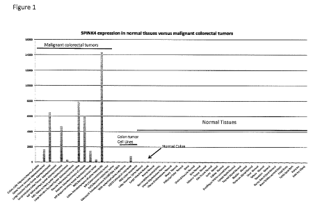
[0042] FIG. l shows the expression of SPINK4 in normal tissues versus
malignant
colorectal tumors.
[0043] FIG. 2 shows the expression of LITDI in colorectal tumors, normal
tissues
and other malignant tumor types.
[0044] FIG, 3 shows the expression of LY6G6D in normal tissues versus
colorectal
tumors.
[0045] FIG. 4 shows the expression of APOBECI in colorectal tumors and other
malignant tumors versus normal tissues.
[0046] FIG. 5 shows the expression of L00729669 in normal tissues versus
colorectal tumors.
[0047] FIG. 6 shows the expression of NOTUM in colorectal tumors and other
malignant tumors versus normal tissues,
[0048] FIG. 7 shows the expression of GRP in normal tissues versus colorectal
tumors.
[0049] FIG. 8 shows the expression of KRT20 in normal tissues versus
colorectal
tumors.
[0050] FIG. 9 shows the expression of MUC17 in normal tissues versus
colorectal
tumors.
16
CA 02844822 2014-02-10
WO 2013/033629
PCT/US2012/053518
[0051] FIG. 10 shows the expression of NOTUM in normal tissues versus
colorectal
tumors.
[0052] FIG. 11 shows the expression of COL11A1 in normal tissues versus
colorectal
tumors and other cancers.
100531 FIG. 12 shows the expression of MMPI 1 in normal tissues versus
colorectal
tumors.
[0054] FIG. 13 shows the expression of MMP12 in normal tissues versus
colorectal
tumors.
[0055] FIG. 14 shows the expression of MMP7 in normal tissues versus
colorectal
tumors.
[0056] FIG. 15 shows the expression of DKK4 in normal tissues versus
colorectal
tumors.
[0057] Fig. 16 shows LY6G6D mRNA expression in normal and colon cancer tissues
measured by qRT-PCR. LY6G6D expression levels were measured by quantitative
PCR
(qPCR), normalized to ACTB expression levels, and are expressed as 2^(-delta
Ct) values.
Input cDNA samples were obtained as TissueScanTm cDNA Arrays (Colon Cancer
Disease
Panel H, Origene). LY6G6D: pritners: UPL479_LY6G6D-F and UPL480_LY6G6D-R,
probe: UPL probe #20. ACTB: TissueScan primers (Origene) in combination with
SYBR
Green I (Applied Biosystems/Life Technologies).
[0058] Fig. 17 shows SPINK4 mRNA expression in normal and colon cancer tissues
measured by qRT-PCR. SPINK4 expression levels were measured by quantitative
PCR
(qPCR), normalized to ACTH expression levels, and are expressed as 2^(-delta
Ct) values.
Input cDNA samples were obtained as TissueScanni cDNA Arrays (Colon Cancer
Disease
Panel 11, Origene). SP1NK4: primers: UPL475 SPINK4F and UPL476_SPINK4 R,
probe:
UPL probe #18. ACTB: TissueScan primers (Origene) in combination with SYBR
Green I
(Applied Biosystems/Life Technologies).
[0059] Fig. 18 shows LITD1 mRNA expression in normal and colon cancer tissues
measured by qRT-PCR. LITD1 expression levels were measured by quantitative PCR
(qPCR), normalized to ACTB expression levels, and are expressed as 2^(-delta
CO values.
Input cDNA samples were obtained as TissueScanrm cDNA Arrays (Colon Cancer
Disease
Panel H, Origene). LlTDI: primers: UPL485-L TD1-F and UPL486-L TD -R, probe:
UPL
probe #42. ACTB: TissueScan primers (Origene) in combination with SYBR Green
I
(Applied Biosystems/Life Technologies).
17
CA 02844822 2014-02-10
WO 2013/033629
PCT/US2012/053518
[0060] Fig. 19 shows DKK4 mRNA expression in normal and colon cancer tissues
measured by qRT-PCR. DICK4 expression levels of one normal (NI) and five colon
cancer
tissues (C1-05) were normalized to GUSB expression levels, and are expressed
as 2'(-delta
Ct) values, DICK4: primers: UPL495_DKK4-F3 and UPL496_DKK4-R3, probe: UPL #19.
GUSB: primers: UPLO81_GUSB-F and UPL082_GUSB-R, probe: UPL probe #57.
[0061] Fig. 20 shows NOTUM mRNA expression in normal and colon cancer tissues
measured by qRT-PCR. NOTUM expression levels of one normal (Ni) and three
colon
cancer tissues (CI-C3) were normalized to GUSB expression levels, and are
expressed as 2''(-
delta Ct) values. NOTUM: primers: UPL489_NOTUM-F and UPL490_NOTUM-R, probe:
UPL #34. GUSB: primers: UPL081_GUSB-F and UPL082_GUSB-R, probe: UPL probe #57.
[0062] FIG. 21 shows the expression of COLI 0A in normal tissues versus
colorectal
tumors and other tumors.
[0063] FIG. 22 shows the expression of SLC35D3in normal tissues versus
colorectal
tumors.
[0064] FIG. 23 shows the expression of COLX (the protein encoded for by COL I
OA)
in normal tissues versus colorectal tumors.
[00651 FIG. 24 shows the expression of MMP I 1 in normal tissues versus
colorectal
tumors.
DETAILED DESCRIPTION
[0066] Before the present compositions and methods are described, it is to be
understood that this invention is not limited to the particular processes,
compositions, or
methodologies described, as these may vary. It is also to be understood that
the terminology
used in the description is for the purpose of describing the particular
versions or embodiments
only, and is not intended to limit the scope of the present invention which
will be limited only
by the appended claims. Unless defined otherwise, all technical and scientific
terms used
herein have the same meanings as commonly understood by one of ordinary skill
in the art.
Although any methods and materials similar or equivalent to those described
herein can be
used in the practice or testing of embodiments of the present disclosure, the
preferred
methods, devices, and materials are now described. Nothing herein is to be
construed as an
admission that the invention is not entitled to antedate such disclosure by
virtue of prior
invention.
[0067] As used herein, the singular forms "a," "an," and "the" include plural
reference unless the context clearly dictates otherwise. Thus, for example,
reference to a
18
CA 02844822 2014-02-10
WO 2013/033629
PCT/US2012/053518
"therapeutic" is a reference to one or more therapeutics and equivalents
thereof known to
those skilled in the art, and so forth.
[0068] As used herein, the term "about" means plus or minus 10% of the
numerical
value of the number with which it is being used. Therefore, about 50% means in
the range of
45% to 55%.
[00691 "Administering," when used in conjunction with a therapeutic, means to
administer a therapeutic directly into or onto a target tissue or to
administer a therapeutic to a
patient whereby the therapeutic positively impacts the tissue to which it is
targeted. Thus, as
used herein, the term "administering," when used in conjunction with a
therapeutic, can
include, but is not limited to, providing the therapeutic into or onto the
target tissue;
providing the therapeutic systemically to a patient by, e.g., intravenous
injection whereby the
therapeutic reaches the target tissue; providing the therapeutic in the form
of the encoding
sequence thereof to the target tissue (e.g., by so-called gene-therapy
techniques).
"Administering" a composition may be accomplished by oral administration,
intravenous
injection, intraperitoneal injection, intramuscular injection, subcutaneous
injection,
transdermal diffusion or electrophoresis, local injection, extended release
deliveiy devices
including locally implanted extended release devices such as bioerodible or
reservoir-based
implants, as protein therapeutics or as nucleic acid therapeutic via gene
therapy vectors,
topical administration, or by any of these methods in combination with other
known
techniques. Such combination techniques include, without limitation, heating,
radiation and
ultrasound.
[0070] "Agent" as used herein refers to a molecule that specifically binds to
a cancer
associated sequence or a molecule encoded for by a cancer associated sequence
or a receptor
that binds to a molecule encoded for by a cancer associated sequence. Examples
of agents
include nucleic acid molecules, such as DNA and proteins such as antibodies.
The agent may
be linked with a label or detectible substance as described infra.
100711 The term "amplify" as used herein means creating an amplification
product
which may include, for example, additional target molecules, or target-like
molecules or
molecules complementary to the target molecule, which molecules are created by
virtue of
the presence of the target molecule in the sample. In the situation where the
target is a
nucleic acid, an amplification product can be made enzymatically with DNA or
RNA
polymerases or reverse transcriptases, or any combination thereof.
[0072] The term "animal," "patient" or "subject" as used herein includes, but
is not
limited to, humans, non-human primates and non-human vertebrates such as wild,
domestic
19
CA 02844822 2014-02-10
WO 2013/033629
PCT/US2012/053518
and farm animals including any mammal, such as cats, dogs, cows, sheep, pigs,
horses,
rabbits, rodents such as mice and rats. In some embodiments, the term
"subject," "patient" or
"animal" refers to a male. In some embodiments, the term "subject," "patient"
or "animal"
refers to a female.
10073] The term "biological sources" as used herein refers to the sources from
which
the target polytmcleotides or proteins or peptide fragments may be derived.
The source can
be of any form of "sample" as described above, including but not limited to,
cell, tissue or
fluid. "Different biological sources" can refer to different
cells/tissues/organs of the same
individual, or cells/tissues/organs from different individuals of the same
species, or
cells/tissues/organs from different species.
[0074] The term "capture reagent" refers to a reagent, for example an antibody
or
antigen binding protein, capable of binding a target molecule or analyte to be
detected in a
sample.
[0075] The term "gene expression result" refers to a qualitative and/or
quantitative
result regarding the expression of a gene or gene product. The gene expression
result can be
an amount or copy number of the gene, the RNA encoded by the gene, the mRNA
encoded
by the gene, the protein product encoded by the gene, or any combination
thereof. The gene
expression result can also be normalized or compared to a standard. The gene
expression
result can be used, for example, to determine if a gene is expressed,
overexpressed, or
differentially expressed in two or more samples.
[0076] The term "homology," as used herein, refers to a degree of
complementarity.
There may be partial homology or complete homology. The word "identity" may
substitute
for the word "homology." A partially complementary nucleic acid sequence that
at least
partially inhibits an identical sequence from hybridizing to a target nucleic
acid is referred to
as "substantially homologous." The inhibition of hybridization of the
completely
complementary nucleic acid sequence to the target sequence may be examined
using a
hybridization assay (Southern or northern blot, solution hybridization, and
the like) under
conditions of reduced stringency. A substantially homologous sequence or
hybridization
probe will compete for and inhibit the binding of a completely homologous
sequence to the
target sequence under conditions of reduced stringency. This is not to say
that conditions of
reduced stringency are such that non-specific binding is permitted, as reduced
stringency
conditions require that the binding of two sequences to one another be a
specific (i.e., a
selective) interaction. The absence of non-specific binding may be tested by
the use of a
second target sequence which lacks even a partial degree of complementarity
(e.g., less than
CA 02844822 2014-02-10
WO 2013/033629
PCT/US2012/053518
about 30% homology or identity). In the absence of non-specific binding, the
substantially
homologous sequence or probe will not hybridize to the second non-
complementary target
sequence.
[0077] As used herein, the term "hybridization" or "hybridizing" refers to
hydrogen
bonding, which may be Watson-Crick, Hoogsteen or reversed Hoogsteen hydrogen
bonding
between complementary nucleoside or nucleotide bases. For example, adenine and
thymine
are complementary nucleobases which pair through the formation of hydrogen
bonds.
"Complementary," as used herein in reference to nucleic acid molecules refers
to the capacity
for precise pairing between two nucleotides. For example, if a nucleotide at a
certain position
of an oligonucleotide is capable of hydrogen bonding with a nucleotide at the
same position
of a DNA or RNA molecule, then the oligonucleotide and the DNA or RNA are
considered to
be complementaiy to each other at that position. The oligonucleotide and the
DNA or RNA
are complementary to each other when a sufficient number of corresponding
positions in each
molecule are occupied by nucleotides which can hydrogen bond with each other.
Thus,
"specifically hybridizable" and "complementary" are terms which are used to
indicate a
sufficient degree of completnentarity or precise pairing such that stable and
specific binding
occurs between the oligonucleotide and the DNA or RNA target. It is understood
in the art
that a nucleic acid sequence need not be 100% complementary to that of its
target nucleic
acid to be specifically hybridizable. A nucleic acid compound is specifically
hybridizable
when there is binding of the molecule to the target, and there is a sufficient
degree of
complementarity to avoid non-specific binding of the molecule to non-target
sequences under
conditions in which specific binding is desired, i.e., under physiological
conditions in the
case of in vivo assays or therapeutic treatment, and in the case of in vitro
assays, under
conditions in which the assays are performed.
[0078] The term "inhibiting" includes the administration of a compound of the
present disclosure to prevent the onset of the symptoms, alleviating the
symptoms, or
eliminating the disease, condition or disorder. The term "inhibiting" may also
refer to
lowering the expression level of gene, such as a gene encoding a cancer
associated sequence.
Expression level of RNA and/or protein may be lowered.
[0079] The term "label" and/or detectible substance refers to a composition
capable
of producing a detectable signal indicative of the presence of the target
polynucleotide in an
assay sample. Suitable labels include radioisotopes, nucleotide chromophores,
enzymes,
substrates, fluorescent molecules, chemiluminescent moieties, magnetic
particles,
bioluminescent moieties, and the like. As such, a label is any composition
detectable by a
21
CA 02844822 2014-02-10
WO 2013/033629
PCT/US2012/053518
device or method, such as, but not limited to, a spectroscopic, photochemical,
biochemical,
immunochemical, electrical, optical, chemical detection device or any other
appropriate
device. In some embodiments, the label may be detectable visually without the
aid of a
device. The term "label" is used to refer to any chemical group or moiety
having a detectable
physical property or any compound capable of causing a chemical group or
moiety to exhibit
a detectable physical property, such as an enzyme that catalyzes conversion of
a substrate
into a detectable product. The. term "label" also encompasses compounds that
inhibit the
expression of a particular physical property. The label may also be a compound
that is a
member of a binding pair, the other member of which bears a detectable
physical property.
[0080] A "microarray" is a linear or two-dimensional array of, for example,
discrete
regions, each having a defined area, formed on the surface of a solid support.
The density of
the discrete regions on a microarray is determined by the total numbers of
target
polynucleotides to be detected on the surface of a single solid phase support,
preferably at
least about 50/cm 2, more preferably at least about 100/cm2, even more
preferably at least
about 500/cm2, and still more preferably at least about 1,000/cm2. As used
herein, a DNA
microarray is an array of oligonucleotide primers placed on a chip or other
surfaces used to
identify, amplify, detect, or clone target polynucleotides. Since the position
of each
particular group of primers in the array is known, the identities of the
target polynucleotides
can be determined based on their binding to a particular position in the
microarray.
[0081] As used herein, the term "naturally occurring" refers to sequences or
structures
that may be in a form normally found in nature. "Naturally occurring" may
include
sequences in a form normally found in any animal,
[0082] The use of "nucleic acid," "polynucleotide" or "oligonucleotide" or
equivalents herein means at least two nucleotides covalently linked together.
In some
embodiments, an oligonucleotide is an oligomer of 6, 8, 10, 12, 20, 30 or up
to 100
nucleotides. In some embodiments, an oligonucleotide is an of igomer of at
least 6, 8, 10, 12,
20, 30, 40, 50, 60, 70, 80, 90, 100, 150, 200, 300, 400, or 500 nucleotides. A
"polynucleotide" or "oligonucleotide" may comprise DNA, RNA, PNA or a polymer
of
nucleotides linked by phosphodiester and/or any alternate bonds.
[00831 As used herein, the term "optional" or "optionally" refers to
embodiments
where the subsequently described structure, event or circumstance may or may
not occur, and
that the description includes instances where the event occurs and instances
where it does not.
[0084] The phrases "percent homology," "% homology," "percent identity," or "%
identity" refer to the percentage of sequence similarity found in a comparison
of two or more
22
CA 02844822 2014-02-10
WO 2013/033629
PCT/US2012/053518
amino acid or nucleic acid sequences. Percent identity can be determined
electronically, e.g.,
by using the MEGALIGN program (LASERGENE software package, DNASTAR). The
MEGALIGN program can create alignments between two or more sequences according
to
different methods, e.g., the Clustal Method. (Higgins, D. G. and P. M. Sharp
(1988) Gene
73:237-244.) The Clustal algorithm groups sequences into clusters by examining
the
distances between all pairs. The clusters are aligned pairwise and then in
groups. The
percentage similarity between two amino acid sequences, e.g., sequence A and
sequence B, is
calculated by dividing the length of sequence A, minus the number of gap
residues in
sequence A, minus the number of gap residues in sequence B, into the sum of
the residue
matches between sequence A and sequence B, times one hundred. Gaps of low or
of no
homology between the two amino acid sequences are not included in determining
percentage
similarity. Percent identity between nucleic acid sequences can also be
calculated by the
Clustal Method, or by other methods known in the art, such as the Jotun Hein
Method. (See,
e.g., Hein, J. (1990) Methods Enzymol. 183:626-645.) Identity between
sequences can also be
determined by other methods known in the art, e.g., by varying hybridization
conditions.
[0085] By "pharmaceutically acceptable", it is meant the carrier, diluent or
excipient
must be compatible with the other ingredients of the formulation and not
deleterious to the
recipient thereof.
100861 "Recombinant protein" as used herein means a protein made using
recombinant techniques, for example, but not limited to, through the
expression of a
recombinant nucleic acid as depicted above. A recombinant protein may be
distinguished
from naturally occurring protein by at least one or more characteristics. For
example, the
protein may be isolated or purified away from some or all of the proteins and
compounds
with which it is normally associated in its wild type host, and thus may be
substantially pure.
For example, an isolated protein is unaccompanied by at least some of the
material with
which it is normally associated in its natural state, preferably constituting
at least about 0.5%,
more preferably at least about 5% by weight of the total protein in a given
sample. A
substantially pure protein comprises about 50-75%, about 80%, or about 90%. In
some
embodiments, a substantially pure protein comprises about 80-99%, 85-99%, 90-
99%, 95-
99%, or 97-99% by weight of the total protein. A recombinant protein can also
include the
production of a cancer associated protein from one organism (e.g. human) in a
different
organism (e.g. yeast, E. cob, or the like) or host cell. Alternatively, the
protein may be made
at a significantly higher concentration than is normally seen, through the use
of an inducible
promoter or high expression promoter, such that the protein is made at
increased
23
CA 02844822 2014-02-10
WO 2013/033629
PCT/US2012/053518
concentration levels. Alternatively, the protein may be in a form not normally
found in
nature, as in the addition of an epitope tag or amino acid substitutions,
insertions and
deletions, as discussed herein.
100871 As used herein, the term "sample" refers to composition that is being
tested or
treated with a reagent, such as but not limited to a therapeutic, drug, or
candidate agent.
Samples may be obtained from subjects. In some embodiments, the sample may be
blood,
plasma, serum, or any combination thereof. A sample may be derived from blood,
plasma,
serum, or any combination thereof. Other typical samples include, but are not
limited to, any
bodily fluid obtained from a mammalian subject, tissue biopsy, sputum,
lymphatic fluid,
blood cells (e.g., peripheral blood mononuclear cells), tissue or fine needle
biopsy samples,
urine, peritoneal fluid, colostrums, breast milk, fetal fluid, fecal material,
tears, pleural fluid,
or cells therefrom. The sample may be processed in some manner before being
used in a
method described herein, for example a particular component to be analyzed or
tested
according to any of the methods described infra. One or more molecules may be
isolated
from a sample.
100881 The terms "specific binding," "specifically binds," and the like, refer
to
instances where two or more molecules form a complex that is measurable under
physiologic
or assay conditions and is selective. An antibody or antigen binding protein
or other
molecule is said to "specifically bind" to a protein, antigen, or epitope if;
under appropriately
selected conditions, such binding is not substantially inhibited, while at the
same time non-
specific binding is inhibited. Specific binding is characterized by a high
affinity and is
selective for the compound, protein, epitope, or antigen. Nonspecific binding
usually has a
low affinity.
100891 As used herein, a polynucleotide "derived from" a designated sequence
refers
to a polynucleotide sequence which is comprised of a sequence of approximately
at least
about 6 nucleotides, preferably at least about 8 nucleotides, more preferably
at least about 10-
12 nucleotides, and even more preferably at least about 15-20 nucleotides
corresponding to a
region of the designated nucleotide sequence. "Corresponding" means homologous
to or
complementary to the designated sequence. Preferably, the sequence of the
region from
which the polynucleotide is derived is homologous to or complementary to a
sequence that is
unique to a cancer associated gene.
[0090] As used herein, the term "tag," "sequence tag" or "primer tag sequence"
refers
to an ofigonucleotide with specific nucleic acid sequence that serves to
identify a batch of
polynucleotides bearing such tags therein. Polynucleotides from the same
biological source
24
CA 02844822 2014-02-10
WO 2013/033629
PCT/US2012/053518
are covalently tagged with a specific sequence tag so that in subsequent
analysis the
polynucleotide can be identified according to its source of origin. The
sequence tags also
serve as primers for nucleic acid amplification reactions.
[0091] The term "support" refers to conventional supports such as beads,
particles,
dipsticks, fibers, filters, membranes, and silane or silicate supports such as
glass slides.
0092] As used herein, the term "therapeutic" or "therapeutic agent" means an
agent
that can be used to treat, combat, ameliorate, prevent or improve an unwanted
condition or
disease of a patient. In part, embodiments of the present disclosure are
directed to the
treatment of cancer or the decrease in proliferation of cells. In some
embodiments, the term
"therapeutic" or "therapeutic agent" may refer to any molecule that associates
with or affects
the target marker, its expression or its function. In various embodiments,
such therapeutics
may include molecules such as, for example, a therapeutic cell, a therapeutic
peptide, a
therapeutic gene, a therapeutic compound, or the like, that associates with or
affects the target
marker, its expression or its function.
100931 A "therapeutically effective amount" or "effective amount" of a
composition
is a predetermined amount calculated to achieve the desired effect, i.e., to
inhibit, block, or
reverse the activation, migration, or proliferation of cells. In some
embodiments, the
effective amount is a prophylactic amount. In some embodiments, the effective
amount is an
amount used to medically treat the disease or condition. The specific dose of
a composition
administered according to this invention to obtain therapeutic and/or
prophylactic effects will,
of course, be determined by the particular circumstances surrounding the case,
including, for
example, the composition administered, the route of administration, and the
condition being
treated. It will be understood that the effective amount administered will be
determined by
the physician in the light of the relevant circumstances including the
condition to be treated,
the choice of composition to be administered, and the chosen route of
administration. A
therapeutically effective amount of composition of this invention is typically
an amount such
that when it is administered in a physiologically tolerable excipient
composition, it is
sufficient to achieve an effective systemic concentration or local
concentration in the targeted
tissue.
[0094] The terms "treat," "treated," or "treating" as used herein can refer to
both
therapeutic treatment or prophylactic or preventative measures, wherein the
object is to
prevent or slow down (lessen) an undesired physiological condition, disorder
or disease, or to
obtain beneficial or desired clinical results. In some embodiments, the term
may refer to both
treating and preventing. For the purposes of this disclosure, beneficial or
desired clinical
CA 02844822 2014-02-10
WO 2013/033629
PCT/US2012/053518
results include, but are not limited to, alleviation of symptoms; diminishment
of the extent of
the condition, disorder or disease; stabilization (i.e., not worsening) of the
state of the
condition, disorder or disease; delay in onset or slowing of the progression
of the condition,
disorder or disease; amelioration of the condition, disorder or disease state;
and remission
(whether partial or total), whether detectable or undetectable, or enhancement
or
improvement of the condition, disorder or disease. Treatment includes
eliciting a clinically
significant response without excessive levels of side effects. Treatment also
includes
prolonging survival as compared to expected survival if not receiving
treatment.
100951 The term "tissue" refers to any aggregation of similarly specialized
cells that
are united in the performance of a particular function.
Cancer Associated Sequences
100961 In some embodiments, the present disclosure provides for nucleic acid
and
protein sequences that are associated with cancer, herein termed "cancer
associated" or "CA"
sequences. In some embodiments, the present disclosure provides nucleic acid
and protein
sequences that are associated with colorectal cancers or carcinomas such as,
without
limitation, adenocarcinoma, leiomyosarcoma, lymphoma, melanoma, neuroendocrine
tumors,
carcinoid tumors, signet ring cell adenocarcinoma, mucinous adenocarcinoma,
gastrointestinal stromal tumor, squamous cell carcinoma, or any combination
thereof. In
some embodiments, the present disclosure provides nucleic acid and protein
sequences that
are associated with colorectal cancers or carcinomas such as, without
limitation,
adenocarcinoma, leiomyosamoma, lymphoma, melanoma, neuroendocrine tumors,
carcinoid
tumors, signet ring cell adenocarcinoma, mucinous adenocarcinoma,
gastrointestinal stromal
tumor, squamous cell carcinoma, or a combination thereof. In some embodiments,
the term
"cancer associated sequences" may indicate that the nucleotide or protein
sequences are
differentially expressed, activated, inactivated or altered in cancers as
compared to normal
tissue. Cancer associated sequences may include those that are up-regulated
(i.e. expressed at
a higher level), as well as those that are down-regulated (i.e. expressed at a
lower level), in
cancers. Cancer associated sequences can also include sequences that have been
altered (i.e.,
translocations, truncated sequences or sequences with substitutions, deletions
or insertions,
including, but not limited to, point mutations) and show either the same
expression profile or
an altered profile. In some embodiments, the cancer associated sequences are
from humans;
however, as will be appreciated by those in the art, cancer associated
sequences from other
organisms may be useful in animal models of disease and drug evaluation; thus,
other cancer
associated sequences may be useful such as, without limitation, sequences from
vertebrates,
26
CA 02844822 2014-02-10
WO 2013/033629
PCT/US2012/053518
including mammals, including rodents (rats, mice, hamsters, guinea pigs,
etc.), primates, and
farm animals (including sheep, goats, pigs, cows, horses, etc.). Cancer
associated sequences
from other organisms may be obtained using the techniques outlined herein.
[0097] In some embodiments, cancer associated sequences may include both
nucleic
acid and amino acid sequences. In some embodiments, the cancer associated
sequences may
include sequences having at least about 60% homology with the disclosed
sequences. In
some embodiments, the cancer associated sequences may have at least about 65%,
about
70%, about 75%, about 80%, about 85%, about 90%, about 95%, about 97%, about
99%,
about 99.8% homology with the disclosed sequences. In some embodiments, the
cancer
associated sequences may be "mutant nucleic acids". As used herein, "mutant
nucleic acids"
refers to deletion mutants, insertions, point mutations, substitutions,
translocations.
[0098] In some embodiments, the cancer associated sequences may be recombinant
nucleic acids. By the term "recombinant nucleic acid" herein refers to nucleic
acid
molecules, originally formed in vitro, in general, by the manipulation of
nucleic acid by
polymerases and endonucleases, in a form not normally found in nature. Thus a
recombinant
nucleic acid may also be an isolated nucleic acid, in a linear form, or cloned
in a vector
formed in vitro by ligating DNA molecules that are not normally joined, are
both considered
recombinant for the purposes of this invention. It is understood that once a
recombinant
nucleic acid is made and reintroduced into a host cell or organism, it can
replicate using the in
vivo cellular machinery of the host cell rather than in vitro manipulations;
however, such
nucleic acids, once produced recombinantly, although subsequently replicated
in vivo, are
still considered recombinant or isolated for the purposes of the invention. As
used herein, a
"polynucleotide" or "nucleic acid" is a polymeric form of nucleotides of any
length, either
ribonucleotides or deoxyribonucleotides. This term includes double- and single-
stranded
DNA and RNA. It also includes known types of modifications, for example,
labels which are
known in the art, methylation, "caps", substitution of one or more of the
naturally occurring
nucleotides with an analog, internucleotide modifications-such as, for
example, those with
uncharged linkages (e.g., phosphorothioates, phosphorodithioates, etc.), those
containing
pendant moieties, such as, for example proteins (including e.g., nucleases,
toxins, antibodies,
signal peptides, poly-L-lysine, etc.), those with intercalators (e.g.,
acridine, psoralen, etc.),
those containing chelators (e.g., metals, radioactive metals, etc.), those
containing alkylators,
those with modified linkages (e.g., alpha anomeric nucleic acids, etc.), as
well as unmodified
forms of the polynucleotide.
27
CA 02844822 2014-02-10
WO 2013/033629
PCT/US2012/053518
[0099] In some embodiments, the cancer associated sequences are nucleic acids.
As
will be appreciated by those skilled in the art and is described herein,
cancer associated
sequences of embodiments herein may be useful in a variety of applications
including
diagnostic applications to detect nucleic acids or their expression levels in
a subject,
therapeutic applications or a combination thereof. Further, the cancer
associated sequences
of embodiments herein may be used in screening applications; for example,
generation of
biochips comprising nucleic acid probes to the cancer associated sequences.
1001001 A nucleic acid of the present disclosure may include phosphodiester
bonds,
although in some cases, as outlined below (for example, in antisense
applications or when a
nucleic acid is a candidate drug agent), nucleic acid analogs may have
alternate backbones,
comprising, for example, phosphoramidate (Beaucage et al., Tetrahedron
49(10):1925 (1993)
and references therein; Letsinger, J. Org. Chem. 35:3800 (1970); Sprinzl et
al., Ern.. J.
Biochem. 81:579 (1977); Letsinger et al,, Nucl. Acids Res. 14:3487 (1986);
Sawai et al,
Chem. Lett. 805 (1984), Letsinger et al., J. Am. Chem. Soc. 110:4470 (1988);
and Pauwels et
al., ('hem/ca Scripta 26:141 91986)), phosphorothioate (Mag et al., Nucleic
Acids Res.
19:1437 (1991); and U.S. Pat, No, 5,644,048), phosphorodithioate (Briu et at.,
J. Am. Chem.
Soc. 111:2321 (1989), 0-methylphosphoroamidite linkages (see Eckstein,
Oligonucleotides
and Analogues: A Practical Approach, Oxford University Press), and peptide
nucleic acid
backbones and linkages (see Eghohn, J. Am. Chem. Soc. 114:1895 (1992); Meier
et al.,
Client. Int. Ed. Engl. 31:1008 (1992); Nielsen, Nature, 365:566 (1993);
Carlsson et al.,
Nature 380:207 (1996),). Other analog nucleic acids include those with
positive backbones
(Denpcy et al., Proc. Natl. Acad. Sci. USA 92:6097 (1995); non-ionic backbones
(U.S. Pat.
Nos. 5,386,023, 5,637,684, 5,602,240, 5,216,141 and 4,469,863; Kiedrowshi et
al., Angew.
Chem. Intl. Ed. English 30:423 (1991); Letsinger et al., J. Am. Chem. Soc.
110:4470 (1988);
Letsinger et al., Nucleoside & Nucleotide 13:1597 (1994); Chapters 2 and 3,
ASC
Symposium Series 580, "Carbohydrate Modifications in Antisense Research", Ed.
Y. S.
Sanghui and P. Dan Cook; Mesmaeker et al., Bioorganic & Medicinal Chem. Lett.
4:395
(1994); Jeffs et al., J. Biomolecular NMR 34:17 (1994); Tetrahedron Left.
37:743 (1996)) and
non-ribose backbones, including those described in U.S. Pat. Nos. 5,235,033
and 5,034,506,
and Chapters 6 and 7, ASC Symposium Series 580, "Carbohydrate Modifications in
Antisense Research", Ed. Y. S. Sanghui and P. Dan Cook, Nucleic acids
containing one or
more carbocyclic sugars are also included within one definition of nucleic
acids (see Jenkins
et al., ('hem. Soc. Rev. (1995) pp. 169-176). Several nucleic acid analogs are
described in
Rawls, C & E News Jun. 2, 1997 page 35. These modifications of the ribose-
phosphate
28
CA 02844822 2014-02-10
WO 2013/033629
PCT/US2012/053518
backbone may be done for a variety of reasons, for example to increase the
stability and half-
life of such molecules in physiological environments for use in anti-sense
applications or as
probes on a biochip.
[00101] As will be appreciated by those skilled in the art, such nucleic acid
analogs
may be used in some embodiments of the present disclosure. In addition,
mixtures of
naturally occurring nucleic acids and analogs can be made; alternatively,
mixtures of
different nucleic acid analogs, and mixtures of naturally occurring nucleic
acids and analogs
may be made.
100102] In some embodiments, the nucleic acids may be single stranded or
double
stranded or may contain portions of both double stranded or single stranded
sequence. As
will be appreciated by those skilled in the art, the depiction of a single
strand also defines the
sequence of the other strand; thus the sequences described herein also
includes the
complement of the sequence. The nucleic acid may be DNA, both gnomic and cDNA,
RNA, or a hybrid, where the nucleic acid contains any combination of deoxyribo-
and ribo-
nucleotides, and any combination of bases, including uracil, adenine, thymine,
cytosine,
guanine, inosine, xanthine, hypoxanthine, isocytosine, isoguanine, etc. As
used herein, the
term "nucleoside" includes nucleotides and nucleoside and nucleotide analogs,
and modified
nucleosides such as amino modified nucleosides. In addition, "nucleoside"
includes non-
naturally occurring analog structures. Thus, for example, the subject units of
a peptide
nucleic acid, each containing a base, are referred to herein as a nucleoside.
[00103] Some embodiments herein are directed to one or more sequences
associated
with colorectal cancers, such as, but not limited to, adenocarcinoma,
leiomyosarcoma,
lymphoma, melanoma, neuroendocrine tumors, carcinoid tumors, signet ring cell
adenocarcinoma, mucinous adenocarcinoma, gastrointestinal stromal tumor,
squamous cell
carcinoma, or any combination thereof. The use of microarray analysis of gene
expression
allows the identification of host sequences associated with colorectal cancer.
These
sequences may then be used in a number of different ways, including diagnosis,
prognosis,
screening for modulators (including both agonists and antagonists), antibody
generation (for
immunotherapy and imaging), etc. However, as will be appreciated by those
skilled in the
art, sequences that are identified in one type of cancer may have a strong
likelihood of being
involved in other types of cancers as well. Thus, while the sequences outlined
herein are
initially identified as correlated with colorectal cancers, they may also be
found in other types
of cancers as well.
29
CA 02844822 2014-02-10
WO 2013/033629
PCT/US2012/053518
1001041 Some embodiments described herein may be directed to the use of cancer
associated sequences for diagnosis and treatment of colorectal cancer. In some
embodiments,
the cancer associated sequence may be selected from: Homo sapiens serine
peptidase
inhibitor, Kazal type 4 (SP1NK4), Homo sapiens LINE-I type transposase domain
containing
1 (LITDI), Homo sapiens solute carrier family 35, member D3 (SLC35D3), Homo
sapiens
lymphocyte antigen 6 complex, locus G6D (LY6G6D), Homo sapiens matrix
metallopeptidase 12 (macrophage elastase) (MMP12), Homo sapiens matrix
metallopeptidase
12 (macrophage elastase) (MMPI2), Homo sapiens apolipoprotein B inRNA editing
enzyme,
catalytic polypeptide 1 (APOBECI), Homo sapiens dickkopf homolog 4 (Xenopus
laevis)
(DKK4), Homo sapiens NADPH oxidase 1 (NOX1), Homo sapiens matrix
metallopeptidase
11 (stromelysin 3) (MIMP11), Homo sapiens ring finger protein 43 (RNF43),
AGENCOURT 10229596 NIH MGC 141 Homo sapiens cDNA clone IMAGE:6563923 5
(BU536065), Homo sapiens KIAA1199 (KIAA1199), Homo sapiens carcinoembiyonie
antigen-related cell adhesion molecule 5 (CEACAM5), Homo sapiens achaete-scute
complex
homolog 2 (Drosophila) (ASCL2), Homo sapiens villin 1 (VILI), Homo sapiens
naked
cuticle homolog 1 (Drosophila) (NKD1), PREDICTED: Homo sapiens hypothetical
L00729669 (L00729669), Homo sapiens mucin 17, cell surface associated (MUC17),
Homo sapiens notum pectinacetylesterase homolog (Drosophila) (NOTUM), Homo
sapiens
collagen, type XI, alpha 1 (COL11A1), Homo sapiens defensin, alpha 5, Paneth
cell-specific
(DEFA5), Homo sapiens notum pectinacetylesterase homolog (Drosophila) (NOTUM),
Homo sapiens phospholipase inhibitor (L00646627), Homo sapiens NADPH oxidase
organizer 1 (NOMA), 1-lomo sapiens lipocalin 15 (LCN15), Homo sapiens
chemokine (C-C
motif) ligand 24 (CCL24), Homo sapiens gastrin-releasing peptide (GRP), Homo
sapiens
pregnancy specific beta-l-glycoprotein 1 (PSG1), Homo sapiens claudin 2
(CLDN2), Homo
sapiens defensin, alpha 6, Paneth cell-specific (DEFA6), Homo sapiens
neuropeptide S
receptor 1 (NPSRI), Homo sapiens cystatin SN (CST1), Homo sapiens keratin 23
(histone
deacetylase inducible) (KRT23), Homo sapiens matrix metallopeptidase 7
(matrilysin,
uterine) (MMP7), Homo sapiens membrane-spanning 4-domains, subfamily A, member
12
(MS4Al2), Homo sapiens keratin 20 (KRT20), or a combination thereof. In some
embodiments, these cancer associated sequences may be associated with
colorectal cancers
including, without limitation, adenocarcinoma, leiomyosarcoma, lymphoma,
melanoma,
neuroendocrine tumors, carcinoid tumors, signet ring cell adenocarcinoma,
mucinous
adenocarcinoma, gastrointestinal stromal tumor, squamous cell carcinoma, or
any
combination thereof,
CA 02844822 2014-02-10
WO 2013/033629
PCT/US2012/053518
[00105] In some embodiments, the cancer associated sequences may be DNA
sequences encoding the above mRNA or the cancer associated protein or cancer
associated
polypeptide expressed by the above mRNA or homologs thereof. In some
embodiments, the
cancer associated sequence may be a mutant nucleic acid of the above disclosed
sequences.
In some embodiments, the homolog may have at least about 60%, at least about
65%, at least
about 70%, at least about 75%, at least about 80%, at least about 85%, at
least about 90%, at
least about 95%, at least about 97%, at least about 98%, at least about 99%,
at least about
99.5% identity with the disclosed polypeptide sequence.
[00106] In some embodiments, an isolated nucleic acid comprises at least 10,
12, 15,
20 or 30 contiguous nucleotides of a sequence selected from the group
consisting of the
cancer associated polynucleotide sequences disclosed in Table 1,
[00107] In some embodiments, the polynucleotide, or its complement or a
fragment
thereof, further comprises a detectable label, is attached to a solid support,
is prepared at least
in part by chemical synthesis, is an antisense fragment, is single stranded,
is double stranded
or comprises a microarray.
[00108] In some embodiments, the invention provides an isolated polypeptide,
encoded within an open reading frame of a cancer associated sequence selected
from the
polynucleotide sequences shown in Table I, or its complement. In some
embodiments, the
invention provides an isolated polypeptide, wherein said polypeptide comprises
the amino
acid sequence encoded by a polynucleotide selected from the group consisting
of sequences
disclosed in Table I. In some embodiments, the invention provides an isolated
polypeptide,
wherein said polypeptide comprises the amino acid sequence encoded by a cancer
associated
polypeptide.
[00109] In some embodiments, the invention further provides an isolated
polypeptide, comprising the amino acid sequence of an epitope of the amino
acid sequence of
a cancer associated polypeptide, wherein the polypeptide or fragment thereof
may be attached
to a solid support. In some embodiments the invention provides an isolated
antibody
(monoclonal or polyclonal) or antigen binding fragment thereof, that binds to
such a
polypeptide. The isolated antibody or antigen binding fragment thereof may be
attached to a
solid support, or further comprises a detectable label.
[00110] Some embodiments also provide for antigens (e.g., cancer-associated
polypeptides) associated with a variety of cancers as targets for diagnostic
and/or therapeutic
antibodies. These antigens may also be useful for drug discovery (e.g., small
molecules) and
for further characterization of cellular regulation, growth, and
differentiation,
31
CA 02844822 2014-02-10
WO 2013/033629
PCT/US2012/053518
Methods of Detecting and Diagnosing Colorectal Cancer
100111] In some embodiments, the method of detecting or diagnosing colorectal
cancer may comprise assaying gene expression of a subject in need thereof. In
some
embodiments, detecting a level of a cancer associated sequence may comprise
techniques
such as, but not limited to, PCR, mass spectroscopy, microarray or other
detection techniques
described herein, Information relating to expression of the receptor can also
be useful in
determining therapies aimed at up or down-regulating the cancer associated
sequence's
signaling using agonists or antagonists.
1001121 In some embodiments, a method of diagnosing colorectal cancer may
comprise detecting a level of the cancer associated protein in a subject. In
some
embodiments, a method of screening for cancer may comprise detecting a level
of the cancer
associated protein. In some embodiments, the cancer associated protein is
encoded by a
nucleotide sequence selected from a sequence disclosed in Table I, a fraction
thereof or a
complementary sequence thereof. In some embodiments, a method of treating
cancer may
comprise administering an antibody against the protein to a subject in need
thereof. In some
embodiments, the antibody may be a monoclonal antibody or a polyclonal
antibody. In some
embodiments, the antibody may be a humanized or a recombinant antibody.
Antibodies can
be made that specifically bind to this region using known methods and any
method is
suitable. In some embodiments, the antibody specifically binds to one or more
of a molecule,
such as protein or peptide, encoded for by one or more cancer associated
sequences disclosed
infra,
1001131 In some embodiments, the antibody binds to an epitope from a protein
encoded by the nucleotide sequence disclosed in sequences disclosed in Table
I. In some
embodiments, the epitope is a fragment of the protein sequence encoded by the
nucleotide
sequence of any of the cancer associated sequences disclosed infra. In some
embodiments,
the epitope comprises about 1-10, 1-20, 1-30, 3-10, or 3-15 residues of the
cancer associated
sequence. In some embodiments, the epitope is not linear,
1001141 In some embodiments, the antibody binds to the regions described'
herein or
a peptide with at least 90, 95, or 99% homology or identity to the region. In
some
embodiments, the fragment of the regions described herein is 5-10 residues in
length. In
some embodiments, the fragment of the regions (e.g. epitope) described herein
are 3-5
residues in length. The fragments are described based upon the length
provided. In some
embodiments, the epitope is about 3, 4, 5, 6, 7, 8, 9, 10, II, 12, 13, 14, 15,
or 20 residues in
length.
32
CA 02844822 2014-02-10
WO 2013/033629
PCT/US2012/053518
[00115] In some embodiments, the sequence to which the antibody binds may
include
both nucleic acid and amino acid sequences. In some embodiments, the sequence
to which
the antibody binds may include sequences having at least about 60% homology
with the
disclosed sequences. In some embodiments, the sequence to which the antibody
binds may
have at least about 65%, about 70%, about 75%, about 80%, about 85%, about
90%, about
95%, about 97%, about 99%, about 99.8% homology with the disclosed sequences.
In some
embodiments, the sequences may be referred to as "mutant nucleic acids" or
"mutant peptide
sequences."
[00116] In some embodiments, a subject can be diagnosed with colorectal cancer
by
detecting the presence of a cancer associated sequence selected from sequences
disclosed in
Table I. In some embodiments, a method of diagnosing a subject with colorectal
cancer
comprises detecting the presence of a cancer associated sequence selected from
sequences
disclosed in Table I, wherein the presence of the cancer associated sequence
indicates that
the subject has colorectal cancer. In some embodiments, the method comprises
detecting the
presence or absence of a cancer associated sequence selected from sequences
disclosed in
Table 1, wherein the absence of the cancer associated sequence indicates that
absence of
colorectal cancer. In some embodiments, the method further comprises treating
the subject
diagnosed with colorectal cancer with an antibody that binds to a cancer
associated sequence
selected from sequences disclosed in Table 1 and inhibits the growth or
progression of the
colorectal cancer. As discussed, colorectal cancer may be detected in any type
of sample,
including, but not limited to, serum, blood, tumor and the like. The sample
may be any type
of sample as it is described herein.
[00117] In some embodiments, the method of diagnosing a subject with
colorectal
cancer comprises obtaining a sample and detecting the presence of a cancer
associated
sequence selected from sequences disclosed in Table 1 wherein the presence of
the cancer
associated sequence indicates the subject has colorectal cancer. In some
embodiments,
detecting the presence of a cancer associated sequence selected from sequences
disclosed in
Table 1 comprises contacting the sample with an antibody or other type of
capture reagent
that specifically binds to the cancer associated sequence's protein and
detecting the presence
or absence of the binding to the cancer associated sequence's protein in the
sample. An
example of an assay that can be used includes but is not limited to, an ELISA.
[00118] In some embodiments, the present disclosure provides a method of
diagnosing colorectal cancer, cancer, or a neoplastic condition in a subject,
the method
comprising obtaining a cancer associated sequence gene expression result of a
cancer
33
CA 02844822 2014-02-10
WO 2013/033629 PCT/US2012/053518
associated sequence selected from sequences disclosed in Table 1 from a sample
derived
from a subject; and diagnosing colorectal cancer or a neoplastic condition in
the subject based
on the cancer associated sequence gene expression result, wherein the subject
is diagnosed as
having colorectal cancer or a neoplastic condition if the cancer associated
sequence is
overexpressed.
[001191 In some embodiments, the subject is diagnosed as not having colorectal
cancer, cancer, or a neoplastic condition if the cancer associated sequence is
not
overexpressed. In some embodiments of the methods described herein and
throughout, the =
cancer that is diagnosed based upon a cancer associated sequence gene
expression result or
the absence or presence of a cancer associated sequence or protein is a cancer
selected from
the group consisting of adenocarcinoma, leiomyosarcoma, lymphoma, melanoma,
neuroendocrine tumors, carcinoid tumors, signet ring cell adenocarcinoma,
mucinous
adenocarcinoma, gastrointestinal stromal tumor, squamous cell carcinoma, or
any
combination thereof,
[001201 In some embodiments, the present disclosure provides methods of
diagnosing cancer or a neoplastic condition in a subject, the method
comprising obtaining a
gene expression result of a cancer associated sequence selected from one or
more of the
cancer associated sequences disclosed infra and diagnosing cancer or a
neoplastic condition
in the subject based on the gene expression result, wherein the subject is
diagnosed as having
cancer or a neoplastic condition if the gene is overexpressed.
[00121] In some embodiments, a method of diagnosing a subject with cancer
comprises obtaining a sample and detecting the presence of a cancer associated
sequence
selected from sequences disclosed in Table 1, wherein the presence of the
cancer associated
sequence indicates the subject has cancer. In some embodiments, detecting the
presence of a
cancer associated sequence selected from sequences disclosed in Table 1
comprises
contacting the sample with an antibody or other type of capture reagent that
specifically binds
to the cancer associated sequence's protein and detecting the presence or
absence of the
binding to the cancer associated sequence's protein in the sample. In some
embodiments, the
cancer is colorectal cancer. In some embodiments, the cancer is selected
from
adenocarcinoma, leiomyosarcoma, lymphoma, melanoma, neuroendocrine tumors,
carcinoid
tumors, signet ring cell adenocarcinoma, mucinous adenocarcinoma,
gastrointestinal stromal
tumor, squamous cell carcinoma, or a combination thereof.
[001221 Some embodiments are directed to a biochip comprising a nucleic acid
segment which encodes a cancer associated protein. In some embodiments, a
biochip
34
CA 02844822 2014-02-10
WO 2013/033629
PCT/US2012/053518
comprises a nucleic acid molecule which encodes at least a portion of a cancer
associated
protein. In some embodiments, the cancer associated protein is encoded by a
sequence
selected from sequences disclosed in Table 1, homologs thereof, combinations
thereof, or a
fragment thereof. In some embodiments, the nucleic acid molecule specifically
hybridizes
with a nucleic acid sequence selected from Sequences disclosed in Table 1. In
some
embodiments, the biochip comprises a first and second nucleic molecule wherein
the first
nucleic acid molecule specifically hybridizes with a first sequence selected
from sequences
disclosed in Table 1 and the second nucleic acid molecule specifically
hybridizes with a
second sequence selected from sequences disclosed in Table I, wherein the
first and second
sequences are not the same sequence. in some embodiments, the present
invention provides
methods of detecting or diagnosing cancer, such as colorectal cancer,
comprising detecting
the expression of a nucleic acid sequence selected from a sequence disclosed
in Table 1,
wherein a sample is contacted with a biochip comprising a sequence selected
from sequences
disclosed in Table 1, homologs thereof, combinations thereof, or a fragment
thereof.
1001231 Also provided herein is a method for diagnosing or determining the
propensity to cancers, for example, by measuring the expression level of one
or more of the
cancer associated sequences disclosed infra in a sample and comparing the
expression level
of the one or more cancer associated sequences in the sample with expression
level of the
same cancer associated sequences in a non-cancerous cell. A higher level of
expression of
one or more of the cancer associated sequences disclosed infra compared to the
non-
cancerous cell indicates a propensity for the development of cancer, e.g.,
colorectal cancer.
[00124] In some embodiments, the invention provides a method for detecting a
cancer associated sequence with the expression of a polypeptide in a test
sample, comprising
detecting a level of expression of at least one polypeptide such as, without
limitation, a
cancer associated protein, or a fragment thereof. In some embodiments, the
method
comprises comparing the level of expression of the polypeptide in the test
sample with a level
of expression of polypeptide in a normal sample, wherein an altered level of
expression of the
polypeptide in the test sample relative to the level of polypeptide expression
in the normal
sample is indicative of the presence of cancer in the test sample. In some
embodiments, the
polypeptide expression is compared to a cancer sample, wherein the level of
expression is at
least the same as the cancer is indicative of the presence of cancer in the
test sample. In some
embodiments, the sample is a cell sample.
[00125] In some embodiments, the invention provides a method for detecting
cancer
by detecting the presence of an antibody in a test serum sample. In some
embodiments, the
CA 02844822 2014-02-10
WO 2013/033629
PCT/US2012/053518
antibody recognizes a polypeptide or an epitope thereof disclosed herein. In
some
embodiments, the antibody recognizes a polypeptide or epitope thereof encoded
by a nucleic
. acid sequence disclosed herein. In some embodiments, the method comprises
detecting a
level of an antibody against an antigenic polypeptide such as, without
limitation, a cancer
associated protein, or an antigenic fragment thereof, In some embodiments, the
method
comprises comparing the level of the antibody in the test sample with a level
of the antibody
in the control sample, wherein an altered level of antibody in said test
sample relative to the
level of antibody in the control sample is indicative of the presence of
cancer in the test
sample. In some embodiments, the control sample is a sample derived from a
normal cell or
non-cancerous sample. In some embodiments, the control is derived from a
cancer sample,
and, therefore, in some embodiments, the method comprises comparing the levels
of binding
and/or the amount of antibody in the sample, wherein when the levels or amount
are the same
as the cancer control sample is indicative of the presence of cancer in the
test sample.
1001261 In some embodiments, a method for diagnosing cancer or a neoplastic
condition comprises a) determining the expression of one or more genes
comprising a nucleic
acid sequence selected from the group consisting of the human genomic and
inRNA
sequences described in Table I, in a first sample type (e.g. tissue) of a
first individual; and b)
comparing said expression of said gene(s) from a second normal sample type
from said first
individual or a second unaffected individual; wherein a difference in said
expression indicates
that the first individual has cancer. In some embodiments, the expression is
increased as
compared to the normal sample. In some embodiments, the expression is
decreased as
compared to the normal sample.
1001271 In some embodiments, the invention also provides a method for
detecting
presence or absence of cancer cells in a subject. In some embodiments, the
method
comprises contacting one or more cells from the subject with an antibody as
described herein.
In some embodiments, the method comprises detecting a complex of a cancer
associated
protein and the antibody, wherein detection of the complex indicates with the
presence of
cancer cells in the subject. In some embodiments the invention provides a
method for
inhibiting growth of cancer cells in a subject. In some embodiments, the
method comprises
administering to the subject an effective amount of a pharmaceutical
composition as
described herein. In some embodiments the invention provides a method for
delivering a
therapeutic agent to cancer cells in a subject, the method comprising:
administering to the
subject an effective amount of a pharmaceutical composition according to
according to the
invention.
36
CA 02844822 2014-02-10
WO 2013/033629
PCT/US2012/053518
[001281 In some embodiments, the present disclosure provides methods of
diagnosing cancer or a neoplastic condition in a subject, the method
comprising: a)
determining the expression of one or more genes or gene products or homologs
thereof; and
b) comparing said expression of the one or more nucleic acid sequences from a
second
normal sample from said first subject or a second unaffected subject, wherein
a difference in
said expression indicates that the first subject has cancer, wherein the gene
or the gene
product is referred to as a gene selected from one or more of the cancer
associated sequences
disclosed infra.
1001291 In some embodiments, the present disclosure provides methods of
detecting
cancer in a test sample, comprising: (i) detecting a level of activity of at
least one polypeptide
that is a gene product; and (ii) comparing the level of activity of the
polypeptide in the test
sample with a level of activity of polypeptide in a normal sample, wherein an
altered level of
activity of the polypeptide in the test sample relative to the level of
polypeptide activity in the
normal sample is indicative of the presence of cancer in the test sample,
wherein said gene
product is a product of a gene selected from one or more of the cancer
associated sequences
provided infra.
Capture Reagents and Specific Binding Partners
1001301 Binding in IgG antibodies, for example, is generally characterized by
an
affinity of at least about III' M or higher, such as at least about 10-8 M or
higher, or at least
about 10-9 M or higher, or at least about 10-10 or higher, or at least about
1041 M or higher, or
at least about 10-12 M or higher. The term is also applicable where, e.g., an
antigen-binding
domain is specific for a particular epitope that is not carried by numerous
antigens, in which
case the antibody or antigen binding protein carrying the antigen-binding
domain will
generally not bind other antigens. In some embodiments, the capture reagent
has a Kd equal
or less than l0-9 M, 1040 M, or I0-11 M for its binding partner (e.g.
antigen). In some
embodiments, the capture reagent has a Ka greater than or equal to l09 M-1 for
its binding
partner. Capture reagent can also refer to, for example, antibodies. Intact
antibodies, also
known as itnmunoglobulins, are typically tetrameric glycosylated proteins
composed of two
light (L) chains of approximately 25 kDa each, and two heavy (H) chains of
approximately
50 kDa each. Two types of light chain, termed lambda and kappa, exist in
antibodies.
Depending on the amino acid sequence of the constant domain of heavy chains,
immunoglobulins are assigned to five major classes: A, D, E, G, and M, and
several of these
may be further divided into subclasses (isotypes), e.g., IgGl, IgG2, IgG3,
IgG4, IgA I, and
IgA2. Each light chain is composed of an N-terminal variable (V) domain (VL)
and a
37
CA 02844822 2014-02-10
WO 2013/033629
PCT/US2012/053518
constant (C) domain (CL). Each heavy chain is composed of an N-terminal V
domain (VH),
three or four C domains (CHs), and a hinge region. The CH domain most proximal
to VH is
designated CHI. The VII and VL domains consist of four regions of relatively
conserved
sequences named framework regions (FRI, FR2, FR3, and FR4), which form a
scaffold for
three regions of hypervariable sequences (complementarity determining regions,
CDRs). The
CDRs contain most of the residues responsible for specific interactions of the
antibody or
antigen binding protein with the antigen. CDRs are referred to as CDR1, CDR2,
and CDR3.
Accordingly, CDR constituents on the heavy chain are referred to as H1, 1-12,
and H3, while
CDR constituents on the light chain are referred to as LI, L2, and L3. CDR3 is
the greatest
source of molecular diversity within the antibody or antigen binding protein-
binding site.
H3, for example, can be as short as two amino acid residues or greater than 26
amino acids.
The subunit structures and three-dimensional configurations of different
classes of
immunoglobulins are well known in the art. For a review of the antibody
structure, see
Antibodies: A Laboratory Manual, Cold Spring Harbor Laboratory, Eds. Harlow et
al., 1988.
One of skill in the art will recognize that each subunit structure, e.g., a
CH, VH, CL, VL,
CDR, and/or FR structure, comprises active fragments. For example, active
fragments may
consist of the portion of the VH, VL, or CDR subunit that binds the antigen,
i.e,, the antigen-
binding fragment, or the portion of the CH subunit that binds to and/or
activates an Fe
receptor and/or complement.
[00131] Non-limiting examples of binding fragments encompassed within the term
"antigen-specific antibody" used herein include: (i) an Fab fragment, a
monovalent fragment
consisting of the VL, VH, CL and CHI domains; (ii) an F(ab1)2 fragment, a
bivalent fragment
comprising two Fab fragments linked by a disulfide bridge at the hinge region;
(iii) an Fd
fragment consisting of the VI-I and CHI domains; (iv) an Fv fragment
consisting of the VL
and VH domains of a single arm of an antibody, (v) a dAb fragment, which
consists of a VH
domain; and (vi) an isolated CDR. Furthermore, although the two domains of the
Fv
fragment, VL and VH, are coded for by separate genes, they may be
recombinantly joined by
a synthetic linker, creating a single protein chain in which the VL and VH
domains pair to
form monovalent molecules (known as single chain Fv (say)). The most commonly
used
linker is a 15-residue (Gly4Ser) 3 peptide, but other linkers are also known
in the art. Single
chain antibodies are also intended to be encompassed within the terms
"antibody or antigen
binding protein," or "antigen-binding fragment" of an antibody. The antibody
can also be a
polyclonal antibody, monoclonal antibody, chimeric antibody, antigen-binding
fragment, Fe
fragment, single chain antibodies, or any derivatives thereof.
38
CA 02844822 2014-02-10
WO 2013/033629
PCT/US2012/053518
[00132] Antibodies can be obtained using conventional techniques known to
those
skilled in the art, and the fragments are screened for utility in the same
manner as intact
antibodies. Antibody diversity is created by multiple germline genes encoding
variable
domains and a variety of somatic events. The somatic events include
recombination of
variable gene segments with diversity (D) and joining (J) gene segments to
make a complete
VII domain, and the recombination of variable and joining gene segments to
make a
complete VL domain. The recombination process itself is imprecise, resulting
in the loss or
addition of amino acids at the V (D) J junctions. These mechanisms of
diversity occur in the
developing B cell prior to antigen exposure. After antigenic stimulation, the
expressed
antibody genes in B cells undergo somatic mutation. Based on the estimated
number of
germline gene segments, the random recombination of these segments, and random
VH-VL
pairing, up to 1.6X101 different antibodies may be produced (Fundamental
Immunology, 3rd
ed. (1993), ed. Paul, Raven Press, New York, N.Y.). When other processes that
contribute to
antibody diversity (such as somatic mutation) are taken into account, it is
thought that
upwards of 1X101 different antibodies may be generated (Immunoglobulin Genes,
2nd ed.
(1995), eds. Jonio et al., Academic Press, San Diego, Calif.). Because of the
many processes
involved in generating antibody diversity, it is unlikely that independently
derived
monoclonal antibodies with the same antigen specificity will have identical
amino acid
sequences.
[00133] Antibody or antigen binding protein molecules capable of specifically
interacting with the antigens, epitopes, or other molecules described herein
may be produced
by methods well known to those skilled in the art. For example, monoclonal
antibodies can
be produced by generation of hybridomas in accordance with known methods.
Hybridomas
formed in this manner can then be screened using standard methods, such as
enzyme-linked
immunosorbent assay (ELBA) and Biacore analysis, to identify one or more
hybridomas that
produce an antibody that specifically interacts with a molecule or compound of
interest. As
an alternative to preparing monoclonal antibody-secreting hybridomas, a
monoclonal
antibody to a polypeptide of the present disclosure may be identified and
isolated by
screening a recombinant combinatorial immunoglobulin library (e.g., an
antibody phage
display library) with a polypeptide of the present disclosure to thereby
isolate
hninunoglobulin library members that bind to the polypeptide. Techniques and
commercially
available kits for generating and screening phage display libraries are well
known to those
skilled in the art. Additionally, examples of methods and reagents
particularly amenable for
39
CA 02844822 2014-02-10
WO 2013/033629
PCT/US2012/053518
use in generating and screening antibody or antigen binding protein display
libraries can be
found in the literature.
1001341 Examples of chimeric antibodies include, but are not limited to,
humanized
antibodies. The antibodies described herein can also be human antibodies. In
some
embodiments, the capture reagent comprises a detection reagent. The detection
reagent can
be any reagent that can be used to detect the presence of the capture reagent
binding to its
specific binding partner. The capture reagent can comprise a detection reagent
directly or the
capture reagent can comprise a particle that comprises the detection reagent.
In some
embodiments, the capture reagent and/or particle comprises a color, colloidal
gold,
radioactive tag, fluorescent tag, or a chemiluminescent substrate. The
particle can be, for
example, a viral particle, a latex particle, a lipid particle, or a
fluorescent particle.
[00135] The capture reagents (e.g. antibody) of the present disclosure can
also
include an anti-antibody, i.e. an antibody that recognizes another antibody
but is not specific
to an antigen, such as, but not limited to, anti-IgG, anti-IgM, or ant-IgE
antibody. This non-
specific antibody can be used as a positive control to detect whether the
antigen specific
antibody is present in a sample.
Treatment of Colorectal Cancer
[00136] In some embodiments, colorectal cancers expressing one of the cancer
associated sequences may be treated by antagonizing the cancer associated
sequence's
activity. In some embodiments, a method of treating colorectal cancer may
comprise
administering a therapeutic such as, without limitation, antibodies that
antagonize the ligand
binding to the cancer associated sequence, small molecules that inhibit the
cancer associated
sequence's expression or activity, siRNAs directed towards the cancer
associated sequence,
or the like. In some embodiments, technologies such as ELISA, as well as other
detection
techniques described herein, may be used to screen for colorectal cancer.
[00137] In some embodiments, a method of treating cancer (e.g. colorectal or
other
types of cancer) comprises detecting the presence of a cancer associated
sequence's receptor
and administering a cancer treatment. The cancer treatment may be any cancer
treatment or
one that is specific to the inhibiting the action of a cancer associated
sequence. For example,
various cancers are tested to determine if a specific molecule is present
before giving a cancer
treatment. In some embodiments, therefore, a sample would be obtained from the
patient and
tested for the presence of a cancer associated sequence or the overexpression
of a cancer
associated sequence as described herein. In some embodiments, if a cancer
associated
sequence is found to be overexpressed a colorectal cancer treatment or
therapeutic is
CA 02844822 2014-02-10
WO 2013/033629
PCT/US2012/053518
administered to the subject. The colorectal cancer treatment may be a
conventional non-
specific treatment, such as chemotherapy, or the treatment may comprise of a
specific
treatment that only targets the activity of the cancer associated sequence or
the receptor to
which the cancer associated sequence binds. These treatments can be, for
example, an
antibody that specifically binds to the cancer associated sequence and
inhibits its activity.
1001381 Some embodiments herein describe method of treating cancer or a
neoplastic
condition comprising administering an antibody against the cancer associated
sequence to a
subject. In some embodiments, the antibody may be monoclonal or polyclonal. In
some
embodiments, the antibody may be humanized or recombinant. In some
embodiments, the
antibody may neutralize biological activity of the cancer associated sequence
by binding to
and/or interfering with the cancer associated sequence's receptor. In some
embodiments,
administering the antibody may be to a biological fluid or tissue, such as,
without limitation,
blood, urine, serum, tumor tissue, or the like. Some embodiments herein may be
directed to a
method of screening for cancer comprising detecting the presence of the cancer
associated
sequence in a biological sample. In some embodiments, the sample may be any
biological
fluid or tissue from a subject, such as, without limitation, blood, urine,
serum, tumor tissue, or
the like.
[00139] In some embodiments, the present disclosure provides methods of
treating
cancer in a subject, the method comprising administering to a subject having
cancer an agent
that inhibits activity of a cancer associated sequence selected from one or
more of the cancer
associated sequences disclosed infra. In some embodiments, the agent comprises
an antibody
that specifically binds to one or more cancer associated sequences disclosed
infra.
[00140] In some embodiments, a method of treating cancer may comprise
administering an agent that interferes with the synthesis, secretion, receptor
binding or
receptor signaling of cancer associated proteins or its receptors. In some
embodiments, the
cancer may be selected from adenocarcinoma, leiomyosarcoma, lymphoma,
melanoma,
neuroendocrine tumors, carcinoid tumors, signet ring cell adenocarcinoma,
mucinous
adenocarcinoma, gastrointestinal stromal tumor, squamous cell carcinoma, or a
combination
thereof.
[00141] In some embodiments, the cancer cell may be targeted specifically with
a
therapeutic based upon the differentially expressed gene or gene product. For
example, in
some embodiments, the differentially expressed gene product may be an enzyme,
which can
convert an anti-cancer prodrug into its active form. Therefore, in normal
cells, where the
differentially expressed gene product is not expressed or expressed at
significantly lower
41
CA 02844822 2014-02-10
WO 2013/033629
PCT/US2012/053518
levels, the prodrug may be either not activated or activated in a lesser
amount, and may be,
therefore less toxic to normal cells. Therefore, the cancer prodrug may, in
some
embodiments, be given in a higher dosage so that the cancer cells can
metabolize the prodrug,
which will, for example, kill the cancer cell, and the normal cells will not
metabolize the
prodrug or not as well, and, therefore, be less toxic to the patient. An
example of this is
where tumor cells overexpress a metalloprotease, which is described in
Atkinson et al.,
British Journal of Pharmacology (2008) 153, 13441352,. Using proteases to
target cancer
cells is also described in Carl et al., PNAS, Vol. 77, No. 4, pp. 2224-2228,
April 1980. For
example, doxorubicin or other type of chemotherapeutic can be linked to a
peptide sequence
that is specifically cleaved or recognized by the differentially expressed
gene product. The
doxorubicin or other type of chemotherapeutic is then cleaved from the peptide
sequence and
is activated such that it can kill or inhibit the growth of the cancer cell
whereas in the normal
cell the chemotherapeutic is never internalized into the cell or is not
metabolized as
efficiently, and is, therefore, less toxic.
1001421 In some embodiments, a method of treating colorectal cancer may
comprise
gene knockdown of one or more cancer associated sequences described herein.
Gene
knockdown refers to techniques by which the expression of one or more of an
organism's
genes is reduced, either through genetic modification (a change in the DNA of
one of the
organism's chromosomes such as, without limitation, chromosomes encoding
cancer
associated sequences) or by treatment with a reagent such as a short DNA or
RNA
oligonucleotide with a sequence complementary to either an naRNA transcript or
a gene. In
some embodiments, the oligonucleotide used may be selected from RNase-H
competent
antisense, such as, without limitation, ssDNA oligonucleotides, ssRNA
oligonucleotides,
phosphorothioate oligonucleotides, or chimeric oligonucleotides; RNase-
independent
antisense, such as morpholino oligonucleotides, 2'-0-methyl phosphorothioate
oligonucleotides, locked nucleic acid oligonucleotides, or peptide nucleic
acid
oligonucleotides; RNAi oligonucleotides, such as, without limitation, siRNA
duplex
oligonucleotides, or shRNA oligonucleotides; or any combination thereof, In
some
embodiments, a plasmid may be introduced into a cell, wherein the plasmid
expresses either
an antisense RNA transcript or an shRNA transcript. The oligo introduced or
transcript
expressed may interact with the target mRNA (ex. sequences disclosed in Table
1) by
complementary base pairing (a sense-antisense interaction).
[00143] The specific mechanism of silencing may vary with the oligo chemistry.
In
some embodiments, the binding of a oligonucleotide described herein to the
active gene or its
42
CA 02844822 2014-02-10
WO 2013/033629
PCT/US2012/053518
transcripts may cause decreased expression through blocking of transcription,
degradation of
the mRNA transcript (e.g. by small interfering RNA (siRNA) or RNase-H
dependent
antisense) or blocking either mRNA translation, pre-mRNA splicing sites or
nuclease
cleavage sites used for maturation of other functional RNAs such as miRNA
(e.g. by
Morpholino oligonucleotides or other RNase-H independent antisense). For
example,
RNase-H competent antisense oligonucleotides (and antisense RNA transcripts)
may form
duplexes with RNA that are recognized by the enzyme RNase-H, which cleaves the
RNA
strand. As another example, RNase-independent oligonucleotides may bind to the
mRNA
and block the translation process. In some embodiments, the oligonucleotides
may bind in
the 5'-UTR and halt the initiation complex as it travels from the 5'-cap to
the start codon,
preventing ribosome assembly. A single strand of RNAi oligonucleotides may be
loaded into
the RISC complex, which catalytically cleaves complementary sequences and
inhibits
translation of some inRNAs bearing partially-complementary sequences. The
oligonucleotides may be introduced into a cell by any technique including,
without limitation,
electroporation, microinjection, salt-shock methods such as, for example,
CaCl2 shock;
transfection of anionic oligo by cationic lipids such as, for example,
Lipofectamine;
transfection of uncharged oligonucleotides by endosomal release agents such
as, for example,
Endo-Porter; or any combination thereof. In some embodiments, the
oligonucleotides may be
delivered from the blood to the cytosol using techniques selected from
nanoparticle
complexes, virally-mediated transfection, oligonucleotides linked to
octaguanidinium
dendrimers (Morpholino oligonucleotides), or any combination thereof.
[00144] In some embodiments, a method of treating colorectal cancer may
comprise
treating cells to knockdown or inhibit expression of a gene encoding the mRNA
disclosed in
Table 1. The method may comprise culturing hES cell-derived clonal embryonic
progenitor
cell lines CM02 and EN13 (see U.S. Patent Publication 2008/0070303, entitled
"Methods to
accelerate the isolation of novel cell strains from pluripotent stem cells and
cells obtained
thereby"; and U.S. patent application Ser. No. 12/504,630 filed on July 16,
2009 and titled
"Methods to Accelerate the Isolation of Novel Cell Strains from Pluripotent
Stem Cells and
Cells Obtained Thereby") with a retrovirus expressing silencing RNA directed
to a cancer-
associated sequence. In some embodiments, the method may further comprise
confirming
down-regulation by qPCR. In some
embodiments, the method further comprises
cryopreserving the cells. In some
embodiments, the method further comprises
reprogramming the cells. In some embodiments, the method comprises
cryopreserving or
reprogramming the cells within two days by the exogenous administration of
OCT4, MYC,
43
CA 02844822 2014-02-10
WO 2013/033629
PCT/US2012/053518
KLF4, and SOX2 (see Takahashi and Yamanaka 2006 Aug 25;126(4):663-76; U.S.
Patent
Application Serial No. 12/086,479, published as US2009/0068742 and entitled
"Nuclear
Reprogramming Factor") and by the method described in PCT/US06/30632,
published as
WO/2007/019398 and entitled "Improved Methods of Reprogramming Animal Somatic
Cells",. In some embodiments, the method may comprise culturing mammalian
differentiated cells under conditions that promote the propagation of ES
cells. In some
embodiments, any convenient ES cell propagation condition may be used, e.g.,
on feeders or
in feeder free media capable of propagating ES cells. In some embodiments, the
method
comprises identifying cells from ES colonies in the culture. Cells from the
identified ES
colony may then be evaluated for ES markers, e.g., Oct4, TRA 1-60, TRA 1-81,
SSEA4, etc.,
and those having ES cell phenotype may be expanded. Control lines that have
not been
preconditioned by the knockdown may be reprogrammed in parallel to demonstrate
the
effectiveness of the preconditioning.
[00145] Some embodiments herein are directed to a method of treating cancer in
a
subject, the method comprising administering to a subject in need thereof a
therapeutic agent
modulating the activity of a cancer associated protein, wherein the cancer
associated protein
is encoded by a nucleic acid comprising a nucleic acid sequence selected from
sequences
disclosed in Table 1, homologs thereof, combinations thereof, or a fragment
thereof. In some
embodiments, the therapeutic agent binds to the cancer associated protein. In
some
embodiments, the therapeutic agent is an antibody. In some embodiments the
antibody may
be a monoclonal antibody or a polyclonal antibody. In some embodiments, the
antibody is a
humanized or human antibody. In some embodiments, a method of treating cancer
may
comprise gene knockdown of genes of sequences disclosed in Table 1. In some
embodiments, a method of treating cancer may comprise treating cells to
knockdown or
inhibit expression of a gene encoding the mRNA disclosed in Table 1. In some
embodiments, the cancer is selected from adenocarcinoma, leiomyosarcoma,
lymphoma,
melanoma, neuroendocrine tumors, carcinoid tumors, signet ring cell
adenocarcinoma,
mucinous adenocarcinoma, gastrointestinal stromal tumor, squamous cell
carcinoma, or a
combination thereof.
[00146] In some embodiments, the cancers treated by modulating the activity or
expression of sequences disclosed in Table 1 or the gene product thereof is a
cancer classified
by site or by histological type.
1001471 In some embodiments, a method for treating cancer comprises
administering
to a subject in need thereof a therapeutic agent modulating the activity of a
cancer associated
44
CA 02844822 2014-02-10
WO 2013/033629
PCT/US2012/053518
protein, wherein the cancer associated protein is encoded by a nucleic acid
comprising a
nucleic acid sequence selected from the group consisting of the human nucleic
acid
sequences in Table I and further wherein the therapeutic agent binds to the
cancer associated
protein.
[00148] In some embodiments, a method of treating cancer comprises
administering
an antibody (e.g. monoclonal antibody, human antibody, humanized antibody,
recombinant
antibody, chimeric antibody, and the like) that specifically binds to a cancer
associated
protein that is expressed on a cell surface. In some embodiments, the antibody
binds to an
extracellular domain of the cancer associated protein. In some embodiments,
the antibody
binds to a cancer associated protein differentially expressed on a cancer cell
surface relative
to a normal cell surface, or, in some embodiments, to at least one human
cancer cell line. In
some embodhnents, the antibody is linked to a therapeutic agent
[00149] In some embodiments, implementation of an immunotherapy strategy for
treating, reducing the symptoms of, or preventing cancer or neoplasms, (e.g.,
a vaccine) may
be achieved using many different techniques available to the skilled artisan.
[00150] Immunotherapy or the use of antibodies for therapeutic purposes has
been
used in recent years to treat cancer. Passive immunotherapy involves the use
of monoclonal
antibodies in cancer treatments. See, for example, Cancer: Principles and
Practice of
Oncology, 6 th Edition (2001) Chapt. 20 pp. 495-508. Inherent therapeutic
biological activity
of these antibodies include direct inhibition of tumor cell growth or
survival, and the ability
to recruit the natural cell killing activity of the body's immune system.
These agents may be
administered alone or in conjunction with radiation or chemotherapeutic
agents.
Alternatively, antibodies may be used to make antibody conjugates where the
antibody is
linked to a toxic agent and directs that agent to the tumor by specifically
binding to the tumor.
Screening for Cancer Therapeutics
[00151] In some embodiments, a method of identifying an anti-cancer agent is
provided, wherein the method comprises contacting a candidate agent to a
sample; and
determining the cancer associated sequence's activity in the sample. In some
embodiments,
the candidate agent is identified as an anti-cancer agent if the cancer
associated sequence's
activity is reduced in the sample after the contacting. In some embodiments,
the candidate
agent is a candidate antibody. In some embodiments, the method comprises
contacting a
candidate antibody that binds to the cancer associated sequence with a sample,
and assaying
for the cancer associated sequence's activity, wherein the candidate antibody
is identified as
an anti-cancer agent if the cancer associated sequence activity is reduced in
the sample after
CA 02844822 2014-02-10
WO 2013/033629
PCT/US2012/053518
the contacting. A cancer associated sequence's activity can he any activity of
the cancer
associated sequence.
[00152] In some embodiments, the present disclosure provides methods of
identifying an anti-cancer (e.g. colorectal cancer) agent, the method
comprising contacting a
candidate agent to a cell sample; and determining activity of a cancer
associated sequence
selected from Homo sapiens serine peptidase inhibitor, Kazal type 4 (SPINK4),
Homo
sapiens LINE-1 type transposase domain containing 1 (L1TD1), Hotno sapiens
solute carrier
family 35, member D3 (SLC35D3), Homo sapiens lymphocyte antigen 6 complex,
locus
G6D (LY6G6D), Homo sapiens matrix metallopeptidase 12 (macrophage elastase)
(MMP12), Homo sapiens matrix metallopeptidase 12 (macrophage elastase)
(MMP12),
Homo sapiens apolipoprotein B mRNA editing enzyme, catalytic polypeptide 1
(APOBEC1),
Hotno sapiens dickkopf homolog 4 (Xenopus laevis) (DKK4), Homo sapiens NADPH
oxidase 1 (NOX1), Homo sapiens matrix metallopeptidase 11 (stromelysin 3)
(MMP11),
Homo 'sapiens ring finger protein 43 (RNF43), AGENCOURT 10229596 NIE_MGC_I 41
Homo sapiens cDNA clone IMAGE:6563923 5 (BU536065), Homo sapiens K1AA1199
(KIAA1199), Homo sapiens carcinoembryonic antigen-related cell adhesion
molecule 5
(CEACAM5), 1-lomo sapiens achaete-scute complex homolog 2 (Drosophila)
(ASCL2),
Homo sapiens villin 1 (VIL1), Homo sapiens naked cuticle homolog I
(Drosophila) (NKD I),
PREDIC FED: Homo sapiens hypothetical L00729669 (L00729669), Homo sapiens
mucin
17, cell surface associated (MUC17), Homo sapiens notutn pectinacetylesterase
homolog
(Drosophila) (NOTUM), Homo sapiens collagen, type XI, alpha 1 (COL11A1), Homo
sapiens defensin, alpha 5, Paneth cell-specific (DEFA5), Homo sapiens notum
pectinacetylesterase homolog (Drosophila) (NOTUM), Homo sapiens phospholipase
inhibitor
(LOC646627), Homo sapiens NADPH oxidase organizer 1 (NOX01), Homo sapiens
lipocalin 15 (LCN15), Homo sapiens chemokine (C-C motif) ligand 24 (CCL24),
Homo
sapiens gastrin-releasing peptide (GRP), Homo sapiens pregnancy specific beta-
1-
glycoprotein 1 (PSG1), Homo sapiens claudin 2 (CLDN2), Homo sapiens defensin,
alpha 6,
Paneth cell-specific (DEFA6), Homo sapiens nettropeptide S receptor 1 (NPSR1),
Homo
sapiens cystatin SN (CST1), Homo sapiens keratin 23 (histone deacetylase
inducible)
(KRT23), Homo sapiens matrix tnetallopeptidase 7 (matrilysin, uterine)
(MIVIP7), Homo
sapiens membrane-spanning 4-domains, subfamily A, member 12 (MS4Al2), Homo
sapiens
keratin 20 (KRT20), or a combination thereof in the cell sample, wherein the
candidate agent
is identified as an anti-cancer agent if the cancer associated sequence's
activity is reduced in
the cell sample after the contacting. In some embodiments, the present
disclosure provides
46
CA 02844822 2014-02-10
WO 2013/033629
PCT/US2012/053518
methods of identifying an anti-cancer agent, the method comprising contacting
a candidate
antibody that binds to a cancer associated sequence selected from Homo sapiens
serine
peptidase inhibitor, Kazal type 4 (SPINK4), Homo sapiens LINE-1 type
transposase domain
containing 1 (LITD1), Homo sapiens solute carrier family 35, member D3
(SLC35D3),
Homo sapiens lymphocyte antigen 6 complex, locus 06D (LY6G6D), Homo sapiens
matrix
metallopeptidase 12 (macrophage elastase) (MMP12), Homo sapiens matrix
metallopeptidase
12 (macrophage elastase) (MMP12), Homo sapiens apolipoprotein B mRNA editing
enzyme,
catalytic polypeptide I (APOBEC1), Homo sapiens dickkopf homolog 4 (Xenopus
laevis)
(DKK4), Homo sapiens NADPH oxidase 1 (NOX I), Homo sapiens matrix
metallopeptidase
11 (stromelysin 3) (MMP11), Homo sapiens ring finger protein 43 (RNF43),
AGENCOURT 10229596 NIE MGC 141 Homo sapiens cDNA clone IMAGE:6563923 5
(BU536065), Homo sapiens KIAA1199 (KIAA1199), Homo sapiens carcinoembryonic
antigen-related cell adhesion molecule 5 (CEACAM5), Homo sapiens achaete-scute
complex
homolog 2 (Drosophila) (ASCL2), Homo sapiens villin 1 (VIL1), Homo sapiens
naked
cuticle homolog 1 (Drosophila) (NKD1), PREDICTED: Homo sapiens hypothetical
L00729669 (L00729669), Homo sapiens mucin 17, cell surface associated (MUC17),
Homo sapiens notum pectinacetylesterase homolog (Drosophila) (NOTUM), Homo
sapiens
collagen, type XI, alpha 1 (COLI IA I), Homo sapiens defensin, alpha 5, Paneth
cell-specific
(DEFA5), Homo sapiens not= pectinacetylesterase homolog (Drosophila) (NOTUM),
Homo sapiens phospholipase inhibitor (L00646627), Homo sapiens NADPH oxidase
organizer I (NOX01), Homo sapiens lipocalin 15 (LCN15), Homo sapiens chemokine
(C-C
motif) ligand 24 (CCL24), Homo sapiens gastrin-releasing peptide (GRP), Homo
sapiens
pregnancy specific beta- I -glycoprotein 1 (PSG1), Homo sapiens claudin 2
(CLDN2), Homo
sapiens defensin, alpha 6, Paneth cell-specific (DEFA6), Homo sapiens
neuropeptide S
receptor 1 (NPSRI), Homo sapiens cystatin SN (CST1), Homo sapiens keratin 23
Oilstone
deacetylase inducible) (KRT23), Homo sapiens matrix metallopeptidase 7
(matrilysin,
uterine) (MMP7), Homo sapiens membrane-spanning 4-domains, subfamily A, member
12
(MS4Al2), Homo sapiens keratin 20 (KRT20), or a combination thereof with a
cell sample,
and assaying for the cancer associated sequence's activity, wherein the
candidate antibody is
identified as an anti-cancer agent if the cancer associated sequence's
activity is reduced in the
cell sample after the contacting.
[00153] In some embodiments, a method of screening drug candidates includes
comparing the level of expression of the cancer-associated sequence in the
absence of the
drug candidate to the level of expression in the presence of the drug
candidate.
47
CA 02844822 2014-02-10
WO 2013/033629
PCT/US2012/053518
[00154] Some embodiments are directed to a method of screening for a
therapeutic
agent capable of binding to a cancer-associated sequence (nucleic acid or
protein), the
method comprising combining the cancer-associated sequence and a candidate
therapeutic
agent, and determining the binding of the candidate agent to the cancer-
associated sequence.
1001551 Further provided herein is a method for screening for a therapeutic
agent
capable of modulating the activity of a cancer-associated sequence. In some
embodiments,
the method comprises combining the cancer-associated sequence and a candidate
therapeutic
agent, and determining the effect of the candidate agent on the bioactivity of
the cancer-
associated sequence. An agent that modulates the bioactivity of a cancer
associated sequence
may be used as a therapeutic agent capable of modulating the activity of a
cancer-associated
sequence.
[00156] A method of screening for anticancer activity, the method comprising:
(a)
contacting a cell that expresses a cancer associated gene which transcribes a
cancer
associated sequence selected from Sequences disclosed in Table I, homologs
thereof,
combinations thereof, or fragments thereof with an anticancer drug candidate;
(b) detecting
an effect of the anticancer drug candidate on an expression of the cancer
associated
polynucleotide in the cell; and (c) comparing the level of expression in the
absence of the
drug candidate to the level of expression in the presence of the drug
candidate; wherein an
effect on the expression of the cancer associate polynucleotide indicates that
the candidate
has anticancer activity.
[00157] In some embodiments, a method of evaluating the effect of a candidate
cancer drug may comprise administering the drug to a patient and removing a
cell sample
from the patient. The expression profile of the cell is then determined. In
some
embodiments, the method may further comprise comparing the expression profile
of the
patient to an expression profile of a healthy individual. In some embodiments,
the expression
profile comprises measuring the expression of one or more or any combination
thereof of the
sequences disclosed herein. In some embodiments, where the expression profile
of one or
more or any combination thereof of the sequences disclosed herein is modified
(increased or
decreased) the candidate cancer drug is said to be effective.
[00158] In some embodiments, the invention provides a method of screening for
anticancer activity comprising: (a) providing a cell that expresses a cancer
associated gene
that encodes a nucleic acid sequence selected from the group consisting of the
cancer
associated sequences shown in Table I, or fragment thereof, (b) contacting the
cell, which
can be derived from a cancer cell with an anticancer drug candidate; (c)
monitoring an effect
48
CA 02844822 2014-02-10
WO 2013/033629
PCT/US2012/053518
of the anticancer drug candidate on an expression of the cancer associated
sequence in the
cell sample, and optionally (d) comparing the level of expression in the
absence of said drug
candidate to the level of expression in the presence of the drug candidate.
The drug candidate
may be an inhibitor of transcription, a G-protein coupled receptor antagonist,
a growth factor
antagonist, a serine-threonine kinase antagonist, a tyrosine kinase
antagonist. In some
embodiments, where the candidate modulates the expression of the cancer
associated
sequence the candidate is said to have anticancer activity. In some
embodiments, the
anticancer activity is determined by measuring cell growth. In some
embodiments, the
candidate inhibits or retards cell growth and is said to have anticancer
activity. In some
embodiments, the candidate causes the cell to die, and thus, the candidate is
said to have
anticancer activity.
[00159] In some embodiments, the present invention provides a method of
screening
for activity against colorectal cancer. In some embodiments, the method
comprises
contacting a cell that overexpresses a cancer associated gene which is
complementary to a
cancer associated sequence selected from sequences disclosed in Table I,
homologs thereof,
combinations thereof, or fragments thereof with a colorectal cancer drug
candidate. In some
embodiments, the method comprises detecting an effect of the colorectal cancer
drug
candidate on an expression of the cancer associated polynucleotide in the cell
or an effect on
the cell's growth or viability. In some embodiments, the method comprises
comparing the
level of expression, cell growth, or viability in the absence of the drug
candidate to the level
of expression, cell growth, or viability in the presence of the drug
candidate; wherein an
effect on the expression of the cancer associated polynucleotide, cell growth,
or viability
indicates that the candidate has activity against a colorectal cancer cell
that overexpresses a
cancer associated gene, wherein said gene comprises a sequence that is a
sequence selected
from sequences disclosed in Table 1, or complementary thereto, homologs
thereof,
combinations thereof, or fragments thereof. In some embodiments, the drug
candidate is
selected from a transcription inhibitor, a 0-protein coupled receptor
antagonist, a growth
factor antagonist, a serine-threonine kinase antagonist, or a tyrosine kinase
antagonist.
[00160] In some embodiments, the invention provides a method for screening for
a
therapeutic agent capable of modulating the activity of a cancer associated
sequence, wherein
said sequence can be encoded by a nucleic acid comprising a nucleic acid
sequence selected
from the group consisting of the polynucleotide sequences shown in Table 1,
said method
comprising: a) combining said cancer associated sequence and a candidate
therapeutic agent;
and b) determining the effect of the candidate agent on the bioactivity of
said cancer
49
CA 02844822 2014-02-10
WO 2013/033629
PCT/US2012/053518
associated sequence. In some embodiments, the therapeutic agent: affects the
expression of
the cancer associated sequence; affects the activity of the cancer associated
sequence. In
some embodiments, the cancer associated sequence is a cancer associated
protein. In some
embodiments, the cancer associated sequence is a cancer associated nucleic
acid molecule.
Methods of identifying Colorectal Cancer Markers
1001611 The pattern of gene expression in a particular living cell may be
characteristic of its current state. Nearly all differences in the state or
type of a cell are
reflected in the differences in RNA levels of one or more genes. Comparing
expression
patterns of uncharacterized genes may provide clues to their function. High
throughput
analysis of expression of hundreds or thousands of genes can help in (a)
identification of
complex genetic diseases, (b) analysis of differential gene expression over
time, between
tissues and disease states, and (c) drug discovery and toxicology studies.
Increase or decrease
in the levels of expression of certain genes correlate with cancer biology.
For example,
oncogenes are positive regulators of tumorigenesis, while tumor suppressor
genes are
negative regulators of tumorigenesis. (Marshall, Cell, 64: 313-326 (1991);
Weinberg,
Science, 254: 1138-1146 (1991)). Accordingly, some embodiments herein provide
for
polynucleotide and polypeptide sequences involved in cancer and, in
particular, in
oncogenesis.
[001621 Oncogenes are genes that can cause cancer. Carcinogenesis can occur by
a
wide variety of mechanisms, including infection of cells by viruses containing
oncogenes,
activation of protooncogenes in the host genome, and mutations of
protooncogenes and tumor
suppressor genes. Carcinogenesis is fundamentally driven by somatic cell
evolution (i.e.
mutation and natural selection of variants with progressive loss of growth
control). The
genes that serve as targets for these somatic imitations are classified as
either protooncogenes
or tumor suppressor genes, depending on whether their mutant phenotypes are
dominant or
recessive, respectively.
[001631 Some embodiments of the invention are directed to cancer associated
sequences ("target markers"). Some embodiments are directed to methods of
identifying
novel target markers useful in the diagnosis and treatment of cancer wherein
expression
levels of in_RNAs, miRNAs, proteins, or protein post translational
modifications including but
not limited to phosphorylation and sumoylation are compared between five
categories of cell
types: (1) immortal pluripotent stem cells (such as embryonic stem ("ES")
cells, induced
pluripotent stem ("iPS") cells, and germ-line cells such as embryonal
carcinoma ("EC") cells)
or gonadal tissues; (2) ES, iPS, or EC-derived clonal embryonic progenitor
("EP") cell lines,
CA 02844822 2014-02-10
WO 2013/033629
PCT/US2012/053518
(3) nucleated blood cells including but not limited to CD34+ cells and CD133+
cells; (4)
normal mortal somatic adult-derived tissues and cultured cells including: skin
fibroblasts,
vascular endothelial cells, normal non-lymphoid and non-cancerous tissues, and
the like, and
(5) malignant cancer cells including cultured cancer cell lines or human tumor
tissue.
mRNAs, miRNAs, or proteins that are generally expressed (or not expressed) in
categories 1,
3, and 5, or categories I and 5 but not expressed (or expressed) in categories
2 and 4 are
candidate targets for cancer diagnosis and therapy. Some embodiments herein
are directed to
human applications, non-human veterinary applications, or a combination
thereof.
[00164] In some embodiments, a method of identifying a target marker comprises
the
steps of: 1) obtaining a molecular profile of the mRNAs, miRNAs, proteins, or
protein
modifications of immortal pluripotent stem cells (such as embryonic stem
("ES") cells,
induced pluripotent stem ("iPS") cells, and germ-line cells such as embryonal
carcinoma
("EC") cells); 2) ES, iPS, or EC-derived clonal embryonic progenitor ("EP")
cell lines
malignant cancer cells including cultured cancer cell lines or human tumor
tissues, and
comparing those molecules to those present in mortal somatic cell types such
as cultured
clonal human embryonic progenitors, cultured somatic cells from fetal or adult
sources, or
normal tissue counterparts to malignant cancer cells. Target markers that are
shared between
pluripotent stem cells such as hES cells and malignant cancer cells, but are
not present in a
majority of somatic cell types may be candidate diagnostic markers and
therapeutic targets.
100165] Cancer associated sequences of embodiments herein are disclosed, for
example, in Table 1. These sequences were extracted from fold-change and
filter analysis
KCI 10729.5. Expression of these cancer associated sequences in normal and
colorectal
tumor tissues is disclosed in Table 2. Once expression was determined, the
gene sequence
results were further filtered by considering fold-change in cancer cell lines
vs. normal tissue;
general specificity; secreted or not, level of expression in cancer cell
lines; and signal to noise
ratio. The cancer associated polynucleotide sequences include sequences shown
in Table 1 or
a homolog thereof. In some embodiments, the polynucleotide sequences may be
mRNA
sequences selected from one or more of the cancer associated sequences
disclosed infra.
[00166] In some embodiments, the methods comprise targeting a marker that is
expressed at abnormal levels in colorectal cancer tissue in comparison to
normal somatic
tissue. In some embodiments, the marker may include sequences disclosed in
Table 1 or any
combination thereof.
[00167] It will be appreciated that there are various methods of obtaining
expression
data and uses of the expression data. For example, the expression data that
can be used to
51
CA 02844822 2014-02-10
WO 2013/033629 PCT/US2012/053518
detect or diagnose a subject with cancer can be obtained experimentally. In
some
embodiments, obtaining the expression data comprises obtaining the sample and
processing
the sample to experimentally determine the expression data. The expression
data can
comprise expression data for one or more of the cancer associated sequences
described
herein. The expression data can be experimentally determined by, for example,
using a
microarray or quantitative amplification method such as, but not limited to,
those. described
herein. In some embodiments, obtaining expression data associated with a
sample comprises
receiving the expression data from a third party that has processed the sample
to
experimentally determine the expression data.
[00168] Detecting a level of expression or similar steps that are described
herein may
be done experimentally or provided by a third-party as is described herein.
Therefore, for
example, "detecting a= level of expression" may refer to experimentally
measuring the data
and/or having the data provided by another party who has processed a sample to
determine
and detect a level of expression data. In some embodiments, the expression
data may be
detected experimentally and provided by a third party.
[00169] The comparison of gene expression on an mRNA level using Illumina gene
expression microarrays hybridized to RNA probe sequences (shown in Table 1)
prepared
from the diverse categories of cell types: 1) human embryonic stem ("ES")
cells, or gonadal
tissues 2) ES, iPS, EC-derived clonal embryonic progenitor ("EP") cell
lines, 3) nucleated
blood cells including but not limited to CD34+ cells and CD133+ cells; 4)
Normal mortal
somatic adult-derived tissues and cultured cells including: skin fibroblasts,
vascular
endothelial cells, normal non-lymphoid and non-cancerous tissues, and the
like, and 5)
malignant cancer cells including cultured cancer cell lines or human tumor
tissue and filters
was performed to detect genes that are generally expressed (or not expressed)
in categories I,
3, and 5, or categories 1 and 5 but not expressed (or expressed) in categories
2 and 4.
Therapies in these cancers based on this observation would be based on
reducing the
expression of the above referenced transcripts up-regulated in cancer, or
otherwise reducing
the expression of the gene products.
[00170] Gene Expression Assays: Measurement of the gene expression levels may
be
performed by any known methods in the art, including but not limited to
quantitative PCR, or
microarray gene expression analysis, bead array gene expression analysis and
Northern
analysis. The gene expression levels may be represented as relative expression
normalized to
the ADPRT (Accession number NM 001618.2), GAPD (Accession munber NM 002046.2),
or other housekeeping genes known in the art. In the case of microarrayed
probes of mRNA
52
CA 02844822 2014-02-10
WO 2013/033629
PCT/US2012/053518
expression, the gene expression data may also be normalized by a median of
medians
method. In this method, each array gives a different total intensity. Using
the median value
is a robust way of comparing cell lines (arrays) in an experiment. As an
example, the median
was found for each cell line and then the median of those medians became the
value for
normalization. The signal from the each cell line was made relative to each of
the other cell
lines,
[00171] RNA extraction: Cells of the present disclosure may be incubated with
0.05% trypsin and 0.5 mM EDTA, followed by collecting in DMEM (Gibe ,
Gaithersburg,
MD) with 0.5% BSA. Total RNA may be purified from cells using the RNeasy Mini
kit
(Qiagen, Hilden, Germany).
[00172] Isolation of total RNA and miRNA from cells: Total RNA or samples
enriched for small RNA species may be isolated from cell cultures that undergo
serum
starvation prior to harvesting RNA to approximate cellular growth arrest
observed in many
mature tissues. Cellular growth arrest may be performed by changing to medium
containing
0.5% serum for 5 days, with one medium change 2-3 days after the first
addition of low
serum medium. RNA may be harvested according to the vendor's instructions for
Qiagen
RNEasy kits to isolate total RNA or Ambion mirVana kits to isolate RNA
enriched for small
RNA species. The RNA concentrations may be determined by spectrophotomeny and
RNA
quality may be determined by denaturing agarose gel electrophoresis to
visualize 28S and
= 18S RNA. Samples with clearly visible 28S and 18S bands without signs of
degradation and
at a ratio of approximately 2:1, 28S:I8S may be used for subsequent miRNA
analysis.
[00173] Assay for miRNA in samples isolated from human cells: The miRNAs may
be quantitated using a Human Panel TaqMan MicroRNA Assay from Applied
Biosystems,
Inc. This is a two-step assay that uses stem-loop primers for reverse
transcription (RT)
followed by real-time TaqMan . The assay includes two steps, reverse
transcription (RT)
and quantitative PCR. Real-time PCR may be performed on an Applied Biosystems
7500
Real-Time PCR System. The copy number per cell may be estimated based on the
standard
curve of synthetic mir-16 miRNA and assuming a total RNA mass of approximately
15pg/cell.
100174] The reverse transcription reaction may be performed using lx cDNA
archiving buffer, 3.35 units MMLV reverse transcriptase, 5mM each dNTP, 1.3
units AB
RNase inhibitor, 2.5 nM 330-plex reverse primer (RP), 3 ng of cellular RNA in
a final
volume of 5 pl. The reverse transcription reaction may be performed on a
BioRad or MJ
53
CA 02844822 2014-02-10
WO 2013/033629
PCT/US2012/053518
thermocycler with a cycling profile of 20 C for 30 sec; 42 C for 30 sec; 50
C for I sec, for
60 cycles followed by one cycle of 85 C for 5 min.
[001751 Real-time PCR. Two microlitres of 1:400 diluted Pre-PCR product may be
used for a 20 ul reaction. All reactions may be duplicated. Because the method
is very
robust, duplicate samples may be sufficient and accurate enough to obtain
values for miRNA
expression levels. TaqMan universal PCR master mix of ART may be used
according to
manufacturer's suggestion. Briefly, lx TaqMan Universal Master Mix (AB!), 1 uM
Forward
Primer, 1 uM Universal Reverse Primer and 0.2 uM TaqMan Probe may be used for
each
real-time PCR. The conditions used may be as follows: 95 C for 10 min,
followed by 40
cycles at 95 C for 15 s, and 60 C for 1 min. All the reactions may be run on
ABI Prism 7000
Sequence Detection System,
[00176J Microarray hybridization and data processing. eDNA samples and
cellular
total RNA (5 pg in each of eight individual tubes) may be subjected to the One-
Cycle Target
Labeling procedure for biotin labeling by in vitro transcription (IVT)
(Affymetrix, Santa
Clara, CA) or using the Illumina Total Prep RNA Labelling kit. For analysis on
Affymetix
gene chips, the cRNA may be subsequently fragmented and hybridized to the
Human
Genome U133 Plus 2.0 Array (Affymetrix) according to the manufacturer's
instructions. The
microarray image data may be processed with the GeneChip Seamier 3000
(Affymetrix) to
generate CEL data. The CEL data may be then subjected to analysis with dChip
software,
which has the advantage of normalizing and processing multiple datasets
simultaneously.
Data obtained from the eight nonamplified controls from cells, from the eight
independently
amplified samples from the diluted cellular RNA, and from the amplified cDNA
samples
from 20 single cells may be normalized separately within the respective
groups, according to
the program's default setting. The model based expression indices (MBEI) may
be calculated
using the PM/MM difference mode with log-2 transformation of signal intensity
and
truncation of low values to zero. The absolute calls (Present, Marginal and
Absent) may be
calculated by the Affymetrix Microarray Software 5.0 (MAS 5.0) algorithm using
the dChip
default setting. The expression levels of only the Present probes may be
considered for all
quantitative analyses described below. The GEO accession number for the
microarray data is
GSE4309. For analysis on Illumina Human HT-12 v4 Expression Bead Chips,
labeled cRNA
may be hybridized according to the manufacturer's instructions.
100177] Calculation of coverage and accuracy. A true positive is defined as
probes
called Present in at least six of the eight nonamplified controls, and the
true expression levels
are defined as the log-averaged expression levels of the Present probes. The
definition of
54
CA 02844822 2014-02-10
WO 2013/033629
PCT/US2012/053518
coverage is (the number of truly positive probes detected in amplified
samples)/(the number
of truly positive probes). The definition of accuracy is (the number of truly
positive probes
detected in amplified samples)/(the number of probes detected in amplified
samples). The
expression levels of the amplified and nonamplified samples may be divided by
the class
interval of 20.5 (20, 20.5, 21, 21.5...), where accuracy and coverage are
calculated. These
expression level bins may be also used to analyze the frequency distribution
of the detected
probes.
1001781 Analysis of gene expression profiles of cells: The unsupervised
clustering
and class neighbor analyses of the microarray data from cells may be performed
using
GenePattern software (http://www.broad.mit.edu/cancer/ software/genepattern/),
which
performs the signal-to-noise ratio analysis/T-test in conjunction with the
permutation test to
preclude the contribution of any sample variability, including those from
methodology and/or
biopsy, at high confidence. The analyses may be conducted on the 14,128 probes
for which
at least 6 out of 20 single cells provided Present calls and at least 1 out of
20 samples
provided expression levels >20 copies per cell. The expression levels
calculated for probes
with Absent/Marginal calls may be truncated to zero. To calculate relative
gene expression
levels, the Ct values obtained with Q-PCR analyses may be corrected using the
efficiencies of
the individual primer pairs quantified either with whole human genome (BD
Biosciences) or
plasmids that contain gene fragments. The relative expression levels may be
further
transformed into copy numbers with a calibration line calculated using the
spike RNAs
included in the reaction mixture (logio[expression level] ¨ 1.05 x logio[copy
number] + 4.65).
The Chi-square test for independence may be performed to evaluate the
association of gene
expressions with Gata4, which represents the difference between cluster 1 and
cluster 2
determined by the unsupervised clustering and which is restricted to PE at
later stages. The
expression levels of individual genes measured with Q-PCR may be classified
into three
categories: high (>100 copies per cell), middle (10-100 copies per cell), and
low (<10 copies
per cell). The Chi-square and P-values for independence from Gata4 expression
may be
calculated based on this classification. Chi squared is defined as follows: x2
= ZZ (n fij fi
fj)2/n fi fj, where i and j represent expression level categories (high,
middle or low) of the
reference (Gata4) and the target gene, respectively; fi, fj, and fij represent
the observed
frequency of categories i, j and ij, respectively; and n represents the sample
number (n = 24).
The degrees of freedom may be defined as (r - 1) x (c - 1), where r and c
represent available
numbers of expression level categories of Gata4 and of the target gene,
respectively.
Generating an Immune Response Against Colorectal Cancer
CA 02844822 2014-02-10
WO 2013/033629
PCT/US2012/053518
[00179] In some embodiments, antigen presenting cells (APCs) may be used to
activate T lymphocytes in vivo or ex vivo, to elicit an immune response
against cells
expressing a cancer associated sequence. APCs are highly specialized cells and
may include,
without limitation, macrophages, monoeytes, and dendritie cells (DCs). APCs
may process
antigens and display their peptide fragments on the cell surface together with
molecules
required for lymphocyte activation. In some embodiments, the APCs may be
dendritic cells.
DCs may be classified into subgroups, including, e.g., follicular dendritic
cells, Langerhans
dendritic cells, and epidermal dendritic cells.
[00180] Some embodiments are directed to the use of cancer associated
polypeptides
and polynueleotides_ encoding a cancer associated sequence, a fragment
thereof, or a mutant
thereof, and antigen presenting cells (such as, without limitation, dendritic
cells), to elicit an
immune response against cells expressing a cancer-associated polypeptide
sequence, such as,
without limitation, cancer cells, in a subject. In some embodiments, the
method of eliciting
an immune response against cells expressing a cancer associated sequence
comprises (1)
isolating a hematopoietic stem cell, (2) genetically modifying the cell to
express a cancer
associated sequence, (3) differentiating the cell into DCs; and (4)
administering the DCs to
the subject (e.g., human patient). In some embodiments, the method of
eliciting an immune
response includes (I) isolating DCs (or isolation and differentiation of DC
precursor cells),
(2) pulsing the cells with a cancer associated sequence, and; (3)
administering the DCs to the
subject. These approaches are discussed in greater detail, infra. In some
embodiments, the
pulsed or expressing DCs may be used to activate T lymphocytes ex vivo. These
general
techniques and variations thereof may be within the skill of those in the art
(see, e.g.,
W097/29182; WO 97/04802; WO 97/22349; WO 96/23060; WO 98/01538; Hsu et al,,
1996,
Nature Med. 2:52-58), and that still other variations may be discovered in the
future. In some
embodiments, the cancer associated sequence is contacted with a subject to
stimulate an
immune response. In some embodiments, the immune response is a therapeutic
immune
response. In some embodiments, the immune response is a prophylactic immune
response.
For example, the cancer associated sequence can be contacted with a subject
under conditions
effective to stimulate an immune response. The cancer associated sequence can
be
administered as, for example, a DNA molecule (e.g. DNA vaccine), RNA molecule,
or
polypeptide, or any combination thereof. Administering a sequence to stimulate
an immune
response was known, but the identity of which sequences to use was not known
prior to the
present disclosure. Any sequence or combination of sequences disclosed herein
or a homolog
thereof can be administered to a subject to stimulate an immune response.
56
CA 02844822 2014-02-10
WO 2013/033629
PCT/US2012/053518
[00181] In some embodiments, dendritic cell precursor cells are isolated for
transduction with a cancer associated sequence, and induced to differentiate
into dendritic
cells. The genetically modified DCs express the cancer associated sequence,
and may display
peptide fragments on the cell surface.
[00182] In some embodiments, the cancer associated sequence expressed
comprises a
sequence of a naturally occurring protein. In some embodiments, the cancer
associate
sequence does not comprise a naturally occurring sequence. As already noted,
fragments of
naturally occurring proteins may be used; in addition, the expressed
polypeptide may
comprise mutations such as deletions, insertions, or amino acid substitutions
when compared
to a naturally occurring polypeptide, so long as at least one peptide epitope
can be processed
by the DC and presented on a MHC class I or IT surface molecule. In some
embodiments, it
may be desirable to use sequences other than "wild type," in order to, for
example, increase
antigenicity of the peptide or to increase peptide expression levels. In some
embodiments,
the introduced cancer associated sequences may encode variants such as
polymorphic
variants (e.g., a variant expressed by a particular human patient) or variants
characteristic of a
particular cancer (e.g., a cancer in a particular subject).
[00183] In some embodiments, a cancer associated expression sequence may be
introduced (transduced) into DCs or stem cells in any of a variety of standard
methods,
including transfection, recombinant vaccinia viruses, adeno-associated viruses
(AAVs),
retroviruses, etc.
[00184] In some embodiments, the transformed DCs of the invention may be
introduced into the subject (e.g., without limitation, a human patient) where
the DCs may
induce an immune response. Typically, the immune response includes a cytotoxic
T-
lymphocyte (CTL) response against target cells bearing antigenic peptides
(e.g., in a MHC
class 1/peptide complex). These target cells are typically cancer cells.
[00185] In some embodiments, when the DCs are to be administered to a subject,
they may preferably isolated from, or derived from precursor cells from, that
subject (i.e., the
DCs may administered to an autologous subject). However, the cells may be
infused into
HLA-matched allogeneie or HLA-mismatched allogeneic subject. In the latter
case,
immunosuppressive drugs may be administered to the subject.
[00186] In some embodiments, the cells may be administered in any suitable
manner.
In some embodiments, the cell may be administered with a pharmaceutically
acceptable
carrier (e.g., saline). In some embodiments, the cells may be administered
through
intravenous, intra-articular, intramuscular, intradermal, intraperitoneal, or
subcutaneous
57
CA 02844822 2014-02-10
WO 2013/033629
PCT/US2012/053518
routes. Administration (i.e., immunization) may be repeated at time intervals.
Infusions of
DC may be combined with administration of cytokines that act to maintain DC
number and
activity (e.g., GM-CSF, 1L-12).
[00187] In some embodiments, the dose administered to a subject may be a dose
sufficient to induce an immune response as detected by assays which measure T
cell
proliferation, T lymphocyte cytotoxicity, and/or effect a beneficial
therapeutic response in the
patient over time, e.g., to inhibit growth of cancer cells or result in
reduction in the number of
cancer cells or the size of a tumor.
[00188] In some embodiments, DCs are obtained (either from a patient or by in
vitro
differentiation of precursor cells) and pulsed with antigenic peptides having
a cancer
associated sequence. The pulsing results in the presentation of peptides onto
the surface
MHC molecules of the cells. The peptide/MHC complexes displayed on the cell
surface may
be capable of inducing a MHC-restricted cytotoxic T-Iymphocyte response
against target
cells expressing cancer associated polypeptides (e.g., without limitations,
cancer cells).
[00189] In some embodiments, cancer associated sequences used for pulsing may
have at least about 6 or 8 amino acids and fewer than about 30 amino acids or
fewer than
about 50 amino acid residues in length. In some embodiments, an immunogenic
peptide
sequence may have from about 8 to about 12 amino acids. In some embodiments, a
mixture
of human protein fragments may be used; alternatively a particular peptide of
defined
sequence may be used. The peptide antigens may be produced by de novo peptide
synthesis,
enzymatic digestion of purified or recombinant human peptides, by purification
of the peptide
sequence from a natural source (e.g., a subject or tumor cells from a
subject), or expression of
a recombinant polynucleotide encoding a human peptide fragment.
[00190] In some embodiments, the amount of peptide used for pulsing DC may
depend on the nature, size and purity of the peptide or polypeptide. In some
embodiments, an
amount of from about 0.05 ug/ml to about 1 mg/ml, from about 0.05 ug/m1 to
about 500
ug/ml, from about 0.05 ug/m1 to about 250 ug/ml, from about 0.5 ug/ml to about
1 mg/ml,
from about 0.5 ug/m1 to about 500 ug/ml, from about 0.5 ug/ml to about 250
ug/ml, or from
about 1 ug/ml to about 100 ug/ml of peptide may be used. After adding the
peptide
antigen(s) to the cultured DC, the cells may then be allowed sufficient time
to take up and
process the antigen and express antigen peptides on the cell surface in
association with either
class I or class II MHC. In some embodiments, the time to take up and process
the antigen
may be about 18 to about 30 hours, about 20 to about 30 hours, or about 24
hours.
58
CA 02844822 2014-02-10
WO 2013/033629
PCT/US2012/053518
[00191] Numerous examples of systems and methods for predicting peptide
binding
motifs for different MHC Class I and II molecules have been described. Such
prediction
could be used for predicting peptide motifs that will bind to the desired MI-
IC Class I or II
molecules. Examples of such methods, systems, and databases that those of
ordinary skill in
the art might consult for such purpose include:
1. Peptide Binding Motifs for MHC Class land H Molecules; William E. Biddison,
Roland Martin, Current Protocols in Immunology, Unit 11 (DOT:
10.1002/0471142735.1ma01 is36; Online Posting Date: May, 2001),
[00192] Reference I above, provides an overview of the use of peptide-binding
motifs to predict interaction with a specific WIC class I or II allele, and
gives examples for
the use of MHC binding motifs to predict T-cell recognition.
[00193] Table 3 provides an exemplary result for a HLA peptide motif search at
the
NIB Center for Information Technology website, BioInformatics and Molecular
Analysis
Section.
[00194] TABLE 3: exemplary result for 1-ILA peptide motif search
59
CA 02844822 2014-02-10
WO 2013/033629 PCT/US2012/053518
User Parameter and Scoring
Information
Method selected to mimic the number of Explicit number
results
Number of results requested 20
HLA molecule type selected A 0201
Length selected for subsequences to be 9
scored
Echoing mode selected for input sequence
Echoing format Numbered lines
Length of user's input peptide sequence 369
Number of subsequence scores calculated 361
=
Number of top-scoring subsequences 20
reported back in scoring output table
Scoring Results
listing half time of
disassociation of a
molecule containing
this subsequence
1 310 SLLKFLAKV (SEQ 2249.173
ID NO: 18)
2 183 MLLVFGIDV (SEQ 1662.432
ID NO: 19)
3 137 KVTDLVQFL (SEQ 339.313
ID NO: 20)
4 254 GLYDGMMEHL 315.870
(SEQ ID NO: 21)
228 ILILSIIFI (SEQ ID 224.357
NO: 22)
6 296 FLWGPRAHA (SEQ 189.678
ID NO: 23)
7 245 VIWEALNMM (SEQ 90.891
CA 02844822 2014-02-10
WO 2013/033629
PCT/US2012/053518
ID NO: 24)
8 308 KMSILKFLA (SEQ 72.836
ID NO: 25)
9 166 KNYEDHFPL (SEQ 37.140
ID NO: 26)
201 FVLVTSLGL (SEQ 31.814
ID NO: 27)
11 174 ILFSEASEC (SEQ 31.249
ID NO: 28)
12 213 GMLSDVQSM (SEQ 30.534
ID NO: 12)
13 226 ILILILSII (SEQ ID 16.725
NO: 29)
14 225 GILILILSI (SEQ ID 12.208
NO: 30)
251 NMMGLYDGM 9.758
(SEQ ID NO: 31)
16 88 Q1ACSSPSV (SEQ 9.563
ID NO:)
17 66 LIPSTPEEV (SEQ 7.966
ID NO: 32)
18 220 SMPKTGILI (SEQ 7.535
ID NO: 33)
19 233 IIFIEGYCT (SEQ ID 6.445
NO: 28)
247 WEALNMGL (SEQ 4.395
ID NO: 34)
[00195] One skilled in the art of peptide-based vaccination may determine
which
peptides would work best in individuals based on their HLA alleles (e.g., due
to "MHC
restriction"). Different HLA alleles will bind particular peptide motifs
(usually 2 or 3 highly
conserved positions out of 8-10) with different energies which can be
predicted theoretically
or measured as dissociation rates. Thus, a skilled artisan may be able to
tailor the peptides to
a subject's 1-ILA profile.
61
CA 02844822 2014-02-10
WO 2013/033629
PCT/US2012/053518
1001961 In some embodiments, the present disclosure provides methods of
eliciting
an immune response against cells expressing a cancer associated sequence
comprising
contacting a subject with a cancer associated sequence under conditions
effective to elicit an
immune response in the subject, wherein said cancer associated sequence
comprises a
sequence or fragment thereof a gene selected from one or more of the cancer
associated
sequences provided infra,
Transfecting Cells With Cancer Associated Sequences
[00197] Cells may be transfected with one or more of the cancer associated
sequences disclosed infra. Transfected cells may be useful in screening
assays, diagnosis and
detection assays. Transfected cells expressing one or more cancer associated
sequence
disclosed herein may be used to obtain isolated nucleic acids encoding cancer
associated
sequences and/or isolated proteins or peptide fragments encoded by one or more
cancer
associated sequences.
[001981 Electroporation may be used to introduce the cancer associated nucleic
acids
described herein into mammalian cells (Neumann, E. et al. (1982) EMBO J. 1,
841-845),
plant and bacterial cells, and may also be used to introduce proteins
(Marrero, M.B. et al.
(1995) J. Biol, Chem. 270, 15734-15738; Nolkrantz, K. et at (2002) Anal. Chem,
74, 4300-
4305; Rid, M. et al. (2002) Lift Sc!. 71, 1771-1778). Cells (such as the cells
of this
invention) suspended in a buffered solution of the purified protein of
interest are placed in a
pulsed electrical field. Briefly, high-voltage electric pulses result in the
formation of small
(nanometer-sized) pores in the cell membrane. Proteins enter the cell via
these small pores or
during the process of membrane reorganization as the pores close and the cell
returns to its
normal state. The efficiency of delivery may be dependent upon the strength of
the applied
electrical field, the length of the pulses, temperature and the composition of
the buffered
medium. Electroporation is successful with a variety of cell types, even some
cell lines that
are resistant to other delivery methods, although the overall efficiency is
often quite low.
Some cell lines may remain refractory even to electroporation unless partially
activated.
[00199] Microinjection may be used to introduce femtoliter volumes of DNA
directly
into the nucleus of a cell (Capecchi, M.R, (1980) Cell 22, 470-488) where it
can be integrated
directly into the host cell genome, thus creating an established cell line
bearing the sequence
of interest. Proteins such as antibodies (Abarzua, P. et al. (1995) Cancer
Res. 55, 3490-3494;
Theiss, C. and Meller, K. (2002) Exp. Cell Res. 281, 197-204) and mutant
proteins
(Naryanan, A. et al. (2003) J. Cell Sci. 116, 177-186) can also be directly
delivered into cells
via microinjection to determine their effects on cellular processes firsthand.
Microinjection
62
CA 02844822 2014-02-10
WO 2013/033629
PCT/US2012/053518
has the advantage of introducing macromolecules directly into the cell,
thereby bypassing
exposure to potentially undesirable cellular compartments such as low-pH
endosomes.
[00200] Several proteins and small peptides have the ability to transduce or
travel
through biological membranes independent of classical receptor-mediated or
endocytosis-
mediated pathways. Examples of these proteins include the HIV-1 TAT protein,
the herpes
simplex virus 1 (HSV-1) DNA-binding protein VP22, and the Drosophila
Antennapedia
(Antp) homeotic transcription factor. In some embodiments, protein
transduction domains
(PTDs) from these proteins may be fused to other macromolecules, peptides or
proteins such
as, without limitation, a cancer associated polypeptide to successfully
transport the
polypeptide into a cell (Schwarze, S.R. et al. (2000) Trends Cell Biol. 10,
290-295).
Exemplary advantages of using fusions of these transduction domains is that
protein entry is
rapid, concentration-dependent and appears to work with difficult cell types
(Fenton, M. et al.
(1998) J. Immunol. Methods 212, 41-48),
[00201] In some embodiments, liposomes may be used as vehicles to deliver
oligonucleotides, DNA (gene) constructs and small drug molecules into cells
(Zabner, J. et al.
(1995) J. Biol, Chem. 270, 18997-19007; Feigner, P.L. et al. (1987) Proc.
Natl. Acad. Sci,
USA 84, 7413-7417). Certain lipids, when placed in an aqueous solution and
sonicated, form
closed vesicles consisting of a circularized lipid bilayer surrounding an
aqueous
compartment. The vesicles or liposomes of embodiments herein may be formed in
a solution
containing the molecule to be delivered. .In addition to encapsulating DNA in
an aqueous
solution, cationic liposomes may spontaneously and efficiently form complexes
with DNA,
with the positively charged head groups on the lipids interacting with the
negatively charged
backbone of the DNA. The exact composition and/or mixture of cationic lipids
used can be
altered, depending upon the macromolecule of interest and the cell type used
(Feigner, J.H. et
al. (1994) J. Biol. Chem. 269, 2550-2561). The cationic liposome strategy has
also been
applied successfully to protein delivery (Zelphati, 0. et at. (2001) J. Biol.
Chem. 276, 35103-
35110). Because proteins are more heterogeneous than DNA, the physical
characteristics of
the protein, such as its charge and hydrophobicity, may influence the extent
of its interaction
with the cationic lipids,
Pharmaceutical Compositions and Modes of Administration
[00202] Modes of administration for a therapeutic (either alone or in
combination
with other pharmaceuticals) can be, but are not limited to, sublingual,
injectable (including
short-acting, depot, implant and pellet forms injected subcutaneously or
intramuscularly), or
63
CA 02844822 2014-02-10
WO 2013/033629
PCT/US2012/053518
by use of vaginal creams, suppositories, pessaries, vaginal rings, rectal
suppositories,
intrauterine devices, and transdermal forms such as patches and creams.
[00203] Specific modes of administration will depend on the indication. The
selection of the specific route of administration and the dose regimen is to
be adjusted or
titrated by the clinician according to methods known to the clinician in order
to obtain the
optimal clinical response. The amount of therapeutic to be administered is
that amount which
is therapeutically effective. The dosage to be administered will depend on the
characteristics
of the subject being treated, e.g., the particular animal treated, age,
weight, health, types of
concurrent treatment, if any, and frequency of treatments, and can be easily
determined by
one of skill in the art (e.g., by the clinician).
[00204] Pharmaceutical formulations containing the therapeutic of the present
disclosure and a suitable carrier can be solid dosage forms which include, but
are not limited
to, tablets, capsules, cachets, pellets, pills, powders and granules; topical
dosage forms which
include, but are not limited to, solutions, powders, fluid emulsions, fluid
suspensions, semi-
solids, ointments, pastes, creams, gels and jellies, and foams; and parenteral
dosage forms
which include, but are not limited to, solutions, suspensions, emulsions, and
dry powder;
comprising an effective amount of a polymer or copolymer of the present
disclosure. It is
also known in the art that the active ingredients can be contained in such
formulations with
pharmaceutically acceptable diluents, fillers, disintegrants, binders,
lubricants, surfactants,
hydrophobic vehicles, water soluble vehicles, emulsifiers, buffers,
humectants, moisturizers,
solubilizers, preservatives and the like. The means and methods for
administration are
known in the art and an artisan can refer to various pharmacologic references
for guidance.
For example, Modern Pharmaceutics, Banker & Rhodes, Marcel Dekker, Inc.
(1979); and
Goodman & Gihnan's The Pharmaceutical Basis of Therapeutics, 6th Edition,
MacMillan
Publishing Co., New York (1980) can be consulted.
[00205] The compositions of the present disclosure can be formulated for
parenteral
administration by injection, e.g., by bolus injection or continuous infusion.
The compositions
can be administered by continuous infusion subcutaneously over a period of
about 15 minutes
to about 24 hours. Formulations for injection can be presented in unit dosage
form, e.g., in
ampoules or in multi-dose containers, with an added preservative. The
compositions can take
such forms as suspensions, solutions or emulsions in oily or aqueous vehicles,
and can
contain formulatory agents such as suspending, stabilizing and/or dispersing
agents.
[00206] For oral administration, the compositions can be formulated readily by
combining the therapeutic with pharmaceutically acceptable carriers well known
in the art.
64
CA 02844822 2014-02-10
WO 2013/033629
PCT/US2012/053518
Such carriers enable the therapeutic of the invention to be formulated as
tablets, pills,
dragees, capsules, liquids, gels, syrups, slurries, suspensions and the like,
for oral ingestion
by a patient to be treated. Pharmaceutical preparations for oral use can be
obtained by adding
a solid excipient, optionally grinding the resulting mixture, and processing
the mixture of
granules, after adding suitable auxiliaries, if desired, to obtain tablets or
dragee cores.
Suitable excipients include, but are not limited to, fillers such as sugars,
including, but not
limited to, lactose, sucrose, mannitol, and sorbitol; cellulose preparations
such as, but not
limited to, maize starch, wheat starch, rice starch, potato starch, gelatin,
gum tragacanth,
methyl cellulose, hydroxypropylmethyl-cellulose, sodium
carboxytnethylcellulose, and
polyvinylpyrrolidone (PVP). If desired, disintegrating agents can be added,
such as, but not
limited to, the cross-linked polyvinyl pyrrolidone, agar, or alginic acid or a
salt thereof such
as sodium alginate.
1002071 Dragee cores can be provided with suitable coatings. For this purpose,
concentrated sugar solutions can be used, which can optionally contain gum
arabic, talc,
polyvinyl pyrrolidone, carbopol gel, polyethylene glycol, and/or titanium
dioxide, lacquer
solutions, and suitable organic solvents or solvent mixtures. Dyestuffs or
pigments can be
added to the tablets or dragee coatings for identification or to characterize
different
combinations of active therapeutic doses,
[00208] Pharmaceutical preparations which can be used orally include, but are
not
limited to, push-fit capsules made of gelatin, as well as soft, sealed
capsules made of gelatin
and a plasticizer, such as glycerol or sorbitol. The push-fit capsules can
contain the active
ingredients in admixture with filler such as, e.g., lactose, binders such as,
e.g., starches,
and/or lubricants such as, e.g., talc or magnesium stearate and, optionally,
stabilizers. In soft
capsules, the active therapeutic can be dissolved or suspended in suitable
liquids, such as
fatty oils, liquid paraffin, or liquid polyethylene glycols. In addition,
stabilizers can be
added. All formulations for oral administration should be in dosages suitable
for such
administration.
1002091 For buccal administration, the pharmaceutical compositions can take
the
form of, e.g., tablets or lozenges formulated in a conventional manner.
[00210] For administration by inhalation, the therapeutic for use according to
the
present disclosure is conveniently delivered in the form of an aerosol spray
presentation from
pressurized packs or a nebulizer, with the use of a suitable propellant, e.g.,
dichlorodifluoromethane, trichlorofluoromethane, dichlorotetrafluoroethane,
carbon dioxide
or other suitable gas. In the case of a pressurized aerosol the dosage unit
can be determined
CA 02844822 2014-02-10
WO 2013/033629
PCT/US2012/053518
by providing a valve to deliver a metered amount, Capsules and cartridges of,
e.g., gelatin
for use in an inhaler or insufflator can be formulated containing a powder mix
of the
therapeutic and a suitable powder base such as lactose or starch.
[00211] The compositions of the present disclosure can also be formulated in
rectal
compositions such as suppositories or retention enemas, e.g., containing
conventional
suppository bases such as cocoa butter or other glycerides.
[00212] In addition to the formulations described previously, the therapeutic
of the
present disclosure can also be formulated as a depot preparation. Such long
acting
formulations can be administered by implantation (for example subcutaneously
or
intramuscularly) or by intramuscular injection.
[00213] Depot injections can be administered at about I to about 6 months or
longer
intervals. Thus, for example, the compositions can be formulated with suitable
polymeric or
hydrophobic materials (for example as an emulsion in an acceptable oil) Or ion
exchange
resins, or as sparingly soluble derivatives, for example, as a sparingly
soluble salt.
[00214] In transdermal administration, the compositions of the present
disclosure, for
example, can be applied to a plaster, or can be applied by transdermal,
therapeutic systems
that are consequently supplied to the organism.
[00215] Pharmaceutical compositions can include suitable solid or gel phase
carriers
or excipients. Examples of such carriers or excipients include but are not
limited to calcium
carbonate, calcium phosphate, various sugars, starches, cellulose derivatives,
gelatin, and
polymers such as, e.g., polyethylene glycols.
[00216] The compositions of the present disclosure can also be administered in
combination with other active ingredients, such as, for example, adjuvants,
protease
inhibitors, or other compatible drugs or compounds where such combination is
seen to be
desirable or advantageous in achieving the desired effects of the methods
described herein.
[00217] In some embodiments, the disintegrant component comprises one or more
of
croscarmellose sodium, carmellose calcium, crospovidone, alginic acid, sodium
alginate,
potassium alginate, calcium alginate, an ion exchange resin, an effervescent
system based on
food acids and an alkaline carbonate component, clay, talc, starch,
pregelatinized starch,
sodium starch glycolate, cellulose floc, carboxymethylcellulose,
hydroxypropylcellulose,
calcium silicate, a metal carbonate, sodium bicarbonate, calcium citrate, or
calcium
phosphate.
[00218] In some embodiments, the diluent component may include one or more of
mannitol, lactose, sucrose, maltodextrin, sorbitol, xylitol, powdered
cellulose,
66
CA 02844822 2014-02-10
WO 2013/033629
PCT/US2012/053518
in icrocrystalline cellulose, carboxymethylcellulose, carboxyethylcellulose,
methylcellulose,
ethylcellulose, hydroxyethylcellulose, methylhydroxyethylcellulose, starch,
sodium starch
glycolate, pregelatinized starch, a calcium phosphate, a metal carbonate, a
metal oxide, or a
metal aluminosilicate.
[00219] In some embodiments, the optional lubricant component, when present,
comprises one or more of stearic acid, metallic stearate, sodium
stearylfumarate, fatty acid,
fatty alcohol, fatty acid ester, glycetylbehenate, mineral oil, vegetable oil,
paraffin, leueine,
silica, silicic acid, talc, propylene glycol fatty acid ester, polyethoxylated
castor oil,
polyethylene glycol, polypropylene glycol, polyalkylene glycol,
polyoxyethylene-glycerol
fatty ester, polyoxyethylene fatty alcohol ether, polyethoxylated sterol,
polyethoxylated
castor oil, polyethoxylated vegetable oil, or sodium chloride.
Kits
[00220] Also provided by the subject invention are kits and systems for
practicing the
subject methods, as described above, such components configured to diagnose
cancer in a
subject, treat cancer in a subject, or perform basic research experiments on
cancer cells (e.g.,
either derived directly from a subject, grown in vitro or ex vivo, or from an
animal model of
cancer. The various components of the kits may be present in separate
containers or certain
compatible components may be pre-combined into a single container, as desired.
[00221] In some embodiments, the invention provides a kit for diagnosing the
presence of cancer in a test sample, said kit comprising at least one
polynueleotide that
selectively hybridizes to a cancer associated polynucleotide sequence shown in
Table 1, or its
complement. In another embodiment the invention provides an electronic library
comprising
a cancer associated polynucleotide, a cancer associated polypeptide, or
fragment thereof,
shown in Table I.
00222] The subject systems and kits may also include one or more other
reagents for
performing any of the subject methods. The reagents may include one or more
matrices,
solvents, sample preparation reagents, buffers, desalting reagents, enzymatic
reagents,
denaturing reagents, probes, polynucleotides, vectors (e.g., plasmid or viral
vectors), etc.,
where calibration standards such as positive and negative controls may be
provided as well.
As such, the kits may include one or more containers such as vials or bottles,
with each
container containing a separate component for carrying out a sample processing
or preparing
step and/or for carrying out one or more steps for producing a normalized
sample according
to the present disclosure.
67
CA 02844822 2014-02-10
WO 2013/033629
PCT/US2012/053518
[00223] In addition to above-mentioned components, the subject kits typically
further
include instructions for using the components of the kit to practice the
subject methods. The
instructions for practicing the subject methods are generally recorded on a
suitable recording
medium. For example, the instructions may be printed on a substrate, such as
paper or
plastic, etc. As such, the instructions may be present in the kits as a
package insert, in the
labeling of the container of the kit or components thereof (i.e., associated
with the packaging
or sub-packaging) etc. In other embodiments, the instructions are present as
an electronic
storage data file present on a suitable computer readable storage medium, e.g.
CD-ROM,
diskette, etc. In yet other embodiments, the actual instructions are not
present in the kit, but
means for obtaining the instructions from a remote source, e.g. via the
Internet, are provided.
An example of this embodiment is a kit that includes a web address where the
instructions
can be viewed and/or from which the instructions can be downloaded. As with
the
instructions, this means for obtaining the instructions is recorded on a
suitable substrate.
1002241 In addition to the subject database, programming and instructions, the
kits
may also include one or more control samples and reagents, e.g., two or more
control samples
for use in testing the kit.
Additional Embodiments of the Invention
[00225] Embodiments of the disclosure are directed to methods of diagnosis,
prognosis and treatment of cancer, including but not limited to colorectal
cancer. The
methods may be used for diagnosing and/or treating colorectal cancers such as,
for example,
adenocarcinoma, leioniyosarcoma, lymphoma, melanoma, neuroendocrine tumors,
carcinoid
tumors, signet ring cell adenocarcinoma, mucinous adenocarcinoma,
gastrointestinal stromal
tumor, squamous cell carcinoma, or a combination thereof.
[00226] In some embodiments, the methods comprise targeting a marker that is
expressed at abnormal levels in colorectal tumor tissue in comparison to
normal somatic
tissue. In some embodiments, the marker may comprise a sequence selected from
sequences
disclosed in Table I, complement thereof, or a combination thereof. In some
embodiments,
the methods for the treatment of cancer and related pharmaceutical
preparations and kits are
provided. Some embodiments . are directed to methods of treating colorectal
cancer
comprising administering a composition including a therapeutic that affects
the expression,
abundance or activity of a target marker. In some embodiments, the target
marker may
include sequences disclosed in Table I or any combination thereof.
[00227] Some embodiments are directed to methods of detecting colorectal
cancer
comprising detecting a level of a target marker associated with the colorectal
cancer. In some
68
CA 02844822 2014-02-10
WO 2013/033629
PCT/US2012/053518
embodiments, the target marker may include sequences disclosed in Table 1, a
complement
thereof or any combination thereof.
[00228J Some embodiments herein provide antigens (i.e. cancer-associated
polypeptides) associated with colorectal cancer as targets for diagnostic
and/or therapeutic
antibodies. In some embodiments, these antigens may be useful for drug
discovery (e.g.,
small molecules) and for further characterization of cellular regulation,
growth, and
differentiation.
1002291 Some embodiments describe a method of diagnosing colorectal cancer in
a
subject, the method comprising: (a) determining the expression of one or more
genes or gene
products or homologs thereof; and (b) comparing the expression of the one or
more nucleic
acid sequences from a second normal sample from the first subject or a second
unaffected
subject, wherein a difference in the expression indicates that the first
subject has colorectal
cancer, wherein the gene or the gene product is referred to as a gene selected
from: Homo
sapiens serine peptidase inhibitor, Kazal type 4 (SPINK4), Homo sapiens LINE-1
type
transposase domain containing 1 (LITD1), Homo sapiens solute carrier family
35, member
D3 (SLC35D3), Homo sapiens lymphocyte antigen 6 complex, locus G6D (LY6G6D),
Homo
sapiens matrix metallopeptidase 12 (macrophage elastase) (MMP12), Homo sapiens
matrix
metallopeptidase 12 (macrophage elastase) (MMPI2), Homo sapiens apolipoprotein
B
in_RNA editing enzyme, catalytic polypeptide 1 (APOBEC1), Homo sapiens
dickkopf
homolog 4 (Xenopus laevis) (DKK4), Homo sapiens NADPH oxidase 1 (NOX1), Homo
sapiens matrix metallopeptidase 11 (stromelysin 3) (MMP11), Homo sapiens ring
finger
'protein 43 (RNF43), AGENCOURT 10229596 NIH_MGC_141 Homo sapiens cDNA clone
1MAGE:6563923 5 (BU536065), Homo sapiens KIAA1199 (KIAA1199), Homo sapiens
carcinoembryonic antigen-related cell adhesion molecule 5 (CEACAM5), Homo
sapiens
achaete-scute complex homolog 2 (Drosophila) (ASCL2), Homo sapiens villhi 1
(VILI),
Homo sapiens naked cuticle hotnolog 1 (Drosophila) (NKD I), PREDICTED: Homo
sapiens
hypothetical L00729669 (L00729669), Homo sapiens mucin 17, cell surface
associated
(MUC17), Homo sapiens notum pectinacetylesterase homolog (Drosophila) (NOTUM),
Homo sapiens collagen, type XI, alpha 1 (COL1 IA1), Homo sapiens defensin,
alpha 5,
Paneth cell-specific (DEFA5), Homo sapiens 'taunt pectinacetylesterase homolog
(Drosophila) (NOTUM), Homo sapiens phospholipase inhibitor (L00646627), Homo
sapiens NADPH oxidase organizer 1 (NOX01), Homo sapiens lipocalin 15 (LCNI5),
Homo
sapiens chemokine (C-C motif) ligand 24 (CCL24), Homo sapiens gastrin-
releasing peptide
(GRP), Homo sapiens pregnancy specific beta-l-glycoprotein 1 (PSG1), Homo
sapiens
69
CA 02844822 2014-02-10
WO 2013/033629
PCT/US2012/053518
claudin 2 (CLDN2), Homo sapiens defensin, alpha 6, Paneth cell-specific
(DEFA6), Homo
sapiens neuropeptide S receptor 1 (NPSRI), Homo sapiens cystatin SN (CST1),
Homo
sapiens keratin 23 (histone deacetylase inducible) (KRT23), Homo sapiens
matrix
metallopeptidase 7 (natrilysin, uterine) (MMP7), Homo sapiens membrane-
spanning 4-
domains, subfamily A, member 12 (MS4Al2), Homo sapiens keratin 20 (KRT20), or
a
combination thereof.
1002301 Some embodiments describe a method of eliciting an immune response
against cells expressing a cancer associated sequence comprising contacting a
subject with a
cancer associated sequence under conditions effective to elicit an immune
response in the
subject, wherein the cancer associated sequence comprises a sequence or
fragment thereof a
gene selected from: Homo sapiens serine peptidase inhibitor, Kazal type 4
(SP1NK4), Homo
sapiens LINE-1 type transposase domain containing 1 (LITD1), Homo sapiens
solute carrier
family 35, member D3 (SLC35D3), Homo sapiens lymphocyte antigen 6 complex,
locus
G6D (LY6G6D), Homo sapiens matrix metallopeptidase 12 (macrophage elastase)
(MMP12), Homo sapiens matrix metallopeptidase 12 (macrophage elastase) (MMP
12),
Homo sapiens apolipoprotein B InRNA editing enzyme, catalytic polypeptide 1
(APOBEC 1),
Homo sapiens dickkopf homolog 4 (Xenopus laevis) (DKK4), Homo sapiens NADPH
oxidase 1 (NOX1), Homo sapiens matrix metallopeptidase 11 (stromelysin 3)
(MMP11),
Homo sapiens ring finger protein 43 (RNF43), AGENCOURT 10229596 NIH_MGC_141
Homo sapiens cDNA clone IMAGE:6563923 5 (BU536065), Homo sapiens K1AA1199
(KIAA1199), Homo sapiens carcinoembryonic antigen-related cell adhesion
molecule 5
(CEACAM5), Homo sapiens achaete-scute complex homolog 2 (Drosophila) (ASCL2),
Homo sapiens villin I (VILE), Homo sapiens naked cuticle homolog 1
(Drosophila) (NKID1),
PREDICTED: Homo sapiens hypothetical L00729669 (L00729669), Homo sapiens mucin
17, cell surface associated (MUC17), Homo sapiens notum pectinacetylesterase
homolog
(Drosophila) (NOTUM), Homo sapiens collagen, type XI, alpha 1 (COL11A1), Homo
sapiens defensin, alpha 5, Paneth cell-specific (DEFA5), Homo sapiens notum
pectinacetylesterase homolog (Drosophila) (NOTUM), Homo sapiens phospholipase
inhibitor
(L00646627), Homo sapiens NADPH oxidase organizer 1 (NOX01), Homo sapiens
lipocalin 15 (LCN15), Homo sapiens chemokine (C-C motif) ligand 24 (CCL24),
Homo
sapiens gastrin-releasing peptide (GRP), Hotno sapiens pregnancy specific beta-
1-
glycoprotein 1 (PSG1), Homo sapiens claudin 2 (CLDN2), Homo sapiens defensin,
alpha 6,
Paneth cell-specific (DEFA6), Homo sapiens neuropeptide S receptor 1 (NPSR1),
Homo
sapiens eystatin SN (CST1), Homo sapiens keratin 23 Oilstone deacetylase
inducible)
CA 02844822 2014-02-10
WO 2013/033629
PCT/US2012/053518
(KRT23), Homo sapiens matrix metallopeptidase 7 (matrilysin, uterine) (MMP7),
Homo
sapiens membrane-spanning 4-domains, subfamily A, member 12 (MS4Al2), Homo
sapiens
keratin 20 (KRT20), or a combination thereof.
[002311 Some embodiments describe a method of detecting colorectal cancer in a
test
sample, comprising: (i) detecting a level of activity of at least one
polypeptide that is a gene
product; and (ii) comparing the level of activity of the polypeptide in the
test sample with a
level of activity of polypeptide in a normal sample, wherein an altered level
of activity of the
polypeptide in the test sample relative to the level of polypeptide activity
in the normal
sample is indicative of the presence of cancer in the test sample, wherein the
gene product is
a product of a gene selected from one or more cancer associated sequences
disclosed infra.
[00232] Some embodiments herein are directed to a method of treating cancer in
a
subject, the method comprising administering to a subject in need thereof a
therapeutic agent
modulating the activity of a cancer associated protein, wherein the cancer
associated protein
is encoded by a nucleic acid comprising a nucleic acid sequence selected from
a sequence
disclosed in Table 1, homologs thereof, combinations thereof, or a fragment
thereof, In some
embodiments, the therapeutic agent binds to the cancer associated protein. In
some
embodiments, the therapeutic agent is an antibody. In some embodiments, the
antibody may
be a monoclonal antibody or a polyclonal antibody. In some embodiments, the
antibody is a
humanized or human antibody. In some embodiments, a method of treating cancer
may
comprise gene knockdown of a gene disclosed in Table 1. In some embodiments, a
method
of treating cancer may comprise treating cells to knockdown or inhibit
expression of a gene
encoding the mRNA disclosed in Table 1. In some embodiments, the cancer is
selected from
adenocarcinoma, leiomyosarcoma, lymphoma, melanoma, neuroendocrine tumors,
carcinoid
tumors, signet ring cell adenocarcinoma, mucinous adenocarcinoma,
gastrointestinal stromal
tumor, squamous cell carcinoma, or a combination thereof,
[00233] In some embodiments, a method of diagnosing a subject with cancer
comprises obtaining a sample and detecting the presence of a cancer associated
sequence
selected from a sequence disclosed in Table I wherein the presence of the
cancer associated
sequence indicates the subject has colorectal cancer. In some embodiments,
detecting the
presence of a cancer associated sequence selected from a sequence disclosed in
Table 1
comprises contacting the sample with an antibody or other type of capture
reagent that
specifically binds to the cancer associated sequence's protein and detecting
the presence or
absence of the binding to the cancer associated sequence's protein in the
sample.
71
CA 02844822 2014-02-10
WO 2013/033629
PCT/US2012/053518
[00234] In some embodiments, the present invention provides methods of
treating
cancer in a subject, the method comprising administering to a subject in need
thereof a
therapeutic agent that modulates the activity of a marker disclosed in Table 1
or homologs
thereof, wherein the therapeutic agent treats the cancer in the subject.
[00235] In some embodiments, the present invention provides methods of
diagnosing
cancer in a subject, the method comprising determining the expression of a
marker disclosed
in Table 1 from a sample; and diagnosing cancer in the subject based on
expression of the
marker, wherein the subject is diagnosed as having cancer if the marker is
overexpressed.
[00236] In some embodiments, the present invention provides methods of
detecting
cancer in a test sample, the method comprising: (i) detecting a level of an
antibody, wherein
the antibody binds to an antigenic polypeptide encoded by a nucleic acid
sequence
comprising a sequence disclosed in Table 1, homologs thereof, combinations
thereof, or a
fragment thereof; and (ii) comparing the level of the antibody in the test
sample with a level
of the antibody in a control sample, wherein an altered level of antibody in
the test sample
relative to the level of antibody in the control sample is indicative of the
presence of cancer in
the test sample.
[00237] In some embodiments, the present invention provides methods of
detecting
cancer in a test sample, comprising: (i) detecting a level of activity of at
least one polypeptide
that is encoded by a nucleic acid comprising a nucleic acid sequence disclosed
in Table I,
homologs thereof, combinations thereof, or a fragment thereof; and (ii)
comparing the level
of activity of the polypeptide in the test sample with a level of activity of
polypeptide in a
normal sample, wherein an altered level of activity of the polypeptide in the
test sample
relative to the level of polypeptide activity in the normal sample is
indicative of the presence
of cancer in the test sample.
[00238] In some embodiments, the present invention provides methods of
detecting
cancer in a test sample, the method comprising: (i) detecting a level of
expression or at least
one polypeptide that is encoded by a nucleic acid comprising a nucleic acid
sequence
disclosed in Table 1, homologs thereof, combinations thereof, or a fragment
thereof; and (ii)
comparing the level of expression of the polypeptide in the test sample with a
level of
expression of polypeptide in a normal sample, wherein an altered level of
expression of the
polypeptide in the test sample relative to the level of polypeptide expression
in the normal
sample is indicative of the presence of cancer in the test sample.
[00239] In some embodiments, the present invention provides methods of
detecting
cancer in a test sample, the method comprising: (i) detecting a level of
expression of a nucleic
72
CA 02844822 2014-02-10
WO 2013/033629
PCT/US2012/053518
acid sequence comprising a sequence disclosed in Table 1, homologs thereof,
mutant nucleic
acids thereof, combinations thereof, Or a fragment thereof; and (ii) comparing
the level of
expression of the nucleic acid sequence in the test sample with a level of
expression of
nucleic acid sequence in a nortnal sample, wherein an altered level of
expression of the
nucleic acid sequence in the test sample relative to the level of nucleic acid
sequence
expression in the normal sample is indicative of the presence of cancer in the
test sample.
[00240] In some embodiments, the present invention provides methods of
screening
for activity against cancer, the method comprising: (a) contacting a cell that
expresses a
cancer associated gene comprising a sequence disclosed in Table 1, a
complement thereof,
homologs thereof, combinations thereof, or fragments thereof with a cancer
drug candidate;
(b) detecting an effect of the cancer drug candidate on an expression of the
cancer associated
polynucleotide in the cell; and (c) comparing the level of expression in the
absence of the
drug candidate to the level of expression in the presence of the drug
candidate; wherein an
effect on the expression of the cancer associate polynucleotide indicates that
the candidate
has activity against cancer.
[00241] In some embodiments, the present invention provides methods of
screening
for activity against cancer, the method comprising: (a) contacting a cell that
overexpresses a
cancer associated gene comprising a sequence disclosed in Table 1, a
complement thereof,
homologs thereof, combinations thereof, or fragments thereof with a cancer
drug candidate;
(b) detecting an effect of the cancer drug candidate on an expression of the
cancer associated
polynucleotide in the cell or an effect on cell growth or viability; and (c)
comparing the level
of expression, cell growth, or viability in the absence of the drug candidate
to the level of
expression, cell growth, or viability in the presence of the drug candidate;
wherein an effect
on the expression of the cancer associated polynucleotide, cell growth, or
viability indicates
that the candidate has activity against cancer cell that overexpresses a
cancer associated gene
comprising a sequence disclosed in Table I, a complement thereof, homologs
thereof,
combinations thereof, or fragments thereof.
[00242] In some embodiments, the present invention provides methods of
diagnosing
cancer in a subject, the method comprising: a) determining the expression of
one or more
nucleic acid sequences, wherein the one or more nucleic acid sequences
comprises a
sequence disclosed in Table 1, homologs thereof, combinations thereof, or
fragments thereof
in a first sample of a first subject; and b) comparing the expression of the
one or more nucleic
acid sequences from a second normal sample from the first subject or a second
unaffected
73
CA 02844822 2014-02-10
WO 2013/033629
PCT/US2012/053518
subject, wherein a difference in the expression of sequences disclosed in
Table 1 indicates
that the first subject has cancer.
[00243] In some embodiments, the present invention provides methods of
diagnosing
cancer in a subject, the method comprising: a) determining the expression of
one or more
genes or gene products or homologs thereof in a subject; and b) comparing the
expression of
the one or more genes or gene products or homologs thereof in the subject to
the expression
of one or more genes or gene products or homologs thereof from a normal sample
from the
subject or a normal sample from an unaffected subject, wherein a difference in
the expression
indicates that the subject has colorectal cancer, wherein the one or more
genes or gene
products comprises a sequence disclosed in Table 1.
[00244] In some embodiments, the present invention provides methods of
detecting
cancer in a test sample, comprising: (i) detecting a level of activity of at
least one
polypeptide; and (ii) comparing the level of activity of the polypeptide in
the test sample with
a level of activity of polypeptide in a normal sample, wherein an altered
level of activity of
the polypeptide in the test sample relative to the level of polypeptide
activity in the normal
sample is indicative of the presence of cancer in the test sample, wherein the
polypeptide is a
gene product of a sequence disclosed in Table 1.
[00245] In some embodiments, the present invention provides methods of
diagnosing
cancer in a subject, the method comprising: obtaining one or more gene
expression results for
one or more sequences, wherein the one or more sequences comprises sequences
disclosed in
Table 1, from a sample derived from a subject; and diagnosing cancer in the
subject based on
the one or more gene expression results, wherein the subject is diagnosed as
having cancer if
one or more genes is overexpressed.
[00246] Embodiments illustrating the method and materials used may be further
understood by reference to the following non-limiting examples.
EXAMPLE 1
[00247] SPINK4: SPINK4 (Accession number NM_014471.1) encodes a serine
peptidase inhibitor, Kazal type 4. Surprisingly, it is disclosed here that
SPINK4 is a novel
marker for colorectal tumors. As shown in Figure 1, SPINK4 expression was
assayed by
Illumina microarray, a probe specific for SPINK4 (probe sequence
OCGGCACTGATMGCTCACATATACGAATGAATGCCAGCTCTGCTTGGCC; (SEQ
ID NO: 1) Illumina probe ID 1LMN_1681263) detected strong gene expression
(>100 RFUs)
in Colon tumor invasive adenocarcinoma, large intestine colon tumor
adenocarcinoma, colon
74
CA 02844822 2014-02-10
WO 2013/033629
PCT/US2012/053518
primary tumor adenocarcinoma, Large intestine rectum tumor adenocarcinoma, and
rectum
primary tumor. In contrast, expression of SPINK4 in a wide variety of normal
tissues
including colon, cervix, endometrium, uterus myometrium, ovary, fallopian
tube, bone,
skeletal muscle, skin, adipose tissue, soft tissue, lung, kidney, esophagus,
lymph node,
thyroid, urinary bladder, pancreas, prostate, rectum, liver, spleen, stomach,
spinal cord, brain,
testis, thyroid, and salivary gland was generally low (<60 RFUs). As shown in
Figure 1, the
expression of SPINK4 was also detected in colorectal carcinoma tumor cell line
LS513 at 832
RFUs. The specificity of elevated SPINK4 expression in malignant tumors of
colorectal
origin shown herein demonstrates that SPINK4 is a marker for the diagnosis of
colorectal
cancer, including but not limited to, Colon tumor invasive adenocarcinoma,
large intestine
colon tumor adenocarcinoma, colon primary tumor adenocarcinoma, Large
intestine rectum
tumor adenocarcinoma, and rectum primary tumor, and is a target for
therapeutic intervention
in colorectal cancer.
[00248] Therapeutics that target SPINK4 can be identified using the methods
described herein and therapeutics that target SPINK4 include, but are not
limited to,
antibodies that modulate the activity of SPINK4. The manufacture and use of
antibodies are
described herein.
EXAMPLE 2
[00249] LI TD I: LI TD1 (Accession number NM 019079.2) encodes "LINE-I type
transposase domain containing 1". Surprisingly, it is disclosed here that L
1TD1 is a novel
marker for colorectal tumors. As shown in Figure 2, L 1TD1 expression was
assayed by
Illumma microarray, a probe specific for LI TD I
(probe sequence
CTTCTACCCAGAAGGATGGA CAGCTAATAGCGTACTTGGGGATGAGGAGC; (SEQ
ID NO: 2) Illumina probe ID ILMN_1769839) detected strong gene expression
(>100 RFUs)
in large intestine colon tumor adenocarcinoma, colon primary tumor
adenocarcinoma,
Rectum tumor adenocarcinoma, and metastatic colon adenocarcinoma. In
contrast,
expression of LITDI in a wide variety of normal tissues including colon,
rectum, cervix,
endometrium, uterus myometrium, ovary, fallopian tube, bone, skeletal muscle,
skin, adipose
tissue, soft tissue, lung, kidney, esophagus, lymph node, thyroid, urinary
bladder, pancreas,
prostate, rectum, liver, spleen, stomach, spinal cord, brain, testis, thyroid,
and salivary gland
was generally low (<60 RFUs). The specificity of elevated LITDI expression in
malignant
tumors of colorectal origin shown herein demonstrates that LITDI is a marker
for the.
diagnosis of colorectal cancer (e.g. including but not limited to, large
intestine colon tumor
CA 02844822 2014-02-10
WO 2013/033629
PCT/US2012/053518
adenocarcinoma, colon primary tumor adenocarcinoma, Rectum tumor
adenocarcinoma, and
metastatic colon adenocarcinoma), and is a target for therapeutic intervention
in colorectal
cancer,
[00250] LlTDI can also be used as diagnostic marker and target for therapeutic
intervention for a number of other malignant tumor types including but not
limited to testis,
esophagus and skin. As shown in Figure 2, robust expression of L 1TD1 was
observed in
seminoma of testis (both primary and metastatic), Adenocarcinoma of
gastroesophageal
junction metastatic, and skin tumor fibrosarcoma (> 400 RFUs), while
expression in normal
testis, esophagus and skin was low (<60 RFUs).
[00251] Therapeutics that target LITD1 can be identified using the methods
described herein and therapeutics that target LlTD I include, but are not
limited to, antibodies
that modulate the activity of LITD1. The manufacture and use of antibodies are
described
herein.
EXAMPLE 3
[00252] LY6G6D: LY6G6D (Accession number NM 021246.2) encodes
"Lymphocyte antigen 6 complex, locus G6D". Surprisingly, it is disclosed here
that
LY6G6D is a novel marker for colorectal tumors. As shown in Figure 3, LY6G6D
expression was assayed by Illumina microarray, a probe specific for LY6G6D
(probe
sequence
TGCAGCAGCTACCGCCCTGACCTGTCTCTTGCCAGGACTGTGGAGCGGAT; (SEQ
ID NO: 3) Illumina probe ID ILMN_I 696295) detected strong gene expression
(>100 RFUs)
in large intestine colon tumor adenocarcinoma, colon primary tumor
adenocarcinoma,
Rectum tumor adenocarcinoma, metastatic colon tumor and metastatic rectum
tumor. In
contrast, expression of LY6G6D in a wide variety of normal tissues including
colon, rectum,
cervix, endometrium, uterus myometrium, ovary, fallopian tube, bone, skeletal
muscle, skin,
adipose tissue, soft tissue, lung, kidney, esophagus, lymph node, thyroid,
urinary bladder,
pancreas, prostate, rectum, liver, spleen, stomach, spinal cord, brain,
testis, thyroid, and
salivary gland was generally low (<90 RFUs).
[00253] As shown in Figure 3, the expression of LY6G6D was also detected in
colorectal adenocarcinoma tumor cell line SW480 at 185 RFUs. The specificity
of elevated
LY6G6D expression in malignant tumors of colorectal origin shown herein
demonstrates that
LY6G6D is a marker for the diagnosis of colorectal cancer (e.g. including, but
not limited, to
76
CA 02844822 2014-02-10
WO 2013/033629
PCT/US2012/053518
the cancers described in this example), and is a target for therapeutic
intervention in
colorectal cancer.
[00254] LY6G6D can also be used as a diagnostic marker and target for
therapeutic
intervention for other malignant tumor types including but not limited to
liver tumors. As
shown in Figure 3, robust expression of LY6G6D was observed in liver tumor
metastatic
adenocarcinoma (285 RFUs), while expression in normal liver was low (<49
RFUs).
[00255] Therapeutics that target LY6G6D can be identified using the methods
described herein and therapeutics that target LY6G6D include, but are not
limited to,
antibodies that modulate the activity of LY6G6D. The manufacture and use of
antibodies are
described herein.
EXAMPLE 4
[00256] APOBEC1: APOBEC1 (Accession number NM 001644.3) encodes
"apolipoprotein B mRNA editing enzyme". Surprisingly, it is disclosed here
that APOBEC I
is a novel marker for colorectal tumors. As shown in Figure 4, APOBEC1 mRNA
expression
was assayed by Illumina microarray, a probe specific for APOBEC I (probe
sequence
GCTGGAGGAATTTTGTCAACTACCCACCTGGGGATGA AGCTCACTGGCCA; (SEQ
ID NO: 4) Illutnina probe ID ILMN_1813881) detected strong gene expression
(>100 RFUs)
in large intestine colon tumor adenocarcinoma, colon primary tumor
adenocarcinoma,
Rectum tumor adenocarcinoma, and metastatic colon adenocarcinoma. In
contrast,
expression of APOBEC1 in a wide variety of normal tissues including colon,
rectum, cervix,
endometrium, uterus myometrium, ovary, fallopian tube, bone, skeletal muscle,
skin, adipose
tissue, soft tissue, lung, kidney, esophagus, lymph node, thyroid, urinary
bladder, pancreas,
prostate, rectum, liver, spleen, stomach, spinal cord, brain, testis, thyroid,
and salivary gland
was generally low (<80 RFUs),
[002571 As shown in Figure 4, the expression of APOBEC I is also detected in
colorectal adenocarcinoma tumor cell line LS513 at 779 RFUs, The specificity
of elevated
APOBEC1 expression in malignant tumors of colorectal origin shown herein
demonstrates
that APOBEC1 is a marker for the diagnosis of colorectal cancer (e.g.
including but not
limited to the cancers described in this example), and is a target for
therapeutic intervention
in colorectal cancer.
[00258] APOBEC1 can also be used as a diagnostic marker and target for
therapeutic
intervention for a number of other malignant tumor types including but not
limited to cervix,
esophagus, stomach and liver. As shown in Figure 4, robust expression of
APOBEC1 was
77
CA 02844822 2014-02-10
WO 2013/033629
PCT/US2012/053518
detected and measured in cervix tumor adenocarcinoma, esophagus tumor
adenocarcinoma,
stomach tumor adenocarcinoma and liver tumor metastatic adenocarcinoma (> 100
RFUs),
while expression in normal cervix, esophagus, stomach and liver was low (<60
RFUs).
[00259] Therapeutics that target APOBEC1 can be identified using the methods
described herein and therapeutics that target APOBEC1 include, but are not
limited to,
antibodies that modulate the activity of APOBEC I. The manufacture and use of
antibodies
are described herein. APOBEC1 functions to edit mRNA via cytidine deaminase
activity, but
it has also been shown to have a DNA mutator effect, causing an increased rate
of mutation
when overexpressed in bacteria and yeast (Lada, et al. PMID 21568845).
Accordingly,
inhibition of the enzymatic activity of APOBEC I or reducing expression levels
of APOBEC1
in tumor cells should decrease mutation rates and thus slow the progress of
tumor evolution
and progression.
EXAMPLE 5
1002601 L00729669: L00729669 (Accession number XM 001130489.1) encodes
an uncharacterized gene. Surprisingly, it is disclosed here that L00729669 is
a novel marker
for colorectal tumors. As shown in Figure 5, L00729669 mRNA expression was
assayed by
Illumina microarray, a probe specific for L00729669 (probe sequence
GGGAGAAGGTAGC TGTCGGGCATTCCCCTGGCGCTGAAGGGCAGATTGCT; (SEQ
ID NO: 5) Illumina probe ID ILMN 3301763) detected strong gene expression
(>100 RFUs)
in adenocarcinoma of colon, colon primary tumor adenocarcinoma, Rectum tumor
adenocarcinoma, and metastatic colon adenocarcinoma. In contrast,
expression of
L00729669 hi a wide variety of normal tissues including colon, cervix,
endometrium, uterus
myometrium, ovaiy, fallopian tube, bone, skeletal muscle, skin, adipose
tissue, soft tissue,
lung, kidney, esophagus, lymph node, thyroid, urinary bladder, pancreas,
prostate, rectum,
liver, spleen, stomach, spinal cord, brain, testis, thyroid, and salivaiy
gland was generally low
(<72 RFUs).
[00261] As shown in Figure 5, the expression of L00729669 was also detected in
colorectal adenocarcinoma tumor cell line SW480 at 125 RFUs. The specificity
of elevated
L00729669 expression in malignant tumors of colorectal origin shown herein
demonstrates
that L00729669 is a marker for the diagnosis of colorectal cancer (e.g.
including but not
limited to the cancers described in this example), and is a target for
therapeutic intervention
in colorectal cancer.
78
CA 02844822 2014-02-10
WO 2013/033629 PCT/US2012/053518
[00262] L00729669 can also be used as a diagnostic marker and target for
therapeutic intervention for a number of other malignant tumor types including
but not
limited to esophagus, stomach and liver. As shown in Figure 5, robust
expression of
L00729669 was detected in esophagus tumor adenocarcinoma, stomach tumor
adenocarcinoma, stomach primary tumor, stomach metastatic tumor and liver
tumor
metastatic adenocarcinoma (> 100 RFUs), while expression in normal esophagus,
stomach
and liver was low (< 70 RFUs).
[00263] Therapeutics that target L00729669 can be identified using the methods
described herein and therapeutics that target L00729669 include, but are not
limited to,
antibodies that modulate the activity of L00729669. The manufacture and use of
antibodies
are described herein.
EXAMPLE 6
[00264] NOTUM: NOTUM (Accession number NM 178493.3) encodes notum
pectinacetylesterase homolog, a gene that has been shown to be overexpressed
in liver
hepatocellular carcinoma (Torisu Y., et al, PMID 18429952). Surprisingly, it
is disclosed
here that NOTUM is also novel marker for colorectal, breast, stomach and
endometrial
tumors. As shown in Figure 6, NOTUM mRNA expression was assayed by Illumina
m icroarray, a probe specific for NOTUM (probe
sequence
AGTGAGCTGCTGGGGATGCTGAGCAACGGAAGC TAGGCAGACTGTCTGGA; (SEQ
ID NO: 6) Illumina probe ID ILMN_2166275) detected strong gene expression
(>140 RFUs)
in colon primary tumor adenocarcinoma and Rectum tumor adenocarcinoma. In
contrast,
expression of NOTUM in a wide variety of normal tissues including colon,
rectum, cervix,
endometrium, uterus myometrium, ovary, fallopian tube, bone, skeletal muscle,
skin, adipose
tissue, soil tissue, lung, kidney, esophagus, lymph node, thyroid, urinary
bladder, pancreas,
prostate, rectum, liver, spleen, stomach, spinal cord, brain, testis, thyroid,
and salivary gland
was generally low (<140 RFUs).
[00265] As shown in Figure 6, the expression of NOTUM was also detected in
colorectal adenocarcinoma tumor cell line SW480 at 490 RFUs. The specificity
of elevated
NOTUM expression in malignant tumors of colorectal origin shown herein
demonstrates that
NOTUM is a marker for the diagnosis of colorectal cancer (e.g. including but
not limited to
the cancers described in this example), and is a target for therapeutic
intervention in
colorectal cancer.
79
CA 02844822 2014-02-10
WO 2013/033629
PCT/US2012/053518
[00266] NOTUM is also a useful diagnostic marker and target for therapeutic
intervention for a number of other malignant tumor types including but not
limited to breast,
endometrium and stomach. As shown in Figure 6, robust expression of NOTUM was
detected in stomach tumor adenocarcinoma, stomach primary tumor, stomach
metastatic
tumor and liver hepatocellular carcinoma (> 140 RFUs), while expression in
normal
endometrium, breast, stomach and liver is low (< 140 RFUs),
[00267] Therapeutics that target NOTUM can be identified using the methods
described herein and therapeutics that target NOTUM include, but are not
limited to,
antibodies that modulate the activity of NOTUM. The manufacture and use of
antibodies are
described herein,
EXAMPLE 7
[00268] Levels of the following proteins encoded for by the genes: GRP, KRT20,
MUC17, NOTUM, COLI1A, MMP1 1, MMP12, MMP7, DKK4 were assayed in serum using
a USCN ELISA kit (USCN) according to the manufacturer's instructions. Samples
came
from cancer patients as well as patients who were cancer free (normal
samples). In brief, 100
iL of the blank, standards, and samples with specified dilutions were added to
the
appropriate wells of a 96 well plate followed by 2 hours of incubation at 37
C. After removal
of the liquid, I0Oul of Detection Reagent A was added to each well and
incubated for 1 hour
at 37 C, After removal of Reagent A, each well was washed 3 times with 350 uL
of wash
solution, 100 uL of Detection Reagent B was added to each well and then
incubated for 30
minutes at 37 C. After removal of Reagent B, each well was washed 5 times with
350 uL of
wash solution. 90 uL of Substrate solution was added to each well and
incubated for 15-25
minutes at 37 C, 50 uL of Stop Solution was added to each well. The plate was
read either
on the Molecular Devices SpectraMax250 or the BioTek Synergy H1 plate reader
at 450mn.
A standard curve was derived from the standards supplied in the kit and the
sample values
were extrapolated from this curve.
[00269] The results shown in Figures 7-15 indicated that each of the markers
analyzed were found to be elevated in the serum of colorectal cancer patients
compared to
normal samples obtained from cancer free subjects.
EXAMPLE 8
100270] Quantitative reverse transcription-polymerase chain reaction (qRT-PCR)
was
performed on tumor samples obtained from colorectal cancer patients. Normal
controls were
either from non-cancerous tissue adjacent to the tumor or from normal tissue
obtained from
non-cancer patients. Expression of the following genes was analyzed: LY6G6D,
SPINK4,
CA 02844822 2014-02-10
WO 2013/033629
PCT/US2012/053518
L 1TD1, DKK4, and NOTUM. Forward primers are provided below in Table 4.
Reverse
Primers are provided in Table 5.
[00271] Total RNA was extracted with the RNeasy Mini Kit (Qiagen) and cDNA
generated using the SuperScript III reverse transcriptase in combination with
random
hexamer primers alone or in combination with oligo-dT primers (all reverse
transcription
components from Invitrogen/Life Technologies). PCRs were carried out on a
7900HT
Sequence Detection System or a 7500 Real Time PCR System (Applied
Biosystems/Life
Technologies) utilizing SYBle Green I (Applied Biosystems/Life Technologies)
or TaqMan
chemistries. TaqMan PCR was conducted with probes from the Universal Probe
Library
(UPL) (Roche) in combination with correspondingly designed primers.
Background: The
UPL System contains a relatively small number of short hydrolysis probes that
cover an
extensive proportion of the human iuRNA transeriptome. UPL probes contain
locked nucleic
acids (LNAs) which increase the probes' melting temperatures. This allows the
probe and the
longer, unmodified, primers to anneal at the same temperature.
1002721 The results are shown in figures 16-20 and show that expression of the
genes
LY6G6D, SPINK4, LITD1, DKK4, and NOTUM is elevated in colorectal tumor tissue
compared to non-tumor normal tissue.
Table 4
DKK4 NM 014420.2 UPL495 DKK4-F3 GAGCTCTGCTGACCTGCAT (SEQ ID NO:6)
L1TD1 NM 019079.3 0PL485-L1TD1-F AGGAAGAGTTTTCCGAGCTAGAG (SEQ ID NO:7)
LY6G6D NM 021246.2 UPL479 LY6G6D-E TTGCAAAGAGGCCGTGAC (SEQ ID NO:8)
SPINK4 NM 014471.1 UPL475 SPINK4 F CCCTCCTTGTTGTGGACAG (SEQ ID NO:9)
NOTUM MM 178493.5 UPL489_NOTUM-F CTGCGCCACACACTCAAG (SEQ ID N0:10)
Table 5
DKK4 NM 014420.2 UPL496 DKK4-R3 AGCACAGAACGGCTTCTCA (SEQ ID NO:11)
L1TD1 NM 019079.3 UPL486-L1TD1-R CATCATCCTCCATCCCTGAG (SEQ ID NO:12)
LY6G6D N14 021246.2 UPL480_1,Y6G6D-R TGGATGTTGGTGAATCAAGG (SEQ ID 140:13)
SPINK4 NM 014471.1 UPL476 SPINK4 R CACAGATGGGCATTCTTGAG (SEQ ID NO:14)
NOTUM NM 178493.5 UPL490 NOTUM-R GTCCCCTTCACCTGGACA (SEQ ID NO:15)
EXAMPLE 9
[002731 qPCR was used to investigate the expression level of the following
genes in
various cancers, benign tumors and normal tissues: AMH_1038; ASCL1_1095; C
I2orf56;
C2orf70 1010; COLI OA, DSCR6 1066; DSCR8 1036; LHX8 1283; IVIMPI I; MMP12;
NMU; SLC35D.
81
CA 02844822 2014-02-10
WO 2013/033629 PCT/US2012/053518
[00274] PCR primers were designed to be specific for the gene transcript of
interest
using the Standard Nucleotide BLAST program (NCBI) and to span at least one
exon
junction. Primers were chosen to have Tins of 58-63 C calculated with the
Breslauer
equation, deltaG values >25Kcalimol and displaying no self-complementarity
using Oligo
Cale software. Primers were ordered salt-free purified from the manufacturer
(Eurofins
MWG)
[00275] RNA was derived from commercial sources (Asterand; OriGene) and
cDNA prepared using the SuperScript HI First-Strand Synthesis System for RT-
PCR
(Invitrogen Cat. No. 18080-051) following the random hexamer protocol. Initial
validation of
primers assessed three major criteria: robustness, linearity and specificity.
Acceptance criteria
for absolute value robustness was that the final 2Adelta Ct value after
subtracting
housekeeping genes (GAPDH and GUSB) Ct values >1. Robustness in terms of
differentiating disease from benign or normal samples required >2Ct difference
of known
positive over negative samples, as determined previously by microarray
analysis (IIlumina).
To assess linearity, primers were used to amplify ten-fold dilutions of cDNA.
Only primers
exhibiting at or near the expected 3.3 Ct shift upon ten-fold dilution of
template proceeded
for further testing. Specificity was determined both by gel electrophoresis
and from
observing a single Tm generated from melting curve analysis on the instrument.
PCR
products were run on a 2% agarose gel and only those generating a single band
of expected
size passed validation.
[00276] Protocols of initial primer validation differed from external
validation
performed on OriGene TissueScan qPCR arrays chiefly in terms of volume and
cDNA target.
[00277] PCR Protocol for Initial Primer
Validation:
Reagent 1 Rx (1.11.) Final Conc
2X Power SYBR Green Master Mix (Invitrogen Cat #4368706) 10.0 1X
100pM F Primer (Eurofins MWG) 0.20 1E.IM
100pM R Primer (Eurofins MWG) 0.20 1pM
or 1neful cONA Template 1.00
Molecular Biology grade H20 (Cellgro Cat No 46-000-CM) 18.6
20.0
Therrnoprogram used on
PCR Instruments both Instruments:
ABl 7500 Real Time PCR System Activation 50 C 2:00
ABI 7900HT Sequence Detection System Denature 95 C 10:00
40 Cycles 95 C 0:15
60 C 1:00
Dissociation 95 C 0:15
60 C 0:15
95 C 0:15
82
CA 02844822 2014-02-10
WO 2013/033629
PCT/US2012/053518
=
[00278] The forward primer for COL10A was GGGCCTCAATGGACCCACCG
(SEQ ID NO: ) and the reverse primer was CTGGGCCTTTGGCCTGCCTT (SEQ ID NO:
16). The forward primer for SLC35D was GCTAITITGAAAATATGAGTTCTTAGC
(SEQ ID NO: and the reverse primer was C IT1ACAGGTGGTCCCTCPTC (SEQ ID NO:
17).
[00279] Initial validation experiments were perfortned using RNA derived from
commercial sources (Asterand, Detroit, MI; OriGene, Rockville, MD) and
prepared into
cDNA using the SuperScript HI First-Strand Synthesis System for RT-PCR (Life
Technologies, Carlsbad, CA) following the random hexamer protocol. The samples
were
amplified in quantitative reverse-transcriptase PCR (qRT-PCR) reactions with
luM final
concentration of each of the forward and reverse primers (Eurofms MWG
Huntsville, AL)
using the Power SYBR Green Master Mix Kit (Life Technologies, Carlsbad, CA)
following
the manufacturer's instructions. Sample input was between 3 to lOng of cDNA in
a final
reaction volume of 20uL.The real-time PCR instruments used were the ABI 7500
Real Time
PCR System or the ABI 7900HT Sequence Detection System with the thermoprogratn
set for
50 C for 2 minutes, then 95 C for 10 minutes, followed by 40 cycles of 95 C
for 15 seconds
and 60 C for 1 minute. Dissociation analysis was immediately performed using
95 C for 15
seconds, 60 C for 15 seconds and 95 C for 15 seconds.
[00280] Primers demonstrating good correlation and specificity for cancer, as
well as
exhibiting robustness and linear dose response to sample input proceeded for
further testing.
TissueScan qPCR arrays (OriGene, Rockville, MD) were used to test larger
number of cDNA
samples. The lyophilized cDNA iii each well of the array was mixed with I uM
final
concentration of each of the forward and reverse primers using the Power SYBR
Green
Master Mix Kit (Life Technologies, Carlsbad, CA) in a final reaction volume of
30uL.The
real-time PCR instrument used was the ABI 7500 Real Time PCR System with the
thermoprogratn set for 50 C for 2 minutes, then 95 C for 10 minutes, followed
by 40 cycles
of 95 C for 15 seconds and 60 C for 1 minute. Dissociation analysis was
immediately
performed using 95 C for 15 seconds, 60 C for 15 seconds and 95 C for 15
seconds.
[00281] The results are presented in Figures 21-22 and show that the markers
COL10A, SLC35D3 are elevated in colorectal cancer.
EXAMPLE 10
[00282] Expression of COLX (the protein encoded for by the gene COL10A) was
investigated by performing immunocytoehemistry on colon cancer tissue and
normal colon
tissue.
83
CA 02844822 2014-02-10
WO 2013/033629
PCT/US2012/053518
[00283] Frozen tissue sections of true normal colon (not adjacent normal to a
tumor),
and colon cancer were obtained from Asterand. The sections were fixed in
formalin and
washed prior to staining. Immunostaining was performed incubating over night
at 4C with a
polyclonal rabbit anti-human ColX antibody (Abeam 4Ab58632) at a 1:100
dilution in LUC-
Tek antibody dilution buffer (NC World CW-1001). The antibody was washed out
by
incubating the slides 30 minutes in IHC-Tek washing buffer (NC World flIW-
1201), with a
change of buffer every 10 minutes. Subsequently the slides were incubated one
hour with
Alexa Fluor 594 goat anti-rabbit IgG (Life sciences 421207) at a 1:200
dilution in antibody
dilution buffer. After this incubation time, the slides were washed as
described above, and
Vectashield mounting medium with DAPI was used to preserve the stained samples
(Vector
Laboratories 4H-1200). Images were taken with an exposure time of 200
milliseconds using a
Nikon Eclipse TE2000-U at a magnification of 10,000 and an X-Cite 120
fluorescence
illumination system (Lumen Dynamics).
[00284] The results are shown in Figure 23 and demonstrate that COLX is
expressed
in colon cancer tissue, but not in normal colon tissue.
EXAMPLE 11
[00285] Expression of MMP11 was investigated by performing
immunocytochemistry on colon cancer tissue and normal colon tissue.
[00286] Paraffin embedded tissue sections of true normal colon (not adjacent
normal
to a tumor), and colon cancer were obtained from Asterand. The sections were
dewaxed in
xylene and rehydrated in cycles of ethanol (100%, 95%, 70%) followed by a wash
in distilled
water. Antigen retrieval was performed in epitope retrieval buffer (IHC World
41W-1100) by
incubating the slides at 95C 40 minutes using an II-IC-Steamer Set (IHC World
4IW-1102).
Itnmunostaining was performed incubating over night at 4C with a monoclonal
rabbit anti-
human MMP11 antibody (Abeam Mb52904) at a 1:100 dilution in LIC-Tek antibody
dilution buffer (NC World 4IW-1001). The antibody was washed out by incubating
the
slides 30 minutes in IHC-Tek washing buffer (H-IC World IIIW-1201), with a
change of
buffer every 10 minutes. Subsequently the slides were incubated one hour with
Alexa Fluor
594 goat anti-rabbit IgG (Life sciences 421207) at a 1:200 dilution in
antibody dilution
buffer. After this incubation time, the slides were washed as described above,
and
Vectashield mounting medium with DAPI was used to preserve the stained samples
(Vector
Laboratories 4H-1200). Images were taken with an exposure time of 200
milliseconds using a
Nikon Eclipse TE2000-U at a magnification of 10,000 and an X-Cite 120
fluorescence
illumination system (Lumen Dynamics).
84
CA 02844822 2014-02-10
WO 2013/033629
PCT/US2012/053518
1002871 The results are shown in Figure 24 and demonstrate that MMP1 I is
expressed in colon cancer tissue, but not in normal colon tissue.
CA 02844822 2014-02-10
WO 2013/033629
PCT/US2012/053518
Symbol Accession Definition Probe_ld Probe_Sequence
TABLE 1
Homo
sapiens
serine
peptidase
inhibitor,
Kazal type 4
NM_014471. (SPINK4), fLMN_16812
GCGGCACTGATGGGCTCACATATACGAATGAATGCCAGCTCTGCTIGGCC (SEQ ID NO:
SPINK4 1 mRNA. 63 35)
Homo
sapiens LINE-
1 type
transposase
domain
containing 1
NM_019079. (L1TD1), ILMN_17698
CTTCTACCCAGAAGGATGGACAGCTAATAGCGTACTIGGGGATGAGGAGC (SEQ ID
L1TD1 2 mRNA. 39 NO: 36)
Homo
sapiens
solute carrier
family 35,
member D3
NM_001008 (SLC3503), ILMN_17024 ACTGAAACCCAGCCAGAAGAGGGACCACCIGTAAAGCAAGTCCI
ICAAG (SEQ ID NO:
5LC35D3 783.1 mRNA. 19 37)
Homo
sapiens
lymphocyte
antigen 6
complex,
locus G6D
NM_021246. (1Y6G6D), ILMN_16962
TGCAGCAGCTACCGCCCTGACCIGTCTCTTGCCAGGACTGTGGAGCGGAT (SEQ ID NO:
LY6G6D 2 mRNA. 95 38)
Homo
sapiens
matrix
metallopepti
dase 12
(macrophage
elastase)
NM_002426. (MMP12), ILMN_17680
TGGCCAACCTIGCCATCTGGCATTGAAGCTGCTTATGAAATTGAAGCCAG (SEQ ID NO:
MMP12 2 mRNA. 35 39)
Homo
sapiens
matrix
metallopepti
dase 12
(macrophage
efastase)
NM_002426. (MMP12), ILMN_20737
TCTATTTGAAGCATGCTCTGTAAGTTGCTTCCTAACATCCTIGGACTGAG (SEQ ID NO:
MMP12 2 mRNA. 58 40)
86
CA 02844822 2014-02-10
WO 2013/033629
PCT/US2012/053518
Homo
sapiens
apolipoprote
in 8 mRNA
editing
enzyme,
catalytic
polypeptIde
1
NM_001644. (APOBEC1), ILMN_18138
GCTGGAGGAATTTTGICAACTACCCACCTGGGGATGAAGCTCACTGGCCA (SEQ ID NO:
APOBEC1 3 mRNA. 81 41)
Homo
sapiens
dickkopf
homolog 4
(Xenopus
laevls)
NM 014420. (DKK4), ILMN_17127
GACACTGCTCAAGCTCCAGAAATCTTCCAGCGTTGCGACTGIGGCCCTGG (SEQ ID NO:
DKK4 2 mRNA. 29 42)
Homo
sapiens
NADPH
oxidase 1
(NOX1),
transcript
NM 013955. variant NOH- ILMN_23196
GCCGCACACTGAGAAAGCAATTGGATCACAACCTCACCTICCACAAGCTG (SEQ ID NO:
NOX1 1 lLy, mRNA. 64 43)
Homo
sapiens
matrix
metallopepti
dase 11
(stromelysin
NM_005940. 3) (MMP11), ILMN_16559
CAGGICTIGGTAGGIGCCMCATCTGICTGCCTTCTGGCTGACAATCCTG (SEQ ID NO:
MMP11 3 mRNA. 15 44)
Homo
sapiens ring
finger
protein 43
NM_017763, (RNF43), ILMN_17006
GCCATACAGGCCAGGGACCCACAGGAGAGIGGATTAGAGCACAAGTCTGG (SEQ ID
RNF43 3 mRNA. 06 NO: 45)
AGENCOURT
_10229596
NIH_MGC_1
41 Homo
sapiens
cDNA clone
IMAGE :6563
923 5, mRNA ILMN_18819 TTCCAGGGCACGAGTTCGAGGCCAGCCTGGICCACATGGGTCGGaaaaaa (SEQ
ID NO:
BU536065 BU536065 sequence 09 46)
87
CA 02844822 2014-02-10
WO 2013/033629
PCT/US2012/053518
Homo
sapiens
KIAA1199
NM_018689. (KIAA1199), ILIVIN_18137 GCAACGCTCCTCTGAAATGLI
________________________ I GTUI I F I CTGTTGCCGAAATAGCTGG (SEQ ID NO:
KIAA1199 1 mRNA. 04 47)
Homo
sapiens
carcinoembr
yonic
antigen-
related cell
adhesion
molecule 5
NM_004363. (CEACAM5), ILMN_16709
GGGATACCGCAGCAACACACACAAGITUCTTTATCGCCAAAATCACGCC (SEQ ID NO:
CEACAM5 2 mRNA. 59 48)
Homo
sapiens
achaete-
scute
complex
hornolog 2
(Drosophila)
NM_005170. (ASCU), ILMN_17234
AAGAACCCTTGACCTGGGGCGTAATAAAGATGACCTGGACCCCTGCCCCC (SEQ ID NO:
ASCL2 2 mRNA. 12 49)
Homo
sapiens villin
NM 007127. 1 (VILA ILMN_16617
CCGAGGGTGTGGACCCCAGCAGGAAGGAGGAACACCTGTCCATTGAAGAT (SEQ ID
VIL1 1 mRNA. 50 NO: 50)
Homo
sapiens
naked cuticle
homolog 1
(Drosophila)
NM_033119. (NKD1), ILMN_16926 GGGATCAAGCCC 1111
CCCCAAGAGTCCCATCTCTICTGCCATGCACGAC (SEQ ID NO:
NKD1 3 mRNA. 74 51)
PREDICTED:
Homo
sapiens
hypothetical
LOC729669
XM_001130 (L00729669) ILIVIN_33017
GGGAGAAGGTAGCTGTCGGGCATTCCCCTGGCGCTGAAGGGCAGATTGCT (SEQ ID
L00729669 489.1 , mRNA. 63 NO: 52)
Homo
sapiens
mucln 17,
cell surface
associated
NM 001040 (MUC17), ILMN_17243
GCCCTCGACCCGCTGTTTACAACCATGACCCCTTGGACACTGGACTGCAT (SEQ ID NO:
MUC17 105.1 mRNA. 75 53)
88
CA 02844822 2014-02-10
WO 2013/033629
PCT/US2012/053518
Homo
sapiens
notum
pectinacetyl
esterase
homolog
(Drosophila)
NM 78493. (NOTUM), ILMN_21662
AGTGAGCTGaGGGGATGCTGAGCAACGGAAGCTAGGCAGACTGTCTGGA (HQ ID
NOTUM 3 rriRNA. 75 NO: 54)
Homo
sapiens
collagen,
type Xi,
alpha 1
= (COL11A1),
transcript
NM 001854. variant A, ILMN_17895 GGTGCCACCAACCCA
______________________________ I 111GTGCCACATGCAAGT1 Ii GAATAAGGATGGT (HQ
ID NO:
COL11A1 3 mRNA. 07 55)
Homo
sapiens
defensln,
alpha 5,
Paneth cell-
specific
W1_021010. (DEFA5), ILMN_17704
TTGIGCTACCCGTGAGTCCCTCTCCGGGGTGTGTGAAATCAGIGGCCGCC (SEQ ID NO:
DEFA5 1 mRNA. 24 56)
Homo
sapiens
notum
pectinacety1
esterase
homolog
(Drosophila)
NM_178493. (NOTUM), 1LMN_17444
TACAAGGTCTACCCGACCCTGCGCTGCCCTGTGTTCGTGGTGCAGTGGCT (SEQ ID NO:
NOTUM 4 mRNA. 55 57)
1 tom
sapiens
phospholipas
e Inhibitor
NM 001085 (L00646627) ILMN_17734
CTICCIGTCGTGGGAAGCCCTGGAAATGCTATGAAGAAGAACAGTGTGTC (SEQ ID NO:
L00646627 474.1 , mRNA. 55 58)
Homo
sapiens
NADPH
oxiclase
organizer 1
(NOX01),
transcript
NM_172167. variant b, ILMN_16933
TTCTGTGCTTCCCGCGCCTACGAGAGCAGCCGCGCAGATGAGCTGICCGT (SEQ ID NO:
NOX01 1 mRNA. 88 59)
89
CA 02844822 2014-02-10
WO 2013/033629
PCT/US2012/053518
Horno
sapiens
lipocalin 15
NM_203347. (LCN15), ILMNJ8038 CTCCACCCCCCGCCIGTGGGATGCCITGTGGGACGICTLA
______ t iCTATICAA (SEQ ID NO:
LCN15 1 mRNA. 62 60)
Homo
sapiens
chemoldne
(C-C motif)
ligand 24
M4_002991. (CCL24), ILMN_16537
CAGCAGITCTGTGGCGACCCCAAGCAGGAGTGGGTCCAGAGGIACATGAA (SEQ ID
CC124 2 mRNA. 66 NO: 61)
Homo
sapiens
gastrin-
releasing
peptide
(GRP),
transcript
NIVL001012 variant 3, ILMN_24133
TCTAGGCTACCIGTTGGITAGATICAAGGCCCCGAGCTGTTACCATTCAC (SEQ ID NO:
GRP 513.1 mRNA. 23 62)
Homo
sapiens
pregnancy
specific beta-
1-
glycoprotein
NM_006905. 1 (PSG1), I1MN_17980
GCAGGCAAAGTCTGAAGICAGCCTIGGTTIGGCFTCCTATTCTCAAGAGG (SEQ ID NO:
PSG1 2 mRNA. 00 63)
Homo
sapiens
claudin 2
NM 020384. (CIDN2), ILMN_17951 TCCTCAGGC1
___________________________________ I GGAGAACTICCICAGCGICACCTCCTTCATTGAGCCTTC
(SEQ ID NO:
CLDN2 2 mRNA. 90 64)
Homo
sapiens
defensin,
alpha 6,
Paneth cell-
specific
NM_001926. (DEFA6), ILMN_17590 AGAGLA I ________ GGGCTCAACMGGGCI
F1CAC1TGCCA11GCAGAA6GTCCTG (SEQ ID NO:
DEFA6 2 mRNA. 89 65)
Homo
sapiens
neuropeptid
e S receptor
1 (NPSR1),
transcript
NM_207172. variant 1, ILMN_17042
CCATCAACCCCCTCATCTACTGTGTCTTCAGCAGCTCCATCTCTITCCCC (SEQ ID NO:
NPSR1 1 mRNA. 06 66)
CA 02844822 2014-02-10
WO 2013/033629
PCT/US2012/053518
Homo
sapiens
cystatin SN
NM_001898. (CST1), ILMN_17534
ATCCAGGIGTCAAGAATCCTAGGGATCTGTGCCAGGCCATTCGCACCAGC (SEQ ID NO:
CSTI 2 mRNA. 49 67)
Homo
sapiens
keratin 23
(historte
deacetylase
inducible)
NM_015515. (KR123), ILMN_17915
GGGCCFCCGAAGGACCTTAGACAACCTGACCATTGTCACAACAGACCTAG (SEQ ID NO:
KRT23 3 mRNA. 45 68)
Homo
sapiens
matrix
metallopepti
dase 7
(matrilysin,
uterine)
NM_002423. (MMP7), ILMN_16854
GCTCACTTCGATGAGGATGAACGCTGGACGGATGGTAGCAGICTAGGGAT (SEQ ID
MMP7 3 mRNA. 03 NO: 69)
Homo
sapiens
membrane-
spanning 4-
domains,
subfamily A,
member 12
NM 017716. (MS4Al2), ILMN_17315 CTCCCTCTTGGAGTFCTTCGTAGCTTGIGCCACAGCCCAI I
I I GCCAACC (SEQ ID NO:
MS4Al2 1 mRNA. 29 70)
Homo
sapiens
keratin 20
NM_019010. (KRT20), ILMN_17947
CCAGGAAGAITAAGACAGTCGTGCAAGAAGTAGIGGATGGCAAGGTCGTG (SEQ ID
KRT20 1 mRNA. 29 NO: 71)
TABLE 1
91