Note: Descriptions are shown in the official language in which they were submitted.
CA 02845549 2014-02-14
WO 2013/024437 PCT/IB2012/054146
DEVICE FOR THE TREATMENT OF AN OCULAR DISEASE
FIELD OF THE INVENTION:
The present invention relates to an electrode device for the treatment of an
ocular disease
in a subject.
BACKGROUND OF THE INVENTION:
In recent years, there have been exciting new advances for the treatment of
ocular diseases
such as age-related macular degeneration and diabetic retinopathy, using
biotherapi es.
Because the eye is a small, confined organ, isolated by barriers, it has been
identified as an
organ of choice for local gene therapy.
For example, hereditary retinal dystrophies are due to mutations in genes
encoding
proteins in photoreceptors (cones and rods), or in retinal pigment epithelial
cells (RPE).
Whilst gene replacement in photoreceptor cells is still under pre clinical
evaluation, the
most striking advances in this field have been made for RPE65 gene replacement
in RPE
cells, for the treatment of Leber congenital amaurosis (LCA). Not only was it
shown that
viral gene transfer in the RPE was feasible and efficient in animal models,
but recently,
patients have received the sub retinal injection of rAAV4 with promising
functional
results, giving real hopes for patients suffering from blinding diseases.
Viral vectors allow efficient transfection of RPE cells and have serve to
validated proof of
concepts, but the long-term persistence of viral particles in the retina and
the brain
continues to raise safety concerns, particularly when treatment is being
applied in young
children.
When injected into the vitreous, viral vectors do not reach the RPE cells and
only their
sub-retinal injection was shown effective for targeting RPE cells or
photoreceptors.
Moreover, using the sub retinal injection, RPE cells are only transfected in,
and at the
vicinity of the detached retina area, which implies detaching the macula when
central
vision recovery is targeted. Such a macular detachment may be associated with
a threat to
vision. Indeed, it is well known that poor vision recovery after retinal
detachment is
correlated with macular detachment. Recent work using spectral domain OCT has
brought
evidence that following successful surgical treatment of retinal detachment,
62% of the
CA 02845549 2014-02-14
WO 2013/024437 2 PCT/IB2012/054146
eyes presented anatomical foveal abnormalities, and that particularly,
external limiting
membrane disruption, observed only when the macula was detached before
surgery, was
associated with the worst prognosis for vision. Even if controversies still
exist regarding
the factors that may predict vision recovery after macular detachment, the
health of the
macula at the time of reattachment is probably the most critical variable. In
diseased eyes,
knowing the uncertainty of central vision recovery after macular detachment,
it is difficult
to ensure that submacular injection is not risky.
Many non-viral gene transfer vectors or methods have been developed and
adapted for
ocular gene therapy (Andrieu-Soler C Mol Vis 2006 12:1334; Bejjani RA Sury
Ophthalmol 2007 52:196; Bloquel C Adv Drug Deliv Rev 2006 58:1224). Among
those,
electroporation, also called "electrotansfer" when the current drives plasmid
DNA into
cells, is among the most efficient ones ((Mir LM Adv Genet 2005 54:83; Mir LM
Methods
Mol Biol 2008 423:3; Isaka Y Expert Opin Drug Deliv 2007 4:561) and has been
developed up to clinical evaluation (Daud AT J Clin Oncol 2008 26:5896).
Previous
reports have shown that after sub retinal administration of the plasmids,
electroporation
allowed the efficient transfection of new-born murine RPE (Matsuda T Proc Natl
Acad Sci
USA 2004 101:16) and delayed retinal degeneration in animal models (Chen B
Science
2009 323:256). Efficient and prolonged RPE transfection was also achieved in
the adult
rat using a combination of sub retinal plasmids injection containing specific
RPE promoter
and electroporation (Kachi S Gene Ther 2005 12:843; Johnson CJ Mol Vis 2008
14:2211).
WO 2006/123248 describes a device for delivering a therapeutic product to the
ocular
sphere.
The suprachoroidal space is a potential space in the eye that is located
between the
choroid, which is the inner vascular tunic, and the sclera, the outer layer of
the eye. The
suprachoroidal space extends from the anterior portion of the eye posterior to
the ciliary
body to the posterior end of the eye up to the optic nerve. The suprachoroidal
space of the
eye has been thus studied as a possible route for drug delivery. See, e.g.,
Olsen, et al.,
American J. Opthamology 142(5): 777-87 (November 2006); PCT Patent Application
Publication No. WO 2007/100745 to Iscience Interventional Corporation. The
suprachoroidal space may indeed provide a potential route of access from the
anterior
CA 02845549 2014-02-14
WO 2013/024437 3 PCT/IB2012/054146
region of the eye to treat the posterior region. However said route has not
been envisaged
for non viral gene therapy.
There is a need for an efficient electroporation device which may be used to
transfer an
agent contained in a pharmaceutical composition introduced in the
suprachoroidal space.
It is an object of the invention to provide such a device.
SUMMARY OF THE INVENTION
The invention concerns an electrode device having an insertion part adapted to
be inserted
into the suprachoroidal space of an eye so as to reach a service position, and
an handling
part for manipulation of the electrode device, said electrode device
comprising:
- a support having a distal part;
- a set of wires supported by said support and mobile between a
retracted
position in which said wires substantially extend along the support, and a
deployed position in which respective parts of said wires, called "outside
parts", project from said distal part of the support;
- an electrically conductive element forming at least a portion of a said
outside part or supported by a said outside part;
- an electrical conductor enabling, in said deployed position, an
electrical
connection between said electrically conductive element and an electrical
generator; and
- an actuator adapted for an operator to move the set of wires from said
retracted position to said deployed position in said service position.
As will emerge in more details hereinafter, it is therefore possible to insert
through a mini-
incision and between the choroid and the sclera, the insertion part of the
electrode device
according to the invention, to deploy the set of wires so as to create a large
area electrode,
and to generate, with the help of a counter electrode, in particular a surface
electrode, and
of an electrical generator, an electrical field efficient for electroporation.
Preferably, a device according to the invention comprises one or more of the
following
optional characteristics:
- The
set of wires may comprise 2, 3, 4, 5, 6, 7, 8, 9 or more wires, and/or
preferably,
less than 20, preferably less than 15 wires;
CA 02845549 2014-02-14
WO 2013/024437 4 PCT/IB2012/054146
- Each of a plurality of said outside parts of said wires, preferably each
of said outside
parts, comprises a respective electrically conductive element, the electrical
conductor
enabling, in said deployed position, an electrical connection between said
electrically
conductive elements and said electrical generator;
- The electrically conductive element of a wire is constituted by the
outside part of said
wire, or by said wire, and/or by an electrically conductive coating of said
outside part
and/or by an electrically conductive web at least partially supported by said
outside
part;
- More than 2, 3, 4, 5, 6, 7, 8, 9 or more of said outside parts,
preferably all of said
outside parts comprise a respective electrically conductive element;
- Each of said outside parts, preferably each of said wires, is made of an
electrically
conductive material, preferably in a conductive non oxidative metal,
preferably
selected from iridium, platinum, iridium/platinum, and gold, or made of
carbon,
- The insertion part of the electrode device is curved. The radius of
curvature is
preferably greater than 9 mm, 10 mm or 11 mm, and/or less than 15 mm, less
than 14
mm, less than 13 mm, or less than 12 mm;
- The width and/or the thickness of the insertion part of the electrode
device is less than
2.0 mm, less than 1.5 mm, less than 1.2 mm, less than 1.0 mm, less than 0.8
mm, or
less than 0.5 mm;
- The distal part of the support is provided with a lumen which laterally
diverges at the
approach of a distal end of the support and opens outwardly at said distal end
and/or
divides (or splits) into a plurality of guiding tubes opening outwardly
through
respective openings, said lumen and said guiding tubes containing, in the
retracted
position, one or several of said wires;
- The diameter of the wires is more than 0.01mm, more than 0,05 mm and/or less
than
1 mm, less than 0.5 mm, less than 0.3 mm, or less than 0.2 mm;
- The length of said outside parts is more than 1 mm, more than 3 mm, more
than 4 mm
and/or less than 15 mm, less than 12 mm, less than 10 mm, less than 8 mm, less
than 6
mm (for each of said outside parts);
- The wires are elastic;
CA 02845549 2014-02-14
WO 2013/024437 5 PCT/IB2012/054146
- In the deployed position, the outside parts of the wires have a curved
shape;
- In the deployed position, said outside parts extend along a spherical
surface. The
radius of curvature of this spherical surface is preferably greater than 9 mm,
10 mm or
11 mm, and/or less than 15 mm, less than 14 mm, less than 13 mm, or less than
12
mm;
- In said deployed position, at least one of said wires projects from said
support in the
shape of a loop;
- The convex surface defined by the outside parts of the wires is more than
10.10-6 m2,
more than 15.10-6 m2, more than 20.10-6 m2, more than 30.10-6 m2, and/or less
than
50.106m2;
- The device comprises a cable which, in the service position, is able to:
- establish, at least in the deployed position, an electrically conductive
path
between said electrically conductive element(s); and/or
- establish a mechanical relationship between the set of wires and the
outside of the eye so that an operator may move said set of wires from the
refracted position to the deployed position;
- The device comprises an optical guide enabling, in the service position,
the
illumination of at least a part of the distal part of the device, in
particular the
illumination of its distal end.
.. In an embodiment, the support is a sleeve, slidably mounted on a cable, so
that a pull or a
push on the support makes the wires exit from or enter into the support.
The invention also concerns an electroporation device comprising an electrode
device
according to the invention, a counter electrode and an electrical generator so
as to polarize
differently said electrically conductive element(s) of said wires of the
electrode device
.. according to the invention and the counter electrode, and adapted so as to
generate an
electrical field enabling electroporation.
The invention also concerns the use of an electroporation device according to
the
invention for the electroporation of a therapeutic nucleic acid after
delivering a
pharmaceutical composition formulated with said therapeutic nucleic acid into
the
suprachoroidal space of a diseased eye.
CA 02845549 2014-02-14
WO 2013/024437 6 PCT/IB2012/054146
DEFINITIONS
- For illustrative purpose only, "upper", "lower", "horizontal" and
"vertical" are
defined according to the vertical direction V represented on figure 2. Of
course, the
electrode device may be used in other positions.
- "Transversal" means perpendicular to the longitudinal direction (axis Y ¨
Y).
- The "service position" is the position of the electrode device in which
the distal tip of
the electrode device has reached an appropriate location, inside the
suprachoroidal
space, for the electroporation.
- The "convex surface" defined by the outside parts of the wires is the
surface of the
convex envelope of these outside parts. The "convex envelope" is the convex,
closed
line, having a minimum length and containing all said outside parts. It may be
compared to the region which would be delimited by a rubber band exclusively
resting on these outside parts. A convex surface X is represented, for
instance, in
figures 7 and 9.
Analogous or similar elements may be designated with the same reference.
Indicia may be
used to distinguish different but similar elements in the same drawing, for
instance 381 and
382. The same reference without indicia, for instance 38, is used to designate
any of these
elements.
"Comprising" means "comprising at least one", unless otherwise described.
BRIEF DESCRIPTION OF THE FIGURES
All the features and advantages of the present invention will become apparent
on reading
the following description and examining the accompanying drawings, in which:
- Figure 1 shows an electrode device according to the invention in a
service
position, the set of flexible wires being in the deployed position;
- Figure 2, shows the insertion part of the electrode device of figure 1 in
a
vertical longitudinal median cross section according to plane C, in the
refracted position;
CA 02845549 2014-02-14
WO 2013/024437 7 PCT/IB2012/054146
- Figure 3 is a view, in a substantially horizontal longitudinal median
cross
section according to the curved plane A, of the insertion part of the
electrode device of figure 2, in the retracted position;
- Figures 4a to 4c represent transversal cross sections according to plane
B,
according to different embodiments of the electrode device of figure 2;
- Figure 5 shows the cross section of figure 3 in the deployed position;
- Figures 6 to 14 represent different embodiments of the insertion part of
an
electrode device according to the invention, in a substantially horizontal
longitudinal median cross sections, in retracted positions (figures 6, 8, 10)
and deployed positions (figures 7,9, 11, 12, 13 and 14);
- Figure 15 illustrates an example of deployment of the wires of an
electrode device according to the invention;
- Figures 16a and 16b show, in a substantially horizontal longitudinal
median cross section according to the curved plane A' and in a transversal
cross section according to plane B', respectively, the support of the
electrode device of figure 15.
DETAILED DESCRIPTION
The inventors have evaluated whether the suprachoroidal injection of a plasmid
solution in
the rat eye, associated with electroporation, could be efficient for the
transfection of the
choroid and the RPE cells and/or the neuroretina. They bring the proof of
concept that
using this minimally invasive technique, that does not require sub retinal
injection and
subsequent detachment, not only RPE cells and choroidal cells, but also
photoreceptors are
efficiently transfected. Such a method may be used for the treatment of an
ocular disease
in a subject, in particular with the help of an electroporation device
according to the
invention.
Electrode device
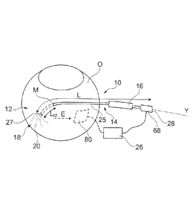
An electrode device 10 according to the invention, represented in figure 1,
comprises an
insertion part 12 and a handling part 14. The insertion part 12 is the part of
the electrode
device which is to be inserted into the suprachoroidal space by the physician.
The handling
part 14 is the part of the electrode device which is manipulated by the
physician to operate
CA 02845549 2014-02-14
WO 2013/024437 8 PCT/IB2012/054146
the electrode device, in particular to insert the insertion part 12 into the
suprachoroidal
space and to deploy and retract the set of wires.
In a service position, the insertion part 12 is inserted between the sclera S
and the choroid
H of an eye. This space is called "suprachoroidal space" I.
The electrode device 10 comprises a set 18 of wires 20, movable, in particular
in the
service position, from a retracted position to a deployed position, and,
preferably,
reversely. The electrode device 10 also comprises a support 25 to support and
guide said
wires 20. The insertion part 12 is adapted for an insertion into the
suprachoroidal space in
the retracted position.
The electrode device also comprises an actuator intended for an operator to
change the
position of the set of wires, and an electrical conductor intended to enable
the
establishment of an electrical connection of the wires 20 with an electrical
generator 26.
The length L of the electrode device, in the retracted position, is preferably
more than
5 cm, more than 8 cm and/or less than 20 cm, less than 15 cm or even less than
10 cm.
The insertion part 12 of the electrode device is preferably curved, so as to
conform to the
outside surface of the choroid H.
As it may be seen from figures 2 and 4, the bottom outer surface 32 of the
insertion part 12
is preferably curved longitudinally (see figure 2) and laterally (see figures
4a to 4c) to
substantially conform to the curved shape of the choroid H. In the same way,
the upper
outer surface 34 of the insertion part is preferably curved, both
longitudinally and laterally,
to substantially conform to the curved shape of the sclera S. Preferably,
these surfaces are
therefore curved spherically. The radius of curvature of these surfaces is
preferably greater
than 9 mm, 10 mm or 11 mm, and/or less than 15 mm, less than 14 mm, less than
13 mm,
or less than 12 mm.
The transversal cross section of the insertion part 12 may have a circular or,
preferably,
flat contour, as represented in figures 4a to 4c. To enable minimally invasive
surgical
access within the suprachoroidal space I, the width W25 and/or the thickness
T25 of the
insertion part 12 is preferably less than 2.0 mm, less than 1.5 mm, less than
1.2 mm, less
than 1.0 mm, less than 0.8 mm, less than 0.5 mm.
CA 02845549 2014-02-14
WO 2013/024437 9 PCT/IB2012/054146
The insertion part 12 of the electrode device has preferably, in the retracted
position, a
flexible rigidity defined by a flexible modulus equal to or less than about
5.2 .10-9 kN/m2,
preferably less than 4 .10-9 kN/m2, less than 3 .10-9 kN/m2, less than 2 .10-9
kN/m2, less
than 1.0 .10-9 kN/m2. The flexibility of the insertion part 12 enables it to
conform to the
anatomy of the sclera and choroid as it is pushed into the service position.
The insertion part 12 preferably has a distal tip 30 conformed so as to be
atraumatic. The
tip 30 is preferably rounded off to achieve a smooth curved surface.
Preferably, it is
tapered in thickness toward the tip 30 so as to be well suited to opening up
the cleavage
plane between the sclera S and the choroid H as the insertion part is pushed
into the
service position. Preferably, the distal tip 30 is not sharp, so that the
electrode device of
the invention is not configured to be used as a needle.
Moreover, preferably, the outer surface of the insertion part 12 is covered,
at least
partially, preferably completely, by a lubricious outer coating.
To limit its introduction in the suprachoroidal space, the electrode device
may be provided
with abutment means (not shown).
Support
In the represented embodiment, the support 25 extends, along its longitudinal
axis Y-Y,
from the distal tip 30 to the proximal end 28 of the electrode device.
Preferably, the shape of the insertion part 12 is provided by a distal part 31
of the support
25, the distal end of the support corresponding to the distal tip 30 of the
electrode device.
The shape of the distal part 31 of the support 25, i.e. its outer surface, is
adapted so that, in
the retracted position, said distal part may be inserted into the
suprachoroidal space of an
eye.
In a preferred embodiment, the support 25, and in particular its distal part
31, is tubular
along more than 50 %, more than 80 %, more than 90 % or even 100% of its
length L.
Preferably, it is provided with a lumen 36 which preferably laterally diverges
at the
approach to the tip 30. In particular, the transversal dimension of the lumen
may increase,
for instance so that the divergence of the lumen be in the shape of a section
of a cone or of
CA 02845549 2014-02-14
WO 2013/024437 10 PCT/IB2012/054146
a nozzle (see Figures 15, 16a and 16b). The lumen 36 may also radiate into a
plurality of
guiding tubes 38 opening outwardly through respective openings 40.
The guiding tubes 38 preferably open outwardly laterally (381) and/or axially
(382). As
represented in figure 2, the guiding tubes 38 may be oriented so as to guide
wires 20
upwardly or downwardly toward the surface of the choroid or of the sclera.
Each guiding tube may contain, in the retracted position, one or several wires
20.
In one embodiment, all the wires exit out of the same opening. The width W25
of the
support may therefore be very small.
The support 25 may be provided with, or constitute an optical guide, so that
light may be
transmitted, in particular in the service position, from the outside of the
eye to the insertion
part 12, and in particular to the distal tip 30. Advantageously, the
determination of the
location of the insertion part 12 is made easier. An optical fibre may also be
fixed on the
support 25 to constitute said optical guide.
As it is represented in figures 4b and 4c, some, or all of the guiding tubes
38 may be
replaced, at least in part, by grooves 40. The grooves 40 may be practiced in
the outer
bottom surface 32 (figure 4b) or in the outer upper surface 34 (figure 4c).
The creation of
grooves advantageously enables a very low thickness T25 for the support 25.
Set of flexible wires
The set of wires may comprise 2, 3, 4, 5, 6, 7, 8, 9 or more wires.
Preferably, the number
of wires is 20 or less, preferably 15 or less.
Each of the wires 20 comprises a respective outside part 64 which, in the
deployed
position, protrudes, i.e. extends away from the support 25. In the preferred
embodiment,
each of said outside parts comprises an electrically conductive element. In
particular, the
electrically conductive element of a wire may be said outside part itself, or
said wire itself,
or a coating applied on a wire core. The electrically conductive element of a
wire is
preferably made of a conductive non oxidative metal selected from iridium,
platinum,
iridium/platinum, and gold, or made of carbon, stainless steel, silver,
aluminium,
tungsten...
CA 02845549 2014-02-14
WO 2013/024437 11 PCT/IB2012/054146
The diameter of any wire 20 is preferably more than 0.01 mm and less than 0.3
mm,
preferably less than 0.1 mm.
Preferably, the wires 20 are elastic, in particular may have a shape memory so
that, in the
deployed position, they may have the desired configuration, as it is described
hereafter.
Each wire 20 has preferably, at its free distal end, a plug 52, for instance
in silicone, so as
to be atraumatic.
At the opposite proximal end 54, the wires 20 are regrouped so as to minimize
their
bulkiness, and reduce the outer dimensions of the support 25.
Retracted position
Preferably, more than 80%, more than 90%, more than 95%, preferably 100% of
the
length of the wires 20 is inside the support 25 in the retracted position.
Preferably, the free
distal ends of the wires are at less than 1 mm, or less than 0.5 mm of the
opening(s)
through which they may exit to reach the deployed position.
In the retracted position, the free distal ends of the wires 20 may partially
define the outer
surface of the electrode device.
Deployed position
The length of the outside parts 64 is preferably more than 1 mm, more than 3
mm, more
than 4 mm and/or less than 15 mm, less than 12 mm, less than 10 mm, less than
8 mm, or
less than 6 mm.
In the deployed position, the outside parts of the wires 20 may all have the
same length or
the lengths may differ.
Preferably, outside parts of wires 20 recover, preferably because of their
elasticity, a
curved shape so that they extend along a spherical surface, the radius of
curvature of
which being preferably greater than 9 mm, 10 mm or 11 mm, and/or less than 15
mm, less
than 14 mm, less than 13 mm, or less than 12 mm, corresponding to that of the
interface
between the sclera and the choroid.
In an embodiment, the guiding tubes 38 contribute to the shape of the outside
parts of the
wires 20.
CA 02845549 2014-02-14
WO 2013/024437 12 PCT/IB2012/054146
Preferably, the convex surface defined by the outside parts of the wires 20 is
more than
10.106 m2, more than 15.106 m2, more than 20.106 m2, more than 30.106 m2,
and/or less
than 50.10-6 m2.
Preferably, all the outside parts of the wires 20 stem from the distal part of
the support 25.
Some of them, as represented in figures 5, 9, 12, 13 and 14, or all of them,
as represented
in figures 7 and 11, may extend beyond the tip 30 of the support 25.
They may project from the support 25 symmetrically to the longitudinal axis Y-
Y, or
symmetrically to the vertical longitudinal median plane C of the support 25,
or not. They
may radiate or not.
Actuator
The actuator is used to move the set of wires 20 from the retracted position
(figure 3) to
the deployed position (figure 5). The actuator may comprise the control handle
16 and a
cable 60 which establishes a mechanical relationship between the set of wires
and the
control handle 16.
In particular, the support 25 may be tubular and the cable 60, attached to the
proximal
ends 54 of the wires, may exit from the proximal part of the support, so that
an operator
may push or pull the cable in the service position (see figure 15). In this
embodiment, the
cable 60 may slide inside the lumen of the tubular support 25, axially, so as
to push or pull
the set of wires, and move said wires from the deployed position to the
retracted position.
In one embodiment, the control handle 16 may be slidably mounted on the
support 25 and
be fixed onto the cable 60, for instance through a longitudinal slot of the
support 25.
The cable 60 may comprise, or even be formed by the wires 20. Advantageously,
the cable
can therefore be part of the electrical conductor which is described
hereafter. In this
embodiment in particular, it preferably comprises a sheath 66 maintaining the
wires
together, like an electrical multistrand cable. The cable 60 may also not
comprise the wires
20.
CA 02845549 2014-02-14
WO 2013/024437 13 PCT/IB2012/054146
Electrical conductor
The electrical conductor is used to establish, at least in the deployed
position, an
electrically conductive path between the electrically conductive elements of
the outside
parts of the wires and a terminal 68 to be connected to an electrical
generator.
.. Preferably, the cable 60 of the actuator enables an electrical connection
of the wires 20
with the terminal 68. In particular when the cable 60 does not comprise any of
the wires
20, it may be made of an electrically conductive material, or be coated with
an electrically
conductive material extending so as to enable the conduction of electrical
current from the
terminal 68 to the wires 20.
.. The electrical conductor may also be supported by the support 25. In
particular, it may be
the support itself or a part of the support, or an electrically conductive
layer covering, at
least partially, the surface of the lumen 36 of the support. This would make
the connection
with the electrical generator easier. However, an electrical connexion with
the electrically
conductive elements of the wires may be more difficult to establish.
In some embodiments, such as represented in figure 11, the wires 20 may be
used as a
support for an electrically conductive web 70 electrically connected with the
electrical
conductor. The use of an electrically conductive web as an electrically
conductive element
advantageously increases the efficiency of the electroporation.
Especially in this embodiment, the wires 20 may not be electrically
conductive, provided
that there is an electrical connection between the web 70 and the electrical
conductor, in
particular the cable 60. The web 70 is preferably made of an elastic material
encouraging
its deployment. The wires 20 may then be arranged so as to tension and stiffen
the web 70
in the deployed position.
The insertion part of the electrode device, and in particular the distal part
of the support
.. 25, may be provided, on their outer upper and/or bottom surfaces, with
electrical
contact(s) 71, for instance a coating, electrically connected with the wires.
As the
provision of conductive webs, this embodiment advantageously increases the
useful
surface (able to create an electrical field) of the electrode device.
CA 02845549 2014-02-14
WO 2013/024437 14 PCT/IB2012/054146
Variations
Many variations of an electrode device of the invention are contemplated.
The openings of the support 25 through which the wires 20 protrude in the
deployed
position are not necessarily positioned at the distal end of the support and
may be disposed
anywhere on the distal part of support, and in particular on the lateral sides
of the distal
part of the support 25 (see in particular figures 8 and 9).
In one embodiment, as represented on figures 6 and 10, the support 25 is a
sleeve, slidably
mounted on a core, the proximal ends of the wires being fixed to the distal
end of the core.
The core may be a cable 60, as previously described.
A pull on the support 25 (arrow F), makes the wires 20 exit from their
respective
openings, i.e. from an axial opening 72 in figure 6 and from lateral openings
in figure 10.
Advantageously, in the service position, the deployment of the wires 20 does
not impart
any movement of the wires frontwards. The risk of damaging the eye is
therefore reduced.
Also with a sliding support, the distal end of the support 25 may be rounded
off so as to
make easier the introduction of the electrode device toward the service
position, as
represented in figure 10.
In one embodiment, as represented in figure 12, the distal end of a wire 20
may not project
from the support 25. Indeed, the distal end 74 of a wire may be fixed to the
support, for
instance inside the support 25, so that, when the wire 20 is pushed toward the
distal tip 30
(or equivalently when the support 25, acting as a sliding sleeve, is pulled
toward the
proximal end of the electrode device), the wire 20 is pushed away from the
support 25 in
the shape of a loop 76. Advantageously, the deployment of a loop limits the
risk of
damaging the eye.
The dimensions and the number of the electrically conductive elements is not
limited,
provided that the electrical conductor enables, in said deployed position, an
electrical
connection between said electrically conductive element(s) and an electrical
generator.
Preferably, the set of electrically conductive elements extends along at least
two
dimensions. For instance, it comprises at least two wires, or it comprises at
least a
conductive web.
CA 02845549 2014-02-14
WO 2013/024437 15 PCT/IB2012/054146
Of course, the characteristics of the different embodiments may be combined.
For
instance, the same electrode device may comprise, in the deployed position,
loops 76, as
represented in figures 12 or 13, and curved line wires, as represented in the
other
embodiments. The electrode device may also comprise one or several loops 76
and a
support in the shape of a sliding sleeve adapted so as to deploy said loop(s).
The number of wires 20 exiting from the same opening is not limited, as
represented in
figures 13 and 14.
Electroporation device
An electroporation device according to the invention is represented in figure
1. It
comprises an electrode device 10 according to the invention, a counter
electrode 80, in the
shape of a surface electrode, and an electrical generator 26 so as to polarize
differently the
wires 20 of the electrode device according to the invention and the counter
electrode 80.
This polarization creates an electrical field E between the wires of the
electrode device of
the invention and the counter electrode.
.. The counter electrode 80 may be a plate electrode, preferably made of a
rigid material,
applied on the outside surface of the eye. The counter electrode may be, for
instance, a
wire type electrode or a plate contact type electrode. Preferably, the counter
electrode is
curved, preferably spherically_ the radius of curvature being preferably
greater than 9
mm, greater than 10 mm or greater than 11 mm, and/or less than 15 mm, less
than 14 mm,
less than 13 mm, or less than 12 mm.
The counter electrode is optionally adapted to be reversibly applied on the
surface of the
eye.
The counter electrode is preferably made of a conductive non oxidative metal
selected for
example from iridium, platinum, iridium/platinum, and gold, or made of carbon,
stainless
steel, silver, aluminium, tungsten...
The electrical generator 26 is adapted so as to generate an electrical field
enabling
electroporation, as described hereafter for instance.
According to the invention, all the wires 20 which are electrically connected
to the
electrical generator 26 have the same polarity. Preferably, all the wires are
electrically
CA 02845549 2014-02-14
WO 2013/024437 16 PCT/IB2012/054146
connected together. However, in one embodiment, some of the wires 20 may not
be
electrically connected to the electrical generator 26.
In one embodiment, the number of wires 20 which are electrically connected
together may
be changed by the operator.
In one embodiment, the operator may change the wires 20 which are electrically
connected to the electrical generator. It becomes therefore possible to change
the shape of
the electrical field E.
Pharmaceutical composition
An electroporation device according to the invention may be used for the
electroporation
of a therapeutic nucleic acid of interest after delivering a pharmaceutical
composition
formulated with said therapeutic nucleic acid into the suprachoroidal space of
a diseased
eye.
The nucleic acid to be used in the instant invention can be any nucleic acid
of interest
exhibiting a biological property. More particularly, the nucleic acid can be
any nucleic
acid encoding a natural, truncated, artificial, chimeric or recombinant
product [e.g., a
polypeptide of interest (including a protein or a peptide), a RNA, etc.]
exhibiting a
biological activity.
The nucleic acid is preferably a desoxyribonucleic acid (DNA) molecule (cDNA,
gDNA,
synthetic DNA, artificial DNA, recombinant DNA, etc.) or a ribonucleic acid
(RNA)
molecule (mRNA, tRNA, RNAi, RNAsi, catalytic RNA, antisens RNA, viral RNA,
etc.).
The nucleic acid may be single stranded or multiple stranded nucleic acid,
preferably
double-stranded nucleic acid or may be complexed. The nucleic acid may
comprise hybrid
sequences or synthetic or semi-synthetic sequences. It may be obtained by any
technique
known to persons skilled in the art, and especially by screening libraries, by
chemical
synthesis, or alternatively by mixed methods including chemical or enzymatic
modification of sequences obtained by screening libraries.
In a particular embodiment, the therapeutic nucleic acid is of synthetic or
biosynthetic
origin, or extracted from a virus or from a unicellular or pericellular
eukaryotic or
prokaryotic organism.
CA 02845549 2014-02-14
WO 2013/024437 17 PCT/IB2012/054146
The therapeutic nucleic acid used in the present invention may be naked, may
be
complexed to any chemical, biochemical or biological agent, may be inserted in
a vector,
etc., when administered to the suprachoroidal space.
As used herein, the term "naked DNA" refers to any nucleic acid molecule which
is not
combined to a synthetic, biosynthetic, chemical, biochemical or biological
agent
improving the delivery or transfer of said DNA, or facilitating its entry into
the cell.
As used herein, the term "vector" refers to a nucleic acid molecule capable of
transporting
another nucleic acid to which it has been linked. This term also refers in the
present
application to any delivery carrier, such as a composition associated to a
therapeutic or
prophylactic nucleic acid in order to increase its cellular delivery.
Preferred vectors are those capable of autonomous replication and/or
expression of nucleic
acids to which they are linked. Vectors capable of directing the expression of
genes to
which they are operatively linked are referred to herein as "expression
vectors". In general,
expression vectors of utility in recombinant DNA techniques are often in the
form of
"plasmids" which refer to circular double stranded DNA loops which, in their
vector form,
are not bound to the chromosome. In the present invention, the plasmid is the
most
commonly used form of vector. The plasmid is a preferred form of naked DNA
according
to the invention.
Vectors may also be episomal DNA, yeast artificial chromosomes,
minichromosomes or
viral vectors wherein the viral vector is selected from the group consisting
of a lentivirus,
an adenovirus, an adeno-associated virus and a virus-like vector.
The vector may also be a lipid vesicle such as a liposome. Lipid based
compounds which
are not liposomes may further be used. For example, lipofectins and
cytofectins are lipid-
based positive ions that bind to negatively charged nucleic acid and form a
complex that
can ferry the DNA across a cell membrane. The invention is intended to include
such other
forms of expression vectors which serve equivalent functions and which become
known in
the art subsequently hereto.
In addition, the nucleic acid according to the invention may also contain one
or more
additional regions, for example regulatory elements of small or large size
which are
available to the skilled artisan such as a promoter region (constitutive,
regulated,
inducible, tissue-specific, etc.), for example sequences allowing and/or
promoting
CA 02845549 2014-02-14
WO 2013/024437 18 PCT/IB2012/054146
expression in the targeted tissue (e.g. choroid or retina) or cells (e.g. RPE
or
photoreceptors), a transcription termination signal, secretion sequences, an
origin of
replication and/or nuclear localization signal (nls) sequences which further
enhance
polynucleotide transfer to the cell nucleus. Such nls sequences have been
described in the
prior art including the SV40 large T antigen sequence.
Additionally, the nucleic acid may further comprise selectable markers useful
in selecting,
measuring, and monitoring nucleic acid transfer results (transfer to which
tissues, duration
of expression, etc.). The types of expression systems and reporter genes that
can be used
or adapted for use are well known in the art. For example, genes coding for a
luciferase
activity, an alkaline phosphatase activity, or a green fluorescent protein
activity are
commonly used.
The nucleic acid according to the invention may contain any nucleotide
sequence of any
size. The nucleic acid may thus vary in size from a simple oligonucleotide to
a larger
molecule such as a nucleotide sequence including exons and/or introns and/or
regulatory
elements of any sizes (small or large), a gene of any size, for example of
large size, or a
chromosome for instance, and may be a plasmid, an episome, a viral genome, a
phage, a
yeast artificial chromosome, a mini chromosome, an anti sense molecule, etc.
In a particularly preferred embodiment, the polynucleotide is a double-
stranded, circular
DNA, such as a plasmid, encoding a product with biological activity.
The nucleic acid can be prepared and produced according to conventional
recombinant
DNA techniques, such as amplification, culture in prokaryotic or eukaryotic
host cells,
purification, etc. The techniques of recombinant DNA technology are known to
those of
ordinary skill in the art.
In a particular embodiment, the nucleic acid of interest is capable of
exerting a beneficial
effect on the targeted cells. It may compensate for a deficiency in or reduce
an excess of
an endogenous substance. Alternatively, it may confer new properties on the
targeted cells.
It may be for example an antisense sequence or nucleic acid encoding a
polypeptide which
can affect the function, morphology, activity and/or metabolism of ocular
cells.
The down regulation of gene expression using antisense nucleic acids can be
achieved at
the translational or transcriptional level. Antisense nucleic acids of the
invention are
preferably nucleic acid fragments capable of specifically hybridizing with a
nucleic acid
19
encoding an endogenous ocular active substance or the corresponding messenger
RNA.
These antisense nucleic acids can be synthetic oligonucleotides, optionally
modified to
improve their stability and selectivity. They can also be DNA sequences whose
expression
in the cell produces RNA complementary to all or part of the mRNA encoding an
endogenous ocular active substance. Antisense nucleic acids can be prepared by
expression of all or part of a nucleic acid encoding an endogenous ocular
active substance,
in the opposite orientation. Any length of antisense sequence is suitable for
practice of the
invention so long as it is capable of down-regulating or blocking expression
of the
endogenous ocular active substance. Preferably, the antisense sequence is at
least 20
nucleotides in length. The preparation and use of antisense nucleic acids, DNA
encoding
antisense RNAs and the use of oligo and genetic antisense is disclosed in
W092/15680.
Among the biologically active polypeptides or proteins optionally expressed by
a nucleic
acid as described above and suitable for practice of the invention are
enzymes, blood
derivatives, hormones, lymphokines, cytokines, chimiokines, anti-inflammatory
factors,
growth factors, trophic factors, neurotrophic factors, haematopoietic factors,
angiogenic
factors, anti-angiogenic factors, inhibitors of metalloproteinase, regulators
of apoptosis,
coagulation factors, receptors thereof, in particular soluble receptors, a
peptide which is an
agonist or antagonist of a receptor or of an adhesion protein, antigens,
antibodies,
fragments or derivatives thereof and other essential constituents of the cell,
proteins
involved in the visual cycle within RPE cells, and structure proteins of
retinal cells.
Various retina-derived neurotrophic factors have the potential to rescue
degenerating
photoreceptor cells, and may be delivered through a method according to the
present
invention. Preferred biologically active agents may be selected from VEGF,
Angiogenin,
Angiopoietin-1 , DeM, acidic or basic Fibroblast Growth Factors (aFGF and
bFGF), FGF-
2, Follistatin, Granulocyte Colony-Stimulating factor (G-CSF), IIepatocytc
Growth Factor
(HGF), Scatter Factor (SF), Leptin, Midkine, Placental Growth Factor (PGF),
Platelet-
Derived Endothelial Cell Growth Factor (PD- ECGF), Platelet-Derived Growth
Factor-BB
(PDGF-BB), Pleiotrophin (PTN), RdCVF (Rod-derived Cone Viability Factor),
Progranulin, Proliferin, Transforming Growth Factor-alpha (TGF-alpha),
Transforming
Growth Factor-beta (TGF-beta), Tumor Necrosis Factor-alpha (TNF-alpha),
Vascular
Endothelial Growth Factor (VEGF), Vascular Permeability Factor (VPF), CNTF,
BDNF,
CA 2845549 2018-10-03
CA 02845549 2014-02-14
WO 2013/024437 20 PCT/IB2012/054146
GDNF, PEDF, NT3, BFGF, angiopoietin, ephrin, EPO, NGF, IGF, GMF, aFGF, NT5,
Gax, a growth hormone, [alpha]-1 -antitrypsin, calcitonin, leptin, an
apolipoprotein, an
enzyme for the biosynthesis of vitamins, hormones or neuromediators,
chemokines,
cytokines such as 1L-1 , 1L-8, IL-10, 1L-12, IL-13, a receptor thereof, an
antibody blocking
anyone of said receptors, TIMP such as TIMP-1 , TIMP-2, TIMP-3, TIMP-4,
angioarrestin, endostatin such as endostatin XVIII and endostatin XV, ATF,
angiostatin, a
fusion protein of endostatin and angiostatin, the C- terminal hemopexin domain
of matrix
metalloproteinase-2, the kringle 5 domain of human plasminogen, a fusion
protein of
endostatin and the kringle 5 domain of human plasminogen, the placental
ribonuclease
inhibitor, the plasminogen activator inhibitor, the Platelet Factor-4 (PF4), a
prolactin
fragment, the Proliferin-Related Protein (PRP), the antiangiogenic
antithrombin III, the
Cartilage-Derived Inhibitor (CDI), a CD59 complement fragment, vasculostatin,
vasostatin (calreticulin fragment), thrombospondin, fibronectin, in particular
fibronectin
fragment gro-beta, an heparinase, human chorionic gonadotropin (hCG),
interferon
alpha/beta/gamma, interferon inducible protein (IP-10), the monokine-induced
by
interferon-gamma (Mig), the interferon-alpha inducible protein 10 (IP10), a
fusion protein
of Mig and IP10, soluble Fms-Like Tyrosine kinase 1 (FLT-1) receptor, Kinase
insert
Domain Receptor (KDR), regulators of apoptosis such as Bc1-2, Bad, Bak, Bax,
Bik, BcI-
X short isoform and Gax, fragments or derivatives thereof and the like.
In a particular embodiment, the nucleic acid encodes a soluble fragment of the
TNF[alpha]
receptor, the TGF[beta]2 receptor, of VEGFR-1, VEGFR-2, VEGFR-3, CCR2 or MIP1.
The nucleic acid may also, in another preferred embodiment, encode an
antibody, a
variable fragment of a single-chain antibody (ScFv) or any other antibody
fragment having
recognition capacities for the purposes of immunotherapy.
In a particular embodiment of the present invention, the biologically active
nucleic acid
encodes a precursor of a therapeutic protein usable in the present invention
such as those
described above.
In another particular embodiment, the electroporation device of the invention
is
particularly suitable for performing gene replacement. Accordingly the nucleic
acid may
encode for a viable protein so as to replace the defective protein which is
naturally
expressed in the targeted tissue. Typically, defective genes that may be
replaced include
CA 02845549 2014-02-14
WO 2013/024437 21 PCT/IB2012/054146
but are not limited to genes that are responsible for retinal degenerative
diseases such as
retinitis pigmentosa (RP), Leber congenital amaurosis (LCA), recessive RP,
Dominant
retinitis pigmentosa, X-linked retinitis pigmentosa, Incomplete X-linked
retinitis
pigmentosa, dominant, Dominant Leber congenital amaurosis, Recessive ataxia,
posterior
column with retinitis pigmentosa, Recessive retinitis pigmentosa with para-
arteriolar
preservation of the RPE, Retinitis pigmentosa RP12, Usher syndrome, Dominant
retinitis
pigmentosa with sensorineural deafness, Recessive retinitis punctata
albescens, Recessive
Alstrom syndrome, Recessive Bardet-Biedl syndrome, Dominant spinocerebellar
ataxia w/
macular dystrophy or retinal degeneration, Recessive abetalipoproteinemia,
Recessive
retinitis pigmentosa with macular degeneration, Recessive Refsum disease,
adult form,
Recessive Refsum disease, infantile form, Recessive enhanced S-cone syndrome,
Retinitis
pigmentosa with mental retardation, Retinitis pigmentosa with myopathy,
Recessive
Newfoundland rod-cone dystrophy, Retinitis pigmentosa sinpigmento, Sector
retinitis
pigmentosa, Regional retinitis pigmentosa, Senior-Loken syndrome, Joubert
syndrome,
Stargardt disease, juvenile, Stargardt disease, late onset, Dominant macular
dystrophy,
Stargardt type, Dominant Stargardt-like macular dystrophy, Recessive macular
dystrophy,
Recessive fundus flavimaculatus, Recessive cone-rod dystrophy, X-linked
progressive
cone-rod dystrophy, Dominant cone-rod dystrophy, Cone-rod dystrophy; de
Grouchy
syndrome, Dominant cone dystrophy, X-linked cone dystrophy, Recessive cone
dystrophy,
Recessive cone dystrophy with supernormal rod electroretinogram, X-linked
atrophic
macular dystrophy, X-linked retinoschisis, Dominant macular dystrophy,
Dominant radial,
macular drusen, Dominant macular dystrophy, bull's-eye, Dominant macular
dystrophy,
butterfly-shaped, Dominant adult vitelliform macular dystrophy, Dominant
macular
dystrophy, North Carolina type, Dominant retinal-cone dystrophy 1, Dominant
macular
dystrophy, cystoid, Dominant macular dystrophy, atypical vitelliform,
Foveomacular
atrophy, Dominant macular dystrophy, Best type, Dominant macular dystrophy,
North
Carolina-like with progressive, Recessive macular dystrophy, juvenile with
hypotrichosis,
Recessive foveal hypoplasia and anterior segment dysgenesis, Recessive delayed
cone
adaptation, Macular dystrophy in blue cone monochromacy, Macular pattern
dystrophy
.. with type II diabetes and deafness, Flecked Retina of Kandori, Pattern
Dystrophy,
Dominant Stickler syndrome, Dominant Marshall syndrome, Dominant vitreoretinal
degeneration, Dominant familial exudative vitreoretinopathy, Dominant
CA 02845549 2014-02-14
WO 2013/024437 22 PCT/IB2012/054146
vitreoretinochoroidopathy; Dominant neovascular inflammatory
vitreoretinopathy,
Goldmann-Favre syndrome, Recessive achromatopsia, Dominant tritanopia,
Recessive rod
monochromacy, Congenital red-green deficiency, Dcuteranopia, Protanopia,
Deuteranomaly, Protanomaly, Recessive Oguchi disease, Dominant macular
dystrophy,
.. late onset, Recessive gyrate atrophy, Dominant atrophia greata, Dominant
central areolar
choroidal dystrophy, X-linked choroideremia, Choroidal atrophy, Central
areolar, Central,
Peripapillary, Dominant progressive bifocal chorioretinal atrophy, Progresive
bifocal
Choroioretinal atrophy, Dominant Doyne honeycomb retinal degeneration
(Malattia
Leventinese), Amelogenesis imperfecta, Recessive Bietti crystalline
corneoretinal
dystrophy, Dominant hereditary vascular retinopathy with Raynaud phenomenon
and
migraine, Dominant Wagner disease and erosive vitreoretinopathy, Recessive
microphthalmos and retinal disease syndrome; Recessive nanophthalmos,
Recessive
retardation, spasticity and retinal degeneration, Recessive Bothnia dystrophy,
Recessive
pseudoxanthoma clasticum, Dominant pseudoxanthoma elasticum; Recessive Batten
.. disease (ceroid-lipofuscinosis), juvenile, Dominant Alagille syndrome,
McKusick-
Kaufman syndrome, hypoprebetalipoproteinemi a, acanthocytosis, palladial
degeneration;
Recessive Hallervorden-Spatz syndrome; Dominant Sorsby's fundus dystrophy,
Oregon
eye disease, Kearns-Sayre syndrome, Retinitis pigmentosa with developmental
and
neurological abnormalities, Basseb Korenzweig Syndrome, Hurler disease,
Sanfilippo
disease, Scieie disease, Melanoma associated retinopathy, Sheen retinal
dystrophy,
Duchenne macular dystrophy, Becker macular dystrophy, and Birdshot
Retinochoroidopathy. Examples of genes include but are not limited to genes
encoding for
ATP-binding cassette transporter, RPE65, RdCVF, CP290...
In another embodiment, the electroporation device of the invention is
particularly suitable
for performing exon skipping for restoring the function of mutated proteins
responsible for
retinal degenerative disease. Exon skipping involves blocking or preventing
the
incorporation into mature mRNA of one or more targeted exon(s) which encodes
amino
sequences that are responsible for a protein dysfunction. This is accomplished
by exposing
the pre-mRNA that includes exons encoding the protein to antisense
oligonucleotides
(AONs) which are complementary to sequence motifs that are required for
correct splicing
of the one or more targeted exons. The AONs bind to complementary required
sequences
in the pre-mRNA and prevent normal splicing. Instead, the targeted exons are
excised and
CA 02845549 2014-02-14
WO 2013/024437 23 PCT/IB2012/054146
are not included in the mature mRNA that is translated into protein, and the
amino acid
sequences encoded by the targeted exons are missing from the translated
protein.
Furthermore, in another embodiment of the present invention, a mixture of
nucleic acids
encoding distinct biologically active products can be used. This variant
allows co-
expression of different products in the ocular cells.
The pharmaceutical composition of the invention may also comprise compatible
or
physiologically acceptable carrier, excipient or diluent.
The term "pharmaceutically" or "pharmaceutically acceptable" refers to
molecular entities
and compositions that do not produce an adverse, allergic or other untoward
reaction when
administered to a mammal, especially a human, as appropriate. A
pharmaceutically
acceptable carrier or excipient refers to a non-toxic solid, semi-solid or
liquid filler,
diluent, encapsulating material or formulation auxiliary of any type.
Pharmaceutically compatible or physiologically acceptable carrier, excipient
or diluent
includes diluents and fillers which are pharmaceutically acceptable for the
methods of the
invention, are sterile, and may be selected from neutral to slightly acidic,
isotonic,
buffered saline (including phosphates, chloride, etc.), aqueous or oleaginous
solutions or
suspensions and more preferably from sucrose, trehalose, surfactants, proteins
and amino
acids. The pharmaceutically compatible or physiologically acceptable carrier,
excipient or
diluent is preferably formulated using suitable dispersing, wetting,
suspending, soothing,
isotonic or viscosity building agents, stabilizers, preservatives and
appropriate buffer to
form an isotonic solution. The particular pharmaceutically acceptable carrier
and the ratio
of active compound to carrier are determined by the solubility and chemical
properties of
the composition, the particular mode of administration, and standard
pharmaceutical
practice. Those skilled in the art will understand how to formulate such
vehicles by known
techniques.
An example of stabilizers is disodium edetate or the like. Examples of
isotonic agents are
glycerin, propylene glycol, polyethylene glycol, sodium chloride, potassium
chloride,
sorbitol and mannitol or the like. Examples of buffers are citric acid, sodium
hydrogenphosphate, glacial acetic acid and trometamol or the like. Examples of
pH
adjusters are hydrochloric acid, citric acid, phosphoric acid, acetic acid,
sodium hydroxide,
sodium carbonate and sodium hydrogencarbonate or the like. An example of
soothing
CA 02845549 2014-02-14
WO 2013/024437 24 PCT/IB2012/054146
agents is benzyl alcohol or the like. Examples of preservatives are
benzalkonium chloride,
benzethonium chloride, p-hydroxybenzoate esters, sodium benzoate and
chlorobutanol or
the like.
Viscosity greater than that of simple aqueous solutions may be desirable to
increase ocular
absorption of the active compound, to decrease variability in dispensing the
formulations,
to decrease physical separation of components of a suspension or emulsion of
formulation
and/or otherwise to improve the ophthalmic formulation. Such viscosity
building agents
include, for example, polyvinyl alcohol, polyvinyl pyrrolidone, methyl
cellulose,
hydroxypropyl methylcellulose, hydroxyethyl cellulose, carboxymethyl
cellulose,
hydroxypropyl cellulose or other agents known to those skilled in the art.
Such agents are
typically employed at a level of from about 0.01 to about 2 wt. %.
Preparation forms of the pharmaceutical composition intended for
administration to
suprachoroidal space are preferably liquid preparations. The liquid
preparations can be
prepared, for example, by dissolving the biologically active agent in BSS
(Balanced Salt
Solution), a glycerin solution, a hyaluronic acid solution and the like. A
particular
composition comprises for example BBS (60%) and hyaluronic acid (40%). A
stabilizer,
an isotonic agent, a buffer, a pH adjustor, a soothing agent, a preservative,
electrolytes,
such as sodium, potassium, calcium, magnesium and/or chloride or the like can
optionally
be added in an adequate amount to the liquid preparations.
The pharmaceutical composition may comprise or the biologically active agent
may be
combined (in a use according to the present invention) with any additional
active
ingredient or adjuvant. The adjuvant may be selected from any substance,
mixture, solute
or composition facilitating or increasing the biological activity of the
prophylactic or
therapeutic agent such as any biologic, synthetic or biosynthetic agent which
improves the
delivery or transfer of said agent and may be assimilated to a vector (as
delivery carrier)
according to the invention. The adjuvant may be conditioned and administered
separately
or sequentially from the prophylactic or therapeutic agent containing
composition and/or
at a distinct site of injection. Treatment with multiple agents and/or
adjuvants according to
the invention need not be done using a mixture of agents and/or adjuvants but
may be
done using separate pharmaceutical preparations. The preparations need not be
delivered
CA 02845549 2014-02-14
WO 2013/024437 25 PCT/IB2012/054146
at the same exact time, but may be coordinated to be delivered to a patient
during the same
period of treatment, i. e., within a week or a month or each other.
Any suitable therapeutic agents can be coordinated with the compositions of
the present
invention. Non-limiting examples of therapeutic agents which may be
administered in
addition to the above biologically active (prophylactic or therapeutic)
agent(s) through a
method according to the present invention also include permeabilizing agents
such as a
virus, a lipid vesicle, hyaluronic acid, lipid-based positive ions,
polycationic emulsions,
cationic peptides, polyplex, etc.; Actual dosage levels of active ingredients
in the
compositions of the present invention may be adapted so as to obtain an amount
of active
ingredient that is effective to obtain a desired biological activity. It
should be understood,
however, that the specific dose level for any particular patient will depend
upon a variety
of factors including the body weight, general health, sex, diet, time, rates
of absorption and
excretion, combination with other drugs and the severity of the particular
disease being
treated.
Kit
In accordance with the present invention, kits for preventing or treating an
ocular disease
are envisioned. An electrode device according to the invention and a
pharmaceutical
composition according to the present invention, and optionally a counter
electrode,
optionally an electrical generator, optionally instructions for use may be
supplied together
in a kit. Within the kit, the components may be separately packaged or
contained.
Instructions can be in a written, video, or audio form, can be contained on
paper, an
electronic medium, or even as a reference to another source, such as a website
or reference
manual.
Other components such as excipients, carriers, other drugs or adjuvants,
instructions for
administration of the active substance or composition, and administration or
injection
devices can be supplied in the kit as well.
Method
According to the invention, a method for treating an ocular disease in a
subject may
comprise the steps consisting of
CA 02845549 2014-02-14
WO 2013/024437 26 PCT/IB2012/054146
i) delivering a pharmaceutical composition formulated with a therapeutic
nucleic
acid of interest into the suprachoroidal space of the diseased eye and
ii) exposing the region where the pharmaceutical composition was delivered
to an
electrical field generated with an electroporation device according to the
invention.
The pharmaceutical composition is preferably chosen among the pharmaceutical
compositions which are described here above.
Diseases
The method of the present invention is particularly suitable for the treatment
of ocular
diseases affecting the posterior region of the eye, and more particularly
ocular diseases
affecting the retina. Non-limiting examples of ocular diseases that may be
treated by the
method of the present invention include ocular diseases affecting the macula
such as age
related macular degeneration (wet and dry) or inherited macular degeneration,
macular
oedema of any origin (age related macular degeneration, diabetes,
inflammation,
degeneration, central serous chorioretinitis or diffuse epitheliopathy....),
inherited retinal
dystrophies, such as Leber congenital amaurosis, retinitis pigmentosa, cone
rod
dystophies, best vitelliforme maculopathy, intraocular inflammation such
retinitis,
chorioretinitis, choroiditisõ ischemic retinopathy (in particular retinopathy
of prematurity
and diabetic retinopathy), retinal vascular diseases, ocular ischemia syndrome
and other
vascular anomalies, choroidal disorders and tumors, vitreous disorders, glial
proliferation
such as proliferative vitreo retinopathy and glial proliferation associated to
diabetic pre
retinal angiogenesis, diabetic retinopathy ischemic or proliferative.
Inherited retinal dystrophies or retinitis pigmentosa are inherited blinding
diseases due to
mutations or deletions in gene implicated in the visual cycle. They begin in
the young age
and progress slowly until total blindness. Loss of photoreceptors is
associated to loss of
retinal pigment cells and to vascular and optic nerve atrophy at the later
stages. Some of
these inherited degeneration are due to mutation in mitochondrial DNA. In
particular, non
limiting examples of retinal degenerative diseases include but are not limited
to retinitis
pigmentosa (RP), Leber congenital amaurosis (LCA), recessive RP, Dominant
retinitis
pigmentosa, X-linked retinitis pigmentosa, Incomplete X-linked retinitis
pigmentosa,
dominant, Dominant Leber congenital amaurosis, Recessive ataxia, posterior
column with
retinitis pigmentosa, Recessive retinitis pigmentosa with para-arteriolar
preservation of the
CA 02845549 2014-02-14
WO 2013/024437 27 PCT/IB2012/054146
RPE, Retinitis pigmentosa RP12, Usher syndrome, Dominant retinitis pigmentosa
with
sensorineural deafness, Recessive retinitis punctata albescens, Recessive
Alstrom
syndrome, Recessive Bardet-Biedl syndrome, Dominant spinocerebellar ataxia w/
macular
dystrophy or retinal degeneration, Recessive abetalipoproteinemia, Recessive
retinitis
pigmentosa with macular degeneration, Recessive Refsum disease, adult form,
Recessive
Refsum disease, infantile form, Recessive enhanced S-cone syndrome, Retinitis
pigmentosa with mental retardation, Retinitis pigmentosa with myopathy,
Recessive
Newfoundland rod-cone dystrophy, Retinitis pigmentosa sinpigmento, Sector
retinitis
pigmentosa, Regional retinitis pigmentosa, Senior-Loken syndrome, Joubert
syndrome,
Stargardt disease, juvenile, Stargardt disease, late onset, Dominant macular
dystrophy,
Stargardt type, Dominant Stargardt-like macular dystrophy, Recessive macular
dystrophy,
Recessive fundus flavimaculatus, Recessive cone-rod dystrophy, X-linked
progressive
cone-rod dystrophy, Dominant cone-rod dystrophy, Cone-rod dystrophy; de
Grouchy
syndrome, Dominant cone dystrophy, X-linked cone dystrophy, Recessive cone
dystrophy,
Recessive cone dystrophy with supernormal rod electroretinogram, X-linked
atrophic
macular dystrophy, X-linked retinoschisis, Dominant macular dystrophy,
Dominant radial,
macular drusen, Dominant macular dystrophy, bull's-eye, Dominant macular
dystrophy,
butterfly-shaped, Dominant adult vitelliform macular dystrophy, Dominant
macular
dystrophy, North Carolina type, Dominant retinal-cone dystrophy 1, Dominant
macular
dystrophy, cystoid, Dominant macular dystrophy, atypical vitelliform,
Foveomacular
atrophy, Dominant macular dystrophy, Best type, Dominant macular dystrophy,
North
Carolina-like with progressive, Recessive macular dystrophy, juvenile with
hypotrichosis,
Recessive foveal hypoplasia and anterior segment dysgenesis, Recessive delayed
cone
adaptation, Macular dystrophy in blue cone monochromacy, Macular pattern
dystrophy
with type II diabetes and deafness, Flecked Retina of Kandori, Pattern
Dystrophy,
Dominant Stickler syndrome, Dominant Marshall syndrome, Dominant vitreoretinal
degeneration, Dominant familial exudative vitreoretinopathy, Dominant
vitreoretinochoroidopathy; Dominant neovascular inflammatory
vitreoretinopathy,
Goldmann-Favre syndrome, Recessive achromatopsia, Dominant tritanopia,
Recessive rod
monochromacy, Congenital red-green deficiency, D euteranopia, Protanopia,
Deuteranomaly, Protanomaly, Recessive Oguchi disease, Dominant macular
dystrophy,
late onset, Recessive gyrate atrophy, Dominant atrophia greata, Dominant
central areolar
CA 02845549 2014-02-14
WO 2013/024437 28 PCT/IB2012/054146
choroidal dystrophy, X-linked choroideremia, Choroidal atrophy, Central
areolar, Central,
Peripapillary, Dominant progressive bifocal chorioretinal atrophy, Progresive
bifocal
Choroioretinal atrophy, Dominant Doyne honeycomb retinal degeneration
(Malattia
Leventinese), Amelogenesis imperfecta, Recessive Bietti crystalline
corneoretinal
dystrophy, Dominant hereditary vascular retinopathy with Raynaud phenomenon
and
migraine, Dominant Wagner disease and erosive vitreoretinopathy, Recessive
microphthalmos and retinal disease syndrome; Recessive nanophthalmos,
Recessive
retardation, spasticity and retinal degeneration, Recessive Bothnia dystrophy,
Recessive
pseudoxanthoma elasticum, Dominant pseudoxanthoma elasticum; Recessive Batten
disease (ceroid-lipofuscinosis), juvenile, Dominant Alagille syndrome,
McKusick-
Kaufman syndrome, hypoprebetalipoproteinemia, acanthocytosis, palladial
degeneration;
Recessive Hallervorden-Spatz syndrome; Dominant Sorsby's fundus dystrophy,
Oregon
eye disease, Kearns-Sayre syndrome, Retinitis pigmentosa with developmental
and
neurological abnormalities, Basseb Korenzweig Syndrome, Hurler disease,
Sanfilippo
disease, Scieie disease, Melanoma associated retinopathy, Sheen retinal
dystrophy,
Du chenn e macular dystrophy, Becker macular dystrophy, and Birdshot
Retinochoroidopathy.
Intraocular inflammation regroups all types of inflammation of the intraocular
tissues,
mainly uvea and retina. Intraocular inflammations may be from immunologic
causes,
infectious causes, iatrogenic causes or of unknown etiologies. They may be
acute,
recurrent or chronic. intraocular inflammations are among the most causes of
curable
blindness. Posterior segment intraocular inflammations may be associated to
vasculitis,
optic neuritis, vitritis and chorio retinitis, retinitis, choriditis,
choroidal neovascularisation,
choroidal neovascularization due to AMD, to myopia, inflammation, diffuse
epitheliopathy, bruch membrane rupture, polypoidal choroidal vasculopathy,
post
traumatic...
There are two major types of glaucoma: chronic glaucoma or primary open-angle
glaucoma (POAG) and acute closed-angle glaucoma. Other variations include
congenital
glaucoma, pigmentary glaucoma, neovascular glaucoma and secondary glaucoma.
Glaucoma is similar to ocular hypertension but with accompanying optic nerve
damage
and vision loss. Glaucoma is usually treated with eye drops, laser, or
conventional eye
surgery. If not treated, glaucoma will cause blindness.
CA 02845549 2014-02-14
WO 2013/024437 29 PCT/IB2012/054146
Angiogenesis is the formation of new capillary blood vessels leading to
neovascularization. Angiogenesis is a complex process which includes a series
of
sequential steps including endothelial cell mediated degradation of vascular
basement
membrane and interstitial matrices, migration of endothelial cells,
proliferation of
endothelial cells, and formation of capillary loops by endothelial cells.
Though
angiogenesis is a normal process for the development or maintenance of the
vasculature,
pathological conditions (i.e., angiogenesis dependent diseases) arise where
blood vessel
growth is actually harmful. Angiogenesis is notably associated with important
diseases of
ocular tissue, including diabetic retinopathies, age related macular
degeneration,
retinopathy of prematurity, corneal graft rejection, neovascular glaucoma and
corneal
scaring. Any abnormal growth of blood vessels in the eye can scatter and block
the
incident light prior to reaching the retina. Neovascularization can occur at
almost any site
in the eye and significantly alter ocular tissue function. Some of the most
threatening
ocular neovascular diseases are those which involve the retina. For example,
many
diabetic patients develop a retinopathy which is characterized by the
formation of leaky,
new blood vessels on the anterior surface of the retina and in the vitreous
causing
proliferative vitreoretinopathy. A subset of patients with age related macular
degeneration
develop subretinal neovascularization which leads to their eventual blindness.
Diabetic Retinopathy occurs when the retinal vessels inside the eye leak blood
and fluids
into the surrounding tissue. About 80% of patient with diabetes develop
diabetic
retinopathy. This disease is generally treated using a laser. However, laser
therapy
involves complications including retinal vein occlusion, loss of visual
acuity, vitreous
hemorrhage and sometimes fails. If left untreated, diabetic retinopathy may
cause
blindness.
Retinopathy of Prematurity (ROP) affects prematurely born babies. It consists
of the
abnormal growth of blood vessels within the retinal and vitreous. Progression
to later
stages of ROP can lead to the formation of scar tissue on the retina, vitreous
hemorrhage,
and retinal detachment. The treatment is usually performed either by laser or
cryotherapy
(freezing).
Ischemic retinopathies are retinopathies associated to vascular occlusion
(capillaries or
large vessels) that lead to neuroretinal suffering, cell death and nco
angiogenesis. Macular
CA 02845549 2014-02-14
WO 2013/024437 30 PCT/IB2012/054146
degeneration is a disease that affects central vision and leads to loss of
vision. Although
there are forms of macular degeneration that strike young people, the
condition occurs
most commonly in people who are over 60 years of age. This disorder is thus
called age-
related macular degeneration (AMD). Because only the center of a person's
vision is
usually affected, blindness rarely occurs from the disease. However, injury to
the macula
in the center of the retina can destroy the ability to see straight ahead
clearly. Dry forms
associate degeneration of neuroretina, RPE cells and choroids. Wet forms
associate
previously described phenomenons and growth of neovessels from the
choriocapillaries
and/or retinal vessels, sub retinal detachment and hemorrhages, sub epithelial
hemorrhages
and tears, etc. Macular degeneration usually occurs after the age of sixty.
While your
central vision is reduced, most patients retain some vision and never go
totally blind.
A particular aspect of the invention is a method of treating intraocular
neovessels or
macular oedema comprising delivering to the suprachoroidal space of a subject
suffering
therefrom a nucleic acid encoding an anti VEGF, an anti VEGF receptor or an
anti PLGF.
A further particular aspect of the invention is a method of treating or
delaying retinitis
pigmentosa comprising delivering to the suprachoroidal space of a subject
suffering
therefrom a nucleic acid encoding a neurotrophic factor as described above.
Another particular aspect of the invention is a method of treating diabetic
retinopathy
comprising delivering to the suprachoroidal space of a subject suffering
therefrom a
nucleic acid encoding a a nucleic acid encoding an anti IRS-I or IGF-L
Operation
A step i), an operator, for instance a physician, injects a pharmaceutical
composition into
the suprachoroidal space I.
The pharmaceutical composition of the invention may be delivered into the
suprachoroidal
space at multiple injection sites. The delivering may also be repeated over
time.
The means to inject the pharmaceutical composition into the suprachoroidal
space may be
an injection needle or preferably a flexible catheter or microcannula. Methods
for injecting
a pharmaceutical injection into the suprachoroidal space are well known in the
art (e.g.
Einmahl S. Invest Ophtamol Vis Sci 2001 42:695: Galimova VU. Vestn Oftalmo
2001
117:20; Olsen TW Am J. Ophtalmol 2006 142:777). Devices for injecting a
CA 02845549 2014-02-14
WO 2013/024437 31 PCT/IB2012/054146
pharmaceutical composition into the suprachoroidal space are also well known
in the art
(e.g. Olsen TW Am J. Ophtalmol 2006 142:777, US 2010173866, WO 2007100745 and
WO 2011053512).
The physician then makes a micro-incision M through the sclera S at an
appropriate place.
He then takes an electrode device of the invention, the set of wires being in
the retracted
position. The set of wires 20 are supported and protected by the support 25,
at least in the
distal part of the support to be inserted. The wires substantially do not
protrude from the
support 25 so as to avoid interference with the introduction of the distal
part of the support
25. The shape of the distal part of the support makes the insertion part 12
adapted for an
insertion into the suprachoroidal space of the eye in the refracted position,
so as to reach
the service position.
The physician inserts the distal tip of the electrode device 10 into the mini-
incision and
guides it between the sclera and choroid so as to avoid any additional lesion.
The shape
and dimensions of the insertion part of the electrode device makes this
insertion easier. If
the electrode device is provided with an optical fibre enabling the
transmission of light
from outside the eye to the distal tip 30 (possibly the support itself), the
physician may
couple this fibre with a light source, outside the eye, and easily localize
the distal tip 30
within the suprachoroidal space, which helps him for placing it in the right
service
position. In the service position, the length L12 of the electrode device
which is extending
in the suprachoroidal space is preferably more than 5 mm, more than 10 mm
and/or less
than 40 mm, less than 30 mm.
Once the electrode device is in the service position, the physician deploys
the set of wires
20.
The deployment and the retraction of the set of wires are operated remotely,
from outside
the eye. Any actuator known for similar purposes may be used, in particular
the actuators
used for catheters. For instance, the physician pushes the cable 60, as
represented in figure
15. He may also manipulate a control handle 16 if available.
The deployment of the wires 20 is facilitated due to the shape of the guiding
tubes and/or
the shape memory o f the wires.
.. The physician also places the counter electrode 80 in a position adapted so
that an
electrical field E may be established between the electrode formed by the
electrically
CA 02845549 2014-02-14
WO 2013/024437 32 PCT/IB2012/054146
conductive elements of the outside parts of the wires of the electrode device
according to
the invention and the counter electrode 80.
In the present invention, when the choroid or retina is targeted, the
electrical field is
applied after applying the counter electrode on the surface of the sclera at
the opposing
side where the suprachoroidal injection was performed.
The electrical generator 26 is connected to the terminal 68 of the electrode
device and to
the counter electrode 80, so that the terminal 68 (and, correspondingly, the
electrically
conductive elements of the outside parts of wires 20) and the counter
electrode 80 have
opposite polarities.
-- Then, the electrical generator generates an appropriate electrical field E
in the region
where the pharmaceutical composition was delivered, which provokes an
electroporation
of the injected therapeutic nucleic acid of interest inside the choroid, RPE
and retina.
Electroporation is indeed suitable for, or increases, permeability of a cell
membrane and/or
at least a portion of a targeted tissue adjacent to the suprachoroidal space
to a biologically
active agent such as a nucleic acid. In addition, a brief electric impulse
with a given field
strength is used to allow transport or migration of agents through the tissue
or across cell
membranes into cells, by an electrophoretic effect. The technique of
electroporation is
well known to those of ordinary skill in the art.
However, to date electroporation failed to transfect adult photoreceptor cells
when the
plasmids were injected either into the ocular cavity or into the sub retinal
space.
In the deployed position, the surface of the choroid which is covered by the
electrically
conductive elements of the outside parts of the wires is advantageously large,
which
enables the generation of an efficient electrical field.
This surface may be increased if
- the distal part of the support is itself made of an electrically conductive
material, or is
at least partially covered by such a material(electrical contacts 71), in
electrical
connection with the electrically conductive elements of the outside parts of
the wires
Or
- the electrically conductive elements of the outside parts of the wires
are in contact
with an electrically conductive web 70, or
- electrically conductive elements of the wires which do not project from
the support in
the deployed position and are electrically connected with the electrically
conductive
CA 02845549 2014-02-14
WO 2013/024437 33 PCT/IB2012/054146
elements of the outside parts of the wires may enter into contact with the
choroid
(figure 4b) or with the sclera (figure 4c).
In a particular embodiment, an electrical field constituted by one or more
electrical
pulse(s) is applied.
The field intensity of which is between about 1 and 600 Volts, preferably 1
and 400 Volts,
even more preferably between about 1 and 200 Volts, advantageously between
about 10
and 100 Volts, or 15 and 70 Volts.
The total duration of application of the electric field may be between 0.01
millisecond and
1 second, preferably between 0.01 and 500 milliseconds, more preferably
between 1 and
500 milliseconds, even more preferably greater than 1 or 10 milliseconds. In a
preferred
embodiment, the total duration of application of the electric field is between
10
milliseconds and 100 milliseconds and is preferably of 20 milliseconds.
The number of electric pulses applied may be between for example 1 and 100
000. Their
frequency may be comprised between 0.1 and 1000 hertz. It is preferably a
regular
frequency.
Electric pulses may also be delivered in an irregular manner relative to each
other, the
function describing the intensity of the electric field as a function of the
time for one pulse
being preferably variable.
Electric pulses may be unipolar or bipolar wave pulses. They may be selected
for example
from square wave pulses, exponentially decreasing wave pulses, oscillating
unipolar wave
pulses of limited duration, oscillating bipolar wave pulses of limited
duration, or other
wave forms. Preferentially, electric pulses comprise square wave pulses or
oscillating
bipolar wave pulses.
At the end of the electroporation step, the physician shuts the generator
down, retracts the
wires inside the support, and pulls the insertion part of the electrode
device, through the
micro-incision M, out of the suprachoroidal space.
As it now clearly appears, the retracted position facilitates the introduction
of the insertion
part of the electrode device, its guidance to the desired site of the eye to
be treated, and its
extraction from the suprachoroidal space, whereas the deployed position
provides a large
electrode surface.
34
Therefore, an electrode device according to the present invention allows an
intervention
that is as minimally invasive as possible, and, with the possibility of
deploying the set of
wires, enables an efficient electroporation.
Throughout this application, various references describe the state of the art
to which this
invention pertains.
CA 2845549 2018-10-03