Note: Descriptions are shown in the official language in which they were submitted.
CA 02845810 2016-04-06
= W02013/028231
PCT/US2012/024570
ANTI-0X40 ANTIBODIES AND METHODS OF USING THE SAME
FIELD OF INVENTION
[0001] This invention relates generally to modulation of the 0X40-
receptor activation, and
more particularly, to modulating the 0X40-receptor to inhibit the
immunosuppressive function
of Interleukin 10 (IL-10) producing CD4+ type 1 regulatory T cells ("Trl
cells") and Foxp3+-
expressing regulatory T cells (also sometimes referred to herein as "Foxp3+ T-
reg" cells), and the
generation of Tr 1 cells from CD4+ cells or nave cells and IL-10 production.
1
CA 02845810 2016-04-06
WO 2013/028231 PCT/US2012/024570
BACKGROUND OF THE INVENTION
[0005] Trl
cells have a critical role in peripheral tolerance. Trl cells are particularly
important in limiting tissue damage to the host during inflammatory immune
responses. The
generation of Trl cells accompanies both TH1 and TH2 immune responses in vivo
and in vitro.
[0006] Trl
cells are generated from naïve CD4+ T cells during an antigen-driven T cell
immune response. Trl cells are anergic in response to signaling through TCR,
CD28 and IL-2
receptors and have the ability to suppress antigen-driven proliferation of
naive CD4+ T cells in
vivo and in vitro. Trl cells have the ability to inhibit the development of
autoimmune diseases
and limit the magnitude of immune responses to microbial pathogens.
[0007] While
the molecular signals that lead to the Trl cells have been studied, little is
known
about the molecular signals that negatively regulate the generation of these
cells. Although
immunosuppressive drugs, cytokines, co-stimulatory molecules, and DCs have
been implicated
in the induction of Trl cells, signals that negatively regulate the generation
of Trl cells remain
elusive.
BRIEF SUMMARY OF THE INVENTION
[0008]
Activation of the 0X40 receptor blocks Tr' generation from naïve or memory
CD4+
T cells as well as IL-10 production from Trl cells and the immunosuppressive
function of the
Trl cells. Activation of the 0X40 receptor also blocks IL-10 production by
Foxp3+ T-reg cells
and immunosuppressive function. As such, presented herein are agonist
antibodies that bind to
the 0X40 receptor, whereby the agonist modulates the activation of the 0X40
receptor to block
IL-10 cytokine secretion and/or the Trl and Foxp3+ T-reg cells overall
immunosuppressive
function. Essentially, the antibodies can mimic the 0X40 ligand and trigger
the 0X40 receptor
on Trl and/or on natural T regulatory cells ("nTregs"), also referred to as
"Foxp3+ T-regs."
2
CA 02845810 2014-02-19
WO 2013/028231 PCT/US2012/024570
[0009] As we have shown in co-pending patent applications US Serial Nos.
11/659,266 and
12/861,135, OX4OL inhibits the generation and function of IL-10-producing Trl
cells from naïve
and memory CD4+ T cells that were induced by the immunosuppressive drugs
dexamethasone
and vitamin D3. We discovered that OX4OL inhibits the generation and function
of IL-10
producing regulatory T cells. These discoveries demonstrate that signaling
0X40 by OX4OL
suppresses the generation of human IL-10 producing immunosuppressive T cells
in culture. This
unique function of OX4OL is not shared by two other co-stimulatory TNF-family
members,
GITR-ligand and 4-1BB-ligand. OX4OL also strongly inhibits the generation and
function of IL-
10-producing Trl cells induced by two physiological stimuli provided by
inducible co-
stimulatory ligand and immature DCs. Signaling the 0X40 receptor on human T
cells by
monoclonal antibodies, small molecules, or by the OX4OL, or protein having at
least 90 percent
homology thereto, modulates and regulates the generation and function of IL-10
producing
immunosuppressive T cells.
[00010] The discovery lends to numerous applications of treatment. For
example, agonistic
antibodies, small molecules, or OX4OL could be used to suppress the generation
and the function
of IL-10 producing immunosuppressive T cells and therefore could be used to
enhance immune
responses to treat cancer and infectious diseases, or as an adjuvant for
cancer vaccines.
Antagonistic antibodies to 0X40 or to OX4OL, or antagonistic small molecules,
could be used to
enhance the generation and the function of IL-10-producing immunosuppressive T
cells and
therefore could be used for the development of therapies for autoimmune
diseases and graft
versus host diseases. Our discovery also provides for high throughput methods
for screening
antibodies or small molecules either activating the 0X40 receptor (or
conversely blocking 0X40
signaling) on T cells for the development of therapeutics for cancer, or
alternatively,
autoimmune diseases, and graft versus host diseases.
[00011] Monoclonal and human antibodies (sometimes referred to herein as an
"anti-0X40
antibody" and/or other variations of the same) that bind human 0X40 receptor
are provided
herein. These antibodies are useful in the treatment or prevention of acute or
chronic diseases or
conditions whose pathology involves 0X40. In one aspect, an isolated human
antibody, or an
antigen-binding portion thereof, that binds to human 0X40 and is effective as
a cancer treatment
3
CA 02845810 2014-02-19
WO 2013/028231 PCT/US2012/024570
or treatment against an autoimmune disease is described. Any of the anti-0X40
antibodies
disclosed herein may be used as a medicament. Any one or more of the anti-0X40
antibodies
may be used to treat one or more a variety of cancers or autoimmune disease
described herein.
[00012] Isolated humanized antibodies that bind to 0X40 are provided herein.
The isolated
antibodies as described herein bind to 0X40, and may bind to 0X40 encoded from
the following
genes: NCBI Accession Number NP 003317, Genpept Accession Number P23510, or
genes
having 90 percent homology thereto. The isolated antibody provided herein may
further bind to
the 0X40 receptor having one of the following GenBank Accession Numbers:
AAB39944,
CAE11757, or AAI05071.
[00013] As taught herein, exemplary is an isolated antibody which binds to
0X40 comprising:
(a) a heavy chain variable region CDR1 comprising the amino acid sequence of
SEQ ID NO: 1;
(b) a heavy chain variable region CDR2 comprising the amino acid sequence of
SEQ ID NO: 2;
(c) a heavy chain variable region CDR3 comprising the amino acid sequence of
SEQ ID NO. 3;
(d) a light chain variable region CDR1 comprising the amino acid sequence of
SEQ ID NO. 7;
(e) a light chain variable region CDR2 comprising the amino acid sequence of
SEQ ID NO. 8;
and (f) a light chain variable region CDR3 comprising the amino acid sequence
of SEQ ID NO.
9.
[00014] Furthermore, another example is an isolated antibody which binds to
0X40
comprising: (a) a heavy chain variable region CDR1 comprising the amino acid
sequence of SEQ
ID NO: 13; (b) a heavy chain variable region CDR2 comprising the amino acid
sequence of SEQ
ID NO: 14; (c) a heavy chain variable region CDR3 comprising the amino acid
sequence of SEQ
ID NO. 15; (d) a light chain variable region CDR1 comprising the amino acid
sequence of SEQ
ID NO. 19; (e) a light chain variable region CDR2 comprising the amino acid
sequence of SEQ
ID NO. 20; and (f) a light chain variable region CDR3 comprising the amino
acid sequence of
SEQ ID NO. 21.
[00015] Furthermore, another example is an isolated antibody which binds to
0X40
comprising: (a) a heavy chain variable region CDR1 comprising the amino acid
sequence of SEQ
ID NO: 25; (b) a heavy chain variable region CDR2 comprising the amino acid
sequence of SEQ
4
CA 02845810 2014-02-19
WO 2013/028231 PCT/US2012/024570
ID NO: 26; (c) a heavy chain variable region CDR3 comprising the amino acid
sequence of SEQ
ID NO. 27; (d) a light chain variable region CDR1 comprising the amino acid
sequence of SEQ
ID NO. 32; (e) a light chain variable region CDR2 comprising the amino acid
sequence of SEQ
ID NO. 33; and (f) a light chain variable region CDR3 comprising the amino
acid sequence of
SEQ ID NO. 34.
[00016] Alternatively, an isolated antibody may have a heavy chain variable
region CDR1
comprising the amino acid sequence of SEQ ID NO: 1 or 13; a heavy chain
variable region
CDR2 comprising the amino acid sequence of SEQ ID NO: 2 or 14; and/or a heavy
chain
variable region CDR3 comprising the amino acid sequence of SEQ ID NO: 3 or 15,
or a heavy
chain variable region CDR having 90 percent homology thereto.
[00017] Further, an isolated antibody may have a light chain variable region
CDR1 comprising
the amino acid sequence of SEQ ID NO: 7 or 19; a light chain variable region
CDR2 comprising
the amino acid sequence of SEQ ID NO: 8 or 20 and/or a light chain variable
region CDR3
comprising the amino acid sequence of SEQ ID NO: 9 or 21, or a heavy chain
variable region
having 90 percent homology thereto.
[00018] The isolated antibody may have a light chain variable region ("VL")
comprising the
amino acid sequence of SEQ ID NO: 10, 11, 22 or 23, or an amino acid sequence
with at least 90
percent identity to the amino acid sequences of SEQ ID NO: 10, 11, 22 or 23.
The isolated
antibody may have a heavy chain variable region ("VH") comprising the amino
acid sequence of
SEQ ID NO: 4, 5, 16 and 17, or an amino acid sequence with at least 90 percent
identity to the
amino acid sequences of SEQ ID NO: 4, 5, 16 and 17. As such, as an example,
the isolated
antibody may comprise a variable heavy sequence of SEQ ID NO:5 and a variable
light sequence
of SEQ ID NO: 11, or a sequence having 90 percent homology thereto. Similarly,
the isolated
antibody can have a variable heavy sequence of SEQ ID NO:17 and a variable
light sequence of
SEQ ID NO: 23 or a sequence having 90 percent homology thereto.
[00019] The isolated antibody may have variable light chain encoded by the
nucleic acid
sequence of SEQ ID NO: 12, or 24, or a nucleic acid sequence with at least 90
percent identity
to the nucleotide sequences of SEQ ID NO: 12 or 24. The isolated antibody may
have variable
CA 02845810 2014-02-19
WO 2013/028231 PCT/US2012/024570
heavy chain encoded by a nucleic acid sequence of SEQ ID NO: 6 or 18, or a
nucleic acid
sequence with at least 90 percent identity to nucleotide sequences of SEQ ID
NO: 6 or 18.
[00020] Also provided herein are monoclonal antibodies. The monoclonal
antibodies may
have a variable light chain comprising the amino acid sequence of SEQ ID NO:
10 or 22, or an
amino acid sequence with at least 90 percent identity to the amino acid
sequences of SEQ ID
NO: 10 or 22. Further provided are monoclonal antibodies having a variable
heavy chain
comprising the amino acid sequence of SEQ ID NO: 4 or 16, or an amino acid
sequence with at
least 90 percent identity to the amino acid sequences of SEQ ID NO: 4 or 16.
[00021] Also provided herein is isolated nucleic acid encoding any of the anti-
0X40
antibodies taught herein. Further provided herein are host cells, each
comprising nucleic acid
encoding any of the anti-0X40 antibodies described herein. Methods of
producing an antibody
(such as the host cell comprising nucleic acid encoding any of the anti-0X40
antibodies
described herein) comprising culturing the host cell so that the antibody is
produced, and/or
recovering the antibody from the host cell, are further provided.
BRIEF DESCRIPTION OF THE DRAWINGS
[00022] So that the manner in which the above-recited features, aspects and
advantages of the
invention, as well as others that will become apparent, are attained and can
be understood in
detail, more particular description of the invention briefly summarized above
can be had by
reference to the embodiments thereof that are illustrated in the drawings that
form a part of this
specification. It is to be noted, however, that the appended drawings
illustrate some
embodiments of the invention and are, therefore, not to be considered limiting
of the invention's
scope, for the invention can admit to other equally effective embodiments.
[00023] FIG. 1 shows that FOXP3+ Tregs infiltrated human follicular lymphoma
(FL) tissues
and co-localized with tumor B cells and monocytes. Left: Double immunostaining
of FOXP3+
Tregs (red) and CD20+ B lymphoma cells (green); Right: FOXP3+ Tregs (red) and
CD11c+
monocytes/macrophage/DC (green).
6
CA 02845810 2014-02-19
WO 2013/028231 PCT/US2012/024570
[00024] FIGS. 2A and 2B show increased numbers of CD4+FOXP3+ Tregs in patients
with
FL. Tumor cells and PBMCs were obtained from six patients with FL at initial
diagnosis before
therapy. PBMCs were also obtained from six normal donors for comparison. The
percentages of
regulatory T cells over total CD4+ T cells were determined by flow cytometric
analysis of
CD4+CD25+CD1271'FOXP3+ Tregs. FIG. 2A shows representative FACS analysis of
Tregs.
FL PBMC and FL tumor cells were divided from the same patient. FIG. 2B shows
the
percentage of Tregs of all donors. Horizontal bar indicate means.
[00025] FIG. 3 shows the isolation of ICOS+FOXP3+ or ICOS-FOXP3+ Tregs from
FL. Single
cell suspension was obtained from a spleen specimen before any treatment.
Cells were thawed on
the day of assay. Enriched CD4+CD8-CD14-CD16-CD56-CD1 1 c-TCR76- T cells were
divided
into CD251' and CD25high subsets. CD4+CD25111ghFOXP3+ Tregs were further
sorted into
ICOShigh and ICOS1' subsets based on surface expression of ICOS. Intracellular
expression of
FOXP3 was determined in all subsets.
[00026] FIG. 4 shows intratumoral Tregs inhibit proliferation of infiltrating
CD4+CD25- T
cells in FL, and the inhibition could be partially blocked by anti-IL-10
neutralization antibodies.
CFSE-labeled CD4+CD25- tumor-infiltrating T cells were cultured with
autologous tumor cells
preactivated by recombinant CD4OL in the presence or absence of autologous
ICOS+FOXP3+
Tregs or ICOS-FOXP3+ Tregs, or anti-IL-10 (10 ps/m1). After 72 hours of
culture, proliferation
of CD4+CD25- cells was determined by flow cytometric analysis of CFSE
dilution.
[00027] FIG. 5A shows the intracellular analysis of cytokine production by
naïve CD4+ T cells
as determined by flow cytometry according to an embodiment of a method of the
present
invention.
[00028] FIG. 5B shows cytokine production by naïve CD4+ T cells as determined
by ELISA
according to an embodiment of a method of the present invention.
[00029] FIG. 5C shows suppressive function by IL-10-producing Trl cells as
determined
by [3H]thymidine incorporation according to an embodiment of a method of the
present
invention.
7
CA 02845810 2014-02-19
WO 2013/028231 PCT/US2012/024570
[00030] FIG. 6A shows the intracellular analysis of cytokine production by
memory CD4+ T
cells as determined by flow cytometry according to an embodiment of a method
of the present
invention.
[00031] FIG. 6B shows IL-10 production by memory CD4+ T cells as determined by
ELISA
according to an embodiment of a method of the present invention.
[00032] FIG. 7A shows the intracellular analysis of cytokine production by
naïve CD4+ T cells
as determined by flow cytometry according to an embodiment of a method of the
present
invention.
[00033] FIG. 7B shows IL-10 production by naïve CD4+ T cells as determined by
ELISA
according to an embodiment of a method of the present invention.
[00034] FIG. 7C shows the number of viable T cells counted according to an
embodiment of a
method of the present invention.
[00035] FIG. 8A shows the intracellular analysis of cytokine production by
naïve CD4+ T cells
as determined by flow cytometry according to an embodiment of a method of the
present
invention.
[00036] FIG. 8B shows IL-10 production by naïve CD4+ T cells as determined by
ELISA
according to an embodiment of a method of the present invention.
[00037] FIG. 8C shows the intracellular analysis of cytokine production by
memory CD4+ T
cells as determined by flow cytometry according to an embodiment of a method
of the present
invention.
[00038] FIG. 8D shows IL-10 production by memory CD4+ T cells as determined by
ELISA
according to an embodiment of a method of the present invention.
[00039] FIG. 8E shows the intracellular analysis of cytokine production by
naïve CD4+ T cells
as determined by flow cytometry according to an embodiment of a method of the
present
invention.
8
CA 02845810 2014-02-19
WO 2013/028231 PCT/US2012/024570
[00040] FIG. 8F shows IL-10 production by naïve CD4+ T cells as determined by
ELISA
according to an embodiment of a method of the present invention.
[00041] FIG. 9 shows IL-10 production by regulatory T cells as determined by
ELISA
according to an embodiment of a method of the present invention.
[00042] FIG. 10 shows the results of screening of anti-human 0X40 hybridoma
supernatants
against L-0X40 versus L parental cells as determined by ELISA.
[00043] FIG. 11 shows the screening of human 0X40-specific monoclonal
antibodies as
determined by flow cytometry analysis according to an embodiment of a method
of the present
invention.
[00044] FIG. 12 shows the confirmation of anti-h0X40 monoclonal antibodies
specificity by
using SUPM2 cells expressing 0X40 (SUPM2-0X40) according to an embodiment of a
method
of the present invention.
[00045] FIG. 13 shows 0X40-specific monoclonal antibodies that can inhibit the
generation of
IL-10 producing cells (Trl) from CD4+ T cells stimulated by vit D3 (0.1
M)/Dex (50 nm),
CD32L/ICOSL and anti-CD3/CD28 (0.2 ps/m1) according to an embodiment of a
method of the
present invention. Representative Fluorescence Activated Cell Sorting (FACS)
data are shown
in A and the percentages of IL-10 producing cells for all 0X40 monoclonal
antibodies treatments
are shown in B.
[00046] FIG. 14 shows the results of h0X40-specific monoclonal antibodies that
inhibit Trl
cell generation also stimulate CD4+ T cell proliferation according to an
embodiment of a method
of the present invention.
[00047] FIGS. 15A, 15B, and 15C details the titration of 0X40 monoclonal
antibodies for their
ability to inhibit the generation of Trl cells from CD4+ T cells according to
an embodiment of a
method of the present invention. Representative FACS data are shown in FIG.
15A and
percentage of Trl cells after treatment with nine 0X40 monoclonal antibodies
are shown in FIG.
15B.
9
CA 02845810 2014-02-19
WO 2013/028231 PCT/US2012/024570
[00048] FIGS. 16A, 16B, and 16C shows 0X40-specific monoclonal antibodies that
inhibit IL-
producing Trl cell generation from CD4+ T cells also inhibit
ICOS+CD4+CD25111ghCD127-
Treg IL-10 production and immunosuppressive function.
Freshly sorted
ICOS+CD4+CD25111ghCD127- Tregs (ICOS+Tregs) were stimulated with anti-CD3 (0.2
ps/m1) in
the presence of CD32L/ICOSL cells and CD32L/OX4OL cells (FIG. 16A) or 0X40
monoclonal
antibodies or control antibody (FIG. 16B) for five days. Cells were then
restimulated with anti-
CD3/CD28 for 24 hours and the supernatants were assayed for IL-10 by enzyme-
linked
immunosorbent assay (ELISA). FIG. 16C is a monocyte-based proliferation assay
showing that
thwo of the antibodies blocked ICOS+Treg function.
[00049] FIGS. 17A and 17B shows the identification of anti-h0X40 monoclonal
antibodies
that inhibit the generation of Trl cells and block FOXP3+CD4+CD25 high Treg
function according
to an embodiment of a method of the present invention. Representative flow
cytometry analyses
are shown in FIG. 17A. Data for six monoclonal antibodies are shown in FIG.
17B.
[00050] FIG. 18 demonstrates the identification of anti-h0X40 monoclonal
antibodies that do
not inhibit Trl cell generation but block FOXP3+CD4+CD25 high Treg function
according to an
embodiment of a method of the present invention.
[00051] FIGS. 19A and 19B shows anti-h0X40 agonist antibodies blocking
lymphoma-
derived CD4+CD25111gh Treg function according to an embodiment of a method of
the present
invention. Representative FACS analyses are shown in FIG. 19A and data for all
experiments
are shown in FIG. 19B.
[00052] FIG. 20 shows that anti-h0X40 monoclonal antibodies can bind to rhesus
CD4+ T
cells. As shown, six of the anti-h0X40 mAbs can bind to rhesus activated CD4+
T cells and will
bind to rhesus 0X40 and activate 0X40 signaling.
[00053] FIG. 21 shows that each of Hu106-222 Lot I and II antibodies of
Example I is
comprised of a heavy chain with a molecular weight of about 50 kD and a light
chain with a
molecular weight of about 25 kD. The purity of Hu106-222 Lot I and II
antibodies appeared to
be more than 95%.
CA 02845810 2014-02-19
WO 2013/028231 PCT/US2012/024570
[00054] FIG. 22 shows the analysis of mouse 106-122, Ch106 and Hu106-222 (Lot
II)
antibodies for binding to L/0X40 cells (Example I).
[00055] FIG. 23 depicts the schematic structure of the expression vector for
Hu106
IgGl/kappa antibody (Expression Vector). Proceeding clockwise from the Sall
site at the top,
the plasmid contains the heavy chain transcription unit starting with the
human cytomegalovirus
(CMV) major immediate early promoter and enhancer (CMV promoter) to initiate
transcription
of the antibody heavy chain gene. The CMV promoter is followed by the VH exon,
a genomic
sequence containing the human gamma-1 heavy chain constant region including
the CH1, hinge,
CH2 and CH3 exons with the intervening introns, and the polyadenylation site
following the
CH3 exon. After the heavy chain gene sequence, the light chain transcription
unit begins with
the CMV promoter, followed by the VL exon and a genomic sequence containing
the human
kappa chain constant region exon (CL) with part of the intron preceding it,
and the
polyadenylation site following the CL exon. The light chain gene is then
followed by the SV40
early promoter (SV40 promoter), the E. coli xanthine guanine phosphoribosyl
transferase gene
(gpt), and a segment containing the 5V40 polyadenylation site (5V40 poly(A)
site). Finally, the
plasmid contains a part of the plasmid pUC19, comprising the bacterial origin
of replication
(pUC ori) and beta-lactamase gene (beta lactamase). Locations of relevant
restriction enzyme
sites are shown in the figure.
[00056] FIG. 24 shows the comparison between Hu 106-222 Lot I and II
antibodies for binding
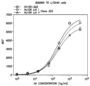
to L/0X40 cells (Example I below).
[00057] FIG. 25 shows Hu119-122 is comprised of a heavy chain with a molecular
weight of
about 50 kD and a light chain with a molecular weight of about 25 kD. The
purity of Hul 19
appeared to be more than 95% (Example II below).
[00058] FIG. 26 shows the result of the FACS analysis of Ch119-122 and Hu119-
122
antibodies described herein (Example II below).
[00059] FIG. 27 shows that humanized anti-human 0X40 mAb clone 119-122
(Hu119), and its
FcR binding mutated antibody (Hu119-AA) enhanced naïve CD4+ T cell
proliferation. Hu119-
122 yielded better T cell stimulatory activity compared to parental mouse anti-
human 0X40
11
CA 02845810 2014-02-19
WO 2013/028231 PCT/US2012/024570
mAb (Mouse119-122). However, chimeric anti-human 0X40 mAb (Ch119, mouse VH and
VL
but human gamma-1 and kappa constant regions) failed to enhance T cell
proliferation.
[00060] FIG. 28 shows FcR binding mutated humanized anti-human 0X40 mAb clone
106-
222 (Hu222AA) and chimeric anti-human 0X40 mAb clone 106-222 (Ch222) enhanced
anti-
CD3 stimulated naïve CD4+ T cell proliferation. These antibodies have similar
stimulatory
activity compared to parental mouse anti-human 0X40 mAb (Mouse222). However,
the fully
humanized anti-human 0X40 Ab, Hu222, did not enhance T cell proliferation
compared to
human IgGl.
[00061] FIG. 29A and B shows that the humanized and mouse anti-human 0X40 mAb
clone
119-122 blocks CD4+ Treg suppressive function.
[00062] FIG. 30 provides data showing anti-human 0X40 antibodies enhance CD4+
and CD8+
T cell proliferation using plate-bound antibodies.
[00063] FIG. 31 shows humanized and mouse anti-human 0X40 antibodies require
cross-
linking in order to enhance T cell proliferation.
[00064] FIG. 32 shows anti-human 0X40 antibodies block the activity of
CD4+FOXP3+nTregs
using plate-bound antibodies.
[00065] FIG. 33 shows that a high concentration of mouse anti-human 0X40
antibodies
preferentially kills FOXP3+ Tregs.
[00066] FIG. 34 shows mouse anti-human 0X40 mAbs act directly on either
effector T cells or
nTregs to block the suppressive function of Tregs.
[00067] FIGS. 35A, 35B, and 35C show the results of anti-h0X40 mAb tumor
treatment in
mice adaptively transferred with h0X4O+CD8+ T cells. The anti-human 0X40 mAb
promotes T
cell expansion and survival in vivo. The therapeutic vaccination regimen is
shown in FIG. 35A.
Representative in vivo bioluminescence images are shown in FIG. 35B. Results
of the antibody
tumor treatment are shown in FIG. 35C.
12
CA 02845810 2014-02-19
WO 2013/028231 PCT/US2012/024570
[00068] FIG. 36 shows the alignment of the amino acid sequences of 106-222,
humanized 106-
222 (Hu106), and human acceptor X61012 (GenBank accession number) VH sequences
are
shown. Amino acid residues are shown in single letter code. Numbers above the
sequences
indicate the locations according to Kabat et al. (Sequences of Proteins of
Immunological
Interests, Fifth edition, NIH Publication No. 91-3242, U.S. Department of
Health and Human
Services, 1991). The same sequences as claimed herein are also provided in the
Sequence
Listing and the position numbers may be different. In Figure 36, CDR sequences
defined by
Kabat et al. (1991) are underlined in 106-222 VH. CDR residues in X61012 VH
are omitted in
the figure. Human VH sequences homologous to the 106-222 VH frameworks were
searched for
within the GenBank database, and the VH sequence encoded by the human X61012
cDNA
(X61012 VH) was chosen as an acceptor for humanization. The CDR sequences of
106-222 VH
were first transferred to the corresponding positions of X61012 VH. Next, at
framework
positions where the three-dimensional model of the 106-222 variable regions
suggested
significant contact with the CDRs, amino acid residues of mouse 106-222 VH
were substituted
for the corresponding human residues. These substitutions were performed at
positions 46 and
94 (underlined in Hu106 VH). In addition, a human framework residue that was
found to be
atypical in the corresponding V region subgroup was substituted with the most
typical residue to
reduce potential immunogenicity. This substitution was performed at position
105 (double-
underlined in Hu106 VH).
[00069] FIG. 37 shows alignment of the amino acid sequences of 106-222,
humanized 106-222
(Hu106), and human acceptor AJ388641 (GenBank accession number) VL sequences
is shown.
Amino acid residues are shown in single letter code. Numbers above the
sequences indicate the
locations according to Kabat et al. (1991). The same sequences as claimed
herein are also
provided in the Sequence Listing although the position numbers may be
different. CDR
sequences defined by Kabat et al. (1) are underlined in 106-222 VH. CDR
residues in AJ388641
VL are omitted in the figure. Human VL sequences homologous to the 106-222 VL
frameworks
were searched for within the GenBank database, and the VL sequence encoded by
the human
AJ388641 cDNA (AJ388641 VL) was chosen as an acceptor for humanization. The
CDR
sequences of 106-222 VL were transferred to the corresponding positions of
AJ388641 VL. No
framework substitutions were performed in the humanized form.
13
CA 02845810 2014-02-19
WO 2013/028231 PCT/US2012/024570
[00070] FIG. 38 shows the nucleotide sequence of the Hu106 VH gene flanked by
SpeI and
HindIII sites (underlined) is shown along with the deduced amino acid
sequence. Amino acid
residues are shown in single letter code. The signal peptide sequence is in
italic. The N-terminal
amino acid residue (Q) of the mature VH is double-underlined. CDR sequences
according to the
definition of Kabat et al. (1991) are underlined. The same sequences as
claimed herein are also
provided in the Sequence Listing and the position numbers may be different in
the Sequence
Listing. The intron sequence is in italic. Hu106 VH gene fragment digested
with SpeI and
HindIII was cloned between the corresponding sites in the Expression Vector
shown in Figure
23.
[00071] FIG. 39 shows the nucleotide sequence of the Hu106-222 VL gene flanked
by NheI
and EcoRI sites (underlined) is shown along with the deduced amino acid
sequence. Amino acid
residues are shown in single letter code. The signal peptide sequence is in
italic. The N-terminal
amino acid residue (D) of the mature VL is double-underlined. CDR sequences
according to the
definition of Kabat et al. (1991) are underlined. The intron sequence is in
italic. Hu106 VL gene
fragment digested with NheI and EcoRI was cloned between the corresponding
sites in the
Expression Vector shown in FIG. 23. The same sequences as claimed herein are
also provided in
the Sequence Listing although the position numbers may be different in the
Sequence Listing.
[00072] FIG. 40 shows the alignment of the amino acid sequences of 119-122,
humanized 119-
122 (Hu119), and human acceptor Z14189 (GenBank accession number) VH sequences
are
shown. Amino acid residues are shown in single letter code. Numbers above the
sequences
indicate the locations according to Kabat et al. (Sequences of Proteins of
Immunological
Interests, Fifth edition, NIH Publication No. 91-3242, U.S. Department of
Health and Human
Services, 1991). CDR sequences defined by Kabat et al. (1991) are underlined
in 119-122 VH.
CDR residues in Z14189 VH are omitted in the figure. Human VH sequences
homologous to the
119-122 VH frameworks were searched for within the GenBank database, and the
VH sequence
encoded by the human Z14189 cDNA (Z14189 VH) was chosen as an acceptor for
humanization. The CDR sequences of 119-122 VH were first transferred to the
corresponding
positions of Z14189 VH. Next, at framework positions where the three-
dimensional model of
the 119-122 variable regions suggested significant contact with the CDRs,
amino acid residues of
14
CA 02845810 2014-02-19
WO 2013/028231 PCT/US2012/024570
mouse 119-122 VH were substituted for the corresponding human residues. These
substitutions
were performed at positions 26, 27, 28, 30 and 47 (underlined in the Hul 19 VH
sequence) as
shown on the figure. The same sequences as claimed herein are also provided in
the Sequence
Listing although the position numbers may be different in the Sequence
Listing.
[00073] FIG. 41 shows the alignment of the amino acid sequences of 119-122,
humanized 119-
122 (Hu119), and human acceptor M29469 (GenBank accession number) VL sequences
are
shown. Amino acid residues are shown in single letter code. Numbers above the
sequences
indicate the locations according to Kabat et al. (1991). CDR sequences defined
by Kabat et al.
(1) are underlined in 119-122 VL. CDR residues in M29469 VL are omitted in the
sequence.
Human VL sequences homologous to the 119-122 VL frameworks were searched for
within the
GenBank database, and the VL sequence encoded by the human M29469 cDNA (M29469
VL)
was chosen as an acceptor for humanization. The CDR sequences of 119-122 VL
were
transferred to the corresponding positions of M29469 VL. No framework
substitutions were
needed in the humanized form. The same sequences as claimed herein are also
provided in the
Sequence Listing although the position numbers may be different in the
Sequence Listing.
[00074] FIG. 42 shows the nucleotide sequence of the Hul 19 VH gene flanked by
SpeI and
HindIII sites (underlined) is shown along with the deduced amino acid
sequence. Amino acid
residues are shown in single letter code. The signal peptide sequence is in
italic. The N-terminal
amino acid residue (E) of the mature VH is double-underlined. CDR sequences
according to the
definition of Kabat et al. (1991) are underlined. The intron sequence is in
italic. Hul 19 VH
gene fragment digested with SpeI and HindIII was cloned between the
corresponding sites in the
Expression Vector shown in FIG. 23. The same sequences as claimed herein are
also provided in
the Sequence Listing although the position numbers may be different in the
Sequence Listing.
[00075] FIG. 43 shows the nucleotide sequence of the Hul 19 VL gene flanked by
NheI and
EcoRI sites (underlined) is shown along with the deduced amino acid sequence.
Amino acid
residues are shown in single letter code. The signal peptide sequence is in
italic. The N-terminal
amino acid residue (E) of the mature VL is double-underlined. CDR sequences
according to the
definition of Kabat et al. (1991) are underlined. The intron sequence is in
italic. Hul 19 VL gene
fragment digested with NheI and EcoRI was cloned between the corresponding
sites in the
CA 02845810 2014-02-19
WO 2013/028231 PCT/US2012/024570
Expression Vector shown in FIG. 23. The same sequences as claimed herein are
also provided in
the Sequence Listing although the position numbers may be different in the
Sequence Listing.
[00076] FIG. 44 shows the nucleotide sequence of mouse 119-43-1 VH cDNA along
with the
deduced amino acid sequence. Amino acid residues are shown in single letter
code. The signal
peptide sequence is in italic. The N-terminal amino acid residue (E) of the
mature VH is double-
underlined. CDR sequences according to the definition of Kabat et al.
(Sequences of Proteins of
Immunological Interests, Fifth edition, NIH Publication No. 91-3242, U.S.
Department of Health
and Human Services, 1991) are underlined.
[00077] FIG. 45 shows the nucleotide sequence of mouse 119-43-1 VL cDNA is
shown the
deduced amnno acid sequence. Amino acid residues are shown in single letter
code. The signal
peptide sequence is in italic. The N-terminal amino acid residue (D) of the
mature VL is double-
underlined. CDR sequences according to the definition of Kabat et al. (1991)
are underlined.
[00078] FIG. 46 shows the nucleotide sequence of the designed 119-43-1 VH gene
flanked by
SpeI and HindIII sites (underlined) along with the deduced amino acid
sequence. Amino acid
residues are shown in single letter code. The signal peptide sequence is in
italic. The N-terminal
amino acid residue (E) of the mature VH is double-underlined. CDR sequences
according to the
definition of Kabat et al. (1991) are underlined. The intron sequence is in
italic.
[00079] FIG. 47 shows the nucleotide sequence of the designed 119-43-1 VL gene
flanked by
NheI and EcoRI sites (underlined) along with the deduced amino acid sequence.
Amino acid
residues are shown in single letter code. The signal peptide sequence is in
italic. The N-terminal
amino acid residue (D) of the mature VL is double-underlined. CDR sequences
according to the
definition of Kabat et al. (1991) are underlined. The intron sequence is in
italic.
[00080] FIG. 48 shows the schematic structure of pCh119-43-1 (referred to as
Expression
Vector in the figure). Proceeding clockwise from the Sall site at the top, the
plasmid contains
the heavy chain transcription unit starting with the human cytomegalovirus
(CMV) major
immediate early promoter and enhancer (CMV promoter) to initiate transcription
of the antibody
heavy chain gene. The CMV promoter is followed by the VH exon, a genomic
sequence
containing the human gamma-1 heavy chain constant region including the CH1,
hinge, CH2 and
16
CA 02845810 2014-02-19
WO 2013/028231 PCT/US2012/024570
CH3 exons with the intervening introns, and the polyadenylation site following
the CH3 exon.
After the heavy chain gene sequence, the light chain transcription unit begins
with the CMV
promoter, followed by the VL exon and a genomic sequence containing the human
kappa chain
constant region exon (CL) with part of the intron preceding it, and the
polyadenylation site
following the CL exon. The light chain gene is then followed by the SV40 early
promoter (SV40
promoter), the E. coli xanthine guanine phosphoribosyl transferase gene (gpt),
and a segment
containing the SV40 polyadenylation site (5V40 poly(A) site). Finally, the
plasmid contains a
part of the plasmid pUC19, comprising the bacterial origin of replication (pUC
ori) and beta-
lactamase gene (beta lactamase).
[00081] FIG. 49 shows the sequences of oligonucleotides used for PCR
amplification and
sequencing of Ch119-43-1 heavy and light chain cDNA.
[00082] FIG. 50 shows the nucleotide sequence of the coding region of gamma-1
heavy chain
in pCh119-43-1 is shown in single letter code. A termination codon is denoted
by "=".
[00083] FIG. 51 shows the nucleotide sequence of the coding region of kappa
light chain in
pCh119-43-1 along with the deduced amino acid sequence. Amino acid residues
are shown in
single letter code. A termination codon is denoted by ".".
[00084] FIG. 52 shows the SDS-PAGE analysis of Ch119-43-1. Five i.ig of Ch119-
43-1 was
run on a 4-20% SDS-PAGE gel under reducing conditions (lane 1). Invitrogen's
SeeBluePlus2
Prestained Standard (Cat # LC5925) was used as molecular weight standards
(lane 2).
[00085] FIG. 53 shows the FACS analysis of the binding of Ch119-43-1 to L/0X40
cells.
Ch119-43-1 was tested at various concentrations, starting at 16 jig/m1 and
serial 4-fold dilutions,
for binding to L/0X40 cells. EC50 value was calculated using GraphPad Prism
(GraphPad
Software, San Diego, CA).
DETAILED DESCRIPTION OF THE INVENTION
[00086] The term "antibody" includes an immunoglobulin molecule comprised of
four
polypeptide chains, two heavy (H) chains and two light (L) chains inter-
connected by disulfide
17
CA 02845810 2014-02-19
WO 2013/028231 PCT/US2012/024570
bonds. Each heavy chain is comprised of a heavy chain variable region
(abbreviated herein as
HCVR or VH) and a heavy chain constant region. The heavy chain constant region
is comprised
of three domains, CH1, CH2 and CH3. Each light chain is comprised of a light
chain variable
region (abbreviated herein as LCVR or VL) and a light chain constant region.
The light chain
constant region is comprised of one domain, CL. The VH and VL regions can be
further
subdivided into regions of hypervariability, termed complementarity
determining regions
(CDRs), interspersed with regions that are more conserved, termed framework
regions (FR).
Each VH and VL is composed of three CDRs and four FRs, arranged from amino-
terminus to
carboxy-terminus in the following order: FR1, CDR1, FR2, CDR2, FR3, CDR3, FR4.
[00087] The term "antigen-binding portion" of an antibody (or "antibody
portion") includes
fragments of an antibody that retain the ability to specifically bind to an
antigen (e.g., h0X40).
It has been shown that the antigen-binding function of an antibody can be
performed by
fragments of a full-length antibody. Examples of binding fragments encompassed
within the
term "antigen-binding portion" of an antibody include (i) a Fab fragment, a
monovalent fragment
consisting of the VL, VH, CL and CH1 domains; (ii) a F(ab')2 fragment, a
bivalent fragment
comprising two Fab fragments linked by a disulfide bridge at the hinge region;
(iii) a Fd
fragment consisting of the VH and CH1 domains; (iv) a Fv fragment consisting
of the VL and
VH domains of a single arm of an antibody, (v) a dAb fragment (Ward et al.,
(1989) Nature
341:544-546), which consists of a VH domain; and (vi) an isolated
complementarity determining
region (CDR). Furthermore, although the two domains of the Fv fragment, VL and
VH, are
coded for by separate genes, they can be joined, using recombinant methods, by
a synthetic
linker that enables them to be made as a single protein chain in which the VL
and VH regions
pair to form monovalent molecules (known as single chain Fv (scFv); see e.g.,
Bird et al. (1988)
Science 242:423-426; and Huston et al. (1988) Proc. Natl. Acad. Sci. USA
85:5879-5883). Such
single chain antibodies are also intended to be encompassed within the term
"antigen-binding
portion" of an antibody. Other forms of single chain antibodies, such as
diabodies are also
encompassed. Diabodies are bivalent, bispecific antibodies in which VH and VL
domains are
expressed on a single polypeptide chain, but using a linker that is too short
to allow for pairing
between the two domains on the same chain, thereby forcing the domains to pair
with
complementary domains of another chain and creating two antigen binding sites
(see e.g.,
18
CA 02845810 2014-02-19
WO 2013/028231 PCT/US2012/024570
Holliger, P., et al. (1993) Proc. Natl. Acad. Sci. USA 90:6444-6448; Poljak,
R. J., et al. (1994)
Structure 2:1121-1123). Still further, an antibody or antigen-binding portion
thereof may be part
of a larger immunoadhesion molecules, formed by covalent or non-covalent
association of the
antibody or antibody portion with one or more other proteins or peptides.
Examples of such
immunoadhesion molecules include use of the streptavidin core region to make a
tetrameric scFv
molecule (Kipriyanov, S. M., et al. (1995) Human Antibodies and Hybridomas
6:93-101) and
use of a cysteine residue, a marker peptide and a C-terminal polyhistidine tag
to make bivalent
and biotinylated scFv molecules (Kipriyanov, S. M., et al. (1994) Mol.
Immunol. 31:1047-1058).
Antibody portions, such as Fab and F(ab')2 fragments, can be prepared from
whole antibodies
using conventional techniques, such as papain or pepsin digestion,
respectively, of whole
antibodies. Moreover, antibodies, antibody portions and immunoadhesion
molecules can be
obtained using standard recombinant DNA techniques, as described herein.
Preferred antigen
binding portions are complete domains or pairs of complete domains.
[00088] 0X40/0X40-ligand (0X40 Receptor)/(OX4OL) are a pair of costimulatory
molecules
critical for T cell proliferation, survival, cytokine production, and memory
cell generation. Early
in vitro experiments demonstrated that signaling through 0X40 on CD4+ T cells
lead to TH2, but
not TH1 development. These results were supported by in vivo studies showing
that blocking
0X40/0X4OL interaction prevented the induction and maintenance of TH2-mediated
allergic
immune responses. However, blocking 0X40/0X4OL interaction ameliorates or
prevents TH1-
mediated diseases. Furthermore, administration of soluble OX4OL or gene
transfer of OX4OL
into tumors were shown to strongly enhance anti-tumor immunity in mice. Recent
studies also
suggest that 0X40/0X4OL may play a role in promoting CD8 T cell-mediated
immune
responses. As discussed herein, 0X40 signaling blocks the inhibitory function
of CD4+CD25+
naturally occurring regulatory T cells and the 0X40/0X4OL pair plays a
critical role in the
global regulation of peripheral immunity versus tolerance.
[00089] The terms "Kabat numbering", "Kabat definitions" and "Kabat labeling"
are used
interchangeably herein. These terms, which are recognized in the art, refer to
a system of
numbering amino acid residues which are more variable (i.e. hypervariable)
than other amino
acid residues in the heavy and light chain variable regions of an antibody, or
an antigen binding
19
CA 02845810 2014-02-19
WO 2013/028231 PCT/US2012/024570
portion thereof (Kabat et al. (1971) Ann. NY Acad, Sci. 190:382-391 and,
Kabat, E. A., et al.
(1991) Sequences of Proteins of Immunological Interest, Fifth Edition, U.S.
Department of
Health and Human Services, NIH Publication No. 91-3242).
[00090] The phrase "recombinant human antibody" includes human antibodies that
are
prepared, expressed, created or isolated by recombinant means, such as
antibodies expressed
using a recombinant expression vector transfected into a host cell, antibodies
isolated from a
recombinant, combinatorial human antibody library, antibodies isolated from an
animal (e.g., a
mouse) that is transgenic for human immunoglobulin genes (see e.g., Taylor, L.
D., et al. (1992)
Nucl. Acids Res. 20:6287-6295) or antibodies prepared, expressed, created or
isolated by any
other means that involves splicing of human immunoglobulin gene sequences to
other DNA
sequences. Such recombinant human antibodies have variable and constant
regions derived from
human germline immunoglobulin sequences (See Kabat, E. A., et al. (1991)
Sequences of
Proteins of Immunological Interest, Fifth Edition, U.S. Department of Health
and Human
Services, NIH Publication No. 91-3242).
[00091] An "isolated antibody" includes an antibody that is substantially free
of other
antibodies having different antigenic specificities (e.g., an isolated
antibody that specifically
binds h0X40 is substantially free of antibodies that specifically bind
antigens other than
h0X40). An isolated antibody that specifically binds h0X40 may bind 0X40
molecules from
other species. Moreover, an isolated antibody may be substantially free of
other cellular material
and/or chemicals.
[00092] The term "activity" includes activities such as the binding
specificity/affinity of an
antibody for an antigen, for example, an anti-human 0X40 antibody that binds
to an 0X40
antigen and/or the activation potency of an antibody, for example, an anti-
0X40 antibody whose
binding to h0X40 receptor activates the biological activity of h0X40 or
activation of receptor
binding in a human L/0X40 cell assay.
[00093] The term "KW, as used herein, is intended to refer to the off rate
constant for
dissociation of an antibody from the antibody/antigen complex. The term "Kd",
as used herein, is
intended to refer to the dissociation constant of a particular antibody-
antigen interaction.
CA 02845810 2014-02-19
WO 2013/028231 PCT/US2012/024570
[00094] The phrase "surface plasmon resonance" includes an optical phenomenon
that allows
for the analysis of real-time biospecific interactions by detection of
alterations in protein
concentrations within a biosensor matrix, for example using the BIAcore system
(Pharmacia
Biosensor AB, Uppsala, Sweden and Piscataway, N.J.). For further descriptions,
see Example 5
and Jonsson, U., et al. (1993) Ann. Biol. Clin. 51:19-26; Jonsson, U., et al.
(1991) Biotechniques
11:620-627; Johnsson, B., et al. (1995) J. Mol. Recognit. 8:125-131; and
Johnnson, B., et al.
(1991) Anal. Biochem. 198:268-277.
[00095] The term "vector" includes a nucleic acid molecule capable of
transporting another
nucleic acid to which it has been linked. One type of vector is a "plasmid",
which refers to a
circular double stranded DNA loop into which additional DNA segments may be
ligated.
Another type of vector is a viral vector, wherein additional DNA segments may
be ligated into
the viral genome. Certain vectors are capable of autonomous replication in a
host cell into which
they are introduced (e.g., bacterial vectors having a bacterial origin of
replication and episomal
mammalian vectors). Other vectors (e.g., non-episomal mammalian vectors) can
be integrated
into the genome of a host cell upon introduction into the host cell, and
thereby are replicated
along with the host genome. Moreover, certain vectors are capable of directing
the expression of
genes to which they are operatively linked. Such vectors are referred to
herein as "recombinant
expression vectors" (or simply, "expression vectors"). In general, expression
vectors of utility in
recombinant DNA techniques are often in the form of plasmids. In the present
specification,
"plasmid" and "vector" may be used interchangeably as the plasmid is the most
commonly used
form of vector. However, the invention is intended to include such other forms
of expression
vectors, such as viral vectors (e.g., replication defective retroviruses,
adenoviruses and adeno-
associated viruses), which serve equivalent functions.
[00096] The phrase "recombinant host cell" (or simply "host cell") includes a
cell into which a
recombinant expression vector has been introduced. It should be understood
that such terms are
intended to refer not only to the particular subject cell but to the progeny
of such a cell. Because
certain modifications may occur in succeeding generations due to either
mutation or
environmental influences, such progeny may not, in fact, be identical to the
parent cell, but are
still included within the scope of the term "host cell" as used herein.
21
CA 02845810 2014-02-19
WO 2013/028231 PCT/US2012/024570
[00097] The term "monoclonal antibody" (monoclonal antibody) refers to an
antibody, or
population of like antibodies, obtained from a population of substantially
homogeneous
antibodies, and is not to be construed as requiring production of the antibody
by any particular
method, including but not limited to, monoclonal antibodies can be made by the
hybridoma
method first described by Kohler and Milstein (Nature, 256: 495-497, 1975), or
by recombinant
DNA methods.
[00098] The term "chimeric antibody" (or "chimeric immunoglobulin") refers to
a molecule
comprising a heavy and/or light chain which is identical with or homologous to
corresponding
sequences in antibodies derived from a particular species or belonging to a
particular antibody
class or subclass, while the remainder of the chain(s) is identical with or
homologous to
corresponding sequences in antibodies derived from another species or
belonging to another
antibody class or subclass, as well as fragments of such antibodies, so long
as they exhibit the
desired biological activity (Cabilly et al. (1984), infra; Morrison et al.,
Proc. Natl. Acad. Sci.
U.S.A. 81:6851).
[00099] The term "humanized antibody" refers to forms of antibodies that
contain sequences
from non-human (eg, murine) antibodies as well as human antibodies. A
humanized antibody
can include conservative amino acid substitutions or non-natural residues from
the same or
different species that do not significantly alter its binding and/or biologic
activity. Such
antibodies are chimeric antibodies that contain minimal sequence derived from
non-human
immunoglobulins. For the most part, humanized antibodies are human
immunoglobulins
(recipient antibody) in which residues from a complementary-determining region
(CDR) of the
recipient are replaced by residues from a CDR of a non-human species (donor
antibody) such as
mouse, rat, camel, bovine, goat, or rabbit having the desired properties.
Furthermore, humanized
antibodies can comprise residues that are found neither in the recipient
antibody nor in the
imported CDR or framework sequences. These modifications are made to further
refine and
maximize antibody performance. Thus, in general, a humanized antibody will
comprise all or
substantially all of at least one, and in one aspect two, variable domains, in
which all or
substantially all of the hypervariable loops correspond to those of a non-
human immunoglobulin
and all or substantially all of the FR regions are those of a human
immunoglobulin sequence.
22
CA 02845810 2016-04-06
WO 2013/028231 PCT/US2012/024570
The humanized antibody optionally also will comprise at least a portion of an
immunoglobulin
constant region (Fc), or that of a human immunoglobulin (see, e.g., Cabilly et
al., U.S. Pat. No.
4,816,567; Cabilly et al., European Patent No. 0,125,023 Bl; Boss et al., U.S.
Pat. No.
4,816,397; Boss et al., European Patent No. 0,120,694 Bl; Neuberger, M. S. et
al., WO
86/01533; Neuberger, M. S. et al,, European Patent No. 0,194,276 B1; Winter,
U.S. Pat. No.
5,225,539; Winter, European Patent No. 0,239,400 Bl; PadIan, E. A. et al.,
European Patent
Application No. 0,519,596 Al; Queen et al. (1989) Proc. Natl. Acad. Sci. USA,
Vol 86:10029-
10033).
[000100] Each of the antibodies described and claimed herein may be referred
to, in the singular
or plural, as: "anti-0X40 antibody;" "anti-h0X40 antibody;" "anti-h0X40
monoclonal
antibody;" "anti-human 0X40 antibody;" "anti-human 0X40 mAb;" "anti-h0X40 mAb"
"h0X40 specific monoclonal antibody;" "anti-OX4OL antibody;" "anti-h0X4OL
antibody;"
"anti-human OX4OL antibody;" "human 0X40 specific antibody;" "human 0X40
specific
monoclonal antibody;" "human 0X40 specific antibody;" "anti-human 0X40
specific antibody;"
"anti-human 0X40 specific monoclonal antibody;" "h-0X40 specific antibody;" "h-
0X40
specific monoclonal antibody;" "h0X40 agonistic antibody;" "h0X40 antagonist"
and/or other
similar variations of the same.
[000101] As disclosed in U.S. Patent Application No. 11/659,266 titled
"Methods to Treat
Disease States by Influencing the Signaling of OX-40-Receptors and High
Throughput
Screening Methods and Identifying Substrates Thereof', it was discovered that
a function of
OX4OL is the negative regulation of the generation of Trl cells induced by
immunosuppressive
agents Dex and vit D3, ICOSL, or immature DCs. This discovery demonstrates a
general
mechanism by which OX4OL enhances immunity and breaks immunological tolerance.
[000102] With the use of immunohistologic analysis (FIG. 1), intracellular
staining (FIG. 2),
and cell sorting (FIG. 3), we have shown that both ICOSIL-10¨producing and
ICOS-TGF-P--
producing Tregs infiltrated human FL tissues. These FL-derived FOXP3+ Tregs
can strongly
inhibit the proliferation of FOXP3-CD4+CD25- tumor-infiltrating T cells in
response to CD40"
ligand preactivated autologous lymphoma cells (FIG. 4). The suppressive
activity of ICOS+
23
CA 02845810 2014-02-19
WO 2013/028231 PCT/US2012/024570
Tregs could be partially blocked by a neutralizing anti-IL-10 antibody,
confirming the role of
ICOSIL-10 producing Tregs in FL (FIG. 4). In the experiment of FIG. 2, tumor
cells and
PBMCS were obtained from 7 patients with an initial diagnosis before therapy.
PBMC's were
also obtained from 7 healthy donors for comparison. The percentages of
regulatory T cells over
total CD4+ T cells were determined by flow cytometric analysis of
CD4+CD25+CD1271'FOXP3+ Tregs. FIG. 2A provides representative FACS analysis of
Tregs,
while FIG. 2B shows the percentage of Tregs of all donors.
[000103] It was also discovered that OX4OL inhibits the generation of Trl
cells from CD4+ T
cells induced by Dex and vit D3. It is known that a combination of the
immunosuppressive
drugs Dex and vit D3 consistently induce the differentiation of naive CD4+ T
cells into Trl cells.
To investigate whether OX4OL can inhibit the generation and function of Trl
cells, naïve CD4+
T cells were cultured with anti-CD3 plus anti-CD28 monoclonal antibodies in
the presence or
absence of OX40L-transfected L cells in four different culture conditions
including: (1) Trl (Dex
and vit D3); (2) TH1 (IL-12); (3) TH2 (IL-4); or (4) neutral (medium alone)
for 7 days (FIG.
5A). IL-10 production by the primed T cells was analyzed by intracellular
cytokine staining and
ELISA.
[000104] In the experiments of FIG. 5A, an intracellular analysis of cytokine
production by
naïve CD4+ T cells was conducted by flow cytometry. Naïve CD4+ T cells were
cultured with
anti-CD3 and anti-CD28 monoclonal antibodies in the presence of IL-2 on
parental L cells or
OX4OL-L cells with the indicated recombinant cytokines or reagents for 7 days.
Percentages of
the respective cytokine-producing T cells are indicated in each dot blot
profile. The results show
that OX4OL inhibits the generation of Trl cells from naïve CD4+ T cells
induced by the different
polarizing signals. As shown in FIG. 5A, between 2% to 4% of Trl cells were
generated from
naïve CD4+ T cells cultured in neutral or TH1 or TH2 conditions. More than 15%
of Trl cells
were generated in culture with Dex plus vit D3. The addition of OX4OL
completely blocked the
generation of Tr 1 cells, while promoting the generation of TNF-a-producing T
cells in all
culture conditions.
[000105] These data were confirmed by ELISA data (FIG. 5B). In the experiments
of FIG. 5B,
cytokine production by naïve CD4+ cells in supernatants after restimulation
with anti-CD3 and
24
CA 02845810 2014-02-19
WO 2013/028231 PCT/US2012/024570
anti-CD28 monoclonal antibodies for 24h was measured by ELISA. Naïve CD4+ T
cells were
cultured with anti-CD3 and anti-CD28 monoclonal antibodies in the presence of
IL-2 on parental
L cells or OX4OL-L cells with the indicated recombinant cytokines or reagents
for 7 days. The
data are shown as mean standard error of the mean (SEM) of four independent
experiments.
The results show that OX4OL inhibits the generation of Trl cells from naïve
CD4+ T cells
induced by the different polarizing signals.
[000106] Naïve CD4+ T cells primed with Trl condition (Dex plus vit D3) were
anergic and had
the ability to suppress the proliferation of naïve CD4+ T cells in response to
anti-CD3 plus anti-
CD28 monoclonal antibodies (FIG. 5C). In the experiments of FIG. 5C,
suppressive function in
T cells was measured by [3H]thymidine incorporation. Mixtures of the indicated
T cell
populations were restimulated by anti-CD3 and anti-CD28 monoclonal antibodies.
Error bars
represent SEM of triplicate wells. It was discovered that naïve CD4+ T cells
primed with the
same Trl condition in the presence of OX4OL proliferated vigorously and failed
to inhibit the
proliferation of naïve CD4+ T cells in response to anti-CD3 plus anti-CD28
monoclonal
antibodies. The data suggest that OX4OL blocks the generation of functional
Trl cells from
naïve CD4+ T cells induced by Dex and vit D3.
[000107] It was discovered that Trl cells can be generated from memory
CD4+CD45RA-
CD45R0+ T cells, and that OX4OL can inhibit the generation of Trl cells from
memory CD4+ T
cells. Memory CD4+CD45RA-CD45R0+ T cells were cultured for 7 days with anti-
CD3 plus
anti-CD28 monoclonal antibodies in the presence or absence of OX4OL-
transfected L cells Trl
condition (Dex plus vit D3). In the experiments of FIG. 6A, an intracellular
analysis of cytokine
production by CD4+ memory T cells was conducted by flow cytometry. Memory
CD4+CD45RO+CD25- memory T cells were cultured with anti-CD3, anti-CD28
monoclonal
antibodies, and IL-2 on parental L cells or OX4OL-L cells in the presence or
absence of Dex plus
vit D3 for 7 days. Percentages of the respective cytokine-producing T cells
are indicated in each
dot blot profile. The results show that OX4OL inhibits the generation of Trl
cells from memory
CD4+ T cells under a condition with Dex plus vit D3. FIG. 6A shows that large
numbers of Trl
cells (>20%) were generated from CD4+ memory T cells in culture with Dex plus
vit D3. The
CA 02845810 2014-02-19
WO 2013/028231 PCT/US2012/024570
addition of OX4OL completely blocked the generation of Trl cells and promoted
generation of
TNF-a-producing cells from memory CD4+ T cells.
[000108] The ability of Dex plus vit D3 to promote IL-10 production from
memory CD4+ T
cells, and that this ability can be inhibited by OX4OL, were confirmed by IL-
10 ELISA analyses
(FIG. 6B). In the experiments of FIG. 6B, IL-10 production by memory CD4+ T
cells was
measured in supernatants after restimulation with anti-CD3 and anti-CD28
monoclonal
antibodies for 24h by ELISA. The data are shown as mean SEM of four
independent
experiments. The results show that OX4OL inhibits the generation of Trl cells
from memory
CD4+ T cells under a condition with Dex plus vit D3.
[000109] It was further discovered that OX4OL inhibits the generation of Trl
cells, while other
TNF-family members (GITRL and 4-1BBL) do not. Within the TNF-superfamily,
OX4OL,
glucocorticoid-induced TNF receptor-ligand (GITRL), and 4-1BB-ligand (4-1BBL)
have
costimulatory function for T cells. To investigate whether OX4OL was unique in
the inhibition
of Trl cells, naïve CD4+ T cells were cultured with anti-CD3 plus anti-CD28
monoclonal
antibodies with Dex plus vit D3, with parental L cells or L cells transfected
with OX4OL,
GITRL, or 4-1BBL for 7 days. While OX4OL, GITRL, and 4-1BBL all promoted the
generation
of TNF-a-producing cells, only OX4OL inhibited the generation of Tr 1 cells
(FIGS. 7A and 7B).
[000110] In the experiments of FIG. 7A, an intracellular analysis of cytokine
production by
naïve CD4+ T cells was conducted by flow cytometry. Naïve CD4+ T cells were
cultured with
anti-CD3, anti-CD28 monoclonal antibodies, and IL-2 on parental L cells, OX4OL-
L cells,
GITRL-L cells, or 4-1BBL-L cells in the presence of Dex plus vit D3 for 7
days. Percentages of
the respective cytokine-producing T cells are indicated in each dot blot
profile. The results show
that OX4OL but not GITRL nor 4-1BBL inhibits the generation of Trl cells.
[000111] In the experiments of FIG. 7B, IL-10 by naïve CD4+ cells was measured
in
supernatants after restimulation with anti-CD3 and anti-CD28 monoclonal
antibodies for 24h by
ELISA. The data are shown as mean SEM of four independent experiments. The
results show
that OX4OL but not GITRL nor 4-1BBL inhibits the generation of Trl cells.
26
CA 02845810 2014-02-19
WO 2013/028231 PCT/US2012/024570
[000112] OX4OL, GITRL, and 4-1BBL all promoted the expansion of total T cell
numbers
(FIG. 7C). In the experiments of FIG. 7C, the number of viable T cells was
counted. The data
are shown as mean SEM of four independent experiments.
[000113] As understood by those of skill in the art, the results of FIGS. 7A,
7B, and 7C show
that OX4OL, but not GITRL nor 4-1BBL, inhibits the generation of Trl cells.
These data
suggest that among the three members of TNF-superfamily known to costimulate T
cells,
OX4OL has a novel and unique function in inhibiting the generation of Trl
cells.
[000114] It was further discovered that OX4OL inhibits the generation of Trl
cells induced by
ICOSL or immature DCs. ICOS and CD28 represent the two positive costimulatory
receptors
within the CD28 family expressed on T cells. Signaling through ICOS by
agonistic antibodies or
ICOSL has been shown to promote CD4+ T cells to produce IL-10. To investigate
whether
OX4OL can inhibit the ability of ICOS to induce IL-10 production by CD4+ T
cells, naïve and
memory CD4+ T cells were cultured with anti-CD3 in the presence of ICOSL-
transfected L cells,
or ICOSL-transfected L cells in the presence of OX4OL for 7 days.
[000115] In the experiments of FIG. 8A, an intracellular analysis of cytokine
production by
naïve CD4+ T cells was conducted by flow cytometry. Naïve CD4+ T cells were
cultured for 7
days on parental L cells, on a mixture of ICOSL-L cells and L cells, or on a
mixture of ICOSL-L
cells and OX4OL-L cells, which were pre-coated with anti-CD3 monoclonal
antibody.
Percentages of the respective cytokine-producing T cells are indicated in each
dot blot profile.
The results show that OX4OL inhibits the generation of Trl cells from naïve
CD4+ T cells
induced by ICOSL.
[000116] In the experiments of FIG. 8B, IL-10 production by naïve CD4+ cells
was measured in
supernatants after restimulation with anti-CD3 and anti-CD28 monoclonal
antibodies for 24h
was measured by ELISA. Naïve CD4+ T cells were cultured for 7 days on parental
L cells, on a
mixture of ICOSL-L cells and L cells, or on a mixture of ICOSL-L cells and
OX4OL-L cells,
which were pre-coated with anti-CD3 monoclonal antibody. The data are shown as
mean SEM
of three independent experiments. The results show that OX4OL inhibits the
generation of Trl
cells from naïve CD4+ T cells induced by ICOSL.
27
CA 02845810 2014-02-19
WO 2013/028231 PCT/US2012/024570
[000117] In the experiments of FIG. 8C, an intracellular analysis of cytokine
production by
memory CD4+ T cells was conducted by flow cytometry. Memory CD4+ T cells were
cultured
for 7 days on parental L cells, on a mixture of ICOSL-L cells and L cells, or
on a mixture of
ICOSL-L cells and OX4OL-L cells, which were pre-coated with anti-CD3
monoclonal antibody.
Percentages of the respective cytokine-producing T cells are indicated in each
dot blot profile.
The results show that OX4OL inhibits the generation of Trl cells from memory
CD4+ T cells
induced by ICOSL.
[000118] In the experiments of FIG. 8D, IL-10 production by memory CD4+ T
cells in
supernatants after restimulation with anti-CD3 and anti-CD28 monoclonal
antibodies for 24h
was measured by ELISA. Memory CD4+ T cells were cultured for 7 days on
parental L cells, on
a mixture of ICOSL-L cells and L cells, or on a mixture of ICOSL-L cells and
OX4OL-L cells,
which were pre-coated with anti-CD3 monoclonal antibody. The data are shown as
mean SEM
of three independent experiments. The results show that OX4OL inhibits the
generation of Trl
cells from memory CD4+ T cells induced by ICOSL.
[000119] The results of the experiments of FIGS. 8A, 8B, 8C, and 8D show that
ICOSL
significantly promoted the generation of Trl cells from both naïve and memory
CD4+ T cells.
The addition of OX4OL completely inhibited the generation of Trl cells from
both naïve and
memory CD4+ T cells, while strongly promoting the generation of cells
producing TNF-a.
[000120] It is known that immature DCs or DCs treated with IFN-a or IL-10 can
induce naïve
CD4+ T cells to differentiate into Trl cells. It was investigated whether
OX4OL could inhibit the
generation of Trl cells induced by DCs. As shown in FIG. 8E, immature DCs or
DCs treated
with IL-10 or IFN-a all induced the generation of more than 10 % of Trl cells
from ntve CD4 +
T cells. By contrast, DCs activated by CD4OL induce a strong TH1 response,
accompanied by
the generation of about 3% Trl cells. Addition of recombinant OX4OL in DC-T
cell cultures
completely inhibited the generation of Trl cells induced by immature DCs and
DCs treated with
IL-10 and IFN-a. In addition, OX4OL also inhibited the generation of the
residual number of
Trl cells induced by the CD4OL activated mature DCs. In the experiments of
FIG. 8E, an
intracellular analysis of cytokine production by CD4+ naïve T cells was
conducted by flow
cytometry. Naïve CD4+ T cells were cocultured in the presence or absence of
soluble
28
CA 02845810 2014-02-19
WO 2013/028231 PCT/US2012/024570
recombinant OX4OL for 7 days with immature DCs or DCs cultured with IFN-a, IL-
10, and
CD4OL. Percentages of the respective cytokine-producing T cells are indicated
in each dot blot
profile. The results show that OX4OL inhibits the generation of Trl cells from
CD4+ T cells
induced by DCs
[000121] The ability of OX4OL to inhibit the generation of Trl cells induced
by DCs was
confirmed by ELISA data (FIG. 8F). In the experiments of FIG. 8F, IL-10
production by naïve
CD4+ cells was measured in supernatants after restimulation with anti-CD3 and
anti-CD28
monoclonal antibodies for 24h by ELISA. Naïve CD4+ T cells were cocultured in
the presence
or absence of soluble recombinant OX4OL for 7 days with immature DCs or DCs
cultured with
IFN-a, IL-10, and CD4OL. The data are shown as mean SEM of three independent
experiments. The results show that OX4OL inhibits the generation of Trl cells
from CD4+ T
cells induced by DCs. Thus, these data demonstrate that OX4OL could inhibit
the generation of
Trl cells induced by more physiological signals provided by ICOSL and DCs.
[000122] It has been previously suggested that regulatory T cells are highly
represented in the
area of B cell non-Hodgkin's lymphoma and that B cells are involved in the
recruitment of
regulatory T cells into the area of the lymphoma. It was investigated whether
influencing the
signaling of 0X40-receptors, such as by OX4OL, could provide a therapy against
B cell
lymphoma. Cryopreserved samples from B cell lymphoma patients were used to
estimate the
ability of OX4OL to shut down Trl cells. The samples used were follicular
lymphoma obtained
from a spleen specimen prior to any treatment. The cells were thawed, with
400x106 frozen cells
yielding 127x106 live cells and 33.9x10⁶ dead cells (79% viability). A
sufficient number of
CD25+ cells were identified by FACS staining. In the experiments of FIG. 9, IL-
10 secretion of
ICOSIL-10 producing Tregs was determined by ELISA. Treg cells were cultured
under two
different conditions. In condition 1, CD25+/ICOS+ cells were cultured with
anti-CD3 in the
presence of IL-2 (900 il1/m1) on parental L cells or OX4OL-L cells with anti-
ICOS antibody for
3-6 days. In condiction 2, CD25+/ICOS+ cells were cultured with anti-CD3 in
the presence of
IL-2 (900 il1/m1) on ICOS-L-L cells or a mixture of OX4OL-L can ICOS-L-L cells
for 3 to 6
days. Cytokine production in the supernatants was measured by ELISA. The
results show that
OX4OL greatly inhibited IL-10 production by Treg cells.
29
CA 02845810 2014-02-19
WO 2013/028231 PCT/US2012/024570
[000123] The findings, that OX4OL has the capacity to inhibit the generation
and function of
Trl cells induced by the immunosuppressive drugs Dex plus vit D3, ICOSL, or
DCs, highlights a
novel mechanism by which OX4OL promotes immunity and breaks tolerance during
different
forms of CD4- or CD8-mediated immune responses, as would be understood by one
of skill in
the art. The ability of OX4OL to inhibit the generation of Trl cells during
both IL-12 induced
TH1 or IL-4 induced TH2 responses suggest that OX4OL may control the magnitude
of TH1- or
TH2-mediated immune responses. Furthermore, the ability of OX4OL to inhibit
the generation
of Trl cells appears to be a unique property of OX4OL, because the two other
TNF-family
members GITRL and 4-1BBL do not have this functional property. Moreover, the
ability of
OX4OL to inhibit IL-10 production by Treg cells identifies OX4OL as a potent
treatment for B
cell lymphoma and other cancers.
[000124] Many molecules have been identified that promote the generation of
Trl cells,
including IL-10, IFN-a, ICOSL, and immunosuppressive compounds such as Dex
plus vit D3.
OX4OL represents a potent inhibitor for the generation of Trl cells not only
from naïve CD4+ T
cells, but also from memory CD4+ T cells and regulatory T cells. This novel
property of
0X40/0X4OL may explain a recent report showing that 0X40 signaling allows
anergic
autoreactive T cells to acquire effector cell functions. Targeting 0X40/0X4OL
thus provides for
treatments for human allergic and autoimmune diseases and as well as for the
development of
treatments for human infectious diseases and cancer including but not limited
to melanoma, brain
cancer, bone cancer, a leukemia, a lymphoma, epithelial cell-derived neoplasia
(epithelial
carcinoma) such as basal cell carcinoma, adenocarcinoma, gastrointestinal
cancer such as lip
cancer, mouth cancer, esophageal cancer, small bowel cancer and stomach
cancer, colon cancer,
liver cancer, bladder cancer, pancreatic cancer, ovary cancer, cervical
cancer, lung cancer, breast
cancer and skin cancer, such as squamous cell and basal cell cancers, prostate
cancer, renal cell
carcinoma, and other known cancers.
[000125] Disorders or conditions that can be prevented or treated by
antibodies and methods
described herein include the prevention or treatment of cancer, such as
cutaneous T-cell
leukemia, head and neck tumors, pancreatic cancer, bladder cancer, high grade
gliomas, brain
metastasis, melanoma, skin cancer, lung cancer, breast cancer, prostate
cancer, colon cancer,
CA 02845810 2014-02-19
WO 2013/028231 PCT/US2012/024570
leukemia, myelodysplastic syndrome (a pre-leukemia condition), and multiple
myeloma. In
general, metastasis of any cancer can be prevented or treated with the
compounds and methods
described herein. The antibodies may also be used to prevent or treat
proliferative angiogenic
conditions including telangectasia, venous angiomas, hemangioblastoma. Other
disorders,
diseases or conditions include viral diseases, some of which may traditionally
considered
"untreatable." The antibodies, for example, may also be used to classify
strains of a single
pathogen. Researchers can use the antibodies described herein to identify and
to trace specific
cells or molecules in an organism.
[000126] Generally, the terms "cancer" and "cancerous" refer to or describe
the physiological
condition in mammals that is typically characterized by unregulated cell
growth. More
specifically, cancers which can be treated or prevented using any one or more
of the antibodies
described herein or a variant thereof, include, but are not limited to,
carcinoma, lymphoma,
blastoma, sarcoma, and leukemia. More particular examples of such cancers
include, but are not
limited to, squamous cell cancer, lung cancer (including small-cell lung
cancer, non-small cell
lung cancer, adenocarcinoma of the lung, and squamous carcinoma of the lung),
cancer of the
peritoneum, hepatocellular cancer, gastric or stomach cancer (including
gastrointestinal cancer
and gastrointestinal stromal cancer), pancreatic cancer, glioblastoma,
cervical cancer, ovarian
cancer, liver cancer, bladder cancer, hepatoma, breast cancer, colon cancer,
colorectal cancer,
endometrial or uterine carcinoma, salivary gland carcinoma, kidney or renal
cancer, liver cancer,
prostate cancer, vulval cancer, thyroid cancer, hepatic carcinoma and various
types of head and
neck cancer, melanoma, superficial spreading melanoma, lentigo maligna
melanoma, acral
lentiginous melanomas, nodular melanomas, as well as B-cell lymphoma
(including low
grade/follicular non-Hodgkin's lymphoma (NHL); small lymphocytic (SL) NHL;
intermediate
grade/follicular NHL; intermediate grade diffuse NHL; high grade immunoblastic
NHL; high
grade lymphoblastic NHL; high grade small non-cleaved cell NHL; bulky disease
NHL; mantle
cell lymphoma; AIDS-related lymphoma; and Waldenstrom's Macroglobulinemia);
chronic
lymphocytic leukemia (CLL); acute lymphoblastic leukemia (ALL); Hairy cell
leukemia; chronic
myeloblastic leukemia; and post-transplant lymphoproliferative disorder
(PTLD), as well as
abnormal vascular proliferation associated with phakomatoses, edema (such as
that associated
with brain tumors), and Meigs' syndrome.
31
CA 02845810 2014-02-19
WO 2013/028231 PCT/US2012/024570
[000127] Methods for treating or preventing an immune disorder are also
provided herein.
These methods comprising administering an effective amount of the antibody to
a subject in need
of such treatment. In some embodiments, the immune disorder is an immune
disorder or an
autoimmune disorder. The disorder is asthma, atopic dermatitis, allergic
rhinitis, inflammatory
bowel disease, multiple sclerosis, GVHD, and/or systemic lupus erythematosus.
In some
embodiments, the disorder is a disease associated with virus, bacteria or
other infectious agent.
[000128] Moreover, the antibodies and methods that are described herein can be
used to prevent
or treat inflammatory diseases and conditions, such as osteoarthritis,
Rheumatoid arthritis,
Crohn's disease, ulcerative colitis, and auto-immune diseases such as lupus
and mixed auto-
immune disease. For example, the antibodies described herein may be useful in
treating a
variety of autoimmune and inflammatory disease comprising the step of
administering a
therapeutically effective amount of the antibody to a subject in need thereof,
wherein the
autoimmune disease or inflammatory disease is any one or more of the following
diseases:
insulin-dependent diabetes mellitus (IDDM), diabetes mellitus, multiple
sclerosis, experimental
autoimmune encephalomyelitis, acute disseminated encephalomyelitis, arthritis,
rheumatoid
arthritis, experimental autoimmune arthritis, myasthenia gravis, thyroiditis,
Hashimoto's disease,
primary myxedema, thyrotoxicosis, pernicious anemia, autoimmune atrophic
gastritis, Addison's
disease, premature menopause, male infertility, juvenile diabetes,
Goodpasture's syndrome,
pemphigus vulgaris, pemphigoid, sympathetic ophthalmia, phacogenic uveitis,
autoimmune
haemolyticanaemia, idiopathic leucophenia, primary biliary cirrhosis, active
chronic hepatitis
HIN_õ, cryptogenic cirrhosis, ulcerative colitis, Sjogren's syndrome,
scleroderma, Wegener's
granulomatosis, Poly/Dermatomyositis, discoid LE, systemic Lupus
erythematosus, Chron's
disease, psoriasis, Ankylosingspondylitisis, Antiphospholipid antibody
syndrome, Aplastic
anemia, Autoimmune hepatitis, Coeliac disease, Graves' disease, Guillain-Barre
syndrome
(GBS), Idiopathic thrombocytopenic purpura, Opsoclonus myoclonus syndrome
(OMS), Optic
neuritis, ORd's thyroiditis, Pemphigus, Polyarthritis, Primary biliary
cirrhosis, Reiter's
syndrome, Takayasu's, Temporal arteritis, Warm autoimmune hemolytic anemia,
Wegener's
granulomatosis, Alopecia universalis, Behcet's disease, Chagas' disease,
Chronic fatigue
syndrome, Dysautonomia, Endometriosis, Hidradenitis suppurativa, Interstitial
cystitis,
Neuromyotonia, Sarcoidosis, S clero derma, Ulcerative colitis, Vitiligo, Vulvo
dyni a,
32
CA 02845810 2014-02-19
WO 2013/028231 PCT/US2012/024570
inflammatory skin diseases, allergic contact dermatitis, H. pylory gastritis,
chronic nasal
inflammatory disease, arteriosclerosis and graft versus host disease.
[000129] More specifically, an "autoimmune disease" as referred herein is a
disease or disorder
arising from and directed against an individual's own tissues or organs or a
co-segregate or
manifestation thereof or resulting condition there from. Autoimmune disease
may refer to a
condition that results from, or is aggravated by, the production by B cells of
antibodies that are
reactive with normal body tissues and antigens. Also, an autoimmune disease is
one that may
involve the secretion of an autoantibody that is specific for an epitope from
a self antigen (e.g. a
nuclear antigen).
[000130] Autoimmune diseases or disorders that are treatable and/or
preventable by any one or
more of the antibodies described herein include, but are not limited to,
arthritis (rheumatoid
arthritis such as acute arthritis, chronic rheumatoid arthritis, gout or gouty
arthritis, acute gouty
arthritis, acute immunological arthritis, chronic inflammatory arthritis,
degenerative arthritis,
type II collagen-induced arthritis, infectious arthritis, Lyme arthritis,
proliferative arthritis,
psoriatic arthritis, Still's disease, vertebral arthritis, and juvenile-onset
rheumatoid arthritis,
osteoarthritis, arthritis chronica progrediente, arthritis deformans,
polyarthritis chronica primaria,
reactive arthritis, and ankylosing spondylitis), inflammatory
hyperproliferative skin diseases,
psoriasis such as plaque psoriasis, gutatte psoriasis, pustular psoriasis, and
psoriasis of the nails,
atopy including atopic diseases such as hay fever and Job's syndrome,
dermatitis including
contact dermatitis, chronic contact dermatitis, exfoliative dermatitis,
allergic dermatitis, allergic
contact dermatitis, dermatitis herpetiformis, nummular dermatitis, seborrheic
dermatitis, non-
specific dermatitis, primary irritant contact dermatitis, and atopic
dermatitis, x-linked hyper IgM
syndrome, allergic intraocular inflammatory diseases, urticaria such as
chronic allergic urticaria
and chronic idiopathic urticaria, including chronic autoimmune urticaria,
myositis,
polymyositis/dermatomyositis, juvenile dermatomyositis, toxic epidermal
necrolysis,
scleroderma (including systemic scleroderma), sclerosis such as systemic
sclerosis, multiple
sclerosis (MS) such as spino-optical MS, primary progressive MS (PPMS), and
relapsing
remitting MS (RRMS), progressive systemic sclerosis, atherosclerosis,
arteriosclerosis, sclerosis
disseminata, ataxic sclerosis, neuromyelitis optica (NMO), inflammatory bowel
disease (IBD)
33
CA 02845810 2014-02-19
WO 2013/028231 PCT/US2012/024570
(for example, Crohn's disease, autoimmune-mediated gastrointestinal diseases,
colitis such as
ulcerative colitis, colitis ulcerosa, microscopic colitis, collagenous
colitis, colitis polyposa,
necrotizing enterocolitis, and transmural colitis, and autoimmune inflammatory
bowel disease),
bowel inflammation, pyoderma gangrenosum, erythema nodosum, primary sclerosing
cholangitis, respiratory distress syndrome, including adult or acute
respiratory distress syndrome
(ARDS), meningitis, inflammation of all or part of the uvea, iritis,
choroiditis, an autoimmune
hematological disorder, rheumatoid spondylitis, rheumatoid synovitis,
hereditary angioedema,
cranial nerve damage as in meningitis, herpes gestationis, pemphigoid
gestationis, pruritis scroti,
autoimmune premature ovarian failure, sudden hearing loss due to an autoimmune
condition,
IgE-mediated diseases such as anaphylaxis and allergic and atopic rhinitis,
encephalitis such as
Rasmussen's encephalitis and limbic and/or brainstem encephalitis, uveitis,
such as anterior
uveitis, acute anterior uveitis, granulomatous uveitis, nongranulomatous
uveitis, phacoantigenic
uveitis, posterior uveitis, or autoimmune uveitis, glomerulonephritis (GN)
with and without
nephrotic syndrome such as chronic or acute glomerulonephritis such as primary
GN, immune-
mediated GN, membranous GN (membranous nephropathy), idiopathic membranous GN
or
idiopathic membranous nephropathy, membrano- or membranous proliferative GN
(MPGN),
including Type I and Type II, and rapidly progressive GN, proliferative
nephritis, autoimmune
polyglandular endocrine failure, balanitis including balanitis circumscripta
plasmacellularis,
balanoposthitis, erythema annulare centrifugum, erythema dyschromicum
perstans, eythema
multiform, granuloma annulare, lichen nitidus, lichen sclerosus et atrophicus,
lichen simplex
chronicus, lichen spinulosus, lichen planus, lamellar ichthyosis,
epidermolytic hyperkeratosis,
premalignant keratosis, pyoderma gangrenosum, allergic conditions and
responses, allergic
reaction, eczema including allergic or atopic eczema, asteatotic eczema,
dyshidrotic eczema, and
vesicular palmoplantar eczema, asthma such as asthma bronchiale, bronchial
asthma, and auto-
immune asthma, conditions involving infiltration of T cells and chronic
inflammatory responses,
immune reactions against foreign antigens such as fetal A-B-0 blood groups
during pregnancy,
chronic pulmonary inflammatory disease, autoimmune myocarditis, leukocyte
adhesion
deficiency, lupus, including lupus nephritis, lupus cerebritis, pediatric
lupus, non-renal lupus,
extra-renal lupus, discoid lupus and discoid lupus erythematosus, alopecia
lupus, systemic lupus
erythematosus (SLE) such as cutaneous SLE or subacute cutaneous SLE, neonatal
lupus
34
CA 02845810 2014-02-19
WO 2013/028231 PCT/US2012/024570
syndrome (NLE), and lupus erythematosus disseminatus, juvenile onset (Type I)
diabetes
mellitus, including pediatric insulin-dependent diabetes mellitus (IDDM),
adult onset diabetes
mellitus (Type II diabetes), autoimmune diabetes, idiopathic diabetes
insipidus, diabetic
retinopathy, diabetic nephropathy, diabetic large-artery disorder, immune
responses associated
with acute and delayed hypersensitivity mediated by cytokines and T-
lymphocytes, tuberculosis,
sarcoidosis, granulomatosis including lymphomatoid granulomatosis, Wegener's
granulomatosis,
agranulocytosis, vasculitides, including vasculitis, large-vessel vasculitis
(including polymyalgia
rheumatica and giant-cell (Takayasu's) arteritis), medium-vessel vasculitis
(including Kawasaki's
disease and polyarteritis nodosa/periarteritis nodosa), microscopic
polyarteritis,
immunovasculitis, CNS vasculitis, cutaneous vasculitis, hypersensitivity
vasculitis, necrotizing
vasculitis such as systemic necrotizing vasculitis, and ANCA-associated
vasculitis, such as
Churg-Strauss vasculitis or syndrome (CSS) and ANCA-associated small-vessel
vasculitis,
temporal arteritis, aplastic anemia, autoimmune aplastic anemia, Coombs
positive anemia,
Diamond Blackfan anemia, hemolytic anemia or immune hemolytic anemia including
autoimmune hemolytic anemia (AIHA), pernicious anemia (anemia perniciosa),
Addison's
disease, pure red cell anemia or aplasia (PRCA), Factor VIII deficiency,
hemophilia A,
autoimmune neutropenia, pancytopenia, leukopenia, diseases involving leukocyte
diapedesis,
CNS inflammatory disorders, Alzheimer's disease, Parkinson's disease, multiple
organ injury
syndrome such as those secondary to septicemia, trauma or hemorrhage, antigen-
antibody
complex-mediated diseases, anti-glomerular basement membrane disease, anti-
phospholipid
antibody syndrome, allergic neuritis, Behcet's disease/syndrome, Castleman's
syndrome,
Goodpasture's syndrome, Reynaud's syndrome, Sjogren's syndrome, Stevens-
Johnson syndrome,
pemphigoid such as pemphigoid bullous and skin pemphigoid, pemphigus
(including pemphigus
vulgaris, pemphigus foliaceus, pemphigus mucus-membrane pemphigoid, and
pemphigus
erythematosus), autoimmune polyendocrinopathies, Reiter's disease or syndrome,
thermal injury,
preeclampsia, an immune complex disorder such as immune complex nephritis,
antibody-
mediated nephritis, polyneuropathies, chronic neuropathy such as IgM
polyneuropathies or IgM-
mediated neuropathy, thrombocytopenia (as developed by myocardial infarction
patients, for
example), including thrombotic thrombocytopenic purpura (TTP), post-
transfusion purpura
(PTP), heparin-induced thrombocytopenia, and autoimmune or immune-mediated
CA 02845810 2014-02-19
WO 2013/028231 PCT/US2012/024570
thrombocytopenia such as idiopathic thrombocytopenic purpura (ITP) including
chronic or acute
ITP, scleritis such as idiopathic cerato-scleritis, episcleritis, autoimmune
disease of the testis and
ovary including autoimmune orchitis and oophoritis, primary hypothyroidism,
hypoparathyroidism, autoimmune endocrine diseases including thyroiditis such
as autoimmune
thyroiditis, Hashimoto's disease, chronic thyroiditis (Hashimoto's
thyroiditis), or subacute
thyroiditis, autoimmune thyroid disease, idiopathic hypothyroidism, Grave's
disease,
polyglandular syndromes such as autoimmune polyglandular syndromes (or
polyglandular
endocrinopathy syndromes), paraneoplastic syndromes, including neurologic
paraneoplastic
syndromes such as Lambert-Eaton myasthenic syndrome or Eaton-Lambert syndrome,
stiff-man
or stiff-person syndrome, encephalomyelitis such as allergic encephalomyelitis
or
encephalomyelitis allergica and experimental allergic encephalomyelitis (EAE),
myasthenia
gravis such as thymoma-associated myasthenia gravis, cerebellar degeneration,
neuromyotonia,
opsoclonus or opsoclonus myoclonus syndrome (OMS), and sensory neuropathy,
multifocal
motor neuropathy, Sheehan's syndrome, autoimmune hepatitis, chronic hepatitis,
lupoid
hepatitis, giant-cell hepatitis, chronic active hepatitis or autoimmune
chronic active hepatitis,
lymphoid interstitial pneumonitis (LIP), bronchiolitis obliterans (non-
transplant) vs NSIP,
Guillain-Barre syndrome, Berger's disease (IgA nephropathy), idiopathic IgA
nephropathy,
linear IgA dermatosis, acute febrile neutrophilic dermatosis, subcorneal
pustular dermatosis,
transient acantholytic dermatosis, cirrhosis such as primary biliary cirrhosis
and
pneumonocirrhosis, autoimmune enteropathy syndrome, Celiac or Coeliac disease,
celiac sprue
(gluten enteropathy), refractory sprue, idiopathic sprue, cryoglobulinemia,
amylotrophic lateral
sclerosis (ALS; Lou Gehrig's disease), coronary artery disease, autoimmune ear
disease such as
autoimmune inner ear disease (AIED), autoimmune hearing loss, polychondritis
such as
refractory or relapsed or relapsing polychondritis, pulmonary alveolar
proteinosis, Cogan's
syndrome/nonsyphilitic interstitial keratitis, Bell's palsy, Sweet's
disease/syndrome, rosacea
autoimmune, zoster-associated pain, amyloidosis, a non-cancerous
lymphocytosis, a primary
lymphocytosis, which includes monoclonal B cell lymphocytosis (e.g., benign
monoclonal
gammopathy and monoclonal gammopathy of undetermined significance, MGUS),
peripheral
neuropathy, paraneoplastic syndrome, channelopathies such as epilepsy,
migraine, arrhythmia,
muscular disorders, deafness, blindness, periodic paralysis, and
channelopathies of the CNS,
36
CA 02845810 2014-02-19
WO 2013/028231 PCT/US2012/024570
autism, inflammatory myopathy, focal or segmental or focal segmental
glomerulosclerosis
(FSGS), endocrine ophthalmopathy, uveoretinitis, chorioretinitis, autoimmune
hepatological
disorder, fibromyalgia, multiple endocrine failure, Schmidt's syndrome,
adrenalitis, gastric
atrophy, presenile dementia, demyelinating diseases such as autoimmune
demyelinating diseases
and chronic inflammatory demyelinating polyneuropathy, Dressler's syndrome,
alopecia greata,
alopecia totalis, CREST syndrome (calcinosis, Raynaud's phenomenon, esophageal
dysmotility,
sclerodactyly, and telangiectasia), male and female autoimmune infertility,
e.g., due to anti-
spermatozoan antibodies, mixed connective tissue disease, Chagas' disease,
rheumatic fever,
recurrent abortion, farmer's lung, erythema multiforme, post-cardiotomy
syndrome, Cushing's
syndrome, bird-fancier's lung, allergic granulomatous angiitis, benign
lymphocytic angiitis,
Alport's syndrome, alveolitis such as allergic alveolitis and fibrosing
alveolitis, interstitial lung
disease, transfusion reaction, leprosy, malaria, parasitic diseases such as
leishmaniasis,
kypanosomiasis, schistosomiasis, ascariasis, aspergillosis, Sampter's
syndrome, Caplan's
syndrome, dengue, endocarditis, endomyocardial fibrosis, diffuse interstitial
pulmonary fibrosis,
interstitial lung fibrosis, pulmonary fibrosis, idiopathic pulmonary fibrosis,
cystic fibrosis,
endophthalmitis, erythema elevatum et diutinum, erythroblastosis fetalis,
eosinophilic faciitis,
Shulman's syndrome, Felty's syndrome, flariasis, cyclitis such as chronic
cyclitis, heterochronic
cyclitis, iridocyclitis (acute or chronic), or Fuch's cyclitis, Henoch-
Schonlein purpura, human
immunodeficiency virus (HIV) infection, SCID, acquired immune deficiency
syndrome (AIDS),
echovirus infection, sepsis, endotoxemia, pancreatitis, thyroxicosis,
parvovirus infection, rubella
virus infection, post-vaccination syndromes, congenital rubella infection,
Epstein-Barr virus
infection, mumps, Evan's syndrome, autoimmune gonadal failure, Sydenham's
chorea, post-
streptococcal nephritis, thromboangitis ubiterans, thyrotoxicosis, tabes
dorsalis, chorioiditis,
giant-cell polymyalgia, chronic hypersensitivity pneumonitis,
keratoconjunctivitis sicca,
epidemic keratoconjunctivitis, idiopathic nephritic syndrome, minimal change
nephropathy,
benign familial and ischemia-reperfusion injury, transplant organ reperfusion,
retinal
autoimmunity, joint inflammation, bronchitis, chronic obstructive
airway/pulmonary disease,
silicosis, aphthae, aphthous stomatitis, arteriosclerotic disorders,
asperniogenese, autoimmune
hemolysis, Boeck's disease, cryoglobulinemia, Dupuytren's contracture,
endophthalmia
phacoanaphylactica, enteritis allergica, erythema nodosum leprosum, idiopathic
facial paralysis,
37
CA 02845810 2014-02-19
WO 2013/028231 PCT/US2012/024570
chronic fatigue syndrome, febris rheumatica, Hamman-Rich's disease,
sensoneural hearing loss,
haemoglobinuria paroxysmatica, hypogonadism, ileitis regionalis, leucopenia,
mononucleosis
infectiosa, traverse myelitis, primary idiopathic myxedema, nephrosis,
ophthalmia symphatica,
orchitis granulomatosa, pancreatitis, polyradiculitis acuta, pyoderma
gangrenosum, Quervain's
thyreoiditis, acquired spenic atrophy, non-malignant thymoma, vitiligo, toxic-
shock syndrome,
food poisoning, conditions involving infiltration of T cells, leukocyte-
adhesion deficiency,
immune responses associated with acute and delayed hypersensitivity mediated
by cytokines and
T-lymphocytes, diseases involving leukocyte diapedesis, multiple organ injury
syndrome,
antigen-antibody complex-mediated diseases, antiglomerular basement membrane
disease,
allergic neuritis, autoimmune polyendocrinopathies, oophoritis, primary
myxedema, autoimmune
atrophic gastritis, sympathetic ophthalmia, rheumatic diseases, mixed
connective tissue disease,
nephrotic syndrome, insulitis, polyendocrine failure, autoimmune polyglandular
syndrome type
I, adult-onset idiopathic hypoparathyroidism (AOIH), cardiomyopathy such as
dilated
cardiomyopathy, epidermolisis bullosa acquisita (EBA), hemochromatosis,
myocarditis,
nephrotic syndrome, primary sclerosing cholangitis, purulent or nonpurulent
sinusitis, acute or
chronic sinusitis, ethmoid, frontal, maxillary, or sphenoid sinusitis, an
eosinophil-related disorder
such as eosinophilia, pulmonary infiltration eosinophilia, eosinophilia-
myalgia syndrome,
Loffler's syndrome, chronic eosinophilic pneumonia, tropical pulmonary
eosinophilia,
bronchopneumonic aspergillosis, aspergilloma, or granulomas containing
eosinophils,
anaphylaxis, seronegative spondyloarthritides, polyendocrine autoimmune
disease, sclerosing
cholangitis, sclera, episclera, chronic mucocutaneous candidiasis, Bruton's
syndrome, transient
hypogammaglobulinemia of infancy, Wiskott-Aldrich syndrome, ataxia
telangiectasia syndrome,
angiectasis, autoimmune disorders associated with collagen disease,
rheumatism, neurological
disease, lymphadenitis, reduction in blood pressure response, vascular
dysfunction, tissue injury,
cardiovascular ischemia, hyperalgesia, renal ischemia, cerebral ischemia, and
disease
accompanying vascularization, allergic hypersensitivity disorders,
glomerulonephritides,
reperfusion injury, ischemic re-perfusion disorder, reperfusion injury of
myocardial or other
tissues, lymphomatous tracheobronchitis, inflammatory dermatoses, dermatoses
with acute
inflammatory components, multiple organ failure, bullous diseases, renal
cortical necrosis, acute
purulent meningitis or other central nervous system inflammatory disorders,
ocular and orbital
38
CA 02845810 2014-02-19
WO 2013/028231 PCT/US2012/024570
inflammatory disorders, granulocyte transfusion-associated syndromes, cytokine-
induced
toxicity, narcolepsy, acute serious inflammation, chronic intractable
inflammation, pyelitis,
endarterial hyperplasia, peptic ulcer, valvulitis, and endometriosis.
[000131] The antibodies described herein may have a variety of academic,
medical and
commercial uses. The antibodies may be used in different types of diagnostic
tests, for example,
to detect a wide variety of diseases or the presence of drugs
(pharmaceuticals), toxins or other
proteins including hormones, either in vitro or in vivo. The antibodies
described herein may be
useful in testing for disease, for example, in serum or blood of patients. The
disease may
including 0X40 related diseases or disease or indications not related to 0X40
including various
cancers, inflammatory or autoimmune disease. Antibodies may also be used in
the
radioimmuno-detection and radioimmuno-therapy of cancer, and some new testing
methods can
utilize these described antibodies to target only the cell membranes of
specific cell types, i.e.,
cancer.
[000132] The antibodies described herein could be made part of a kit or other
diagnostic
package. As such, provided herein is a diagnostic kit, or article of
manufacture for use with the
pretreatment method herein. The diagnostic kit may comprise any one or more of
the following:
antagonist/antibody/drug reference material; positive control neutralizing
antibody (preferably
goat of cyno monkey); Protein A+G column (e.g. Protein A/G column);
delipidation reagent;
immunoglobulin affinity purification buffer(s) (for example binding, elution
and neutralization
buffers); complement serum; assay diluent for cells; instruction manual or
literature; vial of
frozen cells (for example, WIL2 cells); cell labeling reagent (such as CELL
TITER GLO®),
etc. By way of example, the diagnostic kit may include but is not limited to:
(a) delipidation
reagent; (b) buffers (e.g. binding and elution buffers) for affinity
purification of
immunoglobulins; and (c) instruction manual instructing the user of the
diagnostic kit to use the
kit to pre-treat a biological sample from an autoimmune disease or cancer
subject prior to
conducting a cell based bioassay (such as a neutralizing antibody assay) on
the sample (e.g. to
avoid the problem of serum interference). The diagnostic kit optionally
further comprises any
one or more of: drug reference material, positive control neutralizing
antibody, complement
serum, assay diluent for cells, and cell labeling reagent, etc.
39
CA 02845810 2014-02-19
WO 2013/028231 PCT/US2012/024570
[000133] The antibodies and other discoveries described herein also provide
for high throughput
screening methods. More specifically, and as understood by those skilled in
the art, high
throughput methods to screen for antagonistic or agonistic monoclonal
antibodies or small
molecules that bind to 0X40-receptors, and that can inhibit the generation and
function of Trl
cells or promote the generation and function of Trl cells, are made possible.
In one such
method, a human T cell line (SU-DHL-1) having the ability to produce IL-10 was
transfected
with the human 0X40-gene (SUOX40). 100,000 SUOX40 cells were cultured with
either
100,000 mouse fibroblast cells (L cells) or 100,000 mouse fibroblast cells
expressing the human
0X40-ligand (0X40-ligand L cells) in 96 well-plates. After 48 hours of
culture, culture
supernatants were collected for the measurement of IL-10 by IL-10-specific
ELISA. In a
representative experiment, 100,000 SUOX40 cells produced up to 6,000 pg/ml IL-
10 cultured in
the absence of 0X40-ligand. In the presence of 0X40-ligand, 100,000 SUOX40
cells produced
less than 1,000 pg/ml IL-10. This culture method may be used to screen for,
inter alia,
antagonistic monoclonal antibodies or small molecules that block the ability
of 0X40-ligand to
inhibit IL-10 production by SUOX40 cells. Alternatively, this culture method
may be modified
by replacing 0X40-ligand expressing L cells with potential agonistic
monoclonal antibodies or
small molecules specific to 0X40 to determine, inter alia, their ability to
inhibit IL-10 production
by SUOX40 cells.
[000134] The anti-0X40 antibodies described herein can be used as an assay or
in an assay for
testing or measuring the activity of a drug or other molecule found in an
organism or organic
sample. They could also be used in a quantitative assay to measure the amount
of a substance in
a sample. Bioassays and immunoassays are among the many varieties of
specialized
biochemical assays by which these antibodies might be used. The anti-0X40
antibodies taught
herein can be used in other assays to measure processes such as enzyme
activity, antigen capture,
stem cell activity, and competitive protein binding.
[000135] Human GITRL, OX4OL, 4-1BBL, ICOSL expressing L cells were generated
by
retroviral mediated transduction, as understood by those of skill in the art.
Briefly, full-length
coding sequence for human GITRL (Accession# NM 005092), OX4OL (Accession#
NM 003326), 4-1BBL (Accession# NM 003811), ICOSL (Accession# NM 015259) was
CA 02845810 2014-02-19
WO 2013/028231 PCT/US2012/024570
amplified by RT-PCR with RNA prepared from HSV-1 stimulated PBMCs.
Subsequently the
cDNAs were cloned into an MSCV based retroviral vector pMIGW2 and the
resulting plasmids
were verified by restriction enzyme digestion and DNA sequencing. To produce
recombinant
retrovirus, each vector was co-transfected with packaging constructs pCL-gp
(gag/pol) and
pHCMV-VSVg (VSV glycoprotein envelop) in HEK293T cells. Two days later, the
virus
containing culture supernatants were harvested and used to infect CD32 L cells
at moi 100.
Under this condition >95% cells were productively transduced.
[000136] Isolated CD14+ monocytes (purity >94%) were cultured in the presence
of 100 ng/ml
GM-CSF and 50 ng/ml IL-4 (both from R&D) for 5 days, as understood by those of
skill in the
art. The resulting immature DCs were washed and cultured for 24 h with IFN-a
(1000U/ml,
PBL Biomedical Laboratories), IL-10 (10 ng/ml, R&D), and irradiated CD4OL-
transfected L
cells (DC to L cell ratio, 4:1) to obtain mature DCs, as understood by those
of skill in the art.
[000137] Naïve CD4+ T cells and memory CD4+ T cells (each purity >99%) were
isolated from
PBMCs using CD4+ T cell Isolation Kit II (Miltenyi Biotec) followed by cell
sorting
(CD4+CD45RA+CD45RO-CD25- fraction as naïve T cells and CD4+CD45RA-CD45RO+CD25-
fraction as memory T cells), as understood by those of skill in the art. 4x10⁴
freshly purified
allogeneic naïve CD4+ T cells were co-cultured with immature or cultured DCs
(DC to T ratio,
1:10) in the presence or absence of recombinant human OX4OL (R&D, 100 ng/ml)
in round-
bottomed 96-well culture plates for 7 days, as understood by those of skill in
the art. Purified
CD4+ T cells were also cultured with IL-12 (10 ng/ml, R&D), IL-4 (25 ng/ml,
R&D), or
combination of dexamethasone (5x10⁻⁸ M, Life Technologies) and 1 alpha,25-
dihydroxyvitamin
D3 (10-7 M) for 7 days in the presence of soluble anti-CD28 monoclonal
antibody (CD28.2, 1
lig/m1) and IL-2 (50 U/ml, R&D) on the irradiated CD32/0X40L-L cells,
CD32/GITRL-L cells,
CD32/4-1BBL-L cells, or parental CD32-L cells which had been pre-coated with
anti-CD3
monoclonal antibody (OKT3, 0.2 ig/m1) in 48-well culture plates (T cell to L
cell ratio, 2.5:1), as
understood by those of skill in the art. In some experiments, CD4+ T cells
were cultured for 7
days on the CD32-L cells, mixture of CD32-L cells and CD32/ICOSL-L cells
(ratio 1:1), or
mixture of CD32/ICOSL-L cells and CD32/0X40L-L cells (ratio 1:1) pre-coated
with anti-CD3
monoclonal antibody (0.2 ig/m1) in 48-well culture plates, as understood by
those of skill in the
41
CA 02845810 2014-02-19
WO 2013/028231 PCT/US2012/024570
art. RPMI 1640 was used and supplemented with 10% FCS, 2 mM L-glutamine, 1 mM
sodium
pyruvate, penicillin G, and streptomycin for the cultures, as understood by
those of skill in the
art.
[000138] The cultured T cells were collected and washed, and then restimulated
with plate-
bound anti-CD3 (5 ig/m1) and soluble anti-CD28 (2 ig/m1) at a concentration of
1x10⁶ cells/ml
for 24 h, as understood by those of skill in the art. The levels of IL-4, IL-
10, TNF-a, and IFN-a
in the supernatants were measured by ELISA (all kits from R&D), as understood
by those of
skill in the art. For intracellular cytokine production, the cultured T cells
were restimulated with
50 ng/ml of PMA plus 2 ig/m1 of ionomycin for 6 h. Brefeldin A (10 ig/m1) was
added during
the last 2 h, as understood by those of skill in the art. The cells were
stained with a combination
of PE-labeled monoclonal antibodies to IL-4 or TNF-a FITC-labeled monoclonal
antibodies to
IFN-a and APC-labeled anti-IL-10 (all from BD) using FIX and PERM kit
(CALTAG), as
understood by those of skill in the art.
[000139] T cells were collected and re-suspended in an EDTA-containing medium
to dissociate
the clusters, as understood by those of skill in the art. Viable cells were
counted by trypan-blue
exclusion of the dead cells, as understood by those of skill in the art. For
suppressive function
assay, naïve CD4+ T cells (A) and Trl cells generated from naïve CD4+ T cells
by anti-CD3
monoclonal antibody, anti-CD28 monoclonal antibody, IL-2, Dex, and vit D3 in
the presence of
parental L cells (B) or OX40L-L cells (C), these three cell types and their
mixtures at a 1:1 ratio
were then restimulated for 5 days by culturing in the presence of 5 jig/m1
anti-CD3 monoclonal
antibody and 1 ig/m1 anti-CD28 monoclonal antibody, after which time the
cellular proliferation
was assessed by [3H]thymidine incorporation, as understood by those of skill
in the art.
[000140] Generation of anti-human 0X40-specific monoclonal antibodies
[000141] We generated multiple agonist mouse monoclonal antibodies against
human 0X40.
The antigen binding specificity of the antibodies was confirmed by flow
cytometry (FIGS. 10-
12). The agonist activity of the antibodies was validated through functional
assays. We found
that nine of the 20 0X40-specific antibodies could block vitamin
D3/dexamethasone-mediated
generation of Trl cells from CD4+ T cells (FIG. 13), enhance CD4+ T-cell
proliferation (FIG.
42
CA 02845810 2014-02-19
WO 2013/028231 PCT/US2012/024570
14), and suppress ICOS+CD4+CD25highFOXP3+ Treg IL-10 production (FIG. 16). We
titrated
the antibodies and found that five possessed potent activity in suppressing
Trl cell generation at
concentrations as low as 4 ng/ml (FIG. 15).
[000142] 0X40 antibodies inhibit CD4+CD25highFOXP3+ Treg function
[000143] Some of the 0X40 monoclonal antibodies inhibit the suppressive
function of FOXP3+
Treg (FIG. 17). Of the five antibodies (119-8B, 119-43, 119-122, 119-173B, and
106-222) that
potently inhibit IL-10 production from Trl cells and CD4+CD25highCD127-FOXP3+
Tregs, three
(119-43, 119-122, and 106-222) were potent in blocking CD4+CD25highCD127-
FOXP3+ Treg
function (FIG. 17). However, two (119-33 and 120-140A) of the 11 antibodies
that have no
activity against IL-10 production, but block CD4+CD25highFOXP3+ Treg function
(FIG. 18).
[000144] Anti-human 0X40 Monoclonal Antibodies
[000145] Generation of anti-human 0X40 monoclonal antibodies was performed for
example,
by immunizing 6-8-wk-old BALB/c mice with a mouse cell line transfected with
human-0X40
following established protocols. Hybridoma clones secreting monoclonal
antibody that
specifically stained OX40+ cells were established and further analyzed.
[000146] We design an exhaustive screening to detect those clones that trigger
0X40 signaling
(i.e., agonists antibodies) by inhibiting the generation and function of Trl
cells. Those clones
were further purified. Agonist antibodies against h0X40 may be humanized and
use in clinical
protocols for human anti tumor therapy, either alone or in combination with
anti tumor
vaccination and other adjuvants. Several different tumor types could be the
target of these
antibodies, including melanoma, lymphoma and breast cancer.
[000147] In another embodiment, 6-8 week-old BALB/c female mice were used for
footpad or
subcutaneous immunization. Each mouse was injected with 5 million murine L
cells transfected
with human-0X40 (L-0X40) 6 times at 3 days intervals. Three days after the
sixth injection,
mice were sacrificed and popliteal lymph nodes (from footpad immunization) or
spleen (from
subcut immunization) were removed and cells were fused with SP2.0 myeloma or
NSO myeloma
cells at a ratio of 1 to 1 to generate hybridoma clones using established
protocols. Hybridoma
43
CA 02845810 2014-02-19
WO 2013/028231 PCT/US2012/024570
clones secreting monoclonal antibody were then screened for their binding
specificity to L-
h0X40 cells by ELISA assays. Hybridoma supernatants that bind to L-h0X40 cells
and not L
parental cells were further confirmed for binding on L-h0X40 and SUPM2-h0X40
cells by flow
cytometry analysis.
[000148] In the experiment of FIG. 10, h0X40 hybridoma supernatants were
screened against
L-h0X40 versus L parental cells by ELISA. Twenty h0X40-specific monoclonal
antibodies
were selected. Twenty million L cells or L cells expressing human 0X40 (L-
h0X40) were
coated on a 96-well plate by mixing cells with 0.01% magnesium calcium
chloride in PBS and
let dried overnight in a laminar hood. Plates were then frozen at -20 C for at
least one day
before use. For antibody binding assays, frozen cells were rehydrated with PBS
and washed
with wash buffer containing PBS plus 0.05% Twen 20, and blocked with 2% BSA in
wash
buffer. Conditioned cells were then used for binding to 0X40 antibody
supernatants. Antibody
binding to cells was then detected with a secondary antibody, anti-mouse IgG
FC HRP. h0X40-
specific hybridoma supernatants recognize L cell expressing 0X40 but not
parental L cells.
[000149] In the experiment of FIG. 11, h0X40-specific monoclonal antibodies
were screened
by flow cytometry analysis. Equal number (100k) of L cells and L-h0X40 were
mixed in FACS
buffer (1% FCS/2mM EDTA/PBS) and incubated with 0.5 lig of FPLC (Protein A
HiTrap/Gentle Ag/Ab elution buffer) purified antibodies. Cells were then
washed and stained
with a secondary antibody, PE-conjugated anti-mouse IgG. Two peaks indicate
positive and
negative stain by anti-h0X40 monoclonal antibody. A single peak suggests no
binding or none-
specific binding of antibodies. Twenty h0X40-specific monoclonal antibodies
were confirmed
by flow cytometry analysis.
[000150] In the experiment of FIG. 12, h0X40 monoclonal antibodies specificity
was
confirmed by using SUPM2 cells expressing h0X40 (SUPM2-h0X40). Equal number
(100k) of
SUPM2 and SUPM2-h0X40 cells were mixed in FACS buffer (1%FCS/2mM EDTA/PBS) and
used for h0X40 monoclonal antibody binding as in FIG. 11. The binding
specificity of each
antibody was analyzed by flow cytometry. Two peaks indicate positive and
negative stain by
anti-h0X40 monoclonal antibody, while a single peak suggests no binding or
none-specific
binding by antibodies. Twenty h0X40-specific monoclonal antibodies were
reconfirmed.
44
CA 02845810 2014-02-19
WO 2013/028231 PCT/US2012/024570
[000151] In the experiment of FIG. 13, we sought to indentify human 0X40-
specific
monoclonal antibodies that can inhibit the generation of Trl cells from CD4+ T
cells stimulated
by VitD3 (10 microMole mM)/Dex (50 nanoM), CD32L/ICOSL and anti-CD3/CD28 (0.2
microgram/m1). Anti-h0X40 monoclonal antibodies were added on day 0 of cell
culture and
CD4+ T cells after 7 days of stimulation were subjected to IL-10 intracellular
staining followed
by flow cytometry analysis. Representative Fluorescence Activated Cell Sorting
(FACS) data
are shown in A and the percentages of Trl cells for all anti-h0X40 monoclonal
antibodies
treatments are shown in B. Using cells obtained from this experiment, we
sought to identify
h0X40-specific monoclonal antibodies that stimulate CD4+ T cell proliferation
(FIG. 14, cells
were counted on day 7 after stimulation) and inhibit Trl generation from CD4+
(FIG. 13).
[000152] In order to indentify such h0X40 monoclonal antibodies for their
ability to inhibit the
generation of Trl cells from CD4+ T cells, Trl cells were generated and
cultured as described in
the experiments for FIG. 13 above. Representative FACS data are shown in A and
percentage of
Trl cells after treatment with nine anti-h0X40 monoclonal antibodies are shown
in B. Five
h0X40-specific monoclonal antibodies strongly inhibited the generation of Trl
cells at 4 ng/ml
concentration (FIG. 15).
[000153] In the experiment of FIGS. 16A, 16B, and 16C, freshly sorted
ICOS+CD4+CD127-
CD25high T cells were stimulated with anti-CD3 (0.2 ps/m1) in the presence of
CD32L/ICOSL
cells and CD32L/h0X4OL cells or anti-h0X40 monoclonal antibodies or control
antibody for 5
days. Then, cells were counted and 5 x 10⁴ cells were restimulated with anti-
CD3/CD28 for 24
hrs and supernatants were assayed for IL-10 secretion with an Elisa kit. We
identified h0X40-
specific monoclonal antibodies that inhibit Trl generation from CD4+ T cells
also inhibit IL-10
production from naturally ICOS+CD4+CD25111gh T cells. Freshly sorted ICOS+
IC05-
CD4+CD127-CD25high Tregs were cultured with CFSE-labeled CD4+CD251' cells in
the
presence of irradiated monocytes and anti-CD3 (0.3 ps/m1) and anti-h0X40 mAbs.
After 3.5
days of culture, cell proliferation was assessed for dilution of CFSE in cells
by FACS (FIG.
16C).
[000154] FIGS. 17 A and 17B shows the identification of anti-h0X40 monoclonal
antibodies
that inhibit the generation of Trl cells and block FOXP3+CD4+CD25111gh Treg
function. Freshly
CA 02845810 2014-02-19
WO 2013/028231 PCT/US2012/024570
sorted FOXP3+CD4+CD127-CD25high T cells (3.5 x 10⁴) were cultured with CFSE-
labeled
CD4+CD251' cells (7 x 10⁴) in the presence of Irradiated monocytes (7 x 10⁴,
6000 rad) and 0.3
ps/m1 anti-CD3 and various concentrations of anti-h0X40 monoclonal antibody.
After 3 to 4
days of culture, cell proliferation was assessed for dilution of CFSE in cells
by Flow cytometry
analysis. Percentage of divided cells is indicated. Representative flow
cytometry analyses are
shown in FIG. 17A. Data for 6 monoclonal antibodies are shown in FIG. 17B.
[000155] In the experiment of FIG. 18, freshly sorted FOXP3+CD4+CD127-CD25high
T cells
(3.5 x 10⁴) were cultured with CFSE-labeled CD4+CD251' cells (7 x 10⁴) in the
presence of
irradiated monocytes (7 x 10⁴, 6000 rad) and 0.3 ps/m1 anti-CD3 and various
concentrations of
0X40 monoclonal antibody. After 3 to 4 days of culture, cell proliferation was
assessed for
dilution of CFSE dye in cells by FACS. Data are representative of two
experiments. We
identified anti-h0X40 monoclonal antibodies that do not inhibit Trl generation
but block
FOXP3+CD4+CD25111gh Treg function.
[000156] In the experiment of FIGS. 19A and 19B, lymphoma-derived
CD4+CD25111gh T cells
were cultured with CFSE-labeled CD4+CD251' cells (7 x 10⁴) isolated from
healthy donor in the
presence of irradiated allogenic monocytes (7 x 10⁴, 6000 rad) and 0.3
microgram/m1 anti-CD3
and 25 ig/m1 of anti-h0X40 monoclonal antibody. After 3 to 4 days of culture,
cell proliferation
was assessed for CFSE dilution by FACS. Representative FACS analyses are shown
in FIG.
19A and data for all experiments are shown in FIG. 19B. We discovered that the
h0X40
agonist antibodies block lymphoma-derived CD4+CD25111gh Treg function.
[000157] FIG. 20 shows the identification of 0X40 agonistic antibodies that
bind specifically to
human and rhesus 0X40. Rhesus peripheral blood mononuclear cells were obtained
by ficoll
centrifugation. CD4+ T cells were obtained by CD4 microbeads. CD4+ T cells
were stimulated
with 10 jug/m1 of lectin phaseolus vulgaris (PHA). Two days after stimulation,
cells were stained
with anti-h0X40 mAbs followed by goat anti-mouse IgG-APC and CD69-PE. 106-317
served
as a negative control. Six anti-h0X40 mAbs that strongly activate T cell
proliferation could bind
activated rhesus CD4+ T cells, is shown. These results indicate that the
toxicity of these six anti-
h0X40 monoclonal bodies can be tested in monkeys.
46
CA 02845810 2014-02-19
WO 2013/028231 PCT/US2012/024570
[000158] Only seven out of 500 anti-human 0X40 positive clones obtained using
the
conventional fusion protocols, exhibited the properties of triggering 0X40,
including but not
limited to, the ability to block IL-10 producing Trl generation and nTreg
suppressive function as
disclosed in Table 1.
Table 1. List of 0X40-specific monoclonal antibodies
Monoclonal
antibody
clone Block IL-10 Block nTreg
1 106-108 - -
2 106-317 - -
3 106-107 - -
4 106-148 - -
119-204A - -
6 119-220C - +
7 119-33A - +
8 119-58 - +
9 119-181A - +
119-157A - +
11 120-140A - +
12 119-8B + -
13 119-173B + -
14 106-132 + +
106-222 + +
16 119-43 + +
17 119-122 + +
18 119-69A + +
19 120-56 + +
120-270 + +
Hybridoma clones 106-222 and 119-122 were selected based on three criteria
1. They inhibit Trl cell generation from CD4+ T cells (inducible Treg)
47
CA 02845810 2014-02-19
WO 2013/028231 PCT/US2012/024570
2. They reverse the suppressive function of FOXP3+nTreg cells
3. They exhibit dose-dependent inhibition of Trl cells shut down and
reversal of
FOXP3+Treg function
[000159] Chimeric and Humanized Antibodies
[000160] Humanization (also called Reshaping or CDR-grafting) is an
established technique for
reducing the immunogenicity of monoclonal antibodies from xenogeneic sources
(including but
not limited to rodents) and for improving their activation of the human immune
system.
Although the mechanics of producing the engineered monoclonal antibody using
the techniques
of molecular biology are known, simple grafting of the rodent complementary-
determining
regions (CDRs) into human frameworks does not always reconstitute the binding
affinity and
specificity of the original monoclonal antibody.
[000161] In order to humanize an antibody, the design of the humanized
antibody becomes the
critical step in reproducing the function of the original molecule. This
design includes various
choices: the extents of the CDRs, the human frameworks to use and the
substitution of residues
from the rodent monoclonal antibody into the human framework regions
(backmutations). The
positions of these backmutations have been identified principally by
sequence/structural analysis
or by analysis of a homology model of the variable regions' 3D structure.
[000162] Recently, phage libraries have been used to vary the amino acids at
chosen positions.
Similarly, many approaches have been used to choose the most appropriate human
frameworks
in which to graft the rodent CDRs. Early experiments used a limited subset of
well-characterized
human monoclonal antibodies (often but not always where the structure was
available),
irrespective of the sequence identity to the rodent monoclonal antibody (the
so-called fixed
frameworks approach). Some groups use variable regions with high amino acid
sequence
identity to the rodent variable regions (homology matching or best-fit);
others use consensus or
germline sequences while still others select fragments of the framework
sequences within each
light or heavy chain variable region from several different human monoclonal
antibodies. There
are also approaches to humanization developed which replace the surface rodent
residues with
the most common residues found in human monoclonal antibodies ("resurfacing"
or "veneering")
48
CA 02845810 2014-02-19
WO 2013/028231 PCT/US2012/024570
and those which use differing definitions of the extents of the CDRs.
Humanzied antibodies are
described below. However, a chimeric antibody comprising the variable heavy
and light regions
of SEQ ID NOs: 4 and 10, or, SEQ ID NOs: 16 and 22 are also described herein.
[000163] Humanized monoclonal antibodies were be derived from the murine anti-
0X40
antibody.
[000164] The isolated humanized anti-0X40 antibody may have a variable heavy
chain CDR1
comprising the amino acid sequence of SEQ ID NO: 1 or 13. The isolated
humanized anti-0X40
antibody may have a variable heavy chain CDR2 comprising the amino acid
sequence of SEQ ID
NO: 2 or 14. The isolated humanized anti-0X40 antibody may have a variable
heavy chain
CDR3 comprising the amino acid sequence of SEQ ID NO: 3 or 15.
[000165] The isolated humanized anti-0X40 antibody may have a variable light
chain CDR1
comprising the amino acid sequence of SEQ ID NO: 7 or 19. The isolated
humanized anti-0X40
antibody may have a variable light chain CDR2 comprising the amino acid
sequence of SEQ ID
NO: 8 or 20. The isolated humanized anti-0X40 antibody may have a variable
light chain CDR3
comprising the amino acid sequence of SEQ ID NO: 9 or 21.
[000166] The isolated humanized anti-0X40 antibody may have a variable light
chain
comprising the amino acid sequence of SEQ ID NO: 11 or 23 ,or an amino acid
sequence with at
least 90 percent identity to the amino acid sequences of SEQ ID NO: 11 or 23.
The isolated
humanized anti-0X40 antibody may have a variable heavy chain comprising the
amino acid
sequence of SEQ ID NO.: 5 or 17, or an amino acid sequence with at least 90
percent identity to
the amino acid sequences of SEQ ID NO: 5 or 17.
[000167] The isolated humanized anti-0X40 antibody may have variable light
chain encoded by
the nucleic acid sequence of SEQ ID NO: 12 or 24, or a nucleic acid sequence
with at least 90
percent identity to the amino acid sequences of SEQ ID NO: 12 or 24. The
isolated humanized
anti-0X40 antibody may have variable heavy chain encoded by a nucleic acid
sequence of SEQ
ID NO: 6 or 18, or a nucleic acid sequence with at least 90 percent identity
to the amino acid
sequences of SEQ ID NO: 6 or 18.
49
CA 02845810 2014-02-19
WO 2013/028231 PCT/US2012/024570
[000168] Expression of Humanized Anti-0X40 Antibodies
[000169] An antibody, or antibody portion, of the invention can be prepared by
recombinant
expression of immunoglobulin light and heavy chain genes in a host cell. To
express an antibody
recombinantly, a host cell is transfected with one or more recombinant
expression vectors
carrying DNA fragments encoding the immunoglobulin light and heavy chains of
the antibody
such that the light and heavy chains are expressed in the host cell and,
preferably, secreted into
the medium in which the host cells are cultured, from which medium the
antibodies can be
recovered. Standard recombinant DNA methodologies are used to obtain antibody
heavy and
light chain genes, incorporate these genes into recombinant expression vectors
and introduce the
vectors into host cells, such as those described in Sambrook, Fritsch and
Maniatis (eds),
Molecular Cloning; A Laboratory Manual, Second Edition, Cold Spring Harbor,
N.Y., (1989),
Ausubel, F. M. et al. (eds.) Current Protocols in Molecular Biology, Greene
Publishing
Associates, (1989) and in U.S. Pat. No. 4,816,397 by Boss et al
[000170] Antibodies and antibody fragments and variants can be produced from a
variety of
animal cells, preferably from mammalian cells, with murine and human cells
being particularly
preferred. Also, recombinant DNA expression systems could include those that
utilize host cells
and expression constructs that have been engineered to produce high levels of
a particular
protein. Such host cells and expression constructs may include Escherichia
coli; harboring
expression constructs derived from plasmids or viruses (bacteriophage); yeast
such as
Saccharomyces cerevisieae or Pichia pastoras harboring episomal or
chromosomally integrated
expression constructs; insect cells and viruses such as 5f9 cells and
baculovirus; and mammalian
cells harboring episomal or chromosomally integrated (including but not
limited to, retroviral)
expression constructs (such methods, for example, can be seen from the
manuscript Verma et al.,
J. Immunol. Methods 216:165-181, 1998). Antibodies can also be produced in
plants (such
methods, for example, can be seen from U.S. Pat. No. 6,046,037; Ma et al.,
Science 268:716-719,
1995) or by phage display technology (such methods, for example, can be seen
from Winter et
al., Annu. Rev. Immunol. 12:433-455, 1994).
[000171] Human anti-0X40 antibodies that displayed a level of activity and
binding
specificity/affinity that are desirable can be further manipulated by standard
recombinant DNA
CA 02845810 2014-02-19
WO 2013/028231 PCT/US2012/024570
techniques, for example to convert the variable region genes to full-length
antibody chain genes,
to Fab fragment genes or to a scFv gene. In these manipulations, a VL- or VH-
encoding DNA
fragment is operatively linked to another DNA fragment encoding another
protein, such as an
antibody constant region or a flexible linker. The term "operatively linked",
as used in this
context, is intended to mean that the two DNA fragments are joined such that
the amino acid
sequences encoded by the two DNA fragments remain in-frame.
[000172] In another aspect, the isolated DNA encoding the VH region can be
converted to a
full-length heavy chain gene by operatively linking the VH-encoding DNA to
another DNA
molecule encoding heavy chain constant regions (CH1, CH2 and CH3). The
sequences of
human heavy chain constant region genes are known in the art (see e.g., Kabat,
E. A., et al.
(1991) Sequences of Proteins of Immunological Interest, Fifth Edition, U.S.
Department of
Health and Human Services, NIH Publication No. 91-3242) and DNA fragments
encompassing
these regions can be obtained by standard PCR amplification. The heavy chain
constant region
can be an IgG-1, IgG2, IgG3, IgG4, IgA, IgE, IgM or IgD constant region and
any allotypic
variant therein as described in Kabat (, Kabat, E. A., et al. (1991) Sequences
of Proteins of
Immunological Interest, Fifth Edition, U.S. Department of Health and Human
Services, NIH
Publication No. 91-3242), but most preferably is an IgG1 or IgG4 constant
region. For a Fab
fragment heavy chain gene, the VH-encoding DNA can be operatively linked to
another DNA
molecule encoding only the heavy chain CH1 constant region.
[000173] Also, a humanized antibody bound to surface antigen can interact with
FcR-bearing
cells. Such interaction can elicit effector function such as ADCC and/or
enhance signaling
because of Fc-mediated cross-linking. The interaction can be beneficial or
harmful for therapy.
Such harmful side effects include chills, fever, hypotension, and in some
cases, dyspnea
(Thistlethwaite JR Jr., Cosimi AB, Delmonico FL, et al.).
[000174] Certain harmful effects can originate in the protein complex found on
the surface of a
T cell. Upon activation of the T cell, the protein complex becomes involved in
the transduction
of signals generated via an antigen receptor. In short, activation of the T
cell starts a cascade of
events which include the enhanced cross-linking of the antigen receptor. The
cross-linking of
the receptor can contribute to strong mitogenic signaling that leads to the
inducement of certain
51
CA 02845810 2016-04-06
WO 2013/028231 PCT/US2012/024570
cytokines such as tumour necrosis factor alpha (TNF-a), interleukin-2 (IL-2)
and interferon
gamma (IFN-y). These cytokines are known to be toxic if generated in large
amounts.
[000175] For example, anti-CD3 mAbs are currently used in the treatment of
autoimmune
disease including as Type I diabetes mellitus in which T cells mediated attack
against pancreatic
islets, producers of insulin (Kaufman A, and Herold K.Anti-CD3 mAbs for
treatment of type 1
diabetes Diabetes Metab Res Rev 2009; 25: 302-306). Anti-CD3 antibodies are
known to inhibit
lysis of targets by T cells and enhance cross-linking of the antigen receptor
CD3. In addition,
together with its potent mitogenic activity, the anti-CD3 antibody is known to
be a potent inducer
of cytokines, specifically, tumour necrosis factor alpha (TNF-a), interleukin-
2 (IL-2) and
interferon gamma (IFN-y). The enormous release of cytokines, particularly TNF-
a from T cells
in response to the drug (Chatenoud L.) produce toxic effects. These
undesirable side effects
have been attributed to the cross-linking of T cells bearing CD3 molecules and
the FcR bearing
cells that bind to the Fc portion of the antibodies. The cross-linking
activates both the T cell and
the FcR bearing cells leading to the massive release of cytokines as
previously mentioned.
[000176] Similarly, potential undesirable side effects could result using anti-
0X40 antibodies.
For instance, the anti-0X40 antibodies which bind to 0X40 expressing T cells
may also bind to
FcR bearing cells and trigger the production of cytokines that may be
beneficial or harmful for
the patients treated with the antibody. To overcome this potential problem, we
have designed
and present herein methods of mutating the FcR portion of the anti-0X40
antibodies to avoid
toxics effects and provide mutations to the FcR portion which may be
desirable.
[000177] The site of human IgG1 that interacts with FcR (CD16, CD32 and CD64)
is known.
It maps to the upper CH2 domain. The most important amino acids are the two
Leu residues at
positions 234 and 235. By mutating these two residues to two Ala residues,
interactions of IgG1
to all FcRs are abolished. Humanized anti-CD3 incorporated these mutations
(HuOKT3AA), is
a much safer drug and has a mechanism of action that is different than that of
HuOKT3. See
e.g., US Pat. No. 6, 491,916.
[000178] The positions of the AA mutant are shown as followed:
234 235
52
CA 02845810 2015-01-23
---A---P---E---L---L---G---G---P--- Wild type IgG1 upper CH2 (SEQ ID NO: 62)
---A---P---E---A---A---G---G---P--- AA Mutant IgG1 upper CH2 (SEQ ID NO: 63)
(0001791 Hu222AA and Hu122AA described herein may contain these mutations. If
the assay
system contains FcR-bearing cells, you rnay sec the difference between the
wild type and the AA
mutant. Otherwise, the two antibodies should behave the same.
(0001801 The isolated DNA encoding the VL region can be converted to a full-
length light
chain gcnc (as well as a Fab light chain gcnc) by operatively linking the VL-
encoding DNA to
another DNA molecule encoding the light chain constant region, CL. The
sequences of human
light chain constant region genes are known in the art (see e.g., Kabat, E.
A., et al. (1991)
Sequences of Proteins of Immunological Interest, Fifth Edition, U.S.
Department of Health and
Human Services, NTH Publication No. 91-3242) and DNA fragments encompassing
these
regions can be obtained by standard PCR amplification. The light chain
constant region can be a
kappa or lambda constant region.
[000181] To create a scFv gene, the VH- and VL-encoding DNA fragments are
operatively
linked to another fragment encoding a flexible linker, e.g., encoding the
amino acid sequence
(Gly4-Ser)3, such that the VH and VL sequences can be expressed as a
contiguous
single-chain protein, with the VL and VH regions joined by the flexible linker
(see Bird et
al. (1988) Science 242:423-426; Huston et al. (1988) Proc. Natl. Acad. Sci.
USA 85:5879-5883;
McCafferty et al., Nature (1990) 348:552-554.
[0001821 Amino acid sequence modification(s) of the antibodies described
herein are
contemplated. For example, it may be desirable to improve the binding affinity
and/or other
biological properties of the antibody. Amino acid sequence variants of the
antibody are prepared
by introducing appropriate nucleotide changes into the antibody nucleic acid,
or by peptide
synthesis. Such modifications include, for example, deletions from, and/or
insertions into and/or
substitutions of, residues within the amino acid sequences of the antibody.
Any combination of
deletion, insertion, and substitution is made to arrive at the final
construct, provided that the final
53
CA 02845810 2016-04-06
WO 2013/028231 PCT/US2012/024570
construct possesses the desired characteristics. The amino acid alterations
may be introduced in
the subject antibody amino acid sequence at the time that sequence is made.
[0001831 A useful method for identification of certain residues or regions of
the antibody that
are preferred locations for mutagenesis is called "alanine scanning
mutagenesis" as described by
Cunningham and Wells (1989) Science, 244:1081-1085. Here, a residue or group
of target
residues are identified (e.g., charged residues such as arg, asp, his, lys,
and glu) and replaced by a
neutral or negatively charged amino acid (most preferably alanine or
polyalanine) to affect the
interaction of the amino acids with antigen. Those amino acid locations
demonstrating functional
sensitivity to the substitutions then are refined by introducing further or
other variants at, or for,
the sites of substitution. Thus, while the site for introducing an amino acid
sequence variation is
predetermined, the nature of the mutation per se need not be predetermined.
For example, to
analyze the performance of a mutation at a given site, ala scanning or random
mutagenesis is
conducted at the target codon or region and the expressed immunoglobulins are
screened for the
desired activity.
[000184] Amino acid sequence insertions include amino- and/or carboxyl-
terminal fusions
ranging in length from one residue to polypeptides containing a hundred or
more residues, as
well as intrasequence insertions of single or multiple amino acid residues.
Examples of terniinal
insertions include an antibody with an N-terminal methionyl residue or the
antibody fused to a
cytotoxic polypeptide. Other insertional variants of the antibody molecule
include the fusion to
the N- or C-terminus of the antibody to an enzyme (e.g. for ADEPT) or a
polypeptide which
increases the serum half-life of the antibody. Another type of amino acid
variant of the antibody
alters the original glycosylation pattern of the antibody. Such altering
includes deleting one or
more carbohydrate moieties found in the antibody, and/or adding one or more
glycosylation sites
that are not present in the antibody.
[0001851 Another type of variant is an amino acid substitution variant. These
variants have at
least one amino acid residue in the antibody molecule replaced by a different
residue. The sites
of greatest interest for substitutional mutagenesis include the hypervariable
regions, but FR
alterations are also contemplated. Conservative substitutions are shown in
Table 1 of US Pat. No.
7,812,133, Col. 43, ls. 55 to Col. 44 1. 49, and under the
54
CA 02845810 2014-02-19
WO 2013/028231 PCT/US2012/024570
heading of "preferred substitutions". If such substitutions result in a change
in biological
activity, then more substantial changes, denominated "exemplary substitutions"
in the Table 1, or
as further described below in reference to amino acid classes, may be
introduced and the
products screened.
[000186] Furthermore, substantial modifications in the biological properties
of the antibody are
accomplished by selecting substitutions that differ significantly in their
effect on maintaining (a)
the structure of the polypeptide backbone in the area of the substitution, for
example, as a sheet
or helical conformation, (b) the charge or hydrophobicity of the molecule at
the target site, or (c)
the bulk of the side chain. Naturally occurring residues are divided into
groups based on common
side-chain properties: (1) hydrophobic: norleucine, met, ala, val, leu, ile;
(2) neutral hydrophilic:
Cys, Ser, Thr, Asn, Gln; (3) acidic: asp, glu; (4) basic: his, lys, arg; (5)
residues that influence
chain orientation: gly, pro; and (6) aromatic: trp, tyr, phe. Non-conversative
substitutions will
entail exchanging a member of one of these classes for another class.
[000187] To express the antibodies, or antibody portions described herein,
DNAs encoding
partial or full-length light and heavy chains, obtained as described above,
are inserted into
expression vectors such that the genes are operatively linked to
transcriptional and translational
control sequences. In this context, the term "operatively linked" is intended
to mean that an
antibody gene is ligated into a vector such that transcriptional and
translational control sequences
within the vector serve their intended function of regulating the
transcription and translation of
the antibody gene. The expression vector and expression control sequences are
chosen to be
compatible with the expression host cell used. The antibody light chain gene
and the antibody
heavy chain gene can be inserted into separate vector or, more typically, both
genes are inserted
into the same expression vector. The antibody genes are inserted into the
expression vector by
standard methods (e.g., ligation of complementary restriction sites on the
antibody gene fragment
and vector, or blunt end ligation if no restriction sites are present).
[000188] As shown in FIG. 23, one such schematic structure of the expression
vector for
Hu106-222 IgGl/kappa antibody. Proceeding clockwise from the Sall site at the
top, the
plasmid contains the heavy chain transcription unit starting with the human
cytomegalovirus
(CMV) major immediate early promoter and enhancer (CMV promoter) to initiate
transcription
CA 02845810 2014-02-19
WO 2013/028231 PCT/US2012/024570
of the antibody heavy chain gene. The CMV promoter is followed by the VH exon,
a genomic
sequence containing the human gamma-1 heavy chain constant region including
the CH1, hinge,
CH2 and CH3 exons with the intervening introns, and the polyadenylation site
following the
CH3 exon. After the heavy chain gene sequence, the light chain transcription
unit begins with
the CMV promoter, followed by the VL exon and a genomic sequence containing
the human
kappa chain constant region exon (CL) with part of the intron preceding it,
and the
polyadenylation site following the CL exon. The light chain gene is then
followed by the SV40
early promoter (SV40 promoter), the E. coli xanthine guanine phosphoribosyl
transferase gene
(gpt), and a segment containing the SV40 polyadenylation site (5V40 poly(A)
site). Finally, the
plasmid contains a part of the plasmid pUC19, comprising the bacterial origin
of replication
(pUC ori) and beta-lactamase gene (beta lactamase). Locations of relevant
restriction enzyme
sites are shown in the figure.
[000189] The recombinant expression vector can encode a signal peptide that
facilitates
secretion of the antibody chain from a host cell. The antibody chain gene can
be cloned into the
vector such that the signal peptide is linked in-frame to the amino terminus
of the antibody chain
gene. The signal peptide can be an immunoglobulin signal peptide or a
heterologous signal
peptide (i.e., a signal peptide from a non-immunoglobulin protein).
[000190] As noted above, in addition to the antibody chain genes, the
recombinant expression
vectors of the invention carry regulatory sequences that control the
expression of the antibody
chain genes in a host cell. The term "regulatory sequence" is intended to
include promoters,
enhancers and other expression control elements (e.g., polyadenylation
signals) that control the
transcription or translation of the antibody chain genes. Such regulatory
sequences are
described, for example, in Goeddel; Gene Expression Technology: Methods in
Enzymology 185,
Academic Press, San Diego, Calif. (1990). It will be appreciated that the
design of the expression
vector, including the selection of regulatory sequences may depend on such
factors as the choice
of the host cell to be transformed, the level of expression of protein
desired, etc. Preferred
regulatory sequences for mammalian host cell expression include viral elements
that direct high
levels of protein expression in mammalian cells, such as promoters and/or
enhancers derived
from cytomegalovirus (CMV) (such as the CMV promoter/enhancer), Simian Virus
40 (5V40)
56
CA 02845810 2014-02-19
WO 2013/028231 PCT/US2012/024570
(such as the SV40 promoter/enhancer), adenovirus, (e.g., the adenovirus major
late promoter
(AdMLP)) and polyoma. For further description of viral regulatory elements,
and sequences
thereof, see e.g., U.S. Pat. No. 5,168,062 by Stinski, U.S. Pat. No. 4,510,245
by Bell et al. and
U.S. Pat. No. 4,968,615 by Schaffner et al., U.S. Pat. No. 5,464,758 by Bujard
et al. and U.S.
Pat. No. 5,654,168 by Bujard et al.
[000191] In addition to the antibody chain genes and regulatory sequences, the
recombinant
expression vectors of the invention may carry additional sequences, such as
sequences that
regulate replication of the vector in host cells (e.g., origins of
replication) and selectable marker
genes. The selectable marker gene facilitates selection of host cells into
which the vector has
been introduced (see e.g., U.S. Pat. Nos. 4,399,216, 4,634,665 and 5,179,017,
all by Axel et al.).
For example, typically the selectable marker gene confers resistance to drugs,
such as G418,
hygromycin or methotrexate, on a host cell into which the vector has been
introduced. Preferred
selectable marker genes include the dihydrofolate reductase (DHFR) gene (for
use in dhfr-
host cells with methotrexate selection/amplification) and the neo gene (for
G418 selection).
[000192] For expression of the light and heavy chains, the expression
vector(s) encoding the
heavy and light chains is transfected into a host cell by standard techniques.
The various forms of
the term "transfection" are intended to encompass a wide variety of techniques
commonly used
for the introduction of exogenous DNA into a prokaryotic or eukaryotic host
cell, e.g.,
electroporation, calcium-phosphate precipitation, DEAE-dextran transfection
and the like.
Although it is theoretically possible to express the antibodies of the
invention in either
prokaryotic or eukaryotic host cells, expression of antibodies in eukaryotic
cells, and most
preferably mammalian host cells, is the most preferred because such eukaryotic
cells, and in
particular mammalian cells, are more likely than prokaryotic cells to assemble
and secrete a
properly folded and immunologically active antibody. Mammalian host cells for
expressing the
recombinant antibodies described herein include Chinese Hamster Ovary (CHO
cells) (such as
dhfr-CHO cells, described in Urlaub and Chasin, (1980) Proc. Natl. Acad. Sci.
USA 77:4216-
4220, used with a DHFR selectable marker, e.g., as described in R. J. Kaufman
and P. A. Sharp
(1982) Mol. Biol. 159:601-621), NSO myeloma cells, COS cells and 5P2 cells.
When
recombinant expression vectors encoding antibody genes are introduced into
mammalian host
57
CA 02845810 2014-02-19
WO 2013/028231 PCT/US2012/024570
cells, the antibodies are produced by culturing the host cells for a period of
time sufficient to
allow for expression of the antibody in the host cells or, secretion of the
antibody into the culture
medium in which the host cells are grown. Antibodies can be recovered from the
culture
medium using standard protein purification methods.
[000193] Host cells can also be used to produce portions of intact antibodies,
such as Fab
fragments or scFv molecules. It will be understood that variations on the
above procedure are
within the scope of the present invention. For example, it may be desirable to
transfect a host cell
with DNA encoding either the light chain or the heavy chain (but not both) of
an antibody of this
invention. Recombinant DNA technology may also be used to remove some or all
of the DNA
encoding either or both of the light and heavy chains that is not necessary
for binding to 0X40
The molecules expressed from such truncated DNA molecules are also encompassed
by the
antibodies of the invention. In addition, bifunctional antibodies may be
produced in which one
heavy and one light chain are an antibody of the invention and the other heavy
and light chain
are specific for an antigen other than 0X40 by crosslinking an antibody of the
invention to a
second antibody by standard chemical crosslinking methods
[000194] Pharmaceutical Compositions and Pharmaceutical Administration
[000195] The antibodies and antibody-portions of the invention can be
incorporated into
pharmaceutical compositions suitable for administration to a subject.
Typically, the
pharmaceutical composition comprises an antibody or antibody portion of the
invention and a
pharmaceutically acceptable carrier. As used herein, "pharmaceutically
acceptable carrier"
includes any and all solvents, dispersion media, coatings, antibacterial and
antifungal agents,
isotonic and absorption delaying agents, and the like that are physiologically
compatible.
Examples of pharmaceutically acceptable carriers include one or more of water,
saline,
phosphate buffered saline, dextrose, glycerol, ethanol and the like, as well
as combinations
thereof. In many cases, it will be preferable to include isotonic agents, for
example, sugars,
polyalcohols such as mannitol, sorbitol, or sodium chloride in the
composition. Pharmaceutically
acceptable carriers may further comprise minor amounts of auxiliary substances
such as wetting
or emulsifying agents, preservatives or buffers, which enhance the shelf life
or effectiveness of
the antibody or antibody portion.
58
CA 02845810 2014-02-19
WO 2013/028231 PCT/US2012/024570
[000196] The antibodies and antibody-portions of the invention can be
incorporated into a
pharmaceutical composition suitable for parenteral administration (e.g.,
intravenous,
subcutaneous, intraperitoneal, intramuscular). The compositions of this
invention may be in a
variety of forms. These include, for example, liquid, semi-solid and solid
dosage forms, such as
liquid solutions (e.g., injectable and infusible solutions), dispersions or
suspensions, tablets, pills,
powders, liposomes and suppositories. The preferred form depends on the
intended mode of
administration and therapeutic application. Typical compositions are in the
form of injectable or
infusible solutions, such as compositions similar to those used for passive
immunization of
humans with other antibodies. The antibody can be administered by intravenous
infusion or
injection or intramuscular or subcutaneous injection.
[000197] The route and/or mode of administration will vary depending upon the
desired results.
In certain embodiments, the active compound may be prepared with a carrier
that will protect the
compound against rapid release, such as a controlled release formulation,
including implants,
transdermal patches, and microencapsulated delivery systems. Biodegradable,
biocompatible
polymers can be used, such as ethylene vinyl acetate, polyanhydrides,
polyglycolic acid,
collagen, polyorthoesters, and polylactic acid. Many methods for the
preparation of such
formulations are patented or generally known to those skilled in the art. See,
e.g., Sustained and
Controlled Release Drug Delivery Systems, J. R. Robinson, ed., Marcel Dekker,
Inc., New York,
1978.
[000198] Supplementary active compounds can also be incorporated into the
compositions. In
certain embodiments, an antibody or antibody portion of the invention is co-
formulated with
and/or co-administered with one or more additional therapeutic agents that are
useful for treating
disorders in which 0X40 inactivation is detrimental. For example, an anti-0X40
antibody or
antibody portion of the invention may be co-formulated and/or co-administered
with one or more
additional antibodies that bind other targets (e.g., antibodies that bind
other cytokines or that
bind cell surface molecules). Furthermore, one or more antibodies of the
invention may be used
in combination with two or more of the foregoing therapeutic agents. Such
combination
therapies may advantageously utilize lower dosages of the administered
therapeutic agents, thus
avoiding possible toxicities or complications associated with the various
monotherapies. It will
59
CA 02845810 2014-02-19
WO 2013/028231 PCT/US2012/024570
be appreciated by the skilled practitioner that when the antibodies of the
invention are used as
part of a combination therapy, a lower dosage of antibody may be desirable
than when the
antibody alone is administered to a subject (e.g., a synergistic therapeutic
effect may be achieved
through the use of combination therapy which, in turn, permits use of a lower
dose of the
antibody to achieve the desired therapeutic effect.
[000199] Antibodies described herein, or antigen binding portions thereof can
be used alone or
in combination to treat such diseases. It should be understood that these
antibodies or antigen
binding portion thereof can be used alone or in combination with an additional
agent, e.g., a
therapeutic agent, said additional agent being selected by the skilled artisan
for its intended
purpose. For example, the additional agent can be a therapeutic agent art-
recognized as being
useful to treat the disease or condition being treated by the antibody taught
herein. The additional
agent also can be an agent which imparts a beneficial attribute to the
therapeutic composition
e.g., an agent which effects the viscosity of the composition.
[000200] The pharmaceutical compositions described herein may include a
"therapeutically
effective amount" or a "prophylactically effective amount" of an antibody or
antibody portion of
the invention. A "therapeutically effective amount" refers to an amount
effective, at dosages and
for periods of time necessary, to achieve the desired therapeutic result. A
therapeutically
effective amount of the antibody or antibody portion may vary according to
factors such as the
disease state, age, sex, and weight of the individual; and the ability of the
antibody or antibody
portion to elicit a desired response in the individual. A therapeutically
effective amount is also
one in which any toxic or detrimental effects of the antibody or antibody
portion are outweighed
by the therapeutically beneficial effects. A "prophylactically effective
amount" refers to an
amount effective, at dosages and for periods of time necessary, to achieve the
desired
prophylactic result.
[000201] Dosage regimens may be adjusted to provide the optimum desired
response (e.g., a
therapeutic or prophylactic response). For example, a single bolus may be
administered, several
divided doses may be administered over time or the dose may be proportionally
reduced or
increased as indicated by the exigencies of the therapeutic situation. It is
especially advantageous
to formulate parenteral compositions in dosage unit form for ease of
administration and
CA 02845810 2014-02-19
WO 2013/028231 PCT/US2012/024570
uniformity of dosage. Dosage unit form as used herein refers to physically
discrete units suited
as unitary dosages for the mammalian subjects to be treated; each unit
containing a
predetermined quantity of active compound calculated to produce the desired
therapeutic effect
in association with the required pharmaceutical carrier. The specification for
the dosage unit
forms are dictated by and directly dependent on (a) the unique characteristics
of the active
compound and the particular therapeutic or prophylactic effect to be achieved,
and (b) the
limitations inherent in the art of compounding such an active compound for the
treatment of
sensitivity in individuals.
EXAMPLE I
[000202] Chimeric and humanized 106-222 IgGl/kappa monoclonal antibodies
(Ch222 and
Hu222, respectively) were purified from culture supernatants of the
corresponding NSO stable
transfectants using a protein A column as described in Appendices A and B.
Hu222 was eluted
from the column by two different ways. Briefly, Hu222 Lot I was eluted with
low pH buffer and
Lot II with Pierce's Gentle Ag/Ab Elution Buffer. The yield of Hu222 was
better when the low
pH buffer was used for elution. Ch222 was eluted from the column with Gentle
Ag/Ab Elution
Buffer.
[000203] Purified Hu222 Lot I and II antibodies were characterized by SDS-PAGE
alongside
with mouse 106-222 according to standard procedures. Five lug of each antibody
was analyzed
under reducing conditions. As shown in FIG. 21, each of Hu222 Lot I and II
antibodies is
comprised of a heavy chain with a molecular weight of about 50 kD and a light
chain with a
molecular weight of about 25 kD. The purity of Hu222 Lot I and II antibodies
appeared to be
more than 95%.
[000204] Endotoxin contamination in the humanized antibodies was analyzed with
Lonza's
Limulus Amebocyte Lysate (LAL) QCL-1000 kit. The endotoxin level was less than
0.5 EU/mg
protein for both Hu222 Lot I and II antibodies.
[000205] Characterization of Hu106-222 for binding to L/0X40 cells
61
CA 02845810 2014-02-19
WO 2013/028231 PCT/US2012/024570
[000206] Binding of mouse 106-222, Ch106-222 and Hu106-222 antibodies to 0X40
was
examined in a FACS binding assay with L/h0X40 cells essentially according to
the protocol
supplied by Dr. Laura Boyer. Antibodies bound to L/h0X40 cells were detected
with PE-labeled
goat anti-mouse IgG antibody (for mouse 106-222) or PE-labeled goat anti-human
IgG antibody
(for Ch106 and Hu106).
[000207] FIG. 22 shows the analysis of mouse 106-222, Ch106 and Hu106-222 (Lot
II)
antibodies for binding to L/0X40 cells. The titration curve of Hu106-222 (Lot
II) was nearly
identical to that of Ch106-222, indicating that the antigen binding affinity
of mouse 106-222 is
retained in Hu106-222. The titration curve of mouse 106-222 was similar to
those of Ch and
Hul 06; however, due to the difference of the secondary antibodies, the data
only indicates that
the affinity of mouse 106-222 is similar to that of Hu106-222.
[000208] FIG. 24 shows the comparison between Hu106-222 Lot I and II
antibodies for binding
to L/h0X40 cells. Although further analysis is needed, the affinity of the two
lots of Hu106-222
appeared to be similar, if not identical, to each other. Hence, acid elution
of Hu106-222 from a
protein A column does not seem to affect its affinity.
[000209] Purification of Ch106-222
[000210] NSO stable transfectant C8 was grown in 500 ml of Invitrogen's
Hybridoma SFM
medium in a roller bottle to exhaustion. The culture was spun down in Coming's
250 ml
Centrifuge Tube (Cat# 430776) in Beckman Coulter's Allegra X-12R Centrifuge
(2000 RPM for
15 min). The culture supernatant was loaded onto a 1 ml GE Healthcare HiTrap
MabSelect
SuRe column (Cat# 11-034-95) using a Pharmacia P1 pump. The column was washed
with Tris-
buffered saline (Pierce, Cat# 28379) and eluted with Pierce's Gentle Ag/Ab
Elution Buffer (Cat
# 21027). Fractions (about 1 ml) were collected and their OD at 280 nm read.
Fraction # OD at 280 nm
3 0.12
4 0.30
0.18
6 0.11
62
CA 02845810 2016-04-06
WO 2013/028231 PCT/US2012/024570
[000211] Fractions 3 to 6 were pooled (volume = 3.0 ml, OD at 280 nm = 0.14).
Pooled
fractions were desalted onto a 10 ml SephadeImG25 medium column in PBS.
Fractions of 1 ml
were collected.
Fraction # OD at 280 rim.
0.09
6 0.19
7 0.12
8 0.12
9 0.00
[000212] Fractions 6 to 9 were pooled (volume = 3.0 ml, OD at 280 nm = 0.11).
Pooled
fractions were dialyzed overnight in PBS. After dialysis, the volume was 3.0
ml and OD at 280
nm was 0.19. This preparation is called Chl 06, lot 8/31/09, with a
concentration of 0.13 mg/ml.
[000213] Purification of Hu106-222
[000214] NSO stable transfectant 1-C6 was grown in 500 ml of Invitrogen's
Hybridoma SFM
medium in a roller bottle to exhaustion. The culture was spun down in
Corning's 250 ml
Centrifuge Tube (Cat# 430776) in Beckman Coulter's Allega X-12R Centrifuge
(2000 RPM for
min).
[000215] Lot 1: 150 ml of the culture supernatant was loaded onto a lml GE
Healthcare HiTrap
MabSelect SuRe column (Cat# 11-034-95) using a Pharmacia P1 pump. The column
was
washed with PBS and bound antibody was eluted with 0.1M glycine-HC1, 0.1 M
NaC1 (pH 3.0).
Eluted fractions (1 ml each) were collected into tubes containing 50 ul 1M
Tris-HC1 (pH 8.0).
Fraction # OD at 280 nm
2 0.88
3 2.84
4 1.29
5 0.63
6 0.18
63
CA 02845810 2014-02-19
WO 2013/028231 PCT/US2012/024570
[000216] Fractions 2 to 5 were pooled (volume = 4.2 ml, OD at 280 nm = 1.59).
Pooled
fractions were dialyzed overnight in PBS. After dialysis, the volume was 4.2
ml and OD at 280
nm was 1.54. The antibody solution (lot 9/18/09 I; 1.1 mg/ml) was filter-
sterilized.
[000217] Lot II:
[000218] The remaining culture supernatant (350 ml) was loaded onto a lml GE
Healthcare
HiTrap MabSelect SuRe column using a Pharmacia P1 pump. The column was washed
with
Tris-buffered saline and eluted with Gentle Ag/Ab Elution Buffer. Fractions
(about 1 ml) were
collected and their OD read at 280 nm.
Fraction # OD at 280 nm
2 0.12
3 0.85
4 2.17
1.47
6 1.02
7 0.81
8 0.66
9 0.54
0.44
11 0.46
[000219] Fractions 3 to 7 were pooled (volume = 4.2 ml, OD at 280 nm = 1.22).
The column
was washed again with Tris-buffered saline and antibody eluted with 0.1M
glycine-HC1, 0.1M
NaC1 (pH 3.0) to examine if elution by GentleAg/Ab Elution Buffer was
efficient.
Fraction # OD at 280 nm
1 0.05
2 0.05
3 1.23
4 0.49
5 0.10
[000220] Fractions 3 to 7 eluted with GentleAg/Ab Elution Buffer were pooled
and desalted
onto a 10 ml Sephadex G25 medium column in PBS. Fractions of 1 ml were
collected.
64
CA 02845810 2014-02-19
WO 2013/028231 PCT/US2012/024570
Fraction # OD at 280 nm
4 0.38
0.96
6 1.38
7 1,33
8 1.10
9 0.12
[000221] Fractions 5 to 8 were pooled (volume = 4.0 ml, OD at 280 nm = 1.12).
Pooled
fractions were dialyzed overnight in PBS. After dialysis, the volume was 4.0
ml and OD at 280
nm was 1.12. The antibody solution (lot 9/18/09 II; 0.8 mg/ml) was filter-
sterilized.
[000222] The high salt elution method with Pierce's Gentle Ag/Ab Elution
Buffer was not as
efficient as the low pH method to elute bound human IgG1 antibody from the
protein A column.
As antibodies were not eluted in a sharp peak with Gentle Ag/Ab Elution
Buffer, it was
necessary to pool many fractions for collection of eluted IgG and desalt the
pooled fractions
before dialysis. The poor elution profile with Gentle Ag/Ab Elution Buffer and
the extra
purification step affected the yield of antibody. It is advised that the high
salt elution method is
used only if IgG to be purified is acid labile.
Example II
[000223] Purification of Ch119-122 and Hu119-122 antibodies
[000224] Chimeric 119-122 IgGl/kappa monoclonal antibody (Ch119) was purified
from
culture supernatant of the corresponding NSO stable transfectant (clone G11)
grown in
Hybridoma-SFM media (Invitrogen) using a protein A column. After elution with
Pierce's
Gentle Ag/Ab Elution Buffer, the buffer of Ch119 was exchanged to PBS by gel
filtration and
then dialysis. The concentration of Ch119 was 0.21 mg/ml.
[000225] Humanized 119-122 IgGl/kappa monoclonal antibody (Hu122) was purified
from
culture supernatant of the corresponding NSO stable transfectant (clone 2F5)
grown in
Hybridoma-SFM media using a protein A column. Hu106-222 was eluted from the
column with
CA 02845810 2014-02-19
WO 2013/028231 PCT/US2012/024570
low pH buffer, neutralized with 1 M Tris-HC1 (pH 8.0), and dialyzed in PBS.
The concentration
of Hu122 was 1.6 mg/ml.
[000226] Purified Hu106-222 was characterized by SDS-PAGE alongside with mouse
119-122
according to standard procedures. Five lug of each antibody was analyzed under
reducing
conditions. As shown in FIG. 25, Hu119-122 is comprised of a heavy chain with
a molecular
weight of about 50 kD and a light chain with a molecular weight of about 25
kD. The purity of
Hul 19 appeared to be more than 95%.
[000227] Characterization of Hu119-122 for binding to L/h0X40 cells
[000228] Binding of mouse 119-122, Ch119-122 and Hu119-122 antibodies to 0X40
was
examined in a FACS binding assay with L/0X40 cells essentially according to
the protocol
supplied by Dr. Laura Boyer. Antibodies bound to L/0X40 cells were detected
with PE-labeled
goat anti-mouse IgG antibody (for mouse 119-122) or PE-labeled goat anti-human
IgG antibody
(for Ch119-122 and Hu119-122).
[000229] FIG. 26 shows the result of the FACS analysis. The titration curve of
Hu119-122 was
similar to that of Ch119-122, suggesting that the antigen binding affinity of
mouse 119-122 is
retained in Hu119-122. However, the MCF values at higher antibody
concentrations of Ch109-
122 and Hu119-122 do not fall right on the corresponding curves. After
adjusting the
experimental conditions, the FACS analysis should be repeated.
EXAMPLE III
[000230] To evaluate the ability of our humanized anti-human 0X40 antibodies
to enhance T
cell proliferation, we performed proliferation assays using anti-CD3 coated
CD32-L cells and
freshly sorted naïve CD4+ T cells. FIG. 27 shows that humanized anti-human
0X40 mAb clone
119-122 (Hu122), and its FcR binding mutated antibody (Hu122-AA) enhanced
naïve CD4+ T
cell proliferation. Hu122 yielded better T cell stimulatory activity compared
to parental mouse
anti-human 0X40 mAb (Mouse122). (FIG. 27)
66
CA 02845810 2014-02-19
WO 2013/028231 PCT/US2012/024570
[000231] FcR binding mutated humanized anti-human 0X40 mAb clone 106-222
(Hu222-AA)
and chimeric anti-human 0X40 mAb clone 106-222 (Ch222) enhanced anti-CD3
stimulated
naïve CD4+ T cell proliferation. These antibodies have similar stimulatory
activity compared to
parental mouse anti-human 0X40 mAb (Mouse106-222). However, the fully
humanized anti-
human 0X40 Ab, Hu106, did not enhance T cell proliferation. (FIG. 28)
[000232] To evaluate the ability of humanized anti-human 0X40 antibodies to
block CD4+
regulatory T cell (Tregs) suppressive function, we performed proliferation
assays using freshly
sorted naïve CD4+ T cells and CD4+CD25111ghCD1271' Tregs. We found that the
chimeric
antibody Ch122 and Fc binding mutated humanized antibody (Hu122-AA) exhibited
better
potency than parental mouse anti-human 0X40 mAb (Mouse122) in blocking CD4+
Treg
suppressive function. (FIG.29 A-B)
[000233] In the experiment of FIG. 27, freshly sorted CD4+CD2510wCD127+CD45RO-
CD45RA+
naïve T cells were stimulated with L cells expressing CD32 (CD32-L) coated
with 4
concentrations of anti-CD3 antibodies plus 2 iz/m1 of anti-human 0X40 Ab clone
119
antibodies or control antibodies. Three days after stimulation, radioisotope
tritium was added and
cultured for additional 16-18 hrs before cell harvest. Data are a
representative of experiments
from two donors. CD32-L cells-expressing h0X40 ligand (CD32-L/h0X4OL) serves
as positive
control, while human and mouse IgG1 serve as negative controls.
[000234] In the experiment of FIG. 28, freshly sorted naïve CD4+ T cells were
stimulated with
CD32-L cells coated with 4 concentrations of anti-CD3 antibodies plus 2 iz/m1
of anti-human
0X40 mAb clone 106-222 (Hu222) antibodies or control antibodies. Three days
after
stimulation, radioisotope tritium was added and cultured for additional 16-18
hrs before cell
harvest. Data are representative of experiments from two donors. CD32-L/h0X4OL
serves as
positive control, while human and mouse IgG1 serve as negative controls.
[000235] In the experiment of FIG. 29. freshly sorted CD4+ naïve T cells were
cultured in the
presence of CD4+CD25111ghCD1271'Tregs at three Tregs: T effector ratios and
were stimulated
with CD32-L cells coated with 0.2 g/m1 of anti-CD3 antibodies plus 10 jug/m1
of anti-human
0X40 mAb clone 119-122 antibodies or control antibodies. Three days after
stimulation,
67
CA 02845810 2014-02-19
WO 2013/028231 PCT/US2012/024570
radioisotope tritium was added and cultured for additional 16-18 hrs before
cell harvest. Data are
representative of three experiments. CD32-L/h0X4OL serves as positive control,
while human
and mouse IgG1 serve as negative controls.
EXAMPLE IV
[000236] Since antibodies will encounter total peripheral blood mononuclear
cells (PBMCs)
when they are given to patients via intravenous injection, we tested the
ability of our anti-human
0X40 antibodies to stimulate T cell proliferation using PBMCs as antigen-
presenting cells
(APCs) in our proliferation assays. However, we obtained highly variable data
with our mouse
anti-human 0X40 mAbs when using PBMCs as APCs that is not seen when using
monocytes as
APCs, suggesting that our antibodies require some kind of cross-linking for
activity. To test this
possibility, plates were coated with our anti-human 0X40 mAbs and anti-CD3,
washed, and used
to stimulate CD4+ or CD8+ T cell proliferation in the absence of accessory
cells. FIG. 30 shows
the results that anti-human 0X40 antibodies enhance CD4+ and CD8+ T cell
proliferation.
[000237] Freshly sorted 1 x 10⁵ of CD4+CD2510WCD45RO-CD45RA+ naïve T cells
(FIG. 30A)
or CD3+CD8+ T cells (FIG. 30B) were stimulated with plate-bound anti-CD3 (3
ig/m1) and anti-
human 0X40 mouse mAb (2 ps/m1). Tritiated thymidine was added on the third day
of culture
and cells were harvested after another 15 hours of incubation. Proliferation
of T cells was
evaluated by thymidine incorporation. Anti-human 0X40 mAbs were derived from
three
hybridoma fusions. Numbers following fusion number denote a specific antibody.
Mouse IgG1
and 119-42 served as negative controls. Each treatment was performed in
triplicate.
Representative data from 4 T cell donors are shown. (FIG. 30C) All three
versions of humanized
anti-human 0X40 mAbs [Hu106-222 and Hu119-122; Hu106-222AA and Hu119-122AA (AA
denotes two of the Fc binding residues were mutated to the amino acid
alanine); and Ch119-122
(similar to humanized 119-122 except that the mouse variable region "paratope"
was
maintained)] stimulated naïve CD4+ T cell proliferation. Anti-CD28 served as a
positive control.
[000238] As shown in FIG. 30, panels A and B, show that plate-bound mouse anti-
human 0X40
mAbs potently stimulated proliferation of naive CD4+ T cells and CD8+ T cells
by a range of 10
to 40 fold. We extended our studies to our humanized anti-human 0X40 mAbs and
found that
68
CA 02845810 2014-02-19
WO 2013/028231 PCT/US2012/024570
the three versions of our humanized antibodies, whether it was fully
humanized, chimeric or had
AA mutants in which residues responsible for binding to the Fc receptor were
altered to alanine,
were potent stimulators of naive CD4+ T cell proliferation (FIG. 30C).
[000239] FIG. 31 shows mouse and humanized anti-human 0X40 antibodies require
cross-
linking in order to enhance T cell proliferation. Freshly sorted naïve CD4+ T
cells were
stimulated with plate-bound anti-CD3 (3 ps/m1) plus plate-bound or soluble
humanized anti-
human 0X40 mAbs (2 ig/m1) in the absence of accessory cells. Tritiated
thymidine was added
on the third day of culture and cells were harvested after another 15 hours of
incubation.
Proliferation of T cells was evaluated by thymidine incorporation. Mouse IgG1
and anti-CD28
served as negative and positive controls, respectively. Representative data
from two donors are
shown. Naïve CD4+ T cells were stimulated with plate-bound anti-CD3 in the
absence of
accessory cells. The next day, anti-human 0X40 mAb 119-122 (2 ps/m1) was added
alone or in
combination with equal amount of a secondary antibody against Fc. Cell
proliferation was
evaluated as described in panel A.
[000240] The potency of our humanized anti-human 0X40 mAbs Hu106-222 and Hu119-
122
was comparable to that of anti-CD28. In contrast, when soluble anti-human 0X40
antibody was
added to the T cell culture, the stimulatory effect was abolished. (FIG. 31A).
However, when
soluble anti-human 0X40 mAb 119-122 was added together with a F(ab')2 fragment
goat anti-
mouse IgG, Fc fragment specific secondary antibody, the stimulatory effect was
restored (FIG.
31B). These results demonstrate that anti-human 0X40 mAbs require cross-
linking for their
biological activities.
[000241] To evaluate the ability of our agonistic, anti-human 0X40 mAbs to
block the
high
suppressive function of CD4+CD25 CD12T nTregs, we performed proliferation
assays in the
presence of CD4+CD25'0w CD127+CD45R0+ T effector cells (Teff) and CD4+ nTregs.
By using
our plate-bound system in which anti-human 0X40 mAbs together with anti-CD3
were coated
on a plate and in the absence of accessory cells, twelve (222, 132, 8B, 33A,
43, 58B, 122, 157A,
173B, 220C, 140A, 270) of our anti-human 0X40 mouse mAbs potently inhibited
nTreg
suppression (FIGS. 32A and 32B). Although the ratio of nTregs to T effector
cell used in these
69
CA 02845810 2014-02-19
WO 2013/028231 PCT/US2012/024570
assays was 1:1, these antibodies were able to stimulate T effector cells to
proliferate 10 to 35
percent above the percentage achieved by T effector cells in the absence of
nTregs. Our
humanized anti-human 0X40 mAbs also reversed the suppressive function of
nTregs at similar
levels (FIG. 32C). These results taken together suggest that our anti-human
0X40 mouse mAbs
are potent stimulators of 0X40, resulting in significant enhancement of T cell
proliferation and
inhibition of nTreg suppressive function. Furthermore, our humanized anti-
human 0X40 mAbs
maintained the potent biological activities of their parental mouse
antibodies.
[000242] FIG. 32 shows anti-human 0X20 mAb block the activity of
CD4+FOXP3+nTregs.
CFSE-labeled CD4+CD25-CD45R0+ T effector cells and CD4+FOXP3+ Tregs were
derived from
the same healthy donor. T cells were stimulated with soluble anti-CD28 (0.5
ig/m1) and plate-
bound anti-CD3 (3 ig/m1) and anti-human 0X40 mAbs (2 ps/m1). Proliferation of
T effector
cells was evaluated by flow cytometry for CFSE dilution. The ratio of nTregs
to T effector cells
was 1:1. Mouse IgG1 served as negative control. Naïve CD4+ T cells served as
control T cells to
demonstrate specific inhibition of T effector cell proliferation by nTregs.
FIG. 32A is a
representative FACS data showing the proliferation of T effector cells in the
presence of naïve
CD4+ T cells, nTregs or nTregs plus the anti-human 0X40 mAb 119-33A. FIG. 32B
shows the
percentage of T effector cell proliferation in the presence of nTregs after
treatment with a mouse
anti-human 0X40 mAb (20 tested). FIG. 32C shows all three versions of
humanized anti-human
0X40 mAbs restored proliferation of T effector cells.
[000243] A recent report suggests that 0X40 triggering can induce apoptosis of
a human T cell
line expressing 0X40 (Yoshiaki Takahashi et aI., 200B, Aids Research and human
Retroviruses,24). We therefore tested the effect of increasing concentrations
of the anti-human
0X40 mAb 106-222 plus a fixed, low dose of anti-CD3 on the survival of three T
cell subsets in
the presence of monocytes. FIG. 33A shows that high concentrations of anti-
human 0X40 mAb
106-222 (20-30 ps/m1) preferentially killed activated FOXP3+ nTregs while
activated naïve and
memory CD4+ Tcells were either resistant or less susceptible to this effort.
To test whether the
anti-human 0X40 mAb acts directly on Tregs to induce cell death, we performed
new
experiments in the absence of accessory cells. FIG. 33B shows that strong 0X40
signaling in
combination with anti-CD3 specifically killed nTregs in the absence of
accessory cells. To
CA 02845810 2014-02-19
WO 2013/028231 PCT/US2012/024570
confirm if the killing effects mediated by anti-human 0X40 mAb mimicked 0X40
triggering by
natural 0X40 ligand, we used a mouse fibroblast L cell line that over-
expressed h0X4OL and
used it to stimulate nTregs in the presence of a low dose of anti-CD3 and
obtained similar killing
effects on nTregs (FIG. 33C). These results suggest that strong 0X40
triggering kills 0X40-
expressing Tregs cells.
[000244] Specifically, FIG. 33 shows high concentration of anti-human 0X40 mAb
preferentially kills FOXP3+ Tregs. In FIG. 33A, T cell subsets (naïve,
CD4+CD251'CD127+CD45RO-CD45RA+; memory, CD4+CD2510WCD127+CD45RA-CD45R0+;
and nTregs, CD4+CD25111ghCD1271') were each cultured with an equal ratio of
CD14+
monocytes in the presence of soluble anti-CD3 (0.3 ps/m1) and increasing
concentrations of the
mouse anti-human 0X40 mAb 106-222 . Cell viability was determined after 3 days
of culture by
flow cytometry analysis, gating on viable lymphocytes. Data from two T cell
donors are shown.
FIGS. 33B and 33C show strong triggering of OX40 kills CD4+FOXP3+ Tregs. FIG.
33B shows
that CD4+FOXP3+ Tregs were stimulated with plate-bound anti-CD3 (2 g/ml) plus
soluble 119-
122 mAb (30 tg per million cells) or mouse IgG1 control antibody. Trypan blue-
negative live
cells after one day of culture were counted with a hemacytometer. FIG. 33C
shows that
CD4+FOXP3+ Tregs were stimulated with soluble anti-CD3 (0.2 ig/m1) plus L
cells or L cells
expressing the h0X40 ligand (L/h0X4OL). Live cells were counted after one day
of stimulation.
[000245] We next sought to determine whether anti-human 0X40 mAb acts directly
on T cells
to block nTreg suppressive function. Freshly sorted CD4+ T effector cells or
nTregs were pre-
activated overnight with anti-CD3 and then pulsed with anti-human 0X40 mAbs
for 4 hours. T
effectors cells were then washed, labeled with CFSE, and co-cultured with
nTregs in the
presence of an equal number of CD14+ monocytes and anti-CD3. Similarly, the
pre-stimulated
nTregs were washed and cultured with untreated CFSE-Iabeled T effector cells.
[000246] FIG. 34 shows anti-human 0X40 mAbs act directly on T cells to block
the suppressive
function of Tregs. FIG. 34A shows anti-human 0X40 mAb acts directly on
effector memory T
cells to confer them resistant to suppression by nTregs. CD4+
CD251'CD127+CD45RA-
CD45R0+ memory T cells were stimulated with plate-bound anti-CD3 (0.8 ps/m1)
in culture
medium (RPMI/10% FCS/ P/S plus IL-2 at 30 IU/ml) for 12 hours, then pulsed
with anti-human
71
CA 02845810 2014-02-19
WO 2013/028231 PCT/US2012/024570
0X40 mAb (119-122, 22 tg per 0.5 million cells) in culture medium for 4 hours,
washed 3
times, and 8 x 10⁴ of CFSE-labeled effector T cells were cultured with
decreasing ratios of
nTregs. Proliferation of effector T cells was evaluated by flow cytometry for
CFSE dilution.
Anti-human 0X40 mAb acts on Tregs making them unable to suppress T effector
cell
proliferation (FIG. 34B). CD4+CD25111ghCD1271' nTregs were pre-stimulated with
plate-bound
anti-CD3 (2 ps/m1) in culture medium for 12 hours, then pulsed with an anti-
human 0X40 mAb,
119-122 or 106-222, or a control antibody, anti-ICOS or mouse IgG1 , as
described in panel A,
washed and cultured with CFSE-labeled T effector memory cells. Proliferation
of T effector
cells was evaluated by flow cytometry for CFSE dilution.
[000247] T effector cells treated with anti-human 0X40 mAb became resistant to
suppression
by nTreg cells. (FIG. 34A) By contrast, proliferation of T effector cells
treated with mouse IgG1
control antibody remained susceptible to suppression by nTregs. FIG. 34B shows
that nTregs
treated with anti-human 0X40 mAbs were unable to suppress proliferation of T
effector cells.
By contrast, nTregs treated with control antibodies, such as anti-I COS or
mouse IgGl, remained
suppressive. These results suggest that our anti-human 0X40 mAbs act directly
on both T
effector cells and nTregs to restore T effector cell proliferation.
EXAMPLE V
[000248] Supplemental preliminary in vivo data showed that anti-human 0X40
antibody works
in mice enhances T cell expansion and tumor rejection in mice. It was
previously shown that
anti-human 0X40 mAb can specifically activate the NF-KB cascade in mouse CD8+
T cells
transduced with human 0X40. To determine whether the anti-h0X40 mAb can
enhance tumor
rejection by promoting effector CD8+ T cell survival and clonal expansion in
vivo, transgenic
Pmel CD8+ T cells transduced with the luciferase gene and h0X40 were
adaptively transferred
into C57BL/6 albino mice bearing non-pigmented MC38 tumors. After adoptive
transfer of the
transduced T cells, mice were treated with Abs. It was found that
significantly more human
OX40+ luciferase+ Pmel T cells migrated into the lung on day 4 in mice treated
with anti-h0X40
mAb compared with mouse treated with IgG1 control antibody (FIG. 35B),
indicating that
h0X40 triggering in mice promoted CD8+ T cell expansion. Upon day 8 (data not
shown) and
day 12 after treatment, it was found that the same group of mice treated with
anti-h0X40 mAb
72
CA 02845810 2014-02-19
WO 2013/028231 PCT/US2012/024570
retained significantly more luciferase+Pmel T cells at the tumor site compared
with the control
group of mice treated with IgG1 (FIG. 35B), again indicating that h0X40
triggering in mice
promoted CD8+ T cell survival. Finally, tumor sizes of mice that received
h0X4O+Pme1 CD8+ T
cells and subsequently treated with anti-h0X40 mAb were significantly smaller
compared with
those of mice that received nontransduced Pmel T cells and treated with anti-
h0X40 mAb or
h0X4O+Pme1 T cells followed by treatment with control mouse IgGl-match
antibody. These
results show that the triggering of human 0X40 in mice results in biological
effects similar to
those of mouse 0X40 (Gough MJ et, 2008). Therefore, the data demonstrates the
ability of anti-
human 0X40 mAb to stimulate CD+Tcell expansion and survival in vivo and
enhance tumor
rejection.
[000249] Anti-human 0X40 mAb promotes T cell expansion and survival in vivo.
[000250] Our therapeutic vaccination regimen is shown in FIG. 35A. C57BL/6
albino mice in
groups of 5 were subcutaneously (S.C) implanted with 5 x 10⁵ non-pigmented
MC38/gp100
tumor cells (day 0). On day 6, lymphopenia was induced by administering a 350
cGy dose of
radiation. On day 7, 1 x 10⁶ luciferase transduced Pmel-1 T cells with or
without human 0X40
expression were adoptively transferred into tumor-bearing mice (n = 5 per
group), followed by
intravenous injection of 5 x 10⁵ Gp100 peptide-pulsed DCs. Recombinant human
IL-2 was
intraperitoneally administered for 3 d after T cell transfer. Antibodies were
administered on days
7, 9 and 11 with 100, 50 and 50 ps, respectively, per injection per mouse
(FIG. 35B). In vivo
bioluminescence images showed accumulation of luciferase-expressing CD8+ pmel-
1 T cells in
the lung and tumor sites on days 4 and 12. Two of five mice per group on day 4
and day 12 are
shown (FIG. 35C). Tumors responded to treatments using anti-h0X40 mAb. Tumor
size was
measured every 3 days. Pmel-1 and Pmel-1 plus mouse IgG1 served as controls.
EXAMPLE VI
[000251] Cloning and sequencing of mouse 119-43-1 variable region genes
[000252] Mouse 119-43-1 hybridoma cells were grown in DME media containing 20%
fetal
bovine serum (FBS; HyClone, Logan, UT), 10 mM HEPES (Sigma, St. Louis, MO), 2
mM
glutamine (Mediatech, Herndon, VA), 1 mM pyruvate (Mediatech), lx MEM non-
essential
73
CA 02845810 2014-02-19
WO 2013/028231 PCT/US2012/024570
amino acids (Sigma), lx HT mix (Sigma), lx penicillin-streptomycin mix
(Mediatech) and 50
liM P-mercaptoethanol (bioWORLD, Dublin, OH) at 37 C in a 7.5% CO2 incubator.
Total RNA
was extracted from approximately 107 hybridoma cells using TRIzol reagent
(Invitrogen,
Carlsbad, CA) according to the supplier's protocol. Oligo dT-primed cDNA for
5'-RACE was
synthesized using the SMARTer RACE cDNA Amplification Kit (Clontech, Mountain
View,
CA) following the supplier's protocol. The variable region cDNAs for 119-43-1
heavy and light
chains were amplified by polymerase chain reaction (PCR) with Phusion DNA
polymerase (New
England Biolabs, Beverly, MA) using 3' primers that anneal respectively to the
mouse gamma-1
and kappa chain constant regions, and the 5'-RACE primer (Universal Primer A
Mix or Nested
Universal Primer A) provided in the SMARTer RACE cDNA Amplification Kit. For
PCR
amplification of heavy chain variable region (VH), the 3' primer has the
sequence 5'-
GCCAGTGGATAGACAGATGG-3'. For PCR amplification of light chain variable region
(VL), the 3' primer has the sequence 5'-GATGGATACAGTTGGTGCAGC-3'. The
amplified
VH and VL cDNAs were subcloned into the pCR4Blunt-TOPO vector (Invitrogen) for
sequence
determination. DNA sequencing was carried out at Tocore (Menlo Park, CA).
Several heavy
and light chain clones were sequenced and unique sequences homologous to
typical mouse
heavy and light chain variable regions were identified. The consensus cDNA
sequences along
with deduced amino acid sequences of 119-43-1 VH and VL are shown in FIGS. 44
and 45,
respectively. No unusual features were noticed in the mature 119-43-1 VH and
VL amino acid
sequences. The cDNA and deduced amino acid sequences of 119-43-1 VH are
represented by
SEQ ID NO: 28 and 29, respectively. The cDNA and deduced amino acid sequences
of 119-43-
1 VL are represented by SEQ ID NO: 35 and 36, respectively.
[000253] Construction of chimeric 119-43-1 IgGl/ic antibody
[000254] A gene encoding 119-43-1 VH was generated as an exon including a
splice donor
signal and appropriate flanking restriction enzyme sites by PCR using 119-43-1
VH cDNA as a
template, 5'-GCTACTAGTACCACCATGTACTTGGGACTGAACTATG-3' (SpeI site is
underlined) as a 5' primer, and
5'-
GGGAAGCTTGTTTTAAGGACTCACCTGAGGAGACTGTGAGAGTGGTGCC-3' (HindIII
site is underlined) as a 3' primer (FIG. 46). A HindIII site in the VH gene
was eliminated by
74
CA 02845810 2014-02-19
WO 2013/028231 PCT/US2012/024570
site-directed mutagenesis. The cDNA and deduced amino acid sequences of the
designed 119-
41-1 VH are represented by SEQ ID NO: 30 and 31, respectively. A gene encoding
119-43-1
VL was also generated as an exon including a splice donor signal and
appropriate flanking
restriction enzyme sites by PCR using 119-43-1 VL cDNA as a template, 5'-
GCAGCTAGCACCACCATGAGACCGTCTATTCAGTTC -3' (NheI site is underlined) as a 5'
primer, and 5'- GCAGAATTCAGAAAAGTGTACTTACGTTTCAGCTCCAGCTTGGTCC-3'
(EcoRI site is underlined) as a 3' primer (FIG. 47). The cDNA and deduced
amino acid
sequences of the designed 119-41-1 VL are represented by SEQ ID NO: 37 and 38,
respectively.
The splice donor signals of the 119-43-1 VH and VL exons were derived from the
mouse
germline JH2 and Jk5 sequences, respectively. PCR-amplified fragments were gel-
purified
using NucleoSpin Extraction II Kit (Macherey-Nagel, Bethlehem PA) and cloned
into the
pCR4Blunt-TOPO vector (Invitrogen) for sequence confirmation. The correct V
fragments were
digested with SpeI and HindIII (for VH) or NheI and EcoRI (for VL), gel-
purified and cloned
into a mammalian expression vector carrying human gamma-1 and kappa constant
regions for
production of chimeric 119-43-1 IgGl/k antibody. The schematic structure of
the resulting
expression vector, pCh119-43-1, is shown in FIG. 48.
[000255] Generation of an NSO stable transfectant producing chimeric 119-43-1
IgGl/ic
antibody
[000256] To obtain a cell line stably producing Ch119-43-1 antibody, the
expression vector
pCh119-43-1 was introduced into the chromosome of a mouse myeloma cell line
NSO (European
Collection of Animal Cell Cultures, Salisbury, Wiltshire, UK). NSO cells were
grown in DME
medium containing 10% FBS at 37 C in a 7.5% CO2 incubator. Stable transfection
into NSO
was carried out by electroporation as described in Bebbington et al.
(Bio/Technology 10: 169-
175, 1992). Before transfection, pCh119-43-1 vector was linearized using FspI.
Approximately
107 cells were transfected with 20 lig of linearized plasmid, suspended in DME
medium
containing 10% FBS, and plated into several 96-well plates. After 48 hr,
selection media (DME
medium containing 10% FBS, HT media supplement (Sigma), 0.25 mg/ml xanthine
and 1 ig/m1
mycophenolic acid) was applied. Approximately 10 days after the initiation of
selection, culture
supernatants were assayed for antibody production.
CA 02845810 2016-04-06
WO 2013/028231 PCT/US2012/024570
[0002571 Expression of Ch119-43-1 antibody was measured by sandwich ELISA. In
a typical
experiment, an ELISA plate was coated overnight at 4 C with 100 ul/well of
1/2,000-diluted
goat anti-human IgG Fey-chain-specific polyclonal antibody (Sigma) in PBS,
washed with Wash
Buffer (PBS containing 0.05% Tween 20), and blocked for 30 min at room
temperature with 300
Ill/well of Block Buffer (PBS containing 2% Skim Milk and 0.05% Tweernm20).
After washing
with Wash Buffer, 100 ul/well of samples appropriately diluted in ELISA Buffer
(PBS
containing 1% Skim Milk and 0.025% Tween 20) were applied to the ELISA plate.
An
appropriate humanized IgGl/x antibody was used as a standard. After incubating
the ELISA
plate for 1 hr at room temperature and washing with Wash Buffer, bound
antibodies were
detected using 100 }II/well of 1/2,000-diluted HRP-conjugated goat anti-human
kappa chain
polyclonal antibody (SouthernBiotech, Birmingham, AL). After incubating for 30
min at room
temperature and washing with Wash Buffer, color development was performed by
adding 100
u1/well of ABTS substrate (bioWORLD). Color development was stopped by adding
100
ul/well of 2% oxalic acid. Absorbance was read at 405 nm. An NSO stable
transfectant
producing a high level of Ch119-43-1 antibody, NSO-Ch119-43-1 l -C6, was
adapted to growth
in serum-free media using Hybridoma SFM (Invitrogen).
[000258] The authenticity of heavy and light chains produced in NSO-Ch119-43-1
1-C6 was
confirmed by cDNA sequencing. Total RNA was extracted from cells using TRIzol
reagent
(Invitrogen) and oligo dT-primed cDNA was synthesized using the SuperScript
III First-Strand
Synthesis System for RT-PCR (Invitrogen) following supplier's protocols. The
coding region of
gamma-1 heavy chain was amplified by two PCR reactions using CMV3 and JNT082,
and
JNT097 and JNT098 as primer sets (FIG. 48) and Phusion DNA polymerase. PCR
fragments
were gel-purified and subjected to sequencing with CMV3 (SEQ ID NO: 39),
.INT082 (SEQ ID
NO: 41), INT097 (SEQ ID NO: 42) and JNT098 (SEQ ID NO: 43) as primers.
Similarly, the
coding region of kappa light chain was amplified using CMV3 and JNT026 (SEQ ID
NO: 40)
(FIG. 49). Gel-purified DNA fragments were subjected to sequencing with CMV3
and JNT026,
as primers. The obtained nucleotide sequence of the coding region for each of
Chl 19-43-1
heavy and light chain matched perfectly with the corresponding sequence in
pCh119-43-1
(FIGS. 50 and 51). The nucleotide and amino acid sequences of the coding
region of the heavy
chain in pCH119-43-1 are represented by SEQ ID NO: 44 and 45, respectively.
The nucleotide
76
CA 02845810 2014-02-19
WO 2013/028231 PCT/US2012/024570
and amino acid sequences of the coding region of the light chain in pCH119-43-
1 are represented
by SEQ ID NO: 46 and 47, respectively.
[000259] Purification of Ch119-43-1 antibody
[000260] NSO-Ch119-43-1 1-C6 cells were grown in 450 ml of Hybridoma-SFM in a
roller
bottle to the density of about 106/ml, fed with 50 ml of 60 mg/ml of
Ultrafiltered Soy
Hydrolysate (Irvine Scientific, Santa Ana, CA) dissolved in SFM4MAb media
(HyClone), and
grown further until the cell viability became less than 50%. After
centrifugation and filtration,
culture supernatant was loaded onto a protein-A Sepharose column (HiTrap
MABSelect SuRe,
GE Healthcare, Piscataway, NJ). The column was washed with PBS before the
antibody was
eluted with 0.1 M glycine-HC1 (pH 3.0). After neutralization with 1 M Tris-HC1
(pH 8), the
buffer of eluted antibody was changed to PBS by dialysis. Antibody
concentration was
determined by measuring absorbance at 280 nm (1 mg/ml = 1.4 OD). The yield was
5 mg.
[000261] Purified Ch119-43-1 was characterized by SDS-PAGE according to
standard
procedures. Analysis under reducing conditions indicated that Ch119-43-1 is
comprised of a
heavy chain with a molecular weight of about 50 kDa and a light chain with a
molecular weight
of about 25 kDa (FIG. 52). The purity of the antibody appeared to be more than
95%.
[000262] Characterization of Ch119-43-1 antibody
[000263] Binding of Ch119-43-1 antibody to 0X40 was examined by flow cytometry
using
mouse fibroblast cell line L stably expressing human 0X40 (L/0X40). In a
typical experiment,
various concentrations of Ch119-43-1 were incubated with 105 L/0X40 cells in
PBS containing
0.5% BSA and 0.05% NaN3 (FACS Buffer) for 1 hour at 4 C. Cells were then
washed with ice-
cold FACS Buffer and subjected to incubation with FITC-labeled goat anti-human
IgG antibody
for 30 min at 4 C. After washing with FACS Buffer, stained cells were analyzed
using a
FACScan flow cytometer (BD Biosciences, San Jose, CA). The result is shown in
FIG. 53.
EC50 value for Ch119-43-1 calculated using GraphPad Prism (GraphPad Software,
San Diego,
CA) was 0.96 g/ml.
77