Note: Descriptions are shown in the official language in which they were submitted.
03/11/2020 09 : 26 AM
Page: 8
TISSUE CASSETTE WITH BIASING ELEMENT
BACKGROUND OF THE INVENTION
[01] The present disclosure relates generally to a tissue cassette for
retaining a tissue sample.
BACKGROUND
[02] A biopsy is the removal of a tissue sample to examine tissue for signs
of cancer or other
disorders. Tissue samples are obtained in a variety of ways using various
medical procedures
involving a variety of the sample collection devices. For example, biopsies
may be open
(surgically removing tissue) or percutaneous (e.g. by fine needle aspiration,
core needle biopsy
or vacuum assisted biopsy).
[03] After the tissue sample is collected, the tissue sample is analyzed at a
lab (e.g. a
pathology lab, biomedical lab, etc.) that is set up to perform the appropriate
tests (such as
histological analysis). Although this disclosure refers to a sample, it should
be understood that
the term sample can refer to one or more samples.
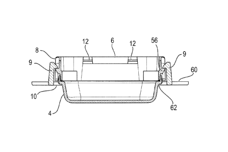
[04] In order to properly process the tissue sample a series of steps may be
performed
including:
[05] 1. Grossing of the sample by cutting the sample to the proper size
for
analysis.
[06] ). Fixing of the sample to immobilize molecular components and/or
prevent
degradation.
[07] 3. Embedding the sample in an embedding material, such as paraffin
wax
[08] 4. Sectioning the embedded sample by using, for example, a
microtome.
[09] In conventional methods, the grossing step involves a lab technician
cutting the tissue to
the appropriate size for analysis and then placing the tissue in a tissue
cassette. During the
fixation stage, the cassettes are generally exposed to a fixing agent or
chemical (e.g., a solution
-1-
CA 2845832 2020-03-11
PAGE 8148* RC _ _ ¨ . Timel*
SVR:OTT235CIFAX01/20*DNIS:3905* CSID:MLT*ANI:8582003000* DURATION (mm-ss):15-
57
03/11/2020 09 : 26 AM
Page: 9
of formaldehyde in water such as formalin) shortly after sample collection.
For example, U.S.
Patent No. 7,156, 814 discloses a cassette which can withstand tissue
preparation procedures.
[10] After the tissue sample has been processed, the medical professional, in
conventional
methods, removes the tissue sample from the individual cassette to perform the
embedding step.
Specifically, the medical professional carefully orients the sample, based on
the diagnostic view
required, into a base mold containing an embedding material such as paraffin
wax. Once the
tissue is oriented properly in the base mold, the molten material is cooled to
fully embed the
tissue sample and hold it in the proper orientation. The paraffin is used to
hold the sample in
position while also providing a uniform consistency to further facilitate
scc,ti.oning. While the
term paraffin is used, this term is not limiting and describes an example of
an embedding
medium.
[11] Then the sample is removed and sliced into a plurality of thin
sections (e.g., 2 to 25 ft
thick sections), often using a microtome, for further processing and
inspection. Such sectioning
of the sample often helps a medical professional properly assess the sample
under a microscope
(e.g. diagnose relationships between cells and other constituents of the
sample, or perform other
assessments).
[12] The current process requires human intervention at both the grossing and
embedding
steps. Such manual handling or the sample can increase the likelihood of mis-
identifying the
sample, cross contaminating the samples, or losing part or the entire sample.
Additionally, the
numerous steps of manual manipulation can often increase the time that it
takes to provide a
proper assessment for each sample, once the sample is collected.
CA 2845832 2020-03-11
PAGE 9/49* LEdSlein uaylight Timel*SYR:OTT235QFAX01/20*
DNIS:3905* CSID:MLT *ANI:8582003000* DURATION (mm-ss):15-57
03/11 / 202 0 09 : 26 AM
Page: 10
SUMMARY OF THE INVENTION
[13] This invention provides a device that allows for the tissue sample to be
orientated during
the grossing step and to remain in the same orientation through all steps to
the embedding step.
Through the multiple embodiments, the tissue sample cassette of this invention
reduces the
manual handling of the tissue samples. Example embodiments of this application
may address
one or more of the above identified issues. However, an embodiment of this
application need not
solve, address, or otherwise improve on existing technologies.
BRIEF DESCRIPTION OF THE DRAWINGS
[14] Figure 1 is an exploded view of a tissue cassette according to a first
embodiment in a
non-assembled state;
115] Figure 2 shows an exterior sectional view the tissue cassette of Figure 1
in an assembled
state;
[16] Figure 3 shows an interior sectional view of the tissue cassette of
Figure 1 in an
assembled state;
[17] Figure 4 shows a cut-out section of the biasing element on the tissue
cassette of the above
embodiment.
[18] Figures 5-6 show an alternate embodiment of the base.
[19] Figure 7 is an exploded view of a tissue cassette according to another
embodiment in a
non-assembled state.
[20] Figure 8 is an interior side view of a tissue cassette according to
another embodiment in
an assembled state with the tissue sample.
[21] Figures 9A, 9B, 10A, 10B, 11A and 11B show the tissue cassette according
to alternative
embodiments.
-3-
PAGE 10/48* RCA 2 8 4 5 8 3 2 2 0 2-0-03-1aylight Time] SVR:OTT235CWAX01/20 "
DNIS:3905* CSID:MLT *ANI:8582003000 * DURATION (mm-ss):15-57
03/11/2020 09 : 26 AM
Page: 11
DETAILED DESCRIPTION OF THE INVENTION
[22] In the following detailed description, reference will be made to the
accompanying
drawing(s), in which similar elements are designated with similar numerals.
The aforementioned
accompanying drawings show by way of illustration and not by way of
limitation, specific
example embodiments and implementations consistent with principles of an
example
embodiment. These implementations are described in sufficient detail to enable
those skilled in
the art to practice an example embodiment and it is to be understood that
other implementations
may be utilized and that structural changes and/or substitutions of various
elements may be made
without departing from the scope and spirit of an example embodiment. The
following detailed
description is, therefore, not to be construed in a limited sense.
[23] Figures 1-3 illustrate a tissue cassette I according to a first
exemplary embodiment of the
present application. The tissue cassette 1 retains a tissue sample 2 in the
proper orientation to
allow for the automation of the processing and a reduction in human error.
[24] A tissue cassette I. according to one embodiment of the invention, has a
base 4 and a
retaining member 6 which cooperate to retain the tissue sample 2, as discussed
below. In
addition, a frame 8 may optionally be provided to surround the outer perimeter
of the retaining
member 6. In this embodiment, the base 4 is connected to the frame 8 by
latching member 9,
and the frame 8 is connected to the retaining member 6 by a locking member 12.
In this way, the
retaining member 6 fits into the inside perimeter of the base 4 as shown in
Figures 2-3. The base
4 may have a sealing member 10 which forms a liquid seal between the frame 8
and the base 4.
[25] Figure 3 shows the base 4 with a bottom surface which corresponds to a
second tissue
engaging surface 14. The retaining member 6 is formed with a rim portion 16
and a tissue
-4-
CA 2845832 2020-03-11
PAGE 11/48 * LcasLern uaylight Time]' SVR:OTT235QFAX01120*
DNIS:3905* CSID:RilLT ANI:8582003000* DURATION (mm-ss):15-57
03/11/2020 09:26 AM
Page: 12
retaining element 18 having a bottom surface corresponding to a first tissue
engaging surface 20.
Further, in a non-limiting embodiment, the retaining member 6 includes a
biasing element 22.
[26] In a non-limiting embodiment, the tissue retaining element 18 is attached
to the rim
portion 16 by the biasing element 22 and locking member 12. The biasing
element 22 urges the
tissue retaining element 18 away from the rim portion 16. The first tissue
engaging surface 20 of
the tissue retaining element 18 may be attached directly to the biasing
element 22, Alternatively,
the first tissue engaging surface 20 of the tissue retaining element 18 may be
connected to the
biasing element 22 by a connecting portion 19, which as shown in Figure 1, may
extend from the
first tissue engaging surface 20 towards the rim portion 16.
[27] Generally, when the base 4 and the retaining member 6 are engaged as
shown in Figure 3,
an interior area 24 is defined between the base 4 and the retaining member 6
where the first
tissue engaging surface 20 and the second tissue engaging surface 14 are
facing each other. Prior
to this engagement, a tissue sample 2 is placed into this interior area 24 in
the desired orientation
so that it rests on the second tissue engaging surface 14 of the base 4. Upon
engagement of the
base 4 with the assembly of retaining member 6 to frame 8, the biasing element
22 urges the first
tissue engaging surface 20 of the tissue retaining element 18 towards the
second tissue engaging
surface 14 of the base 4 to firmly hold the tissue sample 2 in the chosen
orientation between the
first and second tissue engaging surfaces 14, 20 such that it can be held in
position for processing
and later be embedded with paraffin or the like.
[28] The biasing element 22 will now be described in additional detail. As
noted above the
tissue retaining element 18 is attached to the retaining member 6 by at least
one biasing element
22. In the illustrated embodiment in Figure 1, the tissue cassette 1 has four
biasing elements 22,
-5-
.
CA 2845832 2020-03-11
PAGE 12148* RCVD AT 3111/2020 11:29:35 AM [Eastern Daylight Time]
SVR:OTT2350FAX01 /20* DNIS:3905 CSID:MLT ANI:8582003000 * DURATION (mm-ss):15-
57
03/ 11 / 202 0 09:26 AM
Page: 13
where two biasing elements arc shown in the Figure on one wall and the other
two are on the
opposite wall.
[29] As shown in Figure I, each biasing element 22 is substantially hinged
having an S or Z
shape and attach at one end to the tissue retaining element I 8 and attach at
the other end to the
inner surface of the rim portion 16. The biasing element 22 urges the tissue
retaining element 18
towards the base 4 to fix the tissue sample 2 between the first and second
tissue engaging
surfaces 14, 20. Thus, the biasing element 22 can take on any shape that
performs this function.
For example, a torsion bar or a biasing element having another shape could
also be used as
discussed in more detail below.
[30] In one non-limiting embodiment, as shown in Figure 4, each biasing
element 22 may
have a first member 26 with a first end 27 and a second end 29. The first end
27 may be
connected to the tissue retaining element 18. Extending downward at an angle
from the hinge or
second end 29 of the first member 26 is a first angled member 28. A second
angled member 30
is connected to the first angled member 28 by a first curved hinged point 36.
The second angled
member 30 extends upwardly from the first angled member 28 at an angle; and in
a non-limiting
embodiment, the second angled member 30 and the first angled member 28 form an
angle less
than 90 . Extending downwardly from the second angled member 30 is a third
angled member
32. The second angled member 30 and the third angled member 32 are connected
by a second
curved hinge point 38. In a non-limiting embodiment, the third angled member
32 and the
second angled member 30 form an angle less than 90. Further, in a non-limiting
embodiment,
the third angled member 32 and the first angled member 28 lbrin an angle less
than 90 . A
second member 34 connects to the third angled member 32 at a hinge point and
extends
-6-
PAGE 13/48* RCA 2845832 2020-03-11
Jaylight Time]' SVR:OTT2350FAX01120 " DNIS:3905* CSID:MLT *ANI:8582003000
DURATION (mm-ss):15-57
03/11/2020 09:26 AM
Page: 14
substantially parallel to the tissue retaining element 18. The second member
34 attaches to the
rim portion 16 of the retaining member 6 in a non-limiting embodiment.
[31] The biasing element 22 has a particular flexibility to ensure that the
tissue sample 2 is
held between the first and second tissue engaging surfaces 14, 20, on the one
hand, but to also
ensure that the tissue sample 2 withstands any permanent damage during
processing. The
preferred maximum biasing force depends on the tissue sample and is up to
about 2.5N.
Typically, biasing force may be measured using a force gauge.
[32] More detail with respect to the retaining member 6 will now be provided
with reference
to Figures 1 and 2. In this exemplary embodiment, the retaining member 6
includes the rim 16,
the biasing element 22, the connector 19, the retaining element 18 and the
first tissue engaging
surface 20. The rim 16 has four walls and a substantially rectangular shape.
On the inside of the
rim 16 one end of the biasing element 22 is attached. One end of the biasing
element 22
attaches to the tissue retaining element 18 at either the connector 19 or the
first tissue engaging
surface 20. The tissue retaining element 18 of the retaining member 6 has a
connector 19 and a
first tissue engaging surface 20 with a substantially planar mesh portion 42.
In this embodiment
the mesh portion 42 is rectangular in shape, but the shape is not limiting and
the mesh portion
can be a variety of shapes. The mesh portion 42 of the first tissue engaging
surface 20 has a
plurality of perforations 44 or cut-outs. When the mesh portion 42 is urged
against the tissue
sample 2 it holds the tissue sample 2 in place and allows reagents, or the
like, to flow to the
tissue sample 2 through the perforations 44 in the mesh portion 42. The
perforations 44 are sized
to allow the flow of fluid to the tissue sample 2 on the one hand, but to
prevent the escape of the
tissue sample 2 on the other hand. Thus, the perforations 44 in the mesh
portion 42 may be sized
-7-
CA 2845832 2020-03-11
PAGE 14148* 'IL.¨ FUVI 'Eastern uaylight Time]* SVR:OTT235QFAX01/20*
DNIS:3905* CSID:MLT *ANI:8582003000 " DURATION (mm-ss):15-57
03/11/2020 09 : 26 AM
Page: 15
according to the size of the tissue sample 2. Further, the first tissue
engaging surface 20, may
alternatively bc solid and have no holes on the surface while still allowing
the agent to flow
underneath the first tissue engaging surface 20 from the periphery.
[33] The first tissue engaging surface 20 of the tissue retaining element 1g,
and/or the second
tissue engaging surface 14 may contain topography to help orient the tissue
sample. For
example, the tissue engaging surfaces 14, 20 may contain prongs 45, ridges,
hooks, or the like as
shown on a second tissue engaging surface 14 in Figure 5. In certain non-
limiting embodiments,
the tissue retaining element 18 has a semi-rigid structure to secure the
tissue sample 2 without
deformation; however, the tissue retaining element 18 may also have a rigid
structure without
changing the scope of the invention.
[34] Further as shown in Figure 1, the tissue retaining element 18 may also
have protrusions
46 which extend downwardly from the tissue retaining element 18 towards the
base 4. The
protnisions 46 act as dead stops to prevent the tissue retaining element 18
from pushing down
too hard against the tissue sample 2.
[35] Additionally, in a non-limiting embodiment, the retaining member 6 may
have handles 48
which function as grips for the lab technician when transporting the tissue
cassette 1. Further, in
a non-limiting embodiment, the retaining member 6 may contain a wire 47 which
extends the
length of the retaining member 6 and can be used fbr retraction when
separating the retaining
member 6 from the base 4.
[36] The base 4 will now be described with reference to Figure 1. As discussed
above, the
tissue cassette I has a base 4 which supports the tissue sample 2 and holds
the paraffin for
embedding. The base 4, as shown in Figure 1, has a generally rectangular shape
with four side
-8-
PAGE 15148* RCA 2845832 2020-03-11
..Jaylight Time] SVR:OTT235QFAX01120 DNIS:3905* CSID:MLT *ANI:8582003000*
DURATION (mm-ss):15-57
03/11/2020 09 : 28 AM
Page: 16
walls and a depressed bottom planar surface, referred to as the second tissue
engaging surface
14. The base 4 has a rectangular shape depicted in the Figures; however, it is
not limited to this
shape and a different shape could be used without changing the scope of the
invention. The base
4 is preferably solid so that it can hold the paraffin for embedding. The
walls of the base 4 are
preferably tapered inward to improve the ease at which the base can be removed
from the
paraffin after the embedding process.
= [37] As shown in Figure 1, the base 4 has a solid, smooth bottom.
However, in some
embodiments the base 4 may have grooves or some other texture. As an example,
the second
tissue engaging surface 14 of the base 4 may have flow channels 43, depicted
in Figure 6, to
assist in retaining the tissue sample 2 and improving fluid flow, without
changing the scope of
the invention. In an alternative embodiment, the base 4 may be have a second
depressed bottom
for receiving the tissue sample such that the second depressed bottom creates
an interior
subsection with an area smaller than the interior area 24. The second
depressed bottom may
be used for tissue samples 2 smaller in size.
[381 In certain embodiments, the base 4 may also have drainage guides 50. The
drainage
guides 50 help to wick away the paraffin and to channel the paraffin away from
the tissue
cassette 1 after the tissue sample 2 has been embedded. The drainage guides 50
extend out from
the outer peripheral of the base. In the embodiment shown in Figure 6, the
drainage guides 50
extend from one of the two end walls of the base; however the drainage guides
50 could extend
from any wall on the base 4.
[39] As noted above, in some embodiments a frame 8 is placed around the
outside perimeter
of the retaining member 6 and functions to secure the retaining member 6 to
the base 4. The
-9-
CA 2845832 2020-03-11
PAGE 16148' RGYD AT 3111/2020 11:29:35 AM [Eastern Daylight Time]
SVR:OTT2350FAX01120* DNIS:3905* CSID:MLT *ANI:8582003000 DURATION (mm-ss):15-
57
03/11/2020 09 : 26 AM
Page: 17
frame 8 may also be used as a means for identifying the tissue sample. As
shown in in Figure 1,
the frame 8 has a substantially rectangular shape with one end have an angled
projection with an
angled face 52. As shown in Fig. 1, a label 54 may be placed on the angled
face 52 to identify
the tissue sample 2. The labels 54 are described in more detail below. In this
embodiment, the
angle of the planar face is about 45 degrees, but the invention is not limited
in this respect. The
angled face 52 can be configured to receive a label such that the label 54
clicks into the angled
face 52 of the frame 8. Alternatively, the frame 8 may have a textured surface
and be put
through an inkjet printing system, such as Leica 1PC ink jet printer. In this
instance, the tissue
cassette I can be assembled after printing or the base 4 along with the frame
8 can be configured
to be sent through the printer.
[40] In a non-limiting embodiment, the frame 8 and the retaining member 6 are
not easily
removed so that once the tissue cassette 1 is used, the label 54 on the frame
8 will remain
matched with the tissue sample 2 contained in the tissue cassette I. In
certain embodiments,
frame 8 has a locking projections 12 which projects from the inside the
perimeter of the frame 8,
shown in Figure 1. The locking projections 12 attach with an engaging portions
55 on the outer
perimeter of the rim portion 16 on the retaining member 6 to secure the frame
8 to the retaining
member 6. Once the frame 8 is connected to the base 4 using this locking
arrangement, it is
difficult to separate them.
[41] The base 4 includes a latching member 9 which acts as a clip or lock to
hold the base 4 to
the frame 8. Alternatively, if a frame 8 is not used, the latching member 9
can lock the base 4 to
the retaining member 6.
-10-
CP, 28,45832 2020-03-11
PAGE 17148* R_ _ Jaylight Time] * SVR:0TT235QFAX01/20" DNIS:3905*
CSID:hilLT *ANI:8582003000 DURATION (mm-ss):15-57
03/11/2020 09 : 26 AM
Page: 18
[42] As shown in Figure 2, the latching member 9 is connected to a releasing
member 60. The
latching member 9 is flexibly attached to the base 4. When the latching member
9 is engaged,
the latching member 9 attaches to the clip surfaces 56 on the outer perimeter
of the frame 8. The
latching member 9 locks the base 4 to the frame 8 which is attached to the
retaining member 6.
In this way, a sealing member 10 connects the latching member 9 to the base 4
to form a seal
between the surfaces on the perimeter of the base 4 and the frame 8 to
sufficiently prevent
paraffin from leaking during embedding. In a non-limiting embodiment a gasket
may be used as
the sealing member 10 to help seal the base 4 and the frame 8. The latching
member 9 is
disengaged by pressing downward on the releasing member 60. When the releasing
member 60
is pressed, the latching member 9 moves away from the base 4 and disengages
from the clip
surfaces 56. In the embodiment described above, the sealing member 10 extends
from the base
4, but the sealing member 10 may also extend from the retaining member 6 or
the frame 8.
[43] An important aspect of tissue sample analysis is properly keeping
track of tissue samples.
In some embodiments, the tissue cassette 1 includes a label 54 or ID tag as
shown in Figure 1.
The label can 54 be located anywhere on the tissue cassette 1, but is
preferably located on the
frame 8. In some embodiments, more than one tag may be present. When more than
one tag is
present, the tags can be physically separated or located together.
[44] The label 54 may be a computer or human readable tag including, but not
limited to,
labels having an incorporated RFID, labels having an incorporated one-
dimensional barcode (1-
D barcode), labels having an incorporated two-dimensional barcode (2-D
barcode), and labels
having an incorporated three-dimensional barcode (3-D barcode). However, the
computer
readable label is not limited to RFID, 1-D barcode, 2-1) barcode, or 3-1)
barcode labels and may
-11 -
PAGE 1848 RCA 2845832 2020-p 3 ¨1.1..)a.,
ylight Time] SVR:OTT235CIFAX01120*DNIS:39058 CSID:MLT "ANI:8582003000"
DURATION (mm-ss):15-57
03/11/2020 09 : 26 AM
Page: 19
include any type of label readable by a computer as would be apparent to a
person of ordinary
skill in the art.
[45] In some embodiments, a label 54 is present that may be sensitive to
changes to the
sample or itself For example, a label 54 may be present that changes physical
(i.e. color) or
chemical (i.e. redox, conjugation, etc.) properties during fixation of the
sample. Similarly, a
label 54 may be present that is sensitive to the processing steps which
precede embedding (i.e.
dehydration), Alternatively, a label 54 may be present that is sensitive to
the embedding step
(i.e. infiltration of paraffin). The label 54 may have a property that changes
incrementally or
switches when the step is complete. In this way, the technician, or an
automated system, will be
able to determine when the sample has finished one step before another is
started.
[46] The tissue cassette I can be made from various materials and the same or
different
materials can be used for the retaining member 6, including the tissue
retaining element 18, the
first tissue engaging surface 20, the mesh portion 42, and the base 4.
Examples of materials used
include: an acetal copolymer, Teflon, polypropylene, and stainless steel. In a
non-limiting
embodiment, the acctal copolymer is DELRIN 900. In a non-limiting embodiment,
the base 4 is
made out of a polypropylene material so that the base 4 does not attach to the
paraffin after the
tissue sample 2 is embedded. In a non-limiting embodiment, the sealing member
10 is made out
of a polypropylene material.
[47] In a non-limiting embodiment, the tissue cassette, including the base,
the retaining
member, and/or the frame, may be produced from a material lacking any dye or
coloring. The
lack of color may allow the technician to view the tissue sample in the tissue
cassette and ensure
that the tissue sample has remained in its desired orientation after
embedding. In these
-12-
CA 2845832 2020-03-11
PAGE 19/48' Daylight Time]' SVR:OTT2350FAX01120*DNIS:3905*
CSID:MLT ANI:8582003000* DURATION (mm-ss):15-57
0 3 / 1 1 / 2 0 2 0
09 : 26 AM Page: 20
embodiments, the tissue cassette, including the base, the retaining member,
and/or the frame may
be at least at least opaque or clear.
[48] Figure 7 shows a further embodiment of the tissue cassette 1. This
embodiment is
different from the previously described embodiments in the following respects.
First, instead of
having a separate frame, the frame of this embodiment is integrally
incorporated into the
retaining member 6. Second, the tissue retaining element 18 is shaped more
like a basket, having
four side walls. Lastly, the latching member 9 is formed on an end wall of the
base 4, but has the
same function of locking the base 4 to the retaining member 6. Other than
these differences
noted, the embodiment shown in Figure 7 has the same configuration and tracks
the same
= structure as discussed above.
[49] Figure 8 shows a further embodiment of the tissue cassette I. This
embodiment is
different from the previously described embodiments in that in this
embodiment, a biasing
member 58 may be provided on either the base 4 or the retaining member 6 or
both, along with
the biasing element 22 as described in the above embodiments. In this
embodiment, the biasing
member 58 on the retaining member 6 may be pushing down and the biasing member
58
attached to the base 4 may provide a biasing force to move the second tissue
engaging surface 14
away from the first tissue engaging surface 20. Furth, the biasing member 58
attached to the
retaining member 6 may permit the retaining member 6 to move away from the
base 4 in
response to the biasing force provided by the base 4. Similarly, the biasing
member 58 attached
to the base 4 may permit the base 4 to move away from the retaining member 6
in response to the
biasing force provided by the retaining member 6. In this embodiment, the
tissue sample
container 1 is stable when either the biasing member 58 attached to the
retaining member 6 or
-13-
CA 2845832 2020-03-11
PAGE 20148' Lattsiern Daylight Timel* SVR:OTT235QFAX01/20 "
DNIS:3905* CSID:MLT *ANI:8582003000* DURATION (mm-ss):15-57
03 / 1 1/ 2 0 2 0
09 : 26 AM Page: 21
biasing member 58 attached to the base 4 is applying a biasing force, or when
both are applying
or not a biasing force.
[50] For example, in this non-limiting embodiment, the biasing member 58 on
the base 4 may
be used only to enable the releasing of the force that is applied by the
biasing member 58 on
retaining member 6. As an example, in this embodiment, the tissue cassette 1
provides a two
position floor. The first position is when the biasing member 58 on the base 4
compresses the
second tissue engaging surface 14 upwardly such that the tissue engaging
surface is compressed
up towards the retaining member 6 to compress the tissue sample 2. The second
position is when
the force of the biasing member 58 on the base is released so that the second
tissue engaging
surface 14 is moves downwardly. In this way, the second tissue engaging
surface 14 retracts
away from the tissue 2, such that the floor of the base retracts, similar to
the first tissue engaging
surface 20 or the previous embodiments retracting towards and away from the
tissue sample 2.
Other than these differences noted, the embodiment shown in Figure 8 has the
same
configuration and tracks the same structure as discussed above.
[51] Figures 9-11 illustrate alternative embodiments of the invention which
are directed
towards maintaining parallel configuration of the first tissue engaging 20
surface when it urges
towards the second tissue engaging 14 or when it retracts away from the second
tissue engaging
surface 14. Figures 9A and 9B illustrate examples of guiding members 64 which
assist the first
tissue engaging surface 20 to maintain parallel configuration to the base 4 as
it urges towards to
the base 4. Figure 9A illustrates a wire guide 66 used as the guiding member.
In Figure 9A the
wire guide 66 is attached to the retaining member 6 . The specific location is
not limited; the
wire guide 66 could be attached anywhere on the retaining member 6 including
directly on the
-14-
CA 2845832 2020-03-11
PAGE 21148 " liaylight Time] SVR:OTT235QFAX01120 DNIS:3905
CSID:FALT ANI:8582003000* DURATION (mm-ss):15-57
03/11/2020 09:26 AM
Page: 22
first tissue engaging surface 20. hi the example shown in Figure 9A, the wire
guide 66 has a
substantial LI-shape with two parallel members 68 connected by a cross member
70. Projections
72 extend out from one end of each of the parallel members 68 to attach to
clips 74 in the center
of the retaining element 18. The wire guide 66 may pivot at the clips 74 such
that when a
downward force is applied to the wire guide 66 the retaining element 18 urges
towards the tissue
sample 2 along a central axis of the clips 74 to maintain a parallel
configuration of the tissue
retaining element 18 and the first tissue engaging surface 20 with the base 4.
The cross member
70 can be locked into place by cross member clips 76 attached to the frame 8
or the retaining
member 6 .
[52] Figure 9B shows pillars 78 as guiding members 64. In this embodiment,
pillars 78
extend vertically upward from the interior of the frame 8. Further, the side
walls of the retaining
member 6 have at least one cut-out 80 which are shaped to receive the pillars
78. Accordingly,
the retaining element 18 can maintain a parallel configuration with the base 4
when it moves
towards the tissue sample 2 or away from the tissue sample 2.
[53] In addition to the guiding members 64 discussed above, there are
alternative designs
relating to the biasing element 22 which help to maintain the parallel
configuration of the first
tissue engaging surface 20 to the base 4. The biasing element 22 described
above is one example
of a means to hold the tissue sample 2 in the tissue cassette I. As noted
above, any design that
performs the function of urging the first tissue engaging surface 20 against
the second tissue
engaging surface 14 can be used. Alternate embodiments of the biasing element
22 to remain
parallel configuration of the first tissue engaging surface 20 with the base 4
are shown in Figures
10-11.
-15 -
PAGE 22148* FCA 2 8 458 ..20.,.2-0-0,-3-1,.1 3aYlight Time]'
SVR:0TT235QFAX01I20 * DNIS:3905* CSID:MLT *ANI:8582003000 * DURATION (mm-
ss):15-57
03/11/2020 09 : 26 AM
Page: 23
[54] Figure 10A and Figure 10B illustrate an alternate embodiment to the
biasing element 22.
In these examples, the biasing element 22 includes two angled members. The
first angled
member 82 is fixed to the retaining member 6 at a fixed point 84 and angles
downward from the
fixed point 84 and attaches to the retaining element 18 at a first moving
point 86. The second
angled member 90 attaches to the retaining member 6 at a sliding point 92. The
second angled
member 90 is not fixed at the sliding point 92 and can slide against an inner
ledge 93 of the
retaining member 6 in a direction parallel to the second tissue engaging
surface 14. The second
angled member 90 extends downward from the sliding point 92 and attaches to
the retaining
element 18 at a second moving point 94. The first angled member 82 and the
second angled
member 90 are angled such that the members cross substantially in the center
of each member at
a hinge point 96.
[55] In this embodiment, the first angled member 82 is fixed to the retaining
member 6 at the
fixed point 84. The second angled member 90 is attached to the retaining
member 6 at the
sliding point 92. Thus, the second angled member 90 can slide only in the
direction parallel to
the second tissue engaging surface 14. Accordingly, as the first angled member
82 and the
second angled member 90 urge the first tissue engaging surface 20 towards the
tissue sample 2,
the first moving point 86 and the second moving point 94 move towards the
tissue sample 2
while keeping the First tissue engaging surface 20 parallel to the base 4, for
example.
[56] In certain embodiments as shown in Figure 10A, the pair of angled members
82, 90 cross
at the hinge point 96 and are connected by a torsion bar 98. As shown in
Figure 10A, the pair of
angled members may be provided on each side of the tissue cassette 1.
Accordingly, the tissue
cassette 1 has two pairs of angled members, although the number of pairs of
angled members is
-16-
CA 2845832 2020-03-11
PAGE 23148 11.4V.JUiv LcasLet ii Daylight Time]* SVR:0TT235QFAX01120*
DNIS:3905 CSID:MLT *ANI:8582003000 " DURATION (mm-ss):15-57
03/11/2020
09:26 AM Page: 24
not limiting. The two pairs of angled members are attached by a connecting bar
100. The
connecting bar 100 can connect the two pairs of angled members at any point
along the
members, also illustrated in Figure 10B described hereinafter,
[57] Figure 10B shows an alternate embodiment, where the biasing element 22 is
provided by
a flexible hinges 102 at the connection points between the first and second
angled member and
the retaining element 18. That is, there are flexible hinges 102 at the first
moving point 86 or the
second moving point 94, Similar to the embodiment described in 10A, the first
tissue engaging
surface 20 can maintain a parallel configuration to the base 4 while moving
towards the tissue
sample 2. The flexible hinges 102 allow pair of angled members to flex under
pressure.
[58] Alternatively, as shown in Figures 11A and 1.113, the biasing element
22 can comprise
two angled members extending in a parallel direction. The first angled member
82 is attached to
the retaining member 6 and the second angled member 90 is attached to the
frame 8. In Figure
11A, the frame 8 and the second angled member 90 are placed over the retaining
member 6 and
the first angled member 82 so that the first angled member 84 and the second
angled member 90
are adjacent to each other and extend in parallel directions.
[59] The second angled member 90 contacts to the retaining member 6 at a hinge
104 and
attaches to the frame a first pivot point 106. The first angled member 82
attaches to the frame
at a second pivot point 108. Accordingly, the retaining element 18 may be
moved towards the
tissue sample 2 in a parallel manner by the first angled member 82 pivoting
about the second
pivot point 108 and the second angled member 90 rotating about the first pivot
point 106. When
the two angled member rotate about their respective pivot points the retaining
element 18 moves
in a substantially a parallel direction. Similar to the embodiment describe
with respect to Figures
-17-
CA 2845832 2020-03-11
PAGE 24/48 " R_ Jaylight Time]' SVR:OTT235CWAX01/20* DNIS:3905* CSID:MLT
ANI:8582003000* DURATION (mm=ss):15-57
03 / 11/2020 09 : 26 AM
Page: 25
11A and 11B, a torsion bar may be provided in this embodiment. The torsion bar
may be located
at any point where the two pair of angled members connect.
[60] An example of the use of the tissue cassette 1 in the analysis process
will now be
described. The tissue sample 2 is extracted and sent to a lab for analysis. In
certain non-limiting
embodiments, a gel may be placed on a tissue engaging surface, for example the
second tissue
engaging surface 14 as an adhesive to further secure the tissue sample 2. An
example of gel for
use include agarose, agarose derivatives, modified agarose, low melt agarosc,
hydroxyethylagarose, low molecular weight agarose, agar, alginates, dextran,
mannan, pectin,
Ghatti gum and cellulose including hydroxypropylcellulose, histogel, hydrogel
or combinations
thereol., Then the tissue sample 2 is orientated and placed onto the second
tissue engaging
surface 14 of the base 4 of the tissue cassette 1. The retaining member 6 is
then placed over the
base 4 and secured in place by the frame member 8. Once the cassette is
assembled, the biasing
element 22 in the retaining member 6 is deflected to urge the tissue retaining
element 18 of the
retaining member 6 towards the tissue sample 2 such that the tissue sample 2
is held in its
oriented position.
[611 The tissue cassette 1 is then processed and exposed to a
molten substrate. In anon-
limiting embodiment, the tissue cassette 1 is filled with paraffin. The molten
paraffin infiltrates
the tissue cassette 1 and enters the interior area 24 to embed the tissue
sample 2 in its oriented
position. The paraffin is then cooled such that it hardens at which point the
tissue sample is
embedded in a paraffin block and ready for sectioning. The base 2 is
disengaged from the frame
8 such that the paraffin block including the tissue sample is exposed, resting
on the first tissue
engaging surface 20 of the retaining member, The paraffin block including the
tissue sample can
-18-
PAGE 25/48' RCA 2845832 2020¨.03-1.3:ja_
ylight Timel* SVR:OTT235(3FAX01/20 " DNIS:3905 " CSID:MLT "ANI:8582003000*
DURATION (mm-ss):15-57
03/11/2020 09:26 AM
Page: 26
then be sectioned using a microtone. After the tissue sample 2 is sliced it is
ready to he placed
on a microscope slide for further processing and inspection.
[62] Although a few example embodiments have been shown and described, these
example
embodiments are provided to convey the subject matter described herein to
people who are
familiar with this field. It should be understood that the subject matter
described herein may be
embodied in various forms without being limited to the described example
embodiments. The
subject matter described herein can be practiced without those specifically
defined or described
matters or with other or different elements or matters not described. It will
be appreciated by
those familiar with this field that changes may be made in these example
embodiments without
= departing from the subject matter described herein as defined in the
appended claims and their
equivalents. Further, any description of structural arrangement of components
or relationship
there between is merely for explanation purposes and should be used to limit
an example
embodiment.
[63] Aspects related to the example embodiment have been set forth in part in
the description
above, and in part should be apparent from the description, or may be learned
by practice of
embodiments of the application. Aspects of the example embodiment may be
realized and
attained using the elements and combinations of various elements and aspects
particularly
pointed out in the foregoing detailed description and the appended claims. It
is to be understood
that both the foregoing descriptions are an example and are explanatory only
and are not
intended to be limiting.
-19-
CA 2845832 2020-03-11
PAGE 26148 *I2Lvu I an 1/LUZU 11:9:J5 AM [Eastern Daylight Time] *
SVR:OTT235QFAX01/20*DNIS:3905* CSID:MLT *ANI:8582003000* DURATION (mm-ss):15-
57