Note: Descriptions are shown in the official language in which they were submitted.
CA 02845884 2014-02-19
WO 2013/028810
PCT/US2012/051948
CLOSTRIDIUM DIFFICILE ANTIBODIES
CROSS REFERENCE TO RELATED APPLICATION
This application claims priority to U.S. Provisional Application No.
61/526,031
filed August 22, 2011, the disclosure of which is incorporated herein by
reference in its
entirety.
FIELD
The invention relates to monoclonal antibodies to Clostridium difficile toxin
A.
The invention further relates to compositions and methods for the treatment or
prevention
of infection by the bacteria, Clostridium difficile, in a vertebrate subject.
Methods are
provided for administering antibodies to the vertebrate subject in an amount
effective to
reduce, eliminate, or prevent relapse from infection. Methods for the
treatment or
prevention of Clostridium difficile infection in an organism are provided.
BACKGROUND
Clostridium difficile (C. difficile) is a common nosocomial pathogen and a
major
cause of morbidity and mortality among hospitalized patients throughout the
world. Kelly
et at., New Eng. J. Med., 330:257-62, 1994. The increased use of broad
spectrum
antibiotics and the emergence of unusually virulent strains of C. difficile
have lead to the
idea that vaccines may be well suited to reduce disease and death associated
with this
bacterium. C. difficile has few traditional antibiotic options and frequently
causes a
recurring disease (25% of cases). C. difficile claims about 20,000 lives in
the USA alone
per year and causes around 500,000 confirmed infections. Recently, more
virulent strains
of C. difficile have emerged that produce more toxin such as the Bl/NAB1/027
strain,
which also has a decreased susceptibility to metronidazole. Outbreaks of C.
difficile have
necessitated ward and partial hospital closure. With the increasing elderly
population and
the changing demographics of the population, C. difficile is set to become a
major
problem in the 21st century. The spectrum of C. difficile disease ranges from
asymptomatic carriage to mild diarrhea to fulminant pseudomembranous colitis.
1
CA 02845884 2014-02-19
WO 2013/028810
PCT/US2012/051948
C. difficile has a dimorphic lifecycle whereby it exists both as an infectious
and
tough spore form and a metabolically active toxin-producing vegetative cell.
C. difficile-
associated disease (CDAD) is believed to be caused by the vegetative cells and
more
specifically the actions of two toxins, enterotoxin toxin A and cytotoxin
toxin B.
Vaccines and therapy for C. difficile have been to date focused upon the
toxins (A and B),
toxoids of A and B, recombinant fragments of A and B, and vegetative cell
surface layer
proteins (SLPAs).
Toxin A is a high-molecular weight protein that possesses multiple functional
domains. The toxin is broken up into 4 functional domains: an amino-terminal
glucosyltransferase that modifies Rho-like GTPases leading to cytoskeletal
dysregulation
in epithelial cells, an autocatalytic cysteine protease domain, a hydrophobic
membrane-
spanning sequence, and a highly repetitive carboxy-terminal host-cell binding
domain.
The carboxy terminal domain anchors the toxin to the host cell carbohydrate
receptors on
intestinal epithelial cells which initiates the internalization process
thereby delivering the
amino-terminal enzymatic domains to the cytoplasm of the target cells. The
delivery of
the enzymatic domain and glucosyltransferase activity leads to diarrhea and
inflammation
due to the apoptotic cell death of the intoxicated cells.
Many studies have shown the importance of antibodies against the toxins in
affecting the disease outcome. Studies have also shown the correlation between
serum
anti-toxinA antibodies with protection from CDAD and relapse. These studies
have led to
the creation of toxin mAb therapies for CDAD.
Despite these advances, there is an unmet need for effective treatment and/or
prevention of C. difficile associated infections including prevention from
relapse of
CDAD. The present invention provides mouse and humanized antibodies to toxin A
to
satisfy these and other needs.
2
CA 02845884 2014-02-19
WO 2013/028810
PCT/US2012/051948
SUMMARY
The present invention provides for antibodies, or antigen-binding portions
thereof,
that bind to Clostridium difficile (C. difficile) toxin A. The antibody or
antigen-binding
portion thereof may bind to fragment 4 of C. difficile toxin A.
In one embodiment, the present invention provides for an isolated monoclonal
antibody, or an antigen-binding portion thereof, comprising a heavy chain
region and a
light chain region, wherein the heavy chain region comprises three
complementarity
determining regions (CDRs), CDR1, CDR2 and CDR3, having amino acid sequences
about 80% to about 100% homologous to the amino acid sequences set forth in
SEQ ID
NOs: 29, 30 and 31, respectively, and wherein the light chain region comprises
three
CDRs, CDR1, CDR2 and CDR3, having amino acid sequences about 80% to about 100%
homologous to the amino acid sequences set forth in SEQ ID NOs: 21, 22 and 23,
respectively.
Also provided is an isolated monoclonal antibody, or an antigen-binding
portion
thereof, that binds to C. difficile toxin A and comprises a heavy chain
region, wherein the
heavy chain region comprises three CDRs, CDR1, CDR2 and CDR3, having amino
acid
sequences about 80% to about 100% homologous to the amino acid sequences set
forth in
SEQ ID NOs: 29, 30 and 31, respectively.
The present invention further provides for an isolated monoclonal antibody, or
an
antigen-binding portion thereof, that binds to C. difficile toxin A and
comprises a light
chain region, wherein the light chain region comprises three CDRs, CDR1, CDR2
and
CDR3, having amino acid sequences about 80% to about 100% homologous to the
amino
acid sequences set forth in SEQ ID NOs: 21, 22 and 23, respectively.
The antibody or antigen-binding portion thereof may have a dissociation
constant
(KD) of less than about 1 x 101 M. The antibody or antigen-binding portion
thereof may
be humanized or chimeric.
In one embodiment, the heavy chain region of the antibody or antigen-binding
portion thereof comprises an amino acid sequence about 80% to about 100%
homologous
to the amino acid sequence set forth in SEQ ID NO: 89; the light chain region
of the
antibody or antigen-binding portion thereof comprises an amino acid sequence
about
80% to about 100% homologous to the amino acid sequence set forth in SEQ ID
NO: 91.
3
CA 02845884 2014-02-19
WO 2013/028810
PCT/US2012/051948
In another embodiment, the heavy chain region of the antibody or antigen-
binding
portion thereof comprises an amino acid sequence about 80% to about 100%
homologous
to the amino acid sequence set forth in SEQ ID NO: 93; the light chain region
of the
antibody or antigen-binding portion thereof comprises an amino acid sequence
about
The antibody or antigen-binding portion thereof may be the following: (a) a
whole
immunoglobulin molecule; (b) an scFv; (c) a Fab fragment; (d) an F(ab')2; and
(e) a
disulfide linked Fv.
The antibody or antigen-binding portion thereof may comprise at least one
One embodiment of the present invention provides for an isolated monoclonal
antibody or an antigen-binding portion thereof, that binds to C. difficile
toxin A and
comprises a heavy chain variable region, wherein the heavy chain variable
region
Another embodiment of the present invention provides for an isolated
monoclonal
antibody, or an antigen-binding portion thereof, that binds to C. difficile
toxin A and
comprises a light chain variable region, wherein the light chain variable
region comprises
Yet another embodiment of the present invention provides for an isolated
monoclonal antibody, or an antigen-binding portion thereof, wherein the
antibody, or
antigen-binding portion thereof, binds to the same epitope of C. difficile
toxin A
Also encompassed by the present invention are an antibody produced by
hybridoma designated CAN20G2 and the hybridoma designated CAN20G2.
30 The present invention provides for an isolated monoclonal antibody, or
an
antigen-binding portion thereof, wherein, in an in vivo toxin A challenge
experiment,
4
CA 02845884 2014-02-19
WO 2013/028810
PCT/US2012/051948
when the antibody, or an antigen-binding portion thereof, is administered to a
mammal at
a dosage ranging from about 8 mg/kg body weight to about 13 mg/kg body weight
about
24 hours before the mammal is exposed to greater than about 100 ng of C.
difficile toxin
A, the chance of survival for the mammal is greater than about 80% within
about 7 days.
Also encompassed by the present invention is an isolated monoclonal antibody,
or
an antigen-binding portion thereof, wherein the antibody, or antigen-binding
portion
thereof, at a concentration ranging from about 4 M to about 17 M,
neutralizes greater
than about 40% of about 150 ng/ml C. difficile toxin A in an in vitro
neutralization assay.
The present invention provides for an isolated nucleic acid encoding a peptide
comprising an amino acid sequence about 80% to about 100% homologous to the
amino
acid sequence set forth in SEQ ID NOs: 12, 28, 44, 60, 4, 20, 36 or 52. The
present
invention also provides for an isolated nucleic acid comprising a nucleic acid
sequence
about 80% to about 100% homologous to the nucleic acid sequence set forth in
SEQ ID
NOs: 68, 69, 70, 71, 72, 73, 74 or 75. Also provided is a cell comprising any
of these
nucleic acids. The cell can be a bacterial cell or a eukaryotic cell, such as
a mammalian
cell. Non-limiting examples of the cells include COS-1, COS-7, HEK293, BHK21,
CHO, BSC-1, Hep G2, 5P2/0, HeLa, myeloma or lymphoma cells.
The present invention provides for a composition comprising the antibody or
antigen-binding portion thereof and at least one pharmaceutically acceptable
carrier.
The present invention provides for a method of preventing or treating C.
difficile-
associated disease comprising administering to a subject an effective amount
of the
present antibody or antigen-binding portion thereof The antibody or antigen-
binding
portion thereof may be administered intravenously, subcutaneously,
intramuscularly or
transdermally. The method may contain another step of administering to the
subject a
second agent. For example, the second agent may be a different antibody or
fragment
thereof, or may be an antibiotic such as vancomycin, metronidazole or
fidaxomicin.
5
CA 02845884 2014-02-19
WO 2013/028810
PCT/US2012/051948
BRIEF DESCRIPTION OF THE DRAWINGS
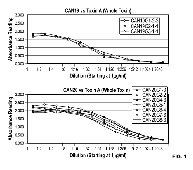
Figure 1 shows a standardized ELISA showing the reactivity of purified murine
mAbs on Clostridium difficile toxin A.
Figure 2 is an ELISA showing the binding activity of purified 1 ug/m1CAN19
mAbs on toxin A (ToxA) and toxin A fragment 4 (ToxAF4). ToxB is toxin B;
ToxBF4 is
toxin B fragment 4.
Figure 3 is an ELISA assay showing the binding activity of purified 1 ug/m1
murine CAN20 mAbs on toxin A and toxin A fragment 4.
Figure 4 shows a Western immunoblot of Purified Murine CAN19 mAbs (0.5
jig/m1). Lane 1: Toxin A; Lane 2: Toxoid A; Lane 4: Toxin A Fragment 4; Lane
5: Toxin
B; Lane 7: Toxin B Fragment 4; Lane 8: PilF (negative control). Expected
sizes: Toxin
A (308 kDa); Toxin A Fragment 4 (114 kDa); Toxin B (280 kDa).
Figure 5 shows a Western blot of Purified CAN20 clones (1ug/m1). Blot A was
probed with CAN20G1, blot B was probed with CAN20G2, blot C was probed with
Toxin A Fragment 4 (114 kDa); Lane3: Toxin B (280 kDa); Lane4: tetanus
toxoid).
Figure 6a is an epitope binning graph showing biotinylated CAN20G1 antibody
binding to SA (streptavidin) biosensor. The bound antibody is then incubated
with free
Toxin A and free CAN20G1. The CAN20G1-Toxin A complex is again incubated with
free antibody. A large nm shift in wavelength will indicate binding of the
analyte
indicating that CAN20G1 and the free antibody have different epitopes. 1,
Biotinylated
CAN20G1 to SA biosensors. 2, Free whole toxin A forming complex with CAN20G1.
3,
Free CAN20G1 associating with biotinylated CAN20G1-Toxin A complex. 4,
Association sample curves. 5, Dissociation step.
Figure 6b is a graph showing the final three steps (3-5) of the full program.
A
large nm shift in wavelength will indicate binding of the analyte indicating
that
CAN20G1 and the free antibody have different epitopes. In this case, only CDA1
(Merck
anti-toxin A mAb used as a control) had a significant nm shift in wavelength
demonstrating that CDA1 binds to a different epitope while CAN20G1, G2, G5,
and G8
bind to the same epitope bin as CAN20G1.
6
CA 02845884 2014-02-19
WO 2013/028810
PCT/US2012/051948
Figure 7 is a bar graph showing the effects of C. difficile toxin A on mouse
survival and the efficacy of the CAN19 mAbs against the toxin A challenge.
Figure 8 is a bar graph showing the effects of C. difficile toxin A on mouse
survival and the efficacy of the CAN19 and CAN20 mAbs against toxin A
challenge.
Figure 9 is a bar graph showing the effects of C. difficile toxin A on mouse
survival and the efficacy of the murine CAN20G2 mAb at full dose and half dose
against
toxin A challenge.
Figure 10 shows primers used for V gene amplification from RNA. The
degenerate base symbols are IUPAC (International union of pure and applied
chemistry)
codes for representing degenerate nucleotide sequence patterns.
Figure 11 shows V-gene sequencing results for muCAN20G2 that includes both
VH and VL sequences from the muCAN20G2 parental clones.
Figure 12 shows alignment of muCAN20G2 v-regions with the closest human
germline v-region. The human germlines were used as acceptor frameworks for
humanization.
Figures 13a and 13b show CDR-huCAN20G2 design. The closest matching
human frameworks are IGHV7-4-1*02 and IGKV1-39*01. The CDRs (IMGT
Numbering) of the muCAN20G2 were inserted into the human framework. Figure 13A
shows the heavy chain variable region, including both nucleic acid sequence
and amino
acid sequence. FR1, FR2 and FR3 are from IGHV7-4-1*02; FR4 is from IGHJ6*01.
Figure 13B shows the light (kappa) chain variable region, including both
nucleic acid
sequence and amino acid sequence. FR1, FR2 and FR3 are from IGKV1-39*01; FR4
is
from IGKJ4*01.
Figures 14a and 14b show HE-huCAN20G2 Design. Resurfaced and altered
codons are in bold. The nucleotide sequence was translated to ensure correct
frame.
Figure 14A shows the heavy chain variable region, including both nucleic acid
sequence
and amino acid sequence. Figure 14B shows the light (kappa) chain variable
region,
including both nucleic acid sequence and amino acid sequence.
Figure 15 shows the HE-huCAN20G2 Heavy Chain. Resurfaced and altered
codons are in bold. After v-region design, an IgG1 constant region was added.
The
introns were removed and the nucleotide sequence was translated to ensure
correct frame.
7
CA 02845884 2014-02-19
WO 2013/028810
PCT/US2012/051948
Figure 16 shows HE-huCAN20G2 Kappa Chain. Resurfaced and altered codons
are in bold. After v-region design, a Kappa constant region was added. The
introns were
removed and the nucleotide sequence was translated to ensure correct frame.
Figure 17 shows AVA-huCAN20G2 kappa V-region alignment. The Avastin
kappa v-region was aligned to the IMGT domain directory and identified the
closest
germline v-region. IGKV1D-33-01 was used as the acceptor framework for the AVA
mAb design.
Figure 18 shows AVA-huCAN20G2. The Avastin kappa v-region was aligned to
the IMGT domain directory and identified the closest germline v-region. After
analysis
and design, a kappa constant region was added. As previously, the constant
regions
contain introns. For the AVA-huCAN20G2 heavy chain, the previously designed
and
resurfaced HE-huCAN20G2 heavy chain was used. FR1, FR2 and FR3 are from
IGKV1D-33-01; FR4 is from IGKJ1-01.
Figures 19a and 19b show chimeric CAN20G2. Murine V-regions were
designed with human constant regions. The introns were removed and the
nucleotide
sequence was translated to ensure correct frame. Figure 19A shows the heavy
chain,
including both nucleic acid sequence and amino acid sequence. Figure 14B shows
the
light (kappa) chain, including both nucleic acid sequence and amino acid
sequence.
Figure 20a shows neutralization data for purified human CAN20G2 clones at 150
ng/ml depicted as a bar graph.
Figure 20b shows neutralization data for purified human CAN20G2 clones at 250
ng/ml depicted as a bar graph.
Figure 21a shows ELISA to screen transfection supernatant for expressed human
Can20G2 mAbs binding to toxin A at 45 minutes.
Figure 21b shows ELISA to screen transfection supernatant for expressed human
Can20G2 mAbs binding to toxin A fragment 4 at 45 minutes.
Figure 21c shows ELISA to screen transfection supernatant for expressed human
Can20G2 mAbs binding to toxin A at 60 minutes.
Figure 21d shows an ELISA to screen transfection supernatant for expressed
human Can20G2 mAbs binding to toxin A fragment 4 at 60 minutes.
Figure 22 shows SDS-PAGE of purified human CAN20G2 clones.
8
CA 02845884 2014-02-19
WO 2013/028810
PCT/US2012/051948
Figure 23 shows Western blot analysis of purified human CAN20G2 clones. An
SDS-page gel was run with tetanus toxoid, whole toxin A, toxin A fragment 4
and BSA.
The gel was transferred to nitrocellulose membrane and probed with each of the
human
CAN20G2 mAbs (1 g/m1). (Lane 1: Toxin A; Lane 2: Toxin A Fragment 4; Lane 3:
tetanus toxoid; Lane 4: BSA).
Figures 24a and 24b show healthy donor T cell proliferation responses to test
antibodies, CDR-huCAN20G2 (Figure 24A) and HE-huCAN20G2 (Figure 24B), on days
5, 6, 7, and 8 after incubation. Proliferation responses with an SI>2.00
(indicated by
dotted line) that were significant (p<0.05) using an unpaired, two sample
student's t test
were considered positive. For each donor, the bars from left to right
represent day 5, day
6, day7 and day 8, respectively.
Figure 25 shows the number of positive T cell proliferation responses to
antibodies CDR-huCAN20G2 (C001) and HE-huCAN20G2 (H001) detected at four time
points.
Figure 26 shows healthy donor T cell IL-2 ELISpot responses to test
antibodies,
CDR-huCAN20G2 (C001) and HE-huCAN20G2 (H001). PBMCs were used to assess
IL-2 secretion in response to stimulation with the two antibodies during an 8-
day
incubation. T cell responses with an SI>2.00 that were significant (p<0.05)
using an
unpaired, two sample student's t test were scored positive. Borderline
responses
(significant p<0.05 with SI>1.90) was shown (*).
Figure 27 shows the comparison of HE-huCAN20G2 ("HE-CAN20G2"), CDR-
huCAN20G2 ("CDR-CAN20G2") and CDA1 (Merck/Medarex) anti-C. difficile toxin A
(anti-TcdA) mAbs tested at a low dose of 0.05mg/mouse. Efficacy of mAbs is
presented
as the percentages of survival compared to control animals (TcdA/PBS). *Fisher
exact
test for statistical significance.
Figure 28 shows the effect of humanized CAN20G2 mAbs, HE-huCAN20G2,
CDR-huCAN20G2 in comparison with CDA1 on survival over time following TcdA
challenge. The effect of mAbs at low dose of Ab (0.05mg) or PBS alone
(control) on
survival related to time after TcdA challenge is depicted. The percent
survival of animals
in each group post TcdA challenge at the indicated time points (hrs) is shown
in the
graph.
9
CA 02845884 2014-02-19
WO 2013/028810
PCT/US2012/051948
Figures 29a and 29b show PK study data of humanized antibodies CDR-
huCAN20G2 (Figure 29a) and HE-huCAN20G2 (Figure 29b) in rats.
CA 02845884 2014-02-19
WO 2013/028810
PCT/US2012/051948
DETAILED DESCRIPTION
The present invention provides for compositions and methods for the prevention
or treatment of Clostridium difficile bacterial infection or bacterial
carriage. The
compositions contain antibodies (or an antigen-binding portion thereof) that
recognize
toxin A of C. difficile, including mouse monoclonal antibodies, humanized
antibodies,
chimeric antibodies, or antigen-binding portions of any of the foregoing.
These
antibodies (or antigen-binding portion thereof) can neutralize toxin A in
vitro and in vivo,
and/or inhibit binding of toxin A to mammalian cells. Therefore, the present
antibodies
or antigen-binding portion thereof can be used in passive immunization to
prevent or treat
C. diffici/e-associated disease (CDAD).
In one embodiment, the present antibodies or antigen-binding portions thereof
provide one or more of the following effects: protect from or treat C.
diffici/e-mediated
colitis, antibiotic-associated colitis, pseudomembranous colitis (PMC) or
other intestinal
disease in a subject; protect from or treat diarrhea in a subject; and/or
treat or inhibit
relapse of C. diffici/e-mediated disease. When administered to a mammal, the
present
antibodies or antigen-binding portions thereof protect the mammal against
toxin A
administered in an amount that would be fatal to the mammal had the antibody
or
antigen-binding portion thereof not administered.
The present antibodies or antigen-binding portions thereof include antibodies
produced by hybridoma clone CAN20G2, CAN20G1, CAN20G5, CAN20G8,
CAN19G1, CAN19G2 or CAN19G3 described herein.
Also encompassed by the present invention are antibodies or antigen-binding
portions thereof that include an antigen-binding portion of an antibody
produced by
hybridoma clone CAN20G2, CAN20G1, CAN20G5, CAN20G8, CAN19G1, CAN19G2
or CAN19G3.
As used herein, CAN20G1, CAN20G2, CAN20G5, CAN20G8, CAN19G1,
CAN19G2 and CAN19G3 refer to the hybridoma clones or the monoclonal antibodies
generated by the corresponding hybridoma clones.
The antibodies or antigen-binding portions thereof can specifically bind to an
epitope within fragment 4 of toxin A, e.g., an epitope between amino acid
residues 1853-
2710 of toxin A. Babcock, G.J. et at., Infection and Immunity, 74: 6339-6347
(2006). In
11
CA 02845884 2014-02-19
WO 2013/028810
PCT/US2012/051948
other embodiments, the antibodies or antigen-binding portions thereof
specifically bind to
an epitope within fragment 1 (amino acid residues 1-659), fragment 2 (amino
acid
residues 660-1256) or fragment 3 (amino acid residues 1257-1852) of toxin A.
In other
embodiments, the antibodies or antigen-binding portions thereof specifically
bind an
epitope within amino acid residues 1-600, 400-600, 415-540, 1-100, 100-200,
200-300,
300-400, 400-500, 500-600, 600-700, 700-800, 900-1000, 1100-1200, 1200-1300,
1300-
1400, 1400-1500, 1500-1600, 1600-1700, 1800-1900, 1900-200, 2100-2200 or 2200-
2300, 2300-2400, 2400-2500, 2500-2600, 2600-2710 of toxin A, or any interval,
portion
or range thereof
The present antibodies, or antigen-binding portions thereof, include, but are
not
limited to, monoclonal antibodies, chimeric antibodies, humanized antibodies,
polyclonal
antibodies, recombinant antibodies, as well as antigen-binding portions of the
foregoing.
An antigen-binding portion of an antibody may include a portion of an antibody
that
specifically binds to a toxin of C. difficile (e.g., toxin A).
The humanized antibody of the present invention is an antibody from a non-
human species where the amino acid sequence in the non-antigen binding regions
(and/or
the antigen-binding regions) has been altered so that the antibody more
closely resembles
a human antibody, and still retains its original binding ability.
Humanized antibodies can be generated by replacing sequences of the variable
region that are not directly involved in antigen binding with equivalent
sequences from
human variable regions. Those methods include isolating, manipulating, and
expressing
the nucleic acid sequences that encode all or part of variable regions from at
least one of
a heavy or light chain. Sources of such nucleic acid are well known to those
skilled in the
art and, for example, may be obtained from a hybridoma producing an antibody
against
toxin A. The recombinant DNA encoding the humanized antibody, or fragment
thereof,
can then be cloned into an appropriate expression vector.
An antibody light or heavy chain variable region consists of a framework
region
interrupted by three hypervariable regions, referred to as complementarity
determining
regions (CDRs). In one embodiment, humanized antibodies are antibody molecules
from
non-human species having one, two or all CDRs from the non-human species and a
framework region from a human immunoglobulin molecule.
12
CA 02845884 2014-02-19
WO 2013/028810
PCT/US2012/051948
The humanized antibodies of the present invention can be produced by methods
known in the art. For example, once non-human (e.g., murine) antibodies are
obtained,
variable regions can be sequenced, and the location of the CDRs and framework
residues
determined. Kabat, E. A., et at. (1991) Sequences of Proteins of Immunological
Interest,
Fifth Edition, U.S. Department of Health and Human Services, NIH Publication
No. 91-
3242. Chothia, C. et at. (1987) J. Mol. Biol., 196:901-917. The light and
heavy chain
variable regions can, optionally, be ligated to corresponding constant
regions. CDR-
grafted antibody molecules can be produced by CDR-grafting or CDR
substitution. One,
two, or all CDRs of an immunoglobulin chain can be replaced. For example, all
of the
CDRs of a particular antibody may be from at least a portion of a non-human
animal
(e.g., mouse such as CDRs shown in Table 1) or only some of the CDRs may be
replaced. It is only necessary to keep the CDRs required for binding of the
antibody to a
predetermined antigen (e.g., toxin A of C. dlfficile). Morrison, S. L., 1985,
Science,
229:1202-1207. Oi et al., 1986, BioTechniques, 4:214. U.S. Patent Nos.
5,585,089;
5,225,539; 5,693,761 and 5,693,762. EP 519596. Jones et at., 1986, Nature,
321:552-
525. Verhoeyan et at., 1988, Science, 239:1534. Beidler et at., 1988, J.
Immunol.,
141:4053-4060.
Also encompassed by the present invention are antibodies or antigen-binding
portions thereof containing one, two, or all CDRs as disclosed herein, with
the other
regions replaced by sequences from at least one different species including,
but not
limited to, human, rabbits, sheep, dogs, cats, cows, horses, goats, pigs,
monkeys, apes,
gorillas, chimpanzees, ducks, geese, chickens, amphibians, reptiles and other
animals.
A chimeric antibody is a molecule in which different portions are derived from
different animal species. For example, an antibody may contain a variable
region derived
from a murine mAb and a human immunoglobulin constant region. Chimeric
antibodies
can be produced by recombinant DNA techniques. Morrison, et al., Proc Natl
Acad Sci,
81:6851-6855 (1984). For example, a gene encoding a murine (or other species)
monoclonal antibody molecule is digested with restriction enzymes to remove
the region
encoding the murine Fc, and the equivalent portion of a gene encoding a human
Fc
constant region is substituted. Chimeric antibodies can also be created by
recombinant
DNA techniques where DNA encoding murine V regions can be ligated to DNA
13
CA 02845884 2014-02-19
WO 2013/028810
PCT/US2012/051948
encoding the human constant regions. Better et al., Science, 1988, 240:1041-
1043. Liu et
al. PNAS, 1987 84:3439-3443. Liu et al., J. Immunol., 1987, 139:3521-3526. Sun
et al.
PNAS, 1987, 84:214-218. Nishimura et al., Canc. Res., 1987, 47:999-1005. Wood
et al.
Nature, 1985, 314:446-449. Shaw et al., J. Natl. Cancer Inst., 1988, 80:1553-
1559.
International Patent Publication Nos. W01987002671 and WO 86/01533. European
Patent Application Nos. 184, 187; 171,496; 125,023; and 173,494. U.S. Patent
No.
4,816,567.
The antibodies can be full-length or can include a fragment (or fragments) of
the
antibody having an antigen-binding portion, including, but not limited to,
Fab, F(ab')2,
Fab', F(ab)', Fv, single chain Fv (scFv), bivalent scFv (bi-scFv), trivalent
scFv (tri-scFv),
Fd, dAb fragment (e.g., Ward et al., Nature, 341:544-546 (1989)), an isolated
CDR,
diabodies, triabodies, tetrabodies, linear antibodies, single-chain antibody
molecules, and
multispecific antibodies formed from antibody fragments. Single chain
antibodies
produced by joining antibody fragments using recombinant methods, or a
synthetic
linker, are also encompassed by the present invention. Bird et al. Science,
1988,
242:423-426. Huston et al., Proc. Natl. Acad. Sci. USA, 1988, 85:5879-5883.
The antibodies or antigen-binding portions thereof of the present invention
may
be monospecific, bi-specific or multispecific. Multispecific or bi-specific
antibodies or
fragments thereof may be specific for different epitopes of one target
polypeptide (e.g.,
toxin A) or may contain antigen-binding domains specific for more than one
target
polypeptide (e.g., antigen-binding domains specific for toxin A and toxin B;
or antigen-
binding domains specific for toxin A and other antigen of C. difficile; or
antigen-binding
domains specific for toxin A and other kind of bacterium or virus). In one
embodiment, a
multispecific antibody or antigen-binding portion thereof comprises at least
two different
variable domains, wherein each variable domain is capable of specifically
binding to a
separate antigen or to a different epitope on the same antigen. Tuft et al.,
1991, J.
Immunol. 147:60-69. Kufer et al., 2004, Trends Biotechnol. 22:238-244. The
present
antibodies can be linked to or co-expressed with another functional molecule,
e.g.,
another peptide or protein. For example, an antibody or fragment thereof can
be
functionally linked (e.g., by chemical coupling, genetic fusion, noncovalent
association
or otherwise) to one or more other molecular entities, such as another
antibody or
14
CA 02845884 2014-02-19
WO 2013/028810
PCT/US2012/051948
antibody fragment to produce a bi-specific or a multispecific antibody with a
second
binding specificity. For example, the present invention includes bi-specific
antibodies
wherein one arm of an immunoglobulin is specific for toxin A, and the other
arm of the
immunoglobulin is specific for a second therapeutic target or is conjugated to
a
therapeutic moiety such as a trypsin inhibitor.
All antibody isotypes are encompassed by the present invention, including IgG
(e.g., IgGl, IgG2, IgG3, IgG4), IgM, IgA (IgAl, IgA2), IgD or IgE. The
antibodies or
antigen-binding portions thereof may be mammalian (e.g., mouse, human)
antibodies or
antigen-binding portions thereof The light chains of the antibody may be of
kappa or
lambda type.
The CDRs of the present antibodies or antigen-binding portions thereof can be
from a non-human or human source. The framework of the present antibodies or
antigen-
binding portions thereof can be human, humanized, non-human (e.g., a murine
framework modified to decrease antigenicity in humans), or a synthetic
framework (e.g.,
a consensus sequence).
In one embodiment, the present antibodies, or antigen-binding portions
thereof,
contain at least one heavy chain variable region and/or at least one light
chain variable
region. The heavy chain variable region (or light chain variable region)
contains three
CDRs and four framework regions (FRs), arranged from amino-terminus to carboxy-
terminus in the following order: FR1, CDR1, FR2, CDR2, FR3, CDR3, FR4. Kabat,
E.
A., et at. Sequences of Proteins of Immunological Interest, Fifth Edition,
U.S.
Department of Health and Human Services, NIH Publication No. 91-3242, 1991.
Chothia, C. et at., J. Mol. Biol. 196:901-917, 1987.
The present antibodies or antigen-binding portions thereof specifically bind
to
toxin A with a dissociation constant (KD) of less than about 10-7 M, less than
about 10-8
M, less than about 10-9 M, less than about 10-10 M, less than about 10-11 M,
or less than
about 10-12 M.
Antibodies with a variable heavy chain region and a variable light chain
region
that are at least about 70%, at least about 75%, at least about 80%, at least
about 85%, at
least about 90%, at least about 95%, at least about 99%, about 70%, about 75%,
about
80%, about 81%, about 82%, about 83%, about 84%, about 85%, about 86%, about
87%,
CA 02845884 2014-02-19
WO 2013/028810
PCT/US2012/051948
about 88%, about 89%, about 90%, about 91%, about 92%, about 93%, about 94%,
about
95%, about 96%, about 97%, about 98%, about 99% or about 100% homologous to
the
variable heavy chain region and variable light chain region of the antibody
produced by
clone CAN20G1, CAN20G2, CAN20G5, CAN20G8, CAN19G1, CAN19G2 or
CAN19G3 can also bind to toxin A.
In related embodiments, anti-toxin A antibodies or antigen-binding portions
thereof include, for example, the CDRs of variable heavy chains and/or
variable light
chains of CAN20G1, CAN20G2, CAN20G5, CAN20G8, CAN19G1, CAN19G2 or
CAN19G3. The CDRs of the variable heavy chain regions from these clones, as
well as
the CDRs of the variable light chain regions from these clones, are shown in
Table 1.
Table 1 Seq ID Nos. 3 - 104
Name Chain, Sequence Seq ID
Region No.
Fragment GWQTINGKKYYFDINTGAALISYKIINGKHFYFNNDG 3
4 of Toxin VMQLGVFKGPDGFEYFAPANTQNNNIEGQAIVYQSK
A FLTLNGKKYYFDNDSKAVTGWRIINNEKYYFNPNNA
IAAVGLQVIDNNKYYFNPDTAIISKGWQTVNGSRYYF
DTDTAIAFNGYKTIDGKHFYFDSDCVVKIGVFSTSNG
FEYFAPANTYNNNIEGQAIVYQSKFLTLNGKKYYFD
NNSKAVTGWQTIDSKKYYFNTNTAEAATGWQTIDG
KKYYFNTNTAEAATGWQTIDGKKYYFNTNTAIASTG
YTIINGKHFYFNTDGIMQIGVFKGPNGFEYFAPANTD
ANNIEGQAILYQNEFLTLNGKKYYFGSDSKAVTGWR
IINNKKYYFNPNNAIAAIHLCTINNDKYYFSYDGILQN
GYITIERNNFYFDANNESKMVTGVFKGPNGFEYFAPA
NTHNNNIEGQAIVYQNKFLTLNGKKYYFDNDSKAVT
GWQTIDGKKYYFNLNTAEAATGWQTIDGKKYYFNL
NTAEAATGWQTIDGKKYYFNTNTFIASTGYTSINGKH
FYFNTDGIMQIGVFKGPNGFEYFAPANTHNNNIEGQA
ILYQNKFLTLNGKKYYFGSDSKAVTGLRTIDGKKYY
FNTNTAVAVTGWQTINGKKYYFNTNTSIASTGYTIIS
GKHFYFNTDGIMQIGVFKGPDGFEYFAPANTDANNIE
GQAIRYQNRFLYLHDNIYYFGNNSKAATGWVTIDGN
RYYFEPNTAMGANGYKTIDNKNFYFRNGLPQIGVFK
GSNGFEYFAPANTDANNIEGQAIRYQNRFLHLLGKIY
YFGNNSKAVTGWQTINGKVYYFMPDTAMAAAGGLF
EIDGVIYFFGVDGVKAPGIYG
CAN20G1 K, QVVLTQSPAIMSASLGERVTMTCTASSSVISSYLHWY 4
16
CA 02845884 2014-02-19
WO 2013/028810 PCT/US2012/051948
variable QQKPGS SPKLWIYST STLAS GVPARF S GS GSGT SYSLT
region IS SMEAEDAATYYCLQYHRSPRTF GGGTKLEIK
CAN20G1 K, SSVISSY 5
CDR1
CAN20G1 K, STS 6
CDR2
CAN20G1 K, CLQYHRSPRTF 7
CDR3
CAN20G1 K, QVVLTQ SPAIM SA SL
GERVTMTC TAS 8
FR1
CAN20G1 K, LHWYQQKPGSSPKLWIY 9
FR2
CAN20G1 K, TLAS GVPARF S GS GS
GTSYSLTIS SMEAEDAATYY 10
FR3
CAN20G1 K, GGGTKLEIK 11
FR4
CAN20G1 H, QIQLVQSGPELKKPGETVKISCKASGYTFTNDGMNW 12
variable VKQAPGKGLKWMGWINTNTGEPTYVEEFKGRFAFS
region LET SAS TAYLQINNLKNED TATYF CYVNYDYYTMDC
WGQGTSVTVSS
CAN20G1 H, GYTFTNDG 13
CDR1
CAN20G1 H, INTNTGEP 14
CDR2
CAN20G1 H, CYVNYDYYTMDCW 15
CDR3
CAN20G1 H,
QIQLVQSGPELKKPGETVKISCKAS 16
FR1
CAN20G1 H, MNWVKQAPGKGLKWMGW 17
FR2
CAN20G1 H, TYVEEFKGRFAFSLETSASTAYLQINNLKNEDTATYF 18
FR3
CAN20G1 H, GQGTSVTVSS 19
FR4
CAN20G2 K, QVVLTQSPAIMSASLGDRVTMTCTASSSVISTYLHWY 20
variable QQKPGSSPKLWIYSTSTLASGVPPRFSGSGSGTSYSLT
region IS SMEAEDAATYYCLQYHRSPRTF GGGTKLEIK
CAN20G2 K, SSVISTY 21
CDR1
CAN20G2 K, STS 22
CDR2
CAN20G2 K, LQYHRSPRT 23
CDR3
17
CA 02845884 2014-02-19
WO 2013/028810 PCT/US2012/051948
CAN20G2 K,
QVVLTQSPAIMSASLGDRVTMTCTAS 24
FR1
CAN20G2 K, LHWYQQKPGSSPKLWIY 25
FR2
CAN20G2 K, TLASGVPPRFSGSGSGTSYSLTISSMEAEDAATYYC 26
FR3
CAN20G2 K, FGGGTKLEIK 27
FR4
CAN20G2 H, QIQLVQSGPEVKKPGETVK_ISCKASGYTFTNQGMNW 28
variable VKQAPGKGLKWMGWINTNTGEPTYTEEFKGRFAFSL
region ETSASTAYLQINNLKNEDTATYFCYVNYDYYTMDF
WGQGTSVTVSS
CAN20G2 H, GYTFTNQG 29
CDR1
CAN20G2 H, 1NTNTGEP 30
CDR2
CAN20G2 H, YVNYDYYTMDF 31
CDR3
CAN20G2 H,
QIQLVQSGPEVKKPGETVKISCKAS 32
FR1
CAN20G2 H, MNWVKQAPGKGLKWMGW 33
FR2
CAN20G2 H, TYTEEFKGRFAFSLETSASTAYLQINNLKNEDTATYF 34
FR3 C
CAN20G2 H, WGQGTSVTVSS 35
FR4
CAN20G5 K, QIVLTQSPAIMSASLGERVTMTCTASSSVYSTYLHWY 36
variable QQKPGSSPKLWIYSTSNLASGVPARFSGSGSGTSYSL
region TISSMEAEDAATYYCHQYHRSPRTFGGGTKLEIK
CAN20G5 K, SSVYSTY 37
CDR1
CAN20G5 K, STS 38
CDR2
CAN20G5 K, CHQYHRSPRTF 39
CDR3
CAN20G5 K,
QIVLTQSPAIMSASLGERVTMTCTAS 40
FR1
CAN20G5 K, LHWYQQKPGSSPKLWIY 41
FR2
CAN20G5 K,
NLASGVPARFSGSGSGTSYSLTISSMEAEDAATYY 42
FR3
CAN20G5 K, GGGTKLEIK 43
FR4
18
CA 02845884 2014-02-19
WO 2013/028810 PCT/US2012/051948
CAN20G5 H, QIQLVQSGPELKKPGETVKISCKASGYSFTNSGMNW 44
variable VKEAPGKGLKWMGWINTNTGEPTYAEEFMGRFAFS
region LETSASTAYLQINNLKNEDTATYFCYVNYDYYTIDY
WGQGTSVTVSS
CAN20G5 H, GYSFTNSG 45
CDR1
CAN20G5 H, INTNTGEP 46
CDR2
CAN20G5 H, CYVNYDYYTIDYW 47
CDR3
CAN20G5 H,
QIQLVQSGPELKKPGETVKISCKAS 48
FR1
CAN20G5 H, MNWVKEAPGKGLKWMGW 49
FR2
CAN20G5 H, TYAEEFMGRFAFSLETSASTAYLQINNLKNEDTATYF 50
FR3
CAN20G5 H, GQGTSVTVSS 51
FR4
CAN20G8 K, QVVLTQSPAIMSASLGERVTMTCTASSSVISSYLHWY 52
variable QQKPGSSPKLWIYSTSILASGVPARFSGSGSGTSYSLTI
region SSMEAEDAATYYCLQYHRSPRTFGGGTKLEIK
CAN20G8 K, SSVISSY 53
CDR1
CAN20G8 K, STS 54
CDR2
CAN20G8 K, CLQYHRSPRTF 55
CDR3
CAN20G8 K,
QVVLTQSPAIMSASLGERVTMTCTAS 56
FR1
CAN20G8 K, LHWYQQKPGSSPKLWIY 57
FR2
CAN20G8 K,
ILASGVPARFSGSGSGTSYSLTISSMEAEDAATYY 58
FR3
CAN20G8 K, GGGTKLEIK 59
FR4
CAN20G8 H, QIQLVQSGPELKKPGETVKISCKASGYAFTNDGMNW 60
variable VKQAPGKGLKWMGWINTNTGEPTYAEEFKGRFAFS
region LETSASTAYLQINNLKNEDTATYFCYVNYDYYTMDC
WGQGTSVTVSS
CAN20G8 H, GYAFTNDG 61
CDR1
CAN20G8 H, INTNTGEP 62
CDR2
19
CA 02845884 2014-02-19
WO 2013/028810 PCT/US2012/051948
CAN20G8 H, CYVNYDYYTMDCW 63
CDR3
CAN20G8 H, QIQLVQSGPELKKPGETVKISCKAS 64
FR1
CAN20G8 H, MNWVKQAPGKGLKWMGW 65
FR2
CAN20G8 H, TYAEEFKGRFAFSLETSASTAYLQINNLKNEDTATYF 66
FR3
CAN20G8 H, GQGTSVTVSS 67
FR4
CAN20G1 Caagttgttetcacccagtctccagcaatcatgtctgcatctctaggggaacgggtca
68
Kappa ccatgacctgcactgccagctcaagtgtaatttccagttatttgcactggtaccagcag
aagccaggatectcceccaaactctggatttatagcacatccaccctggcttctggag
tcccagctcgcttcagtggcagtgggtctgggacctcttactetctcacaatcagcag
catggaggctgaagatgctgccacttattactgectccagtatcatcgttccccacgg
acgttcggtggaggcaccaagctggaaatcaaacgggctgatgctgcaccaactgt
atccatcttcccaccatccagtgagcagttaacatctggaggtgcctcagtcgtgtgc
ttcttgaacaacttctaccccaaagacatcaatgtcaagtggaagattgatggcagtg
aacgacaaaatggcgtectgaacagttggactgatcaggacagcaaagacagcac
aag
CAN20G1 Cagatccagttggtgcagtctggacctgagctgaagaagcctggagagacagtca 69
Heavy agatctectgcaaggettctgggtataccttcacaaacgatggaatgaactgggtga
aacaggetccaggaaagggtttaaagtggatgggctggataaacaccaacactgg
agagccaacatatgttgaagagttcaagggacggtttgccttctctttagaaacctctg
ccagcactgcctatttgcagatcaacaacctcaaaaatgaggacacggctacatattt
ctgttatgttaactacgattattatactatggactgctggggtcaaggaacctcagtcac
cgtacctcagccaaaacgacacccccatctgtctatccactggccectggatctgct
gcccaaactaactccatggtgaccctgggatgcctggtcaagggctatttccctgag
ccagtgacagtgacctggaactctggatccctgtccagcggtgtgcacaccttccca
gctstcctaag
CAN20G2 Caagttgttacacccagtctccagcaatcatgtctgcatctctaggggategggtca 70
Kappa ccatgacctgcactgccagctcaagtgtaatttccacttacttgcactggtatcagcag
aagccaggatcctcccccaaactctggatttatagcacatccaccctggcttctggag
tcccacctcgcttcagtggcagtgggtctgggacctcttactctctcacaatcagcag
catggaggctgaagatgctgccacttattactgcctccagtatcaccgttecccacgg
acgttcggtggaggcaccaagctggaaatcaaacgggctgatgctgcaccaactgt
atccatctteccaccatccagtgagcagttaacatctggaggtgcctcagtcgtgtgc
ttcttgaacaacttctaccccaaagacatcaatgtcaagtggaagattgatggcagtg
aacgacaaaatggcgtcctgaacagttggactgatcaggacagcaaagacagcac
aag
CAN20G2 Cagatccagttggtgcagtctggacctgaggtgaagaagcctggagagacagtca 71
Heavy agatctcctgcaaggcttctgggtataccttcacaaaccaaggaatgaactgggtga
aacaggetccaggaaagggtttaaagtggatgggctggataaacaccaacactgg
CA 02845884 2014-02-19
WO 2013/028810 PCT/US2012/051948
agagccaacatatactgaagagttcaagggacggtttgccttetctttagaaacctct
gccagcactgcctatttgcagatcaacaacctcaaaaatgaggacacggctacatat
ttctgttatgttaactacgattattatactatggacttctggggtcaaggaacctcggtca
ccgtctectcagccaaaacaacagccccatcggtetatccactggccectgtgtgtg
gagatacaactggctecteggtgactetaggatgcctggtcaagggttatttecctga
gccagtgaccttgacctggaactctggatccctgtccagtggtgtgcacaccttccca
gctstcctaag
CAN20G5 Caaattgttctcacccagtctccagcaatcatgtctgcttctctaggggaacgggtca
72
Kappa ccatgacctgcactgccagctcaagtgtatattccacttacttgcactggtaccagca
gaagccaggatccteccccaaactctggatttatagcacatccaacctggcttctgga
gtcccagctcgcttcagtggcagtgggtctgggacctcttactctctcacaatcagca
gcatggaggctgaagatgctgccacttattactgccaccagtatcatcgttccccacg
gacgtteggtggaggcaccaagctggaaatcaaacgggctgatgctgcaccaact
gtatccatctteccaccatccagtgagcagttaacatctggaggtgcctcagtegtgt
gettcttgaacaacttctaccccaaagacatcaatgtcaagtggaagattgatggcag
tgaacgacaaaatggcgtcctgaacagttggactgatcaggacagcaaagacagc
acaag
CAN20G5 Cagatccagttggtacagtctggacctgagctgaagaagcctggagagacagtca 73
Heavy agatctcctgcaaggcttctgggtattccttcacaaactctggaatgaactgggtgaa
agaggaccaggaaagggtttaaagtggatgggctggataaacaccaacactgga
gagccaacatatgctgaagaattcatgggacggtttgccttctattggaaacctctgc
cagcactgcctatttgcagatcaacaacctcaaaaatgaagacacggctacatatttc
tgttatgttaactacgattactatactatagactactggggtcaaggaacctcagtcac
cgtacctcagccaaaacgacacccccatctgtctatccactggccectggatctgct
gcccaaactaactccatggtgaccctgggatgcctggtcaagggctatttccctgag
ccagtgacagtgacctggaactctggatccctgtccagcggtgtgcacaccttccca
gctstcctaag
CAN20G8 Cactggtaccagcagaagccaggatecteccccaaactctggatttatagcacatc 74
Kappa catectggettctggagteccagctcgcttcagtggcagtgggtctgggacctcttac
tctetcacaatcagcagcatggaggctgaagatgctgccacttattactgcctccagt
atcatcgttccccacggacgttcggtggaggcaccaagctggaaatcaaacgggct
gatgctgcaccaactgtatccatctteccaccatccagtgagcagttaacatctggag
gtgectcagtcgtgtgcttcttgaacaacttctaccccaaagacatcaatgtcaagtgg
aagattgatggcagtgaacgacaaaatggcgtcctgaacagttggactgatcagga
cagcaaagacagcacaag
CAN20G8 Cagatccagttggtgcagtctggacctgagctgaagaagcctggagagacagtca 75
Heavy agatctcctgcaaggcttctgggtatgccttcacaaacgatggaatgaactgggtga
aacaggaccaggaaagggtttaaagtggatgggctggataaacaccaacactgg
agagccaacatatgctgaagagttcaagggacggtttgccttctattagaaacctct
gccagcactgcctatttgcagatcaacaacctcaaaaatgaggacacggctacatat
ttctgttatgttaactacgattattatactatggactgctggggtcaaggaacctcagtc
accgtctcctcagccaaaacgacacccccatctgtctatccactggcccctggatct
21
CA 02845884 2014-02-19
WO 2013/028810
PCT/US2012/051948
gctgcccaaactaactccatggtgaccctgggatgcctggtcaagggctatttccct
gagccagtgacagtgacctggaactctggatccctgtccagcggtgtgcacaccttc
ccagctstcctaag
'mVK- GGTGCAGATTTTCAGCTTCC 76
Lead-1
3 'KappaC GTGCTGTCTTTGCTGTCCTG 77
onstRT
5 'mVH- BTNCTYYTCTKCCTGRT 78
Lead-2
5 'mVH- TGGSTGTGGAMCTTGCTATT 79
Lead-2A
3 'rnIG1- AGGASAGCTGGGAAGGTGTG 80
2C RT
5 'mVK- CTWKGRSTKCTGCTKYTCTG 81
Lead-3
5 'mVK- CCTGTTAGGCTGTTGGTGCT 82
Lead-3A
5 'mVH- RKCARCARCTRCAGGTGTCC 83
IGHV1-
Lead
5 'mVH- CCYWNTTTTAMAWGGTGTCCAKTGT 84
Lead-1
5 'mVH- GGATGGAGCTRTATCATBCTC 85
Lead-3
5 'mVH- GRTCTTTMTYTTHHTCCTGTCA 86
Lead-4
5 'mVH- VCCTTWMMTGGTATCCWGTST 87
Lead-5
CDR- H, GCCGCCACCATGGCATGCCCTGGCTTCCTGTGGGC 88
huCAN20 variable ACTTGTGATCTC CAC CTGTCTTGAATTTTCCATGGC
G2 region TCaggtgcagctggtgcaatctgggictgagttgaagaagcctggggcctcagtg
(Fig.13A) aaggtttcctgcaaggcttctGGGTATACCTTCACAAACCAAG
GAAtgaattgggtgcgacaggccectggacaagggcttgagtggatgggatgg
ATAAACACCAACACTGGAGAGCCAAcgtatgcccagggctt
cacaggacggffigtcttetcettggacacctctgteagcacggcatatetgeagatc
agcagcctaaaggctgaggacactgccgtgtattactgtTATgtcaatTACGA
TTATTATACTATGGACTTCtgggggcaagggaccacggtcaccgt
ctcctca
CDR- H, QVQLVQSGSELKKPGASVKVSCKASGYTFTNQGMN 89
huCAN20 variable WVRQAPGQGLEWMGWINTNTGEPTYAQGFTGRFVF
G2 region SLDTSVSTAYLQISSLKAEDTAVYYCYVNYDYYTMD
(Fig. 13A) FWGQGTTVTVSS
CDR- K, GCCGCCACCATGGCATGCCCTGGCTTCCTGTGGGC 90
huCAN20 variable ACTTGTGATCTCCACCTGTCTTGAATTTTCCATGGC
G2 region TGacatccagatgacccagtctccatcctccctgtetgcatetgtaggagacagagt
22
CA 02845884 2014-02-19
WO 2013/028810 PCT/US2012/051948
(Fig.13B) caccatcacttgccgggcaagtTCAAGTGTAATTTCCACTTACT
taaattggtatcagcagaaaccagggaaagcccctaagctcctgatctatAGCA
CATCCAgtagcaaagtggggtcccatcaaggttcagtggcagtggatctggg
acagatttcactetcaccatcagcagtctgcaacctgaagattttgcaacttactactgt
CTCCAGTATCACCGTTCCCCACGGACGtteggeggaggga
ccaaggtggagatcaaa
CDR- K, DIQMTQSPSSLSASVGDRVTITCRASSSVISTYLNWYQ 91
huCAN20 variable QKPGKAPKLLIYSTSSLQSGVPSRFSGSGSGTDFTLTIS
G2 region SLQPEDFATYYCLQYHRSPRTFGGGTKVEIK
(Fig.13B)
HE- H, GCCGCCACCATGGCATGCCCTGGCTTCCTGTGGGC 92
huCAN20 variable ACTTGTGATCTCCACCTGTCTTGAATTTTCCATGGC
G2 region TCAGatcCAGttgGTGcagTCTggaCCTgagCTGaagAAGcct
(Fig.14A) GGAgagACAgtcAAGatcTCCtgcAAGgctTCTgggTATaccT
TCacaAACcaaGGAatgAACtggGTGaaaCAGgaCCAggaA
AGggtTTAaagTGGatgGGCtggATAaacACCaacACTggaG
AGccaACAtatACTGCCGATttcACAggaCGGtttGCCttcTC
TttaGAAaccTCTGTGAGCactGCCtatTTGcagATCaacTC
CctcAAAGCTGAGgacACGgctACAtatTTCtgtTATgtcaatta
cGATtatTATactATGgacTTCTGGGGTCAAGGAaccCTG
gtcACCgteTCCtca
HE- H, QIQLVQSGPELKKPGETVKISCKASGYTFTNQGMNW 93
huCAN20 variable VKQAPGKGLKWMGWINTNTGEPTYTADFTGRFAFS
G2 region LETSVSTAYLQINSLKAEDTATYFCYVNYDYYTMDF
(Fig.14A) WGQGTLVTVSS
HE- K, GCCGCCACCATGGCATGCCCTGGCTTCCTGTGGGC 94
huCAN20 variable ACTTGTGATCTCCACCTGTCTTGAATTTTCCATGGC
G2 region TGACgttCAGetcACCcagTCTccaAGCatcATGtetGCAtctC
(Fig.14B) TAgggGATcggGTCaccATGaccTGCactGCCagcTCAagtGT
AattTCCactTACttgCACtggTATcagCAGaagCCAggaTCCtc
cCCCaaaCTCtggATTtatAGCacaTCCaccCTGgctTCTggaG
TCccaAGCcgcTTCagtGGCagtGGGtctGGGaccGACtacTC
TctcACAatcAGCagcATGgagCCTgaaGATgctGCCactTAT
tacTGCctcCAGtatCACcgtTCCccaCGGacgTTCggtGGAgg
cACCaagGTGgaaATCaaa
HE- K, DVIILTQSPSIMSASLGDRVTMTCTASSSVISTYLHWY 95
huCAN20 variable QQKPGSSPKLWIYSTSTLASGVPPRFSGSGSGTDYSLT
G2 region ISSMEPEDAATYYCLQYHRSPRTFGGGTKVEIK
(Fig.14B)
HE- H GCCGCCACCATGGCATGCCCTGGCTTCCTGTGGGC 96
huCAN20 ACTTGTGATCTCCACCTGTCTTGAATTTTCCATGGC
G2 TCAGatcCAGttgGTGcagTCTggaCCTgagCTGaagAAGcct
(Fig.15) GGAgagACAgtcAAGatcTCCtgcAAGgctTCTgggTATaccT
TCacaAACcaaGGAatgAACtggGTGaaaCAGgaCCAggaA
AGggtTTAaagTGGatgGGCtggATAaacACCaacACTggaG
AGccaACAtatACTGCCGATttcACAggaCGGtttGCCttcTC
23
CA 02845884 2014-02-19
WO 2013/028810
PCT/US2012/051948
TttaGAAaccTCTGTGAGCactGCCtatTTGcagATCaacTC
CctcAAAGCTGAGgacACGgctACAtatTTCtgtTATgtcaatta
cGATtatTATactATGgacTTCTGGGGTCAAGGAaccCTG
gtcACCgtcTCCtcaGGTGAGTGCGGCCGCGAGCCCAG
ACACTGGACGCTGAACCTCGCGGACAGTTAAGAAC
CCAGGGGCCTCTGCGCCCTGGGCCCAGCTCTGTCC
CACACCGCGGTCACATGGCACCACCTCTCTTGCAG
CCTCCACCAAGGGCCCATCGGTCTTCCCCCTGG
CACCCTCCTCCAAGAGCACCTCTGGGGGCACAG
CGGCCCTGGGCTGCCTGGTCAAGGACTACTTCC
CCGAACCGGTGACGGTGTCGTGGAACTCAGGC
GCCCTGACCAGCGGCGTGCACACCTTCCCGGCT
GTCCTACAGTCCTCAGGACTCTACTCCCTCAGC
AGCGTGGTGACCGTGCCCTCCAGCAGCTTGGGC
ACCCAGACCTACATCTGCAACGTGAATCACAAG
CCCAGCAACACCAAGGTGGACAAGAGAGTTGGT
GAGAGGCCAGCACAGGGAGGGAGGGTGTCTGCTG
GAAGCCAGGCTCAGCGCTCCTGCCTGGACGCATCC
CGGCTATGCAGTCCCAGTCCAGGGCAGCAAGGCAG
GCCCCGTCTGCCTCTTCACCCGGAGGCCTCTGCCC
GCCCCACTCATGCTCAGGGAGAGGGTCTTCTGGCT
TTTTCCCCAGGCTCTGGGCAGGCACGGGCTAGGTG
CCCCTAACCCAGGCCCTGCACACAAAGGGGCAGGT
GCTGGGCTCAGACCTGCCAAGAGCCATATCCGGGA
GGACCCTGCCCCTGACCTAAGCCCACCCCAAAGGC
CAAACTCTCCACTCCCTCAGCTCGGACACCTTCTCT
CCTCCCAGATTCCAGTAACTCCCAATCTTCTCTCTG
CAGAGCCCAAATCTTGTGACAAAACTCACACAT
GCCCACCGTGCCCAGGTAAGCCAGCCCAGGCCTC
GCCCTCCAGCTCAAGGCGGGACAGGTGCCCTAGAG
TAGCCTGCATCCAGGGACAGGCCCCAGCCGGGTGC
TGACACGTCCACCTCCATCTCTTCCTCAGCACCTG
AACTCCTGGGGGGACCGTCAGTCTTCCTCTTCC
CCCCAAAACCCAAGGACACCCTCATGATCTCCC
GGACCCCTGAGGTCACATGCGTGGTGGTGGAC
GTGAGCCACGAAGACCCTGAGGTCAAGTTCAAC
TGGTACGTGGACGGCGTGGAGGTGCATAATGCC
AAGACAAAGCCGCGGGAGGAGCAGTACAACAG
CACGTACCGTGTGGTCAGCGTCCTCACCGTCCT
GCACCAGGACTGGCTGAATGGCAAGGAGTACAA
GTGCAAGGTCTCCAACAAAGCCCTCCCAGCCCC
CATCGAGAAAACCATCTCCAAAGCCAAAGGTGG
GACCCGTGGGGTGCGAGGGCCACATGGACAGAGG
CCGGCTCGGCCCACCCTCTGCCCTGAGAGTGACCG
CTGTACCAACCTCTGTCCCTACAGGGCAGCCCCG
AGAACCACAGGTGTACACCCTGCCCCCATCCCG
24
CA 02845884 2014-02-19
WO 2013/028810 PCT/US2012/051948
GGAGGAGATGACCAAGAACCAGGTCAGCCTGA
CCTGCCTGGTCAAAGGCTTCTATCCCAGCGACA
TCGCCGTGGAGTGGGAGAGCAATGGGCAGCCG
GAGAACAACTACAAGACCACGCCTCCCGTGCTG
GACTCCGACGGCTCCTTCTTCCTCTATAGCAAG
CTCACCGTGGACAAGAGCAGGTGGCAGCAGGG
GAACGTCTTCTCATGCTCCGTGATGCATGAGGC
TCTGCACAACCACTACACGCAGAAGAGCCTCTC
CCTGTCTCCGGGTAAATGATGAGCTAGC
HE- H QIQLVQSGPELKKPGETVKISCKASGYTFTNQGMNW 97
huCAN20 VKQAPGKGLKWMGWINTNTGEPTYTADFTGRFAFS
G2 LETSVSTAYLQINSLKAEDTATYFCYVNYDYYTMDF
(Fig.15) WGQGTLVTVSSASTKGPSVFPLAPSSKSTSGGTAALG
CLVKDYFPEPVTVSWNSGALTSGVHTFPAVLQSSGL
YSLSSVVTVPSSSLGTQTYICNVNHKPSNTKVDKRVE
PKSCDKTHTCPPCPAPELLGGPSVFLFPPKPKDTLMIS
RTPEVTCVVVDVSHEDPEVKFNWYVDGVEVHNAKT
KPREEQYNSTYRVVSVLTVLHQDWLNGKEYKCKVS
NKALPAPIEKTISKAKGQPREPQVYTLPPSREEMTKN
QVSLTCLVKGFYPSDIAVEWESNGQPENNYKTTPPVL
DSDGSFFLYSKLTVDKSRWQQGNVFSCSVMHEALH
NHYTQKSLSLSPGK
HE- K GCCGCCACCATGGCATGCCCTGGCTTCCTGTGGGC 98
huCAN20 ACTTGTGATCTCCACCTGTCTTGAATTTTCCATGGC
G2 TGACgttCAGetcACCcagTCTccaAGCatcATGtctGCAtctC
(Fig.16) TAgggGATeggGTCaccATGaccTGCactGCCageTCAagtGT
AattTCCactTACttgCACtggTATcagCAGaagCCAggcAGCt
coCCCaaaCTCtggATTtatAGCacaTCCaccCTGgetTCTgga
GTCccaAGCcgoTTCagtGGCagtGGGtctGGGaccGACtacT
CTetcACAatcAGCagcATGgagCCTgaaGATgctGCCactTA
TtacTGCctcCAGtatCACcgtTCCecaCGGacgTTCggtGGAg
gcACCaagGTGgaaATCaaaCGTAAGTGCACTTTGCGG
CCGCTAGGAAGAAACTCAAAACATCAAGATTTTAA
ATACGCTTCTTGGTCTCCTTGCTATAATTATCTGGG
ATAAGCATGCTGTTTTCTGTCTGTCCCTAACATGCC
CTGTGATTATCCGCAAACAACACACCCAAGGGCAG
AACTTTGTTACTTAAACACCATCCTGTTTGCTTCTT
TCCTCAGGAACTGTGGCTGCACCATCTGTCTTCA
TCTTCCCGCCATCTGATGAGCAGTTGAAATCTG
GAACTGCCTCTGTTGTGTGCCTGCTGAATAACT
TCTATCCCAGAGAGGCCAAAGTACAGTGGAAGG
TGGATAACGCCCTCCAATCGGGTAACTCCCAGG
AGAGTGTCACAGAGCAGGACAGCAAGGACAGC
ACCTACAGCCTCAGCAGCACCCTGACGCTGAGC
AAAGCAGACTACGAGAAACACAAAGTCTACGCC
TGCGAAGTCACCCATCAGGGCCTGAGCTCGCCC
CA 02845884 2014-02-19
WO 2013/028810 PCT/US2012/051948
GTCACAAAGAGCTTCAACAGGGGAGAGTGTTGA
TAGTTAACG
HE- K DVQLTQSPSIMSASLGDRVTMTCTASSSVISTYLHWY 99
huCAN20 QQKPGSSPKLWIYSTSTLASGVPPRFSGSGSGTDYSLT
G2 ISSMEPEDAATYYCLQYHRSPRTFGGGTKVEIKRTVA
(Fig16) APSVFIFPPSDEQLKSGTASVVCLLNNFYPREAKVQW
KVDNALQSGNSQESVTEQDSKDSTYSLSSTLTLSKAD
YEKHKVYACEVTHQGLSSPVTKSFNRGEC
AVA- K GCCGCCACCATGGCATGCCCTGGCTTCCTGTGGGC 100
huCAN20 ACTTGTGATCTCCACCTGTCTTGAATTTTCCATGGC
G2 TGACatcCAGatgACCcagTCTccaTCCtecCTGtctGCAtctG
(Fig.18) TAggaGACagaGTCaccATCactTGCAGCGCGagtTCAAG
TGTAATTTCCACTTACTTAaatTGGtatCAGcagAAAcca
GGGaaaGCCectAAGgIgCTGatcTACAGCACATCCAGCt
tgeauGGGgtcCCAtcaAGGttcAGTggaAGTggaTCTggg
ACAgatTTTactgaccATCagcAGCctgCAGcctGAAgatttcg
caACAtatTACtgtCTCCAGTATCACCGTTCCCCACGGA
CGttcggccaagggaccaaggtggaaatcaaaCGTAAGTGCACTTT
GCGGCCGCTAGGAAGAAACTCAAAACATCAAGAT
TTTAAATACGCTTCTTGGTCTCCTTGCTATAATTAT
CTGGGATAAGCATGCTGTTTTCTGTCTGTCCCTAAC
ATGCCCTGTGATTATCCGCAAACAACACACCCAAG
GGCAGAACTTTGTTACTTAAACACCATCCTGTTTGC
TTCTTTCCTCAGGAACTGTGGCTGCACCATCTGT
CTTCATCTTCCCGCCATCTGATGAGCAGTTGAA
ATCTGGAACTGCCTCTGTTGTGTGCCTGCTGAA
TAACTTCTATCCCAGAGAGGCCAAAGTACAGTG
GAAGGTGGATAACGCCCTCCAATCGGGTAACTC
CCAGGAGAGTGTCACAGAGCAGGACAGCAAGG
ACAGCACCTACAGCCTCAGCAGCACCCTGACGC
TGAGCAAAGCAGACTACGAGAAACACAAAGTCT
ACGCCTGCGAAGTCACCCATCAGGGCCTGAGCT
CGCCCGTCACAAAGAGCTTCAACAGGGGAGAGT
GTTGATAGTTAACG
Chimeric H GCCGCCACCATGGCATGCCCTGGCTTCCTGTGGGC 101
CAN20G2 ACTTGTGATCTCCACCTGTCTTGAATTTTCCATGGC
(Fig.19A) TCAGATCCAGTTGGTGCAGTCTGGACCTGAGGTGA
AGAAGCCTGGAGAGACAGTCAAGATCTCCTGCAA
GGCTTCTGGGTATACCTTCACAAACCAAGGAATGA
ACTGGGTGAAACAGGCTCCAGGAAAGGGTTTAAA
GTGGATGGGCTGGATAAACACCAACACTGGAGAG
CCAACATATACTGAAGAGTTCAAGGGACGGTTTGC
CTTCTCTTTAGAAACCTCTGCCAGCACTGCCTATTT
GCAGATCAACAACCTCAAAAATGAGGACACGGCT
ACATATTTCTGTTATGTTAACTACGATTATTATACT
26
CA 02845884 2014-02-19
WO 2013/028810
PCT/US2012/051948
ATGGACTTCTGGGGTCAAGGAACCTCGGTCACCGT
CTCCTCAGGTGAGTGCGGCCGCGAGCCCAGACACT
GGACGCTGAACCTCGCGGACAGTTAAGAACCCAG
GGGCCTCTGCGCCCTGGGCCCAGCTCTGTCCCACA
CCGCGGTCACATGGCACCACCTCTCTTGCAGCCTC
CACCAAGGGCCCATCGGTCTTCCCCCTGGCACC
CTCCTCCAAGAGCACCTCTGGGGGCACAGCGGC
CCTGGGCTGCCTGGTCAAGGACTACTTCCCCGA
ACCGGTGACGGTGTCGTGGAACTCAGGCGCCCT
GACCAGCGGCGTGCACACCTTCCCGGCTGTCCT
ACAGTCCTCAGGACTCTACTCCCTCAGCAGCGT
GGTGACCGTGCCCTCCAGCAGCTTGGGCACCCA
GACCTACATCTGCAACGTGAATCACAAGCCCAG
CAACACCAAGGTGGACAAGAGAGTTGGTGAGAG
GCCAGCACAGGGAGGGAGGGTGTCTGCTGGAAGC
CAGGCTCAGCGCTCCTGCCTGGACGCATCCCGGCT
ATGCAGTCCCAGTCCAGGGCAGCAAGGCAGGCCCC
GTCTGCCTCTTCACCCGGAGGCCTCTGCCCGCCCC
ACTCATGCTCAGGGAGAGGGTCTTCTGGCTTTTTCC
CCAGGCTCTGGGCAGGCACGGGCTAGGTGCCCCTA
ACCCAGGCCCTGCACACAAAGGGGCAGGTGCTGG
GCTCAGACCTGCCAAGAGCCATATCCGGGAGGACC
CTGCCCCTGACCTAAGCCCACCCCAAAGGCCAAAC
TCTCCACTCCCTCAGCTCGGACACCTTCTCTCCTCC
CAGATTCCAGTAACTCCCAATCTTCTCTCTGCAGA
GCCCAAATCTTGTGACAAAACTCACACATGCCC
ACCGTGCCCAGGTAAGCCAGCCCAGGCCTCGCCC
TCCAGCTCAAGGCGGGACAGGTGCCCTAGAGTAGC
CTGCATCCAGGGACAGGCCCCAGCCGGGTGCTGAC
ACGTCCACCTCCATCTCTTCCTCAGCACCTGAACT
CCTGGGGGGACCGTCAGTCTTCCTCTTCCCCCC
AAAACCCAAGGACACCCTCATGATCTCCCGGAC
CCCTGAGGTCACATGCGTGGTGGTGGACGTGAG
CCACGAAGACCCTGAGGTCAAGTTCAACTGGTA
CGTGGACGGCGTGGAGGTGCATAATGCCAAGA
CAAAGCCGCGGGAGGAGCAGTACAACAGCACG
TACCGTGTGGTCAGCGTCCTCACCGTCCTGCAC
CAGGACTGGCTGAATGGCAAGGAGTACAAGTGC
AAGGTCTCCAACAAAGCCCTCCCAGCCCCCATC
GAGAAAACCATCTCCAAAGCCAAAGGTGGGACC
CGTGGGGTGCGAGGGCCACATGGACAGAGGCCGG
CTCGGCCCACCCTCTGCCCTGAGAGTGACCGCTGT
ACCAACCTCTGTCCCTACAGGGCAGCCCCGAGAA
CCACAGGTGTACACCCTGCCCCCATCCCGGGAG
GAGATGACCAAGAACCAGGTCAGCCTGACCTGC
CTGGTCAAAGGCTTCTATCCCAGCGACATCGCC
27
CA 02845884 2014-02-19
WO 2013/028810
PCT/US2012/051948
GTGGAGTGGGAGAGCAATGGGCAGCCGGAGAA
CAACTACAAGACCACGCCTCCCGTGCTGGACTC
CGACGGCTCCTTCTTCCTCTATAGCAAGCTCAC
CGTGGACAAGAGCAGGTGGCAGCAGGGGAACG
TCTTCTCATGCTCCGTGATGCATGAGGCTCTGC
ACAACCACTACACGCAGAAGAGCCTCTCCCTGT
CTCCGGGTAAATGATGA
Chimeric H AATMACPGFLWALVISTCLEFSMAQIQLVQSGPEVK 102
CAN20G2 KPGETVKISCKASGYTFTNQGMNWVKQAPGKGLKW
(Fig.19A) MGWINTNTGEPTYTEEFKGRFAFSLETSASTAYLQIN
NLKNEDTATYFCYVNYDYYTMDFWGQGTSVTVS SA
STKGPSVFPLAPSSKSTSGGTAALGCLVKDYFPEPVT
VSWNSGALTSGVHTFPAVLQS SGLYSLS SVVTVPS SS
LGTQTYICNVNHKPSNTKVDKRVEPKSCDKTHTCPP
CPAPELLGGPSVFLFPPKPKDTLMISRTPEVTCVVVD
VSHEDPEVKFNWYVDGVEVHNAKTKPREEQYNSTY
RVVSVLTVLHQDWLNGKEYKCKVSNKALPAPIEKTI
SKAKGQPREPQVYTLPPSREEMTKNQVSLTCLVKGF
YPSDIAVEWESNGQPENNYKTTPPVLDSDGSFFLYSK
LTVDKSRWQQGNVFSCSVMHEALHNHYTQKSLSLSP
GK
Chimeric K GCCGCCACCATGGCATGCCCTGGCTTCCTGTGGGC 103
CAN20G2 ACTTGTGATCTCCACCTGTCTTGAATTTTCCATGGC
(Fig.19B) TCAAGTTGTTCTCACCCAGTCTCCAGCAATCATGTC
TGCATCTCTAGGGGATCGGGTCACCATGACCTGCA
CTGCCAGCTCAAGTGTAATTTCCACTTACTTGCACT
GGTATCAGCAGAAGCCAGGcTCtTCCCCCAAACTCT
GGATTTATAGCACATCCACCCTGGCTTCTGGAGTC
CCACCTCGCTTCAGTGGCAGTGGGTCTGGGACCTC
TTACTCTCTCACAATCAGCAGCATGGAGGCTGAAG
ATGCTGCCACTTATTACTGCCTCCAGTATCACCGTT
CCCCACGGACGTTCGGTGGAGGCACCAAGCTGGAA
ATCAAACGTAAGTGCACTTTGCGGCCGCTAGGAAG
AAACTCAAAACATCAAGATTTTAAATACGCTTCTT
GGTCTCCTTGCTATAATTATCTGGGATAAGCATGCT
GTTTTCTGTCTGTCCCTAACATGCCCTGTGATTATC
CGCAAACAACACACCCAAGGGCAGAACTTTGTTAC
TTAAACACCATCCTGTTTGCTTCTTTCCTCAGGAAC
TGTGGCTGCACCATCTGTCTTCATCTTCCCGCC
ATCTGATGAGCAGTTGAAATCTGGAACTGCCTC
TGTTGTGTGCCTGCTGAATAACTTCTATCCCAG
AGAGGCCAAAGTACAGTGGAAGGTGGATAACG
CCCTCCAATCGGGTAACTCCCAGGAGAGTGTCA
CAGAGCAGGACAGCAAGGACAGCACCTACAGC
CTCAGCAGCACCCTGACGCTGAGCAAAGCAGAC
TACGAGAAACACAAAGTCTACGCCTGCGAAGTC
28
CA 02845884 2014-02-19
WO 2013/028810 PCT/US2012/051948
ACCCATCAGGGCCTGAGCTCGCCCGTCACAAAG
AGCTTCAACAGGGGAGAGTGTTGATAG
Chimeric K AATMACPGFLWALVISTCLEFSMAQVVLTQSPAIMS 104
CAN20G2 ASLGDRVTMTCTASSSVISTYLHWYQQKPGSSPKLWI
(Fig.19B) YSTSTLASGVPPRFSGSGSGTSYSLTISSMEAEDAATY
YCLQYHRSPRTFGGGTKLEIKRTVAAPSVFIFPPSDEQ
LKSGTASVVCLLNNFYPREAKVQWKVDNALQSGNS
QESVTEQDSKDSTYSLSSTLTLSKADYEKHKVYACE
VTHQGLSSPVTKSFNRGEC
In Table 1, the CDRs are IMGT numbering. H: heavy chain; K: kappa chain.
In certain embodiments, the antibodies or antigen-binding portions thereof
include
a variable heavy chain region comprising an amino acid sequence at least about
70%, at
least about 75%, at least about 80%, at least about 85%, at least about 90%,
at least about
95%, at least about 99%, about 70%, about 75%, about 80%, about 81%, about
82%,
about 83%, about 84%, about 85%, about 86%, about 87%, about 88%, about 89%,
about
90%, about 91%, about 92%, about 93%, about 94%, about 95%, about 96%, about
97%,
about 98%, about 99% or about 100% homologous to a variable heavy chain region
amino acid sequence of the antibody produced by clone CAN20G1 (SEQ ID NO: 12),
CAN20G2 (SEQ ID NO: 28), CAN20G5 (SEQ ID NO: 44), or CAN20G8 (SEQ ID NO:
60).
In certain embodiments, the antibodies or antigen-binding portions thereof
include
a variable light chain region comprising an amino acid sequence at least about
70%, at
least about 75%, at least about 80%, at least about 85%, at least about 90%,
at least about
95%, at least about 99%, about 70%, about 75%, about 80%, about 81%, about
82%,
about 83%, about 84%, about 85%, about 86%, about 87%, about 88%, about 89%,
about
90%, about 91%, about 92%, about 93%, about 94%, about 95%, about 96%, about
97%,
about 98%, about 99% or about 100% homologous to a variable light chain region
amino
acid sequence of the antibody produced by clone CAN20G1 (SEQ ID NO: 4),
CAN20G2
(SEQ ID NO: 20), CAN20G5 (SEQ ID NO: 36), or CAN20G8 (SEQ ID NO: 52).
In certain embodiments, the antibodies or antigen-binding portions thereof
each
include both a variable heavy chain region comprising an amino acid sequence
at least
about 70%, at least about 75%, at least about 80%, at least about 85%, at
least about 90%,
at least about 95%, at least about 99%, about 70%, about 75%, about 80%, about
81%,
29
CA 02845884 2014-02-19
WO 2013/028810
PCT/US2012/051948
about 82%, about 83%, about 84%, about 85%, about 86%, about 87%, about 88%,
about
89%, about 90%, about 91%, about 92%, about 93%, about 94%, about 95%, about
96%,
about 97%, about 98%, about 99% or about 100% homologous to a variable heavy
chain
region amino acid sequence of the antibody produced by clone CAN20G1 (SEQ ID
NO:
12), CAN20G2 (SEQ ID NO: 28), CAN20G5 (SEQ ID NO: 44), or CAN20G8 (SEQ ID
NO: 60), and a variable light chain region including an amino acid sequence at
least
about 70%, at least about 75%, at least about 80%, at least about 85%, at
least about 90%,
at least about 95%, at least about 99%, about 70%, about 75%, about 80%, about
81%,
about 82%, about 83%, about 84%, about 85%, about 86%, about 87%, about 88%,
about
89%, about 90%, about 91%, about 92%, about 93%, about 94%, about 95%, about
96%,
about 97%, about 98%, about 99% or about 100% homologous to a variable light
chain
amino acid sequence of clone CAN20G1 (SEQ ID NO: 4), CAN20G2 (SEQ ID NO: 20),
CAN20G5 (SEQ ID NO: 36), or CAN20G8 (SEQ ID NO: 52).
In various embodiments, the antibodies or antigen-binding portions thereof
specifically bind to an epitope that overlaps with, or are at least about 70%,
at least about
75%, at least about 80%, at least about 85%, at least about 90%, at least
about 95%, at
least about 99%, about 70%, about 75%, about 80%, about 81%, about 82%, about
83%,
about 84%, about 85%, about 86%, about 87%, about 88%, about 89%, about 90%,
about
91%, about 92%, about 93%, about 94%, about 95%, about 96%, about 97%, about
98%,
about 99% or about 100% homologous to, an epitope bound by an antibody
produced by
clone CAN20G1, CAN20G2, CAN20G5, or CAN20G8 and/or compete for binding to
toxin A with an antibody produced by clone CAN20G1, CAN20G2, CAN20G5, or
CAN20G8.
A variable heavy chain region of the antibodies or antigen-binding portions
thereof can comprise one, two three or more complementarity determining
regions
(CDRs) that are at least about 70%, at least about 75%, at least about 80%, at
least about
85%, at least about 90%, at least about 95%, at least about 99%, about 70%,
about 75%,
about 80%, about 81%, about 82%, about 83%, about 84%, about 85%, about 86%,
about
87%, about 88%, about 89%, about 90%, about 91%, about 92%, about 93%, about
94%,
about 95%, about 96%, about 97%, about 98%, about 99% or about 100% homologous
to
CDRs of the antibody produced by clone CAN20G1 (SEQ ID NOs: 13, 14, 15),
CA 02845884 2014-02-19
WO 2013/028810
PCT/US2012/051948
CAN20G2 (SEQ ID NOs: 29, 30, 31), CAN20G5 (SEQ ID NOs: 45, 46, 47), or
CAN20G8 (SEQ ID NOs: 61, 62, 63).
A variable light chain region of the antibodies or antigen-binding portions
thereof
can comprise one, two three or more CDRs that are at least about 70%, at least
about
75%, at least about 80%, at least about 85%, at least about 90%, at least
about 95%, at
least about 99%, about 70%, about 75%, about 80%, about 81%, about 82%, about
83%,
about 84%, about 85%, about 86%, about 87%, about 88%, about 89%, about 90%,
about
91%, about 92%, about 93%, about 94%, about 95%, about 96%, about 97%, about
98%,
about 99% or about 100% homologous to CDRs of a variable light chain region of
the
antibody produced by clone CAN20G1 (SEQ ID NOs: 5, 6, 7), CAN20G2 (SEQ ID NOs:
21, 22, 23), CAN20G5 (SEQ ID NOs: 37, 38, 39), or CAN20G8 (SEQ ID NOs: 53, 54,
55).
A variable heavy chain region of the antibodies or antigen-binding portions
thereof can comprise one, two three or more complementarity determining
regions
(CDRs) that are at least about 70%, at least about 75%, at least about 80%, at
least about
85%, at least about 90%, at least about 95%, at least about 99%, about 70%,
about 75%,
about 80%, about 81%, about 82%, about 83%, about 84%, about 85%, about 86%,
about
87%, about 88%, about 89%, about 90%, about 91%, about 92%, about 93%, about
94%,
about 95%, about 96%, about 97%, about 98%, about 99% or about 100% homologous
to
CDRs of the antibody produced by clone CAN20G1 (SEQ ID NOs: 13 - 15), CAN20G2
(SEQ ID NOs: 29 - 31), CAN20G5 (SEQ ID NOs: 45 - 47), or CAN20G8 (SEQ ID NOs:
61 - 63), and a variable light chain region of the antibodies or antigen-
binding portions
thereof can comprise one, two three or more CDRs that are at least about 70%,
at least
about 75%, at least about 80%, at least about 85%, at least about 90%, at
least about 95%,
at least about 99%, about 70%, about 75%, about 80%, about 81%, about 82%,
about
83%, about 84%, about 85%, about 86%, about 87%, about 88%, about 89%, about
90%,
about 91%, about 92%, about 93%, about 94%, about 95%, about 96%, about 97%,
about
98%, about 99% or about 100% homologous to CDRs of a variable light chain
region of
the antibody produced by clone CAN20G1 (SEQ ID NOs: 5 - 7), CAN20G2 (SEQ ID
NOs: 21 - 23), CAN20G5 (SEQ ID NOs: 37 - 39), or CAN20G8 (SEQ ID NOs: 53 -
55).
A variable heavy chain region of the antibodies or antigen-binding portions
31
CA 02845884 2014-02-19
WO 2013/028810
PCT/US2012/051948
thereof can include three CDRs that are at least about 70%, at least about
75%, at least
about 80%, at least about 85%, at least about 90%, at least about 95%, at
least about 99%,
about 70%, about 75%, about 80%, about 81%, about 82%, about 83%, about 84%,
about
85%, about 86%, about 87%, about 88%, about 89%, about 90%, about 91%, about
92%,
about 93%, about 94%, about 95%, about 96%, about 97%, about 98%, about 99% or
about 100% homologous to CDRs of a variable heavy chain region of the antibody
produced by clone CAN20G1 (SEQ ID NOs: 13 - 15), CAN20G2 (SEQ ID NOs: 29 -
31), CAN20G5 (SEQ ID NOs: 45 - 47), or CAN20G8 (SEQ ID NOs: 61 - 63).
In one embodiment, a variable light chain region of the antibodies or antigen-
binding portions thereof includes three CDRs that are at least about 70%, at
least about
75%, at least about 80%, at least about 85%, at least about 90%, at least
about 95%, at
least about 99%, about 70%, about 75%, about 80%, about 81%, about 82%, about
83%,
about 84%, about 85%, about 86%, about 87%, about 88%, about 89%, about 90%,
about
91%, about 92%, about 93%, about 94%, about 95%, about 96%, about 97%, about
98%,
about 99% or about 100% homologous to CDRs of a variable light chain region of
the
antibody produced by CAN20G1 (SEQ ID NOs: 5 - 7), CAN20G2 (SEQ ID NOs: 21 -
23), CAN20G5 (SEQ ID NOs: 37 - 39), or CAN20G8 (SEQ ID NOs: 53 - 55).
In one embodiment, a variable heavy chain region of the antibodies or antigen-
binding portions thereof includes three CDRs that are at least about 70%, at
least about
75%, at least about 80%, at least about 85%, at least about 90%, at least
about 95%, at
least about 99%, about 70%, about 75%, about 80%, about 81%, about 82%, about
83%,
about 84%, about 85%, about 86%, about 87%, about 88%, about 89%, about 90%,
about
91%, about 92%, about 93%, about 94%, about 95%, about 96%, about 97%, about
98%,
about 99% or about 100% homologous to CDRs of a variable heavy chain region of
the
antibody produced by clone CAN20G1 (SEQ ID NOs: 13 - 15), CAN20G2 (SEQ ID
NOs: 29 - 31), CAN20G5 (SEQ ID NOs: 45 - 47), or CAN20G8 (SEQ ID NOs: 61 -
63),
and a variable light chain region of the antibodies or antigen-binding
portions thereof
includes one, two or three CDRs that are at least about 70%, at least about
75%, at least
about 80%, at least about 85%, at least about 90%, at least about 95%, at
least about 99%,
about 70%, about 75%, about 80%, about 81%, about 82%, about 83%, about 84%,
about
85%, about 86%, about 87%, about 88%, about 89%, about 90%, about 91%, about
92%,
32
CA 02845884 2014-02-19
WO 2013/028810
PCT/US2012/051948
about 93%, about 94%, about 95%, about 96%, about 97%, about 98%, about 99% or
about 100% homologous to CDRs of a variable light chain region of the antibody
produced by clone CAN20G1 (SEQ ID NOs: 5 - 7), CAN20G2 (SEQ ID NOs: 21 - 23),
CAN20G5 (SEQ ID NOs: 37 - 39), or CAN20G8 (SEQ ID NOs: 53 - 55).
In certain embodiments, a variable heavy chain region of the antibodies or
antigen-binding portions thereof includes three CDRs that are homologous to
CDRs of a
variable heavy chain region of the antibody produced by clone CAN20G1 (SEQ ID
NOs:
13 - 15), CAN20G2 (SEQ ID NOs: 29 - 31), CAN20G5 (SEQ ID NOs: 45 - 47), or
CAN20G8 (SEQ ID NOs: 61 - 63), and a variable light chain region of the
antibodies or
antigen-binding portions thereof includes three CDRs that are homologous to
CDRs of a
variable light chain region of the antibody produced by clone CAN20G1 (SEQ ID
NOs: 5
- 7), CAN20G2 (SEQ ID NOs: 21 - 23), CAN20G5 (SEQ ID NOs: 37 - 39), or
CAN20G8 (SEQ ID NOs: 53 - 55).
In certain embodiments, CDRs corresponding to the CDRs in Table 1 have
sequence variations. For example, CDRs, in which 1, 2 3, 4, 5, 6, 7 or 8
residues, or less
than 20%, less than 30%, or less than about 40% of total residues in the CDR,
are
substituted or deleted can be present in an antibody (or antigen-binding
portion thereof)
that binds toxin A.
In one embodiment, the antibody or antigen-binding portion thereof contains a
variable light chain region and variable heavy chain region homologous to a
variable
light chain region and variable heavy chain region of the antibody produced by
clone
CAN20G1 (SEQ ID NO: 4 and SEQ ID NO:12, respectively), CAN20G2 (SEQ ID
NO:20 and SEQ ID NO:28, respectively), CAN20G5 (SEQ ID NO:36 and SEQ ID
NO:44, respectively), or CAN20G8 (SEQ ID NO:52 and SEQ ID NO:60,
respectively).
The antibodies or antigen-binding portions thereof are peptides. The peptides
may also include variants, analogs, orthologs, homologs and derivatives of
peptides, that
exhibit a biological activity, e.g., binding of an antigen. The peptides may
contain one or
more analogs of an amino acid (including, for example, non-naturally occurring
amino
acids, amino acids which only occur naturally in an unrelated biological
system, modified
amino acids from mammalian systems etc.), peptides with substituted linkages,
as well as
other modifications known in the art.
33
CA 02845884 2014-02-19
WO 2013/028810
PCT/US2012/051948
Also within the scope of the invention are antibodies or antigen-binding
portions
thereof in which specific amino acids have been substituted, deleted or added.
These
alternations do not have a substantial effect on the peptide's biological
properties such as
binding activity. For example, antibodies may have amino acid substitutions in
the
framework region, such as to improve binding to the antigen. In another
example, a
selected, small number of acceptor framework residues can be replaced by the
corresponding donor amino acids. The donor framework can be a mature or
germline
human antibody framework sequence or a consensus sequence. Guidance concerning
how to make phenotypically silent amino acid substitutions is provided in
Bowie et at.,
Science, 247: 1306-1310 (1990). Cunningham et al., Science, 244: 1081-1085
(1989).
Ausubel (ed.), Current Protocols in Molecular Biology, John Wiley and Sons,
Inc.
(1994). T. Maniatis, E. F. Fritsch and J. Sambrook, Molecular Cloning: A
Laboratory
Manual, Cold Spring Harbor laboratory, Cold Spring Harbor, N.Y. (1989).
Pearson,
Methods Mol. Biol. 243:307-31 (1994). Gonnet et at., Science 256:1443-45
(1992).
The antibody, or antigen-binding portion thereof, can be derivatized or linked
to
another functional molecule. For example, an antibody can be functionally
linked (by
chemical coupling, genetic fusion, noncovalent interaction, etc.) to one or
more other
molecular entities, such as another antibody, a detectable agent, a cytotoxic
agent, a
pharmaceutical agent, a protein or peptide that can mediate association with
another
molecule (such as a streptavidin core region or a polyhistidine tag), amino
acid linkers,
signal sequences, immunogenic carriers, or ligands useful in protein
purification, such as
glutathione-S-transferase, histidine tag, and staphylococcal protein A. One
type of
derivatized protein is produced by crosslinking two or more proteins (of the
same type or
of different types). Suitable crosslinkers include those that are
heterobifunctional, having
two distinct reactive groups separated by an appropriate spacer (e.g., m-
maleimidobenzoyl-N-hydroxysuccinimide ester) or homobifunctional (e.g.,
disuccinimidyl suberate). Such linkers are available from Pierce Chemical
Company,
Rockford, Ill. Useful detectable agents with which a protein can be
derivatized (or
labeled) include fluorescent compounds, various enzymes, prosthetic groups,
luminescent
materials, bioluminescent materials, and radioactive materials. Non-limiting,
exemplary
fluorescent detectable agents include fluorescein, fluorescein isothiocyanate,
rhodamine,
34
CA 02845884 2014-02-19
WO 2013/028810
PCT/US2012/051948
and, phycoerythrin. A protein or antibody can also be derivatized with
detectable
enzymes, such as alkaline phosphatase, horseradish peroxidase, beta-
galactosidase,
acetylcholinesterase, glucose oxidase and the like. A protein can also be
derivatized with
a prosthetic group (e.g., streptavidin/biotin and avidin/biotin).
The present peptides may be the functionally active variant of antibodies of
antigen-binding portions thereof disclosed herein, e.g., with less than about
30%, about
25%, about 20%, about 15%, about 10%, about 5% or about 1% amino acid residues
substituted or deleted but retain essentially the same immunological
properties including,
but not limited to, binding to toxin A.
The invention also encompasses a nucleic acid encoding the present antibody or
antigen-binding portion thereof that specifically binds to toxin A of C.
difficile. The
nucleic acid may be expressed in a cell to produce the present antibody or
antigen-
binding portion thereof The isolated nucleic acid of the present invention
comprises a
sequence encoding a peptide at least about 70%, at least about 75%, at least
about 80%,
at least about 85%, at least about 90%, at least about 95%, at least about
99%, about 70%,
about 75%, about 80%, about 81%, about 82%, about 83%, about 84%, about 85%,
about
86%, about 87%, about 88%, about 89%, about 90%, about 91%, about 92%, about
93%,
about 94%, about 95%, about 96%, about 97%, about 98%, about 99% or about 100%
homologous to SEQ ID NOs: 4, 12, 20, 28, 36, 44, 52 or 60.
The isolated nucleic acid may comprise a sequence at least about 70%, at least
about 75%, at least about 80%, at least about 85%, at least about 90%, at
least about 95%,
at least about 99%, about 70%, about 75%, about 80%, about 81%, about 82%,
about
83%, about 84%, about 85%, about 86%, about 87%, about 88%, about 89%, about
90%,
about 91%, about 92%, about 93%, about 94%, about 95%, about 96%, about 97%,
about
98%, about 99% or about 100% homologous to SEQ ID NOs: 68, 69, 70, 71, 72, 73,
74
or 75.
The invention also features expression vectors including a nucleic acid
encoding a
peptide at least about 70%, at least about 75%, at least about 80%, at least
about 85%, at
least about 90%, at least about 95%, at least about 99%, about 70%, about 75%,
about
80%, about 81%, about 82%, about 83%, about 84%, about 85%, about 86%, about
87%,
CA 02845884 2014-02-19
WO 2013/028810
PCT/US2012/051948
about 88%, about 89%, about 90%, about 91%, about 92%, about 93%, about 94%,
about
95%, about 96%, about 97%, about 98%, about 99% or about 100% homologous to
SEQ
ID NOs: 4, 12, 20, 28, 36, 44, 52 or 60. The expression vector may include a
nucleic acid
sequence at least about 70%, at least about 75%, at least about 80%, at least
about 85%,
at least about 90%, at least about 95%, at least about 99%, about 70%, about
75%, about
80%, about 81%, about 82%, about 83%, about 84%, about 85%, about 86%, about
87%,
about 88%, about 89%, about 90%, about 91%, about 92%, about 93%, about 94%,
about
95%, about 96%, about 97%, about 98%, about 99% or about 100% homologous to
SEQ
ID NOs: 68, 69, 70, 71, 72, 73, 74 or 75.
Nucleic acid molecules encoding a functionally active variant of the present
antibody or antigen-binding portion thereof are also encompassed by the
present
invention. These nucleic acid molecules may hybridize with a nucleic acid
encoding any
of the present antibody or antigen-binding portion thereof under medium
stringency, high
stringency, or very high stringency conditions. Guidance for performing
hybridization
reactions can be found in Current Protocols in Molecular Biology, John Wiley &
Sons,
N.Y. 6.3.1-6.3.6, 1989, which is incorporated herein by reference. Specific
hybridization
conditions referred to herein are as follows: 1) medium stringency
hybridization
conditions: 6XSSC at about 45 C, followed by one or more washes in 0.2XSSC,
0.1%
SDS at 60 C; 2) high stringency hybridization conditions: 6XSSC at about 45 C,
followed by one or more washes in 0.2XSSC, 0.1% SDS at 65 C; and 3) very high
stringency hybridization conditions: 0.5 M sodium phosphate, 7% SDS at 65 C,
followed
by one or more washes at 0.2XSSC, 1% SDS at 65 C.
A nucleic acid encoding the present antibody or antigen-binding portion
thereof
may be introduced into an expression vector that can be expressed in a
suitable
expression system, followed by isolation or purification of the expressed
antibody or
antigen-binding portion thereof Optionally, a nucleic acid encoding the
present antibody
or antigen-binding portion thereof can be translated in a cell-free
translation system. U.S.
Patent No. 4,816,567. Queen et at., Proc Natl Acad Sci USA, 86:10029-10033
(1989).
Anti-toxin antibodies or portions thereof can be produced by host cells
transformed with DNA encoding light and heavy chains (or portions thereof) of
a desired
antibody. Antibodies can be isolated and purified from these culture
supernatants and/or
36
CA 02845884 2014-02-19
WO 2013/028810
PCT/US2012/051948
cells using standard techniques. For example, a host cell may be transformed
with DNA
encoding the light chain, the heavy chain, or both, of an antibody.
Recombinant DNA
technology may also be used to remove some or all of the DNA encoding either
or both
of the light and heavy chains that is not necessary for binding, e.g., the
constant region.
The present nuceic acids can be expressed in various suitable cells, including
prokaryotic and eukaryotic cells, e.g., bacterial cells, (e.g., E. coli),
yeast cells, plant
cells, insect cells, and mammalian cells. A number of mammalian cell lines are
known in
the art and include immortalized cell lines available from the American Type
Culture
Collection (ATCC). Non-limiting examples of the cells include all cell lines
of
mammalian origin or mammalian-like characteristics, including but not limited
to,
parental cells, derivatives and/or engineered variants of monkey kidney cells
(COS, e.g.,
COS-1, COS-7), HEK293, baby hamster kidney (BHK, e.g., BHK21), Chinese hamster
ovary (CHO), NSO, PerC6, BSC-1, human hepatocellular carcinoma cells (e.g.,
Hep G2),
SP2/0, HeLa, Madin-Darby bovine kidney (MDBK), myeloma and lymphoma cells. The
engineered variants include, e.g., glycan profile modified and/or site-
specific integration
site derivatives.
The present invention also provides for cells comprising the nucleic acids
described herein. The cells may be a hybridoma or transfectant. The types of
the cells are
discussed above.
The present antibody or antigen-binding portion thereof can be expressed in
various cells. The types of the cells are discussed above.
Alternatively, the present antibody or antigen-binding portion thereof can be
synthesized by solid phase procedures well known in the art. Solid Phase
Peptide
Synthesis: A Practical Approach by E. Atherton and R. C. Sheppard, published
by IRL at
Oxford University Press (1989). Methods in Molecular Biology, Vol. 35: Peptide
Synthesis Protocols (ed. M. W.Pennington and B. M. Dunn), chapter 7. Solid
Phase
Peptide Synthesis, 2nd Ed., Pierce Chemical Co., Rockford, IL (1984). G.
Barany and R.
B. Merrifield, The Peptides: Analysis, Synthesis, Biology, editors E. Gross
and J.
Meienhofer, Vol. 1 and Vol. 2, Academic Press, New York, (1980), pp. 3-254. M.
Bodansky, Principles of Peptide Synthesis, Springer-Verlag, Berlin (1984).
37
CA 02845884 2014-02-19
WO 2013/028810 PCT/US2012/051948
The present invention provides for methods for making an antibody or antigen-
binding portion thereof that specifically binds to toxin A of C. difficile.
For example, a
non-human animal is immunized with a composition that includes an inactivated
toxin A,
and then a specific antibody is isolated from the animal. The method can
further include
evaluating binding of the antibody to toxin A.
Any of a variety of Clostridium difficile toxin proteins, particularly toxin
A, may
be used in the practice of the present invention. C. difficile disease is
mediated primarily
by toxin A and toxin B. Both toxins are cytotoxic, and lethal when injected
intravenously
or intraperitoneally into a mouse. Toxin A is also a potent enterotoxin, as
demonstrated
by the induction of fluid accumulation in the mouse ligated intestinal loop
diarrhea
model. See, e.g., Babcock, G.J. et at., Infection and Immunity, 74: 6339-6347
(2006) and
references contained therein for background.
Table 2 provides amino acid sequences of Clostridium difficile toxin A.
Variants
and fragments of the sequences provided below can also be used as an antigen
to generate
antibodies.
Table 2
SEQ Protein Amino acid Sequence
ID Name
NO
1 MSLISKEELIKLAYSIRPRENEYKTILTNLDEYNKLTTNNNEN
KYLQLKKLNESIDVFMNKYKNSSRNRALSNLKKDILKEVILI
Toxin A KNSNTSPVEKNLHFVWIGGEVSDIALEYIKQWADINAEYNIK
LWYDSEAFLVNTLKKAIVESSTTEALQLLEEEIQNPQFDNMK
FYKKRMEFIYDRQKRFINYYKSQINKPTVPTIDDIIKSHLVSEY
NRDETLLESYRTNSLRKINSNHGIDIRANSLFTEQELLNIYSQE
LLNRGNLAAASDIVRLLALKNFGGVYLDVDMLPGIHSDLFK
TIPRPSSIGLDRWEMIKLEAIMKYKKYINNYTSENFDKLDQQL
KDNFKLIIESKSEKSEIFSKLENLNVSDLEIKIAFALGSVINQAL
ISKQGSYLTNLVIEQVKNRYQFLNQHLNPAIESDNNFTDTTKI
FHDSLFNSATAENSMFLTKIAPYLQVGFMPEARSTISLSGPGA
YASAYYDFINLQENTIEKTLKASDLIEFKFPENNLSQLTEQEIN
SLWSFDQASAKYQFEKYVRDYTGGSLSEDNGVDFNKNTAL
DKNYLLNNKIPSNNVEEAGSKNYVHYIIQLQGDDISYEATCN
LFSKNPKNSIIIQRNMNESAKSYFLSDDGESILELNKYRIPERL
KNKEKVKVTFIGHGKDEFNTSEFARLSVDSLSNEISSFLDTIK
LDISPKNVEVNLLGCNMFSYDFNVEETYPGKLLLSIMDKITST
LPDVNKDSITIGANQYEVRINSEGRKELLAHSGKWINKEEAI
MSDLSSKEYIFFDSIDNKLKAKSKNIPGLASISEDIKTLLLDAS
38
CA 02845884 2014-02-19
WO 2013/028810 PCT/US2012/051948
SEQ Protein Amino acid Sequence
ID Name
NO
VSPDTKFILNNLKLNIESSIGDYIYYEKLEPVKNIIHNSIDDLID
EFNLLENVSDELYELKKLNNLDEKYLISFEDISKNNSTYSVRF
INKSNGESVYVETEKEIFSKYSEHITKEISTIKNSIITDVNGNLL
DNIQLDHTSQVNTLNAAFFIQSLIDYSSNKDVLNDLSTSVKV
QLYAQLFSTGLNTIYDSIQLVNLISNAVNDTINVLPTITEGIPIV
STILDGINLGAAIKELLDEHDPLLKKELEAKVGVLAINMSLSI
AATVASIVGIGAEVTIFLLPIAGISAGIPSLVNNELILHDKATSV
VNYFNHLSESKEYGPLKTEDDKILVPIDDLVISEIDFNNNSIKL
GTCNILAMEGGSGHTVTGNIDHFFSSPYISSHIPSLSVYSAIGI
KTENLDFSKKIMMLPNAPSRVFWWETGAVPGLRSLENNGTK
LLDSIRDLYPGKFYWRFYAFFDYAITTLKPVYEDTNTKIKLD
KDTRNFIMPTITTDEIRNKLSYSFDGAGGTYSLLLSSYPISMNI
NLSKDDLWIFNIDNEVREISIENGTIKKGNLIEDVLSKIDINKN
KLIIGNQTIDFSGDIDNKDRYIFLTCELDDKISLIIEINLVAKSY
SLLLSGDKNYLISNLSNTIEKINTLGLDSKNIAYNYTDESNNK
YFGAISKTSQKSIIHYKKDSKNILEFYNGSTLEFNSKDFIAEDI
NVFMKDDINTITGKYYVDNNTDKSIDFSISLVSKNQVKVNGL
YLNESVYSSYLDFVKNSDGHHNTSNFMNLFLNNISFWKLFGF
ENINFVIDKYFTLVGKTNLGYVEFICDNNKNIDIYFGEWKTSS
SKSTIFSGNGRNVVVEPIYNPDTGEDISTSLDFSYEPLYGIDRY
INKVLIAPDLYTSLININTNYYSNEYYPEIIVLNPNTFHKKVNI
NLDSSSFEYKWSTEGSDFILVRYLEESNKKILQKIRIKGILSNT
QSFNKMSIDFKDIKKLSLGYIMSNFKSFNSENELDRDHLGFKI
IDNKTYYYDEDSKLVKGLININNSLFYFDPIESNLVTGWQTIN
GKKYYFDINTGAASTSYKIINGKHFYFNNNGVMQLGVFKGP
DGFEYFAPANTQNNNIEGQAIVYQSKFLTLNGKKYYFDNDS
KAVTGWRIINNEKYYFNPNNAIAAVGLQVIDNNKYYFNPDT
AIISKGWQTVNGSRYYFDTDTAIAFNGYKTIDGKHFYFDSDC
VVKIGVFSGSNGFEYFAPANTYNNNIEGQAIVYQSKFLTLNG
KKYYFDNNSKAVTGWQTIDSKKYYFNTNTAEAATGWQTID
GKKYYFNTNTAEAATGWQTIDGKKYYFNTNTSIASTGYTIIN
GKYFYFNTDGIMQIGVFKVPNGFEYFAPANTHNNNIEGQAIL
YQNKFLTLNGKKYYFGSDSKAITGWQTIDGKKYYFNPNNAI
AATHLCTINNDKYYFSYDGILQNGYITIERNNFYFDANNESK
MVTGVFKGPNGFEYFAPANTHNNNIEGQAIVYQNKFLTLNG
KKYYFDNDSKAVTGWQTIDSKKYYFNLNTAVAVTGWQTID
GEKYYFNLNTAEAATGWQTIDGKRYYFNTNTYIASTGYTIIN
GKHFYFNTDGIMQIGVFKGPDGFEYFAPANTHNNNIEGQAIL
YQNKFLTLNGKKYYFGSDSKAVTGLRTIDGKKYYFNTNTAV
AVTGWQTINGKKYYFNTNTYIASTGYTIISGKHFYFNTDGIM
QIGVFKGPDGFEYFAPANTDANNIEGQAIRYQNRFLYLHDNI
YYFGNDSKAATGWATIDGNRYYFEPNTAMGANGYKTIDNK
NFYFRNGLPQIGVFKGPNGFEYFAPANTDANNIDGQAIRYQN
39
CA 02845884 2014-02-19
WO 2013/028810 PCT/US2012/051948
SEQ Protein Amino acid Sequence
ID Name
NO
RFLHLLGKIYYFGNNSKAVTGWQTINSKVYYFMPDTAMAA
AGGLFEIDGVIYFFGVDGVKAPGIYG
Table 3 provides nucleic acid sequences encoding the proteins of Table 2.
Table 3
SEQ Accession Nucleotide Sequence
ID Number
NO And Gene
Name
2 atgtctttaa tatctaaaga agagttaata aaactcgcat atagcattag
accaagagaa
aatgagtata aaactatact aactaattta gacgaatata ataagttaac tacaaacaat
Toxin A aatgaaaata aatatttaca attaaaaaaa ctaaatgaat caattgatgt
ttttatgaat
aaatataaaa attcaagcag aaatagagca ctctctaatc taaaaaaaga tatattaaaa
gaagtaattc ttattaaaaa ttccaataca agtcctgtag aaaaaaattt acattttgta
tggataggtg gagaagtcag tgatattgct cttgaataca taaaacaatg ggctgatatt
aatgcagaat ataatattaa actgtggtat gatagtgaag cattcttagt caatacacta
aaaaaggcta tagttgaatc ttctaccact gaagcattac agctactaga ggaagagatt
caaaatcctc aatttgataa tatgaaattt tacaaaaaaa ggatggaatt tatatatgat
agacaaaaaa ggtttataaa ttattataaa tctcaaatca ataaacctac agtacctaca
atagatgata ttataaagtc tcatctagta tctgaatata atagagatga aactttatta
gaatcatata gaacaaattc tttgagaaaa ataaatagta atcatgggat agatatcagg
gctaatagtt tgtttacaga acaagagtta ttaaatattt atagtcagga gttgttaaat
cgtgggaatt tagctgcagc atctgacata gtaagattat tagccctaaa aaattttggc
ggagtatatt tagatgttga tatgcttcca ggtattcact ctgatttatt taaaacaata
cctagaccta gctctattgg actagaccgt tgggaaatga taaaattaga ggctattatg
aagtataaaa aatatataaa taattataca tcagaaaact ttgataaact tgatcaacaa
ttaaaagata attttaaact cattatagaa agtaaaagtg aaaaatctga gatattttct
aaattagaaa atttaaatgt atctgatctt gaaattaaaa tagctttcgc tttaggcagt
gttataaatc aagccttgat atcaaaacaa ggttcatatc ttactaacct agtaatagaa
caagtaaaaa atagatatca atttttaaac caacacctta acccagccat agagtctgac
aataacttca cagatactac taagaffitt catgattcac tatttaattc agctaccgca
gaaaactcta tgtttttaac aaaaatagca ccatacttac aagtaggttt tatgccagaa
gctcgctcca caataagttt aagtggtcca ggagcttatg catcagctta ctatgatttc
ataaatttac aagaaaatac tatagaaaaa actttaaaag catcagattt aatagaattt
aaattcccag aaaataatct atctcaattg acagaacaag aaataaatag tctatggagc
tttgatcaag caagtgcaaa atatcaattt gagaaatatg taagagatta tactggtgga
tctctttctg aagacaatgg ggtagacttt aataaaaata ctgccctcga caaaaactat
ttattaaata ataaaattcc atcaaacaat gtagaagaag ctggaagtaa aaattatgtt
cattatatca tacagttaca aggagatgat ataagttatg aagcaacatg caatttattt
tctaaaaatc ctaaaaatag tattattata caacgaaata tgaatgaaag tgcaaaaagt
tactttttaa gtgatgatgg agaatctatt ttagaattaa ataaatatag gatacctgaa
agattaaaaa ataaggaaaa agtaaaagta acctttattg gacatggtaa agatgaattc
aacacaagcg aatttgctag attaagtgta gattcacttt ccaatgagat aagttcattt
CA 02845884 2014-02-19
WO 2013/028810
PCT/US2012/051948
Table 3
SEQ Accession Nucleotide Sequence
ID Number
NO And Gene
Name
ttagatacca taaaattaga tatatcacct aaaaatgtag aagtaaactt gcttggatgt
aatatgttta gttatgattt taatgttgaa gaaacttatc ctggtaagtt actattaagt
attatggaca aaattacttc cactttacct gatgtaaata aagattctat tactatagga
gcaaatcaat atgaagtaag aattaatagt gagggaagaa aagaacttct agctcactca
ggtaaatgga taaataaaga ggaagctatt atgagcgatt tatctagtaa agaatacatt
tifittgatt ccatagataa taagctaaaa gcaaagtcca agaatattcc aggtttagcg
tcaatatcag aagatataaa aacattatta cttgatgcaa gtgttagtcc tgatacaaaa
tttattttaa ataatcttaa gcttaatatt gaatcttcta ttggtgatta catttattat
gaaaaattag aacctgttaa aaatataatc cacaattcta tagatgattt aatagatgag
ttcaatctac ttgaaaatgt atctgatgaa ttatatgaat taaaaaaatt aaataatcta
gatgagaagt atttaatatc ttttgaagat atctcaaaaa ataattcaac ttattctgta
agatttatta acaaaagtaa tggtgaatca gtttatgtag agacagaaaa agaaattttt
tcaaaatata gcgaacatat tacaaaagaa ataagtacta taaagaatag tataattaca
gatgttaatg gtaatttatt ggataatata cagttagatc atacttctca agttaataca
ttaaacgcag cattctttat tcaatcatta atagattata gtagcaataa agatgtactg
aatgatttaa gtacctcagt taaggttcaa ctttatgctc aactatttag tacaggttta
aatactatat atgactctat ccaattagta aatttaatat caaatgcagt aaatgatact
ataaatgtac tacctacaat aacagagggg atacctattg tatctactat attagacgga
ataaacttag gtgcagcaat taaggaatta ctagacgaac atgacccatt actaaaaaaa
gaactagaag ctaaggtggg tgttttagca ataaatatgt cattatctat agctgcaacg
gtagcttcaa ttgttggaat aggtgctgaa gttactattt tcttattacc tatagctggt
atatctgcgg gaataccttc attagttaat aatgaattaa tattgcatga taaggcaact
tcagtggtaa actattttaa tcatttgtct gaatctaaag aatatggccc tcttaaaaca
gaagatgata aaattttagt tcctattgat gatttagtaa tatcagaaat agattttaat
aataattcga taaaactagg aacatgtaat atattagcaa tggagggggg atcaggacac
acagtgactg gtaatataga tcacifittc tcatctccat atataagctc tcatattcct
tcattatcag tttattctgc aataggtata aaaacagaaa atctagattt ttcaaaaaaa
ataatgatgt taccaaatgc tccttcaaga gtgttttggt gggaaactgg agcagttcca
ggtttaagat cattggaaaa taatgggact aaattgcttg attcaataag agatttatac
ccaggcaaat tttactggag attctatgcc ttificgatt atgcaataac tacattaaaa
ccagtgtatg aagacactaa tactaaaatt aaactagata aagatactag aaactttata
atgccaacta taactactga cgaaattaga aacaaattat cttattcatt tgatggagca
ggaggaactt actctttatt attatcttca tatccaatat caatgaatat aaatttatct
aaagatgatt tatggatatt taatattgat aatgaagtaa gagaaatatc tatagaaaat
ggtactatta aaaaaggaaa tttaatagaa gatgttttaa gtaaaattga tataaataaa
aataaactta ttataggcaa tcaaacaata gattificag gtgatataga taacaaagat
agatatatat tcttgacttg tgagttagat gataaaatta gtttaataat agaaataaat
cttgttgcaa aatcttatag ifigttattg tctggggata aaaattattt gatatccaat
ttatctaata ctattgagaa aatcaatact ttaggcctag atagtaaaaa tatagcttac
aattacactg atgaatctaa taataaatat tttggagcta tatctaaaac aagtcaaaaa
agcataatac attataaaaa agacagtaaa aatatattag aattttataa tggcagtaca
41
CA 02845884 2014-02-19
WO 2013/028810
PCT/US2012/051948
Table 3
SEQ Accession Nucleotide Sequence
ID Number
NO And Gene
Name
ttagaattta acagtaaaga ctttattgct gaagatataa atgtatttat gaaagatgat
attaatacta taacaggaaa atactatgtt gataataata ctgataaaag tatagatttc
tctatttctt tagttagtaa aaatcaagta aaagtaaatg gattatattt aaatgaatcc
gtatactcat cttaccttga ttttgtgaaa aattcagatg gacaccataa tacttctaat
tttatgaatt tatttttgaa caatataagt ttctggaaat tgtttgggtt tgaaaatata
aattttgtaa tcgataaata ctttaccctt gttggtaaaa ctaatcttgg atatgtagaa
tttatttgtg acaataataa aaatatagat atatattttg gtgaatggaa aacatcgtca
tctaaaagca ctatatttag cggaaatggt agaaatgttg tagtagagcc tatatataat
cctgatacgg gtgaagatat atctacttca ctagatifit cctatgaacc tctctatgga
atagatagat atatcaataa agtattgata gcacctgatt tatatacaag tttaataaat
attaatacca attattattc aaatgagtac taccctgaga ttatagttct taacccaaat
acattccaca aaaaagtaaa tataaattta gatagttctt cifitgagta taaatggtct
acagaaggaa gtgactttat tttagttaga tacttagaag aaagtaataa aaaaatatta
caaaaaataa gaatcaaagg tatcttatct aatactcaat catttaataa aatgagtata
gattttaaag atattaaaaa actatcatta ggatatataa tgagtaattt taaatcattt
aattctgaaa atgaattaga tagagatcat ttaggattta aaataataga taataaaact
tattactatg atgaagatag taaattagtt aaaggattaa tcaatataaa taattcatta
ttctattttg atcctataga atctaactta gtaactggat ggcaaactat caatggtaaa
aaatattatt ttgatataaa tactggagca gcttcaacta gttataaaat tattaatggt
aaacactttt attttaataa taatggtgtg atgcagttag gagtatttaa aggacctgat
ggatttgagt attttgcacc tgccaatact cagaataata acatagaagg tcaggctata
gtttatcaaa gtaaattctt aactttgaat ggcaaaaaat attattttga taatgactca
aaagcagtca ctggatggag gattattaac aatgagaaat attactttaa tcctaataat
gctattgctg cagtcggatt gcaagtaatt gacaataata agtattattt caatcctgac
actgctatca tctcaaaagg ttggcagact gttaatggta gtagatacta ctttgatact
gataccgcta ttgcctttaa tggttataaa actattgatg gtaaacactt ttattttgat
agtgattgtg tagtgaaaat aggtgtgttt agtggctcta atggatttga atatttcgca
cctgctaata cttataataa taacatagaa ggtcaggcta tagtttatca aagtaaattc
ttaactttga atggtaaaaa atattacttt gataataact caaaagcagt taccggatgg
caaactattg atagtaaaaa atattacttt aatactaaca ctgctgaagc agctactgga
tggcaaacta ttgatggtaa aaagtattac tttaatacta acactgctga agcagctact
ggatggcaaa ctattgatgg taaaaaatat tactttaata ctaacacttc tatagcttca
actggttata caattattaa tggtaaatat ttttatttta atactgatgg tattatgcag
ataggagtgt ttaaagtacc taatggattt gaatactttg cacctgctaa tactcataat
aataacatag aaggtcaagc tatactttac caaaataaat tcttaacttt gaatggtaaa
aaatattact ttggtagtga ctcaaaagca attactggat ggcaaaccat tgatggtaaa
aaatattact ttaatcctaa taatgctatt gctgcgactc atctatgcac tataaataac
gacaagtatt actttagtta tgatggaatt cttcaaaatg gatatattac tattgaaaga
aataatttct attttgatgc taataatgaa tctaaaatgg taacaggagt atttaaagga
cctaatggat ttgagtattt tgcacctgct aatactcata ataataacat agaaggtcag
gctatagttt accagaataa attcttaact ttgaatggca aaaaatatta ttttgataat
42
CA 02845884 2014-02-19
WO 2013/028810 PCT/US2012/051948
Table 3
SEQ Accession Nucleotide Sequence
ID Number
NO And Gene
Name
gactcaaaag cagttactgg atggcaaact attgatagta aaaaatatta ctttaatctt
aacactgctg ttgcagttac tggatggcaa actattgatg gtgaaaaata ttactttaat
cttaacactg ctgaagcagc tactggatgg caaactattg atggtaaaag atactacttt
aatactaaca cttatatagc ttcaactggt tatacgatta ttaatggtaa acatttttat
tttaatactg atggtattat gcagatagga gtgtttaaag gacctgatgg atttgaatac
tttgcacctg ctaatactca taataataac atagaaggtc aagctatact ttaccaaaat
aaattcttaa ctttgaatgg taaaaaatat tactttggta gtgactcaaa agcagttacc
ggattgcgaa ctattgatgg taaaaaatat tactttaata ctaacactgc tgttgcagtt
actggatggc aaactattaa tggtaaaaaa tactacttta atactaacac ttatatagct
tcaactggtt atacaattat tagtggtaaa catttttatt ttaatactga tggtattatg
cagataggag tgtttaaagg acctgatgga tttgaatact ttgcacctgc taatacggat
gctaacaaca tagaaggtca agctatacgt tatcaaaata gattcctata tttacatgac
aatatatatt actttggcaa tgattcaaaa gcggctactg gttgggcaac tattgatggt
aatagatatt acttcgagcc taatacagct atgggtgcga atggttataa aactattgat
aataaaaatt tttactttag aaatggttta cctcagatag gagtgtttaa aggacctaat
ggatttgaat actttgcacc tgctaatacg gatgctaaca atatagatgg tcaagctata
cgttatcaaa atagattcct acatttactt ggaaaaatat attactttgg taataactca
aaagcagtta ctggatggca aactattaat agtaaagtat attactttat gcctgatact
gctatggctg cagctggtgg acttttcgag attgatggtg ttatatattt ctttggtgtt
gatggagtaa aagcccctgg gatatatggc taa
In one embodiment, the present invention provides for a method for making a
hybridoma that expresses an antibody that specifically binds to toxin A of C.
difficile.
The method contains the following steps: immunizing an animal with a
composition that
includes inactivated toxin A (e.g., toxoid A); isolating splenocytes from the
animal;
generating hybridomas from the splenocytes; and selecting a hybridoma that
produces an
antibody that specifically binds to toxin A. Kohler and Milstein, Nature, 256:
495, 1975.
Harlow, E. and Lane, D. Antibodies: A Laboratory Manual, Cold Spring Harbor
Laboratory Press, Cold Spring Harbor, N.Y., 1988.
Toxins can be inactivated, for example, by treatment with formaldehyde,
glutaraldehyde, UDP-dialdehyde, peroxide, oxygen or by mutation (e.g., using
recombinant methods). Relyveld et at., Methods in Enzymology, 93:24, 1983.
Woodrow
and Levine, eds., New Generation Vaccines, Marcel Dekker, Inc., New York,
1990.
Genth et at., Inf. and Immun., 68(3):1094-1101, 2000. Mutant C. difficile
toxins with
43
CA 02845884 2014-02-19
WO 2013/028810
PCT/US2012/051948
reduced toxicity can be produced using recombinant methods. U.S. Patent Nos.
5,085,862; 5,221,618; 5,244,657; 5,332,583; 5,358,868; and 5,433,945. A full-
length or
fragment of the toxins or toxoids can be used as immunogens.
In one embodiment, inactivated toxin A is used to immunize mice
intraperitoneally or intravenously. One or more boosts may or may not be
given. The
titers of the antibodies in the plasma can be monitored by, e.g., ELISA
(enzyme-linked
immunosorbent assay) or flow cytometry. Mice with sufficient titers of anti-
toxin A
antibodies are used for fusions. Mice may or may not be boosted with antigen 3
days
before sacrifice and removal of the spleen. The mouse splenocytes are isolated
and fused
with PEG to a mouse myeloma cell line. The resulting hybridomas are then
screened for
the production of antigen-specific antibodies. Cells are plated, and then
incubated in
selective medium. Supernatants from individual wells are then screened by
ELISA for
human anti-toxin monoclonal antibodies. The antibody secreting hybridomas are
replated, screened again, and if still positive for anti-toxin monoclonal
antibodies, can be
subcloned by limiting dilution. For example, the hybridoma clone CAN20G2 of
the
present invention has been subcloned. One of the subclones is CAN20G2-2-1.
Adjuvants that may be used to increase the immunogenicity of one or more of
the
Clostridium difficile toxin antigens, particularly toxin A include any
compound or
compounds that act to increase an immune response to peptides or combination
of
peptides. Non-limiting examples of adjuvants include alum, aluminum phosphate,
aluminum hydroxide, MF59 (4.3% w/v squalene, 0.5% w/v polysorbate 80 (Tween
80),
0.5% w/v sorbitan trioleate (Span 85)), CpG-containing nucleic acid, Q521
(saponin
adjuvant), MPL (Monophosphoryl Lipid A), 3DMPL (3-0-deacylated MPL), extracts
from Aquilla, ISCOMS (see, e.g., Sjolander et at. (1998) J. Leukocyte Biol.
64:713;
W090/03184; W096/11711; WO 00/48630; W098/36772; W000/41720;
W006/134423 and W007/026190), LT/CT mutants, poly(D,L-lactide-co-glycolide)
(PLG) microparticles, Quil A, interleukins, Freund's, N-acetyl-muramyl-L-
threonyl-D-
isoglutamine (thr-MDP), N-acetyl-nor-muramyl-L-alanyl-D-isoglutamine (CGP
11637,
referred to as nor-MDP), N-acetylmuramyl-L-alanyl-D-isoglutaminyl-L-alanine-2-
(1 '-2'-
dip- almitoyl-sn-glycero-3-hydroxyphosphoryloxy)-ethylamine (CGP 19835A,
referred
to as MTP-PE), and RIBI, which contains three components extracted from
bacteria,
44
CA 02845884 2014-02-19
WO 2013/028810
PCT/US2012/051948
monophosphoryl lipid A, trehalose dimycolate and cell wall skeleton
(MPL+TDM+CWS) in a 2% squalene/Tween 80 emulsion.
The immunized animal can be any animal that is capable of producing
recoverable antibodies when administered an immunogen, such as, but not
limited to,
rabbits, mice, rats, hamsters, goats, horses, monkeys, baboons and humans. In
one
aspect, the host is transgenic and produces human antibodies, e.g., a mouse
expressing
the human immunoglobulin gene segments. U.S. Patent No. 8,236,311; 7,625,559
and
5,770,429, the disclosure of each of which is incorporated herein by reference
in its
entirety. Lonberg et at., Nature 368(6474): 856-859, 1994. Lonberg, N.,
Handbook of
Experimental Pharmacology 113:49-101, 1994. Lonberg, N. and Huszar, D.,
Intern. Rev.
Immunol., 13: 65-93, 1995. Harding, F. and Lonberg, N., Ann. N.Y. Acad. Sci.,
764:536-
546, 1995.
After the host is immunized and the antibodies are produced, the antibodies
are
assayed to confirm that they are specific for the antigen of interest and to
determine
whether they exhibit any cross reactivity with other antigens. One method of
conducting
such assays is a sera screen assay as described in U.S. Patent Publication No.
2004/0126829. Anti-toxin antibodies can be characterized for binding to the
toxin by a
variety of known techniques. For example, in an ELISA, microtiter plates are
coated with
the toxin or toxoid antigen in PBS, and then blocked with irrelevant proteins
such as
bovine serum albumin (BSA) diluted in PBS. Dilutions of plasma from toxin-
immunized
mice are added to each well and incubated. The plates are washed and then
incubated
with a secondary antibody conjugated to an enzyme (e.g., alkaline
phosphatase). After
washing, the plates are developed with the enzyme's substrate (e.g., ABTS),
and
analyzed at a specific OD. In other embodiments, to determine if the selected
monoclonal
antibodies bind to unique epitopes, the antibody can be biotinylated which can
then be
detected with a streptavidin labeled probe. Anti-toxin antibodies can be
tested for
reactivity with the toxin by Western blotting.
Neutralization assays can also be used to measure activity of the anti-toxin
antibodies. For example, in vitro neutralization assays can be used to measure
the ability
of an antibody to inhibit a cytopathic effect on cells in culture (see
Examples 7 and 12
CA 02845884 2014-02-19
WO 2013/028810
PCT/US2012/051948
below). In one embodiment, the present antibody, or antigen-binding portion
thereof, at a
concentration ranging from about 1 M to about 50 M, from about 2 M to about
40
M, from about 3 M to about 30 M, from about 4 M to about 20 M, from about
4
M to about 17 M, from about 5 M to about 15 M, or about 10 M neutralizes
greater than about 20%, greater than about 30%, greater than about 40%,
greater than
about 50%, greater than about 60%, greater than about 70%, greater than about
80%, or
greater than about 90% of about 150 ng/ml C. difficile toxin A in an in vitro
neutralization assay. In vivo assays can be used to measure toxin
neutralization as well.
In another embodiment, in an in vivo toxin A challenge experiment (e.g.,
procedures as
described in Examples 5, 6, and 7, as well as Babcock et at., Human Monoclonal
Antibodies Directed against Toxins A and B prevent Clostridium difficile-
Induced
Mortality in Hamsters. Infection and Immunity (2006) 74(11):6339), when the
antibody,
or an antigen-binding portion thereof, is administered to a mammal at a dosage
ranging
from about 1 mg/kg body weight to about 50 mg/kg body weight, from about 2
mg/kg
body weight to about 40 mg/kg body weight, from about 3 mg/kg body weight to
about
30 mg/kg body weight, from about 5 mg/kg body weight to about 20 mg/kg body
weight,
from about 8 mg/kg body weight to about 13 mg/kg body weight, or about 10
mg/kg
body weight about 24 hours before the mammal is exposed to greater than about
100 ng,
or about 100 ng of C. difficile toxin A, the chance of survival for the mammal
is greater
than about 40%, greater than about 50%, greater than about 60%, greater than
about 70%,
greater than about 80%, or greater than about 90% within about 7 days.
Hybridomas that produce antibodies that bind, preferably with high affinity,
to the
toxin can than be subcloned and further characterized. One clone from each
hybridoma,
which retains the reactivity of the parent cells (by ELISA), can then be
chosen for making
a cell bank, and for antibody purification.
To purify the anti-toxin antibodies, supernatants from the cultured hybridomas
can be filtered and concentrated before affinity chromatography with protein A-
Sepharose (Pharmacia, Piscataway, N.J.).
Antibodies, or antigen-binding fragments, variants or derivatives thereof of
the
present disclosure can also be described or specified in terms of their
binding affinity to
46
CA 02845884 2014-02-19
WO 2013/028810
PCT/US2012/051948
an antigen. The affinity of an antibody for an antigen can be determined
experimentally
using any suitable method (see, e.g., Berzofsky et at., "Antibody-Antigen
Interactions,"
In Fundamental Immunology, Paul, W. E., Ed., Raven Press: New York, N.Y.
(1984);
Kuby, Janis Immunology, W. H. Freeman and Company: New York, N.Y. (1992); and
methods described herein). The measured affinity of a particular antibody-
antigen
interaction can vary if measured under different conditions (e.g., salt
concentration, pH).
Thus, measurements of affinity and other antigen-binding parameters (e.g.,
KID, Ka, KO
are preferably made with standardized solutions of antibody and antigen, and a
standardized buffer.
The present antibodies or antigen-binding portions thereof have in vitro and
in
vivo therapeutic, prophylactic, and/or diagnostic utilities. For example,
these antibodies
can be administered to cells in culture, e.g., in vitro or ex vivo, or to a
subject, e.g., in
vivo, to treat, inhibit, prevent relapse, and/or diagnose C. difficile and
disease associated
with C. difficile.
The antibodies or antigen-binding portions thereof can be used on cells in
culture,
e.g., in vitro or ex vivo. For example, cells can be cultured in vitro in
culture medium and
contacted by the anti-toxin antibody or fragment thereof The methods can be
performed
on cells present in a subject, as part of an in vivo (e.g., therapeutic or
prophylactic)
protocol. For in vivo embodiments, the contacting step is effected in a
subject and
includes administering an anti-toxin antibody or portion thereof to the
subject under
conditions effective to permit binding of the antibody, or portion thereof, to
a toxin (e.g.,
toxin A) expressed by C. difficile in the subject, e.g., in the gut.
The antibody or antigen-binding portion thereof can be administered alone or
in
combination with another therapeutic agent, e.g., a second monoclonal or
polyclonal
antibody or antigen-binding portion thereof. In one example, the antibody or
antigen-
binding portion thereof specifically binds to C. difficile toxin A is combined
with a
antibody (monoclonal or polyclonal) or antigen-binding portion thereof
specifically binds
to C. difficile toxin B. In another example, the second agent is an
antibiotic, e.g.,
vancomycin, bacitracin or metronidazole. The antibodies can be used in
combination
47
CA 02845884 2014-02-19
WO 2013/028810
PCT/US2012/051948
with probiotic agents such as Saccharomyces boulardii. The antibodies can also
be
administered in combinations with a C. difficile vaccine, e.g., a toxoid
vaccine.
The present invention also provides compositions containing an antibody or
antigen-binding portion thereof described herein, and a pharmaceutically
acceptable
carrier. The composition may contain an isolated nucleic acid encoding the
present
antibody or antigen-binding portion thereof, and a pharmaceutically acceptable
carrier.
Pharmaceutically acceptable carriers include any and all solvents, dispersion
media,
isotonic and absorption delaying agents, and the like that are physiologically
compatible.
In one embodiment, the composition is effective to reduce, eliminate, or
prevent
Clostridium difficile bacterial infection in a subject.
The invention also features methods of treating C. difficile disease in a
subject by
administering to the subject the present antibody or antigen-binding portion
thereof in an
amount effective to inhibit C. difficile disease. Routes of administration of
the present
compositions include, but are not limited to, intravenous, intramuscular,
subcutaneous,
oral, topical, subcutaneous, intradermal, transdermal, subdermal, parenteral,
rectal,
spinal, or epidermal administration.
The compositions of the present invention can be prepared as injectables,
either as
liquid solutions or suspensions, or as solid forms which are suitable for
solution or
suspension in liquid vehicles prior to injection. The composition can also be
prepared in
solid form, emulsified or the active ingredient encapsulated in liposome
vehicles or other
particulate carriers used for sustained delivery. For example, the composition
can be in
the form of an oil emulsion, water-in-oil emulsion, water-in-oil-in-water
emulsion, site-
specific emulsion, long-residence emulsion, stickyemulsion, microemulsion,
nanoemulsion, liposome, microparticle, microsphere, nanosphere, nanoparticle
and
various natural or synthetic polymers, such as nonresorbable impermeable
polymers such
as ethylenevinyl acetate copolymers and Hytrel0 copolymers, swellable polymers
such
as hydrogels, or resorbable polymers such as collagen and certain polyacids or
polyesters
such as those used to make resorbable sutures, that allow for sustained
release of the
vaccine.
The present antibodies or antigen-binding portions thereof are formulated into
compositions for delivery to a mammalian subject. The composition is
administered
48
CA 02845884 2014-02-19
WO 2013/028810
PCT/US2012/051948
alone, and/or mixed with a pharmaceutically acceptable vehicle or excipient.
Suitable
vehicles are, for example, water, saline, dextrose, glycerol, ethanol, or the
like, and
combinations thereof. In addition, the vehicle can contain minor amounts of
auxiliary
substances such as wetting or emulsifying agents, pH buffering agents, or
adjuvants. The
compositions of the present invention can also include ancillary substances,
such as
pharmacological agents, cytokines, or other biological response modifiers.
Furthermore, the compositions can be formulated into compositions in either
neutral or salt forms. Pharmaceutically acceptable salts include the acid
addition salts
(formed with the free amino groups of the active polypeptides) and which are
formed
with inorganic acids such as, for example, hydrochloric or phosphoric acids,
or organic
acids such as acetic, oxalic, tartaric, mandelic, and the like. Salts formed
from free
carboxyl groups can also be derived from inorganic bases such as, for example,
sodium,
potassium, ammonium, calcium, or ferric hydroxides, and such organic bases as
isopropylamine, trimethylamine, 2-ethylamino ethanol, histidine, procaine, and
the like.
Actual methods of preparing such dosage forms are known, or will be apparent,
to
those skilled in the art. See, e.g., Remington's Pharmaceutical Sciences, Mack
Publishing
Company, Easton, Pennsylvania, 21st edition.
Compositions can be administered in a single dose treatment or in multiple
dose
treatments on a schedule and over a time period appropriate to the age, weight
and
condition of the subject, the particular composition used, and the route of
administration.
In one embodiment, a single dose of the composition according to the invention
is
administered. In other embodiments, multiple doses are administered. The
frequency of
administration can vary depending on any of a variety of factors, e.g.,
severity of the
symptoms, degree of immunoprotection desired, whether the composition is used
for
prophylactic or curative purposes, etc. For example, in one embodiment, the
composition
according to the invention is administered once per month, twice per month,
three times
per month, every other week (qow), once per week (qw), twice per week (biw),
three
times per week (tiw), four times per week, five times per week, six times per
week, every
other day (qod), daily (qd), twice a day (qid), or three times a day (tid).
The duration of administration of a polypeptide according to the invention,
e.g.,
the period of time over which the composition is administered, can vary,
depending on
49
CA 02845884 2014-02-19
WO 2013/028810
PCT/US2012/051948
any of a variety of factors, e.g., subject response, etc. For example, the
composition can
be administered over a period of time ranging from about one day to about one
week,
from about two weeks to about four weeks, from about one month to about two
months,
from about two months to about four months, from about four months to about
six
months, from about six months to about eight months, from about eight months
to about
1 year, from about 1 year to about 2 years, or from about 2 years to about 4
years, or
more.
The compositions can be combined with a pharmaceutically acceptable carrier
(excipient) to form a pharmacological composition. Pharmaceutically acceptable
carriers
can contain a physiologically acceptable compound that acts to, e.g.,
stabilize, or increase
or decrease the absorption or clearance rates of the pharmaceutical
compositions of the
invention. Physiologically acceptable compounds can include, e.g.,
carbohydrates, such
as glucose, sucrose, or dextrans, antioxidants, such as ascorbic acid or
glutathione,
chelating agents, low molecular weight proteins, detergents, liposomal
carriers, or
excipients or other stabilizers and/or buffers. Other physiologically
acceptable
compounds include wetting agents, emulsifying agents, dispersing agents or
preservatives. See e.g., the 21st edition of Remington's Pharmaceutical
Science, Mack
Publishing Company, Easton, Pa. ("Remington's").
In one aspect, a solution of the composition are dissolved in a
pharmaceutically
acceptable carrier, e.g., an aqueous carrier if the composition is water-
soluble. Examples
of aqueous solutions include, e.g., water, saline, phosphate buffered saline,
Hank's
solution, Ringer's solution, dextrose/saline, glucose solutions and the like.
The
formulations can contain pharmaceutically acceptable auxiliary substances as
required to
approximate physiological conditions, such as buffering agents, tonicity
adjusting agents,
wetting agents, detergents and the like. Additives can also include additional
active
ingredients such as bactericidal agents, or stabilizers. For example, the
solution can
contain sodium acetate, sodium lactate, sodium chloride, potassium chloride,
calcium
chloride, sorbitan monolaurate or triethanolamine oleate.
Solid formulations can be used in the present invention. They can be
formulated
as, e.g., pills, tablets, powders or capsules. For solid compositions,
conventional solid
carriers can be used which include, e.g., mannitol, lactose, starch, magnesium
stearate,
CA 02845884 2014-02-19
WO 2013/028810
PCT/US2012/051948
sodium saccharin, talcum, cellulose, glucose, sucrose, magnesium carbonate,
and the like.
Suitable pharmaceutical excipients include e.g., starch, cellulose, talc,
glucose, lactose,
sucrose, gelatin, malt, rice, flour, chalk, silica gel, magnesium stearate,
sodium stearate,
glycerol monostearate, sodium chloride, dried skim milk, glycerol, propylene
glycol,
water, ethanol.
When administered orally, the present compositions may be protected from
digestion. This can be accomplished either by complexing the antibody or
antigen-
binding portion thereof with a composition to render it resistant to acidic
and enzymatic
hydrolysis or by packaging the antibody or antigen-binding portion thereof in
an
appropriately resistant carrier such as a lipo some. Means of protecting
compounds from
digestion are well known in the art. Fix, Pharm Res. 13: 1760-1764, 1996.
Samanen, J.
Pharm. Pharmacol. 48: 119-135, 1996. U.S. Patent No. 5,391,377.
For transmucosal or transdermal administration, penetrants appropriate to the
barrier to be permeated can be used in the formulation. Such penetrants are
generally
known in the art, and include, e.g., for transmucosal administration, bile
salts and fusidic
acid derivatives. In addition, detergents can be used to facilitate
permeation.
Transmucosal administration can be through nasal sprays or using
suppositories. Sayani,
Crit. Rev. Ther. Drug Carrier Syst. 13: 85-184, 1996. For topical, transdermal
administration, the agents are formulated into ointments, creams, salves,
powders and
gels. Transdermal delivery systems can also include, e.g., patches.
The present compositions can also be administered in sustained delivery or
sustained release mechanisms. For example, biodegradeable microspheres or
capsules or
other biodegradeable polymer configurations capable of sustained delivery of a
peptide
can be included in the formulations of the invention (see, e.g., Putney, Nat.
Biotechnol.
16: 153-157, 1998).
For inhalation, the present compositions can be delivered using any system
known
in the art, including dry powder aerosols, liquids delivery systems, air jet
nebulizers,
propellant systems, and the like. Patton, Biotechniques 16: 141-143, 1998.
Also can be
used in the present invention are product and inhalation delivery systems for
polypeptide
macromolecules by, e.g., Dura Pharmaceuticals (San Diego, Calif.), Aradigrn
(Hayward,
Calif.), Aerogen (Santa Clara, Calif.), Inhale Therapeutic Systems (San
Carlos, Calif.),
51
CA 02845884 2014-02-19
WO 2013/028810
PCT/US2012/051948
and the like. For example, the pharmaceutical formulation can be administered
in the
form of an aerosol or mist. For aerosol administration, the formulation can be
supplied in
finely divided form along with a surfactant and propellant. In another aspect,
the device
for delivering the formulation to respiratory tissue is an inhaler in which
the formulation
vaporizes. Other liquid delivery systems include, e.g., air jet nebulizers.
Compositions or nucleic acids, polypeptides, or antibodies of the invention
can be
delivered alone or as pharmaceutical compositions by any means known in the
art, e.g.,
systemically, regionally, or locally; by intra-arterial, intrathecal (IT),
intravenous (IV),
parenteral, intra-pleural cavity, topical, oral, or local administration, as
subcutaneous,
intra-tracheal (e.g., by aerosol) or transmucosal (e.g., buccal, bladder,
vaginal, uterine,
rectal, nasal mucosa). For a "regional effect," e.g., to focus on a specific
organ, one mode
of administration includes intra-arterial or intrathecal (IT) injections,
e.g., to focus on a
specific organ, e.g., brain and CNS (see e.g., Gurun, Anesth Analg. 85: 317-
323, 1997).
For example, intra-carotid artery injection can be used where it is desired to
deliver a
nucleic acid, peptide or polypeptide of the invention directly to the brain.
Actual methods
for preparing parenterally administrable compositions will be known or
apparent to those
skilled in the art and are described in detail. Bai, J. Neuroimmunol. 80: 65-
75, 1997.
Warren, J. Neurol. Sci. 152: 31-38, 1997. Tonegawa, J. Exp. Med. 186: 507-515,
1997.
In one aspect, the pharmaceutical formulations comprising compositions or
nucleic acids, polypeptides, or antibodies of the invention are incorporated
in lipid
monolayers or bilayers, e.g., liposomes. U.S. Patent Nos. 6,110,490;
6,096,716;
5,283,185 and 5,279,833. Aspects of the invention also provide formulations in
which
water soluble nucleic acids, peptides or polypeptides of the invention have
been attached
to the surface of the monolayer or bilayer. For example, peptides can be
attached to
hydrazide-PEG-(distearoylphosphatidyl) ethanolamine-containing liposomes (see,
e.g.,
Zalipsky, Bioconjug. Chem. 6: 705-708, 1995). Liposomes or any form of lipid
membrane, such as planar lipid membranes or the cell membrane of an intact
cell, e.g., a
red blood cell, can be used. Liposomal formulations can be by any means,
including
administration intravenously, transdermally (see, e.g., Vutla, J. Pharm. Sci.
85: 5-8,
1996), transmucosally, or orally. The invention also provides pharmaceutical
preparations
in which the nucleic acid, peptides and/or polypeptides of the invention are
incorporated
52
CA 02845884 2014-02-19
WO 2013/028810
PCT/US2012/051948
within micelles and/or liposomes (see, e.g., Suntres, J. Pharm. Pharmacol. 46:
23-28,
1994; Woodle, Pharm. Res. 9: 260-265, 1992). Liposomes and liposomal
formulations
can be prepared according to standard methods and are also well known in the
art.
Akimaru, Cytokines Mol. Ther. 1: 197-210, 1995. Alving, Immunol. Rev. 145: 5-
31,
1995. Szoka, Ann. Rev. Biophys. Bioeng. 9: 467, 1980. U.S. Patent Nos.
4,235,871;
4,501,728 and 4,837,028.
In one aspect, the compositions are prepared with carriers that will protect
the
peptide against rapid elimination from the body, such as a controlled release
formulation,
including implants and microencapsulated delivery systems. Biodegradable,
biocompatible polymers can be used, such as ethylene vinyl acetate,
polyanhydrides,
polyglycolic acid, collagen, polyorthoesters, and polylactic acid. Methods for
preparation
of such formulations will be apparent to those skilled in the art. Liposomal
suspensions
can also be used as pharmaceutically acceptable carriers. U.S. Patent No.
4,522,811.
It is advantageous to formulate oral or parenteral compositions in dosage unit
form for ease of administration and uniformity of dosage. Dosage unit form as
used
herein refers to physically discrete units suited as unitary dosages for the
subject to be
treated; each unit containing a predetermined quantity of active compound
calculated to
produce the desired therapeutic effect in association with the required
pharmaceutical
carrier.
The data obtained from the cell culture assays and animal studies can be used
in
formulating a range of dosage for use in humans. In one embodiment, the dosage
of such
compounds lies within a range of circulating concentrations that include the
ED50 with
little or no toxicity. The dosage can vary within this range depending upon
the dosage
form employed and the route of administration utilized. In another embodiment,
the
therapeutically effective dose can be estimated initially from cell culture
assays. A dose
can be formulated in animal models to achieve a circulating plasma
concentration range
that includes the IC50 (i.e., the concentration of the test compound which
achieves a half-
maximal inhibition of symptoms) as determined in cell culture. Sonderstrup,
Springer,
Sem. Immunopathol. 25: 35-45, 2003. Nikula et at., Inhal. Toxicol. 4(12): 123-
53, 2000.
An exemplary, non-limiting range for a therapeutically or prophylactically
effective amount of an antibody or antigen-binding portion of the invention is
from about
53
CA 02845884 2014-02-19
WO 2013/028810
PCT/US2012/051948
0.001 to about 60 mg/kg body weight, about 0.01 to about 30 mg/kg body weight,
about
0.01 to about 25 mg/kg body weight, about 0.5 to about 25 mg/kg body weight,
about 0.1
to about 20 mg/kg body weight, about 10 to about 20 mg/kg body weight, about
0.75 to
about 10 mg/kg body weight, about 1 to about 10 mg/kg body weight, about 2 to
about 9
mg/kg body weight, about 1 to about 2 mg/kg body weight,about 3 to about 8
mg/kg
body weight, about 4 to about 7 mg/kg body weight, about 5 to about 6 mg/kg
body
weight, about 8 to about 13 mg/kg body weight, about 8.3 to about 12.5 mg/kg
body
weight, about 4 to about 6 mg/kg body weight, about 4.2 to about 6.3 mg/kg
body weight,
about 1.6 to about 2.5 mg/kg body weight, about 2 to about 3 mg/kg body
weight, or
about 10 mg/kg body weight.
The composition is formulated to contain an effective amount of the present
antibody or antigen-binding portion thereof, wherein the amount depends on the
animal
to be treated and the condition to be treated. In one embodiment, the present
antibody or
antigen-binding portion thereof is administered at a dose ranging from about
0.01 mg to
about 10 g, from about 0.1 mg to about 9 g, from about 1 mg to about 8 g, from
about 1
mg to about 7 g, from about 5 mg to about 6 g, from about 10 mg to about 5 g,
from
about 20 mg to about 1 g, from about 50 mg to about 800 mg, from about 100 mg
to
about 500 mg, from about 0.01mg to about 10 g, from about 0.05 iug to about
1.5 mg,
from about 10 iug to about 1 mg protein, from about 30 iug to about 500 lug,
from about
40 pg to about 300 pg, from about 0.1 i.ig to about 200 mg, from about 0.1
i.ig to about 5
from about 5 i.ig to about 10 i.tg, from about 10 i.ig to about 25 i.tg, from
about 25 i.ig
to about 50 i.tg, from about 50 i.ig to about 100 i.tg, from about 100 i.ig to
about 500
from about 500 i.ig to about 1 mg, from about 1 mg to about 2 mg. The specific
dose
level for any particular subject depends upon a variety of factors including
the activity of
the specific peptide, the age, body weight, general health, sex, diet, time of
administration, route of administration, and rate of excretion, drug
combination and the
severity of the particular disease undergoing therapy.
In therapeutic applications, the present compositions are administered to a
subject
at risk for Clostridium difficile bacterial infection or suffering from active
infection in an
amount sufficient to at least partially arrest or prevent the condition or a
disease and/or its
complications.
54
CA 02845884 2014-02-19
WO 2013/028810
PCT/US2012/051948
An anti-toxin antibody (e.g., monoclonal antibody) can also be used to isolate
toxins by standard techniques, such as affinity chromatography or
immunoprecipitation.
Moreover, an anti-toxin antibody can be used to detect the toxin, e.g., to
screen samples
(e.g., in a stool sample) for the presence of C. difficile. Anti-toxin
antibodies can be used
diagnostically to monitor levels of the toxin in tissue as part of a clinical
testing
procedure, e.g., to, for example, determine the efficacy of a given treatment
regimen.
The invention also provides kits containing an anti-toxin antibody or antigen-
binding portion thereof Additional components of the kits may include one or
more of
the following: instructions for use; other reagents, a therapeutic agent, or
an agent useful
for coupling an antibody to a label or therapeutic agent, or other materials
for preparing
the antibody for administration; pharmaceutically acceptable carriers; and
devices or
other materials for administration to a subject.
Various combinations of antibodies can be packaged together. For example, a
kit
can include antibodies that bind to toxin A and antibodies that bind to toxin
B (e.g.,
monoclonal anti-toxin B antibodies, or polyclonal antisera reactive with toxin
B). The
antibodies can be mixed together, or packaged separately within the kit.
Instructions for use can include instructions for therapeutic application
including
suggested dosages and/or modes of administration, e.g., in a patient with a
symptom of
CDAD. Other instructions can include instructions on coupling of the antibody
to a label
or a therapeutic agent, or for purification of a conjugated antibody, e.g.,
from unreacted
conjugation components.
The kit may or may not contain at least one nucleic acid encoding anti-toxin
antibodies or fragment thereof, and instructions for expression of the nucleic
acids. Other
possible components of the kit include expression vectors and cells.
The present antibodies, antigen-binding portions thereof, compositions and
methods can be used in all vertebrates, e.g., mammals and non-mammals,
including
human, mice, rats, guinea pigs, hamsters, dogs, cats, cows, horses, goats,
sheep, pigs,
monkeys, apes, gorillas, chimpanzees, rabbits, ducks, geese, chickens,
amphibians,
reptiles and other animals.
CA 02845884 2014-02-19
WO 2013/028810
PCT/US2012/051948
The following examples of specific aspects for carrying out the present
invention
are offered for illustrative purposes only, and are not intended to limit the
scope of the
present invention in any way.
EXAMPLES
Example 1: Hybridoma Fusion
A classical hybridoma fusion was performed. Mice receive their first
immunization with toxoid A using Complete Freund's Adjuvant (CFA) and two
subsequent boosters on days 28 and 48 with toxoid A and Incomplete Freund's
Adjuvant
(IFA). A trial bleed was performed at day 55 and the serum was tested to check
for titres
of anti-toxoid A antibody. If IgG titres were high enough fusions were
performed. If not,
mice received two more boosts with IFA and a second trial bleed was taken.
Fusions
were performed using 2 mice at a time. Mice were given a final push
intraperitoneally
(i.p.) with toxoid A in PBS three days prior to the fusion.
On the day of the fusion, mice are sacrificed and their spleens removed.
Splenocytes are washed from the spleen using a syringe and needle and
collected in a 50
ml tube for fusion with myeloma cells. Myelomas are an immortal tumor cell
line used as
fusion partners, grown in the presence of 8-azaguanine, a toxic nucleotide
analog which
blocks the salvage pathway. Cells grown in the presence of 8-aza survive only
by
incurring defective mutations in the hypoxanthine-guanine phosphoribosyl
transferase
(HGPRT) gene. B cells are fused with the myeloma cells using Polyethylene
Glycol
(PEG) 1500. Fused cells are mixed into semi-solid agarose with drug selection
and plated
out into petri dishes. HAT media containing Hypoxanthine, Aminopterin, and
Thymidine
is used for drug selection. Aminopterin is a drug which inhibits the de novo
pathway for
nucleotide metabolism which is absolutely required for survival/cell growth in
myeloma
lines defective in HGPRT, and allows selection usually within 24-48 hours.
Example 2: Hybridoma Screening
The next step is screening of the growing hybridomas. A commercial semisolid
agarose within which the cells grow as "balls" of cells in the 3-D matrix was
used. This
facilitates the picking of these balls by hand (by visual inspection) and
transferring these
56
CA 02845884 2014-02-19
WO 2013/028810
PCT/US2012/051948
clonal balls into a 96 well plate containing suitable media. The cells were
allowed to
grow for 3-7 days and then the supernatant removed for screening and replaced
with fresh
media. Positive binding in ELISA (or other tests) resulted in continuing to
grow the
hybridomas by transferring them up into larger tissue culture vessels with
increasing
volume. The mAbs were isotyped using a suitable commercial isotyping kit for
murine
mAbs using the spent supernatant. The decision to move a clone to the next
stage of
selection is based on its reactivity to native toxin A using an ELISA and its
survival,
usually based upon serial dilutions and reactivity of at least 1/8 or 1/16 or
higher, as well
as IgG class; therefore the number of clones decreased throughout the
selection
procedure. The mAbs that underwent further characterization were: CAN20G2,
CAN20G1, CAN20G5 & CAN20G8 and CAN19G1, CAN19G2, CAN19G3.
Example 3: ELISA Assay of Mouse Monoclonal Antibodies
An ELISA was used to test the binding of the toxA mAbs against whole toxin A
and recombinant toxin A fragment 4 as well as to determine if they were cross-
reactive to
whole toxin B and toxin B fragment 4. The mAb clones were compared to CDA1
(Merck
anti-toxin A mAb used as a control). The ELISA plate was coated with 100 g/ml
of
Toxin fragment 4 and 400 ug/m1 of whole Toxin so that the coatings were
equimolar.
The wells were blocked with 1% skim milk then probed with serially diluted
CAN19 or
CAN20 mAbs (0.1 g/ml tol g/ml) and binding was detected with a commercial
goat
anti-mouse IgG-HRP antibody. Negative and positive controls were also run. The
chimeric human mAb 13C6 is specific to Ebolavirus GP and served as the
negative
control for human the Fc of CDA-1. The CDA-1 mAb and the polyclonal toxoid A
antibody (pAb) served as positive controls, however, the CDA-1 mAb is human
and the
polyclonal is rabbit, thus, they both used a different secondary antibody
making direct
comparisons between them and the murine mAbs impossible. The secondary
antibody
control is for the murine secondary antibody. The plate was read at 405 nm
after 60 min
incubation with substrate. The titration data for each antibody is shown in
Figure 1.
Results: As shown in Figures 2 and 3, CAN19G1 and CAN19G2 mAbs bind to
whole toxin A and toxin A fragment 4 at a similar level to CDAl. The CAN19
mAbs
showed little cross-reactivity to toxin B. CAN20 mAbs bind to toxin A. CAN20G2
and
57
CA 02845884 2014-02-19
WO 2013/028810
PCT/US2012/051948
CAN20G5 bind to toxin A fragment 4 at a similar level to CDAl. None of CAN20
mAbs showed cross-reactivity to toxin B.
Example 4: Western Blot of Mouse Monoclonal Antibodies
A 4-12% SDS-PAGE gel was run for 1.5 hours at 200 volts with a combination of
C. difficile proteins; whole toxin A, toxoid A (commercial), recombinant toxin
fragment 4
and toxin B (whole, toxoid and fragment 4). The gel was then transferred to a
nitrocellulose membrane for 45 min at 45 volts. The membrane was blocked
overnight at
4 C with 1% skim milk in 1xTBST and the next day washed with 1xTBST to remove
the
skim milk. The mAbs (1 Ab) were diluted in 1xTBST at a concentration of
1iug/m1 and
used to probe the membrane containing the transferred products for 2 hours at
room
temperature (RT) on a shaker. The membranes were then washed with 1xTBST to
remove unbound 10 Ab and probed with anti-mouse IgG-HRP (2 Ab) at a dilution
of
1:4000 for 1 hour at RT on a shaker.
Results: As shown in Figures 4 and 5, all three CAN19 mAbs showed binding to
whole toxin A, toxoid A and toxin A fragment 4. They all showed only weak or
no
cross-reactivity to toxin B or to the negative control. CAN20G1, CAN20G2,
CAN20G5
and CAN20G8 mAbs all showed strong binding to whole toxin A and toxin A
fragment
4. There was no cross-reactivity to toxin B or to the negative control.
Example 5: Affinity Analysis of Mouse Monoclonal Antibodies
Biolayer interferometry was used to measure the interactions between whole
Toxin A and the anti-toxin A antibodies. The Octet QKe instrument (ForteBio)
was
equipped with Streptavidin (SA) biosensors. 40 g/m1 of biotinylated whole
Toxin A was
coupled to SA sensors and the toxin A mAbs, in a dilution series from 100 nM
to 1.56
nM, were reacted on the toxin-coated pins for 10 minutes followed by a
dissociation step
in PBS for another 10 minutes. The results were then analyzed using ForteBio
Data
Analysis software to determine the dissociation constant (KD), which is the
measure used
to describe the binding strength between antibody and antigen, k011(1/Ms), the
on-rate at
which antibody antigen complexes form, and kd,s(1/s), the off-rate at which
the antibody
58
CA 02845884 2014-02-19
WO 2013/028810
PCT/US2012/051948
antigen complexes dissociate. The samples were run over two separate days.
Table 4
shows affinity data for purified CAN2OG versions, as well as CDA1.
Table 4 Affinity data for purified CAN2OG versions and CDA1
ID Name KD (M) k011(1 /Ms) 4s) kdis (1/s)
CAN20G1 1.79E-09 1.23E+05 2.19E-04
CAN20G2 4.19E-12 1.04E+05 4.35E-07
CAN20G5 2.01E-09 8.38E+04 1.68E-04
CAN20G8 1.65E-09 1.31E+05 2.16E-04
CDA1 6.24E-10 4.80E+05 2.91E-04
Example 6: Epitope Binning of Mouse Monoclonal Antibodies
The Octet QKe is a label free real-time biosensor that uses disposable fiber-
optic
sensors that detect biomolecular interactions via biolayer interferometry. The
epitope
binning assay was performed against the previously characterized CDA1 anti-
toxin A
mAb to examine whether the present toxin A mAbs share a similar or a different
epitope
with CDA-1. Secondly, the assay was used to confirm shared single or
potentially
multiple epitope bins between the toxin A mAbs. The classical sandwich method
was
used and involves coupling the mAb to sensor, binding antigen, and then
binding to
another mAb. The second mAb can bind the captured Ag only if its epitope does
not
overlap that of the immobilized mAb.
Results: The strong nM shift in wavelength above the CAN20G1 and PBS control
(a vertical increase in the binding curve) indicates more binding is able to
occur and that
the test antibody is binding to an exposed and distinct epitope. As shown in
Figures 6a
and 6b, the results indicate that there is an elevated shift in wavelength for
the CDA1
antibody. This indicates that the CDA1 and CAN20 mAbs bind to distinct
epitopes. All
the CAN20 mAbs share the same epitope. There is a slight nm elevation for the
CAN20G2 indicating a slight increase in binding which could be due to a
somatic
mutation between the known VH and VL chains of CAN20G1 and CAN20G2, different
antibody epitopes or both.
Example 7: In vitro neutralization of Mouse Monoclonal Antibodies
59
CA 02845884 2014-02-19
WO 2013/028810
PCT/US2012/051948
The in vitro neutralization assays described herein were performed using VERO
(green monkey) cells and Toxin A purchased from List Biological Laboratories.
(BIAD
report: Clostridium difficile Toxin A Monoclonal Antibody Characterization).
The
protocols used for xCelligence (Roche Diagnostics) and Bioassy methods are
summarized below.
Cell attachment Phase ¨ xCelligence Method. (1) Trypsinized cells in source
flask. (2) Added 2 mL of trypsin to flask and washed cells to remove traces of
media
then aspirate. (3) Added 3 mL of trypsin and incubated at 37 C for
approximately 10
minutes until cells were detached. (4) Added 6 mL of assay media or growth
media to
flask. (4) Centrifuged at 1300 rpm for 8 minutes. (5) Aspirated supernatant
and
resuspended cells with 6 mL of Assay media or growth media. (6) Counted cells
and
calculated required cell density. (For Vero cells, lx105 cells/mL and for T84
cells, 8 x 10⁵
cells/mL.) (7) To a 96 well E-plate added 100 L of Assay media to wells Al
thru H10
and 100 L of T84 media to wells All thru H12. (8) Performed background
reading on
xCelligence. (9) Removed 50 L of Assay media from wells Al ¨ H10. (10) Added
50
L of 1.0x10⁵ cells/mL suspension to these wells for a final 5.0x10⁴ cells/mL
seeding
density. (11) Added 100 L of T84 8x10⁵ cells/mL cell suspension to All and
Al2. (12)
Serially diluted 2-fold down through Hll and H12. (13) Remove 100 L from Hll
and
H12. (14) Added 100 L of T84 media to All ¨ H12 for a final volume 200 L.
(15)
Incubated plate at room temperature for 20 ¨ 30 minutes to allow cells to
settle evenly.
(16) Placed plate in 37 C incubator with 5% CO2 overlay for 20 ¨ 24 hours.
Cell attachment phase ¨ Bioassy method. (1) Trypsinized cells in source
flasks.
(2) Pooled cells from source flasks. (3) Centrifuged cells at 1270 RPM for 8
minutes. (4)
Removed supernatant and resuspended cells in assay medium. (5) Six mL of
medium
should be used for every flask pooled. (6) Counted cells to determine cell
viability and
quantity of cells required to plate at 1.0 x 10⁵ cells/mL. (7) Final
concentration will be 0.5
x 105 cells/mL when plated. (8) Added 50 L of 10% Assay Media to wells B2 ¨
Gll of
a 96 well black clear-bottom microplate. (9) Overlayed 50 L of cells to wells
B2 ¨ Gll
of a 96 well flat-bottom microplate at 1.0x10⁵ cells/mL. (10) To the outer
edge wells,
added 100 L of warmed assay media. (11) Mixed on a plate shaker for a
homogeneous
suspension. (12) Left plate at room temperature for 20 ¨ 30 minutes to allow
cells to
CA 02845884 2014-02-19
WO 2013/028810
PCT/US2012/051948
settle evenly across the wells. (13) Placed cell plates in a 37 C, 5% CO2
humidified
incubator for 20-24 hours.
Toxin A preparation: (1) Prepared Toxin A primary stock (20 iug/mL) by adding
100 iut of sterile LW to one vial (2.0 lug) of Toxin A. (2) Diluted primary
stock as shown
in Table 5.
Table 5
TcdA Final Plating Volume of TcdA Volume of 10%
Concentration Concentration Primary Stock (20 Assay Medium
(ng/mL) (ng/mL) iug/mL)
60 20 12 iut 3988 iut
Sample Preparation: To test potency, all the monoclonal antibodies were at a
starting concentration of 30 iug/mL. Samples were prepared as shown in Table
6.
Table 6
Sample (Stock Preparation Final Plating Volume Volume Assay
concentration) Concentration Concentration Sample Stock Medium
CDA (1.556 30 iug/mL 10 iug/mL 28.9 iut 1471.1 iut
mg/mL)
CAN19 G1 30 iug/mL 10 iug/mL 150 iut / plate n/a
Purified 30
iLig/mL
CAN19 G2 30 iug/mL 10 iug/mL 150 iut / plate n/a
Purified
30 iug/mL
CAN19 G3 30 iug/mL 10 iug/mL 150 iut / plate n/a
Purified
30 iug/mL
Dilution plate preparation-xCelligence: (1) Added assay media and 150 iut of
sample to wells as shown below in Table 7. (2) Serially diluted each sample 2-
fold down
the column by taking 75 iut from Row A and adding to Row B, mixed 3 to five
times and
repeated down through to Row G. (3) Added appropriate controls to wells as
shown in
61
(...h
C
pp
Cr E cs'
0
0 c'T
1¨,
0 .
o
C.) 1-,
c...)
,--,.
1-, Lti 0
0
0
"4'tµ=
=
,--,.
1-,CIO Lti ,-1
'7-ti 0 E
es) µ....., ,-,
0 '-,) 0
=
PL., cr
n
.. 0
tD 0 0
'7i,__, = =
1¨,= CS' cr
CI- E
Cell Density: 0.5e5 cells/mL :
___________________________________________
.
:
.
.= =
CDA CDA G1 G1 G2 G2 G3
G3 G1 Sup G1 Sup G2 Sup G2 Sup
.0 (..,..)
rg
C) c4 i=
............................. i rs cr
1-, ,-t ,... A Sample Sample Sale Sale
Sale ii Sample CDA Ctl CDA Ctl Sle Sle i 150 uL 150 uL es)
0
mp mp mp :.amp
amp ampi iv
B 75 uL 75 uL 75 uL 75 uL 75 uL 75 uL 75 uL
75 uL 75 uL 75 uL 150 uL 150 uL
rg , õ 11.
C 75 uL 75 uL 75 uL 75 uL 75 uL i 75 uL 75
uL 75 uL 75 uL 75 uL 150 uL 150 uL
0 cl-
01 0 ,--, D 75 uL 75 uL 75 uL 75 uL 75 uL 75
uL =75 uL 75 uL 75 uL 75 uL 150 uL 150 uL
=
0 co
11.
E 75 uL 75 uL 75 uL 75 uLi
75 uL i 75 uL =75 uL 75
uL 75 uLi
75 uL
i 150 uL 150 uL
P
I-1
i
i o
= 0
c5 '-
F 75 uL 75 uL 75 uL 75 uL 75 uL i 75 uL 75
uL 75 uL 75 uL 75 uL i 150 uL 150 uL 0 H
0
G
0 75 uL 75 uL 75 uL 75 uL 75 uL i75 uL 75
uL 75 uL Cell Ctl Cell Ctl .. Tox Ctl Tox Ctl o1 0 0
1-3
,-, H CDA Ctl CDA Ctl G1 Ctl G1 Ctl G2 Ctl ii G2 Ctl
G3 Ctl G3 Ctl G1 Sup Ct G1 Sup Ct G2 Sup Ct. G2 Sup
Ct 0 n.)
P
Cell control =iO pi" AM- ... 75 uL /' 150 uL
= volume AM
H
P cr
ko
,-I-
=
CD IN) Cr Toxin control = 75 pL toxin + 75 pL AM
.
lzk
o mAb control = 75 pL mAb + 75 pL AM Sample = 150 uL respective
sample V)
P
''=
0
CD LI:
0
n 4, Pcpr,
P IV
-'¨' (-7'
'0 n
,-,-
n
r:,-, = ro
k....,
P 0
1¨,
1¨,
CD k....)
C)
CA
1¨,
IN)
p, V0
4=,
1¨, 00
Cell Density: 0.5e5 cells/mL
.= _
,r,.. Et
50 pL Serial Dilutions
Controls '4k (7k ',=k. '2 '1-'i 0
'F;
1¨,
1 2 3 4 5 6 7 8 9 10
11 12
A 200 pL AN 200 pL AM 200 pL AN 200 pL AN 200 pL AN 200 pL AN 200 pL AN 200 pL
AN 200 pL AN 200 pL AN 200 pL AN 200 pL AN
c.,.)
B 200 pL AN CDA 75 pL AM 75 pL AM 75 pL AM 75 pL AM 75 pL AM 75 pL AM
75 pL AM CDA ctl Cell 200 pL AN 0
oe
AD
:7_,i, = 4 n c,
C 200 pL AN CNJ mAb 75 pL AM 75 pL AM 75 pL AM 75 pL AM 75 pL AM 75 pL AM 75
pL AM rrAb Ctl Toxin 200 pL AN
o
D 200 pL AN CNJ sup 75 pL AM 75 pL AM 75 pL AM 75 pL AM 75 pL AM 75 pL AM
75 pL AM sub Ctl Cell 200 pL AN
oe = c 0 P ;_-2,
E 200 pL AN CDA 75 pL AM 75 pL AM 75 pL AM 75 pL AM 75 pL AM 75 pL AM
75 pL AM CDA ctl Toxin 200 pL AN = = 0 pp ,=,, ,,,
F 200 pL AN CNJ mAb 75 pL AM 75 pL AM 75 pL AM 75 pL AM 75 pL AM 75 pL AM 75
pL AM rrAb CtlmAb 200pLA
I
...............................................................................
............... ,----, i¨ ,-I- ==-==
G 200 pL AN CNJ sup 75 pL AM 75 pL AM 75 pL AM 75 pL AM 75 pL AM 75 pL AM
75 pL AM sub Ctl bAb 200 pL AN PL 0 CD
1--,8 ,_..
H 200 pL AN 200 pL AM 200 pL AN 200 pL AN 200 pL AN 200 pL AN 200 pL AN 200 pL
AN 200 pL AN 200 pL AN 200 pL AN 200 pL AN `4 0 p 0
rg i'-k c0 0 7 2
.............. Cell control = 150 pL AM tC P
........................................................ PL. pp
Toxin control = 75 pL toxin + 75 pL AM
. 1¨. Q. = CD
mAb control = 75 pL mAb + 75 pL AM
K '4'D
=
E , ... _ ..., 0 52, 0 CD
IV
CO
co
CS
rD CT 0 r'7' -P ETk co
11.
PL.
fD
1¨k
IV
Cr 0 CD :ID 4' 0
AD 5 p_, Ek 2 ,--, H
11.
8
E rtI.D k O
=
1 o 0L iv
,i. -.1 cr s=:) 0 0 i
ul ,z-i = E' P- H
to
=-p :""'
O t.)
,¨, 0
=-=
,¨ (5 E '6 n
O 0 0 .
¨
8 (J E <)
c) .0
= Fii cp
w
CD
CD 0 0
PL Q.
1¨,
,¨ CD
N
C4
0 ..,.
a-,
.6.
cfc?
oe
CA 02845884 2014-02-19
WO 2013/028810
PCT/US2012/051948
Sample addition to cell plates: (1) Following 1 hour incubation, cell plates
were
removed from incubator. (2) Removed 50 iut of cell suspension carefully with
multichannel pipette being sure not to disturb cell monolayer. (3) Transfered
100 iut of
samples from dilution plate to appropriate wells of cell plate. (4) Mixed on
plate shaker
for a homogeneous solution. Incubated 72 hours at 37 C with a 5% CO2 overlay.
Data analysis: The xCelligence system captures data in real-time. For the
purposes of comparison to the conventional bioassay methods, the final read
time data is
analyzed. For this, we normalized the cell index at the time point before
toxin / antibody
addition to the plate, using the appropriate toxin wells as baseline. This
will create a
baseline normalized cell index on the Y axis versus log concentration of
antibody.
We analyzed the data to determine potency of CAN19 mAbs in comparison to CDA.
% Neutralization is calculated as follows with xCelligence:
% Neutralization = (Sample CI index / Antibody Control CI index) * 100
% Neutralization is calculated as follows with Bioassay fluorescence:
% Neutralization = (Mean Sample RFU/ Mean Toxin RFU)/ (Mean Cell RFU/ Mean
Toxin RFU)*100
The procedures of this Example were also performed on CAN20 mAbs.
Results: CAN19 mAbs were less neutralizing than CDA 1. CAN20G2 is the most
potent mAb in vitro and is more potent than CDAl. CAN20G3, G5 and G8 are also
neutralizing.
Table 9 summarizes the IC50 data generated for each CAN19 mAb demonstrating
that the CAN19 clones are less neutralizing compared to CDAl.
Table 9
Sample IC50
0.347
CDA ug/mL standard
1 2.31 ug/mL CAN19G1
2 2.17 ug/mL CAN19G2
3 2.44 ug/mL CAN19G3
64
CA 02845884 2014-02-19
WO 2013/028810
PCT/US2012/051948
Table 10 summarizes the EC50 data generated for each CAN20 mAb
demonstrating that CAN20G2, CAN20G3, CAN20G5, and CAN20G8 are the most
neutralizing of the clones.
Table 10
ID Name Calculated anti- EC50 Value
TcdA IgG ( g/mL)1
Concentration By
Biacore( g/mL)
CAN20G1 188.0 0.17
CAN20G2 142.2 0.0101
CAN20G3 5.9 0.076
CAN20G4 22.8 0.147
CAN20G5 87.5 0.13
CAN20G6 314.2 0.151
CAN20G7 134.9 0.158
CAN20G8 272.4 0.137
'The EC50 value is the concentration of antibody which neutralizes 50% of the
TcdA
toxin dose.
Example 8: Mouse in vivo toxin challenge
The mouse in vivo toxin challenge test was based on previous publications
(Babcock et at., Human Monoclonal Antibodies Directed against Toxins A and B
prevent
Clostridium difficile-Induced Mortality in Hamsters. Infection and Immunity
(2006)
74(11):6339). Swiss webster mice weighing 20-30 g were given 250 iug of mAb or
controls at day 0 and allowed to rest. After 24 hrs (day 1), the mice were
given a lethal
dose of TcdA (100 ng). This dose kills 90-100% of animals by 24 hours in an
unprotected
state. The mice were observed for 7 days (days 1 ¨ 7) for signs of abnormality
and local
and systemic disease. The mice were euthanized on Day 7. All observations were
recorded and the % survival was determined for each treatment group.
Results: As shown in Figures 7, 8, and 9, the study results indicate that the
CAN19 and CAN20 mAbs protect mice against toxin A. There was > 90% survival
with
CAN19G1, G2 and G3. All three CAN19 mAbs showed efficacy. All the CAN20 mAbs
were efficacious. CAN20G1, G2, G5 and G8 showed 100% protection at a dose of
0.25
mg/mouse. CAN20G2 showed 100% protection at 0.125 mg/mouse. The experiment was
repeated to confirm the efficacy of CAN20G2. The results confirmed the
previous study.
CA 02845884 2014-02-19
WO 2013/028810
PCT/US2012/051948
CAN20G2 showed 100% protection at the full does of 0.25 mg/mouse and 90%
protection at the half dose 0.125 mg/mouse.
Example 9: muCAN20G2 V Gene Sequencing
RNA was isolated from the CAN20G2 parental hybridoma clonal cell line
using the RNeasy Mini Kit. The amplification of V genes from the RNA was
performed
using the Qiagen OneStep RT-PCR Kit. Several combinations of primer sets were
used as
follows: for immunoglobulin variable region gene sequence confirmation from
the
hybridomas, a set of Variable region gene (V-gene) subgroup-specific
oligonucleotide
primers are used. These include 5'mVK-Lead-1, 3'KappaConstRT, 5'mVH-Lead-2,
5'mVH-Lead-2A, and 3'mIG1-2C RT. In order to rule out potential contamination
from
the known and endogenous aberrant kappa light chain V-gene mRNA (found within
P3X63 myelomas) (Yuan, X. et at., J. Immunol. Methods, 294: 199-207 (2004)),
the RT-
PCR was also performed using non-subgroup specific primer sets, 5'mVK-Lead-1A,
5 'mVK-Lead-1A, 5 'mVK-Lead-3, 5 'mVK-Lead-3A, 5 'mVH-IGHV1 -Lead, 5 'mVH-
Lead-1, 5'mVH-Lead-3, 5'mVH-Lead-4, and 5'mVH-Lead-5. Refer to Figure 10 for a
list of the primers and their sequences. The results of the PCR amplification
reactions
were determined by examining the PCR products on an analytical agarose gel,
and the
visualized bands at approximately 500bp were gel isolated for cloning. The
extracted
DNA was directly TA cloned into the pCR2.1-TOPO vector using the low melt
agarose
method in the TOPO TA Cloning manual. Five colonies of each CAN2OG clone
reaction
were sequenced in both directions using the M13 Forward and M13 Reverse
primers.
Sequence data was analyzed using DNAStar Lasergene software. The resulting
rearranged V-gene sequences were compared to IMGTN-Quest reference directory
sets
and to the NCBI immunoglobulin blast search (Figure 11).
Example 10: Humanization of muCAN20G2
Three humanized IgG/k versions of CAN20G2 mAb have been created as
well as a chimeric IgGl/k. For the humanized versions, maximum identity
alignment
with human germline alleles was used (from the IMGT and NCBI websites) to help
to
identify acceptor frameworks. All 6 CDRs were inserted. Other residues were
changed
66
CA 02845884 2014-02-19
WO 2013/028810
PCT/US2012/051948
or maintained due to surface exposure or involvement in folding or interchain
contacts,
respectively. The CDRs of the murine mAb sequence (CAN20G2) match very well
with
the germline CDRs of the closest human alleles. This resembles the
"superhumanization"
approach where CDR matching rather than total framework is used in a variation
of the
use of germline sequences as acceptor frameworks. In the case of Tan et at.,
J. Immunol.
2002, 169:1119-1125, the authors used the CDR sequences and tried to match the
so
called canonical classes of CDRs based upon the Chothia classification system.
However,
because particular CDRs are germline encoded and particular canonical
conformations
tend to be found in certain frameworks, the "Superhumanization" method of
choosing
acceptor frameworks does not in all cases result in the selection of a
different candidate
acceptor framework. It is empirical and remains to be tested for multiple mAb
specificities. This is in part because the straight-up alignment of frameworks
for identity
inherently encompasses the CDRs as well in the comparison. Table 11 shows the
percent
humanness, at the amino acid level, of each of the humanized constructs of
CAN20G2.
Table 11
CONSTRUCT PERCENT
"HUMAN"
MURINE CAN20G2 66%
CHIMERIC CAN20G2 90%
HE-CAN20G2 91%
hCDR-CAN20G2 97%
AVA-CAN20G2 95%
Figure 12 shows the alignment of muCAN20G2 v-regions with the closest human
germline v-region. The human germlines were used as acceptor frameworks for
humanization.
CDR-huCAN20G2 ¨ CDR Grafted only. The best matching germline allele
for both VH and Vk were used as an acceptor framework for grafting the CDRs.
No other
changes were made to the acceptor frameworks. Figures 13a and 13b show the
design of
the CDR-huCAN20G2 design we used. The closest matching human frameworks are
IGHV7-4-1*02 and IGKV1-39*01. The CDRs (IMGT Numbering) of the muCAN20G2
67
CA 02845884 2014-02-19
WO 2013/028810
PCT/US2012/051948
were inserted into the human framework. The heavy CDR3 contained a HpaI
restriction
site that was altered for cloning into pcDNA3002Neo. A 5' Kozak and HAVT20
leader
sequence was added for correct translation and trafficking.
HE-huCAN20G2 "Human engineered" This humanized version was
generated using a strategy most similar to the "human engineering" strategy
used by
Studnicka et at (1994) used to humanize a murine mAb to CD5. Essentially, the
closest
human germline allele for both CAN20G2 VH and Vk were identified,
individually, and
designed for use as acceptor frameworks. The CAN20G2 VH has a 76% identity
with the
human IgVH7-4-1*02 allele. The CDRs were grafted or altered to match the
CAN20G2
mAb sequences. The HE-hCAN20G2 antibodies are shown in Figures 14, 15, and 16.
Some residues were modified or maintained as described in the legend. In this
case,
crystal structural inference was taken from Avastin / Bevacizumab. Avastin is
a
humanized monoclonal antibody that recognizes and blocks vascular endothelial
growth
factor A (VEGF-A) and is marketed for the treatment of advanced colorectal
cancer.
Avastin turns out to have highest identity with the same human germline gene
as
CAN20G2 VH and the crystal structure of its variable region structure has been
determined.
AVA-huCAN20G2 "Avastinized" ¨ Alignment of the translation of the
Avastin VH and Vic/Jic alleles with the respective humanized CAN20G2 VH and
Vic
immunoglobulin variable regions is shown in Figure 17. Many mAbs have been
humanized capitalizing on the natural sequence pairing of VH and VL found in
other
mAbs with crystal structural data. In this case, we used the same VH as in
Version 1 ¨ HE
(which has high identity with Avastin VH), and we used the Avastin Vk as the
Light
chain acceptor framework. This allowed us to exploit the known interchain
contacts and
modification in our design (Figure 18).
Chimeric-huCAN20G2 Chimeric Version: A chimeric CAN20G2 was
designed as a control. Certain residues outside the CDRs are involved in the
structure of
the hypervariable regions. During the humanization process some of the
residues may be
altered. Because sequence variation within the canonical structures will
modulate the
conformation of the paratope, it is essential to determine whether the
loss/gain in
affinity/function/neutralization is due to the humanization process or the
human Fc
68
CA 02845884 2014-02-19
WO 2013/028810
PCT/US2012/051948
region. The CAN20G2 murine v-regions were designed onto human IgG1 and human
Kappa constant regions. The construct contains Kozak, HAVT20 Leader and double
stop
sequences (Figures 19A and 19B).
Example 11: SDS Page and Western Blot Analyses of Humanized Antibodies
A large scale transfection (300m1) was performed in HEK293F cells to obtain
a large quantity of each huCAN20G2 mAb. A total of 3x10⁸ cells were
transfected with
300 iLig of huCAN20G2 plasmid DNA. The supernatant was harvested by
centrifugation
(3000 rpm, 15 min, RT) 3 days and 7 days post-transfection. The transfected
supernatant
was filtered through a 0.22 gm filter. The filtered sup was purified on a
Protein G column
(HiTRAP HP, GE Healthcare) using the AktaPurifier FPLC. The eluted protein was
buffer exchanged into D-PBS and the concentration determined by BCA assay. A
range
of 30-45 mg was purified from the 300 ml cultures. The purified protein was
run on an
SDS-page to confirm its size (Figure 22). The purified mAb was also used to
probe a
membrane with whole toxin A and toxin A fragment 4 to confirm the binding
characteristics of the mAbs (Figure 23).
Example 12: In vitro Neutralization Assay of Humanized Antibodies
An in vitro neutralization assay for C.difficile Toxins using CT-26 cells was
performed to test the neutralization capability of the humanized mAb clones
against C.
difficile toxin A. The CT-26 cells were seeded in a 96 well plate at a
concentration of 2.5-
3x10⁴ cells/100u1/well and the plate was incubated in a CO2 incubator for 4-5
hrs at
37 C. Two blank wells containing only media (no cells) were also included in
the plate.
The toxin and toxin/Ab mixtures were prepared in tubes and diluted to the
desired concentrations using RPMI media. The tubes were left to incubate at
room
temperature for 1 hour. The media was removed from the wells of the plate and
each of
the tubes, containing either media alone, toxin alone, or toxin/Ab mixtures,
was
transferred to its designated well. The plates were left to incubate for 48
hours at 37 C
and 5% CO2. The WST-1 detection reagent was added to each well (10 ill of
reagent/100
ill volume in the well) and incubated for 1 hour at 37 C and 5% CO2. The plate
was
shaken for 1 min and then read at 450 nm.
69
CA 02845884 2014-02-19
WO 2013/028810
PCT/US2012/051948
Cell viability was determined based on the cell controls as below:
% Cell viability =Mean OD of test/MeanOD of cell control x 100.
Toxin neutralization is calculated by the formula as below:
% Neutralization = (Sample OD ¨ Toxin control OD)/ (Cell control OD ¨ toxin
control
OD)*100
Results: As shown in Figures 20a and 20b, the chimeric CAN20G2 and the
HE-CAN20G2 are the most neutralizing at all mAb concentrations. The HE-CAN20G2
is
more neutralizing at most mAb concentrations at either Toxin A concentration.
The
Medarex CDA IgG and the hCDR mAbs show similar modest neutralization ability
and
the AVA-CAN20G2 shows very little neutralization ability.
Example 13: Affinity Assay of Humanized Antibodies
Biolayer interferometry was used to measure the interactions between whole
Toxin A and the humanized CAN20G2 antibodies. The Octet QKe instrument was
equipped with Streptavidin (SA) biosensors. 40 ug/m1 of biotinylated whole
Toxin A was
coupled to SA sensors and the humanized versions, in a dilution series from
100nM to
1.56nM, was allowed to associate with the toxin for 10 minutes followed by a
dissociation step in PBS for another 10 minutes. The results were then
analyzed using
ForteBio Data Analysis software to determine KD (nM), the measure used to
describe the
binding strength between antibody and antigen, k011(1/Ms), the rate at which
antibody
antigen complexes form, and kd,s(1/s), the rate at which the antibody antigen
complexes
dissociate.
Results: The results from two experiments were averaged and show that the
muCAN20G2 and the chCAN20G2 are within threefold indicating no loss in
affinity
(Table 10). In contrast, the AVA-CAN20G2 showed almost a full log loss in
affinity. The
CDR-huCAN20G2 showed loss in affinity nearing that of the AVA humanized
version.
The binding affinity of the HE-huCAN20G2 version is slightly higher than all
the other
humanized versions but within the acceptable threefold range showing little or
no loss of
CA 02845884 2014-02-19
WO 2013/028810
PCT/US2012/051948
affinity compared to the chimeric CAN20G2. We believe this is the optimal
comparator
because we cannot predict the effects of exchanging the human constant regions
for the
murine IgG2a constant regions and this comparison takes this into account. The
three fold
range comparison is considered by the ForteBio experts as insiginificant
variation.
Table 12 Affinity data for purified human CAN20G2 versions.
KD(M) k0d(1/Ms) kd1s(1/s)
muCAN20G2-2-1 1.66E-10 1.08E+05 1.80E-05
chCAN20G2 1.72E-10 1.14E+05 1.93E-05
AVA-CAN20G2 1.33E-09 5.45E+04 9.02E-05
HE-huCAN20G2 3.32E-10 9.39E+04 3.14E-05
CDR-huCAN20G2 8.00E-10 6.76E+04 5.41E-05
Example 14: ELISA Testing of Humanized Antibodies
A medium scale (150 ml) transfection was performed in HEK293F cells to
test for expression of the huCAN20G2 mAb. A total of 1.5x10⁸ cells were
transfected
with 150 gg of DNA. The supernatant was harvested by centrifugation (3000 rpm,
15
min, RT) 3 days and 7 days post-transfection. The transfected supernatant was
filtered
through a 0.22 gm filter. The filtered supernatant from the medium scale
transfection was
screened with an ELISA prior to purification. An ELISA was run to test the
binding of
the human mAb clones against whole toxin A and toxin A fragment 4. The human
mAb
clones were compared to CDA1 and the chimeric CAN20G2. The ELISA plate was
coated with 100 gg/ml of Toxin A fragment 4 and 400 n/m1 of whole Toxin A so
that
the coats were equimolar. The coats were probed with serially diluted mAb
(0.128 ng/ml
to10 gg/ml) and binding was detected with anti-human IgG-HRP antibody. The
plate was
read at 405 nm after 60 min incubation with substrate.
Results: As shown in Figures 21 a-d, all three humanized versions of mAb
CAN20G2, in addition to the chimeric version, bind to whole toxin A with
similar
intensity in ELISA. In contrast, there are clearly differences in the binding
of the
humanized mAbs to recombinant toxin A fragment 4, which is the domain of Tcd A
to
which the parental CAN20G2 is known to map and bind. This may be indicative of
the
functionality if this binding to fragment 4 correlates with in vitro and in
vivo protection
and may allow the development of domain 4 assays as a surrogate for CAN20G2
71
CA 02845884 2014-02-19
WO 2013/028810
PCT/US2012/051948
efficacy. The chimeric and HE mAbs appear to bind similarly whereas the CDR
mAb
binds to a lesser degree and the AVA mAb does not appear to bind to the toxin
A
fragment 4.
Example 15: In vivo challenge with Tcd A
Based on the in vitro data, the CDR and HE humanized versions of CAN20G2
were tested in vivo and compared to the chimeric version in the mouse lethal
toxin
challenge model (as noted in Example 8 above). Swiss Webster mice weighing 20-
30g
were given 250ug of mAb or controls at day 0 and allowed to rest. After 24
hours, the
mice were administered a lethal dose of TcdA (100 ng). This dose kills 100% of
animals
by 24 hours in an unprotected state. The mice were observed for a period of 4
days for
clinical symptoms, abnormality and local and systemic disease. All
observations were
recorded and the results summarized in Table 13 which shows all the antibodies
tested,
including the HE and CDR versions are effective at neutralizing toxin A and
protecting
against toxin A challenge in vivo.
Table 13 Effect of Can20G2 humanized MAbs against Tcd A challenge in mice.
#
Groups Treatment N #
Survivors Dead/euthanized
during the study
A chimeric-Can20G2 5 5 0
B HE- Can20G2 5 5 0
C hCDR-Can20G2 5 5 0
D muCan20G2 5 5 0
E CDA1-1 5 5 0
F Rb-polyclonal 5 5 0
G TcdA/PBS controls 5 0
5
H PBS alone 4 4 0
Example 16: Immunogenicity Analysis of Humanized Antibodies
In order to determine their immunogenicity, CDR-huCAN20G2 and HE-
huCAN20G2 were tested in the EpiScreenTM (Antitope Ltd) time course T cell
assays,
using two markers (proliferation and IL-2 production) to measure T cell
activation.
72
CA 02845884 2014-02-19
WO 2013/028810
PCT/US2012/051948
Specifically, peripheral blood mononuclear cells (PBMCs) were prepared from a
cohort
of 21 healthy donors with representing HLA (Human Leukocyte Antigen)
allotypes. Bulk
cultures were established using CD8'-depleted PBMCs. CD4 ' T cell
proliferation by
incorporation of [3H]-Thymidine was measured at various time points after the
addition
of the antibodies. IL-2 secretion was also measured using ELISpot assays in
parallel to
the proliferation analysis.
Methods
Preparation and selection of donor PBMCs
Peripheral blood mononuclear cells (PBMCs) were isolated from healthy
community donor buffy coats (from blood drawn within 24 hours). PBMCs were
isolated
from buffy coats by Lymphoprep (Axis-shield, Dundee, UK) density
centrifugation and
CD8 ' T cells were depleted using CD8 RosetteSepTM (StemCell Technologies Inc,
London, UK). Donors were characterized by identifying HLA-DR haplotypes using
an
HLA SSP-PCR based tissue-typing kit (Biotest, Solihull, UK). T cell responses
to a
control antigen (Keyhole Limpet Haemocyanin (KLH), [Pierce (Perbio),
Cramlington,
UK]), as well as peptides derived from Influenza A and Epstein Barr viruses
were also
determined. PBMCs were then frozen and stored in liquid nitrogen until
required.
Preparation of Antibodies
The two test antibodies were diluted in AIM-VD culture medium (Invitrogen,
Paisley, UK) just before use and the final assay concentration was 0.3mM. KLH
was
used as a reproducibility control and stored at -20 C as a 10mg/m1 stock
solution in
water. For the studies, an aliquot of KLH was thawed before immediately
diluting to
400m/m1 in AIM-VD (final concentration 100m/m1). Phytohaemagglutanin (PHA,
Sigma, Poole, UK) was used as a positive control in the ELISpot and a lmg/m1
stock was
stored at -20 C before diluting to a final concentration of 2.5m/m1 in cell
cultures.
Assessment of cell viability
On day 7, bulk cultures (previously established for the proliferation assay)
were
gently resuspended and 10m1 of each sample was removed from all donors and
mixed
73
CA 02845884 2014-02-19
WO 2013/028810
PCT/US2012/051948
with 10m1 trypan blue. These samples were then assessed for viability using
trypan blue
dye exclusion with a Countess Automated Cell Counter instrument (Invitrogen).
EpiScreenTM time course T cell proliferation assays
PBMCs from each donor were thawed, counted and viability assessed. Cells were
revived in room temperature AIM-V culture medium, washed and resuspended in
AIM-
V to 4-6x10⁶PBMC/ml. For each donor, bulk cultures were established in which
lml
proliferation cell stock was added to the appropriate wells of a 24 well
plate. 0.5m1 of
culture medium and 0.5m1 of each diluted antibody were added to the PBMC to
give a
final concentration of 0.304. For each donor, a reproducibility control (cells
incubated
with 100[tg/mlKLH), a positive control (cells incubated with 2.5n/m1 PHA) and
a
culture medium-only well were also included. Cultures were incubated for a
total of 8
days at 37 C with 5% CO2. On days 5, 6, 7 and 8, the cells in each well were
gently
resuspended and 3 x 100W aliquots transferred to each well of a round bottomed
96 well
plate. The cultures were pulsed with 0.75[Ci [3H]-Thymidine (Perkin ElmerR,
Beaconsfield, UK) in 100W AIM-VR culture medium and incubated for a further 18
hours before harvesting onto filter mats (Perkin ElmerR) using a Skatron Micro
96S -
10056 cell harvester. Counts per minute (cpm) for each well were determined by
MeltilexTM (Perkin ElmerR) scintillation counting on a 1450 Microbeta Wallac
Trilux
Liquid Scintillation Counter (Perkin ElmerR) in paralux, low background
counting.
EpiScreenTM IL-2 ELISpot assays
Homologous donors to those used in the proliferation assay were also used for
the
IL-2 ELISpot assay. Cells were thawed and revived as described above. ELISpot
plates
(Millipore, Watford, UK) were pre-wetted and coated overnight with 100W/well
IL-2
capture antibody (R&D Systems, Abingdon, UK) in PBS. Plates were then washed 3
times in PBS, incubated overnight in blocking buffer (1% BSA in PBS) and
washed in
AIM-V medium. The cell density for each donor was adjusted to 4-6x10⁶ PBMC/m1
in
AIM-V culture medium and 100W of cells were added to each well. 50p1 of
samples
and controls were added to the appropriate wells as well as 50m1 of AIMV to
bring the
total volume to 200m1/well. Antibodies were tested in sextuplicate cultures
and, for each
74
CA 02845884 2014-02-19
WO 2013/028810
PCT/US2012/051948
donor, a negative control (AIM-V medium alone), no cells control and a
mitogen
positive control (PHA at 2.5m/m1 - used as an internal test for ELISpot
function and cell
viability), were also included on each plate. After an 8 day incubation
period, ELISpot
plates were developed by sequential washing in dH20 and PBS (x3) prior to the
addition
of 100p1 filtered, biotinylated detection antibody (R&D Systems) in PBS / 1%
BSA.
Following incubation at 37 C for 1.5 hours, plates were further washed in PBS
(x3) and
100p1 filtered streptavidin-AP (R&D Systems) in PBS /1% BSA was added for 1.5
hours
(incubation at room temperature). Streptavidin-AP was discarded and plates
were washed
in PBS (x4). 100p1 BCIP/NBT substrate (R&D Systems) was added to each well and
incubated for 30 minutes at room temperature. Spot development was stopped by
washing the wells and the backs of the wells three times with dH20. Dried
plates were
scanned on an Immunoscan0 Analyser and spots per well (spw) were determined
using
ImmunoscanR Version 4 software.
EpiScreenTM data analysis
For proliferation and IL-2 ELISpot assays, an empirical threshold of a
stimulation
index (SI) equal to or greater than 2 (SI>2.00) has been previously
established, whereby
samples inducing responses above this threshold are deemed positive
(borderline SIs >
1.90 are also highlighted). Extensive assay development and previous studies
have shown
that this is the minimum signal-to-noise threshold allowing maximum
sensitivity without
detecting large numbers of false positive responses or omitting subtle
immunogenic
events. For both proliferation (n=3) and IL-2 ELISpot data (n=6) sets,
positive responses
were defined by statistical and empirical thresholds as follows:
1. Significance (p<0.05) of the response by comparing cpm or spw of test wells
against medium control wells using unpaired two sample student's t-test.
2. Stimulation index greater than or equal to 2 (SI>2.00), where SI = mean of
test
wells (cpm or spw) / baseline (cpm or spw). Data presented in this way is
indicated as SI>2.00, p<0.05.
In addition, intra-assay variation was assessed by calculating the coefficient
of variance
and standard deviation (SD) of the raw data from replicate cultures.
CA 02845884 2014-02-19
WO 2013/028810
PCT/US2012/051948
Results & Discussion
While there is generally a good correlation between IL-2 production and
proliferation after T cells have been activated, proliferation and IL-2
ELISpot assays have
been interpreted independently. Inter-assay variability was assessed using KLH
as a
reproducibility control where the frequency of positive T cell responses
against KLH
were compared in two separate EpiScreenTM assays. The results show that
interassay
variability for KLH-specific T cell responses is within the acceptable range
and
consistent with previous studies (<10 %).
Assessment of cell viability
An initial assessment of any gross effect of the antibodies and the buffer on
PBMC viability was performed for 10 donors used in the EpiScreenTM time course
assays. Cell viabilities were calculated using trypan blue dye exclusion of
PBMC 7 days
after culture with the antibodies. It was clear that the two test antibodies
and buffer
formulation did not significantly affect the viability of the cells because
PBMC from
medium alone cultures had a mean viability similar to that of the samples and
KLH
treated cells (between 93-97%).
EpiScreenTM time course proliferation assay
Figure 24 and Table 12 show the results obtained in the EpiScreenTM time
course
T cell proliferation assay of CD4+ T cell responses induced by the antibodies.
Both test
antibodies induced positive proliferation responses with SI>2.00 (p<0.05) in
one or more
donors in the proliferation assay. Borderline responses SI>1.90 (p<0.05) are
also
highlighted. Positive proliferation responses ranged between 5% and 24% of the
donor
cohort (Table 14).
76
CA 02845884 2014-02-19
WO 2013/028810 PCT/US2012/051948
Table 14 Summary of T cell proliferation and IL-2 ELISpot responses
Donor CDR-hu HE-hu Buffer KLH
CAN20G2 CAN20G2
Donor 1 PE
Donor 2 PE
Donor 3 PE PE PE
Donor 4 PE* PE
Donor 5 PE
Donor 6 PE
Donor 7 PE E
Donor 8 PE
Donor 9 PE
Donor 10 PE
Donor 11 N/A PE
Donor 12 N/A PE
Donor 13 N/A PE
Donor 14 PE N/A E
Donor 15 N/A PE
Donor 16 PE N/A E
Donor 17 N/A PE
Donor 18 N/A P
Donor 19 N/A PE
Donor 20 N/A PE
Donor 21 N/A PE
Proliferation % 24 5 0 86
ELISpot % 24 5 0 95
Proliferation 24 5 0 81
and ELISpot %
Correlation % 100 100 N/A 94
77
CA 02845884 2014-02-19
WO 2013/028810
PCT/US2012/051948
In Table 14, during the entire time course (days 5-8), positive T cell
proliferation
responses (SI>2.00, significant p<0.05) were indicated as "P", and positive T
cell IL-2
ELISpot responses (SI>2.00, significant p<0.05) were indicated as "E".
Borderline
responses (significant p<0.05 with SI>1.90) was shown as (*).No data was
obtained on
day 8 of the proliferation assay for donor 7 (I). Formulation buffer was
tested on donors
1-10 only donor 11-21 were not tested with the buffer (grey boxes). N/A
indicated no
data is available.
Antibody CDR-HuCAN20G2 was associated with the most frequent T cell
proliferation response, inducing positive responses in 24% (5 donors) of the
study cohort.
In contrast, antibody HE-HuCAN20G2 induced fewer T cell proliferation
responses with
only 5% of the cohort responding positively. These results showed that the
frequency of
T cell proliferation responses is high for antibody CDR-HuCAN20G2 but low for
HE-
HuCAN20G2. No T cell proliferation responses were detected against the buffer
control.
Analysis of the magnitude of T cell proliferation responses showed that
although
antibody CDR-HuCAN20G2 had a high frequency of response, the magnitude of
responses were low (mean SI 2.13). For antibody HE-HuCAN20G2 no conclusions
can
be made regarding the magnitude of the T cell response due to the low number
of
responding donors (Table 15). Thus, the overall immunogenic potential of the
antibodies
was determined based on the frequency (%) of the positive T cell proliferation
responses
in the study cohort with CDR-HuCAN20G2 being more immunogenic than HE-
HuCAN20G2.
Table 15 Summary of the mean magnitude (+SD) of positive T cell proliferation
responses against the antibodies.
Sample Mean SI +1- SD Frequency (%) of
Response
CDR-HuCAN20G2 2.13 0.09 24
HE-HuCAN20G2 2.25 0.13 5
KLH 2.60 0.78 86
78
CA 02845884 2014-02-19
WO 2013/028810
PCT/US2012/051948
The mean SI was calculated from the average of all positive donor responses
observed
during the entire time course (days 5-8). The data includes borderline
proliferation
responses (SI>1.90, p<0.05).
Kinetics of T cell responses
The overall timing of the proliferative responses can provide information as
to the
potential type of T cell response (naïve or recall). Maximal T cell
proliferation detected
on day 5 indicates that existing T cell precursor frequencies are high,
whereas maximal
proliferation on later days indicates a low existing T cell precursor
frequency. A high
immunogenic potential would be concordant with stimulation of T cells during
the early
phase of the time course. Figure 25 summarizes the number of positive
proliferation
responses occurring against the samples on each day of the four day time
course. The T
cell responses against antibody CDR-HuCAN20G2 were observed mostly on days 7
and
8, suggesting that for this antibody the number of existing T cell precursors
is low.
Antibody HE-HuCAN20G2 induced one donor to respond and this was observed on
days
6, 7 and 8. However, since only one responding donor was detected it is
difficult to make
a conclusion as to the number of T cell precursors for antibody HE-HUCAN20G2.
EpiScreenTM IL-2 ELISpot assay
Figure 26 and Table 12 show the responses obtained in the IL-2 ELISpot assay
which measures IL-2 secretion by CD4+ T cells following stimulation with the
two test
antibodies. Similar to the proliferation assay, positive responses were
recorded in donors
that produced an SI>2.00 with a significant (p<0.05) difference observed
between test
spw and background (untreated medium control). Borderline responses SI>1.90
(p<0.05)
are also highlighted. All samples induced positive IL-2 ELISpot responses in
one or more
donors and these were all significant (p<0.05) using an unpaired, two sample
student's t-
test. All PHA wells were positive for the presence of spots although SI values
were not
prepared for the ELISpot data as, after 8 days, the majority of wells
contained spots too
numerous to count (data not shown).
For the two test antibodies, the overall results of the IL-2 ELISpot assay
were
homologous to those obtained in the proliferation assay with both antibodies
inducing the
79
CA 02845884 2014-02-19
WO 2013/028810
PCT/US2012/051948
same frequency of T cell responses (Table 16). As in the proliferation assay,
antibody
CDR-HuCAN20G2 induced the most frequent T cell responses in the study cohort
with
24% of donors responding positively (SI>2.00, p<0.05), whereas antibody HE-
HuCAN20G2 induced T cell responses in 5% of the study cohort. Assessment of
the
mean magnitude of positive (including borderline SI>1.90, p<0.05) T cell
responses
against both antibodies was low (mean positive SI 2.39 for CDR-HuCAN20G2).
The frequency of T cell responses was low for HE-HuCAN20G2 which precludes
making any direct correlation between strength of T cell response (magnitude)
and
immunogenicity. Assessment of the relative risk of immunogenicity of the test
antibodies
(based on the frequency of positive responses in the IL-2 ELISpot assay)
showed that
CDR-HuCAN20G2 was more immunogenic than HE-HuCAN20G2.
Table 16 Summary of the mean magnitude (+SD) of positive T cell IL-2 secretion
responses against the antibodies.
Sample Mean SI +1- SD Frequency (%) of
Response
CDR-HuCAN20G2 2.39 0.52 24
HE-HuCAN20G2 2.63 N/A 5
KLH 4.13 1.48 95
The data includes borderline responses (SI>1.90, p<0.05). N/A indicates no
data
available.
Interpretation of results
The proliferation and IL-2 ELISpot assay data show that positive T cell
responses
were detected against both test antibodies in a proportion of the donors. The
overall
correlation between proliferation and IL-2 ELISpot assays was high (94% for
KLH,
Table 14) and thus, as in previous studies, responding donors were defined as
those that
mounted a positive response to each sample in both IL-2 ELISpot and
proliferation
assays. Table 14 shows a summary of positive responses against the antibodies
in both
proliferation and IL-2 ELISpot assays. Comparison of the data obtained from
the
CA 02845884 2014-02-19
WO 2013/028810
PCT/US2012/051948
proliferation and IL-2 ELISpot assays showed that the antibodies tested
induced
homologous frequencies of positive T cell responses between the assays. All
donors
produced a positive T cell response against PHA in the IL-2 ELISpot assay
indicating
that cells in the ex vivo cultures were functional (data not shown). Analysis
of the
combined datasets from these two assays revealed that the overall frequency
and
magnitude of responses was high for antibody CDR-HuCAN20G2 with 24% of donors
responding in both proliferation and ELISpot assays and low for antibody HE-
HuCAN20G2 with 5% of donors responding.
Conclusion
The overall correlation between proliferation and IL-2 ELISpot assay was high,
responding donors were defined as those that mounted a positive response to
each sample
in both assays. Analysis of the combined datasets from two assays reveals that
overall
response was high for antibody CDR-huCAN20G2 with 24% of donors responding in
both assays and low for antibody HE-huCAN20G2 with 5% of donors responding.
Previous EpiScreenTM T cell assays with a range of biologics have showed a
clear
correlation between the percentage of donor T cell responses in the assay and
the level of
immunogenicity observed in clinic, whereas the protein therapeutics that
induced >10%
positive response are associated with risk of immunogenicity in the clinic.
The current
study results showed that, in comparison with other protein therapeutics
tested in
EpiScreenTM assays, antibody CDR-huCAN20G2 would be considered as having a
risk of
clinical immunogenicity. In contrast, antibody HE-huCAN20G2 would be
considered as
having a low risk of clinical immunogenicity.
Example 17: In vivo efficacy of humanized CAN20G2 mAbs against toxin A
challenge
The in vivo protective efficacy of the two humanized CAN20G2 anti-TcdA mAbs,
HE-CAN20G2 and CDR-CAN20G2 were evaluated in the mouse lethal toxin challenge
model (as noted in Example 8 above) by testing a low dose of antibody. Swiss
Webster
mice weighing 20-30g were given 5Oug of mAb or controls at day 0 and allowed
to rest.
After 24 hrs, the mice were given a lethal dose of TcdA (100 ng). This dose
kills 90-
81
CA 02845884 2014-02-19
WO 2013/028810
PCT/US2012/051948
100% of animals by 24 hours in an unprotected state. The mice were observed
for a
period of 14 days for clinical symptoms, abnormality and local and systemic
disease. All
observations were recorded and the % survival was determined for each
treatment group.
Results
As shown in Figures 27 and 28, both humanized CAN20G2 mAbs were
efficacious in protecting against toxin A in vivo challenge. HE-CAN20G2
conferred
better in vivo protection compared to CDA1 and CDR-CAN20G2. At the low dose of
0.05mg/mouse, HE-CAN20G2 recipient mice had a higher survival rate (90%)
compared
to those treated with CDA1 (80%) and CDR-CAN20G2 (70%) against TcdA lethal
challenge.
Example 18: Pharmacokinetic Analysis of Humanized Antibodies
Pharmacokinetic studies were conducted for CDR-huCAN20G2 and HE-
huCAN20G2 in hamster model and rat model. In hamster study, Golden Syrian
hamsters
were injected intraperitoneally with 50 mg/kg of CDR-huCAN20G2. Blood samples
were
collected at 2h, 24h, 48h, 72h, 96h, 168h, 240h and 336h post-injection.
Control samples
were collected from test animal 5 days before injection and sentinel group at
different
time points. The blood samples were centrifuged at 8000 rpm for 10 minutes to
obtain
sera. In rat study, two groups of Sprague-Dawley rats were instrumented with a
femoral
vein catheter (FVC) for intravenous dosing and a jugular vein catheter (JVC)
for blood
collection. Two antibodies, CDR-huCAN20G2 and HE-huCAN20G2, were injected to
each group of rats at 10 mg/kg dose level via single IV bolus followed by 0.5
mL saline
flush. Blood samples were collected at pre-dose, 0.083, 1, 2, 4, 8, 24, 48,
72, 96, 120,
144, 168, 192, 216, 240, 264, 288 and 312 hours post-dose from the JVC. Whole
blood
(300 L) samples were centrifuged at 2200 x g for 10 minutes to isolate sera.
The antibody concentration in the sera was determined via ELISA. 96-well
ELISA plate were coated overnight with goat anti-human IgG, affinity purified
and
monkey serum adsorbed (Novus Biologicals) at 1 g/mL. Plates were washed with
PBS-T
and blocked with blocking buffer. The antibody reference standard was diluted
in 1%
pooled naïve hamster serum to generate a standard curve with a range of 0.098
¨ 100
82
CA 02845884 2014-02-19
WO 2013/028810
PCT/US2012/051948
ng/mL. Diluted test samples and standards were incubated 1.5 hours at room
temperature.
Plates were washed and incubated with HRP-goat anti-human IgG, affinity
purified and
monkey serum adsorbed (Novus Biologicals), developed with TMB peroxidase
substrate
system (R&D systems) and stopped with TMB peroxidase stop solution (R&D
system).
Plates were read on a SpectraMax plate reader at 450 nm. Antibody
concentration in each
animal at different time points as calculated using the standard curves.
Results: For hamster PK study, noncompartmental pharmacokinetic analysis was
performed using SAS Version 9.2 for Windows, the data are shown in Table 17.
As
indicated, CDR-huCAN20G2 demonstrated a terminal half life around 6 days with
50
mg/kg administration dose, which ensured antibody retention in future efficacy
studies.
For rat PK study, noncompartmental pharmacokinetic analysis was performed
using Watson, version 7.2Ø02 and the data are illustrated in Table 18 and
Figures 29A
and 29B. As indicated, the PK profiles of the two monoclonal antibodies are
very
similar. Comparable levels of exposure were exhibited and metabolism was close
to the
same rate.
Table 17 PK Study of Humanized Antibodies in Hamsters
mAb Cmax Tmax AUC (0-0 t'/2
Half-life
(ftg/mL) (hour) (ftehour/mL) (hour)
CDR-
huCAN20G2 244.9 24 36777.5 166.44
50 mg/kg
Table 18 PK Study of Humanized Antibodies in Rats
tY2
AUC (o-x) Cl(o) Vdss(o-x)
mAb Half-life
ftehour/mL mL/kg/hr mL/kg
(hour)
CDR-
huCAN20G2 14533 0.689 81.8 170
10mg/kg
HE-
huCAN20G2 17500 0.573 70.2 209
10mg/kg
83
CA 02845884 2014-02-19
WO 2013/028810
PCT/US2012/051948
While specific aspects of the invention have been described and illustrated,
such
aspects should be considered illustrative of the invention only and not as
limiting the
invention as construed in accordance with the accompanying claims. All
publications and
patent applications cited in this specification are herein incorporated by
reference in their
entirety for all purposes as if each individual publication or patent
application were
specifically and individually indicated to be incorporated by reference for
all purposes.
Although the foregoing invention has been described in some detail by way of
illustration
and example for purposes of clarity of understanding, it will be readily
apparent to one of
ordinary skill in the art in light of the teachings of this invention that
certain changes and
modifications can be made thereto without departing from the spirit or scope
of the
appended claims.
84