Note: Descriptions are shown in the official language in which they were submitted.
DEMANDE OU BREVET VOLUMINEUX
LA PRESENTE PARTIE DE CETTE DEMANDE OU CE BREVET COMPREND
PLUS D'UN TOME.
CECI EST LE TOME 1 DE 2
CONTENANT LES PAGES 1 A 74
NOTE : Pour les tomes additionels, veuillez contacter le Bureau canadien des
brevets
JUMBO APPLICATIONS/PATENTS
THIS SECTION OF THE APPLICATION/PATENT CONTAINS MORE THAN ONE
VOLUME
THIS IS VOLUME 1 OF 2
CONTAINING PAGES 1 TO 74
NOTE: For additional volumes, please contact the Canadian Patent Office
NOM DU FICHIER / FILE NAME:
NOTE POUR LE TOME / VOLUME NOTE:
CA 02846083 2014-02-20
WO 2013/040433 PCT/US2012/055539
METHODS OF PROMOTING DIFFERENTIATION
CROSS REFERENCE TO RELATED APPLICATIONS
[0001] This application claims priority under 35 USC 119 to United States
Provisional
Application Number 61/535,336, filed September 15, 2011, the contents of which
are incorporated
by reference in its entirety.
SEQUENCE LISTING
[0002] The instant application contains a Sequence Listing which has been
submitted in ASCII
format via EFS-Web and is hereby incorporated by reference in its entirety.
Said ASCII copy,
created on September 14, 2012, is named P4745R1W0.txt and is 49,096 bytes in
size.
FIELD OF THE INVENTION
[0003] Provided herein are methods of promoting cell fate change, particularly
differentiation of tumor
cells, by inhibition of USP1, UAF1, and/or ID (e.g., ID1, ID2, and/or ID3).
BACKGROUND
[0004] Basic-helix-loop-helix (bHLH) transcription factors comprise the third-
largest family of
recognized transcription factors in the human genome (Tupler et al., 2001) and
are essential regulators of
development and differentiation through binding DNA elements termed E boxes
(Massari and Murre,
2000). Class I bHLH homodimers are expressed broadly and promote expression of
antiproliferative
genes such as CDKN1A, CDKN2A, and CDKN2B (Yokota and Mori, 2002). Class II
bHLH proteins
show more restricted expression and form heterodimers with class I proteins to
drive tissue-specific
genes such as IGH@ and SP7/0STERIX (Lassar et al., 1991; Weintraub et al.,
1994). Through the
combined induction of tissue-specific and antiproliferative genes, bHLH
transcription factors serve as
integrators of lineage commitment.
[0005] DNA binding of bHLH proteins is limited by heterodimerization with
inhibitor of DNA-binding
proteins, or IDs. The ID family consists of four members, ID1, ID2, ID3, and
ID4 (Lasorella et al.,
2001), with overlapping spatial and temporal expression profiles. All four IDs
bind the various bHLH
proteins with similar affinities to regulate gene expression (Prabhu et al.,
1997). IDs are induced
transcriptionally by myriad growth factors including bone morphogenic
proteins, platelet-derived growth
factor, epidermal growth factor, as well as by T cell receptor ligation
(Yokota and Mori, 2002). ID1,
ID2, and ID3, but not ID4, are subject to K48-linked polyubiquitination and
subsequent degradation by
the 26S proteasome. Consequently, IDs are short lived in most tissues
(Bounpheng et al., 1999). The
ubiquitously expressed APC/Cdhl complex is an E3 ubiquitin ligase that governs
ID stability and
abundance (Lasorella et al., 2006), but ID proteins are stable in some
contexts.
[0006] IDs are essential for mammalian development; disruption of two or more
ID genes results in
embryonic lethality (Lyden et al., 1999). In contrast, overexpression of ID
proteins in transgenic mice
produces fatal malignancies (Kim et al., 1999). Similarly, elevated ID protein
levels are observed in a
1
CA 02846083 2014-02-20
WO 2013/040433 PCT/US2012/055539
broad range of dedifferentiated primary human malignancies ranging from
pancreatic carcinoma to
neuroblastoma (Perk et al., 2005). An engineered ID-suppressing HLH protein
was reported to
differentiate neuroblastoma tumors (Ciarapica et al., 2009). Although ID
proteins are scarce in normal
adult differentiated tissues, they are abundant in proliferating tissues,
including embryonic and adult
stem cell populations, which suggests that IDs might maintain "stemness"
(Yokota and Mori, 2002).
More work is required to elucidate the role of ID genes in cancer stem cell
biology.
SUMMARY
[0007] Provided herein are methods of screening and/or identifying and methods
of promoting a change
in cell fate and/or cell cycle arrest using USP1 antagonists, UAF1
antagonists, and/or ID antagonists
(e.g., ID1, ID2, and/or ID3).
[0008] Provided herein are methods of screening for and/or identifying an USP1
antagonist, UAF1
antagonist, and/or an ID antagonist which promotes a change in cell fate said
method comprising:
comparing (i) a reference cell fate, wherein the reference cell fate is the
cell fate of a reference cell with
(ii) a candidate cell fate, wherein the candidate cell fate is the cell fate
of the reference cell in the
presence of an USP1 candidate antagonist, UAF1 candidate antagonist, and/or an
ID candidate
antagonist, wherein the USP1 candidate antagonist binds USP1, wherein the UAF1
candidate antagonist
binds UAF1, and/or the ID candidate antagonist binds ID, whereby a difference
in cell fate between the
reference cell fate and the candidate cell fate identifies the USP1 candidate
antagonist and/or the ID
candidate antagonist as promoting a change in cell fate.
[0009] Provided herein are also methods of screening for and/or identifying an
USP1 antagonist, UAF1
antagonist, and/or an ID antagonist which induces cell cycle arrest said
method comprising: (i)
contacting a reference cell in the presence of an USP1 candidate antagonist,
UAF1 candidate antagonist,
and/or an ID candidate antagonist, wherein the USP1 candidate antagonist binds
USP1, wherein the
UAF1 candidate antagonist binds UAF1, and/or the ID candidate antagonist binds
ID, whereby cell cycle
arrest identifies the USP1 candidate antagonist and/or the ID candidate
antagonist as inducing cell cycle
arrest.
[00010] In some embodiments of any of the methods of screening, the USP1
candidate antagonist, UAF1
candidate antagonist, and/or the ID candidate antagonist is USP1 candidate
antagonist. In some
embodiments of any of the methods of screening, the USP1 candidate antagonist,
UAF1 candidate
antagonist, and/or the ID candidate antagonist is ID candidate antagonist. In
some embodiments, the ID
candidate antagonist is an ID1 candidate antagonist, an ID2 candidate
antagonist, and/or an ID3
candidate antagonist. In some embodiments of any of the methods of screening,
the USP1 candidate
antagonist, UAF1 antagonist, and/or the ID candidate antagonist is UAF1
candidate antagonist.
[0010] In some embodiments of any of the methods of screening, the reference
cell fate is a stem cell
fate. In some embodiments, the stem cell fate is a mesenchymal stem cell fate.
In some embodiments of
2
CA 02846083 2014-02-20
WO 2013/040433 PCT/US2012/055539
any of the methods of screening, the candidate cell fate is an osteoblast cell
fate, chondrocyte cell fate,
or adipocyte cell fate. In some embodiments, the candidate cell fate is an
osteoblast cell fate.
[0011] In some embodiments of any of the methods of screening, the USP1
candidate antagonist, UAF1
candidate antagonist, and/or the ID candidate antagonist is an antibody,
binding polypeptide, binding
small molecule, or polynucleotide.
[0012] Further provided herein are methods of promoting a change in cell fate
of a cell comprising
contacting the cell with an effective amount of USP1 antagonist, UAF1
antagonist, and/or an ID
antagonist. Provided herein are also methods of inducing cell cycle arrest
comprising contacting the cell
with an effective amount of USP1 antagonist, UAF1 antagonist, and/or an ID
antagonist. In some
embodiments, the cell is a cell with a stem cell fate (e.g., mesenchymal stem
cell fate).
[0013] Provided herein are methods of treating a disease or disorder
comprising administering to an
individual an effective amount of an USP1 antagonist, UAF1 antagonist, and/or
an ID antagonist.
[0014] In some embodiments, the individual is selected for the treatment based
upon elevated
expression levels of one or more genes selected from the group consisting of
CD90, CD105, CD106,
USP1, UAF1, and ID (e.g., ID1, ID2, or ID3) (e.g., compared to an internal
reference (e.g., CD144)) or
the individual is not selected for the treatment based upon low expression
levels of one or more genes
selected from the group consisting of CD90, CD105, CD106, USP1, UAF1, and ID
(e.g., ID1, ID2, or
ID3) (e.g., compared to an internal reference (e.g., CD144)). In some
embodiments, the individual is
selected for the treatment based upon low expression levels of one or more
genes selected from the
group consisting of p21, RUNX2, OSTERIX, SPARC/OSTEONECTIN, SPP1/0STEOPONTIN,
BGLAP/OSTEOCALCIN, and alkaline phosphatase (ALP) (e.g., compared to an
internal reference (e.g.,
CD144)) or the individual is not selected for the treatment based upon
elevated expression levels of one
or more genes selected from the group consisting of p21, RUNX2, OSTERIX,
SPARC/OSTEONECTIN,
SPP1/0STEOPONTIN, BGLAP/OSTEOCALCIN, and alkaline phosphatase (ALP) (e.g.,
compared to
an internal reference (e.g., CD144)).
[0015] In some embodiments, the individual is likely responsive to treatment
based upon elevated
expression levels of one or more genes selected from the group consisting of
p21, RUNX2, OSTERIX,
SPARC/OSTEONECTIN, SPP1/0STEOPONTIN, BGLAP/OSTEOCALCIN, and alkaline
phosphatase
(ALP) (e.g., compared to an internal reference (e.g., CD144)) (e.g., from a
time point at, during, or prior
to the start of treatment to a later time point) or the individual is likely
not responsive to treatment based
upon reduced or no significant change of expression levels of one or more
genes selected from the group
consisting of p21, RUNX2, OSTERIX, SPARC/OSTEONECTIN, SPP1/0STEOPONTIN,
BGLAP/OSTEOCALCIN, and alkaline phosphatase (ALP) (e.g., compared to an
internal reference (e.g.,
CD144)) (e.g., from a time point at, during, or prior to the start of
treatment to a later time point).
3
CA 02846083 2014-02-20
WO 2013/040433 PCT/US2012/055539
[0016] In some embodiments of any of the methods, the USP1 antagonist, UAF1
antagonist, and/or an
ID antagonist induces cell cycle arrest. In some embodiments of any of the
methods, the USP1
antagonist, UAF1 antagonist, and/or an ID antagonist is capable of promoting a
change in cell fate.
[0017] In some embodiments of any of the methods, promoting a change in cell
fate is indicated by
reduced expression levels of one or more genes selected from the group
consisting of CD90, CD105,
CD106, USP1, UAF1, and ID (e.g., ID1, ID2, or ID3) (e.g., compared to an
internal reference (e.g.,
CD144)). In some embodiments of any of the methods, promoting a change in cell
fate is indicated by
elevated expression levels of one or more genes selected from the group
consisting of p21, RUNX2,
OSTERIX, SPARC/OSTEONECTIN, SPP1/0STEOPONTIN, BGLAP/OSTEOCALCIN, and alkaline
phosphatase (ALP). In some embodiments, expression levels of one or more genes
is elevated compared
to an internal reference (e.g., CD144).
[0018] In some embodiments of any of the methods, the disease or disorder
comprises a cell with a stem
cell fate (e.g., mesenchymal stem cell fate). In some embodiments of any of
the methods, the cell
expresses one or more genes selected from the group consisting of CD90, CD105,
CD106, USP1, UAF1,
and ID (e.g., ID1, ID2, or ID3). In some embodiments, expression levels of one
or more genes is elevated
compared to an internal reference (e.g., CD144). In some embodiments of any of
the methods, the cell
does not significantly express (e.g., does not express or expresses at low
levels compared to an internal
reference (e.g., CD144)) one or more genes selected from the group consisting
of p21, RUNX2,
OSTERIX, SPARC/OSTEONECTIN, SPP1/0STEOPONTIN, BGLAP/OSTEOCALCIN, and alkaline
phosphatase (ALP).
[0019] In some embodiments of any of the methods, the disease or disorder is
cancer. In some
embodiments, the cancer is osteosarcoma. In some embodiments, the cancer
expresses one or more genes
selected from the group consisting of CD90, CD105, CD106, USP1, UAF1, and ID
(e.g., ID1, ID2, or
ID3). In some embodiments, expression levels of one or more genes is elevated
compared to an internal
reference (e.g., CD144).
[0020] In some embodiments of any of the methods, the USP1 antagonist, UAF1
antagonist, and/or the
ID antagonist is USP1 antagonist. In some embodiments of any of the methods,
the USP1 antagonist,
UAF1 antagonist, and/or the ID antagonist is ID antagonist. In some
embodiments, wherein the ID
antagonist is an ID1 antagonist, an ID2 antagonist, and/or an ID3 antagonist.
In some embodiments of
any of the methods, the USP1 antagonist, UAF1 antagonist, and/or the ID
antagonist is UAF1 antagonist.
[0021] In some embodiments of any of the methods, the USP1 antagonist, UAF1
antagonist, and/or the
ID antagonist is an antibody, binding polypeptide, binding small molecule, or
polynucleotide. In some
embodiments, the USP1 antagonist, UAF1 antagonist, and/or the ID antagonist is
an antibody. In some
embodiments, the antibody is a monoclonal antibody. In some embodiments, the
antibody is a human,
humanized, or chimeric antibody. In some embodiments, the antibody is an
antibody fragment and the
antibody fragment binds USP1, UAF, and/or an ID.
4
CA 02846083 2014-02-20
WO 2013/040433 PCT/US2012/055539
BRIEF DESCRIPTION OF THE FIGURES
[0022] The patent or application file contains at least one drawing executed
in color. Copies of this
patent or patent application publication with color drawing(s) will be
provided by the Office upon
request and payment of the necessary fee.
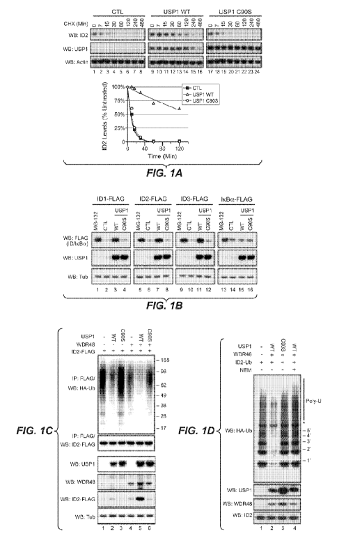
[0023] Figure 1. USP1 Deubiquitinates and Stabilizes ID Proteins. (A) Western
blot (WB) analysis of
293T cells transfected with vector only (CTL), wild-type USP1 (WT), or
catalytically inactive USP1
C905. Cells were treated with 25 mg/ml cycloheximide (CHX) for the times
indicated (left panel). ID2
was quantified by densitometry (right panel). (B) 293T cells were
cotransfected with Flag-tagged ID1,
ID2, ID3, or IkBa, and empty vector (CTL), wild-type USP1, or USP1 C905. Where
indicated, cells
were treated with 10 mM MG-132 for 4 hr. (C) Deubiquitination of 1D2-Flag by
USP1 or USP1 C905
and WDR48 in 293T cells cotransfected with HA-tagged ubiquitin. (D) USP1-Flag,
USP1 C905-Flag,
WDR48-Flag, and ubiquitinated 1D2-Flag were affinity purified separately from
293T extracts and then
combined together for 6 hr in an in vitro deubiquitination assay. NEM, N-
ethylmaleimide.
[0024] Figure 2. Identification of USP1 as an 1D2-Deubiquitinating Enzyme and
Mapping of the USP1-
1D2 Binding Interface. (A) Western blot (WB) analysis of 293T transfected with
Flag-tagged
deubiquitinases (DUBs) or an empty vector (-). Where indicated, cells were
treated with 10 mM MG-132
for 4 hr. (B) Flag-tagged DUBs were immunoprecipitated (IP) from 293T cells
cotransfected with ID2
and treated with 10 mM MG-132 for 6 hr. (C) USP1 mutants expressed in 293T
cells were
immunoprecipitated and blotted for co-expressed ID2. (D) Western blot analysis
of endogenous ID2 in
293T cells transfected with wild-type (WT) or mutant USP1.
[0025] Figure 3. USP1 Is Overexpressed in Osteosarcoma and Correlates with ID2
Protein Expression.
(A) Box and whisker plots of USP1 mRNA expression in primary human bone
biopsies from normal and
diseased tissue. (B) Western blot (WB) analysis of USP1 and ID2 protein
expression in primary human
osteoblasts and osteosarcoma tumor samples. (C and D) RT-PCR quantification of
USP1 (C) and ID2
(D) expression in the samples in (B). Bars represent the mean SD of
triplicate observations. (E and F)
Immunohistochemical detection of ID2 in 293T cells transfected with an ID2
expression vector (top
panel) or an ID2 shRNA (bottom panel) (E) or in a primary human osteosarcoma
biopsy (F). (G)
Immunohistochemical staining of USP1 and ID2 in serial sections from primary
osteosarcoma tissue.
Control staining was with an isotype-control antibody.
[0026] Figure 4. USP1 Physically Engages and Stabilizes ID Proteins in
Osteosarcoma. (A) Western
blot (WB) analysis of U2-OS cells cotransfected with USP1 or control (CTL)
shRNAs, plus either empty
vector (CTL) or shRNA-resistant USP1 (wild-type [WT] or USP1 mutant C905). (B)
Luciferase activity
of U2-OS cells treated as in (A) and cotransfected with an E box-driven
luciferase reporter. Bars
represent the mean SD of triplicate observations. (C) U2-OS cells were
transfected with shRNAs and,
where indicated, treated with 10mM MG-132 for 4 hr. (D) U2-OS cells were
cotransfected with 1D2-
Flag, HA-ubiquitin, and either CTL or USP1 shRNAs. Where indicated, cells were
treated with
CA 02846083 2014-02-20
WO 2013/040433 PCT/US2012/055539
10mMMG-132 for 4 hr. 1D2-Flag was immunoprecipitated from SDS/heat-denatured
cell lysates. (E and
F) USP1 (E) or ID2 (F) was immunoprecipitated from U2-OS cells. Control
immunoprecipitations were
with nonspecific IgG. Asterisk (*) denotes a band of unknown identity
recognized by the anti-1D2
antibody.
[0027] Figure 5. USP1 Regulates ID Proteins in Multiple Osteosarcoma Cell
Lines. (A) Western blot
(WB) analysis of cultured primary human osteoblasts and human osteosarcoma
cell lines. (B)
Osteosarcoma cell lines were treated with 10 mM MG-132 for 4 hr. (C)
Osteosarcoma cell lines were
transfected with control (CTL) or USP1 shRNAs. (D) Osteosarcoma cells were
transfected with empty
vector or WDR48, or were treated with 10 mM MG-132 for 4 hr. (E) USP1 was
immunoprecipitated
from HOS cells. Control immunoprecipitations were with nonspecific IgG. (F)
Analysis of USP1
(WT) and USP1-/- DT40 cells. (G) Real-time RT-PCR quantification of USP1 mRNA
in WT and USP1-'DT40 cells. Bars represent the mean s.d. of triplicate
observations. (H) WT and USP1-/- DT40 cells
were treated with 10mM MG-132 for 2 hr. (I) USP1-/- DT40 cells were
transfected with empty vector
(CTL), USP1 wild-type (WT), or USP1 C9OS and compared to USP1-/- DT40 cells.
Un, untransfected.
[0028] Figure 6. USP1 Regulates Cell Cycling via ID Proteins in Osteosarcoma.
(A) Western blot (WB)
analysis of U2-OS cells treated as in Figure 4A. (B) Outgrowth of U2-OS cells
treated as in (A) was
enumerated after 5 days of culture. (C) Cell cycle status of propidium iodide-
stained U2-OS cells treated
as in (A). (D) U2-OS cells transfected with indicated shRNAs and control or
CDKN1A/p21 siRNAs. (E)
Quantification of cells in S phase in cells treated as in (D). (F) U2-OS cells
transfected with indicated
shRNAs and shRNA-resistant USP1 (shRes USP1), ID1, ID2, and ID3, or control
expression vectors.
(G) Quantification of cells in S phase in U2-OS cells treated as in (F). Bars
represent the mean SD of
triplicate observations.
[0029] Figure 7. USP1 Regulates Proliferation and Cell-Cycle Arrest via ID
Proteins. (A) U2-OS cells
were transfected with control (CTL) or USP1 shRNAs for 3 days, plated at
equivalent density, and
viable cells were counted on subsequent days. (B) U2-OS cells cotransfected
with shRNAs and, where
indicated, shRNA-resistant USP1 (wild-type or mutant). (C) Percentage of cells
in (B) in S-phase of the
cell cycle. (D) Osteosarcoma cells were transfected with shRNAs and cells
enumerated at day 8. (E)
DNA content of U2-OS cells treated as in (A) and stained with propidium iodide
(PI). (F) U2-OS cells
were transfected with indicated shRNAs and with control or p21 siRNAs. (G)
Cells in (F) were stained
with propidium iodide and analyzed by flow cytometry. Bars represent the mean
percentage of cells in 5-
phase. (H) U2-OS cells were transfected with the indicated shRNAs. (I-K) Cells
in (H) were assessed by
real-time RT-PCR (I) and flow cytometry after PI staining (J, K). (L) U2-OS
cells were transfected with
shRNAs and control or p53 siRNAs. Where indicated, cells were treated with 10
mM etoposide for 1 hr.
Bars represent the mean s.d. of triplicate observations.
[0030] Figure 8. USP1 Promotes Retention of Stem Cell Identity in
Osteosarcoma. (A) Western blot
(WB) analysis of U2-OS cells transfected with CTL or USP1 shRNAs. (B) Cells in
(A) were stained and
6
CA 02846083 2014-02-20
WO 2013/040433 PCT/US2012/055539
analyzed by fluorescence microscopy. (C) Immunohistochemical staining for USP1
or ID2 in xenografts
of 143B cells with doxycycline (DOX)-inducible shUSP1. (D) Quantification of
tumor volume of 143B
xenografts as described in (C). Bars represent the mean SD of ten
xenografts. (E and F) RT-PCR
quantification of USP1, ID2, OSTEONECTIN (ON), RUNX2 (RX2), OSTERIX (OSX), and
OSTEOPONTIN (OP) mRNA levels (E) and ALP activity (F) from 143B xenografts in
(C). Bars
represent the mean SD of triplicate observations. (G) Representative
xenograft tumors from (C) were
stained with hematoxylin and eosin (H&E) or trichrome stain. Scale bars, 100
mm.
[0031] Figure 9. Depletion of USP1 Induces Loss of Stem Markers and Initiates
Osteogenic Program in
Osteosarcoma Cell Lines. (A) Osteosarcoma cells were serially transfected with
control (CTL), USP1, or
ID shRNAs. Surface expression of the indicated mesenchymal stem cell markers
was determined by flow
cytometry after 11 days. (B) Cells in (A) were analyzed by real time RT-PCR
for RUNX2, OSTERIX
(OSX), and OSTEONECTIN gene expression. (C) Cells in (A) were assessed for
alkaline phosphatase
activity by p-nitrophenol¨phosphate (pNPP) cleavage. (D) Western blot (WB)
analysis of 143B cells
transduced with doxycycline-inducible CTL or USP1 shRNAs. Where indicated,
cells were treated with
3 mg/ml doxycycline (DOX) for 4 days. (E) Bright field and dark field
microscopy of OSTEOCALCIN
gene expression by in situ hybridization in sections of 143B shUSP1 xenograft
tumors following 5 days
of doxycyline treatment. Scale bars, 100 mm. (F) Real-time RT-PCR analysis of
USP1 gene expression
in control and USP1 shRNA-containing 143B xenograft tumors. Bars represent the
mean s.d. of
triplicate observations.
[0032] Figure 10. USP1 and IDs Regulate Mesenchymal Stem Cell Differentiation.
(A) Western blot
(WB) analysis of hMSCs grown in osteogenic differentiation medium (ODM), or in
nondifferentiating
medium (Un). (B) hMSCs were transduced with ID2, USP1 wild-type (WT), USP1
C905, or empty
vector (CTL) and cultured in ODM for 9 days. (C and D) hMSCs in (B) were
assessed for ALP activity
(C) and OSTEONECTIN, RUNX2, and OSTERIX mRNA (D). Bars represent the mean SD
of
triplicate observations. (E) hMSCs in (B) stained with alizarin red to
visualize calcium deposition. Scale
bars, 100 mm. (F) Enumeration of hMSCs in (B) after the indicated number of
days of culture. Bars
represent the mean SD of triplicate observations.
[0033] Figure 11. USP1 Induces ID-Dependent Transformation of NIH 3T3 Cells.
(A) Western blot
(WB) analysis of NIH 3T3 cells transduced with ID2, USP1 wild-type (WT), USP1
C905, or an empty
control vector. (B) Cells in (A) were grown in soft agar, and colonies were
enumerated. Bars represent
the mean s.d. of triplicate observations. (C) Representative colonies formed
by NIH 3T3 cells
transduced with control (CTL), ID2, USP1 wild-type (WT), or USP1 C905. Scale
bars, 100 mm. (D)
NIH 3T3 cells in (A) were implanted subcutaneously in C.B-17 SCID.bg mice (top
panel) or NCr nude
mice (bottom panel) and tumor volume was monitored. Data points represent the
mean s.d. of ten mice.
(E) C.B-17 SCID.bg (top panels) and NCr nude mice (bottom panels) from (A) at
the end of the study.
(F) Empty vector (CTL)- or USP1-transduced NIH 3T3 cells were sequentially
transduced with control
7
CA 02846083 2014-02-20
WO 2013/040433 PCT/US2012/055539
(CTL) or ID shRNAs. (G) Cells in (F) were grown in soft agar, and colonies
were enumerated. Bars
represent the mean s.d. of triplicate observations.
[0034] Figure 12. USP1 Is Required for Normal Skeletogenesis. (A)
Microcomputed tomography of 12-
day-old USP1' (WT) and USP1-/- mice (top) and femurs (bottom). (B and C)
Mean bone mineralized
density (BMD) (B) and mineralized bone volume (Minz. Vol.) (C) of mice in (A).
Bars represent the
mean SD of four femurs of each genotype. (D) Western blot (WB) analysis of
femoral metaphyses
from E18.5 USP1 / (WT) and USP1-/- mice. (E) BALP in the sera of E18.5 USP1
/ (WT) and USP1-/-
embryos. Bars represent the mean SD of four embryos of each genotype.
[0035] Figure 13. USP1 Is Required for Normal Mouse Skeletogenesis. (A) USP1
targeting strategy to
delete exon 3, which encodes the catalytic cysteine of USP1. Yellow boxes
represent exons. (B) Micro-
computed tomography of E18.5 USP1 / (WT) and USP1-/- embryos. (C)
Mineralized bone volume
(Minz. Vol.) of mice in (b). Bars represent the mean s.d. of 3 mice of each
genotype. (D) Hematoxylin
and eosin (H&E) stained sections of P12 USP1+1+ (WT) and USP1-/- femurs. Scale
bars, 100 mm. (E)
Osteoid area per length of spicule in P12 USP1+1+ (WT) and USP1-/- femurs.
Bars represent the mean
s.d. of 3 mice of each genotype. (F) H&E, trichrome, and Von Kossa stains of
P12 USP1 / (WT) and
USP1-/- femoral metaphyses. Scale bars, 100 mm. (G) TRAP labeling of resident
osteoclasts in P12
USP1+1+ (WT) and USP1-/- femurs. Scale bars, 100 mm. (H) Enumeration of TRAP-
positive cells in P12
USP1 / (WT) and USP1-/- femur sections. (I) Creatinine-normalized
deoxypyridinoline (DPD) levels in
E18.5 amniotic fluid. (J) USP1 and ID2 expression in P12 USP1 / (WT) and
USP1-/- femoral
metaphyses. Scale bars, 100 mm.
DETAILED DESCRIPTION
I. Definitions
[0036] The terms "ubiquitin specific peptidase 1," "deubiquitinating enzyme
1," and "USP1" refer herin
to a native sequence USP1 polypeptide, polypeptide variants and fragments of a
native sequence
polypeptide and polypeptide variants (which are further defined herein). The
USP polypeptide described
herein may be that which is isolated from a variety of sources, such as from
human tissue types or from
another source, or prepared by recombinant or synthetic methods.
[0037] A "native sequence USP1 polypeptide" comprises a polypeptide having the
same amino acid
sequence as the corresponding USP1 polypeptide derived from nature. In one
embodiment, a native
sequence USP1 polypeptide comprises the amino acid sequence of SEQ ID NO: 1.
[0038] "USP1 polypeptide variant", or variations thereof, means an USP1
polypeptide, generally an
active USP1 polypeptide, as defined herein having at least about 80% amino
acid sequence identity with
any of the native sequence USP1 polypeptide sequences as disclosed herein.
Such USP1 polypeptide
variants include, for instance, USP1 polypeptides wherein one or more amino
acid residues are added, or
deleted, at the N¨ or C-terminus of a native amino acid sequence. Ordinarily,
a USP1 polypeptide variant
will have at least about 80% amino acid sequence identity, alternatively at
least about 81%, 82%, 83%,
8
CA 02846083 2014-02-20
WO 2013/040433 PCT/US2012/055539
84%, 85%, 86%, 87%, 88%, 89%, 90%, 91%, 92%, 93%, 94%, 95%, 96%, 97%, 98%, or
99% amino
acid sequence identity, to a native sequence USP1 polypeptide sequence as
disclosed herein. Ordinarily,
USP1 variant polypeptides are at least about 10 amino acids in length,
alternatively at least about 20, 30,
40, 50, 60, 70, 80, 90, 100, 110, 120, 130, 140, 150, 160, 170, 180, 190, 200,
210, 220, 230, 240, 250,
260, 270, 280, 290, 300, 310, 320, 330, 340, 350, 360, 370, 380, 390, 400,
410, 420, 430, 440, 450, 460,
470, 480, 490, 500, 510, 520, 530, 540, 550, 560, 570, 580, 590, 600 amino
acids in length, or more.
Optionally, USP1 variant polypeptides will have no more than one conservative
amino acid substitution
as compared to a native USP1 polypeptide sequence, alternatively no more than
2, 3, 4, 5, 6, 7, 8, 9, or
conservative amino acid substitution as compared to the native USP1
polypeptide sequence.
[0039] The term "USP1 antagonist" as defined herein is any molecule that
partially or fully blocks,
inhibits, or neutralizes a biological activity mediated by a native sequence
USP1. In certain embodiments
such antagonist binds to USP1. According to one embodiment, the antagonist is
a polypeptide.
According to another embodiment, the antagonist is an anti-USP1 antibody.
According to another
embodiment, the antagonist is a small molecule antagonist. According to
another embodiment, the
antagonist is a polynucleotide antagonist.
[0040] The terms "WD repeat domain 48," "USP1-associated factor 1," and "UAF1"
refer herein to a
native sequence UAF1 polypeptide, polypeptide variants and fragments of a
native sequence polypeptide
and polypeptide variants (which are further defined herein). The UAF1
polypeptide described herein
may be that which is isolated from a variety of sources, such as from human
tissue types or from another
source, or prepared by recombinant or synthetic methods.
[0041] A "native sequence UAF1 polypeptide" comprises a polypeptide having the
same amino acid
sequence as the corresponding UAF1 polypeptide derived from nature. In one
embodiment, a native
sequence UAF1 polypeptide comprises the amino acid sequence of SEQ ID NO:40.
[0042] "UAF1 polypeptide variant", or variations thereof, means an UAF1
polypeptide, generally an
active UAF1 polypeptide, as defined herein having at least about 80% amino
acid sequence identity with
any of the native sequence UAF1 polypeptide sequences as disclosed herein.
Such UAF1 polypeptide
variants include, for instance, UAF1 polypeptides wherein one or more amino
acid residues are added, or
deleted, at the N- or C-terminus of a native amino acid sequence. Ordinarily,
a UAF1 polypeptide
variant will have at least about 80% amino acid sequence identity,
alternatively at least about 81%, 82%,
83%, 84%, 85%, 86%, 87%, 88%, 89%, 90%, 91%, 92%, 93%, 94%, 95%, 96%, 97%,
98%, or 99%
amino acid sequence identity, to a native sequence UAF1 polypeptide sequence
as disclosed herein.
Ordinarily, UAF1 variant polypeptides are at least about 10 amino acids in
length, alternatively at least
about 20, 30, 40, 50, 60, 70, 80, 90, 100, 110, 120, 130, 140, 150, 160, 170,
180, 190, 200, 210, 220,
230, 240, 250, 260, 270, 280, 290, 300, 310, 320, 330, 340, 350, 360, 370,
380, 390, 400, 410, 420, 430,
440, 450, 460, 470, 480, 490, 500, 510, 520, 530, 540, 550, 560, 570, 580,
590, 600 amino acids in
length, or more. Optionally, UAF1 variant polypeptides will have no more than
one conservative amino
9
CA 02846083 2014-02-20
WO 2013/040433 PCT/US2012/055539
acid substitution as compared to a native UAF1 polypeptide sequence,
alternatively no more than 2, 3, 4,
5, 6, 7, 8, 9, or 10 conservative amino acid substitution as compared to the
native UAF1 polypeptide
sequence.
[0043] The term "UAF1 antagonist" as defined herein is any molecule that
partially or fully blocks,
inhibits, or neutralizes a biological activity mediated by a native sequence
UAF1. In certain
embodiments such antagonist binds to UAF1. According to one embodiment, the
antagonist is a
polypeptide. According to another embodiment, the antagonist is an anti- UAF1
antibody. According to
another embodiment, the antagonist is a small molecule antagonist. According
to another embodiment,
the antagonist is a polynucleotide antagonist.
[0044] The terms "inhibitor of DNA binding" and "ID" refer herin to a native
sequence ID polypeptide,
polypeptide variants and fragments of a native sequence polypeptide and
polypeptide variants (which are
further defined herein). The ID polypeptide described herein may be that which
is isolated from a variety
of sources, such as from human tissue types or from another source, or
prepared by recombinant or
synthetic methods.
[0045] A "native sequence ID polypeptide" comprises a polypeptide having the
same amino acid
sequence as the corresponding ID polypeptide derived from nature. In some
embodiments of any of the
native sequence ID polypeptides, the native sequence ID polypeptide includes a
native sequence ID1
isoform a polypeptide of SEQ ID NO:2. In some embodiments of any of the native
sequence ID
polypeptides, the native sequence ID polypeptide includes a native sequence
ID1 isoform b polypeptide
of SEQ ID NO:3. In some embodiments of any of the native sequence ID
polypeptides, the native
sequence ID polypeptide includes a native sequence ID2 polypeptide of SEQ ID
NO:4. In some
embodiments of any of the native sequence ID polypeptides, the native sequence
ID polypeptide includes
a native sequence ID3 polypeptide of SEQ ID NO:5.
[0046] "ID polypeptide variant", or variations thereof, means an ID
polypeptide, generally an active ID
polypeptide, as defined herein having at least about 80% amino acid sequence
identity with any of the
native sequence ID polypeptide sequences as disclosed herein. Such ID
polypeptide variants include, for
instance, ID polypeptides wherein one or more amino acid residues are added,
or deleted, at the N- or C-
terminus of a native amino acid sequence. Ordinarily, an ID polypeptide
variant will have at least about
80% amino acid sequence identity, alternatively at least about 81%, 82%, 83%,
84%, 85%, 86%, 87%,
88%, 89%, 90%, 91%, 92%, 93%, 94%, 95%, 96%, 97%, 98%, or 99% amino acid
sequence identity, to
a native sequence ID polypeptide sequence as disclosed herein. Ordinarily, ID
variant polypeptides are at
least about 10 amino acids in length, alternatively at least about 20, 30, 40,
50, 60, 70, 80, 90, 100, 110,
120, 130, 140, 150, 160, 170, 180, 190, 200, 210, 220, 230, 240, 250, 260,
270, 280, 290, 300, 310, 320,
330, 340, 350, 360, 370, 380, 390, 400, 410, 420, 430, 440, 450, 460, 470,
480, 490, 500, 510, 520, 530,
540, 550, 560, 570, 580, 590, 600 amino acids in length, or more. Optionally,
ID variant polypeptides
will have no more than one conservative amino acid substitution as compared to
a native ID polypeptide
CA 02846083 2014-02-20
WO 2013/040433 PCT/US2012/055539
sequence, alternatively no more than 2, 3, 4, 5, 6, 7, 8, 9, or 10
conservative amino acid substitution as
compared to the native ID polypeptide sequence. In some embodiments of any of
the ID polypeptide
variants, the ID polypeptide variant includes an ID1 polypeptide variant. In
some embodiments of any of
the ID polypeptide variants, the ID polypeptide variant includes an ID2
polypeptide variant. In some
embodiments of any of the ID polypeptide variants, the ID polypeptide variant
includes an ID3
polypeptide variant.
[0047] The term "ID antagonist" as defined herein is any molecule that
partially or fully blocks,
inhibits, or neutralizes a biological activity mediated by a native sequence
ID. In certain embodiments
such antagonist binds to ID. According to one embodiment, the antagonist is a
polypeptide. According to
another embodiment, the antagonists is an anti-ID antibody. According to
another embodiment, the
antagonist is a small molecule antagonist. According to another embodiment,
the antagonist is a
polynucleotide antagonist. In some embodiments of any of the ID antagonists,
the ID antagonist is an
ID1 antagonist. In some embodiments of any of the ID antagonists, the ID
antagonist is an ID2
antagonist. In some embodiments of any of the ID antagonists, the ID
antagonist is an ID3 antagonist.
[0048] "Polynucleotide," or "nucleic acid," as used interchangeably herein,
refer to polymers of
nucleotides of any length, and include DNA and RNA. The nucleotides can be
deoxyribonucleotides,
ribonucleotides, modified nucleotides or bases, and/or their analogs, or any
substrate that can be
incorporated into a polymer by DNA or RNA polymerase, or by a synthetic
reaction. A polynucleotide
may comprise modified nucleotides, such as methylated nucleotides and their
analogs. If present,
modification to the nucleotide structure may be imparted before or after
assembly of the polymer. The
sequence of nucleotides may be interrupted by non-nucleotide components. A
polynucleotide may be
further modified after synthesis, such as by conjugation with a label. Other
types of modifications
include, for example, "caps", substitution of one or more of the naturally
occurring nucleotides with an
analog, internucleotide modifications such as, for example, those with
uncharged linkages (e.g., methyl
phosphonates, phosphotriesters, phosphoamidates, carbamates, etc.) and with
charged linkages (e.g.,
phosphorothioates, phosphorodithioates, etc.), those containing pendant
moieties, such as, for example,
proteins (e.g., nucleases, toxins, antibodies, signal peptides, ply-L-lysine,
etc.), those with intercalators
(e.g., acridine, psoralen, etc.), those containing chelators (e.g., metals,
radioactive metals, boron,
oxidative metals, etc.), those containing alkylators, those with modified
linkages (e.g., alpha anomeric
nucleic acids, etc.), as well as unmodified forms of the polynucleotide(s).
Further, any of the hydroxyl
groups ordinarily present in the sugars may be replaced, for example, by
phosphonate groups, phosphate
groups, protected by standard protecting groups, or activated to prepare
additional linkages to additional
nucleotides, or may be conjugated to solid or semi-solid supports. The 5' and
3' terminal OH can be
phosphorylated or substituted with amines or organic capping group moieties of
from 1 to 20 carbon
atoms. Other hydroxyls may also be derivatized to standard protecting groups.
Polynucleotides can also
contain analogous forms of ribose or deoxyribose sugars that are generally
known in the art, including,
11
CA 02846083 2014-02-20
WO 2013/040433 PCT/US2012/055539
for example, 2'-0-methyl-, 2'-0-allyl, 2'-fluoro- or 2'-azido-ribose,
carbocyclic sugar analogs, a-anomeric
sugars, epimeric sugars such as arabinose, xyloses or lyxoses, pyranose
sugars, furanose sugars,
sedoheptuloses, acyclic analogs and abasic nucleoside analogs such as methyl
riboside. One or more
phosphodiester linkages may be replaced by alternative linking groups. These
alternative linking groups
include, but are not limited to, embodiments wherein phosphate is replaced by
P(0)S("thioate"), P(S)S
("dithioate"), "(0)NR2 ("amidate"), P(0)R, P(0)OR', CO or CH2 ("formacetal"),
in which each R or R' is
independently H or substituted or unsubstituted alkyl (1-20 C) optionally
containing an ether (-0-)
linkage, aryl, alkenyl, cycloalkyl, cycloalkenyl or araldyl. Not all linkages
in a polynucleotide need be
identical. The preceding description applies to all polynucleotides referred
to herein, including RNA and
DNA.
[0049] "Oligonucleotide," as used herein, generally refers to short, generally
single stranded, generally
synthetic polynucleotides that are generally, but not necessarily, less than
about 200 nucleotides in
length. The terms "oligonucleotide" and "polynucleotide" are not mutually
exclusive. The description
above for polynucleotides is equally and fully applicable to oligonucleotides.
[0050] The term "small molecule" refers to any molecule with a molecular
weight of about 2000 daltons
or less, preferably of about 500 daltons or less.
[0051] The terms "host cell," "host cell line," and "host cell culture" are
used interchangeably and refer
to cells into which exogenous nucleic acid has been introduced, including the
progeny of such cells.
Host cells include "transformants" and "transformed cells," which include the
primary transformed cell
and progeny derived therefrom without regard to the number of passages.
Progeny may not be
completely identical in nucleic acid content to a parent cell, but may contain
mutations. Mutant progeny
that have the same function or biological activity as screened or selected for
in the originally
transformed cell are included herein.
[0052] The term "vector," as used herein, refers to a nucleic acid molecule
capable of propagating
another nucleic acid to which it is linked. The term includes the vector as a
self-replicating nucleic acid
structure as well as the vector incorporated into the genome of a host cell
into which it has been
introduced. Certain vectors are capable of directing the expression of nucleic
acids to which they are
operatively linked. Such vectors are referred to herein as "expression
vectors."
[0053] An "isolated" antibody is one which has been separated from a component
of its natural
environment. In some embodiments, an antibody is purified to greater than 95%
or 99% purity as
determined by, for example, electrophoretic (e.g., SDS-PAGE, isoelectric
focusing (IEF), capillary
electrophoresis) or chromatographic (e.g., ion exchange or reverse phase
HPLC). For review of methods
for assessment of antibody purity, see, e.g., Flatman et al., J. Chromatogr. B
848:79-87 (2007).
[0054] An "isolated" nucleic acid refers to a nucleic acid molecule that has
been separated from a
component of its natural environment. An isolated nucleic acid includes a
nucleic acid molecule
contained in cells that ordinarily contain the nucleic acid molecule, but the
nucleic acid molecule is
12
CA 02846083 2014-02-20
WO 2013/040433 PCT/US2012/055539
present extrachromosomally or at a chromosomal location that is different from
its natural chromosomal
location.
[0055] The term "antibody" herein is used in the broadest sense and
encompasses various antibody
structures, including but not limited to monoclonal antibodies, polyclonal
antibodies, multispecific
antibodies (e.g., bispecific antibodies), and antibody fragments so long as
they exhibit the desired
antigen-binding activity.
[0056] The terms "anti-USP1 antibody" and "an antibody that binds to USP1"
refer to an antibody that
is capable of binding USP1 with sufficient affinity such that the antibody is
useful as a diagnostic and/or
therapeutic agent in targeting USP1. In one embodiment, the extent of binding
of an anti-USP1 antibody
to an unrelated, non-USP1 protein is less than about 10% of the binding of the
antibody to USP1 as
measured, e.g., by a radioimmunoassay (RIA). In certain embodiments, an anti-
USP1 antibody binds to
an epitope of USP1 that is conserved among USP1 from different species.
[0057] The terms "anti-ID antibody" and "an antibody that binds to ID" refer
to an antibody that is
capable of binding ID with sufficient affinity such that the antibody is
useful as a diagnostic and/or
therapeutic agent in targeting ID. In one embodiment, the extent of binding of
an anti-ID antibody to an
unrelated, non-ID protein is less than about 10% of the binding of the
antibody to ID as measured, e.g.,
by a radioimmunoassay (RIA). In certain embodiments, an anti-ID antibody binds
to an epitope of ID
that is conserved among ID from different species. In some embodiments of any
of the anti-ID
antibodies, the ID antibody is an anti-ID1 antibody. In some embodiments of
any of the anti-ID
antibodies, the ID antibody is an anti-1D2 antibody. In some embodiments of
any of the anti-ID
antibodies, the ID antibody is an anti-1D3 antibody.
[0058] A "blocking" antibody or an "antagonist" antibody is one which inhibits
or reduces biological
activity of the antigen it binds. Preferred blocking antibodies or antagonist
antibodies substantially or
completely inhibit the biological activity of the antigen.
[0059] "Affinity" refers to the strength of the sum total of noncovalent
interactions between a single
binding site of a molecule (e.g., an antibody) and its binding partner (e.g.,
an antigen). Unless indicated
otherwise, as used herein, "binding affinity" refers to intrinsic binding
affinity which reflects a 1:1
interaction between members of a binding pair (e.g., antibody and antigen).
The affinity of a molecule X
for its partner Y can generally be represented by the dissociation constant
(Kd). Affinity can be
measured by common methods known in the art, including those described herein.
Specific illustrative
and exemplary embodiments for measuring binding affinity are described in the
following.
[0060] An "affinity matured" antibody refers to an antibody with one or more
alterations in one or more
hypervariable regions (HVRs), compared to a parent antibody which does not
possess such alterations,
such alterations resulting in an improvement in the affinity of the antibody
for antigen.
[0061] An "antibody fragment" refers to a molecule other than an intact
antibody that comprises a
portion of an intact antibody that binds the antigen to which the intact
antibody binds. Examples of
13
CA 02846083 2014-02-20
WO 2013/040433 PCT/US2012/055539
antibody fragments include but are not limited to Fv, Fab, Fab', Fab'-SH,
F(ab')2; diabodies; linear
antibodies; single-chain antibody molecules (e.g. scFv); and multispecific
antibodies formed from
antibody fragments.
[0062] An "antibody that binds to the same epitope" as a reference antibody
refers to an antibody that
blocks binding of the reference antibody to its antigen in a competition assay
by 50% or more, and
conversely, the reference antibody blocks binding of the antibody to its
antigen in a competition assay by
50% or more. An exemplary competition assay is provided herein.
[0063] The term "chimeric" antibody refers to an antibody in which a portion
of the heavy and/or light
chain is derived from a particular source or species, while the remainder of
the heavy and/or light chain
is derived from a different source or species.
[0064] The "class" of an antibody refers to the type of constant domain or
constant region possessed by
its heavy chain. There are five major classes of antibodies: IgA, IgD, IgE,
IgG, and IgM, and several of
these may be further divided into subclasses (isotypes), e.g., IgGi, IgG2,
IgG3, IgG4, IgAi, and IgA2. The
heavy chain constant domains that correspond to the different classes of
immunoglobulins are called a,
6, c, y, and 11., respectively.
[0065] The terms "full length antibody," "intact antibody," and "whole
antibody" are used herein
interchangeably to refer to an antibody having a structure substantially
similar to a native antibody
structure or having heavy chains that contain an Fc region as defined herein.
[0066] The term "monoclonal antibody" as used herein refers to an antibody
obtained from a population
of substantially homogeneous antibodies, i.e., the individual antibodies
comprising the population are
identical and/or bind the same epitope, except for possible variant
antibodies, e.g., containing naturally
occurring mutations or arising during production of a monoclonal antibody
preparation, such variants
generally being present in minor amounts. In contrast to polyclonal antibody
preparations, which
typically include different antibodies directed against different determinants
(epitopes), each monoclonal
antibody of a monoclonal antibody preparation is directed against a single
determinant on an antigen.
Thus, the modifier "monoclonal" indicates the character of the antibody as
being obtained from a
substantially homogeneous population of antibodies, and is not to be construed
as requiring production
of the antibody by any particular method. For example, the monoclonal
antibodies to be used in
accordance with the present invention may be made by a variety of techniques,
including but not limited
to the hybridoma method, recombinant DNA methods, phage-display methods, and
methods utilizing
transgenic animals containing all or part of the human immunoglobulin loci,
such methods and other
exemplary methods for making monoclonal antibodies being described herein.
[0067] A "human antibody" is one which possesses an amino acid sequence which
corresponds to that
of an antibody produced by a human or a human cell or derived from a non-human
source that utilizes
human antibody repertoires or other human antibody-encoding sequences. This
definition of a human
antibody specifically excludes a humanized antibody comprising non-human
antigen-binding residues.
14
CA 02846083 2014-02-20
WO 2013/040433 PCT/US2012/055539
[0068] A "humanized" antibody refers to a chimeric antibody comprising amino
acid residues from
non-human HVRs and amino acid residues from human FRs. In certain embodiments,
a humanized
antibody will comprise substantially all of at least one, and typically two,
variable domains, in which all
or substantially all of the HVRs (e.g., CDRs) correspond to those of a non-
human antibody, and all or
substantially all of the FRs correspond to those of a human antibody. A
humanized antibody optionally
may comprise at least a portion of an antibody constant region derived from a
human antibody. A
"humanized form" of an antibody, e.g., a non-human antibody, refers to an
antibody that has undergone
humanization.
[0069] An "immunoconjugate" is an antibody conjugated to one or more
heterologous molecule(s),
including but not limited to a cytotoxic agent.
[0070] "Percent (%) amino acid sequence identity" with respect to a reference
polypeptide sequence is
defined as the percentage of amino acid residues in a candidate sequence that
are identical with the
amino acid residues in the reference polypeptide sequence, after aligning the
sequences and introducing
gaps, if necessary, to achieve the maximum percent sequence identity, and not
considering any
conservative substitutions as part of the sequence identity. Alignment for
purposes of determining
percent amino acid sequence identity can be achieved in various ways that are
within the skill in the art,
for instance, using publicly available computer software such as BLAST, BLAST-
2, ALIGN or
Megalign (DNASTAR) software. Those skilled in the art can determine
appropriate parameters for
aligning sequences, including any algorithms needed to achieve maximal
alignment over the full length
of the sequences being compared. For purposes herein, however, % amino acid
sequence identity values
are generated using the sequence comparison computer program ALIGN-2. The
ALIGN-2 sequence
comparison computer program was authored by Genentech, Inc., and the source
code has been filed with
user documentation in the U.S. Copyright Office, Washington D.C., 20559, where
it is registered under
U.S. Copyright Registration No. TXU510087. The ALIGN-2 program is publicly
available from
Genentech, Inc., South San Francisco, California, or may be compiled from the
source code. The
ALIGN-2 program should be compiled for use on a UNIX operating system,
including digital UNIX
V4.0D. All sequence comparison parameters are set by the ALIGN-2 program and
do not vary.
[0071] In situations where ALIGN-2 is employed for amino acid sequence
comparisons, the % amino
acid sequence identity of a given amino acid sequence A to, with, or against a
given amino acid sequence
B (which can alternatively be phrased as a given amino acid sequence A that
has or comprises a certain
% amino acid sequence identity to, with, or against a given amino acid
sequence B) is calculated as
follows:
100 times the fraction X/Y
where X is the number of amino acid residues scored as identical matches by
the sequence alignment
program ALIGN-2 in that program's alignment of A and B, and where Y is the
total number of amino
acid residues in B. It will be appreciated that where the length of amino acid
sequence A is not equal to
CA 02846083 2014-02-20
WO 2013/040433 PCT/US2012/055539
the length of amino acid sequence B, the % amino acid sequence identity of A
to B will not equal the %
amino acid sequence identity of B to A. Unless specifically stated otherwise,
all % amino acid sequence
identity values used herein are obtained as described in the immediately
preceding paragraph using the
ALIGN-2 computer program.
[0072] An "effective amount" of an agent refers to an amount effective, at
dosages and for periods of
time necessary, to achieve the desired therapeutic or prophylactic result.
[0073] A "therapeutically effective amount" of a substance/molecule of the
invention, agonist or
antagonist may vary according to factors such as the disease state, age, sex,
and weight of the individual,
and the ability of the substance/molecule, agonist or antagonist to elicit a
desired response in the
individual. A therapeutically effective amount is also one in which any toxic
or detrimental effects of the
substance/molecule, agonist or antagonist are outweighed by the
therapeutically beneficial effects. A
"prophylactically effective amount" refers to an amount effective, at dosages
and for periods of time
necessary, to achieve the desired prophylactic result. Typically but not
necessarily, since a prophylactic
dose is used in subjects prior to or at an earlier stage of disease, the
prophylactically effective amount
will be less than the therapeutically effective amount.
[0074] The term "pharmaceutical formulation" refers to a preparation which is
in such form as to permit
the biological activity of an active ingredient contained therein to be
effective, and which contains no
additional components which are unacceptably toxic to a subject to which the
formulation would be
administered.
[0075] A "pharmaceutically acceptable carrier" refers to an ingredient in a
pharmaceutical formulation,
other than an active ingredient, which is nontoxic to a subject., A
pharmaceutically acceptable carrier
includes, but is not limited to, a buffer, excipient, stabilizer, or
preservative.
[0076] As used herein, "treatment" (and grammatical variations thereof such as
"treat" or "treating")
refers to clinical intervention in an attempt to alter the natural course of
the individual being treated, and
can be performed either for prophylaxis or during the course of clinical
pathology. Desirable effects of
treatment include, but are not limited to, preventing occurrence or recurrence
of disease, alleviation of
symptoms, diminishment of any direct or indirect pathological consequences of
the disease, preventing
metastasis, decreasing the rate of disease progression, amelioration or
palliation of the disease state, and
remission or improved prognosis. In some embodiments, antibodies of the
invention are used to delay
development of a disease or to slow the progression of a disease.
[0077] The term "anti-cancer therapy" refers to a therapy useful in treating
cancer. Examples of anti-
cancer therapeutic agents include, but are limited to, e.g., chemotherapeutic
agents, growth inhibitory
agents, cytotoxic agents, agents used in radiation therapy, anti-angiogenesis
agents, apoptotic agents,
anti-tubulin agents, and other agents to treat cancer, anti-CD20 antibodies,
platelet derived growth
factor inhibitors (e.g., GleevecT' (Imatinib Mesylate)), a COX-2 inhibitor
(e.g., celecoxib), interferons,
cytokines, antagonists (e.g., neutralizing antibodies) that bind to one or
more of the following targets
16
CA 02846083 2014-02-20
WO 2013/040433 PCT/US2012/055539
PDGFR-beta, BlyS, APRIL, BCMA receptor(s), TRAIL/Apo2, and other bioactive and
organic chemical
agents, etc. Combinations thereof are also included in the invention.
[0078] The term "cytotoxic agent" as used herein refers to a substance that
inhibits or prevents the
function of cells and/or causes destruction of cells. The term is intended to
include radioactive isotopes
(e.g., At211, 1131, 1125, y90, Re186, Re188, sm153, Bi212, -.32
f
and radioactive isotopes of Lu), chemotherapeutic
agents e.g. methotrexate, adriamicin, vinca alkaloids (vincristine,
vinblastine, etoposide), doxorubicin,
melphalan, mitomycin C, chlorambucil, daunorubicin or other intercalating
agents, enzymes and
fragments thereof such as nucleolytic enzymes, antibiotics, and toxins such as
small molecule toxins or
enzymatically active toxins of bacterial, fungal, plant or animal origin,
including fragments and/or
variants thereof, and the various antitumor or anticancer agents disclosed
below. Other cytotoxic agents
are described below. A tumoricidal agent causes destruction of tumor cells.
[0079] A "chemotherapeutic agent" refers to a chemical compound useful in the
treatment of cancer.
Examples of chemotherapeutic agents include alkylating agents such as thiotepa
and cyclosphosphamide
(CYTOXANO); alkyl sulfonates such as busulfan, improsulfan and piposulfan;
aziridines such as
benzodopa, carboquone, meturedopa, and uredopa; ethylenimines and
methylamelamines including
altretamine, triethylenemelamine, triethylenephosphoramide,
triethylenethiophosphoramide and
trimethylomelamine; acetogenins (especially bullatacin and bullatacinone);
delta-9-tetrahydrocannabinol
(dronabinol, MARINOLO); beta-lapachone; lapachol; colchicines; betulinic acid;
a camptothecin
(including the synthetic analogue topotecan (HYCAMTINO), CPT-11 (irinotecan,
CAMPTOSARO),
acetylcamptothecin, scopolectin, and 9-aminocamptothecin); bryostatin;
callystatin; CC-1065 (including
its adozelesin, carzelesin and bizelesin synthetic analogues);
podophyllotoxin; podophyllinic acid;
teniposide; cryptophycins (particularly cryptophycin 1 and cryptophycin 8);
dolastatin; duocarmycin
(including the synthetic analogues, KW-2189 and CB1-TM1); eleutherobin;
pancratistatin; a
sarcodictyin; spongistatin; nitrogen mustards such as chlorambucil,
chlornaphazine, chlorophosphamide,
estramustine, ifosfamide, mechlorethamine, mechlorethamine oxide
hydrochloride, melphalan,
novembichin, phenesterine, prednimustine, trofosfamide, uracil mustard;
nitrosoureas such as
carmustine, chlorozotocin, fotemustine, lomustine, nimustine, and
ranimnustine; antibiotics such as the
enediyne antibiotics (e. g., calicheamicin, especially calicheamicin gammalI
and calicheamicin omegaIl
(see, e.g., Nicolaou et al., Angew. Chem Intl. Ed. Engl., 33: 183-186 (1994));
CDP323, an oral alpha-4
integrin inhibitor; dynemicin, including dynemicin A; an esperamicin; as well
as neocarzinostatin
chromophore and related chromoprotein enediyne antibiotic chromophores),
aclacinomysins,
actinomycin, authramycin, azaserine, bleomycins, cactinomycin, carabicin,
carminomycin, carzinophilin,
chromomycins, dactinomycin, daunorubicin, detorubicin, 6-diazo-5-oxo-L-
norleucine, doxorubicin
(including ADRIAMYCINO, morpholino-doxorubicin, cyanomorpholino-doxorubicin, 2-
pyrrolino-
doxorubicin, doxorubicin HC1 liposome injection (DOXILO), liposomal
doxorubicin TLC D-99
(MYOCETO), peglylated liposomal doxorubicin (CAELYXO), and deoxydoxorubicin),
epirubicin,
17
CA 02846083 2014-02-20
WO 2013/040433 PCT/US2012/055539
esorubicin, idarubicin, marcellomycin, mitomycins such as mitomycin C,
mycophenolic acid,
nogalamycin, olivomycins, peplomycin, porfiromycin, puromycin, quelamycin,
rodorubicin,
streptonigrin, streptozocin, tubercidin, ubenimex, zinostatin, zorubicin; anti-
metabolites such as
methotrexate, gemcitabine (GEMZARO), tegafur (UFTORALO), capecitabine
(XELODAO), an
epothilone, and 5-fluorouracil (5-FU); folic acid analogues such as
denopterin, methotrexate, pteropterin,
trimetrexate; purine analogs such as fludarabine, 6-mercaptopurine,
thiamiprine, thioguanine; pyrimidine
analogs such as ancitabine, azacitidine, 6-azauridine, carmofur, cytarabine,
dideoxyuridine,
doxifluridine, enocitabine, floxuridine; androgens such as calusterone,
dromostanolone propionate,
epitiostanol, mepitiostane, testolactone; anti-adrenals such as
aminoglutethimide, mitotane, trilostane;
folic acid replenisher such as frolinic acid; aceglatone; aldophosphamide
glycoside; aminolevulinic acid;
eniluracil; amsacrine; bestrabucil; bisantrene; edatraxate; defofamine;
demecolcine; diaziquone;
elfornithine; elliptinium acetate; an epothilone; etoglucid; gallium nitrate;
hydroxyurea; lentinan;
lonidainine; maytansinoids such as maytansine and ansamitocins; mitoguazone;
mitoxantrone;
mopidanmol; nitraerine; pentostatin; phenamet; pirarubicin; losoxantrone; 2-
ethylhydrazide;
procarbazine; PSKO polysaccharide complex (JHS Natural Products, Eugene, OR);
razoxane; rhizoxin;
sizofiran; spirogermanium; tenuazonic acid; triaziquone; 2,2',2'-
trichlorotriethylamine; trichothecenes
(especially T-2 toxin, verracurin A, roridin A and anguidine); urethan;
vindesine (ELDISINEO,
FILDESINO); dacarbazine; mannomustine; mitobronitol; mitolactol; pipobroman;
gacytosine;
arabinoside ("Ara-C"); thiotepa; taxoid, e.g., paclitaxel (TAXOLO), albumin-
engineered nanoparticle
formulation of paclitaxel (ABRAXANETI"), and docetaxel (TAXOTERE0);
chloranbucil; 6-
thioguanine; mercaptopurine; methotrexate; platinum agents such as cisplatin,
oxaliplatin (e.g.,
ELOXATINO), and carboplatin; vincas, which prevent tubulin polymerization from
forming
microtubules, including vinblastine (VELBANO), vincristine (ONCOVINO),
vindesine (ELDISINEO,
FILDESINO), and vinorelbine (NAVELBINE0); etoposide (VP-16); ifosfamide;
mitoxantrone;
leucovorin; novantrone; edatrexate; daunomycin; aminopterin; ibandronate;
topoisomerase inhibitor RFS
2000; difluoromethylornithine (DMF0); retinoids such as retinoic acid,
including bexarotene
(TARGRETINO); bisphosphonates such as clodronate (for example, BONEFOSO or
OSTACO),
etidronate (DIDROCALO), NE-58095, zoledronic acid/zoledronate (ZOMETAO),
alendronate
(FOSAMAXO), pamidronate (AREDIAO), tiludronate (SKELIDO), or risedronate
(ACTONEL0);
troxacitabine (a 1,3-dioxolane nucleoside cytosine analog); antisense
oligonucleotides, particularly those
that inhibit expression of genes in signaling pathways implicated in aberrant
cell proliferation, such as,
for example, PKC-alpha, Raf, H-Ras, and epidermal growth factor receptor (EGF-
R); vaccines such as
THERATOPEO vaccine and gene therapy vaccines, for example, ALLOVECTINO
vaccine,
LEUVECTINO vaccine, and VAXIDO vaccine; topoisomerase 1 inhibitor (e.g.,
LURTOTECANO);
rmRH (e.g., ABARELIX0); BAY439006 (sorafenib; Bayer); SU-11248 (sunitinib,
SUTENTO, Pfizer);
perifosine, COX-2 inhibitor (e.g. celecoxib or etoricoxib), proteosome
inhibitor (e.g. PS341);
18
CA 02846083 2014-02-20
WO 2013/040433 PCT/US2012/055539
bortezomib (VELCADE0); CCI-779; tipifarnib (R11577); orafenib, ABT510; Bc1-2
inhibitor such as
oblimersen sodium (GENASENSE0); pixantrone; EGFR inhibitors (see definition
below); tyrosine
kinase inhibitors (see definition below); serine-threonine kinase inhibitors
such as rapamycin (sirolimus,
RAPAMUNE0); farnesyltransferase inhibitors such as lonafarnib (SCH 6636,
SARASARTI"); and
pharmaceutically acceptable salts, acids or derivatives of any of the above;
as well as combinations of
two or more of the above such as CHOP, an abbreviation for a combined therapy
of cyclophosphamide,
doxorubicin, vincristine, and prednisolone; and FOLFOX, an abbreviation for a
treatment regimen with
oxaliplatin (ELOXATINTI") combined with 5-FU and leucovorin.
[0080] Chemotherapeutic agents as defined herein include "anti-hormonal
agents" or "endocrine
therapeutics" which act to regulate, reduce, block, or inhibit the effects of
hormones that can promote the
growth of cancer. They may be hormones themselves, including, but not limited
to: anti-estrogens with
mixed agonist/antagonist profile, including, tamoxifen (NOLVADEXO), 4-
hydroxytamoxifen,
toremifene (FARESTONO), idoxifene, droloxifene, raloxifene (EVISTAO),
trioxifene, keoxifene, and
selective estrogen receptor modulators (SERMs) such as SERM3; pure anti-
estrogens without agonist
properties, such as fulvestrant (FASLODEXO), and EM800 (such agents may block
estrogen receptor
(ER) dimerization, inhibit DNA binding, increase ER turnover, and/or suppress
ER levels); aromatase
inhibitors, including steroidal aromatase inhibitors such as formestane and
exemestane (AROMASINO),
and nonsteroidal aromatase inhibitors such as anastrazole (ARIMIDEXO),
letrozole (FEMARAO) and
aminoglutethimide, and other aromatase inhibitors include vorozole (RIVISORO),
megestrol acetate
(MEGASEO), fadrozole, and 4(5)-imidazoles; lutenizing hormone-releaseing
hormone agonists,
including leuprolide (LUPRONO and ELIGARDO), goserelin, buserelin, and
tripterelin; sex steroids,
including progestines such as megestrol acetate and medroxyprogesterone
acetate, estrogens such as
diethylstilbestrol and premarin, and androgens/retinoids such as
fluoxymesterone, all transretionic acid
and fenretinide; onapristone; anti-progesterones; estrogen receptor down-
regulators (ERDs); anti-
androgens such as flutamide, nilutamide and bicalutamide; and pharmaceutically
acceptable salts, acids
or derivatives of any of the above; as well as combinations of two or more of
the above.
[0081] The term "prodrug" as used in this application refers to a precursor or
derivative form of a
pharmaceutically active substance that is less cytotoxic to tumor cells
compared to the parent drug and is
capable of being enzymatically activated or converted into the more active
parent form. See, e.g.,
Wilman, "Prodrugs in Cancer Chemotherapy" Biochemical Society Transactions,
14, pp. 375-382, 615th
Meeting Belfast (1986) and Stella et al., "Prodrugs: A Chemical Approach to
Targeted Drug Delivery,"
Directed Drug Delivery, Borchardt et al., (ed.), pp. 247-267, Humana Press
(1985). The prodrugs of this
invention include, but are not limited to, phosphate-containing prodrugs,
thiophosphate-containing
prodrugs, sulfate-containing prodrugs, peptide-containing prodrugs, D-amino
acid-modified prodrugs,
glycosylated prodrugs, P-lactam-containing prodrugs, optionally substituted
phenoxyacetamide-
containing prodrugs or optionally substituted phenylacetamide-containing
prodrugs, 5-fluorocytosine
19
CA 02846083 2014-02-20
WO 2013/040433 PCT/US2012/055539
and other 5-fluorouridine prodrugs which can be converted into the more active
cytotoxic free drug.
Examples of cytotoxic drugs that can be derivatized into a prodrug form for
use in this invention include,
but are not limited to, those chemotherapeutic agents described above.
[0082] A "growth inhibitory agent" when used herein refers to a compound or
composition which
inhibits growth of a cell (e.g., a cell whose growth is dependent upon USP1
expression either in vitro or
in vivo). Examples of growth inhibitory agents include agents that block cell
cycle progression (at a
place other than S phase), such as agents that induce G1 arrest and M-phase
arrest. Classical M-phase
blockers include the vincas (vincristine and vinblastine), taxanes, and
topoisomerase II inhibitors such as
doxorubicin, epirubicin, daunorubicin, etoposide, and bleomycin. Those agents
that arrest G1 also spill
over into S-phase arrest, for example, DNA alkylating agents such as
tamoxifen, prednisone,
dacarbazine, mechlorethamine, cisplatin, methotrexate, 5-fluorouracil, and ara-
C. Further information
can be found in The Molecular Basis of Cancer, Mendelsohn and Israel, eds.,
Chapter 1, entitled "Cell
cycle regulation, oncogenes, and antineoplastic drugs" by Murakami et al. (WB
Saunders: Philadelphia,
1995), especially p. 13. The taxanes (paclitaxel and docetaxel) are anticancer
drugs both derived from
the yew tree. Docetaxel (TAXOTEREO, Rhone-Poulenc Rorer), derived from the
European yew, is a
semisynthetic analogue of paclitaxel (TAXOLO, Bristol-Myers Squibb).
Paclitaxel and docetaxel
promote the assembly of microtubules from tubulin dimers and stabilize
microtubules by preventing
depolymerization, which results in the inhibition of mitosis in cells.
[0083] By "radiation therapy" is meant the use of directed gamma rays or beta
rays to induce sufficient
damage to a cell so as to limit its ability to function normally or to destroy
the cell altogether. It will be
appreciated that there will be many ways known in the art to determine the
dosage and duration of
treatment. Typical treatments are given as a one time administration and
typical dosages range from 10
to 200 units (Grays) per day.
[0084] An "individual" or "subject" is a mammal. Mammals include, but are not
limited to,
domesticated animals (e.g., cows, sheep, cats, dogs, and horses), primates
(e.g., humans and non-human
primates such as monkeys), rabbits, and rodents (e.g., mice and rats). In
certain embodiments, the
individual or subject is a human.
[0085] The term "concurrently" is used herein to refer to administration of
two or more therapeutic
agents, where at least part of the administration overlaps in time.
Accordingly, concurrent administration
includes a dosing regimen when the administration of one or more agent(s)
continues after discontinuing
the administration of one or more other agent(s).
[0086] By "reduce or inhibit" is meant the ability to cause an overall
decrease of 20%, 30%, 40%, 50%,
60%, 70%, 75%, 80%, 85%, 90%, 95%, or greater. Reduce or inhibit can refer to
the symptoms of the
disorder being treated, the presence or size of metastases, or the size of the
primary tumor.
[0087] The term "package insert" is used to refer to instructions customarily
included in commercial
packages of therapeutic products, that contain information about the
indications, usage, dosage,
CA 02846083 2014-02-20
WO 2013/040433 PCT/US2012/055539
administration, combination therapy, contraindications and/or warnings
concerning the use of such
therapeutic products.
[0088] It is understood that aspect and embodiments of the invention described
herein include
"consisting" and/or "consisting essentially of' aspects and embodiments. As
used herein, the singular
form "a", "an", and "the" includes plural references unless indicated
otherwise.
H. Methods and Uses
[0089] Provided herein are methods utilizing an USP1 antagonist, UAF1
antagonist, and/or an ID
antagonist. For example, provided herein are methods of promoting a change in
cell fate of a cell
comprising contacting the cell with an effective amount of USP1 antagonist,
UAF1 antagonist, and/or an
ID antagonist. Provided herein are also methods of inducing cell cycle arrest
comprising contacting the
cell with an effective amount of USP1 antagonist, UAF1 antagonist, and/or an
ID antagonist. In some
embodiments, the cell is a cell with a stem cell fate (e.g., mesenchymal stem
cell fate).
[0090] Provided herein are methods of treating a disease or disorder
comprising administering to an
individual an effective amount of an USP1 antagonist, UAF1 antagonist, and/or
an ID antagonist.
[0091] Provided herein are methods of inducing bone growth comprising
administering to an individual
an effective amount of an USP1 antagonist, UAF1 antagonist, and/or an ID
antagonist.
[0092] Provided herein are methods of sensitizing and/or resensitizing an
individual to a
chemotherapeutic agent comprising administering to an individual an effective
amount of an USP1
antagonist, UAF1 antagonist, and/or an ID antagonist.
[0093] Provided herein are methods of inducing and/or promoting EMT comprising
administering to an
individual an effective amount of an USP1 antagonist, UAF1 antagonist, and/or
an ID antagonist.
[0094] Provided herein are methods of treating cancer resistant to
chemotherapeutic agent comprising
administering to an individual an effective amount of an USP1 antagonist, UAF1
antagonist, and/or an
ID antagonist.
[0095] In some embodiments, the individual is selected for the treatment based
upon elevated
expression levels of one or more genes selected from the group consisting of
CD90, CD105, CD106,
USP1, UAF1, and ID (e.g., ID1, ID2, or ID3) (e.g., compared to a reference
value and/or to an internal
reference (e.g., CD144)) or the individual is not selected for the treatment
based upon low expression
levels of one or more genes selected from the group consisting of CD90, CD105,
CD106, USP1, UAF1,
and ID (e.g., ID1, ID2, or ID3) (e.g., compared to a reference value and/or an
internal reference (e.g.,
CD144)). In some embodiments, the individual is selected for the treatment
based upon low expression
levels of one or more genes selected from the group consisting of p21, RUNX2,
OSTERIX,
SPARC/OSTEONECTIN, SPP1/0STEOPONTIN, BGLAP/OSTEOCALCIN, and alkaline
phosphatase
(ALP) (e.g., compared to a reference value and/or an internal reference (e.g.,
CD144)) or the individual
is not selected for the treatment based upon elevated expression levels of one
or more genes selected
from the group consisting of p21, RUNX2, OSTERIX, SPARC/OSTEONECTIN,
21
CA 02846083 2014-02-20
WO 2013/040433 PCT/US2012/055539
SPP1/0STEOPONTIN, BGLAP/OSTEOCALCIN, and alkaline phosphatase (ALP) (e.g.,
compared to a
reference value and/or an internal reference (e.g., CD144)).
[0096] In some embodiments, the individual is likely responsive to treatment
based upon elevated
expression levels of one or more genes selected from the group consisting of
p21, RUNX2, OSTERIX,
SPARC/OSTEONECTIN, SPP1/0STEOPONTIN, BGLAP/OSTEOCALCIN, and alkaline
phosphatase
(ALP) (e.g., compared to a reference value and/or an internal reference (e.g.,
CD144)) (e.g., from a time
point at, during, or prior to the start of treatment to a later time point) or
the individual is likely not
responsive to treatment based upon reduced or no significant change of
expression levels of one or more
genes selected from the group consisting of p21, RUNX2, OSTERIX,
SPARC/OSTEONECTIN,
SPP1/0STEOPONTIN, BGLAP/OSTEOCALCIN, and alkaline phosphatase (ALP) (e.g.,
compared to a
reference value and/or an internal reference (e.g., CD144)) (e.g., from a time
point at, during, or prior to
the start of treatment to a later time point).
[0097] In some embodiments of any of the methods, the USP1 antagonist, UAF1
antagonist, and/or an
ID antagonist induces cell cycle arrest. In some embodiments of any of the
methods, the USP1
antagonist, UAF1 antagonist, and/or an ID antagonist is capable of promoting a
change in cell fate. In
some embodiments of any of the methods, the USP1 antagonist, UAF1 antagonist,
and/or an ID
antagonist is capable of promoting and/or inducing EMT.
[0098] In some embodiments of any of the methods, promoting a change in cell
fate is indicated by
reduced expression levels of one or more genes selected from the group
consisting of CD90, CD105,
CD106, USP1, UAF1, and ID (e.g., ID1, ID2, or ID3) (e.g., compared to a
reference value and/or an
internal reference (e.g., CD144)). In some embodiments of any of the methods,
promoting a change in
cell fate is indicated by elevated expression levels of one or more genes
selected from the group
consisting of p21, RUNX2, OSTERIX, SPARC/OSTEONECTIN, SPP1/0STEOPONTIN,
BGLAP/OSTEOCALCIN, and alkaline phosphatase (ALP). In some embodiments,
expression levels of
one or more genes is elevated compared to an internal reference (e.g., CD144).
[0099] In some embodiments of any of the methods, the disease or disorder
comprises a cell with a stem
cell fate (e.g., mesenchymal stem cell fate). In some embodiments of any of
the methods, the cell
expresses one or more genes selected from the group consisting of CD90, CD105,
CD106, USP1, UAF1,
and ID (e.g., ID1, ID2, or ID3). In some embodiments, expression levels of one
or more genes is elevated
compared to an internal reference (e.g., CD144). In some embodiments of any of
the methods, the cell
does not significantly express (e.g., does not express or expresses at low
levels compared to an internal
reference (e.g., CD144)) one or more genes selected from the group consisting
of p21, RUNX2,
OSTERIX, SPARC/OSTEONECTIN, SPP1/0STEOPONTIN, BGLAP/OSTEOCALCIN, and alkaline
phosphatase (ALP).
[0100] In some embodiments of any of the methods, the disease or disorder is
cancer. Examples of
cancer include, but are not limited to, carcinoma, lymphoma, blastoma
(including medulloblastoma and
22
CA 02846083 2014-02-20
WO 2013/040433 PCT/US2012/055539
retinoblastoma), sarcoma (including liposarcoma and synovial cell sarcoma),
neuroendocrine tumors
(including carcinoid tumors, gastrinoma, and islet cell cancer), mesothelioma,
schwannoma (including
acoustic neuroma), meningioma, adenocarcinoma, melanoma, and leukemia or
lymphoid malignancies.
More particular examples of such cancers include squamous cell cancer (e.g.
epithelial squamous cell
cancer), lung cancer including small-cell lung cancer (SCLC), non-small cell
lung cancer (NSCLC),
adenocarcinoma of the lung and squamous carcinoma of the lung, cancer of the
peritoneum,
hepatocellular cancer, gastric or stomach cancer including gastrointestinal
cancer, pancreatic cancer,
glioblastoma, cervical cancer, ovarian cancer, liver cancer, bladder cancer,
hepatoma, breast cancer
(including metastatic breast cancer), colon cancer, rectal cancer, colorectal
cancer, endometrial or
uterine carcinoma, salivary gland carcinoma, kidney or renal cancer, prostate
cancer, vulval cancer,
thyroid cancer, hepatic carcinoma, anal carcinoma, penile carcinoma,
testicular cancer, esophageal
cancer, tumors of the biliary tract, as well as head and neck cancer. In some
embodiments, the cancer is
osteosarcoma. In some embodiments, the cancer is not Ewing's sarcoma. In some
embodiments, the
cancer is breast cancer. In some embodiments, the cancer is not breast cancer.
In some embodiments, the
cancer expresses (has been shown to express) one or more genes selected from
the group consisting of
CD90, CD105, CD106, USP1, UAF1, and ID (e.g., ID1, ID2, or ID3). In some
embodiments, expression
levels of one or more genes is elevated compared to an internal reference
(e.g., CD144). In some
embodiments, the cancer is refractory to treatment with one or more
chemotherapeutic agent. In some
embodiments, the cancer has been previously treated with a chemotherapeutic
agent.
[0101] In some embodiments of any of the methods, the USP1 antagonist, UAF1
antagonist, and/or the
ID antagonist is USP1 antagonist. In some embodiments of any of the methods,
the USP1 antagonist,
UAF1 antagonist, and/or the ID antagonist is ID antagonist. In some
embodiments, wherein the ID
antagonist is an ID1 antagonist, an ID2 antagonist, and/or an ID3 antagonist.
In some embodiments of
any of the methods, the USP1 antagonist, UAF1 antagonist, and/or the ID
antagonist is UAF1 antagonist.
[0102] In some embodiments of any of the methods, the USP1 antagonist, UAF1
antagonist, and/or the
ID antagonist is an antibody, binding polypeptide, binding small molecule, or
polynucleotide. In some
embodiments, the USP1 antagonist, UAF1 antagonist, and/or the ID antagonist is
an antibody. In some
embodiments, the antibody is a monoclonal antibody. In some embodiments, the
antibody is a human,
humanized, or chimeric antibody. In some embodiments, the antibody is an
antibody fragment and the
antibody fragment binds USP1, UAF, and/or an ID.
[0103] An "individual" according to any of the above embodiments may be a
human.
[0104] In a further aspect, the invention provides a method for treating a
cancer. In one embodiment, the
method comprises administering to an individual having such cancer an
effective amount of aUSP1
antagonist, a UAF1 antagonist and/or an ID antagonist. In one such embodiment,
the method further
comprises administering to the individual an effective amount of at least one
additional therapeutic
agent, as described below. An "individual" according to any of the above
embodiments may be a human.
23
CA 02846083 2014-02-20
WO 2013/040433 PCT/US2012/055539
[0105] In a further aspect, the invention provides a method for inducing
and/or promoting EMT,
promoting bone growth, inhibiting cell proliferation, promoting cell cycle
arrest or promoting a change
in a cell fate in an individual. In one embodiment, the method comprises
administering to the individual
an effective amount of a USP1 antagonist, UAF1 antagonist and/or ID antagonist
to induce and/or
promote EMT, promote bone growth, inhibit cell proliferation, promote cell
cycle arrest or promote a
change in a cell fate. In one embodiment, an "individual" is a human. In some
embodiments, the
individual has cancer. In some embodiments, the cancer is refractory or
resistant to treatment with a
chemotherapeutic agent.
[0106] In a further aspect, the invention provides pharmaceutical formulations
comprising any of the
USP1 antagonist, UAF1 antagonist and/or ID antagonist provided herein, e.g.,
for use in any of the
above therapeutic methods. In one embodiment, a pharmaceutical formulation
comprises any of the
USP1 antagonist, UAF1 antagonist and/or ID antagonist provided herein and a
pharmaceutically
acceptable carrier. In another embodiment, a pharmaceutical formulation
comprises any of the USP1
antagonist, UAF1 antagonist and/or ID antagonist provided herein and at least
one additional therapeutic
agent, e.g., as described below.
[0107] Antagonists of the invention can be used either alone or in combination
with other agents in a
therapy. For instance, an antibody of the invention may be co-administered
with at least one additional
therapeutic agent. In certain embodiments, an additional therapeutic agent is
a chemotherapeutic agent.
[0108] Such combination therapies noted above encompass combined
administration (where two or
more therapeutic agents are included in the same or separate formulations),
and separate administration,
in which case, administration of the antagonist of the invention can occur
prior to, simultaneously,
and/or following, administration of the additional therapeutic agent and/or
adjuvant. Antagonists of the
invention can also be used in combination with radiation therapy.
[0109] An antagonist (e.g., an antibody) of the invention (and any additional
therapeutic agent) can be
administered by any suitable means, including parenteral, intrapulmonary, and
intranasal, and, if desired
for local treatment, intralesional administration. Parenteral infusions
include intramuscular, intravenous,
intraarterial, intraperitoneal, or subcutaneous administration. Dosing can be
by any suitable route, e.g. by
injections, such as intravenous or subcutaneous injections, depending in part
on whether the
administration is brief or chronic. Various dosing schedules including but not
limited to single or
multiple administrations over various time-points, bolus administration, and
pulse infusion are
contemplated herein.
[0110] Antagonists (e.g., antibodies) of the invention would be formulated,
dosed, and administered in a
fashion consistent with good medical practice. Factors for consideration in
this context include the
particular disorder being treated, the particular mammal being treated, the
clinical condition of the
individual patient, the cause of the disorder, the site of delivery of the
agent, the method of
administration, the scheduling of administration, and other factors known to
medical practitioners. The
24
CA 02846083 2014-02-20
WO 2013/040433 PCT/US2012/055539
antibody need not be, but is optionally formulated with one or more agents
currently used to prevent or
treat the disorder in question. The effective amount of such other agents
depends on the amount of
antagonist present in the formulation, the type of disorder or treatment, and
other factors discussed
above. These are generally used in the same dosages and with administration
routes as described herein,
or about from 1 to 99% of the dosages described herein, or in any dosage and
by any route that is
empirically/clinically determined to be appropriate.
[0111] For the prevention or treatment of disease, the appropriate dosage of
an antibody of the
invention (when used alone or in combination with one or more other additional
therapeutic agents) will
depend on the type of disease to be treated, the type of antibody, the
severity and course of the disease,
whether the antibody is administered for preventive or therapeutic purposes,
previous therapy, the
patient's clinical history and response to the antibody, and the discretion of
the attending physician. The
antibody is suitably administered to the patient at one time or over a series
of treatments. Depending on
the type and severity of the disease, about 1 [tg/kg to 15 mg/kg (e.g.
0.1mg/kg-10mg/kg) of antibody can
be an initial candidate dosage for administration to the patient, whether, for
example, by one or more
separate administrations, or by continuous infusion. One typical daily dosage
might range from about 1
[tg/kg to 100 mg/kg or more, depending on the factors mentioned above. For
repeated administrations
over several days or longer, depending on the condition, the treatment would
generally be sustained until
a desired suppression of disease symptoms occurs. One exemplary dosage of the
antibody would be in
the range from about 0.05 mg/kg to about 10 mg/kg. Thus, one or more doses of
about 0.5 mg/kg, 2.0
mg/kg, 4.0 mg/kg or 10 mg/kg (or any combination thereof) may be administered
to the patient. Such
doses may be administered intermittently, e.g. every week or every three weeks
(e.g. such that the patient
receives from about two to about twenty, or e.g. about six doses of the
antibody). An initial higher
loading dose, followed by one or more lower doses may be administered. An
exemplary dosing regimen
comprises administering. However, other dosage regimens may be useful. The
progress of this therapy is
easily monitored by conventional techniques and assays.
[0112] It is understood that any of the above formulations or therapeutic
methods may be carried out
using an immunoconjugate of the invention in place of or in addition to the
USP1 antagonist, UAF1
antagonist, and/or an ID antagonist.
HI. Therapeutic Compositions
[0113] Provided herein are USP1 antagonists, UAF1 antagonists, and/or ID
antagonists (e.g., ID1, ID2,
and/or ID3) useful in the methods described herein. In some embodiments, the
USP1 antagonists, UAF1
antagonists, and/or ID antagonists (e.g., ID1, ID2, and/or ID3) are an
antibody, binding polypeptide,
binding small molecule, or polynucleotide.
A. Antibodies
[0114] In one aspect, provided herein isolated antibodies that bind to USP1,
UAF1, and/or ID (e.g., ID1,
ID2, or ID3). In any of the above embodiments, an antibody is humanized.
CA 02846083 2014-02-20
WO 2013/040433 PCT/US2012/055539
[0115] In some embodiments, the antibody is an USP1 antagonist. In some
embodiments, the antibody
is an UAF1 antagonist. In some embodiments, the antibody is an ID1 antagonist.
In some embodiments,
the antibody is an ID2 antagonist. In some embodiments, the antibody is an ID3
antagonist. In some
embodiments, the antibody is capable of inhibiting more than one ID (e.g., two
IDs, three IDs, or four
IDs). In some embodiments, the antibody inhibits interaction of USP1 with
UAF1. In some
embodiments, the antibody blocks deubiquitination of ID. In some embodiment,
the antibody inhibits
interaction of ID with bHLH.
[0116] In some embodiments, the antibody is an USP1 antagonist and the USP1
antagonist is an
antibody disclosed in US Patent Publication No. 2010/0330599, the contents of
which are incorporated
by referenced herein in its entirety. In some embodiments, the antibody is an
ID1 antagonist and the ID1
antagonist is an antibody disclosed in US Patent No. 7,517,663, the contents
of which are incorporated
by referenced herein in its entirety. In some embodiments, the antibody is an
ID3 antagonist and the ID3
antagonist is an antibody disclosed in US Patent No. 7,629,131, the contents
of which are incorporated
by referenced herein in its entirety.
[0117] In some embodiments, the anti-1D3 antibody comprises a variable light
chain sequence
comprising: QVLTQTPSPVSAAVGGTVTINCQASQSIYNDNDLAWFQQKPG
QPPKLLIYDASTLTSGVPSRFKGSGSGTQFTLTISDLDCDDAATYYCAARYSGNIYGF (SEQ ID
NO: 41) and/or a variable heavy chain sequence comprising:
QSVEESGGRLVTPGTPLTLTCTVSGIDLSSYAMSW
VRQAPGKGLEWIGVIFPSNNVYYASWAKGRFTISKTSTTVDLKITSPTTEDTATYFCASMGAFDS
WGPGTLVTVSSG (SEQ ID NO: 42). In some embodiments, the anti-1D3 antibody
comprises a
variable light chain sequence comprising:
AVLTQTPSPVSAAVGGTVSISCQSSQSVWNNNWLSWFQQKPGQPPKLLIY
ETSKLESGVPSRFKGSGSGTQFTLTISDVQCDDAATYYCLGGYWTTSDNNVFGGGTEVVVK
(SEQ ID NO: 43) and/or a variable heavy chain sequence comprising:
QSVEESGGRLVTPGTPLTLTCTASGFSLSNV
YIHWVRQAPGKGLEWIGYISDGDTARYATWAKGRFTISKTSSTTVNLKMTSLTTEDTATYFCAR
QGFNIWGPGTLVTVSL (SEQ ID NO: 44). In some embodiments, the anti-1D3 antibody
comprises a
variable light chain sequence comprising:
AVLTQTPSPVSAAVGGTVTSCQSSQSVYNNNWLSWFQQKSGQPP
KLLIYETSKLESGVPSRFKGSGSGTQFTLTIIDVQCDDAATYYCLGGYWTTSDNNIFGGGTEVVV
K (SEQ ID NO: 45) and/or a variable heavy chain sequence comprising:
QSVEESGGRLVTPGTPLTLTCTASGFSLSSY
YIHWVRQAPGKALEWIGYISDGGTTYYASWAKGRFTISKTSSTTVDLKMTSLTTEDTATYFCAR
QGFNIWGPGTLVTVSL (SEQ ID NO: 46). In some embodiments, the anti-1D3 antibody
comprises a
variable light chain sequence comprising:
26
CA 02846083 2014-02-20
WO 2013/040433 PCT/US2012/055539
AVLTQTPSPVSAAVGGTVSISCQSSQSVWNNNWLSWFQQKPGQPPKLL
IYETSKLESGVPSRFKGSGSGTQFTLTISDVQCDDAATYYCLGGYWTTSDNNVFGGGTEVVVK
(SEQ ID NO: 47) and/or a variable heavy chain sequence comprising:
QSVEESGGRLVTPGTPLTLTCTASGFSLSNVYIHWVRQAPGKGLEWIGYISDGDTARYATWAKG
RFTISKTSSTTVNLKMTSLTTEDTATYFCARQGFNIWGPGTLVTVSL (SEQ ID NO: 48). In some
embodiments, the anti-1D3 antibody comprises a variable light chain sequence
comprising:
AVLTQTPSPVSAAVGGTV
TISCQSSQSVYNNNWLSWFQQKSGQPPKLLIYETSKLESGVPSRFKGSGSGTQFTLTIIDVQCDDA
ATYYCLGGYWSTSDNNIFGGGTEVVVK (SEQ ID NO: 49) and/or a variable heavy chain
sequence
comprising:
QSVEESGGRLVTPGTPLTLTCTASGFSLSSYYIHWVRQAPGKALEWIGYISDGGTTYYASWAKG
RFTISKTSSTTVDLKMTSLTTEDTATYFCARQGFNIWGPGTLVTVSL (SEQ ID NO: 50).
[0118] In a further aspect of the invention, an anti-USP1 antibody, an anti-
UAF1 antibody and/or an
anti-ID antibody according to any of the above embodiments is a monoclonal
antibody, including a
chimeric, humanized or human antibody. In one embodiment, an anti-USP1
antibody, an anti-UAF1
antibody and/or an anti-ID antibody is an antibody fragment, e.g., a Fv, Fab,
Fab', scFv, diabody, or
F(ab')2 fragment. In another embodiment, the antibody is a full length
antibody, e.g., an intact IgGl"
antibody or other antibody class or isotype as defined herein.
[0119] In a further aspect, an anti-USP1 antibody, an anti-UAF1 antibody
and/or an anti-ID antibody
according to any of the above embodiments may incorporate any of the features,
singly or in
combination, as described in Sections below:
1. Antibody Affinity
[0120] In certain embodiments, an antibody provided herein has a dissociation
constant (Kd) of < l[tM.
In one embodiment, Kd is measured by a radiolabeled antigen binding assay
(RIA) performed with the
Fab version of an antibody of interest and its antigen as described by the
following assay. Solution
binding affinity of Fabs for antigen is measured by equilibrating Fab with a
minimal concentration of
(125I)-labeled antigen in the presence of a titration series of unlabeled
antigen, then capturing bound
antigen with an anti-Fab antibody-coated plate (see, e.g., Chen et al., J.
Mol. Biol. 293:865-881(1999)).
To establish conditions for the assay, MICROTITER multi-well plates (Thermo
Scientific) are coated
overnight with 5 [tg/m1 of a capturing anti-Fab antibody (Cappel Labs) in 50
mM sodium carbonate (pH
9.6), and subsequently blocked with 2% (w/v) bovine serum albumin in PBS for
two to five hours at
room temperature (approximately 23 C). In a non-adsorbent plate (Nunc
#269620), 100 pM or 26 pM
[1251]-antigen are mixed with serial dilutions of a Fab of interest (e.g.,
consistent with assessment of the
anti-VEGF antibody, Fab-12, in Presta et cd., Cancer Res. 57:4593-4599
(1997)). The Fab of interest is
then incubated overnight; however, the incubation may continue for a longer
period (e.g., about 65
hours) to ensure that equilibrium is reached. Thereafter, the mixtures are
transferred to the capture plate
27
CA 02846083 2014-02-20
WO 2013/040433 PCT/US2012/055539
for incubation at room temperature (e.g., for one hour). The solution is then
removed and the plate
washed eight times with 0.1% polysorbate 20 (TWEEN-20 ) in PBS. When the
plates have dried, 150
[LI/well of scintillant (MICROSCINT-20 TM; Packard) is added, and the plates
are counted on a
TOPCOUNT TM gamma counter (Packard) for ten minutes. Concentrations of each
Fab that give less
than or equal to 20% of maximal binding are chosen for use in competitive
binding assays.
[0121] According to another embodiment, Kd is measured using surface plasmon
resonance assays
using a BIACORE -2000 or a BIACORE -3000 (BIAcore, Inc., Piscataway, NJ) at
25 C with
immobilized antigen CM5 chips at ¨10 response units (RU). Briefly,
carboxymethylated dextran
biosensor chips (CMS, BIACORE, Inc.) are activated with N-ethyl-N'- (3-
dimethylaminopropy1)-
carbodiimide hydrochloride (EDC) and N-hydroxysuccinimide (NHS) according to
the supplier's
instructions. Antigen is diluted with 10 mM sodium acetate, pH 4.8, to 5
[tg/m1 (-0.2 [LM) before
injection at a flow rate of 5 [LI/minute to achieve approximately 10 response
units (RU) of coupled
protein. Following the injection of antigen, 1 M ethanolamine is injected to
block unreacted groups. For
kinetics measurements, two-fold serial dilutions of Fab (0.78 nM to 500 nM)
are injected in PBS with
0.05% polysorbate 20 (TWEEN-20m4) surfactant (PBST) at 25 C at a flow rate of
approximately 25
[LI/min. Association rates (kon) and dissociation rates (koff) are calculated
using a simple one-to-one
Langmuir binding model (BIACORE Evaluation Software version 3.2) by
simultaneously fitting the
association and dissociation sensorgrams. The equilibrium dissociation
constant (Kd) is calculated as the
ratio koff/kon. See, e.g., Chen et al., J. Mot. Biol. 293:865-881 (1999). If
the on-rate exceeds 106 M-1 s-
1 by the surface plasmon resonance assay above, then the on-rate can be
determined by using a
fluorescent quenching technique that measures the increase or decrease in
fluorescence emission
intensity (excitation = 295 nm; emission = 340 nm, 16 nm band-pass) at 250C of
a 20 nM anti-antigen
antibody (Fab form) in PBS, pH 7.2, in the presence of increasing
concentrations of antigen as measured
in a spectrometer, such as a stop-flow equipped spectrophometer (Aviv
Instruments) or a 8000-series
SLM-AMINCO TM spectrophotometer (ThermoSpectronic) with a stirred cuvette.
2. Antibody Fragments
[0122] In certain embodiments, an antibody provided herein is an antibody
fragment. Antibody
fragments include, but are not limited to, Fab, Fab', Fab'-SH, F(ab')2, Fv,
and scFv fragments, and other
fragments described below. For a review of certain antibody fragments, see
Hudson et al. Nat. Med.
9:129-134 (2003). For a review of scFv fragments, see, e.g., Pluckthiin, in
The Pharmacology of
Monoclonal Antibodies, vol. 113, Rosenburg and Moore eds., (Springer-Verlag,
New York), pp. 269-315
(1994); see also WO 93/16185; and U.S. Patent Nos. 5,571,894 and 5,587,458.
For discussion of Fab
and F(ab')2 fragments comprising salvage receptor binding epitope residues and
having increased in vivo
half-life, see U.S. Patent No. 5,869,046.
[0123] Diabodies are antibody fragments with two antigen-binding sites that
may be bivalent or
bispecific. See, for example, EP 404,097; WO 1993/01161; Hudson et al., Nat.
Med. 9:129-134 (2003);
28
CA 02846083 2014-02-20
WO 2013/040433 PCT/US2012/055539
and Hollinger et al., Proc. Natl. Acad. Sci. USA 90: 6444-6448 (1993).
Triabodies and tetrabodies are
also described in Hudson et al., Nat. Med. 9:129-134 (2003).
[0124] Single-domain antibodies are antibody fragments comprising all or a
portion of the heavy chain
variable domain or all or a portion of the light chain variable domain of an
antibody. In certain
embodiments, a single-domain antibody is a human single-domain antibody
(Domantis, Inc., Waltham,
MA; see, e.g., U.S. Patent No. 6,248,516 B1).
[0125] Antibody fragments can be made by various techniques, including but not
limited to proteolytic
digestion of an intact antibody as well as production by recombinant host
cells (e.g. E. coli or phage), as
described herein.
3. Chimeric and Humanized Antibodies
[0126] In certain embodiments, an antibody provided herein is a chimeric
antibody. Certain chimeric
antibodies are described, e.g., in U.S. Patent No. 4,816,567; and Morrison et
al., Proc. Natl. Acad. Sci.
USA, 81:6851-6855 (1984)). In one example, a chimeric antibody comprises a non-
human variable
region (e.g., a variable region derived from a mouse, rat, hamster, rabbit, or
non-human primate, such as
a monkey) and a human constant region. In a further example, a chimeric
antibody is a "class switched"
antibody in which the class or subclass has been changed from that of the
parent antibody. Chimeric
antibodies include antigen-binding fragments thereof
[0127] In certain embodiments, a chimeric antibody is a humanized antibody.
Typically, a non-human
antibody is humanized to reduce immunogenicity to humans, while retaining the
specificity and affinity
of the parental non-human antibody. Generally, a humanized antibody comprises
one or more variable
domains in which HVRs, e.g., CDRs, (or portions thereof) are derived from a
non-human antibody, and
FRs (or portions thereof) are derived from human antibody sequences. A
humanized antibody optionally
will also comprise at least a portion of a human constant region. In some
embodiments, some FR
residues in a humanized antibody are substituted with corresponding residues
from a non-human
antibody (e.g., the antibody from which the HVR residues are derived), e.g.,
to restore or improve
antibody specificity or affinity.
[0128] Humanized antibodies and methods of making them are reviewed, e.g., in
Almagro and
Fransson, Front. Biosci. 13:1619-1633 (2008), and are further described, e.g.,
in Riechmann et al.,
Nature 332:323-329 (1988); Queen et al., Proc. Nat'l Acad. Sci. USA 86:10029-
10033 (1989); US Patent
Nos. 5, 821,337, 7,527,791, 6,982,321, and 7,087,409; Kashmiri et al., Methods
36:25-34 (2005)
(describing SDR (a-CDR) grafting); Padlan, Mol. Immunol. 28:489-498 (1991)
(describing
"resurfacing"); Dall'Acqua et al., Methods 36:43-60 (2005) (describing "FR
shuffling"); and Osbourn et
al., Methods 36:61-68 (2005) and Klimka et al., Br. J. Cancer, 83:252-260
(2000) (describing the
"guided selection" approach to FR shuffling).
[0129] Human framework regions that may be used for humanization include but
are not limited to:
framework regions selected using the "best-fit" method (see, e.g., Sims et al.
J. Immunol. 151:2296
29
CA 02846083 2014-02-20
WO 2013/040433 PCT/US2012/055539
(1993)); framework regions derived from the consensus sequence of human
antibodies of a particular
subgroup of light or heavy chain variable regions (see, e.g., Carter et al.
Proc. Natl. Acad. Sci. USA,
89:4285 (1992); and Presta et al. J. Immunol., 151:2623 (1993)); human mature
(somatically mutated)
framework regions or human germline framework regions (see, e.g., Almagro and
Fransson, Front.
Biosci. 13:1619-1633 (2008)); and framework regions derived from screening FR
libraries (see, e.g.,
Baca et cd., J. Biol. Chem. 272:10678-10684 (1997) and Rosok et cd., J. Biol.
Chem. 271:22611-22618
(1996)).
4. Human Antibodies
[0130] In certain embodiments, an antibody provided herein is a human
antibody. Human antibodies
can be produced using various techniques known in the art. Human antibodies
are described generally in
van Dijk and van de Winkel, Curr. Opin. Pharmacol. 5: 368-74 (2001) and
Lonberg, Curr. Opin.
Immunol. 20:450-459 (2008).
[0131] Human antibodies may be prepared by administering an immunogen to a
transgenic animal that
has been modified to produce intact human antibodies or intact antibodies with
human variable regions
in response to antigenic challenge. Such animals typically contain all or a
portion of the human
immunoglobulin loci, which replace the endogenous immunoglobulin loci, or
which are present
extrachromosomally or integrated randomly into the animal's chromosomes. In
such transgenic mice, the
endogenous immunoglobulin loci have generally been inactivated. For review of
methods for obtaining
human antibodies from transgenic animals, see Lonberg, Nat. Biotech. 23:1117-
1125 (2005). See also,
e.g., U.S. Patent Nos. 6,075,181 and 6,150,584 describing XENOMOUSEIm
technology; U.S. Patent No.
5,770,429 describing HuMab0 technology; U.S. Patent No. 7,041,870 describing K-
M MOUSE
technology, and U.S. Patent Application Publication No. US 2007/0061900,
describing VelociMouse0
technology). Human variable regions from intact antibodies generated by such
animals may be further
modified, e.g., by combining with a different human constant region.
[0132] Human antibodies can also be made by hybridoma-based methods. Human
myeloma and mouse-
human heteromyeloma cell lines for the production of human monoclonal
antibodies have been
described. (See, e.g., Kozbor J. Immunol., 133: 3001 (1984); Brodeur et al.,
Monoclonal Antibody
Production Techniques and Applications, pp. 51-63 (Marcel Dekker, Inc., New
York, 1987); and
Boerner et al., J. Immunol., 147: 86 (1991).) Human antibodies generated via
human B-cell hybridoma
technology are also described in Li et al., Proc. Natl. Acad. Sci. USA,
103:3557-3562 (2006). Additional
methods include those described, for example, in U.S. Patent No. 7,189,826
(describing production of
monoclonal human IgM antibodies from hybridoma cell lines) and Ni, Xiandai
Mianyixue, 26(4):265-
268 (2006) (describing human-human hybridomas). Human hybridoma technology
(Trioma technology)
is also described in Vollmers and Brandlein, Histology and Histopathology,
20(3):927-937 (2005) and
Vollmers and Brandlein, Methods and Findings in Experimental and Clinical
Pharmacology, 27(3):185-
91 (2005).
CA 02846083 2014-02-20
WO 2013/040433 PCT/US2012/055539
101331 Human antibodies may also be generated by isolating Fv clone variable
domain sequences
selected from human-derived phage display libraries. Such variable domain
sequences may then be
combined with a desired human constant domain. Techniques for selecting human
antibodies from
antibody libraries are described below.
5. Library-Derived Antibodies
[0134] Antibodies of the invention may be isolated by screening combinatorial
libraries for antibodies
with the desired activity or activities. For example, a variety of methods are
known in the art for
generating phage display libraries and screening such libraries for antibodies
possessing the desired
binding characteristics. Such methods are reviewed, e.g., in Hoogenboom et al.
in Methods in Molecular
Biology 178:1-37 (O'Brien et al., ed., Human Press, Totowa, NJ, 2001) and
further described, e.g., in the
McCafferty et al., Nature 348:552-554; Clackson et al., Nature 352: 624-628
(1991); Marks et al., J.
Mol. Biol. 222: 581-597 (1992); Marks and Bradbury, in Methods in Molecular
Biology 248:161-175
(Lo, ed., Human Press, Totowa, NJ, 2003); Sidhu et al., J. Mol. Biol. 338(2):
299-310 (2004); Lee et al.,
J. Mol. Biol. 340(5): 1073-1093 (2004); Fellouse, Proc. Natl. Acad. Sci. USA
101(34): 12467-12472
(2004); and Lee et al., J. Immunol. Methods 284(1-2): 119-132(2004).
[0135] In certain phage display methods, repertoires of VH and VL genes are
separately cloned by
polymerase chain reaction (PCR) and recombined randomly in phage libraries,
which can then be
screened for antigen-binding phage as described in Winter et al., Ann. Rev.
Immunol., 12: 433-455
(1994). Phage typically display antibody fragments, either as single-chain Fv
(scFv) fragments or as Fab
fragments. Libraries from immunized sources provide high-affinity antibodies
to the immunogen without
the requirement of constructing hybridomas. Alternatively, the naive
repertoire can be cloned (e.g., from
human) to provide a single source of antibodies to a wide range of non-self
and also self antigens
without any immunization as described by Griffiths et al., EMBO J, 12: 725-734
(1993). Finally, naive
libraries can also be made synthetically by cloning unrearranged V-gene
segments from stem cells, and
using PCR primers containing random sequence to encode the highly variable
CDR3 regions and to
accomplish rearrangement in vitro, as described by Hoogenboom and Winter, J.
Mol. Biol., 227: 381-
388 (1992). Patent publications describing human antibody phage libraries
include, for example: US
Patent No. 5,750,373, and US Patent Publication Nos. 2005/0079574,
2005/0119455, 2005/0266000,
2007/0117126, 2007/0160598, 2007/0237764, 2007/0292936, and 2009/0002360.
[0136] Antibodies or antibody fragments isolated from human antibody libraries
are considered human
antibodies or human antibody fragments herein.
6. Multispecific Antibodies
[0137] In certain embodiments, an antibody provided herein is a multispecific
antibody, e.g. a bispecific
antibody. Multispecific antibodies are monoclonal antibodies that have binding
specificities for at least
two different sites. In certain embodiments, one of the binding specificities
is for USP1 or an ID (e.g.,
ID1, ID2, or ID3) and the other is for any other antigen. In certain
embodiments, bispecific antibodies
31
CA 02846083 2014-02-20
WO 2013/040433 PCT/US2012/055539
may bind to two different epitopes of USP1 or an ID (e.g., ID1, ID2, or ID3).
Bispecific antibodies may
also be used to localize cytotoxic agents to cells which express USP1 and/or
an ID (e.g., ID1, ID2,
and/or ID3). Bispecific antibodies can be prepared as full length antibodies
or antibody fragments.
[0138] Techniques for making multispecific antibodies include, but are not
limited to, recombinant co-
expression of two immunoglobulin heavy chain-light chain pairs having
different specificities (see
Milstein and Cuello, Nature 305: 537 (1983)), WO 93/08829, and Traunecker et
al., EMBO J. 10: 3655
(1991)), and "knob-in-hole" engineering (see, e.g., U.S. Patent No.
5,731,168). Multi-specific antibodies
may also be made by engineering electrostatic steering effects for making
antibody Fc-heterodimeric
molecules (WO 2009/089004A1); cross-linking two or more antibodies or
fragments (see, e.g., US
Patent No. 4,676,980, and Brennan et al., Science, 229: 81(1985)); using
leucine zippers to produce bi-
specific antibodies (see, e.g., Kostelny et al., J. Immunol., 148(5):1547-1553
(1992)); using "diabody"
technology for making bispecific antibody fragments (see, e.g., Hollinger et
al., Proc. Natl. Acad. Sci.
USA, 90:6444-6448 (1993)); and using single-chain Fv (sFv) dimers (see,e.g.
Gruber et al., J. Immunol.,
152:5368 (1994)); and preparing trispecific antibodies as described, e.g., in
Tutt et al. J. Immunol. 147:
60 (1991).
[0139] Engineered antibodies with three or more functional antigen binding
sites, including "Octopus
antibodies," are also included herein (see, e.g. US 2006/0025576A1).
[0140] The antibody or fragment herein also includes a "Dual Acting FAb" or
"DAF" comprising an
antigen binding site that binds to USP1 or an ID (e.g., ID1, ID2, or ID3) as
well as another, different
antigen (see, US 2008/0069820, for example).
7. Antibody Variants
a) Glycosylation variants
[0141] In certain embodiments, an antibody provided herein is altered to
increase or decrease the extent
to which the antibody is glycosylated. Addition or deletion of glycosylation
sites to an antibody may be
conveniently accomplished by altering the amino acid sequence such that one or
more glycosylation sites
is created or removed.
[0142] Where the antibody comprises an Fc region, the carbohydrate attached
thereto may be altered.
Native antibodies produced by mammalian cells typically comprise a branched,
biantennary
oligosaccharide that is generally attached by an N-linkage to Asn297 of the
CH2 domain of the Fc
region. See, e.g., Wright et al. TIBTECH 15:26-32 (1997). The oligosaccharide
may include various
carbohydrates, e.g., mannose, N-acetyl glucosamine (G1cNAc), galactose, and
sialic acid, as well as a
fucose attached to a GlcNAc in the "stem" of the biantennary oligosaccharide
structure. In some
embodiments, modifications of the oligosaccharide in an antibody of the
invention may be made in order
to create antibody variants with certain improved properties.
[0143] In one embodiment, antibody variants are provided having a carbohydrate
structure that lacks
fucose attached (directly or indirectly) to an Fc region. For example, the
amount of fucose in such
32
CA 02846083 2014-02-20
WO 2013/040433 PCT/US2012/055539
antibody may be from 1% to 80%, from 1% to 65%, from 5% to 65% or from 20% to
40%. The amount
of fucose is determined by calculating the average amount of fucose within the
sugar chain at Asn297,
relative to the sum of all glycostructures attached to Asn 297 (e. g. complex,
hybrid and high mannose
structures) as measured by MALDI-TOF mass spectrometry, as described in WO
2008/077546, for
example. Asn297 refers to the asparagine residue located at about position 297
in the Fc region (Eu
numbering of Fc region residues); however, Asn297 may also be located about
3 amino acids upstream
or downstream of position 297, i.e., between positions 294 and 300, due to
minor sequence variations in
antibodies. Such fucosylation variants may have improved ADCC function. See,
e.g., US Patent
Publication Nos. US 2003/0157108 (Presta, L.); US 2004/0093621 (Kyowa Hakko
Kogyo Co., Ltd).
Examples of publications related to "defucosylated" or "fucose-deficient"
antibody variants include: US
2003/0157108; WO 2000/61739; WO 2001/29246; US 2003/0115614; US 2002/0164328;
US
2004/0093621; US 2004/0132140; US 2004/0110704; US 2004/0110282; US
2004/0109865; WO
2003/085119; WO 2003/084570; WO 2005/035586; WO 2005/035778; W02005/053742;
W02002/031140; Okazaki et al. J. Mol. Biol. 336:1239-1249 (2004); Yamane-
Ohnuki et al. Biotech.
Bioeng. 87: 614 (2004). Examples of cell lines capable of producing
defucosylated antibodies include
Lec13 CHO cells deficient in protein fucosylation (Ripka et al. Arch. Biochem.
Biophys. 249:533-545
(1986); US Pat Appl No US 2003/0157108 Al, Presta, L; and WO 2004/056312 Al,
Adams et al.,
especially at Example 11), and knockout cell lines, such as alpha-1,6-
fucosyltransferase gene, FUT8,
knockout CHO cells (see, e.g., Yamane-Ohnuki et al. Biotech. Bioeng. 87: 614
(2004); Kanda, Y. et al.,
Biotechnol. Bioeng., 94(4):680-688 (2006); and W02003/085107).
[0144] Antibodies variants are further provided with bisected
oligosaccharides, e.g., in which a
biantennary oligosaccharide attached to the Fc region of the antibody is
bisected by GlcNAc. Such
antibody variants may have reduced fucosylation and/or improved ADCC function.
Examples of such
antibody variants are described, e.g., in WO 2003/011878 (Jean-Mairet et al.);
US Patent No. 6,602,684
(Umana et al.); and US 2005/0123546 (Umana et al.). Antibody variants with at
least one galactose
residue in the oligosaccharide attached to the Fc region are also provided.
Such antibody variants may
have improved CDC function. Such antibody variants are described, e.g., in WO
1997/30087 (Patel et
al.); WO 1998/58964 (Raju, S.); and WO 1999/22764 (Raju, S.).
b) Fc region variants
[0145] In certain embodiments, one or more amino acid modifications may be
introduced into the Fc
region of an antibody provided herein, thereby generating an Fc region
variant. The Fc region variant
may comprise a human Fc region sequence (e.g., a human IgGl, IgG2, IgG3 or
IgG4 Fc region)
comprising an amino acid modification (e.g. a substitution) at one or more
amino acid positions.
[0146] In certain embodiments, the invention contemplates an antibody variant
that possesses some but
not all effector functions, which make it a desirable candidate for
applications in which the half life of
the antibody in vivo is important yet certain effector functions (such as
complement and ADCC) are
33
CA 02846083 2014-02-20
WO 2013/040433 PCT/US2012/055539
unnecessary or deleterious. In vitro and/or in vivo cytotoxicity assays can be
conducted to confirm the
reduction/depletion of CDC and/or ADCC activities. For example, Fc receptor
(FcR) binding assays can
be conducted to ensure that the antibody lacks FcyR binding (hence likely
lacking ADCC activity), but
retains FcRn binding ability. The primary cells for mediating ADCC, NK cells,
express FcyRIII only,
whereas monocytes express FcyRI, FcyRII and FcyRIII. FcR expression on
hematopoietic cells is
summarized in Table 3 on page 464 of Ravetch and Kinet, Annu. Rev. Immunol.
9:457-492 (1991). Non-
limiting examples of in vitro assays to assess ADCC activity of a molecule of
interest is described in
U.S. Patent No. 5,500,362 (see, e.g. Hellstrom, I. et al. Proc. Nat'l Acad.
Sci. USA 83:7059-7063 (1986))
and Hellstrom, Jet al., Proc. Nat'l Acad. Sci. USA 82:1499-1502 (1985);
5,821,337 (see Bruggemann,
M. et al., J. Exp. Med. 166:1351-1361 (1987)). Alternatively, non-radioactive
assays methods may be
employed (see, for example, ACTITm non-radioactive cytotoxicity assay for flow
cytometry
(CellTechnology, Inc. Mountain View, CA; and CytoTox 96 non-radioactive
cytotoxicity assay
(Promega, Madison, WI). Useful effector cells for such assays include
peripheral blood mononuclear
cells (PBMC) and Natural Killer (NK) cells. Alternatively, or additionally,
ADCC activity of the
molecule of interest may be assessed in vivo, e.g., in a animal model such as
that disclosed in Clynes et
al. Proc. Nat'l Acad. Sci. USA 95:652-656 (1998). Clq binding assays may also
be carried out to
confirm that the antibody is unable to bind Clq and hence lacks CDC activity.
See, e.g., Clq and C3c
binding ELISA in WO 2006/029879 and WO 2005/100402. To assess complement
activation, a CDC
assay may be performed (see, for example, Gazzano-Santoro et al., J. Immunol.
Methods 202:163
(1996); Cragg, M.S. et al., Blood 101:1045-1052 (2003); and Cragg, M.S. and
M.J. Glennie, Blood
103:2738-2743 (2004)). FcRn binding and in vivo clearance/half life
determinations can also be
performed using methods known in the art (see, e.g., Petkova, S.B. et al.,
Int?. Immunol. 18(12):1759-
1769 (2006)).
[0147] Antibodies with reduced effector function include those with
substitution of one or more of Fc
region residues 238, 265, 269, 270, 297, 327 and 329 (U.S. Patent No.
6,737,056). Such Fc mutants
include Fc mutants with substitutions at two or more of amino acid positions
265, 269, 270, 297 and 327,
including the so-called "DANA" Fc mutant with substitution of residues 265 and
297 to alanine (US
Patent No. 7,332,581).
[0148] Certain antibody variants with improved or diminished binding to FcRs
are described. (See, e.g.,
U.S. Patent No. 6,737,056; WO 2004/056312, and Shields et al., J. Biol. Chem.
9(2): 6591-6604 (2001).)
In certain embodiments, an antibody variant comprises an Fc region with one or
more amino acid
substitutions which improve ADCC, e.g., substitutions at positions 298, 333,
and/or 334 of the Fc region
(EU numbering of residues). In some embodiments, alterations are made in the
Fc region that result in
altered (i.e., either improved or diminished) Clq binding and/or Complement
Dependent Cytotoxicity
(CDC), e.g., as described in US Patent No. 6,194,551, WO 99/51642, and
Idusogie et al. J. Immunol.
164: 4178-4184 (2000).
34
CA 02846083 2014-02-20
WO 2013/040433 PCT/US2012/055539
[0149] Antibodies with increased half lives and improved binding to the
neonatal Fc receptor (FcRn),
which is responsible for the transfer of maternal IgGs to the fetus (Guyer et
al., J. Immunol. 117:587
(1976) and Kim et al., J. Immunol. 24:249 (1994)), are described in
US2005/0014934A1 (Hinton et al.).
Those antibodies comprise an Fc region with one or more substitutions therein
which improve binding of
the Fc region to FcRn. Such Fc variants include those with substitutions at
one or more of Fc region
residues: 238, 256, 265, 272, 286, 303, 305, 307, 311, 312, 317, 340, 356,
360, 362, 376, 378, 380, 382,
413, 424 or 434, e.g., substitution of Fc region residue 434 (US Patent No.
7,371,826). See also Duncan
& Winter, Nature 322:738-40 (1988); U.S. Patent No. 5,648,260; U.S. Patent No.
5,624,821; and WO
94/29351 concerning other examples of Fc region variants.
c) Cysteine engineered antibody variants
[0150] In certain embodiments, it may be desirable to create cysteine
engineered antibodies, e.g.,
"thioMAbs," in which one or more residues of an antibody are substituted with
cysteine residues. In
particular embodiments, the substituted residues occur at accessible sites of
the antibody. By substituting
those residues with cysteine, reactive thiol groups are thereby positioned at
accessible sites of the
antibody and may be used to conjugate the antibody to other moieties, such as
drug moieties or linker-
drug moieties, to create an immunoconjugate, as described further herein. In
certain embodiments, any
one or more of the following residues may be substituted with cysteine: V205
(Kabat numbering) of the
light chain; A118 (EU numbering) of the heavy chain; and S400 (EU numbering)
of the heavy chain Fc
region. Cysteine engineered antibodies may be generated as described, e.g., in
U.S. Patent No.
7,521,541.
B. Immunoconjugates
[0151] Further provided herein are immunoconjugates comprising an anti-USP1
antibody and/or an
anti-ID antibody (e.g., anti-ID1 antibody, anti-1D2 antibody, or anti-1D3
antibody) herein conjugated to
one or more cytotoxic agents, such as chemotherapeutic agents or drugs, growth
inhibitory agents, toxins
(e.g., protein toxins, enzymatically active toxins of bacterial, fungal,
plant, or animal origin, or fragments
thereof), or radioactive isotopes.
[0152] In one embodiment, an immunoconjugate is an antibody-drug conjugate
(ADC) in which an
antibody is conjugated to one or more drugs, including but not limited to a
maytansinoid (see U.S. Patent
Nos. 5,208,020, 5,416,064 and European Patent EP 0 425 235 B1); an auristatin
such as
monomethylauristatin drug moieties DE and DF (MMAE and MMAF) (see U.S. Patent
Nos. 5,635,483
and 5,780,588, and 7,498,298); a dolastatin; a calicheamicin or derivative
thereof (see U.S. Patent Nos.
5,712,374, 5,714,586, 5,739,116, 5,767,285, 5,770,701, 5,770,710, 5,773,001,
and 5,877,296; Hinman et
al., Cancer Res. 53:3336-3342 (1993); and Lode et al., Cancer Res. 58:2925-
2928 (1998)); an
anthracycline such as daunomycin or doxorubicin (see Kratz et al., Current
Med. Chem. 13:477-523
(2006); Jeffrey et al., Bioorganic & Med. Chem. Letters 16:358-362 (2006);
Torgov et al., Bioconj.
Chem. 16:717-721 (2005); Nagy et al., Proc. Natl. Acad. Sci. USA 97:829-834
(2000); Dubowchik et al.,
CA 02846083 2014-02-20
WO 2013/040433 PCT/US2012/055539
Bioorg. & Med. Chem. Letters 12:1529-1532 (2002); King et al., J. Med. Chem.
45:4336-4343 (2002);
and U.S. Patent No. 6,630,579); methotrexate; vindesine; a taxane such as
docetaxel, paclitaxel,
larotaxel, tesetaxel, and ortataxel; a trichothecene; and CC1065.
[0153] In another embodiment, an immunoconjugate comprises an antibody as
described herein
conjugated to an enzymatically active toxin or fragment thereof, including but
not limited to diphtheria
A chain, nonbinding active fragments of diphtheria toxin, exotoxin A chain
(from Pseudomonas
aeruginosa), ricin A chain, abrin A chain, modeccin A chain, alpha-sarcin,
Aleurites fordii proteins,
dianthin proteins, Phytolaca americana proteins (PAPI, PAPII, and PAP-S),
momordica charantia
inhibitor, curcin, crotin, sapaonaria officinalis inhibitor, gelonin,
mitogellin, restrictocin, phenomycin,
enomycin, and the tricothecenes.
[0154] In another embodiment, an immunoconjugate comprises an antibody as
described herein
conjugated to a radioactive atom to form a radioconjugate. A variety of
radioactive isotopes are available
for the production of radioconjugates. Examples include At211, 1131, 1125,
y90, Re186, Re188, sm153, Bi212,
P32, Pb212 and radioactive isotopes of Lu. When the radioconjugate is used for
detection, it may comprise
a radioactive atom for scintigraphic studies, for example tc99 or 1123, or a
spin label for nuclear magnetic
resonance (NMR) imaging (also known as magnetic resonance imaging, mri), such
as iodine-123 again,
iodine-131, indium-111, fluorine-19, carbon-13, nitrogen-15, oxygen-17,
gadolinium, manganese or iron.
[0155] Conjugates of an antibody and cytotoxic agent may be made using a
variety of bifunctional
protein coupling agents such as N-succinimidy1-3-(2-pyridyldithio) propionate
(SPDP), succinimidy1-4-
(N-maleimidomethyl) cyclohexane-l-carboxylate (SMCC), iminothiolane (IT),
bifunctional derivatives
of imidoesters (such as dimethyl adipimidate HC1), active esters (such as
disuccinimidyl suberate),
aldehydes (such as glutaraldehyde), bis-azido compounds (such as bis (p-
azidobenzoyl) hexanediamine),
bis-diazonium derivatives (such as bis-(p-diazoniumbenzoy1)-ethylenediamine),
diisocyanates (such as
toluene 2,6-diisocyanate), and bis-active fluorine compounds (such as 1,5-
difluoro-2,4-dinitrobenzene).
For example, a ricin immunotoxin can be prepared as described in Vitetta et
al., Science 238:1098
(1987). Carbon-14-labeled 1-isothiocyanatobenzy1-3-methyldiethylene
triaminepentaacetic acid (MX-
DTPA) is an exemplary chelating agent for conjugation of radionucleotide to
the antibody. See
W094/11026. The linker may be a "cleavable linker" facilitating release of a
cytotoxic drug in the cell.
For example, an acid-labile linker, peptidase-sensitive linker, photolabile
linker, dimethyl linker or
disulfide-containing linker (Chari et al., Cancer Res. 52:127-131 (1992); U.S.
Patent No. 5,208,020)
may be used.
[0156] The immunuoconjugates or ADCs herein expressly contemplate, but are not
limited to such
conjugates prepared with cross-linker reagents including, but not limited to,
BMPS, EMCS, GMBS,
HBVS, LC-SMCC, MBS, MPBH, SBAP, SIA, SIAB, SMCC, SMPB, SMPH, sulfo-EMCS, sulfo-
GMBS, sulfo-KMUS, sulfo-MBS, sulfo-SIAB, sulfo-SMCC, and sulfo-SMPB, and SVSB
(succinimidyl-
3 6
CA 02846083 2014-02-20
WO 2013/040433 PCT/US2012/055539
(4-vinylsulfone)benzoate) which are commercially available (e.g., from Pierce
Biotechnology, Inc.,
Rockford, IL., U.S.A).
C. Binding Polypeptides
[0157] Binding polypeptides are polypeptides that bind, preferably
specifically, to USP1, UAF1, and/or
ID (e.g., ID1, ID2, and/or ID3) as described herein. Binding polypeptides may
be chemically synthesized
using known polypeptide synthesis methodology or may be prepared and purified
using recombinant
technology. Binding polypeptides are usually at least about 5 amino acids in
length, alternatively at least
about 6, 7, 8, 9, 10, 11, 12, 13, 14, 15, 16, 17, 18, 19, 20, 21, 22, 23, 24,
25, 26, 27, 28, 29, 30, 31, 32,
33, 34, 35, 36, 37, 38, 39, 40, 41, 42, 43, 44, 45, 46, 47, 48, 49, 50, 51,
52, 53, 54, 55, 56, 57, 58, 59, 60,
61, 62, 63, 64, 65, 66, 67, 68, 69, 70, 71, 72, 73, 74, 75, 76, 77, 78, 79,
80, 81, 82, 83, 84, 85, 86, 87, 88,
89, 90, 91, 92, 93, 94, 95, 96, 97, 98, 99, or 100 amino acids in length or
more, wherein such binding
polypeptides that are capable of binding, preferably specifically, to a
target, USP1, UAF1, and/or ID
(e.g., ID1, ID2, and/or ID3), as described herein. Binding polypeptides may be
identified without undue
experimentation using well known techniques. In this regard, it is noted that
techniques for screening
polypeptide libraries for binding polypeptides that are capable of
specifically binding to a polypeptide
target are well known in the art (see, e.g., U.S. Patent Nos. 5,556,762,
5,750,373, 4,708,871, 4,833,092,
5,223,409, 5,403,484, 5,571,689, 5,663,143; PCT Publication Nos. WO 84/03506
and W084/03564;
Geysen et al., Proc. Natl. Acad. Sci. U.S.A., 81:3998-4002 (1984); Geysen et
al., Proc. Natl. Acad. Sci.
U.S.A., 82:178-182 (1985); Geysen et al., in Synthetic Peptides as Antigens,
130-149 (1986); Geysen et
al., J. Immunol. Meth,, 102:259-274 (1987); Schoofs et al., J. Immunol,,
140:611-616 (1988), Cwirla, S.
E. et al. (1990) Proc. Natl. Acad. Sci. USA, 87:6378; Lowman, H.B. et al.
(1991) Biochemistry,
30:10832; Clackson, T. et al. (1991) Nature, 352: 624; Marks, J. D. et al.
(1991), J. Mol. Biol., 222:581;
Kang, A.S. et al. (1991) Proc. Natl. Acad. Sci. USA, 88:8363, and Smith, G. P.
(1991) Current Opin.
Biotechnol., 2:668).
[0158] In this regard, bacteriophage (phage) display is one well known
technique which allows one to
screen large polypeptide libraries to identify member(s) of those libraries
which are capable of
specifically binding to a target polypeptide, USP1, UAF1, and/or ID (e.g.,
ID1, ID2, and/or ID3). Phage
display is a technique by which variant polypeptides are displayed as fusion
proteins to the coat protein
on the surface of bacteriophage particles (Scott, J.K. and Smith, G. P. (1990)
Science, 249: 386). The
utility of phage display lies in the fact that large libraries of selectively
randomized protein variants (or
randomly cloned cDNAs) can be rapidly and efficiently sorted for those
sequences that bind to a target
molecule with high affinity. Display of peptide (Cwirla, S. E. et al. (1990)
Proc. Natl. Acad. Sci. USA,
87:6378) or protein (Lowman, H.B. et al. (1991) Biochemistry, 30:10832;
Clackson, T. et al. (1991)
Nature, 352: 624; Marks, J. D. et al. (1991), J. Mol. Biol., 222:581; Kang,
A.S. et al. (1991) Proc. Natl.
Acad. Sci. USA, 88:8363) libraries on phage have been used for screening
millions of polypeptides or
oligopeptides for ones with specific binding properties (Smith, G. P. (1991)
Current Opin. Biotechnol.,
37
CA 02846083 2014-02-20
WO 2013/040433 PCT/US2012/055539
2:668). Sorting phage libraries of random mutants requires a strategy for
constructing and propagating a
large number of variants, a procedure for affinity purification using the
target receptor, and a means of
evaluating the results of binding enrichments. U.S. Patent Nos. 5,223,409,
5,403,484, 5,571,689, and
5,663,143.
[0159] Although most phage display methods have used filamentous phage,
lambdoid phage display
systems (WO 95/34683; U.S. 5,627,024), T4 phage display systems (Ren et al.,
Gene, 215: 439 (1998);
Zhu et al., Cancer Research, 58(15): 3209-3214 (1998); Jiang et al., Infection
&Immunity, 65(11): 4770-
4777 (1997); Ren et al., Gene, 195(2):303-311 (1997); Ren, Protein Sci., 5:
1833 (1996); Efimov et al.,
Virus Genes, 10: 173 (1995)) and T7 phage display systems (Smith and Scott,
Methods in Enzymology,
217: 228-257 (1993); U.S. 5,766,905) are also known.
[0160] Additional improvements enhance the ability of display systems to
screen peptide libraries for
binding to selected target molecules and to display functional proteins with
the potential of screening
these proteins for desired properties. Combinatorial reaction devices for
phage display reactions have
been developed (WO 98/14277) and phage display libraries have been used to
analyze and control
bimolecular interactions (WO 98/20169; WO 98/20159) and properties of
constrained helical peptides
(WO 98/20036). WO 97/35196 describes a method of isolating an affinity ligand
in which a phage
display library is contacted with one solution in which the ligand will bind
to a target molecule and a
second solution in which the affinity ligand will not bind to the target
molecule, to selectively isolate
binding ligands. WO 97/46251 describes a method of biopanning a random phage
display library with an
affinity purified antibody and then isolating binding phage, followed by a
micropanning process using
microplate wells to isolate high affinity binding phage. The use of
Staphlylococcus aureus protein A as
an affinity tag has also been reported (Li et al. (1998) Mol Biotech., 9:187).
WO 97/47314 describes the
use of substrate subtraction libraries to distinguish enzyme specificities
using a combinatorial library
which may be a phage display library. A method for selecting enzymes suitable
for use in detergents
using phage display is described in WO 97/09446. Additional methods of
selecting specific binding
proteins are described in U.S. Patent Nos. 5,498,538, 5,432,018, and WO
98/15833.
[0161] Methods of generating peptide libraries and screening these libraries
are also disclosed in U.S.
Patent Nos. 5,723,286, 5,432,018, 5,580,717, 5,427,908, 5,498,530, 5,770,434,
5,734,018, 5,698,426,
5,763,192, and 5,723,323.
[0162] In some embodiments, the binding polypeptide is an USP1 antagonist. In
some embodiments, the
binding polypeptide is an UAF1 antagonist. In some embodiments, the binding
polypeptide is an ID1
antagonist. In some embodiments, the binding polypeptide is an ID2 antagonist.
In some embodiments,
the binding polypeptide is an ID3 antagonist. In some embodiments, the binding
polypeptide is capable
of inhibiting more than one ID (e.g., two IDs, three IDs, or four IDs). In
some embodiments, the binding
polypeptide inhibits interaction of USP1 with UAF1. In some embodiments, the
binding polypeptide
38
CA 02846083 2014-02-20
WO 2013/040433 PCT/US2012/055539
blocks deubiquitination of ID. In some embodiment, the binding polypeptide
inhibits interaction of ID
with bHLH. In some embodiments, the binding polypeptide inhibits cleavage of
USP1.
[0163] In some embodiments, the binding polypeptide is an ID antagonist and
the ID antagonist is
polypeptide which inhibits the transport of an ID protein to the cytoplasm. In
some embodiments, the
binding polypeptide is an ID antagonist and the ID antagonist is polypeptide
which sequesters an ID
protein in the cytoplasm. In some embodiments, the binding polypeptide is an
ID antagonist and the ID
antagonist is a protein comprising at least one LIM domain. The LIM domain is
a cysteine-rich double
zinc finger motif, which mediates protein-protein interactions. In some
embodiments, the binding
polypeptide is an ID antagonist and the ID antagonist is a protein comprising
at least one LIM-PDZ
protein. A "LIM-PDZ protein family" member, or "LIM-PDZ" protein, refers to a
naturally occurring
group of proteins (and homologues, mutants, variants thereof) that share a
high degree of amino acid
similarity in their PDZ and LIM protein domains (up to 70% sequence
similarity). The family now
contains seven proteins, each of which contains one N-terminal PDZ domain
followed either by one C-
terminal LIM domain (ALP subfamily; ALP, RIL, CLP-36/hCliml/Elfin, Mystique)
or three C-terminal
LIM domains (Enigma subfamily; Enigma/LMP-1, ENH, ZASP/Cypherl) (Xia et al.,
J. Cell Biol., 271:
15934-15941, 1997). In some embodiments, the binding polypeptide is an ID
antagonist and the ID
antagonist is an enigma homolog (ENH) protein or fragment thereof See, e.g.,
US Patent Publication
No. 2007/0041944, the contents of which are incorporated by reference in its
entirety. In some
embodiments, the binding polypeptide is an ID2 antagonist and the ID2
antagonist is an ENH protein
thereof In some embodiments, the ENH protein comprises the amino acid sequence
(SEQ ID NO: 51)
1 MSNYSVSLVG PAPWGFRLQG GKDFNMPLTI SSLKDGGKAA QANVRIGDVV LSIDGINAQG
61 MTHLEAQNKI KGCTGSLNMT LQRASAAPKP EPVPVQKGEP KEVVKPVPIT SPAVSKVTST
121 NNMAYNKAPR PFGSVSSPKV TSIPSPSSAF TPAHATTSSH ASPSPVAAVT PPLFAASGLH
181 ANANLSADQS PSALSAGKTA VNVPRQPTVT SVCSETSQEL AEGQRRGSQG DSKQQNGPPR
241 KHIVERYTEF YHVPTHSDAS KKRLIEDTED WRPRTGTTQS RSFRILAQIT GTEHLKESEA
301 DNTKKANNSQ EPSPQLASSV ASTRSMPESL DSPTSGRPGV TSLTTAAAFK PVGSTGVIKS
361 PSWQRPNQGV PSTGRISNSA TYSGSVAPAN SALGQTQPSD QDTLVQRAEH IPAGKRTPMC
421 AHCNQVIRGP FLVALGKSWH PEEFNCAHCK NTMAYIGFVE EKGALYCELC YEKFFAPECG
481 RCQRKILGEV INALKQTWHV SCFVCVACGK PIRNNVFHLE DGEPYCETDY YALFGTICHG
541 CEFPIEAGDM FLEALGYTWH DTCFVCSVCC ESLEGQTFFS KKDKPLCKKH AHSVNF
[0164] In some embodiments, the ENH protein comprises the amino acid sequence
(SEQ ID NO: 52):
39
CA 02846083 2014-02-20
WO 2013/040433 PCT/US2012/055539
1 MSNYSVSLVG PAPWGFRLQG GKDFNMPLTI SSLKDGGKAA QANVRIGDVV LSIDGINAQG
61 MTHLEAQNKI KGCTGSLNMT LQRASAAPKP EPVPVQKPTV TSVCSETSQE LAEGQRRGSQ
121 GDSKQQNGPP RKHIVERYTE FYHVPTHSDA SKKRLIEDTE DWRPRTGTTQ SRSFRILAQI
181 TGTEHLKESE ADNTKKANNS QEPSPQLASS VASTRSMPES LDSPTSGRPG VTSLTTAAAF
241 KPVGSTGVIK SPSWQRPNQG VPSTGRISNS ATYSGSVAPA NSALGQTQPS DQDTLVQRAE
301 HIPAGKRTPM CAHCNQVIRG PFLVALGKSW HPEEFNCAHC KNTMAYIGFV EEKGALYCEL
361 CYEKFFAPEC GRCQRKILGE VINALKQTWH VSCFVCVACG KPIRNNVFHL EDGEPYCETD
421 YYALFGTICH GCEFPIEAGD MFLEALGYTW HDTCFVCSVC CESLEGQTFF SKKDKPLCKK
481 HAHSVNF
[0165] In some embodiments, the binding polypeptide is an UAF1 antagonist and
the UAF1 antagonist
is polypeptide which binds USF1 WD40 repeat(s), e.g., WD40 repeats 2-4, WD40
repeat 2, WD40
repeat 3, WD40 repeat 4, WD40 repeat 8.
D. Binding Small Molecules
[0166] Provided herein are binding small molecules for use as USP1
antagonists, UAF1 and/or ID
antagonists (e.g., ID1 antagonist, ID2 antagonist, and/or ID3 antagonists).
[0167] Binding small molecules are preferably organic molecules other than
binding polypeptides or
antibodies as defined herein that bind, preferably specifically, to USP1,
UAF1, and/or ID (e.g., ID1, ID2,
and/or ID3) as described herein. Binding organic small molecules may be
identified and chemically
synthesized using known methodology (see, e.g., PCT Publication Nos.
W000/00823 and
W000/39585). Binding organic small molecules are usually less than about 2000
daltons in size,
alternatively less than about 1500, 750, 500, 250 or 200 daltons in size,
wherein such organic small
molecules that are capable of binding, preferably specifically, to a
polypeptide as described herein may
be identified without undue experimentation using well known techniques. In
this regard, it is noted that
techniques for screening organic small molecule libraries for molecules that
are capable of binding to a
polypeptide target are well known in the art (see, e.g., PCT Publication Nos.
W000/00823 and
W000/39585). Binding organic small molecules may be, for example, aldehydes,
ketones, oximes,
hydrazones, semicarbazones, carbazides, primary amines, secondary amines,
tertiary amines, N-
substituted hydrazines, hydrazides, alcohols, ethers, thiols, thioethers,
disulfides, carboxylic acids,
esters, amides, ureas, carbamates, carbonates, ketals, thioketals, acetals,
thioacetals, aryl halides, aryl
sulfonates, alkyl halides, alkyl sulfonates, aromatic compounds, heterocyclic
compounds, anilines,
alkenes, alkynes, diols, amino alcohols, oxazolidines, oxazolines,
thiazolidines, thiazolines, enamines,
sulfonamides, epoxides, aziridines, isocyanates, sulfonyl chlorides, diazo
compounds, acid chlorides, or
the like.
CA 02846083 2014-02-20
WO 2013/040433 PCT/US2012/055539
[0168] In some embodiments, the binding small molecule is an USP1 antagonist.
In some embodiments,
the binding small molecule is an UAF1 antagonist. In some embodiments, the
binding small molecule is
an ID1 antagonist. In some embodiments, the binding small molecule is an ID2
antagonist. In some
embodiments, the binding small molecule is an ID3 antagonist. In some
embodiments, the binding small
molecule is capable of inhibiting more than one ID (e.g., two IDs, three IDs,
or four IDs). In some
embodiments, the binding small molecule inhibits interaction of USP1 with
UAF1. In some
embodiments, the binding small molecule blocks deubiquitination of ID. In some
embodiment, the
binding small molecule inhibits interaction of ID with bHLH. In some
embodiments, the binding small
molecule inhibits cleavage of USP1.
[0169] In some embodiments, the binding small molecule is an USP1 antagonist
and the USP1
antagonist is ubiquitin aldehyde. In this case, the USP1 antagonist is thought
to act by forming a tight
complex with the USP1 enzyme, as described in Hershko et al. (Ubiquitin-
aldehyde: a general inhibitor
of ubiquitin-recycling processes. Proc Natl Acad Sci 1987 April; 84(7):1829-
33), which is incorporated
herein by reference. Ubiquitin aldehyde is available from, e.g., Enzo Life
Sciences. In some
embodiments, the binding small molecule is an USP1 antagonist and the USP1
antagonist is
camptothecin. Camptothecin is thought to inhibit formation of USP1 and UAF1
complex. See, e.g.,
Mura et al. Mol Cell Biol (2011) 31:2462. In some embodiments, the binding
small molecule is an USP1
antagonist and the USP1 antagonist NSC 632839 hydrochloride ( 3,5-Bis[(4-
methylphenyl)methylene]-
4-piperidone hydrochloride; CAS No. 157654-67-6)(Tocris).
[0170] In some embodiments, the binding small molecule is an ID antagonist and
the ID antagonist is
capable of inhibiting more than one ID (e.g., two IDs, three IDs, or four
IDs). In some embodiments, the
binding small molecule is an ID antagonist and the ID antagonist is capable of
inhibiting ID1 and ID3. In
some embodiments, the ID antagonist capable of inhibiting ID1 and ID3 is
tetracycline. US Patent
Publication No. 2003/0022871 describes the use of tetracycline as an
antagonist of Idl and Id3, the
contents of which are incorporated by reference in its entirety.
"Tetracycline" refers to a compound
having an elemental formula of C 22H24N208 and nomenclature of [45-
(4I,5aI,5aI, 6J,12aI)]-4-
(Dimethylamino)-1,4,4a,5,5a,6-11,12a-octahydro-3,6, 10,12,12a-peiztaiydroxy-6-
methy1-1,11-dioxo-2-
naphthacenecarboxamide. The structure of tetracycline is set forth below:
H3c,., ..cH3
HO ,CH3 N
, OH
40.00081
R4
/
N
\
R5
OH
OH 0 OH 0 0
41
CA 02846083 2014-02-20
WO 2013/040433 PCT/US2012/055539
[0171] Alternatively, the compound comprises an analog or derivative of
tetracycline. Numerous
analogs and derivatives of tetracycline have applications in a method
described herein. In a particular
embodiment, an analog or derivative of tetracycline having applications herein
has a general structure
comprising:
R2,... ,..R3
HO R1 N
4.00* OH
NiR4
\
R5
OH
OH 0 OH 0 0
wherein RI, R2, R3, R4, and R5 may be the same or different, and comprise H,
lower alkyl (C1-C4), Cl-c4
alkoxyl, cycloalkyl, aryl, or heterocyclic ring structures.
[0172] Other examples of analogs or derivatives of tetracycline having
applications herein are set forth
in U.S. Pat. Nos. 5,589,470; 5,064,821, 5,811,412; 4,089,900; 4,960,913;
4,066,694; 4,060,605;
3,911,111; and 3, 951,962, the contents of which are hereby incorporated by
reference herein in their
entireties.
E. Antagonist Polynucleotides
[0173] Provided herein are polynucleotide antagonists. The polynucleotide may
be an antisense nucleic
acid and/or a ribozyme. The antisense nucleic acids comprise a sequence
complementary to at least a
portion of an RNA transcript of an USP1 gene, and UAF1 gene, and/or an ID gene
(e.g., ID1, ID2 and/or
ID3). However, absolute complementarity, although preferred, is not required.
A sequence
"complementary to at least a portion of an RNA," referred to herein, means a
sequence having sufficient
complementarity to be able to hybridize with the RNA, forming a stable duplex;
in the case of double
stranded USP1, UAF1 and/or ID antisense nucleic acids, a single strand of the
duplex DNA may thus be
tested, or triplex formation may be assayed. The ability to hybridize will
depend on both the degree of
complementarity and the length of the antisense nucleic acid. Generally, the
larger the hybridizing
nucleic acid, the more base mismatches with an USP1, UAF1 and/or ID RNA it may
contain and still
form a stable duplex (or triplex as the case may be). One skilled in the art
can ascertain a tolerable
degree of mismatch by use of standard procedures to determine the melting
point of the hybridized
complex.
[0174] Polynucleotides that are complementary to the 5' end of the message,
e.g., the 5' untranslated
sequence up to and including the AUG initiation codon, should work most
efficiently at inhibiting
translation. However, sequences complementary to the 3' untranslated sequences
of mRNAs have been
shown to be effective at inhibiting translation of mRNAs as well. See
generally, Wagner, R., 1994,
42
CA 02846083 2014-02-20
WO 2013/040433 PCT/US2012/055539
Nature 372:333-335. Thus, oligonucleotides complementary to either the 5'- or
3'-non-translated, non-
coding regions of the USP1, UAF1 and/or ID gene, could be used in an antisense
approach to inhibit
translation of endogenous X mRNA. Polynucleotides complementary to the 5'
untranslated region of the
mRNA should include the complement of the AUG start codon. Antisense
polynucleotides
complementary to mRNA coding regions are less efficient inhibitors of
translation but could be used in
accordance with the invention. Whether designed to hybridize to the 5'-, 3'-
or coding region of USpl,
UAF1 and/or ID mRNA, antisense nucleic acids should be at least six
nucleotides in length, and are
preferably oligonucleotides ranging from 6 to about 50 nucleotides in length.
In specific aspects the
oligonucleotide is at least 10 nucleotides, at least 17 nucleotides, at least
25 nucleotides or at least 50
nucleotides.
[0175] In one embodiment, the USP1, UAF1 and/or ID antisense nucleic acid of
the invention is
produced intracellularly by transcription from an exogenous sequence. For
example, a vector or a portion
thereof, is transcribed, producing an antisense nucleic acid (RNA) of the
USP1, UAF1 and/or ID gene.
Such a vector would contain a sequence encoding the USP1, UAF1 and/or ID
antisense nucleic acid.
Such a vector can remain episomal or become chromosomally integrated, as long
as it can be transcribed
to produce the desired antisense RNA. Such vectors can be constructed by
recombinant DNA technology
methods standard in the art. Vectors can be plasmid, viral, or others know in
the art, used for replication
and expression in vertebrate cells. Expression of the sequence encoding USP1,
UAF1 and/or ID, or
fragments thereof, can be by any promoter known in the art to act in
vertebrate, preferably human cells.
Such promoters can be inducible or constitutive. Such promoters include, but
are not limited to, the
5V40 early promoter region (Bernoist and Chambon, Nature 29:304-310 (1981),
the promoter contained
in the 3' long terminal repeat of Rous sarcoma virus (Yamamoto et al., Cell
22:787-797 (1980), the
herpes thymidine promoter (Wagner et al., Proc. Natl. Acad. Sci. U.S.A.
78:1441-1445 (1981), the
regulatory sequences of the metallothionein gene (Brinster, et al., Nature
296:39-42 (1982)), etc.
[0176] Antagonist polynucleotides are disclosed and exemplified herein.
[0177] In some embodiments, the antagonist polynucleotide is an USP1
antagonist and the USP1
antagonist is 5'-TTGGCAAGTTATGAATTGATA-3' (SEQ ID NO: 53) and/or 5'-
TCGGCAATACTTGCTATCTTA-3'(SEQ ID NO: 54). In one embodiment, the antagonist
polynucleotide is an USP1 antagonist and the USP1 antagonist is 5'-
ACAGTTCGCTTCTACACAA-3' (SEQ ID NO: 55). See, e.g., US Patent Publication No.
2010/0330599, the contents of which is hereby incorporated by reference herein
in its entirety.
[0178] In some embodiments, the antagonist polynucleotide is an ID2 antagonist
and the ID2
antagonist is 5'- gcggtgttcatgatttctt -3' (SEQ ID NO: 56) and/or 5'-
caaagcactgtgtgtgggctga -3'
(SEQ ID NO: 57). In some embodiments, the antagonist polynucleotide is an ID2
antagonist and
43
CA 02846083 2014-02-20
WO 2013/040433 PCT/US2012/055539
the ID2 antagonist is disclosed in W01997/005283W02009/059201 and
W01997/005283, the
contents of which are hereby incorporated by reference herein in their
entireties.
[0179] In some embodiments, the antagonist polynucleotide is an ID1, ID2, ID3
and/or and ID4
antagonist and the ID1, ID2, ID3 and/or and ID4 antagonist is disclosed in
W02001/066116, the
contents of which is hereby incorporated by reference in its entirety.
[0180] In some embodiments, the antagonist polynucleotide is an UAF1
antagonist and the UAF1
antagonist is 5'-CCGGTCGAGACTCTATCATAA-3' (SEQ ID NO: 58) and/or 5'-
CACAAGCAAGATCCATATATA-3'(SEQ ID NO: 59). In some embodiments, the antagonist
polynucleotide is an UAF1 antagonist and the UAF1 antagonist is 5'-
CAAGCAAGATCCATATATA-3' (SEQ ID NO: 60).
F. Antibody and Binding Polypeptide Variants
[0181] In certain embodiments, amino acid sequence variants of the antibodies
and/or the binding
polypeptides provided herein are contemplated. For example, it may be
desirable to improve the binding
affinity and/or other biological properties of the antibody. Amino acid
sequence variants of an antibody
and/or binding polypeptides may be prepared by introducing appropriate
modifications into the
nucleotide sequence encoding the antibody and/or binding polypeptide, or by
peptide synthesis. Such
modifications include, for example, deletions from, and/or insertions into
and/or substitutions of residues
within the amino acid sequences of the antibody and/or binding polypeptide.
Any combination of
deletion, insertion, and substitution can be made to arrive at the final
construct, provided that the final
construct possesses the desired characteristics, e.g., target-binding.
[0182] In certain embodiments, antibody variants and/or binding polypeptide
variants having one or
more amino acid substitutions are provided. Sites of interest for
substitutional mutagenesis include the
HVRs and FRs. Conservative substitutions are shown in Table 1 under the
heading of "conservative
substitutions." More substantial changes are provided in Table 1 under the
heading of "exemplary
substitutions," and as further described below in reference to amino acid side
chain classes. Amino acid
substitutions may be introduced into an antibody of interest and the products
screened for a desired
activity, e.g., retained/improved antigen binding, decreased immunogenicity,
or improved ADCC or
CDC.
TABLE 1
Original Residue Exemplary Substitutions
Preferred Substitutions
Ala (A) Val; Leu; Ile Val
Arg (R) Lys; Gln; Asn Lys
Asn (N) Gln; His; Asp, Lys; Arg Gln
Asp (D) Glu; Asn Glu
Cys (C) Ser; Ala Ser
44
CA 02846083 2014-02-20
WO 2013/040433 PCT/US2012/055539
Original Residue Exemplary Substitutions
Preferred Substitutions
Gin (Q) Asn; Glu Asn
Glu (E) Asp; Gin Asp
Gly (G) Ala Ala
His (H) Asn; Gin; Lys; Arg Arg
Ile (I) Leu; Val; Met; Ala; Phe; Norleucine Leu
Leu (L) Norleucine; Ile; Val; Met; Ala; Phe Ile
Lys (K) Arg; Gin; Asn Arg
Met (M) Leu; Phe; Ile Leu
Phe (F) Trp; Leu; Val; Ile; Ala; Tyr Tyr
Pro (P) Ala Ala
Ser (S) Thr Thr
Thr (T) Val; Ser Ser
Trp (W) Tyr; Phe Tyr
Tyr (Y) Trp; Phe; Thr; Ser Phe
Val (V) Ile; Leu; Met; Phe; Ala; Norleucine Leu
[0183] Amino acids may be grouped according to common side-chain properties:
(1) hydrophobic: Norleucine, Met, Ala, Val, Leu, Ile;
(2) neutral hydrophilic: Cys, Ser, Thr, Asn, Gin;
(3) acidic: Asp, Glu;
(4) basic: His, Lys, Arg;
(5) residues that influence chain orientation: Gly, Pro;
(6) aromatic: Trp, Tyr, Phe.
[0184] Non-conservative substitutions will entail exchanging a member of one
of these classes for
another class.
[0185] One type of substitutional variant involves substituting one or more
hypervariable region
residues of a parent antibody (e.g. a humanized or human antibody). Generally,
the resulting variant(s)
selected for further study will have modifications (e.g., improvements) in
certain biological properties
(e.g., increased affinity, reduced immunogenicity) relative to the parent
antibody and/or will have
substantially retained certain biological properties of the parent antibody.
An exemplary substitutional
variant is an affinity matured antibody, which may be conveniently generated,
e.g., using phage display-
based affinity maturation techniques such as those described herein. Briefly,
one or more HVR residues
are mutated and the variant antibodies displayed on phage and screened for a
particular biological
activity (e.g. binding affinity).
CA 02846083 2014-02-20
WO 2013/040433 PCT/US2012/055539
[0186] Alterations (e.g., substitutions) may be made in HVRs, e.g., to improve
antibody affinity. Such
alterations may be made in HVR "hotspots," i.e., residues encoded by codons
that undergo mutation at
high frequency during the somatic maturation process (see, e.g., Chowdhury,
Methods MoL Biol.
207:179-196 (2008)), and/or SDRs (a-CDRs), with the resulting variant VH or VL
being tested for
binding affinity. Affinity maturation by constructing and reselecting from
secondary libraries has been
described, e.g., in Hoogenboom et al. in Methods in Molecular Biology 178:1-37
(O'Brien et al., ed.,
Human Press, Totowa, NJ, (2001).) In some embodiments of affinity maturation,
diversity is introduced
into the variable genes chosen for maturation by any of a variety of methods
(e.g., error-prone PCR,
chain shuffling, or oligonucleotide-directed mutagenesis). A secondary library
is then created. The
library is then screened to identify any antibody variants with the desired
affinity. Another method to
introduce diversity involves HVR-directed approaches, in which several HVR
residues (e.g., 4-6
residues at a time) are randomized. HVR residues involved in antigen binding
may be specifically
identified, e.g., using alanine scanning mutagenesis or modeling. CDR-H3 and
CDR-L3 in particular are
often targeted.
[0187] In certain embodiments, substitutions, insertions, or deletions may
occur within one or more
HVRs so long as such alterations do not substantially reduce the ability of
the antibody to bind antigen.
For example, conservative alterations (e.g., conservative substitutions as
provided herein) that do not
substantially reduce binding affinity may be made in HVRs. Such alterations
may be outside of HVR
"hotspots" or SDRs. In certain embodiments of the variant VH and VL sequences
provided above, each
HVR either is unaltered, or contains no more than one, two or three amino acid
substitutions.
[0188] A useful method for identification of residues or regions of the
antibody and/or the binding
polypeptide that may be targeted for mutagenesis is called "alanine scanning
mutagenesis" as described
by Cunningham and Wells (1989) Science, 244:1081-1085. In this method, a
residue or group of target
residues (e.g., charged residues such as arg, asp, his, lys, and glu) are
identified and replaced by a neutral
or negatively charged amino acid (e.g., alanine or polyalanine) to determine
whether the interaction of
the antibody with antigen is affected. Further substitutions may be introduced
at the amino acid locations
demonstrating functional sensitivity to the initial substitutions.
Alternatively, or additionally, a crystal
structure of an antigen-antibody complex to identify contact points between
the antibody and antigen.
Such contact residues and neighboring residues may be targeted or eliminated
as candidates for
substitution. Variants may be screened to determine whether they contain the
desired properties.
[0189] Amino acid sequence insertions include amino- and/or carboxyl-terminal
fusions ranging in
length from one residue to polypeptides containing a hundred or more residues,
as well as intrasequence
insertions of single or multiple amino acid residues. Examples of terminal
insertions include an antibody
with an N-terminal methionyl residue. Other insertional variants of the
antibody molecule include the
fusion to the N- or C-terminus of the antibody to an enzyme (e.g. for ADEPT)
or a polypeptide which
increases the serum half-life of the antibody.
46
CA 02846083 2014-02-20
WO 2013/040433 PCT/US2012/055539
G. Antibody and Binding Polyp eptide Derivatives
[0190] In certain embodiments, an antibody and/or binding polypeptide provided
herein may be further
modified to contain additional nonproteinaceous moieties that are known in the
art and readily available.
The moieties suitable for derivatization of the antibody and/or binding
polypeptide include but are not
limited to water soluble polymers. Non-limiting examples of water soluble
polymers include, but are not
limited to, polyethylene glycol (PEG), copolymers of ethylene glycol/propylene
glycol,
carboxymethylcellulose, dextran, polyvinyl alcohol, polyvinyl pyrrolidone,
poly-1, 3-dioxolane, poly-
1,3,6-trioxane, ethylene/maleic anhydride copolymer, polyaminoacids (either
homopolymers or random
copolymers), and dextran or poly(n-vinyl pyrrolidone)polyethylene glycol,
propropylene glycol
homopolymers, prolypropylene oxide/ethylene oxide co-polymers,
polyoxyethylated polyols (e.g.,
glycerol), polyvinyl alcohol, and mixtures thereof Polyethylene glycol
propionaldehyde may have
advantages in manufacturing due to its stability in water. The polymer may be
of any molecular weight,
and may be branched or unbranched. The number of polymers attached to the
antibody may vary, and if
more than one polymer are attached, they can be the same or different
molecules. In general, the number
and/or type of polymers used for derivatization can be determined based on
considerations including, but
not limited to, the particular properties or functions of the antibody and/or
binding polypeptide to be
improved, whether the antibody derivative and/or binding polypeptide
derivative will be used in a
therapy under defined conditions, etc.
[0191] In another embodiment, conjugates of an antibody and/or binding
polypeptide to
nonproteinaceous moiety that may be selectively heated by exposure to
radiation are provided. In one
embodiment, the nonproteinaceous moiety is a carbon nanotube (Kam et al.,
Proc. Nad. Acad. Sci. USA
102: 11600-11605 (2005)). The radiation may be of any wavelength, and
includes, but is not limited to,
wavelengths that do not harm ordinary cells, but which heat the
nonproteinaceous moiety to a
temperature at which cells proximal to the antibody-nonproteinaceous moiety
are killed.
H. Recombinant Methods and Compositions
[0192] Antibodies and/or binding polypeptides may be produced using
recombinant methods and
compositions, e.g., as described in U.S. Patent No. 4,816,567. In one
embodiment, isolated nucleic acid
encoding an anti-USP1 antibody, an anti-USP1 antibody or an anti-ID antibody
(e.g., anti-ID1 antibody,
anti-1D2 antibody, or anti-1D3 antibody). Such nucleic acid may encode an
amino acid sequence
comprising the VL and/or an amino acid sequence comprising the VH of the
antibody (e.g., the light
and/or heavy chains of the antibody). In a further embodiment, one or more
vectors (e.g., expression
vectors) comprising such nucleic acid encoding the antibody and/or binding
polypeptide are provided. In
a further embodiment, a host cell comprising such nucleic acid is provided. In
one such embodiment, a
host cell comprises (e.g., has been transformed with): (1) a vector comprising
a nucleic acid that encodes
an amino acid sequence comprising the VL of the antibody and an amino acid
sequence comprising the
VH of the antibody, or (2) a first vector comprising a nucleic acid that
encodes an amino acid sequence
47
CA 02846083 2014-02-20
WO 2013/040433 PCT/US2012/055539
comprising the VL of the antibody and a second vector comprising a nucleic
acid that encodes an amino
acid sequence comprising the VH of the antibody. In one embodiment, the host
cell is eukaryotic, e.g. a
Chinese Hamster Ovary (CHO) cell or lymphoid cell (e.g., YO, NSO, Sp20 cell).
In one embodiment, a
method of making an antibody such as an anti-USP1 antibody, an anti-UAF1
antibody and/or an anti-ID
antibody (e.g., anti-ID1 antibody, anti-1D2 antibody, or anti-1D3 antibody)
and/or binding polypeptide is
provided, wherein the method comprises culturing a host cell comprising a
nucleic acid encoding the
antibody and/or binding polypeptide, as provided above, under conditions
suitable for expression of the
antibody and/or binding polypeptide, and optionally recovering the antibody
and/or polypeptide from the
host cell (or host cell culture medium).
[0193] For recombinant production of an antibody such as an anti-USP1
antibody, an anti-UAF1
antibody and/or an anti-ID antibody (e.g., anti-ID1 antibody, anti-1D2
antibody, or anti-1D3 antibody)
and/or binding polypeptide, nucleic acid encoding an antibody and/or binding
polypeptide, e.g., as
described above, is isolated and inserted into one or more vectors for further
cloning and/or expression
in a host cell. Such nucleic acid may be readily isolated and sequenced using
conventional procedures
(e.g., by using oligonucleotide probes that are capable of binding
specifically to genes encoding the
heavy and light chains of the antibody).
[0194] Suitable host cells for cloning or expression of vectors include
prokaryotic or eukaryotic cells
described herein. For example, antibodies may be produced in bacteria, in
particular when glycosylation
and Fc effector function are not needed. For expression of antibody fragments
and polypeptides in
bacteria, see, e.g., U.S. Patent Nos. 5,648,237, 5,789,199, and 5,840,523.
(See also Charlton, Methods in
Molecular Biology, Vol. 248 (B.K.C. Lo, ed., Humana Press, Totowa, NJ, 2003),
pp. 245-254, describing
expression of antibody fragments in E. coli.) After expression, the antibody
may be isolated from the
bacterial cell paste in a soluble fraction and can be further purified.
[0195] In addition to prokaryotes, eukaryotic microbes such as filamentous
fungi or yeast are suitable
cloning or expression hosts for vectors, including fungi and yeast strains
whose glycosylation pathways
have been "humanized," resulting in the production of an antibody with a
partially or fully human
glycosylation pattern. See Gerngross, Nat. Biotech. 22:1409-1414 (2004), and
Li et al., Nat. Biotech.
24:210-215 (2006).
[0196] Suitable host cells for the expression of glycosylated antibody and/or
glycosylated binding
polypeptides are also derived from multicellular organisms (invertebrates and
vertebrates). Examples of
invertebrate cells include plant and insect cells. Numerous baculoviral
strains have been identified which
may be used in conjunction with insect cells, particularly for transfection of
Spodoptera frugiperda cells.
[0197] Plant cell cultures can also be utilized as hosts. See, e.g., US Patent
Nos. 5,959,177, 6,040,498,
6,420,548, 7,125,978, and 6,417,429 (describing PLANTIBODIESTm technology for
producing
antibodies in transgenic plants).
48
CA 02846083 2014-02-20
WO 2013/040433 PCT/US2012/055539
[0198] Vertebrate cells may also be used as hosts. For example, mammalian cell
lines that are adapted
to grow in suspension may be useful. Other examples of useful mammalian host
cell lines are monkey
kidney CV1 line transformed by SV40 (COS-7); human embryonic kidney line (293
or 293 cells as
described, e.g., in Graham et al., J. Gen Virol. 36:59 (1977)); baby hamster
kidney cells (BHK); mouse
sertoli cells (TM4 cells as described, e.g., in Mather, Biol. Reprod. 23:243-
251 (1980)); monkey kidney
cells (CV1); African green monkey kidney cells (VERO-76); human cervical
carcinoma cells (HELA);
canine kidney cells (MDCK; buffalo rat liver cells (BRL 3A); human lung cells
(W138); human liver
cells (Hep G2); mouse mammary tumor (MMT 060562); TRI cells, as described,
e.g., in Mather et al.,
Annals N.Y. Acad. Sci. 383:44-68 (1982); MRC 5 cells; and FS4 cells. Other
useful mammalian host cell
lines include Chinese hamster ovary (CHO) cells, including DHFR- CHO cells
(Urlaub et al., Proc. Natl.
Acad. Sci. USA 77:4216 (1980)); and myeloma cell lines such as YO, NSO and
Sp2/0. For a review of
certain mammalian host cell lines suitable for antibody production, see, e.g.,
Yazaki and Wu, Methods in
Molecular Biology, Vol. 248 (B.K.C. Lo, ed., Humana Press, Totowa, NJ), pp.
255-268 (2003).
[0199] While the description relates primarily to production of antibodies
and/or binding polypeptides
by culturing cells transformed or transfected with a vector containing
antibody- and binding polypeptide-
encoding nucleic acid. It is, of course, contemplated that alternative
methods, which are well known in
the art, may be employed to prepare antibodies and/or binding polypeptides.
For instance, the
appropriate amino acid sequence, or portions thereof, may be produced by
direct peptide synthesis using
solid-phase techniques [see, e.g., Stewart et al., Solid-Phase Peptide
Synthesis, W.H. Freeman Co., San
Francisco, CA (1969); Merrifield, J. Am. Chem. Soc., 85:2149-2154 (1963)]. In
vitro protein synthesis
may be performed using manual techniques or by automation. Automated synthesis
may be
accomplished, for instance, using an Applied Biosystems Peptide Synthesizer
(Foster City, CA) using
manufacturer's instructions. Various portions of the antibody or binding
polypeptide may be chemically
synthesized separately and combined using chemical or enzymatic methods to
produce the desired
antibody or binding polypeptide.
[0200] Forms of antibody and binding polypeptide may be recovered from culture
medium or from host
cell lysates. If membrane-bound, it can be released from the membrane using a
suitable detergent
solution (e.g. Triton-X 100) or by enzymatic cleavage. Cells employed in
expression of antibody and
binding polypeptide can be disrupted by various physical or chemical means,
such as freeze-thaw
cycling, sonication, mechanical disruption, or cell lysing agents.
[0201] It may be desired to purify antibody and binding polypeptide from
recombinant cell proteins or
polypeptides. The following procedures are exemplary of suitable purification
procedures: by
fractionation on an ion-exchange column; ethanol precipitation; reverse phase
HPLC; chromatography
on silica or on a cation-exchange resin such as DEAE; chromatofocusing; SDS-
PAGE; ammonium
sulfate precipitation; gel filtration using, for example, Sephadex G-75;
protein A Sepharose columns to
remove contaminants such as IgG; and metal chelating columns to bind epitope-
tagged forms of the
49
CA 02846083 2014-02-20
WO 2013/040433 PCT/US2012/055539
antibody and binding polypeptide. Various methods of protein purification may
be employed and such
methods are known in the art and described for example in Deutscher, Methods
in Enzymology, 182
(1990); Scopes, Protein Purification: Principles and Practice, Springer-
Verlag, New York (1982). The
purification step(s) selected will depend, for example, on the nature of the
production process used and
the particular antibody or binding polypeptide produced.
[0202] When using recombinant techniques, the antibody can be produced
intracellularly, in the
periplasmic space, or directly secreted into the medium. If the antibody is
produced intracellularly, as a
first step, the particulate debris, either host cells or lysed fragments, are
removed, for example, by
centrifugation or ultrafiltration. Carter et al., Bio/Technology 10:163-167
(1992) describe a procedure for
isolating antibodies which are secreted to the periplasmic space of E. coli.
Briefly, cell paste is thawed in
the presence of sodium acetate (pH 3.5), EDTA, and
phenylmethylsulfonylfluoride (PMSF) over about
30 min. Cell debris can be removed by centrifugation. Where the antibody is
secreted into the medium,
supernatants from such expression systems are generally first concentrated
using a commercially
available protein concentration filter, for example, an Amicon or Millipore
Pellicon ultrafiltration unit.
A protease inhibitor such as PMSF may be included in any of the foregoing
steps to inhibit proteolysis
and antibiotics may be included to prevent the growth of adventitious
contaminants.
[0203] The antibody composition prepared from the cells can be purified using,
for example,
hydroxylapatite chromatography, gel electrophoresis, dialysis, and affinity
chromatography, with affinity
chromatography being the preferred purification technique. The suitability of
protein A as an affinity
ligand depends on the species and isotype of any immunoglobulin Fc domain that
is present in the
antibody. Protein A can be used to purify antibodies that are based on human
71, 72 or 74 heavy chains
(Lindmark et al., J. Immunol. Meth. 62:1-13 (1983)). Protein G is recommended
for all mouse isotypes
and for human -y3 (Guss et al., EMBO J. 5:15671575 (1986)). The matrix to
which the affinity ligand is
attached is most often agarose, but other matrices are available. Mechanically
stable matrices such as
controlled pore glass or poly(styrenedivinyl)benzene allow for faster flow
rates and shorter processing
times than can be achieved with agarose. Where the antibody comprises a CH3
domain, the Bakerbond
ABXTmresin (J. T. Baker, Phillipsburg, NJ) is useful for purification. Other
techniques for protein
purification such as fractionation on an ion-exchange column, ethanol
precipitation, Reverse Phase
HPLC, chromatography on silica, chromatography on heparin SEPHAROSETM
chromatography on an
anion or cation exchange resin (such as a polyaspartic acid column),
chromatofocusing, SDS-PAGE, and
ammonium sulfate precipitation are also available depending on the antibody to
be recovered.
[0204] Following any preliminary purification step(s), the mixture comprising
the antibody of interest
and contaminants may be subjected to low pH hydrophobic interaction
chromatography using an elution
buffer at a pH between about 2.5-4.5, preferably performed at low salt
concentrations (e.g., from about
0-0.25M salt).
CA 02846083 2014-02-20
WO 2013/040433 PCT/US2012/055539
HI. Methods of Screening and/or Identifying USP1 Antagonists, UAF1 Antagonists
and/or Id
Antagonists With Desired Function
[0205] Techniques for generating antibodies, binding polypeptides, and/or
small molecules have been
described above. One may further select antibodies such as anti-USP1
antibodies, anti-UAF1 antibodies
and/or an anti-ID antibody (e.g., anti-ID1 antibody, anti-1D2 antibody, or
anti-1D3 antibody), binding
polypeptides, and/or binding small molecules provided herein may be
identified, screened for, or
characterized for their physical/chemical properties and/or biological
activities by various assays known
in the art.
[0206] The growth inhibitory effects of an antibody, binding polypeptide or
binding small molecules of
the invention may be assessed by methods known in the art, e.g., using cells
which express USP1, UAF1,
and/or ID (e.g., ID1, ID2, and/or ID3) either endogenously or following
transfection with the respective
gene(s). For example, appropriate tumor cell lines, and USP1, UAF1, and/or ID
(e.g., ID1, ID2, and/or
ID3) polypeptide-transfected cells may be treated with a monoclonal antibody,
binding polypeptide or
other small molecule of the invention at various concentrations for a few days
(e.g., 2-7) days and
stained with crystal violet or MTT or analyzed by some other colorimetric
assay. Another method of
measuring proliferation would be by comparing 3H-thymidine uptake by the cells
treated in the presence
or absence an antibody, binding polypeptide or binding small molecule of the
invention. After treatment,
the cells are harvested and the amount of radioactivity incorporated into the
DNA quantitated in a
scintillation counter. Appropriate positive controls include treatment of a
selected cell line with a growth
inhibitory antibody known to inhibit growth of that cell line. Growth
inhibition of tumor cells in vivo can
be determined in various ways known in the art. The tumor cell may be one that
overexpresses an USP1,
UAF1, and/or ID (e.g., ID1, ID2, and/or ID3) polypeptide. The antibody,
binding polypeptide, and/or
binding small molecule will inhibit cell proliferation of USP1, UAF1, and/or
ID (e.g., ID1, ID2, and/or
1D3)-expressing tumor cell in vitro or in vivo by about 25-100% compared to
the untreated tumor cell,
more preferably, by about 30-100%, and even more preferably by about 50-100%
or about70-100%, in
one embodiment, at an antibody concentration of about 0.5 to 30 [tg/ml. Growth
inhibition can be
measured at an antibody concentration of about 0.5 to about 30 [ig/m1 or about
0.5 nM to about 200 nM
in cell culture, where the growth inhibition is determined 1-10 days after
exposure of the tumor cells to
the antibody. The antibody is growth inhibitory in vivo if administration of
the antibody at about 1 [tg/kg
to about 100 mg/kg body weight results in reduction in tumor size or reduction
of tumor cell
proliferation within about 5 days to 3 months from the first administration of
the antibody, preferably
within about 5 to 30 days.
[0207] To select for an antibody, binding polypeptide, and/or binding small
molecule which inhibits
deubiquitination, deubiquitinase activity or USP1 and/or UAF1 may be measured
accoding to methods
disclosed in US2010/0330599 and US2007/0061907, the contents of which are
hereby incorporated by
reference in their entireties.
51
CA 02846083 2014-02-20
WO 2013/040433 PCT/US2012/055539
[0208] To select for an antibody, binding polypeptide, and/or binding small
molecule which induces
cell death, loss of membrane integrity as indicated by, e.g., propidium iodide
(PI), trypan blue or 7AAD
uptake may be assessed relative to control. A PI uptake assay can be performed
in the absence of
complement and immune effector cells. USP1, UAF1, and/or ID (e.g., ID1, ID2,
and/or ID3)
polypeptide-expressing tumor cells are incubated with medium alone or medium
containing the
appropriate antibody (e.g, at about 10 g/m1), binding polypeptide or binding
small molecule. The cells
are incubated for a 3-day time period. Following each treatment, cells are
washed and aliquoted into 35
mm strainer-capped 12 x 75 tubes (1m1 per tube, 3 tubes per treatment group)
for removal of cell clumps.
Tubes then receive PI (10 g/m1). Samples may be analyzed using a FACSCANO flow
cytometer and
FACSCONVERTO CellQuest software (Becton Dickinson). Those antibodies, binding
polypeptides or
binding small molecules that induce statistically significant levels of cell
death as determined by PI
uptake may be selected as cell death-inducing antibodies, binding polypeptides
or binding small
molecules.
[0209] To screen for antibodies, binding polypeptides, and/or binding small
molecules which bind to an
epitope on a polypeptide bound by an antibody of interest, a routine cross-
blocking assay such as that
described in Antibodies, A Laboratory Manual, Cold Spring Harbor Laboratory,
Ed Harlow and David
Lane (1988), can be performed. This assay can be used to determine if a test
antibody, binding
polypeptide or binding small molecule binds the same site or epitope as a
known antibody. Alternatively,
or additionally, epitope mapping can be performed by methods known in the art.
For example, the
antibody sequence can be mutagenized such as by alanine scanning, to identify
contact residues. The
mutant antibody is initially tested for binding with polyclonal antibody to
ensure proper folding. In a
different method, peptides corresponding to different regions of a polypeptide
can be used in
competition assays with the test antibodies or with a test antibody and an
antibody with a characterized
or known epitope.
[0210] Provided herein are methods of screening for and/or identifying an USP1
antagonist, UAF1
antagonist, and/or an ID antagonist which promotes a change in cell fate said
method comprising:
comparing (i) a reference cell fate, wherein the reference cell fate is the
cell fate of a reference cell with
(ii) a candidate cell fate, wherein the candidate cell fate is the cell fate
of the reference cell in the
presence of an USP1 candidate antagonist, UAF1 candidate antagonist, and/or an
ID candidate
antagonist, wherein the USP1 candidate antagonist binds USP1, wherein the UAF1
candidate antagonist
binds UAF1, and/or the ID candidate antagonist binds ID, whereby a difference
in cell fate between the
reference cell fate and the candidate cell fate identifies the USP1 candidate
antagonist and/or the ID
candidate antagonist as promoting a change in cell fate.
[0211] Provided herein are also methods of screening for and/or identifying an
USP1 antagonist, UAF1
antagonist, and/or an ID antagonist which induces cell cycle arrest said
method comprising: comparing
(i) contacting a reference cell in the presence of an USP1 candidate
antagonist, UAF1 candidate
52
CA 02846083 2014-02-20
WO 2013/040433 PCT/US2012/055539
antagonist, and/or an ID candidate antagonist, wherein the USP1 candidate
antagonist binds USP1,
wherein the UAF1 candidate antagonist binds UAF1, and/or the ID candidate
antagonist binds ID,
whereby cell cycle arrest identifies the USP1 candidate antagonist and/or the
ID candidate antagonist as
inducing cell cycle arrest.
[0212] In some embodiments of any of the methods of screening, the USP1
candidate antagonist, UAF1
candidate antagonist, and/or the ID candidate antagonist is USP1 candidate
antagonist. In some
embodiments of any of the methods of screening, the USP1 candidate antagonist,
UAF1 candidate
antagonist, and/or the ID candidate antagonist is ID candidate antagonist. In
some embodiments, the ID
candidate antagonist is an ID1 candidate antagonist, an ID2 candidate
antagonist, and/or an ID3
candidate antagonist. In some embodiments of any of the methods of screening,
the USP1 candidate
antagonist, UAF1 antagonist, and/or the ID candidate antagonist is UAF1
candidate antagonist.
[0213] In some embodiments of any of the methods of screening, the reference
cell fate is a stem cell
fate. In some embodiments, the stem cell fate is a mesenchymal stem cell fate.
In some embodiments of
any of the methods of screening, the candidate cell fate is an osteoblast cell
fate, chondrocyte cell fate,
or adipocyte cell fate. In some embodiments, the candidate cell fate is an
osteoblast cell fate.
[0214] In some embodiments of any of the methods of screening, the USP1
candidate antagonist, UAF1
candidate antagonist, and/or the ID candidate antagonist is an antibody,
binding polypeptide, binding
small molecule, or polynucleotide.
1. Binding assays and other assays
[0215] In one aspect, an antibody of the invention is tested for its antigen
binding activity, e.g., by
known methods such as ELISA, Western blot, etc.
J. Methods and Compositions for Diagnostics and Detection
[0216] In certain embodiments, any of the anti-USP1 antibodies, anti-UAF1
antibodies, and/or anti-ID
antibodies (e.g., ID1, ID2, and/or ID3) provided herein is useful for
detecting the presence of USP1,
UAF1, and/or ID (e.g., ID1, ID2, and/or ID3) in a biological sample. In
certain embodiments, any of the
anti-USP1 binding polypeptides, anti-UAF1 binding polypeptides, and/or anti-ID
binding polypeptides
(e.g., ID1, ID2, and/or ID3) provided herein is useful for detecting the
presence of USP1, UAF1, and/or
ID (e.g., ID1, ID2, and/or ID3) in a biological sample. The term "detecting"
as used herein encompasses
quantitative or qualitative detection. In certain embodiments, a biological
sample comprises a cell or
tissue, such as bone.
[0217] In one embodiment, anti-USP1 antibodies, anti-UAF1 antibodies, and/or
anti-ID antibodies (e.g.,
ID1, ID2, and/or ID3) for use in a method of diagnosis or detection are
provided. In a further aspect, a
method of detecting the presence of USP1, UAF1, and/or ID (e.g., ID1, ID2,
and/or ID3) in a biological
sample is provided. In a further aspect, provided are methods of identifying
an individual as suitable for
treatment with an USP1, UAF1 and/or ID antagonist comprising determining
expression (e.g.,
expression levels) of one or more genes selected from the group consisting of
USP1, UAF1 and/or ID
53
CA 02846083 2014-02-20
WO 2013/040433 PCT/US2012/055539
(e.g, ID1, ID3 and/or ID3). In a further aspect, provided are methods of
identifying an individual as
suitable for treatment with an USP1, UAF1 and/or ID antagonist comprising
determining expression
(e.g., expression levels) of one or more genes selected from the group
consisting of CD90, CD105,
CD106, USP1, UAF1, and ID (e.g., ID1, ID2, or ID3) (e.g., compared to a
reference value and/or to an
internal reference (e.g., CD144)). In some embodiments, the individual is
selected for treatment based on
elevated expression levels of one or more genes selected from the group
consisting of CD90, CD105,
CD106, USP1, UAF1, and ID (e.g., ID1, ID2, or ID3) (e.g., compared to a
reference value and/or to an
internal reference (e.g., CD144)). In some embodiments, the individual is not
selected for the treatment
based upon low expression levels of one or more genes selected from the group
consisting of CD90,
CD105, CD106, USP1, UAF1, and ID (e.g., ID1, ID2, or ID3) (e.g., compared to a
reference value
and/or an internal reference (e.g., CD144)). In some embodiments, the
individual is selected for the
treatment based upon low expression levels of one or more genes selected from
the group consisting of
p21, RUNX2, OSTERIX, SPARC/OSTEONECTIN, SPP1/0STEOPONTIN, BGLAP/OSTEOCALCIN,
and alkaline phosphatase (ALP) (e.g., compared to a reference value and/or an
internal reference (e.g.,
CD144)). In some embodiments, the individual is not selected for the treatment
based upon elevated
expression levels of one or more genes selected from the group consisting of
p21, RUNX2, OSTERIX,
SPARC/OSTEONECTIN, SPP1/0STEOPONTIN, BGLAP/OSTEOCALCIN, and alkaline
phosphatase
(ALP) (e.g., compared to a reference value and/or an internal reference (e.g.,
CD144)).
[0218] In certain embodiments, expression is protein expression. In certain
embodiments, expression is
polynucleotide expression. In certain embodiments, the polynucleotide is DNA.
In certain embodiments,
the polynucleotide is RNA.
[0219] Various methods for determining expression of mRNA, protein, or gene
amplification
include, but are not limited to, gene expression profiling, polymerase chain
reaction (PCR) including
quantitative real time PCR (qRT-PCR), RNA-Seq, FISH, microarray analysis,
serial analysis of gene
expression (SAGE), MassARRAY, proteomics, immunohistochemistry (IHC), etc. In
some
embodiments, protein expression is quantified. Such protein analysis may be
performed using IHC,
e.g., on patient tumor samples.
[0220] In one aspect, level of biomarker is determined using a method
comprising: (a) performing
gene expression profiling, PCR (such as rtPCR), RNA-seq, microarray analysis,
SAGE,
MassARRAY technique, or FISH on a sample (such as a patient cancer sample);
and b) determining
expression of a biomarker in the sample. In one aspect, level of biomarker is
determined using a
method comprising: (a) performing IHC analysis of a sample (such as a patient
cancer sample) with
an antibody; and b) determining expression of a biomarker in the sample. In
some embodiments,
IHC staining intensity is determined relative to a reference value.
54
CA 02846083 2014-02-20
WO 2013/040433 PCT/US2012/055539
[0221] In certain embodiments, the method comprises contacting the biological
sample with anti-USP1
antibodies, anti-UAF1 antibodies, and/or anti-ID antibodies (e.g., ID1, ID2,
and/or ID3) as described
herein under conditions permissive for binding of the anti-USP1 antibody to
USP1, UAF1, and/or ID
(e.g., ID1, ID2, and/or ID3), and detecting whether a complex is formed
between the anti-USP1
antibodies, anti-UAF1 antibodies, and/or anti-ID antibodies (e.g., ID1, ID2,
and/or ID3) and USP1,
UAF1, and/or ID (e.g., ID1, ID2, and/or ID3). Such method may be an in vitro
or in vivo method. In one
embodiment, an anti-USP1 antibody is used to select subjects eligible for
therapy with anti-USP1
antibodies, anti-UAF1 antibodies, and/or anti-ID antibodies (e.g., ID1, ID2,
and/or ID3), e.g. where
USP1, UAF1, and/or ID (e.g., ID1, ID2, and/or ID3) is a biomarker for
selection of patients.
[0222] In certain embodiments, labeled anti-USP1 antibodies, anti-UAF1
antibodies, and/or anti-ID
antibodies (e.g., ID1, ID2, and/or ID3) are provided. Labels include, but are
not limited to, labels or
moieties that are detected directly (such as fluorescent, chromophoric,
electron-dense,
chemiluminescent, and radioactive labels), as well as moieties, such as
enzymes or ligands, that are
detected indirectly, e.g., through an enzymatic reaction or molecular
interaction. Exemplary labels
include, but are not limited to, the radioisotopes 32P, 14C, 125-r,
1 3H, and 1311, fluorophores such as rare earth
chelates or fluorescein and its derivatives, rhodamine and its derivatives,
dansyl, umbelliferone,
luceriferases, e.g., firefly luciferase and bacterial luciferase (U.S. Patent
No. 4,737,456), luciferin, 2,3-
dihydrophthalazinediones, horseradish peroxidase (HRP), alkaline phosphatase,
I3-galactosidase,
glucoamylase, lysozyme, saccharide oxidases, e.g., glucose oxidase, galactose
oxidase, and glucose-6-
phosphate dehydrogenase, heterocyclic oxidases such as uricase and xanthine
oxidase, coupled with an
enzyme that employs hydrogen peroxide to oxidize a dye precursor such as HRP,
lactoperoxidase, or
microperoxidase, biotin/avidin, spin labels, bacteriophage labels, stable free
radicals, and the like.
K. Pharmaceutical Formulations
[0223] Pharmaceutical formulations of an USP1 antagonist, UAF1 antagonists
and/or an ID antagonist
(e.g., ID1 antagonist, ID2 antagonist, or ID3 antagonist) as described herein
are prepared by mixing such
antibody having the desired degree of purity with one or more optional
pharmaceutically acceptable
carriers (Remington's Pharmaceutical Sciences 16th edition, Osol, A. Ed.
(1980)), in the form of
lyophilized formulations or aqueous solutions. In some embodiments, the USP1
antagonist and/or an ID
antagonist (e.g., ID1 antagonist, ID2 antagonist, or ID3 antagonist) is a
binding small molecule, an
antibody, binding polypeptide, or polynucleotide. Pharmaceutically acceptable
carriers are generally
nontoxic to recipients at the dosages and concentrations employed, and
include, but are not limited to:
buffers such as phosphate, citrate, and other organic acids; antioxidants
including ascorbic acid and
methionine; preservatives (such as octadecyldimethylbenzyl ammonium chloride;
hexamethonium
chloride; benzalkonium chloride; benzethonium chloride; phenol, butyl or
benzyl alcohol; alkyl parabens
such as methyl or propyl paraben; catechol; resorcinol; cyclohexanol; 3-
pentanol; and m-cresol); low
molecular weight (less than about 10 residues) polypeptides; proteins, such as
serum albumin, gelatin, or
CA 02846083 2014-02-20
WO 2013/040433 PCT/US2012/055539
immunoglobulins; hydrophilic polymers such as polyvinylpyrrolidone; amino
acids such as glycine,
glutamine, asparagine, histidine, arginine, or lysine; monosaccharides,
disaccharides, and other
carbohydrates including glucose, mannose, or dextrins; chelating agents such
as EDTA; sugars such as
sucrose, mannitol, trehalose or sorbitol; salt-forming counter-ions such as
sodium; metal complexes (e.g.
Zn-protein complexes); and/or non-ionic surfactants such as polyethylene
glycol (PEG). Exemplary
pharmaceutically acceptable carriers herein further include insterstitial drug
dispersion agents such as
soluble neutral-active hyaluronidase glycoproteins (sHASEGP), for example,
human soluble PH-20
hyaluronidase glycoproteins, such as rHuPH20 (HYLENEX , Baxter International,
Inc.). Certain
exemplary sHASEGPs and methods of use, including rHuPH20, are described in US
Patent Publication
Nos. 2005/0260186 and 2006/0104968. In one aspect, a sHASEGP is combined with
one or more
additional glycosaminoglycanases such as chondroitinases.
[0224] Exemplary lyophilized antibody formulations are described in US Patent
No. 6,267,958.
Aqueous antibody formulations include those described in US Patent No.
6,171,586 and
W02006/044908, the latter formulations including a histidine-acetate buffer.
[0225] The formulation herein may also contain more than one active
ingredients as necessary for the
particular indication being treated, preferably those with complementary
activities that do not adversely
affect each other. Such active ingredients are suitably present in combination
in amounts that are
effective for the purpose intended.
[0226] Active ingredients may be entrapped in microcapsules prepared, for
example, by coacervation
techniques or by interfacial polymerization, for example,
hydroxymethylcellulose or gelatin-
microcapsules and poly-(methylmethacylate) microcapsules, respectively, in
colloidal drug delivery
systems (for example, liposomes, albumin microspheres, microemulsions, nano-
particles and
nanocapsules) or in macroemulsions. Such techniques are disclosed in
Remington's Pharmaceutical
Sciences 16th edition, Osol, A. Ed. (1980).
[0227] Sustained-release preparations may be prepared. Suitable examples of
sustained-release
preparations include semipermeable matrices of solid hydrophobic polymers
containing the antibody,
which matrices are in the form of shaped articles, e.g. films, or
microcapsules.
[0228] The formulations to be used for in vivo administration are generally
sterile. Sterility may be
readily accomplished, e.g., by filtration through sterile filtration
membranes.
L. Articles of Manufacture
[0229] In another aspect of the invention, an article of manufacture
containing materials useful for the
treatment, prevention and/or diagnosis of the disorders described above is
provided. The article of
manufacture comprises a container and a label or package insert on or
associated with the container.
Suitable containers include, for example, bottles, vials, syringes, IV
solution bags, etc. The containers
may be formed from a variety of materials such as glass or plastic. The
container holds a composition
which is by itself or combined with another composition effective for
treating, preventing and/or
56
CA 02846083 2014-02-20
WO 2013/040433 PCT/US2012/055539
diagnosing the condition and may have a sterile access port (for example the
container may be an
intravenous solution bag or a vial having a stopper pierceable by a hypodermic
injection needle). At least
one active agent in the composition is an antibody of the invention. The label
or package insert indicates
that the composition is used for treating the condition of choice. Moreover,
the article of manufacture
may comprise (a) a first container with a composition contained therein,
wherein the composition
comprises an USP1 antagonist, an UAF1 antagonist and/or an ID antagonist
(e.g., ID1 antagonist, ID2
antagonist, or ID3 antagonist); and (b) a second container with a composition
contained therein, wherein
the composition comprises a further cytotoxic or otherwise therapeutic agent.
The article of manufacture
in this embodiment of the invention may further comprise a package insert
indicating that the
compositions can be used to treat a particular condition. Alternatively, or
additionally, the article of
manufacture may further comprise a second (or third) container comprising a
pharmaceutically-
acceptable buffer, such as bacteriostatic water for injection (BWFI),
phosphate-buffered saline, Ringer's
solution and dextrose solution. It may further include other materials
desirable from a commercial and
user standpoint, including other buffers, diluents, filters, needles, and
syringes.
[0230] It is understood that any of the above articles of manufacture may
include an immunoconjugate
of the invention in place of or in addition to an anti-USP1 antibody, anti-
UAF1 antibody, and/or an anti-
ID antibody (e.g., anti-ID1 antibody, anti-1D2 antibody, or anti-1D3
antibody).
EXAMPLES
[0231] The following are examples of methods and compositions of the
invention. It is understood that
various other embodiments may be practiced, given the general description
provided above.
Materials and Methods for the Examples
Cell lines and culture conditions
[0232] The human cell lines 143B, 293T, HOS, MG-63, SAOS-2, SJSA, and U2-OS
(ATCC) were
maintained in DMEM with 10% FBS (Sigma), 10 units/m1 penicillin, and 10 [tg/ml
streptomycin
(Gibco). Primary human osteoblasts (PromoCell) were expanded in primary
osteoblast medium
(PromoCell) and subcultured in DMEM supplemented as above. Primary human
mesenchymal stem cells
derived from normal bone marrow (Lonza) were subcultured in mesenchymal stem
cell growth medium
(Lonza). For osteogenic differentiation studies, hMSC were cultured in
osteogenic differentiation
medium (Lonza) supplemented with 100 ng/mL BMP-9 (R&D Systems). Primary human
osteosarcomas
were obtained from CytoMix, LLC, and from the Cooperative Human Tissue
Network. Primary
osteoblasts were used at passage 2-3, and were validated by expression of
alkaline phosphatase, alazarin
red reactivity, and osteoblast-specific transcripts Osterix and Osteonectin as
assessed by RT-PCR.
Murine NIH-3T3 (ATCC) were cultured in supplemented DMEM. Wild-type and USPI/-
DT-40 cells
were a kind gift from K. Patel and were cultured in RPMI with 7% FBS and 3%
chicken serum (Gibco).
MG-132 (Calbiochem) was used at 10 [LM. Cycloheximide (Sigma) was used at 25
[tg/ml.
57
CA 02846083 2014-02-20
WO 2013/040433 PCT/US2012/055539
Expression vectors
[0233] cDNAs for human deubiquitinases, including USP1 and mutant USP1 C90S,
were synthesized
(Blue Heron Biotechnology) and cloned into pRK2001 with or without an in-frame
C-terminal Flag
epitope. shRNA-resistant USP1 was generated via codon-preserving site-directed
mutagenesis. ID1, ID2,
and ID3 were amplified from Jurkat-derived cDNAs and cloned in-frame with a C-
terminal Flag epitope
into pRK2001. WDR48 was amplified from an expression vector (Origene) and
subcloned into
pRK2001. The plasmid encoding HA-ubiquitin has been described (Wertz, et al.,
Nature 430, 694,
2004). pMACS, a truncated murine MHC class I H-2K'-expression vector, was
obtained from Miltenyi
Biotec. For viral expression studies, ID2 and USP1 variants were cloned into
the retroviral vector
pQCXIP (Clontech) or the lentiviral vector pHUSH.Lenti.Puro (David Davis,
Genentech, BMC
Biotechnol. 2007 Sep 26;7:61) .
[0234] shRNA vectors in the pRS expression vector targeting USP1 (A-TI333874
(SEQ ID NO:6) 5'-
TGGTGGACTTTCCAAGATCAACACTCCTT-3') or (B- TI333876 (SEQ ID NO:7) 5'-
CAAGGAATCCAGTGACCAAACAGGCATTA-3'), ID1 (TI315979 (SEQ ID NO:8) 5'-
GAGATTCTCCAGCACGTCATCGACTACAT-3'), ID2 (TI349048 (SEQ ID NO:9) 5'-
CCTTCTGAGTTAATGTCAAATGACAGCAA-3'), ID3 (TI375157 (SEQ ID NO:10) 5'-
TGGTCTCCTTGGAGAAAGGTTCTGTTGCC-3') or a non-targeting control sequence
(pRS30003
(SEQ ID NO:11) 5'-TGACCACCCTGACCTACGGCGTGCAGTGC-3') were obtained from
Origene.
Unless otherwise indicated, shUSP1-B was used in USP1 knockdown experiments.
[0235] Doxycycline-inducible shRNA constructs in the pTRIPZ expression vector
targeting human
USP1 (5'-AGGCAATACTTGCTATCTTAAT-3' (SEQ ID NO:12)), murine ID1 (5'-
CGCAGCACGTCATCGACTACAT-3'(SEQ ID NO:13)) , murine ID2 (5'-
CGCAAAGTACTCTGTGGCTAAA-3' (SEQ ID NO:14))(5'-CGCAGCACGTCATCGATTACAT-3'
(SEQ ID NO:15)), (5'-CTGACTGCTACTCCAAGCTCAA-3' (SEQ ID NO:16)), murine ID3 (5'-
CGCCCTGATTATGAACTCTATA-3' (SEQ ID NO:17)), (5'-ACCTGATTATGAACTCTATAAT-3'
(SEQ ID NO:18)), (5'-CGCCCTCTTCACTTACCCTGAA-3' (SEQ ID NO:19)) or a non-
targeting
control were obtained from Open Biosystems.
Transfection, cell sorting, RNA, and protein extraction
[0236] U2-0S, HOS, SJSA, SAOS, or MG-63 cells were grown to 10-25% confluence
and transfected
with the plasmids indicated in combination with the marker plasmid pMACS using
FuGENE 6 (Sigma).
Transfected cells were sorted with MACS-select H-2K' microbeads (Miltenyi
Biotec). RNA was
extracted from cultured or sorted transfected cells with Qiagen RNeasy Mini
kits. Protein was extracted
from sorted or cultured cells via lysis in NP-40 buffer (1% NP-40, 120 mM
NaC1, 50 mM Tris, pH = 7.4,
1 mM EDTA) supplemented with protease inhibitor cocktail I and phosphatase
inhibitor cocktails 1 and
2 (Calbiochem). Lysates were clarified by centrifugation at 15,000xG for 10
minutes prior to analysis.
58
CA 02846083 2014-02-20
WO 2013/040433 PCT/US2012/055539
Protein content was normalized by BCA protein assay (Thermo Scientific). For
dual shRNA/siRNA
experiments, cells were transfected with DNA expression vectors via Fugene and
subsequently
transfected with control siRNA (sense- 5'-AAUUCUCCGAACGUGUCACGU-3' (SEQ ID
NO:20)) or
siRNA targeting p21 (5'-CGATGGAACTTCGACTTTGTT-3' (SEQ ID NO:21)) via
nucleofection with
program X-001 in nucleofection solution V (Lonza). DT-40 cells were
transfected by nucleofection with
program B-023 in nucleofection solution T (Lonza).
Antibodies, western blotting, and immunoprecipitation
[0237] Rat monoclonal antibodies were raised against the C-terminal 100 amino-
acids of human USP1
or WDR48 to produce the monoclonal USP1 antibody 5E10 and WDR48 antibody 9F10.
Antibodies
recognizing ID1, ID2, ID3, E47, and p53 (Santa Cruz Biotechnology), GAPDH
(Assay Designs), Flag,
HA, tubulin, and actin (Sigma), p21WAFUCIP1 (Cell Signaling), E-cadherin and N-
cadherin (BD
Transduction Labs), and fibronectin (Calbiochem) were obtained from commercial
sources.
Immunoprecipitations were performed in the presence of 10 1.M MG-132 with the
indicated antibodies
and protein A/G agarose beads (Pierce). Protein extracts were separated on Bis-
Tris gels (Invitrogen)
and transferred to 0.2 1.M nitrocellulose membranes (Invitrogen) for
immunoblot analysis.
RNA analysis
[0238] RNA was extracted using Qiagen RNEasy RNA isolation kits. DNA
oligonucleotide primers
targeting USP1 (5': 5'-GCCACTCAGCCAAGGCGACTG-3' (SEQ ID NO:22); 3': 5'-
CAGAATGCCTCATACTGTCCATCTCTATGC-3' (SEQ ID NO:23)), ID1 (5': 5'-
GAGCTGGTGCCCACCCTGC-3' (SEQ ID NO :24); 3': 5'-GATCGTCCGCAGGAACGCAT-3' (SEQ ID
NO:25)), ID2 (5': 5'-CAAGAAGGTGAGCAAGATGGAAATCCT-3'(SEQ ID NO:26); 3': 5'-
ACAGTGCTTTGCTGTCATTTGACATTAACTC-3' (SEQ ID NO :27)), ID3 (5': 5'-
GAGCCGCTGAGCTTGCTGGA-3' (SEQ ID NO:28); 3': 5'-ATGACAAGTTCCGGAGTGAGCTCG-3'
(SEQ ID NO:29)), p21 (5': 5'-CTTGGCCTGCCCAAGCTCTACCTTCCCACG-3' (SEQ ID NO:30);
3':
5'-GGGCTTCCTCTTGGAGAAGATCAGCCGGCG-3' (SEQ ID NO:31)), Runx2 (5': 5'-
ATGGGACTGTGGTTACTGTCATGGCGGG-3' (SEQ ID NO:32); 3': 5'-
CTGGGTTCCCGAGGTCCATCTACTGTAACTTTAATTGC-3' (SEQ ID NO :33)), Osterix (5': 5'-
CTCTCCATCTGCCTGGCTCCTTGGGAC-3' (SEQ ID NO:34); 3': 5'-
CCTCAGGCTATGCTAATGATTACCCTCCCTTTTCCC-3' (SEQ ID NO :35)), Osteonectin (5': 5'-
GCACCATGAGGGCCTGGATCTTCTTTCTCC-3' (SEQ ID NO :36); 3': 5'-
GGTTCTGGCAGGGATTTTCCGCCACC-3' (SEQ ID NO:37)) or 13-actin (5': 5'-
GTCGACAACGGCTCCGGC-3' (SEQ ID NO:38); 3': 5'-GGTGTGGTGCCAGATTTTCT-3' (SEQ ID
NO:39)) were used to amplify mRNA from each respective gene using the
QuantiTect SYBR Green RT-
PCR system (Qiagen) and thermocycled with an ABI 7500 Real Time PCR System
(Applied
Biosystems). Data were analyzed with Sequence Detection Software v1.4 (Applied
Biosystems). I3-actin
59
CA 02846083 2014-02-20
WO 2013/040433 PCT/US2012/055539
mRNA levels were used to normalize USP1 and ID mRNA levels to correct for
loading and sample
error.
102391 Primary tumor RNA data was obtained from GeneLogic microarray analysis
(Ocimum
Biosolutions) of RNA expression levels in the indicated human bone samples
using expression probes
208937_s_at (ID1), 213931_at (ID2), 207826_s_at (ID3), 202412_s_at (USP1),
202284_s_at (p21),
219534_x_at (p57), 236313_at (p15), and 207039_at (p16). Samples were
hybridized to HGU133P
Affymetrix chips.
Immunohistochemistry
[0240] Formalin-fixed, paraffin-embedded tissue sections were slide mounted,
deparaffinized, and
rehydrated with dH20. To recover antigen, samples were incubated in Target
Retrieval Solution (Dako)
at 99 C for 20 minutes and cooled to 74 C for 20 minutes. Endogenous
peroxidase, avidin, biotin, and
immunoglobulin were quenched by incubation in Avidin/Biotin Blocking Kit
buffer (Vector Labs),
followed by incubation in 3% BSA for 30 minutes at RT. Following quenching,
samples were incubated
in primary antibody for 60 minutes at RT, rinsed twice in Dako wash buffer,
and incubated in Vectastain
Kit (Vector Labs) buffer for 30 minutes. Staining was visualized by incubation
in Peroxidase Substrate
Buffer (Pierce). Samples were counterstained with Mayer's Hematoxylin and
mounted with coverslips
prior to imaging. RT = room temperature.
Flow Cytometo;
[0241] Cell cycle analysis was performed on transfected cells by staining with
FITC-conjugated H-2K'
antibodies (Miltenyi Biotec), followed by 70% ethanol fixation and DNA
labeling with propidium iodide
(Sigma) with RNAse A (Sigma) (Krishan, et al., J. Cell Biol. 66:188, 1975).
DNA content of FITC+
cells was assessed using a FACSCalibur flow cytometer (BD Biosciences). Data
were analyzed with
FlowJo v8.7.3 software (Tree Star, Inc.). Cell cycle percentiles were
quantified with the FlowJo cell
cycle platform.
[0242] hMSC marker expression was assessed on osteosarcoma cell lines and hMSC
by staining with
PE-conjugated antibodies specific to CD90 (Chemicon), CD105 (R&D Systems),
CD106
(SouthernBiotech), and CD144 (eBioscience), or an isotype control (R&D
Systems). Geometric mean
expression of markers was quantitated using a FACSCalibur flow cytometer.
Immunofluorescence assays
[0243] U2-OS cells stably transduced with vectors as described were grown to
confluence on chamber
slides and treated with 3 [tg/m1 doxycycline (Clontech) for 14 days, fixed in
1% PFA in PBS, and probed
with E-cadherin-FITC, N-cadherin (BD transduction laboratories), or
fibronectin (Calbiochem).
Unlinked antibodies were detected with goat-anti-mouse FITC (Southern Biotech
Associates).
Coverslips were mounted with ProLong Gold mounting agent (Invitrogen).
CA 02846083 2014-02-20
WO 2013/040433 PCT/US2012/055539
In vivo deubiquitination assays
[0244] 293T cells were transfected as described and treated with 10[LM MG-132
for 30 minutes prior to
lysis in NP-40 buffer supplemented with 10 [LM MG-132 and 10 mM N-
ethylmaleimide (Sigma).
Clarified lysates were dissociated with 1% SDS and boiled at 95 C for 5
minutes, then diluted 1:20 in
lysis buffer prior to immunoprecipitation with M2-agarose anti-Flag beads
(Sigma). Ubiquitin levels
were assessed by immunoblot with HA antibodies.
In vitro deubiquitination assays
[0245] 293T cells were transfected with USP1-Flag, USP1-C905-Flag or 1D2-Flag
and HA-ubiquitin in
separate batches. Ubiquitinated 1D2-Flag was immunoprecipitated as described
above from SDS-boiled
lysates. USP1 and USP1 C905 were immunoprecipitated with Flag M2-agarose beads
following lysis in
NP-40 lysis buffer. All samples were eluted from beads with 500 [tg/m1 3xFlag
peptide (Sigma).
Samples were recombined in deubiquitination buffer (20 mM HEPES, 20 mM NaC1,
100 [tg/m1 BSA,
500 [LM EDTA, 1 mM DTT, pH=8.3) and incubated for the indicated times at room
temperature.
Ubiquitin levels were assessed by immunoblot with HA antibodies.
Osteosarcoma differentiation assay
[0246] U2-0S, HOS, or SAOS cells were transfected three times with pRS shUSP1
or shCTL as
follows: Cells were initially transfected with shUSP1 or shCTL, cultured for 2
days, and selected with
puromycin for 3 days. Cells were retransfected with pMACS and shUSP1 or shCTL,
cultured for 2 days,
sorted by anti-H-2K' bead sort (Miltenyi), cultured for 1 day, and serially re-
transfected with shUSP1 or
shCTL. Cells were cultured for an additional 3 days and osteoblast and hMSC
markers were assessed by
flow cytometry, real-time RT-PCR, and ALP assay. 143B cells were transduced
with pTRIPZ-based
inducible USP1 or control shRNA vectors and puromycin-resistant cells were
subcultured.
p-nitrophenol¨phosphate cleavage assay
[0247] Cell lysates were produced with NP-40 buffer with proteasome
inhibitors, but without
phosphatase inhibitors and normalized for protein content. Lysates were added
to p-nitrophenyl
phosphatase substrate system solution (Sigma) in clear-bottomed 96-well plates
and incubated at room
temperature for 0.5-20 hours. A dilution series of known quantities of 4-
nitrophenol (Sigma) was used as
a reference. Activity was quantitated by OD absorbance measurement at 405 nm
with a SpectraMax 190
spectrophotometer (Molecular Devices), and analyzed with SoftMax Pro v5.3
software (MDS Analytical
Technologies). Data were normalized for protein input and reaction time.
Alizarin red staining
[0248] Differentiated hMSC plated on 8-well chamber slides were fixed with ice-
cold 70% ethanol for
30 minutes, washed with deionized water, and incubated in 0.2% alizarin red
stain (Ricca Chemical), pH
6.4, for 30 minutes. Following staining, cells were rinsed twice in deionized
water and images were
acquired.
61
CA 02846083 2014-02-20
WO 2013/040433 PCT/US2012/055539
3T3 transformation assay
[0249] Murine 3T3 fibroblasts were transduced with USP1-Flag, USP1-C90S-Flag,
1D2-Flag, or an
empty control expression vector and following two days of expression, plated
in DMEM with FBS and
penicillin/streptomycin with 0.5% low-melting agar on 1% agar beds. Cells were
incubated for 21 days
and colonies of 8 or more cells were scored by visual inspection. Transformed
colonies from USP1-
transformed samples were recovered from agar, passaged, and reseeded in soft
agar to confirm
transformation. Strong colony growth was observed in all passaged samples
(data not shown) .
[0250] For ID1, ID2, ID3 knockdown studies, USP1-transduced 3T3 cells were
transduced with
pTRIPZ-based ID1-, ID2,- and ID3- inducible shRNA expression vectors, or
control shRNA vector and
treated with 3 [tg/m1 doxycycline (Clontech) for 72 hours prior to agar
embedding. 3[tg/m1 doxycycline
was included in both 1% and 0.5% agar.
In vivo studies
[0251] 8-week-old female NCr nude mice (Taconic Laboratories, Hudson, NY) or
C.B-17 SCID.bg
mice (Charles Rivers Laboratories, Hollister, CA) were injected subcutaneously
in the right hind flank
with 1 x 10⁶ murine 3T3 fibroblast cells (transduced with USP1, USP1-C905, ID-
2, or vector control) in
a volume of 100 1 of HBSS. Mice were monitored for tumor establishment and
growth as well as body
weight changes. When mice in a given group achieved a mean tumor volume of
2000 mm3 and/or
reached 40 days post-inoculation, mice were euthanized and dissected to
confirm the presence or
absence of tumor formation.
[0252] 8-week-old female NCr nude mice were injected subcutaneously in the
right hind flank with 2.5
x 106143B shUSP1 cells in a volume of 100111 of HBSS + matrigel. Doxycycline-
treated mice were fed
1 mg/mL solutions of doxycycline in 5% sucrose water. Mice were monitored for
tumor establishment
and growth as well as body weight changes. When mice in a given group achieved
a mean tumor volume
of 2000 mm3 and/or reached 78 days post-inoculation, mice were euthanized and
dissected to confirm
the presence or absence of tumor formation.
Generation of USP1-/- mice
[0253] USP1 gene-targed C57BL/6 murine ES cells were obtained from the
Knockout Mouse Project
(KOMP) Repository (Davis, CA). The conditional allele was deleted in ES cells
by electroporation with
Cre recombinase prior to blastocyst injection.
Micro-computed tomography
[0254] d12.5 mouse pups, embryos 18.5 days post conception, or femurs
dissected thereof were imaged
with a [LCT 40 (SCANCO Medical, Basserdorf, Switzerland) x-ray micro-computed
tomography system
with the following parameters: x-ray tube energy level = 70 kV for femurs or
45 kV for whole mice, or
x-ray tube energy level = 45 kV, current = 177 A, integration time = 300
msec, 1000 projections for
femurs. Axial images were obtained at an isotropic resolution of 12 [tin for
femur analyses or 30 [tin for
fetuses/pups. A hydroxyapatite (HA) phantom was used to calibrate x-ray
absorption to bone mineral
62
CA 02846083 2014-02-20
WO 2013/040433 PCT/US2012/055539
density (BMD). Micro-computed tomography scans were analyzed with Analyze
(AnalyzeDirect Inc.,
Lenexa, KS, USA). Maximum-intensity projections and 3D surface renderings in
the sagittal plane were
created for each sample. Based on scan settings, a threshold followed by an
erosion-dilation was applied
to segment the mineralized skeleton from soft tissue.
In situ TRAP staining of osteoclasts
[0255] Slide-mounted 5 [tin sections from formalin fixed, paraffin embedded
P12 murine femurs were
prepared and subjected to TRAP staining (SIGMA) with the 387A Acid
phosphatase, leukocyte (TRAP)
kit according to the manufacturer's instructions. For enumeration studies,
TRAP-positive osteoclasts
were counted in ten fields.
Deoxypyridino line and creatinine quantification
[0256] Amniotic fluid was collected from El 8.5 mice, and deoxypyridinoline
was detected by ELISA
(TSZ ELISA) and creatinine was detected by colorimetric chemical assay (R&D
Systems) as per
manufacture's protocol.
USP1 Deubiquitinates and Stabilizes ID Proteins
[0257] To identify deubiquitinases (DUBs) that stabilize ID proteins, 94 human
DUBs with C-terminal
Flag epitopes were overexpressed in 293T cells and assessed endogenous ID2
abundance by western blot
(see Table Si available online). 293T cells degrade ID2 in a proteasome-
dependent manner because ID2
accumulates after treatment with the proteasome inhibitor MG-132 (Figure 2A).
DUBs that increased
endogenous ID2 were U5P36, U5P33, SENP3, SENP5, U5P37, OTUD5, USP9Y, U5P45,
and USP1
(Figure 2A). To exclude indirect mechanisms increasing ID2 expression, 1D2-
interacting DUBs USP1
and U5P33 were focused on. (Figure 2B). However, unlike USP1, U5P33 lacked
deubiquitinating
activity against ID2 (data not shown).
[0258] To determine whether USP1 extended the half-life of ID2, 293T cells
were transfected with
USP1 and monitored ID2 abundance after treatment with the translational
inhibitor cycloheximide. In
the absence of new protein synthesis, ID2 was cleared rapidly from cells
transfected with a control
vector, with a half-life of approximately 2 min (Figure 1A). Overexpressed
USP1 extended the half-life
of ID2 to over 80 min. The proteolytic activity of USP1 was necessary for ID2
accumulation because the
catalytically inactive point mutant USP1 C905 (Nijman et al., 2005) neither
increased the half-life of
ID2 (Figure 1A) nor altered ID2 steady-state abundance (Figure 1B). Similar
results were obtained with
ID1 and ID3. USP1 appeared to target IDs specifically because it did not
enhance expression of labile
IkBa (Palombella et al., 1994).
[0259] Next, the effect of USP1 on ID2 ubiquitination was accessed. Wild-type
USP1, but not USP1
C905, reduced the amount of ID2 modified with HA-tagged ubiquitin (Figure 1C).
Both basal and
USP1-induced ID2 deubiquitination was enhanced by coexpression of USP1
cofactor WDR48 (Cohn et
al., 2007) (Figure 1C). To address whether USP1 deubiquitinated ID2 directly,
ubiquitinated ID2
purified from 293T cells was incubated in vitro with either wild-type USP1 or
USP1 C905 purified
63
CA 02846083 2014-02-20
WO 2013/040433 PCT/US2012/055539
separately from 293T cells. Ubiquitinated ID2 was decreased by wild-type USP1
but not USP1 C9OS
(Figure 1D), indicating that deubiquitination was unlikely a consequence of a
coeluted protease. The
decrease in ID2 ubiquitination also was sensitive to N-ethylmaleimide,
confirming the involvement of a
cysteine protease (Figure 1D). Consistent with ubiquitinated ID2 being a USP1
substrate, deletion
mutant USP1D260-300 interacted poorly with ID2 (Figure 2C) and did not enhance
ID2 abundance
(Figure 2D).
USP1 and ID2 Are Coordinately Overexpressed in a Subset of Primary
Osteosarcoma Tumors
[0260] To identify a biological context in which USP1 deubiquitinates ID
proteins, USP1 expression
patterns were analyzed. Microarray analyses of healthy and diseased human
tissues revealed that
osteosarcoma tumors expressed more USP1 mRNA than healthy or osteoarthritic
bone biopsies (Figure
3A). Western blotting of a separate set of primary human osteosarcoma biopsies
found that USP1 was
elevated in 7 of 14 osteosarcomas when compared to 3 normal primary human
osteoblast samples
(Figure 3B). Strikingly, ID2 protein abundance in these primary human tumor
samples correlated well
with USP1 abundance. One anomalous sample contained abundant USP1 but little
ID2 (Figure 3B, lane
6), perhaps due to poor expression of the USP1 cofactor WDR48. Another sample
contained abundant
ID2 and little USP1 (Figure 3B, lane 16), which may reflect reduced ID2
ubiquitination or that other
DUBs are active.
[0261] The amount of USP1 protein in the primary osteosarcomas correlated
largely with USP1 mRNA
abundance (Figure 3C), suggesting that elevated USP1 in osteosarcoma is due to
transcriptional
upregulation. In contrast, ID2 protein and mRNA levels correlated poorly
(Figure 3D). The coincident
overexpression of USP1 and ID2 in primary osteosarcoma was confirmed by
immunohistochemistry
(Figures 3E-3G). These results strongly suggest that USP1 modifies ID proteins
posttranslationally in
osteosarcoma.
USP1 Stabilizes ID Proteins in Osteosarcoma
[0262] USP1 abundance and ID2 stability in human osteosarcoma cell lines and
in primary osteoblasts
was also assessed (Figure 5A). In U2-OS osteosarcoma cells, USP1 was elevated,
and the normally
labile ID2 was stable (Figures 5A and 5B). Knockdown of USP1 with two distinct
USP1 shRNAs caused
a reduction in ID1, ID2, and ID3 but had no effect on ID4 (Figure 4A). ID1,
ID2, and ID3 mRNAs were
not reduced, excluding decreased transcription as the reason for the drop in
ID protein abundance
(Figure 71). USP1 knockdown specificity was confirmed with shRNA-resistant
USP1, which restored
ID1, ID2, and ID3 to basal levels. USP1 catalytic activity was essential for
ID stability because shRNA-
resistant U5P1 C905 did not restore ID protein levels. Similar results were
observed in osteosarcoma
cell lines HOS, SAOS, and SJSA (Figure 5C). USP1 knockdown did not impact ID2
abundance in MG-
63 osteosarcoma cells, likely because these cells express very little WDR48
(Figure 5C). Consistent with
WDR48 deficiency limiting USP1 activity in MG-63 cells, ectopic WDR48
increased ID2 (Figure 5D).
64
CA 02846083 2014-02-20
WO 2013/040433 PCT/US2012/055539
[0263] To determine if USP1 knockdown and decreased IDs 1-3 modulated bHLH
transcriptional
activity, U2-OS cells were transfected with an E box-driven luciferase
reporter gene. USP1 shRNAs
enhanced expression of this reporter 7- to 10-fold over a control shRNA,
consistent with activation of
bHLH proteins when ID proteins are decreased (Figure 4B). shRNA-resistant wild-
type USP1, but not
USP1 C90S, suppressed E box-driven reporter activity caused by USP1 knockdown,
confirming that
USP1 catalytic activity is required for bHLH-dependent transcription as well
as ID protein stabilization.
[0264] The acute loss of IDs 1-3 following knockdown of endogenous USP1
suggested ID protein
destabilization via proteasomemediated degradation. As anticipated, the
proteasome inhibitor MG-132
did not by itself alter ID protein abundance in U2-OS cells, suggesting that
the IDs are intrinsically
stable in cells that express USP1 highly (Figure 4C). However, MG-132
treatment did restore ID
expression after USP1 knockdown, indicating that ID proteins are subject to
proteasome-mediated
clearance upon USP1 depletion. In keeping with this scenario, USP1 knockdown
in MG-132-treated U2-
OS cells increased the amount of ubiquitinated ID2 (Figure 4D).
[0265] Next, endogenous USP1 was confirmed to associate with an endogenous ID
in osteosarcoma
cells. ID2 coimmunoprecipitated with endogenous USP1 from U2-OS cells (Figure
4E), albeit not with
1:1 stoichiometry, but this is to be expected for a transient enzyme-substrate
interaction. Similar results
were obtained in HOS cells (Figure 5E). USP1 also coimmunoprecipitated with
ID2 (Figure 4F).
Collectively, theses results suggest that USP1 is a potent DUB and stabilizing
factor for ID1, ID2, and
ID3 in osteosarcoma.
[0266] ID2 stabilization by USP1 was not limited to the setting of
osteosarcoma. USP1-/- DT40 chicken
B cells (Oestergaard et al., 2007) expressed less ID2 protein than their wild-
type counterparts (Figure
5F) despite expressing similar levels of ID2 mRNA (Figure 5G). Consistent with
USP1 deubiquitinating
and stabilizing ID2, proteasome inhibition with MG-132 increased ID2 in USP1,
but not wild-type,
DT40 cells (Figure 5H). In addition, USP1-/- DT40 cells reconstituted with
wild-type USP1, but not
USP1 C905, contained equivalent ID2 to wild-type DT40 cells (Figure 51).
USP1 Suppresses p21-Mediated Cell-Cycle Arrest in Osteosarcoma
[0267] One potential consequence of USP1 deficiency and increased bHLH
transcriptional activity in
osteosarcoma cells is induction of bHLH-regulated CDKI p21. Indeed, p21 was
increased in U2-OS cells
transfected with USP1 shRNAs relative to cells transfected with a control
shRNA (Figure 6A). shRNA-
resistant wild-type USP1, but not USP1 C905, reduced p21 to levels observed in
control cells,
confirming knockdown specificity in this setting. The tumor suppressor p53, a
well-known inducer of
CDKN1A, was not increased by USP1 knockdown, suggesting that increased p21 was
p53 independent.
[0268] p21 is a potent inhibitor of cell cycle progression (Polyak et al.,
1996), so the proliferative
capacity of U2-OS cells following USP1 knockdown was assessed. Consistent with
increased p21, USP1
knockdown reduced U2-OS cell proliferation (Figure 6B and Figure 7A). shRNA-
resistant wild-type
USP1, but neither USP1 C905 nor USP1D260-300, restored cell proliferation
(Figures 7B and 7C),
CA 02846083 2014-02-20
WO 2013/040433 PCT/US2012/055539
indicating that bothUSP1 catalytic activity and ID substrate recognition are
required to maintain U2-OS
cell proliferation. USP1 knockdown similarly reduced proliferation in
[0269] HOS, SAOS, and SJSA, but not MG-63 osteosarcoma cells (Figure 7D). Flow
cytometric
analysis of the DNA content in U2-OS cells afterUSP1 knockdown revealed a
moderate increase in cells
inGlandG2phases of the cell cycle with a pronounced reduction of cells
inSphase (Figure 6C and Figure
7E). Apoptosis induction following USP1 knockdown was not prominent; few cells
with a subdiploid
DNA content were observed, there was no increase in cells stained with annexin
V, and increased
processing of caspase-3 was not detected (Figure 7E; data not shown).
Significantly, CDKN1A siRNAs
restored S phase entry in USP1-deficient U2-OS cells (Figure 7F and 7G),
indicating that p21 is essential
for the cell-cycle arrest induced by USP1 knockdown.
USP1 Regulates p21 Expression and Cell-Cycle Arrest in Osteosarcoma via ID
Proteins
[0270] If ID degradation in the absence of USP1 caused p21 induction, then
knockdown of the ID
proteins should phenocopy USP1 knockdown. shRNA knockdown of IDs 1-3
individually did not alter
p21 levels, but combined knockdown of ID1, ID2, and ID3 increased p21 similar
to USP1 knockdown
(Figure 7H). ID and USP1-deficient cells also expressed comparable levels of
CDKN1A mRNA (Figure
71). Consistent with these observations, ID deficiency caused cell-cycle
arrest similar to USP1 deficiency
(Figures 53J and S3K), and this was rescued by p21 knockdown (Figures 6D and
6E).
[0271] CDKN1A is regulated by many transcription factors, including p53, which
is activated in
response to DNA damage (Kastan et al., 1991). p53 knockdown inhibited
etoposide-induced p21 in U2-
OS cells but did not block the increase in p21 protein seen after USP1
knockdown (Figure 7L),
supporting a p53-independent mechanism of p21 induction. Because USP1 is
reported to target PCNA
and FANCD2 during DNA repair (Nijman et al., 2005; Huang et al., 2006),
production of DNA damage
as a result USP1 knockdown was determined. H2AX phosphorylation that is
associated with DNA
damage (Rogakou et al., 1999) increased after etoposide treatment but not USP1
knockdown (data not
shown). These observations, combined with the ability of USP1 shRNAs to arrest
p53-deficient SAOS
cells (Figure 7D), exclude general DNA damage, and p53 in particular, as
intermediaries in p21
induction following USP1 knockdown.
[0272] USP1 was confirmed to regulate p21 expression and cell cycling via the
IDs by rescuing the
effects of USP1 knockdown in U2-OS cells with ectopic expression of ID1, ID2,
and ID3. ID expression
in USP1-depleted cells inhibited p21 expression (Figure 6F) and blocked cell-
cycle arrest (Figure 6G).
Taken together, the results demonstrate that USP1 suppresses p21 via ID
protein stabilization and
inhibition of bHLH transcriptional activity in osteosarcoma.
USP1 and ID Proteins Restrict Osteogenic Commitment in Osteosarcoma
[0273] Osteosarcomas are heterogeneous tumors comprised of disorganized masses
of osteoblasts,
chondrocytes, and adipocytes. These tumors are thought to develop from a
mesenchymal stem cell
population that can give rise to all three lineages (Tang et al., 2008).
Accordingly, osteosarcoma cell
66
CA 02846083 2014-02-20
WO 2013/040433 PCT/US2012/055539
lines fail to express classical osteoblast markers such as RUNX2, OSTERIX,
SPARC/OSTEONECTIN,
and alkaline phosphatase (ALP) (Luo et al., 2008).0steosarcoma cell lines also
express surfacemarkers
characteristic of mesenchymal stem cells, includingCD90,CD105, and CD106 (Di
Fiore et al., 2009). In
light of the role that IDs play in stem cell maintenance and regulation of
differentiation, triggering of
osteoblastic differentiation by either USP1 or ID knockdown in osteosarcoma
was investigated. U2-0S
cells transfected with USP1 or ID shRNAs expressed less CD105, CD106, and CD90
relative to control
cells, whereas all cells expressed equivalent amounts of the unrelated surface
marker CD144 (Figure
9A). Similar results were observed with HOS, SJSA, and SAOS cell lines. USP1
or ID knockdown also
increased expression of osteoblastic RUNX2, OSTERIX, and OSTEONECTIN (Figure
9B), and
increased ALP activity (Figure 9C). Increased E-cadherin expression and
reduced N-cadherin and
fibronectin following USP1 knockdown in U2-0S cells indicated a reversal of
the epithelial to
mesenchymal transition that accompanies the malignant state of osteosarcoma
(Figures 8A and8B)
(Thiery et al., 2009). Collectively, these data suggest that ID protein
stabilization by USP1 in
osteosarcoma blocks a normal osteogenic differentiation program.
[0274] The potential of USP1 inhibition as a tumor differentiation strategy
was investigated in the 143B
osteosarcoma xenograft model. A doxycycline-induced USP1 shRNA suppressed USP1
expression and
reduced ID1 and ID2 in the xenografts (Figure 8C and Figure 9D). ID3 was not
detectable in this setting
(data not shown). USP1 knockdown also reduced 143B tumor growth (Figure 8D),
promoted
OSTEONECTIN, RUNX2, SPP1/0STEOPONTIN, OSTERIX, and BGLAP/OSTEOCALCIN
expression (Figure 8E and Figure 9E), and enhanced ALP activity (Figure 8F).
Remarkably, four of ten
USP1-deficient xenograft tumors achieved stasis and differentiation in situ,
displaying markedly altered
cellular morphology and accumulation of acellular collagenous masses
consistent with proto-ossification
(Figure 8G). The tumors that continued to proliferate showed evidence of
escape from knockdown,
presumably due to loss or silencing of the shRNA (Figure 9F). These data
indicate that reducing USP1 is
sufficient to initiate an osteogenic differentiation program in osteosarcoma.
Dysregulated USP1 Expression Inhibits hMSC Differentiation
[0275] Next, it was determined whether USP1 stabilization of the IDs
contributes to normal
mesenchymal stem cell maintenance. USP1 was expressed in primary hMSCs but
declined steadily as
the cells were cultured in conditions favoring osteoblastic differentiation
(Figure 10A). Consistent with a
previous study (Peng et al., 2003), ID1 and ID2 were induced transiently and
then declined as well. ID3
was not detected (data not shown). These data, together with a study showing
that misregulated ID
expression inhibits osteogenic differentiation (Peng et al., 2004), prompted
investigation as to whether
USP1 overexpression disrupts hMSC differentiation. hMSCs overexpressing USP1
and cultured in
osteogenic differentiation medium expressed abnormally high levels of ID1 and
ID2 (Figure 10B),
exhibited low ALP activity (Figure 10C), showed minimal induction of RUNX2,
OSTERIX, and
OSTEONECTIN (Figure 10D), and stained poorly with alizarin red, which reveals
mineral deposition
67
CA 02846083 2014-02-20
WO 2013/040433 PCT/US2012/055539
that is a classic marker of osteoblast activity (Figure 10E). These data imply
that the hMSCs
overexpressing USP1 failed to differentiate. A similar differentiation defect
was observed in hMSCs
overexpressing ID2, whereas hMSCs overexpressing USP1 C9OS differentiated
similarly to control cells.
Thus, the catalytic activity of USP1 was necessary and ID stabilization
sufficient to inhibit osteogenic
differentiation.
[0276] Coincident with their apparent failure to differentiate, hMSCs
overexpressing USP1 or ID2
proliferated significantly in the presence of excess osteogenic
differentiation factors (Figure 10F). In
contrast, proliferation of control hMSCs, or those expressing USP1 C9OS,
slowed as they differentiated
in culture. Collectively, these observations suggest that overexpression of
USP1 or ID2 is sufficient to
block osteoblastic differentiation, promote retention of stem-like features,
and render cells resistant to
differentiation cues.
USP1 Promotes Transformation and Tumor Formation
[0277] The ability of USP1 to inhibit mesenchymal stem cell differentiation
and sustain proliferation of
osteosarcoma cell lines suggested that USP1 might promote cell transformation.
NIH 3T3 cells were
stably transduced with empty vector, ID2, USP1, or USP1 C9OS. Wild-type USP1
increased expression
of IDs 1-3 (Figure 11A) and caused anchorage-independent cell proliferation in
soft agar (Figure 11B),
which is a classic hallmark of oncogenic transformation (Hanahan and Weinberg,
2000). In contrast,
cells transduced with empty vector or USP1 C9OS did not grow well in soft agar
(Figures 11B and 11C).
Interestingly, USP1 produced larger and more numerous colonies than ID2
(Figure 11C), suggesting that
stabilization of multiple ID proteins may be more transforming than ID2
overexpression alone.
[0278] In vitro observations were recapitulated in vivo when NIH 3T3 cells
were implanted
subcutaneously into C.B-17 SCID.bg mice. Control cells and cells expressing
USP1 C9OS failed to
produce measurable tumors, whereas cells overexpressing USP1 or ID2 produced
measurable tumors as
early as 7 days postimplantation (Figure 11D). Gross visual inspection of
tumors at the study endpoint
confirmed that cells overexpressing USP1 or ID2 produced aggressive
malignancies (Figure 11E).
Similar results were observed in NCr nude mice.
[0279] The contribution of the IDs to NIH 3T3 cell transformation by USP1 with
Idl, Id2, and Id3
shRNAs was accessed. Suppression of IDs 1-3 (Figure 11F) blocked colony
formation in soft agar
(Figure 11G), indicating that the IDs are essential for USP1 transformation of
NIH 3T3 cells.
USP1 Regulates Bone Development
[0280] Because USP1 overexpression impaired osteoblastic differentiation of
mesenchymal precursors,
whereas USP1 loss caused osteoblastic differentiation of osteosarcoma cells,
the role of USP1 in
regulation of normal bone development was evaluated with USP1 gene-targeted
mice. P12 USP1-/- mice
were osteopenic with defects in ossification of the cranial and long bones
(Figure 12A). Underdeveloped
sternal ribs likely contribute to the lethal cyanotic respiratory failure in
USP1-/- pups (Kim et al., 2009).
Bone mineral density and volume in USP1-/- neonates and E18.5 embryos were
much less than in wild-
68
CA 02846083 2014-02-20
WO 2013/040433 PCT/US2012/055539
type littermates (Figures 12B and 12C and Figures 13B and 13C). Neither FANCD2-
nor PCNA-deficient
mice exhibit perinatal lethality (Parmar et al., 2010; Roa et al., 2008),
excluding destabilization of these
USP1 substrates as the primary cause of the perinatal lethality associated
with USP1 deficiency.
[0281] USP1-/- and USP1 / femurs contained similar numbers of resting,
transitional, proliferating, and
hypertrophic chondrocytes, but deposition of osteoid on emergent bone spicules
was diminished,
suggesting reduced activity of osteoid-depositing osteoblasts (Figures 13D-
13F). Consistent with a
defect in osteoblast function, serum levels of bone alkaline phosphatase
(BALP), a marker of systemic
osteoblast activity, were reduced in USP1-/- E18.5 embryos (Figure 12E). USP1
deficiency did not alter
osteoclast abundance or activity (Figures 13G-131), excluding increased bone
resorption in the USP1-/-
mice. Significantly, and in keeping with observations made in osteosarcoma and
mesenchymal stem cell
cultures, USP1-/- femoral metaphyses contained less ID1 and ID2 than their
wild-type counterparts
(Figure 12D and Figure 13J). These data indicate that the USP1-ID axis
regulating differentiation in
osteosarcoma is recapitulated n normal skeletal development.
DISCUSSION
[0282] These experiments show that USP1 deubiquitinates and stabilizes ID1,
ID2, and ID3, resulting in
their increased abundance. Significantly, elevated USP1 protein and mRNA in a
subset of primary
osteosarcoma tumors correlated with increased ID protein levels. USP1
knockdown in osteosarcoma
cells caused ID protein destabilization, p53-independent induction of CDKN1A
encoding cyclin-
dependent kinase inhibitor (CDKI) p21, and cell cycle arrest. In addition,
expression of mesenchymal
stem cell markers was decreased and osteogenic differentiation resumed. These
data suggest that
osteosarcomas, like acute promyelocytic leukemia, may be amenable to
differentiation therapies (Soignet
et al., 1998). In contrast to USP1 knockdown, USP1 overexpression in primary
human mesenchymal
stem cells (hMSCs) caused ID protein accumulation and interfered with normal
differentiation. Indeed,
USP1 promoted transformation in a mesenchymal cell line. Finally, loss of USP1
in gene-targeted mice
caused severe osteopenia, which is consistent with a role for USP1 in the
mesenchymal lineage. These
results strongly suggest that USP1 has oncogenic potential and promotes
tumorigenesis through
disruption of normal mesenchymal stem cell commitment and differentiation.
[0283] In particular, in this study ID stabilization by USP1 was shown to
sustain a significant fraction
of human osteosarcomas. USP1 was overexpressed frequently in primary
osteosarcomas and
osteosarcoma cell lines (Figure 3), and by deubiquitinating the ID proteins
(Figures 1 and 4), inhibited
bHLH-dependent expression of CDKI p21 (Figure 6) resulting in unchecked cell
proliferation (Figure 8).
USP1 overexpression not only was necessary for the proliferation of several
osteosarcoma cell lines, it
also was sufficient to prevent normal mesenchymal cell differentiation,
capturing the cells in a stem-like
state (Figure 10). By contrast, USP1 knockdown in osteosarcoma cell lines
reduced expression of
mesenchymal stem cell markers and initiated an osteogenic development program
(Figure 8). USP1
deficiency in mice impaired normal osteogenesis and resulted in pronounced
osteopenia (Figure 12).
69
CA 02846083 2014-02-20
WO 2013/040433 PCT/US2012/055539
Therefore, it is posit that overexpressed USP1 interferes with mesenchymal
stem cell differentiation and
thereby fosters the development of malignant mesenchymal cell populations.
USP1 Is an ID DUB Overexpressed in Osteosarcoma
[0284] The screen for DUBs capable of stabilizing ID2 (Figure 2) identified
both USP1 and USP33,
although USP33 was unable to deubiquitinate ID2 (data not shown). USP33
binding ID2 may have
precluded ID2 recognition by the proteasome and prevented its degradation.
Other DUBs that enhanced
ID2 expression in the screen did not appear to interact with ID2 and must
influence ID2 abundance
indirectly. These DUBs may upregulate ID gene expression, interfere with the
ubiquitin-conjugation
machinery, or otherwise impair proteasome function. For example USP9X may
upregulate ID2 gene
expression by deubiquitinating and stabilizing the transcription factor SMAD4
(Dupont et al., 2009).
[0285] The mechanism responsible for USP1 overexpression in a subset of
osteosarcomas (Figure 3) is
unclear. USP1 mRNA and protein levels correlated strongly implying
transcriptional upregulation.
Notably, recent CGH analyses found that the USP1 locus 1p31.3 was amplified in
26%-57% of
osteosarcoma tumors (Ozaki et al., 2003; Stock et al., 2000).
USP1 Promotes Proliferation via ID-Mediated Repression of CDKI p21
[0286] ID protein stabilization by USP1 was shown to disrupt bHLH dependent
p21 expression in
osteosarcoma (Figure 4 and Figure S3). Thus, USP1 overexpression perturbs
normal osteoblast
differentiation, which is characterized by p53-independent upregulation of
multiple CDKIs (Funato et
al., 2001; Kenner et al., 2004; Matsumoto et al., 1998; Yan et al., 1997;
Zhang et al., 1997). CDKI
function often is compromised in osteosarcomas; CDKN2A/p16INK4a and
CDKN2B/p15INK4b gene
deletions are common (Miller et al., 1996; Nielsen et al., 1998), as is gene
inactivation due to promoter
methylation (Oh et al., 2006). In contrast, CDK4, a target of CDKIs, is
frequently overexpressed in
osteosarcoma due to gene amplification (Ozaki et al., 2003). ID-mediated
transcriptional repression of
p21 represents an additional oncogenic mechanism in osteosarcoma.
[0287] ID protein overexpression has been observed in various human cancers
but has been attributed
largely to increased ID transcription (Perk et al., 2005). For example ID2 is
transcriptionally upregulated
by the EWS-Ets translocation in Ewing's sarcoma (Nishimori et al., 2002),
which is an osteoid tumor
bearing strong resemblance to osteosarcoma. Patients with a disrupted copy of
the RB1 gene are strongly
sensitized to development of osteosarcoma (Friend et al., 1986), RB being able
to sequester and
inactivate ID2 (Iavarone et al., 1994; Lasorella et al., 2000). The study
reveals an additional mechanism
by which ID proteins and, in turn, CDKIs can be dysregulated in osteosarcoma.
ID Proteins Modulate Osteogenic Development of Mesenchymal Precursors
[0288] These data also implicate ID proteins in normal osteogenic development.
ID2 or USP1
overexpression in mesenchymal stem cells inhibited osteogenic differentiation
and promoted retention of
mesenchymal stem cell features (Figure 10). These findings support a recent
study describing a role for
ID proteins in mesenchymal differentiation (Peng et al., 2004). Intriguingly,
Idl/Id3 compound
CA 02846083 2014-02-20
WO 2013/040433 PCT/US2012/055539
heterozygous mutant mice display calvarial defects and reduced osteoblast
outgrowth (Maeda et al.,
2004), suggestive of a mesenchymal proliferation defect. It is unknown if
additional Id2 deficiency
would exacerbate this phenotype due to early lethality. Restricting Id gene
deletion to the mesenchymal
lineage may prove informative.
[0289] The osteopenia that occurs in USP1-/- mice is consistent with the
phenotype predicted by USP1
knockdown in osteosarcoma and USP1 overexpression in primary hMSCs (Figure 12
and Figure 13). In
each setting these data suggest the USP1-ID axis inhibits lineage commitment.
An independent USP1-
deficient mouse strain also demonstrated runting and perinatal lethality (Kim
et al., 2009). Mice lacking
multiple Id genes die early in embryogenesis (Lyden et al., 1999), which could
indicate that an additional
DUB regulates ID protein stability in early development, or that other DUBs
can compensate for the
absence of USP1.
[0290] Recent studies suggest that the bHLH proteins inhibited by IDs 1-3
during osteogenesis may
belong to the Hey/Hes family. Heyl overexpression promoted osteoblastic
differentiation, whereas Heyl
knockdown inhibited it (Sharff et al., 2009). Similarly, Hesl overexpression
promoted osteocommitment
(Suh et al., 2008). It is possible that multiple bHLH transcription factors
act in parallel to promote
osteoblast development. USP1 and ID proteins would be positioned to broadly
restrain bHLH-driven
commitment signals engaged during differentiation of mesenchymal stem cells.
Based on these studies, it
is proposed that USP1 belongs to an emerging set of caulo-oncogenes that
promotes tumorigenesis
through subversion of normal stem (Latin "caulo") cell biology.
[0291] A consequence of these findings, one that has significant therapeutic
ramifications, is that
inhibition of USP1 protease activity should institute a differentiation
program in malignant osteosarcoma
leading to a precipitous decline in proliferative capacity and potential
reversal of the transformed
phenotype. Targeting USP1 would be expected to impact all USP1 substrates
including FANCD2, but
this may be beneficial because defective DNA repair in tumor cells lacking a
normal p53 checkpoint is
predicted to sensitize them to crosslinking chemotherapeutic agents or PARP
inhibitors (D'Andrea,
2010). Differentiation treatments for cancer, as evidenced by the spectacular
success of arsenic as a
differentiation therapy for acute promyelocytic leukemia, provide an exciting
option for the effective
treatment of previously lethal cancers. Targeting USP1 may provide such an
opportunity for osteogenic
sarcoma.
Sequences
USP1 (SEQ ID NO:1)
mpgvipsesn glsrgspskk nfislkffqk ketkraldft dsgeneekas eyraseidqv vpaaqsspin
cekrenllpf vglnnlgntc
ylnsilqvly fcpgfIcsgvk hlfniisrkk ealkdeanqk dkgnckedsl asyelicslq sliisveglq
asfllnpeky tdelatqprr
llntlrelnp myegylghda gevIgcilgn igetcgllkk eevknvaelp tkveeiphpk eemnginsie
mdsmrhsedf keklpkgngk
rksdtefgnm Idckvklskeh gsleengrgt rskrkatsdt lesppkiipk yisenesprp sqkksrvkin
wlksatkgps ilskfcslgk
ittnggvkgq skenecdpee dlgkcesdnt tngcglespg ntvtpvnvne vkpinkgeeq igfelveklf
ggglvlrtrc leceslterr
71
CA 02846083 2014-02-20
WO 2013/040433 PCT/US2012/055539
edfqdisvpv qedelskvee sseispepkt emktlrwais qfasverivg edkyfcench hyteaersll
fdkmpeviti hlkcfaasgl
efdcygggls kintplltpl klsleewstk ptndsyglfa vvmhsgitis sghytasvkv tdlnsleldk
gnfvvdqmce igkpepinee
eargvvenyn deevsirvgg ntqpskvink knveaigllg gqkskadyel ynkasnpdkv astafaenrn
setsdttgth esdrnkessd
qtginisgfe nkisyvvqsl keyegkwllf ddsevkvtee kdflnslsps tsptstpyll fykkl
UAF (SEQ ID NO:40)
maahhrqnta grrkvqvsyv irdevekynr ngvnalqldp alnrlftagr dsiiriwsvn qhkqdpyias
mehhtdwvnd ivlccngktl
isassdttvk vwnahkgfcm stlrthkdyv kalayakdke lvasagldrq iflwdvntlt altasnntvt
tsslsgnkds iyslamnqlg
tiivsgstek vlrvwdprtc aklmklkght dnvkalllnr dgtqclsgss dgtirlwslg qqrciatyrv
hdegvwalqv ndafthvysg
grdrkiyctd lrnpdirvli ceekapvlkm eldrsadppp aiwvattkst vnkwtlkgih nfrasgdydn
dctnpitplc tqpdqvikgg
asiiqchiln dkrhiltkdt nnnvaywdvl kackvedlgk vdfedeikkr flunvyvpnwf svdlktgmlt
itldesdcfa awvsakdagf
sspdgsdpkl nlgglllqal leywprthvn pmdeeenevn hvngegenry qkgngyfqvp phtpvifgea
ggrtlfrllc rdsggetesm
llnetvpqwv iditvdknmp kfnkipfylq phassgaktl kkdrlsasdm lqvrkvmehv yekiinldne
sqttsssnne kpgeqekeed
iavlaeekie llcqdqvldp nmdlrtvkhf iwksggdltlhyrqkst
ID1 isoform a (SEQ ID NO:2)
mkvasgstat aaagpscalk agktasgage vvrclseqsv aisrcaggag arlpalldeq qvnvllydmn
gcysrlkelv ptlpqnrkvs
kveilqhvid yirdlqleln sesevgtpgg rglpvrapls fingeisalt aeaacvpadd rilcr
ID1 isoform b (SEQ ID NO:3)
mkvasgstat aaagpscalk agktasgage vvrclseqsv aisrcaggag arlpalldeq qvnvllydmn
gcysrlkelv ptlpqnrkvs
kveilqhvid yirdlqleln sesevgtpgg rglpvrapls fingeisalt aevrsrsdh
ID2 (SEQ ID NO:4)
mkafspyrsv rknslsdhsl gisrsktpvd dpmsllynmn dcysklkelv psipqnkkvs kmeilqhvid
yildlqiald shptivslhh
qrpgqnqasr tplttlntdi silslqasef pselmsndsk alcg
ID3 (SEQ ID NO:5)
mkalspvrgc yeavcclser slaiargrgk gpaaeepls1 lddmnhcysr lrelvpgvpr gtqlsqveil
qrvidyildl qvvlaepapg
ppdgphlpiq taeltpelvi sndkrsfch
Partial List of References
Bounpheng, M.A. et al. (1999). FASEB J. 13, 2257-2264.
Ciarapica, R.. etal. (2009). Oncogene 28, 1881-1891.
Cohn, M.A. et al. (2007). Mol. Cell 28, 786-797.
D'Andrea, A.D. (2010). N. Engl. J. Med. 362, 1909-1919.
Di Fiore, R.. et al. (2009). J. Cell. Physiol. 219, 301-313.
Dupont, S. et al. (2009). Cell 136, 123-135.
Friend, S.H. et al. (1986). Nature 323, 643-646.
Funato, N.et al. (2001). Mol. Cell. Biol. 21, 7416-7428.
Hanahan, D., and Weinberg, R.A. (2000). Cell 100, 57-70.
Huang, T.T. et al. (2006). Nat. Cell Biol. 8, 339-347.
72
CA 02846083 2014-02-20
WO 2013/040433
PCT/US2012/055539
Iavarone, A.et al. (1994). Genes Dev. 8, 1270-1284.
Kastan, M.B. etal. (1991). Cancer Res. 51, 6304-6311.
Kenner, L. et al. (2004). J. Cell Biol. 164, 613-623.
Kim, D. et al. (1999). Mol. Cell. Biol. 19, 8240-8253.
Kim, J.M. etal. (2009). Dev. Cell 16, 314-320.
Lasorella, A. et al. (2000). Nature 407, 592-598.
Lasorella, A. et al. (2001). Oncogene 20, 8326-8333.
Lasorella, A. et al. (2006). Nature 442, 471-474.
Lassar, A.B. etal. (1991). Cell 66, 305-315.
Luo, X. et al. (2008). Lab. Invest. 88, 1264-1277.
Lyden, D. et al. (1999). Nature 401, 670-677.
Maeda, Y. et al.. (2004). J. Cell. Biochem. 93, 337-344.
Massari, M.E., and Murre, C. (2000). Mol. Cell. Biol. 20, 429-440.
Matsumoto, T. et al. (1998). Cancer Lett. 129, 61-68.
Miller, C.W. et al. (1996). Cancer Genet. Cytogenet. 86, 136-142.
Nielsen, G.P. et al. (1998). Am. J. Pathol. 153, 159-163.
Nijman, S.M. et al. (2005). Mol. Cell 17, 331-339.
Nishimori, H. et al. (2002). Oncogene 21, 8302-8309.
Oestergaard, V.H. et al. (2007). Mol. Cell 28, 798-809.
Oh, J.H. et al. (2006). Clin. Orthop. Relat. Res. 442, 216-222.
Ozaki, T. et al. (2003). Cancer Genet. Cytogenet. 140, 145-152.
Palombella, V.J. et al. (1994). Cell 78, 773-785.
Parmar, K. etal. (2010). Stem Cells 28, 1186-1195.
Peng, Y. et al. (2003). J. Cell. Biochem. 90, 1149-1165.
Peng, Y. et al. (2004). J. Biol. Chem. 279, 32941-32949.
Perk, J. et al. (2005). Nat. Rev. Cancer 5, 603-614.
Polyak, K.. et al. (1996). Genes Dev. 10, 1945-1952.
Prabhu, S. et al. (1997). Mol. Cell. Biol. 17, 5888-5896.
Roa, S. et al. (2008). Proc. Natl. Acad. Sci. USA 105, 16248-16253.
Rogakou, E.P. et al. (1999). J. Cell Biol. 146, 905-916.
Sharff, K.A. et al. (2009). J. Biol. Chem. 284, 649-659.
Soignet, S.L. et al. (1998). N. Engl. J. Med. 339, 1341-1348.
Stock, C. et al. (2000). Genes Chromosomes Cancer 28, 329-336.
Suh, J.H. et al. (2008). Biochem. Biophys. Res. Commun. 367, 97-102.
Tang, N. et al. (2008). Clin. Orthop. Relat. Res. 466, 2114-2130.
Thiery, J.P. et al. (2009). Cell 139, 871-890.
73
CA 02846083 2014-02-20
WO 2013/040433 PCT/US2012/055539
Tupler, R. et al. (2001). Nature 409, 832-833.
Weintraub, H. etal. (1994). Genes Dev. 8,2203-2211.
Yan, Y. et al. (1997). Genes Dev. 11, 973-983.
Yokota, Y., and Mori, S. (2002). J. Cell. Physiol. 190, 21-28.
Zhang, P. et al. (1997). Nature 387, 151-158.
[0292] Although the foregoing invention has been described in some detail by
way of illustration and
example for purposes of clarity of understanding, the descriptions and
examples should not be construed
as limiting the scope of the invention. The disclosures of all patent and
scientific literature cited herein
are expressly incorporated in their entirety by reference.
74
DEMANDE OU BREVET VOLUMINEUX
LA PRESENTE PARTIE DE CETTE DEMANDE OU CE BREVET COMPREND
PLUS D'UN TOME.
CECI EST LE TOME 1 DE 2
CONTENANT LES PAGES 1 A 74
NOTE : Pour les tomes additionels, veuillez contacter le Bureau canadien des
brevets
JUMBO APPLICATIONS/PATENTS
THIS SECTION OF THE APPLICATION/PATENT CONTAINS MORE THAN ONE
VOLUME
THIS IS VOLUME 1 OF 2
CONTAINING PAGES 1 TO 74
NOTE: For additional volumes, please contact the Canadian Patent Office
NOM DU FICHIER / FILE NAME:
NOTE POUR LE TOME / VOLUME NOTE: