Note: Descriptions are shown in the official language in which they were submitted.
CA 02846344 2014-03-14
=
METHOD AND DEVICE FOR MONITORING AND
TREATMENT OF SEASONAL AFFECTIVE DISORDER
FIELD OF USE
The present invention relates to devices and methods used to diagnose and
treat
seasonal affective disorder (SAD). More specifically, to energized biomedical
ophthalmic
devices capable of monitoring SAD symptoms for light therapy treatments.
BACKGROUND OF THE INVENTION
Seasonal affective disorder (SAD) is a well-established mood disorder wherein
sufferers
experience depressive symptoms in a certain season of the year, most
frequently during the
winter months. Those affected by SAD often have normal mental health during
most of the
year. Symptoms of SAD may include, but are not limited to, excessive sleeping,
lack of
energy, craving carbohydrates, difficulty concentrating, and withdrawal from
social activities.
The symptoms result in feelings of depression, hopelessness, pessimism, and
lack of pleasure.
Seasonal mood variations are believed to be related to changes in exposure to
light.
Individuals in geographic areas, such as the Arctic region, that experience
fewer daylight hours,
lower sunlight intensity, or significant periods of overcast skies exhibit a
greater incidence of
SAD. Variations in prevalence of SAD within the adult population are evident
within the
United States, ranging from low rates in Florida and other sunny states to
notably higher rates
in Alaska, New Hampshire and other northern or overcast areas.
Light therapy has been researched and established as a prominent and effective
treatment for classic, or winter-based, seasonal affective disorder.
Conventional light therapy
employs a device which emits significantly more lumens than a standard
incandescent lamp.
Common implementations include the preferred bright white full spectrum light
at 10,000 lux,
or optionally blue light at a wavelength of 480 nm at 2,500 lux, or green
light at a wavelength
of 500 nm at 350 lux. Light therapy normally requires a patient to sit with
their eyes open at a
prescribed distance from the light source for thirty to sixty minutes each
day. This seasonal
treatment is maintained for several weeks until the patient experiences
frequent exposure to
1
CA 02846344 2014-03-14
natural light. A majority of patients find the existing therapy inconvenient
and a considerable
percentage, in some studies up to 19%, therefore stop the treatment. New
methods and
approaches are therefore desirable to deliver light therapy in more
convenient, controlled, and
intelligent manners.
SUMMARY OF THE INVENTION
The foregoing needs are met, to a great extent, by the present invention,
wherein in one
aspect it provides for an Energized Biomedical Ophthalmic Device capable of
testing small
volumes of tear fluid to monitor and provide Intelligent Light Therapy to
treat SAD. Included
in this description are a disclosure of a method to monitor SAD and deliver
Intelligent Light
Therapy accordingly, and an Energized Biomedical Ophthalmic Device with a
biomarker
sensor used to monitor SAD symptoms and in logical communication with a Light
Source. In
some embodiments, the Energized Biomedical Ophthalmic Device can be an
Energized
Ophthalmic Lens comprising one or more sensor(s) and an integrated Light
Source capable of
treating SAD. In alternative embodiments, the Energized Ophthalmic Lens can
comprise one
or more sensor(s) and communication means to transfer sensor measured data to
a controller in
communication with a non-integrated Light Source capable of treating SAD.
In some aspects of the present invention, a personalized dosing regimen of
Light
Therapy can be achieved. The personalized dosing regimen can result in
Intelligent Light
Therapy when various data is analyzed to make compensation to the Programmed
Therapy
Schedule. Data analyzed can include, but is not limited to, sensor measured
data relating to
changes in biomarkers in the tear film of the Energized Biomedical Ophthalmic
Device user.
Compensations can include shifting treatment frequencies, durations, and/or
light intensities to
provide more effective treatment, while taking into account user's
preferences, to provide a
more positive experience to the user.
In some embodiments, monitoring of biomarkers may be achieved through one or
more
electrochemical sensor(s) with analytical sensitivity and contained in the
Biomedical
Ophthalmic Device. The electrochemical sensor(s) can analyze biomarkers in
tear film
including, for example, the presence and/or concentrations of symptom
correlated
biomolecules. Biomolecules interrelated to various symptoms of SAD can include
but are not
2
CA 02846344 2014-03-14
limited to: Serotonin, Melatonin, and Interleukin-6. Analysis of biomolecules
may occur at
predetermined frequencies or times of the day, for example, every hour, or
three hours, or
during specific activities, or times of the day when the user is most
susceptible to experience
SAD symptoms. Other sensors that can help monitor SAD symptoms may also be
included by
some embodiments, including for example, light sensors, or sensors capable of
sensing changes
in the circadian rhythm of the user.
According to some embodiments, the sensors can be a microchip with
electrophoresis
and selective chemoluminescence analytical sensitivity capabilities. In some
preferred sensors,
the analytical sensitivity may be achieved through an energized microchip
component that can
measure and data from the tear film biomolecules, for example, one or more of:
electrical
conductance, resistance or capacitance; changes in fluorescence, absorbance,
light scatter or
plasmon resonance, light exposure, and circadian rhythm, to monitor, diagnose,
and/or provide
Intelligent Light Therapy to treat SAD.
BRIEF DESCRIPTION OF THE DRAWINGS
Fig. 1 illustrates method steps that may be used to implement some aspects of
the
present invention.
Fig. 2 illustrates an exemplary energized biomedical ophthalmic device with a
biomarker sensor that may be used in some lens embodiments of the present
invention.
Fig. 3 illustrates an exemplary processor that may be used in some embodiments
of the
present invention.
Fig. 4 illustrates an energized biomedical ophthalmic device with an exemplary
media
insert including a microcontroller that may be used in some lens embodiments
of the present
invention.
Fig. 5 illustrates a cross section view of an exemplary energized biomedical
ophthalmic
device containing light sources according to some lens embodiments of the
present invention.
Fig. 6 illustrates the back view of exemplary complementary eyeglasses with
light
sources embedded in the lenses and with supporting electronics that may be
used with some
embodiments of the present invention.
3
CA 02846344 2014-03-14
,
Fig. 7 illustrates a cross-section view of exemplary complementary eye glasses
with
embedded light sources directing light into an energized biomedical ophthalmic
device
according to some contact lens embodiments of the present invention.
Fig. 8 illustrates a cross-section view of exemplary complementary eyeglasses
with
supporting electronics in wireless communication with an energized biomedical
ophthalmic
device containing light sources according to some contact lens embodiments of
the present
invention.
Fig. 9A illustrates an energized biomedical ophthalmic device comprising an
exemplary
coil type of antenna according to some ophthalmic lens embodiments of the
present invention.
Fig. 9B illustrates an energized biomedical ophthalmic device comprising an
exemplary
spiral type of antenna according to some contact lens embodiments of the
present invention.
Fig. 9C is a block diagram representation of an antenna and receiver circuit
in
accordance to some embodiments of the present invention.
Fig. 10 is a schematic diagram of a processor that may be used to implement
some
embodiments of the present invention.
DETAILED DESCRIPTION OF THE INVENTION
The present invention includes methods and an Energized Biomedical Ophthalmic
Device for monitoring SAD symptoms and controlling light therapy used to treat
SAD. In
particular, the present invention includes methods and device embodiments that
are capable of
monitoring biomarkers in tear film, and/or ocular surface conditions and
characteristics
correlated to symptoms of SAD to provide Intelligent Light Therapy.
In the following sections detailed descriptions of embodiments of the
invention will be
given. The description of both preferred and alternative embodiments are
exemplary
embodiments only, and it is understood that to those skilled in the art
variations, modifications
and alterations will be apparent. It is therefore to be understood that said
exemplary
embodiments do not limit the scope of the underlying invention.
GLOSSARY
In this description directed to the present invention, various terms may be
used for
4
. CA 02846344 2014-03-14
. .
which the following definitions may apply:
"Biomedical Ophthalmic Device" refers to any ophthalmic device that is capable
of
residing in or on the eye. These devices can provide one or more of: optical
correction, therapy,
and may be cosmetic. For example, the biomedical ophthalmic device can refer
to an energized
contact lens, intraocular lens, overlay lens, ocular insert, optical insert,
punctal plug, or other
similar ophthalmic device through which vision is corrected or modified, an
eye condition can
be enhanced or prevented, and/or through which eye physiology is cosmetically
enhanced (e.g.,
iris color). In some embodiments, the biomedical ophthalmic device of the
invention can
include soft contact lenses made from silicone elastomers or hydrogels, which
include but are
not limited to silicone hydrogels, and fluorohydrogels.
"Component" as used herein refers to a device which draws electrical current
from an
Energy Source to perform one or more of a change of logical state or physical
state.
"Energized" as used herein refers to the state of being able to supply
electrical current
to or to have electrical energy stored within.
"Energy Harvesters" as used herein refers to a device capable of extracting
energy from
the environment and converting it to electrical energy.
"Energy Source" as used herein refers to a device capable of supplying Energy
or
placing a biomedical device in an Energized state.
"Energy" as used herein refers to the capacity of a physical system to do
work. Many
uses within this invention may relate to the said capacity being able to
perform electrical
actions in doing work.
"Intelligent light therapy" as used herein may refer to a method of delivering
light
therapy whereby a processor evaluates various data and, based on data
analysis, dynamically
makes compensating adjustments to a programmed light therapy schedule and/or
function.
Intelligent light Therapy can occur, for example, by adjusting light therapy
based on one or
more conditions, including but not limited to, the user's exposure to ambient
light, measured
biomarkers in tear film, and monitored circadian rhythm.
"Light Source" as used herein refers to a device capable of emitting light.
"Light therapy" as used herein refers to exposure to specific wavelengths of
light,
controlled with various devices, and administered for a specified amount of
time, at a specified
5
CA 02846344 2014-03-14
intensity and, in some cases, at a specified time of day.
"Lithium Ion Cell" refers to an electrochemical cell where Lithium ions move
through
the cell to generate electrical energy. This electrochemical cell, typically
called a battery, may
be reenergized or recharged in its typical forms.
"Lux" as used herein refers to units of illumination in the International
System of Units
(SI). Lux provides a measure of luminous power per area. One lux is the amount
of
illumination provided when one lumen is evenly distributed over an area of one
square meter.
This is also equivalent to the illumination that would exist on a surface from
all points of which
are one meter from a point source of one international candle. One lux is
equal to 0.0929 foot-
candle.
"Optical Zone" as used herein refers to an area of an ophthalmic lens through
which a
wearer of the ophthalmic lens sees.
"Power" as used herein refers to work done or energy transferred per unit of
time.
"Programmed light therapy schedule" as used herein refers to a set of
automated
instructions that controls light therapy timing, duration and intensity based
on variables such as
measured data, dates, geographic region, and severity of a user's seasonal
affective disorder
symptoms. A programmed light therapy schedule may be set by an eye care
professional, a
medical doctor, a software code incorporated in a processor, and/or a user.
"Rechargeable or Re-energizable" as used herein refers to a capability of
being restored
to a state with higher capacity to do work. Many uses within this invention
may relate to the
capability of being restored with the ability to flow electrical current at a
certain rate for a
certain, reestablished time period.
"Reenergize or Recharge" as used herein refers to restoring to a state with
higher
capacity to do work. Many uses within this invention may relate to a restoring
device with the
capability to flow electrical current at a certain rate for a certain,
reestablished time period.
"Seasonal Affective Disorder (SAD)" as used herein it may refer to a recurrent
state of
mood altering symptoms, usually experienced by people due to lack of sunlight,
or light at
certain wavelengths. It may include a mood disorder that occurs during seasons
when exposure
to sunlight is limited, characterized by symptoms of depression and relieved
by the arrival of
spring or by light therapy.
6
CA 02846344 2014-03-14
Humans' eyes, like other mammalian eyes, contain a fluid coating known as tear
fluid.
Tear fluid can hydrate and lubricate the ocular surface, protect it, and
generally provides an
adequate environment for ocular health and vision. Like blood and saliva,
components of tear
fluid including some protein biomolecules can come from diverse sources and
may vary in
concentrations according to physiological factors and/or environmental
surrounding factors.
The ability to measure biomolecules' characteristics, such as, concentrations,
can provide
helpful information for identifying, correlating conditions and symptoms,
and/or monitoring
optimum levels, for health management and intervention.
Protein biomolecules in tear fluid may be analyzed using methods including
electrophoresis, microfluidic chip based systems, spectrometry, and liquid
chromatography.
However, tear fluid collection has presented challenges including the
collection of small
volumes for testing and preventing contamination in ways that are relatively
innocuous to the
individual, particularly due to the pronounced sensitivity of most healthy
eyes. The present
invention provides for methods and Energized Biomedical Ophthalmic Devices
that can
analyze biomolecules and, more specifically, biomolecules with identified
proteins correlated
to conditions or symptoms, also known as biomarkers.
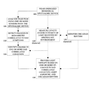
Referring now to Fig. 1, method steps that may be used to monitor SAD related
symptoms are illustrated. At 101, one or more energized Biomedical Ophthalmic
Device(s)
can be worn by an individual. An energized Biomedical Ophthalmic Device can
reside in or on
the eye. Some Biomedical Ophthalmic Devices are preferably placed on the
anterior ocular
surface and may be used to provide one or more of: optical correction,
therapy, and may be
cosmetic. For example, it may be an energized ophthalmic lens or energized
ophthalmic device,
including but not limited to a contact lens, intraocular lens, overlay lens,
ocular insert, optical
insert, punctal plug, or other similar ophthalmic device through which vision
can be corrected
or modified, an eye condition can be enhanced or prevented, and/or through
which eye
physiology can be enhanced cosmetically.
In some aspects of the present invention, the Energized Biomedical Device may
be used
to monitor one or more SAD related symptoms. Monitoring of the symptoms may
take place
through the analysis of biomarkers in tear film through the use of sensors
comprised by the
7
CA 02846344 2014-03-14
Energized Biomedical Ophthalmic Device. Additionally or alternatively, in some
embodiments, it may also include measuring length and/or intensity of light
received by the
ophthalmic environment of the user 120, and/or monitoring the circadian rhythm
125 of the
user.
When analysis of biomarkers in tear film through the use of sensors takes
place 105, the
biomarkers' changes can be correlated to known SAD symptoms 110. Examples of
correlated
symptoms of SAD may include, but are not limited to, excessive sleeping, lack
of energy,
craving carbohydrates, difficulty concentrating, and withdrawal from social
activities. These
symptoms can often result in feelings of depression, hopelessness, pessimism,
and lack of
pleasure which can be correlated to changes in specific tear film biomarkers.
Changes in
biomarker of tear film can include, but are not limited to, variations in
serotonin levels and
genetic polymosphisms, melatonin concentration changes signaling a phase
change in circadian
rhythm, and increased levels of Interleukin-6.
Known levels and thresholds of biomarkers concentrations in tear film related
to SAD
may pre-programed into a Component of the device used for the monitoring and,
additionally
or alternatively, the device may continue to learn from inputs and collected
data particular to
the user. In addition, because concentrations may vary with factors, such as,
age and
environmental conditions, normal values measured in blood, serum or saliva
analytes of the
individual, or of a comparable population, may be correlated to tear film
values of the user.
The changes or determined values then may be monitored 115 and light therapy
based on the
changes may be provided to the user 130 when it is needed.
Referring now to Fig. 2, an exemplary energized biomedical ophthalmic device
with a
biomarker sensor Component 203 that may be used in some energized ophthalmic
lens 200
embodiments of the present invention is depicted. In addition to the biomarker
sensor
Component 203, the exemplary energized ophthalmic lens 200 comprises an Energy
Source
202 and a Light Source 202A. The Energy Source 202 can be in electrical
communication with
a Light Source 202A and the Component 203. The Light Source 202A can include
light-
emitting diodes (LEDs) or other lights which emit blue light at wavelengths of
450 to 500
nanometers, most preferably at 470 to 480 nanometers, and at 2,000 to 3,000
lux. Alternatively,
LEDs or other lights may emit green light at wavelengths of 475 to 525
nanometers, most
8
CA 02846344 2014-03-14
preferably at 490 to 510 nanometers, and at 300 to 400 lux. In another
embodiment, a single
Light Source may be piped to one or more locations in an ophthalmic lens 201
to provide the
illumination required for SAD light therapy.
The Component 203 can include any light sensor and/or electrochemical sensor
device
with analytical sensitivity to detect changes in biomarkers. The component may
include a
microchip with electrophoresis and selective chemoluminescence capabilities
including, for
example, capability to detect changes in fluorescence, absorbance, light
scatter or plasmon
resonance of tear film, light exposure, and circadian rhythm. In some
embodiments,
Component 203 can react to an electrical change with a change in state and be,
for example: a
microchip such as a semiconductor type chip; a passive electrical device; an
optical device such
as a crystal lens; a processor with a micro-electromechanical machine (MEMS),
or a nano-
electromechanical machine (NEMS).
Moreover, the Component 203 can include or be in logical connection with an
electrical
storage device such as a capacitor; ultracapacitor; supercapacitor; or other
storage component.
An Energy Source 202 can include, for example: a lithium ion battery located
in the periphery
of an ophthalmic lens outside of the optic zone and be chargeable via one or
more of radio
frequency; photo voltaics, and magnetic inductance into an Energy Source 202.
As illustrated, in some embodiments, the Energy Source portion 202, the Light
Source
202A, and the Component 203 can preferably be located outside of an Optic Zone
204, wherein
the Optic Zone 204 includes that portion of the ophthalmic lens 200 providing
a line of sight
for an ophthalmic lens 200 wearer. Other embodiments may include an Energy
Source 202 in
the optic zone portion of an ophthalmic lens. For example, such embodiments
can include an
Energy Source 202 of conductive particles too small to be viewable without aid
to the human
eye.
In some embodiments, a preferred ophthalmic lens type can include a lens 201
that
includes a silicone containing component. A "silicone-containing component" is
one that
contains at least one [¨Si-0¨] unit in a monomer, macromer or prepolymer.
Preferably, the
total Si and attached 0 are present in the silicone-containing component in an
amount greater
than about 20 weight percent, and more preferably greater than 30 weight
percent of the total
molecular weight of the silicone-containing component. Useful silicone-
containing components
9
CA 02846344 2014-03-14
preferably comprise polymerizable functional groups such as acrylate,
methacrylate,
acrylamide, methacrylamide, vinyl, N-vinyl lactam, N-vinylamide, and styryl
functional
groups.
Referring now to Fig. 3, an exemplary processor that may be used in some
Energized
Biomedical Ophthalmic Device embodiments of the present invention is
illustrated at 300. In
this illustration, the Energy Source 310 may include a thin film, rechargeable
lithium ion
battery. The battery may have contact points 370 to allow for interconnection.
Wires may be
wire bond wires to the contact points 370 and connect the battery to a
photoelectric cell 360
which may be used to reenergize the battery Energy Source 310. Additional
wires may connect
the Energy Source to a flexible circuit interconnect via wire bonded contacts
on a second set of
contact points 350. These contact points 350 may be a portion of a flexible
interconnect
substrate 355 which may also include a Light Source 330.
The interconnect substrate may be formed into a shape approximating a typical
lens
conical form or other form depending on the Biomedical Ophthalmic Device.
However to add
additional flexibility needed in some embodiments, the interconnect substrate
355 may include
additional shape features such as radial cuts 345 along its length. Radial
cuts may be used to
form individual flap shaped structured of the interconnect substrate 355 and
may be connected
various electronic Components like ICs, discrete Components, passive
Components and such
devices which are shown as item 390. Components can be interconnected by wires
or other
connection means 340 to the conduction paths within the interconnect substrate
355. By way
of non-limiting example, the various components may be connected to the
flexible interconnect
substrate 355 by the various means of making interconnections to the battery.
The combination
of the various electrical Components may define a control signal for control
of the biomarker
monitoring, light source, and in some embodiments, for an electro-optical
device shown as item
390. This control signal may be conducted respectively along interconnect 320.
This type of
exemplary energized ophthalmic lens with energized functions is provided only
for the purpose
of example. In no way should this description be construed to limit the scope
of the inventive
art as it will be apparent to one skilled in the art that many different
embodiments of function,
design, interconnection scheme, energization scheme and overall utilization of
the concepts of
this invention may exist from this disclosure. For example, in some
embodiments there may be
= CA 02846344 2014-03-14
manners of affecting the ophthalmic lens' appearance. Aesthetics of the thin
film microbattery
surface may be altered in various manners which demonstrate a particular
appearance when
embedded in the electroactive contact lens or shaped hydrogel article. The
thin film
microbattery may be produced with aesthetically pleasing patterned and/or
colored packaging
materials which could serve to either give a muted appearance of the thin film
microbattery or
alternatively provide iris-like colored patterns, solid and/or mixed color
patterns, reflective
designs, iridescent designs, metallic designs, or potentially any other
artistic design or pattern.
In other embodiments, the thin film battery may be partially obscured by other
Components
within the lens, for example, a photovoltaic chip mounted to the battery
anterior surface, or
alternatively, by placement of the battery behind all or a portion of a
flexible circuit. In further
embodiments, the thin film battery may be strategically located such that
either the upper or
lower eyelid partially or wholly obscures the visibility of the battery.
In preferred embodiments, the Energy Source and Light Source may not obstruct
the
transmission of light through the ophthalmic lens. Consequently, designs can
be so that the
Optical Zone, central 5-8 mm, of the energized lens may not be significantly
obstructed by any
opaque portions of the Energy Source and Light Source. There may be many
different
embodiments relating to the design of various Energy Sources and Light Sources
to interact
favorably with the optically relevant portions of an energized ophthalmic
lens.
According to some aspects of the present invention, the Energy Source and
Light
Source should be placed at a certain distance from the outer edge of the
contact lens to enable
advantageous design of the contact lens edge profile in order to provide good
comfort while
minimizing occurrence of adverse events. Examples of such adverse events to be
avoided may
include superior epithelial arcuate lesions or giant papillary conjunctivitis.
In some embodiments, a cathode, electrolyte and anode features of embedded
electrochemical cells can be included and be formed, for example, by printing
appropriate inks
in shapes to define such cathode, electrolyte and anode regions. It may be
apparent that
batteries thus formed could include both single use cells, based for example
on manganese
oxide and zinc chemistries, and rechargeable thin batteries based on lithium
chemistry thin film
battery chemistry. It can also be apparent to one skilled in the art that a
variety of different
embodiments of the various features and methods of forming Energized
Biomedical
11
CA 02846344 2014-03-14
Ophthalmic Devices may involve the use of printing techniques.
Referring now to Fig. 4, a cross section of an Energized Biomedical Ophthalmic
Device
400 with an exemplary media insert 401 including a microcontroller 404 that
may be used in
some lens embodiments of the present invention is depicted. An activator or
processor 405 can
be used to implement one or more executable programs included within memory
storage in the
Microcontroller 404. Programs can be operative to control a light source (not
shown) in logical
communication with the Microcontroller. One or more Light Source may be
included in the
media insert, outside the media insert in/on the biomedical ophthalmic device,
or in proximity
thereto; for example, in complementary spectacles (further described in Fig.
6). Additionally,
in some embodiments, a program executed via the Microcontroller 404 can cause
a change of
state in a Component 403. The memory storage can include a random access
memory
semiconductor; a read only memory semiconductor; a static memory; an erasable
programmable read only memory; or other component capable of storing digital
data and
providing the data on command.
An Energy Harvester, such as a photoreceptor 402 can be included for
recharging an
Energy Source 408, such as a lithium based battery or a capacitor. The
microcontroller 404 can
be used to manage a Re-energizing process. For example, the processor 405 can
receive data
indicative of an amount of charge present in an energy source 408 and open a
circuit allowing
current to flow from an Energy Harvester 402, for example, a photoreceptor to
the Energy
Source 408 (other examples can include a magnetic or inductive device). In
another aspect, the
processor can also be programmed to monitor when the Energy Harvester 402 can
be capable
of providing sufficient current to charge an Energy Source 408 and provide an
electrical
pathway via circuitry suitable for such charging. Electrical circuitry for
charging can include,
for example, transistors acting as switches and diodes for ensuring a proper
direction of current
flow.
Referring now to Fig. 5, a cross section view of an exemplary energized
biomedical
ophthalmic device 500 containing light sources502 according to some lens
embodiments of the
present invention is depicted. In the present example, the exemplary energized
ophthalmic lens
501 is a contact lens and is depicted directing light 503 onto the cornea 504
of an eye 505. In
some embodiments, a cross-section view 500 may be a top-down view, wherein one
or more
12
CA 02846344 2014-03-14
embedded Light Sources 502 are placed near the sides of a contact lens 501. In
other
embodiments, a cross-section view 500 may be a side view, such that one or
more embedded
Light Sources 502 are placed near the top and bottom of a contact lens 501. A
number of Light
Sources 502 and an arrangement of Light Sources 502 around a perimeter of a
contact lens 501
may vary. A Light Source 502 directs illumination toward a wearer's eye such
that illumination
may not be obvious to an observer. A contact lens 501 may also include a
coating which shields
light therapy luminescence from being readily noticed by an observer to not
diminish a user's
Light Therapy.
Embedded Light Sources 502 can include light-emitting diodes (LEDs) or other
Light
Sources 502 capable of emitting light 1003 for Light Therapy. Light Sources
502 may include
light-emitting diodes (LEDs) or other lights which emit blue light at
wavelengths of 450 to 500
nanometers, most preferably at 470 to 480 nanometers, and at 2,000 to 3,000
lux.
Alternatively, LEDs or other lights may emit green light at wavelengths of 475
to 525
nanometers, most preferably at 490 to 510 nanometers, and at 300 to 400 lux.
Another
embodiment includes a single Light Source from which light may be piped to one
or more
locations within an ophthalmic lens 501 to provide illumination.
The exemplary ophthalmic lens 501 includes supporting electronics, not
illustrated in
this figure, with Components such as light sensors, biomarker sensors, Energy
Source,
capacitors, memory, processor, and communication device. Light sensors are
used to detect
ambient white light, blue light or green light. An Energy Source and
capacitors can supply
energy to other Components of an Energized Biomedical Ophthalmic Device.
Memory may be
used, by way of non-limiting example, to store pre-programmed Light Therapy
Schedules, to
store data collected from one or more sensors, to store user's preferences, to
store actual Light
Therapy dates, times, durations and intensities, and to store data related to
a Light Source and
light sensor operation in order to detect device failures. Moreover, a
processor may be used, for
example, to run programmed Light Therapy Schedules stored in memory, to
analyze light
sensor data and determine a unique personalized Light Therapy Schedule based
on the wearer's
exposure to ambient light, to evaluate manual changes to a programmed Light
Therapy
schedule and provide compensating adjustments, i.e., Intelligent Light
Therapy, and to analyze
light source and light sensor data to detect device failures.
13
CA 02846344 2014-03-14
A communication device may be used to electronically control one or more of:
the
transfer of digital data to and from an energized biomedical ophthalmic device
and external
devices, and the transfer of digital data between components within the
energized biomedical
ophthalmic device. The communication device may be used to wirelessly
communicate with
one or more external devices including, by way of non-limiting examples, a
fob, a personal
digital assistant (PDA), or a smartphone application used to control the
Energized Biomedical
Ophthalmic Device. Within Energized Biomedical Ophthalmic Devices,
communication
between Components may be via physical connection, such as via a direct
conductive path, or
may be wireless. Communication between internal components may include, for
example,
control of a Light Source from a processor and data transfer between sensors
and memory.
Supporting electronics are in logical and electrical communication with Light
Sources
502 contained within the energized biomedical ophthalmic device including, for
example, a
contact lens 501. Communication may be via a direct conductive path between
supporting
electronics and Light Sources 502 or via wireless communication. Wireless
modes of
communication may include, for example, inductance accomplished via an antenna
located
proximate to a Light Source 502 in the contact lens 501 and a power source
transmitting power
from another area within the contact lens 501 to the antenna.
In some embodiments, supporting electronics may be included in a fob, jewelry,
hat,
clothing, or other items worn by a user such that sensors, such as light
sensors, detect ambient
light experienced by the user and supporting electronics are near a contact
lens for purposes of
wireless communication. Wireless modes of communication can include, for
example,
inductance. Inductance may be accomplished via an antenna located in/on the
energized
biomedical ophthalmic device and a power source transmitting power from
supporting
electronics in jewelry, clothing, or other item proximate to the antenna.
In some embodiments, a user may adjust timing, duration and intensity of light
therapy
using an external device, including but not limited to one or more of: a fob,
a personal digital
assistant, computer, tablet, and a Smartphone application. Some embodiments
provide for a
basic operational state, wherein Light Therapy is controlled manually by a
user starting and
stopping therapy at appropriate times.
According to the present embodiment, a programmed Light Therapy Schedule may,
for
14
CA 02846344 2014-03-14
example, automatically adjust light therapy timing, duration and intensity
based on variables
such as, dates, geographic region, user's preferences, and biomarkers sensor
collected data
correlated to SAD symptoms and the severity of a user's SAD symptoms. A
Programmed Light
Therapy Schedule may be set by an eye care professional, a medical doctor, or
a user. In some
embodiments, the light therapy schedule may learn from past responses and
adjust to provide
Intelligent Light Therapy. For example, an response during programmed light
therapy can
include, a user adjusting light intensity based on an activity, such as, for
example, decreasing
light intensity when reading, working on a computer, or driving. Conversely,
it may be
desirable to increase light intensity during work breaks, lunch break, or
other less visually
active times. Accordingly, in some embodiments Intelligent Light Therapy can
be delivered
when a processor evaluates manual changes and detected user changes of a
programmed Light
Therapy Schedule and provides compensating adjustments in duration,
frequencies and/or
intensity of treatment. Intelligent Light Therapy can also be achieved when
data from light
sensors is analyzed by a processor and a programmed Light Therapy Schedule is
dynamically
adjusted based on a wearer's exposure to ambient light.
In another embodiment of the present invention, a user may manually adjust
light
therapy based on the results of tear fluid measured data, and/or blood, saliva
testing including
but not limited to testing for concentration of one or more of: melatonin,
serotonin and
interleukin-6 levels. Concentration of biomarkers can increase or decrease
based on light
exposure or a SAD symptom. For example, melatonin levels are inhibited by
light and increase
with darkness. Higher levels of melatonin promote sleepiness and lethargy,
symptoms of
seasonal affective disorder.
In still other embodiments, as part of an user's preferences, a user may
manually adjust
light therapy to intentionally alter their sleep cycle. The use of light
therapy for sleep cycle
alteration may be valuable for persons working night shifts, for persons
travelling to
significantly different time zones, for military personnel preparing for night
operations, and
other uses. Similarly, Light Therapy initiated by the user upon awakening may
be used to treat
circadian rhythm disorders such as delayed sleep phase syndrome (DSPS) and non-
24-hour
sleep-wake syndrome.
Referring now to Fig. 6, the back view of exemplary eyeglasses 600 with light
sources
CA 02846344 2014-03-14
602 embedded in the lenses 603 and with supporting electronics is depicted. In
other
embodiments, Light Sources 602 may also be mounted on the surface of lenses
603. Light
Sources 602 may include light-emitting diodes (LEDs) or other lights which
emit blue light at
wavelengths of 450 to 500 nanometers, most preferably at 470 to 480
nanometers, and at 2,000
to 3,000 lux. Alternatively, LEDs or other lights may emit green light at
wavelengths of 475 to
525 nanometers, most preferably at 490 to 510 nanometers, and at 300 to 400
lux. In yet
another embodiment, a single light source may be piped to one or more
locations within an
eyeglass lens 603 or eyeglass frame 601 to provide illumination. Light pipes
may include, for
example, fiber optic pathways.
An example of illuminated light sources is illustrated at 604. A light source
602
provides illumination toward a wearer's eyes such that an illumination is not
obvious to an
observer.
Light Sources 602 can be connected to one another via conductive paths 605.
Conductive paths 605 may be wires embedded within a lens 603 or may be a
conductive
material including, for example, gold, copper, silver or other metal or
conductive fiber applied
to a surface of a lens 603 via pad printing, sputter coating, vapor deposition
or another suitable
method. Conductive paths 605 can be in electrical and logical communication
with supporting
electronics contained within one or both temple pieces 609. In some
embodiments, supporting
electronics are miniaturized such that they may be contained in other areas of
eyeglasses such
as in areas near a hinge 607, within a frame above a lens 608, within a bridge
610, within an
earpiece 611, or other area.
One or more light sensors 606 can be used to detect ambient white light, blue
light or
green light. Light sensors 606 may be located within an eyeglass frame 601
near a hinge 607,
within a frame above a lens 608, within a temple piece 609, within a bridge
610, or other
appropriate area where a sensor 606 will not be obstructed, for example, by
hair. A light sensor
606 is in electrical and logical communication with supporting electronics
contained within one
or both temple pieces 609 or other area of eyeglasses.
In some embodiments, a user control element 612, such as a switch or button,
can be
provided to allow a user to adjust timing, duration and intensity of light
therapy. One or more
user control elements 612 may be present in temple pieces 609 or other areas
of eyeglasses
16
CA 02846344 2014-03-14
including, for example, in areas near a hinge 607, within a frame above a lens
608, within a
bridge 610, within an earpiece 611, or other area.
Referring now to Fig. 7, a cross-section view 700 of exemplary eye glasses 701
with
embedded light sources 702 directing light into a complementary energized
biomedical
ophthalmic device 705 according to some contact lens embodiments of the
present invention is
depicted. Cross-section view 700 includes an eyeglass lens 701 with embedded
light sources
702 directing light 703 into light scattering areas 704 of a complimentary
contact lens 705. A
light scattering area 704 can result in light 706 being dispersed across a
cornea 707 of an eye
708. A light scattering area 704 may include diffractive properties,
refractive properties,
reflective properties or any combination of diffractive, refractive and
reflective properties.
In some embodiments, a cross-section view 700 may be a top-down view, wherein
one
or more embedded light sources 702 are placed near the sides of an eyeglass
lens 701. In other
embodiments, a cross-section view 700 may be a side view, such that one or
more embedded
light sources 702 are placed near the top and bottom of an eyeglass lens 701.
In still other
embodiments, embedded light sources 702 may be embedded in or mounted on an
eyeglass
frame rather than an eyeglass lens 701. Embedded light sources 702 can
include, for
example, the light-emitting diodes (LEDs) or other light sources 702
previously described
herein. Supporting electronics (not shown) can be contained in an eyeglass
frame and in the
energized biomedical ophthalmic device and be in communication with each
other. For
example, for the biomarker sensor of the energized biomedical ophthalmic
device to send
collected biomarker concentration data to a communication Component of the
eyeglasses.
Supporting electronics can be Components located in one or the complementary
devices of
both, and may include components including, for example, light sensors,
batteries, capacitors,
memory, processors, and a USB connector. Moreover, supporting electronics are
in logical and
electrical communication with light sources 702 and biomarker sensors (not
depicted).
Electrical communication may be provided, for example, via a conductive
contact between a
source located in a temple of a pair of eyeglasses, via a conductive wire, a
conductive ribbon
wire, or via wireless modes, such as inductance. Inductance may be
accomplished, for
example, between an antenna located in the eyeglasses and complementary lens.
In some embodiments, light scattering areas 704 of a complimentary contact
lens 705
17
CA 02846344 2014-03-14
form a ring within a perimeter area of a complimentary contact lens 705 such
that directed light
703 need not strike a limited target area. The orientation of a complimentary
contact lens 705
on an eye 708 relative to light sources 702 within an eyeglass lens 701 is
therefore
inconsequential when light 703 is directed toward a light scattering area 704
continuously
present around a perimeter area of a complimentary contact lens 705.
In some preferred embodiments, a complimentary contact lens 705 may include an
internal barrier between a light scattering area 704 and an Optical Zone in a
central portion of a
lens. An internal barrier prevents light 703 intended for light therapy from
being dispersed into
an Optical Zone of a complimentary contact lens 705. This way, light 703
intended for Light
Therapy is only dispersed around a perimeter of a cornea 707, minimizing its
effect on normal
vision.
In still other embodiments, an entire complimentary contact lens 705 includes
light
scattering properties such as diffraction, refraction and reflection. Light
scattering properties
are designed such that they disperse only light 703 of wavelengths emitted by
embedded light
sources 702. This embodiment supports maximum dispersion of light 703
wavelengths
intended for Light Therapy within an eye 708 while not causing dispersion of
light wavelengths
that would affect normal vision.
Referring now to Fig. 8, a cross-section view 800 of exemplary eye glasses 801
with
supporting electronics 802 in wireless communication with an energized
biomedical
ophthalmic device 805 containing light sources 804 according to some contact
lens
embodiments of the present invention is depicted. Cross-section view 800
includes an eyeglass
frame 801 containing supporting electronics 802. Supporting electronics 802
may include
Components such as light sensors, batteries, capacitors, memory, processors,
and a USB
connector. Supporting electronics 802 are in wireless communication 803 with a
complimentary contact lens 805 containing embedded Light Sources 804 directing
light 806
onto a cornea 807 of an eye 808. Supporting electronics 802 may be placed in
various
locations embedded in or mounted on an eyeglass frame 801.
In other embodiments, supporting electronics 802 may be included in jewelry,
hats,
clothing, or other items worn by a user such that light sensors detect ambient
light experienced
by the user and supporting electronics 802 are near a complimentary contact
lens 805 for
18
CA 02846344 2014-03-14
purposes of wireless communication. Wireless modes of communication may
include, for
example, inductance. Inductance may be accomplished via an antenna located in
a
complimentary contact lens 805 and a power source transmitting power from an
eyeglass frame
801, jewelry, clothing, or other item proximate to the antenna.
In some embodiments of the present invention, a cross-section view 800 may be
a top-
down view, wherein supporting electronics 802 are placed near the sides of an
eyeglass frame
801. In other embodiments, a cross-section view 800 may be a side view, such
that supporting
electronics 802 are placed near the top and bottom of a side of an eyeglass
frame 801. A
number of embedded light sources 804 and an arrangement of embedded light
sources 804
around a perimeter of a complimentary contact lens 805 may vary. Embedded
light sources
804 include previously described light-emitting diodes (LEDs) or other light
sources 804
emitting light 806 for light therapy.
In some embodiments, Light Sources 804 may direct light 806 into an interior
portion
of a complimentary contact lens 805 in which the Light Sources 804 can be
embedded or
positioned onto a surface of the contact lens. Light 806 may be directed into
a light scattering
area, not depicted, including diffractive properties, refractive properties,
reflective properties,
or any combination of diffractive, refractive and reflective properties. A
light scattering area
may form a ring within a perimeter area of a complimentary contact lens 805.
Light 806
striking a light scattering area causes a generally broad dispersion of light
806 onto a cornea
807 of an eye 808.
In some preferred embodiments, a complimentary contact lens 805 may also
include an
internal barrier between a light scattering area around a perimeter of a lens
and an optical zone
in a central portion of a lens, and light scattering properties as previously
described.
Antennas or antenna systems may serve as a means for receiving signals, as a
means for
transmitting signals, as an inductive coupling means, or any combination
thereof. The function
of an antenna determines its design as well as its supporting circuitry. For
example, an antenna
may be coupled to a receiver unit, a transmitter circuit, an inductive
coupling circuit or to any
combination thereof. Basically, an antenna is an electrical device that
converts electromagnetic
waveforms, or electrical signals into different electrical signals. The
discussion of Fig.9A and
Fig.9B focuses on exemplary assemblies that comprise antenna systems and
Fig.9C represents
19
CA 02846344 2014-03-14
a block diagram of an antenna and receiver circuit in accordance to the
exemplary assemblies
of Figs. 9A and 9B.
Referring now to Fig. 9A, an exemplary antenna system according to some
embodiments of the present invention is depicted. Circuit board 904A that may
be utilized with
one or more Component of the energized biomedical ophthalmic device, such as
the biomarker
sensor, Light Source and/or an optical lens assembly of an ophthalmic lens.
Circuit board
904A comprises both top side conductive interconnect traces 912A1 and bottom
side
conductive interconnected traces 912A2 (shown in phantom), through-holes or
vias 918A for
making electrical connections between the top and bottom sides, mounting pads
914A, a center
opening 916A, and one or more spiral antenna structures 920A. However, in some
embodiments, a single loop antenna may be appropriate. Each of the one or more
spiral
antenna structures 920A can comprise one or more turns of wire, conductive
traces or the like
formed in either or both of the top side or the bottom side of the circuit
board 904A. If multiple
antennas are utilized on opposite sides, the through-hole or vias 908A may be
utilized to make
connections therebetween. It will be appreciated that the circuit board 904A
may comprise
additional metal layers and that any combination of layers may be used to
construct the spiral
antenna structures 920A. The antenna structures alternately may be embedded on
an inner
conducting layer, with other conducting layers above and/or below the antenna
structures
920A.
Referring now to Fig. 9B, another exemplary antenna system according to some
embodiments of the present invention is depicted. Like the previous example,
circuit board
904A that may be utilized with one or more Component of the Energized
Biomedical
Ophthalmic Device, such as the biomarker sensor, Light Source and/or an
optical lens assembly
of an ophthalmic lens. Circuit board 904B comprises both top side conductive
interconnect
traces 912B1 and bottom side conductive interconnected traces 912B2 (shown in
phantom),
through-holes or vias 918B for making electrical connections between the top
and bottom sides,
mounting pads 914B, a center opening 916B, and a multi-turn loop antenna 920B.
However, in
some embodiments a single loop antenna may be appropriate. The multi-loop
antenna 920B
comprises two or more turns of wire, conductive traces or the like formed in
either or both of
the top side or the bottom side of the circuit board 904B. If multiple
antennas are utilized on
CA 02846344 2014-03-14
opposite sides, the through-hole or vias 908B may be utilized to make
connections
therebetween. It will be appreciated that the circuit board 904B may comprise
additional metal
layers and that any combination of layers may be used to construct the multi-
turn loop antenna
920B.
Before the description of an exemplary block diagram of an antenna and
receiver
circuit, it is important to note that the circuits set forth and described
subsequently may be
implemented in a number of ways. In one exemplary embodiment, the circuits may
be
implements using discrete analog components. In another exemplary embodiment,
the circuits
may be implemented in integrated circuits or a combination of integrated
circuits and discrete
components. In yet another alternate exemplary embodiment, the circuits or
particular
functions may be implemented via software running on a microprocessor or
microcontroller.
Referring now to Fig. 9C, a block diagram representation of an antenna and
receiver
circuit in accordance to some embodiments of the present invention is
illustrated. The radio
receiver electronic circuit 900C can comprise an antenna match circuit 904C, a
receiver circuit
906C, a controller 908C, an actuator 910C, a battery 912C and a power
management circuit
914C. In this exemplary configuration, the antenna 904C can be adapted to
receive an
electromagnetic signal 901C and to provide a received electrical signal to the
antenna match
circuit 904C. The antenna match circuit 904C may comprise any suitable
circuitry necessary
for balancing the impedance between the source and the load to maximize power
transfer
and/or minimize reflection from the load. Essentially, antenna impedance is
the ratio of voltage
to current at any point on the antenna and for efficient operation, the
antenna impendence
should be matched to the load, and thus a match circuit is utilized.
Accordingly, the match circuit 904C can be adopted to provide an impedance
match
between the antenna 902C and the receiver circuit 906C for an optimum power
match, noise
match or other match condition as is known in the radio and circuit design
arts. The receiver
circuit 906C can comprise any suitable circuitry necessary to process the
modulated signal
received by the antenna 902C and provide a demodulated signal to the
controller 908C. For
purposes of clarity, modulation involves varying one or more properties of a
signal or
electromagnetic waveform. For example, a waveform may be amplitude (AM),
frequency
modulated (FM) or phase modulated (PM). Other forms of analog as well as
digital modulation
21
CA 02846344 2014-03-14
can also be implemented in some embodiments. Demodulation, on the other
hand, can
include extracting the original information bearing signal from the modulated
carrier wave. It
is this demodulated information signal that can provide instructions to the
controller 908C. The
controller 908C in turn can provide a control signal to the actuator 910C
based upon the
demodulated signal in order to control a state or operation of the actuator
910C. The control
signal may be further based on any internal state of the controller, for
example, to implement
control laws, and/or any other circuits coupled to the controller, for
example, to implement a
feedback control system or to modify the actuator operation based on other
information such as
information based upon sensor data.
The battery 912C provides a source of electrical energy for Components in the
electronic circuit 900C requiring energy. The power management circuit 914C
can be adapted
to receive a current from the battery 912C and condition it or regulate it to
provide a workable
output voltage suitable for use by the other active circuits in the electronic
circuit 900C. The
controller 908C may also be utilized to control the receiver circuit 906 or
other circuits in the
receiver 900C. The antenna 902C may comprise, for example, one or more of the
configurations previously described. Other embodiments may include single turn
loop antenna,
a multi-turn loop antenna, a spiral antenna, a coil antenna subassembly, a
stacked die
configuration or arrangement or a suitable combination thereof.
As is known in the relevant art, a preferred method for the transfer of power
between an
antenna and a receiving and/or transmitting circuit may require matching the
impedance
presented and/or transmitting circuit requires matching the impedance
presented to the antenna
and the impedance presented to the circuit. Essentially, suitable power
transfer can occur when
the reactive components of the antenna and circuit impedance are cancelled and
the resistive
components of the impedances are equal. A matching circuit may be introduced
to couple the
antenna to the circuit that meets the optimum power transfer criterion at
each, thereby allowing
for optimum power transfer to occur between the antenna and circuit.
Alternatively, a different
criterion may be selected to optimize a different parameter such as maximum
current or voltage
at the circuit. Matching circuits are well known in the art and may be
implemented with
discrete circuit component such as capacitors, inductors and resistors, or
with conductive
structures such as traces in a circuit board, that provide a desired
impendence characteristic.
22
CA 02846344 2014-03-14
=
Impedances of small RF loop antennas are typically between 20 and 50
nanohenries,
and matching component valves can be in the range of 0.5 to 10 picofarads for
capacitors and 3
to 50 nanohenries for inductors. Impedances of inductive charging coils are
typically between
100 nanohenries and 5 nanohenries and associated capacitors for resonating the
circuits are
between 20 and 100 picoforads.
The actuator 910C may comprise any number of suitable devices. For example,
the
actuator 910C may comprise any type of electromechanical device, for example,
a pump or
transducer. The actuator may also comprise an electrical device, a chemical
release device or
any combination thereof. The actuator 910C may be replaced or include a
controlled device,
for example, a Light Source used to deliver Light Therapy, or diode array or
any other suitable
display, or user interface.
The battery 912C may comprise any suitable device for the storage of
electrical energy
as previously described. In alternate exemplary embodiments, no battery may be
required as
explained above with respect to RF energy harvesting or near field inductive
coupling.
Alternatively, mechanical vibration and similar means may be utilized to
generate or harvest
power.
The power management circuit 914C may comprise additional circuitry for a wide
variety of functions in addition to regulating the output of the battery 912C.
For example, the
power management circuit 914C may comprise circuitry for monitoring various
battery
parameters such as charge, preventing overdischarge of the battery, and/or
supervising the
startup and shut down of the electronic circuit 900C.
Referring now to Fig. 10, the block diagram of a controller 1000 that may be
used to
implement some embodiments of the present invention is depicted. The
controller 1000
includes a processor 1010, which may include one or more processor components
coupled to a
communication device 1020. In some embodiments, a controller 1000 can be used
to transmit
energy to an Energy Source, sensor, and/or Light Source placed in an energized
biomedical
ophthalmic device.
The controller can include one or more processors, coupled to a communication
device
configured to communicate energy via a communication channel. The
communication device
may be used to electronically control the transfer of digital data to and from
an ophthalmic
23
CA 02846344 2014-03-14
device and/or control of a Light Source or other component incorporated into
the ophthalmic
lens.
The communication device 1020 may also be used to communicate, for example,
with
one or more controller apparatus or manufacturing equipment components.
The processor 1010 is also in communication with a storage device 1030. The
storage device
1030 may comprise any appropriate information storage device, including
combinations of
magnetic storage devices, optical storage devices, and/or semiconductor memory
devices such
as Random Access Memory (RAM) devices and Read Only Memory (ROM) devices.
The storage device 1030 can store a program 1040 for controlling the processor
1010.
The processor 1010 performs instructions of the program 1040, and thereby
operates in
accordance with the present invention. The storage device 1030 can also store
data, such as,
ophthalmic data, geographic data, sensor data, and related data in one or more
databases. The
database may include customized Energy Source and Light Source designs, and
specific control
sequences for controlling energy to and from an Energy Source, sensor, and a
Light Source.
Conclusion
A number of embodiments of the present invention have been described. While
this
specification contains many specific implementation details, there should not
be construed as
limitations on the scope of any inventions or of what may be claimed, but
rather as descriptions
of features specific to particular apparatus embodiments of the present
invention.
Certain apparatus and Lens features that are described in this specification
in the
context of separate embodiments can also be implemented in combination in a
single
embodiment. Conversely, various features that are described in the context of
a single
embodiment can also be implemented in combination in multiple embodiments
separately or in
any suitable subcombination. Moreover, although features may be described
above as acting in
certain combinations and even initially claimed as such, one or more features
from a claimed
combination can in some cases be excised from the combination, and the claimed
combination
may be directed to a subcombination or variation of a subcombination.
Similarly, while method steps are depicted in the drawings in a particular
order, this
should not be understood as requiring that such method steps be performed in
the particular
24
CA 02846344 2014-03-14
order shown or in sequential order, or that all illustrated operations be
performed, to achieve
desirable results. In certain circumstances, multitasking and parallel may be
advantageous.
Moreover, the separation of various apparatus components in the embodiments
described above
should not be understood as requiring such separation in all embodiments, and
it should be
understood that the described apparatus components and method steps can
generally be
integrated together in a single apparatus or method or used in multiple
apparatus or methods.
Thus, particular embodiments of the subject matter have been described. Other
embodiments are within the scope of the following claims. In some cases, the
method steps
recited in the claims can be performed in a different order and still achieve
desirable results. In
addition, the processes depicted in the accompanying figures do not
necessarily require the
particular order show, or sequential order, to achieve desirable results.
Nevertheless, it will be
understood that various modifications may be made without departing from the
spirit and scope
of the claimed invention.