Note: Descriptions are shown in the official language in which they were submitted.
CA 02846865 2014-02-26
WO 2013/033260
PCT/US2012/052925
USE OF HDL-RELATED MOLECULES TO TREAT AND PREVENT
PROINFLAMMATORY CONDITIONS
This application claims the benefit of United States provisional patent
applications 61/646,772, filed
May 14, 2012, 61/624,333, filed April 15, 2012, and 61/528,447, filed August
29, 2011, the entire
contents of each of which are incorporated herein by reference.
This application is related to United States provisional patent application
number 61/389,618, filed
October 4, 2010, and to United States patent application number 12/860,293,
filed August 20, 2010,
which is a continuation-in-part of application number 12/630,458, filed
December 3, 2009, which is
a divisional of application number 11/571,986, filed July 18, 2007, now Patent
No. 7,670,792, which
is a national stage filing under 35 U.S.C. 5371 of PCT/U52005/024985, filed
July 14, 2005, which
claims the benefit of United States provisional patent application numbers
60/674,489, filed April
25, 2005, and 60/588,007, filed July 14, 2004, the entire contents of each of
which are incorporated
herein by reference.
Throughout this application various publications are referenced. The
disclosures of these
publications in their entireties are hereby incorporated by reference into
this application in order to
describe more fully the state of the art to which this invention pertains.
TECHNICAL FIELD OF THE INVENTION
The present invention relates generally to prevention and treatment of
proinflammatory conditions
and cancer through the use of HDL-related molecules. The invention is more
specifically related to
apolipoprotein A-I (ApoA-I), HDL, and HDL mimetics, and their use in
preventing and treating
proinflammatory conditions, including skin and systemic proinflammatory
conditions, particularly
epithelial cancers as well as Alzheimer's disease, inflammatory skin diseases,
inflammatory bowel
disease, and inflammatory diseases associated with aging. Molecules, including
full-length ApoA-I
protein, HDL, antibodies and antisense/interference nucleotides that modulate
and/or mimic the
expression and/or function of these targets can be used in oral supplements,
vaccines and
pharmaceutical compositions for the treatment of various conditions, alone or
in combination with
other anti-oxidants.
1
CA 02846865 2014-02-26
WO 2013/033260
PCT/US2012/052925
BACKGROUND OF THE INVENTION
Proinflammation is a widespread phenomenon that has strong association with
stress and is
connected with various diseases. Proinflammatory activities in general are
initiated to overcome
infection or invasion of potentially deleterious biological agents (bacteria,
viruses, parasites etc.).
While fighting invasion, proinflammation has beneficial and deteriorating
capacities and can exert
detrimental effects. The sequelae of an unbalanced systemic inflammatory
reaction include
derangement of microcirculation, shock, transudation into organs and defects
of coagulation. An
unbalanced systemic compensatory anti-inflammatory response often results in
anergy and
immunosuppression.
There remains a need for improved tools to prevent and treat proinflammatory
conditions, including
proinflammatory skin conditions and epithelial cancers.
SUMMARY OF THE INVENTION
The invention provides HDL-related molecules and methods of using same to
treat and prevent
proinflammatory conditions and cancer. HDL-related molecules include ApoA-I,
bovine HDL, and
HDL mimetics. As described in further detail below, ApoA-I, in its natural,
full-length form, can
prevent UV-induced cell death and oxidative stress. Also described in further
detail below is the
unexpected discovery that HDL mimetics, ApoA-I and bovine HDL (bHDL) can be
used to treat
and prevent various cancers.
In one embodiment, the invention provides a method of inhibiting tumor growth.
The method
comprises contacting tumor cells with an HDL-related molecule selected from
the group consisting
of HDL mimetic peptides (such as those shown in SEQ ID NO: 1, 3-9, 12, 14 or
26-28), bovine
HDL, and ApoA-I. Another embodiment provides a method of treating or
preventing cancer in a
subject. The method comprises administering to the subject an HDL-related
molecule selected
from the group consisting of HDL mimetic peptides (such as those shown in SEQ
ID NO: 1, 3-9,
12, 14 or 26-28), bovine HDL, and ApoA-I. In yet another embodiment of the
invention, a method
of reducing death and/or oxidative stress in epithelial cells exposed to
oxidative stress is provided.
The method comprises contacting the epithelial cells with an HDL-related
molecule selected from
the group consisting of HDL mimetic peptides (such as those shown in SEQ ID
NO: 1, 3-9, 12, 14
or 26-28), bovine HDL, and ApoA-I. In one embodiment, the contacting occurs
prior to exposure
to oxidative stress. In a typical embodiment, the contacting occurs at least
12-24 hours prior to the
exposure to oxidative stress. The oxidative stress may comprise, for example,
exposure to
ultraviolet radiation.
2
CA 02846865 2014-02-26
WO 2013/033260
PCT/US2012/052925
The HDL-related molecule can, optionally, be administered as an oral
supplement. Subjects to be
treated with methods of the invention can be, for example, mammalian subjects,
typically human
subjects.
For use in methods of the invention, the ApoA-I may be full-length protein,
which can be
administered as recombinant ApoA-I and/or in unmodified form. In one
embodiment, the ApoA-I
is natural, full-length, unmodified ApoA-I.
The method of any one of claims 1-6, wherein the HDL mimetic peptide is
selected from the group
consisting of SEQ ID NO: 1,3-9, 12, 14 and 26-28.
In one embodiment, the invention provides an HDL-related molecule for
treatment of cancer, for
inhibiting tumor growth, and/or for reducing death and/or oxidative stress in
epithelial cells. The
HDL-related molecule is selected from the group consisting of HDL mimetic
peptides (SEQ ID
NO: 1, 3-9, 12, 14 or 26-28), bovine HDL, and ApoA-I. In one embodiment, the
invention
provides novel HDL mimetic peptides, including those having the amino acid
sequences shown in
SEQ ID NO: 1, 3-9, 12, 14 or 26-28. In a typical embodiment, the peptide
consists of the amino
acid sequence shown in SEQ ID NO: 1 or any of those shown in SEQ ID NO: 3-9.
BRIEF DESCRIPTION OF THE FIGURES
Figure 1. Bar graph plotting results of assay of cell viability for UV exposed
NIH3T3 cells, and
showing protective effect of ApoA-1 treatment.
Figure 2. Bar graphs plotting cell viability and showing that ApoA-1 (upper
panel) pre-treatment (10
g/ml) protects NIH3T3 cells from UV-induced cell death, while ApoA-II (lower
panel), a protein
that is also associated with HDL like apoA-I, did not prevent UV-induced cell
death of NIH3T3
cells.
Figure 3. Graphic and digital photomicrographic depiction of lung weight and
tumor volume,
comparing treatment with bHDL and vehicle control in APCnnn/ mice, a mouse
model for human
familial adenomatous polyposis.
Figures 4A-4E. Graphic and digital photomicrographic depiction of effects on
flank tumor weights
and volumes in BALB/c mice treated with sc-4F compared with mice treated with
L-4F and L-4F2.
Figures 4A and 4B show tumor weight and volume, respectively. Figures 4C and
4D show the
percentage distributions of the scores (control as 100%) of weight and volume,
respectively, for each
of the three groups. Representative photographs of flank tumors from the three
groups are shown
in Figure 4E.
3
CA 02846865 2014-02-26
WO 2013/033260
PCT/US2012/052925
Figures 5A-5E. Graphic and digital photomicrographic depiction of effects on
flank tumor weights
and volumes in BALB/c mice treated with 28AA and 28AA-2 peptide that had been
injected with
CT26 cells subcutaneously in the flank. The mice were treated with either
vehicle (n=12) or 28AA
(n=10) or 28AA-2 (n=11) at 10mg/kg by subcutaneous injection daily for 15 days
at a site distant
from the site where the CT26 cells were injected. Figures 5A and 5B show tumor
weight and
volume, respectively. Figures 5C and 5D show the percentage distributions of
the scores (control as
100%) of weight and volume for each of the three groups. Representative
photographs of flank
tumors from the three groups are shown in Figure 5E.
Figures 6A and 6B plot results of an MTS cell viability assay. CT26 cells were
treated with L-4F, L-
4F2, 28AA or 28AA-2 peptides (101.1g/m1) and compared with control (Fig 6A).
NIH3T3 cell
viability was also determined in vitro with the treatment with all of 4
peptides. NIH3T3 cell viability
was not affected by any of the 4 peptides (Fig 6B).
Figures 7A-7D. Digital photomicrographs showing that ApoA-I mimetic peptide L-
4F inhibits
HIF-1a expression in vivo and in vitro. Fig. 7A, an apoA-I mimetic peptide, L-
4F, inhibits HIF-1 cc
expression and angiogenesis in vivo. Flank tumors were established in wild-
type C57BL/6J mice as
described in Example 4. Two weeks after tumor growth, mice were treated with
scrambled peptide
(sc-4F) or L-4F (10 mg/kg s.c., daily injection) for 3 weeks. Frozen sections
(51.tm) from dissected
tumors were subjected to hematoxylin and eosin (H&E) staining (left), HIF-1a
staining (center), and
CD31 staining (right). Analysis was done from four randomly selected fields
per slide (n=4 mice per
group). Representative figures are shown at 400X magnification. Arrows
indicate HIF-1a -positive
staining. Fig. 7B, pretreatment of L-4F inhibits CoC12- and insulin-induced
HIF-1a expression in
human ovarian cancer cell lines. Cells were treated with vehicle or different
concentrations of L-4F
(1, 3, and 10 Kg/m1) for 1 h, and the indicated stimulators were added for
another 4 h. Left,
pretreatment of L-4F inhibits CoC12- and insulin-induced HIF-1a expression in
0V2008 cells. Right,
pretreatment of L-4F inhibits CoC12- and insulin-induced HIF-1a expression in
CAOV-3 cells. Fig.
7C and Fig. 7D, L-4F decreases CoC12-induced (Fig. 7C) and insulin-induced
(Fig. 7D) nuclear
expression of HIF-1a in 0V2008 cells. Cells were immunostained with a mouse
monoclonal anti-
HIF-1a primary antibody and a goat anti-mouse IgG labeled with Alexa Fluor 568
(red fluorescence)
as the secondary antibody. DAPI was used to stain nuclei (blue in
corresponding published
manuscript). Images are shown at the original magnification of 200X. Dotted
line and boxes show
the area where the enlarged images originated. Representative photographs of
two independent
experiments with similar results are shown. The concentrations of stimulators
used were: CoClõ 100
1.1M, and insulin, 200 nM.
4
CA 02846865 2014-02-26
WO 2013/033260
PCT/US2012/052925
Figures 8A-8D. Bar graphs showing that HIF-1a target gene expression is
inhibited by L-4F in
0V2008 cells. Fig. 8A, CoC12-stimulated HRE reporter gene transcription is
inhibited by
pretreatment of L-4F. 0V2008 cells were transfected with pGL3-Epo-HRE-Luc
plasmid and grown
in complete growth media for 24 h. After an overnight starvation, cells were
first treated with L-4F
(10 Kg/m1) for 1 h and then treated with CoC12 (100 M) for an additional 6 h.
Luciferase activity
was determined as described in Example 4. Fig. 8B, L-4F inhibits expression of
HIF-1a target genes
in CoC12-treated cells. After serum starvation overnight, 0V2008 cells were
treated with L-4F (10
1.1g/m1) for 1 h and then treated with CoC12 (100 M) for an additional 6 h.
Total RNA was isolated,
and the expression of VEGF, glucose transporter 1 (GLUT1), and aldolase-A
(ALDO-A) mRNA
levels were measured by real-time RT-PCR. GAPDH was used for normalization.
Fig. 8C, insulin-
stimulated HRE reporter gene transcription is inhibited by the pretreatment of
L-4F. 0V2008 cells
were transfected with pGL3-Epo-HRE-Luc plasmid and grown in complete growth
media for 24 h.
After starvation overnight, cells was treated with L-4F (101.1g/m1) for 1 h
and then treated with
insulin (200 nNI) for an additional 16 h. Luciferase activity was determined
as described in Example
4. Fig. 8D, L-4F inhibits the expression of HIF-1a target genes in insulin-
treated cells. After serum
starvation overnight, 0V2008 cells were treated with L-4F (101.1g/m1) for 1 h
and then treated with
insulin (200 nNI) for an additional 16 h. Total RNA was isolated and the
expression of VEGF,
glucose transporter 1 (GLUT1), and aldolase-A (ALDO-A) mRNA levels were
measured by real-
time RT-PCR. GAPDH was used for normalization. #, p <0.05, compared with the
corresponding
control group. ##, p <0.01, compared with the corresponding control group. *,
p <0.05,
compared with the corresponding CoC12- or insulin-treated groups. **, p <
0.01, compared with the
corresponding CoC12- or insulin-treated groups. n = 3 for each group.
Figures 9A-9D. Post-treatment of L-4F decreases HIF-1a protein level and
activity in CoC12- and
insulin-treated 0V2008 cells. Cells were treated with CoC12 (100 M) or insulin
(200 nNI) for 24 h
and then treated with vehicle or L-4F (101.1g/m1) for an additional 1, 2, or 4
h. Fig. 9A, post-
treatment of L-4F at 10 Kg/m1 decreases HIF-1a protein level in CoC12- and
insulin-treated 0V2008
cells. Fig. 9B, post-treatment of L-4F at 10 Kg/m1 for 4 h decreases CoC12-
and insulin-induced
increases of nuclear levels of HIF-1a in 0V2008 cells. Cells were
immunostained with a mouse
monoclonal anti- HIF-1a primary antibody and a goat anti-mouse IgG labeled
with Alexa Fluor 568
(red fluorescence) as the secondary antibody. DAPI was used to stain nuclei
(blue in corresponding
published manuscript). Images are shown at the original magnification of 400X.
Representative
photographs of two independent experiments with similar results are shown.
Fig. 9C and Fig. 9D,
inhibition of HRE reporter gene transcription in CoC12- and insulin-treated
cells by post-treatment
of L-4F. 0V2008 cells were transfected with pGL3-Epo-HRE-Luc plasmid and grown
in complete
growth media for 24 h. After starvation overnight, cells was treated with
CoC12 (100 M) or insulin
5
CA 02846865 2014-02-26
WO 2013/033260
PCT/US2012/052925
(200 nM) for 24 h and then treated with L-4F (101.1g/m1) for an additional 4 h
(Fig. 9C) or 24 h (Fig.
9D). Luciferase activity was determined as described in Example 4. **, p
<0.01, compared with the
corresponding CoC12- or insulin-treated groups. n = 3 for each group.
Figures 10A-10B. Effect of L-4F on the insulin-stimulated activation of
downstream signaling
molecules in 0V2008 cells. After an overnight starvation, 0V2008 cells were
treated with L-4F (10
1.1g/m1) for 1 h, and insulin was added at a final concentration of 200 nM.
Cell lysates were collected
at various time points and subjected to Western blot analysis. Fig. 10A, L-4F
inhibits insulin-
stimulated phosphorylation of p70s6 kinase and subsequent HIF-1a expression in
0V2008 cells.
Fig. 10B, effect of L-4F on insulinstimulated phosphorylation of ERK1/2 and
Akt in 0V2008 cells.
Figures 11A-11B. Effect of L-4F on HIF-1a protein stability in 0V2008 cells.
Fig. 11A, left,
pretreatment of L-4F promotes HIF-1a degradation in 0V2008 cells. After an
overnight starvation,
0V2008 cells were treated with insulin (200 nM) for 3 h, L-4F (10 Kg/m1) for 1
h, and CHX (20
Kg/m1) for various durations. Cell lysates were collected and subjected to
Western blot analysis.
Representative data from three independent experiments with similar results
are shown. Right, L-4F
treatment promotes HIF-1a degradation in 0V2008 cells. After an overnight
starvation, 0V2008
cells were treated with insulin (200 nM) for 3 h and then treated with L-4F
(10 Kg/m1) and CHX (20
1.1g/m1) at the same time. Cell lysates were collected at various time points
and subjected to Western
blot analysis. Representative data from three independent experiments with
similar results are
shown. Fig. 11B, effect of pretreatment of L-4F on proteasome- mediated
degradation of HIF-1a in
insulin-treated 0V2008 cells. After an overnight starvation, 0V2008 cells were
treated with MG-132
(10 M) for 3 h, L-4F (101.1g/m1) for 1 h, and insulin (200 nM) for an
additional 4 h. Cell lysates
were collected and subjected to Western blot analysis. Representative data
from three independent
experiments with similar results are shown.
Figures 12A-12B. Effect of L-4F on CoC12- and insulin-stimulated ROS
production. 0V2008 cells
were pretreated with L-4F (10 Kg/m1) for 1 h, and then treated with insulin
(200 nM)/ CoC12 (100
1.1M) and DCFH-DA (10 M) for 30 min. After washing cells twice with PBS,
images of cells were
captured with a fluorescence microscope. Representative figures are shown at
the original
magnification of 200X. Fig. 12A, L-4F inhibits insulin-stimulated ROS
production in 0V2008 cells.
Fig. 12B, L-4F inhibits CoC12-stimulated ROS production in 0V2008 cells.
Figures 13A-13F. CT26 cell¨mediated lung tumors and flank tumors are
significantly decreased in
BALB/c mice treated with HDL mimetic, L-4F by subcutaneously. Lung tumors were
established in
BALB/c mice (n = 11 per group) as described in Example 5. Mice were sacrificed
3 weeks after
CT26 cells were administered by tail vein injection. Lungs were harvested and
weighed. Lung tumors
6
CA 02846865 2014-02-26
WO 2013/033260
PCT/US2012/052925
were counted. Fig. 13A, the data shown are lung weights for mice receiving sc-
4F or L-4F
administered subcutaneously daily at 10 mg/kg. P <0.01. Fig. 13B, the data
shown are the number
of tumors counted on the lung surface from the 2 groups of mice. P < 0.001.
Fig. 13C,
representative tumors from the 2 groups of mice showing tumor nodules on the
lung surface. Fig.
13D and Fig. 13E, flank tumors were established in BALB/c mice as described in
Example 5. Mice
were sacrificed 15 days after CT26 cells were administered subcutaneously and
tumor weight was
measured. Fig. 13D, the data shown are tumor weights for mice receiving sc-4F
or L-4F at 10
mg/kg subcutaneously daily. P < 0.05. Fig. 13E, representative tumors are
shown from 2 groups of
mice. w/sc-4F, mice treated with sc-4F; w/L-4F, mice treated with L-4F. F,
plasma IL-6 levels from
the experiment shown in A. P < 0.05.
Figures 14A-14D. CT26 cell¨mediated lung tumors are significantly decreased in
BALB/c mice
treated with L-4F administered in mouse chow. Lung tumors were established in
BALB/c mice as
described in Example 5. Mice were sacrificed 3 weeks after CT26 cells were
administered by tail vein
injection. Lungs were harvested and weighed. Lung tumors were counted. Fig.
14A, the data shown
are lung weights for mice receiving sc-4F (n = 12) or L-4F (n = 9) mixed into
the chow diet at 100
mg/kg/d (2 mg/mouse/d). P < 0.05. Fig. 14B, the data shown are the tumor
numbers counted on
the lung surface from the 2 groups of mice. P < 0.0001. Fig. 14C, tumor
tissues from the lung
surface were sectioned and CD31 immunostaining was done with anti-CD31
antibody for detection
of endothelial cells in microvessels. The red stain represents CD31 staining.
w/sc-4F, mice treated
with sc-4F; w/L-4F, mice treated with L-4F. Fig. 14D, plasma LPA levels were
measured as
described in Example 5. P < 0.01.
Figures 15A-15C. Effect of L-4F treatment in chow diet on tumor number and
size in the intestinal
tract of C57BL/6J- APCnnn/' mice. APCnnn/' mice were sacrificed after 8 weeks
treatment with sc-4F
or L-4F administered in mouse chow as described in Example 5. Fig. 15A, total
tumor numbers in
the intestinal tract after treatment with L-4F administered in mouse chow for
8 weeks represented as
a percent of the control (i.e., mice treated with sc-4F), P < 0.05. Fig. 15B,
numbers of tumors in
different size categories defined by the diameter of the tumor in mm. w/sc-4F,
mice treated with sc-
4F; w/L-4F, mice treated with L-4F. Fig. 15C, plasma LPA levels are
significantly decreased (>50%)
in C57BL/6J- APCnnn/' mice treated with L-4F compared with control mice. P <
0.01.
Figures 16A-16D. HDL mimetic, L-4F reduces viability, inhibits proliferation,
and affects cell cycle
and cyclin proteins in CT26 cells. CT26 cells were cultured as described in
Example 5 and incubated
with either vehicle (control) or L-4F at a concentration of 10 mg/mL. Fig.
16A, cells were assayed
for viability using theMTSassay kit. P< 0.001. Fig. 16B, BrdUrd incorporation
was analyzed as
described in Example S. P < 0.001. Fig. 16C, quantitative analysis of cells in
different phases in cell
7
CA 02846865 2014-02-26
WO 2013/033260
PCT/US2012/052925
cycle. Data are represented as the mean SD of the percent of control cells.
Fig. 16D, the
expression of cyclin D1 and cyclin A. All experiments were conducted in
triplicate and each assay
was carried out in quadruplicates.
Figures 17A-17B. HDL mimetic, L-4F inhibits LPA induced viability of CT26
cells and reduces
LPA levels in cell culture medium. Fig. 17A, CT26 cells were cultured as
described in Example 5 and
incubated with either L-4F at 10 mg/mL or LPA at a concentration 5, 10, 20
mmol/L, or cells were
treated with both L-4F and LPA for 48 hours. All experiments were conducted in
triplicate and each
assay was carried out in quadruplicates. Data are represented as the mean SD
of the percent of
control cells. Fig. 17B, LPA levels were measured in the cell culture medium
after 48 hours of
treatment.
Figures 18A-18E. G* (L[113-122]apop peptide has effects similar to L-4F in
vivo and in vitro.
Lung tumors were established in BALB/c as described in Example 5. Mice were
sacrificed 3 weeks
after CT26 cells were injected into the tail vein. Lungs were harvested and
weighed. Lung tumors
were counted. Fig. 18A, the data shown are lung weights for mice receiving sc-
4F (n 1/4 12), G*
peptide (n 1/4 12) at 100 mg/kg/d (2 mg/mouse/d) administered in mouse chow. P
< 0.05. Fig. 18B,
the data shown are the tumor numbers on the lung surface from 2 group mice of
A. P < 0.0001. Fig.
18C, cells were assayed for viability using the MTS assay. P < 0.05. D, serum
LPA levels from the
mice described in Fig. 18A and Fig. 18B were determined as described in
Example 5. Fig. 18E, the
expression of cyclin D1 and cyclin A by Western blot. w/sc-4F, mice treated
with sc-4F; w/G*,
mice treated with G* peptide.
Figure 19. CT26 cells treated in vitro with various HDL mimetic peptides
exhibit reduced cell
viability (per MTS assay) within 48 hours of treatment as compared to vehicle-
treated controls. The
HDL mimetics assayed were L-4F, L-4F2, K4,15-4F, K4,15-4F2, and the 20 amino
acid peptide
formed from ApoE and G*, LRKI RKRLLR LVGRQLEEFL (SEQ ID NO: 1).
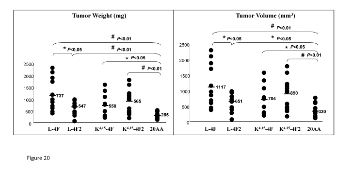
Figure 20. BALB/c mice that received subcutaneous flank injections of CT26
cells and were
subsequently treated with subcutaneous HDL mimetic peptides showed significant
reductions in
tumor weight (left panel) and tumor volume (right panel).
DETAILED DESCRIPTION
The present invention is based on the discovery that HDL-related molecules can
be used to treat
and prevent proinflammatory conditions. HDL-related molecules include ApoA-I,
bovine HDL,
and HDL mimetics. As described in further detail below, ApoA-I, in its
natural, full-length form,
can prevent UV-induced cell death and oxidative stress. Also described in
further detail below is the
8
CA 02846865 2014-02-26
WO 2013/033260
PCT/US2012/052925
unexpected discovery that HDL mimetics, ApoA-I and bovine HDL (bHDL) can be
used to treat
and prevent various cancers. ApoA-I and other HDL-related molecules provide
potent and
effective agents for the treatment and prevention of proinflammatory
conditions, including skin
conditions, and systemic proinflammatory conditions, including cancer and
other diseases, such as
Alzheimer's disease. Cancers to be treated include epithelial cancers, such as
cancer of the vagina,
vulva, ovaries, cervix, uterus, prostate, colon, breast, pancreas, lung, skin
(e.g., melanoma), brain (e.g.
glioblastoma), and gastric cancer. The HDL-related molecules described herein
can also be used in
anti-aging treatments, as they can be used to delay the aging process and
reduce or eliminate
oxidative stress, and in treatment of eye conditions, such as macular
degeneration, retinitis
pigmentosa, and autoimmune diseases, such as arthritis.
The invention provides a method of reducing death and/or oxidative stress in
epithelial cells
exposed to oxidative stress. The method comprises contacting the epithelial
cells with an HDL-
related molecule prior to exposure to oxidative stress. In some embodiments,
the oxidative stress
comprises exposure to ultraviolet radiation. In a typical embodiment, the
contacting occurs at least
12-24 hours prior to the exposure to oxidative stress.
Definitions
All scientific and technical terms used in this application have meanings
commonly used in the art
unless otherwise specified. As used in this application, the following words
or phrases have the
meanings specified.
As used herein, "HDL-related molecule" means ApoA-I, bovine HDL, and HDL
mimetics,
including peptides and synthetic molecules.
As used herein, "ApoA-I" refers to full-length and unmodified ApoA-I, unless
context clearly
indicates otherwise. For example, "ApoA-I peptides" refers to small portions
of full-length ApoA-I.
Typically, the ApoA-I is human ApoA-I, a 28.2 kDa protein of 244 amino acids.
As used herein, "HDL mimetics" refers to modified apolipoproteins that mimic
the function of
HDL, typically providing an HDL-related molecule having enhanced efficacy.
Tyically, the
apolipoproteins are modified by altering or substituting one or more amino
acids, and/or by
combining two or more HDL peptides to form a chimeric HDL-related molecule.
As used herein, "polypeptide" includes proteins, fragments of proteins, and
peptides, whether
isolated from natural sources, produced by recombinant techniques or
chemically synthesized.
Polypeptides of the invention typically comprise at least about 6 amino acids.
Shorter polypeptides,
e.g., those less than about 50 amino acids in length, are typically referred
to as "peptides".
9
CA 02846865 2014-02-26
WO 2013/033260
PCT/US2012/052925
As used herein, "vector" means a construct, which is capable of delivering,
and preferably
expressing, one or more gene(s) or sequence(s) of interest in a host cell.
Examples of vectors
include, but are not limited to, viral vectors, naked DNA or RNA expression
vectors, plasmid,
cosmid or phage vectors, DNA or RNA expression vectors associated with
cationic condensing
agents, DNA or RNA expression vectors encapsulated in liposomes, and certain
eukaryotic cells,
such as producer cells.
As used herein, "expression control sequence" means a nucleic acid sequence
that directs
transcription of a nucleic acid. An expression control sequence can be a
promoter, such as a
constitutive or an inducible promoter, or an enhancer. The expression control
sequence is operably
linked to the nucleic acid sequence to be transcribed.
The term "nucleic acid" or "polynucleotide" refers to a deoxyribonucleotide or
ribonucleotide
polymer in either single- or double-stranded form, and unless otherwise
limited, encompasses
known analogs of natural nucleotides that hybridize to nucleic acids in a
manner similar to naturally
occurring nucleotides.
As used herein, "pharmaceutically acceptable carrier" or "excipient" includes
any material which,
when combined with an active ingredient, allows the ingredient to retain
biological activity and is
non-reactive with the subject's immune system. Examples include, but are not
limited to, any of the
standard pharmaceutical carriers such as a phosphate buffered saline solution,
water, emulsions such
as oil/water emulsion, and various types of wetting agents. Preferred diluents
for aerosol or
parenteral administration are phosphate buffered saline or normal (0.9%)
saline.
Compositions comprising such carriers are formulated by well known
conventional methods (see,
for example, Remington's Pharmaceutical Sciences, 18th edition, A. Gennaro,
ed., Mack Publishing Co.,
Easton, PA, 1990).
As used herein, "a" or "an" means at least one, unless clearly indicated
otherwise.
HDL Mimetics
The present invention provides HDL mimetics, including chimeras of HDL
peptides and modified
and/or synthetic molecules that also serve as HDL mimetics. In one embodiment,
substitution of
alanines in known HDL mimetic peptides with a-aminoisobutyric acid (Aib)
generates novel HDL
mimetics (NHMs). In a typical embodiment, the chimera comprises two HDL
peptides selected
from peptides of ApoA-I, ApoE and ApoJ. In one embodiment, the HDL mimetics
are obtained
via substitution of alanines with a-aminoisobutyric acid (Aib) in an 18 amino
acid peptide of Apo A-
I that is chimerized with a 10 amino acid peptide of Apo E), to generate NHMs
1-7 described
CA 02846865 2014-02-26
WO 2013/033260
PCT/US2012/052925
hereinbelow. In another embodiment, the HDL mimetics are obtained via
combining ApoE and
ApoJ (G*) to generate, for example, the novel HDL mimetic LRKI RKRLLR
LVGRQLEEFL
(SEQ ID NO: 1).
Substitution of Aib for alanines in E18A (ref) results in a series of seven
NHMs.
E18A peptide (ref) = LRKI RKRLLRDWLKAFYDKVAEKI KFAF (SEQ ID NO: 2)
NHMs:
NHM1 = LRKI RKRLLRDWLKAibFYDKVAEKI KEAF (SEQ ID NO: 3)
NHM2 = LRKI RKRLLRDWLKAFYDKVAibEKI KFAF (SEQ ID NO: 4)
NHM3 = LRKI RKRLLRDWLKAFYDKVAEKI KFAibF (SEQ ID NO: 5)
NHM4 - LRKI RKRLLRDWLKAibFYDKVAibEKI KFAF (SEQ ID NO: 6)
NHM5 = LRKI RKRLLRDWLKAFYDKVAibEKI KFAibF (SEQ ID NO: 7)
NHM6 = LRKI RKRLLRDWLKAibFYDKVAEKI KFAibF (SEQ ID NO: 8)
NHM7 = LRKI RKRLLRDWLKAibFYDKVAibEKI KFAibF (SEQ ID NO: 9)
See: Oleg F Sharifov, et al., 2011, Apolipoprotein E Mimetics and Cholesterol
Lowering Properties,
American Journal of Cardiovascular Drugs 11 (6) :371 -381.
Surprisingly, the novel HDL mimetic peptides described herein, alone or in
combination with other
anti-oxidants, can be used for the prevention and treatment of pro-
inflammatory skin and systemic
pro-inflammatory conditions, including cancer. These molecules provide potent
and effective anti-
oxidants for the prevention and treatment of pro-inflammatory skin and
systemic pro-inflammatory
conditions, including cancer. This has been proved in principle using cell
culture models, and has
been shown through in vivo studies to inhibit tumor development in an animal
model.
Bovine HDL
Bovine HDL (bHDL) as described herein includes the native protein, and
heterologous sequences
may be present. Typically, the bHDL is used in its natural, full-length,
unmodified form. Bovine
HDL is typically purified from serum, and can be obtained from, for example,
Biomedical
Technologies, Inc. (Stoughton, MA). Bovine HDL is advantageous relative to the
HDL of other
species due to its high level of ApoA-I and its high serum levels, as well as
its suitability for
administration to humans.
11
CA 02846865 2014-02-26
WO 2013/033260
PCT/US2012/052925
ApoA-I Polypeptides
ApoA-I polypeptides as described herein include the native protein, and
heterologous sequences
may be present. Typically, the ApoA-I is human ApoA-I, used in its natural,
full-length, unmodified
and mature form.
NCBI Reference Sequence: NP 000030.1 (SEQ ID NO: 10):
1 mkaavltlav lfltgsgarh fwqqdeppqs pwdrvkdlat vyvdvlkdsg rdyvscifegs
61 algkqlnlkl ldnwdsvtst fsklreqlgp vtqefwdnle keteglrqem skdleevkak
121 vqpylddfqk kwqeemelyr qkveplrael gegarqklhe lgeklsplge emrdrarahv
181 dalrthlapy sdelrqrlaa rlealkengg arlaeyhaka tehlstlsek akpaledlrq
241 gllpvlesfk vsflsaleey tkklntq;
In the above sequence, the signal peptide is at amino acids 1-18, the mature
proprotein is at amino
acids 19-267, and the mature ApoA-I protein is at amino acids 25-267:
DEPPQSPWDRVKDLATVYVDVLKDSGRDYVSQFEGSALGKQLNLKLLDNWDSVTSTFSKLREQLGPVTQEFWDNLEK
ETEGLRQEMSKDLEEVKAKVQPYLDDFQKKWQEEMELYRQKVEPLRAELQEGARQKLHELQEKLSPLGEEMRDRARA
HVDALRTHLAPYSDELRQRLAARLEALKENGGARLAEYHAKATEHLSTLSEKAKPALEDLRQGLLPVLESFKVSFLS
ALEEYTKKLNTQ (SEQ ID NO: 11).
While ApoA-I peptides, and particularly ApoA-I mimetic peptides have been
developed in efforts to
identify molecules having similar function and/or ease of productive compared
to full-length ApoA-
I protein for some areas of use, the modifications of these ApoA-I mimetic
peptides (e.g., alpha-
helical peptides) have rendered them entirely different from natural ApoA-I;
in fact, the mimetic
peptides share no structural similarity with the full length ApoA-I protein
molecule. Moreover, in
the area of cardiovascular treatment, the mimetic peptides have been less
effective and require such
large quantities that therapeutic use of these peptides is impractical.
Interestingly, the term mimetic
peptide is a term developed over 2 decades ago that refers to an attempt to
identify structurally
dissimilar molecules that may share some functional properties with the full-
length ApoA-I protein;
and in fact, no structural similarities exist between these alpha-helical
peptides and the full-length
ApoA-I molecule. Hence, the term "mimetic peptide" is, in this context, a
misnomer, since the
ApoA-I full length protein shares nothing structurally in common with its
mimetic peptides. The
ApoA-I mimetic peptides attempt only to mimic some of the features of the ApoA-
I full length
protein function.
Variant Polypeptides
A polypeptide of the invention can comprise a variant of a native protein. A
polypeptide "variant,"
as used herein, is a polypeptide that differs from a native protein in one or
more substitutions,
deletions, additions and/or insertions, such that the therapeutic efficacy of
the polypeptide is not
substantially diminished. In other words, the efficacy may be enhanced or
unchanged, relative to the
12
CA 02846865 2014-02-26
WO 2013/033260
PCT/US2012/052925
native protein, or may be diminished by less than 50%, and preferably less
than 20%, relative to the
native protein. Preferred variants include those in which one or more
portions, such as an N-
terminal leader sequence, have been removed. Other preferred variants include
variants in which a
small portion (e.g., 1-30 amino acids, preferably 5-15 amino acids) has been
removed from the N-
and/or C-terminal of the mature protein. Polypeptide variants preferably
exhibit at least about 70%,
more preferably at least about 90% and most preferably at least about 95%
identity (determined as
described above) to the identified polypeptides.
Preferably, a variant contains conservative substitutions. A "conservative
substitution" is one in
which an amino acid is substituted for another amino acid that has similar
properties, such that one
skilled in the art of peptide chemistry would expect the secondary structure
and hydropathic nature
of the polypeptide to be substantially unchanged. Amino acid substitutions may
generally be made
on the basis of similarity in polarity, charge, solubility, hydrophobicity,
hydrophilicity and/or the
amphipathic nature of the residues. For example, negatively charged amino
acids include aspartic
acid and glutamic acid; positively charged amino acids include lysine and
arginine; and amino acids
with uncharged polar head groups having similar hydrophilicity values include
leucine, isoleucine
and valine; glycine and alanine; asparagine and glutamine; and serine,
threonine, phenylalanine and
tyrosine. Other groups of amino acids that may represent conservative changes
include: (1) ala, pro,
gly, glu, asp, gln, asn, ser, thr; (2) cys, ser, tyr, thr; (3) val, ile, leu,
met, ala, phe; (4) lys, arg, his; and
(5) phe, tyr, trp, his. A variant may also, or alternatively, contain
nonconservative changes. In a
preferred embodiment, variant polypeptides differ from a native sequence by
substitution, deletion
or addition of five amino acids or fewer. Variants may also (or alternatively)
be modified by, for
example, the deletion or addition of amino acids that have minimal influence
on the
immunogenicity, secondary structure and hydropathic nature of the polypeptide.
Preparation of Polypeptides
Polypeptides may comprise a signal (or leader) sequence at the N-terminal end
of the protein that
co-translationally or post-translationally directs transfer of the protein.
The polypeptide may also be
conjugated to a linker or other sequence for ease of synthesis, purification
or identification of the
polypeptide.
Polypeptides may be purified from natural sources, such as serum. In some
embodiments, the
polypeptides are purified from the same subject to whom the composition will
be administered. In
other embodiments, the polypeptide is purified from a heterologous species,
such as bovine HDL or
ApoA-I for administration to humans.
13
CA 02846865 2014-02-26
WO 2013/033260
PCT/US2012/052925
Recombinant polypeptides encoded by DNA sequences as described herein may be
readily prepared
from the DNA sequences using any of a variety of expression vectors known to
those of ordinary
skill in the art. Expression may be achieved in any appropriate host cell that
has been transformed
or transfected with an expression vector containing a DNA molecule that
encodes a recombinant
polypeptide. Suitable host cells include prokaryotes, yeast and higher
eukaryotic cells. Preferably,
the host cells employed are E. co/i, yeast, insect cells or a mammalian cell
line such as COS or CHO.
Supernatants from suitable host/vector systems that secrete recombinant
protein or polypeptide
into culture media may be first concentrated using a commercially available
filter. Following
concentration, the concentrate may be applied to a suitable purification
matrix such as an affinity
matrix or an ion exchange resin. Finally, one or more reverse phase HPLC steps
can be employed
to further purify a recombinant polypeptide.
Portions and other variants having fewer than about 100 amino acids, and
generally fewer than
about 50 amino acids, may also be generated by synthetic means, using
techniques well known to
those of ordinary skill in the art. For example, such polypeptides may be
synthesized using any of
the commercially available solid-phase techniques, such as the Merrifield
solid-phase synthesis
method, where amino acids are sequentially added to a growing amino acid
chain. See Merrifield,"
Am. Chem. Soc. 85:2149-2146, 1963. Equipment for automated synthesis of
polypeptides is
commercially available from suppliers such as Perkin Elmer/Applied BioSystems
Division (Foster
City, CA), and may be operated according to the manufacturer's instructions.
Polypeptides can be synthesized on a Perkin Elmer/Applied Biosystems Division
430A peptide
synthesizer using FMOC chemistry with HPTU (0-Ben2otria2oleN,N,N',N'-
tetramethyluronium
hexafluorophosphate) activation. A Gly-Cys-Gly sequence may be attached to the
amino terminus
of the peptide to provide a method of conjugation, binding to an immobilized
surface, or labeling of
the peptide. Cleavage of the peptides from the solid support may be carried
out using the following
cleavage mixture: trifluoroacetic acid:ethanedithiohthioanisole:water:phenol
(40:1:2:2:3). After
cleaving for 2 hours, the peptides may be precipitated in cold methyl-t-butyl-
ether. The peptide
pellets may then be dissolved in water containing 0.1% trifluoroacetic acid
(TFA) and lyophilized
prior to purification by C18 reverse phase HPLC. A gradient of 0%-60%
acetonitrile (containing
0.1% TFA) in water may be used to elute the peptides. Following lyophilization
of the pure
fractions, the peptides may be characterized using electrospray or other types
of mass spectrometry
and by amino acid analysis.
14
CA 02846865 2014-02-26
WO 2013/033260
PCT/US2012/052925
Fusion Proteins
In some embodiments, the polypeptide is a fusion protein that comprises
multiple polypeptides as
described herein, or that comprises at least one polypeptide as described
herein and an unrelated
sequence. In some embodiments, the fusion protein comprises an ApoA-I
polypeptide and an
immunogenic polypeptide. The immunogenic polypeptide can comprise, for
example, all or a
portion of an additional protein.
Additional fusion partners can be added. A fusion partner may, for example,
serve as an
immunological fusion partner by assisting in the provision of T helper
epitopes, preferably T helper
epitopes recognized by humans. As another example, a fusion partner may serve
as an expression
enhancer, assisting in expressing the protein at higher yields than the native
recombinant protein.
Certain preferred fusion partners are both immunological and expression
enhancing fusion partners.
Other fusion partners may be selected so as to increase the solubility of the
protein or to enable the
protein to be targeted to desired intracellular compartments. Still further
fusion partners include
affinity tags, which facilitate purification of the protein.
Fusion proteins may generally be prepared using standard techniques, including
chemical
conjugation. Preferably, a fusion protein is expressed as a recombinant
protein, allowing the
production of increased levels, relative to a non-fused protein, in an
expression system. Briefly,
DNA sequences encoding the polypeptide components may be assembled separately,
and ligated
into an appropriate expression vector. The 3' end of the DNA sequence encoding
one polypeptide
component is ligated, with or without a peptide linker, to the 5' end of a DNA
sequence encoding
the second polypeptide component so that the reading frames of the sequences
are in phase. This
permits translation into a single fusion protein that retains the biological
activity of both component
polypeptides.
A peptide linker sequence may be employed to separate the first and the second
polypeptide
components by a distance sufficient to ensure that each polypeptide folds into
its secondary and
tertiary structures. Such a peptide linker sequence is incorporated into the
fusion protein using
standard techniques well known in the art. Suitable peptide linker sequences
may be chosen based
on the following factors: (1) their ability to adopt a flexible extended
conformation; (2) their inability
to adopt a secondary structure that could interact with functional epitopes on
the first and second
polypeptides; and (3) the lack of hydrophobic or charged residues that might
react with the
polypeptide functional epitopes. Preferred peptide linker sequences contain
Gly, Asn and Ser
residues. Other near neutral amino acids, such as Thr and Ala may also be used
in the linker
sequence. Amino acid sequences which may be usefully employed as linkers
include those disclosed
CA 02846865 2014-02-26
WO 2013/033260
PCT/US2012/052925
in Maratea et al., Gene 40:39-46, 1985; Murphy et al., Proc. Natl. Acad. Sci.
USA 83:8258-8262,
1986; U.S. Patent No. 4,935,233 and U.S. Patent No. 4,751,180. The linker
sequence may generally
be from 1 to about 50 amino acids in length. Linker sequences are not required
when the first and
second polypeptides have non-essential N-terminal amino acid regions that can
be used to separate
the functional domains and prevent steric interference.
The ligated DNA sequences are operably linked to suitable transcriptional or
translational regulatory
elements. The regulatory elements responsible for expression of DNA are
located 5' to the DNA
sequence encoding the first polypeptides. Similarly, stop codons required to
end translation and
transcription termination signals are present 3' to the DNA sequence encoding
the second
polypeptide.
Fusion proteins are also provided that comprise a polypeptide of the present
invention together with
an unrelated immunogenic protein. Preferably the immunogenic protein is
capable of eliciting a
memory response. Examples of such proteins include tetanus, tuberculosis and
hepatitis proteins
(see, for example, Stoute et al., New Engl. J. Med. 336:86-91, 1997).
Within preferred embodiments, an immunological fusion partner is derived from
protein D, a
surface protein of the gram-negative bacterium Haemophilus influenza B (WO
91/18926).
Preferably, a protein D derivative comprises approximately the first third of
the protein (e.g., the
first N-terminal 100-110 amino acids), and a protein D derivative may be
lipidated. Other fusion
partners include the non-structural protein from influenzae virus, NS I
(hemaglutinin). Typically,
the N-terminal 81 amino acids are used, although different fragments that
include T-helper epitopes
may be used.
In another embodiment, the immunological fusion partner is the protein known
as LYTA, or a
portion thereof (preferably a C-terminal portion). LYTA is derived from
Streptococcus pneumoniae,
which synthesizes an N-acetyl-L-alanine amidase known as amidase LYTA (encoded
by the LytA
gene; Gene 43:265-292, 1986). LYTA is an autolysin that specifically degrades
certain bonds in the
peptidoglycan backbone. The C-terminal domain of the LYTA protein is
responsible for the affinity
to the choline or to some choline analogues such as DEAR This property has
been exploited for the
development of E. co/i C-LYTA expressing plasmids useful for expression of
fusion proteins.
Purification of hybrid proteins containing the C-LYTA fragment at the amino
terminus has been
described (see Biotechnology 10:795-798, 1992). Within a preferred embodiment,
a repeat portion
of LYTA may be incorporated into a fusion protein. A repeat portion is found
in the C-terminal
region starting at residue 178. A particularly preferred repeat portion
incorporates residues 188-305.
16
CA 02846865 2014-02-26
WO 2013/033260
PCT/US2012/052925
In general, polypeptides (including fusion proteins) and polynucleotides as
described herein are
isolated. An "isolated" polypeptide or polynucleotide is one that is removed
from its original
environment. For example, a naturally occurring protein is isolated if it is
separated from some or
all of the coexisting materials in the natural system. Preferably, such
polypeptides are at least about
90% pure, more preferably at least about 95% pure and most preferably at least
about 99% pure. A
polynucleotide is considered to be isolated if, for example, it is cloned into
a vector that is not a part
of the natural environment.
Polynucleotides of the Invention
The invention provides polynucleotides that encode one or more HDL-related
polypeptides,
including bHDL, ApoA-I and HDL mimetcs. Polynucleotides that are fully
complementary to any
such sequences are also encompassed by the present invention. Polynucleotides
may be single-
stranded (coding or antisense) or double-stranded, and may be DNA (genomic,
cDNA or synthetic)
or RNA molecules, including siRNA. RNA molecules include HnRNA molecules,
which contain
introns and correspond to a DNA molecule in a one-to-one manner, and mRNA
molecules, which
do not contain introns. Additional coding or non-coding sequences may, but
need not, be present
within a polynucleotide of the present invention, and a polynucleotide may,
but need not, be linked
to other molecules and/or support materials. Portions of such polynucleotides
can be useful as
primers and probes for the amplification and detection of related molecules.
Polynucleotides may comprise a native sequence (i.e., an endogenous sequence
that encodes an
HDL-related polypeptide or a portion thereof) or may comprise a variant of
such a sequence.
Polynucleotide variants contain one or more substitutions, additions,
deletions and/or insertions
such that the immunogenicity of the encoded polypeptide is not diminished,
relative to a native
protein. Variants preferably exhibit at least about 70% identity, more
preferably at least about 80%
identity and most preferably at least about 90% identity to a polynucleotide
sequence that encodes a
native protein or a portion thereof.
Two polynucleotide or polypeptide sequences are said to be "identical" if the
sequence of
nucleotides or amino acids in the two sequences is the same when aligned for
maximum
correspondence as described below. Comparisons between two sequences are
typically performed
by comparing the sequences over a comparison window to identify and compare
local regions of
sequence similarity. A "comparison window" as used herein, refers to a segment
of at least about 20
contiguous positions, usually 30 to about 75, 40 to about 50, in which a
sequence may be compared
to a reference sequence of the same number of contiguous positions after the
two sequences are
optimally aligned.
17
CA 02846865 2014-02-26
WO 2013/033260
PCT/US2012/052925
Optimal alignment of sequences for comparison may be conducted using the
Megalign program in
the Lasergene suite of bioinformatics software (DNASTAR, Inc., Madison, WI),
using default
parameters. This program embodies several alignment schemes described in the
following
references: Dayhoff, M.O. (1978) A model of evolutionary change in proteins -
Matrices for
detecting distant relationships. In Dayhoff, M.O. (ed.) Atlas of Protein
Sequence and Structure,
National Biomedical Research Foundation, Washington DC Vol. 5, Suppl. 3, pp.
345-358; Hein J.
(1990) Unified Approach to Alignment and Phylogenes pp. 626-645 Methods in
Enzymology vol.
183, Academic Press, Inc., San Diego, CA; Higgins, D.G. and Sharp, P.M. (1989)
CABIOS 5:151-
153; Myers, E.W. and Muller W. (1988) CABIOS 4:11-17; Robinson, E.D. (1971)
Comb. Theor.
11:105; Santou, N., Nes, M. (1987) Mol. Biol. Evol. 4:406-425; Sneath, P.H.A.
and Sokal, R.R.
(1973) Numerical Taxonomy the Principles and Practice of Numerical Taxonomy,
Freeman Press,
San Francisco, CA; Wilbur, W.J. and Lipman, D.J. (1983) Proc. Natl. Acad. Sci.
USA 80:726-730.
Preferably, the "percentage of sequence identity" is determined by comparing
two optimally aligned
sequences over a window of comparison of at least 20 positions, wherein the
portion of the
polynucleotide or polypeptide sequence in the comparison window may comprise
additions or
deletions (i.e. gaps) of 20 percent or less, usually 5 to 15 percent, or 10 to
12 percent, as compared
to the reference sequences (which does not comprise additions or deletions)
for optimal alignment
of the two sequences. The percentage is calculated by determining the number
of positions at which
the identical nucleic acid bases or amino acid residue occurs in both
sequences to yield the number
of matched positions, dividing the number of matched positions by the total
number of positions in
the reference sequence (i.e. the window size) and multiplying the results by
100 to yield the
percentage of sequence identity.
Variants may also, or alternatively, be substantially homologous to a native
gene, or a portion or
complement thereof. Such polynucleotide variants are capable of hybridizing
under moderately
stringent conditions to a naturally occurring DNA sequence encoding a native
protein (or a
complementary sequence).
Suitable "moderately stringent conditions" include prewashing in a solution of
5 X SSC, 0.5% SDS,
1.0 mM EDTA (pH 8.0); hybridizing at 50 C-65 C, 5 X SSC, overnight; followed
by washing twice
at 65 C for 20 minutes with each of 2X, 0.5X and 0.2X SSC containing 0. 1 %
SDS.
As used herein, "highly stringent conditions" or "high stringency conditions"
are those that: (1)
employ low ionic strength and high temperature for washing, for example 0.015
M sodium
chloride/0.0015 M sodium citrate/0.1% sodium dodecyl sulfate at 50 C; (2)
employ during
hybridization a denaturing agent, such as formamide, for example, 50% (v/v)
formamide with 0.1 A
18
CA 02846865 2014-02-26
WO 2013/033260
PCT/US2012/052925
bovine serum albumin/0.1% Fico11/0.1% polyvinylpyrrolidone/50mM sodium
phosphate buffer at
pH 6.5 with 750 mM sodium chloride, 75 mM sodium citrate at 42 C; or (3)
employ 50%
formamide, 5 x SSC (0.75 M NaC1, 0.075 M sodium citrate), 50 mM sodium
phosphate (pH 6.8),
0.1% sodium pyrophosphate, 5 x Denhardt's solution, sonicated salmon sperm DNA
(50 Kg/m1),
0.1% SDS, and 10% dextran sulfate at 42 C, with washes at 42 C in 0.2 x SSC
(sodium
chloride/sodium citrate) and 50% formamide at 55 C, followed by a high-
stringency wash
consisting of 0.1 x SSC containing EDTA at 55 C. The skilled artisan will
recognize how to adjust
the temperature, ionic strength, etc. as necessary to accommodate factors such
as probe length and
the like.
It will be appreciated by those of ordinary skill in the art that, as a result
of the degeneracy of the
genetic code, there are many nucleotide sequences that encode a polypeptide as
described herein.
Some of these polynucleotides bear minimal homology to the nucleotide sequence
of any native
gene. Nonetheless, polynucleotides that vary due to differences in codon usage
are specifically
contemplated by the present invention. Further, alleles of the genes
comprising the polynucleotide
sequences provided herein are within the scope of the present invention.
Alleles are endogenous
genes that are altered as a result of one or more mutations, such as
deletions, additions and/or
substitutions of nucleotides. The resulting mRNA and protein may, but need
not, have an altered
structure or function. Alleles may be identified using standard techniques
(such as hybridization,
amplification and/or database sequence comparison).
Polynucleotides may be prepared using any of a variety of techniques known in
the art. DNA
encoding an ApoA-I protein may be obtained from a cDNA library prepared from
tissue expressing
the corresponding mRNA. Accordingly, human ApoA-I DNA can be conveniently
obtained from a
cDNA library prepared from human tissue. The ApoA-I protein-encoding gene may
also be
obtained from a genomic library or by oligonucleotide synthesis. Libraries can
be screened with
probes (such as antibodies to ApoA-I or oligonucleotides of at least about 20-
80 bases) designed to
identify the gene of interest or the protein encoded by it. Screening the cDNA
or genomic library
with the selected probe may be conducted using standard procedures, such as
those described in
Sambrook et al., Molecular Cloning: A Laboratog Manual (New York: Cold Spring
Harbor Laboratory
Press, 1989). An alternative means to isolate the gene encoding ApoA-I is to
use PCR methodology
(Sambrook et al., supra; Dieffenbach et al., PCR Primer: A Laboratog Manual
(Cold Spring Harbor
Laboratory Press, 1995)).
The oligonucleotide sequences selected as probes should be sufficiently long
and sufficiently
unambiguous that false positives are minimized. The oligonucleotide is
preferably labeled such that
it can be detected upon hybridization to DNA in the library being screened.
Methods of labeling are
19
CA 02846865 2014-02-26
WO 2013/033260
PCT/US2012/052925
well known in the art, and include the use of radiolabels, such as 3213-
labeled ATP, biotinylation or
enzyme labeling. Hybridization conditions, including moderate stringency and
high stringency, are
provided in Sambrook et al., supra.
Polynucleotide variants may generally be prepared by any method known in the
art, including
chemical synthesis by, for example, solid phase phosphoramidite chemical
synthesis. Modifications
in a polynucleotide sequence may also be introduced using standard mutagenesis
techniques, such as
oligonucleotide-directed site-specific mutagenesis (see Adelman et al., DNA
2:183, 1983).
Alternatively, RNA molecules may be generated by in vitro or in vivo
transcription of DNA sequences
encoding an ApoA-I protein, or portion thereof, provided that the DNA is
incorporated into a
vector with a suitable RNA polymerase promoter (such as T7 or SP6). Certain
portions may be
used to prepare an encoded polypeptide, as described herein. In addition, or
alternatively, a portion
may be administered to a patient such that the encoded polypeptide is
generated in vivo.
Any polynucleotide may be further modified to increase stability in vivo.
Possible modifications
include, but are not limited to, the addition of flanking sequences at the 5'
and/or 3' ends; the use of
phosphorothioate or 2' 0-methyl rather than phosphodiesterase linkages in the
backbone; and/or
the inclusion of nontraditional bases such as inosine, queosine and
wybutosine, as well as acetyl-
methyl-, thio- and other modified forms of adenine, cytidine, guanine, thymine
and uridine.
Nucleotide sequences can be joined to a variety of other nucleotide sequences
using established
recombinant DNA techniques. For example, a polynucleotide may be cloned into
any of a variety
of cloning vectors, including plasmids, phagemids, lambda phage derivatives
and cosmids. Vectors
of particular interest include expression vectors, replication vectors, probe
generation vectors and
sequencing vectors. In general, a vector will contain an origin of replication
functional in at least
one organism, convenient restriction endonuclease sites and one or more
selectable markers. Other
elements will depend upon the desired use, and will be apparent to those of
ordinary skill in the art.
Within certain embodiments, polynucleotides may be formulated so as to permit
entry into a cell of
a mammal, and to permit expression therein. Such formulations are particularly
useful for
therapeutic purposes, as described below. Those of ordinary skill in the art
will appreciate that there
are many ways to achieve expression of a polynucleotide in a target cell, and
any suitable method
may be employed. For example, a polynucleotide may be incorporated into a
viral vector such as,
but not limited to, adenovirus, adeno-associated virus, retrovirus, or
vaccinia or other pox virus (e.g.,
avian pox virus). Techniques for incorporating DNA into such vectors are well
known to those of
ordinary skill in the art. A retroviral vector may additionally transfer or
incorporate a gene for a
selectable marker (to aid in the identification or selection of transduced
cells) and/or a targeting
CA 02846865 2014-02-26
WO 2013/033260
PCT/US2012/052925
moiety, such as a gene that encodes a ligand for a receptor on a specific
target cell, to render the
vector target specific. Targeting may also be accomplished using an antibody,
by methods known to
those of ordinary skill in the art.
Other formulations for therapeutic purposes include colloidal dispersion
systems, such as
macromolecule complexes, nanocapsules, microspheres, beads, and lipid-based
systems including
oil-in-water emulsions, micelles, mixed micelles, and liposomes. A preferred
colloidal system for use
as a delivery vehicle in vitro and in vivo is a liposome (i.e., an artificial
membrane vesicle). The
preparation and use of such systems is well known in the art.
Pharmaceutical Compositions
The invention provides ApoA-I polypeptide, polynucleotides, and related
molecules that are
incorporated into pharmaceutical compositions. In a typical embodiment, the
polypeptide is ApoAI
in natural, full-length, unmodified form. As is understood in the art, ApoAI
is a significant
component of high-density lipoprotein (HDL). Accordingly, one can administer
ApoAI by
administering HDL.
Pharmaceutical compositions comprise one or more such compounds and,
optionally, a
physiologically acceptable carrier. Administration of ApoAI is facilitated by
preparation with inert
lipids, e.g. to form micelles. In a typical embodiment, ApoAI is administered
orally, as part of an
oral supplement. Alternatively, it can be administered transdermally, such as
via a patch adhered to
the subject's skin.
While any suitable carrier known to those of ordinary skill in the art may be
employed in the
pharmaceutical compositions of this invention, the type of carrier will vary
depending on the mode
of administration. Compositions of the present invention may be formulated for
any appropriate
manner of administration, including for example, topical, oral, nasal,
intravenous, intracranial,
intraperitoneal, subcutaneous, intradermal, transdermal or intramuscular
administration. For
parenteral administration, such as subcutaneous injection, the carrier
preferably comprises a fat, and
optionally water, saline, alcohol, a wax or a buffer. For oral administration,
any of the above carriers
or a solid carrier, such as mannitol, lactose, starch, magnesium stearate,
sodium saccharine, talcum,
cellulose, glucose, sucrose, and magnesium carbonate, may be employed.
Biodegradable
microspheres (e.g., polylactate polyglycolate) may also be employed as
carriers for the
pharmaceutical compositions of this invention.
In addition, the carrier may contain other pharmacologically-acceptable
excipients for modifying or
maintaining the pH, osmolarity, viscosity, clarity, color, sterility,
stability, rate of dissolution, or odor
21
CA 02846865 2014-02-26
WO 2013/033260
PCT/US2012/052925
of the formulation. Similarly, the carrier may contain still other
pharmacologically-acceptable
excipients for modifying or maintaining the stability, rate of dissolution,
release, or absorption or
penetration across the blood-brain barrier of the molecule. Such excipients
are those substances
usually and customarily employed to formulate dosages for parenteral
administration in either unit
dose or multi-dose form or for direct infusion into the CSF by continuous or
periodic infusion from
an implanted pump.
Such compositions may also comprise buffers (e.g., neutral buffered saline or
phosphate buffered
saline), carbohydrates (e.g., glucose, mannose, sucrose or dextrans),
mannitol, proteins, polypeptides
or amino acids such as glycine, antioxidants, chelating agents such as EDTA or
glutathione,
adjuvants (e.g., aluminum hydroxide) and/or preservatives. Alternatively,
compositions of the
present invention may be formulated as a lyophilizate. Compounds may also be
encapsulated within
liposomes using well known technology.
A pharmaceutical composition can contain DNA encoding one or more of the
polypeptides as
described above, such that the polypeptide is generated in situ. As noted
above, the DNA may be
present within any of a variety of delivery systems known to those of ordinary
skill in the art,
including nucleic acid expression systems, bacteria and viral expression
systems. Numerous gene
delivery techniques are well known in the art, such as those described by
Rolland, Crit. Rev. Therap.
Drug Carrier Systems 15:143-198, 1998, and references cited therein.
Appropriate nucleic acid
expression systems contain the necessary DNA sequences for expression in the
patient (such as a
suitable promoter and terminating signal). Bacterial delivery systems involve
the administration of a
bacterium (such as Bacillus-Calmette-Guenin) that expresses an immunogenic
portion of the
polypeptide on its cell surface or secretes such an epitope.
In a preferred embodiment, the DNA may be introduced using a viral expression
system (e.g.,
vaccinia or other pox virus, retrovirus, or adenovirus), which may involve the
use of a non-
pathogenic (defective), replication competent virus. Suitable systems are
disclosed, for example, in
Fisher-Hoch et al., Proc. Natl. Acad. Sci. USA 86:317-321, 1989; Flexner et
al., Ann. N. Y. Acad
Sci. 569:86-103, 1989; Flexner et al., Vaccine 8:17-21, 1990; U.S. Patent Nos.
4,603,112, 4,769,330,
and 5,017,487; WO 89/01973; U.S. Patent No. 4,777,127; GB 2,200,651; EP
0,345,242; WO
91/02805; Berkner-Biotechniques 6:616-627, 1988; Rosenfeld et al., Science
252:431-434, 1991;
Kolls et al., Proc. Natl. Acad. Sci. USA 91:215-219, 1994; Kass-Eisler et al.,
Proc. Natl. Acad. Sci.
USA 90:11498-11502, 1993; Guzman et al., Circulation 88:2838-2848, 1993; and
Guzman et al., Cir.
Res. 73:1202-1207, 1993. Techniques for incorporating DNA into such expression
systems are well
known to those of ordinary skill in the art. The DNA may also be "naked," as
described, for
example, in Ulmer et al., Science 259:1745-1749, 1993 and reviewed by Cohen,
Science 259:1691-
22
CA 02846865 2014-02-26
WO 2013/033260
PCT/US2012/052925
1692, 1993. The uptake of naked DNA may be increased by coating the DNA onto
biodegradable
beads, which are efficiently transported into the cells.
Any of a variety of adjuvants may be employed in the compositions of this
invention. Most
adjuvants contain a substance designed to protect the peptide from rapid
catabolism, such as
aluminum hydroxide or mineral oil, and a stimulator of immune responses, such
as lipid A, Bortadella
pertussis or Mycobacterium tuberculosis derived proteins. Suitable adjuvants
are commercially available as,
for example, Freund's Incomplete Adjuvant and Complete Adjuvant (Difco
Laboratories, Detroit,
MI); Merck Adjuvant 65 (Merck and Company, Inc., Rahway, NJ); aluminum salts
such as aluminum
hydroxide gel (alum) or aluminum phosphate; salts of calcium, iron or zinc; an
insoluble suspension
of acylated tyrosine acylated sugars; cationically or anionically derivatized
polysaccharides;
polyphosphazenes biodegradable microspheres; monophosphoryl lipid A and quil
A. Cytokines,
such as GM CSF or interleukin-2, -7, or -12, may also be used as adjuvants.
The compositions described herein may be administered as part of a sustained
release formulation
(i.e., a formulation such as a capsule or sponge that effects a slow release
of compound following
administration). Such formulations may generally be prepared using well known
technology and
administered by, for example, oral, rectal or subcutaneous implantation, or by
implantation at the
desired target site, such as a site of surgical excision of a tumor. Sustained-
release formulations may
contain a polypeptide, polynucleotide or antibody dispersed in a carrier
matrix and/or contained
within a reservoir surrounded by a rate controlling membrane. Carriers for use
within such
formulations are biocompatible, and may also be biodegradable; preferably the
formulation provides
a relatively constant level of active component release. The amount of active
compound contained
within a sustained release formulation depends upon the site of implantation,
the rate and expected
duration of release and the nature of the condition to be treated or
prevented.
Administration and Dosage
The compositions are administered in any suitable manner, often with
pharmaceutically acceptable
carriers or in the form of a pharmaceutically acceptable salt. Suitable
methods of administering
ApoA-I in the context of the present invention to a subject are available,
and, although more than
one route can be used to administer a particular composition, a particular
route can often provide a
more immediate and more effective reaction than another route.
The dose administered to a patient, in the context of the present invention,
should be sufficient to
effect a beneficial therapeutic response in the patient over time, or to
inhibit disease progression.
Thus, the composition is administered to a subject in an amount sufficient to
elicit an effective to
alleviate, reduce, cure or at least partially arrest symptoms and/or
complications from the disease.
23
CA 02846865 2014-02-26
WO 2013/033260
PCT/US2012/052925
An amount adequate to accomplish this is defined as a "therapeutically
effective dose." In general,
for pharmaceutical compositions comprising one or more polypeptides, the
amount of each
polypeptide present in a dose ranges from about 100 lig to 5 mg per kg of
host. Suitable volumes
will vary with the size of the patient, but will typically range from about
0.1 mL to about 5 mL.
Routes and frequency of administration of the therapeutic compositions
disclosed herein, as well as
dosage, will vary from individual to individual, and may be readily
established using standard
techniques. In general, the pharmaceutical compositions may be administered,
by injection (e.g.,
intracutaneous, intratumoral, intramuscular, intravenous or subcutaneous),
intranasally (e.g., by
aspiration) or orally. Preferably, between 1 and 10 doses may be administered
over a 52 week
period. Preferably, 6 doses are administered, at intervals of 1 month, and
booster vaccinations may
be given periodically thereafter. Alternate protocols may be appropriate for
individual patients. In
one embodiment, 2 or more oral supplements are administered 10 days apart.
In general, an appropriate dosage and treatment regimen provides the active
compound(s) in an
amount sufficient to provide therapeutic and/or prophylactic benefit. Such a
response can be
monitored by establishing an improved clinical outcome (e.g., more frequent
remissions, complete
or partial, or longer disease-free survival) in treated patients as compared
to non-treated patients.
Treatment includes prophylaxis and therapy. Prophylaxis or therapy can be
accomplished by a
single administration at a single time point or multiple time points to a
single or multiple sites.
Administration can also be nearly simultaneous to multiple sites. Patients or
subjects include
mammals, such as human, bovine, equine, canine, feline, porcine, and ovine
animals. The subject is
preferably a human. In a typical embodiment, treatment comprises administering
to a subject
ApoAI in its natural, unmodified, full-length form.
EXAMPLES
The following examples are presented to illustrate the present invention and
to assist one of
ordinary skill in making and using the same. The examples are not intended in
any way to otherwise
limit the scope of the invention.
Example 1: ApoA-1 Prevents UV-Induced Cell Death and Oxidative Stress In NIH-
3T3 Fibroblasts
This example demonstrates that ApoA-1 treatment prevents UV-induced cell death
and oxidative
stress in NIH-3T3 fibroblasts (skin cells). NIH 3T3 (1x10⁶) ells were seeded
in 96 well plates in 4
separate plates. After 24hrs, cells were starved overnight. Apo A-1 was used
at a concentration (10
g/ml) to treat the cells for 24hrs. After treatment of cells were washed with
PBS. One plate was
used as a control without UV treatment. The remaining three plates were used
for UV treatment at
24
CA 02846865 2014-02-26
WO 2013/033260
PCT/US2012/052925
5, 10, and 20 mJ/cm2. Following UV treatment, cells were given complete media
and were cultured
for another 24hrs. Cell viability was measured for all the plates as described
previously (Ganapathy
E, et al., 2011, D-4F, an apoA-1 mimetic peptide inhibits proliferation and
tumorigenicity of
epithelial ovarian cancer cells by upregulating the antioxidant enzyme MnSOD,
Int J Cancer
130:1071-1081).
Results showed that UV treatment reduces cell viability in NIH3T3 cells
(Figure 1). ApoA-1
treatment (10 ig/m1) protects NIH3T3 cells from UV-induced cell death (Figure
2). ApoA-II, a
protein that is also associated with HDL like apoA-I, did not prevent UV-
induced cell death of
NIH3T3 cells (Figure 2). Thus, ApoA-1 effectively prevents UV-induced cell
death and oxidative
stress in NIH-3T3 fibroblasts (skin cells). ApoA-I has a potential role in the
prevention and
treatment of pro-inflammatory skin conditions.
Example 2: Inhibition of Tumor Growth and Development Using Bovine HDL
This example demonstrates that bHDL (bovine HDL) affects pro-inflammatory
conditions, such as
tumor growth and development, in mouse models of colon cancer. bHDL reduced
viability and
proliferation of CT26 cells, a mouse colon adenocarcinoma cell line and
decreased CT26 cell-
mediated tumor burden in BALB/c mice when administered subcutaneously or
orally. Plasma levels
of lysophosphatidic acid (LPA), a serum biomarker for colon cancer, were
significantly reduced in
mice that received bHDL mimetics as well, suggesting that binding and removal
of pro-
inflammatory lipids is a potential mechanism for the inhibition of tumor
development by bHDL.
Furthermore, bHDL significantly reduced size and number of polyps in APCnnn/
mice, a mouse
model for human familial adenomatous polyposis.
Recent studies suggest that HDL levels are inversely related to colon cancer
risk. HDL mimetics
constructed from a number of peptides and proteins with varying structures
possess anti-
inflammatory and antioxidant properties reminiscent of HDL. The results
presented in this example
show that bHDL molecules are effective in inhibiting the development of both
induced and
spontaneous pro-inflammatory conditions, such as cancers of the colon. These
results, for the first
time, identify bHDL as a novel therapeutic strategy for the treatment of pro-
inflammatory
conditions, here exemplified by the prevention and treatment of colon cancer.
Mice
The Animal Research Committee at the University of California at Los Angeles
approved all mouse
protocols. 6-week-old BALB/c female mice and 6-week-old C57BL/6J-APC'/' male
mice were
purchased from The Jackson Laboratory.
CA 02846865 2014-02-26
WO 2013/033260
PCT/US2012/052925
bHDL
bHDL were obtained from Biomedical Technologies Inc. For administration of
bHDL in the diet,
the bHDL was mixed into standard mouse chow (Ralston Purina) using techniques
essentially as
described previously for a Western diet (18). However, the Western diet was
not administered in any
of the experiments reported here; the mice only received standard mouse chow
with or without the
bHDL.
Cell-Culture Experiments
CT26 cell line derived from N-nitroso-N-methyl urethane-induced mouse colon
carcinoma of
BALB/c origin was purchased from the American Type Culture Collection (ATCC).
CT26 cells
(2,000 cells per well) were first cultured in complete medium in 96-well
culture plates, and 24 hours
later the medium was replaced with serum free medium. Following an overnight
incubation, the cells
were either treated with vehicle (control), or treated with 10 pg/mL of either
bHDL. The bHDL
were dissolved in H20. Cells were incubated for an additional 48 hours and
assayed for viability
using the MTS assays kit (Promega) according to the manufacturer's protocol.
For proliferation
assay, cells were labeled with BrdU for the last 4 hours of the 48 hours
incubation. Cells were
subsequently washed, fixed, and incubated with mouse anti-BrdU antibody for 1
hour at room
temperature and detected by a peroxidase-coupled goat anti-mouse secondary
antibody
(Calbiochem). Absorbance was measured using dual wavelengths 450 and 540 nm.
Tumor-Load Study
6-week-old BALB/c female mice were given a 100 pT subcutaneous injection of 1
x 106 CT26 cells
prepared as a single cell suspension in PBS, and the mice were treated with
bHDLor BHDLat 10
mg/kg administered subcutaneously (SQ) daily for 15 days. The mice were
sacrificed and tumor
weights were measured.
Pulmonary Metastasis in Vivo.
BALB/c mice were intravenously injected with 2 x 10⁴ CT26 cells in 1001.iL of
PBS via tail vein
injection and the mice were treated with bHDL at 10 mg/kg/day administered SQ
for 3 weeks; or
treated with bHDL at 100mg/kg /day administered in a chow diet for 3 weeks.
After 3 weeks
treatment, the mice were sacrificed; lungs were harvested, weighed and fixed
with Bouin's solution
(Sigma). Tumor nodules on the lung surface were counted.
26
CA 02846865 2014-02-26
WO 2013/033260
PCT/US2012/052925
AP C min/+ Mice Study
6-week-old APC mln/' male mice on a C57BL/6J background were treated with bHDL
100mg/kg/day administered in a chow diet. After 8 weeks treatment, mice were
sacrificed. The
entire intestine was immediately removed, fixed in formalin and 70% ethanol.
The intestine was
opened and examined under a dissecting microscope to count and measure the
tumors.
Immunohistochemistry (IHC) Staining
Tumor tissues from the lung surface were fixed and embedded with paraffin,
sectioned at 51.tm
thickness. Sections were deparaffinized with xylene, rehydrated with 100%,
90%, 70%, and 50%
ethanol, treated with proteinase K at 201.tg/mL for 30 min, and treated with
3% H202 for 30 min at
room temperature to inhibit endogenous peroxidase, blocked with 10% normal
goat serum and 4%
BSA prepared in PBS for 3 h, and then incubated with 1:50 rat anti-mouse
monoclonal CD31
antibody overnight at 4 C. The sections were incubated with corresponding
biotinylated secondary
antibody for 1 hour, followed by incubation with Vectastain ABC Elite
reagents.
Cell Cycle Analysis
CT26 cells were cultured in 6-well plates overnight and then serum starved for
48 hours. Cells were
either treated with vehicle (control), or treated with 10 [tg/mL of BHDLor G*
bHDL, and
incubated for an additional 48 hours. Cells were collected, washed with PBS,
and fixed with 70%
ice-cold methanol overnight at 4 C. The fixed cells were collected by
centrifugation, washed with
PBS, and resuspended in 0.3 ml of PBS containing 40 [tg/mL RNaseA and 100
[tg/mL Propidium
Iodide, and subjected to flow cytometric cell-cycle analysis by FACScan from
BD Biosciences.
Western Blot Analysis
Total cell proteins were collected after treatment in cell lysis buffer
containing 0.1M NaC1, 5 mNI
EDTA, 50 mNI sodium orthovanadate, 1% Triton X-100, and protease inhibitor
tablet in 50 mNI
Tris buffer (pH 7.5). 20 lig of total proteins were separated by SDS-PAGE and
transferred onto
nitrocellulose membrane, and followed by incubation with primary antibody at 4
C in 5% skim milk
and 0.1% Tween-20. Anti-Cyclin D1 and anti-Cyclin A rabbit polyclonal
antibodies were used at
1:1000 dilution, and anti-I3-actin molyclonal antibody was used at 1:2000
dilution.
ELISA Analysis
11-6 concentrations were measured in plasma by a competition ELISA according
to the
manufacture's protocol (Invitrogen).
27
CA 02846865 2014-02-26
WO 2013/033260
PCT/US2012/052925
LPA Binding Affinity and Serum LPA Levels
LPA (20:4) was purchased from Avanti Polar Lipids. LPA levels were determined
as described
previously (Murph et al., 2007, Methods Enumo/433:1-25).
Statistical Analyses
The data are shown as means SD for each group. We performed statistical
analyses by unpaired t
test. All results were considered statistically significant at P < 0.05.
Results
The results are shown in Figure 3. Evaluation of both lung weight and tumor
volume, as well as
visual inspection, showed that bHDL significantly reduced size and number of
polyps in APCnnn/+
mice, a mouse model for human familial adenomatous polyposis.
Example 3: Inhibition of Tumor Development Using HDL Mimetics
This example demonstrates that HDL mimetics can be used to inhibit tumor
development in a
mouse model of colon cancer.
Mice
The Animal Research Committee at the University of California at Los Angeles
approved all mouse
protocols. 6-week-old BALB/c female mice were purchased from The Jackson
Laboratory.
Peptides
An apoA-I mimetic peptide L-4F (Ac D W F K A F Y D K V A E KF KE A F NF-
12; SEQ ID
NO: 12) and a scrambled peptide (sc-4F) containing the same amino acids as in
the 4F peptides but
arranged in a sequence (Ac D W F A KD YF K K A F V E EF AK NF-12; SEQ ID
NO: 13)
that prevents the formation of a class A amphipathic helix were all
synthesized from all L-amino
acids. Also tested was another peptide, named L-4F2 (Ac DWF K A F Y D K V Aib
EKFK
E-Aib-F-NH2; SEQ ID NO: 14), in which A11 andA17 were substituted with a-
aminoisobutyric acid
(Aib). Peptide Ac-11E18A-NI-I2 (28.AA) has the amino acid sequence
ILKAF YI) KVA EKLKEA F (SEQ 11) NO: 2), which has the dual domain, derived by
covalently linking the heparin binding domain 141-150 SEQ ID Na
15)
of a.poE to 1.8A, a class A amphipathic helical peptide. Peptide 28.AA-2 with
sequence L-R-K-L-R-
(SEQ ID NO: 7) in which A11 andA17
were substituted with a-aminoisobutyric acid (Aib). All the peptides were
dissolved in H20.
28
CA 02846865 2014-02-26
WO 2013/033260
PCT/US2012/052925
Cell-Culture Experiments
CT26 and NIH3T3 cells (2,000 cells per well) were first cultured in complete
medium in 96-well
culture plates, and 24 hours later the medium was replaced with serum-free
medium. Following an
overnight incubation, the cells were either treated with vehicle (control), or
treated with 10 [tg/mL
of either L-4F or L-4F2 or 28AA or 28AA-2 peptide. Cells were incubated for an
additional 48
hours and assayed for viability using MTS assays kit (Promega) according to
the manufacturer's
protocol.
Tumor-Load Study
6-week-old BALB/c female mice were given a 100 t1 subcutaneous injection of 1
x 106 CT26 cells
prepared as a single cell suspension in PBS, treated with peptide at 10mg/kg
by SQ daily for 15 days.
The mice were killed and tumor weights were measured. The tumor volumes were
measured using
the formula V = 1/2 (L x
LPA Binding Affinity and Serum LPA Levels
LPA (20:4) was purchased from Avanti Polar Lipids. Serum LPA levels were
determined as
described previously (18).
Statistical Analyses
The data are shown as means SD for each group. We performed statistical
analyses by unpaired t
test. All results were considered statistically significant at P < 0.05.
The peptides inhibit tumor development following CT26 cell injection in BALB/c
mice.
CT-26 is a colon adenocarcinoma cell line which develops metastatic pulmonary
tumors when
introduced intravenously into immunocompetent BALB/c mice. We first examined
the effect of L-
4F, L-4F2 and sc-4F (a scrambled peptide containing the same amino acids as in
the 4F peptide but
arranged in a sequence that prevents the formation of a class A amphipathic
helix) administered by
SQ at 10mg/kg/day on flank tumor formation in BALB/c mice injected with 1 X10⁶
CT26 cells
subcutaneously in the flank. The mice were treated with either sc-4F (n=9) or
L-4F (n=8) or L-4F2
(n=10) at 10mg/kg by subcutaneous injection daily for 15 days at a site
distant from the site where
the CT26 cells were injected. The flank tumor weights and volumes were
significantly larger in
BALB/c mice treated with sc-4F compared with mice treated with L-4F, as
expected (273mg
vs.179mg, P < 0.05; 555mm3 vs.313mm3, P < 0.05. Fig 4A,4B); and also the tumor
weights and
volumes were significantly larger in mice treated with sc-4F compared with
mice treated with L-4F2
29
CA 02846865 2014-02-26
WO 2013/033260
PCT/US2012/052925
(273mg vs.118mg, P < 0.001; 555mm3 vs.197mm3, P < 0.001. Fig 4A,4B). The
tumors from the
mice treated with L-4F2 were significantly smaller compared with mice treated
with L-4F (179mg
vs.118mg, P < 0.05; 313mm3 vs.197mm3. Fig 4A,4B). Representative photographs
of flank tumors
from the three groups are shown in Figure 4E. Figure 4C and 4D show the
percentage distributions
of the scores (control as 100%) of weight and volume for each of the three
groups.
We next examined whether 28AA and 28AA-2 peptide treatment affects the
development of tumors
in the flanks of BALB/c mice. 6-week-old BALB/c female mice were injected with
1X10⁶CT26
cells subcutaneously in the flank. The mice were treated with either vehicle
(n=12) or 28AA (n=10)
or 28AA-2 (n=11) at 10mg/kg by subcutaneous injection daily for 15 days at a
site distant from the
site where the CT26 cells were injected. The flank tumor size and weight were
significantly larger in
BALB/c mice treated with vehicle compared with mice treated with 28AA (371mg
vs. 188mg, P <
0.05) (Fig 5A, 5B). Figure 5C and 5D show the percentage distributions of the
scores (control as
100%) of weight and volume for each of the three groups. Representative
photographs of flank
tumors from the three groups are shown in Figure 5E.
The peptides inhibit CT26 cell viability, but not NIH3T3 cells in vitro.
To examine the mechanisms by which the peptides inhibit CT26 cell-mediated
tumor development
in mice, the effect of the peptides on CT26 cell viability was determined in
vitro. Cell viability was
reduced by more than 20% (P < 0.05) in CT26 cells that were treated with L-4F
(10 g/ml) when
compared with control (Fig 6A), and also cell viability was reduced more than
30% (P < 0.0001) in
CT26 cells that were treated with L-4F2 (10 g/m1) compared with control (Fig
6A). Moreover,
CT26 cell viability was significantly reduced (P < 0.05) with the treatment
with L-4F2 compared
with L-4F treatment (Fig 6A).CT26 cell viability was determined in vitro with
the treatment with
28AA and 28AA-2 peptide. Cell viability was reduced by 70% (P < 0.0001) in
CT26 cells that were
treated with 28AA peptide (10 g/m1) and reduced by 64% (P < 0.0001) in cells
that were treated
with 28AA-2 (10 g/m1), when compared with control (Fig 6A). NIH3T3 cell
viability was also
determined in vitro with the treatment with all of 4 peptides. NIH3T3 cell
viability was not affected
by any of 4 peptides (Fig 6B).
Example 4: Apolipoprotein A-I Mimetic Peptides Inhibit Expression and Activity
of Hypoxia-
Inducible Factor-1 in Human Ovarian Cancer Cell Lines and a Mouse Ovarian
Cancer Model
This example demonstrates that apoA-I mimetic peptides inhibit the expression
and activity of
hypoxia-inducible factor-1 a (HIF-1 a) , which plays a critical role in the
production of angiogenic
factors and angiogenesis. Immunohistochemistry staining was used to examine
the expression of
HIF-1 a in tumor tissues. Immunoblotting, real-time polymerase chain reaction,
CA 02846865 2014-02-26
WO 2013/033260
PCT/US2012/052925
immunofluorescence, and luciferase activity assays were used to determine the
expression and
activity of HIF-1a in human ovarian cancer cell lines. Immunohistochemistry
staining demonstrated
that L-4F treatment dramatically decreased HIF-1a expression in mouse ovarian
tumor tissues. L-4F
inhibited the expression and activity of HIF-1a induced by low oxygen
concentration, cobalt
chloride (CoClõ a hypoxiamimic compound), lysophosphatidic acid, and insulin
in two human
ovarian cancer cell lines, 0V2008 and CAOV-3. L-4F had no effect on the
insulin-induced
phosphorylation of Akt, but inhibited the activation of extracellular signal-
regulated kinase and
p70s6 kinase, leading to the inhibition of HIF-1a synthesis. Pretreatment with
L-4F dramatically
accelerated the proteasome- dependent protein degradation of HIF-1a in both
insulinand CoC12-
treated cells. The inhibitory effect of L-4F on HIF-1a expression is in part
mediated by the reactive
oxygen species scavenging effect of L-4F. ApoA-I mimetic peptides inhibit the
expression and
activity of HIF-1a in both in vivo and in vitro models, suggesting the
inhibition of HIF-1a may be a
critical mechanism responsible for the suppression of tumor progression by
apoA-I mimetic
peptides.
Tumor angiogenesis plays a critical role in the growth and progression of
solid tumors, including
ovarian cancer (Folkman, 1971; Hanahan and Folkman, 1996; Carmeliet and Jain,
2000; note that
complete citations to REFERENCES throughout Example 4 can be found in Gao et
al., 2012,1
Pharm. Exper.Ther. 342:255-262). Among the angiogenic factors, vascular
endothelial growth factor
(VEGF) is involved in every step of new vessel formation, including the
proliferation, migration,
invasion, tube formation of endothelial cells, and recruitment of various
types of angiogenesis-
associated cells, including VEGF receptor 1-positive cells and endothelial
progenitor cells (Rafli et
al., 2002; Adams and Alitalo, 2007; Ellis and Hicklin, 2008). More recently,
we showed that the
suppression of tumor growth is mediated, at least in part, by inhibition of
the production of VEGF
and subsequent tumor angiogenesis (Gao et al., 2011).
Expression and activity of hypoxia-inducible factor 1 (HIF-1) is crucial for
the production of VEGF
and other angiogenic factors in tumor tissues. HIF-1 is a heterodimeric
transcription factor that
consists of a constitutively expressed HIF-1a and an inducible a-subunit, HIF-
la. When tumor
tissues overgrow, tumor cells located more than 1001.tm from vessels are under
hypoxic conditions.
Because of the oxygen-dependent nature of HIF-la degradation, low oxygen
concentration leads to
decreases of protein degradation, resulting in HIF-1a accumulation. On the
other hand, some
hormones and growth factors, including insulin and lysophosphatidic acid
(LPA), also promote
protein accumulation of HIF-la by activating various signaling pathways under
normoxic conditions
(Cao et al., 2004; Lee et al., 2006, 2009). HIF-la binds to HIF-la,
translocates into the nucleus, and
contributes to tumorigenesis through the transcriptional activation of
downstream genes, the protein
31
CA 02846865 2014-02-26
WO 2013/033260
PCT/US2012/052925
products of which are required for angiogenesis (including VEGF and
angiopoietins), glucose
transport, and cell survival (Semenza, 2003; Pouysse'gur and Mechta-Grigoriou,
2006; Pouyssegur et
al., 2006). In this example, we examined the effect of L-4F and L-5F on the
expression and activity
of HIF-la in human ovarian cancer cell lines and mouse ovarian tumor tissues
to delineate the
mechanisms behind the antiangiogenic and antitumorigenic effects of apoA-I
mimetic peptides.
Cells, Cell Culture, and Reagents. 0V2008 cells were cultured in RPMI 1640
media with 10%
fetal bovine serum, penicillin (100 U/ml), streptomycin (100 pg/m1), 1 X
minimal essential medium
nonessential amino acid solution (Invitrogen, Carlsbad, CA), and insulin (0.25
U/ml) (Invitrogen).
CAOV-3 cells were cultured in complete media consisting of Dulbecco's modified
Eagle's medium
with high glucose and L-glutamine (2 mM), 10% fetal bovine serum, penicillin
(100 U/ml),
streptomycin (100 g/m1), and insulin (0.02 U/ml). To create hypoxic
conditions, cells were
transferred to a hypoxic chamber (model 3130; Thermo Fisher Scientific,
Waltham, MA), where
they were maintained at 37 C in an atmosphere containing 5% CO2, 1% 02, and
94% N2. L-4F
(the peptide Ac DWFKAFYDKVAEKFKEAF NH, (SEQ ID NO: 12) synthesized
from all L amino acids) was dissolved in water at 1 mg/ml (freshly prepared
every time) and used
between 1 and 10 g/ml. L-5F was synthesized by Peptisyntha Inc. (Torrance,
CA), dissolved in
ABCT buffer (50 mM ammonium bicarbonate, pH 7.0, containing 0.1 mg/ml Tween
20) at 1
mg/ml, and diluted to the required concentrations before use. Cobalt chloride
(CoC12), insulin,
cycloheximide (CHX), and N-(benzyloxycarbonyl)leucinylleucinylleucinal- Z-Leu-
Leu-Leu-al (MG-
132) were purchased from Sigma-Aldrich (St. Louis, MO). LPA (Avanti Polar
Lipids, Alabaster, AL)
in chloroform was dried as recommended by the manufacturer, dissolved in
ethanol at a
concentration of 20mM as a stock solution, and diluted to the required
concentrations in the
corresponding cell culture media before use.
Quantitative Real-Time PCR. Total RNA was extracted from cells by using a
PureLink RNA
Mini Kit (Invitrogen). The quantity and quality of RNA were assessed by using
a SmartSpec 3000
Spectrophotometer (Bio-Rad Laboratories, Hercules, CA). cDNA was synthesized
by using the
High Capacity cDNA Reverse Transcription Kit (Applied Biosystems, Foster City,
CA) according to
the manufacturer's instructions. PCRs were performed by using the CFX96
realtime PCR system
(Applied Biosystems). The cycling conditions were as follows: 3 min at 95 C
followed by 40 cycles
of 95 C, 10 s; 60 C, 10 s; 72 C, 30 s followed by a final extension at 72 C
for 10 min. Each 25-111
reaction contained 0.4 lig of cDNA, 12.5 Ill of SYBR Green qPCR SuperMix (Bio-
Rad
Laboratories), and 250 nNI forward and reverse primers in nuclease-free water.
Primers used were:
HIF-la, 5'-TCC AGT TAC GTT CCT TCG ATC A-3' (SEQ ID NO: 16) and 5LTTI GAG GAC
G CGC TIT CA-3' (SEQ ID NO: 17), VEGF, 5'-CGG CGA AGA GAA GAG ACA CA-3'
(SEQ ID NO: 18) and 5'-GGA GGA AGG TCA ACC ACT CA-3' (SEQ ID NO: 19); glucose
32
CA 02846865 2014-02-26
WO 2013/033260
PCT/US2012/052925
transporter- 1, 5'-CGG GCC AAG AGT GTG CTA AA-3' (SEQ ID NO: 20) and 5'-TGA
CGA
TAC CGG AGC CAA TG-3' (SEQ ID NO: 21); aldolase-A, 5'-TGC TAC TAC CAG CAC CAT
GC-3' (SEQ ID NO: 22) and 5'-ATG CTC CCA GTG GAC TCA TC-3' (SEQ ID NO: 23);
and
GAPDH, 5'-GGA AGG TGA AGG TCG GAG TCA-3' (SEQ ID NO: 24) and 5'-GTC ATI
GAT GGC AAC AAT ATC CAC T-3' (SEQ ID NO: 25). The experiment was repeated once
with
triplicate measurements in each experiment.
Western Blot Analysis. Western blot analyses were performed as described
previously (Gao et al.,
2011). In brief, cell lysates were collected in a lysis buffer containing
0.1MNaC1, 5 mNI EDTA, 50
1.1,M sodium orthovanadate, 1% Triton X-100, and protease inhibitor tablet
(Roche Diagnostics,
Indianapolis, IN) in 50 mNI Tris buffer, pH 7.5, loaded onto 4 to 12% Bis-Tris
gel (Invitrogen),
transferred to polyvinylidene difluoride membrane, and incubated with the
appropriate antibodies.
Anti-pThr202/Tyr204-Erk, anti-Erk, anti-pThr489- p70 S6 kinase, anti-p70 S6
kinase, anti-p5er474-Akt,
and anti-Akt antibodies were purchased from Cell Signaling Technology
(Danvers, MA); mouse anti-
human HIF-1a antibody was purchased from BD Pharmingen (San Diego, CA); rabbit
anti-mouse
HIF-1a antibody was purchased from Abcam Inc. (Cambridge, MA); and anti-GAPDH
antibody
was purchased from Santa Cruz Biotechnology Inc. (Santa Cruz, CA).
Measurement of Cellular Reactive Oxygen Species. As described previously (Zhou
et al., 2007;
Lee et al., 2009), 0V2008 cells were plated onto a glass slip (Thermo Fisher
Scientific) in a 24-well
plate at 4 x 10⁴ cells per well, cultured overnight in normal cultured
condition, starved in serum-free
media overnight, and treated with L-4F (101.ig/m1) for 1 h. Then,
dichlorofluorescein diacetate
(DCFHDA, 10 M) and insulin (200 nM)/CoC12 (100 M) were added and incubated
with the cells
for an additional 0.5 h. The cells were washed twice with phosphate-buffered
saline (PBS). The
images were captured with a fluorescence microscope (Olympus IX70; Olympus,
Tokyo, Japan).
Hypoxia Response Element Reporter Assay. In brief, 0V2008 cells were plated at
2 x 10⁵ cells
per well in a six-well plate and grown in complete media overnight. Then, pGL3-
Epo-hypoxia
response element (HRE)-Luc plasmid was transfected into cells by using
Lipofectamine 2000
(Invitrogen). After 24 h, cells were starved overnight and subjected to L-4F
treatment in the
presence or absence of stimulators. A reporter assay system (Promega, Madison,
WI) was used for
the measurement of luciferase activity.
Immunofluorescence Staining of HIF-la. Immunofluorescence staining was
performed as
described previously (Lee et al., 2006). In brief, 0V2008 cells were plated
onto a glass slip (Thermo
Fisher Scientific) in 24-well plates at 4 x 10⁴ cells per well and grown in
complete medium overnight.
After starvation overnight, cells were subjected to L-4F treatment in the
presence or absence of
33
CA 02846865 2014-02-26
WO 2013/033260
PCT/US2012/052925
stimulators. Then, cells were fixed in 4% neutral buffered formaldehyde for 25
min at room
temperature, permeabilized with 0.5% Triton X-100 in PBS for 10 min, and
blocked with 10%
normal goat serum, 1% bovine serum albumin, and 0.3 M glycine prepared in PBS
for 1 h. Cells
were incubated with mouse anti- HIF-1a (1:200) overnight at 4 C and incubated
with Alexa Fluor
568 goat anti-mouse IgG (Invitrogen) for 1 h. Finally, cells were covered with
VectaMount solution
containing DAPI (Vector Laboratories, Burlingame, CA), and images were
captured with a
fluorescence microscope (Olympus IX70).
In Vivo Tumor Model. Nine-week-old C57BL/6J female mice were given a 0.5-ml
subcutaneous
injection of 5 x 10⁶ ID8 cells prepared as a single cell suspension in PBS
mixed with an equal volume
of cold Matrigel (BD Biosciences, San Jose, CA). After 2 weeks, mice started
to receive scrambled
4F peptide (sc-4F) or L-4F (10 mg/kg) by subcutaneous injection at a site
distant from the site
where the ID8 cells were injected daily for 3 weeks. After 3 weeks, the mice
were sacrificed for
tumor collection and further analyses.
Immunohistochemistry Staining. Frozen tumor tissues were sectioned at a
thickness of 51.tm and
fixed with cold acetone for 10 min at -20 C. The sections were blocked with
10% normal goat
serum and 4% bovine serum albumin prepared in PBS for 3 h and immediately
incubated with
rabbit anti-mouse polyclonal HIF-1a antibody (1:200) (Abcam Inc.) or rat anti-
mouse monoclonal
CD31 antibody (1:25) (Abcam Inc.) overnight at 4 C. The sections were then
incubated with
corresponding biotinylated secondary antibodies (Vector Laboratories) for 30
min at room
temperature followed by incubation with Vectastain ABC Elite reagents (Vector
Laboratories) to
visualize the staining. Finally, sections were lightly counterstained with
hematoxylin, dehydrated, and
coverslipped with Vecta- Mount solution (Vector Laboratories).
Statistics. Data are shown as mean S.D. for each group. We performed
statistical analyses by
unpaired t test. Results for all tests were considered significant if P <
0.05.
L-4F Inhibits HIF-la Expression and Angiogenesis In Vivo. Our previous data
showed that
the apoA-I mimetic peptides L-4F and L-5F inhibited tumor growth and
angiogenesis in an
immunocompetent mouse model of ovarian cancer that uses the epithelial cancer
cell line ID8 (Gao
et al., 2011). Given the importance of HIF-1a in the production of VEGF, a
critical growth factor
implicated in tumor angiogenesis, we first examined the effect of L-4F on HIF-
1a expression by
using the same model. Immunohistochemistry staining showed that L-4F treatment
decreased HIF-
1a expression in tumor tissues compared with a control peptide (sc-4F)-treated
group (Fig. 7A).
Consistent with our previous report (Gao et al., 2011), we observed a
reduction in the number of
34
CA 02846865 2014-02-26
WO 2013/033260
PCT/US2012/052925
vessels in L-4F-treated mice compared with the control group (Fig. 7A; see
also supplemental
materials included in online version of Gao et al., 2012, JPET 342:255-262).
L-4F and L-5F Inhibits HIF-la Expression in Cell Cultures. To examine whether
L-4F inhibits
HIF-1a expression in cells under hypoxic conditions, low oxygen concentration
(1% 02) and a
hypoxia mimetic chemical, CoClõ were used to induce HIF-1a expression in a
human ovarian
cancer cell line, 0V2008. Western blot analysis showed that L-4F dose-
dependently suppressed
hypoxia-induced HIF-1a protein expression (Fig. 7B; see also supplemental
materials in online
version). Similar results were observed when 0V2008 cells were treated with
insulin at 100 nNI and
200 nNI (Fig. 7B) and LPA at 201AM (see also supplemental materials).
To further confirm the inhibitory role of L-4F in HIF-1a expression, two other
human ovarian
cancer cell lines, CAOV-3 and SKOV3, were studied. Consistent with the data
for 0V2008 cells, L-
4F dose-dependently inhibited CoC12- and insulin-induced HIF-1a expression in
both CAOV-3 cells
(Fig. 7A) and SKOV3 cells.
To examine whether the inhibitory effect on HIF-1a is specific to L-4F,
another apoA-I mimetic
peptide, L-5F, was used to treat 0V2008 cells. Similar to L-4F treatment, L-5F
dose-dependently
inhibited low oxygen- and CoC12-stimulated HIF-1a expression (see supplemental
materials).
As a transcription factor, HIF-1a functions in nuclei and activates expression
of downstream genes.
Immunofluorescence staining was used to examine the effect of L-4F on the
nuclear levels of HIF-
1a protein. CoC12 and insulin treatments greatly increased the accumulation of
HIF-1a in nuclei of
0V2008 cells, and pretreatment of L-4F dramatically reversed these effects
(Fig. 7, C and D).
Inhibition of HIF-la -Dependent Gene Transcription by L-4F. To determine
whether L-4F
inhibits HIF-1a -driven gene transcription, 0V2008 cells were transfected with
a HRE containing
luciferase reporter plasmid. L-4F treatment significantly inhibited CoC12- and
insulin-mediated
induction of luciferase activity (Fig. 8, A and C). Moreover, L-4F treatment
abrogated CoC12- and
insulin-induced increase in mRNA levels of HIF-1a target genes including VEGF,
glucose
transporter 1, and aldolase-A (Fig. 8, B and D), suggesting that L-4F inhibits
both HIF-1a protein
expression and activity.
Post-Treatment of L-4F Decreases HIF-la Protein Level and Activity in CoC12-
and Insulin-
Treated 0V2008 Cells. Because HIF-1a expression is elevated in advanced tumors
that are
presented clinically, we next examined whether L-4F given after hypoxia or
growth factor
stimulation inhibited HIF-1a expression. 0V2008 cells were stimulated first
with CoC12 or insulin
CA 02846865 2014-02-26
WO 2013/033260
PCT/US2012/052925
for 3 h (see supplemental materials) or 24 h (Fig. 9), and then treated with L-
4F for various
durations. Post-treatment of L-4F significantly decreased HIF-1a expression in
0V2008 cells (Fig.
9A; see also supplemental materials). Immunofluorescence analysis showed
decreased nuclear
expression of HIF-1a by post-treatment of L-4F (Fig. 9B; see also supplemental
materials).
Moreover, down-regulation of HIF-1a protein in the nucleus correlated with the
inhibition of the
transcription of downstream HIF-1a target genes (Fig. 9C; see also
supplemental materials).
L-4F Does Not Affect HIF-la Transcription. To determine whether L-4F affects
HIF-1a
synthesis at the transcriptional level, we quantified HIF-1a mRNA content to
determine whether a
change in HIF-1a mRNA level precedes that of protein. Real-time RT-PCR
analyses indicated that
L-4F had no effect on the basal level of HIF-1a mRNA (see supplemental
materials). Moreover,
consistent with previous reports (Semenza, 2003; Pouysse'gur et al., 2006; Lee
et al., 2009), low
oxygen and insulin did not affect HIF-1a gene transcription (see supplemental
materials), suggesting
that the regulation of HIF-1a protein expression by L-4F occurs at the
posttranscriptional level.
L-4F inhibits S6 Kinase Phosphorylation in an ERKDependent Manner. Activation
of S6
kinase is critical for insulin-induced de novo synthesis of HIF-1a (Semenza,
2003). To determine the
molecular mechanism of HIF-1a inhibition by L-4F, we tested whether L-4F
affects the insulin-
stimulated protein synthesis of HIF-1a. Our data showed that L-4F at 101.1g/m1
prevented
phosphorylation of S6 kinase (Fig. 10A). S6 kinase phosphorylation is
regulated by the activation of
upstream signaling molecules ERK and Akt. As shown in Fig. 10B, L-4F inhibited
activation of
ERK1/2, but had no effect on the phosphorylation of Akt, except at 0.5 h,
suggesting that the
inhibition of S6 kinase activation may most likely be a result of the
suppression of ERK
phosphorylation. It is noteworthy that we did not observe an effect of CoC12
on the phosphorylation
of ERK, Akt, and S6 kinase in 0V2008 cells (see supplemental materials). This
result is not
surprising because CoC12 treatment mimics hypoxia, which leads to decreases in
HIF-1a protein
degradation (Pouysse'- gur and Mechta-Grigoriou, 2006).
L-4F Treatment Promotes Proteasome-Dependent Protein Degradation. We next
examined
whether L-4F changes the stability of the HIF-1a protein. CHX, a compound that
prevents new
protein synthesis, was used to inhibit de novo HIF-1a protein synthesis. Our
data showed that
0V2008 cells treated with CHX in combination with insulin exhibited a gradual
decrease in HIF-1a
as a function of time, and simultaneous L-4F treatment accelerated the
degradation of HIF-1a
protein (Fig. 11A). We observed a similar effect of L-4F on CoC12-treated
0V2008 cells (see
supplemental materials). Furthermore, MG-132, a proteasome inhibitor, led to a
reversal of the
inhibitory effect of L-4F on insulin-mediated HIF-1a expression (Fig. 11B).
These results suggest
36
CA 02846865 2014-02-26
WO 2013/033260
PCT/US2012/052925
that L-4F inhibits insulin- and CoC12-induced HIF-1a expression and activity
in ovarian cancer cells,
in part, by accelerating the degradation of HIF-1a protein.
Inhibition of Insulin- and CoC12-Induced ROS Production by L-4F Treatment. It
is reported
that insulin treatment (Zhou et al., 2007; Lee et al., 2009) and CoC12
treatment (Chandel et al., 2000;
Griguer et al., 2006) significantly increase cellular ROS levels, which
subsequently promotes the
synthesis of HIF-1a and inhibits its degradation. As shown by
dichlorofluorescein oxidation assay
(Fig. 12), treatment of insulin and CoC12 led to an increase of cellular ROS
levels in 0V2008 cells.
Pretreatment of L-4F dramatically prevented the cellular ROS production
induced by insulin and
CoC12 (Fig. 12), suggesting that the inhibitory role of L-4F on HIF-1a
expression may be a result of
the inhibition of ROS accumulation.
Discussion
HIF-1 is a key cellular survival protein under hypoxia and is associated with
tumor progression and
metastasis in various solid tumors (Seeber et al., 2011). Targeting HIF-1a
could be an attractive
anticancer therapeutic strategy (Se- menza, 2003; Belozerov and Van Meir,
2005; Seeber et al., 2011).
Expression of HIF-1a is increased by both hypoxic and nonhypoxic stimuli. Low
oxygen
concentration or treatment with CoClõ a hypoxic mimetic compound, inhibits the
degradation of
HIF-1a and increases HIF-1a protein stability and accumulation. Some growth
factors, including
insulin and LPA, also promote post-transcriptional protein synthesis and up-
regulate the expression
and activity of HIF-1a (Semenza, 2003; Pouysse'gur and Mechta-Grigoriou, 2006;
Pouysse'gur et
al., 2006). In this article, we demonstrate that: 1) L-4F inhibits HIF-1a
expression in mouse tumor
tissues (Fig. 7A); 2) pretreatment and post-treatment of L-4F and L-5F
decrease low oxygen-, CoC12-
, insulin-, and LPA-induced expression and nuclear levels of HIF-1a in human
ovarian cancer cell
lines (Figs. 7 and 9; see also supplemental materials); and 3) L-4F inhibits
CoC12- and insulin-
stimulated expression of HRE-driven reporter gene and activation of HIF-1a
target genes (Figs. 8
and 9; see also supplemental materials). Real-time RT-PCR analyses indicated
that L-4F has no
effect on HIF-1a gene transcription in 0V2008 cells (see supplemental
materials), indicating that the
regulation of HIF-1a protein by L-4F occurs at the post-transcriptional level.
There is compelling evidence that ROS are key players in the regulation of HIF-
1a under normoxia
as well as hypoxia (Pouysse'gur and Mechta-Grigoriou, 2006). As reported
previously, treatment of
cells with low oxygen concentration (Chandel et al., 2000; Guzy et al., 2005;
Guzy and Schumacker,
2006), CoC12 (Chandel et al., 2000; Griguer et al., 2006), insulin (Zhou et
al., 2007; Lee et al., 2009),
and LPA (Chen et al., 1995; Saunders et al., 2010) lead to ROS generation. ROS
production is
critical for HIF-1a expression in cells, and removal of ROS impairs HIF-1a
accumulation induced
37
CA 02846865 2014-02-26
WO 2013/033260
PCT/US2012/052925
by hypoxia and insulin (Brunelle et al., 2005; Mansfield et al., 2005;
Carnesecchi et al., 2006; Biswas
et al., 2007). Ganapathy et al. (2012) reported that D-4F, an apoA-I mimetic
peptide, significantly
decreases the production of superoxide and H202 and improves the oxidative
status on ID8 cells.
However, it is unknown whether peptide treatment affects hypoxia- or growth
factor-mediated ROS
production. Here, we report that L-4F treatment dramatically inhibits insulin-
and CoC12-induced
ROS production in 0V2008 cells (Fig. 12). Furthermore, L-4F accelerated HIF-1a
degradation in
cancer cells exposed to insulin and CoC12 (Fig. 11A; see also supplemental
materials). MG-132, a 26S
proteasome inhibitor, reversed the inhibitory effect of L-4F on insulin-
mediated HIF-1 a expression
(Fig. 11B). Taken together, these data demonstrate that L-4F decreases the
protein stability of HIF-
1a and inhibits the accumulation of transcriptionally active HIF-1a, at least
in part, through its ROS-
scavenging effect.
In an effort to find the molecular mechanism of HIF-1a inhibition, we
determined whether L-4F
affects the synthesis of HIF-1 a protein. Insulin activates receptor tyrosine
kinase and downstream
signaling molecules, most notably S6 kinase, leading to increases of mRNA
translation and de novo
synthesis of HIF-1 a (Treins et al., 2002; Semenza, 2003). L-4F inhibits
insulin-stimulated
phosphorylation of S6 kinase at various time points, resulting in a decrease
of HIF-1a protein level
(Fig. 10A). Further experiments showed that down-regulation of S6 kinase
activity may be a result of
the inhibition of the activation of ERK1/2, but not Akt (Fig. 10B). It is
noteworthy that it is
reported that ROS is involved in insulin-stimulated phosphorylation of ERK1/2
and S6 kinase, but
not Akt (Zhou et al., 2007), indicating that ROS removal may also be involved
in the inhibition of
the de novo synthesis of HIF-1a by L-4F.
Previous reports showed that D-4F (an apoA-I mimetic peptide identical to L-4F
but synthesized
with all D amino acids) increases the expression and activity of two
antioxidant enzymes, heme
oxygenase 1 and extracellular superoxide dismutase (SOD), in aorta from
control and diabetic rats
(Kruger et al., 2005). More recently, we also demonstrated that D-4F up-
regulates the antioxidant
enzyme Mn-SOD in ID8 cells, and knockdown of Mn-SOD results in the complete
loss of
antitumorigenic effects of D-4F in a mouse ovarian cancer model (Ganapathy et
al., 2012). Because
SOD activity modulates ROS production and cellular oxidative stress, induction
of SOD may be an
important part of the mechanism of action of apoA-I mimetic peptides.
In conclusion, our data demonstrate that apoA-I mimetic peptides inhibit the
expression and activity
of HIF-1a both in vivo and in cell culture. The inhibition of HIF-1a may be a
critical mechanism
responsible for the suppression of tumor progression by apoA-I mimetic
peptides.
38
CA 02846865 2014-02-26
WO 2013/033260
PCT/US2012/052925
References
A complete list of citations to references provided throughout Example 4 can
be found in Gao et
al., 2012,1 Pharm. E.,cper.Ther. 342:255-262. The online version of this
article also contains
supplemental materials referenced in Example 4.
Example 5: HDL Mimetics Inhibit Tumor Development in Both Induced and
Spontaneous Mouse
Models of Colon Cancer
This example demonstrates that HDL mimetics, L-4F (an apolipoprotein A-I
mimetic peptide) and
G* (an apolipoprotein J mimetic peptide) affect tumor growth and development
in mouse models of
colon cancer. HDL mimetics reduced viability and proliferation of CT26 cells,
a mouse colon
adenocarcinoma cell line, and decreased CT26 cell¨mediated tumor burden in
BALB/c mice when
administered subcutaneously or orally. Plasma levels of lysophosphatidic acid
(LPA), a serum
biomarker for colon cancer, were significantly reduced in mice that received
HDL mimetics,
suggesting that binding and removal of proinflammatory lipids is a potential
mechanism for the
inhibition of tumor development by HDL mimetics. Furthermore, L-4F
significantly reduced size
and number of polyps in APCnnn/ mice, a mouse model for human familial
adenomatous polyposis,
suggesting that HDL mimetics are effective in inhibiting the development of
both induced and
spontaneous cancers of the colon. These results identify HDL mimetics as a
novel therapeutic
strategy for the treatment of colon cancer.
Mice
The Animal Research Committee at University of California at Los Angeles
approved all mouse
protocols. Six-week-old BALB/c female mice and 6-week-old C57BL/6J-APC min/+
male mice were
purchased from The Jackson Laboratory.
Peptides
HDL mimetics, the apoA-I peptide L-4F (Ac D WFK A F YD KV A E KFKE A F NF12;
SEQ ID NO: 12) and a scrambled peptide (sc-4F) containing the same amino acids
as in the 4F
peptides but arranged in a sequence (Ac D WF A KD YFKK A F VE E F A K
NF12; SEQ
ID NO: 13) that prevents the formation of a class A amphipathic helix, and the
apoJ mimetic,
named G* peptide }Ac LVGRQLEEF LNH, (SEQ ID NO: 26) corresponding to amino
acids 113 to 122 in apoJ (L- [113-122] apoJ)}, were synthesized from all L-
amino acids. The
peptides were dissolved in F120 for administration by injection. For
administration of peptides in the
diet, the peptides were mixed into standard mouse chow (Ralston Purina) using
techniques
essentially as described previously for a Western diet (18). However, the
Western diet was not
39
CA 02846865 2014-02-26
WO 2013/033260
PCT/US2012/052925
administered in any of the experiments reported here; the mice only received
standard mouse chow
with or without the peptides.
Cell culture experiments
CT26 cell line derived from N-nitroso-N-methyl urethane-induced mouse colon
carcinoma of
BALB/c origin was purchased from the American Type Culture Collection. CT26
cells (2,000 cells
per well) were first cultured in complete medium in 96-well culture plates,
and 24 hours later the
medium was replaced with serum-free medium. Following an overnight incubation,
the cells were
either treated with vehicle (control) or treated with 10 mg/mL of either L-4F
or G* peptide. The
peptides were dissolved in 1-120. Cells were incubated for an additional 48
hours and assayed for
viability using the MTS assay kit (Promega) according to the manufacturer's
protocol. For
proliferation assay, cells were labeled with bromodeoxyuridine (BrdUrd) for
the last 4 hours of the
48 hours incubation. Cells were subsequently washed, fixed, and incubated with
mouse anti-BrdUrd
antibody for 1 hour at room temperature and detected by a peroxidase-coupled
goat anti-mouse
secondary antibody (Calbiochem). Absorbance was measured using dual
wavelengths 450 and 540
nm.
Tumor load study
Six-week-old BALB/c female mice were given a 100 mL subcutaneous injection of
1 x 10⁶ CT26
cells prepared as a single cell suspension in PBS, and the mice were treated
with sc-4F or L-4F at 10
mg/kg administered subcutaneously daily for 15 days. The mice were sacrificed
and tumor weights
were measured.
Pulmonary metastasis in vivo
BALB/c mice were intravenously injected with 2 x 10⁴ CT26 cells in 100 mL of
PBS via tail vein
injection and the mice were treated with L-4F or sc-4F at 10 mg/kg/d
administered subcutaneously
for 3 weeks, or treated with sc-4F or L-4F or G* peptide at 100 mg/kg/d
administered in a chow
diet for 3 weeks. After 3 weeks treatment, the mice were sacrificed; lungs
were harvested, weighed,
and fixed with Bouin solution (Sigma). Tumor nodules on the lung surface were
counted.
APC min/+ mice study
Six-week-oldAPC m`n/' male mice on a C57BL/6J background were treated with L-
4F or sc-4F at
100 mg/kg/d administered in a chow diet. After 8 weeks treatment, mice were
sacrificed. The entire
intestine was immediately removed, fixed in formalin and 70% ethanol. The
intestine was opened
and examined under a dissecting microscope to count and measure the tumors.
CA 02846865 2014-02-26
WO 2013/033260
PCT/US2012/052925
Immunohistochemistry staining
Tumor tissues from the lung surface were fixed and embedded with paraffin,
sectioned at 5 mm
thickness. Sections were deparaffinized with xylene, rehydrated with 100%,
90%, 70%, and 50%
ethanol, treated with proteinase K at 20 mg/mL for 30 minutes, and treated
with 3% H202 for 30
minutes at room temperature to inhibit endogenous peroxidase, blocked with 10%
normal goat
serum and 4% bovine serum albumin prepared in PBS for 3 hours, and then
incubated with 1:50 rat
antimouse monoclonal CD31 antibody overnight at 4 C. The sections were
incubated with
corresponding biotinylated secondary antibody for 1 hour, followed by
incubation with Vectastain
ABC Elite reagents.
Cell-cycle analysis
CT26 cells were cultured in 6-well plates overnight and then serum starved for
48 hours. Cells were
either treated with vehicle (control), or treated with 10 mg/mL of L-4F or
G*peptide, and incubated
for an additional 48 hours. Cells were collected, washed with PBS, and fixed
with 70% ice-cold
methanol overnight at 4 C. The fixed cells were collected by centrifugation,
washed with PBS, and
resuspended in 0.3 mL of PBS containing 40 mg/mL RNaseA and 100 mg/mL
propidium iodide,
and subjected to flow cytometric cell-cycle analysis by FACScan from BD
Biosciences.
Western blot analysis
Total cell proteins were collected after treatment in cell lysis buffer
containing 0.1 mol/L NaC1, 5
mmol/L EDTA, 50 mmol/L sodium orthovanadate, 1% Triton X-100, and protease
inhibitor tablet
in 50 mmol/L Tris buffer (pH 7.5). Twenty micrograms of total proteins were
separated by SDS-
PAGE and transferred onto nitrocellulose membrane and followed by incubation
with primary
antibody at 4 C in 5% skim milk and 0.1% Tween-20. Anti-cyclin D1 and anti-
cyclin A rabbit
polyclonal antibodies were used at 1:1,000 dilution, and anti¨b-actin
polyclonal antibody was used at
1:2,000 dilution.
ELISA analysis
Interleukin (IL)-6 concentrations were measured in plasma by a competition
ELISA according to
the manufacture's protocol (Invitrogen).
LPA binding affinity and serum LPA levels
LPA (20:4) was purchased from Avanti Polar Lipids. LPA levels were determined
as described
previously (19).
41
CA 02846865 2014-02-26
WO 2013/033260
PCT/US2012/052925
Statistical analyses
The data are shown as means SD for each group. We carried out statistical
analyses by unpaired t
test. All results were considered statistically significant at P < 0.05.
HDL mimetic L-4F inhibits tumor development following CT26 cell injection in
BALB/c
mice
CT26 is a colon adenocarcinoma cell line that develops metastatic pulmonary
tumors when
introduced intravenously into immunocompetent BALB/c mice (20-22). CT26 cell
line has been
widely used as a syngeneic tumor model to study therapeutic applications for
cancer in mouse
models and therefore we chose CT26 cells for the colon cancer study in our HDL
mimetic studies.
We first examined the effect of L-4F and sc-4F (a scrambled peptide containing
the same amino
acids as in the 4F peptide but arranged in a sequence that prevents the
formation of a class A
amphipathic helix) administered subcutaneously at 10 mg/kg/d for 3 weeks on
lung tumor
formation in BALB/c mice injected with 2x10⁴ CT26 cells via tail vein. The
lung weights (Fig. 13A)
and the tumor numbers counted on the lung surface (Fig. 13B) in BALB/c mice
treated with L-4F
(n = 11 per group) were significantly reduced compared with mice treated with
sc-4F (280 vs. 225
mg, P < 0.01; 33 vs.18, P < 0.001). Representative photographs of lung tumors
from the 2 groups
are shown in Fig. 13C. We next examined whether L-4F treatment effects the
development of
tumors in the flanks of BALB/c mice. Six-week-old BALB/c female mice were
injected with 1 x 10⁶
CT26 cells subcutaneously in the flank. The mice were treated with either sc-
4F (n=9) or L-4F (n=8)
at 10 mg/kg administered subcutaneously daily for 15 days at a site distant
from the site where the
CT26 cells were injected. The flank tumor weights were significantly larger in
BALB/c mice treated
with sc-4F compared with mice treated with L-4F (778 vs. 389 mg, P < 0.05;
Fig. 13D).
Representative photographs of flank tumors from the 2 groups are shown in Fig.
13E. We also
measured IL-6 levels in plasma from the experiment shown in Fig. 13A. IL-6 was
significantly
decreased in mice with L-4F treatment compared with control group (Fig. 13F).
Tumor development following CT26 cell injection is significantly decreased in
mice that
were treated with L-4F administered in mouse chow
We recently reported that 4F is effective in animal models of atherosclerosis
whether administered
subcutaneously or orally (18). To determine whether L-4F could reduce tumor
development when
administered orally, BALB/c mice were injected with 2 x 10⁴ CT26 cells via
tail vein and treated with
L-4F (n 1/4=9) or sc-4F (n = 12) at 100 mg/kg/d administered in the chow diet
for 3 weeks. The
lung weights (Fig. 14A) and the tumor numbers (Fig. 14B) in BALB/c mice
treated with sc-4F were
significantly larger compared with mice treated with L-4F (296 vs. 238 mg, P <
0.05; 21 vs. 12, P <
0.0001). We previously reported that L-4F inhibits angiogenesis in vivo (23).
Immunohistochemical
staining of tumor sections from this experiment showed a significant decrease
in CD31 expression
42
CA 02846865 2014-02-26
WO 2013/033260
PCT/US2012/052925
in tumors derived from mice treated with L-4F compared with control mice (Fig.
14C).
Furthermore, plasma LPA levels were significantly reduced in mice receiving L-
4F peptide
compared with their corresponding control mice, P < 0.01 (Fig. 14D).
Tumor numbers and sizes in the intestinal tract are significantly decreased in
C57BL/6J-
Apc "/+ mice treated with L-4F administered in mouse chow
We next examined whether HDL mimetics could affect the development of colon
tumors in a
spontaneous model of colon cancer. APC min/+ mouse is an established mouse
model for colon
cancer and mirrors the development of familial adenomatous polyposis in humans
(24, 25). Six-
week-old C57BL/6J-Apc m'n/+ male mice were treated with L-4F (n = 5) or sc-4F
(n = 6) at 100
mg/kg/d administered in mouse chow for 8 weeks. The tumor numbers and sizes in
the intestinal
tract from mice treated with L-4F were significantly reduced compared with
mice treated with sc-4F
(100% vs. 60%, P < 0.05; 1-3 mm: 56.5 vs. 36.8, P <0.05; >3 mm: 12.8 vs. 5, P
< 0.05; Fig. 15A
and 15B). Plasma LPA levels from this experiment were significantly reduced in
mice receiving L-4F
peptide compared with to control mice, P < 0.01 (Fig. 15C).
L-4F alters CT26 cell viability, proliferation, cell cycle, and expression of
cell-cycle¨related
proteins in vitro
To examine the mechanisms by which HDL mimetic, L-4F, inhibits CT26
cell¨mediated tumor
development in mice, the effect of L-4F on CT26 cell viability was determined
in vitro. Cell viability
was reduced by more than 25% (P < 0.001) in CT26 cells that were treated with
L-4F (10 mg/mL)
when compared with control (Fig. 16A). Moreover, L-4F significantly inhibited
proliferation of
CT26 cells (P < 0.001) as measured by BrdUrd incorporation (Fig. 16B). To
investigate whether L-
4F inhibited cell proliferation through changes in cell-cycle progression, the
effect of L-4F on the
cell-cycle profile was assessed in CT26 cells. Cell-cycle analysis showed that
L-4F treatment for 48
hours induced an increase in GO/G1 phase and arrest in S phase (Fig. 16C).
Moreover, Western blot
analysis showed that expression of the cell-cycle proteins cyclin D1 and
cyclin A were significantly
lower in cells treated with L-4F (Fig. 16D).
HDL mimetic L-4F inhibits LPA-induced viability of CT26 cells
LPA has been identified as an important mediator of tumor development,
progression, and
metastases in humans (26, 27). We have previously shown that apoAI mimetic
peptides inhibit LPA-
induced viability of ID8 cells and reduce serum LPA levels in mice injected
with ID8 cells (17). L-4F
binds LPA (17), as expected, LPA (10¨ 20 mmol/L) significantly improved CT26
cell growth, and
L-4F significantly reduced LPA-induced viability at all doses tested, P <
0.001 (Fig. 17A). We
measured LPA levels in cell culture medium by liquid chromatography¨mass
spectrometry and
43
CA 02846865 2014-02-26
WO 2013/033260
PCT/US2012/052925
found that LPA 16:0 and 18:0 were significantly decreased with L-4F treatment
compared with the
control medium. LPA 20:4 and 18:1 were not detectable in cell culture medium
(Fig. 17B).
HDL mimetic, G* peptide (L4113-122] apoD inhibits CT26 cell growth and CT26-
mediated
tumor development
G* (L-[113-122]apop peptide was used to repeat the studies in vivo and in
vitro. Pulmonary tumor
development following CT26 cell injection was significantly decreased in mice
treated with G*
peptide at 100 mg/kg/d administered in mouse chow for 3 weeks (Lung weights
were 296 vs. 250
mg, P < 0.05; tumor numbers were 21 vs. 10, P < 0.0001; Fig. 18A and 18B).
Cell viability was
approximately 40% lower in CT26 cells treated with G* peptide (10 mg/mL) when
compared with
Discussion
stress has long been thought to be associated with the pathophysiology of
cancer (28-30). Lipid
oxidation and resulting oxidized lipid-mediated inflammation seem to be common
to the etiology of
a number of inflammatory diseases (31, 32) implicating a role for lipoproteins
in the development
and progression of several diseases, including cancer. HDL is recognized as an
integral part of the
We have recently shown that L-4F and L-5F, 2 apoA-I mimetic peptides, reduced
viability and
44
CA 02846865 2014-02-26
WO 2013/033260
PCT/US2012/052925
the effect of 2 HDL mimetics, apoA-I mimetic peptide L-4F and an apoJ mimetic
peptide G* (42),
in the development and progression of colon cancer. Consistent with our
hypothesis, our results
showed that HDL mimetics inhibit the development of colon cancer generated by
injecting CT26
cells into immunocompetent BALB/c mice. Furthermore, we show here for the
first time using the
mouse model of FAP (APC min/+ ) that oral administration of HDL mimetics is
able to suppress the
spontaneous development of colon cancer in a mouse model.
There have been 2 sets of clinical trials using the 4F peptides. Bloedon and
colleagues (43) found
that administration of doses of 4F orally of 4.3 and 7.14 mg/kg significantly
improved HDL ant-
inflammatory properties despite very low plasma levels (8-16 ng/mL). Bloedon
and colleagues (43)
also found that administering doses of peptide of 0.43 and 1.43 mg/kg were not
effective. Watson
and colleagues (44) targeted plasma levels and L-4F was administered daily by
either intravenous
infusion for 7 days or subcutaneously for 28 days in patients with coronary
heart disease. Using a
dose of 0.43 mg/kg, Watson and colleagues (44) achieved very high plasma
levels but did not
achieve any improvement in HDL anti-inflammatory properties. It was concluded
that the doses
needed for improving HDL function in humans maybe much higher than those used
by Watson and
colleagues (44) and at least as high as those used by Bloedon and colleagues
(43). Recently, Navab
and colleagues (45) reported that the dose of the HDL mimetic peptide 4F that
was administered,
and not the plasma level achieved, determines efficacy and the intestine maybe
a major site of action
for the peptide regardless of the route of administration. Our results show
that the HDL mimetics
are effective whether given orally or subcutaneously in mouse models at doses
greater than those
used by Bloedon and colleagues (43). Given our results with HDL mimetics in
mouse colon cancer
models and the results of Navab and colleagues (45) indicating that dose
determines efficacy and not
plasma levels, it will be important to test the high doses used here in any
future clinical trials.
One of the downstream targets for the general mechanism of anti-tumorigenic
activity of HDL
mimetics seems to be angiogenesis, as seen by the reduction in CD31 staining
in treated tumors.
LPA plays an important role in inflammation, angiogenesis, and cancer, and has
become a promising
target for therapy (46). Moreover, consistent with our previous findings (17,
23) and current
findings, the binding and removal of proinflammatory/proangiogenic lipids such
as LPA may be a
major part of the mechanism of action for the HDL mimetics.
In conclusion, the results show that HDL mimetics inhibit both induced and
spontaneous colon
cancer development in mice. The binding and removal of protumorigenic lipids
by HDL mimetic
peptides likely alters the proliferation capacity of the tumor cells as well
asangiogenesis associated
with the tumors. Identifying the target lipid(s) is an important next step in
delineating the specific
mechanism of action for these HDL mimetics.
CA 02846865 2014-02-26
WO 2013/033260
PCT/US2012/052925
References
A complete list of citations to references provided throughout Example 5
(identified with numerals
in parentheses) can be found in Su et al., 2012, Md. Cancer Ther. 11(6):1311-
1319.
Example 6: Additional HDL Mimetics Inhibit Tumor Growth and Development in
Mouse Model of
Colon Cancer
This example demonstrates that CT26 cells treated in vitro with various HDL
mimetic peptides
exhibit reduced cell viability (per MTS assay described above) within 48 hours
of treatment as
compared to vehicle-treated controls (Figure 19). The HDL mimetics assayed
were L-4F (SEQ ID
NO: 12), L-4F2 (SEQ ID NO: 14), K4,15-4F (SEQ ID NO: 27), K4,15-4F2 (SEQ ID
NO: 28), and
a novel 20 amino acid peptide ("20AA"), LRKI RKRLLR LVGRQLEEFL (SEQ ID NO:
1). The
K4,15-4F (SEQ ID NO: 27) and K4,15-4F2 (SEQ ID NO: 28) peptides were based on
the K14,15
peptides described in Nayyar et al., 2012,J. Lipid Res. 53(5):849-58, in which
the lysines are
substituted with arginines at residues 4 and 15, with the latter, K4,15-4F2,
further modified to
introduce the Aib substitution for alanine at positions 11 and 17. The novel
20 amino acid peptide
was formed from peptides of ApoE and G* to create the peptide: LRKI RKRLLR
LVGRQLEEFL
(SEQ ID NO: 1).
In addition, BALB/c mice that received subcutaneous flank injections of CT26
cells and were
subsequently treated with subcutaneous HDL mimetic peptides showed significant
reductions in
tumor weight and tumor volume (Figure 20). These results confirm that a broad
class of HDL
mimetics can be used in the treatment of cancer.
From the foregoing it will be appreciated that, although specific embodiments
of the invention have
been described herein for purposes of illustration, various modifications may
be made without
deviating from the spirit and scope of the invention. Accordingly, the
invention is not limited
except as by the appended claims.
46