Note: Descriptions are shown in the official language in which they were submitted.
CA 02847087 2014-02-27
WO 2013/041721 PCT/EP2012/068760
1
MEDICAL IMPLANTABLE OCCLUSION DEVICE, AND METHOD FOR
IMPLANTATION THEREOF
Field of the Invention
This invention pertains in general to the field of
medical implants. More particularly the invention relates
to an intraluminally deliverable occlusion device for
selective occlusion of a target site in a body lumen, such
as the body's circulatory system, and more particularly for
occlusion of paravalvular leaks, and method for
implantation of such occlusion device.
Background of the Invention
Various intravascular deliverable devices are used
for treating specific conditions via access through body
lumina, such as patient's circulatory system. The target
site may for instance be an atrial or ventricular septum
having a defective opening to be occluded, such as devices
for treating septal defects and the like. In certain
circumstances, it may be necessary to occlude a patient's
lumen, vessel, chamber, channel, hole, or cavity such as to
stop blood flow there through. One such condition known in
the art is Para-Valvular Leak (PVL) which may occur in
association with surgical implantation of prosthetic valves
in the heart, and with interventional valve implantations
in general, i.e. transcatheter aortic valve intervention
(TAVI). When the prosthetic valve is fixed by sutures
micro-holes are created where the sutures penetrate the
tissue. These micro-holes can become dilated over time and
grow larger and also merge together, thereby creating
undesired blood passages around the valve compromising the
normal flow of blood through the valve. Any surgical
procedure around the valve may create such undesired leaks.
Whether it is implantation of a prosthetic valve or
procedures around the native heart valve, sutures or other
means that must penetrate the surrounding tissue may be the
CA 02847087 2014-02-27
WO 2013/041721 PCT/EP2012/068760
2
source of such leaks. Leaks around the valve may also arise
because of other undesired conditions. For example, after
the replacement of the valve the pressure increases which
could cause damages on the degenerated tissue around the
valve area, such that leaks occur. That tissue can also be
perforated with guide wires or guiding catheters during any
other heart surgery procedure, with leaks as a consequence.
In the case of prosthetic valves, over 210.000 valve
replacements are performed each year world wide. In between
3-12% of the operations there is paravalvular leakage, and
3-4% is critical and needs reoperation. The diagnosis of
paravalvular leak is done during the first year of the
implantation. The patient may have a small PVL that may not
effect the blood transfusion but can be diagnosed with
imaging techniques such as TEE. Usually surgical therapy is
the standard for treating paravalvular leaks but
reoperation increases mortality and morbidity as compared
to the first operation, i.e. reoperation is more difficult
and increases the risk factor. After surgical reoperations
20% of the patients has residual or recurrent paravalvular
leak. Another possibility is to use medical therapy, which
is palative, i.e. the symptoms can be decreased but
hemodynamic anomalies can not be regulated.
Occlusion devices exist that are used for treating
PVL. Figs. la-b shows such occlusion device when positioned
at the periphery of the prosthetic valve from an atrial
view (Fig. la) and from a ventricular view (Fig. lb). The
occlusion device has portions positioned on either side of
the valve.
A problem with such previous occlusion devices is the
disruption of the blood flow they create. Disruption of the
blood flow is increasing risks for the patient for other
complications and is detrimental to patient safety. The
disruption can cause turbulence in the blood flow, which
could increase the risks of embolies.
CA 02847087 2014-02-27
WO 2013/041721
PCT/EP2012/068760
3
A further problem is the insufficient sealing that
previous occlusion devices provide. Insufficient sealing
may lead to further reoperations and unnecessary
complications for the patient.
Another problem with the previous occlusion devices
is the inability to adapt to the irregular and varying
anatomy of the implantation site. Conformation to varying
anatomies is critical for secure deployment of the
occlusion device, without having to risk dislodgement
and/or insufficient sealing.
A further problem with previous devices is problems
with orientation and delivery of the device. Proper
orientation is important for achieving the correct function
of the device, and also for ease of the procedure.
All aforementioned problems affect not only patient
safety but also available resources in the health care
system as each patient will take longer to treat. Patient
risks of previous paravalvular leak closure devices and
methods include embolization of the device, stroke,
arythmia, perforation of the biological prosthetic valve,
and dysfunction of the valve prosthesis.
W02008153872 discloses a device to be positioned on
either side of the wall of a tubular blood vessel. Arcuate
portions of the device conform to the tubular blood vessels
surface.
Hence, an improved implant would be advantageous and
in particular allowing for increased patient safety,
flexibility, and/or cost-effectiveness would be
advantageous.
Summary of the Invention
Accordingly, embodiments of the present invention
preferably seeks to mitigate, alleviate or eliminate one or
more deficiencies, disadvantages or issues in the art, such
as the above-identified, singly or in any combination by
CA 02847087 2014-02-27
WO 2013/041721 PCT/EP2012/068760
4
providing a device and a method according to the appended
patent claims.
Embodiments of the present invention may be well
suited for the selective occlusion of a vessel, lumen,
channel, hole, cavity, or the like. One particular example,
without limitation, of such a condition is Para-Valvular
Leak (PVL). Another example is a vessel, lumen, channel,
hole or shunt, through which blood flows from one vessel to
another vessel such as an Atrial Septal Defect (ASD) or a
Ventricular Septal Defect (herein after VSD), or Patent
Ductus Arteriosus (PDA). Other examples could be an
Arterial Venous Fistula (AVF), Arterial Venous Malformation
(AVM), a Patent Foramen Ovale (PFO).
According to a first aspect of the invention a
medical implantable occlusion device is provided comprising
a fabric of at least one thread and a structural formation
thereof having a collapsed and an expanded shape. The
formation comprises a proximal and a distal portion, a
longitudinal axis extending between the proximal and distal
portion, and at least one of the proximal and distal
portions comprises a peripheral edge having a first and a
second radius of curvature in a direction substantially
perpendicular to the longitudinal axis, and the first
radius of curvature is different from the second radius of
curvature.
According to a second aspect of the invention a
medical method of occluding an opening such as a PVL is
provided, comprising providing a device according to the
first aspect of the invention, inserting the device in a
collapsed state into the opening, expanding and releasing
the device in the opening, thus anchoring the device in the
opening for occluding the latter by the device.
Further embodiments of the invention are defined in
the dependent claims, wherein features for the second and
CA 02847087 2014-02-27
WO 2013/041721 PCT/EP2012/068760
subsequent aspects of the invention are as for the first
aspect mutatis mutandis.
Some embodiments of the invention provide for
unrestricted blood flow through a prosthetic or native
5 heart valve.
Some embodiments of the invention provide for
flexible positioning of a medical implant to varying
anatomical sites in a body of a human or animal.
Some embodiments of the invention also provide for
secure attachment of a medical implant in a patient's
vascular system.
Some embodiments of the invention provide for a
medical implant that can be safely delivered and oriented
at treatment site in a patient.
It should be emphasized that the term
"comprises/comprising" when used in this specification is
taken to specify the presence of stated features, integers,
steps or components but does not preclude the presence or
addition of one or more other features, integers, steps,
components or groups thereof.
Brief Description of the Drawings
These and other aspects, features and advantages of
which embodiments of the invention are capable of will be
apparent and elucidated from the following description of
embodiments of the present invention, reference being made
to the accompanying drawings, in which
Figs. la-b shows a medical implantable occlusion
device according to prior art;
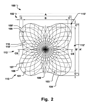
Fig. 2 is an illustration of a medical implantable
occlusion device according to an embodiment of the
invention;
Figs. 3a-c are side views along cross-section a (CS)
of the medical implantable occlusion device in Fig. 2;
CA 02847087 2014-02-27
WO 2013/041721 PCT/EP2012/068760
6
Fig. 4 is an illustration of a medical implantable
occlusion device according to an embodiment of the
invention;
Figs. 5a-c are illustrations of a medical implantable
occlusion device according to an embodiment of the
invention;
Fig. 6 is an illustration of a medical implantable
occlusion device according to an embodiment of the
invention when implanted at a treatment site;
Figs. 7a-d are illustrations of a medical implantable
occlusion device according to an embodiment of the
invention;
Fig. 8 is an illustration of a medical implantable
occlusion device according to an embodiment of the
invention;
Fig. 9 is a flow chart illustrating a method of
occluding a PVL in a body lumen with a medical implantable
occlusion device according to an embodiment of the
invention; and
Figs. 10a-c are illustrations of a medical
implantable occlusion device according to an embodiment of
the invention, shown in top-down view (a), and side views
(b)-(c), respectively.
Description of embodiments
Specific embodiments of the invention will now be
described with reference to the accompanying drawings.
This invention may, however, be embodied in many different
forms and should not be construed as limited to the
embodiments set forth herein; rather, these embodiments are
provided so that this disclosure will be thorough and
complete, and will fully convey the scope of the invention
to those skilled in the art. The terminology used in the
detailed description of the embodiments illustrated in the
accompanying drawings is not intended to be limiting of the
CA 02847087 2014-02-27
WO 2013/041721 PCT/EP2012/068760
7
invention. In the drawings, like numbers refer to like
elements.
The following description focuses on an embodiment of
the present invention applicable to a Para-Valvular Leak
device (PLD). However, it will be appreciated that the
invention is not limited to this application but may be
applied to any other purposes of cardiac or vascular
occlusion, and many other medical implantable devices,
including for example filters, stents, Left Atrial
Appendage (LAA) occluders, aneurysm treatment devices,
grafts, etc.
Fig. 1 shows a medical implantable occlusion device
100 according to an embodiment of the invention. Fig. 4
shows a device 200 in another embodiment of the invention,
which is similar to the device 100 in Fig. 1 but with other
relative dimensions. The device 100, 200, comprises a
fabric, mesh or braiding of at least one thread 101. The
fabric may be formed from one thread or several. Figs. 3a-c
are cross-sections of the device 100, 200, along the line
CS in Fig. 1 or Fig. 4, which will be discussed in further
detail below.
The device 100, 200, or more particularly the
structural formation 102 of the fabric of threads 101, has
an unloaded expanded shape and a collapsed shape. Thus, in
the expanded shape, wherein the device 100 has a shape as
depicted in Fig. 1 and Fig. 2, no external force acts on
the device 100, 200. The device 100, 200, may be stretched
and thereby exhibit a smaller cross-section, in order to
fit inside a delivery device such as a catheter. The device
100, 200, may be self-expandable between the collapsed
shape and the expanded shape, i.e. when the device 100,
200, is removed from the confinement of the catheter the
cross-section of the device 100, 200, returns to its
originally defined value in the unloaded expanded shape.
The device may be self-expandable due to an inherent
CA 02847087 2014-02-27
WO 2013/041721 PCT/EP2012/068760
8
elasticity of the threads in the fabric or braiding. The
device may also have a shape memory, e.g. triggerable to go
to the expanded shape at a switching temperature, such as
body temperature.
The shape of the device 100, 200, in the expanded
shape may be defined in a heat treatment procedure of the
device 100, 200, or more particularly of the braiding of
the device. The dimensions of the device 100, 200, in the
expanded, relaxed, shape are defined in the heat treatment
procedure of the braiding.
The entire device 100, 200, may be comprised of a
single, continuous fabric or braiding. The braiding may be
made of a material suitable for implanting in a human or
animal body, and suitable for being formed in a heat
treatment procedure to a desired shape in the expanded
shape and also in the stretched state. For example NiTinol
may be used as a material for the device 100, 200. However,
suitable materials for embodiments of the braiding are
various and include shape memory materials, metal,
superelastic alloys (such as NiTinol), or polymers, such as
degradable polymers.
The structural formation 102 of the device 100, 200,
comprises a proximal portion 103, and a distal portion 104.
A longitudinal axis 105 extends between the proximal and
the distal portion, which is best illustrated in Fig. 3a.
In Fig. 3a it is also seen that the proximal, and distal
portions 103, 104, may comprise expanded diameter portions
103, 104, that are separated by a waist 106 of reduced
cross-section between the proximal and distal portions 103,
104. The length 120 of the waist 106 may correspond
substantially to the wall thickness of the defect to be
occluded, when the proximal and distal portions 103, 104,
are positioned on either side of such defect. The flexible
nature of the at least one thread 101 of the device 100,
200, however allows the device to adapt to a wide range
CA 02847087 2014-02-27
WO 2013/041721 PCT/EP2012/068760
9
defect dimensions. The proximal and distal portions 103,
104, will strive in a direction towards each other to the
expanded shape when separated by the defect, thereby
closing against the walls on either side of the defect and
providing the occluding effect. The width 121 of the waist
106 may approximate the width of the defect, i.e. the width
of the opening of the paravalvular leak defect.
Figs. 10b and 10c show two different types of waists
106, along the cross-section A of the device 100 seen in a
top-down view in Fig. 10a. According to one embodiment the
waist may comprise narrowly or tightly twisted threads 101,
of the fabric or braiding of the device 100, around the
longitudinal axis 105, in order to produce a waist 106 of
small cross-section relative to the diameter of the
proximal and distal portions 103, 104. This small cross-
section allows for fitting of the device in small openings
to be occluded. During manufacturing of the device 100, the
proximal and distal portions 103, 104, may be twisted in
relation to each other around the longitudinal axis 105,
during a heat setting step, to produce a waist 106 with
twisted threads 101. This may be part of a subsequent heat
setting step, after a first heat setting step for forming
the expanded diameter portions, i.e. the proximal and
distal portions 103, 104, and the reduced diameter portion,
i.e. the waist. Alternatively, the twisting is made during
the same first heat setting step.
The waist 106 may be made of a portion of parallel
threads or a more densely braided section of the fabric at
the waist 106, providing for particular strength in the
longitudinal direction. The waist 106 may be arranged
concentrically with respect to the proximal and distal
portions 103, 104, but an asymmetric configuration may be
suitable in particular anatomies to be occluded.
At least one of the proximal and distal portions 103,
104, comprises a peripheral edge 107, 108, having a first
CA 02847087 2014-02-27
WO 2013/041721 PCT/EP2012/068760
109, 109', and a second 110, 110', radius of curvature in a
direction substantially perpendicular to the longitudinal
axis 105. The first radius of curvature 109, 109', is
different from the second radius of curvature 110, 110'. In
5 this way the peripheral edge 107, 108, may conform to
various anatomical geometries neighboring the defect to be
occluded, hence avoiding unnecessary blockage and
disruption of e.g. blood flow, while still providing the
occlusion of the defect.
10 In case of paravalvular leak defects (PVL) the at least
one of the first and second radius of curvatures may be
chosen such that the curvature of at least a section of the
peripheral edge 107, 108, corresponds substantially to a
valve curvature 116 of a valve 115 for regulating blood
flow. This is illustrated in Fig. 6, where the device 200
occludes a PVL close to the outer boundary of the valve
116. The device 200 has a peripheral edge 108 with a second
radius of curvature 110' that corresponds substantially to
the valve curvature 115. The first radius of curvature 109'
as exemplified in Fig. 6 is different from the second
radius of curvature 110' of the peripheral edge 108, and
the peripheral edge 108 may have any shape to conform to
varying neighboring geometries where the influence of the
occlusion device must be minimized while providing the
necessary occlusion effect.
A prior art device 10 is shown in Figs. la-b, which is
a typical example of the influence such prior art devices
have on the prosthetic valve because of its substantial
blockage of the valve 115 when positioned in a PVL at the
periphery of the valve 20. Fig. la is a view from the
atrial side, and Fig. lb is a view from the ventricular
side, where the latter most clearly shows the device 10
extending over a substantial portion of the valve 20. Such
device 10 may disrupt the blood flow, create turbulence and
lead to various complications as discussed above. Returning
CA 02847087 2014-02-27
WO 2013/041721 PCT/EP2012/068760
11
to Fig. 6, the corresponding overreach across the valve 115
resulting from such prior art devices is marked with dashed
line 121. The varying radius of curvature 109', 110', of
the device 100, 200, allows occlusion of PVL close to the
valve 115 without any overreach across the valve and the
associated complications. Figs. 5a-c illustrates the amount
of area that is saved by the device 100, 200, which
otherwise would have negative impact. Fig. 5a shows the
coverage by a prior art device 10 (dashed lines), and Fig.
5b shows the coverage by the device 200, while Fig. Sc
shows the differential area 122 that is saved which will
not block the flow of blood through the valve 115.
Both the proximal and distal portions 103, 104, may
have peripheral edges 107, 108, with varying radius of
curvature. Fig. 2 shows the first and second radius of
curvature 109, 110, for the proximal portion 103, and the
first and second radius of curvature 109', 110', of the
distal portion 104 for the device 100. Fig. 4 and 6 shows a
similar configuration for the device 200. In this way the
blood flow will not be disrupted on any side of the valve
115.
The dimensions of the device 100, 200, such as
indicated in Fig. 2 and 3, c.f. A, A', B, B', C, C', C",
C"', D, E, may be adapted such that proper alignment of
the device 100, 200, to the valve curvature 116 is
achieved.
As seen in Figs. 2, 4, 5, 6, the peripheral edge 107,
108, is concave radially outwards. I.e. in a direction
substantially perpendicular to the longitudinal axis 105,
Fig. 3a. This allows the peripheral edge 107, 108, to
follow the convex shape of the valve curvature 116, so that
no overlapping of the valve 115 occurs. The radius of
curvature of the concave part can be varied as desired in
order to achieve the closest correspondence with the valve
curvature 116. The number of concave sections of the
CA 02847087 2014-02-27
WO 2013/041721 PCT/EP2012/068760
12
peripheral edge 107, 108, may vary. The devices 100, 200,
in Figs. 2, 4, 5, 6, have four concave sections, but it
could be one, two, three, five or more. Leaks can occur
around the valve 115 at a 360 deg location. The curvature
of the peripheral edge 107, 108, may be sized and shaped to
cover several PVL's around the valve curvature 116. Due to
the varying radius of curvature or the concave peripheral
edge 107, 108, several PVL's may be occluded with a single
device 100, 200, without extending across the valve 115 and
disturbing the blood flow.
As further shown, e.g. in Fig. 2, the peripheral edge
107, 108, comprises edge sections 112, 112', 113, 113',
that are alternatingly concave and convex radially outwards
in a direction substantially perpendicular to the
longitudinal axis 105. Each of the concave edge sections
112, 112', also seen in Fig. 4 and denoted 114, 114', may
be positioned against the valve curvature 116. The devices
100, 200, have the convex sections 113, 113', positioned in
between the concave sections 112, 112', which results from
having a several concave sections.
The geometric terms concave and convex as used herein
is to be interpreted for the purposes of the invention as
their normal geometrical meaning including any recesses in
the device for the purpose of the term concave and
protrusions of the device for the purposes of the term
convex, where such recesses and protrusions may also define
the spatial extent of the device 100, 200, i.e. the
peripheral edge 107, 108, such that the device 100, 200,
may follow the valve curvature 116. The peripheral edge
107, 108, may be continuous without sharp interruptions,
kinks or corners, as illustrated in the Figures, or
comprise discontinuous sections.
The device 100, 200, has radially opposed edge sections
112, 112', 114, 114', of the peripheral edge 107, 108, that
have substantially the same radius of curvature, e.g. as
CA 02847087 2014-02-27
WO 2013/041721 PCT/EP2012/068760
13
seen in Fig. 2 and 4. Such symmetry may provide ease of
positioning against the valve curvature 116. In Fig. 2, the
device 100, has substantially the same radius of curvature
for all concave sections of the peripheral edge 107, 108.
Alternatively, the device 100, 200, may comprise concave
edge sections having different radius of curvatures 109,
109', 110, 110', which allows the device 100, 200, to
conform to a wide range of valves 115, having different
valve curvatures 116.
Fig. 4 shows a device 200 having first radially opposed
edge sections 112, 112', of the peripheral edge 107, 108,
having a radius of curvature that is larger than the radius
of curvature of second radially opposed edge sections 114,
114'. As mentioned above this may provide selectivity to
various geometries of the valve 115. By simply rotating the
device 200, the physician may select one peripheral edge
with a particular radius of curvature that conforms best to
the valve curvature 116, and/or the opening to be occluded.
Also, the device 200 may provide increased holding strength
against the defect to be occluded by its increased radial
extent along a first axis, while maintaining the limited
radial extent along a second axis, being perpendicular to
the first axis, i.e. the second axis extending in direction
across the valve 115. Overlap across the valve 115 by the
device 200 (along the aforementioned second axis) is
thereby avoided, while increased holding strength is
provided.
The peripheral edge may comprise at least two edge
sections 112, 112', or 114, 114', that are concave radially
outwards in a direction substantially perpendicular to said
longitudinal axis. Having more than one concave edge may
allow selectivity as described above, and/or ease of
positioning if the edges have similar radius of curvature.
Further, by having a rotational symmetric device 100,
200, around axis 105, the ease of handling and insertion,
CA 02847087 2014-02-27
WO 2013/041721 PCT/EP2012/068760
14
and also stability and structural integrity of the device
can be increased. This can be realized by having two
radially opposed concave edges as described above. The
radius of curvature of the peripheral edge 107, 108, may
correspond to a particular defect to be occluded and/or the
curvature of the valve.
The device 100, 200, may have a peripheral edge 107,
108, that defines a generally rectangular shape of the
proximal or distal portion 103, 104. As seen in Figs. 2 and
4, the device 100, 200, has four convex corners, see e.g.
edge sections 113, 113', and concave sections in between,
112, 112'. The peripheral edge 107, 108, may have a radius
of curvature that vary considerably, e.g. the convex
corners 113, 113', of the device 100, 200, may be in the
form of a sharp transition from one concave edge section to
the next, as alternative to a smooth continuous transition.
In either case the device 100, 200, may be referred to as
having a generally rectangular shape due to having four
corners in the Figs. As mentioned above the number of
concave sections 112, 112', and corners, i.e. convex
sections 113, 113' may vary, and the device 100, 200, may
have generally triangular, pentagonal shapes etc, as long
as the peripheral edge 107, 108, has at least a section of
its curvature that can be positioned close to the valve
curvature 116 without extending across the valve 115, when
the device 100, 200, is in its implanted site.
At least one of the proximal and distal portions 103,
104, may be deflected towards the other portion with an
angle V, V'. In this way the device 100, 200, may better
accommodate to the anatomy at the implanted site and
thereby provide a closer fit against the tissue by the
proximal and/or distal portion 103, 104, for improved
occlusion. For example, at the periphery of the valve 115,
there is often a "volcano crest", i.e. a protrusion going
around the periphery. When the proximal or distal portion
CA 02847087 2014-02-27
WO 2013/041721
PCT/EP2012/068760
103, 104, is positioned close to that protrusion the
deflection of the aforementioned portions towards each
other with angle, V, V', allows these portions to reach
over the protrusion and down to the tissue nest to the
5 protrusion for a secure fit. Fig. 3a shows the cross-
section of the device 100, 200, where the proximal portion
103 is deflected towards the distal portion 104 with an
angle V, and the distal portion 104 is deflected towards
the proximal portion 103 with an angle V'. The angles V and
10 V' may be substantially the same or different depending on
the anatomy of the site in the vascular system to be
occluded. E.g. the distances 123, 124, as indicated in Fig.
3a, may be varied. Only one of the portions 103, 104, may
be angled towards the other. The device 100, 200, may
15 thereby be adapted to the irregular and varying anatomy of
the implantation site. This also allows for a particular
stable long-term construction even in anatomical situations
where a continuous movement at the implantation site is
present.
One of the proximal and distal portions 103, 104, may
have a larger diameter than the other portion, thereby
creating an overlap 117 between the proximal and distal
portions 103, 104. The overlap may provide increased
sealing ability of the device 100, 200, e.g. when the
portions 103, 104, being pressed towards each other. The
overlap may substantially be in the radial direction,
perpendicular to longitudinal axis 105. As seen in Fig. 3a,
the distal portion 104 overlaps the proximal portion 103 in
the radial direction, which is also seen in e.g. Fig. 2
with respect to peripheral edges 107, 108. When the distal
portion 104 is placed on the side of the defect being
exposed to high pressure, e.g. on the ventricular side of
the heart (depending on which valve that has PVL; Aortic,
Mitral, Tricuspid, or Pulmonary), the larger area of the
distal portion 104 will improve the sealing against the
CA 02847087 2014-02-27
WO 2013/041721 PCT/EP2012/068760
16
tissue, while the smaller area of the proximal portion
minimizes overlap across the valve 115. Thus a secure
occlusion is achieved even before the device 100, 200, is
securely covered with endothelia and tissue integrated with
the surrounding tissue.
The diameter may be equivalent to the largest cross-
section throughout the disclosure.
The proximal and distal portions 103, 104, may be
substantially flat and having a diameter larger than the
opening of the PVL which it is placed.
Figs. 7a-d shows perspective view of the device 100,
200, i.e. Fig. 7a is a tilted side view, Fig. 7b is a side
view, Fig. 7c is a top-down view facing the proximal
portion 103, and Fig. 7d is a top-down view facing the
distal portion 104. Even though the device in Figs. 7a-d
more closely reassembles the device 100 in Fig. 2 due to
having substantially sides of equal length, the perspective
views in the Figs. is also representative of the device 200
in Fig. 4. The device 100, 200, may comprise at least one a
marker element 118, 118', for aiding in orienting the
device 100, 200. Such marker 118, 118', allows
identification of the device and reassurance that the
device has been implanted correctly. For example, it can be
determined whether the concave edge section 112 of the
peripheral edge 107, 108, has been aligned against the
valve curvature 116. Thus, the at least one marker element
118, 118', may be arranged on one of the proximal and
distal portions 103, 104, at a position corresponding
substantially to the location of the peripheral edge 107,
108. Fig. 8 illustrates the location of two markers 118,
118', which are close to the peripheral edge 107 of the
proximal portion 103. The markers 118, 118', may be
arranged on opposite concave sections, as illustrated in
the figure for allowing correct positioning. The markers
118, 118', may be attached to the proximal portion 103, or
CA 02847087 2014-02-27
WO 2013/041721 PCT/EP2012/068760
17
to the distal portion 104. Hence, the markers 118, 118', in
Fig. 8 could be attached to the distal portion 104, for
marking out the position of the peripheral edge 107 of the
proximal portion 103. As the distal portion 104 may have
increased diameter or circumference, the markers 118, 118',
could be attached to the distal portion 104 at a distance
from the peripheral edge 108 of the distal portion 104,
while still marking out the peripheral edge 107 of the
proximal portion. This may allow for easier attachment of
the markers 118, 118', to the device 100, 200, and less
interference with the operation of the device 100, 200, as
the markers do not have to be attached to the proximal
peripheral edge 107, while still allowing exact positioning
with respect to the valve curvature 116 with the proximal
peripheral edge 107.
The marker element 118 may comprise a radiopaque
material, hence being identifiable in X-ray, or comprise
material for easy identification in MRI. The device 100,
200, may comprise two markers 118 as shown in Fig. 7d,
arranged across the radial direction of the distal portion
104, and/or alternatively of the proximal portion 103. Any
number of markers 118 may be used for identification. The
markers 118 may be fixated to an occluding element such as
a patch, fibers or the like comprising a biocompatible
material (e.g. PET) for supporting the sealing of the blood
flow through the device 100, 200, or fixed to the fabric of
threads 101 of the device 100, 200, itself.
As shown in Figs. 3b, 3c and Figs. 7a-c, the device
100, 200, may comprise a connecting member 111 attached to
one of the distal and proximal portions 103, 104, for
connection to a delivery device (not shown). The delivery
device may grasp the connection member 111 which may be
spherical in shape, thus providing a pivoting motion of the
device 100, 200, in relation to the delivery device in
combination with secure attachment. The connection member
CA 02847087 2014-02-27
WO 2013/041721 PCT/EP2012/068760
18
111 may be arranged on the proximal portion 103 as shown in
Fig. 3b, or on the distal portion 104 as shown in Fig. 3c.
Fig. 7a-c illustrates the device 100, 200, having the
connection member 111 on the proximal portion 103. In
reality the portion having the connection member 111
becomes the proximal portion in use of the device, but the
above conventions are used for conciseness in the
description and figures. Hence, the connection member 111
may be arranged on the expanded diameter portion 104, or
the increased diameter portion 104. This allows the
possibility to access the PVL from both sides of the leak.
The connecting member 111 may be configured for
connection to a delivery device in a predetermined
orientation. Hence a specific orientation of the device
100, 200, could be maintained relative to the delivery
device during implantation which may aid in positioning the
device 100, 200, in relation to the valve curvature 116.
The ends of the at least one thread 101 forming the
fabric may be fixed to the connecting member 111. The
connecting member 111 may thus be a weld or any other
attachment means for the threads 101 of the fabric. The
distal portion 104 may comprise returning loops 119 of the
at least one thread 101, meaning that opposite ends of the
at least one thread 101 forming the distal portion 104 are
fixed to the connecting member 111. By having returning
loops only one collection point for the ends of the at
least one thread 101 is needed. The connection member 111
may thus serve as a connection for these ends, thereby
avoiding multiple connection points such as welds on the
distal portion 104. Hence, a flat distal portion 104 may be
provided, that increases the compactness of the device 100,
200. The flat distal portion 104 may thus be a closed
continuous distal wall 301 of the braiding forming the
device 100, 200, i.e. free from a thread ends. This reduces
the risk thromboembolic complications. E.g. nothing is
CA 02847087 2014-02-27
WO 2013/041721 PCT/EP2012/068760
19
protruding into the blood stream, and there are no
discontinuities that may cause thromboembolic
complications. Further, due to the connection member 111 on
the proximal end 103, the device 100, 200, may be delivered
through the vena cava with improved safety to the patient.
The implantation techniques are different for each PVL
according to the valve and the location of the leak.
Delivery to the high pressure arterial side of the vascular
system is avoided, which provides for less complications
and a medical procedure which is simpler to perform.
Fig. 9 illustrates a medical method 900 of occluding an
opening in a body lumen, comprising providing 901 a device
100, 200, inserting 902 the device 100, 200 in a collapsed
state into the opening, expanding 903 and releasing the
device 100, 200, in the opening, thus anchoring 904 the
device 100, 200, in the opening for occluding the latter by
the device 100, 200. The opening may be a Para-Valvular
Leak (PVL), and the method may comprise positioning 905, or
rotating, the device 100, 200, such that a concave edge
section 112, 112' of the device 100, 200, substantially
follows the valve curvature 116 of the valve 115.
The present invention has been described above with
reference to specific embodiments. However, other
embodiments than the above described are equally possible
within the scope of the invention. The different features
and steps of the invention may be combined in other
combinations than those described. The scope of the
invention is only limited by the appended patent claims.
More generally, those skilled in the art will readily
appreciate that all parameters, dimensions, materials, and
configurations described herein are meant to be exemplary
and that the actual parameters, dimensions, materials,
and/or configurations will depend upon the specific
CA 02847087 2014-02-27
WO 2013/041721
PCT/EP2012/068760
application or applications for which the teachings of the
present invention is/are used.