Note: Descriptions are shown in the official language in which they were submitted.
CA 02847310 2014-03-21
WO 2007/076061 PCT/US2006/049128
TITLE
A POROUS MEMBRANE DEVICE THAT PROMOTES THE
DIFFERENTIATION OF MONOCYTES INTO DENDRITIC CELLS
CROSS REFERENCE TO RELATED CASES
This application claims the benefit of Provisional U.S. Application Serial No.
60/752,033, filed December 21, 2005, which is incorporated by reference herein
in its
entirety.
BACKGROUND OF THE INVENTION
The generation of protective immunity against pathogens and tumors in
mammals requires specialized cells that can present foreign or altered self
antigens to T
cells. Dendritic cells (DCs) are thought to be the most potent of these
antigen-
presenting cells (APCs) because they efficiently acquire and process antigen
for
presentation in major histocompatibility complex (MHC) molecules and express
high
levels of T cell costimulatory ligands, both of which are necessary to trigger
complete
differentiation of naïve T cells into competent effector cells. It is also
thought that DCs
are more capable than other APCs of cross-presenting exogenous proteins
through the
endogenous (MHC class I) pathway, making them particularly important for
generating
cyiotoxic T lymphocyte responses against tumors and extracellular pathogens.
Dendritic cells are typically found in most tissues of the body and are
derived
from circulating monocytes that traverse the vascular endothelium into
peripheral
tissues. Under normal conditions, these cells have a high capacity for antigen
acquisition, but low levels of surface MHC and costimulatory molecule
expression.
Injury or infection triggers a marked increase in the number of DCs at the
affected site. Additionally, these DCs acquire an activated phenotype,
characterized by
increased expression of soluble and membrane-bound molecules, decreased
capacity to
acquire antigen, and enhanced migration towards secondary lymphoid tissues. In
lymph nodes, these cells are potent stimulators of antigen-specific T cell
activation.
For a more complete synopsis on the biology of DCs, see the recent review by
Rossi &
Young (J Immunol 175:1373-1381 (2005).
CA 02847310 2014-03-21
WO 2007/076061 PCT/1JS2006/049128
It is beneficial to construct a wholly in vitro immune response for screening
and
assessing the immunogenicity of vaccines, drugs, or other compounds. Employing
human subjects for this purpose may be dangerous and is costly, while using
laboratory
Until now, there has been no convenient, cost effective, and automatable in
vitro technique for preparing DCs from peripheral blood cells in a manner that
simulates what occurs in the body. Monocytes can be segregated from peripheral
blood
The generation of protective immunity against infection and tumors requires
specialized cells that can present foreign or altered self antigens to T
cells. While
several cell types can act as APCs, DCs are the most potent of these and the
only ones
capable of inducing CD4+ and CD8+ T cell responses against naïve antigens.
Under
2
CA 02847310 2014-03-21
WO 2007/076061 PCT/US2006/049128
Tissue-resident DCs comprise a heterogeneous population of cells that is found
in most organs of the body. Short-lived circulating monocytes, which give rise
to
iDCs, traverse the vascular endothelium into peripheral tissues in a
constitutive manner,
though infection or injury triggers an increased accumulation of these cells
at the
inflamed site. Within tissues, a subset of the extravasated monocytes
differentiate into
iDCs, with the milieu of the local microenvironment often influencing the
phenotype
and functional activity of APCs residing in a particular site. For example,
gut-
associated DCs populate Peyer's patches, where they receive antigens from M
cells and
act as the resident APCs of mucosal tissue. Langerhans cells, on the other
hand, are
found primarily in the skin and play a key role in the induction of adaptive
responses
following infection.
Several laboratories have worked to develop in vitro systems which
recapitulate
the cell interactions and signaling pathways that trigger monocyte to DC
differentiation
in vivo. For instance, the groups of Muller and Randolph (Qu et al. (2003)J
Immunol
170, 1010-1018; Randolph etal. (1998) Science 282, 480-483) pioneered the
development of tissue constructs that utilize HUVECs grown on a support matrix
to
promote the generation of human DCs from blood monocytes that have
transmigrated
through the endothelial layer. The APCs derived from this system resembled DCs
in
phenotype and ability to trigger allogeneic and primary antigen-specific T
cell
responses (Qu et al. (2003)J Immunol 170, 1010-1018; Randolph et aL (1998)
Science
282, 480-483). While this tissue model might generate APCs that more
accurately
represent DC populations found in vivo, its complexity makes it impractical
for
widespread use. In another approach, adherent monocytes cocultured directly
with
human or porcine endothelial cells gave rise to potent APCs that produced
proinfiammatory cytokines, expressed high levels of costimulatory ligands, and
efficiently stimulated allogeneic T cells. A limitation of this technique is
that the DCs
had to be selected from contaminating endothelial cells by magnetic bead
selection
before any functional analyses could be performed.
There has been tremendous interest in better understanding the biology of DCs
because of their specialized role in orchestrating primary cellular and
humoral immune
responses. The paucity of DCs in the body, combined with the limited
availability of
tissue samples from humans, make it difficult to evaluate these cells in an ex
vivo
3
CA 02847310 2014-03-21
WO 2007/076061 PCT/US2006/049128
manner. As a result, the study of cytokine-derived DCs, i.e., purified blood
monocytes
that have been cultured in exogenous growth factors (GM-CSF and IL-4), has
contributed great insight into this unique cell population and provided a
source of APC
for clinical applications. The utility of cytokine-derived DCs is limited,
however,
because this culture method fails to replicate the physiology involved in the
development of DCs from circulating monocytes in the body. Additionally, some
researchers have suggested that this DC population lacks full APC
functionality and
may not accurately represent DC populations found under physiologic conditions
(Romani et al. (1994) J Exp Med 180:83-93 ; Sallusto & Lanzavecchia (1994)J
Exp
Med 179,1109-1118; Thurnher et a/. (2001) FASEB J. 15, 1054-1061).
BRIEF SUMMARY OF THE INVENTION
The present invention provides a method for generating large numbers of
dendritic cells comprising: =
-culturing endothelial cells on top of a porous membrane, wherein said
membrane is housed in an upper chamber of a well that is suspended over, and
is
separable from, a lower chamber of a well:
-applying peripheral blood mononuclear cells (PBMCs) to the endothelial cells
on the porous membrane;
-at least about 48 hours after application of the PBMCs, removing the upper
chamber of the well, housing the porous membrane and endothelial cells; and
-isolating dendritic cells from the lower chamber of the well.
The present invention also provides a method of evaluating the potential
reaction of an
animal to an agent, said method comprising:
-producing a first well comprising:
-a first porous membrane as the base;
-a ECM material affixed on top of said first porous membrane; and
-a second porous membrane affixed on top of said ECM material;
-inverting said first well into a second well comprising cell media;
-culturing endothelial cells on bottom of said first porous membrane;
-applying peripheral blood mononuclear cells (PBMCs) to the endothelial cells;
4
CA 02847310 2014-03-21
WO 2007/076061 PCT/US2006/049128
-after ¨1.5 hours washing said PBMCs and said endothelial cells off of the
bottom of said first porous membrane, wherein dendritic cells are now present
in said ECM material;
-removing said first well from said second well comprising cell media and
placing said first well with said second porous membrane facing up into a
third
well comprising as its base a three-dimensional artificial lymphoid tissue,
comprising a second ECM material and a plurality of lymphocytes and
leukocytes;
-applying an agent to the top of said second porous membrane, said antigen
allowing the dendritic cells to migrate out of said first ECM material and
into
said three-dimensional artificial lymphoid tissue; and
-evaluating the immune response to said agent.
The present invention further provides a method for generating large numbers
of dendritic cells comprising:
-producing a first well comprising:
-a first porous membrane as the base;
-endothelial cells cultured on the bottom of said first porous membrane;
-a second porous membrane situated above, and separated from, said
first porous membrane;
-endothelial cells cultured on the top of said second porous membrane;
and
-cell culture media comprising an agent located between said first
porous membrane and said second porous membrane;
-inverting said first well into a second well comprising cell media;
-applying peripheral blood mononuclear cells (PBMCs) to the endothelial cells
cultured on the top of said second porous membrane;
-at least about 48 hours after application of the PBMCs, removing said first
well
from said second well; and
-isolating dendritic cells from said second well.
BRIEF DESCRIPTION OF THE FIGURES
Figure 1. Schematic diagram of an embodiment of the invention, using a
Transwell device. HUVECs are grown to confluency on Transwell membranes and
5
CA 02847310 2014-03-21
WO 2007/076061 PCT/US2006/049128
then total PBMC are applied to the upper chamber for-4.5 h (step 1). Unbound
cells
are washed away and the remaining leukocytes are allowed to transmigrate for
¨48 h.
The Transwell is removed and DCs are then collected for analysis or pulsed
with
antigen for an additional ¨2 days (step 2).
Figure 2. In other embodiments, the complexity of the membrane device can
be increased by, for example, the inclusion of secondary cell populations, ECM
materials and additional membrane layers. Monocytes that traverse through an
endothelial monolayer can contact ECM in the lower chamber of the membrane
device
(A). Two membrane devices can be used to mimic the normal pathway of monocyte
migration from the blood into the tissue (through the HUVECs) and from the
tissue into
the lymphatics (through a second cell layer, such as, for example, lymphatic
endothelial
cells). The second monolayer can be cultured on the upper (B) or lower (C)
side of the
membrane device, mimicking transmigration or reverse transmigration,
respectively.
The membrane can be coated on both sides with the same or different cell types
(D);
ECM can also be incorporated into the lower chamber with this design,(E). A
modified
Transwell can be constructed that contains a central chamber sandwiched
between two
membranes/cell monolayers (F). Fibroblasts or other cells types that are
important in
DC differentiation or antigen-presenting activity can be included in the lower
chamber
of a single membrane device (G or H) or in the middle of a dual membrane
device (1
and J). ECM can also be incorporated into the dual-membrane device (H).
Figure 3. HUVECs form confluent monolayers on Transwell membranes.
Primary HUVECs were seeded in the upper chamber of Transwell s and analyzed
for
confluency and the formation of tight-gap junctions. (A) On day 7 after
seeding, the
cells were fixed, surface-labeled with an antibody specific for CD31, and the
nuclei
were stained with DAPI. (B) At the indicated time points, electrical
resistance (TEER)
readings were collected and normalized against the values for empty Transwell
s on
the same day. The error bars represent 1 SD of triplicate readings in each
well. (C)
Diffusion through the endothelial layer was measured with a 70kDa FITC-dextran
conjugate at the indicated time points.
Figure 4. Monocyte transmigration through an endothelial monolayer is
sufficient to trigger their differentiation towards a DC phenotype. (A) Cells
that
6
CA 02847310 2014-03-21
WO 2007/076061 PCT/US2006/049128
passed through a PC membrane in the absence (left) or presence (right) of a
HUVEC
monolayer were imaged by phase microscopy (20x objective). Arrows indicate
contaminating red blood cells or lymphocytes. (B) CD14-purified monocytes (non-
transmigrated) were put into culture and then labeled with monoclonal
antibodies
specific for the indicated markers ¨2 d later. The dotted line indicates
background
fluorescence with the appropriate isotype control. (C) Cells that
transmigrated through
the membrane in the absence and presence of a HUVEC monolayer were also
examined for expression of the indicated surface proteins ¨2 d after PBMC were
applied to the Transwell . The expression level of migrated monocytcs is
plotted as a
percent increase or decrease over the MFI on non-migrated monocytes, which are
set to
100%. All analysis plots are gated on monocytes only.
Figure 5. Transwell -derived DC are potent stimulators of antigen-specific T
cell responses. Transwell -derived DC were pulsed with antigen and cultured at
a
¨1:20 ratio with autologous T cells that had been labeled with CFSE. About 7 d
later,
the T cells were restimulated with autologous antigen-pulsed DC (-1:10 ratio
to T
cells) for ¨8 h and then assayed for 1L-2 production by ICCS. Unpulsed DC were
included as a negative control. (A) Dot plots showing representative CFSE and
IL-2
staining patterns. The capacity of Transwell - and cytokine-derived DCs to
stimulate
recall C. albicans -specific T cell responses were compared in (B), while
transmigrated
cells from HUVEC-negative and -positive Transwelles served as APCs in (C). In
both
assays, the T cells were analyzed for cytokine production by flow cytometry
and the
graph shows the frequency of lymphocyte-gated CD3 CFSEI0wIL-2+ cells.
Different
donors were used in each assay.
Figure 6. Example configurations of the vac,cination site.
Figure 7. An example of laser-micromachined polycarbonate (PC) membranes
in a Transwell -based model and an outline of the process of casting collagen
in a well-
based model.
Figure 8. Cell migration within the collagen membrane and transmigrated cells
on the bottom of the plate.
7
CA 02847310 2014-03-21
WO 2007/076061 PCT/US2006/049128
Figure 9. Cell migration and reverse transmigration.
Figure 10. Levels of expression of HLA-DR in the migrated cells each model.
Figure 11. Levels of expression of CD86 in the migrated cells each model.
Figure 12. Levels of expression of CCR7 in the migrated cells each model.
Figure 13. Building-in complexity to the VS model.
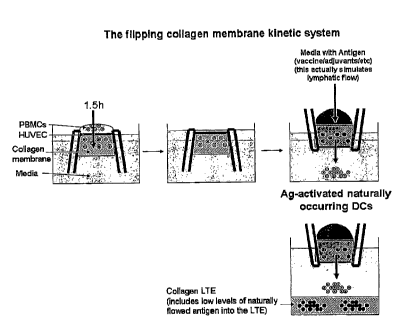
Figure 14. Antigen introduction into the VS model. The flipping collagen
membrane model as an example.
Figure 15. Adherent transmigrated monocytes phenotypically resemble
macrophages. PBMCs were applied to the upper chamber of a Transwell
containing
HUVEC and at ¨48 h the migrated, non-adherent and adherent cells were
collected
from the lower chamber. The cells were labeled with specific antibodies and
analyzed
by flow cytometry. The MFI for each marker on adherent and non-adherent cells
are
represented graphically.
Figure 16. Transmigrated APC have phagocytic activity. The non-adherent
transmigrated APC were harvested from Transwell s and incubated with FITC-
labeled
dextran beads (¨I p.m) or zymosan particles, in the absence (thin line) or
presence
(thick line) of 20 ug/mL cytochalasin D, for ¨24 h. The cells were analyzed by
flow
cytometry in the presence of trypan blue, which quenches any extracellular
FITC
fluorescence. This ensures that the only signal detected originates from
material within
= the cell.
Figure 17. Transmigratory DCs respond to maturation stimuli. ¨2 d after
PBMC application to the Transwell s, the transmigrated cells were harvested
and
incubated for an additional ¨48 h in the absence or presence of TNF-a and LPS.
Thereafter, the cells were incubated with the antibodies specific for the
indicated
markers and analyzed by flow cytometry. Thin solid lines = non-matured cells;
Thick
8
CA 02847310 2014-03-21
WO 2007/076061 PCT/US2006/049128
solid line = matured cells; dotted lines = isotype controls. All analysis
plots include
only gated monocytes.
Figure 18. A transformed endothelial cell line can be used to trigger
monocytes
differentiation to DCs in the Transwell system. The ability of Transwell -
derived
APC from cultures containing primary and transformed HUVEC were compared, as
described in Fig. 5, using PBMC from a single donor. This data is
representative of at
least 3 experiments.
DESCRIPTION OF THE INVENTION
Embodiments of the present invention comprise a simple and convenient
Transwele-based culture method for the endothelial cell-mediated
differentiation of
DCs from blood monocytes. This system produces DCs with a frequency and purity
comparable to more traditional culture methods for culturing DCs in vitro, but
does so
in only about two days, in the absence of exogenous factors and without the
need for a
tissue construct containing a support matrix. The transmigrated APCs derived
from
these cultures resemble classical in vitro DCs in their expression of MHC and
costimulatory molecules and capacity to induce antigen-specific T cell
responses. In
another embodiment, the use of a durable and fast-growing transformed
endothelial cell
line, which yields APCs that are comparable to those obtained when primary
endothelial cells were used in the system, makes this approach a highly
convenient are
reliable technique for the generation of highly purified DCs.
Human dendritic cells (DCs) for research and clinical applications are
typically
derived from purified blood monocytes that are cultured in a cocktail of
cytokines for a
week or more. Because it has been suggested that these cytokine-derived DCs
may be
deficient in some important immunological functions and might not accurately
represent antigen-presenting cell (APC) populations found under physiologic
conditions, there is a continuing need for methods that allow for the
generation of DCs
in a more physiologically relevant manner. Previous studies have demonstrated
that
endothelial cells can be used to promote the differentiation of monocytes into
DCs,
9
CA 02847310 2014-03-21
WO 2007/076061 PCT/US2006/049128
The present invention comprises a simple and reliable method for generating
large numbers of highly purified DCs that is based on a single migration of,
for
example, human blood monocytes through, for example, human umbilical vein
endothelial cells (HUVECs) that are cultured in, for example, a Transwell
device, or
another similar device. The resultant APCs, harvested from the lower Transwell
chamber, resemble other in vitro-generated DC populations in their expression
of major
histocompatibility (MHC) and costimulatory molecules, ability to phagocytose
foreign
antigens, and capacity to trigger antigen-specific T cell responses. In
another
embodiment of the invention, a fast-growing, transformed endothelial cell line
is used,
instead of primary HUVECs, to trigger the differentiation of monocytes into
iDCs.
The present invention comprises a novel approach for the endothelial-driven
development of human DCs from blood monocytes in the absence of exogenous
factors. Figure 1 provides a diagrammatic representation of the method, which
starts
with a layer of endothelial cells being grown to confluency in, for example, a
Transwell chamber. A non-immunogenic and biologically inert polycarbonate
(PC)
membrane, with, for example, ¨1-5 pm pores, preferably ¨5 p.m pores that
permit cell
transmigration, provides support for the growth of HUVECs. The membrane is
housed
in an upper chamber that is suspended over, and is separable from, a lower
chamber
(tissue culture well). When whole PBMCs are applied to the upper chamber, the
endothelial cells permit the selective passage of monocytes through the
membrane and
concomitantly regulate and promote their differentiation into DCs. About two
days
after the Transwell is seeded with PBMCs, the upper chamber is removed and
antigen,
in the presence or absence of additional maturation stimuli, is added to the
DCs in the
lower chamber.
This technique offers several advantages over the current methods of in vitro
cytokine-driven DC development, including:
= the rapidity of this approach, with DC differentiation occuring in only
about two
days,
= the differentiation process itself, which is more akin to the development
of DCs
under physiologic conditions,
10.
CA 02847310 2014-03-21
WO 2007/076061 PCT/US2006/049128
= the cost-effectiveness of the system, because no monocyte pre-selection
is
necessary and DC development occurs in the absence of expensive recombinant
cytokines.
In an embodiment of the present invention, the method uses endothelial cells
to
drive the development of DCs in about two days, in the absence of any
exogenous
growth factors and without the pre-selection of monocytes from bulk PBMC. The
present invention, which loosely replicates the process of blood monocyte
extravasation
through vessel walls, allows the generation of a highly purified APC
population that
resemble classical DCs in morphology, phenotype, and function.
While others have developed tissue constructs to generate human in vitro DCs
in a
related manner (Qu et al. (2003) J Immunol 170, 1010-1018 ; Randolph et al.
(1998)
Science 282, 480-483), the methods of the present invention are unique in
their
simplicity. The present invention requires no 3-dimensional support matrix for
the
culture of endothelial cells, as has been used previously, and unlike other
methods, is
amenable to the use of fast-growing transformed endothelial cell lines, which
are
advantageous compared to primary HUVEC because of their consistency and rapid
growth rates.
Circulating monocytes can differentiate into either iDCs or macrophages once
they
traverse the vasculature into tissues. The tissue construct described here
supports the
differentiation of blood monocytes into both cell types; cells that reverse-
transmigrate
out of the subendothelial collagen resemble iDCs, whereas macrophages remain
in the
extracellular matrix (Qu etal. (2003) J Immunol 170, 1010-1018 ; Randolph et
al.
(1998) Science 282, 480-483). The geometry of the Transwell device, with
monocytes traversing a confluent endothelial layer in the upper chamber, is
such that
both subpopulations are collected in the lower chamber. Conveniently, the non-
adherent/loosely adherent iDCs are readily isolated from the strongly adherent
=
macrophages by gently washing the wells with warm media; this approach yields
90%
pure DCs (data not shown). ¨100x106 PBMC applied to the Transwell -endothelial
cell system yields ¨5x10⁶ non-adherent iDCs, which is comparable to the ¨4x10⁶
iDCs
11
CA 02847310 2014-03-21
WO 2007/076061 PCT/1JS2006/049128
that can be expected when monocytes are purified from the same number of PBMC
and
cultured in exogenous cytokines for ¨7 days (data not shown).
A key role for endothelial cells in promoting the differentiation of monocytes
to
DCs in this Transwell -based system was highlighted by the finding that cells
having
transmigrated through a PC membrane in the absence of HUVEC layer were similar
to
non-migrated cells in their surface marker profile arid ability to trigger
antigen-specific
T cell responses. These results are consistent with a previous observation
that
monocytes having contacted HUVECs were more adept than unmanipulated monocytes
at stimulating T cell activity (Qu et al. (2003) J Immunol 170, 1010-1018 ;
Randolph et
al. (1998) Science 282, 480-483). Previous studies have suggested that this
differentiation is promoted by direct cell-cell contact between the
endothelial cells and
monocytes, though the specific interactions mediating this differentiation
program
remain undefined. Although our results on the use of endothelial cell-layered
porous
membranes to promote the development of DCs may differ with other reports in
the
literature, the disparity in the results are likely easily explained by
differences in the
model systems. For instance, Seguin et al. observed that monocytes
transmigrating
through an endothelial cell layer on a porous membrane were actually worse
than non-
migrated monocytes in APC functionality, but these results were obtained with
brain-
derived endothelial cells (Seguin et al. (2003) J Neuroimmunol 135, 96-106).
Because DCs are a heterogeneous population, with phenotypes that are
reflective of
the tissue microenvironment in which they are found, it has thus far been
difficult to
identify a single marker that is common to all DC populations. For this
reason, it is
important to use several criteria to accurately discriminate DCs from other
cell types.
The non-adherent APCs harvested from the Transwell system had many of the
functional attributes that are characteristic of DCs derived from other in
vivo and in
vitro sources. For instance, the cells had long processes, or dendrites,
extending from
the cell body (data not shown), which have been shown to aid antigen
presentation by
increasing the surface area of the cell. As well, they efficiently acquired
antigen, as
demonstrated by their ability to phagocytose latex beads and yeast particles,
and were
equal to cytokine-derived DCs in their ability to trigger functional T cell
responses
against recall antigens. This latter feature of the Transwell -derived APCs
provides the
most compelling argument that these cells are indeed DCs, as no other APC
population
12
CA 02847310 2014-03-21
WO 2007/076061
PCT/US2006/049128
is capable of stimulating the proliferation and differentiation of T cells
into competent
effectors (Rossi & Young (2005) J Immunol 175, 1373-1381).
While the Transwell -derived APC had all the functional traits of DCs, they
expressed a unique surface profile that differed from other in vitro DC
populations.
Cytokine-derived human DCs (i.e., those generated from monocytes that have
been
cultured in GM-CSF and IL-4) are typically negative for the monocyte marker,
CD14,
and positive for the DC marker, CD1a.
" In contrast, the Transwell -derived DCs had a marker profile that included
the
expression of CD14 and a lack of CD1a. These opposing results might be
explained
simply by differences in culture conditions specific to each method. For
example, the
lack of CD la on Transwell -derived APCs is not unexpected as it has
previously been
demonstrated that DCs derived in culture media containing human serum lack
expression of this particular surface protein. We anticipate that APCs derived
from
Transwell 's containing fetal bovine serum would express CD1a. If compared
solely
against cytokine-derived DC, the retention of CD14 on Transwell -derived APCs
might suggest that these cells have not fully differentiated into DC. However,
these
results are consistent with other reports suggesting that monocytes triggered
to
differentiate into DC via contact with endothelial cells do not lose CD14
expression
(Randolph et al. (1998) Science 282, 480-483; Li et al. (2003)J Immunol. 171,
669-
677). In fact, Li et al. demonstrated that endothelial cells may actively
promote the
expression of CDI 4 on these cells, as monocytes cultured on plate-bound P-
selectin (an
endothelial cell ligand), in addition to IL-4 and GM-CSF, gave rise to DCs
that retained
CD14. The retention of CD14 did not inhibit the APC function of these cells.
Furthermore, CD14+ DC populations have been identified in vivo.
The flexibility of the system of the present invention makes it well suited
for the
study of different DC populations, such as those found in various tissue
niches in vivo.
While in the examples described here, HUVECs were used to drive the
differentiation
of monocytes into DCs, in other embodiments of the invention, other
endothelial cell
populations can be used within the Transwell system to preferentially drive
the
differentiation of monocytes into other tissue-specific DC subpopulations. For
example, a previous report showed that intestinal epithelial cells cultured in
a
13
CA 02847310 2014-03-21
WO 2007/076061 PCT/US2006/049128
Transwell bucket gave rise to a unique DC population that lacked
costimulatory and
MHC class II molecules and were poor stimulators of T cell responses. This in
vitro
population resembled tolerogenic DCs found in the intestinal mucosa in vivo.
Monocytes that migrated through Transwell s containing brain endothelial cells
had lower functionality than non-migrated monocytes (Seguin et al. (2003) J
Neuroimmunol 135, 96-106), a result that contrasted with our findings with
Transwees containing HUVECs.
The modular design of the Transwell device allows for multiple embodiments of
the present invention that permit a greater dissection of DC
development/differentiation
pathways. For example, transmigrated APCs harvested from a Transwell can be
passed through a second Transwell chamber containing a monolayer of lymphatic
cells, a process that more closely recapitulate the migration of
matured/activated tissue-
resident DCs through the lymphatics in vivo.
Additional cell types that might be involved in promoting the differentiation
or
function of DC can also be introduced into the system. For example,
fibroblasts
contained in the lower chamber of the Transwell device can serve as a source
of
inflammatory signals and act as an antigen depot during the application of
certain
adjuvants or pathogens. Other support cells, such as stromal cells, can also
be
contained in the lower chamber of the device of the present invention.
The present invention comprises a novel and convenient approach for generating
large numbers of highly purified human DCs from blood monocytes. Using, for
example, a flexible and well-characterized Transwell device as a support
structure for
the culture of endothelial cells and transmigration of monocytes through this
confluent
monolayer, a population of non-adherent APCs was generated that resemble other
in
vitro DC populations in phenotype.and function. In another embodiment, a
transformed endothelial cell line was used to promote the development of DCs.
The
methods of the present invention provide a simple means of generating human
DCs in a
manner that more closely mimics their development in vivo.
14
CA 02847310 2014-03-21
WO 2007/076061 PCT/US2006/049128
The present invention involves the use of a membrane device, for example, a
commercially available Transwell , as a means of developing DCs to participate
in an
immune response. A non-immunogenic and biologically inert membrane with pores
of
a size that permit cell transmigration provides support for the growth of
endothelial
cells (e.g., human umbilical vascular endothelial cells (HUVECs) or other
mammalian
endothelial cells or cell lines), enabling the selective passage of monocytes
through the
membrane and concomitantly regulating and promoting their differentiation into
DCs.
The membrane can be housed in an upper chamber suspended over and separable
from a lower tissue culture well (chamber). An embodiment of the invention is
shown
in Figure 1. In an embodiment, endothelial cells can be cultured to confluency
on the
porous membrane and then PBMC can be applied to the upper chamber (Fig. 1,
step 1).
About two days after leukocyte seeding, the upper chamber is removed and
antigen, in
the presence or absence of additional stimuli, is added to the lower chamber
(Fig. 1,
step 2). The DCs acquire the antigen and then can be used, for example, in T
cell
stimulation experiments or other APC functional assays.
In an embodiment of the present invention, using a porous membrane bearing an
endothelial cell layer that is close to, or has achieved, confluent or even
multilayer
growth is a convenient method for developing dendritic cells for in vitro
experimentation and in vivo therapeutics.
The membrane supports endothelial cell growth and can provide a barrier that
selects for or enriches monocytes from peripheral blood cells. If, for
example,
peripheral blood cells are added to an upper chamber that has an endothelial
cell-
layered membrane as its bottom, such as in a Transwell , monocytes
preferentially
migrate through the cell-layered membrane and differentiate into DCs by virtue
of their
interaction with the endothelial cells (Qu et al. (2003)J Immunol 170:1010-
1018;
Randolph et al. (1998) Science 282:480-483).
The transmigrated cells enter a lower chamber that is separate and free from
the
upper chamber, such that the possibility of "reverse transmigration" observed
in the
collagen substrate-endothelium model (Randolph et al. (1998) Science 282:480-
483) is
significantly reduced. Thus, antigen can be easily added to this separate
compartment
=
CA 02847310 2014-03-21
WO 2007/076061 PCT/US2006/049128
for processing by the immature DCs. Additional agents, such as adjuvants,
proinflammatory agents, vaccines, cosmetics, drugs, biologics, immunotherapy
candidates, or chemical compounds, can also be added to the lower chamber to
assess
their effects, independently or together with antigen, on the activation and
maturation
of the transmigrated cells.
The modular design of the membrane device allows for multiple cell layers and
different matrix materials, and other compounds such as cytokines or
stimulants to be
introduced into the system. The layers can be discrete and separable, thereby
allowing
the cells to undergo sequential processes without interference from the
products or
reactants of a previous event. For instance, monocytes that transverse an
endothelial
layer in the upper chamber can interact with an ECM (extracellular matrix)
material in
the lower chamber of the Transwell device (Fig. 2A). The ECM material used in
any
of the embodiments of the invention preferably comprises a material selected
from the
group consisting of gelatin, collagen, synthetic ECM materials, PLGA, PGA,
natural
ECM materials, chitosan, protosan, and mixtures thereof. Transmigrated DC that
have
processed antigen can also be passed through a second chamber with a membrane
bearing a layer of lymphatic or other endothelial cell types on its top (Fig.
2B) or
bottom (Fig. 2C). Alternatively, cells can be cultured on both the upper and
lower sides
of the membrane, such that monocytes pass through two cell monolayers before
acquiring antigen or agents (as defined above) (Fig. 2D). ECM material could
also be
incorporated into this design (Fig. 2E) and also can be present with the
endothelial cells
being cultured on the membrane. In a dual-membrane device, monocytes will
migrate
through one cell layer, acquire antigen or agents (as defined above), and then
migrate
through a second cell population (Fig. 2F). The separable membranes with
independent
chambers allows for the easy addition of reactants and flexibility in the
timing of
events. In each of these example designs, the migration of monocytes through
lymphatic endothelial cells can further promote their differentiation towards
DC,
similar to the maturation that occurs when the cells migrate into lymphatic
vessels
under physiologic conditions.
Additional cell types that might be necessary to promote the differentiation
or
function of DC could also be introduced into the system. For example,
fibroblasts
could serve as a source of inflammatory signals and act as an antigen depot
during the
16
CA 02847310 2014-03-21
WO 2007/076061 PCT/US2006/049128
application of certain adjuvants or pathogens. Other support cells, such as
stromal
cells, can also be contained in the system. Thus, these cells could be added
to the lower
chamber of the one-membrane device (Figs. 2G and 2H) or between the layers in
a
dual-membrane device (Figs. 21 and 2J). In this latter example, ECM can also
be added
between the membrane layers (Fig. 2J).
The process described here is scalable and automatable because Transwelles are
commercially available in 12-, 24-, and 96-well plate and larger scale
formats, and
robotic liquid handling systems are available that can automate the transport
of cells,
liquids, chemical agents, or other materials between wells, and the removal of
the upper
Transwelle chamber for access to the lower chamber.
Many sources of endothelial cells are suitable for use in the Transwelle
device.
Primary endothelial cells arc available from medical institutions and can be
purchased
commercially (e.g., Cambrex (East Rutherford, NJ) and VEC Technologies
(Rensselaer, NY)). Although freshly thawed cells were used in the experiments
described here, expanded primary cells can be used with similar results.
Secondary
(immortal) endothelial cells are convenient because of their longevity and
rapid growth
rates. For example, experiments suggest that EA.hy926, a long-term HUVEC line
(Edgell etal. (1983) Proc Natl Acad Sci USA 80:3734-3737), and primary
endothelial
cells trigger transmigrated monocytes to undergo a similar differentiation
program.
It has been shown that direct contact between monocytes and endothelial cells
is
required to promote their differentiation towards DCs (Qu et al. (2003)
Immunol
170:1010-1018), suggesting that surface-bound, but not soluble, proteins
expressed by
endothelial cells trigger monocyte differentiation. Thus, it is likely that
cell membranes
isolated from cultured endothelial cells, when tethered to the polycarbonate
(PC)
membrane, may be sufficient to promote rnonocyte differentiation. In such an
embodiment, endothelial membranes could be stored long-term, either separately
or
already integrated into the Transwelle device, eliminating the need for live
endothelial
cells.
Our findings on the use of endothelial cell-layered porous membranes or
supports for the purpose of differentiating monocytes into DCs are contrary to
some
17
CA 02847310 2014-03-21
WO 2007/076061 PCT/US2006/049128
data in the literature. Specifically, Seguin et al. (2003) observed that
monocytes that
transmigrated through a brain endothelial cell layer on a porous membrane had
no
altered morphology and were equal to non-migrated monocytes in APC function,
as
assessed by their ability to stimulate allogeneic T cells (J Neuroimmunol
135:96-106).
In contrast, our data suggest that transmigrated monocytes differ
phenotypically and
functionally from non-transmigrated cells. The data presented in our examples
confirm
the use of a membrane-endothelial cell device for promoting the
differentiation of
monocytes towards a DC phenotype.
=
EXAMPLES
Example 1.
HUVECs. Primary HUVECs were obtained, for example, at passage #2 from
VEC Technologies (Rensselaer, NY). Frozen stocks of primary endothelial cells
were
thawed and applied directly to 12-well Transwell devices (Coming, Corning,
NY) at a
density of ¨9x10⁵ cells/cm2 in MCBD-131 complete media (VEC Technologies).
¨85% of the media was exchanged every other day and HUVECs were typically
cultured on Transwell membranes for ¨7 d before being used in monocyte
migration
assays. Although Transwell s with ¨5 gm polyearbonate membranes were used for
these assays, other membranes of various inert materials and/or pore sizes are
also
suitable.
Example 2..
HUVEC confluenev, The formation of tight-gap junctions in HUVEC
monolayers was visualized by fluorescence microscopy. The staining process
involved
fixing the cells with 3.2% paraforrnaldehyde (32% stock from Electron
Microscopy
Science, Hatfield, PA) for ¨10 min and permeabilizing them with methanol at -
20*C for
¨5 min. The cells were then labeled with a 1:10 dilution of an antibody
against human
CD31 (M89D3; BD Pharmingen) for ¨1 h at RT in a humidified chamber, followed
by
1 mg/mL DAPI (Sigma) for ¨5 min to label the nuclei. Next, the cells were
fixed again
with 3.2% paraformaldehyde for ¨10 min at RT and then covered with Gel Mount
(Biomedia, Foster City, CA). Extensive washes with phosphate-buffered saline
(PBS)
were included between steps. The labeled cells were examined using an Olympus
1X81
18
CA 02847310 2014-03-21
WO 2007/076061
PCT/US2006/049128
fluorescence microscope. The permeability of the endothelial cell monolayer
was
measured by a standard diffusion assay. HUVECs were cultured on membranes as
described above, except that the cells were switched into assay media
(Iscove's
modified Dulbecco's medium (IMDM; Mediatech, Inc., Herndon, VA), containing 5%
heat-inactivated (56 C, 30 min.) autologous or human AB serum, 2 mM L-
glutamine,
100 U/ml penicillin, and 0.1mg/m1 streptomycin) 24 h prior to, and diffusion
media
(IMDM supplemented with 1% BSA) 1 h before the start of the diffusion assay.
FITC-
conjugated dextran (70 kDa; Sigma) diluted to 1 mg/mL in diffusion media was
added
to the upper well and four 10012L aliquots were taken from the lower well at
30 min
intervals. To avoid changes in hydrostatic pressure, an equal volume of fresh
diffusion
media was added to the lower chamber after the samples were removed. The
fluorescence of the media samples were measured with a Bio-Tek (Winooski, VT)
Synergy HT spectrophotometer, using a 480/520 nm filter set. A standard curve,
established by measuring the fluorescence of known amounts of FITC-dextran,
was
used to calculate the concentration of dextran that permeated through the
HUVEC
monolayer.
Example 3.
Transendothelial electrical resistance (TEER) was used as a second method to
examine the integrity of the HUVEC monolayer. Endothelial cells were cultured
on
Transwell membranes in MCBD-131 complete media, switched into assay media for
24 h, and then TEER was calculated with a Voltohmeter (EVOM-ENDOHM-6, World
Precision Instruments, Sarasota, FL) using a resistance chamber compatible
with the
Transwell inserts. The voltohmeter was calibrated each day, per the
manufacturer's
instructions, and 3 individual readings were taken for each well. The TEER
readings of
the HUVEC monolayers on Transwell membranes were normalized against values
collected from Transwell inserts alone (in the absence of endothelial cells).
Example 4.
Human PBMC preparation. Enriched leukocytes were obtained from the
Central Florida Blood Bank (Orlando, FL). All of the donors were in good
health and
all blood products were negative for blood-borne pathogens, as detected by
standard
assays. PBMCs were enriched by density centrifugation. Briefly, ¨45-50 mL
19
CA 02847310 2014-03-21
WO 2007/076061 PCT/US2006/049128
leukocytes were resuspended in citrate buffer (PBS containing 0.1% BSA and
0.6% Na
citrate) to a final volume of-140 mL. Diluted blood (-35 mL) was layered onto
¨15 mL Ficoll-Paque PLUS (Amersham Bioscicnces, Piscataway, NJ) in a 50 mL
conical tube and centrifuged (400 g, 25 min, room temperature). The buffy
coats were
removed, washed twice with citrate buffer, recentrifuged (400g, 10 min, 4 C),
and
resuspended in assay media. The PBMC were kept at 4 C for up to 24 h prior to
being
used in assays or were frozen and stored in liquid nitrogen for extended
storage.
Example 5.
Monacyte transmkgration assays. PBMC were applied to confluent endothelial
cells that had been transferred into assay media ¨24 h earlier. ¨10x106 total
PBMC
were applied to each 12-well Transwell and incubated for ¨1.5 h. The upper
chambers were washed twice with assay media to remove non-adherent and loosely
bound cells, and the Transwell plates were incubated for an additional ¨48 h
to allow
for leukocyte transmigration and differentiation. The upper chambers were then
removed and the cells in the lower chamber were harvested for phenotypic or
functional analyses.
Example 6.
DC phenotyping. PE-, APC-, or PerCP-Cy5.5-conjugated monoclonal
antibodies specific for human CD1a (H1149), CD14 (M5E2) CD16 (3G8), CD40
(5C3),
CD80 (L307.4), CD86 (2331), CD83 (HB15e), and HLA-DR (L243) were purchased
from BD Pharmingen and diluted as suggested by the manufacturer. Isotype
controls
included MIgG2a (0155-178) and MIgG1 (MOPC-21), which were also purchased
from BD Pharmingen. ¨1-2x10⁵ transmigrated cells from HUVEC-negative and -
positive Transwell s were collected at various times following PBMC seeding
and
labeled with specific antibody for ¨45 min at 4 C, washed extensively, and
fixed with
2% paraformaldehyde. The buffer used for cell labeling was PBS with 2% BSA and
0.05% sodium azide. Samples were acquired on a FACSArray (BD Pharmingen) and
FlowJo software (Treestar, Ashland, OR) was used for analysis.
Example 7.
CA 02847310 2014-03-21
WO 2007/076061 PCT/US2006/049128
T cell stimulation assay. About two days after PBMC were applied to the
HUVEC monolayer, the transmigrated cells were pulsed with ¨20 g/mL Candida
albicans protein antigen extract (Greer Laboratories, Lenoir, NC). ¨48 h
later,
transmigrated cells were collected, washed, and combined with syngeneic T
cells.
Cytokine-derived DCs were prepared using standard procedures. Briefly,
monocytes
were purified from total PBMC using anti-CD14 antibody-conjugated magnetic
beads
(Miltenyi Biotec) and then cultured for ¨7 d at ¨1x10⁶/m1 in assay media
containing
¨100 ng/mL GM-CSF (R & D Systems, Minneapolis, MN) and ¨25 ng/mL IL-4
(Endogen,Rockford, IL). The cells were pulsed with ¨20 tig/mL Candida albicans
on
day 5 of culture.
Frozen stocks of syngeneic PBMC were used as a source of lymphocytes. Total
T cells were purified by negative selection, using magnetic beads and the
autoMACS
system (Miltenyi Biotec (Auburn, CA)). Purified T cells were washed with PBS,
labeled with 5 jaM CFDA-SE (CFSE; Invitrogen, Carlsbad, CA), and then washed
two
times with assay media, to quench the labeling reaction. The cells were plated
at
¨2-3 x10⁵/well in 96-well flat-bottom tissue culture plates (Corning, Inc.,
Corning, NY)
and DCs were added at the indicated ratios. Each well contained a final volume
of
¨200 pL.
The leukocyte cocultures were incubated for ¨7 d at 37 C and 5% CO2 and then
the activated T cells were tested for intracellular cytokine production (i.e.,
antigen
specificity) using standard procedures. Target APCs (cytokine-derived DCs)
were
prepared as described above. On day 5, a fraction of the cells was pulsed with
20 ug/mL Candida albicans and then on day 6, these cells were further
activated by
adding 25 ng/mL TNFa (Endogen). On day 7, target Des were cultured with
activated
T cells for ¨8 h at a ¨1:10 ratio in the presence of 1 ug/m1_, brefeldin A
(Sigma, St.
Louis, MO). The cells were surface-labeled with an antibody specific for CD3E
(SK7;
BD Pharmingen), and then intracellularly labeled with an antibody specific for
human
IL-2 (Endogen) using Cytofix/Cytoperm and perm/wash reagents from BD
Pharmingen.
Example 8.
21
CA 02847310 2014-03-21
WO 2007/076061 PCT/US2006/049128
It has been suggested that a confluent HUVEC layer is required to facilitate
the
differentiation of transmigrated monocytes into dendritic cells (Qu et al.
(2003)
Immunol 170:1010-1018; Randolph et al. (1998) Science 282:480-483). As such,
endothelial cells grown on Transwell membranes were examined at several time
points post-seeding for the formation of tight-gap junctions, indicative of
quiescent
endothelial cells (Dye et al. (2001) Microvasc Res 62:94-113). Although the
cells were
seeded at a density sufficient to form a confluent layer within ¨1-2 days, the
formation
of tight-gap junctions, as demonstrated by surface CD31 (PECAM-1) fluorescence
(Dusserre et al. (2004) Arterioscler Thromb Vasc Biol 24:1796-1802), was not
evident
until day ¨3-4 (Fig. 3A illustrates cells at 7 days post-seeding). DAPI was
used to
counterstain the cell nuclei (Fig. 3A, right panel). It is also well
established that the
formation of tight-gap junctions in endothelial cells is associated with
increased
transendothelial resistance (TEER) and decreased diffusion across the
monolayer.
Thus, the results of Fig. 3B demonstrating that TEER increased dramatically
between
about days 3 and 4 post-seeding, and Fig. 3C, which highlights a loss of FITC-
dextran
diffusion through the HUVEC between days 2 and 7 following PBMC application,
further support the conclusion that endothelial cells could be cultured to
confluency/quiescence on polycarbonate- Transwell membranes. In subsequent
experiments, the Transwell s were used at 7 days after HUVEC seeding.
Example 9.
The effect of endothelial cells on monocyte transmigration and differentiation
was assessed using published protocols (Qu et al. (2003) .1. Immunol 170:1010-
1018;
Randolph etal. (1998) Science 282:480-483) that were modified to fit the
Transwell
system. An important observation from previous studies is that monocytes can
be
significantly enriched from total blood leukocytes that are applied to
quiescent
endothelial cells on a collagen matrix because they cross the HUVEC monolayer
more
quickly and in greater numbers than other cell types (Randolph et al. (1998)
Science
282:480-483). Similarly, when PBMCs were applied to a confluent endothelial
monolayer on a Transwell -PC membrane, nearly all of the transmigrated cells
at ¨2 d
post-seeding were uniform in size and had processes/veils extending from the
cell body
that are characteristic of DC (Fig. 4A, right panel). In contrast, the absence
of an
endothelial cell monolayer allowed for the transmigration of a more
heterogeneous
population, including cells that morphologically resembled erythrocytes and
22
CA 02847310 2014-03-21
WO 2007/076061 PCT/US2006/049128
lymphocytes, through the Transwell membrane (indicated by arrows in Fig. 4A,
left
panel).
Example 10.
To determine whether HUVECs affect the differentiation state of transmigrating
monocytes, cells that passed through a Transwell membrane in the presence or
absence of an endothelial cell layer were labeled with antibodies specific for
surface
proteins characteristic of DCs. Because it was possible that the simple
process of
migrating through a porous membrane might trigger an altered phenotype in
monocytes, non-migrated cells were included for comparison. To this end, CD14-
positive monocytes were cultured for ¨2 d in assay media without any exogenous
cytokines and then profiled by flow cytometry (Fig. 4B). The results of this
experiment
are consistent with previous reports showing that circulating monocytes are
positive for
some ARC markers, such as HLA-DR and CD86, but are negative for others,
including
CD40 and CD80 (Elkord et al. (2005) Immunology 114:204-212; Salck-Ardakani et
al.
(2004) J Immunol 173:321-331). The median fluorescence intensity (MF1) of
surface
proteins on non-migrated monocytes shown in Fig. 4B was used to establish a
baseline
profile against which migrated monocytes were compared (Fig. 4C).
Example 11.
Monocytes differentiated into DCs in the presence of exogenous cytokines
typically lose the membrane CD14 and acquire the immature DC marker, CD1a.
That
transmigrated DCs had a different profile was not surprising, however, because
CD1a
typically remains low and CD14 is not lost on monocyte-derived DCs that are
cultured
in human serum (Piemonti et al. (2000) Cancer Immunol Immunother 49:544-550).
The low expression of CD83, a marker of mature DCs, on transmigrated cells
suggests
that they are in a largely immature state (Fig. 4C). The other molecules
included in this
analysis are important for antigen acquisition and T cell costimulation.
Specifically,
the low affinity IgG receptor, FeyRIII (CDI6), which was upregulated on
monocytes
that migrated through the HUVEC layer, plays an important role in the uptake
of
antibody-coated proteins. CD86 and CD80 provide important stimulatory signals
for
naïve T cell activation and proliferation. Although CD86 was already expressed
at a
high level on purified monocytes (Fig. 48), CD80 was negative on non-migrated
23
CA 02847310 2014-03-21
WO 2007/076061 PCT/US2006/049128
monocytes and only increased after monocytes migrated through an endothelial
monolayer (Figs. 4B and 4C, respectively). Similar results were obtained for
CD40, a
marker that provides maturation signals to the DC itself.
Example 12.
While surface markers expressed by DCs can be useful in distinguishing them
from other cell types, the defining characteristic of APCs is their ability to
trigger T cell
responses. The functionality of Transwell -derived DCs was gauged against
cytokine-
derived DCs from the same donor in a syngeneic T cell stimulation assay. Both
cell
types efficiently triggered T cell proliferation (CFSE dilution) and elicited
a similar
frequency of effector cells that were capable of secreting 1L-2 following
short-term
antigen stimulation (Fig. 5). This response was likely antigen-specific as the
only T.
cells capable of secreting IL-2 at levels above background were those that
encountered
Candida albicans during both the 7-day stimulation and 8 h ICCS assay (Fig.
5).
Example 13.
To ensure that the process of migrating through a membrane alone was not
sufficient to trigger potent APC activity in monocytes, transmigrated cells
that had
passed through a membrane in the absence and presence of HUVECs were tested
for
their capacity to trigger T cell responses. The results demonstrated that the
interaction
of monocytes with endothelial cells was necessary to promote their complete
differentiation, because cells that passed through the membrane alone had no
antigen-
specific T cell stimulatory activity above background. The disparity in the
frequency of
T cells that respond to Transwell -derived DCs in Figs. 5A and 5B is likely
due to the
immune history of the donors that were used in these independent experiments.
Example 14.
In an embodiment of the present invention, antigenic molecules introduced into
an artificial immune system (AIS) are acquired by dendritic cells (DCs) at the
vaccination site (VS). The DCs are then transferred to the lymphoid tissue
equivalent
(LTE), where they present the antigen to T cells, activating their immune
function.
Activated helper T cells co-stimulate B cells to induce antibody production,
while
activated cytotoxic T cells lyse antigen-bearing cells Solublized antigen can
also be
introduced to the LTE to directly activate B cells.
24
CA 02847310 2014-03-21
WO 2007/076061 PCT/US2006/049128
Integration experiments were performed using DCs from a Transwee-based
vaccination site (VS) model that were placed into the microcarrier lymphoid
tissue
equivalent (LTE). These APCs are able to present antigen to T cells and show
antigen-
specific T cell responses (proliferation). The results are compared to those
observed in
2D culture dishes.
Example 15.
Vaccination Site. In embodiments of the present invention, various
configurations have been used with collagen, a porous polycarbonate membrane,
incorporation of antigen into a membrane-based model, and the ability to
increase the
complexity of a membrane based VS model in a manufacturable manner (e.g., the
addition of stromal cells, the addition of an epithelium, etc.). The porous
polycarbonate
(PC) membrane can act as a support layer for the extracellular matrix (ECM),
such as
collagen (see examples in Figure 6).
The effects of these variables on the phenotype and the numbers of
transmigrated antigen-specific DCs has been examined. Experimental variables
examined include the configurations shown in Figure 6.
In embodiments of the present invention, we examined a collagen cushion in a
standard 96-well plate model, a simple Transwelle-based model, a model with a
collagen matrix integrated with a filter membrane, a model with the
polycarbonate
membranes laser-micromachined to increase porosity and potentially cell flux,
and a
model in which two endothelial layers were created, one on the top and one on
the
bottom of the VS membrane construct to examine the influence of one endothelia-
1 layer
versus two endothelial layers on cell migration pathways, cell migration
numbers, DC
phenotype, and DC function.
Example 16.
In an embodiment of the present invention, a collagen membrane was cast in a
well-based format. We have developed a method to cast collagen in a membrane
format in a simple well-based system. As examples, we have examined three
support
structures with the collagen membrane: a continuous polycarbonate (PC)
membrane
CA 02847310 2014-03-21
WO 2007/076061
PCT/US2006/049128
(-8nm pore diameter), a laser-micromachined PC membrane (a range of pore
diamaters
available [-100-550 m]), and a nylon mesh (a range of mesh size available [-
100-
500 pm]). Figure 7 shows examples of the laser-rnicromachined PC membranes and
steps taken to create the collagen membrane in either a 96-well format or a
simple
single-well format.
Example 17.
We examined whether the collagen and/or PC membrane impeded cell
migration. We examined cell migration in a model comprising a collagen
membrane
on a continuous PC membrane support (third model in Figure 6). A confluent
HUVEC
monolayer was grown on top of the collagen membrane and PBMCs were added for
monocyte selection and migration through the HUVEC layer.
After ¨1.5 h, non-migratory cells were washed off and the migratory cells were
left in the construct for ¨48 h to allow for reverse-transmigration back
through the
HUVEC layer, transmigration through the collagen and PC membrane, or retention
within the collagen membrane. We found that even though the cell migration
numbers
were lower for the cells that transmigrate through the collagen and the PC
membrane
compared to those that reverse-transmigrate back up through the endothelial
cell layer
on top of the collagen, neither the collagen nor the PC membrane impeded cell
migration.
Figure 8 shows the appearance of migratory cells throughout the collagen
membrane, as well as cells on the bottom of the plate that migrated completely
through
the construct. Cell migration through the constructs also depended on other
factors
such as collagen density, the thickness of the collagen membrane, and adding a
second
HUVEC layer to the bottom of the construct.
Example 18.
In various vaccination site models, we examined the effects of some of the
design variables on the phenotype and the numbers of transmigrated cells. We
compared the phenotype and cell numbers of transmigrated cells from the model
consisting of collagen on a continuous PC membrane (the third model shown in
Figure
26
CA 02847310 2014-03-21
WO 2007/076061 PCT/US2006/049128
6) with the collagen cushion model (the first model shown in Figure 6), and
the
Transwell -based model (the second model shown in Figure 6). Figure 9 shows
cell
numbers from each model.
Example 19.
We also compared the DC phenotype of the cells that migrated from each of the
three models. Figures 10, 11, and 12 show the levels of expression of HLA-DR,
CD86,
and CCR7 of the migrated cells each model, respectively. As the DCs produced
from
the VS are of an immature phenotype, zymosart (known to mature DCs) was added
as a
test sample for each model to compare the mature DC phenotype from each of the
three
models.
As shown in Figure 10, the levels of HLA-DR expression for the mature
migrated cells (exposed to zymosan) from the collagen on PC membrane model was
very similar to that of the collagen cushion model, and both were higher than
that seen
for the Transwell model. The same was also seen for the levels of CD86 and
CCR7,
shown in Figures 11 and 12, respectively. The phenotype analysis showed that
the cells
migrating from the model of collagen on a PC membrane have a similar phenotype
to
those migrating from the collagen cushion model and we expect them to function
similarly,
Example 20.
The ability to build-in complexity. In Figure 13, we show other embodiments of
the invention and how complexity can be added. As an example, we can form a
confluent endothelium over the collagen membrane, we have been able to observe
monocyte transendothelial migration into the collagen membrane, we have been
able to
observe monocyte differentiation into DCs and resident macrophages, we have
been
able to introduce fibroblasts into the collagen, and we have been able to show
that these
embodiments can be manufactured in, for example, a 96-well format.
Example 21.
Antigen introduction into the VS. Figure 14 shows an example of how antigen
can be added to a membrane-based AIS integrated into a well-based format. This
figure shows a means of introducing antigen into a collagen membrane with a
confluent
27
CA 02847310 2014-03-21
WO 2007/076061 PCT/US2006/049128
HUVEC layer present. Once the HUVECs are seeded and grown to confluency,
PBMCs are applied to the HUVEC face and allowed to extravasate through the
endothelium. After ¨1.5 h (typical protocol), non-migratory cells were washed
off the
endothelium surface and the bucket/well can then be inverted and placed into
the LTE
section of an AIS system. The antigen, the vaccine and/or adjuvants can then
be
introduced through the back side of the inverted VS construct. Maturing DCs
can then
migrate out of the VS and fall into the LTE below. Antigen uptake occurs while
the
monocyte-derived DCs are in the collagen membrane. An additional benefit of
this
approach is that solubilized antigen that is not engulfed by APCs, can also
fall into the
LTE where it can be processed directly by B cells.
Example 22.
Primary HUVEC cultures were grown in MCDB-131 complete media,
containing 10% fetal bovine serum, 10 ng/mL endothelial growth factor, 1 pg/mL
hydrocortisone, 0.2 mg/mL ENDOGRO, 0.1 mg/mL heparin, and an
antibiotic/antimycotic solution (all reagents from VEC technologies). The
transformed
endothelial cell line, EA.hy926 (Edgell et al. (1983) Proc Nati d4cad Sci USA
80, 3734-
3737), was a gift from Cora-Jean Edgell (University of North Carolina at
Chapel Hill,
Chapel Hill, NC). These cells were grown in M199 media (Invitrogen),
containing
10% fetal bovine serum and passaged ¨1:10 every ¨6-7 days.
All immune cell cultures and assays were performed in Iscove's modified
Dulbecco's medium (IMDM; MediaTech), supplemented with 0.2 mM L-glutamine,
100 LT/mL penicillin and 0.1 mg/mL streptomycin (all from Sigma), and varying
concentrations of heat-inactivated (56 C, 30 min) human plasma or fetal bovine
serum
(HyClone Laboratories).
Exam_ple 23.
Transmigratory monocytes were collected ¨2 d after PBMC application and
incubated overnight with 1 p.m-diameter orange fluorescent beads or AlexaFluor
488-
labeled zymosan particles at a ratio of ¨3: l to the cells (both reagents from
Molecular
Probes). Then, the cells were washed once in FACS buffer and analyzed by flow
cytometry. In some cases, the APCs were treated with 20 pg/pL cytochalasin D
for 2 h
28
CA 02847310 2014-03-21
WO 2007/076061 PCT/US2006/049128
at 37 C prior to incubation with the beads or particles to block phagocytic
activity (Fig.
16).
Example 24.
Previous studies have shown that HUVEC grown to confluency on a collagen
substrate create a highly restrictive barrier for the migration of most PBMC
populations, except monocytes, through the endothelial monolayer (Randolph et
al.
(1998) Science 282, 480-483). Similarly, when PBMCs were applied to confluent
HUVECs in the upper Transwell bucket, nearly all of the transmigrated cells
were
uniform in size and morphology (Fig. 4A, right panel). In contrast, the
absence of a
HUVEC monolayer permitted a more heterogeneous PBMC population, including
erythrocytes and lymphocytes, to pass through the PC membrane into the lower
Transwell chamber (Fig. 4A, left panel). (The use of non-adherent plates in
this
particular assay prevented any of the transmigrated monocytes from binding to
the
lower Transwell chamber.) When between ¨1-5x10⁶ PBMC were applied to the
upper Transwell chamber, approximately 10% of the leukocytes transmigrated
through
the HUVECs. When the cultures were established in standard tissue culture-
treated
plastic dishes, about 50% of the transmigrated cells were low/non-adherent,
while the
other half exhibited strong adherence and morphologically resembled
macrophages
(data not shown).
Example 25.
Previous studies suggest that monocytes which have made two passes through a
confluent endothelial cell monolayer differentiate into APCs that resemble
classical
DCs in phenotype and function (Qu et al. (2003) J Immunol 170, 1010-1018;
Randolph
et al. (1998) Science 282, 480-483). We sought to determine whether a single
migration of monocytes through a confluent HUVEC layer, as occurs in the
Transwell
= system, is sufficient to promote their differentiation towards iDCs. To
this end,
transmigrated APCs were collected from the lower Transwell chamber ¨48 h
after
PBMCs were applied to the upper chamber and examined for characteristic
features of
DCs. For many of these analyses, the role of endothelial cells in regulating
the
differentiation state of monocytes was examined by comparing cells that had
migrated
through PC membranes in the absence or presence of a HUVEC monolayer.
29
CA 02847310 2014-03-21
WO 2007/076061 PCT/US2006/049128
Example 26.
Immune cells, and the various activation/maturation states of these
populations,
are often defined by their expression of a particular pattern of surface
proteins.
Therefore, the impact of HUVECs on monocyte differentiation was examined
initially
by comparing the phenotype of transmigrated monocytes that had contacted
endothelial
cells with those that had passed through an empty Transwell bucket. As it was
possible that monocytes passing through a porous PC membrane in the absence of
a
HUVEC layer might also experience a change in their marker profile, non-
migrated
CD14+ cells that had been cultured for two days in assay media absent of
exogenous
factors were used to establish a baseline expression level for each marker of
interest. In
Fig. 4B, the median fluorescence intensity (MFI) of markers on the non-
migrated
monocytes was set at 100% and compared against the change in MFI of the same
markers on monocytes that had transmigrated through the PC membrane 48 h
earlier.
The presence of a HUVEC monolayer caused a marked increase in expression of
two
molecules, CD40 and CD80, on the transmigrated APCs that provide critical
costimulatory/activating signals to DCs and T cells, respectively. The low
affinity IgG
receptor, FcyRIII (CD16), which is important for the uptake of antibody-coated
proteins, was upregulated on APCs that migrated through a HUVEC layer, though
it
was also elevated to a lesser extent on cells that passed through a PC
membrane lacking
an endothelial monolayer. The minimal increases in expression of CD86 and HLA-
DR
on transmigrated monocytes was not surprising since these proteins were
already
expressed at a high level on non-migrated monocytes (data not shown).
Transwell -
derived APC were unlike traditional cytokine-derived DC in their retention of
the
monocyte marker, CD14, and lack of the DC marker, CDI a (Fig. 4B). Flow
cytometric
data for monocytes that had transmigrated through a confluent endothelial
monolayer,
which was shown graphically in Fig. 4B, is shown in histogram form in Fig. 4C.
Example 27.
In vivo and in vitro data indicate that monocytes can differentiate into
either
macrophages or iDCs (Randolph et al. (1998) Science 282, 480-483). In keeping
with
these reports, it was evident, by morphology and adherence, that the
transendothelial
migrated cells comprised at least 2 distinct populations (data not shown).
Phenotype
analysis (Fig. 15) revealed several distinctions between the migrated adherent
and non-
CA 02847310 2014-03-21
WO 2007/076061 PCT/US2006/049128
adherent cells. The low-level expression of the DC marker, DC-SIGN, coupled
with a
high expression of CD68, on the adherent population suggests that these cells
are
indeed macrophages. The opposite phenotype of these markers on the non-
adherent
cells, specifically the elevated expression of DC-SIGN, further supports our
contention
that these transmigrated cells are differentiating towards DCs.
Example 28.
The increased expression of costimulatoryligands on Transwell -derived cells
suggested that a single transendothelial migration might be sufficient to
trigger the
differentiation of these cells into potent APCs. Additional experiments were
performed
to determine whether the changes in phenotype were also associated with
increased
functionality of the Transwell -derived cells. For instance, a defining
characteristic of
DCs is their ability to capture soluble and particulate material for MI-IC
class I and II
processing and presentation. The ability of APCs to acquire fluorescently
labeled
¨1 um latex beads and zyrnosan (yeast) particles is indicative of strong
phagocytic
activity. As shown in Fig. 16, zymosan particles were captured by nearly all
of the
Transwell -derived APC, and about 30% of the cells acquired latex beads. While
both
materials are captured via mannose receptors, it is possible that the reduced
accumulation of latex beads within the APCs could be related to the size of
the beads, a
it has been previously noted that small (-0.2 pim) beads are far more
efficiently
phagocytosed than larger beads. On the other hand, the increased efficiency of
yeast
particle uptake by the Transwell -derived cells could be mediated by other
receptors,
such as TLR2. The addition of cytochalasin D, an inhibitor of phagocytosis,
triggered a
partial reduction in the uptake of both materials by the Transwee-derived
APCs,
suggesting that the particles were ingested by an active mechanism.
Example 29.
Another hallmark feature of DCs is their ability to undergo a
maturation/activation program, which includes an altered expression of
molecules
associated with antigen presentation and T cell stimulation, following an
encounter
with various inflammatory stimuli. To assess the maturation potential of
Transwell -
derived APCs, migrated cells harvested from the lower chamber were stimulated
for
¨24 h with TNF-a and LPS and analyzed by flow cytometry for changes in their
31
CA 02847310 2014-03-21
WO 2007/076061 PCT/US2006/049128
surface marker profile (Fig. 17). Markers associated with antigen uptake, such
as the
low affinity Fe receptor, CD32, decreased on activated DC, while others, such
as
CD40, CD80 and CD86, that serve important costimulatory functions for the
induction
of adaptive immunity, were elevated on the TNF-ct/LPS-treated cells. The fact
that
MHC class 11 (HLA-DR) and CD14 were unaffected by the maturation stimuli
further
highlights the unique phenotype of Transwell -derived APCs, as cytokine-
derived
human matured DCs are typically triggered to upregulate MHC class II and
further
downregulate CD14 (data not shown).
Example 30.
The important feature that distinguishes DCs from other APC populations is
their ability to stimulate antigen-specific T cell responses. For this reason,
transendothelial-migrated APCs from the Transwell device were evaluated for
their
ability to induce antigen-specific T cell responses, including
lymphoproliferation and
effector function. Candida albicans (C. albicans), a component of the natural
microflora in humans, was chosen as an antigen source for these assays.
Transwell -
derived APC were pulsed with a whole protein antigen from C. albicans, matured
with
TNF-c, and then cultured for ¨7 d with autologous T cells that had been
labeled with
the proliferation-sensitive dye, 5-(and-6-)-carboxyfluorescein diacetate,
succinimidyl
ester (CFDA-SE; CFSE). Thereafter, the T cells were evaluated for
proliferation
(CFSE dilution) and the production of cytokines following short-term TCR
stimulation
with target APCs that had been pulsed with specific antigen. The presence of
C.
albicans-specific CFSEI0wlL-21T cells, following a short-term antigen
restimulation-,
provides strong evidence of the capacity of the Transwell -derived APCs to
trigger the
complete differentiation of antigen-specific T cells into fully competent
effectors.
Controls in this assay included DCs stimulators and targets that had not been
pulsed
with C. albicans antigens (Fig. 18). The quality of the Transwell -derived
APCs as
stimulators of T cell responses was gauged against cytokine-derived DCs that
were
prepared from the same donor. The results of Fig. 18 demonstrate that
Transwell -
derived APC are nearly equal to classic DCs in their ability to trigger T cell
responses,
since both APC types elicited a similar frequency of C. albicans-specific
effecter cells
that were capable of secreting 1L-2 following TCR ligation.
32
CA 02847310 2014-03-21
WO 2007/076061 PCT/US2006/049128
Example 31.
Although, in our hands, non-migrated monocytes were unable to trigger specific
T cell responses (data not shown), we considered the possibility that
monocytes which
had passed through a PC membrane in the absence of a.HUVEC monolayer might
have
strong T cell stimulatory capacity. The results of Fig. 4C demonstrate,
however, that
the interaction of monocytes with endothelial cells is important to promote
their
complete differentiation into APCs, because cells that passed through a PC
membrane
alone were unable to trigger specific T-cell responses above background. The
disparity
in the frequency of T cells that responded to Transwell -derived DCs in Figs.
4B and
4C is likely due to differences in the immune histories of the two donors that
were used
in these experiments. The increased T cell stimulatory capacity of
transendothelial-
migrated monocytes could be related to the increased expression of
costimulatory
ligands, namely CD40 and CD86, on the Transwell -derived APCs (Fig. 4C),
though
further experimentation will be necessary to confirm this possibility.
Example 32.
A constraint on the overall utility of the Transwell -based system described
here is the use of primary HUVEC to drive the monocyte to DC differentiation.
To
overcome the use of these slow-growing cells, we repeated this series of
experiments
with a durable, fast-growing transformed endothelial cell line, EA.hy926, that
was
derived by fusing HUVEC with a human lung carcinoma cell line {Edgell, 1983
#24)
(Edgell et al. (1983) Proc Natl Acad Sci USA 80, 3734-3737). In data not
shown, the
EA.hy926 cells grew to confluency on PC membranes and formed tight-gap
junctions
more quickly than the HUVECs. The transendothelial-migrated non-adherent APCs
= 25 resembled Transwell -derived APCs from primary endothelial cultures in
surface
marker phenotype pre- and post-stimulation with maturation factors (data not
shown).
Most importantly, over a series of donors, the T cell stimulatory capacity of
APC
derived from Transwees that contained secondary HUVECs was comparable to other
Transwell APCs and eytokine-derived DCs.
The results presented in these examples show that the Transwell -endothelial
cell device provides a simple and quick approach to deriving DC-like cells
from
monocytes. The transmigrated monocytes morphologically resemble DCs and have
increased expression of costimulatory molecules that are important in
triggering
33
CA 02847310 2014-03-21
WO 2007/076061 PCT/US2006/049128
complete T cell activation. Transwell -derived DCs are also very comparable to
the
well-characterized cytokine-derived DCs in generating T cell responses against
Candida albicans in standard T cell assays. The advantages of the Transwell
device,
including the short incubation time required to get DC differentiation from
monocytes,
the modular design that allows for increasing system complexity that might
make DCs
more comparable to in vivo APCs, and its relatively low cost, make it an
attractive
alternative to current methods for generating DCs for in vitro
experimentation.
The above description and examples are for the purpose of teaching the person
of ordinary skill in the art how to practice the present invention, and it is
not intended to
detail all those obvious modifications and variations of it that will become
apparent to
the skilled worker upon reading the description. It is intended, however, that
all such
obvious modifications and variations be included within the scope of the
present
invention, which is defined by the following claims. The claim's are intended
to cover
the claimed components and steps in any sequence that is effective to meet the
objectives there intended, unless the context specifically indicates the
contrary.
34