Note: Descriptions are shown in the official language in which they were submitted.
CA 02847524 2014-03-03
WO 2013/033607 PCT/US2012/053464
Improved Vaginal Speculum
FIELD OF THE INVENTION
The present invention relates to vaginal specula and, in particular, to a
radially
expanding speculum that improves visualization of the cervix and thereby
enhances cervical
analysis and procedures.
BACKGROUND
Vaginal specula are used to dilate the vagina and visualize the uterine cervix
to screen
and treat for cancerous and benign lesions of the cervix. Generally, existing
vaginal specula
are two-bladed including a stationary blade (relative to the speculum handle)
and a pivoting
blade. Some designs allow the pivot point to move linearly away from the
stationary blade.
Nonetheless, the blades are substantially limited to moving apart and back
together in
relation to one axis.
There are several drawbacks to existing speculum designs. The most important
of
these is the potential failure to fully visualize the cervix which could lead
to failure to
diagnose cervical cancer-a life threatening condition. In some women, with the
two-bladed
speculum, the vaginal walls collapse between the two-blades and obscure the
view of the
cervix. The current two-blade design has relatively large blades that are
difficult to introduce
into the vagina of an apprehensive patient. In addition the current speculum
also does not
take into account the variation in patient anatomy. The uterine cervix
typically sits at a 90
angle to the vagina. The two-bladed speculum, as designed, opens
asymmetrically. This
may cause excessive dilatation in certain parts of the vagina thus causing
discomfort to the
patient.
Moreover, when closing and removing the two-bladed speculum, there are two
"pinch
points' along the length of the blade members, which can cause patient
discomfort upon
closing of the blades in preparation for withdrawal. In addition, the current
handles on
1
CA 02847524 2014-03-03
WO 2013/033607 PCT/US2012/053464
vaginal speculums are generally oriented at 90 degrees relative to the blades
necessitating a
specialized gynecologic table with stirrups. Certain existing specula also
require a halogen
light source that is costly and requires AC/DC current. Lastly, the current
speculum on the
market when opened creates a very disconcerting clicking sound.
SUMMARY
The present invention provides a new and unique design for a vaginal speculum
that
reduces or eliminates these existing drawbacks. The ideal speculum, in
accordance with the
present invention, would be comfortable and non-threatening for the patient,
consistently
accurate at visualizing the cervix, universal for all body types and anatomy,
simple and easy
to use for the clinician, and cost effective to manufacture and use on an
ongoing basis.
In accordance with one aspect of the present invention, a vaginal speculum is
provided that expands in more than one dimension. As noted above, a common
type of
speculum on the market today expands substantially only in relation to a
single dimension.
That is, the speculum has two-blades, one of which pivots about an axis so
that the associated
blade moves on an arcuate path away from or towards the stationary blade.
Although the
moveable blade and its pivot point may also be moved linearly towards or away
from the
stationary blade, expansion of the speculum is still substantially limited to
a single axis
transverse to the longitudinal axis of the blades. This has a number of
disadvantages, as
described above, including that the vaginal walls of some patients can
collapse between the
blades impairing visualization of the procedure site.
The inventive speculum in accordance with the present aspect of the invention
includes a handle, a dilation assembly for separating and retaining the
vaginal walls of a
patient and a dilation actuator. The dilation assembly has a proximal end
portion near the
handle and a distal end portion remote from the handle and is movable between
a contracted
configuration, wherein the distal end portion has a reduced circumference, and
expanded
configuration wherein the distal end portion is expanded for improved
visualization of the
cervix. The dilation actuator is operative to expand the distal end portion of
the dilation
assembly in relation to at least a first axis and a second axis transverse to
the first axis.
2
CA 02847524 2014-03-03
WO 2013/033607 PCT/US2012/053464
Unlike conventional specula that have a stationary blade (fixed in relation to
the
handle) and a moveable blade, the inventive speculum may include multiple (two
or more)
moveable blades. Moreover, the inventive speculum preferably has at least
three blades. In
one embodiment, the speculum has three or more blades, each of which moves
outward from
a central axis of the dilation assembly. In some embodiments the blades move
radially
outward whereas, in other embodiments, the blades expand radially outwardly
while
concomitantly traveling circumferentially in relation to the central axis.
Such movement may
be actuated by an obturator which is inserted into a hollow interior of the
dilation assembly,
and withdrawn therefrom, so as to move between the expanded and contracted
configurations. The obturator may be moved into and out of the dilation
assembly in linear
fashion or by operation of a screw mechanism. The speculum may also include a
light
source receptacle assembly for receiving a light source so that light can be
transmitted
through the dilation assembly to a procedure site.
In accordance with another aspect of the present invention, a method for using
a
vaginal speculum is provided. The method includes the steps of: introducing a
dilation
assembly of a speculum into the introitus of a patient; operating a dilation
actuator to expand
the dilation assembly with respect to a first axis and with respect to second
axis transverse to
the first axis; upon concluding a medical procedure, operating the dilation
actuator to
contract the dilation assembly to a contracted configuration; and withdrawing
the dilation
assembly from the introitus of the patient. The step of expanding the dilation
assembly may
involve, for example, advancing an obturator into a hollow interior of the
dilation assembly
from a proximate end of the dilation assembly so as to force the dilation
assembly into the
expanded configuration. The process may further involve operating a light
source mounted
in a handle of the speculum to transmit light through the dilation assembly so
as to illuminate
the procedure site.
3
CA 02847524 2014-03-03
WO 2013/033607 PCT/US2012/053464
BRIEF DESCRIPTION OF THE DRAWINGS
For a more complete understanding of the present invention, and further
advantages
thereof, reference is now made to the following detailed description taken in
conjunction
with the drawings in which:
FIGS. lA and 1B show perspective views of a vaginal speculum, constructed in
accordance with the present invention, in a contracted (closed) and an
expanded (open)
configuration, respectively;
FIGS. 2A and 2B illustrate a vaginal speculum in accordance with the present
invention in contracted and expanded configurations, respectively, where the
speculum in
shown inserted into the introitus of a patient and certain physiology of the
patient is depicted
for purposes of illustration;
FIGS. 3A and 3B are perspective views of a vaginal speculum, in accordance
with an
alternate embodiment of the present invention, in expanded and contracted
configurations,
respectively;
FIG. 3C is a side view of the speculum of FIGS. 3A-3B in the contracted
configuration;
FIG. 3D is a side view of the speculum of FIG. 3A in the expanded
configuration;
FIG. 3E is a end view of the dilation assembly of the speculum of FIGS. 3A-3B;
FIG. 3F is a expanded view of the worm gear ratchet mechanism of the speculum
of
FIGS. 3A and 3B;
FIG. 3G is a expanded view of the linkage for interconnecting the worm gear
racket
assembly to the dilation assembly of the speculum of FIGS. 3A and 3B.
FIG. 4A shows a speculum, in accordance with a still further embodiment of the
present invention, positioned for inspection of a patient's cervix;
FIGS. 4B-4C are side views showing the speculum of FIG. 4A in the open and
closed configurations respectively;
4
CA 02847524 2014-03-03
WO 2013/033607 PCT/US2012/053464
FIGS. 4D-4E show perspective views of a portion of the speculum of FIG. 4A in
the
open and closed configurations, respectively;
FIGS. 5A-5B, are perspective views of a speculum, in accordance with another
embodiment of the present invention, in closed and open configurations,
respectively;
FIGS. 5C-5D, are top views of the speculum of FIGS. 5A-5B in the closed and
open
configurations, respectively; and
FIGS. 5D-5F are side views of distal end blade portions of the speculum of
FIGS.
5A-5B in the closed and open configurations, respectively.
DETAILED DESCRIPTION
In the following description, the invention is set forth with respect certain
specific
embodiments of vaginal specula. While these embodiments illustrate the
principles of the
present invention, it is anticipated that further embodiments of the invention
are possible and
will be apparent to those skilled in the art upon consideration of the present
disclosure.
Accordingly, the invention is not limited to the embodiments as set forth
herein.
FIGS. lA and B illustrate perspective views of a speculum 100 in accordance
with
the present invention. Specifically, FIG. lA illustrates the speculum 100 in
contracted or
closed configuration and FIG. 1B illustrates the speculum 100 in an expanded
or open
configuration. The speculum 100 includes a handle 102 for gripping by a
physician or other
user, a dilation assembly 104 for dilating and retaining the vaginal walls of
the patient so as
to facilitate visual inspection of the uterine walls and cervix as well as
associated medical
procedures, and an obturator 106 for use in introducing the dilation assembly
104 into the
patient and for forcing the dilation assembly 104 to the expanded
configuration as shown in
FIG. 1B. Withdrawing the obturator 106 from the dilation assembly 104 allows
the dilation
assembly 104 to return to the contracted configuration as shown in FIG. 1A.
The illustrated dilation assembly 104 includes a number of blades 107. As will
described in more detail below, at the distal end 108 of the dilation assembly
104, remote
from the handle 102, the blades 107 can spread apart from one another so as to
define the
expanded configuration and can come back together in order to define the
contracted
CA 02847524 2014-03-03
WO 2013/033607 PCT/US2012/053464
configuration. The dilation assembly 104 preferably includes at least three
blades 107 to
allow expansion with respect to at least two axes or two dimensions, e.g., the
Y and Z
dimensions as shown in FIGS. lA and 1B where the X, Y and Z axes are mutually
orthogonal and the X axis is aligned with the longitudinal axis 110 of the
dilation assembly
104. The illustrated dilation assembly 104 includes four blades 107 each of
which extends
about approximately one quarter or 90 of the periphery of the dilation
assembly 104 at the
distal end 108 in the contracted configuration. The blades 107 may
alternatively overlap or
remain somewhat separated (e.g., to avoid pinching) in the contracted
configuration.
The dilation assembly 104 has a generally hollow, truncated conical or bullet-
shaped
configuration. In the contracted configuration as shown in FIG. 1A, the
dilation assembly
104 has a diameter, D1, at the proximal end 112, thereof, adjacent the handle
102 of about
two inches and a diameter, D2, at the distal end thereof about 0.75 inches. In
the expanded
configuration as shown in FIG. 1B, the diameter D2 is, for example, about 1.5
inches. The
illustrated dilation assembly 104 further includes finger grips 114 that may
be gripped by the
physician or other user to facilitate insertion of the obturator 106 as will
be described in more
detail below. The dilation assembly 104 as well as the handle 102 and/or
obturator 106 may
be formed from a clear plastic resin, other plastic or metal. In this regard,
plastic or resin
materials allow for low cost construction as may be desired for single use
disposable
applications. The speculum 100 may be constructed from metal materials to
allow for
sterilization and reused if desired. In the illustrated embodiment, the
dilation assembly 104 is
formed from a clear plastic resin.
For example, the body of the dilation assembly 104 may be constructed by
obtaining
or molding the plastic resin in generally cylinderal or conical shape. The
plastic resin can
then be cut or slit from the distal end toward the proximate end 112 to define
the blades 107.
Alternatively, the blades 107 may be formed by appropriate molding. In any
event, the
blades 107 in the illustrated embodiment do not extend the full length of the
dilation
assembly 104. Rather, the blades 107 come together at a location near the
proximal end 112
to form a continuous cylinderal side wall. In this manner, the blades 107 flex
outwardly to
the expanded configuration when the obturator of 106 is advanced into the
hollow interior of
dilation assembly 104 from the proximal end 112. When the obturator is
withdrawn from the
6
CA 02847524 2014-03-03
WO 2013/033607 PCT/US2012/053464
hollow interior of the dilation assembly 104, the blades 107 collapse to the
contracted
configuration, e.g., due to material memory of the clear plastic resin
material or forces
exerted on the exterior of the dilation assembly 104 by the vaginal walls of
the patient or by
the user. Where metal materials are utilized, the dilation assembly 104 can
move between
the expanded and the contracted configurations by flexing of the metal
materials or by hinge
mechanisms.
As noted above, the obturator 106 may be formed from plastic, metal or other
materials. In the illustrated embodiment, the obturator is formed from a clear
plastic resin
material. The obturator 106 may have a generally cylindrical or conical
configuration and is
dimensioned to be received within the hollow interior of the dilation assembly
104 at the
proximal end 112 thereof That is, the outside diameter of the obturator 106
(at least the
proximal end thereof) is slightly smaller than the inside diameter of the
dilation assembly 104
at the proximal end 112. For example, the outside diameter of the obturator
106 at its
proximal end thereof may be between about 1.5 and 2 inches.
The illustrated obturator 106 has a thumb grip 116 extending from the rear
surface
thereof The thumb grip 116 can be gripped by the user to advance the obturator
106 into
dilation assembly 104 and to withdraw the obturator 106 from the dilation
assembly 104. In
the illustrated embodiment, the obturator 106 includes a rib (not shown)
extending from the
bottom of the obturator. This rib and/or the bottom of thump grip 116 runs in
a longitudinal
obturator track 118 formed in an outer surface of the handle 102 so as to
guide the
longitudinal movement of the obturator 106. The thumb grip 116 may be
ergonomically
shaped and textured so as to facilitate operation by a physician or other
user. In the case of a
conical obturator 106 can be inserted, distal end first, into the dilation
assembly 104 to
facilitate introduction of the dilation assembly 104 into the introitus. The
obturator can then
be flipped and reinserted into the dilation assembly 104 proximal (fat) end
first to expand the
dilation assembly 104 to the extent desired. In the case of a cylindrical
obturator 106, the
obturator 106 would be advanced into the dilation assembly 104 only after the
dilation
assembly 104 is positioned within the introitus. In such cases, the dilation
assembly 104 may
be bullet-shaped to better resist blade separation during introduction. In
this regard, a
cylindrical obturator 106 may facilitate better visualization as it provides a
wide aperture
7
CA 02847524 2014-03-03
WO 2013/033607 PCT/US2012/053464
across its entire length. The obtuator may be advanced linearly (and may
thereafter maintain
its position by friction or a ratchet mechanism) or may be threaded so as to
advance into the
dilation assembly 104 via a rotary, screw-like motion.
The illustrated speculum 100 also includes a silicone sleeve 120 to protect
against
penetration of the vaginal walls between the blades and potential pinching. As
can be seen in
FIG. 1B, the blades 107 are separated from one another by spaces in the
expanded
configuration. As the blades 107 collapse to the contracted configuration, the
edges of the
blades come together creating a risk that of tissue of a patient will be
captured there between
and pinched. This risk can be reduced by use of the optional silicone sleeve
120. The
silicone sleeve 120 can be placed over the dilation assembly 104 at one end
thereof and
unrolled like a condom to extend around substantially the entire external
surface of the
dilation assembly 104. In this manner, the silicone sleeve 120 guards against
collapsing of
the patient's uterine wall tissue into the spaces between the blades 107.
The handle 102 of the illustrated embodiment has a generally cylindrical
configuration. If desired, the exterior surface of the handle 102 may be
formed for improved
ergonomics. The illustrated handle 102 has a hollow interior cylinder
receptacle 122
dimensioned to receive a light source. The light source can be activated by
the user to
transmit light through the handle 102 and through the dilation assembly 104 so
as to
illuminate a procedure site such as the patient's uterine walls and/or cervix.
In the illustrated
embodiment a light pipe 124 is formed in a portion of the dilation assembly
104 to guide
light to and concrete light on the procedure site. Conventional vaginal
specula typically
require an expensive custom light source. Though such light sources can be
provided in
connection with illustrated speculum 100, the illustrated speculum 100 can
also be designed
to receive an inexpensive pen light within the cylinder receptacle 122. The
cylinder
receptacle 122 may be formed so that the pen light is turned on, e.g., by
depressing a button
on the pen light, when the pen light is inserted into the cylinder receptacle
122.
Alternatively, the pen light may have an on/off button exposed at a rear end
thereof that can
be accessed by the user after the pen light is inserted into cylinder
receptacle 122.
FIGS 2A and 2B illustrate a speculum 200, generally similar in construction to
the
speculum 100 of FIGS. lA and 1B but with a slightly different configuration,
in use on a
8
CA 02847524 2014-03-03
WO 2013/033607 PCT/US2012/053464
patient. Specifically, in use, the speculum 200 can be introduced into the
introitus of the
patient in a contracted configuration as shown in FIG. 2A. As shown, the
speculum 200 is
advanced into the patient until the distal end of the speculum 200 is adjacent
to the patient's
cervix 201. It will be appreciated that the speculum 200 is dimensioned
appropriately in this
regard. For example, the dilation assembly 203 may have a length of about 6.5
inches and
the handle 205 may have a length of about 3.5 inches for an overall speculum
length of about
inches. Such dimensions are believed to accommodate a substantial range of
physiological variability among patients. Once the speculum 200 has been
inserted to the
full extent desired, the physician or other user can advance the obturator 207
into the
proximal end of the dilation assembly 203 so that the blades of the dilation
assembly are
radially separated.
It will be appreciated, that, in the case of a four bladed dilation assembly
as described
in connection with FIGS. lA and 1B, two of the blades may separate along a
front to back
axis with respect to the patient and two of the blades may separate along a
side to side axis
with respect to the patient. This creates an unobstructed view. The blades may
be formed to
separate along other axes if desired. The user can then insert or otherwise
activate a light
source at the speculum handle 205 to illuminate the uterine walls and cervix
of the patient.
The physician or other user can then visually inspect the uterine walls and
cervix of the
patient by looking through the hollow interior of the obturator 207 and
dilation assembly 203
to obtain a clear view of the procedure site. When the inspection or any other
desired
procedure (e.g., obtaining an analysis sample by introducing an instrument
through the
hollow interior of the speculum) is complete, the obturator 207 is withdrawn
from the
dilation assembly 203 allowing the dilation assembly 203 to collapse to the
contracted
configuration. The speculum 200 can then be withdrawn from the patient's
introitus and
disposed of and or sterilized as appropriate.
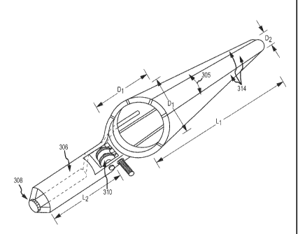
FIGS. 3A-3G illustrates a speculum 300 constructed in accordance with
alternative
embodiment of the present invention. The speculum 300 generally includes a
dilation
assembly 302 a handle 304 including a receptacle 306 for holding a light
source 308 and a
ratchet assembly 310 for use in expanding the dilation assembly 302. The
ratchet assembly
310 is operated using a thumb lever 312.
9
CA 02847524 2014-03-03
WO 2013/033607 PCT/US2012/053464
The speculum 300 of FIGS. 3A-3E shares many characteristics with the speculum
of
FIGS. lA and IB. For example, the speculum 300 is used by inserting the
dilation assembly
302 into the patient's introitus with the speculum 300 in a contracted
configuration (as shown
in FIGS. 3B and 3C). The speculum 300 is then expanded to the expanded
configuration (as
shown in FIGS. 3A and 3D). The light source 308 can then be activated to
illuminate
patient's vaginal walls and cervix which can be inspected visually by looking
through the
hollow dilation assembly 302. Moreover, like the embodiment of FIGS. lA and
IB, the
speculum 300 expands radially with respect to multiple axes for improved
viewing without
interference due to collapsing vaginal walls.
However, the speculum 300 has some differences in relation to the embodiment
of
FIGS. lA and IB. In particular, where as the blades in FIG. lA and IB are
separated by
spaces at least in the expanded configuration, the blades 314 of the speculum
300 overlap as
can best be seen in FIGS. 3E and 3G. When the dilation assembly 302 is
expanded or
contracted, the blades slide circumferentially over one another (as generally
indicated by
arrows 305) in manner analogons to a collapsible colander. Accordingly, there
are no spaces
between the blades in either the expanded contracted configuration. This may
further protect
against collapsing of the vaginal walls and potential pinching.
Another difference between the illustrated speculum 300 and that of FIGS. lA
and
IB is the mechanism for actuating expansion of the dilation assembly 302.
Specifically, the
dilation assembly 302 is expanded by operation of the thumb lever 312. The
thumb lever
312 interfaces with a worm gear ratchet as shown in FIG. 3F such that
depressing the thumb
lever closes the speculum 300 to the contracted configuration and pulling
outwardly on the
thumb lever 312 causes the speculum 300 to be expanded to the expanded
configuration.
The thumb lever 312 causes the worm gear of ratchet assembly 316 to rotate.
The worm gear
ratchet assembly 316 is then connected to the proximal ends 313 of the blades
314 by
appropriate linkage (as shown in FIGS. 3F and 3G) to expand and contract the
dilation
assembly 302 as desired.
The illustrated speculum 300 is dimensioned to accommodate a range of patients
including larger patients. For example, the diameter D1, of the proximal end
of the dilation
assembly 302 may be about 1.5 inches. The diameter, D2, of the distal end of
the dilation
CA 02847524 2014-03-03
WO 2013/033607
PCT/US2012/053464
assembly may be about 1.4 inches in the expanded configuration and about 0.7
inches in the
contracted configuration. The dilation assembly 302 has a length, L1, of about
6.5 inches and
the handle 304 has a length, L2, of about 3.5 inches for an overall length,
L3, of about 10
inches for the speculum 300.
FIGS. 4A - 4E illustrate a still further embodiment of a speculum 400 in
accordance with the
present invention. The speculum 400 includes a number of overlapping speculum
blades 402
generally similar to the blades in the embodiment of the FIGS. 3A - 3G. In
this case,
however, the blades are expanded and contracted directly by rotating retention
ring 404
rather than using a ratchet assembly as described in connection with the
embodiment of
FIGS. 3A - 3G. In addition, the handle 406 is offset vertically from the
expansion assembly
408 which may facilitate visual inspection through the expansion assembly 408.
The handle
406 further includes a receptacle 410 for receiving a light source and a light
pipe 412 for
directing light from the source to the patient's cervix. FIGS.
5A-5F illustrate a
speculum 500 in accordance with a still further embodiment of the present
invention. The
speculum 500 is similar to the speculum 100 of FIGS. IA-1B, with some
additional features
shown and minor differences in configuration. The speculum 500 generally
includes: a
generally conical dilation assembly 502 including a number of blades 504; a
generally
cylindrical obturator 506 for expanding the dilation assembly 502 and allowing
it to contract;
and a handle 508 including a receptacle 510 for receiving a light source 512.
As discussed
above, the speculum can be formed, for example, from clear plastic or metal as
desired.
The illustrated blades 504 are formed in an overlapping, collapsible
configuration.
That is, adjacent blades 504 extend circumferentially over one another, and
slide over one
another as the dilation assembly 502 is expanded and contracted. In this
manner, gaps
between the blades 504 are avoided, even in the expanded configuration, thus
reducing the
likelihood that tissue of the patient will be pinched due to operation of the
speculum 500.
The speculum 500 further includes a ratchet mechanism 514 for advancing and
withdrawing the obturator 506 into and out of the dilation assembly 502. The
ratchet
mechanism 514 includes a ratcheted handle surface 516 that interfaces with a
bottom of a
thumb lever 518. The thumb lever 518 includes an advance surface 520 and a
release surface
522. The physician or other user can press on the advance surface 520, as
generally indicated
11
CA 02847524 2014-03-03
WO 2013/033607 PCT/US2012/053464
by arrow 524, to move the thumb lever 518 forward. The thumb lever 518 presses
against
the obturator 506 so that it also moves forward thus expanding the dilation
assembly 502.
The ratchet mechanism 514 is then effective to hold the speculum in the
expanded
configuration.
To release the ratchet mechanism 514 so that the obturator 506 can be
withdrawn
from the dilation assembly 502 to close the blades 504, the user can press on
the release
surface 522 as generally indicated by arrow 526. This causes the rear edge of
the thumb
lever 578 to lift and disengages the ratchet mechanism 514. The user can then
slide the
thumb lever 518 rearwardly to withdraw the obturator 506 from the dilation
assembly 502.
As noted above, the handle 508 includes a receptacle 510 for receiving a light
source
512. Although any appropriate light source can be used, the illustrated
receptacle 510 can
receive a low-cost pen light type of light source 512, thereby reducing costs
and
inconvenience in relation to some conventional systems. The light source 512
may have an
on/off button at its rear end that can be easily accessed by the user during a
procedure. Light
from the light source is guided through the handle 508, and directed through
the dilation
assembly 502 to the procedure site by a plastic light pipe 528. Optionally, a
brightly colored
tag 530 or strap may be attached to the light source 512 to assist in locating
the light source
and to remind the user not to accidentally dispose of the light source 512
when the speculum
500 is discarded after a single use.
The blades 504 of the illustrated speculum 500 overlap, as indicated by arrow
532, so
that there are substantially no spaces between the blades 504 in the expanded
configuration.
In this regard, the blades 504 may move linearly (or arcuately with
substantially no
circumferential component) in a radial direction when expanding while
maintaining their
overlapped, stacked relationship at their proximal ends like flower petals, or
the blades 504
may slide circumferentially over one another while expanding like an
expandable colander.
The speculum 500 is preferably dimensioned to accommodate a range of patients.
For example, the dilation assembly 502 may have a length L1, of about 3.5
inches and the
handle 508 may have a length, L2, of about 3.5 inches for an overall speculum
length of 7
inches. In the contracted configuration, the distal end of the dilation
assembly 502 has a
12
CA 02847524 2014-03-03
WO 2013/033607 PCT/US2012/053464
diameter, D1, of about 1.5 inches. The distal end of the dilation assembly 502
preferably has
a bullet-shaped configuration, as can be seen in FIG. 5E, that helps maintain
the assembly
502 in the contracted configuration as the assembly 502 is introduced into the
introitus.
Optionally, one or more pegs 534 and mating receptacles may be provided at the
distal end of
the dilation assembly 502 to further assist in maintaining the contracted
configuration.
In the various embodiments disclosed above, the handles generally extend
rearwardly
in alignment with or at an acute angle to the longitudinal axis of the
dilation assembly in each
case.
13