Note: Descriptions are shown in the official language in which they were submitted.
CA 02847846 2014-03-05
WO 2013/040201 PCT/US2012/055155
ABLATION DEVICE WITH MULTIPLE ABLATION MODES
CROSS-REFERENCE TO RELATED APPLICATION
[0001] This application claims priority to U.S. Provisional Application
No.
61/534,590, filed September 14, 2011, which is herein incorporated by
reference in
its entirety.
[0002] This application is related to co-pending U.S. Provisional
Application
No. 61/534,587, entitled "Ablation Device With lonically Conductive Balloon,"
filed on
September 14, 2011. The content of this related application is incorporated
herein
by reference in its entirety for all purposes.
TECHNICAL FIELD
[0003] The present disclosure relates generally to an ablation device.
More
specifically, the present disclosure pertains to an ablation device including
an
ionically conductive balloon for performing radio-frequency ablation therapy
on body
tissue.
BACKGROUND
[0004] The treatment of cardiac arrhythmias is sometimes performed in
conjunction with an ablation catheter inserted into a chamber of the heart or
in one of
the vessels leading into or from the heart. In the treatment of atrial
fibrillation, for
example, a radio frequency (RF) ablation catheter equipped with a number of
electrodes can be brought into contact with cardiac tissue for creating one or
more
ablation points along the tissue. During ablation, an RF generator supplies
electrical
energy to the electrodes, generating an electric field in the tissue. The
resulting heat
from this electric field forms a controlled lesion that blocks the electrical
impulses
from being conducted through the tissue and serves to promote the normal
conduction of electrical impulses through the proper electrical pathway within
the
heart.
[0005] In certain catheter ablation procedures, it may be difficult to
electrically
isolate the tissue to be treated. In the treatment of paroxysmal atrial
fibrillation, for
example, it is often tedious and time consuming to isolate the pulmonary veins
using
an ablation catheter having an ablation electrode that directly contacts the
tissue.
Moreover, the ablations created by some ablation electrodes can cause
dehydration
CA 02847846 2014-03-05
WO 2013/040201 PCT/US2012/055155
in the tissue, which can result in scarring and calcification as the lesion
heals. Due
to the discrete nature of the ablation points, there is also the potential for
leaving
small gaps of electrically conductive tissue in the ablation line that may
continue to
initiate points of arrhythmias.
SUMMARY
[0006] The present disclosure relates generally to an ablation device
including
an ionically conductive balloon for performing radio-frequency ablation
therapy on
body tissue.
[0007] In Example 1, an ablation device for treating body tissue,
comprises:
an elongate shaft having a proximal section, a distal section, and at least
one fluid
lumen configured to receive an electrically conductive fluid; an inflatable
balloon
coupled to the distal section of the shaft and including an interior section
in fluid
communication with the at least one fluid lumen for actuating the balloon
between a
collapsed state and an expanded state, wherein the balloon comprises a
composite
structure having a proximal balloon section including a first polymeric
material and a
distal balloon section including a second polymeric material different from
the first
material; and at least one electrode located within the interior space of the
balloon.
[0008] In Example 2, the ablation device according to Example 1, wherein
the
first polymeric material is a hydrophobic polymer.
[0009] In Example 3, the ablation device according to any of Examples 1-
2,
wherein the second polymeric material is a hydrophilic polymer.
[0010] In Example 4, the ablation device according to any of Examples 1-
3,
further comprising at least one additional fluid lumen for recirculating fluid
through
the device.
[0011] In Example 5, the ablation device of according to any of Examples
1-4,
wherein, in the expanded state, the balloon is conically shaped.
[0012] In Example 6, the ablation device according to any of Examples 1-
5,
wherein the distal section of the balloon is invaginated.
[0013] In Example 7, the ablation device according to any of Examples 1-
6,
wherein the distal section of the balloon is semi-permeable.
2
CA 02847846 2014-03-05
WO 2013/040201 PCT/US2012/055155
[0014] In Example 8, the ablation device according to any of Examples 1-
7,
wherein a thickness of the balloon tapers along a length of the balloon from
the
proximal balloon section to the distal balloon section.
[0015] In Example 9, the ablation device according to any of Examples 1-
8,
wherein the balloon comprises a multi-layered structure.
[0016] In Example 10, the ablation device according to any of Examples 1-
9,
further comprising a temperature sensing element coupled to the distal section
of the
balloon.
[0017] In Example 11, the ablation device according to any of Examples 1-
10,
further comprising at least one electrocardiogram sensor coupled to the distal
section of the balloon.
[0018] In Example 12, the ablation device according to any of Examples 1-
11,
further comprising a spring-actuated plunger assembly configured to bias the
balloon
in the collapsed state.
[0019] In Example 13, the ablation device according to Example 12,
wherein
the plunger assembly comprises a plunger mechanism and a spring configured to
bias the plunger mechanism against the balloon.
[0020] In Example 14, the ablation device according to Example 13,
wherein
the plunger mechanism includes a plunger shaft and an atraumatic tip.
[0021] In Example 15, the ablation device according to Example 14,
wherein
the plunger shaft is slidably disposed within the catheter shaft and the
electrode.
[0022] In Example 16, an ablation device for treating body tissue
comprises:
an elongate shaft having a proximal section, a distal section, and at least
one fluid
lumen configured to receive an electrically conductive fluid; an inflatable
balloon
coupled to the distal section of the shaft and including an interior section
in fluid
communication with the at least one fluid lumen for actuating the balloon
between a
collapsed state and an expanded state; at least one electrode located within
the
interior space of the balloon; and a spring mechanism configured to bias the
balloon
in the collapsed state.
[0023] In Example 17, a method of forming a balloon of an ablation
catheter,
the balloon having a proximal section and a distal section, the method
comprising:
masking the proximal section of the balloon; irradiating the distal section of
the
balloon with an ionizing radiation source; etching the balloon to form a
plurality of
3
CA 02847846 2014-03-05
WO 2013/040201 PCT/US2012/055155
micropores through the distal section of the balloon; and securing the balloon
to a
catheter.
[0024] In Example 18, the method according to Example 17, wherein the
ionizing radiation source comprises an argon ion source.
[0025] In Example 19, the method according to any of Examples 17-18,
wherein the proximal section of the balloon comprises a hydrophobic polymer
and
the distal section of the balloon comprises a hydrophilic polymer.
[0026] In Example 20, the method according to any of Examples 17-19,
wherein a pore size of the micropores is between about 0.1 microns to 5
microns in
diameter.
[0027] In Example 21, a system for ablating body tissue comprises: an RF
generator including a switching mechanism operable between a first position
and a
second position; a fluid source including a supply of electrically conductive
fluid; and
an ablation device, the ablation device including an elongate shaft having a
proximal
section, a distal section, and at least one fluid lumen; an inflatable balloon
coupled to
the distal section of the shaft and including an interior section in fluid
communication
with the fluid source for actuating the balloon between a collapsed state and
an
expanded state; a first electrode disposed within the interior space of the
balloon and
electrically coupled to the RF generator, the first electrode configured for
supplying a
first RF electrical field through the balloon and into the body tissue when
operating in
the first position; a second electrode coupled to a distal end portion of the
elongate
shaft and electrically coupled to the RF generator, the second electrode
configured
for supplying a second RF electric field directly into the tissue when
operating in the
second position.
[0028] In Example 22, the system according to Example 21, wherein the
balloon comprises a composite structure having a proximal balloon section
including
a hydrophobic polymeric material and a distal balloon section including a
hydrophilic
polymeric material.
[0029] In Example 23, the system according to any of Examples 21-22,
wherein, in the expanded state, the balloon is conically shaped.
[0030] In Example 24, the system according to any of Examples 21-23,
wherein the distal section of the balloon is invaginated.
4
CA 02847846 2014-03-05
WO 2013/040201 PCT/US2012/055155
[0031] In Example 25, the system according to any of Examples 21-24,
wherein the distal section of the balloon is semi-permeable.
[0032] In Example 26, the system according to any of Examples 21-25,
wherein a thickness of the balloon tapers along a length of the balloon from a
proximal balloon section to a distal balloon section.
[0033] In Example 27, the system according to any of Examples 21-26,
wherein the balloon comprises a multi-layered structure.
[0034] In Example 28, the system according to any of Examples 21-27,
further
comprising a spring-actuated plunger assembly configured to bias the balloon
in the
collapsed state.
[0035] In Example 29, a method for performing ablation therapy on the
body
of a patient comprises: advancing an ablation device to a target body tissue
region,
the ablation device including an inflatable balloon coupled to an elongate
shaft, a first
electrode disposed within an interior space of the balloon, and a second
electrode
located outside of the balloon; injecting an electrically conductive fluid
into the
interior section of the balloon and inflating the balloon from a collapsed
state to an
expanded state within the body; selectively energizing the first electrode and
generating a first RF electrical field within the balloon interior; forming at
least one
ablation lesion within the body tissue using the first RF electrical field;
selectively
energizing the second electrode and generating a second RF electrical field;
and
forming at least one ablation lesion within the body tissue using the second
RF
electrical field.
[0036] In Example 30, the method according to Example 29, further
comprising an RF generator including a switching mechanism, and wherein
selectively energizing the first or second electrodes includes operating the
switching
mechanism between a first and second switch position.
[0037] In Example 31, the method according to any of Examples 29-30,
wherein forming at least one ablation lesion within the body tissue using the
first RF
electrical field includes forming a lesion in the body tissue at a location
distal to the
elongate shaft.
[0038] In Example 32, the method according to any of Examples 29-31,
wherein the at least one ablation lesion formed within the body tissue using
the first
CA 02847846 2014-03-05
WO 2013/040201 PCT/US2012/055155
RF electric field is larger than the at least one ablation lesion formed in
the body
tissue using the second RF electric field.
[0039] In Example 33, an ablation device for treating body tissue
comprises:
an elongate shaft having a proximal section, a distal section, and at least
one fluid
lumen configured to receive an electrically conductive fluid; an inflatable
balloon
coupled to the distal section of the shaft and including an interior section
in fluid
communication with the at least one fluid lumen for actuating the balloon
between a
collapsed state and an expanded state; and at least one electrode located
within the
interior space of the balloon, the at least one electrode configured for
transmitting an
RF electric field through the balloon and into body tissue in contact with the
balloon;
wherein the balloon is configured to transmit the RF electric field in a
direction
distally towards a leading end of the ablation device.
[0040] In Example 34, the ablation device according to Example 33,
wherein
the balloon comprises a composite structure having a proximal balloon section
including a hydrophobic polymeric material and a distal balloon section
including a
hydrophilic polymeric material.
[0041] In Example 35, the ablation device according to any of Examples 33-
34, wherein, in the expanded state, the balloon is conically shaped.
[0042] In Example 36, the ablation device according to any of Examples 33-
35, wherein the distal section of the balloon is invaginated.
[0043] In Example 37, the ablation device according to any of Examples 33-
36, wherein the distal section of the balloon is semi-permeable.
[0044] In Example 38, the ablation device according to any of Examples 33-
37, wherein a thickness of the balloon tapers along a length of the balloon
from a
proximal balloon section to a distal balloon section.
[0045] In Example 39, the ablation device according to any of Examples 33-
38, wherein the balloon comprises a multi-layered structure.
[0046] In Example 40, the ablation device according to any of Examples 33-
39, further comprising a spring-actuated plunger assembly configured to bias
the
balloon in the collapsed state.
[0047] While multiple embodiments are disclosed, still other embodiments
of
the present invention will become apparent to those skilled in the art from
the
following detailed description, which shows and describes illustrative
embodiments
6
CA 02847846 2014-03-05
WO 2013/040201 PCT/US2012/055155
of the invention. Accordingly, the drawings and detailed description are to be
regarded as illustrative in nature and not restrictive.
BRIEF DESCRIPTION OF THE DRAWINGS
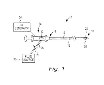
[0048] Figure 1 is a schematic view of an ablation device in accordance
with
an illustrative embodiment;
[0049] Figure 2 is a partial cross-sectional view showing the distal
section of
the ablation device of Figure 1 in a collapsed state;
[0050] Figure 3 is another partial cross-sectional view showing the
distal
section of the ablation device of Figure 1 in an expanded state;
[0051] Figure 4 is a flow diagram showing an example method for
fabricating
a porous balloon of an ablation device;
[0052] Figure 5 is a perspective view showing an example composite
balloon
in accordance with an illustrative embodiment;
[0053] Figure 6 is a partial cross-sectional view showing the distal
section of
an ablation device in accordance with another illustrative embodiment;
[0054] Figure 7 is a partial cross-sectional view showing the distal
section of
an ablation device in accordance with another illustrative embodiment;
[0055] Figure 8 is a partial cross-sectional view showing the distal
section of
an ablation device in accordance with another illustrative embodiment;
[0056] Figure 9 is a schematic view of an ablation device in accordance
with
another illustrative embodiment; and
[0057] Figure 10 is a flow diagram of an illustrative method of
performing a
cardiac ablation procedure using the ablation device of Figure 9.
[0058] While the invention is amenable to various modifications and
alternative forms, specific embodiments have been shown by way of example in
the
drawings and are described in detail below. The intention, however, is not to
limit
the invention to the particular embodiments described. On the contrary, the
invention is intended to cover all modifications, equivalents, and
alternatives falling
within the scope of the invention as defined by the appended claims.
DETAILED DESCRIPTION
7
CA 02847846 2014-03-05
WO 2013/040201 PCT/US2012/055155
[0059] Figure 1 is a schematic view of an ablation device 10 in
accordance
with an illustrative embodiment. As shown in Figure 1, the ablation device 10
includes an elongate shaft 12 having a proximal section 14, a distal section
16, and
at least one lumen 18 extending through the shaft 12 between the proximal and
distal sections 14, 16. An inflatable ablation balloon 20 coupled to the
distal section
16 of the shaft 12 can be inflated at a target location within the body (e.g.,
within a
cardiac vessel) and brought into contact with the body tissue to be treated.
In some
embodiments, and as further described below, an RF electrode assembly 22
located
within an interior portion of the balloon 20 generates an RF electric field
that can be
used for creating controlled lesions within the tissue. In the treatment of
paroxysmal
atrial fibrillation, for example, the balloon 20 and RF electrode 22 can be
used for
performing electrical isolation within a pulmonary vein to prevent the
aberrant
conduction of electrical signals within the left side of the heart. The
ablation device
can also be used for treating other types of cardiac arrhythmias and/or
cardiovascular diseases within the body. The ablation device 10 can also be
used
for treating other conditions commonly performed by ablation devices.
[0060] A handle 24 coupled to the proximal section 14 of the shaft 12 can
be
used by the clinician for manipulating and steering the distal section 16 to a
target
site within the body for performing an ablation. In some embodiments, the
handle 24
includes a fluid port 26 and valve 28 in fluid communication with a source of
electrically conductive fluid 30. In some embodiments, for example, the fluid
30 can
comprise saline or a solution of saline and a fluoroscopic contrast medium
that is
both conductive and biocompatible. During an ablation procedure, pressurized
fluid
30 can be delivered via the fluid lumen 18 to the interior of the balloon 20,
causing
the balloon 20 to inflate while also creating an electrical pathway between
the
electrode 22 and the portion of the balloon 20 in contact with the body tissue
to be
treated. In some embodiments, multiple fluid ports can be provided to
recirculate the
fluid 30 through the ablation device 10 as part of a closed-loop system for
controlling
the temperature within the balloon 20.
[0061] In some embodiments, the ablation device 10 further includes a
steering mechanism 32 that can be used to mechanically steer the distal
section 16
of the shaft 12 within the body. In certain embodiments, for example, the
steering
mechanism 32 comprises a slider or lever mechanism on the handle 24 that can
be
8
CA 02847846 2014-03-05
WO 2013/040201 PCT/US2012/055155
actuated by the clinician to engage a number of steering wires located within
the
shaft 12. During delivery of the device 10 to a target region within the body,
the
steering mechanism 32 can be engaged to deflect the distal section 16 of the
shaft
12, allowing the clinician to better navigate the device 10 through the
vasculature.
[0062] An RF generator 34 is configured to supply radio-frequency energy
to
the electrode assembly 22. In some embodiments, the device 10 is configured to
operate in a bipolar mode, in which ablation energy supplied by the RF
generator 34
flows from one electrode of the electrode assembly 22 to another electrode of
the
electrode assembly 22 or provided at a different location along the device 10
(e.g.,
along the distal section 16 of the shaft 12). In other embodiments, the device
10 is
configured to operate in a unipolar mode, in which an indifferent electrode
(e.g., an
electrode patch) is attached to the patient's back or other exterior skin area
and
ablation energy from the RF generator 34 flows from one electrode of the
assembly
22 to the indifferent electrode.
[0063] Figure 2 is a partial cross-sectional view showing the distal
section 16
of the ablation device 10 of Figure 1 in greater detail. As can be further
seen in
Figure 2, and in some embodiments, the electrode assembly 22 comprises at
least
one RF electrode 36 located within an interior space 38 of the balloon 20. The
RF
electrode 36 is fixedly secured to a distal end 40 of the shaft 12 (e.g.,
using a
suitable adhesive at both ends of the electrode 36), and is electrically
coupled to the
RF generator 34. In the embodiment of Figure 2, the RF electrode 36 comprises
a
metal tubular member made from a suitably conductive metal such as platinum,
and
is electrically coupled to the RF generator 34 via a number of conductor wires
(not
shown) located within the shaft 12. The configuration of the RF electrode 36
can
vary from that shown, however. For example, the RF electrode 36 can comprise a
coil, ring, flat ribbon, or other suitable shape. In some embodiments, the
electrode
assembly 22 can include multiple electrodes 36 as part of either a bipolar RF
ablation system, or as part of a unipolar system with multiple electrodes.
[0064] The device 10 includes at least one fluid lumen for transmitting
pressurized fluid 30 to the interior space 38 of the balloon 20. In the
embodiment of
Figure 2, the device 10 includes a central fluid lumen 18 that extends
longitudinally
through the shaft 12 and through a portion of the RF electrode 36. In some
embodiments, the fluid lumen 18 terminates distally at a number of inflation
ports 42
9
CA 02847846 2014-03-05
WO 2013/040201 PCT/US2012/055155
disposed circumferentially about the RF electrode 36. In some embodiments, the
same fluid lumen 18 can be used for both inflating and deflating the balloon
20. In
other embodiments, separate fluid lumens are used for inflating and deflating
the
balloon 20. Such a configuration can provide continuous infusion and
evacuation of
fluid within the balloon 20 to maintain both a controlled operating pressure
and
temperature within the balloon 20. In one embodiment, multiple fluid lumens
within
the shaft 12 may permit the electrically conductive fluid 30 to be
recirculated through
the device 10 during the ablation procedure. The fluid 30 can also include a
contrast
medium to facilitate visualization of the balloon 20 under fluoroscopy.
[0065] In the embodiment of Figure 2, the balloon 20 is coupled to the
distal
section 16 of the shaft 12 at or near the distal shaft end 40, and is
inflatable from an
initial, collapsed position having a low-profile that facilitates traversal of
the device 10
through the body, to a second, expanded position that contacts and engages the
body tissue to be ablated. In certain embodiments, the balloon 20 has a
composite
structure formed from different polymeric materials, which helps to direct and
focus
the RF energy from the RF electrode 36 into the body tissue located at or near
a
distal end 44 of the balloon 20. In one embodiment, for example, the composite
balloon 20 includes a proximal, non-conductive section 46a made from a
hydrophobic polymer and a distal, conductive section 46b made from a
hydrophilic
polymer. The polymer of the non-conductive section 46a can be non-ionically
conductive and the polymer of the distal section 46b can be ionically
conductive. In
some embodiments, for example, the composite balloon structure can comprise a
proximal section 46a made from a hydrophobic polyurethane material such as
TECOPHILIC 60D-35 and a distal section 46b made from a hydrophilic
polyurethane material such as TECOPHILIC 60D , both of which are available
from
Thermedics Polymer Products of Woburn, Massachusetts. TECOPHILIC is a
polyether-based aliphatic polyurethane and exhibits sufficient elasticity so
as to be
capable of stretching substantially beyond its equilibrium dimensions when the
balloon 20 is inflated. Other polymeric materials can also be used to impart
differing
hydrophilic characteristics to the proximal and distal sections 46a, 46b. As
used
herein, the term "hydrophilic" indicates that the polymer, when in contact
with an
aqueous solution, can absorb a quantity of water while still maintaining its
structural
integrity.
CA 02847846 2014-03-05
WO 2013/040201 PCT/US2012/055155
[0066]
When inflated with the electrically conductive fluid 30, the distal section
46b of the composite balloon 20 is rendered conductive by hydration due to the
ionic
content of the fluid 30 when the RF energy is supplied to the RF electrode 36.
As a
result, electrical current is transmitted through the fluid 30 and into the
tissue in
contact with the distal section 46b of the balloon 20. In some cases, current
passes
through all areas of the balloon material that are hydrophilic but does not
pass
through areas of the balloon that are hydrophobic or non-conductive.
[0067] The
composite balloon structure can be formed using a number of
different techniques. For example, the different sections 46a, 46b of the
balloon 20
can be formed by separately dip-coating each section of the balloon 20 on a
mandrel
that has a defined size and shape. The balloon 20 can also be formed using
other
techniques, such as by spin-coating in a hollow mold or by injection or blow-
molding.
Another example method for constructing a composite balloon structure having a
permeable or semi-permeable distal section is discussed further herein with
respect
to Figure 4.
[0068] In
some embodiments, the device 10 further includes one or more
temperature sensing elements that can be used to sense the temperature of
fluid 30
within the balloon 20. In certain embodiments, and as shown in Figure 2, a
temperature sensing element 48 such as a thermocouple or thermistor is coupled
to
the inner surface 50 of the balloon 20 at the distal section 46b. In
other
embodiments, the temperature sensing element 48 is coupled to an outer surface
52
of the balloon 20 at the distal section 48, or is coupled to another portion
of the
balloon 20 or to the shaft 12. In another embodiment, the temperature sensing
element 48 is encased within the interior of the balloon material. In
some
embodiments, multiple temperature sensing elements can be coupled to the inner
and/or outer surfaces 50, 52 of the balloon and/or to the shaft 12 for sensing
temperature at multiple locations.
[0069] In
some embodiments, the temperature sensing element 48 senses the
temperature of the fluid 30 contained within the interior section 38 of the
balloon 20,
and is connected to temperature sensing circuitry (e.g., based on a
thermometer)
located outside of the body. During ablation, the RF generator 34 can be
controlled
so as to adjust the temperature of the fluid 30 contained in the balloon 20 to
a
desired temperature. In those embodiments in which multiple fluid ports are
utilized
11
CA 02847846 2014-03-05
WO 2013/040201 PCT/US2012/055155
for recirculating fluid through the device 10, the flow of fluid can also be
controlled
based on feedback from the temperature sensing element 48 to maintain the
fluid
within the balloon 20 at a particular temperature or within a range of
temperatures.
In various embodiments, a temperature sensor is located on the outer surface
of the
balloon and/or within the wall of the balloon. Such a configuration can
measure the
temperature of the tissue undergoing ablation. In these or other embodiments
referenced herein, the intensity of ablation therapy (e.g., power) can be
automatically
modulated based on the measured temperature to limit the temperature of the
tissue
undergoing ablation. Such a configuration can provide protection from steam
pops,
where a small gaseous rupture in tissue can otherwise be created by water in
the
tissue turning into steam when the temperature reaches 100 C or greater.
[0070] One or more electrocardiogram sensors coupled to the balloon 20
can
also be used in some embodiments for sensing electrical activity in or near
the heart.
In the embodiment of Figure 2, for example, an electrocardiogram sensor 54 is
coupled to the inner surface 50 of the balloon 20 at the distal section 46b,
allowing
the clinician to monitor for the presence of any electrical activity at the
target ablation
site. In other embodiments, the electrocardiogram sensor 54 is coupled to the
outer
surface 52 of the balloon 20 at the distal section 46, or is coupled to
another portion
of the balloon 20 or shaft 12. In another embodiment, the electrocardiogram
sensor
52 is encased within the interior of the balloon material. In some
embodiments,
multiple electrocardiogram sensors can be coupled to and/or encased within the
balloon 20 and/or to the shaft 12 for sensing electrical activity at multiple
locations.
[0071] A spring actuated plunger assembly 56 can be used to maintain the
balloon 20 in a collapsed, low-profile position to facilitate delivery of the
device 10
through the body prior to inflating the balloon 20 at the desired target
tissue location.
In the embodiment of Figure 2, the assembly 56 includes a plunger mechanism 58
and a spring 60. The spring 60 is located within the interior of the shaft 12
proximal
to the RF electrode 36, and is configured to mechanically bias the plunger
mechanism 58 in a distal direction towards the distal end 44 of the balloon
20, thus
maintaining the balloon 20 in an extended position until inflated.
[0072] In some embodiments, the plunger mechanism 58 comprises a plunger
shaft 62 slidably disposed within the interior section 38 of the balloon 20
and through
a portion of the RF electrode 36. The distal end of the plunger shaft 62
includes an
12
CA 02847846 2014-03-05
WO 2013/040201 PCT/US2012/055155
atraumatic tip 64 which, when the plunger mechanism 58 is fully engaged
distally, is
configured to contact and engage the distal end 44 of the balloon 20 causing
the
balloon 20 to collapse and assume a low-profile position, as shown. The shape
of
the tip 64 is curved to conform to the shape of the balloon 20 at the distal
end 44.
The proximal end of the plunger shaft 62 is coupled to a plunger seal 66,
which
provides a surface against which the spring 60 engages the plunger shaft 62. A
shoulder 68 located within the interior of the shaft 12 proximal to the spring
60
provides a proximal stop to prevent proximal movement of the spring 60 when
the
spring 60 is compressed.
[0073] Figure 3 is another partial cross-sectional view of the ablation
device
of Figure 1, showing the balloon 20 in a second, fully expanded position. As
can
be further seen in Figure 3, when pressurized fluid 30 is injected into the
interior
section 38 of the balloon 20, the fluid pressure exerted against the surface
of the
plunger seal 66 is configured to overcome the spring bias provided by the
spring 60,
causing the spring 60 to move to a second, compressed position within the
shaft
interior. Once the balloon 20 is inflated, the pressure within the interior
section 38 of
the balloon 20 pushes the plunger assembly 56 in a proximal direction. As a
result,
the plunger shaft 62 is drawn proximally into the shaft interior, causing the
atraumatic
tip 64 to disengage from the distal end 44 of the balloon 20.
[0074] When the tip 64 disengages from the distal end 44 of the balloon
20,
and as shown in Figure 3, the balloon 20 is configured to expand to its
second,
expanded position. In some embodiments, the shape of the inflated balloon 20
may
vary along its length such that the proximal section 46a of the balloon 20 has
a
profile and shape that is different from that of the distal section 46b. In
the
embodiment of Figure 3, for example, the inflated balloon 20 has a
substantially
conical shape such that the distal, conductive section 46b of the balloon 20
exposes
a relatively large area towards the distal end 44 of the balloon 20. The
conical shape
of the distal section 46b facilitates contact of the balloon 20 with body
tissue located
primarily distally of the device 10. The proximal section 46a of the balloon
20, in
turn, has a relatively low profile, and thus does not contact the body tissue.
In
contrast to the distal section 46b, the hydrophobic material of the proximal
section
46a also does not conduct with the fluid 30 within the balloon 20.
13
CA 02847846 2014-03-05
WO 2013/040201 PCT/US2012/055155
[0075]
Although the illustrative balloon 20 in Figure 3 has a conical shape
when expanded, in other embodiments the balloon 20 can have a different shape
and/or profile when inflated. Examples of other balloon shapes can include
elliptical,
spherical, or dumbbell. In some embodiments, the balloon shape can be similar
to
one of the self-anchoring balloon shapes described in U.S. Patent No.
7,736,362, the
contents of which are incorporated herein by reference in their entirety for
all
purposes. Other balloon configurations are also possible.
[0076] In
some embodiments, the distal section 46b of the balloon 20 is semi-
permeable, allowing at least some of the pressurized fluid 30 within the
interior
section 38 of the balloon 20 to seep into the body at or near the target
ablation site.
In some embodiments, the distal section 46b of the balloon 20 is permeable,
allowing the pressurized fluid 30 within the interior section 38 of the
balloon 20 to
seep into the body at or near the target ablation site. During ablation, the
presence
of the electrically conductive fluid at this interface region aids in creating
an electrical
conduit for the electrical field generated by the RF electrode 36, and further
serves to
cool the ablation site. As the RF energy is applied to the RF electrode 36
inside the
balloon 20, the RF energy is transmitted to the tissue in contact with the
balloon 20
through the electrically conductive fluid seeping through the balloon 20. The
permeability or semi-permeability of the distal section 46b also permits the
delivery
of an agent or drug contained within the fluid 30. In this manner, the balloon
20 may
also act as a drug delivery device by introducing one or more drugs into the
conductive fluid 30 and permitting the drugs to pass through the balloon 20
and into
the tissue.
[0077]
Figure 4 is a flow diagram showing an example method 70 for
fabricating a porous balloon. The method 70 may begin generally at block 72,
by
fabricating a composite balloon having a proximal, non-conductive section and
a
distal, conductive section. It is noted that in some embodiments the distal
section is
non-conductive. In certain embodiments, for example, a composite balloon 20
such
as that shown in Figures 2-3 can be fabricated using a suitable process such
as dip-
coating, spin-coating, injection molding, or blow-molding.
Other fabricating
techniques for fabricating a composite balloon can also be utilized.
[0078] The
balloon material or materials can be selected so as to facilitate
further processing steps to create micropores through the balloon material. In
some
14
CA 02847846 2014-03-05
WO 2013/040201 PCT/US2012/055155
embodiments, for example, the workpiece used to create the composite balloon
can
be formed from a thermoplastic polymer resin such as polyethylene
terephthalate
(PET). The thermal and/or chemical characteristics of PET permit subsequent
processing steps to be performed on the balloon while maintaining the desired
tensile strength and elasticity characteristics of the balloon.
[0079] Once the composite balloon has been fabricated, the proximal, non-
conductive section of the balloon is masked (block 74), and the distal (e.g.,
conductive) section of the balloon is irradiated with ions from an ionizing
radiation
source (block 76). In one embodiment, the composite balloon is irradiated with
Argon atoms from an Argon plasma source. Other suitable ionic radiation
sources
can also be used to irradiate the distal section of the balloon with ions.
[0080] Once irradiated, the balloon is then subjected to a sodium
hydroxide
(NaOH) etching process for a period of time to produce uniform micropores in
the
distal section of the balloon (block 78). In certain embodiments, for example,
the
balloon can be inserted into an etching bath and treated for a period of
approximately 10 to 15 minutes until pores of a desired size are formed
through the
balloon material. The pore size can be controlled by the duration of the
ionizing
radiation and etching steps, the strength of the ionizing radiation, and the
strength of
the etching solution. Other factors such as the balloon composition, balloon
thickness, as well as other characteristics can also affect the pore size. An
example
pore size that can be generated using this process can be between about 0.1
microns to about 5 microns in diameter, although other pore sizes greater or
smaller
are also contemplated. For example, in some cases pores can be up to 20
microns
in diameter.
[0081] Once the micropores are created in the distal section of the
balloon,
additional processing steps can then be performed to secure the balloon onto
the
shaft (block 80). In one embodiment, the balloon can be mounted to the distal
end of
a shaft, similar to that shown in the illustrative embodiment shown in Figures
2-3.
The balloon can be secured to the shaft in a variety of ways, including
adhesive
bonding, thermal bonding, mechanical bonding, screws, winding, or a
combination of
these.
[0082] Figure 5 is a perspective view showing an example composite
balloon
20 that has been treated using the method 70 of Figure 4. As can be seen in
Figure
CA 02847846 2014-03-05
WO 2013/040201 PCT/US2012/055155
5, the distal section 46b of the balloon 20 includes a plurality of micropores
82 which,
due to the size and shape of the distal section 46b in its inflated state,
face
substantially in a distal direction away from the distal end 44 of the balloon
20 in the
direction indicated generally by arrow 84. When a steady flow of electrically
conductive fluid is provided to the interior section 38 of the balloon 20, at
least a
portion of the fluid 30 seeps through the micropores 82 and into contact with
body
tissue located distally of the balloon 20. The proximal section 46a of the
balloon 20
is substantially non-porous, and thus prohibits the flow of pressurized fluid
through
the proximal section 46a.
[0083] Figure 6 is a partial cross-sectional view showing the distal
section of
an ablation device 86 in accordance with another illustrative embodiment. The
ablation device 86 includes an elongate shaft 88 coupled to an inflatable
ablation
balloon 90. The proximal section of the shaft 88 (not shown) is coupled to an
electrically conductive fluid source and an RF generator. In the embodiment of
Figure 6, the distal section 92 of the shaft 88 extends through the interior
94 of the
balloon 90, and includes a number of fluid ports 96, 98 for circulating fluid
through
the balloon interior 94. A first fluid port 96 in fluid communication with a
first lumen
within the shaft 88 is configured to deliver electrically conductive fluid
from an
external fluid source into the balloon interior 94. A second fluid port 98 in
fluid
communication with a return fluid lumen of the shaft 88, in turn, functions as
a return
port for recirculating heated fluid within the balloon interior 94 to a
location outside of
the patient's body for cooling.
[0084] An electrode assembly 100 disposed within the interior 94 of the
balloon 90 is electrically coupled to an RF generator, and is configured to
generate
an RF electric field for creating controlled lesions within tissue located
adjacent to
the balloon 90. In some embodiments, and as shown in Figure 6, the electrode
assembly 100 comprises a metal coil RF electrode 102 having a helical shape
that
extends about a portion of the shaft 88 located within the balloon interior
94. In other
embodiments, the RF electrode 102 can comprise a tubular member, ring, flat
ribbon, or other suitable shape. In some embodiments, the electrode assembly
100
can include multiple electrodes 102 as part of either a bipolar RF ablation
system, or
as part of a unipolar system with multiple electrodes.
16
CA 02847846 2014-03-05
WO 2013/040201 PCT/US2012/055155
[0085] In the embodiment of Figure 6, a proximal section 112a of the
balloon
90 is coupled to the distal section 92 of the elongate shaft 88. A distal
section 112b
of the balloon 90, in turn, is coupled to the distal end 108 of the elongate
shaft 88. In
some embodiments, and as shown in Figure 6, the distal section 112b of the
balloon
90 has an invaginated configuration created by folding or turning a portion of
the
balloon 90 back upon itself and attaching the distal end 106 of the balloon 90
to an
interior surface of the shaft distal end 108. The balloon 90 is inflatable
from an initial,
collapsed position having a low-profile that facilitates traversal of the
device 86
through the body, to a second, expanded position that contacts and engages the
body tissue to be ablated. In some embodiments, the balloon 90 has a composite
structure formed from different polymeric materials, which helps to direct and
focus
the RF energy from the RF electrode 100 into body tissue located at or near a
distal
section 112b of the balloon 90. In one embodiment, for example, the composite
balloon 90 includes a proximal, non-conductive section 112a made from a
hydrophobic polymer and a distal, conductive section 112b made from a
hydrophilic
polymer. In some embodiments, for example, the composite balloon structure can
comprise a proximal section 112a made from a hydrophobic polyurethane material
such as TECOPHILIC 60D-35 and a distal section 112b made from a hydrophilic
polyurethane material such as TECOPHILIC 60D . Other polymeric materials can
also be used to impart differing hydrophilic characteristics to the proximal
and distal
sections 112a, 112b, as desired.
[0086] When inflated with an electrically conductive fluid, the distal
section
112b of the balloon 90 is rendered conductive by hydration due to the ionic
content
of the fluid when RF energy is supplied to the RF electrode 102. An electrical
current is thus transmitted through the fluid and into the tissue in contact
with the
distal section 112b of the balloon 90. When inflated, the invaginated
configuration of
the balloon 90 also serves to direct the RF electrical field towards the
distal section
112b of the balloon 90.
[0087] The ablation device 86 can further include one or more features
described with respect to other embodiments, including one or more temperature
sensors for sensing the temperature of fluid within or on the surface of the
balloon
90, and one or more electrocardiogram sensors for sensing electrical activity
in or
near the heart. The device 86 can also include other features such as a spring-
17
CA 02847846 2014-03-05
WO 2013/040201 PCT/US2012/055155
actuated plunger assembly. In certain embodiments, the balloon 90 can also be
made permeable or semi-permeable, allowing at least some of the pressurized
fluid
within the interior section 94 of the balloon 90 to seep into the body at or
near the
target ablation site.
[0088] Figure 7 is a partial cross-sectional view showing the distal
section of
an ablation device 114 in accordance with another illustrative embodiment. The
ablation device 114 includes an elongate shaft 116 coupled to an inflatable
ablation
balloon 118. The proximal section of the shaft 116 is coupled to an
electrically
conductive fluid source and an RF generator. In the embodiment of Figure 7,
the
distal section 120 of the shaft 116 extends through the interior 122 of the
balloon
118, and includes a number of fluid ports 124, 126 for circulating fluid
through the
balloon interior 122. A first fluid port 124 in fluid communication with a
first lumen
within the shaft 116 is configured to deliver electrically conductive fluid
from an
external fluid source into the balloon interior 122. A second fluid port 126
in fluid
communication with a return fluid lumen within the shaft 116, in turn,
functions as a
return port for recirculating heated fluid within the balloon interior 122 to
a location
outside of the patient's body for cooling.
[0089] An electrode assembly 128 disposed within the interior 122 of the
balloon 118 is electrically coupled to an RF generator, and is configured to
generate
an RF electric field for creating controlled lesions within tissue located
adjacent to
the balloon 118. In some embodiments, and as shown in Figure 7, the electrode
assembly 128 comprises a metal coil RF electrode 130 having a helical shape
that
extends about a portion of the shaft 116 located within the balloon interior
122. In
other embodiments, the RF electrode 130 can comprise a tubular member, ring,
flat
ribbon, or other suitable shape. In some embodiments, the electrode assembly
128
can include multiple electrodes 130 as part of either a bipolar RF ablation
system, or
as part of a unipolar system with multiple electrodes.
[0090] In the embodiment of Figure 7, a proximal end portion 134 of the
balloon 118 is coupled to the distal section 120 of the elongate shaft 118.
The
balloon 118 is inflatable from an initial, collapsed position having a low-
profile that
facilitates traversal of the device 114 through the body, to a second,
expanded
position that contacts and engages the body tissue to be ablated. In some
embodiments, and as shown, the thickness of the balloon 118 can taper along a
18
CA 02847846 2014-03-05
WO 2013/040201 PCT/US2012/055155
length of the balloon 118 that is generally parallel with the shaft 116 such
that the
thickness of the proximal section 134a is greater than the thickness of the
distal
section 134b. In certain embodiments, the thickness of the balloon 118 tapers
continuously along the length of the balloon 118 between the proximal and
distal
sections 134a, 134b. In one embodiment, for example, the balloon 118 may
continuously taper from a thickness of between about 5 mils (0.005 inches) to
15
mils (0.015 inches) at or near the location 132 where the proximal section
134a of
the balloon 118 attaches to the elongate shaft 116, to a thickness of between
about
0.5 mil to 5 mils at or near a distal end portion 136 of the balloon 118.
[0091] In other embodiments, the balloon 118 may transition in thickness
at
one or more discrete locations along the length of the balloon 118 such that
the
thickness of the proximal section 134a is greater than the thickness of the
distal
section 134b. In one embodiment, for example, the balloon 118 thickness may
transition from a relatively thick configuration at the proximal portion 134a
of the
balloon 118 to a relatively thin configuration at the distal section 134b of
the balloon
118 at a location substantially midway along the length of the balloon 118.
The
balloon 118 may also stepwise transition in thickness at multiple locations
along the
proximal and/or distal sections 134a, 136b of the balloon 118. Other
configurations
are also possible.
[0092] The balloon 118 can comprise a hydrophilic polymer that
facilitates the
transmission of the electromagnetic field generated by the RF electrode 130
through
the balloon material and into contact with the tissue. In some embodiments,
the
balloon 118 comprises a composite structure in which multiple materials are
used to
transition the balloon 118 from a relatively hydrophobic composition along
proximal
section 134a of the balloon 118 to a relatively hydrophilic composition along
the
distal section 134b of the balloon 118. In some embodiments, for example, the
composite balloon structure can comprise a proximal section 134a made from a
hydrophobic polyurethane material such as TECOPHILIC 60D-35 and a distal
section 134b made from a hydrophilic polyurethane material such as TECOPHILIC
60D , as discussed herein. The resulting structure is a composite balloon 118
that
transitions both in material composition and in thickness along the length of
the
balloon 118. During an ablation, this reduction in thickness, (and in some
embodiments also a change in material composition) along the length of the
balloon
19
CA 02847846 2014-03-05
WO 2013/040201 PCT/US2012/055155
118 causes a greater amount of the electric field generated by the RF
electrode 130
to pass through the distal section 134b of the balloon 118, allowing the
clinician to
target body tissue that is situated distally of the balloon 118.
[0093] The ablation device 114 can further include one or more features
described with respect to other embodiments herein, including one or more
temperature sensors for sensing the temperature of fluid within or on the
outer
surface of the balloon 118 and/or one or more electrocardiogram sensors for
sensing
electrical activity in or near the heart. The device 114 can also include
other features
such as a spring-actuated plunger assembly. In certain embodiments, the
balloon
118 can also be made permeable or semi-permeable, allowing at least some of
the
pressurized fluid within the interior section 122 of the balloon 118 to seep
into the
body at or near the target ablation site.
[0094] Figure 8 is a partial cross-sectional view showing the distal
section of
an ablation device 138 in accordance with another illustrative embodiment. The
ablation device 138 includes an elongate shaft 140 coupled to an inflatable
ablation
balloon 142. The proximal section of the shaft 140 is coupled to an
electrically
conductive fluid source and an RF generator. In the embodiment of Figure 8,
the
distal section 144 of the shaft 140 extends through the interior 146 of the
balloon
142, and includes a number of fluid ports 148, 150 for circulating fluid
through the
balloon interior 146. A first fluid port 148 in fluid communication with a
first lumen
within the shaft 140 is configured to deliver electrically conductive fluid
from an
external fluid source into the balloon interior 146. A second fluid port 150
in fluid
communication with a return fluid lumen within the shaft 140, in turn,
functions as a
return port for recirculating heated fluid within the balloon interior 146 to
a location
outside of the patient's body for cooling.
[0095] An electrode assembly 152 disposed within the interior 146 of the
balloon 142 is electrically coupled to an RF generator, and is configured to
generate
an RF electric field for creating controlled lesions within tissue located
adjacent to
the balloon 142. In some embodiments, and as shown in Figure 8, the electrode
assembly 152 comprises a metal coil RF electrode 154 having a helical shape
that
extends about a portion of the shaft 140 located within the balloon interior
146. In
other embodiments, the RF electrode 154 can comprise a tubular member, ring,
flat
ribbon, or other suitable shape. In some embodiments, the electrode assembly
152
CA 02847846 2014-03-05
WO 2013/040201 PCT/US2012/055155
can include multiple electrodes 154 as part of either a bipolar RF ablation
system, or
as part of a unipolar system with multiple electrodes.
[0096] In the embodiment of Figure 8, a proximal end portion 156 of the
balloon 142 is coupled to the distal section 144 of the elongate shaft 140.
The
balloon 142 is inflatable from an initial, collapsed position having a low-
profile that
facilitates traversal of the device 138 through the body, and a second,
expanded
position that contacts and engages the body tissue to be ablated. In some
embodiments, and as shown in Figure 8, the balloon 142 comprises a multi-
layered
structure having a first layer 158 and a second layer 160. The first layer 158
of the
balloon 142 comprises a hydrophilic hydratable, ionically conductive material
layer
that extends across the entire surface area of the balloon 142, along both a
proximal
section 162a and a distal section 162b of the balloon 142. In certain
embodiments,
for example, the first layer 158 comprises a hydrophilic polyurethane material
such a
TECOPHILIC 60D . In certain embodiments, the thickness of the first layer 158
is
between about 1 mil to 3 mils.
[0097] In some embodiments, first layer 158 has a uniform thickness along
the
entire length of the balloon 142. In other embodiments, the thickness of the
first
layer 158 may transition in thickness along the length of the balloon 142. For
example, in some embodiments, the first layer 158 of the balloon 142 may taper
in
thickness along the length of the balloon 142 such that the portion of first
layer 158
located along the proximal section 162a of the balloon 142 is thicker than the
portion
of the first layer 158 located along the distal section 162b. The thickness of
the first
layer 158 can taper either continuously or at one or more discrete locations
along the
length of the balloon 142. In some embodiments, the thickness of the first
layer 158
may transition in thickness from about 3 mils at or near the location where
the
proximal end portion 156 of the balloon 142 attaches to the elongate shaft 140
to a
thickness of about 1 mil at or near the distal end portion 164 of the balloon
142.
[0098] The second layer 160 of the balloon 142 comprises a hydrophobic
material, and extends across only a portion of the balloon 142. In the
embodiment of
Figure 8, for example, the second layer 160 is located along only the proximal
section 162a of the balloon 142. In some embodiments, the second layer 160
comprises a hydrophobic polymer mask that is spray-coated onto the first layer
158
during the balloon manufacturing process. An example hydrophobic material that
21
CA 02847846 2014-03-05
WO 2013/040201 PCT/US2012/055155
can be used to form the second layer 160 comprises TECOPHILIC 60D-35 . Other
techniques can also be used for forming the second layer 160, including
sputtering,
adhesion, or co-extrusion.
[0099] In the embodiment of Figure 8, the thickness of the second layer
160
tapers continuously along its length. In other embodiments, the second layer
160
reduces in thickness at one or more discrete locations along its length. In
some
embodiments, the thickness of the second layer 160 may transition from between
about 5 mils at or near the location where the proximal end portion 156 of the
balloon
142 attaches to the elongate shaft 140 to a thickness of about 1 mil at or
near the
location where the second layer 160 terminates.
[00100] During ablation, the presence of the hydrophobic second layer 160
over the first layer 158 of the balloon 142 causes a greater amount of the
electrical
field generated by the RF electrode 154 to pass through the distal section
162b of
the balloon 142, allowing the clinician to target body tissue that is situated
distally of
the balloon 142. In some cases during ablation, the presence of the
hydrophobic
second layer 160 over the first layer 158 of the balloon 142 causes the RF
current to
be concentrated and evenly distributed through only the unmasked hydrophilic
distal
surface of the balloon, allowing the clinician to target body tissue that is
situated
distally of the balloon 142.
[00101] The ablation device 138 can further include one or more features
described with respect to other embodiments, including one or more temperature
sensors for sensing the temperature of fluid within or on the surface of the
balloon
and/or one or more electrocardiogram sensors for sensing electrical activity
in or
near the heart. The device 138 can also include other features such as a
spring-
actuated plunger assembly. In certain embodiments, the balloon 142 can also be
made permeable or semi-permeable, allowing at least some of the pressurized
fluid
within the interior 146 of the balloon 142 to seep into the body at or near
the target
ablation site.
[00102] Figure 9 is a schematic view of an ablation device 166 in
accordance
with another illustrative embodiment. The ablation device 166 includes an
elongate
shaft 168 having a proximal section 170, a distal section 172, and at least
one lumen
173 extending through the shaft 168 between the proximal and distal sections
170,
172. An inflatable balloon 174 coupled to the distal section 172 of the shaft
168 can
22
CA 02847846 2014-03-05
WO 2013/040201 PCT/US2012/055155
be inflated at a target location within the body and brought into contact with
the body
tissue to be treated. In the embodiment of Figure 9, the distal section 172 of
the
shaft 168 extends through an interior 176 of the balloon 174, and includes a
number
of fluid ports 176, 178 for circulating fluid through the balloon interior
176. A first fluid
port 178 in fluid communication with a first lumen within the shaft 168 is
configured to
deliver electrically conductive fluid from an external fluid source into the
balloon
interior 176. A second fluid port 180 in fluid communication with a return
fluid lumen
within the shaft 168, in turn, functions as a return port for recirculating
heated fluid
within the balloon interior 176 to a location outside of the patient's body
for cooling.
[00103] An electrode assembly 182 disposed within the interior 176 of the
balloon 174 is electrically coupled to an RF generator 34 that can be used to
generate an RF electric field for creating controlled lesions within tissue.
In the
embodiment of Figure 9, the electrode assembly 182 includes a first electrode
184
and a second electrode 186. The first electrode 184 comprises a metal coil RF
electrode having a helical shape that extends about a portion of the shaft 168
located within the balloon interior 176. In other embodiments, the first
electrode 184
can comprise a tubular member, ring, flat ribbon, or other suitably shaped
electrode.
The second electrode 186, in turn, is coupled to the distal end portion 188 of
the
elongate shaft 168, and is located outside of the balloon 174 and directly
contacts
the body tissue to be ablated.
[00104] In some embodiments, the RF generator 34 includes a switch 190 for
selectively activating either the first electrode 184 or the second electrode
186. In
one embodiment, and as shown, the switch 190 includes a first electrical wire
192
electrically coupled to the first electrode 184 and a second electrical wire
194
electrically coupled to the second electrode 186. During an ablation
procedure, the
ability to switch back and forth between the first and second electrodes 184,
186
allows the operator to adjust between providing ablation over a relatively
large area
via conduction through the balloon174 or over a relatively small, focused area
via the
second electrode 186, which is in direct contact with the tissue and which has
a
smaller contact surface area than the balloon 174.
[00105] In the embodiment of Figure 9, a proximal section 196a of the
balloon
174 is coupled to the distal section 172 of the elongate shaft 168. A distal
section
196b of the balloon 174, in turn, is coupled to the distal end 188 of the
elongate shaft
23
CA 02847846 2014-03-05
WO 2013/040201 PCT/US2012/055155
168. In certain embodiments, the balloon 166 has a composite structure formed
from different polymeric materials. In one embodiment, for example, the
composite
balloon 166 includes a proximal, non-conductive section 196a made from a
hydrophobic polymer and a distal, hydratable ionically conductive section 196b
made
from a hydrophilic polymer. In some embodiments, for example, the composite
balloon structure can comprise a proximal section 196a made from a hydrophobic
polyurethane material such as TECOPHILIC 60D-35 and a distal section 196b
made from a hydrophilic polyurethane material such as TECOPHILIC 60D . Other
polymeric materials can be used to impart differing hydrophilic
characteristics to the
proximal and distal sections 196a, 196b.
[00106] When inflated with an electrically conductive fluid, the distal
section
196b of the balloon 174 is rendered conductive by hydration due to the ionic
content
of the fluid when the RF energy is supplied to the first RF electrode 184. As
a result,
electrical current is transmitted through the fluid and into the tissue in
contact with
the distal section 196b of the balloon 174.
[00107] The ablation device 166 can further include one or more features
described with respect to other embodiments, including one or more temperature
sensors for sensing the temperature of fluid within the balloon or on the
surface of
the balloon at the balloon-tissue interface, and one or more electrocardiogram
sensors for sensing electrical activity in or near the heart. The device 166
can also
include other features such as a spring-actuated plunger assembly. In certain
embodiments, the balloon 174 can also be made semi-permeable, allowing at
least
some of the pressurized fluid within the interior section 176 the balloon 174
to seep
into the body at or near the target ablation site.
[00108] Figure 10 is a flow diagram of an illustrative method 198 of
performing
an ablation procedure of using an ablation device. Figure 10 may represent,
for
example, several example steps that can be used in conjunction with the
ablation
device 166 of Figure 9 for performing ablation on cardiac tissue. The method
198
can be performed using any of the ablation devices described herein, and can
be
used for performing other types of ablation therapy. In one embodiment, for
example, the method 198 can be used for performing ablation therapy on brain
tissue for treating neurological disorders such as Parkinson's disease.
24
CA 02847846 2014-03-05
WO 2013/040201 PCT/US2012/055155
[00109] To
perform the therapy, a clinician inserts the ablation device 166 into
the lumen of a guide catheter, and advances the ablation device 166 to a
region in or
near the heart to be treated (block 200). In the treatment of paroxysmal
atrial
fibrillation, for example, the clinician may insert the guide catheter and
ablation
device into a main vein or artery (e.g., a femoral artery), and advance the
assembly
through the vasculature into position within a heart chamber or cardiac vessel
to be
treated (e.g., a pulmonary vein). In some embodiments, a steering mechanism
within the guide catheter or within the ablation device 166 itself can be used
to steer
the distal end of the device 166 into position to the desired treatment site.
[00110]
Once in position, an electrically conductive fluid is then injected into the
balloon 174, causing the balloon 174 to inflate (block 202). If necessary, the
switch
190 on the RF generator 34 can then be set to activate the first (i.e.,
balloon)
electrode 184 (block 204), causing energy to flow from the electrode 184 to
the distal
conductive section 196b of the balloon 174 through conduction through the
fluid and
balloon material. The clinician may then form a relatively wide lesion on the
tissue
by contacting the distal section 196b of the balloon 174 with the tissue
(block 206).
[00111] The
size and shape of the distal balloon section 196b produces a
lesion that is very uniform in nature and is void of dehydrated or charred
areas that
can result in catheters that use an electrode in direct contact with the
tissue to be
ablated. In some procedures, the size and shape of the inflated balloon 174
can
also facilitate forming overlapping lesions to ensure a contiguous ablation
line is
created and that the aberrant electrical conduction is completely blocked. In
those
embodiments in which the distal section 196b is also porous, a steady flow of
electrically conductive fluid can be maintained throughout the ablation
period, which
further serves to create an electrical pathway between the balloon 174 and the
body
tissue.
[00112] If,
during the ablation procedure, the operator desires to provide a pin-
point lesion on the tissue, the switch 190 can then be set to operate using
the
second electrode 186 (block 208). Once set, the energy from the RF generator
34 is
then transmitted to the second (i.e., tip) electrode 186, which directs RF
energy
directly into the tissue. In contrast to the first electrode 184, which has a
relatively
large surface area in contact with the tissue to be ablated, the second
electrode 186
produces a smaller, focused ablation (block 210). In
certain procedures, for
CA 02847846 2014-03-05
WO 2013/040201 PCT/US2012/055155
example, the second electrode 186 can be used to generate narrow, focused
ablation points whereas the first electrode 184 can be used to generate wider,
less-
focused ablation points. The process of switching back and forth between each
of
the electrodes 184, 186 can be repeated one or more times until the ablation
procedure is complete.
[00113] Various modifications and additions can be made to the exemplary
embodiments discussed without departing from the scope of the present
invention.
For example, while the embodiments described above refer to particular
features,
the scope of this invention also includes embodiments having different
combinations
of features and embodiments that do not include all of the described features.
Accordingly, the scope of the present invention is intended to embrace all
such
alternatives, modifications, and variations as fall within the scope of the
claims,
together with all equivalents thereof.
26