Note: Descriptions are shown in the official language in which they were submitted.
PLATFORM FOR ENGINEERED IMPLANTABLE TISSUES AND ORGANS AND
METHODS OF MAKING THE SAME
[0001]
BACKGROUND OF THE INVENTION
[0002] A number of pressing problems confront the healthcare industry. As of
December 2009
there were 105,305 patients registered by United Network for Organ Sharing
(UNOS) as needing
an organ transplant. Between January and September 2009, only 21,423
transplants were
performed. Each year more patients are added to the UNOS list than transplants
are performed,
resulting in a net increase in the number of patients waiting for a
transplant. For example, at the
beginning of 2008, 75,834 people were registered as needing a kidney; at the
end of that year, the
number had grown to 80,972. 16,546 kidney transplants were performed that
year, but 33,005
new patients were added to the list. The 2008 transplant rate for a patient
registered by UNOS as
needing a kidney was 20%. The mortality rate of waitlist patients was 7%.
SUMMARY OF THE INVENTION
[0003] There is a need for materials, tools, and techniques that facilitate
application of
regenerative medicine and tissue engineering technologies to relieving the
urgent need for
implantable tissues and organs. Moreover, there is a need for implantable
tissues and organs that
are suitable for wound repair, tissue augmentation, organ repair, and organ
replacement.
Accordingly, the inventors describe herein implantable tissues, organs, and
methods of making
the same.
[0004] In one aspect, disclosed herein are living, three-dimensional
engineered tissues or organs
comprising one or more layers, the one or more layers characterized by one or
more of: a)
substantially scaffold-free at the time of use; and b) bioprinted, the one or
more layers suitable
for implantation in a vertebrate subject upon sufficient maturation; provided
that at least one
layer of the engineered tissue or organ comprises muscle cells and that the
engineered tissue or
organ is not a vascular tube. In some embodiments, at least one layer
comprises a plurality of cell
types, the cell types spatially arranged relative to each other to create a
planar geometry. In
further embodiments, at least one layer is at least 100 gm thick in its
smallest dimension at the
time of fabrication. In some embodiments, the tissue or organ comprises a
plurality of layers, at
least one layer compositionally or architecturally distinct from at least one
other layer to create a
-1-
CA 2848044 2020-03-04
CA 02848044 2014-03-06
WO 2013/040087 PCMJS2012/054935
laminar geometry. In further embodiments, at least one layer is at least
100ium thick in its
smallest dimension at the time of fabrication. In some embodiments, the tissue
or organ is a sac,
sheet, or tube, wherein said tube is not a vascular tube. In some embodiments,
the tissue or organ
is substantially free of any pre-formed scaffold at the time of use. In some
embodiments, the
tissue or organ is bioprinted. In some embodiments, the one or more layers
generates an
extracellular matrix. In some embodiments, the muscle cells are smooth muscle
cells. In some
embodiments, the muscle cells are skeletal muscle cells. In some embodiments,
the muscle cells
are cardiac muscle cells. In some embodiments, the muscle cells were derived
from stem cells or
progenitor cells capable of differentiating into the muscle cells. In further
embodiments, the stem
cells or progenitor cells were differentiated into the muscle cells before,
during, or after
fabrication. In some embodiments, the tissue or organ further comprises cells
selected from:
endothelial cells, nerve cells, pericytes, fibroblasts, tissue-specific
epithelial cells, chondrocytes,
skeletal muscle cells, cardiomyocytes, bone-derived cells, soft tissue-derived
cells, mesothelial
cells, tissue-specific stromal cells, stem cells, progenitor cells, endoderm-
derived cells, ectoderm-
derived cells, mesoderm-derived cells, and combinations thereof In some
embodiments, cells
other than muscle cells were dispensed on at least one surface of the one or
more layers. In
further embodiments, cells other than muscle cells were bioprinted on at least
one surface of the
one or more layers. In some embodiments, the cells other than muscle cells
were dispensed on
the one or more layers at substantially the same time the one or more layers
was fabricated,
following fabrication of the one or more layers, during maturation of the one
or more layers, or
following maturation of the one or more layers. In some embodiments, cells
other than muscle
cells were dispensed on the one or more layers as a layer of cells about 1 to
about 20 cells thick.
In some embodiments, the one or more layers are substantially planar. In
further embodiments,
the tissue or organ is a muscle cell-comprising sheet or patch suitable for
wound repair, tissue
replacement, or tissue augmentation. In some embodiments, the one or more
layers are
substantially tubular. In further embodiments, the tissue or organ is a
ureter, a urinary conduit, a
portoduodenal intestinal conduit, a fallopian tube, a uterus, trachea,
bronchus, lymphatic vessel, a
urethra, an intestine, a colon, an esophagus, or portion thereof In some
embodiments, the one or
more layers are substantially a sac. In further embodiments, the tissue or
organ is a stomach, a
bladder, a uterus, or a gallbladder, or portion thereof. In some embodiments,
the tissue or organ is
selected from the group consisting of: urethra, urinary conduit, portoduodenal
intestinal conduit,
ureter, bladder, fallopian tube, uterus, trachea, bronchus, lymphatic vessel,
esophagus, stomach,
gallbladder, intestine, and colon.
[0005] In another aspect, disclosed herein is implantation of the tissues
and/or organs.
-2-
CA 02848044 2014-03-06
WO 2013/040087 PCMJS2012/054935
[0006] In another aspect, disclosed herein is maintenance of the tissues
and/or organs in culture
for ex-vivo research use.
[0007] In another aspect, disclosed herein are methods for making an
implantable tissue or organ
comprising a muscle cell-containing layer, the method comprising: bioprinting
bio-ink
comprising muscle cells into a form; and fusing the bio-ink into a cohesive
cellular structure;
provided that the tissue or organ is implantable in a vertebrate subject and
not a vascular tube. In
some embodiments, the implantable tissue or organ is substantially free of any
pre-formed
scaffold at the time of use. In some embodiments, the muscle cells are smooth
muscle cells. In
some embodiments, the muscle cells are skeletal muscle cells. In some
embodiments, the muscle
cells are cardiac muscle cells. In some embodiments, the muscle cells are
differentiated from
progenitors. In some embodiments, the muscle cells are generated from a tissue
sample. In
further embodiments, the tissue sample is lipoaspirate. In some embodiments,
the form is a sac or
sheet. In some embodiments, the form is a tube having an inner diameter of
about 0.15 mm or
larger at the time of bioprinting, wherein the tube is not intended for use as
vascular bypass graft
or an arterio-venous shunt. In some embodiments, the bio-ink further comprises
cells selected
from: endothelial cells, nerve cells, pericytes, fibroblasts, tissue-specific
epithelial cells,
chondrocytes, skeletal muscle cells, cardiomyocytes, bone-derived cells, soft
tissue-derived cells,
mesothelial cells, tissue-specific stromal cells, stem cells, progenitor
cells, endoderm-derived
cells, ectoderm-derived cells, mesoderm-derived cells, and combinations
thereof. In some
embodiments, the method further comprises the step of bioprinting, spraying,
painting, applying,
dip coating, grafting, seeding, injecting, or layering cells other than muscle
cells into or onto the
bioprinted form. In some embodiments, the method further comprises
bioprinting, spraying,
painting, applying, dip coating, grafting, injecting, seeding, or layering
cells other than muscle
cells into or onto the cohesive cellular structure.
[0008] In another aspect, disclosed herein are living, three-dimensional
engineered tissues or
organs comprising one or more layers, the one or more layers characterized by
one or more of: a)
substantially scaffold-free at the time of use; and b) bioprinted, the one or
more layers matured
into implantation-ready status for a vertebrate subject; the engineered tissue
or organ consisting
essentially of cellular material; provided that at least one layer of the
engineered tissue or organ
comprises muscle cells and that the engineered tissue or organ is not a
vascular tube. In some
embodiments, at least one layer comprises a plurality of cell types, the cell
types spatially
arranged relative to each other to create a planar geometry. In further
embodiments, at least one
layer is at least 100 iLtm thick in its smallest dimension at the time of
fabrication. In some
embodiments, the tissue or organ comprises a plurality of layers, at least one
layer
compositionally or architecturally distinct from at least one other layer to
create a laminar
-3-
CA 02848044 2014-03-06
WO 2013/040087 PCMJS2012/054935
geometry. In further embodiments, at least one layer is at least 100 lam thick
in its smallest
dimension at the time of fabrication. In some embodiments, the tissue or organ
is a sac, sheet, or
tube, wherein said tube is not a vascular tube. In some embodiments, the
muscle cells are smooth
muscle cells. In some embodiments, the muscle cells are skeletal muscle cells.
In some
embodiments, the muscle cells are cardiac muscle cells. In some embodiments,
the tissue or
organ further comprises cells selected from: endothelial cells, nerve cells,
pericytes, fibroblasts,
tissue-specific epithelial cells, chondrocytes, skeletal muscle cells,
cardiomyocytes, bone-derived
cells, soft tissue-derived cells, mesothelial cells, tissue-specific stromal
cells, stem cells,
progenitor cells, endoderm-derived cells, ectoderm-derived cells, mesoderm-
derived cells, and
combinations thereof. In some embodiments, cells are dispensed on at least one
surface of the at
least one layer. In further embodiments, cells are bioprinted on at least one
surface of the at least
one layer.
[0009] In another aspect, disclosed herein is implantation of the tissues
and/or organs.
[0010] In another aspect, disclosed herein is maintenance of the tissues
and/or organs in culture
for ex-vivo research use.
BRIEF DESCRIPTION OF THE DRAWINGS
[0011] The novel features of the invention are set forth with particularity in
the appended claims.
A better understanding of the features and advantages of the present invention
will be obtained
by reference to the following detailed description that sets forth
illustrative embodiments, in
which the principles of the invention are utilized, and the accompanying
drawings of which:
[0012] Fig. 1 depicts non-limiting examples of bioprinted smooth muscle
patches (e.g., sheets),
constructed with bio-ink comprised of smooth muscle cells (SMC) and also
containing
endothelial cells (EC). In this example, the bio-ink was configured in a
cylindrical format prior to
bioprinting. Various histologic stains are shown to indicate distribution and
position of cell types.
[0013] Fig. 2 depicts non-limiting examples of bioprinted planar smooth muscle
patches (e.g.,
sheets), constructed with bio-ink comprised solely of SMC. In this example,
the SMC bio-ink
was free of any scaffold or exogenously added biomaterial and was bioprinted
on the NovoGen
MMX bioprinter using a cylindrical bioprinting format. In this example, a
second cell type
(endothelial cells) was bioprinted as a thin layer on a single surface of the
bioprinted SMC patch
immediately after fabrication. Various histological stains are shown to
indicate distribution and
position of cell types.
[0014] Fig. 3a depicts non-limiting examples of bioprinted planar smooth
muscle patches (e.g.,
sheets) out of bio-ink that consisted of human artery-derived SMCs. In this
example, the SMCs
were printed on top of a layer of human dermal fibroblasts (HDF) to mimic the
native biology of
-4-
CA 02848044 2014-03-06
WO 2013/040087 PCMJS2012/054935
a smooth muscle cell layer adjacent to a fibroblast-comprising adventitia. In
this example, a third
cell type (human artery-derived endothelial cells) was bioprinted as a thin
layer atop the
bioprinted smooth muscle patch. HASMC are stained for alpha SMA.
[0015] Fig. 3b is a macroscopic image depicting a non-limiting example of a
smooth muscle
patch (composed of SMC bio-ink), shown immediately after bioprinting on the
NovoGen MMX
bioprinter. In this example, a non-adherent hydrogel confinement material
(NovoGelTM) was
utilized as a base support onto which the construct was printed, as well as a
confinement window
around the bioprinted smooth muscle patch.
[0016] Fig. 4a is a macroscopic image depicting a non-limiting example of a
bioprinted planar
smooth muscle patches (e.g., sheets) constructed with cylindrical bio-ink
comprised of human
artery-derived SMCs in combination with human artery-derived endothelial
cells, mixed at a
ratio of 85:15.
[0017] Fig. 4b depicts non-limiting examples of bioprinted planar smooth
muscle patches (e.g.,
sheets) constructed with cylindrical bio-ink comprised of SMC:EC at a ratio of
85:15. The EC
(endothelial cells) were identified by immunostaining for CD31, a specific
marker of endothelial
cells.
[0018] Fig. 5 is a non-limiting example of a bioprinted smooth muscle sheet
that has been
bioprinted within a non-adherent hydrogel support structure, wherein the
confinement material
placed on top of the bioprinted smooth muscle patch is configured in a lattice
structure to allow
direct contact with at least some portion(s) of the bioprinted sheet and a
nutrient media; also
depicted are exemplary steps for fabricating the same.
[0019] Fig. 6 is a non-limiting example of bioprinted smooth muscle-comprising
tube. In this
example, the bio-ink comprised SMC combined with two additional cell types
(fibroblasts and
endothelial cells) at ratios of 75:25:5, 47.5:47.5:5, and 85:10:5, from left
to right.
[0020] Fig. 7 is a macroscopic image depicting a non-limiting example of an
engineered liver
tissue, in this case, a liver tissue bioprinted using a continuous deposition
mechanism using bio-
ink composed of cells encapsulated in an extrusion compound (e.g., PF-127).
(A) shows a
schematic diagram of a single functional unit; (B) a multi-layer sheet with
planar, tessellated
geometry in each layer; (C) and (D) show the construct after application of
media and dissolution
of the extrusion compound.
[0021] Fig. 8 is a photomicrograph of the H&E stained multi-layered construct
of Fig. 7,
depicting an exemplary "spoke" in the tessellated geometry.
[0022] Fig. 9 is a line graph illustrating possible admixtures in a two-cell
system.
-5-
CA 02848044 2014-03-06
WO 2013/040087 PCMJS2012/054935
[0023] Fig. 10 is a schematic diagram of tubular constructs in cross-section.
An exemplary
naming convention consists of the number of cylindrical bio-ink units followed
by the number of
axial NovoGe1TM cylinders (A) 6/1, (B) 12/4, (C) 10/4, and (D) 12/7.
[0024] Fig. 11 depicts a bioprinted 6/1 tubular constructs with polytypic bio-
ink composition
including (A) macroscopic gross morphology, (B) magnification of gross
morphology showing
opacity and smooth surface, and photomicrographs of cross sectional histology
(lower row).
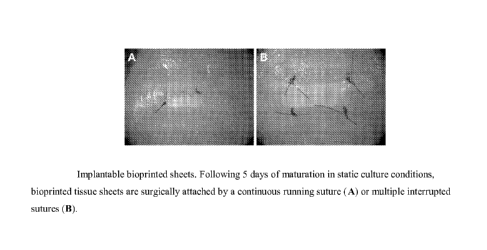
[0025] Fig. 12 illustrates bioprinted tissue sheets surgically attached by a
continuous running
suture (A) or multiple interrupted sutures (B).
[0026] Fig. 13 depicts a bioprinted skeletal muscle tissue fabricated onto a
multi-well insert for
long-term maintenance and maturation (A). H&E stain of a bioprinted skeletal
muscle tissue after
3 days and 9 days in culture (B, C).
DETAILED DESCRIPTION OF THE INVENTION
[0027] The invention relates to the field of regenerative medicine and
tissue/organ engineering.
More particularly, the invention relates to tissues and organs comprising at
least one layer
comprising muscle cells, wherein the engineered tissue or organ consists
essentially of cellular
material, and methods of making the same. An advantage of the tissues, organs,
and methods
disclosed herein include, by way of example, flexible three-dimensional tissue
geometry that
allows fabrication of optionally layered sheets, tubes, and sacs comprising
muscle cells. Another
advantage is a flexible layered approach allowing for one or more cell types
other than muscle
cells to be disposed, dispensed, and/or bioprinted on at least one surface of
the layer. These
advantages result in engineered tissues and organs that mimic native tissue
composition and
architecture.
[0028] Disclosed herein, in certain embodiments, are living, three-dimensional
engineered
tissues or organs comprising one or more layers, the one or more layers
characterized by one or
more of: a) substantially scaffold-free at the time of use; and b) bioprinted,
the one or more layers
suitable for implantation in a vertebrate subject upon sufficient maturation;
provided that at least
one layer of the engineered tissue or organ comprises muscle cells and that
the engineered tissue
or organ is not a vascular tube.
[0029] Also disclosed herein, in certain embodiments, is implantation of the
tissues and/or
organs.
[0030] Also disclosed herein, in certain embodiments, are methods for making
an implantable
tissue or organ comprising a muscle cell-containing layer, the method
comprising: bioprinting
bio-ink comprising muscle cells into a form; and fusing the bio-ink into a
cohesive cellular
-6-
CA 02848044 2014-03-06
WO 2013/040087 PCMJS2012/054935
structure; provided that the tissue or organ is implantable in a vertebrate
subject and not a
vascular tube.
[00311 Also disclosed herein, in certain embodiments, are living, three-
dimensional engineered
tissues or organs comprising one or more layers, the one or more layers
characterized by one or
more of: a) substantially scaffold-free at the time of use; and b) bioprinted,
the one or more layers
matured into implantation-ready status for a vertebrate subject; the
engineered tissue or organ
consisting essentially of cellular material; provided that at least one layer
of the engineered tissue
or organ comprises muscle cells and that the engineered tissue or organ is not
a vascular tube.
[00321 Also disclosed herein, in certain embodiments, is implantation of the
tissues and/or
organs.
Certain Definitions
[00331 Unless otherwise defined, all technical and scientific terms used
herein have the same
meaning as commonly understood by one of ordinary skill in the art to which
this invention
belongs.
[00341 As used in this specification and the appended claims, the singular
forms "a," "an," and
"the" include plural references unless the context clearly dictates otherwise.
Thus, for example,
references to "a nucleic acid" includes one or more nucleic acids, and/or
compositions of the type
described herein which will become apparent to those persons skilled in the
art upon reading this
disclosure and so forth. Any reference to "or" herein is intended to encompass
"and/or" unless
otherwise stated.
[0035] As used herein, `tio-ink" means a liquid, semi-solid, or solid
composition comprising a
plurality of cells. In some embodiments, bio-ink comprises cell solutions,
cell aggregates, cell-
comprising gels, multicellular bodies, or tissues. In some embodiments, the
bio-ink additionally
comprises support material. In some embodiments, the bio-ink additionally
comprises non-
cellular materials that provide specific biomechanical properties that enable
bioprinting.
[00361 As used herein, "bioprinting" means utilizing three-dimensional,
precise deposition of
cells (e.g., cell solutions, cell-containing gels, cell suspensions, cell
concentrations, multicellular
aggregates, multicellular bodies, etc.) via methodology that is compatible
with an automated or
semi-automated, computer-aided, three-dimensional prototyping device (e.g., a
bioprinter).
[00371 As used herein, "blood vessel" means a tube of smooth muscle cells
further comprising
vascular endothelial cells, and having an internal diameter greater than 100
lam, and intended for
use in vivo as an interpositional vascular graft, a bypass vascular graft, or
an arterio-venous
vascular shunt. As used herein, "blood vessel" expressly does not include the
integral vascular
components (arteries, veins, arterioles, venules, capillaries, and
microvasculature) of other organs
-7-
CA 02848044 2014-03-06
WO 2013/040087 PCMJS2012/054935
or tissues. For example, the vascular network associated with the bladder,
intestine, or esophagus
would not be included in the definition of "blood vessel" as presented herein.
[0038] As used herein, "cohere," "cohered," and "cohesion" refer to cell-cell
adhesion properties
that bind cells, cell aggregates, multicellular aggregates, multicellular
bodies, and/or layers
thereof. The terms are used interchangeably with "fuse," "fused," and
"fusion."
[0039] As used herein, -extracellular matrix" means proteins that are produced
by cells and
transported out of the cells into the extracellular space, where they serve as
a support to hold
tissues together, to provide tensile strength, and/or to facilitate cell
signaling.
[0040] As used herein, "implantable" means biocompatible and capable of being
inserted or
grafted into or affixed onto a living organism either temporarily or
substantially permanently.
[0041] As used herein, "laminar" means a multi-layered bioprinted tissue in
which two or more
planar layers are combined to increase the overall thickness of the tissue in
the z-plane. In some
embodiments, each planar layer is substantially similar in architecture and/or
composition. In
other embodiments, each planar layer is substantially distinct in architecture
and/or composition.
[0042] As used herein, "multi-layered" means being comprised of two or more
layers of tissue,
wherein each tissue layer is one or more cell-layers in thickness. In some
embodiments, layers of
tissue are deposited one at a time. In other embodiments, multiple layers are
deposited
simultaneously. Optionally, each layer is comprised of multiple cell types.
Further, the multiple
cell types within each layer are optionally arranged relative to each other in
a spatially-defined
architecture in the x-y planes (i.e., horizontal planes). Furthermore,
addition of layers in the z-
plane (i.e., vertical plane), in some cases, results in controlled spatial
positioning of the cells
within the layers relative to each other so that a spatially-defined
architecture is continued in the
z-plane.
[0043] As used herein, "organ" means a collection of tissues joined into
structural unit to serve a
common function. Examples of organs include, but are not limited to, skin,
urethra, conduit,
ureter, bladder, fallopian tube, uterus, trachea, bronchus, lymphatic vessel,
esophagus, stomach,
gallbladder, small intestine, large intestine, and colon.
[0044] As used herein, "planar" means a layer of multicellular bioprinted
tissue in which
multiple bio-ink compositions and/or void spaces are spatially arranged into a
defined pattern
relative to each other within the x-y plane of the tissue layer. "Planar" also
means substantially
flat when used to describe the shape of a tissue "sheet" or "patch."
[0045] As used herein, "scaffold" refers to synthetic scaffolds such as
polymer scaffolds and
porous hydrogels, non-synthetic scaffolds such as pre-formed extracellular
matrix layers, dead
cell layers, and decellularized tissues, and any other type of pre-formed
scaffold that is integral to
the physical structure of the engineered tissue and/or organ and not able to
be removed from the
-8-
CA 02848044 2014-03-06
WO 2013/040087 PCMJS2012/054935
tissue and/or organ without damage/destruction of said tissue and/or organ. In
further
embodiments, decellularized tissue scaffolds include decellularized native
tissues or
decellularized cellular material generated by cultured cells in any manner;
for example, cell
layers that are allowed to die or are decellularized, leaving behind the ECM
they produced while
living. The term "scaffold-free" as used herein indicates that the cell-
comprising tube, sac, or
sheet is substantially free from scaffold (as defined above) at the time of
use. "Scaffold-free" is
used interchangeably with "scaffoldless" and "free of pre-formed scaffold."
[0046] As used herein, "stem cell" means a cell that exhibits potency and self-
renewal. Stem
cells include, but are not limited to, totipotent cells, pluripotent cells,
multipotent cells,
oligopotent cells, unipotent cells, and progenitor cells. In various
embodiments, stem cells are
embryonic stem cells, pen-natal stem cells, adult stem cells, amniotic stem
cells, and induced
pluripotent stem cells.
[0047] As used herein, "subject" means any individual. The term is
interchangeable with
"patient," "recipient," and "donor." None of the terms should be construed as
requiring the
supervision (constant or otherwise) of a medical professional (e.g.,
physician, nurse, nurse
practitioner, physician's assistant, orderly, hospice worker, social worker
and a clinical research
associate) or a scientific researcher.
[0048] As used herein, "tissue" means an aggregate of cells. Examples of
tissues include, but are
not limited to, connective tissue (e.g., areolar connective tissue, dense
connective tissue, elastic
tissue, reticular connective tissue, and adipose tissue), muscle tissue (e.g.,
skeletal muscle,
smooth muscle and cardiac muscle), genitourinary tissue, gastrointestinal
tissue, pulmonary
tissue, bone tissue, nervous tissue, and epithelial tissue (e.g., simple
epithelium and stratified
epithelium), endoderm-derived tissue, mesoderm-derived tissue, and ectoderm-
derived tissue.
Tissue Engineering
[0049] Tissue engineering is an interdisciplinary field that applies and
combines the principles of
engineering and life sciences toward the development of biological substitutes
that restore,
maintain, or improve tissue function through augmentation, repair, or
replacement of an organ or
tissue. The basic approach to classical tissue engineering is to seed living
cells into a
biocompatible and eventually biodegradable environment (e.g., a scaffold), and
then culture this
construct in a bioreactor so that the initial cell population expands further
and mature to generate
the target tissue upon implantation. With an appropriate scaffold that mimics
the biological
extracellular matrix (ECM), the developing tissue adopts both the form and
function of the
desired organ after in vitro and in vivo maturation. However, achieving high
enough cell density
with a native tissue-like architecture is challenging due to the limited
ability to control the
distribution and spatial arrangement of the cells throughout the scaffold.
These limitations often
-9-
CA 02848044 2014-03-06
WO 2013/040087 PCMJS2012/054935
result in tissues or organs with poor mechanical properties and/or
insufficient function.
Additional challenges exist with regard to biodegradation of the scaffold,
entrapment of residual
polymer, and industrial scale-up of manufacturing processes. Scaffoldless
approaches have been
attempted. Current scaffoldless approaches are subject to several limitations:
= Complex geometries, such as multi-layered structures wherein each layer
comprises a
different cell type, often require definitive, high-resolution placement of
cell types within a
specific architecture to reproducibly achieve a native tissue-like outcome.
= Scale and geometry are limited by diffusion and/or the requirement for
functional vascular
networks for nutrient supply.
= The viability of the tissues in many cases is compromised by confinement
material that
limits diffusion and restricts the cells' access to nutrients.
[0050] Disclosed herein, in certain embodiments, are engineered tissues and
organs, and methods
of fabrication. The tissue engineering methods disclosed herein have the
following advantages:
= They are capable of producing cell-comprising tissues and/or organs.
= They mimic the environmental conditions found within the development,
homeostasis,
and/or pathogenesis of natural tissues by re-creating native tissue-like
intercellular
interactions.
= They optionally achieve a broad array of complex topologies (e.g.,
multilayered
structures, segments, sheets, tubes, sacs, etc.).
= They are compatible with automated means of manufacturing and are
scalable.
[0051] Bioprinting enables improved methods of generating cell-comprising
implantable tissues
that are useful in tissue repair, tissue augmentation, tissue replacement, and
organ replacement
(see below).
Bioprinting
[0052] In some embodiments, at least one component of the engineered,
implantable tissues
and/or organs was bioprinted. In further embodiments, the engineered,
implantable tissues and/or
organs were entirely bioprinted. In still further embodiments, bioprinted
constructs are made with
a method that utilizes a rapid prototyping technology based on three-
dimensional, automated,
computer-aided deposition of cells, including cell solutions, cell
suspensions, cell-comprising
gels or pastes, cell concentrations, multicellular bodies (e.g., cylinders,
spheroids, ribbons, etc.),
and confinement material onto a biocompatible surface (e.g., composed of
hydrogel and/or a
porous membrane) by a three-dimensional delivery device (e.g., a bioprinter).
As used herein, in
some embodiments, the term "engineered," when used to refer to tissues and/or
organs means
that cells, cell solutions, cell suspensions, cell-comprising gels or pastes,
cell concentrates,
multicellular aggregates, and layers thereof are positioned to form three-
dimensional structures
-10-
CA 02848044 2014-03-06
WO 2013/040087 PCMJS2012/054935
by a computer-aided device (e.g., a bioprinter) according to a computer
script. In further
embodiments, the computer script is, for example, one or more computer
programs, computer
applications, or computer modules. In still further embodiments, three-
dimensional tissue
structures form through the post-printing fusion of cells or multicellular
bodies similar to self-
assembly phenomena in early morphogenesis.
[00531 While a number of methods are available to arrange cells, multicellular
aggregates, and/or
layers thereof on a biocompatible surface to produce a three-dimensional
structure including
manual placement, positioning by an automated, computer-aided machine such as
a bioprinter is
advantageous. Advantages of delivery of cells or multicellular bodies with
this technology
include rapid, accurate, and reproducible placement of cells or multicellular
bodies to produce
constructs exhibiting planned or pre-determined orientations or patterns of
cells, multicellular
aggregates and/or layers thereof with various compositions. Advantages also
include assured
high cell density, while minimizing cell damage.
[0054] In some embodiments, the method of bioprinting is continuous and/or
substantially
continuous. A non-limiting example of a continuous bioprinting method is to
dispense bio-ink
from a bioprinter via a dispense tip (e.g., a syringe, capillary tube, etc.)
connected to a reservoir
of bio-ink. In further non-limiting embodiments, a continuous bioprinting
method is to dispense
bio-ink in a repeating pattern of functional units. In various embodiments, a
repeating functional
unit has any suitable geometry, including, for example, circles, squares,
rectangles, triangles,
polygons, and irregular geometries. In further embodiments, a repeating
pattern of bioprinted
function units comprises a layer and a plurality of layers are bioprinted
adjacently (e.g., stacked)
to form an engineered tissue or organ. In various embodiments, 2, 3, 4, 5, 6,
7, 8, 9, 10, 11, 12,
13, 14, 15, or more layers are bioprinted adjacently (e.g., stacked) to form
an engineered tissue or
organ.
[00551 In some embodiments, a bioprinted functional unit repeats in a
tessellated pattern. A
"tessellated pattern" is a plane of figures that fills the plane with no
overlaps and no gaps. Fig.
7A shows an example of a functional unit that is optionally repeated to
produce the tessellation
pattern depicted in Fig. 7B. Advantages of continuous and/or tessellated
bioprinting include, by
way of non-limiting example, increased productivity of bioprinted tissue.
Another non-limiting,
exemplary advantage is eliminating the need to align the bioprinter with
previously deposited
elements of bio-ink. Continuous bioprinting also facilitates printing larger
tissues from a large
reservoir of bio-ink, optionally using a syringe mechanism.
[0056] In various embodiments, methods in continuous bioprinting involves
optimizing and/or
balancing parameters such as print height, pump speed, robot speed, or
combinations thereof
independently or relative to each other. In one example, the bioprinter head
speed for deposition
-11-
CA 02848044 2014-03-06
WO 2013/040087 PCMJS2012/054935
was 3 mm/s, with a dispense height of 0.5 mm for the first layer and dispense
height was
increased 0.4 mm for each subsequent layer. In some embodiments, the dispense
height is
approximately equal to the diameter of the bioprinter dispense tip. Without
limitation a suitable
and/or optimal dispense distance does not result in material flattening or
adhering to the
dispensing needle. In various embodiments, the bioprinter dispense tip has an
inner diameter of
about, 20, 50, 100, 150, 200, 250, 300, 350, 400, 450, 500, 550, 600, 650,
700, 750, 800, 850,
900, 950, 1000 gm, or more, including increments therein. In various
embodiments, the bio-ink
reservoir of the bioprinter has a volume of about .5, 1, 2, 3, 4, 5, 6, 7, 8,
9, 10, 15, 20, 25, 30, 35,
40, 45, 50, 55, 60, 65, 70, 75, 80, 85, 90, 95, 100 cubic centimeters, or
more, including
increments therein. In some embodiments, the pump speed is suitable and/or
optimal when the
residual pressure build-up in the system is low. In some embodiments,
favorable pump speeds
depend on the ratio between the cross-sectional areas of the reservoir and
dispense needle with
larger ratios requiring lower pump speeds. In some embodiments, a suitable
and/or optimal print
speed enables the deposition of a uniform line without affecting the
mechanical integrity of the
material.
[0057] The inventions disclosed herein include business methods. In some
embodiments, the
speed and scalability of the techniques and methods disclosed herein are
utilized to design, build,
and operate industrial and/or commercial facilities for production of
engineered tissues and/or
organs for implantation. In further embodiments, the engineered tissues and/or
organs are
produced, stored, distributed, marketed, advertised, and sold as, for example,
materials, tools, and
kits for in vivo uses such as medical treatment of tissue damage, tissue
disease, and/or organ
failure. In other embodiments, the engineered tissues and/or organs are
produced, stored,
distributed, marketed, advertised, and sold as, for example, materials, tools,
and kits for in vitro
uses such as scientific and/or medical research. In further embodiments, the
engineered tissues
and/or organs are maintained in cell culture environments and used in
scientific and/or medical
research.
Engineered tissues and organs
[00581 Disclosed herein, in certain embodiments, are engineered, implantable
tissues and/or
organs comprising one or more layers, wherein at least one layer comprises
muscle cells. In some
embodiments, the one or more layers are characterized by being either
substantially scaffold-free
at the time of use, at the time of bioprinting (a technology described
herein), or both. In further
embodiments, the one or more layers and/or the engineered tissue or organ
consist essentially of
cellular material. In some embodiments, the one or more layers are suitable
for implantation in a
vertebrate subject upon sufficient maturation. In some embodiments, the one or
more layers are
matured into implantation-ready status for a vertebrate subject.
-12-
CA 02848044 2014-03-06
WO 2013/040087 PCMJS2012/054935
[0059] In some embodiments, the engineered tissues and organs consist
essentially of cellular
material. In various further embodiments, the cell-comprising portions of the
engineered tissues
and organs consist of 30, 35, 40, 45, 50, 55, 60, 65, 70, 75, 80, 85, 90, 95,
96, 97, 98, 99, 99.5,
99.9, and 100% cellular material, including increments therein, at the time of
construction. In
other various embodiments, the cell-comprising portions of the engineered
tissues and organs
consist of 30, 35, 40, 45, 50, 55, 60, 65, 70, 75, 80, 85, 90, 95, 96, 97, 98,
99, 99.5, 99.9, and
100% cellular material, including increments therein, at the time of use. In
some embodiments,
the engineered tissues are cohered and/or adhered aggregates of cells. In some
embodiments, the
non-cellular components are removed prior to use. In further embodiments, the
non-cellular
components are removed by physical, chemical, or enzymatic means. In some
embodiments, a
proportion of the non-cellular components remains associated with the cellular
components at the
time of use. In some embodiments, the non-cellular components are selected
from a group that
includes: hydrogels, surfactant polyols, thermo-responsive polymers,
hyaluronates, alginates,
collagens, or other biocompatible natural or synthetic polymers.
[0060] The engineered tissues optionally mimic any human or mammalian tissue.
Exemplary
tissues include epithelial tissue, connective tissue, muscle tissue, nervous
tissue, and the like. In
some embodiments, the engineered organs are collections of tissues joined into
structural unit(s)
to serve a common function. The organs suitably mimic any natural human or
mammalian organ.
Exemplary organs include, by way of non-limiting example trachea, bronchus,
esophagus,
stomach, intestine, colon, gall bladder, uterus, fallopian tube, ureter,
bladder, urethra, lymph
vessel, and the like, including portions thereof
[0061] In some embodiments, the engineered tissues and organs are implantable.
In further
embodiments, implantable tissues and organs are biocompatible, meaning that
they pose limited
risk of injury or toxicity to organisms that they contact. In some
embodiments, implantation
involves inserting or grafting a tissue or organ into a subject. In further
embodiments, insertion
and/or grafting is performed surgically. In other embodiments, implantation
involves affixing a
tissue or organ to a subject. The tissues and organs disclosed herein are
suitably implanted for
various durations. In some embodiments, the tissues and/or organs are suitably
implanted, by
way of non-limiting example, temporarily, semi-permanently, and permanently.
In some
embodiments, implanted tissues and/or organs are absorbed, incorporated, or
dissolved over time.
In other embodiments, implanted tissues and/or organs retain a distinct form
for some period of
time.
[0062] The engineered tissues and organs are suitable for implantation in any
vertebrate subject
in need thereof In various embodiments, vertebrate subjects include, by way of
non-limiting
examples, human subjects, vertebrate veterinary subjects, and those classified
as Mammalia
-13-
CA 02848044 2014-03-06
WO 2013/040087 PCMJS2012/054935
(mammals), Ayes (birds), Reptilia (reptiles), Amphibia (amphibians),
Osteichthyes (bony fishes),
Chondrichthyes (cartilaginous fishes), Agnatha (jawless fishes), etc.
[0063] In various embodiments, engineered tissues and organs are suitable for
implantation in
any vertebrate subject in need of, for example, wound repair, tissue repair,
tissue augmentation,
tissue replacement, and/or organ replacement. In some embodiments, the
engineered tissues are
used for wound repair or tissue repair. For example, an engineered sheet is
used to temporarily or
permanently repair human skin damaged by injury. In some embodiments, the
engineered tissues
are used for tissue augmentation. For example, an engineered sheet is used to
temporarily or
permanently patch or repair a defect in the muscle wall of a human bladder or
stomach. In some
embodiments, the engineered tissues are used for tissue replacement. For
example, an engineered
sheet or tube is used to temporarily or permanently repair or replace the wall
of a segment of
human small intestine. In some embodiments, the engineered organs are used for
organ
replacement. For example, an engineered tube is used to temporarily or
permanently replace a
human fallopian tube damaged by an ectopic pregnancy. In some embodiments, an
engineered
tubular structure is used to create new connections with organ systems; for
example, a smooth
muscle-comprising tube could be used to extend a connection from the
gastrointestinal system or
the kidney through the body wall to enable waste collection in certain disease
states. In other
embodiments, engineered tubular structures are used to extend the length of
certain native tissues
(e.g., esophagus, intestine, colon, etc.) to eliminate or ameliorate specific
diseases that are
congenital in nature (e.g., short gut syndrome, etc.) or occur as a
consequence of other diseases
or injuries.
[0064] The engineered, implantable tissues and organs, in various embodiments,
are any suitable
shape. In some embodiments, the shape is selected to mimic a particular
natural tissue or organ.
[0065] In some embodiments, a layer comprising muscle cells or an overall
engineered tissue or
organ is substantially in the form of a sheet or a form that comprises a
sheet. In further
embodiments, a sheet is a substantially planar form with a range of suitable
geometries including,
by way of non-limiting example, planar square, rectangle, polygon, circle,
oval, or irregular. A
bioprinted sheet has a wide range of suitable dimensions. In some embodiments,
the dimensions
are selected to facilitate a specific use including, by way of non-limiting
examples, wound repair,
tissue repair, tissue augmentation, tissue replacement, and engineered organ
construction. In
further embodiments, the dimensions are selected to facilitate a specific use
in a specific subject.
For instance, in one embodiment, a sheet is bioprinted to repair a particular
defect in a muscle-
comprising tissue of a specific human subject.
[0066] The engineered, implantable tissues and organs, in various embodiments,
are any suitable
size. In some embodiments, the size of engineered tissues and organs,
including those bioprinted,
-14-
CA 02848044 2014-03-06
WO 2013/040087 PCMJS2012/054935
change over time. In further embodiments, a bioprinted tissue or organ shrinks
or contracts after
bioprinting due to, for example, cell migration, cell death, cell-adhesion-
mediated contraction, or
other forms of shrinkage. In other embodiments, a bioprinted tissue or organs
grows or expands
after bioprinting due to, for example, cell migration, cell growth and
proliferation, cell
maturation, or other forms of expansion.
[00671 In some embodiments, a bioprinted sheet is at least 150 ium thick at
the time of
bioprinting. In various embodiments, a bioprinted sheet is about 10, 15, 20,
25, 30, 35, 40, 45, 50,
55, 60, 65, 70, 75, 80, 85, 90, 95, 100, 125, 150, 175, 200, 225, 250, 275,
300, 325, 350, 375,
400, 425, 450, 475, 500 pm or more thick, including increments therein. In
further various
embodiments, a bioprinted sheet is characterized by having a length, width, or
both, of about 50,
100, 150, 200, 250, 300, 350, 400, 450, 500, 550, 600, 650, 700, 750, 800,
850, 900, 950, 1000
Jim or more, including increments therein. In other various embodiments, a
bioprinted sheet is
characterized by having a length, width, or both, of about 1, 2, 3, 4, 5, 6,
7, 8, 9, 10, 11, 12, 13,
14, 15, 20, 25, 30, 35, 40, 45, 50, 55, 60, 65, 70, 75, 80, 85, 90, 95, 100 mm
or more, including
increments therein. In other various embodiments, a bioprinted sheet is
characterized by having a
length, width, or both, of about 1, 2, 3, 4, 5, 6, 7, 8, 9, 10, 11, 12, 13,
14, 15, 20, 25, 30, 35, 40,
45, 50, 55, 60, 65, 70, 75, 80, 85, 90, 95, 100 cm or more, including
increments therein. See, e.g.,
Example 6 (and Fig. 1), Example 7 (and Fig. 2), Example 9 (and Figs. 3a and
3b), Example 10
(and Figs. 4a and 4b).
[00681 In some embodiments, a layer comprising muscle cells or an overall
engineered tissue or
organ is substantially in the form of a tube or a form that comprises a tube.
In further
embodiments, a tube is a substantially a rolled sheet or a hollow cylinder. In
some embodiments,
a bioprinted tube is used to construct an engineered organ. In further
embodiments, a bioprinted
tube is used to construct an engineered ureter, urinary conduit, fallopian
tube, uterus, trachea,
bronchus, lymphatic vessel, urethra, intestine, colon, esophagus, or portion
thereof. In further
embodiments, the tubes disclosed herein are not blood vessels or vascular
tubes. A bioprinted
tube has a wide range of suitable dimensions. In some embodiments, the
dimensions are selected
to facilitate a specific use including, by way of non-limiting examples, wound
repair, tissue
repair, tissue augmentation, tissue replacement, engineered organ
construction, and organ
replacement. In further embodiments, the dimensions are selected to facilitate
a specific use in a
specific subject. For instance, in one embodiment, a tube is bioprinted to
repair a particular
segment of lymph vessel of a specific human subject. In some embodiments, a
bioprinted tube is
characterized by having a tubular wall that is at least 150 jim thick at the
time of bioprinting. In
various embodiments, the wall of a bioprinted tube is about 10, 15, 20, 25,
30, 35, 40, 45, 50, 55,
60, 65, 70, 75, 80, 85, 90, 95, 100, 125, 150, 175, 200, 225, 250, 275, 300,
325, 350, 375, 400,
-15-
CA 02848044 2014-03-06
WO 2013/040087 PCMJS2012/054935
425, 450, 475, 500 or more ium thick, including increments therein. In some
embodiments, the
bioprinted tubes are characterized by having an inner diameter of at least
about 250 tan at the
time of bioprinting. In various embodiments, the inner diameter of a
bioprinted tube is about 50,
60, 70, 80, 90, 100, 125, 150, 175, 200, 225, 250, 275, 300, 325, 350, 375,
400, 425, 450, 475,
500, 600, 700, 800, 900, 1000 iitm or more, including increments therein. In
other various
embodiments, the inner diameter of a bioprinted tube is about 1, 2, 3, 4, 5,
6, 7, 8, 9, 10, 11, 12,
13, 14, 15, 16, 17, 18, 19, 20, 21, 22, 23, 24, 25, 26, 27, 28, 29, 30 mm or
more, including
increments therein. In some embodiments, the length of a bioprinted tube is
about 1, 2, 3, 4, 5, 6,
7, 8, 9, 10, 11, 12, 13, 14, 15, 16, 17, 18, 19, 20, 21, 22, 23, 24, 25, 26,
27, 28, 29, 30 mm or
more, including increments therein. In other embodiments, the length of a
bioprinted tube is
about 1, 2, 3, 4, 5, 6, 7, 8, 9, 10, 11, 12, 13, 14, 15, 16, 17, 18, 19, 20,
21, 22, 23, 24, 25, 26, 27,
28, 29, 30 cm or more, including increments therein. See, e.g., Example 13
(and Fig. 6).
[00691 In some embodiments, a layer comprising muscle cells or an overall
engineered tissue or
organ is substantially in the form of a sac or a form that comprises a sac. In
further embodiments,
a sac is a substantially a rolled sheet or a hollow cylinder with at least one
closed end (e.g., a
pouch, cup, hollow, balloon, etc.). In some embodiments, a sac is an
expandable structure
intended for containment of ingested material, a fetus and related fluids,
bodily fluids, or bodily
wastes, and has at least one opening for input and at least one opening for
output. In some
embodiments, a bioprinted sac is used to augment an existing organ or tissue.
In other
embodiments, a bioprinted sac is used to replace an existing organ or tissue.
In further
embodiments, a bioprinted sac is used to construct an engineered stomach,
bladder, uterus,
gallbladder, or portion thereof. A bioprinted sac has a wide range of suitable
dimensions. In some
embodiments, the dimensions are selected to facilitate a specific use
including, by way of non-
limiting examples, wound repair, tissue repair, tissue augmentation, tissue
replacement,
engineered organ construction, and organ replacement. In further embodiments,
the dimensions
are selected to facilitate a specific use in a specific subject. For instance,
in one embodiment, a
sac is bioprinted to augment or replace the bladder of a specific human
subject. In some
embodiments, a bioprinted sac is characterized by having a wall that is at
least 150 iitm thick at
the time of bioprinting. In various embodiments, the wall of a bioprinted sac
is about 10, 15, 20,
25, 30, 35, 40, 45, 50, 55, 60, 65, 70, 75, 80, 85, 90, 95, 100, 125, 150,
175, 200, 225, 250, 275,
300, 325, 350, 375, 400, 425, 450, 475, 500 ium or more thick, including
increments therein.
[00701 In some embodiments, an implantable tissue or organ is used for
scientific and/or medical
research. Suitable scientific and/or medical research includes both in vivo
and in vitro research.
In further embodiments, the engineered, tissues and/or organs described
herein, are for in vitro
-16-
CA 02848044 2014-03-06
WO 2013/040087 PCMJS2012/054935
research uses including, by way of non-limiting examples, disease modeling,
drug discovery, and
drug screening.
Cells
[0071] Disclosed herein, in some embodiments, are engineered, implantable
tissues and organs
comprising one or more types of cells. In some embodiments, the engineered
tissues and organs
include at least one layer comprising muscle cells. Therefore, in some
embodiments, the cells
include muscle cells (e.g., smooth muscle cells, skeletal muscle cells,
cardiac muscle cells). In
further embodiments, the layer comprising muscle cells also includes
additional cells types such
as those disclosed herein (e.g., fibroblasts, endothelial cells, etc.). In
some embodiments, the
engineered tissues and organs include cells other than muscle cells dispensed
on at least one
surface of a layer comprising muscle cells. In further embodiments, the cells
dispensed on at least
one surface of a layer comprising muscle cells include, by way of non-limiting
examples,
endothelial cells, nerve cells, pericytes, fibroblasts, tissue-specific
epithelial cells, chondrocytes,
skeletal muscle cells, cardiomyocytes, bone-derived cells, soft tissue-derived
cells, mesothelial
cells, tissue-specific stromal cells, stem cells, progenitor cells, and
combinations thereof
[0072] In some embodiments, any vertebrate cell is suitable for inclusion in
the engineered,
implantable tissues and organs. In further embodiments, the cells are, by way
of non-limiting
examples, contractile or muscle cells (e.g., skeletal muscle cells,
cardiomyocytes, smooth muscle
cells, and myoblasts), connective tissue cells (e.g., bone cells, cartilage
cells, fibroblasts, and
cells differentiating into bone forming cells, chondrocytes, or lymph
tissues), bone marrow cells,
endothelial cells, skin cells, epithelial cells, breast cells, vascular cells,
blood cells, lymph cells,
neural cells, Schwann cells, gastrointestinal cells, liver cells, pancreatic
cells, lung cells, tracheal
cells, corneal cells, genitourinary cells, kidney cells, reproductive cells,
adipose cells,
parenchymal cells, pericytes, mesothelial cells, stromal cells,
undifferentiated cells (e.g.,
embryonic cells, stem cells, and progenitor cells), endoderm-derived cells,
mesoderm-derived
cells, ectoderm-derived cells, and combinations thereof.
[0073] In one embodiment, the cells are smooth muscle cells. In another
embodiment, the cells
are smooth muscle cells combined with at least one additional cell type. In
some embodiments,
the other cell type is fibroblasts. In some embodiments, the fibroblasts
provide structural and
biological support for the engineered tissue. In yet another embodiment, the
other cell type is
endothelial cells. In some embodiments, the endothelial cells facilitate
vascularization and/or
microvascularization of the engineered tissue. In still another embodiment,
the cells are smooth
muscle cells, fibroblasts, and endothelial cells. In embodiments including
more than one cell
type, the cell types are present in many suitable ratios, examples of which
are described herein.
-17-
[0074] In some embodiments, the cells are adult, differentiated cells. In
further embodiments,
"differentiated cells" are cells with a tissue-specific phenotype consistent
with, for example, a
muscle cell, a fibroblast, or an endothelial cell at the time of isolation,
wherein tissue-specific
phenotype (or the potential to display the phenotype) is maintained from the
time of isolation to
the time of use. In other embodiments, the cells are adult, non-differentiated
cells. In further
embodiments, "non-differentiated cells" are cells that do not have, or have
lost, the definitive
tissue-specific traits of for example, muscle cells, fibroblasts, or
endothelial cells. In some
embodiments, non-differentiated cells include stem cells. In further
embodiments, "stem cells"
are cells that exhibit potency and self-renewal. Stem cells include, but are
not limited to,
totipotent cells, pluripotent cells, multipotent cells, oligopotent cells,
unipotent cells, and
progenitor cells. In various embodiments, stem cells are embryonic stem cells,
adult stem cells,
amniotic stem cells, and induced pluripotent stem cells. In other embodiments,
the cells are a
mixture of adult, differentiated cells and adult, non-differentiated cells.
[0075] In some embodiments, the smooth muscle cells are human smooth muscle
cells. In some
embodiments, suitable smooth muscle cells originated from tissue including, by
way of non-
limiting example, blood vessel, lymphatic vessel, tissue of the digestive
tract, tissue of the
genitourinary tract, adipose tissue, tissue of the respiratory tract, tissue
of the reproductive
system, bone marrow, and umbilical tissue. In some embodiments, additional
(non-smooth-
muscle) cellular components originated from the target tissue of interest. In
other embodiments,
additional (non-smooth-muscle) cellular components originated from a tissue
other than the
target tissue of interest. In further embodiments, some or all of the cells
are cultured from the
stromal vascular fraction of mammalian lipoaspirate. See Example 1.
[0076] In various embodiments, the cell types and/or source of the cells are
selected, configured,
treated, or modulated based on a specific goal or objective. In some
embodiments, one or more
specific cell types are derived from two or more distinct human donors. In
some embodiments,
one or more specific cell types are derived from a particular vertebrate
subject. In further
embodiments, one or more specific cell types are derived from a particular
mammalian subject.
In still further embodiments, one or more specific cell types are derived from
a particular human
subject.
Methods of culturing cells
[0077] The cell types used in the engineered tissues of the invention are
suitably cultured in any
manner known in the art. Methods of cell and tissue culturing are known in the
art, and are
described, for example, in Freshney, R., Culture of Animal Cells: A Manual of
Basic Techniques,
Wiley (1987). General mammalian cell culture techniques, cell lines, and cell
culture systems
-18-
CA 2848044 2020-03-04
suitably used in conjunction with the present invention are also described in
Doyle, A., Griffiths,
J. B., Newell, D. G., (eds.) Cell and Tissue Culture: Laboratory Procedures,
Wiley (1998).
[0078] Appropriate growth conditions for mammalian cells in culture are well
known in the art.
See, e.g., Example 1. Cell culture media generally include essential nutrients
and, optionally,
additional elements such as growth factors, salts, minerals, vitamins, etc.,
selected according to
the cell type(s) being cultured. Particular ingredients are optionally
selected to enhance cell
growth, differentiation, secretion of specific proteins, etc. In general,
standard growth media
include Dulbecco's Modified Eagle Medium (DMEM), low glucose with 110 mg/L
pyruvate and
glutamine, supplemented with 1-20% fetal bovine serum (FBS), calf serum, or
human serum and
100 U/mL penicillin, 0.1 mg/mL streptomycin are appropriate as are various
other standard
media well known to those in the art. Preferably cells are cultured under
sterile conditions in an
atmosphere of 1-21% 02 and preferably 3-5% CO2, at a temperature at or near
the body
temperature of the animal of origin of the cell. For example, human cells are
preferably cultured
at approximately 37 C.
[0079] The cells are optionally cultured with cellular differentiation agents
to induce
differentiation of the cell along the desired line. For instance, cells are
optionally cultured with
growth factors, cytokines, etc. In some embodiments, the term "growth factor"
refers to a protein,
a polypeptide, or a complex of polypeptides, including cytokines, that are
produced by a cell and
affect itself and/or a variety of other neighboring or distant cells.
Typically growth factors affect
the growth and/or differentiation of specific types of cells, either
developmentally or in response
to a multitude of physiological or environmental stimuli. Some, but not all,
growth factors are
hormones. Exemplary growth factors are insulin, insulin-like growth factor
(IGF), nerve growth
factor (NGF), vascular endothelial growth factor (VEGF), keratinocyte growth
factor (KGF),
fibroblast growth factors (FGFs), including basic FGF (bFGF), platelet-derived
growth factors
(PDGFs), including PDGF-AA and PDGF-AB, hepatocyte growth factor (HGF),
transforming
growth factor alpha (TGF-a), transforming growth factor beta (TGF-f3),
including TGFP1 and
TGFI33, epidermal growth factor (EGF), granulocyte-macrophage colony-
stimulating factor
(GM-CSF), granulocyte colony-stimulating factor (G-CSF), interleukin-6 (IL-6),
IL-8, and the
like. Growth factors are discussed in, among other places, Molecular Cell
Biology, Scientific
American Books, Darnell et al., eds., 1986; Principles of Tissue Engineering,
2d ed., Lanza et al.,
eds., Academic Press, 2000. The skilled artisan will understand that any and
all culture-derived
growth factors in the conditioned media described herein are within the scope
of the invention.
Bio-ink and multicellular a22regates
-19-
CA 2848044 2020-03-04
CA 02848044 2014-03-06
WO 2013/040087 PCMJS2012/054935
[0080] Disclosed herein, in certain embodiments, are engineered, implantable
tissues and/or
organs comprising one or more layers, wherein at least one layer comprises
muscle cells. In some
embodiments, the one or more layers and/or the engineered tissue or organ
consist essentially of
cellular material. In further embodiments, cells other than muscle cells were
dispensed on at least
one surface of the one or more layers. In some embodiments, the one or more
layers are
substantially scaffold-free at the time of use.
[0081] In some embodiments, cells and/or layers are bioprinted by depositing
or extruding bio-
ink from a bioprinter. In some embodiments, "bio-ink" includes liquid, semi-
solid, or solid
compositions comprising a plurality of cells. In some embodiments, bio-ink
comprises liquid or
semi-solid cell solutions, cell suspensions, or cell concentrations. In
further embodiments, a cell
solution, suspension, or concentration comprises a liquid or semi-solid (e.g.,
viscous) carrier and
a plurality of cells. In still further embodiments, the carrier is a suitable
cell nutrient media, such
as those described herein. In some embodiments, bio-ink comprises semi-solid
or solid
multicellular aggregates or multicellular bodies. In further embodiments, the
bio-ink is produced
by 1) mixing a plurality of cells or cell aggregates and a biocompatible
liquid or gel in a pre-
determined ratio to result in bio-ink, and 2) compacting the bio-ink to
produce the bio-ink with a
desired cell density and viscosity. In some embodiments, the compacting of the
bio-ink is
achieved by centrifugation, tangential flow filtration ("TFF"), or a
combination thereof. In some
embodiments, the compacting of the bio-ink results in a composition that is
extrudable, allowing
formation of multicellular aggregates or multicellular bodies. In some
embodiments,
"extrudable" means able to be shaped by forcing (e.g., under pressure) through
a nozzle or orifice
(e.g., one or more holes or tubes). In some embodiments, the compacting of the
bio-ink results
from growing the cells to a suitable density. The cell density necessary for
the bio-ink will vary
with the cells being used and the tissue or organ being produced. In some
embodiments, the cells
of the bio-ink are cohered and/or adhered. In some embodiments, "cohere,"
"cohered," and
"cohesion" refer to cell-cell adhesion properties that bind cells,
multicellular aggregates,
multicellular bodies, and/or layers thereof In further embodiments, the terms
are used
interchangeably with "fuse," "fused," and "fusion." In some embodiments, the
bio-ink
additionally comprises support material, cell culture medium (or supplements
thereof),
extracellular matrix (or components thereof), cell adhesion agents, cell death
inhibitors, anti-
apoptotic agents, anti-oxidants, extrusion compounds, and combinations thereof
[0082] In various embodiments, the cells are any suitable cell. In further
various embodiments,
the cells are vertebrate cells, mammalian cells, human cells, or combinations
thereof In some
embodiments, the type of cell used in a method disclosed herein depends on the
type of construct
or tissue being produced. In some embodiments, the bio-ink comprises one type
of cell (also
-20-
CA 02848044 2014-03-06
WO 2013/040087 PCMJS2012/054935
referred to as a "homogeneous" or "monotypic" bio-ink). In some embodiments,
the bio-ink
comprises more than one type of cell (also referred to as a "heterogeneous" or
"polytypic" bio-
ink).
[0083] In various embodiments, bio-ink comprises 30, 35, 40, 45, 50, 55, 60,
65, 70, 75, 80, 85,
90, 95, 96, 97, 98, 99, 99.5, 99.9, and 100% cellular material, including
increments therein, at the
bio-ink is prepared. In various embodiments, bio-ink comprises 30, 35, 40, 45,
50, 55, 60, 65, 70,
75, 80, 85, 90, 95, 96, 97, 98, 99, 99.5, 99.9, and 100% cellular material,
including increments
therein, at the bio-ink is used in bioprinting.
Cell culture media
[0084] In some embodiments, the bio-ink comprises a cell culture medium. The
cell culture
medium is any suitable medium. In various embodiments, suitable cell culture
media include, by
way of non-limiting examples, Dulbecco's Phosphate Buffered Saline, Earle's
Balanced Salts,
Hanks' Balanced Salts, Tyrode's Salts, Alsever's Solution, Gey's Balanced Salt
Solution,
Kreb's-Henseleit Buffer Modified, Kreb's-Ringer Bicarbonate Buffer, Puck's
Saline, Dulbecco's
Modified Eagle's Medium, Dulbecco's Modified Eagle's Medium/Nutrient F-12 Ham,
Nutrient
Mixture F-10 Ham (Ham's F-10), Medium 199, Minimum Essential Medium Eagle,
RPMI-1640
Medium, Ames' Media, BGJb Medium (Fitton-Jackson Modification), Click's
Medium, CMRL-
1066 Medium, Fischer's Medium, Glascow Minimum Essential Medium (GMEM),
Iscove's
Modified Dulbecco's Medium (IMDM), L-15 Medium (Leibovitz), McCoy's 5A
Modified
Medium, NCTC Medium, Swim's S-77 Medium, Waymouth Medium, William's Medium E,
or
combinations thereof In some embodiments, the cell culture medium is modified
or
supplemented. In some embodiments, the cell culture medium further comprises
albumin,
selenium, transferrins, fetuins, sugars, amino acids, vitamins, growth
factors, cytokines,
hormones, antibiotics, lipids, lipid carriers, cyclodextrins, platelet-rich
plasma, or a combination
thereof
Extracellular matrix
[0085] In some embodiments, the bio-ink further comprises one or more
components of an
extracellular matrix or derivatives thereof In some embodiments,
"extracellular matrix" includes
proteins that are produced by cells and transported out of the cells into the
extracellular space,
where they serve as a support to hold tissues together, to provide tensile
strength, and/or to
facilitate cell signaling. Examples, of extracellular matrix components
include, but are not
limited to, collagen, fibronectin, laminin, hyaluronates, elastin, and
proteoglycans. For example,
the multicellular aggregates, in some cases, contain various ECM proteins
(e.g., gelatin,
fibrinogen, fibrin, collagen, fibronectin, laminin, elastin, and/or
proteoglycans). In some
embodiments, ECM components or derivatives of ECM components are added to the
cell paste
-21-
CA 02848044 2014-03-06
WO 2013/040087 PCMJS2012/054935
used to form the multicellular aggregate. In further embodiments, ECM
components or
derivatives of ECM components added to the cell paste are purified from a
human or animal
source, or produced by recombinant methods known in the art. Alternatively,
the ECM
components or derivatives of ECM components are naturally secreted by the
cells in the elongate
cellular body, or the cells used to make the elongate cellular body are
genetically manipulated by
any suitable method known in the art to vary the expression level of one or
more ECM
components or derivatives of ECM components and/or one or more cell adhesion
molecules or
cell-substrate adhesion molecules (e.g., selectins, integrins,
immunoglobulins, and adherins). In
some embodiments, ECM components or derivatives of ECM components promote
cohesion of
the cells in the multicellular aggregates. For example, in some embodiments,
gelatin and/or
fibrinogen is suitably be added to the cell paste, which is used to form
multicellular aggregates.
In further embodiments, the fibrinogen is converted to fibrin by the addition
of thrombin.
[0086] In some embodiments, the bio-ink further comprises an agent that
encourages cell
adhesion.
[0087] In some embodiments, the bio-ink further comprises an agent that
inhibits cell death (e.g.,
necrosis, apoptosis, or autophagocytosis). In some embodiments, the bio-ink
further comprises an
anti-apoptotic agent. Agents that inhibit cell death include, but are not
limited to, small
molecules, antibodies, peptides, peptibodies, or combination thereof. In some
embodiments, the
agent that inhibits cell death is selected from: anti-TNF agents, agents that
inhibit the activity of
an interleukin, agents that inhibit the activity of an interferon, agents that
inhibit the activity of an
GCSF (granulocyte colony-stimulating factor), agents that inhibit the activity
of a macrophage
inflammatory protein, agents that inhibit the activity of TGF-B (transforming
growth factor B),
agents that inhibit the activity of an MMP (matrix metalloproteinase), agents
that inhibit the
activity of a caspase, agents that inhibit the activity of the MAPK/JNK
signaling cascade, agents
that inhibit the activity of a Src kinase, agents that inhibit the activity of
a JAK (Janus kinase), or
a combination thereof. In some embodiments, the bio-ink comprises an anti-
oxidant.
Extrusion compounds
[0088] In some embodiments, the bio-ink further comprises an extrusion
compound (i.e., a
compound that modifies the extrusion properties of the bio-ink). Examples of
extrusion
compounds include, but are not limited to gels, hydrogels, peptide hydrogels,
amino acid-based
gels, surfactant polyols (e.g., Pluronic F-127 or PF-127), thermo-responsive
polymers,
hyaluronates, alginates, extracellular matrix components (and derivatives
thereof), collagens,
other biocompatible natural or synthetic polymers, nanofibers, and self-
assembling nanofibers.
[0089] Gels, sometimes referred to as jellies, have been defined in various
ways. For example,
the United States Pharmacopoeia defines gels as semisolid systems consisting
of either
-22-
CA 02848044 2014-03-06
WO 2013/040087 PCMJS2012/054935
suspensions made up of small inorganic particles or large organic molecules
interpenetrated by a
liquid. Gels include a single-phase or a two-phase system. A single-phase gel
consists of organic
macromolecules distributed uniformly throughout a liquid in such a manner that
no apparent
boundaries exist between the dispersed macromolecules and the liquid. Some
single-phase gels
are prepared from synthetic macromolecules (e.g., carbomer) or from natural
gums (e.g.,
tragacanth). In some embodiments, single-phase gels are generally aqueous, but
will also be
made using alcohols and oils. Two-phase gels consist of a network of small
discrete particles.
[0090] Gels, in some cases, are classified as being hydrophobic or
hydrophilic. In certain
embodiments, the base of a hydrophobic gel consists of a liquid paraffin with
polyethylene or
fatty oils gelled with colloidal silica, or aluminum or zinc soaps. In
contrast, the base of
hydrophobic gels usually consists of water, glycerol, or propylene glycol
gelled with a suitable
gelling agent (e.g., tragacanth, starch, cellulose derivatives,
carboxyvinylpolymers, and
magnesium-aluminum silicates). In certain embodiments, the rheology of the
compositions or
devices disclosed herein is pseudo plastic, plastic, thixotropic, or dilatant.
[0091] Suitable hydrogels include those derived from collagen, hyaluronate,
fibrin, alginate,
agarose, chitosan, and combinations thereof. In other embodiments, suitable
hydrogels are
synthetic polymers. In further embodiments, suitable hydrogels include those
derived from
poly(acrylic acid) and derivatives thereof, poly(ethylene oxide) and
copolymers thereof,
poly(vinyl alcohol), polyphosphazene, and combinations thereof In various
specific
embodiments, the confinement material is selected from: hydrogel, NovoGelTM,
agarose,
alginate, gelatin, MatrigelTM, hyaluronan, poloxamer, peptide hydrogel,
poly(isopropyl n-
polyacrylamide), polyethylene glycol diacrylate (PEG-DA), hydroxyethyl
methacrylate,
polydimethylsiloxane, polyacrylamide, poly(lactic acid), silicon, silk, or
combinations thereof
[0092] In some embodiments, hydrogel-based extrusion compounds are
thermoreversible gels
(also known as thermo-responsive gels or thermogels). In some embodiments, a
suitable
thermoreversible hydrogel is not a liquid at room temperature. In specific
embodiments, the
gelation temperature (Tgel) of a suitable hydrogel is about 10 C, 11 C, 12 C,
13 C, 14 C, 15 C,
16 C, 17 C, 18 C, 19 C, 20 C, 21 C, 22 C, 23 C, 24 C, 25 C, 26 C, 27 C, 28 C,
29 C, 30 C,
31 C, 32 C, 33 C, 34 C, 35 C, 36 C, 37 C, 38 C, 39 C, 40 C, including
increments therein. In
certain embodiments, the Tgel of a suitable hydrogel is about 10 C to about 40
C. In further
embodiments, the Tgel of a suitable hydrogel is about 20 C to about 30 C. In
some
embodiments, the bio-ink (e.g., comprising hydrogel, one or more cell types,
and other additives,
etc.) described herein is not a liquid at room temperature. In some
embodiments, a suitable
thermoreversible hydrogel is not a liquid at mammalian body temperature. In
specific
embodiments, the gelation temperature (Tgel) of a suitable hydrogel is about
22 C, 23 C, 24 C,
-23-
CA 02848044 2014-03-06
WO 2013/040087 PCMJS2012/054935
25 C, 26 C, 27 C, 28 C, 29 C, 30 C, 31 C, 32 C, 33 C, 34 C, 35 C, 36 C, 37 C,
38 C, 39 C,
40 C, 41 C, 41 C, 43 C, 44 C, 45 C, 46 C, 47 C, 48 C, 49 C, 50 C, 51 C, 52 C,
including
increments therein. In certain embodiments, the Tgel of a suitable hydrogel is
about 22 C to
about 52 C. In further embodiments, the Tgel of a suitable hydrogel is about
32 C to about 42 C.
In some embodiments, the bio-ink (e.g., comprising hydrogel, one or more cell
types, and other
additives, etc.) described herein is not a liquid at mammalian body
temperature. In specific
embodiments, the gelation temperature (Tgel) of a bio-ink described herein is
about 10 C, about
15 C, about 20 C, about 25 C, about 30 C, about 35 C, about 40 C, about 45 C,
about 50 C,
about 55 C, including increments therein. In a specific embodiment, the Tgel
of a bio-ink
described herein is about 10 C to about 15 C. In another specific embodiment,
the Tgel of a bio-
ink described herein is about 15 C to about 20 C. In another specific
embodiment, the Tgel of a
bio-ink described herein is about 20 C to about 25 C. In another specific
embodiment, the Tgel
of a bio-ink described herein is about 25 C to about 30 C. In another specific
embodiment, the
Tgel of a bio-ink described herein is about 30 C to about 35 C. In another
specific embodiment,
the Tgel of a bio-ink described herein is about 35 C to about 40 C. In another
specific
embodiment, the Tgel of a bio-ink described herein is about 40 C to about 45
C. In another
specific embodiment, the Tgel of a bio-ink described herein is about 45 C to
about 50 C.
[0093] Polymers composed of polyoxypropylene and polyoxyethylene form
thermoreversible
gels when incorporated into aqueous solutions. These polymers have the ability
to change from
the liquid state to the gel state at temperatures maintainable in a bioprinter
apparatus. The liquid
state-to-gel state phase transition is dependent on the polymer concentration
and the ingredients
in the solution.
[0094] Poloxamer 407 (Pluronic F-127 or PF-127) is a nonionic surfactant
composed of
polyoxyethylene-polyoxypropylene copolymers. Other poloxamers include 188 (F-
68 grade), 237
(F-87 grade), 338 (F-108 grade). Aqueous solutions of poloxamers are stable in
the presence of
acids, alkalis, and metal ions. PF-127 is a commercially available
polyoxyethylene-
polyoxypropylene triblock copolymer of general formula E106 P70 E106, with an
average molar
mass of 13,000. In some embodiments, the polymer is further purified by
suitable methods that
will enhance gelation properties of the polymer. It contains approximately 70%
ethylene oxide,
which accounts for its hydrophilicity. It is one of the series of poloxamer
ABA block
copolymers. PF-127 has good solubilizing capacity, low toxicity and is,
therefore, considered a
suitable extrusion compound.
[0095] In some embodiments, the viscosity of the hydrogels and bio-inks
presented herein is
measured by any means described. For example, in some embodiments, an LVDV-
II+CP Cone
Plate Viscometer and a Cone Spindle CPE-40 is used to calculate the viscosity
of the hydrogels
-24-
CA 02848044 2014-03-06
WO 2013/040087 PCMJS2012/054935
and bio-inks. In other embodiments, a Brookfield (spindle and cup) viscometer
is used to
calculate the viscosity of the hydrogels and bio-inks. In some embodiments,
the viscosity ranges
referred to herein are measured at room temperature. In other embodiments, the
viscosity ranges
referred to herein are measured at body temperature (e.g., at the average body
temperature of a
healthy human).
[0096] In further embodiments, the hydrogels and/or bio-inks are characterized
by having a
viscosity of between about 500 and 1,000,000 centipoise, between about 750 and
1,000,000
centipoise; between about 1000 and 1,000,000 centipoise; between about 1000
and 400,000
centipoise; between about 2000 and 100,000 centipoise; between about 3000 and
50,000
centipoise; between about 4000 and 25,000 centipoise; between about 5000 and
20,000
centipoise; or between about 6000 and 15,000 centipoise.
[0097] In some embodiments, the bio-ink comprises cells and extrusion
compounds suitable for
continuous bioprinting. In specific embodiments, the bio-ink has a viscosity
of about 1500
mPa.s. Ion some embodiments, a mixture of Pluronic F-127 and cellular material
is suitable for
continuous bioprinting. In further embodiment, such a bio-ink is prepared by
dissolving Pluronic
F-127 powder by continuous mixing in cold (4 C) phosphate buffered saline
(PBS) over 48
hours to 30% (w/v). Pluronic F-127 is also suitably dissolved in water. Cells
are optionally
cultivated and expanded using standard sterile cell culture techniques. In
some embodiments, the
cells are pelleted at 200g for example, and re-suspended in the 30% Pluronic F-
127. In further
embodiments, the cells are aspirated into a reservoir affixed to a bioprinter
and allowed to
solidify at a gelation temperature from about 10 to about 25 C. Gelation of
the bio-ink prior to
bioprinting is optional. The bio-ink, including bio-ink comprising Pluronic F-
127 is optionally
dispensed as a liquid.
[0098] In various embodiments, the concentration of Pluronic F-127 is any
value with suitable
viscosity and/or cytotoxicity properties. In some embodiments, a suitable
concentration of
Pluronic F-127 is able to support weight while retaining its shape when
bioprinted. In some
embodiments, the concentration of Pluronic F-127 is about 10%, about 15%,
about 20%, about
25%, about 30%, about 35%, about 40%, about 45%, or about 50%. In some
embodiments, the
concentration of Pluronic F-127 is between about 30% and about 40%, or between
about 30%
and about 35%.
[0099] In some embodiments, the non-cellular components of the bio-ink (e.g.,
extrusion
compounds, etc.) are removed prior to use. In further embodiments, the non-
cellular components
are, for example, hydrogels, peptide hydrogels, amino acid-based gels,
surfactant polyols,
thermo-responsive polymers, hyaluronates, alginates, collagens, or other
biocompatible natural or
synthetic polymers. In still further embodiments, the non-cellular components
are removed by
-25-
CA 02848044 2014-03-06
WO 2013/040087 PCMJS2012/054935
physical, chemical, or enzymatic means. In some embodiments, a proportion of
the non-cellular
components remain associated with the cellular components at the time of use.
[00100] In some embodiments, the cells are pre-treated to increase cellular
interaction. For
example, cells are optionally incubated inside a centrifuge tube after
centrifugation in order to
enhance cell-cell interactions prior to shaping the bio-ink. By way of further
example, cells are
optionally exposed to molecules or reagents that facilitate cell-cell
interactions, such as those that
modulate ionic balance.
Exemplary cell ratios
[00101] In some embodiments, the bio-ink utilized to build a tissue layer
comprises
multicellular bodies, which further comprise muscle cells (e.g., smooth muscle
cells, skeletal
muscle cells, and/or cardiac muscle cells) and one or more additional cell
types. In further
embodiments, the ratio of muscle cells to other cellular components is any
suitable ratio. In still
further embodiments, the ratio of muscle cells to other cellular components is
about 90:10 to
about 60:40. In a particular embodiment, the multicellular bodies comprise
muscle cells and
endothelial cells and the ratio of muscle cells to endothelial cells is about
85:15. In another
particular embodiment, the multicellular bodies comprise muscle cells and
endothelial cells and
the ratio of muscle cells to endothelial cells is about 70:30.
[00102] In some embodiments, the bio-ink utilized to build a tissue layer
comprises
multicellular bodies which further comprise muscle cells and fibroblasts. In
further embodiments,
the ratio of muscle cells to fibroblasts is any suitable ratio. In still
further embodiments, the ratio
of muscle cells to fibroblasts is about 90:10 to about 60:40.
[00103] In some embodiments, the bio-ink utilized to build a tissue layer
comprises
multicellular bodies, which further comprise muscle cells, fibroblasts, and
endothelial cells. In
further embodiments, the ratio of muscle cells, fibroblasts, and endothelial
cells is any suitable
ratio. In still further embodiments, the ratio of muscle cells to fibroblasts
and endothelial cells is
about 70:25:5.
Self-sorting of cells
[00104] In some embodiments, multicellular aggregates used to form the
construct or
tissue comprises all cell types to be included in the engineered tissue or
organ (e.g., muscle cells
and one or more additional cell types); in such an example, each cell type
migrates to an
appropriate position (e.g., during maturation) to form the engineered tissue
or organ. In other
embodiments, the multicellular aggregates used to form the structure comprises
fewer than all the
cell types to be included in the engineered tissue. In some embodiments, cells
of each type are
uniformly distributed within a multicellular aggregates, or region or layer of
the tissue. In other
-26-
CA 02848044 2014-03-06
WO 2013/040087 PCMJS2012/054935
embodiments, cells of each type localize to particular regions within a
multicellular aggregate or
layers or regions of the tissue.
[00105] For example, in the case of an engineered smooth muscle sheet
comprising
smooth muscle cells and endothelial cells in a suitable ratio (e.g., 85:15,
70:30, etc.),
neighboring, bioprinted cohered polytypic cylindrical bio-ink units fuse.
During maturation,
endothelial cells localize to some extent to the periphery of the construct
and collagen is formed.
See, e.g., Figs. 1, 2, 3a, and 4b. By way of further example, in the case of a
bioprinted smooth
muscle patch comprising smooth muscle cells, fibroblasts, and endothelial
cells in a suitable ratio
(e.g., 70:25:5, etc.), bioprinted polytypic cylindrical bio-ink units fuse and
endothelial cells
localize to some extent to the periphery of the construct. In some
embodiments, localization of
cell types within a construct mimics the layered structure of in vivo or ex
vivo mammalian
tissues. In some embodiments, the sorting or self-sorting of cells is
accelerated, enhanced, or
augmented by the application of one or more layers of cells. For example, in
some embodiments,
a construct bioprinted with polytypic bio-ink comprising smooth muscle cells
and other cell types
(such as endothelial cells and/or fibroblasts) is further subjected to
application of a layer of a
second cell type on one or more surfaces of the construct. In further
embodiments, the result of
applying a layer of a second cell type is augmentation of the spatial sorting
of cells within the
polytypic bio-ink.
Pre-formed scaffold
[00106] In some embodiments, disclosed herein are engineered, implantable
tissues and
organs that are free or substantially free of any pre-formed scaffold. In
further embodiments,
"scaffold" refers to synthetic scaffolds such as polymer scaffolds and porous
hydrogels, non-
synthetic scaffolds such as pre-formed extracellular matrix layers, dead cell
layers, and
decellularized tissues, and any other type of pre-formed scaffold that is
integral to the physical
structure of the engineered tissue and/or organ and not removed from the
tissue and/or organ. In
further embodiments, decellularized tissue scaffolds include decellularized
native tissues or
decellularized cellular material generated by cultured cells in any manner;
for example, cell
layers that are allowed to die or are decellularized, leaving behind the ECM
they produced while
living.
[00107] In some embodiments, the engineered, implantable tissues and organs
do not
utilize any pre-formed scaffold, e.g., for the formation of the tissue, any
layer of the tissue, or
formation of the tissue's shape. As a non-limiting example, the engineered
tissues of the present
invention do not utilize any pre-formed, synthetic scaffolds such as polymer
scaffolds, pre-
formed extracellular matrix layers, or any other type of pre-formed scaffold.
In some
embodiments, the engineered tissues and organs are substantially free of any
pre-formed
-27-
CA 02848044 2014-03-06
WO 2013/040087 PCMJS2012/054935
scaffolds at the time of use. In further embodiments, the tissues and organs
contain a detectable,
but trace or trivial amount of scaffold at the time of use, e.g., less than
about 2.0% of the total
composition. In still further embodiments, trace or trivial amounts of
scaffold are insufficient to
affect long-term behavior of the tissue or interfere with its primary
biological function. In
additional embodiments, scaffold components are removed post-printing, by
physical, chemical,
or enzymatic methods, yielding an engineered tissue that is free or
substantially-free of scaffold
components. In still further embodiments, the engineered, implantable tissues
and organs contain
biocompatible scaffold up to about 70% based on volume. In still further
embodiments, the
engineered, implantable tissues and organs contain biocompatible scaffold up
to about 50%
based on volume, at the time of use.
[00108] In some embodiments, the engineered, implantable tissues and organs
free, or
substantially free, of pre-formed scaffold disclosed herein are in stark
contrast to those developed
with certain other methods of tissue engineering in which a scaffolding
material is formed in a
first step, and then cells are seeded onto the scaffold in a second step.
Subsequently the cells
proliferate to fill and take the shape of the scaffold, for example. In one
aspect, the methods of
bioprinting described herein allow production of viable and useful tissues
that are substantially
free of pre-formed scaffold. In another aspect, the cells of the invention
are, in some
embodiments, held in a desired three-dimensional shape using a confinement
material. The
confinement material is distinct from a scaffold at least in the fact that the
confinement material
is temporary and/or removable from the cells and/or tissue.
Layer comprising muscle cells
[00109] Disclosed herein, in certain embodiments, are engineered,
implantable tissues and
organs comprising or more layers, wherein at least one layer of the engineered
tissue or organ
comprises muscle cells. In some embodiments, the engineered, implantable
tissues and organs
comprise at least one layer of muscle. A suitable layer comprising muscle
cells and/or muscle
includes cellular material. In various embodiments, a suitable layer
comprising muscle cells has a
composition of about 25, 30, 35, 40, 45, 50, 55, 60, 65, 70, 75, 80, 85, 90,
95, 96, 97, 98, 99,
99.5, 99.9, and 100% cellular material, including increments therein, at the
time of construction.
In other various embodiments, a suitable layer comprising muscle cells has a
composition of
about 25, 30, 35, 40, 45, 50, 55, 60, 65, 70, 75, 80, 85, 90, 95, 96, 97, 98,
99, 99.5, 99.9, and
100% cellular material, including increments therein, at the time of use. In
some embodiments,
the layer or layers comprising muscle cells comprise fused cellular elements
in a three-
dimensional geometry. In further embodiments, the layer or layers comprising
muscle cells were
bioprinted.
-28-
CA 02848044 2014-03-06
WO 2013/040087 PCMJS2012/054935
[001101 In some embodiments, a layer comprising muscle cells includes
smooth muscle.
In some embodiments, a layer comprising muscle cells includes skeletal muscle.
In some
embodiments, a layer comprising muscle cells includes cardiac muscle. In some
embodiments,
the layer or layers comprising muscle cells include any type of mammalian cell
(in addition to
muscle cells), such as those described herein. In various further embodiments,
the layers include
1, 2, 3, 4, 5, 6, 7, 8, 9, 10, 11, 12, 13, 14, 15, 16, 17, 18, 19, 20 or more
additional cell types. In
some embodiments, the engineered tissues and organs include one or more cell
types derived
from one or more specific human subjects. In various embodiments, the
engineered tissues and
organs include cell types derived from 2, 3, 4, 5, 6, 7, 8, 9, 10, or more
specific human subjects.
In other embodiments, one or more specific cell types are derived from a
particular vertebrate
subject. In further embodiments, one or more specific cell types are derived
from a particular
mammalian subject. In still fiirther embodiments, one or more specific cell
types are derived
from a particular human subject.
[00111] In some embodiments, a layer of smooth muscle includes smooth
muscle cells and
endothelial cells. Example 3 demonstrates fabrication of cylindrical bio-ink
consisting of human
aortic smooth muscle cells and human aortic endothelial cells while Example 5
demonstrates
fabrication of bio-ink consisting of smooth muscle cells and endothelial cells
cultured from the
stromal vascular fraction of human lipoaspirate. Example 6 demonstrates
bioprinting and fusion
of such cylinders to form smooth muscle patches. In other embodiments, a layer
of smooth
muscle includes smooth muscle cells and fibroblasts. In yet other embodiments,
a layer of
smooth muscle includes smooth muscle cells, endothelial cells, and
fibroblasts. Example 4
demonstrates fabrication of polytypic bio-ink consisting of human aortic
smooth muscle cells,
human dermal fibroblasts, and human aortic endothelial cells. In some
embodiments, the cells of
a layer of smooth muscle are "cohered" or "adhered" to one another. In further
embodiments,
cohesion or adhesion refers to cell-cell adhesion properties that bind cells,
multicellular
aggregates, multicellular bodies, and/or layers thereof
[00112] The engineered, implantable tissues and organs include any suitable
number of
layers. In various embodiments, the engineered tissues and organs include 1,
2, 3, 4, 5, 6, 7, 8, 9,
10, 11, 12, 13, 14, 15 or more layers. In some embodiments, a layer is
bioprinted and has an
orientation defined by the placement, pattern, or orientation of multicellular
bodies (e.g.,
elongate, cylindrical, or ribbon-like bodies). In further embodiments, an
engineered tissue or
organ includes more than one layer and each layer is characterized by having a
particular
orientation relative to one or more other layers. In various embodiments, one
or more layers has
an orientation that includes rotation relative to an adjacent layer, wherein
the rotation is about 5,
10, 15, 20, 25, 30, 35, 40, 45, 50, 55, 60, 65, 70, 75, 80, 85, 90, 95, 100,
105, 110, 115, 120, 125,
-29-
CA 02848044 2014-03-06
WO 2013/040087 PCMJS2012/054935
130, 135, 140, 145, 150, 155, 160, 165, 170, 175, or 180 degrees, or
increments therein. In other
embodiments, all layers are oriented substantially similarly.
[00113] A suitable layer is characterized by having any suitable thickness.
In various
embodiments, a suitable layer has a thickness of about 10, 20, 30, 40, 50, 60,
70, 80, 90, 100,
110, 120, 130, 140, 150, 160, 170, 180, 190, 200, 210, 220, 230, 240, 250,
260, 270, 280, 290,
300, 310, 320, 330, 340, 350, 360, 370, 380, 390, 400, 410, 420, 430, 440,
450, 460, 470, 480,
490, 500, 510, 520, 530, 540, 550, 600, 610, 620, 630, 640, 650, 660, 670,
680, 690, 700, 750,
800, 850, 900, 950, 1000 gm or more, including increments therein, at the time
of construction.
In other various embodiments, a suitable layer has a thickness of about 10,
20, 30, 40, 50, 60, 70,
80, 90, 100, 110, 120, 130, 140, 150, 160, 170, 180, 190, 200, 210, 220, 230,
240, 250, 260, 270,
280, 290, 300, 310, 320, 330, 340, 350, 360, 370, 380, 390, 400, 410, 420,
430, 440, 450, 460,
470, 480, 490, 500, 510, 520, 530, 540, 550, 600, 610, 620, 630, 640, 650,
660, 670, 680, 690,
700, 750, 800, 850, 900, 950, 1000 gm or more, including increments therein,
at the time of use.
[00114] A suitable layer comprising muscle cells is characterized by having
any suitable
thickness. In various embodiments, a suitable layer comprising muscle cells
has a thickness of
about 10, 20, 30, 40, 50, 60, 70, 80, 90, 100, 110, 120, 130, 140, 150, 160,
170, 180, 190, 200,
210, 220, 230, 240, 250, 260, 270, 280, 290, 300, 310, 320, 330, 340, 350,
360, 370, 380, 390,
400, 410, 420, 430, 440, 450, 460, 470, 480, 490, 500, 510, 520, 530, 540,
550, 600, 610, 620,
630, 640, 650, 660, 670, 680, 690, 700, 750, 800, 850, 900, 950, 1000 gm or
more, including
increments therein, at the time of construction. In other various embodiments,
a suitable layer
comprising muscle cells has a thickness of about 10, 20, 30, 40, 50, 60, 70,
80, 90, 100, 110, 120,
130, 140, 150, 160, 170, 180, 190, 200, 210, 220, 230, 240, 250, 260, 270,
280, 290, 300, 310,
320, 330, 340, 350, 360, 370, 380, 390, 400, 410, 420, 430, 440, 450, 460,
470, 480, 490, 500,
510, 520, 530, 540, 550, 600, 610, 620, 630, 640, 650, 660, 670, 680, 690,
700, 750, 800, 850,
900, 950, 1000 gm or more, including increments therein, at the time of use.
[00115] In some embodiments, a layer comprising muscle cells is
substantially in the form
of a sheet or a form that comprises a sheet. In further embodiments, a
bioprinted sheet of muscle
is used to construct an engineered tissue or organ. In still further
embodiments, a bioprinted sheet
of muscle is used to surgically construct all or part of a muscle wall. In
still further embodiments,
a bioprinted sheet of muscle is used to surgically construct all or part of a
gastrointestinal wall, a
urologic wall, or an airway wall. In still further embodiments, a bioprinted
sheet of muscle is
used to surgically construct all or part of a bladder, a stomach, an
intestine, an esophagus, a
urethra, a uterus, a ureter, or a portion thereof In still further
embodiments, a bioprinted sheet of
muscle is used to surgically construct all or part of a bladder wall, a
stomach wall, an intestinal
wall, an esophageal wall, a urethral wall, a uterine wall, a ureter wall, or a
portion thereof.
-30-
CA 02848044 2014-03-06
WO 2013/040087 PCMJS2012/054935
[00116] In some embodiments, a layer comprising muscle cells is
substantially in the form
of a tube or a form that comprises a tube. In further embodiments, a
bioprinted tube of muscle is
used to construct an engineered organ. In still further embodiments, a
bioprinted tube of muscle
is used to construct an engineered ureter, urinary conduit, portoduodenal
intestinal conduit,
fallopian tube, uterus, trachea, bronchus, lymphatic vessel, urethra,
intestine, colon, esophagus,
or portion thereof In some embodiments, the tubes disclosed herein are not
blood vessels.
[00117] In some embodiments, a layer comprising muscle cells is
substantially in the form
of a sac or a form that comprises a sac. In further embodiments, a bioprinted
sac of muscle is
used to construct an engineered organ. In still further embodiments, a
bioprinted sac of muscle is
used to construct an engineered stomach, bladder, uterus, gallbladder, or
portion thereof
Cells other than muscle cells
[00118] In some embodiments, the engineered, implantable tissues and organs
disclosed
herein include at least one layer comprising muscle cells. In further
embodiments, the
engineered, implantable tissues and organs disclosed herein include at least
one layer comprising
muscle and/or muscle cells. In still further embodiments, the engineered,
implantable tissues and
organs disclosed herein include at least one layer comprising smooth muscle
and/or smooth
muscle cells. In still further embodiments, the engineered, implantable
tissues and organs
disclosed herein include at least one layer comprising skeletal muscle and/or
skeletal muscle
cells. In still further embodiments, the engineered, implantable tissues and
organs disclosed
herein include at least one layer comprising cardiac muscle and/or cardiac
muscle cells. In further
embodiments, the engineered, implantable tissues and organs include cells
other than muscle
cells. In some embodiments, the cells other than muscle cells are incorporated
into a layer
comprising muscle cells. In various further embodiments, 1, 2, 3, 4, 5, 6, 7,
8,9, 10, 11, 12, 13,
14, 15, or more types of cells are incorporated into a layer comprising muscle
cells. In some
embodiments, the cells other than muscle cells are dispensed on at least one
surface of a layer
comprising muscle cells. In various further embodiments, 1, 2, 3, 4, 5, 6, 7,
8,9, 10, 11, 12, 13,
14, 15, or more types of cells are dispensed onto a layer comprising muscle
cells. In still further
various embodiments, cells other than muscle cells are dispensed onto 1, 2, 3,
4, or more surfaces
of a layer comprising muscle cells.
[00119] In some embodiments, the cells dispensed on at least one surface of
a layer
comprising muscle cells include any type of mammalian cell, such as those
described herein. In
some embodiments, the dispensed cells include one or more cell types derived
from one or more
specific human subjects. In various embodiments, the dispensed cells include
cell types derived
from 2, 3, 4, 5, 6, 7, 8, 9, 10, or more specific human subjects. In other
embodiments, one or
more specific cell types are derived from a particular vertebrate subject. In
further embodiments,
-31-
CA 02848044 2014-03-06
WO 2013/040087 PCMJS2012/054935
one or more specific cell types are derived from a particular mammalian
subject. In still further
embodiments, one or more specific cell types are derived from a particular
human subject.
[00120] In some embodiments, cells other than muscle cells are dispensed
onto one or
more surfaces of the muscle as a layer of cells. In further embodiments, a
dispensed layer of cells
comprises a monolayer of cells. In further embodiments, the monolayer is
confluent. In other
embodiments, monolayer is not confluent. In some embodiments, cells other than
muscle cells
are dispensed onto one or more surfaces of the muscle as one or more sheets of
cells. In various
embodiments, a sheet of cells is about 1, 2, 3, 4, 5, 6, 7, 8, 9, 10, 20, 30,
40, 50, 60, 70, 80, 90,
100 or more cells thick, including increments therein. In other various
embodiments, a sheet of
cells is about 10, 15, 20, 25, 30, 35, 40, 45, 50, 55, 60, 65, 70, 75, 80, 85,
90, 95, 100, 125, 150,
175, 200, 225, 250, 275, 300, 325, 350, 375, 400, 425, 450, 475, 500 pm or
more thick, including
increments therein. In some embodiments, cells other than muscle cells are
dispensed onto one or
more surfaces of the muscle as fused aggregates of cells. In further
embodiments, prior to fusion,
the aggregates of cells have, by way of non-limiting examples, substantially
spherical, elongate,
substantially cylindrical and ribbon-like shape. In various embodiments, fused
aggregates of cells
form a layer about 10, 15, 20, 25, 30, 35, 40, 45, 50, 55, 60, 65, 70, 75, 80,
85, 90, 95, 100, 125,
150, 175, 200, 225, 250, 275, 300, 325, 350, 375, 400, 425, 450, 475, 500 p.m
or more thick,
including increments therein.
[00121] In some embodiments, the engineered tissues and organs include a
second non-
muscle cell type dispensed on one or more surfaces of a layer comprising
muscle. Example 7
demonstrates construction of a smooth muscle patch by bioprinting human
vascular smooth
muscle cell aggregates (e.g., cylinders) followed by bioprinting a layer of
endothelial cells to the
top surface of the SMC construct. Example 8 demonstrates construction of
smooth muscle
patches by bioprinting human aortic smooth muscle cell aggregates (e.g.,
cylinders) followed by
application of a layer of a second cell type to the top surface, achieved by
deposition of
specifically positioned droplets of an endothelial cell suspension onto the
SMC construct. In
some embodiments, the engineered tissues and organs include a third cell type,
such as
fibroblasts, dispensed on one or more surfaces of a layer of smooth muscle.
[00122] Example 9 demonstrates construction of smooth muscle patches by
bioprinting
human aortic smooth muscle cell aggregates (e.g., cylinders) directly onto a
layer comprised of a
second cell type (e.g., fibroblasts), followed by application of a layer of a
third cell type (e.g.,
endothelial cells) to the top surface. The top cell layer is applied by
deposition of specifically
positioned droplets of cell suspension onto the smooth muscle layer. The
procedures of Example
9 result in a tissue comprising cohered smooth muscle cells, a layer of
fibroblasts on one surface
-32-
CA 02848044 2014-03-06
WO 2013/040087 PCMJS2012/054935
of the smooth muscle cells, and a layer of endothelial cells on an opposing
surface of the smooth
muscle cells.
[00123] Cells other than muscle cells are dispensed into and/or onto one or
more layers
comprising muscle cells via any suitable technique. Suitable deposition
techniques include those
capable of delivering a somewhat controlled quantity or volume of cells
without substantially
damaging them. In various embodiments, suitable deposition techniques include,
by way of non-
limiting examples, spraying, ink-jetting, painting, dip coating, grafting,
seeding, injecting,
layering, bioprinting, and combinations thereof.
[00124] Cells other than muscle cells are dispensed on one or more layers
comprising
muscle cells at any suitable time in the fabrication process. In some
embodiments, the cells are
dispensed at substantially the same time as the muscle was fabricated or
constructed (e.g.,
simultaneously, immediately thereafter, etc.). In other embodiments, the cells
are dispensed
following fabrication or construction of the muscle. In various further
embodiments, the cells are
dispensed 1, 2, 3, 4, 5, 6, 7, 8, 9, 10, 20, 30, 40, 50, 60, 70, 80, 90, or
more minutes, including
increments therein, following fabrication or construction of the muscle. In
other various
embodiments, the cells are dispensed 1, 2, 3, 4, 5, 6, 7, 8, 9, 10, 12, 24,
48, or more hours,
including increments therein, following fabrication or construction of the
muscle. In yet other
various embodiments, the cells are dispensed 1, 2, 3, 4, 5, 6, 7, 8, 9, 10, or
more days, including
increments therein, following fabrication or construction of the muscle. In
some embodiments,
the cells are dispensed during maturation of one or more layers comprising
muscle cells.
Methods
[00125] Disclosed herein, in some embodiments, are methods of making
implantable
tissues or organs comprising a muscle cell-containing layer. In further
embodiments, the method
comprises bioprinting bio-ink comprising muscle cells into a form and fusing
the bio-ink into a
cohesive cellular structure. In still further embodiments, the implantable
tissue or organ is
substantially free of any pre-formed scaffold at the time of use. In various
embodiments, the
muscle cells are smooth muscle cells, skeletal muscle cells, and/ or cardiac
muscle cells. In some
embodiments, the methods produce cell-comprising engineered tissues and organs
substantially
free of any pre-formed scaffold.
[00126] Making bio-ink comprising muscle cells
[00127] In some embodiments, the methods involve making bio-ink comprising
muscle
cells. In some embodiments, the methods involve preparing cohered
multicellular aggregates
comprising muscle cells. In some embodiments, the methods involve preparing
cohered
multicellular aggregates further comprising other cell types. In further
embodiments, the methods
involve preparing multicellular aggregates further comprising endothelial
cells. See, e.g.,
-33-
CA 02848044 2014-03-06
WO 2013/040087 PCMJS2012/054935
Examples 3 and 5. In some embodiments, the methods involve preparing cohered
multicellular
aggregates further comprising fibroblasts. See, e.g., Example 4.
[00128] There are various ways to make bio-ink comprising multicellular
aggregates with
the characteristics described herein. In some embodiments, a multicellular
aggregate is fabricated
from a cell paste containing a plurality of living cells or with a desired
cell density and viscosity.
In further embodiments, the cell paste is shaped into a desired shape and a
multicellular body
formed through maturation (e.g., incubation). In some embodiments, the
multicellular aggregates
are substantially cylindrical. In some embodiments, the multicellular
aggregates are substantially
spherical. In other embodiments, the engineered tissues are constructed from
multicellular
aggregates with a range of shapes. In a particular embodiment, an elongate
multicellular body is
produced by shaping a cell paste including a plurality of living cells into an
elongate shape (e.g.,
a cylinder). In further embodiments, the cell paste is incubated in a
controlled environment to
allow the cells to adhere and/or cohere to one another to form the elongate
multicellular body. In
another particular embodiment, a multicellular body is produced by shaping a
cell paste including
a plurality of living cells in a device that holds the cell paste in a three-
dimensional shape. In
further embodiments, the cell paste is incubated in a controlled environment
while it is held in
the three dimensional shape for a sufficient time to produce a body that has
sufficient cohesion to
support itself on a flat surface.
[00129] In various embodiments, a cell paste is provided by: (A) mixing
cells or cell
aggregates (of one or more cell types) and a biocompatible gel or liquid, such
as cell culture
medium (e.g., in a pre-determined ratio) to result in a cell suspension, and
(B) compacting the
cellular suspension to produce a cell paste with a desired cell density and
viscosity. In various
embodiments, compacting is achieved by a number of methods, such as by
concentrating a
particular cell suspension that resulted from cell culture to achieve the
desired cell concentration
(density), viscosity, and consistency required for the cell paste. In a
particular embodiment, a
relatively dilute cell suspension from cell culture is centrifuged for a
determined time to achieve
a cell concentration in the pellet that allows shaping in a mold. Tangential
flow filtration ("TFF")
is another suitable method of concentrating or compacting the cells. In some
embodiments,
compounds are combined with the cell suspension to lend the extrusion
properties required.
Suitable compounds include, by way of non-limiting examples, surfactant
polyols, collagens,
hydrogels, MatrigelTM, nanofibers, self-assembling nanofibers, gelatin,
fibrinogen, etc.
[00130] In some embodiments, the cell paste is produced by mixing a
plurality of living
cells with a tissue culture medium, and compacting the living cells (e.g., by
centrifugation). One
or more ECM component (or derivative of an ECM component) is optionally
included by,
resuspending the cell pellet in one or more physiologically acceptable buffers
containing the
-34-
CA 02848044 2014-03-06
WO 2013/040087 PCMJS2012/054935
ECM component(s) (or derivative(s) of ECM component(s)) and the resulting cell
suspension
centrifuged again to form a cell paste.
[00131] In some embodiments, the cell density of the cell paste desired for
further
processing varies with cell types. In further embodiments, interactions
between cells determine
the properties of the cell paste, and different cell types will have a
different relationship between
cell density and cell-cell interaction. In still further embodiments, the
cells are optionally pre-
treated to increase cellular interactions before shaping the cell paste. For
example, cells are
optionally incubated inside a centrifuge tube after centrifugation in order to
enhance cell-cell
interactions prior to shaping the cell paste.
[00132] In various embodiments, many methods are used to shape the cell
paste. For
example, in a particular embodiment, the cell paste is manually molded or
pressed (e.g., after
concentration/compaction) to achieve a desired shape. By way of a further
example, the cell paste
is taken up (e.g., aspirated) into an instrument, such as a micropipette
(e.g., a capillary pipette),
that shapes the cell paste to conform to an interior surface of the
instrument. The cross-sectional
shape of the micropipette (e.g., capillary pipette) is alternatively circular,
square, rectangular,
triangular, or other non-circular cross-sectional shape. In some embodiments,
the cell paste is
shaped by depositing it into a preformed mold, such as a plastic mold, metal
mold, or a gel mold.
In some embodiments, centrifugal casting or continuous casting is used to
shape the cell paste.
[00133] In some embodiments, substantially spherical multicellular
aggregates, either
alone or in combination with elongate cellular bodies, are also suitable to
build the tissues and
organs described herein. Spherical aggregates are suitably produced by a
variety of
methodologies, including self-assembly, the use of molds, and hanging drop
methods. In further
embodiments, a method to produce substantially spherical multicellular
aggregates comprises the
steps of 1) providing a cell paste containing a plurality of pre-selected
cells or cell aggregates
with a desired cell density and viscosity, 2) manipulating the cell paste into
a cylindrical shape,
3) cutting cylinders into equal fragments, 4) letting the fragments round up
overnight on a
gyratory shaker, and 5) forming the substantially spherical multicellular
aggregates through
maturation.
[00134] In some embodiments, a partially adhered and/or cohered cell paste
is transferred
from the shaping device (e.g., capillary pipette) to a second shaping device
(e.g., a mold) that
allows nutrients and/or oxygen to be supplied to the cells while they are
retained in the second
shaping device for an additional maturation period. One example of a suitable
shaping device
that allows the cells to be supplied with nutrients and oxygen is a mold for
producing a plurality
of multicellular aggregates (e.g., substantially identical multicellular
aggregates). By way of
further example, such a mold includes a biocompatible substrate made of a
material that is
-35-
CA 02848044 2014-03-06
WO 2013/040087 PCMJS2012/054935
resistant to migration and ingrowth of cells into the substrate and resistant
to adherence of cells to
the substrate. In various embodiments, the substrate is suitably made of
Teflon , (PTFE),
stainless steel, agarose, polyethylene glycol, glass, metal, plastic, or gel
materials (e.g.,
hydrogel), and similar materials. In some embodiments, the mold is also
suitably configured to
allow supplying tissue culture media to the cell paste (e.g., by dispensing
tissue culture media
onto the top of the mold).
[00135] Thus, in embodiments where a second shaping device is used, the
partially
adhered and/or cohered cell paste is transferred from the first shaping device
(e.g., a capillary
pipette) to the second shaping device (e.g., a mold). In further embodiments,
the partially adhered
and/or cohered cell paste is transferred by the first shaping device (e.g.,
the capillary pipette) into
the grooves of a mold. In still further embodiments, following a maturation
period in which the
mold is incubated along with the cell paste retained therein in a controlled
environment to allow
the cells in the cell paste to further adhere and/or cohere to one another to
form the multicellular
aggregate, the cohesion of the cells will be sufficiently strong to allow the
resulting multicellular
aggregate to be picked up with an implement (e.g., a capillary pipette). In
still further
embodiments, the capillary pipette is suitably be part of a printing head of a
bioprinter or similar
apparatus operable to automatically place the multicellular aggregate into a
three-dimensional
construct.
[00136] In some embodiments, the cross-sectional shape and size of the
multicellular
aggregates will substantially correspond to the cross-sectional shapes and
sizes of the first
shaping device and optionally the second shaping device used to make the
multicellular
aggregates, and the skilled artisan will be able to select suitable shaping
devices having suitable
cross-sectional shapes, cross-sectional areas, diameters, and lengths suitable
for creating
multicellular aggregates having the cross-sectional shapes, cross-sectional
areas, diameters, and
lengths discussed above.
[00137] In some embodiments, the bio-ink is formulated so that it is
bioprintable using an
automated, computer-aided, three-dimensional prototyping system capable of
shaping and
dispensing the bio-ink in a single step. In some embodiments, formulation of
the bio-ink for
single-step shaping and dispensing includes the addition of extrusion
compounds.
Bioprinting the hio-ink into a form
[00138] In some embodiments, the methods involve bioprinting bio-ink into a
form.
Bioprinting is a methodology described herein. Many three-dimensional forms
are suitable and
capable of production via bioprinting. In various embodiments, suitable forms
include, by way of
non-limiting examples, sheets, tubes, and sacs, all described further herein.
In some
embodiments, the form is bioprinted with dimensions suitable for replacing,
partially replacing,
-36-
CA 02848044 2014-03-06
WO 2013/040087 PCMJS2012/054935
or augmenting a native tissue or organ with an engineered, implantable tissue
or organ. In further
embodiments, the form is bioprinted with dimensions suitable for replacing,
partially replacing,
or augmenting a particular tissue or organ in a particular subject.
[00139] As described herein, in various embodiments, bio-ink comprises
multicellular
aggregates with many suitable shapes and sizes. In some embodiments,
multicellular aggregates
are elongate with any of several suitable cross-sectional shapes including, by
way of non-limiting
example, circular, oval, square, triangular, polygonal, and irregular. In
further embodiments,
multicellular aggregates are elongate and in the form of a cylinder. In some
embodiments,
elongate multicellular aggregates are of similar lengths and/or diameters. In
other embodiments,
elongate multicellular aggregates are of differing lengths and/or diameters.
In some
embodiments, multicellular aggregates are substantially spherical. In some
embodiments, the
engineered tissues include substantially spherical multicellular aggregates
that are substantially
similar in size. In other embodiments, the engineered tissues include
substantially spherical
multicellular aggregates that are of differing sizes. In some embodiments,
engineered tissues of
different shapes and sizes are formed by arranging multicellular aggregates of
various shapes and
sizes.
[00140] In some embodiments, the cohered multicellular aggregates are
placed onto a
support. In various embodiments, the support is any suitable biocompatible
surface. In still
further embodiments, suitable biocompatible surfaces include, by way of non-
limiting examples,
polymeric material, porous membranes, plastic, glass, metal, hydrogel, and
combinations thereof.
In some embodiments, the support is coated with a biocompatible substance
including, by way of
non-limiting examples, a hydrogel, a protein, a chemical, a peptide,
antibodies, growth factors, or
combinations thereof. In one embodiment, the support is coated with NovoGelTM.
[00141] Once placement of the cohered multicellular aggregates is complete,
in some
embodiments, a tissue culture medium is poured over the top of the construct.
In further
embodiments, the tissue culture medium enters the spaces between the
multicellular bodies to
support the cells in the multicellular bodies.
[00142] In some embodiments, the bioprinted form is a sheet. In further
embodiments, a
sheet is a substantially planar form with a range of suitable geometries
including, by way of non-
limiting example, planar square, rectangle, polygon, circle, oval, or
irregular. A bioprinted sheet
has a wide range of suitable dimensions. In some embodiments, the dimensions
are selected to
facilitate a specific use including, by way of non-limiting examples, wound
repair, tissue repair,
tissue augmentation, tissue replacement, and engineered organ construction. In
further
embodiments, the dimensions are selected to facilitate a specific use in a
specific subject. For
instance, in one embodiment, a sheet is bioprinted to repair a particular
wound or defect in the
-37-
CA 02848044 2014-03-06
WO 2013/040087 PCMJS2012/054935
muscle wall of an organ or tissue of a specific human subject. In some
embodiments, a bioprinted
sheet is at least 150 gm thick at the time of bioprinting. In various
embodiments, a bioprinted
sheet is about 10, 15, 20, 25, 30, 35, 40, 45, 50, 55, 60, 65, 70, 75, 80, 85,
90, 95, 100, 125, 150,
175, 200, 225, 250, 275, 300, 325, 350, 375, 400, 425, 450, 475, 500 pm or
more thick, including
increments therein. In further various embodiments, a bioprinted sheet is
characterized by having
a length, width, or both, of about 50, 100, 150, 200, 250, 300, 350, 400, 450,
500, 550, 600, 650,
700, 750, 800, 850, 900, 950, 1000 ium or more, including increments therein.
In other various
embodiments, a bioprinted sheet is characterized by having a length, width, or
both, of about 1, 2,
3,4, 5, 6, 7, 8, 9, 10, 11, 12, 13, 14, 15, 20, 25, 30, 35, 40, 45, 50, 55,
60, 65, 70, 75, 80, 85, 90,
95, 100 mm or more, including increments therein. In other various
embodiments, a bioprinted
sheet is characterized by having a length, width, or both, of about 1, 2, 3,
4, 5, 6, 7, 8,9, 10, 11,
12, 13, 14, 15, 20, 25, 30, 35, 40, 45, 50, 55, 60, 65, 70, 75, 80, 85, 90,
95, 100 cm or more,
including increments therein. See, e.g., Example 6 (and Fig. 1), Example 7
(and Fig. 2),
Example 9 (and Figs. 3a and 3b), Example 10 (and Figs. 4a and 4b).
[00143] In some embodiments, the bioprinted form is a tube. In further
embodiments, a
tube is a substantially a rolled sheet or a hollow cylinder. In some
embodiments, a bioprinted
tube is used to construct an engineered organ. In further embodiments, a
bioprinted tube is used
to construct an engineered ureter, urinary conduit, fallopian tube, uterus,
trachea, bronchus,
lymphatic vessel, urethra, intestine, colon, or esophagus. In other
embodiments, the bioprinted
tube is used to extend the length of a native tubular tissue, such as
esophagus, intestine, colon, or
urethra. In other embodiments, the bioprinted tube is used to create a new
connection to serve as
a conduit or bypass, a urinary conduit, for example, or a portoduodenal
intestinal bypass, for
example). In further embodiments, the tubes disclosed herein are not blood
vessels. A bioprinted
tube has a wide range of suitable dimensions. In some embodiments, the
dimensions are selected
to facilitate a specific use including, by way of non-limiting examples, wound
repair, tissue
repair, tissue augmentation, tissue replacement, engineered organ
construction, and organ
replacement. In further embodiments, the dimensions are selected to facilitate
a specific use in a
specific subject. For instance, in one embodiment, a tube is bioprinted to
repair or replace a
particular segment of lymph vessel of a specific human subject. In some
embodiments, a
bioprinted tube is characterized by having a tubular wall that is at least 150
gm thick at the time
of bioprinting. In various embodiments, the wall of a bioprinted tube is about
10, 15, 20, 25, 30,
35, 40, 45, 50, 55, 60, 65, 70, 75, 80, 85, 90, 95, 100, 125, 150, 175, 200,
225, 250, 275, 300,
325, 350, 375, 400, 425, 450, 475, 500 pm or more thick, including increments
therein. In some
embodiments, the bioprinted tubes are characterized by having an inner
diameter of at least about
250 pm at the time of bioprinting. In various embodiments, the inner diameter
of a bioprinted
-38-
CA 02848044 2014-03-06
WO 2013/040087 PCMJS2012/054935
tube is about 50, 60, 70, 80, 90, 100, 125, 150, 175, 200, 225, 250, 275, 300,
325, 350, 375, 400,
425, 450, 475, 500, 600, 700, 800, 900, 1000ium or more, including increments
therein. In other
various embodiments, the inner diameter of a bioprinted tube is about 1, 2, 3,
4, 5, 6, 7, 8, 9, 10,
11, 12, 13, 14, 15, 16, 17, 18, 19, 20, 21, 22, 23, 24, 25, 26, 27, 28, 29, 30
mm or more, including
increments therein. In some embodiments, the length of a bioprinted tube is
about 1, 2, 3, 4, 5, 6,
7, 8, 9, 10, 11, 12, 13, 14, 15, 16, 17, 18, 19, 20, 21, 22, 23, 24, 25, 26,
27, 28, 29, 30 mm or
more, including increments therein. In other embodiments, the length of a
bioprinted tube is
about 1,2, 3,4, 5, 6, 7, 8,9, 10, 11, 12, 13, 14, 15, 16, 17, 18, 19, 20, 21,
22, 23, 24, 25, 26, 27,
28, 29, 30 cm or more, including increments therein. See, e.g., Example 13
(and Fig. 6).
[00144] In some embodiments, the bioprinted form is a sac. In further
embodiments, a sac
is a substantially a rolled sheet or a hollow cylinder with at least one
closed end (e.g., a pouch,
cup, hollow, balloon, etc.). In some embodiments, a bioprinted sac is used to
augment, repair, or
replace a muscle-comprising tissue or organ. In further embodiments, a
bioprinted sac is used to
construct all or part of an engineered stomach, bladder, uterus, or
gallbladder. A bioprinted sac
has a wide range of suitable dimensions. In some embodiments, the dimensions
are selected to
facilitate a specific use including, by way of non-limiting examples, wound
repair, tissue repair,
tissue augmentation, tissue replacement, engineered organ construction, and
organ replacement.
In further embodiments, the dimensions are selected to facilitate a specific
use in a specific
subject. For instance, in one embodiment, a sac is bioprinted to replace the
bladder of a specific
human subject. In some embodiments, a bioprinted sac is characterized by
having a wall that is at
least 150 ,t,m thick at the time of bioprinting. In various embodiments, the
wall of a bioprinted
sac is about 10, 15, 20, 25, 30, 35, 40, 45, 50, 55, 60, 65, 70, 75, 80, 85,
90, 95, 100, 125, 150,
175, 200, 225, 250, 275, 300, 325, 350, 375, 400, 425, 450, 475, 500 um or
more thick, including
increments therein.
Fusion of the bio-ink
[00145] In some embodiments, the methods involve fusing bio-ink into a
cohesive cellular
structure. In further embodiments, fusion of the bio-ink comprising
multicellular aggregates is
facilitated by incubation. In further embodiments, the incubation allows the
multicellular
aggregates adhere and/or cohere to form a tissue or an organ. In some
embodiments, the
multicellular aggregates cohere to form a tissue in a cell culture environment
(e.g., a Petri dish,
cell culture flask, bioreactor, etc.). In further embodiments, the
multicellular aggregates cohere to
form a tissue in an environment with conditions suitable to facilitate growth
of the cell types
included in the multicellular aggregates. In one embodiment, the multicellular
aggregates are
incubated at about 37 C, in a humidified atmosphere containing about 5% CO2,
containing about
-39-
CA 02848044 2014-03-06
WO 2013/040087 PCMJS2012/054935
1%-21% 02, in the presence of cell culture medium containing factors and/or
ions to foster
adherence and/or coherence.
[00146] The incubation, in various embodiments, has any suitable duration.
In further
various embodiments, the incubation has a duration of about 20, 30, 40, 50,
60, 70, 80, 90, 100,
110, 120, 130, 140, 150, 160, 170, 180, or more minutes, including increments
therein. In further
various embodiments, the incubation has a duration of about 1, 2, 3, 4, 5, 6,
7, 8, 9, 10, 11, 12,
13, 14, 15, 16, 17, 18, 19, 20, 21, 22, 23, 24, 36, 48, or more hours,
including increments therein.
In further various embodiments, the incubation has a duration of about 1, 2,
3, 4, 5, 6, 7, 8, 9, 10,
11, 12, 13, 14, 15, 16, 17, 18, 19, 20, or more days, including increments
therein. Several factors
influence the time required for multicellular aggregates to cohere to form a
tissue including, by
way of non-limiting examples, cell types, cell type ratios, culture
conditions, and the presence of
additives such as growth factors.
Applying cells into or onto the bioprinted form
[00147] In some embodiments, the methods further involve applying cells
into or onto the
bioprinted form. A number of methods and techniques are suitable to apply the
cells. In various
further embodiments, the cells are, for example, bioprinted, sprayed, painted,
dip coated, grafted,
seeded, injected, layered or bioprinted into or onto the form. For example, in
some embodiments,
applying cells comprises coating one or more surfaces of a muscle construct
with a suspension,
sheet, monolayer, or fused aggregates of cells. In various embodiments, 1, 2,
3, 4, or more
surfaces of the muscle construct are coated.
[00148] In some embodiments, applying cells comprises bioprinting an
additional layer of
fused multicellular aggregates. In other embodiments, applying a layer of
cells comprises
bioprinting, spraying, or ink-jetting a solution, suspension, or liquid
concentrate of cells. In
further embodiments, a suitable cell suspension comprises about 1 x 10⁴ to
about 1 x 10⁶
cells/4. In still further embodiments, a suitable cell suspension comprises
about 1 x 10⁵ to about
1.5 x 10⁵ cells/iuL. In further embodiments, applying cells comprises
dispensing a suspension of
cells directly onto one or more surfaces of a tissue construct as spatially-
distributed droplets. In
still further embodiments, applying cells comprises dispensing a suspension of
cells directly onto
one or more surfaces of a tissue construct as a spray. Layers of cells arc, in
various embodiments,
applied at any suitable time in the construction process. In some embodiments,
one or more
layers of cells are applied on one or more external surfaces of the smooth
muscle construct
immediately after bioprinting (e.g., up to 10 min.). In other embodiments, one
or more layers are
applied after bioprinting (e.g., after 10 min.). In yet other embodiments, one
or more layers are
applied during maturation of the construct.
-40-
CA 02848044 2014-03-06
WO 2013/040087 PCMJS2012/054935
[00149] Any type of cell is suitable for application by bioprinting as
cohered multicellular
aggregates. Moreover, any type of cell is suitable for application by
deposition as droplets of
suspension, solution, or concentrate, or spraying as a suspension, solution,
or concentrate. In
some embodiments, fibroblasts are applied on one or more external surfaces of
the smooth
muscle construct. In other embodiments, endothelial cells are applied on one
or more external
surfaces of the smooth muscle construct. In further embodiments, a layer of
endothelial cells is
applied to one or more external surfaces of the smooth muscle construct and a
layer of fibroblasts
is applied to one or more distinct surfaces of the construct.
[00150] Example 7 demonstrates smooth muscle constructs bioprinted with
cohered
smooth muscle cell aggregates, which were further coated with a second cell
type consisting of
an endothelial cell concentrate (e.g., l -1.5x1 05 cells/i.11). The techniques
of Example 7 resulted in
a smooth muscle construct comprised of SMC. See, e.g., Fig. 2.
[00151] Example 8 demonstrates smooth muscle constructs bioprinted with
cohered
human aortic smooth muscle cell aggregates. Further, a second cell type
consisting of human
aortic endothelial cells in suspension was dispensed from a bioprinter on top
of the bioprinted
smooth muscle cell layer as 2.5 iaL droplets.
[00152] In some embodiments, the methods further comprise the step of
culturing a layer
of cells on a support. In such embodiments, applying cells, in some cases,
comprises placing one
or more surfaces of the smooth muscle construct in direct contact with a pre-
existing layer of
cells. In further embodiments, the construct is bioprinted directly onto a
cultured layer of cells or
a monolayer of cells. Any type of cultured cell layer on a biocompatible
support is suitable. In
some embodiments, multicellular aggregates are bioprinted onto a layer of
endothelial cells. In
other embodiments, multicellular aggregates are bioprinted onto a layer of
fibroblasts. In further
embodiments, the layer of cells adheres and/or coheres with the multicellular
aggregates of the
bioprinted construct.
[00153] Example 9 demonstrates construction of the same constructs of
Example 8;
however, the constructs were bioprinted onto a support on which a confluent
monolayer of
human dermal fibroblasts had been pre-cultured. The techniques of Example 9
resulted in a
smooth muscle construct comprised of SMC with additional layers comprising
both an
endothelial layer and a fibroblast layer. See, e.g., Figs. 3a and 3b.
Additional steps for increasing viability of the engineered tissue
[00154] In some embodiments, the method further comprises steps for
increasing the
viability of the engineered tissue or organ after bioprinting and before
implantation. In further
embodiments, these steps involve providing direct contact between the tissue
or organ and a
nutrient medium through a temporary or semi-permanent lattice of confinement
material. In some
-41-
CA 02848044 2014-03-06
WO 2013/040087 PCMJS2012/054935
embodiments, the tissue is constrained in a porous or gapped material. Direct
access of at least
some of the cells of the engineered tissue to nutrients increases the
viability of the engineered
tissue.
[00155] In further embodiments, the additional and optional steps for
increasing the
viability of the engineered tissue include: 1) optionally disposing base layer
of confinement
material prior to placing cohered multicellular aggregates; 2) optionally
disposing a perimeter of
confinement material; 3) bioprinting cells of the tissue within a defined
geometry; and 4)
disposing elongate bodies (e.g., cylinders, ribbons, etc.) of confinement
material overlaying the
nascent tissue in a pattern that introduces gaps in the confinement material,
such as a lattice,
mesh, or grid. See, e.g., Example 12 and Fig. 5.
[00156] Many confinement materials are suitable for use in the methods
described herein.
In some embodiments, hydrogels are exemplary confinement materials possessing
one or more
advantageous properties including: non-adherent, biocompatible, extrudable,
bioprintable, non-
cellular, of suitable strength, and not soluble in aqueous conditions. In some
embodiments,
suitable hydrogels are natural polymers. In one embodiment, the confinement
material is
comprised of NovoGe1TM. In further embodiments, suitable hydrogels include
those derived from
surfactant polyols such as Pluronic F-127, collagen, hyaluronate, fibrin,
alginate, agarose,
chitosan, derivatives or combinations thereof In other embodiments, suitable
hydrogels are
synthetic polymers. In further embodiments, suitable hydrogels include those
derived from
poly(acrylic acid) and derivatives thereof, poly(ethylene oxide) and
copolymers thereof,
poly(vinyl alcohol), polyphosphazene, and combinations thereof In various
specific
embodiments, the confinement material is selected from: hydrogel, NovoGelTM,
agarose,
alginate, gelatin, MatrigelTM, hyaluronan, poloxamer, peptide hydrogel,
poly(isopropyl n-
polyacrylamide), polyethylene glycol diacrylate (PEG-DA), hydroxyethyl
methacrylate,
polydimethylsiloxane, polyacrylamide, poly(lactic acid), silicon, silk, or
combinations thereof
[00157] In some embodiments, the gaps overlaying pattern are distributed
uniformly or
substantially uniformly around the surface of the tissue. In other
embodiments, the gaps are
distributed non-uniformly, whereby the cells of the tissue are exposed to
nutrients non-uniformly.
in non-uniform embodiments, the differential access to nutrients is exploited
to influence one or
more properties of the tissue. For instance, in some cases it is desirable to
have cells on one
surface of a bioprinted tissue proliferate faster than cells on another
surface of the bioprinted
tissue. In some embodiments, the exposure of various parts of the tissue to
nutrients is optionally
changed at various times to influence the development of the tissue toward a
desired endpoint.
[00158] In some embodiments, the confinement material is removed at any
suitable time,
including but not limited to, immediately after bioprinting (e.g., within 10
minutes), after
-42-
CA 02848044 2014-03-06
WO 2013/040087 PCMJS2012/054935
bioprinting (e.g., after 10 minutes), before the cells are substantially
cohered to each other, after
the cells are substantially cohered to each other, before the cells produce an
extracellular matrix,
after the cells produce an extracellular matrix, just prior to use, and the
like. In various
embodiments, confinement material is removed by any suitable method. For
example, in some
embodiments, the confinement material is excised, pulled off the cells,
digested, or dissolved.
[00159] In some embodiments, the methods further comprise the step of
subjecting the
engineered tissue to shear force, caused by fluid flow, on one or more sides.
Particular exemplary embodiments
[00160] In certain embodiments, disclosed herein are engineered tissues and
organs
comprising at least one layer comprising muscle cells, wherein the engineered
tissue or organ
consists essentially of cellular material and is implantable in a vertebrate
subject, and wherein the
engineered tissue or organ is not a blood vessel. In some embodiments, the
tissue or organ is a
sac, sheet, or tube, wherein said tube is not a blood vessel. In some
embodiments, the layer of
muscle was formed by fusion of bioprinted aggregates of cells. In further
embodiments, the layer
of muscle is substantially free of any pre-formed scaffold. In still further
embodiments, the layer
of muscle was not shaped using a pre-formed scaffold. In some embodiments, the
tissue or organ
consists essentially of cellular material that generates an extracellular
matrix following
bioprinting. In some embodiments, the layer of muscle is smooth muscle and is
at least 150 gm
thick at the time of bioprinting. In further embodiments, the layer of smooth
muscle is at least
about 250 gm at the time of bioprinting. In further embodiments, the layer of
smooth muscle is at
least about 500 gm thick at the time of bioprinting. In some embodiments, the
tissue or organ
further comprises cells selected from the group consisting of: endothelial
cells, nerve cells,
pericytes, fibroblasts, tissue-specific epithelial cells, chondrocytes,
skeletal muscle cells,
cardiomyocytes, bone-derived cells, soft tissue-derived cells, mesothelial
cells, tissue-specific
stromal cells, stem cells, progenitor cells, and combinations thereof. In some
embodiments, cells
other than smooth muscle cells are dispensed on at least one surface of the
layer of smooth
muscle. In further embodiments, cells other than smooth muscle cells were
bioprinted on at least
one surface of the layer of smooth muscle. In still further embodiments, the
cells are selected
from the group consisting of: endothelial cells, nerve cells, pericytes,
fibroblasts, tissue-specific
epithelial cells, chondrocytes, skeletal muscle cells, cardiomyocytes, bone-
derived cells, soft
tissue-derived cells, mesothelial cells, tissue-specific stromal cells, stem
cells, progenitor cells,
and combinations thereof In some embodiments, the cells other than smooth
muscle cells were
dispensed on the smooth muscle layer at substantially the same time as the
smooth muscle layer
was bioprinted. In some embodiments, the cells other than smooth muscle cells
were dispensed
on the smooth muscle layer following bioprinting of the smooth muscle layer.
In some
-43-
CA 02848044 2014-03-06
WO 2013/040087 PCMJS2012/054935
embodiments, the cells other than smooth muscle cells were dispensed on the
smooth muscle
layer during maturation of the smooth muscle layer. In some embodiments, the
cells other than
smooth muscle cells were dispensed on the smooth muscle layer following
maturation of the
smooth muscle layer. In some embodiments, the cells other than smooth muscle
cells were
dispensed on the smooth muscle layer within 24 hours of bioprinting the smooth
muscle layer. In
some embodiments, the cells other than smooth muscle cells were dispensed on
the smooth
muscle layer following 24 hours after bioprinting the smooth muscle layer. In
some
embodiments, cells other than smooth muscle cells were dispensed on the layer
of smooth muscle
as one or more layers of cells. In some embodiments, cells other than smooth
muscle cells were
dispensed on the layer of smooth muscle as a layer of cells less than about
100 gm thick. In other
embodiments, cells other than smooth muscle cells were dispensed on the layer
of smooth muscle
as a layer of cells greater than about 100 gm thick and less than about 500 gm
thick. In some
embodiments, fibroblast cells were dispensed in or on the layer of smooth
muscle. In some
embodiments, endothelial cells were dispensed in or on the layer of smooth
muscle. In further
embodiments, the endothelial cells are tissue-specific. In some embodiments,
the layer of smooth
muscle is substantially planar. In further embodiments, the plane is at least
150 gm thick at the
time of bioprinting. In still further embodiments, the tissue is a smooth
muscle cell-comprising
sheet or patch suitable for wound repair or tissue augmentation. In some
embodiments, the layer
of smooth muscle is tubular. In further embodiments, the tube has an inner
diameter of at least
about 150 gm at the time of bioprinting. In further embodiments, the tubular
wall is at least 150
gm thick at the time of bioprinting. In still further embodiments, the organ
is a ureter or a portion
of a ureter, a urinary conduit or a portion of a urinary conduit, a bladder or
a portion of a bladder,
a fallopian tube or a portion of a fallopian tube, a uterus or a portion of a
uterus, a trachea or a
portion of a trachea, a bronchus or a portion of a bronchus, a lymphatic
vessel or a portion of a
lymphatic vessel, a urethra or a portion of a urethra, an intestine or portion
of an intestine, a
colon or a portion of a colon, an esophagus or a portion of an esophagus. In
some embodiments,
the inner diameter and outer diameter of the tube arc substantially similar to
the diameters of a
corresponding native tissue or organ. In some embodiments, the layer of smooth
muscle
comprises a sac or portion of a sac. In further embodiments, the sac wall is
at least 150 gm thick
at the time of bioprinting. In still further embodiments, the sac-like organ
is a stomach, a bladder,
a uterus, or a gallbladder. In some embodiments, the internal and external
dimensions of the sac
are substantially similar to the dimensions of a corresponding native organ.
In some
embodiments, the layer of smooth muscle was bioprinted with dimensions
suitable for replacing
a native organ with the engineered implantable organ. In some embodiments, the
layer of smooth
muscle was bioprinted with dimensions suitable for partially replacing a
native organ with the
-44-
CA 02848044 2014-03-06
WO 2013/040087 PCMJS2012/054935
engineered implantable organ. In some embodiments, the layer of smooth muscle
was bioprinted
with dimensions suitable for augmenting a native organ with the engineered
implantable organ.
In some embodiments, the smooth muscle-comprising tube, sheet, or sac was
supported by a non-
adherent hydrogel confinement material during bioprinting. In further
embodiments, the non-
adherent hydrogel confinement material remained associated with the smooth
muscle-comprising
tube, sheet, or sac after bioprinting. In still further embodiments, the non-
adherent hydrogel
confinement material was dissociated from the smooth muscle-comprising tube,
sheet or sac at
some time point after bioprinting and before implantation in vivo. In further
embodiments, non-
adherent hydrogel confined the bioprinted cells to the suitable dimensions. In
still further
embodiments, the non-adherent hydrogel confinement material was configured to
allow at least
some of the bioprinted cells to contact a nutrient medium. In some
embodiments, the cells
comprise adult, differentiated cells. In other embodiments, the cells comprise
adult, non-
differentiated cells. In some embodiments, the smooth muscle cells are tissue-
specific. In further
embodiments, the smooth muscle cells are human aortic smooth muscle cells or
human umbilical
vein smooth muscle cells. In some embodiments, the smooth muscle cells are
derived from
human lipoaspirate. In some embodiments, the tissue or organ comprises
additional non-smooth-
muscle cell types derived from human lipoaspirate. In some embodiments, the
cells are derived
from a particular vertebrate subject. In some embodiments, the cells are
selected to mimic a
particular disease state. In some embodiments, the tissue or organ is selected
from the group
consisting of: urethra, urinary conduit, ureter, bladder, fallopian tube,
uterus, trachea, bronchus,
lymphatic vessel, esophagus, stomach, gallbladder, small intestine, large
intestine, and colon.
1001611 In certain embodiments, disclosed herein is implantation of the
engineered tissues
and/or organs in a vertebrate subject, wherein the tissues and organs comprise
at least one layer
of smooth muscle, wherein the engineered tissue or organ consists essentially
of cellular material,
and wherein the engineered tissue or organ is not a blood vessel.
[00162] In certain embodiments, disclosed herein are methods for making an
implantable
tissue or organ comprising smooth muscle tissue, the method comprising: making
bio-ink
comprising smooth muscle cells; bioprinting the bio-ink into a form; and
fusion of the bio-ink
into a cohesive cellular structure, wherein the implantable tissue or organ is
not a blood vessel. In
some embodiments, the implantable tissue or organ is substantially free of any
pre-formed
scaffold. In some embodiments, the smooth muscle cells are isolated from
native smooth muscle
tissues of a mammalian subject. In some embodiments, the smooth muscle cells
are differentiated
from progenitors. In some embodiments, the smooth muscle cells are generated
from a tissue
sample. In further embodiments, the tissue sample is lipoaspirate. In some
embodiments, the
form is matured for about 2 hours to about 10 days. In further embodiments,
maturation occurs
-45-
CA 02848044 2014-03-06
WO 2013/040087 PCMJS2012/054935
over a period of up to 4 weeks. In some embodiments, the form is a sheet. In
other embodiments,
the form is a sac. In yet other embodiments, the form is a tube having an
inner diameter of about
0.15 mm or larger at the time of bioprinting, wherein the tube is not a blood
vessel. In some
embodiments, the bio-ink further comprises cells selected from the group
consisting of:
endothelial cells, nerve cells, pericytes, fibroblasts, tissue-specific
epithelial cells, non-vascular
smooth muscle cells, chondrocytes, skeletal muscle cells, cardiomyocytes, bone-
derived cells,
soft tissue-derived cells, mesothelial cells, tissue-specific stromal cells,
stem cells, progenitor
cells, and combinations thereof In some embodiments, the method further
comprises the step of
bioprinting, spraying, painting, applying, dip coating, grafting, seeding,
injecting, or layering
cells into or onto the bioprinted form. In some embodiments, the method
further comprises the
step of bioprinting, spraying, painting, applying, dip coating, grafting,
injecting, seeding, or
layering cells into or onto the cohesive cellular structure. In some
embodiments, the method
further comprises the step of biomechanically or biochemically conditioning
the bioprinted form
to mature toward a targeted application.
[00163] In certain embodiments, disclosed herein are engineered tissues for
use in making
an implantable engineered organ, wherein said tissue comprises at least one
layer of smooth
muscle; wherein said at least one layer of smooth muscle comprises fused
cellular elements in a
three-dimensional geometry, and wherein the tissue is not a blood vessel. In
some embodiments,
the at least one layer of smooth muscle was bioprinted. In further
embodiments, the tissue is
substantially free of any pre-formed scaffold. In some embodiments, the three-
dimensional
geometry was confined by a non-adherent material or mold. In some embodiments,
the tissue
further comprises cells selected from the group consisting of: endothelial
cells, nerve cells,
pericytes, fibroblasts, tissue-specific epithelial cells, non-vascular smooth
muscle cells,
chondrocytes, skeletal muscle cells, cardiomyocytes, bone-derived cells, soft
tissue-derived cells,
mesothelial cells, tissue-specific stromal cells, stem cells, progenitor
cells, and combinations
thereof In some embodiments, cells other than smooth muscle cells are
dispensed on at least one
surface of the layer of smooth muscle. In further embodiments, cells other
than smooth muscle
cells are bioprinted on at least one surface of the layer of smooth muscle. In
still further
embodiments, the cells other than smooth muscle cells are selected from the
group consisting of:
endothelial cells, nerve cells, pericytes, fibroblasts, tissue-specific
epithelial cells, non-vascular
smooth muscle cells, chondrocytes, skeletal muscle cells, cardiomyocytes, bone-
derived cells,
soft tissue-derived cells, mesothelial cells, tissue-specific stromal cells,
stem cells, progenitor
cells, and combinations thereof In some embodiments, the cellular layer is at
least 150 iLtm thick
at the time of bioprinting. In some embodiments, the tissue is affixed to a
tissue culture-
compatible surface. In some embodiments, the tissue is suitable for
implantation in a vertebrate
-46-
CA 02848044 2014-03-06
WO 2013/040087 PCMJS2012/054935
subject. In certain embodiments, disclosed herein are engineered tissue
culture systems
comprising a three-dimensional cell-based element and a temporary or removable
confinement,
wherein the confinement material allows for direct contact between the cells
and a nutrient
medium. In some embodiments, the engineered, three-dimensional cell-based
element was
bioprinted. In further embodiments, the engineered, three-dimensional cell-
based element is free
of any pre-formed scaffold. In some embodiments, the confinement material has
at least one of
the following features: does not substantially adhere to the cells; is
biocompatible; is extrudable;
is non-cellular; is of sufficient strength to provide support for the cells;
and is not soluble in
aqueous conditions. In further embodiments, the confinement material is not
plastic, is not glass,
and is not ceramic. In some embodiments, the confinement material is a
hydrogel. In further
embodiments, the confinement material is NovoGelTM. In further embodiments,
the confinement
material comprises one or more of: agarose, polyethylene glycol diacrylate
(PEG-DA),
hyaluronan, gelatin, poloxamer, hydroxyethyl methacrylate, peptide hydrogel,
MatrigelTM,
polydimethylsiloxane, silicon, silk, polyacrylamide, poly lactic acid, a
surfactant polyol, and
alginate. In some embodiments, at least one of: the cells and/or the
confinement material were
extruded from a bioprinter. In further embodiments, there are gaps in the
confinement material
and wherein the nutrient medium is capable of contacting the cells through the
gaps. In still
further embodiments, the gaps were between about 100 gm and about 30 mm wide.
In some
embodiments, the gaps were distributed non-uniformly around the structure,
whereby the cells of
the tissue were exposed to nutrients non-uniformly. In some embodiments,
wherein at least about
10% of the surface area of the tissue was exposed to gaps suitable for contact
with a nutrient
medium. In some embodiments, the confinement material was overlaid on the
cells as at least one
elongated element. In further embodiments, the elongated element of
confinement material had a
cross-sectional thickness between about 100 gm and about 1 mm. In some
embodiments, there
were gaps between the elongated elements of confinement material. In some
embodiments, gaps
were left between elongated elements when extruding the confinement material
from a
bioprinter. In other embodiments, at least some of the confinement material
was removed from
the system to provide gaps. In some embodiments, the elongated elements of
confinement
material were substantially parallel and the gaps were elongated. In some
embodiments, the
elongated elements of confinement material were arranged in a lattice. In some
embodiments, the
elongated elements of confinement material affix the structure to the
supporting surface. In some
embodiments, the system was suitable for shipping. In some embodiments, the
bioprinted cells
comprise at least one of: smooth muscle cells, endothelial cells, fibroblasts,
and epithelial cells.
In some embodiments, the nutrient medium comprised at least one of: oxygen
(02), a carbon
-47-
CA 02848044 2014-03-06
WO 2013/040087 PCMJS2012/054935
source, a nitrogen source, growth factors, salts, minerals, vitamins, serum,
antibiotics, chemicals,
proteins, nucleic acids, pharmaceutical compounds, and antibodies.
[00164] In certain embodiments, disclosed herein are engineered, living
tissues comprising
a three-dimensional cell-comprising element, held in place by a hydrogel,
wherein the hydrogel
was dispensed on said cell-comprising element as cylinders or ribbons with
gaps between the
cylinders or ribbons through which the cells access nutrients, and wherein the
hydrogel is
removable from the tissue.
[00165] In certain embodiments, disclosed herein are methods for increasing
the viability
of an engineered tissue comprising providing direct contact between the tissue
and a nutrient
medium through a temporary or semi-permanent lattice, wherein the tissue is
free of any pre-
formed scaffold. In some embodiments, the step of providing direct contact
between the tissue
and a nutrient medium through a temporary or semi-permanent lattice comprises
constraining
said tissue in a porous or gapped material. In further embodiments, the pores
or gaps were
between about 100 lam and about 30 mm wide. In further embodiments, the porous
or gapped
material was a hydrogel. In still further embodiments, the porous or gapped
material was agarose.
In some embodiments, viability of an engineered tissue is increased ex vivo.
In some
embodiments, viability of at least a portion of the cells comprising an
engineered tissue is
extended. In further embodiments, viability of the cells is extended by 1 day
or more. In some
embodiments, the at least one nutrient is selected from the group consisting
of: a carbon source, a
nitrogen source, growth factors, salts, minerals, vitamins, serum,
antibiotics, proteins, nucleic
acids, pharmaceutical compounds, ad antibodies. In some embodiments, at least
one nutrient is
oxygen (02). In further embodiments, the porous or gapped hydrogel confinement
is designed to
provide the bioprinted cells with differential exposure to nutrients on one or
more surfaces.
[00166] In certain embodiments, disclosed herein are methods of making
tissue culture
systems comprising the steps of: establishing a three-dimensional cell-
comprising element on a
biocompatible substrate; and dispensing confinement material overlaying the
three-dimensional
cell-comprising element, wherein the overlaid confinement material allows at
least some of the
cells to contact a growth medium.
[00167] In certain embodiments, disclosed herein are methods of making
tissue culture
systems comprising the steps of: dispensing a perimeter of confinement
material on a surface;
dispensing cells within the perimeter; and dispensing confinement material
overlaying the cells,
wherein the overlaid confinement material allows at least some of the cells to
contact a growth
medium. In some embodiments, dispensing confinement material is accomplished
by bioprinting.
In some embodiments, the method comprises or further comprises culturing the
system in a
suitable medium to mature the bioprinted cellular construct.
-48-
CA 02848044 2014-03-06
WO 2013/040087 PCMJS2012/054935
EXAMPLES
[00168] The following specific examples are to be construed as merely
illustrative, and not
limitative of the remainder of the disclosure in any way whatsoever. Without
further elaboration,
it is believed that one skilled in the art can, based on the description
herein, utilize the present
invention to its fullest extent.
Example 1 ¨ Cell culture
[00169] Smooth muscle cells
[00170] Primary human aortic smooth muscle cells (HASMC; GIBCO/Invitrogen
Corp.,
Carlsbad, CA) were maintained and expanded in low glucose dulbecco's modified
eagle medium
(DMEM; Invitrogen Corp., Carlsbad, CA) supplemented with 10% fetal bovine
serum (FBS),
100 U,/mL Penicillin, 0.1 mWmL streptomycin, 0.25 litg/mL of amphotericin B,
0.01M of HEPES
(all from Invitrogen Corp., Carlsbad, CA), 50 mg/L of proline, 50 mg/L of
glycine, 20 mg/L of
alanine, 50 mg/L of ascorbic acid, and 3 itig/L of CuSO4 (all from Sigma, St.
Louis, MO) at 37 C
and 5% CO2. Confluent cultures of HASMC between passage 4 and 8 were used in
all studies.
[00171] Endothelial cells
[00172] Primary human aortic endothelial cells (HAEC; GIBCO/Invitrogen
Corp.,
Carlsbad, CA) were maintained and expanded in Medium 199 (Invitrogen Corp.,
Carlsbad, CA)
supplemented with 10% FBS, 1 ittg(mL of hydrocortisone, 10 ng/mL of human
epidermal growth
factor, 3 ng/mL of basic fibroblast growth factor, 10 iii,WmL of heparin, 100
U/mL Penicillin, 0.1
mg/mL streptomycin, and 0.25 1.,tg/mL of amphotericin B (all from Invitrogen
Corp., Carlsbad,
CA). The cells were grown on gelatin (from porcine serum; Sigma, St. Louis,
MO) coated tissue
culture treated flasks at 37 C and 5% CO2. Confluent cultures of HAEC between
passage 4 and 8
were used in all studies.
[00173] Fibroblasts
[00174] Primary human dermal fibroblasts (HDF; GIBCO/Invitrogen Corp.,
Carlsbad,
CA) were maintained and expanded in Medium 106 (Invitrogen Corp., Carlsbad,
CA)
supplemented with 2% FBS, 1 ittg(mL of hydrocortisone, 10 ng/mL of human
epidermal growth
factor, 3 ng/mL of basic fibroblast growth factor, 10 ittg/mL of heparin, 100
U/mL. Penicillin, and
0.1 mg/mL streptomycin (all from Invitrogen Corp., Carlsbad, CA) at 37 C and
5% CO2.
Confluent cultures of HDF between passage 4 and 8 were used in all studies.
[00175] SMC-like cells from the SVF of human lipoaspirate
[00176] SMC-like cells were generated from the adherent fraction of cells
isolated after
collagenase digestion of lipoaspirates. This digestion produces a population
of cells known as the
stromal vascular fraction (SVF). The cells of the SVF are optionally plated on
standard tissue
culture plastic and adherent cells are further selected via appropriate
culture conditions. SMC-
-49-
CA 02848044 2014-03-06
WO 2013/040087 PCMJS2012/054935
like cells from the SVF of adipose tissue lipoaspirates were maintained and
expanded in high
glucose dulbecco's modified eagle medium (DMEM; Invitrogen Corp., Carlsbad,
CA)
supplemented with 10% fetal bovine serum (FBS), 100 U/mL Penicillin, 0.1 mg/mL
streptomycin, 0.25 [tg/mL of amphotericin B, 0.01M of HEPES (all from
Invitrogen Corp.,
Carlsbad, CA), 50 mg/L of prolinc, 50 mg/L of glycinc, 20 mg/L of alanine, 50
mg/L of ascorbic
acid, and 3 [tg/L of CuSO4 (all from Sigma, St. Louis, MO) at 37 C and 5% CO2.
Confluent
subcultures of SVF-SMC between passage 3 and 8 were used in all studies.
[00177] EC from the SVF of human lipoaspirate
[00178] Endothelial cells from the stromal vascular fraction (SVF) were
maintained and
expanded in growth media that is commonly used to grow primary isolates of
bona fide
endothelial cells (EC). Specifically, SVF-EC were maintained in M199
supplemented with 10%
FBS, 1 ittg/mL of hydrocortisone, 10 ng/mL of human epidermal growth factor, 3
ng/mL basic
fibroblast growth factor, 10 lag/mL of heparin, 100 U/mL Penicillin, and 0.1
mg/mL
streptomycin. The cells were grown on tissue culture-treated flasks at 37 C
and 5% CO2.
Confluent cultures of SVF-EC between passage 3 and 8 were used in all studies.
Example 2 ¨ NovoGelTM solutions and mold
[00179] Preparation of 2% and 4% (w/v) NovoGelTM solution
[00180] 1 g or 2 g (for 2% or 4% respectively) of NovoGelTM (Organovo, San
Diego, CA)
was dissolved in 50 nit of Dulbecco's phosphate buffered saline (DPBS;
Invitrogen Corp.,
Carlsbad, CA). Briefly, the DPBS and NovoGelTM are heated to 85 C on a hot
plate with
constant stirring until the NovoGelTM dissolves completely. NovoGelTM solution
is sterilized by
steam sterilization at 125 C for 25 minutes. The NovoGelTM solution remains in
liquid phase as
long as the temperature is maintained above 65.5 C. Below this temperature a
phase transition
occurs, the viscosity of the NovoGelTM solution increases and the NovoGelTM
forms a solid gel.
[00181] Preparation of NovoGelTM mold
[00182] An NovoGelTM mold was fabricated for the incubation of cylindrical
bio-ink using
a Teflon mold that fit a 10 cm Petri dish. Briefly, the Teflon mold was pre-
sterilized using
70% ethanol solution and subjecting the mold to UV light for 45 minutes. The
sterilized mold
was placed on top of the 10 cm Petri dish (VWR International LLC, West
Chester, PA) and
securely attached. This assembly (Teflon mold + Petri dish) was maintained
vertically and 45
mL of pre-warmed, sterile 2% NovoGelTM solution was poured in the space
between the Teflon
mold and the Petri dish. The assembly was then placed horizontally at room
temperature for 1
hour to allow complete gelation of the NovoGelTM. After gelation, the Teflon
print was removed
and the NovoGelTM mold was washed twice using DPBS. Then 17.5 mL of HASMC
culture
medium was added to the NovoGelTM mold for incubating the polytypic
cylindrical bio-ink.
-50-
CA 02848044 2014-03-06
WO 2013/040087 PCMJS2012/054935
Example 3 ¨ Fabrication of HASMC-HAEC polytypic cylindrical bio-ink
[00183] To prepare polytypic cylindrical bio-ink, HASMC and HAEC were
individually
collected and then mixed at pre-determined ratios. Briefly, the culture medium
was removed
from confluent culture flasks and the cells were washed with DPBS (1 m1/5 cm2
of growth area).
Cells were detached from the surface of the culture flasks by incubation in
the presence of trypsin
(1 m1115 cm2 of growth area; lnvitrogen Corp., Carlsbad, CA) for 10 minutes.
HASMC were
detached using 0.15% trypsin while HAEC were detached using 0.1% trypsin.
Following the
incubation appropriate culture medium was added to the flasks (2X volume with
respect to
trypsin volume). The cell suspension was centrifuged at 200g for 6 minutes
followed by
complete removal of supernatant solution. Cell pellets were resuspended in
respective culture
medium and counted using a hemocytometer. Appropriate volumes of HASMC and
HAEC were
combined to yield a polytypic cell suspension containing 15% HAEC and
remainder 85%
HASMC (as a % of total cell population). The polytypic cell suspension was
centrifuged at 200g
for 5 minutes followed by complete removal of supernatant solution. Polytypic
cell pellets were
resuspended in 6 mL of HASMC culture medium and transferred to 20 mL glass
vials (VWR
International LLC, West Chester, PA), followed by incubation on a orbital
shaker at 150 rpm for
60 minutes, and at 37 C and 5% CO2. This allows the cells to aggregate with
one another and
initiate cell-cell adhesions. Post-incubation, the cell suspension was
transferred to a 15 mL
centrifuge tube and centrifuged at 200g for 5 minutes. After removal of the
supernatant medium,
the cell pellet was resuspended in 400 jsl of HASMC culture medium and
pipetted up and down
several times to ensure all cell clusters were broken. The cell suspension was
transferred into a
0.5 mL microfuge tube (VWR International LLC, West Chester, PA) placed inside
a 15 mL
centrifuge tube followed by centrifugation at 2000g for 4 minutes to form a
highly dense and
compact cell pellet. The supernatant medium was aspirated and the cells were
transferred into
capillary tubes (OD 1.5 mm, ID 0.5 mm, L 75 mm; Drummond Scientific Co.,
Broomall, PA) by
aspiration so as to yield cylindrical bio-ink 50 mm in length. The cell paste
inside the capillaries
was incubated in HASMC medium for 20 minutes at 37 C and 5% CO2. The
cylindrical bio-ink
was then extruded from the capillary tubes into the grooves of a NovoGelTM
mold (see, e.g.,
Example 2) (covered with HASMC medium) using the plunger supplied with the
capillaries. The
cylindrical bio-ink was incubated for 24 hours at 37 C and 5% CO2.
Example 4 ¨ Fabrication of HASMC-HDF-HAEC polytypic cylindrical bio-ink
[00184] To prepare polytypic cylindrical bio-ink, HASMC, HDF, and HAEC were
individually collected and then mixed at pre-determined ratios (e.g.,
HASMC:HDF:HAEC ratios
of 70:25:5). Briefly, the culture medium was removed from confluent culture
flasks and the cells
were washed with DPBS (1 naU10 cm2 of growth area). Cells were detached from
the surface of
-51-
CA 02848044 2014-03-06
WO 2013/040087 PCMJS2012/054935
the culture flasks by incubation in the presence of trypsin (1 m1/15 cm2 of
growth area;
Invitrogen Corp., Carlsbad, CA) for 10 minutes. HASMC and HDF were detached
using 0.15%
trypsin while HAEC were detached using 0.1% trypsin. Following the incubation
appropriate
culture medium was added to the flasks (2X volume with respect to trypsin
volume). The cell
suspension was centrifuged at 200g for 6 minutes followed by complete removal
of supernatant
solution. Cell pellets were resuspended in respective culture medium and
counted using a
hemocytometer. Appropriate volumes of HASMC, HDF, and HAEC were combined to
yield
polytypic cell suspensions. The polytypic cell suspensions were centrifuged at
200g for 5 minutes
followed by aspiration of the supernatant solution. Polytypic cell pellets
were resuspended in 6
mL of HASMC culture medium and transferred to 20 mL glass vials (VWR
International LLC,
West Chester, PA), followed by incubation on a orbital shaker at 150 rpm for
60 minutes, and at
37 C and 5% CO2. This allows the cells to aggregate with one another and
initiate cell-cell
adhesions. Post-incubation, the cell suspension was transferred to a 15 mL
centrifuge tube and
centrifuged at 200g for 5 minutes. After removal of the supernatant medium,
the cell pellet was
resuspended in 4001..1 of HASMC culture medium and pipetted up and down
several times to
ensure all cell clusters were broken. The cell suspension was transferred into
a 0.5 mL microfuge
tube (VWR International LLC, West Chester, PA) placed inside a 15 mL
centrifuge tube
followed by centrifugation at 2000g for 4 minutes to form a highly dense and
compact cell pellet.
The supernatant medium was aspirated and the cells were transferred into
capillary tubes (OD
1.25 mm, ID 0.266 mm, L 75 mm; Drummond Scientific Co., Broomall, PA) by
aspiration so as
to yield cylindrical bio-ink 50 mm in length. The cell paste inside the
capillaries was incubated in
HASMC medium for 20 minutes at 37 C and 5% CO2. The cylindrical bio-ink was
then extruded
from the capillary tubes into the grooves of a NovoGelTM mold (covered with
HASMC medium)
using the plunger supplied with the capillaries. The cylindrical bio-ink was
incubated for 6 to 24
hours at 37 C and 5% CO2.
Example 5 ¨ Fabrication of SVF-SMC-SVF-EC polytypic cylindrical bio-ink
[00185] To prepare polytypic cylindrical bio-ink, SVF-SMC and SVF-EC were
individually collected and then mixed at pre-determined ratios. Briefly, the
culture medium was
removed from confluent culture flasks and the cells were washed with DPBS (1
m1/5 cm2 of
growth area). Cells were detached from the surface of the culture flasks by
incubation in the
presence of TrypLE (Invitrogen Corp., Carlsbad, CA) for 5 to 10 minutes.
Following the
incubation appropriate culture medium was added to the flasks to quench enzyme
activity. The
cell suspension was centrifuged at 200g for 6 minutes followed by complete
removal of
supernatant solution. Cell pellets were resuspended in respective culture
medium and counted
using a hemocytometer. Appropriate volumes of SVF-SMC and SVF-EC were combined
to yield
-52-
CA 02848044 2014-03-06
WO 2013/040087 PCMJS2012/054935
a polytypic cell suspension containing 15% SVF-EC and remainder 85% SVF-SMC
(as a % of
total cell population). The polytypic cell suspension was centrifuged at 200g
for 5 minutes
followed by complete removal of supernatant solution. Polytypic cell pellets
were resuspended in
6 mL of SVF-SMC culture medium and transferred to 20 mL glass vials (VWR
International
LLC, West Chester, PA), followed by incubation on a orbital shaker at 150 rpm
for 60 minutes,
and at 37 C and 5% CO2. This allows the cells to aggregate with one another
and initiate cell-cell
adhesions. Post-incubation, the cell suspension was transferred to a 15 mL
centrifuge tube and
centrifuged at 200g for 5 minutes. After removal of the supernatant medium,
the cell pellet was
resuspended in 400 ul of SVF-SMC culture medium and pipetted up and down
several times to
ensure all cell clusters were broken. The cell suspension was transferred into
a 0.5 mL microfuge
tube (VWR International LLC, West Chester, PA) placed inside a 15 mL
centrifuge tube
followed by centrifugation at 2000g for 4 minutes to form a highly dense and
compact cell pellet.
The supernatant medium was aspirated and the cells were transferred into
capillary tubes (OD
1.25 mm, ID 0.266 mm, L 75 mm; Drummond Scientific Co., Broomall, PA) by
aspiration so as
to yield cylindrical bio-ink 50 mm in length. The cell paste inside the
capillaries was incubated in
SVF-SMC medium for 20 minutes at 37 C and 5% CO2. The cylindrical bio-ink was
then
extruded from the capillary tubes into the grooves of a NovoGelTM mold
(covered with SVF-
SMC medium) using the plunger supplied with the capillaries. The cylindrical
bio-ink was
incubated for 6 to 12 hours at 37 C and 5% CO2.
Example 6 ¨ Bioprinting blood vessel wall segments comprising a mixture of
vascular SMC
and EC
[00186] Blood vessel wall constructs were bioprinted utilizing a NovoGen
MMX
BioprinterTM (Organovo, Inc., San Diego, CA) into the wells of 6-well culture
plates that had
been previously covered with 1.5 mL of 2% (w/v) NovoGelTM. Cellular bio-ink
cylinders were
prepared with a mixture of human vascular smooth muscle cells (SMC) and human
endothelial
cells (EC) at an SMC:EC ratio of 85:15 or 70:30. Bio-ink cylinders were
generated by aspiration
of a cell pellet (SMC:EC) into a glass microcapillary tube with either a 500
um or 266 [tm inner
diameter (ID). The bio-ink cylinders were then extruded into a NovoGelTM mold
covered with
appropriate culture medium. Prior to bioprinting, the cylindrical bio-ink was
held for 6 to 18
hours. Cylinders containing a mixture of SMC and EC were used. In these
experiments the EC
within the cylinders sorted to the periphery of the cylinders resulting in a
construct that is
covered with EC and contains a SMC-rich construct wall. This process resulted
in the
development of a smooth muscle construct that contains a wall comprised of SMC
and a
covering of EC. The constructs were bioprinted in the center of the culture
well using bioprinting
protocols and the culture well was filled with appropriate culture media and
the constructs
-53-
CA 02848044 2014-03-06
WO 2013/040087 PCMJS2012/054935
returned to the incubator for maturation and evaluation. Following
bioprinting, the construct was
covered with an appropriate amount of culture media (e.g., 4 mL for 1 well of
a 6-well plate). In
summary, this example describes the use of vascular SMC and EC for bioprinting
a small-scale
smooth muscle construct within a standard size multi-well tissue culture
plate. The resulting
smooth muscle construct is characterized by an external layer or layers of EC
and internal wall
comprised largely or solely of SMC.
Example 7 ¨ Bioprinting blood vessel wall segments comprising human vascular
SMC with
a covering of EC
[00187] Blood vessel wall constructs were bioprinted utilizing a NovoGen
MMX
BioprinterTM (Organovo, Inc., San Diego, CA) into the wells of 6-well culture
plates that had
been previously covered with 1.5 mL of 2% (w/v) NovoGelTM. Cellular bio-ink
cylinders were
prepared with human vascular smooth muscle cells (SMC). Bio-ink cylinders were
generated by
aspiration of a cell pellet (SMC) into a glass microcapillary tube with either
a 500 lam or 266 i_tm
inner diameter (ID). The bio-ink cylinders were then extruded into a NovoGe1TM
mold covered
with appropriate culture medium. Prior to bioprinting, the cylindrical bio-ink
was held for 6 to 18
hours. An EC-concentrate (1-1.5x10⁵ cells4t1) was bioprinted directly on top
of the previously
bioprinted SMC structure. This process resulted in the development of a smooth
muscle construct
that contains a wall comprised of SMC and a covering of EC. The constructs
were bioprinted in
the center of the culture well using bioprinting protocols. Following
bioprinting, the construct
was covered with an appropriate amount of culture media (e.g., 4 mL for 1 well
of a 6-well plate)
and returned to the incubator for maturation and evaluation. In summary, this
example describes
the use of vascular SMC and EC for bioprinting a smooth muscle construct
within a standard size
multi-well tissue culture plate. The resulting smooth muscle construct is
characterized by an
external layer of EC and internal wall comprised largely or solely of SMC.
Example 8 ¨ Bioprinting blood vessel wall segments comprising HASMC layered
with
HAEC utilizing NovoGelTM containment
[00188] Blood vessel wall-mimicking segments were bioprinted utilizing a
NovoGen
MMX BioprinterTM (Organovo, Inc., San Diego, CA) either inside NovoGelTM
coated wells or
directly onto Corning Transwell inserts in a multi-well plate (e.g., 6-well
plates). This process
involved the following three phases:
[00189] Preparation of HASMC cylinders
[00190] Cultures of human aortic smooth muscle cells (HASMC) were
trypsinized, and
then shaken for 60 minutes on a rotary shaker. Post-shaking, cells were
collected, centrifuged,
and aspirated into 266 or 500 [im (ID) glass microcapillaries. Finally, the
cells were extruded into
media covered NovoGelTM plates and incubated for a minimum of 6 hours.
-54-
CA 02848044 2014-03-06
WO 2013/040087 PCMJS2012/054935
[00191] Bioprinting of HASMC patches layered with HAEC
[00192] Just prior to bioprinting of patches (e.g., segments), human aortic
endothelial cell
(HAEC) cultures were trypsinized, counted, and then resuspended in HAEC medium
at a
working concentration of 1x10⁶ cells/10 1t1_, of medium. The HAEC suspension
was placed in the
bioprinter to be utilized for layering bioprinted patches. In the case of
printing onto NovoGelTM
beds inside the wells of a multi-well plate, a first layer of NovoGel'm
cylinders was bioprinted.
Then, on top of it a box was bioprinted using NovoGelim rods such that the
space inside was 8
mm long x 1.25 mm wide. Matured HASMC cylinders at the end of the incubation
period from
above were re-aspirated into the microcapillaries and loaded onto the
bioprinter for printing
inside the box. HAEC in suspension were then drawn into a clean microcapillary
by the
bioprinter and dispensed on top of the printed HASMC cylinders 4 times near
the 4 corners of the
printed patch. Each drop was 2.5 ut in volume. The construct was incubated for
a period of 15-
30 minutes before proceeding to print the third layer. Finally, a third layer
of NovoGelTm
cylinders was printed on top of the second to create a lattice/mesh type
structure on top. In the
case of printing onto Transwell inserts inside the wells of the plate, the
first layer of NovoGelTm
rods described earlier was eliminated. The bioprinted constructs were then
covered with
appropriate cell culture medium and incubated.
[00193] Maturation of bioprinted constructs
[00194] The bioprinted constructs were incubated for a period of 1-7 days
to allow the
construct to mature and provide the HAEC sufficient time to form a uniformly
thin monolayer on
top of the HASMC patch. In some experiments, the three-dimensional smooth
muscle patch was
subjected to shear forces (i.e., pulsatile flow).
Example 9 ¨ Bioprinting blood vessel wall segments comprising HASMC layered
with
HAEC onto a HDFa monolayer utilizing NovoGelTM containment
[00195] Smooth muscle constructs were bioprinted utilizing a NovoGen MMX
BioprinterTM (Organovo, Inc., San Diego, CA) directly onto Corning Transwell
inserts in a
multi-well plate (e.g., 6-well plates). This process involved the following
four phases:
[00196] Culture of HDFa's onto Transwell membranes
[00197] Human adult dermal fibroblasts (HDFa) were seeded onto Transwell
membranes
at a density of 20,000 cells/cm2 and cultured for a minimum of 6 days. This
allowed the cells to
adhere, grow and become a confluent layer on the Transwell membrane.
[00198] Preparation of HASMC cylinders
[00199] Cultures of human aortic smooth muscle cells (HASMC) were
trypsinized, and
shaken for 60 minutes on a rotary shaker. Post-shaking, cells were collected,
centrifuged, and
-55-
CA 02848044 2014-03-06
WO 2013/040087 PCMJS2012/054935
aspirated into 266 or 500 um (ID) glass microcapillaries. The cells were then
extruded into media
covered NovoGelTM plates and incubated for a minimum of 6 hours.
[00200] Bioprinting of HASMC patches layered with HAEC
[00201] Just prior to bioprinting of patches (e.g., segments), human aortic
endothelial cell
(HAEC) cultures were trypsinizcd, counted, and then resuspended in HAEC medium
at a
working concentration of 1x10⁶ cells/10 [Li, of medium. The HAEC suspension
was placed in the
bioprinter to be utilized for layering bioprinted patches. The culture media
in the multi-well
plates having the HDFa's grown on Transwell membranes was completely
aspirated and the
plate transferred to the bioprinter. A box was bioprinted using NovoGeffm rods
such that the
space defined was 8 mm long x 1.25 mm wide directly on top of the HDFa's on
the membrane.
Matured HASMC cylinders at the end of the incubation period from above were re-
aspirated into
the microcapillaries and loaded onto the bioprinter for printing inside the
box. HAEC in
suspension were then drawn into a clean microcapillary tube by the bioprinter
and dispensed on
top of the printed HASMC cylinder 4 times near the 4 corners of the printed
patch. Each drop
was 2.5 [t1_, in volume. The construct was incubated for a period of 15-30
minutes before
proceeding to print the top NovoGelTM rod layer. Finally, a top layer of
NovoGelTM cylinders was
printed to create a lattice/mesh type structure. The bioprinted constructs
were then covered with
appropriate cell culture medium and incubated.
[00202] Maturation of bioprinted constructs
[00203] The bioprinted constructs were incubated for a period of 1-7 days
to allow the
construct to mature and provide the HAEC sufficient time to form a uniformly
thin monolayer on
top of the HASMC patch.
Example 10 ¨ Bioprinting smooth muscle constructs comprising HASMC and HAEC
polytypic bio-ink
[00204] Smooth muscle constructs were bioprinted utilizing a NovoGen MMX
BioprinterTM (Organovo, Inc., San Diego, CA) either on NovoGe1TM base plates
(100 mm Petri
dish size), inside NovoGelTM coated wells, or directly onto Corning Transwell
inserts in a
multi-well plate (e.g., 6-well plates). This process involves the following
three phases:
[00205] Preparation of HASMC-HAEC polytypic bio-ink
[00206] Cultures of human aortic smooth muscle cells (HASMC) and human
aortic
endothelial cells (HAEC) were trypsinized, counted, and mixed in appropriate
quantities to yield
cylinders that comprised HASMC:HAEC at either a 85:15 or 70:30 ratio. The
polytypic cell
suspension was shaken for 60 minutes on a rotary shaker, collected, and
centrifuged. Cells were
drawn into 266 or 500 um (ID) glass microcapillaries, then extruded into media
covered
NovoGelTM plates and incubated for a minimum_ of 6 hours.
-56-
CA 02848044 2014-03-06
WO 2013/040087 PCMJS2012/054935
[00207] Bioprinting of patches / three-dimensional smooth muscle sheets
[00208] In the case of printing onto NovoGelTM beds inside the wells of a
multi-well plate
or on NovoGelTM base plates (100 mm Petri dish size), a first layer of
NovoGelTM cylinders was
bioprinted. Then, on top of it a box was bioprinted using NovoGelTM rods such
that the space
inside was 8 mm long x 1.25 mm wide. Matured polytypic cylindrical bio-ink at
the end of the
incubation period from above was re-aspirated into the microcapillaries and
loaded onto the
bioprinter for printing inside the box. Finally, a third layer of NovoGelim
cylinders was printed
on top of the second that either covers the entire length of cells or creates
a lattice/mesh type
structure on top. In the case of printing onto Transwell inserts inside the
wells of the plate, the
first layer of NovoGelTm rods described earlier was eliminated. The bioprinted
constructs were
then covered with appropriate cell culture medium and incubated during which
the adjoining
segments of the extruded bio-ink fused to form a three-dimensional patch of
cells.
[00209] Maturation of bioprinted constructs
[00210] The bioprinted constructs comprising the HASMC-HAEC polytypic bio-
ink were
incubated for a period of 1-7 days to allow the construct to mature and
provide the HAEC
sufficient time to sort to the periphery of the construct thereby yielding a
smooth muscle
construct with a layer comprising a second cell type (endothelial cells, in
this example). In some
experiments, the three-dimensional smooth muscle patch was subjected to shear
forces (i.e.,
pulsatile flow) to aid the process of HAEC sorting.
Example 11 ¨ Bioprinting blood vessel wall segments comprising HASMC, HAEC,
and
HDFa polytypic cylindrical bio-ink
[00211] Smooth muscle constructs were bioprinted utilizing a NovoGen MMX
BioprinterTM (Organovo, Inc., San Diego, CA) either on NovoGe1TM base plates
(100 mm Petri
dish size), inside NovoGelTM coated wells, or directly onto Corning Transwell
inserts in a
multi-well plate (e.g., 6-well plates). This process involves the following
three phases:
[00212] Preparation of HASMC-HDFa-HAEC polytypic bio-ink
[00213] Cultures of HASMC, HAEC, and HDFa were trypsinized, counted, and
mixed in
appropriate quantities to yield cylindrical bio-ink that comprised
HASMC:HDFa:HAEC at a
70:15:15 ratio. The polytypic cell suspension was shaken for 60 minutes on a
rotary shaker,
collected, and centrifuged. Cells were drawn into 266 or 500 [tm (ID) glass
microcapillaries, then
extruded into media covered NovoGelTM plates and incubated for a minimum of 6
hours.
[00214] Bioprinting of patches / three-dimensional cell sheets
[00215] In the case of printing onto NovoGelTM beds inside the wells of a
multi-well plate
or on NovoGelTM base plates (100 mm Petri dish size), a first layer of
NovoGelTM cylinders was
bioprinted. Then, on top of it a box was bioprinted using NovoGelTM rods such
that the space
-57-
CA 02848044 2014-03-06
WO 2013/040087 PCMJS2012/054935
inside was 8 mm long x 1.25 mm wide. Matured polytypic cylindrical bio-ink at
the end of the
incubation period from above was re-aspirated into the microcapillaries and
loaded onto the
bioprinter for printing inside the box. Finally, a third layer of NovoGelTM
cylinders was printed
on top of the second that either covers the entire length of cells or creates
a lattice/mesh type
structure on top. In the case of printing onto Transwell inserts inside the
wells of the plate, the
first layer of NovoGelim rods described earlier was eliminated. The bioprinted
constructs were
then covered with appropriate cell culture medium and incubated during which
the adjoining
segments of the cell cylinder fused to form a three-dimensional patch of
cells.
[00216] Maturation of bioprinted constructs
[00217] The bioprinted constructs comprising the HASMC-HDFa-HAEC polytypic
bio-
ink were incubated for a period of 1-7 days to allow the constructs to mature
and provide the
HAEC sufficient time to sort to the periphery of the construct thereby
yielding a smooth muscle
construct with layer(s) representing other cell types (endothelial cells and
fibroblasts, in this
example). In some experiments, the three-dimensional smooth muscle construct
was subjected to
shear forces (i.e., pulsatile flow) to aid the process of HAEC sorting.
Example 12 ¨ Hydrogel lattice used to spatially confine a construct while
allowing for direct
contact with media
[00218] Cylindrical hydrogel elements were dispensed utilizing a NovoGen
MMX
BioprinterTM (Organovo, Inc., San Diego, CA) across a portion of the top
surface of a three-
dimensional smooth muscle construct. The lattice provided spatial confinement
to the bioprinted
tissue and allowed for direct contact between the construct and the
surrounding media. First, a
hydrogel base layer was dispensed. Second, a hydrogel window was dispensed
defining a space 8
mm long x 1.25 mm wide. Third, smooth muscle bio-ink was bioprinted inside the
hydrogel
window to form the three-dimensional cell sheet. And, fourth, the hydrogel
lattice structure was
dispensed. In various experiments, the size of the hydrogel elements was
approximately 100 ium
to 1 mm in diameter, and the spacing between the elements was approximately
100 gm to 10
mm.
[00219] In some experiments, the hydrogel elements were dispensed along one
direction to
create long open channels on top of the smooth muscle sheet. In other
experiments, the hydrogel
elements were dispensed in multiple directions to create a grid-like pattern
of open areas on top
of the sheet. The hydrogel was comprised of NovoGelTm. The lattice structure
was optionally
extended past the structure and onto the support surface to allow for the
application of additional
material to affix the structure to the print surface. The resulting lattice
was used to spatially
confine the construct, but allow for some of the cellular construct to have
direct contact with the
surrounding nutritive media.
-58-
CA 02848044 2014-03-06
WO 2013/040087 PCMJS2012/054935
Example 13 ¨ Bioprinting implantable tubes, sheets, and sacs without use of
synthetic
polymer or exogenous extracellular matrix
[00220] Human smooth muscle cells (SMC) were cultured from native SMC
tissue sources
or generated from the stromal vascular fraction (SVF) of adipose tissue and
utilized to generate
bio-ink. The bio-ink comprised self-assembled aggregates of cells, 180-500 Jim
in diameter, in
either spherical or cylindrical form. The bio-ink was loaded onto a NovoGen
MMX Bioprinterim
(Organovo, Inc., San Diego, CA) and used to build three-dimensional structures
layer by layer.
Within 24-72 hours, the bioprinted structures fused to generate stable tubes
or thick sheets
comprised of SMCs. In some cases, fibroblasts, endothelial cells, or
epithelial cells were
incorporated in admixture with the SMC, or as specific layers or components of
the bioprinted
construct. In some experiments, additional cell layers of endothelial cells or
tissue-specific
epithelial cells were applied post-printing. In some cases, the bioprinted
construct was subjected
to specific biomechanical or biochemical conditioning to facilitate
specification of the construct
toward a targeted application. The resulting constructs recapitulated human
tissue architecture
and generated sufficient extracellular matrix in situ that they could be
handled and manipulated
as solid tissues.
Example 14 ¨ Liver tissue bioprinted using continuous deposition and multi-
layered,
tessellated geometry
[00221] Engineered liver tissue was bioprinted utilizing a NovoGen MMX
BioprinterTM
(Organovo, Inc., San Diego, CA) using a continuous deposition mechanism. The
three-
dimensional structure of the liver tissue was based on a functional unit
repeating in a planar
geometry, in this case, a hexagon. The bio-ink was composed of hepatic
stellate cells and
endothelial cells encapsulated in an extrusion compound (surfactant polyol ¨
PF-127).
[00222] Preparation of 30% PF-127
[00223] A 30% PF-127 solution (w/w) was made using PBS. PF-127 powder was
mixed
with chilled PBS using a magnetic stir plate maintained at 4 C. Complete
dissolution occurred in
approximately 48 hours.
[00224] Cell Preparation and bioprinting
[00225] A cell suspension comprised of 82% stellate cells (SC) and 18%
human aortic
endothelial cells (HAEC) and human adult dermal fibroblasts (HDFa) was
separated into 15 mL
tubes in order to achieve three cell concentrations: 50 x 106 cells/mL, 100 x
106 cells/mL, and
200 x 106 cells/mL following centrifugation. Each cell pellet was resuspended
in 30% PF-127
and aspirated into a 3 cc reservoir using the bioprinter. With a 510 pm
dispense tip, the
encapsulated cells were bioprinted onto a PDMS base plate into a single
hexagon (see Fig. 7A)
or hexagon tessellation configuration (see Fig. 7B). Each construct received
approximately 200
-59-
CA 02848044 2014-03-06
WO 2013/040087 PCMJS2012/054935
[EL of media and was incubated for 20 minutes at room temperature to evaluate
construct
integrity.
[00226] Multi-layer bioprinting
[00227] For hexagon tessellation experiments, up to (4) sequential layers
were bioprinted
resulting in a taller structure with more cellular material present. Following
fabrication, each
construct initially received approximately 200 g1_, of complete media to
assess construct integrity.
Constructs were incubated for 20 minutes at room temperature and were then
submerged in 20
mLs of complete media.
[00228] Results
[00229] Following 18 hours of culture in growth media containing 10% fetal
bovine serum
(which dissolves PF127), cells contained within the bioprinted geometry were
cohered to each
other sufficiently to generate an intact, contiguous sheet of tissue that
retained the geometrical
patterning of the original design (see Fig. 7D). Shown in Fig. 8 is H&E
staining of a single
segment of the tessellated construct, after fixation in 10% neutral buffered
formalin. Cells were
found to be viable, intact, and confined to their original printed geometry.
Example 15 ¨ Planar geometry in a multi-layered bioprinted tissue patch
[00230] Bio-ink was formed as previously described into cylindrical, stable
cellular
aggregates, typically 250 gm or 500 gm in diameter. Briefly, cells were
propagated under typical
laboratory conditions and when cells achieved 70%-80% confluence they were
detached from the
cell culture surface through the application of 0.1% Trypsin without EDTA.
Following
trypsinization, cells were washed once in serum-containing media, collected,
counted and
centrifuged to form a large cell pellet. Cell pellets were either aspirated
into capillaries for
generating homogeneous (i.e., monotypic) bio-ink, or resuspended in order to
create user-defmed
cell mixtures (see Table 1, below) yielding heterogeneous (i.e., polytypic)
complex bio-ink
admixtures. See, e.g., Fig. 9. Bio-ink cylinders created in this fashion are
optionally utilized
directly for bioprinting tubular constructs.
[00231] Table 1
Putative Bio-ink Compositions CYO Potential cell types Working prototypes
(')/0)
100 (monotypic, n=1) Smooth muscle cells (SMCs) 100, SMCs
30:70 (polytypic, n=2) Epithelial cells (Epi) 30:70, SMC:Fib
50:50 (polytypic, n=2) Fibroblasts (Fib) 70:30, SMC:Fib
5:25:70 (polytypic, n=3) Endothelial cells (ECs) 5:25:70, EC:SMC:Fib
5:20:75 (polytypic, n=3) Monocytes/Macrophages 5:25:70, EC:Fib:SMC
10:30:60 (polytypic, n=3) Stellate cells 5:25:70, Epi:Fib:SMC
10:10:10:70 (polytypic, n=4) Hepatocytes 5:25:70, Epi:SMC:Fib
-60-
CA 02848044 2014-03-06
WO 2013/040087 PCMJS2012/054935
25:25:25:25 (polytypic, n=4) Osteocytes 50:50 SMC:Fib
[00232] Table 1 is an incomplete list of bio-ink formulations based on
cellular
composition is presented. Formulations optionally consist of either single
cell types or
admixtures of different cell types at a variety of proportions in order to
address native tissue
architecture and/or cellular reorganization in bioprinted neo-tissues.
Putative bio-ink
compositions are expressed as percent composition with a listing of cell types
that have been
examined and numerous prototypes that have been created.
[00233] The working prototypes enumerated in Table 1 are optionally
generated in a
variety of different sizes based on the intended targeted application of the
tubular construct. For
example, several commonly-utilized schemas for tubular structures are
presented in cross-section
in Fig. 10.
[00234] Fig. 11 demonstrates a 6/1 working prototype tubular construct
bioprinted with
bio-ink consisting of 70:30 SMC:Fib.
[00235] Implantable tubular tissues of a variety of cell mixtures, but in
particular, smooth
muscle cell (SMCs) components provide suitable composition and functional
characteristics for
application in numerous target locations within the body. Some exemplary
applications include
respiratory grafts, gastrointestinal grafts, and urological grafts.
[00236] In some embodiments, implantable bioprinted sheets are surgically
attached by
either a continuous running suture or multiple interrupted sutures. See Fig.
12.
Example 16 ¨ Bioprinted Skeletal Muscle Patches
[00237] Cellular bio-ink cylinders were prepared with a myoblast cell line
(C2C12),
human aortic endothelial cells (HAEC), and/or human dermal fibroblasts (HDFa).
Cells were
propagated under standard laboratory conditions with media comprised of
components typically
found in the primary literature to be conducive to standard cell culture
practices for those
particular cell types. Once the desired confluence was reached (typically 60-
100%), cells were
liberated from the standard tissue culture plastic by washing with cation-free
phosphate buffered
saline (PBS) and then exposed to 0.05% - 0.1% trypsin (Invitrogen). Liberated
cells were washed
in serum-containing media, collected, counted, combined in an appropriate
ratio, and pelleted by
centrifugation. Typically, C2C12 were mixed in the following ratios: 100%
C2C12, 90:10
(C2C12:HAEC), 90:10 (C2C12:HDFa), or 80:10:10 (C2C12:HAEC:HDFa). The
supernatant was
removed and cells were resuspended in fibrinogen (2 mg/mL). The cell mixture
was pelleted by
centrifugation, supernatant was removed from the cell pellet, and the cell
mixture was aspirated
into a glass capillary of a desired diameter, typically 250 or 500 um.
Following a 15-20 minute
-61-
CA 02848044 2014-03-06
WO 2013/040087 PCMJS2012/054935
submersion in media, the contents of each capillary were extruded into a non-
adherent hydrogel
mold containing linear channels and incubated in media for 4 to 24 hours.
[00238] Skeletal muscle constructs were then bioprinted onto the membrane
of a cell
culture well insert (Transwell , BD) using the cellular bio-ink cylinders
containing C2C12,
HAEC, and/or HDFa. Skeletal muscle tissue segments were fabricated with
initial dimensions of
1.25 mm x 8.00 nim x 0.25 mm (W x L x H). Following fabrication, the skeletal
muscle patches
were submerged in complete serum-containing cell culture media and placed in a
standard
humidified chamber, supplemented with 5% CO2 for maturation. The bioprinted
skeletal muscle
segments were then cultured in static conditions or stimulated through the
addition of cytokine(s)
or biomechanical signals. Bioprinted skeletal muscle constructs were cultured
for up to nine days
and evaluated for cell organization, extracellular matrix production, cell
viability, and construct
integrity. See, e.g., Figs. 13A, B, and C.
[00239] Results
[00240] Bioprinted skeletal muscle tissue constructs comprising of C2C12,
HAEC, and/or
HDFa were successfully fabricated and maintained in culture.
[00241] While preferred embodiments of the present invention have been
shown and
described herein, it will be obvious to those skilled in the art that such
embodiments are provided
by way of example only. Numerous variations, changes, and substitutions will
now occur to
those skilled in the art without departing from the invention. It should be
understood that various
alternatives to the embodiments of the invention described herein are
optionally employed in
practicing the invention.
-62-