Note: Descriptions are shown in the official language in which they were submitted.
CA 02849011 2014-03-17
WO 2013/052155
PCT/US2012/032621
METHODS OF TREATING LIVER CONDITIONS USING NOTCH2
ANTAGONISTS
CROSS-REFERENCE TO RELATED APPLICATIONS
This application claims the benefit of U.S. Provisional Application No.
61/543,483,
filed October 5, 2011, the disclosure of which is incorporated herein by
reference as if set
forth in its entirety.
FIELD OF THE INVENTION
The present invention relates to methods of treating liver conditions using
Notch2
antagonists. Compositions for the treatment of such conditions are also
provided.
BACKGROUND
The Notch receptor family is a class of evolutionarily conserved transmembrane
receptors that transmit signals affecting development in organisms as diverse
as sea urchins
and humans. Notch receptors and their ligands Delta and Serrate (known as
Jagged in
mammals) are transmembrane proteins with large extracellular domains that
contain
epidermal growth factor (EGF)-like repeats. The number of Notch paralogues
differs
between species. For example, there are four Notch receptors in mammals
(Notchl-Notch4),
two in Caenorhabditis elegans (LIN-12 and GLP-1) and one in Drosophila
melanogaster
(Notch). Notch receptors are proteolytically processed during transport to the
cell surface by
a furin-like protease at a site 51, which is N-terminal to the transmembrane
domain,
producing an extracellular Notch (ECN) subunit and a Notch transmembrane
subunit (NTM).
These two subunits remain non-covalently associated and constitute the mature
heterodimeric
cell-surface receptor.
Notch2 ECN subunits contain 36 N-terminal EGF-like repeats followed by three
tandemly repeated Lin 12/Notch Repeat (LNR) modules that precede the 51 site.
Each LNR
module contains three disulfide bonds and a group of conserved acidic and
polar residues
predicted to coordinate a calcium ion. Within the EGF repeat region lie
binding sites for the
activating ligands.
The Notch2 NTM comprises an extracellular region (which harbors the S2
cleavage
site), a transmembrane segment (which harbors the S3 cleavage site), and a
large intracellular
1
CA 02849011 2014-03-17
WO 2013/052155
PCT/US2012/032621
portion that includes a RAM23 domain, six ankyrin repeats, a ti
carboxy-teminal PEST sequence. Stable association of the ECN and NTM subunits
is
dependent on a heterodimerization domain (HD) comprising the carboxy-terminal
end of the
ECN (termed HD-N) and the extracellular amino-terminal end of NTM (termed HD-
C).
Before ligand-induced activation, Notch is maintained in a resting
conformation by a negative
regulatory region (NRR), which comprises the three LNRs and the HD domain. The
crystal
structure of the Notch2 NRR is reported in Gordon et at., (2007) Nature
Structural &
Molecular Biology 14:295-300, 2007.
Binding of a Notch ligand to the ECN subunit initiates two successive
proteolytic
cleavages that occur through regulated intramembrane proteolysis. The first
cleavage by a
metalloprotease (ADAM17) at site S2 renders the Notch transmembrane subunit
susceptible
to a second cleavage at site S3 close to the inner leaflet of the plasma
membrane. Site S3
cleavage, which is catalyzed by a multiprotein complex containing presenilin
and nicastrin
and promoting y-secretase activity, liberates the intracellular portion of the
Notch
transmembrane subunit, allowing it to translocate to the nucleus and activate
transcription of
target genes. (For review of the proteolytic cleavage of Notch, see, e.g.,
Sisodia et at., Nat.
Rev. Neurosci. 3:281-290, 2002.)
Five Notch ligands of the Jagged and Delta-like classes have been identified
in
humans (Jaggedl (also termed Serratel), Jagged2 (also termed Serrate2), Delta-
likel (also
termed DLL1), Delta-like3 (also termed DLL3), and Delta-like4 (also termed
DLL4)). Each
of the ligands is a single-pass transmembrane protein with a conserved N-
terminal Delta,
Serrate, LAG-2 (DSL) motif essential for binding Notch. A series of EGF-like
modules C-
terminal to the DSL motif precede the membrane-spanning segment. Unlike the
Notch
receptors, the ligands have short cytoplasmic tails of 70-215 amino acids at
the C-terminus.
In addition, other types of ligands have been reported (e.g., DNER, NB3, and
F3/Contactin).
(For review of Notch ligands and ligand-mediated Notch activation, see, e.g.,
D'Souza et at.,
Oncogene 27:5148-5167, 2008.)
The Notch pathway functions during diverse developmental and physiological
processes including those affecting neurogenesis in flies and vertebrates. In
general, Notch
signaling is involved in lateral inhibition, lineage decisions, and the
establishment of
boundaries between groups of cells. (See, e.g., Bray, Mol. Cell Biol. 7:678-
679, 2006.) A
variety of human diseases, including cancers and neurodegenerative disorders
have been
2
CA 02849011 2014-03-17
WO 2013/052155
PCT/US2012/032621
shown to result from mutations in genes encoding Notch recept
Nam et at., Curr. Opin. Chem. Biol. 6:501-509, 2002.)
Certain anti-Notch2 antagonist antibodies having therapeutic efficacy have
been
described. (See U.S. Patent Application Publication No. US 2009/0081238 Al,
expressly
incorporated by reference in its entirety herein.) For example, such
antibodies bind to the
negative regulatory region (NRR) of Notch2, block Notch2 signaling, and
inhibit the growth
of melanoma cell lines, diffuse large B-cell lymphoma (DLBCL) cell lines, and
marginal zone
B cells. Certain anti-Notch2 antibodies described therein bind to the LNR-A
domain (the
first of the three LIN12/Notch Repeats) and the HD-C domain of Notch2 NRR.
Adult liver has the capacity to regenerate after injury. It has been
speculated that
biliary-hepatocyte progenitor cells (oval cells) in or near intrahepatic bile
ducts can
differentiate into adult hepatocytes (Brues and Marble, J. Exp. Med., 65(1):15
(1937); Zajicek
et at., Liver, 5(6):293 (1985)), which subsequently mature as they move toward
the central
vein and eventually undergo apoptosis and elimination (Benedetti et at., J.
Hepatol., 7(3):319
(1988)). Recent lineage-tracing studies have supported a role of progenitor
cells in liver
homeostasis and repair, but the signals that govern precursor differentiation
into hepatocytes
are poorly understood. While Notch signaling is known to be critical for the
proper formation
of the intrahepatic biliary system during development (Lozier et at., PLoS One
3(3):e1851
(2008); McCright et at., Development 129(4):1075 (2002)), it was not known
what role, if
any, Notch signaling plays in adult hepatocyte formation and in adult
hepatobiliary disease.
Chronic liver disease is marked by gradual destruction of liver tissue,
especially of
hepatocytes and the functional lobular unit, leading to fibrosis (replacement
of liver tissue
with scar tissue) and cirrhosis (fibrosis with ineffective nodular
regeneration and associated
loss of liver function). Moreover, chronic liver disease often includes
pathological biliary
hyperplasia and may increase the risk of liver cancer.
There is a need in the art for further therapeutic methods of treating liver
conditions.
The invention described herein meets the above-described needs and provides
other benefits.
SUMMARY
The present invention relates to the treatment of liver conditions using
Notch2
antagonists. The present invention is based, in part, on the observation that
anti-Notch2 NRR
antibodies (a) improve liver histologic appearance and hepatocyte function in
an acute liver
3
CA 02849011 2014-03-17
WO 2013/052155
PCT/US2012/032621
damage model in vivo and (b) reduce biliary damage and impro
chronic liver damage model in vivo.
In one aspect, a method of treating a liver condition characterized by liver
damage is
provided, the method comprising administering to a patient having such
condition an
effective amount of a Notch2-specific antagonist. In certain embodiments, the
liver condition
is chronic liver disease. In certain embodiments, the liver condition is liver
fibrosis.
In any of the above embodiments, the Notch2-specific antagonist may be an anti-
Notch2 antagonist antibody. In certain embodiments, the anti-Notch2 antagonist
antibody is
an anti-Notch2 NRR antibody. In one such embodiment, the anti-Notch2 NRR
antibody
binds to the LNR-A and HD-C domains of Notch2 NRR. In another such embodiment,
the
anti-Notch2 NRR antibody is Antibody D, Antibody D-1, Antibody D-2, or
Antibody D-3. In
another such embodiment, the anti-Notch2 NRR antibody comprises the heavy and
light
chain variable region CDRs of Antibody D, Antibody D-1, Antibody D-2, or
Antibody D-3.
In certain embodiments, the anti-Notch2 antagonist antibody is an anti-Notch2
antibody that
binds to one or more EGF-like repeats of Notch2.
The above and further aspects and embodiments of the invention are provided
herein.
BRIEF DESCRIPTION OF THE FIGURES
Figures 1A-G show transcriptional profiling of hepatic progenitor cells and
Identification of active Notch signaling in hepatic progenitors in vivo.
Figures 2A-0 show that Notch signaling inhibition promotes hepatocyte
differentiation of hepatic progenitors in vitro.
Figures 3A-J show that inhibition of Notch2 signaling in vivo promotes
hepatocyte
differentiation and improved liver function in chronic and acute liver damage
models.
Figures 4A-E illustrate a liver progenitor cell isolation strategy.
Figures 5A-H show an analysis of oval cell-specific gene expression signature.
Figures 6A-E show a validation of oval cell gene signature and expression
pattern of
putative hepatic stem cell markers.
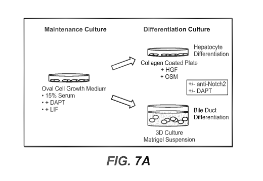
Figures 7A-B show a strategy for in vitro differentiation.
Figures 8A-F show the efficacy of anti-Notch2 antibody treatment in vivo and
its
effect on liver growth and proliferation following partial hepatectomy.
4
CA 02849011 2014-03-17
WO 2013/052155
PCT/US2012/032621
Figures 9A-H show the effect of anti-Notch2 antibody t
hepatobiliary function markers following partial hepatectomy.
Figures 10A-F show the effect of anti-Notch2 antibody treatment on
hepatobiliary and
Notch signaling gene expression following partial hepatectomy.
Figures 11A-I show serum hepatobiliary function markers following 4 weeks of
antibody administration in normal and DDC-fed mice.
Figure 12 shows the H1, H2, and H3 heavy chain hypervariable region (HVR)
sequences of anti-Notch2 NRR monoclonal antibodies designated Antibody D,
Antibody D-1,
Antibody D-2, and Antibody D-3. Amino acid positions are numbered according to
the Kabat
numbering system as described below.
Figure 13 shows the Li, L2, and L3 light chain HVR sequences of anti-Notch2
NRR
monoclonal antibodies designated Antibody D, Antibody D-1, Antibody D-2, and
Antibody
D-3. Amino acid positions are numbered according to the Kabat numbering system
as
described below.
Figure 14 shows an alignment of the heavy chain variable region sequences of
Antibody D, Antibody D-1, Antibody D-2, and Antibody D-3. HVRs are enclosed in
boxes.
Figure 15 shows an alignment of the light chain variable region sequences of
Antibody D, Antibody D-1, Antibody D-2, and Antibody D-3. HVRs are enclosed in
boxes.
Figures 16A-B show exemplary acceptor human variable heavy (VH) consensus
framework sequences for use in practicing the instant invention. Sequence
identifiers are as
follows:
- human VH subgroup I consensus framework "A" minus Kabat CDRs (SEQ ID
NOs:32, 33, 34, 35).
- human VH subgroup I consensus frameworks "B," "C," and "D" minus extended
hypervariable regions (SEQ ID NOs:36, 37, 34, 35; SEQ ID NOs:36, 37, 38, 35;
and SEQ ID NOs:36, 37, 39, 35).
- human VH subgroup II consensus framework "A" minus Kabat CDRs (SEQ ID
NOs:40, 41, 42, 35).
- human VH subgroup II consensus frameworks "B," "C," and "D" minus
extended
hypervariable regions (SEQ ID NOs:43, 44, 42, 35; SEQ ID NOs:43, 44, 45, 35;
and SEQ ID NOs:43, 44, 46, and 35).
- human VH subgroup III consensus framework "A" minus Kabat CDRs (SEQ ID
NOs:47, 48, 49, 35).
5
CA 02849011 2014-03-17
WO 2013/052155
PCT/US2012/032621
- human VH subgroup III consensus frameworks "B,"
hypervariable regions (SEQ ID NOs:50, 51, 49, 35; SEQ ID NOs:50, 51, 52, 35;
and SEQ ID NOs:50, 51, 53, 35).
- human VH acceptor framework "A" minus Kabat CDRs (SEQ ID NOs:54, 48, 55,
35).
- human VH acceptor frameworks "B" and "C" minus extended hypervariable
regions (SEQ ID NOs:50, 51, 55, 35; and SEQ ID NOs:50, 51, 56, 35).
- human VH acceptor 2 framework "A" minus Kabat CDRs (SEQ ID NOs:54, 48,
57, 35).
- human VH acceptor 2 framework "B," "C," and "D" minus extended
hypervariable regions (SEQ ID NOs:50, 51, 57, 35; SEQ ID NOs:50, 51, 58, 35;
and SEQ ID NOs:50, 51, 59, 35).
Figure 17 shows exemplary acceptor human variable light (VL) consensus
framework
sequences for use in practicing the instant invention. Sequence identifiers
are as follows:
- human VL kappa subgroup I consensus framework (cv1): SEQ ID NOs:60, 61,
62, 63
- human VL kappa subgroup II consensus framework (icv2): SEQ ID NOs:64, 65,
66, 63
- human VL kappa subgroup III consensus framework (icv3): SEQ ID NOs:67,
68,
69, 63
- human VL kappa subgroup IV consensus framework (icv4): SEQ ID NOs:70, 71,
72, 63
Figure 18 shows framework sequences of huMAb4D5-8 light and heavy chains.
Numbers in superscript/bold indicate amino acid positions according to Kabat.
Figure 19 shows framework sequences of huMAb4D5-8 light and heavy chains with
the indicated modifications. Numbers in superscript/bold indicate amino acid
positions
according to Kabat.
DETAILED DESCRIPTION OF EMBODIMENTS
I. DEFINITIONS
For purposes of interpreting this specification, the following definitions
will apply and
whenever appropriate, terms used in the singular will also include the plural
and vice versa. In
6
CA 02849011 2014-03-17
WO 2013/052155
PCT/US2012/032621
the event that any definition set forth below conflicts with any c
by reference, the definition set forth below shall control.
The term "Notch," as used herein, refers, unless specifically or contextually
indicated
otherwise, to any native or variant (whether native or synthetic) Notch
polypeptide (Notch 1-
54). The term "native sequence" specifically encompasses naturally occurring
truncated forms
(e.g., an extracellular domain sequence or a transmembrane subunit sequence),
naturally
occurring variant forms (e.g., alternatively spliced forms) and naturally-
occurring allelic
variants. The term "wild-type Notch" generally refers to a polypeptide
comprising an amino
acid sequence of a naturally occurring, non-mutated Notch protein. The term
"wild-type
Notch sequence" generally refers to an amino acid sequence found in a
naturally occurring,
non-mutated Notch.
The term "Notch2," as used herein, refers, unless specifically or contextually
indicated
otherwise, to any native or variant (whether native or synthetic) Notch2
polypeptide. The
term "native sequence" specifically encompasses naturally occurring truncated
forms (e.g., an
extracellular domain sequence or a transmembrane subunit sequence), naturally
occurring
variant forms (e.g., alternatively spliced forms) and naturally occurring
allelic variants. The
term "wild-type Notch2" generally refers to a polypeptide comprising an amino
acid sequence
of a naturally occurring, non-mutated Notch2 protein. The term "wild type
Notch2 sequence"
generally refers to an amino acid sequence found in a naturally occurring, non-
mutated
Notch2.
The term "Notch2 ligand," as used herein, refers, unless specifically or
contextually
indicated otherwise, to any native or variant (whether native or synthetic)
Notch2 ligand (for
example, Jaggedl, Jagged2, Delta-likel, Delta-like3, and/or Delta-like4)
polypeptide. The
term "native sequence" specifically encompasses naturally occurring truncated
forms (e.g., an
extracellular domain sequence or a transmembrane subunit sequence), naturally
occurring
variant forms (e.g., alternatively spliced forms) and naturally occurring
allelic variants. The
term "wild-type Notch2 ligand" generally refers to a polypeptide comprising an
amino acid
sequence of a naturally occurring, non-mutated Notch2 ligand. The term "wild
type Notch2
ligand sequence" generally refers to an amino acid sequence found in a
naturally occurring,
non-mutated Notch2 ligand.
The term "Notch2 NRR," as used herein, refers, unless specifically or
contextually
indicated otherwise, to any native or variant (whether native or synthetic)
polypeptide region
of Notch2 consisting of the 3 LNR modules and the amino acid sequences
extending from the
7
CA 02849011 2014-03-17
WO 2013/052155
PCT/US2012/032621
carboxy-terminus of the LNR modules to the transmembrane di
including the HD domain (HD-N and HD-C). An exemplary Notch2 NRR consists of
the
region from about amino acid 1422-1677 of human Notch2 (SEQ ID NO:73). An
exemplary
human Notch2 NRR is also shown in SEQ ID NO:74. The term "native sequence
Notch2
NRR" specifically encompasses naturally occurring truncated forms, naturally
occurring
variant forms (e.g., alternatively spliced forms) and naturally-occurring
allelic variants of a
Notch2 NRR. The term "wild-type Notch2 NRR" generally refers to a naturally
occurring,
non-mutated Notch2 NRR. In some embodiments, a Notch2 NRR is contained in a
Notch2,
such as, for example, a Notch2 processed at the 51, S2 and/or S3 site(s), or
an unprocessed
Notch2. In some embodiments, a Notch2 NRR contains two or more non-covalently
linked
fragments of a Notch2 NRR amino acid sequence, e.g., a fragment containing
amino acids
1422 to 1608 of SEQ ID NO:73 non-covalently linked to a fragment containing
amino acids
1609 to 1677 of SEQ ID NO:73.
The term "increased Notch2 signaling," as used herein, refers to an increase
in Notch2
signaling that is significantly above the level of Notch2 signaling observed
in a control under
substantially identical conditions. In certain embodiments, the increase in
Notch2 signaling is
at least two fold, three fold, four fold, five fold, or ten fold above the
level observed in the
control.
The term "decreased Notchl signaling," as used herein, refers to a decrease in
Notch2
signaling that is significantly below the level of Notch2 signaling observed
in a control under
substantially identical conditions. In certain embodiments, the decrease in
Notch2 signaling
is at least two fold, three fold, four fold, five fold, or ten fold below the
level observed in the
control.
In certain embodiments, Notch2 signaling (i.e., increased or decreased Notch2
signaling) is assessed using a suitable reporter assay, e.g, as described in
U.S. Patent
Application Publication No. US 2010/0080808 Al.
The term "anti-Notch2 antibody" or "an antibody that binds to Notch2" refers
to an
antibody that is capable of binding Notch2 with sufficient affinity such that
the antibody is
useful as a diagnostic and/or therapeutic agent in targeting Notch2.
Preferably, the extent of
binding of an anti-Notch2 antibody to an unrelated, non-Notch protein is less
than about 10%
of the binding of the antibody to Notch2 as measured, e.g., by a
radioimmunoassay (RIA). In
certain embodiments, an antibody that binds to Notch2 has a dissociation
constant (Kd) of
< liAM, < 0.5 [tM, < 100 nM, < 50 nM, < 10 nM, < 5 nM, < 1 nM, < 0.5 nM, or <
0.1 nM. In
8
CA 02849011 2014-03-17
WO 2013/052155
PCT/US2012/032621
certain embodiments, an anti-Notch2 antibody binds to an epitc
among Notch2 from different species, e.g., rodents (mice, rats) and primates.
The term "anti-Notch2 NRR antibody" or "an antibody that binds to Notch2 NRR"
refers to an antibody that is capable of binding Notch2 NRR with sufficient
affinity such that
the antibody is useful as a diagnostic and/or therapeutic agent in targeting
Notch2.
Preferably, the extent of binding of an anti-Notch2 NRR antibody to an
unrelated, non-Notch
protein is less than about 10% of the binding of the antibody to Notch2 NRR as
measured,
e.g., by a radioimmunoassay (RIA). In certain embodiments, an antibody that
binds to
Notch2 NRR has a dissociation constant (Kd) of < 1[LM, < 0.5 [tM, < 100 nM, <
50 nM, < 10
nM, < 5 nM, < 1 nM, < 0.5 nM, or < 0.1 nM. In certain embodiments, an anti-
Notch2 NRR
antibody binds to an epitope of Notch that is conserved among Notch from
different species,
e.g., rodents (mice, rats) and primates.
The term "Notch2-specific antagonist" refers to an agent that effects
decreased
Notch2 signaling, as defined above, and does not significantly affect
signaling by another
Notch receptor (Notch 1, 3, or 4 in mammals).
An "anti-Notch2 antagonist antibody" is an anti-Notch2 antibody (including an
anti-
Notch2 NRR antibody) that effects decreased Notch2 signaling, as defined
above.
The term "antagonist" refers to an agent that significantly inhibits (either
partially or
completely) the biological activity of a target molecule.
The term "antibody" herein is used in the broadest sense and specifically
covers
monoclonal antibodies, polyclonal antibodies, multispecific antibodies (e.g.
bispecific
antibodies) formed from at least two intact antibodies, and antibody fragments
so long as they
exhibit the desired biological activity.
An "isolated" antibody is one which has been identified and separated and/or
recovered from a component of its natural environment. Contaminant components
of its
natural environment are materials which would interfere with research,
diagnostic or
therapeutic uses for the antibody, and may include enzymes, hormones, and
other
proteinaceous or nonproteinaceous solutes. In some embodiments, an antibody is
purified (1)
to greater than 95% by weight of antibody as determined by, for example, the
Lowry method,
and in some embodiments, to greater than 99% by weight; (2) to a degree
sufficient to obtain
at least 15 residues of N-terminal or internal amino acid sequence by use of,
for example, a
spinning cup sequenator, or (3) to homogeneity by SDS-PAGE under reducing or
nonreducing conditions using, for example, Coomassie blue or silver stain.
Isolated antibody
9
CA 02849011 2014-03-17
WO 2013/052155
PCT/US2012/032621
includes the antibody in situ within recombinant cells since at 11
antibody's natural environment will not be present. Ordinarily, however,
isolated antibody
will be prepared by at least one purification step.
"Native antibodies" are usually heterotetrameric glycoproteins of about
150,000
daltons, composed of two identical light (L) chains and two identical heavy
(H) chains. Each
light chain is linked to a heavy chain by one covalent disulfide bond, while
the number of
disulfide linkages varies among the heavy chains of different immunoglobulin
isotypes. Each
heavy and light chain also has regularly spaced intrachain disulfide bridges.
Each heavy
chain has at one end a variable domain (VH) followed by a number of constant
domains.
Each light chain has a variable domain at one end (VI) and a constant domain
at its other end;
the constant domain of the light chain is aligned with the first constant
domain of the heavy
chain, and the light chain variable domain is aligned with the variable domain
of the heavy
chain. Particular amino acid residues are believed to form an interface
between the light
chain and heavy chain variable domains.
The "variable region" or "variable domain" of an antibody refers to the amino-
terminal domains of the heavy or light chain of the antibody. The variable
domain of the
heavy chain may be referred to as "VH." The variable domain of the light chain
may be
referred to as "VL." These domains are generally the most variable parts of an
antibody and
contain the antigen-binding sites.
The term "variable" refers to the fact that certain portions of the variable
domains
differ extensively in sequence among antibodies and are used in the binding
and specificity of
each particular antibody for its particular antigen. However, the variability
is not evenly
distributed throughout the variable domains of antibodies. It is concentrated
in three
segments called hypervariable regions (HVRs) both in the light-chain and the
heavy-chain
variable domains. The more highly conserved portions of variable domains are
called the
framework regions (FR). The variable domains of native heavy and light chains
each
comprise four FR regions, largely adopting a beta-sheet configuration,
connected by three
HVRs, which form loops connecting, and in some cases forming part of, the beta-
sheet
structure. The HVRs in each chain are held together in close proximity by the
FR regions
and, with the HVRs from the other chain, contribute to the formation of the
antigen-binding
site of antibodies (see Kabat et at., Sequences of Proteins of Immunological
Interest, Fifth
Edition, National Institute of Health, Bethesda, MD (1991)). The constant
domains are not
CA 02849011 2014-03-17
WO 2013/052155
PCT/US2012/032621
involved directly in the binding of an antibody to an antigen, bt
functions, such as participation of the antibody in antibody-dependent
cellular toxicity.
The "light chains" of antibodies (immunoglobulins) from any vertebrate species
can
be assigned to one of two clearly distinct types, called kappa (x) and lambda
(X), based on the
amino acid sequences of their constant domains.
Depending on the amino acid sequences of the constant domains of their heavy
chains, antibodies (immunoglobulins) can be assigned to different classes.
There are five
major classes of immunoglobulins: IgA, IgD, IgE, IgG, and IgM, and several of
these may be
further divided into subclasses (isotypes), e.g., IgGi, IgG2, IgG3, IgG4,
IgAi, and IgA2 . The
heavy chain constant domains that correspond to the different classes of
immunoglobulins are
called a, 6, 8, y, and it, respectively. The subunit structures and three-
dimensional
configurations of different classes of immunoglobulins are well known and
described
generally in, for example, Abbas et at. Cellular and Mol. Immunology, 4th ed.
(W.B.
Saunders, Co., 2000). An antibody may be part of a larger fusion molecule,
formed by
covalent or non-covalent association of the antibody with one or more other
proteins or
peptides.
The terms "full length antibody," "intact antibody" and "whole antibody" are
used
herein interchangeably to refer to an antibody in its substantially intact
form, not antibody
fragments as defined below. The terms particularly refer to an antibody with
heavy chains
that contain an Fc region.
A "naked antibody" for the purposes herein is an antibody that is not
conjugated to a
cytotoxic moiety or radiolabel.
"Antibody fragments" comprise a portion of an intact antibody, preferably
comprising
the antigen binding region thereof. Examples of antibody fragments include
Fab, Fab',
F(ab')2, and Fv fragments; diabodies; linear antibodies; single-chain antibody
molecules; and
multispecific antibodies formed from antibody fragments.
Papain digestion of antibodies produces two identical antigen-binding
fragments,
called "Fab" fragments, each with a single antigen-binding site, and a
residual "Fc" fragment,
whose name reflects its ability to crystallize readily. Pepsin treatment
yields an F(ab')2
fragment that has two antigen-combining sites and is still capable of cross-
linking antigen.
"Fv" is the minimum antibody fragment which contains a complete antigen-
binding
site. In one embodiment, a two-chain Fv species consists of a dimer of one
heavy- and one
light-chain variable domain in tight, non-covalent association. In a single-
chain Fv (scFv)
11
CA 02849011 2014-03-17
WO 2013/052155
PCT/US2012/032621
species, one heavy- and one light-chain variable domain can be
peptide linker such that the light and heavy chains can associate in a
"dimeric" structure
analogous to that in a two-chain Fv species. It is in this configuration that
the three HVRs of
each variable domain interact to define an antigen-binding site on the surface
of the VH-VL
dimer. Collectively, the six HVRs confer antigen-binding specificity to the
antibody.
However, even a single variable domain (or half of an Fv comprising only three
HVRs
specific for an antigen) has the ability to recognize and bind antigen,
although at a lower
affinity than the entire binding site.
The Fab fragment contains the heavy- and light-chain variable domains and also
contains the constant domain of the light chain and the first constant domain
(CH1) of the
heavy chain. Fab' fragments differ from Fab fragments by the addition of a few
residues at
the carboxy terminus of the heavy chain CH1 domain including one or more
cysteines from
the antibody hinge region. Fab'-SH is the designation herein for Fab' in which
the cysteine
residue(s) of the constant domains bear a free thiol group. F(ab')2 antibody
fragments
originally were produced as pairs of Fab' fragments which have hinge cysteines
between
them. Other chemical couplings of antibody fragments are also known.
"Single-chain Fv" or "scFv" antibody fragments comprise the VH and VL domains
of
antibody, wherein these domains are present in a single polypeptide chain.
Generally, the
scFv polypeptide further comprises a polypeptide linker between the VH and VL
domains
which enables the scFv to form the desired structure for antigen binding. For
a review of
scFv, see, e.g., Pluckthiin, in The Pharmacology of MonoclonalAntibodies, vol.
113,
Rosenburg and Moore eds., (Springer-Verlag, New York, 1994), pp. 269-315.
The term "diabodies" refers to antibody fragments with two antigen-binding
sites,
which fragments comprise a heavy-chain variable domain (VH) connected to a
light-chain
variable domain (VL) in the same polypeptide chain (VH-VL). By using a linker
that is too
short to allow pairing between the two domains on the same chain, the domains
are forced to
pair with the complementary domains of another chain and create two antigen-
binding sites.
Diabodies may be bivalent or bispecific. Diabodies are described more fully
in, for example,
EP 404,097; WO 1993/01161; Hudson et al., Nat. Med. 9:129-134 (2003); and
Hollinger et
al., Proc. Natl. Acad. Sci. USA 90: 6444-6448 (1993). Triabodies and
tetrabodies are also
described in Hudson et al., Nat. Med. 9:129-134 (2003).
The term "monoclonal antibody" as used herein refers to an antibody obtained
from a
population of substantially homogeneous antibodies, i.e., the individual
antibodies
12
CA 02849011 2014-03-17
WO 2013/052155
PCT/US2012/032621
comprising the population are identical except for possible mut
occurring mutations, that may be present in minor amounts. Thus, the modifier
"monoclonal"
indicates the character of the antibody as not being a mixture of discrete
antibodies. In
certain embodiments, such a monoclonal antibody typically includes an antibody
comprising
a polypeptide sequence that binds a target, wherein the target-binding
polypeptide sequence
was obtained by a process that includes the selection of a single target
binding polypeptide
sequence from a plurality of polypeptide sequences. For example, the selection
process can
be the selection of a unique clone from a plurality of clones, such as a pool
of hybridoma
clones, phage clones, or recombinant DNA clones. It should be understood that
a selected
target binding sequence can be further altered, for example, to improve
affinity for the target,
to humanize the target binding sequence, to improve its production in cell
culture, to reduce
its immunogenicity in vivo, to create a multispecific antibody, etc., and that
an antibody
comprising the altered target binding sequence is also a monoclonal antibody
of this
invention. In contrast to polyclonal antibody preparations, which typically
include different
antibodies directed against different determinants (epitopes), each monoclonal
antibody of a
monoclonal antibody preparation is directed against a single determinant on an
antigen. In
addition to their specificity, monoclonal antibody preparations are
advantageous in that they
are typically uncontaminated by other immunoglobulins.
The modifier "monoclonal" indicates the character of the antibody as being
obtained
from a substantially homogeneous population of antibodies, and is not to be
construed as
requiring production of the antibody by any particular method. For example,
the monoclonal
antibodies to be used in accordance with the present invention may be made by
a variety of
techniques, including, for example, the hybridoma method (e.g., Kohler and
Milstein, Nature,
256:495-97 (1975); Hongo et at., Hybridoma, 14 (3): 253-260 (1995), Harlow et
at.,
Antibodies: A Laboratory Manual, (Cold Spring Harbor Laboratory Press, 2nd ed.
1988);
Hammerling et at., in: Monoclonal Antibodies and T-Cell Hybridomas 563-681
(Elsevier,
N.Y., 1981)), recombinant DNA methods (see, e.g., U.S. Patent No. 4,816,567),
phage-
display technologies (see, e.g., Clackson et at., Nature, 352: 624-628 (1991);
Marks et at., J.
Mot. Biol. 222: 581-597 (1992); Sidhu et at., J. Mot. Biol. 338(2): 299-310
(2004); Lee et at.,
J. Mot. Biol. 340(5): 1073-1093 (2004); Fellouse, Proc. Natl. Acad. Sci. USA
101(34): 12467-
12472 (2004); and Lee et at., J. Immunol. Methods 284(1-2): 119-132(2004), and
technologies for producing human or human-like antibodies in animals that have
parts or all
of the human immunoglobulin loci or genes encoding human immunoglobulin
sequences
13
CA 02849011 2014-03-17
WO 2013/052155
PCT/US2012/032621
(see, e.g., WO 1998/24893; WO 1996/34096; WO 1996/33735
et at., Proc. Natl. Acad. Sci. USA 90: 2551 (1993); Jakobovits et at., Nature
362: 255-258
(1993); Bruggemann et at., Year in Immunol. 7:33 (1993); U.S. Patent Nos.
5,545,807;
5,545,806; 5,569,825; 5,625,126; 5,633,425; and 5,661,016; Marks et at.,
Bio/Technology 10:
779-783 (1992); Lonberg et al., Nature 368: 856-859 (1994); Morrison, Nature
368: 812-813
(1994); Fishwild et at., Nature Biotechnol. 14: 845-851 (1996); Neuberger,
Nature
Biotechnol. 14: 826 (1996); and Lonberg and Huszar, Intern. Rev. Immunol. 13:
65-93 (1995).
The monoclonal antibodies herein specifically include "chimeric" antibodies in
which
a portion of the heavy and/or light chain is identical with or homologous to
corresponding
sequences in antibodies derived from a particular species or belonging to a
particular antibody
class or subclass, while the remainder of the chain(s) is identical with or
homologous to
corresponding sequences in antibodies derived from another species or
belonging to another
antibody class or subclass, as well as fragments of such antibodies, so long
as they exhibit the
desired biological activity (see, e.g.,U U.S. Patent No. 4,816,567; and
Morrison et at., Proc.
Natl. Acad. Sci. USA 81:6851-6855 (1984)). Chimeric antibodies include
PRIMATIZEDO
antibodies wherein the antigen-binding region of the antibody is derived from
an antibody
produced by, e.g., immunizing macaque monkeys with the antigen of interest.
"Humanized" forms of non-human (e.g., murine) antibodies are chimeric
antibodies
that contain minimal sequence derived from non-human immunoglobulin. In one
embodiment, a humanized antibody is a human immunoglobulin (recipient
antibody) in
which residues from a HVR of the recipient are replaced by residues from a HVR
of a non-
human species (donor antibody) such as mouse, rat, rabbit, or nonhuman primate
having the
desired specificity, affinity, and/or capacity. In some instances, FR residues
of the human
immunoglobulin are replaced by corresponding non-human residues. Furthermore,
humanized antibodies may comprise residues that are not found in the recipient
antibody or in
the donor antibody. These modifications may be made to further refine antibody
performance. In general, a humanized antibody will comprise substantially all
of at least one,
and typically two, variable domains, in which all or substantially all of the
hypervariable
loops correspond to those of a non-human immunoglobulin, and all or
substantially all of the
FRs are those of a human immunoglobulin sequence. The humanized antibody
optionally
will also comprise at least a portion of an immunoglobulin constant region
(Fc), typically that
of a human immunoglobulin. For further details, see, e.g., Jones et at.,
Nature 321:522-525
(1986); Riechmann et at., Nature 332:323-329 (1988); and Presta, Curr. Op.
Struct. Biol.
14
CA 02849011 2014-03-17
WO 2013/052155
PCT/US2012/032621
2:593-596 (1992). See also, e.g., Vaswani and Hamilton, Ann.
1:105-115 (1998); Harris, Biochem. Soc. Transactions 23:1035-1038 (1995);
Hurle and
Gross, Curr. Op. Biotech. 5:428-433 (1994); and U.S. Pat. Nos. 6,982,321 and
7,087,409.
A "human antibody" is one which possesses an amino acid sequence which
corresponds to that of an antibody produced by a human and/or has been made
using any of
the techniques for making human antibodies as disclosed herein. This
definition of a human
antibody specifically excludes a humanized antibody comprising non-human
antigen-binding
residues. Human antibodies can be produced using various techniques known in
the art,
including phage-display libraries. Hoogenboom and Winter, J. Mot. Biol.,
227:381 (1991);
Marks et at., J. Mot. Biol., 222:581 (1991). Also available for the
preparation of human
monoclonal antibodies are methods described in Cole et at., Monoclonal
Antibodies and
Cancer Therapy, Alan R. Liss, p. 77 (1985); Boerner et at., J. Immunol.,
147(1):86-95 (1991).
See also van Dijk and van de Winkel, Curr. Opin. Pharmacol., 5: 368-74 (2001).
Human
antibodies can be prepared by administering the antigen to a transgenic animal
that has been
modified to produce such antibodies in response to antigenic challenge, but
whose
endogenous loci have been disabled, e.g., immunized xenomice (see, e.g.,U U.S.
Pat. Nos.
6,075,181 and 6,150,584 regarding XENOMOUSETm technology). See also, for
example, Li
et at., Proc. Natl. Acad. Sci. USA, 103:3557-3562 (2006) regarding human
antibodies
generated via a human B-cell hybridoma technology.
The term "hypervariable region," "HVR," or "HV," when used herein refers to
the
regions of an antibody variable domain which are hypervariable in sequence
and/or form
structurally defined loops. Generally, antibodies comprise six HVRs; three in
the VH (H1,
H2, H3), and three in the VL (L1, L2, L3). In native antibodies, H3 and L3
display the most
diversity of the six HVRs, and H3 in particular is believed to play a unique
role in conferring
fine specificity to antibodies. See, e.g.,Xu et at., Immunity 13:37-45 (2000);
Johnson and
Wu, in Methods in Molecular Biology 248:1-25 (Lo, ed., Human Press, Totowa,
NJ, 2003).
Indeed, naturally occurring camelid antibodies consisting of a heavy chain
only are functional
and stable in the absence of light chain. See, e.g., Hamers-Casterman et at.,
Nature 363:446-
448 (1993); Sheriff et at., Nature Struct. Biol. 3:733-736 (1996).
A number of HVR delineations are in use and are encompassed herein. The Kabat
Complementarity Determining Regions (CDRs) are based on sequence variability
and are the
most commonly used (Kabat et at., Sequences of Proteins of Immunological
Interest, 5th Ed.
Public Health Service, National Institutes of Health, Bethesda, MD. (1991)).
Chothia refers
CA 02849011 2014-03-17
WO 2013/052155
PCT/US2012/032621
instead to the location of the structural loops (Chothia and Lesk
(1987)). The AbM HVRs represent a compromise between the Kabat HVRs and
Chothia
structural loops, and are used by Oxford Molecular's AbM antibody modeling
software. The
"contact" HVRs are based on an analysis of the available complex crystal
structures. The
residues from each of these HVRs are noted below.
Loop Kabat AbM Chothia Contact
L1 L24-L34 L24-L34 L26-L32 L30-L36
L2 L50-L56 L50-L56 L50-L52 L46-L55
L3 L89-L97 L89-L97 L91-L96 L89-L96
H1 H31-H35B H26-H35B H26-H32 H30-H35B
(Kabat Numbering)
H1 H31-H35 H26-H35 H26-H32 H30-H35
(Chothia Numbering)
H2 H50-H65 H50-H58 H53-H55 H47-H58
H3 H95-H102 H95-H102 H96-H101 H93-H101
HVRs may comprise "extended HVRs" as follows: 24-36 or 24-34 (L1), 46-56 or 50-
56 (L2) and 89-97 or 89-96 (L3) in the VL and 26-35 (H1), 50-65 or 49-65 (H2)
and 93-102,
94-102, or 95-102 (H3) in the VH. The variable domain residues are numbered
according to
Kabat et at., supra, for each of these definitions.
"Framework" or "FR" residues are those variable domain residues other than the
HVR
residues as herein defined.
The term "variable domain residue numbering as in Kabat" or "amino acid
position
numbering as in Kabat," and variations thereof, refers to the numbering system
used for
heavy chain variable domains or light chain variable domains of the
compilation of antibodies
in Kabat et at., supra. Using this numbering system, the actual linear amino
acid sequence
may contain fewer or additional amino acids corresponding to a shortening of,
or insertion
into, a FR or HVR of the variable domain. For example, a heavy chain variable
domain may
include a single amino acid insert (residue 52a according to Kabat) after
residue 52 of H2 and
inserted residues (e.g. residues 82a, 82b, and 82c, etc. according to Kabat)
after heavy chain
FR residue 82. The Kabat numbering of residues may be determined for a given
antibody by
alignment at regions of homology of the sequence of the antibody with a
"standard" Kabat
numbered sequence.
16
CA 02849011 2014-03-17
WO 2013/052155
PCT/US2012/032621
The Kabat numbering system is generally used when rel
variable domain (approximately residues 1-107 of the light chain and residues
1-113 of the
heavy chain) (e.g., Kabat et at., supra). The "EU numbering system" or "EU
index" is
generally used when referring to a residue in an immunoglobulin heavy chain
constant region
(e.g., the EU index reported in Kabat et at., supra). The "EU index as in
Kabat" refers to the
residue numbering of the human IgG1 EU antibody. Unless stated otherwise
herein,
references to residue numbers in the variable domain of antibodies means
residue numbering
by the Kabat numbering system. Unless stated otherwise herein, references to
residue
numbers in the constant domain of antibodies means residue numbering by the EU
numbering
system (e.g., see United States Patent Application Publication US 2008/0181888
Al, Figures
for EU numbering).
An "affinity matured" antibody is one with one or more alterations in one or
more
HVRs thereof which result in an improvement in the affinity of the antibody
for antigen,
compared to a parent antibody which does not possess those alteration(s). In
one
embodiment, an affinity matured antibody has nanomolar or even picomolar
affinities for the
target antigen. Affinity matured antibodies may be produced using certain
procedures known
in the art. For example, Marks et at. Bio/Technology 10:779-783 (1992)
describe affinity
maturation by VH and VL domain shuffling. Random mutagenesis of HVR and/or
framework residues is described by, for example, in Barbas et at. Proc Nat.
Acad. Sci. USA
91:3809-3813 (1994); Schier et at. Gene 169:147-155 (1995); Yelton et at. J.
Immunol.
155:1994-2004 (1995); Jackson et at., J. Immunol. 154(7):3310-9 (1995); and
Hawkins et at,
J. Mot. Biol. 226:889-896 (1992).
Antibody "effector functions" refer to those biological activities
attributable to the Fc
region (a native sequence Fc region or amino acid sequence variant Fc region)
of an antibody,
and vary with the antibody isotype. Examples of antibody effector functions
include: Clq
binding and complement dependent cytotoxicity (CDC); Fc receptor binding;
antibody-
dependent cell-mediated cytotoxicity (ADCC); phagocytosis; down regulation of
cell surface
receptors (e.g. B cell receptor); and B cell activation.
"Binding affinity" generally refers to the strength of the sum total of
noncovalent
interactions between a single binding site of a molecule (e.g., an antibody)
and its binding
partner (e.g., an antigen). Unless indicated otherwise, as used herein,
"binding affinity" refers
to intrinsic binding affinity which reflects a 1:1 interaction between members
of a binding
pair (e.g., antibody and antigen). The affinity of a molecule X for its
partner Y can generally
17
CA 02849011 2014-03-17
WO 2013/052155
PCT/US2012/032621
be represented by the dissociation constant (Kd). Affinity can 1
methods known in the art, including those described herein. Low-affinity
antibodies
generally bind antigen slowly and tend to dissociate readily, whereas high-
affinity antibodies
generally bind antigen faster and tend to remain bound longer. A variety of
methods of
measuring binding affinity are known in the art, any of which can be used for
purposes of the
present invention. Specific illustrative and exemplary embodiments for
measuring binding
affinity are described in the following.
In one embodiment, the "Kd" or "Kd value" according to this invention is
measured
by a radiolabeled antigen binding assay (RIA) performed with the Fab version
of an antibody
of interest and its antigen as described by the following assay. Solution
binding affinity of
Fabs for antigen is measured by equilibrating Fab with a minimal concentration
of (125I)
labeledantigen in the presence of a titration series of unlabeled antigen,
then capturing bound
antigen with an anti-Fab antibody-coated plate (see, e.g., Chen, et at., J.
Mot. Biol. 293:865-
881(1999)). To establish conditions for the assay, MICROTITER multi-well
plates (Thermo
Scientific) are coated overnight with 5 [tg/ml of a capturing anti-Fab
antibody (Cappel Labs)
in 50 mM sodium carbonate (pH 9.6), and subsequently blocked with 2% (w/v)
bovine serum
albumin in PBS for two to five hours at room temperature (approximately 23 C).
In a non-
adsorbent plate (Nunc #269620), 100 pM or 26 pM
[1251]-antigen are mixed with serial
dilutions of a Fab of interest (e.g., consistent with assessment of the anti-
VEGF antibody,
Fab-12, in Presta et at., Cancer Res. 57:4593-4599 (1997)). The Fab of
interest is then
incubated overnight; however, the incubation may continue for a longer period
(e.g., about 65
hours) to ensure that equilibrium is reached. Thereafter, the mixtures are
transferred to the
capture plate for incubation at room temperature (e.g., for one hour). The
solution is then
removed and the plate washed eight times with 0.1% TWEEN-20Tm in PBS. When the
plates
have dried, 150 [d/well of scintillant (MICROSCINT-20 TM; Packard) is added,
and the plates
are counted on a TOPCOUNT TM gamma counter (Packard) for ten minutes.
Concentrations
of each Fab that give less than or equal to 20% of maximal binding are chosen
for use in
competitive binding assays.
According to another embodiment, the Kd or Kd value is measured by using
surface
plasmon resonance assays using a BIACORE -2000 or a BIACORE -3000 (BIAcore,
Inc.,
Piscataway, NJ) at 25 C with immobilized antigen CM5 chips at ¨10 response
units (RU).
Briefly, carboxymethylated dextran biosensor chips (CM5, BIACORE, Inc.) are
activated
with N-ethyl-N'- (3-dimethylaminopropy1)-carbodiimide hydrochloride (EDC) and
N-
18
CA 02849011 2014-03-17
WO 2013/052155
PCT/US2012/032621
hydroxysuccinimide (NHS) according to the supplier's instructi
mM sodium acetate, pH 4.8, to 5 jig/ml (-0.2 uM) before injection at a flow
rate of 5
p1/minute to achieve approximately 10 response units (RU) of coupled protein.
Following the
injection of antigen, 1 M ethanolamine is injected to block unreacted groups.
For kinetics
5 measurements, two-fold serial dilutions of Fab (0.78 nM to 500 nM) are
injected in PBS with
0.05% TWEEN-20Tm surfactant (PBST) at 25 C at a flow rate of approximately 25
ul/min.
Association rates (kon) and dissociation rates (koff) are calculated using a
simple one-to-one
Langmuir binding model (BIACORE Evaluation Software version 3.2) by
simultaneously
fitting the association and dissociation sensorgrams. The equilibrium
dissociation constant
10 (Kd) is calculated as the ratio koff/kon. See, e.g., Chen et at., J.
Mol. Biol. 293:865-881
(1999). If the on-rate exceeds 106 M-1 5-1 by the surface plasmon resonance
assay above,
then the on-rate can be determined by using a fluorescent quenching technique
that measures
the increase or decrease in fluorescence emission intensity (excitation = 295
nm; emission =
340 nm, 16 nm band-pass) at 250C of a 20 nM anti-antigen antibody (Fab form)
in PBS, pH
7.2, in the presence of increasing concentrations of antigen as measured in a
spectrometer,
such as a stop-flow equipped spectrophometer (Aviv Instruments) or a 8000-
series SLM-
AMINCO TM spectrophotometer (ThermoSpectronic) with a stirred cuvette.
An "on-rate," "rate of association," "association rate," or "kon" according to
this
invention can also be determined as described above using a BIACORE -2000 or
a
BIACORE c)-3000 system (BIAcore, Inc., Piscataway, NJ).
The term "substantially similar" or "substantially the same," as used herein,
denotes a
sufficiently high degree of similarity between two numeric values (for
example, one
associated with an antibody of the invention and the other associated with a
reference/comparator antibody), such that one of skill in the art would
consider the difference
between the two values to be of little or no biological and/or statistical
significance within the
context of the biological characteristic measured by said values (e.g., Kd
values). The
difference between said two values is, for example, less than about 50%, less
than about 40%,
less than about 30%, less than about 20%, and/or less than about 10% as a
function of the
reference/comparator value.
The phrase "substantially reduced," or "substantially different," as used
herein,
denotes a sufficiently high degree of difference between two numeric values
(generally one
associated with a molecule and the other associated with a
reference/comparator molecule)
such that one of skill in the art would consider the difference between the
two values to be of
19
CA 02849011 2014-03-17
WO 2013/052155
PCT/US2012/032621
statistical significance within the context of the biological chan
values (e.g., Kd values). The difference between said two values is, for
example, greater than
about 10%, greater than about 20%, greater than about 30%, greater than about
40%, and/or
greater than about 50% as a function of the value for the reference/comparator
molecule.
An "acceptor human framework" or a "human acceptor framework" for the purposes
herein is a framework comprising the amino acid sequence of a VL or VH
framework derived
from a human immunoglobulin framework or a human consensus framework. An
acceptor
human framework "derived from" a human immunoglobulin framework or a human
consensus framework may comprise the same amino acid sequence thereof, or it
may contain
pre-existing amino acid sequence changes. In some embodiments, the number of
pre-existing
amino acid changes are 10 or less, 9 or less, 8 or less, 7 or less, 6 or less,
5 or less, 4 or less, 3
or less, or 2 or less. Where pre-existing amino acid changes are present in a
VH, preferably
those changes occur at only three, two, or one of positions 71H, 73H and 78H;
for instance,
the amino acid residues at those positions may be 71A, 73T and/or 78A. In one
embodiment,
the VL acceptor human framework is identical in sequence to the VL human
immunoglobulin
framework sequence or human consensus framework sequence.
A "human consensus framework" is a framework which represents the most
commonly occurring amino acid residues in a selection of human immunoglobulin
VL or VH
framework sequences. Generally, the selection of human immunoglobulin VL or VH
sequences is from a subgroup of variable domain sequences. Generally, the
subgroup of
sequences is a subgroup as in Kabat et al., supra. In one embodiment, for the
VL, the
subgroup is subgroup kappa I as in Kabat et al., supra. In one embodiment, for
the VH, the
subgroup is subgroup III as in Kabat et al., supra.
A "VH subgroup III consensus framework" comprises the consensus sequence
obtained from the amino acid sequences in variable heavy subgroup III of Kabat
et al., supra.
In one embodiment, a human acceptor framework is derived from the VH subgroup
III
consensus framework and comprises an amino acid sequence comprising at least a
portion or
all of each of the following sequences: (SEQ ID NO:50)-H1-(SEQ ID NO:51)-H2-
(SEQ ID
NO:57 or 59)-H3-(SEQ ID NO: 35). In some embodiments, the last residue (S11)
of SEQ ID
NO:35 is substituted with an alanine.
A "VL subgroup I consensus framework" comprises the consensus sequence
obtained
from the amino acid sequences in variable light kappa subgroup I of Kabat et
al., supra. In
one embodiment, the VH subgroup I consensus framework amino acid sequence
comprises at
CA 02849011 2014-03-17
WO 2013/052155
PCT/US2012/032621
least a portion or all of each of the following sequences: (SEQ l
NO:61)-L2-(SEQ ID NO:62)-L3-(SEQ ID NO:63).
A "disorder" is any condition or disease that would benefit from treatment
with a
composition or method of the invention. This includes chronic and acute
disorders including
those pathological conditions which predispose the mammal to the disorder in
question.
Non-limiting examples of disorders to be treated herein include conditions
such as cancer.
The terms "cell proliferative disorder" and "proliferative disorder" refer to
disorders
that are associated with some degree of abnormal cell proliferation. In one
embodiment, the
cell proliferative disorder is cancer.
"Tumor," as used herein, refers to all neoplastic cell growth and
proliferation, whether
malignant or benign, and all pre-cancerous and cancerous cells and tissues.
The terms
"cancer," "cancerous," "cell proliferative disorder," "proliferative
disorder," and "tumor" are
not mutually exclusive as referred to herein.
A cancer that "responds" to a therapeutic agent is one that shows a
significant
decrease in cancer or tumor progression, including but not limited to, (1)
inhibition, to some
extent, of tumor growth, including slowing down and complete growth arrest;
(2) reduction in
the number of cancer or tumor cells; (3) reduction in tumor size; (4)
inhibition (i.e., reduction,
slowing down or complete stopping) of cancer cell infiltration into adjacent
peripheral organs
and/or tissues; and/or (5) inhibition (i.e. reduction, slowing down or
complete stopping) of
metastasis.
As used herein, "treatment" (and variations such as "treat" or "treating")
refers to
clinical intervention in an attempt to alter the natural course of the
individual or cell being
treated, and can be performed either for prophylaxis or during the course of
clinical
pathology. Desirable effects of treatment include preventing occurrence or
recurrence of
disease, alleviation of symptoms, diminishment of any direct or indirect
pathological
consequences of the disease, preventing metastasis, decreasing the rate of
disease progression,
amelioration or palliation of the disease state, and remission or improved
prognosis. In some
embodiments, Notch2 antagonists of the invention are used to delay development
of a disease
or disorder or to slow the progression of a disease or disorder.
An "individual," "subject," or "patient" is a vertebrate. In certain
embodiments, the
vertebrate is a mammal. Mammals include, but are not limited to, farm animals
(such as
cows), sport animals, pets (such as cats, dogs, and horses), primates, mice
and rats. In certain
embodiments, a mammal is a human.
21
CA 02849011 2014-03-17
WO 2013/052155
PCT/US2012/032621
The term "pharmaceutical formulation" refers to a prep
as to permit the biological activity of the active ingredient to be effective,
and which contains
no additional components which are unacceptably toxic to a subject to which
the formulation
would be administered. Such formulations may be sterile.
An "effective amount" refers to an amount effective, at dosages and for
periods of
time necessary, to achieve the desired therapeutic or prophylactic result.
The term "progenitor," "hepatic progenitor," "liver progenitor" or "oval cell"
refers to
small epithelial cells that can differentiate into both hepatocytes and intra-
hepatic bile duct
cells.
II. EMBODIMENTS OF THE INVENTION
The present invention relates to the treatment of liver conditions using
Notch2
antagonists. The present invention is based, in part, on the observation that
anti-Notch2 NRR
antibodies (a) improve liver appearance and hepatocyte function in an acute
liver damage
model in vivo and (b) reduce biliary damage and improve hepatocyte function in
a chronic
liver damage model in vivo. Without being bound by any particular theory or
operation, the
Notch2 antagonist might improve liver conditions by promoting hepatocyte
differentiation
and/or by decreasing aberrant bile duct proliferation.
In various aspects of the invention, a method of treating a liver condition
characterized by liver damage is provided, the method comprising administering
to a patient
having such condition an effective amount of a Notch2-specific antagonist. In
certain
embodiments, the liver condition is chronic liver disease, including but not
limited to fibrosis,
cirrhosis, viral hepatitis (e.g., hepatitis A, B, C, D, E, or G), autoimmune
liver diseases (e.g.,
autoimmune hepatitis, primary biliary cirrhosis, or primary sclerosing
cholangitis), genetic
liver diseases (e.g., alpha-1 antitrypsin deficiency, Crigler-Najjar syndrome,
familial
amyloidosis, Gilbert's syndrome, Dubin-Johnson syndrome, hereditary
hemchromatosis,
primary oxalosis, or Wilson's disease), alcoholic hepatitis or nonalcoholic
fatty liver disease.
In certain embodiments, the liver condition is an acute liver condition, such
as acute liver
failure, acute liver injury, or acute liver toxicity, e.g., acetaminophen
toxicity. In certain
embodiments, the liver condition is liver cancer, e.g., hepatocellular
carcinoma (HCC),
intrahepatic cholangiocarcinoma (bile duct cancer), or hepatoblastoma.
In some embodiments, treatment results in improved liver histological
appearance,
including but not limited to, e.g., larger cell size, lower nuclear-to-
cytoplasmic ratio, two
nuclei, as compared to cell size, nuclear-to-cytoplasmic ration and nuclei
number in cultured
22
CA 02849011 2014-03-17
WO 2013/052155
PCT/US2012/032621
adult oval cells. In some embodiments, treatment results in a if
morphology, e.g., as compared to morphology of cultured adult oval cells.
In some embodiments, treatment results in decreased expression of Keratin-19
biomarker in liver cells, e.g., decreased expression relative to expression of
Keratin-19
biomarker in cultured adult oval cells. Methods for detecting keratin-19
biomarker (e.g.,
Keratin-19 gene expression, e.g., mRNA expression) are well known in the art
and are also
exemplified herein.
In some embodiments, treatment results in increased expression of albumin and
AFP
e.g., increased expression relative to expression of albumin and/or AFP
biomarkers in
cultured adult oval cells. Methods for detecting albumin and/or AFP biomarkers
(e.g., gene
expression, e.g., mRNA expression) are well known in the art and are also
exemplified
herein.
In some embodiments, treatment results in a reduced number of Hesl positive
intrahepatic bile duct cells.
In some embodiment, treatment results in reduced liver progenitor cells (e.g.,
adult
liver oval cell) proliferation within the bile ducts. Reduced proliferation
may be determined,
e.g., by determining average cross-sectional area of K19-positive tissue as
compared to the
total liver cross sectional area.
In some embodiments, treatment results in improved hepatocyte function.
Hepatocyte
function may be measured by methods known in the art, including but not
limited to: no
significant elevation of biomarkers associated with biliary dysfunction, such
as those
biomarkers described in Figure 11. In some embodiments, a biomarker associated
with
biliary dysfunction is total and/or direct serum bilirubin level. In some
embodiments, a
biomarker associated with biliary dysfunction is the differentiation quotient,
as further
described and exemplified herein.
In some embodiments, improved hepatocyte function is determined, e.g., by
assessment of heptobiliary function biomarkers, including but not limited to
the serum
heptobiliary function biomarker described in Figures 2 and 5. In some
embodiments, serum
heptobiliary function biomarker is serum albumin level.
In some embodiments, improved hepatocyte function is increased rate of
recovery of
liver function.
The invention also provides methods for promoting hepatocyte differentiation
and/or
by decreasing aberrant bile duct proliferation, the method comprising
administering to a
23
CA 02849011 2014-03-17
WO 2013/052155
PCT/US2012/032621
patient in need of such treatment an effective amount of a Note]
some embodiments, the patient has a liver condition characterized by liver
damage. In certain
embodiments, the liver condition is chronic liver disease, including but not
limited to fibrosis,
cirrhosis, viral hepatitis (e.g., hepatitis A, B, C, D, E, or G), autoimmune
liver diseases (e.g.,
autoimmune hepatitis, primary biliary cirrhosis, or primary sclerosing
cholangitis), genetic
liver diseases (e.g., alpha-1 antitrypsin deficiency, Crigler-Najjar syndrome,
familial
amyloidosis, Gilbert's syndrome, Dubin-Johnson syndrome, hereditary
hemchromatosis,
primary oxalosis, or Wilson's disease), alcoholic hepatitis or nonalcoholic
fatty liver disease.
In certain embodiments, the liver condition is an acute liver condition, such
as acute liver
failure, acute liver injury, or acute liver toxicity, e.g., acetaminophen
toxicity. In certain
embodiments, the liver condition is liver cancer, e.g., hepatocellular
carcinoma (HCC),
intrahepatic cholangiocarcinoma (bile duct cancer), or hepatoblastoma. In some
embodiment,
treatment results in reduced liver progenitor cell (e.g., adult liver oval
cell) proliferation.
Reduced proliferation may be determined, e.g., by determining average cross-
sectional area of
K19 positive tissue as compared to the total liver cross sectional area.
The invention also provides methods for improving liver histological
appearance, the
method comprising administering to a patient in need of such treatment an
effective amount
of a Notch2-specific antagonist. In some embodiments, treatment results in
improved liver
histological appearance, including but not limited to: larger cell size, lower
nuclear-to-
cytoplasmic ratio, two nucleic, e.g., as compared to cell size, nuclear-to-
cytoplasmic ratio and
nuclei number in cultured adult oval cells. In some embodiments, treatment
results in a more
differentiated morphology, e.g., as compared to morphology of cultured adult
oval cells.
In some embodiments, treatment results in decreased expression of Keratin-19
biomarker in liver cells, e.g., decreased expression relative to expression of
Keratin-19
biomarker in cultured adult oval cells. Methods for detecting keratin-19
biomarker (e.g.,
Keratin-19 gene expression, e.g., mRNA expression) are well known in the art
and are also
exemplified herein.
In some embodiments, treatment results in increased expression of albumin and
AFP
e.g., increased expression relative to expression of albumin and/or AFP
biomarkers in
cultured adult oval cells. Methods for detecting albumin and/or AFP biomarkers
(e.g., gene
expression, e.g., mRNA expression) are well known in the art and are also
exemplified
herein.
24
CA 02849011 2014-03-17
WO 2013/052155
PCT/US2012/032621
In some embodiments, treatment results in reduced num
intrahepatic bile duct cells.
The invention also provides methods for reducing serum bile acids, serum
bilirubin,
serum alkaline phosphatase, serum ALT, and/or serum AST following hepatic
injury, the
method comprising administering to a patient in need thereof an effective
amount of a
Notch2-specific antagonist.
The invention also provides methods for reducing the number of CK19-positive
cells
in cell population that comprises an oval cell, the method comprising the step
of contacting
the oval cell with a Notch2-specific antagonist.
The invention also provides methods for reducing the expression or secretion
of bile
acids, bilirubin, alkaline phosphatase, ALT, and/or AST, the method comprising
contacting
an oval cell with an effective amount of a Notch2-specific antagonist.
The invention provides methods for identifying a patient eligible for
receiving
treatment of a liver condition characterized by liver damage by administering
to a patient
having such condition an effective amount of a Notch2-specific antagonist, the
method
comprising determining expression of one or more of the genes listed in Table
2 in a sample
obtained from the patient. In some embodiments, the genes belong to the Notch
pathway,
e.g., JAG1. In some embodiments, a sample or biopsy from the patient is
analyzed for
mRNA expression of one of the genes listed in Table 1 using methods well known
in the art,
such as, e.g., quantitative PCR analysis, and compared to expression of the
same gene or
genes in a biopsy obtained from a control individual or compared to a
reference value. In
some embodiments, elevated expression of one or more genes listed in Table 1
in the biopsy
obtained from the patient, relative to the control, identifies the patient as
suitable for receiving
treatment with a Notch2-specific antagonist, as described herein. In some
embodiments,
additional parameters, such as, e.g., examination by a physician, histologic
evaluation of a
biopsy, determination of serum levels indicative of liver damage, etc. are
employed to
identify the patient for receiving the treatment. Also, elevated hepatic
expression by a patient
of one or more of the genes identified in Table 2 is specifically contemplated
as one possible
embodiment of any of the methods provided herein.
In some embodiments patients are selected for treatment with a Notch2-specific
antagonist as described herein by measuring other known markers of oval cells
or aggressive
HCC (see, e.g., Woo et al 2011 Mol Carcinog. 2011 Apr;50(4):235-43). In some
CA 02849011 2014-03-17
WO 2013/052155
PCT/US2012/032621
embodiments, patients are selected for treatment by analyzing 11
example by detection of the activated form of Notch2 as described herein.
Examples of Notch2-specific antagonists include, but are not limited to,
soluble Notch
receptors, soluble Notch ligand variants, e.g., dominant negative ligand
variants, aptamers or
oligopeptides that bind Notch2 or Notch2 ligands, organic or inorganic
molecules that
interfere specifically with Notch2 signaling, anti-Notch2 antagonist
antibodies and anti-
Notch2 ligand antagonist antibodies. Examples of Notch2-specfic antagonists
include those
described in U.S. Patent Application Publication No. US 2010/0111958.
In certain embodiments, the Notch2-specific antagonist is an anti-Notch2
antagonist
antibody. In one such embodiment, the anti-Notch2 antagonist antibody is an
antibody that
binds to the extracellular domain of Notch2 and effects decreased Notch2
signaling. In one
such embodiment, the anti-Notch2 antagonist antibody is an anti-Notch2 NRR
antibody.
Anti-Notch2 NRR antibodies include, but are not limited to, any of the anti-
Notch2 NRR
antibodies disclosed in U.S. Patent Application Publication No. US
2010/0080808 Al, which
is expressly incorporated by reference herein in its entirety. Such antibodies
include, but are
not limited to anti-Notch2 NRR antibodies that bind to the LNR-A and HD-C
domains of
Notch2 NRR. Exemplary anti-Notch2 NRR antibodies are monoclonal antibodies
designated
Antibody D, Antibody D-1, Antibody D-2, or Antibody D-3 that were derived from
a phage
library, as disclosed in US 2010/0080808. Antibody D that binds to Notch2 NRR
was
isolated. That antibody was affinity matured to generate Antibody D-1,
Antibody D-2, and
Antibody D-3. The sequences of the heavy chain and light chain hypervariable
regions
(HVRs) of Antibody D, Antibody D-1, Antibody D-2, and Antibody D-3 are shown
in
Figures 12 and 13. The sequences of the heavy and light chain variable domains
of Antibody
D, Antibody D-1, Antibody D-2, and Antibody D-3 are shown in Figures 14 and
15. Further
embodiments of anti-Notch2 NRR antibodies are provided as follows.
In one aspect, an antagonist antibody that specifically binds to Notch2 NRR is
provided, wherein the antibody comprises at least one, two, three, four, five,
or six HVRs
selected from:
(a) an HVR-Hl comprising an amino acid sequence that conforms to the consensus
sequence of SEQ ID NO:3;
(b) an HVR-H2 comprising the amino acid sequence of SEQ ID NO:4;
(c) an HVR-H3 comprising the amino acid sequence of SEQ ID NO:5;
26
CA 02849011 2014-03-17
WO 2013/052155
PCT/US2012/032621
(d) an HVR-L1 comprising an amino acid sequence that
sequence of SEQ ID NO:10;
(e) an HVR-L2 comprising an amino acid sequence that conforms to the consensus
sequence of SEQ ID NO:14; and
(f) an HVR-L3 comprising an amino acid sequence that conforms to the consensus
sequence of SEQ ID NO:19.
In a further aspect, the antibody comprises an HVR-H3 comprising the amino
acid sequence
of SEQ ID NO:5 and at least one, two, three, four, or five HVRs selected from
(a), (b), (d),
(e), and (f) above. In a further aspect, the antibody comprises (a), (b), (c),
(d), (e), and (f)
above. With respect to (a), (d), (e), and (f), any one or more of the
following embodiments
are contemplated: HVR-H1 comprises an amino acid sequence selected from SEQ ID
NOs:1-
2; HVR-L1 comprises an amino acid sequence selected from SEQ ID NOs:6-9; HVR-
L2
comprises an amino acid sequence selected from SEQ ID NOs:11-13; and HVR-L3
comprises
an amino acid sequence selected from SEQ ID NOs:15-18.
In another aspect, an antibody that specifically binds to Notch2 NRR is
provided,
wherein the antibody comprises an HVR-H1 comprising an amino acid sequence
that
conforms to the consensus sequence of SEQ ID NO:3, an HVR-H2 comprising the
amino
acid sequence of SEQ ID NO:4, and an HVR-H3 comprising the amino acid sequence
of SEQ
ID NO:5. In one embodiment, HVR-H1 comprises an amino acid sequence selected
from
SEQ ID NOs:1-2.
In another aspect, an antibody that specifically binds to Notch2 NRR is
provided,
wherein the antibody comprises an HVR-L1 comprising an amino acid sequence
that
conforms to the consensus sequence of SEQ ID NO:10, an HVR-L2 comprising an
amino
acid sequence that conforms to the consensus sequence of SEQ ID NO:14, and an
HVR-L3
comprising an amino acid sequence that conforms to the consensus sequence of
SEQ ID
NO:19. The following embodiments are contemplated in any combination: HVR-L1
comprises an amino acid sequence selected from SEQ ID NOs:6-9; HVR-L2
comprises an
amino acid sequence selected from SEQ ID NOs:11-13; and HVR-L3 comprises an
amino
acid sequence selected from SEQ ID NOs:15-18. In one embodiment, an antibody
that binds
to Notch2 NRR comprises an HVR-L1 comprising the amino acid sequence of SEQ ID
NO:6;
an HVR-L2 comprising the amino acid sequence of SEQ ID NO:11; and an HVR-L3
comprising the amino acid sequence of SEQ ID NO:15. In another embodiment, an
antibody
that binds to Notch2 NRR comprises an HVR-L1 comprising the amino acid
sequence of
27
CA 02849011 2014-03-17
WO 2013/052155
PCT/US2012/032621
SEQ ID NO:7; an HVR-L2 comprising the amino acid sequenc
HVR-L3 comprising the amino acid sequence of SEQ ID NO:16. In another
embodiment, an
antibody that binds to Notch2 NRR comprises an HVR-L1 comprising the amino
acid
sequence of SEQ ID NO:8; an HVR-L2 comprising the amino acid sequence of SEQ
ID
NO:12; and an HVR-L3 comprising the amino acid sequence of SEQ ID NO:17. In
another
embodiment, an antibody that binds to Notch2 NRR comprises an HVR-L1
comprising the
amino acid sequence of SEQ ID NO:9; an HVR-L2 comprising the amino acid
sequence of
SEQ ID NO:13; and an HVR-L3 comprising the amino acid sequence of SEQ ID
NO:18.
In one embodiment, an antibody that specifically binds to Notch2 NRR is
provided,
wherein the antibody comprises:
(a) an HVR-H1 comprising the amino acid sequence of SEQ ID NO:1;
(b) an HVR-H2 comprising the amino acid sequence of SEQ ID NO:4;
(c) an HVR-H3 comprising the amino acid sequence of SEQ ID NO:5;
(d) an HVR-L1 comprising the amino acid sequence of SEQ ID NO:6;
(e) an HVR-L2 comprising the amino acid sequence of SEQ ID NO:11; and
(f) an HVR-L3 comprising the amino acid sequence of SEQ ID NO:15.
In another embodiment, an antibody that specifically binds to Notch2 NRR is
provided, wherein the antibody comprises:
(a) an HVR-H1 comprising the amino acid sequence of SEQ ID NO:2;
(b) an HVR-H2 comprising the amino acid sequence of SEQ ID NO:4;
(c) an HVR-H3 comprising the amino acid sequence of SEQ ID NO:5;
(d) an HVR-L1 comprising the amino acid sequence of SEQ ID NO:7;
(e) an HVR-L2 comprising the amino acid sequence of SEQ ID NO:11; and
(f) an HVR-L3 comprising the amino acid sequence of SEQ ID NO:16.
In another embodiment, an antibody that specifically binds to Notch2 NRR is
provided, wherein the antibody comprises:
(a) an HVR-H1 comprising the amino acid sequence of SEQ ID NO:2;
(b) an HVR-H2 comprising the amino acid sequence of SEQ ID NO:4;
(c) an HVR-H3 comprising the amino acid sequence of SEQ ID NO:5;
(d) an HVR-L1 comprising the amino acid sequence of SEQ ID NO:8;
(e) an HVR-L2 comprising the amino acid sequence of SEQ ID NO:12; and
(f) an HVR-L3 comprising the amino acid sequence of SEQ ID NO:17.
28
CA 02849011 2014-03-17
WO 2013/052155
PCT/US2012/032621
In another embodiment, an antibody that specifically bil
provided, wherein the antibody comprises:
(a) an HVR-H1 comprising the amino acid sequence of SEQ ID NO:2;
(b) an HVR-H2 comprising the amino acid sequence of SEQ ID NO:4;
(c) an HVR-H3 comprising the amino acid sequence of SEQ ID NO:5;
(d) an HVR-L1 comprising the amino acid sequence of SEQ ID NO:9;
(e) an HVR-L2 comprising the amino acid sequence of SEQ ID NO:13; and
(f) an HVR-L3 comprising the amino acid sequence of SEQ ID NO:18.
In certain embodiments, any of the above antibodies further comprises at least
one
framework selected from a VH subgroup III consensus framework and a VL
subgroup I
consensus framework.
In certain embodiments, an anti-Notch2 NRR antibody is affinity matured. For
example, any one or more of the following substitutions in the indicated HVR
positions
(Kabat numbered) may be made in any combination:
- in HVR-H1 (SEQ ID NO:1): 528T; T305;
- in HVR-L1 (SEQ ID NO:6): 528N; I29N or V; 530R or K; S31R; Y32F
- in HVR-L2 (SEQ ID NO:11): G5OR; S53I or T; A55E
- in HVR-L3 (SEQ ID NO:15): S93I or R; L96W or H
The specific antibodies disclosed herein, i.e., Antibody D as well as affinity
matured forms of
Antibody D (D-1, D-2, and D-3), may undergo further affinity maturation.
Accordingly,
affinity matured forms of any of the antibodies described herein are provided.
In certain embodiments, an anti-Notch2 NRR antibody having any of the above
HVR
sequences can further comprise any suitable framework variable domain
sequence, provided
binding activity to Notch2 NRR is substantially retained. In certain
embodiments, an anti-
Notch2 NRR antibody comprises a human variable heavy (VH) consensus framework
sequence, as in any of the VH consensus framework sequences shown in Figures
16A and
16B. In one embodiment, the VH consensus framework sequence comprises a human
subgroup III heavy chain framework consensus sequence, e.g., as shown in
Figures 16A and
16B. In another embodiment, the VH consensus framework sequence comprises an
"Acceptor 2" framework sequence, e.g., as shown in Figures 16A and 16B. In a
particular
embodiment, the VH framework consensus sequence comprises FR1-FR4 of Acceptor
2B or
Acceptor 2D, wherein the FR4 comprises SEQ ID NO:35 (Figures 16A and 16B),
with the
last residue of SEQ ID NO:35 (S11) optionally being substituted with alanine.
In a further
29
CA 02849011 2014-03-17
WO 2013/052155
PCT/US2012/032621
particular embodiment, the VH framework consensus sequence
SEQ ID NOs:50; 51; 57 or 59; and 35, wherein Sll of SEQ ID NO:35 is optionally
substituted with alanine.
In certain embodiments, an anti-Notch2 NRR antibody having any of the above
HVR
sequences can further comprise a human variable light (VL) consensus framework
sequence
as shown in Figure 17. In one embodiment, the VL consensus framework sequence
comprises a human VL kappa subgroup I consensus framework (icv1) sequence,
e.g., as
shown in Figure 17. In another embodiment, the VL framework consensus sequence
comprises FR1-FR4 of huMAb4D5-8 as shown in Figures 18 or 19. In a particular
embodiment, the VL framework consensus sequence comprises the sequences of SEQ
ID
NOs:60, 61, 62, and 63.
In another aspect, an anti-Notch2 NRR antibody comprises a heavy chain
variable
domain (VH) sequence having at least 90%, 91%, 92%, 93%, 94%, 95%, 96%, 97%,
98%,
99%, or 100% sequence identity to an amino acid sequence selected from SEQ ID
NOs:20-
21. In certain embodiments, a VH sequence having at least 90%, 91%, 92%, 93%,
94%,
95%, 96%, 97%, 98%, or 99% identity contains substitutions (e.g., conservative
substitutions), insertions, or deletions relative to the reference sequence,
but an anti-Notch2
NRR antibody comprising that sequence retains the ability to bind to Notch2
NRR. In certain
embodiments, a total of 1 to 10 amino acids have been substituted, inserted
and/or deleted in
an amino acid sequence selected from SEQ ID NOs:20-21. In certain embodiments,
substitutions, insertions, or deletions occur in regions outside the HVRs
(i.e., in the FRs). In
a particular embodiment, the VH comprises one, two or three HVRs selected
from: (a) an
HVR-H1 comprising an amino acid sequence that conforms to the consensus
sequence of
SEQ ID NO:3, (b) an HVR-H2 comprising the amino acid sequence of SEQ ID NO:4,
and (c)
an HVR-H3 comprising the amino acid sequence of SEQ ID NO:5. In one such
embodiment,
HVR-H1 comprises an amino acid sequence selected from SEQ ID NOs:1-2.
In another aspect, an antibody that specifically binds to Notch2 NRR is
provided,
wherein the antibody comprises a light chain variable domain (VL) having at
least 90%, 91%,
92%, 93%, 94%, 95%, 96%, 97%, 98%, 99%, or 100% sequence identity to an amino
acid
sequence selected from SEQ ID NOs:22-25. In certain embodiments, a VL sequence
having
at least 90%, 91%, 92%, 93%, 94%, 95%, 96%, 97%, 98%, or 99% identity contains
substitutions (e.g., conservative substitutions), insertions, or deletions
relative to the reference
sequence, but an anti-Notch2 NRR antibody comprising that sequence retains the
ability to
CA 02849011 2014-03-17
WO 2013/052155
PCT/US2012/032621
bind to Notch2 NRR. In certain embodiments, a total of 1 to 1(
substituted, inserted and/or deleted in an amino acid sequence selected from
SEQ ID NOs:22-
25. In certain embodiments, the substitutions, insertions, or deletions occur
in regions outside
the HVRs (i.e., in the FRs). In a particular embodiment, the VL comprises one,
two or three
HVRs selected from (a) an HVR-L1 comprising an amino acid sequence that
conforms to the
consensus sequence of SEQ ID NO:10; (b) an HVR-L2 comprising an amino acid
sequence
that conforms to the consensus sequence of SEQ ID NO:14; and (c) an HVR-L3
comprising
an amino acid sequence that conforms to the consensus sequence of SEQ ID
NO:19. In one
such embodiment, the VL comprises one, two or three HVRs selected from (a) an
HVR-L1
comprising an amino acid sequence selected from SEQ ID NOs:6-9; (b) an HVR-L2
comprising an amino acid sequence selected from SEQ ID NOs:11-13; and (c) an
HVR-L3
comprising an amino acid sequence selected from SEQ ID NOs:15-18. In one such
embodiment, the VL comprises one, two or three HVRs selected from (a) an HVR-
L1
comprising the amino acid sequence of SEQ ID NO:6; (b) an HVR-L2 comprising
the amino
acid sequence of SEQ ID NO:11; and (c) an HVR-L3 comprising the amino acid
sequence of
SEQ ID NO:15. In another such embodiment, the VL comprises one, two or three
HVRs
selected from (a) an HVR-L1 comprising the amino acid sequence of SEQ ID NO:7;
(b) an
HVR-L2 comprising the amino acid sequence of SEQ ID NO:11; and (c) an HVR-L3
comprising the amino acid sequence of SEQ ID NO:16. In another such
embodiment, the VL
comprises one, two or three HVRs selected from (a) an HVR-L1 comprising the
amino acid
sequence of SEQ ID NO:8; (b) an HVR-L2 comprising the amino acid sequence of
SEQ ID
NO:12; and (c) an HVR-L3 comprising the amino acid sequence of SEQ ID NO:17.
In
another such embodiment, the VL comprises one, two or three HVRs selected from
(a) an
HVR-L1 comprising the amino acid sequence of SEQ ID NO:9; (b) an HVR-L2
comprising
the amino acid sequence of SEQ ID NO:13; and (c) an HVR-L3 comprising the
amino acid
sequence of SEQ ID NO:18.
In certain embodiments of the variant VH and VL sequences provided above,
substitutions, insertions, or deletions may occur within the HVRs. In such
embodiments,
substitutions, insertions, or deletions may occur within one or more HVRs so
long as such
alterations do not substantially reduce the ability of the antibody to bind
antigen. For
example, conservative alterations that do not substantially reduce binding
affinity may be
made in HVRs. In certain instances, alterations in HVRs may actually improve
antibody
affinity. Such alterations may be made in HVR "hotspots" (i.e., residues
encoded by codons
31
CA 02849011 2014-03-17
WO 2013/052155
PCT/US2012/032621
that undergo mutation at high frequency during the somatic mai
increase antibody affinity. (See, e.g., Chowdhury, Methods Mol. Biol. 207:179-
196, 2008.)
In certain embodiments of the variant VH and VL sequences provided above, each
HVR
either is conserved (unaltered), or contains no more than a single amino acid
substitution,
insertion or deletion.
In another aspect, an antibody that specifically binds Notch2 NRR is provided,
wherein the antibody comprises a VH as in any of the embodiments provided
above, and a
VL as in any of the embodiments provided above. In one embodiment, the
antibody
comprises a VH having at least 90%, 91%, 92%, 93%, 94%, 95%, 96%, 97%, 98%,
99%, or
100% sequence identity to the amino acid sequence of SEQ ID NO:20, and a VL
having at
least 90%, 91%, 92%, 93%, 94%, 95%, 96%, 97%, 98%, 99%, or 100% sequence
identity to
the amino acid sequence of SEQ ID NO:22. In one such embodiment, the VH
comprises one,
two or three HVRs selected from: (a) an HVR-H1 comprising the amino acid
sequence of
SEQ ID NO:1, (b) an HVR-H2 comprising the amino acid sequence of SEQ ID NO:4,
and (c)
an HVR-H3 comprising the amino acid sequence of SEQ ID NO:5, and the VL
comprises
one, two or three HVRs selected from (a) an HVR-L1 comprising the amino acid
sequence of
SEQ ID NO:6; (b) an HVR-L2 comprising the amino acid sequence of SEQ ID NO:11;
and
(c) an HVR-L3 comprising the amino acid sequence of SEQ ID NO:15. In a
particular
embodiment, the antibody comprises a VH comprising the amino acid sequence of
SEQ ID
NO:20, and a VL comprising the amino acid sequence of SEQ ID NO:22.
In another embodiment, an anti-Notch2 NRR antibody that specifically binds
Notch2
NRR comprises a VH having at least 90%, 91%, 92%, 93%, 94%, 95%, 96%, 97%,
98%,
99%, or 100% sequence identity to the amino acid sequence of SEQ ID NO :21,
and a VL
having at least 90%, 91%, 92%, 93%, 94%, 95%, 96%, 97%, 98%, 99%, or 100%
sequence
identity to an amino acid sequence selected from SEQ ID NOs:23-25. In one such
embodiment, the VH comprises one, two or three HVRs selected from: (a) an HVR-
H1
comprising the amino acid sequence of SEQ ID NO:2, (b) an HVR-H2 comprising
the amino
acid sequence of SEQ ID NO:4, and (c) an HVR-H3 comprising the amino acid
sequence of
SEQ ID NO:5, and the VL comprises one, two or three HVRs selected from (a) an
HVR-L1
comprising an amino acid sequence selected from SEQ ID NOs:7-9; (b) an HVR-L2
comprising an amino acid sequence selected from SEQ ID NOs:11-13; and (c) an
HVR-L3
comprising an amino acid sequence selected from SEQ ID NOs:16-18. In
particular
32
CA 02849011 2014-03-17
WO 2013/052155
PCT/US2012/032621
embodiments, the antibody comprises a VH comprising the am
NO:21 and a VL comprising an amino acid sequence selected from SEQ ID NOs:23-
25.
In certain embodiments, an affinity-matured form of any of the above
antibodies is
provided. In further embodiments, a recombinant protein that specifically
binds Notch2 NRR
is provided, wherein the recombinant protein comprises an antigen binding
site(s) of any of
the above antibodies. In one such embodiment, a recombinant protein comprises
any one or
more of the HVRs provided above.
In certain embodiments, a polynucleotide encoding any of the above antibodies
is
provided. In one embodiment, a vector comprising the polynucleotide is
provided. In one
embodiment, a host cell comprising the vector is provided. In one embodiment,
the host cell
is eukaryotic. In one embodiment, the host cell is a CHO cell. In one
embodiment, a method
of making an anti-Notch2 NRR antibody is provided, wherein the method
comprises culturing
the host cell under conditions suitable for expression of the polynucleotide
encoding the
antibody, and isolating the antibody.
In another embodiment, an isolated antibody is provided that binds to the same
epitope as an antibody provided herein. In one embodiment, an isolated anti-
Notch2 NRR
antibody is provided that binds to the same epitope as an antibody selected
from Antibody D,
Antibody D-1, Antibody D-2, and Antibody D-3. In another embodiment, the
invention
provides an anti-Notch2 NRR antibody that competes for binding with an
antibody selected
from Antibody D, Antibody D-1, Antibody D-2, and Antibody D-3. In another
embodiment,
an isolated antibody is provided that binds to at least one domain selected
from the LNR-A
domain and the HD-C domain of Notch2. In one such embodiment, the antibody
binds to
both the LNR-A domain and the HD-C domain. In another such embodiment, the
antibody
further binds to the LNR-B and/or HD-N domains.
Any of the Notch2-specific antagonists provided herein may be used in
therapeutic
methods. In one aspect, a Notch2-specific antagonist for use as a medicament
is provided. In
further aspects, a Notch2-specific antagonist for use in treating a liver
condition characterized
by liver damage is provided. In certain embodiments, a Notch2-specific
antagonist for use in
a method of treatment is provided. In certain embodiments, the invention
provides a Notch2-
specific antagonist for use in a method of treating an individual having a
liver condition
characterized by liver damage comprising administering to the individual an
effective amount
of the Notch2-specific antagonist. In one such embodiment, the method further
comprises
administering to the individual an effective amount of at least one additional
therapeutic
33
CA 02849011 2014-03-17
WO 2013/052155
PCT/US2012/032621
agent, e.g., as described below. An "individual" according to a
is preferably a human.
In a further aspect, the invention provides for the use of a Notch2-specific
antagonist
in the manufacture or preparation of a medicament. In one embodiment, the
medicament is
for treatment of a liver condition characterized by liver damage. In a further
embodiment, the
medicament is for use in a method of treating a liver condition characterized
by liver damage
comprising administering to an individual having a liver condition
characterized by liver
damage an effective amount of the medicament. In one such embodiment, the
method further
comprises administering to the individual an effective amount of at least one
additional
therapeutic agent, e.g., as described below. An "individual" according to any
of the above
embodiments may be a human.
In a further aspect, the invention provides pharmaceutical formulations
comprising
any of the Notch2-specific antagonists provided herein, e.g., for use in any
of the above
therapeutic methods. In one embodiment, a pharmaceutical formulation comprises
any of the
Notch2-specific antagonists provided herein and a pharmaceutically acceptable
carrier. In
another embodiment, a pharmaceutical formulation comprises any of the Notch2-
specific
antagonists provided herein and at least one additional therapeutic agent,
e.g., as described
below.
Antibodies of the invention can be used either alone or in combination with
other
agents in a therapy. For instance, an antibody of the invention may be co-
administered with
at least one additional therapeutic agent.
Such combination therapies noted above encompass combined administration
(where
two or more therapeutic agents are included in the same or separate
formulations), and
separate administration, in which case, administration of the antagonist of
the invention can
occur prior to, simultaneously, and/or following, administration of the
additional therapeutic
agent and/or adjuvant. Antagonists of the invention can also be used in
combination with
radiation therapy.
The antagonist can be administered to a human patient by any known method,
such as
intravenous administration, e.g., as a bolus or by continuous infusion over a
period of time,
by intramuscular, intraperitoneal, intracerobrospinal, subcutaneous, intra-
articular,
intrasynovial, intrathecal, oral, topical, or inhalation routes. The Notch2-
specific antagonist
might be administered as a protein or as a nucleic acid encoding a protein
(see, for example,
W096/07321). Other therapeutic regimens may be combined with the
administration of the
34
CA 02849011 2014-03-17
WO 2013/052155
PCT/US2012/032621
Notch2-specific antagonist. The combined administration inch
separate formulations or a single pharmaceutical formulation, and consecutive
administration
in either order, wherein preferably there is a time period while both (or all)
active agents
simultaneously exert their biological activities. In some embodiments, such
combined
therapy results in a synergistic therapeutic effect.
The dosage and mode of administration will be chosen by the physician
according to
known criteria. The appropriate dosage of antibody, oligopeptide or organic
molecule will
depend on the type of disease to be treated, the severity and course of the
disease, whether the
antibody, oligopeptide or organic molecule is administered for preventive or
therapeutic
purposes, previous therapy, the patient's clinical history and response to the
Notch2-specific
antagonist, and the discretion of the attending physician. The Notch2-specific
antagonist can
be administered to the patient at one time or over a series of treatments.
Success of treatment of liver disease can be monitored by assessing parameters
of
liver function and recovery. Such parameters include, but are not limited to,
improved liver
function tests, (e.g., assessing serum albumin, bilirubin, bile acids, total
protein, clotting
times), liver enzymes (e.g., alanine transaminase, aspartate transaminase,
alkaline
phosphatase, gamma glutamyl transpeptidase), histologic appearance (e.g.,
needle biopsy
showing improved hepatic architecture), and imaging modalities (e.g.,
ultrasound, magnetic
resonance imaging for fibrosis and liver size).
In a further aspect, an anti-Notch2 antibody used in any of the above
embodiments
may incorporate any of the features, singly or in combination, as described in
Sections 1-7
below.
1. Antibody Affinity
In certain embodiments, an antibody provided herein has a dissociation
constant (Kd)
of < liAM, < 100 nM, < 10 nM, < 1 nM, < 0.1 nM, < 0.01 nM, or < 0.001 nM
(e.g., 108M or
less, e.g. from 10-8M to 10-13M, e.g., from 10-9M to 10-13 M). For example,
the exemplary
phage Antibody D-3 binds to Notch2 with a Kd of 5 nM.
In one embodiment, Kd is measured by a radiolabeled antigen binding assay
(RIA)
performed with the Fab version of an antibody of interest and its antigen as
described by the
following assay. Solution binding affinity of Fabs for antigen is measured by
equilibrating
Fab with a minimal concentration of (125I)-labeled antigen in the presence of
a titration series
of unlabeled antigen, then capturing bound antigen with an anti-Fab antibody-
coated plate
CA 02849011 2014-03-17
WO 2013/052155
PCT/US2012/032621
(see, e.g., Chen et al., J. Mot. Biol. 293:865-881(1999)). To es1
assay, MICROTITER multi-well plates (Thermo Scientific) are coated overnight
with 5
[tg/ml of a capturing anti-Fab antibody (Cappel Labs) in 50 mM sodium
carbonate (pH 9.6),
and subsequently blocked with 2% (w/v) bovine serum albumin in PBS for two to
five hours
at room temperature (approximately 23 C). In a non-adsorbent plate (Nunc
#269620), 100
pM or 26 pM [12511-antigen are mixed with serial dilutions of a Fab of
interest (e.g., consistent
with assessment of the anti-VEGF antibody, Fab-12, in Presta et al., Cancer
Res. 57:4593-
4599 (1997)). The Fab of interest is then incubated overnight; however, the
incubation may
continue for a longer period (e.g., about 65 hours) to ensure that equilibrium
is reached.
Thereafter, the mixtures are transferred to the capture plate for incubation
at room
temperature (e.g., for one hour). The solution is then removed and the plate
washed eight
times with 0.1% polysorbate 20 (TWEEN-20 ) in PBS. When the plates have dried,
150
[Ll/well of scintillant (MICROSCINT-20 TM; Packard) is added, and the plates
are counted on
a TOPCOUNT TM gamma counter (Packard) for ten minutes. Concentrations of each
Fab that
give less than or equal to 20% of maximal binding are chosen for use in
competitive binding
assays.
According to another embodiment, Kd is measured using surface plasmon
resonance
assays using a BIACORE -2000 or a BIACORE 8-3000 (BIAcore, Inc., Piscataway,
NJ) at
C with immobilized antigen CM5 chips at ¨10 response units (RU). Briefly,
20 carboxymethylated dextran biosensor chips (CM5, BIACORE, Inc.) are
activated with N-
ethyl-N'- (3-dimethylaminopropy1)-carbodiimide hydrochloride (EDC) and N-
hydroxysuccinimide (NHS) according to the supplier's instructions. Antigen is
diluted with
10 mM sodium acetate, pH 4.8, to 5 jig/ml (-0.2 [tM) before injection at a
flow rate of 5
p1/minute to achieve approximately 10 response units (RU) of coupled protein.
Following the
25 injection of antigen, 1 M ethanolamine is injected to block unreacted
groups. For kinetics
measurements, two-fold serial dilutions of Fab (0.78 nM to 500 nM) are
injected in PBS with
0.05% polysorbate 20 (TWEEN-20Tm) surfactant (PBST) at 25 C at a flow rate of
approximately 25 [Ll/min. Association rates (kon) and dissociation rates
(koff) are calculated
using a simple one-to-one Langmuir binding model (BIACORE Evaluation
Software
version 3.2) by simultaneously fitting the association and dissociation
sensorgrams. The
equilibrium dissociation constant (Kd) is calculated as the ratio koff/kon.
See, e.g., Chen et
al., J. Mot. Biol. 293:865-881 (1999). If the on-rate exceeds 106 M-1 5-1 by
the surface
plasmon resonance assay above, then the on-rate can be determined by using a
fluorescent
36
CA 02849011 2014-03-17
WO 2013/052155
PCT/US2012/032621
quenching technique that measures the increase or decrease in f
(excitation = 295 nm; emission = 340 nm, 16 nm band-pass) at 250C of a 20 nM
anti-antigen
antibody (Fab form) in PBS, pH 7.2, in the presence of increasing
concentrations of antigen
as measured in a spectrometer, such as a stop-flow equipped spectrophometer
(Aviv
Instruments) or a 8000-series SLM-AMINCO TM spectrophotometer
(ThermoSpectronic) with
a stirred cuvette.
2. Antibody Fragments
In certain embodiments, an antibody provided herein is an antibody fragment.
Antibody fragments include, but are not limited to, Fab, Fab', Fab'-SH,
F(a1302, Fv, and scFv
fragments, and other fragments described below. For a review of certain
antibody fragments,
see Hudson et al. Nat. Med. 9:129-134 (2003). For a review of scFy fragments,
see, e.g.,
Pluckthiin, in The Pharmacology of Monoclonal Antibodies, vol. 113, Rosenburg
and Moore
eds., (Springer-Verlag, New York), pp. 269-315 (1994); see also WO 93/16185;
and U.S.
Patent Nos. 5,571,894 and 5,587,458. For discussion of Fab and F(ab)2
fragments
comprising salvage receptor binding epitope residues and having increased in
vivo half-life,
see U.S. Patent No. 5,869,046.
Diabodies are antibody fragments with two antigen-binding sites that may be
bivalent
or bispecific. See, for example, EP 404,097; WO 1993/01161; Hudson et al.,
Nat. Med.
9:129-134 (2003); and Hollinger et al., Proc. Natl. Acad. Sci. USA 90: 6444-
6448 (1993).
Triabodies and tetrabodies are also described in Hudson et al., Nat. Med.
9:129-134 (2003).
Single-domain antibodies are antibody fragments comprising all or a portion of
the
heavy chain variable domain or all or a portion of the light chain variable
domain of an
antibody. In certain embodiments, a single-domain antibody is a human single-
domain
antibody (Domantis, Inc., Waltham, MA; see, e.g., U.S. Patent No. 6,248,516
B1).
Antibody fragments can be made by various techniques, including but not
limited to
proteolytic digestion of an intact antibody as well as production by
recombinant host cells
(e.g. E. coli or phage).
3. Chimeric and Humanized Antibodies
In certain embodiments, an antibody provided herein is a chimeric antibody.
Certain
chimeric antibodies are described, e.g., in U.S. Patent No. 4,816,567; and
Morrison et al.,
Proc. Natl. Acad. Sci. USA, 81:6851-6855 (1984)). In one example, a chimeric
antibody
comprises a non-human variable region (e.g., a variable region derived from a
mouse, rat,
37
CA 02849011 2014-03-17
WO 2013/052155
PCT/US2012/032621
hamster, rabbit, or non-human primate, such as a monkey) and
further example, a chimeric antibody is a "class switched" antibody in which
the class or
subclass has been changed from that of the parent antibody. Chimeric
antibodies include
antigen-binding fragments thereof
In certain embodiments, a chimeric antibody is a humanized antibody.
Typically, a
non-human antibody is humanized to reduce immunogenicity to humans, while
retaining the
specificity and affinity of the parental non-human antibody. Generally, a
humanized antibody
comprises one or more variable domains in which HVRs, e.g., CDRs, (or portions
thereof)
are derived from a non-human antibody, and FRs (or portions thereof) are
derived from
human antibody sequences. A humanized antibody optionally will also comprise
at least a
portion of a human constant region. In some embodiments, some FR residues in a
humanized
antibody are substituted with corresponding residues from a non-human antibody
(e.g., the
antibody from which the HVR residues are derived), e.g., to restore or improve
antibody
specificity or affinity.
Humanized antibodies and methods of making them are reviewed, e.g., in Almagro
and Fransson, Front. Biosci. 13:1619-1633 (2008), and are further described,
e.g., in
Riechmann et al., Nature 332:323-329 (1988); Queen et al., Proc. Nat'l Acad.
Sci. USA
86:10029-10033 (1989); US Patent Nos. 5, 821,337, 7,527,791, 6,982,321, and
7,087,409;
Kashmiri et at., Methods 36:25-34 (2005) (describing SDR (a-CDR) grafting);
Padlan, Mo/.
Immunol. 28:489-498 (1991) (describing "resurfacing"); Dall'Acqua et al.,
Methods 36:43-60
(2005) (describing "FR shuffling"); and Osbourn et al., Methods 36:61-68
(2005) and Klimka
et al., Br. J. Cancer, 83:252-260 (2000) (describing the "guided selection"
approach to FR
shuffling).
Human framework regions that may be used for humanization include but are not
limited to: framework regions selected using the "best-fit" method (see, e.g.,
Sims et al. J.
Immunol. 151:2296 (1993)); framework regions derived from the consensus
sequence of
human antibodies of a particular subgroup of light or heavy chain variable
regions (see, e.g.,
Carter et al. Proc. Natl. Acad. Sci. USA, 89:4285 (1992); and Presta et al. J.
Immunol.,
151:2623 (1993)); human mature (somatically mutated) framework regions or
human
germline framework regions (see, e.g., Almagro and Fransson, Front. Biosci.
13:1619-1633
(2008)); and framework regions derived from screening FR libraries (see, e.g.,
Baca et al., J.
Biol. Chem. 272:10678-10684 (1997) and Rosok et al., J. Biol. Chem. 271:22611-
22618
(1996)).
38
CA 02849011 2014-03-17
WO 2013/052155
PCT/US2012/032621
4. Human Antibodies
In certain embodiments, an antibody provided herein is a human antibody. Human
antibodies can be produced using various techniques known in the art. Human
antibodies are
described generally in van Dijk and van de Winkel, Curr. Opin. Pharmacol. 5:
368-74 (2001)
and Lonberg, Curr. Opin. Immunol. 20:450-459 (2008).
Human antibodies may be prepared by administering an immunogen to a transgenic
animal that has been modified to produce intact human antibodies or intact
antibodies with
human variable regions in response to antigenic challenge. Such animals
typically contain all
or a portion of the human immunoglobulin loci, which replace the endogenous
immunoglobulin loci, or which are present extrachromosomally or integrated
randomly into
the animal's chromosomes. In such transgenic mice, the endogenous
immunoglobulin loci
have generally been inactivated. For review of methods for obtaining human
antibodies from
transgenic animals, see Lonberg, Nat. Biotech. 23:1117-1125 (2005). See also,
e.g., U.S.
Patent Nos. 6,075,181 and 6,150,584 describing XENOMOUSETm technology; U.S.
Patent
No. 5,770,429 describing HuMABO technology; U.S. Patent No. 7,041,870
describing K-M
MOUSE technology, and U.S. Patent Application Publication No. US
2007/0061900,
describing VELociMousE0 technology). Human variable regions from intact
antibodies
generated by such animals may be further modified, e.g., by combining with a
different
human constant region.
Human antibodies can also be made by hybridoma-based methods. Human myeloma
and mouse-human heteromyeloma cell lines for the production of human
monoclonal
antibodies have been described. (See, e.g., Kozbor J. Immunol., 133: 3001
(1984); Brodeur et
al., Monoclonal Antibody Production Techniques and Applications, pp. 51-63
(Marcel
Dekker, Inc., New York, 1987); and Boerner et al., J. Immunol., 147: 86
(1991).) Human
antibodies generated via human B-cell hybridoma technology are also described
in Li et al.,
Proc. Natl. Acad. Sci. USA, 103:3557-3562 (2006). Additional methods include
those
described, for example, in U.S. Patent No. 7,189,826 (describing production of
monoclonal
human IgM antibodies from hybridoma cell lines) and Ni, Xiandai Mianyixue,
26(4):265-268
(2006) (describing human-human hybridomas). Human hybridoma technology (Trioma
technology) is also described in Vollmers and Brandlein, Histology and
Histopathology,
20(3):927-937 (2005) and Vollmers and Brandlein, Methods and Findings in
Experimental
and Clinical Pharmacology, 27(3):185-91 (2005).
39
CA 02849011 2014-03-17
WO 2013/052155
PCT/US2012/032621
Human antibodies may also be generated by isolating Fi
sequences selected from human-derived phage display libraries. Such variable
domain
sequences may then be combined with a desired human constant domain.
5. Library-Derived Antibodies
Antibodies of the invention may be isolated by screening combinatorial
libraries for
antibodies with the desired activity or activities. For example, a variety of
methods are
known in the art for generating phage display libraries and screening such
libraries for
antibodies possessing the desired binding characteristics. Such methods are
reviewed, e.g., in
Hoogenboom et al. in Methods in Molecular Biology 178:1-37 (O'Brien et al.,
ed., Human
Press, Totowa, NJ, 2001) and further described, e.g., in the McCafferty et
al., Nature
348:552-554; Clackson et al., Nature 352: 624-628 (1991); Marks et al., J.
Mol. Biol. 222:
581-597 (1992); Marks and Bradbury, in Methods in Molecular Biology 248:161-
175 (Lo,
ed., Human Press, Totowa, NJ, 2003); Sidhu et al., J. Mol. Biol. 338(2): 299-
310 (2004); Lee
et al., J. Mol. Biol. 340(5): 1073-1093 (2004); Fellouse, Proc. Natl. Acad.
Sci. USA 101(34):
12467-12472 (2004); and Lee et al., J. Immunol. Methods 284(1-2): 119-
132(2004).
In certain phage display methods, repertoires of VH and VL genes are
separately
cloned by polymerase chain reaction (PCR) and recombined randomly in phage
libraries,
which can then be screened for antigen-binding phage as described in Winter et
al., Ann. Rev.
Immunol., 12: 433-455 (1994). Phage typically display antibody fragments,
either as single-
chain Fv (scFv) fragments or as Fab fragments. Libraries from immunized
sources provide
high-affinity antibodies to the immunogen without the requirement of
constructing
hybridomas. Alternatively, the naive repertoire can be cloned (e.g., from
human) to provide a
single source of antibodies to a wide range of non-self and also self antigens
without any
immunization as described by Griffiths et al., EMBO J, 12: 725-734 (1993).
Finally, naive
libraries can also be made synthetically by cloning unrearranged V-gene
segments from stem
cells, and using PCR primers containing random sequence to encode the highly
variable
CDR3 regions and to accomplish rearrangement in vitro, as described by
Hoogenboom and
Winter, J. Mol. Biol., 227: 381-388 (1992). Patent publications describing
human antibody
phage libraries include, for example: US Patent No. 5,750,373, and US Patent
Publication
Nos. 2005/0079574, 2005/0119455, 2005/0266000, 2007/0117126, 2007/0160598,
2007/0237764, 2007/0292936, and 2009/0002360.
CA 02849011 2014-03-17
WO 2013/052155
PCT/US2012/032621
Antibodies or antibody fragments isolated from human .
considered human antibodies or human antibody fragments herein.
6. Multispecific Antibodies
In certain embodiments, an antibody provided herein is a multispecific
antibody, e.g. a
bispecific antibody. Multispecific antibodies are monoclonal antibodies that
have binding
specificities for at least two different sites. In certain embodiments, one of
the binding
specificities is for Notch2 and the other is for any other antigen. In certain
embodiments,
bispecific antibodies may bind to two different epitopes of Notch2. Bispecific
antibodies may
also be used to localize cytotoxic agents to cells which express Notch2.
Bispecific antibodies
can be prepared as full length antibodies or antibody fragments.
Techniques for making multispecific antibodies include, but are not limited
to,
recombinant co-expression of two immunoglobulin heavy chain-light chain pairs
having
different specificities (see Milstein and Cuello, Nature 305: 537 (1983)), WO
93/08829, and
Traunecker et al., EMBO J. 10: 3655 (1991)), and "knob-in-hole" engineering
(see, e.g., U.S.
Patent No. 5,731,168). Multi-specific antibodies may also be made by
engineering
electrostatic steering effects for making antibody Fc-heterodimeric molecules
(WO 2009/089004A1); cross-linking two or more antibodies or fragments (see,
e.g., US
Patent No. 4,676,980, and Brennan et al., Science, 229: 81(1985)); using
leucine zippers to
produce bi-specific antibodies (see, e.g., Kostelny et al., J. Immunol.,
148(5):1547-1553
(1992)); using "diabody" technology for making bispecific antibody fragments
(see, e.g.,
Hollinger et al., Proc. Natl. Acad. Sci. USA, 90:6444-6448 (1993)); and using
single-chain Fv
(sFv) dimers (see,e.g. Gruber et al., J. Immunol., 152:5368 (1994)); and
preparing trispecific
antibodies as described, e.g., in Tutt et al. J. Immunol. 147: 60 (1991).
Engineered antibodies with three or more functional antigen binding sites,
including
"Octopus antibodies," are also included herein (see, e.g. US 2006/0025576A1).
The antibody or fragment herein also includes a "Dual Acting FAb" or "DAF"
comprising an antigen binding site that binds to Notch2 as well as another,
different antigen
(see, US 2008/0069820, for example).
7. Antibody Variants
In certain embodiments, amino acid sequence variants of the antibodies
provided
herein are contemplated. For example, it may be desirable to improve the
binding affinity
and/or other biological properties of the antibody. Amino acid sequence
variants of an
41
CA 02849011 2014-03-17
WO 2013/052155
PCT/US2012/032621
antibody may be prepared by introducing appropriate modificat
sequence encoding the antibody, or by peptide synthesis. Such modifications
include, for
example, deletions from, and/or insertions into and/or substitutions of
residues within the
amino acid sequences of the antibody. Any combination of deletion, insertion,
and
substitution can be made to arrive at the final construct, provided that the
final construct
possesses the desired characteristics, e.g., antigen-binding.
a) Substitution, Insertion, and Deletion Variants
In certain embodiments, antibody variants having one or more amino acid
substitutions are provided. Sites of interest for substitutional mutagenesis
include the HVRs
and FRs. Conservative substitutions are shown in Table 1 under the heading of
"conservative
substitutions." More substantial changes are provided in Table 1 under the
heading of
"exemplary substitutions," and as further described below in reference to
amino acid side
chain classes. Amino acid substitutions may be introduced into an antibody of
interest and
the products screened for a desired activity, e.g., retained/improved antigen
binding,
decreased immunogenicity, or improved ADCC or CDC.
Non-conservative substitutions will entail exchanging a member of one of these
classes for another class.
One type of substitutional variant involves substituting one or more
hypervariable
region residues of a parent antibody (e.g. a humanized or human antibody).
Generally, the
resulting variant(s) selected for further study will have modifications (e.g.,
improvements) in
certain biological properties (e.g., increased affinity, reduced
immunogenicity) relative to the
parent antibody and/or will have substantially retained certain biological
properties of the
parent antibody. An exemplary substitutional variant is an affinity matured
antibody, which
may be conveniently generated, e.g., using phage display-based affinity
maturation techniques
such as those described herein. Briefly, one or more HVR residues are mutated
and the
variant antibodies displayed on phage and screened for a particular biological
activity (e.g.
binding affinity).
42
CA 02849011 2014-03-17
WO 2013/052155
PCT/US2012/032621
TABLE 1
Original Exemplary
Preferred
Residue Substitutions
Substitutions
Ala (A) Val; Leu; Ile Val
Arg (R) Lys; Gln; Asn Lys
Asn (N) Gln; His; Asp, Lys; Arg Gln
Asp (D) Glu; Asn Glu
Cys (C) Ser; Ala Ser
Gln (Q) Asn; Glu Asn
Glu (E) Asp; Gln Asp
Gly (G) Ala Ala
His (H) Asn; Gln; Lys; Arg Arg
Ile (I) Leu; Val; Met; Ala; Phe; Norleucine Leu
Leu (L) Norleucine; Ile; Val; Met; Ala; Phe Ile
Lys (K) Arg; Gln; Asn Arg
Met (M) Leu; Phe; Ile Leu
Phe (F) Trp; Leu; Val; Ile; Ala; Tyr Tyr
Pro (P) Ala Ala
Ser (S) Thr Thr
Thr (T) Val; Ser Ser
Trp (W) Tyr; Phe Tyr
Tyr (Y) Trp; Phe; Thr; Ser Phe
Val (V) Ile; Leu; Met; Phe; Ala; Norleucine Leu
Amino acids may be grouped according to common side-chain properties:
(1) hydrophobic: Norleucine, Met, Ala, Val, Leu, Ile;
(2) neutral hydrophilic: Cys, Ser, Thr, Asn, Gln;
(3) acidic: Asp, Glu;
(4) basic: His, Lys, Arg;
(5) residues that influence chain orientation: Gly, Pro;
(6) aromatic: Trp, Tyr, Phe.
Alterations (e.g., substitutions) may be made in HVRs, e.g., to improve
antibody
affinity. Such alterations may be made in HVR "hotspots," i.e., residues
encoded by codons
that undergo mutation at high frequency during the somatic maturation process
(see, e.g.,
Chowdhury, Methods Mol. Biol. 207:179-196 (2008)), and/or SDRs (a-CDRs), with
the
43
CA 02849011 2014-03-17
WO 2013/052155
PCT/US2012/032621
resulting variant VH or VL being tested for binding affinity. Af
constructing and reselecting from secondary libraries has been described,
e.g., in
Hoogenboom et al. in Methods in Molecular Biology 178:1-37 (O'Brien et al.,
ed., Human
Press, Totowa, NJ, (2001).) In some embodiments of affinity maturation,
diversity is
introduced into the variable genes chosen for maturation by any of a variety
of methods (e.g.,
error-prone PCR, chain shuffling, or oligonucleotide-directed mutagenesis). A
secondary
library is then created. The library is then screened to identify any antibody
variants with the
desired affinity. Another method to introduce diversity involves HVR-directed
approaches,
in which several HVR residues (e.g., 4-6 residues at a time) are randomized.
HVR residues
involved in antigen binding may be specifically identified, e.g., using
alanine scanning
mutagenesis or modeling. CDR-H3 and CDR-L3 in particular are often targeted.
In certain embodiments, substitutions, insertions, or deletions may occur
within one or
more HVRs so long as such alterations do not substantially reduce the ability
of the antibody
to bind antigen. For example, conservative alterations (e.g., conservative
substitutions as
provided herein) that do not substantially reduce binding affinity may be made
in HVRs.
Such alterations may be outside of HVR "hotspots" or SDRs. In certain
embodiments of the
variant VH and VL sequences provided above, each HVR either is unaltered, or
contains no
more than one, two or three amino acid substitutions.
A useful method for identification of residues or regions of an antibody that
may be
targeted for mutagenesis is called "alanine scanning mutagenesis" as described
by
Cunningham and Wells (1989) Science, 244:1081-1085. In this method, a residue
or group of
target residues (e.g., charged residues such as arg, asp, his, lys, and glu)
are identified and
replaced by a neutral or negatively charged amino acid (e.g., alanine or
polyalanine) to
determine whether the interaction of the antibody with antigen is affected.
Further
substitutions may be introduced at the amino acid locations demonstrating
functional
sensitivity to the initial substitutions. Alternatively, or additionally, a
crystal structure of an
antigen-antibody complex to identify contact points between the antibody and
antigen. Such
contact residues and neighboring residues may be targeted or eliminated as
candidates for
substitution. Variants may be screened to determine whether they contain the
desired
properties.
Amino acid sequence insertions include amino- and/or carboxyl-terminal fusions
ranging in length from one residue to polypeptides containing a hundred or
more residues, as
well as intrasequence insertions of single or multiple amino acid residues.
Examples of
44
CA 02849011 2014-03-17
WO 2013/052155
PCT/US2012/032621
terminal insertions include an antibody with an N-terminal met]
insertional variants of the antibody molecule include the fusion to the N- or
C-terminus of the
antibody to an enzyme (e.g. for ADEPT) or a polypeptide which increases the
serum half-life
of the antibody.
b) Glycosylation variants
In certain embodiments, an antibody provided herein is altered to increase or
decrease
the extent to which the antibody is glycosylated. Addition or deletion of
glycosylation sites
to an antibody may be conveniently accomplished by altering the amino acid
sequence such
that one or more glycosylation sites is created or removed.
Where the antibody comprises an Fc region, the carbohydrate attached thereto
may be
altered. Native antibodies produced by mammalian cells typically comprise a
branched,
biantennary oligosaccharide that is generally attached by an N-linkage to
Asn297 of the CH2
domain of the Fc region. See, e.g., Wright et al. TIB TECH 15:26-32 (1997).
The
oligosaccharide may include various carbohydrates, e.g., mannose, N-acetyl
glucosamine
(G1cNAc), galactose, and sialic acid, as well as a fucose attached to a GlcNAc
in the "stem"
of the biantennary oligosaccharide structure. In some embodiments,
modifications of the
oligosaccharide in an antibody of the invention may be made in order to create
antibody
variants with certain improved properties.
In one embodiment, antibody variants are provided having a carbohydrate
structure
that lacks fucose attached (directly or indirectly) to an Fc region. For
example, the amount of
fucose in such antibody may be from 1% to 80%, from 1% to 65%, from 5% to 65%
or from
20% to 40%. The amount of fucose is determined by calculating the average
amount of
fucose within the sugar chain at Asn297, relative to the sum of all
glycostructures attached to
Asn 297 (e. g. complex, hybrid and high mannose structures) as measured by
MALDI-TOF
mass spectrometry, as described in WO 2008/077546, for example. Asn297 refers
to the
asparagine residue located at about position 297 in the Fc region (Eu
numbering of Fc region
residues); however, Asn297 may also be located about 3 amino acids upstream
or
downstream of position 297, i.e., between positions 294 and 300, due to minor
sequence
variations in antibodies. Such fucosylation variants may have improved ADCC
function. See,
e.g., US Patent Publication Nos. US 2003/0157108 (Presta, L.); US 2004/0093621
(Kyowa
Hakko Kogyo Co., Ltd). Examples of publications related to "defucosylated" or
"fucose-
deficient" antibody variants include: US 2003/0157108; WO 2000/61739; WO
2001/29246;
CA 02849011 2014-03-17
WO 2013/052155
PCT/US2012/032621
US 2003/0115614; US 2002/0164328; US 2004/0093621; US :
2004/0110704; US 2004/0110282; US 2004/0109865; WO 2003/085119; WO
2003/084570;
WO 2005/035586; WO 2005/035778; W02005/053742; W02002/031140; Okazaki et al.
J.
Mot. Biol. 336:1239-1249 (2004); Yamane-Ohnuki et al. Biotech. Bioeng. 87: 614
(2004).
Examples of cell lines capable of producing defucosylated antibodies include
Lec13 CHO
cells deficient in protein fucosylation (Ripka et al. Arch. Biochem. Biophys.
249:533-545
(1986); US Pat Appl No US 2003/0157108 Al, Presta, L; and WO 2004/056312 Al,
Adams
et at., especially at Example 11), and knockout cell lines, such as alpha-1,6-
fucosyltransferase
gene, FUT8, knockout CHO cells (see, e.g., Yamane-Ohnuki et al. Biotech.
Bioeng. 87: 614
(2004); Kanda, Y. et al., Biotechnol. Bioeng., 94(4):680-688 (2006); and
W02003/085107).
Antibodies variants are further provided with bisected oligosaccharides, e.g.,
in which
a biantennary oligosaccharide attached to the Fc region of the antibody is
bisected by
GlcNAc. Such antibody variants may have reduced fucosylation and/or improved
ADCC
function. Examples of such antibody variants are described, e.g., in WO
2003/011878 (Jean-
Mairet et al.); US Patent No. 6,602,684 (Umana et al.); and US 2005/0123546
(Umana et al.).
Antibody variants with at least one galactose residue in the oligosaccharide
attached to the Fc
region are also provided. Such antibody variants may have improved CDC
function. Such
antibody variants are described, e.g., in WO 1997/30087 (Patel et al.); WO
1998/58964
(Raju, S.); and WO 1999/22764 (Raju, S.).
c) Fc region variants
In certain embodiments, one or more amino acid modifications may be introduced
into
the Fc region of an antibody provided herein, thereby generating an Fc region
variant. The Fc
region variant may comprise a human Fc region sequence (e.g., a human IgGl,
IgG2, IgG3 or
IgG4 Fc region) comprising an amino acid modification (e.g. a substitution) at
one or more
amino acid positions.
In certain embodiments, the invention contemplates an antibody variant that
possesses
some but not all effector functions, which make it a desirable candidate for
applications in
which the half life of the antibody in vivo is important yet certain effector
functions (such as
complement and ADCC) are unnecessary or deleterious. In vitro and/or in vivo
cytotoxicity
assays can be conducted to confirm the reduction/depletion of CDC and/or ADCC
activities.
For example, Fc receptor (FcR) binding assays can be conducted to ensure that
the antibody
lacks FcyR binding (hence likely lacking ADCC activity), but retains FcRn
binding ability.
46
CA 02849011 2014-03-17
WO 2013/052155
PCT/US2012/032621
The primary cells for mediating ADCC, NK cells, express Fcyl
express FcyRI, FcyRII and FcyRIII. FcR expression on hematopoietic cells is
summarized in
Table 3 on page 464 of Ravetch and Kinet, Annu. Rev. Immunol. 9:457-492
(1991). Non-
limiting examples of in vitro assays to assess ADCC activity of a molecule of
interest is
described in U.S. Patent No. 5,500,362 (see, e.g. Hellstrom, I. et al. Proc.
Nat'l Acad. Sci.
USA 83:7059-7063 (1986)) and Hellstrom, Jet al., Proc. Nat'l Acad. Sci. USA
82:1499-1502
(1985); 5,821,337 (see Bruggemann, M. et al., J. Exp. Med. 166:1351-1361
(1987)).
Alternatively, non-radioactive assays methods may be employed (see, for
example, ACTITm
non-radioactive cytotoxicity assay for flow cytometry (CellTechnology, Inc.
Mountain View,
CA; and CytoTox 96 non-radioactive cytotoxicity assay (Promega, Madison, WI).
Useful
effector cells for such assays include peripheral blood mononuclear cells
(PBMC) and Natural
Killer (NK) cells. Alternatively, or additionally, ADCC activity of the
molecule of interest
may be assessed in vivo, e.g., in a animal model such as that disclosed in
Clynes et al. Proc.
Nat'l Acad. Sci. USA 95:652-656 (1998). Clq binding assays may also be carried
out to
confirm that the antibody is unable to bind Clq and hence lacks CDC activity.
See, e.g., Clq
and C3c binding ELISA in WO 2006/029879 and WO 2005/100402. To assess
complement
activation, a CDC assay may be performed (see, for example, Gazzano-Santoro et
at., J.
Immunol. Methods 202:163 (1996); Cragg, M.S. et al., Blood 101:1045-1052
(2003); and
Cragg, M.S. and M.J. Glennie, Blood 103:2738-2743 (2004)). FcRn binding and in
vivo
clearance/half life determinations can also be performed using methods known
in the art (see,
e.g., Petkova, S.B. et al., Intl. Immunol. 18(12):1759-1769 (2006)).
Antibodies with reduced effector function include those with substitution of
one or
more of Fc region residues 238, 265, 269, 270, 297, 327 and 329 (U.S. Patent
No. 6,737,056).
Such Fc mutants include Fc mutants with substitutions at two or more of amino
acid positions
265, 269, 270, 297 and 327, including the so-called "DANA" Fc mutant with
substitution of
residues 265 and 297 to alanine (US Patent No. 7,332,581).
Certain antibody variants with improved or diminished binding to FcRs are
described.
(See, e.g., U.S. Patent No. 6,737,056; WO 2004/056312, and Shields et al., J.
Biol. Chem.
9(2): 6591-6604 (2001).)
In certain embodiments, an antibody variant comprises an Fc region with one or
more
amino acid substitutions which improve ADCC, e.g., substitutions at positions
298, 333,
and/or 334 of the Fc region (EU numbering of residues).
47
CA 02849011 2014-03-17
WO 2013/052155
PCT/US2012/032621
In some embodiments, alterations are made in the Fe rel
either improved or diminished) Clq binding and/or Complement Dependent
Cytotoxicity
(CDC), e.g., as described in US Patent No. 6,194,551, WO 99/51642, and
Idusogie et al. J.
Immunol. 164: 4178-4184 (2000).
Antibodies with increased half lives and improved binding to the neonatal Fe
receptor
(FcRn), which is responsible for the transfer of maternal IgGs to the fetus
(Guyer et al., J.
Immunol. 117:587 (1976) and Kim et al., J. Immunol. 24:249 (1994)), are
described in
US2005/0014934A1 (Hinton et al.). Those antibodies comprise an Fe region with
one or
more substitutions therein which improve binding of the Fe region to FcRn.
Such Fe variants
include those with substitutions at one or more of Fe region residues: 238,
256, 265, 272, 286,
303, 305, 307, 311, 312, 317, 340, 356, 360, 362, 376, 378, 380, 382, 413, 424
or 434, e.g.,
substitution of Fe region residue 434 (US Patent No. 7,371,826).
See also Duncan & Winter, Nature 322:738-40 (1988); U.S. Patent No. 5,648,260;
U.S. Patent No. 5,624,821; and WO 94/29351 concerning other examples of Fe
region
variants.
d) Cysteine engineered antibody variants
In certain embodiments, it may be desirable to create cysteine engineered
antibodies,
e.g., "thioMAbs," in which one or more residues of an antibody are substituted
with cysteine
residues. In particular embodiments, the substituted residues occur at
accessible sites of the
antibody. By substituting those residues with cysteine, reactive thiol groups
are thereby
positioned at accessible sites of the antibody and may be used to conjugate
the antibody to
other moieties, such as drug moieties or linker-drug moieties, to create an
immunoconjugate,
as described further herein. In certain embodiments, any one or more of the
following
residues may be substituted with cysteine: V205 (Kabat numbering) of the light
chain; A118
(EU numbering) of the heavy chain; and S400 (EU numbering) of the heavy chain
Fe region.
Cysteine engineered antibodies may be generated as described, e.g., in U.S.
Patent No.
7,521,541.
e) Antibody Derivatives
In certain embodiments, an antibody provided herein may be further modified to
contain additional nonproteinaceous moieties that are known in the art and
readily available.
The moieties suitable for derivatization of the antibody include but are not
limited to water
soluble polymers. Non-limiting examples of water soluble polymers include, but
are not
48
CA 02849011 2014-03-17
WO 2013/052155
PCT/US2012/032621
limited to, polyethylene glycol (PEG), copolymers of ethylene 1
carboxymethylcellulose, dextran, polyvinyl alcohol, polyvinyl pyrrolidone,
poly-1, 3-
dioxolane, poly-1,3,6-trioxane, ethylene/maleic anhydride copolymer,
polyaminoacids (either
homopolymers or random copolymers), and dextran or poly(n-vinyl
pyrrolidone)polyethylene
glycol, propropylene glycol homopolymers, prolypropylene oxide/ethylene oxide
co-
polymers, polyoxyethylated polyols (e.g., glycerol), polyvinyl alcohol, and
mixtures thereof
Polyethylene glycol propionaldehyde may have advantages in manufacturing due
to its
stability in water. The polymer may be of any molecular weight, and may be
branched or
unbranched. The number of polymers attached to the antibody may vary, and if
more than
one polymer are attached, they can be the same or different molecules. In
general, the number
and/or type of polymers used for derivatization can be determined based on
considerations
including, but not limited to, the particular properties or functions of the
antibody to be
improved, whether the antibody derivative will be used in a therapy under
defined conditions,
etc.
In another embodiment, conjugates of an antibody and nonproteinaceous moiety
that
may be selectively heated by exposure to radiation are provided. In one
embodiment, the
nonproteinaceous moiety is a carbon nanotube (Kam et al., Proc. Natl. Acad.
Sci. USA 102:
11600-11605 (2005)). The radiation may be of any wavelength, and includes, but
is not
limited to, wavelengths that do not harm ordinary cells, but which heat the
nonproteinaceous
moiety to a temperature at which cells proximal to the antibody-
nonproteinaceous moiety are
killed.
B. Recombinant Methods and Compositions
Antibodies may be produced using recombinant methods and compositions, e.g.,
as
described in U.S. Patent No. 4,816,567. In one embodiment, isolated nucleic
acid encoding
an anti-Notch2 antibody described herein is provided. Such nucleic acid may
encode an
amino acid sequence comprising the VL and/or an amino acid sequence comprising
the VH of
the antibody (e.g., the light and/or heavy chains of the antibody). In a
further embodiment,
one or more vectors (e.g., expression vectors) comprising such nucleic acid
are provided. In a
further embodiment, a host cell comprising such nucleic acid is provided. In
one such
embodiment, a host cell comprises (e.g., has been transformed with): (1) a
vector comprising
a nucleic acid that encodes an amino acid sequence comprising the VL of the
antibody and an
amino acid sequence comprising the VH of the antibody, or (2) a first vector
comprising a
49
CA 02849011 2014-03-17
WO 2013/052155
PCT/US2012/032621
nucleic acid that encodes an amino acid sequence comprising ft
second vector comprising a nucleic acid that encodes an amino acid sequence
comprising the
VH of the antibody. In one embodiment, the host cell is eukaryotic, e.g. a
Chinese Hamster
Ovary (CHO) cell or lymphoid cell (e.g., YO, NSO, Sp20 cell). In one
embodiment, a method
of making an anti-Notch2 antibody is provided, wherein the method comprises
culturing a
host cell comprising a nucleic acid encoding the antibody, as provided above,
under
conditions suitable for expression of the antibody, and optionally recovering
the antibody
from the host cell (or host cell culture medium).
For recombinant production of an anti-Notch2 antibody, nucleic acid encoding
an
antibody, e.g., as described above, is isolated and inserted into one or more
vectors for further
cloning and/or expression in a host cell. Such nucleic acid may be readily
isolated and
sequenced using conventional procedures (e.g., by using oligonucleotide probes
that are
capable of binding specifically to genes encoding the heavy and light chains
of the antibody).
Suitable host cells for cloning or expression of antibody-encoding vectors
include
prokaryotic or eukaryotic cells described herein. For example, antibodies may
be produced in
bacteria, in particular when glycosylation and Fc effector function are not
needed. For
expression of antibody fragments and polypeptides in bacteria, see, e.g., U.S.
Patent Nos.
5,648,237, 5,789,199, and 5,840,523. (See also Charlton, Methods in Molecular
Biology,
Vol. 248 (B.K.C. Lo, ed., Humana Press, Totowa, NJ, 2003), pp. 245-254,
describing
expression of antibody fragments in E. coli.) After expression, the antibody
may be isolated
from the bacterial cell paste in a soluble fraction and can be further
purified.
In addition to prokaryotes, eukaryotic microbes such as filamentous fungi or
yeast are
suitable cloning or expression hosts for antibody-encoding vectors, including
fungi and yeast
strains whose glycosylation pathways have been "humanized," resulting in the
production of
an antibody with a partially or fully human glycosylation pattern. See
Gerngross, Nat.
Biotech. 22:1409-1414 (2004), and Li et al., Nat. Biotech. 24:210-215 (2006).
Suitable host cells for the expression of glycosylated antibody are also
derived from
multicellular organisms (invertebrates and vertebrates). Examples of
invertebrate cells
include plant and insect cells. Numerous baculoviral strains have been
identified which may
be used in conjunction with insect cells, particularly for transfection of
Spodoptera
frugiperda cells.
CA 02849011 2014-03-17
WO 2013/052155
PCT/US2012/032621
Plant cell cultures can also be utilized as hosts. See, e.g
6,040,498, 6,420,548, 7,125,978, and 6,417,429 (describing PLANTIBODIESTm
technology
for producing antibodies in transgenic plants).
Vertebrate cells may also be used as hosts. For example, mammalian cell lines
that are
adapted to grow in suspension may be useful. Other examples of useful
mammalian host cell
lines are monkey kidney CV1 line transformed by SV40 (COS-7); human embryonic
kidney
line (293 or 293 cells as described, e.g., in Graham et al., J. Gen Virol.
36:59 (1977)); baby
hamster kidney cells (BHK); mouse sertoli cells (TM4 cells as described, e.g.,
in Mather,
Biol. Reprod. 23:243-251(1980)); monkey kidney cells (CV1); African green
monkey kidney
cells (VERO-76); human cervical carcinoma cells (HELA); canine kidney cells
(MDCK;
buffalo rat liver cells (BRL 3A); human lung cells (W138); human liver cells
(Hep G2);
mouse mammary tumor (MMT 060562); TRI cells, as described, e.g., in Mather et
al., Annals
N.Y. Acad. Sci. 383:44-68 (1982); MRC 5 cells; and FS4 cells. Other useful
mammalian host
cell lines include Chinese hamster ovary (CHO) cells, including DHFR- CHO
cells (Urlaub et
al., Proc. Natl. Acad. Sci. USA 77:4216 (1980)); and myeloma cell lines such
as YO, NSO and
Sp2/0. For a review of certain mammalian host cell lines suitable for antibody
production,
see, e.g., Yazaki and Wu, Methods in Molecular Biology, Vol. 248 (B.K.C. Lo,
ed., Humana
Press, Totowa, NJ), pp. 255-268 (2003).
C. Assays
Anti-Notch2 antibodies provided herein may be identified, screened for, or
characterized for their physical/chemical properties and/or biological
activities by various
assays known in the art.
1. Binding assays and other assays
In one aspect, an antibody of the invention is tested for its antigen binding
activity,
e.g., by known methods such as ELISA, Western blot, etc.
In another aspect, competition assays may be used to identify an antibody that
competes with Antibody D, Antibody D-1, Antibody D-2, or Antibody D-3 for
binding to
Notch2. In certain embodiments, such a competing antibody binds to the same
epitope (e.g.,
a linear or a conformational epitope) that is bound by Antibody D, Antibody D-
1, Antibody
D-2, or Antibody D-3. Detailed exemplary methods for mapping an epitope to
which an
51
CA 02849011 2014-03-17
WO 2013/052155
PCT/US2012/032621
antibody binds are provided in Morris (1996) "Epitope Mappin
Molecular Biology vol. 66 (Humana Press, Totowa, NJ).
In an exemplary competition assay, immobilized Notch2 is incubated in a
solution
comprising a first labeled antibody that binds to Notch2 (e.g., Antibody D,
Antibody D-1,
Antibody D-2, or Antibody D-3) and a second unlabeled antibody that is being
tested for its
ability to compete with the first antibody for binding to Notch2. The second
antibody may be
present in a hybridoma supernatant. As a control, immobilized Notch2 is
incubated in a
solution comprising the first labeled antibody but not the second unlabeled
antibody. After
incubation under conditions permissive for binding of the first antibody to
Notch2, excess
unbound antibody is removed, and the amount of label associated with
immobilized Notch2
is measured. If the amount of label associated with immobilized Notch2 is
substantially
reduced in the test sample relative to the control sample, then that indicates
that the second
antibody is competing with the first antibody for binding to Notch2. See
Harlow and Lane
(1988) Antibodies: A Laboratory Manual ch.14 (Cold Spring Harbor Laboratory,
Cold
Spring Harbor, NY).
2. Activity assays
In one aspect, assays are provided for identifying anti-Notch2 antibodies
thereof
having biological activity. Biological activity may include, e.g., inhibition
or reduction of
Notch2 activity, e.g., Notch2 signaling. Antibodies having such biological
activity in vivo
and/or in vitro are also provided.
In certain embodiments, an anti-Notch2 NRR antibody of the invention is tested
for its
ability to inhibit generation of marginal zone B cells. An exemplary assay is
provided in the
Examples. In certain other embodiments, an antibody of the invention is tested
for its ability to
inhibit expression of a reporter gene that is responsive to Notch2 signaling.
D. Immunoconjugates
The invention also provides immunoconjugates comprising an anti-Notch2
antibody
herein conjugated to one or more cytotoxic agents, such as chemotherapeutic
agents or drugs,
growth inhibitory agents, toxins (e.g., protein toxins, enzymatically active
toxins of bacterial,
fungal, plant, or animal origin, or fragments thereof), or radioactive
isotopes.
In one embodiment, an immunoconjugate is an antibody-drug conjugate (ADC) in
which an antibody is conjugated to one or more drugs, including but not
limited to a
maytansinoid (see U.S. Patent Nos. 5,208,020, 5,416,064 and European Patent EP
0 425 235
52
CA 02849011 2014-03-17
WO 2013/052155
PCT/US2012/032621
B1); an auristatin such as monomethylauristatin drug moieties I
MMAF) (see U.S. Patent Nos. 5,635,483 and 5,780,588, and 7,498,298); a
dolastatin; a
calicheamicin or derivative thereof (see U.S. Patent Nos. 5,712,374,
5,714,586, 5,739,116,
5,767,285, 5,770,701, 5,770,710, 5,773,001, and 5,877,296; Hinman et al.,
Cancer Res.
53:3336-3342 (1993); and Lode et al., Cancer Res. 58:2925-2928 (1998)); an
anthracycline
such as daunomycin or doxorubicin (see Kratz et al., Current Med. Chem. 13:477-
523 (2006);
Jeffrey et al., Bioorganic & Med. Chem. Letters 16:358-362 (2006); Torgov et
al., Bioconj.
Chem. 16:717-721 (2005); Nagy et al., Proc. Natl. Acad. Sci. USA 97:829-834
(2000);
Dubowchik et al., Bioorg. & Med. Chem. Letters 12:1529-1532 (2002); King et
al., J. Med.
Chem. 45:4336-4343 (2002); and U.S. Patent No. 6,630,579); methotrexate;
vindesine; a
taxane such as docetaxel, paclitaxel, larotaxel, tesetaxel, and ortataxel; a
trichothecene; and
CC1065.
In another embodiment, an immunoconjugate comprises an antibody as described
herein conjugated to an enzymatically active toxin or fragment thereof,
including but not
limited to diphtheria A chain, nonbinding active fragments of diphtheria
toxin, exotoxin A
chain (from Pseudomonas aeruginosa), ricin A chain, abrin A chain, modeccin A
chain,
alpha-sarcin, Aleurites fordii proteins, dianthin proteins, Phytolaca
americana proteins (PAPI,
PAPII, and PAP-S), momordica charantia inhibitor, curcin, crotin, sapaonaria
officinalis
inhibitor, gelonin, mitogellin, restrictocin, phenomycin, enomycin, and the
tricothecenes.
In another embodiment, an immunoconjugate comprises an antibody as described
herein conjugated to a radioactive atom to form a radioconjugate. A variety of
radioactive
isotopes are available for the production of radioconjugates. Examples include
At211, 1131,
11255 y905 Re1865 Re1885 sm1535 Bi2125 p325 pb212 and radioactive isotopes of
Lu. When the
radioconjugate is used for detection, it may comprise a radioactive atom for
scintigraphic
studies, for example tc99m or 1123, or a spin label for nuclear magnetic
resonance (NMR)
imaging (also known as magnetic resonance imaging, mri), such as iodine-123
again, iodine-
131, indium-111, fluorine-19, carbon-13, nitrogen-15, oxygen-17, gadolinium,
manganese or
iron.
Conjugates of an antibody and cytotoxic agent may be made using a variety of
bifunctional protein coupling agents such as N-succinimidy1-3-(2-
pyridyldithio) propionate
(SPDP), succinimidy1-4-(N-maleimidomethyl) cyclohexane-l-carboxylate (SMCC),
iminothiolane (IT), bifunctional derivatives of imidoesters (such as dimethyl
adipimidate
HC1), active esters (such as disuccinimidyl suberate), aldehydes (such as
glutaraldehyde), bis-
53
CA 02849011 2014-03-17
WO 2013/052155
PCT/US2012/032621
azido compounds (such as bis (p-azidobenzoyl) hexanediamine
(such as bis-(p-diazoniumbenzoy1)-ethylenediamine), diisocyanates (such as
toluene 2,6-
diisocyanate), and bis-active fluorine compounds (such as 1,5-difluoro-2,4-
dinitrobenzene).
For example, a ricin immunotoxin can be prepared as described in Vitetta et
al., Science
238:1098 (1987). Carbon-14-labeled 1-isothiocyanatobenzy1-3-methyldiethylene
triaminepentaacetic acid (MX-DTPA) is an exemplary chelating agent for
conjugation of
radionucleotide to the antibody. See W094/11026. The linker may be a
"cleavable linker"
facilitating release of a cytotoxic drug in the cell. For example, an acid-
labile linker,
peptidase-sensitive linker, photolabile linker, dimethyl linker or disulfide-
containing linker
(Chari et al., Cancer Res. 52:127-131(1992); U.S. Patent No. 5,208,020) may be
used.
The immunuoconjugates or ADCs herein expressly contemplate, but are not
limited to
such conjugates prepared with cross-linker reagents including, but not limited
to, BMPS,
EMCS, GMBS, HBVS, LC-SMCC, MBS, MPBH, SBAP, SIA, SIAB, SMCC, SMPB,
SMPH, sulfo-EMCS, sulfo-GMBS, sulfo-KMUS, sulfo-MBS, sulfo-SIAB, sulfo-SMCC,
and
sulfo-SMPB, and SVSB (succinimidy1-(4-vinylsulfone)benzoate) which are
commercially
available (e.g., from Pierce Biotechnology, Inc., Rockford, IL., U.S.A).
III. EXAMPLES
Example 1: Isolation and transcriptional profiling of liver progenitor cells.
To identify the signals that regulate hepatocyte differentiation from liver
progenitor
cells, transcriptional profiling was performed on adult liver progenitors
(oval cells) from mice
fed a choline deficient, ethionine supplemented (CDE) diet (FIG. 1A; FIG. 5A).
This CDE
model is known in the art as a model of chronic liver disease. CDE is also a
steatohepatitis
model (NASH). Chronic CDE can also lead to hepatocellular carcinoma (HCC),
thereby also
serving as a model for HCC. To induce an oval cell response, 8-12 week-old
female
C57BL/6N mice (Charles River) were fed a choline deficient diet (20% Lard;
Teklad
TD.04523) supplemented with 0.15% (w/v) Ethionine in the drinking water
(Akhurst et at.,
Hepatology 34(3):519 (2001)). Liver non-parenchymal cells were isolated
according to the
protocol of del Castillo (del Castillo et at., Am. J. Pathol., 172(5):1238
(2008)) with the
addition of 0.04% Hyaluronidase (Sigma) to the in vitro dissociation step.
Epithelial Cell Adhesion Molecule (EpCAM)-expressing progenitor cells and
normal
bile duct cells from livers of CDE-fed and control mice were isolated by
fluorescence
activated cell sorting (FACS; FIG. 4A-B). C57BL/6 mouse livers were perfused
with a
Collagenase/Pronase solution and the liver was dissociated and further
incubated in the
54
CA 02849011 2014-03-17
WO 2013/052155
PCT/US2012/032621
presence of DNase and Hyaluronidase (FIG. 4B). The resulting
over a 30%/70% Percoll density gradient by centrifugation at 2500 RPM for 30
minutes.
Cells from the 30%/70% Percoll interface, consisting mostly of non-parenchymal
cells, were
stained with fluorescently labeled antibodies to EpCAM (BioLegend) and CD45
(BD
Pharmingen). Flow cytometry was used to isolate EpCAM VCD45- cells from mice
fed CDE
or standard rodent diet. The majority of EpCAM VCD45- cells from CDE-fed mice
were oval
cells, while the majority of EpCAM VCD45- cells from standard diet-fed mice
were bile duct
cells. QRT-PCR analysis on RNA from FACS sorted cells confirmed that EpCAM
VCD45--
sorted cells were greatly enriched for EpCAM as well as CK19 (FIG. 4B),
indicating a
successful positive selection of EpCAM ' progenitor and bile duct cells.
In addition to isolating progenitor cells by FACS sorting, progenitor and
normal bile
duct cells were isolated by laser capture microdissection (LCM) from
hematoxylin and eosin
(H&E) stained liver sections. Livers from C57BL/6N 8-12 week-old female mice
fed normal
chow or CDE diet were removed and immediately flash frozen in liquid Nitrogen.
Flash
frozen liver pieces were placed in prechilled plastic molds, embedded in
TISSUE-TEK OCT
Compound (Sakura, The Netherlands) and immediately placed on a dry ice/2-
methylbutane
bath until frozen. The embedded frozen liver pieces were cut into 7-8 gm
sections at -14 C,
adhered to metal frame membrane slides (MMI, Eching, Germany), fixed, stained
with
hematoxylin and eosin, and dried. Laser microdissection was used to isolate 1-
2 mm2 of oval
cell or normal bile duct tissue per sample.
RNA from flow-sorted and laser microdissected tissue was isolated using the
RNEASY Micro Kit (Qiagen). For microarray analysis RNA from flow-sorted and
laser
microdissected tissue, as well as whole liver controls, spiked with Agilent
RNA Spike-In
RNA (Agilent), was submitted to two rounds of amplification with Message AmpII
(Ambion)
and hybridized to Whole Mouse Genome Oligo 44k microarrays. Log expression
ratios were
exported and analyzed using Partek Genomic Suite (Partek). Quantitative real-
time PCR
(QRTPCR) was performed using the TAQMAN One-Step RT-PCR Kit for one step
reactions
using the 7900 HT RT-PCR system (Applied Biosystems) with TAQMAN probes
(Applied
Biosystems) or High Capacity cDNA RT kit with TAQMAN Fast Advanced Master Mix
using the Viia7 RT-PCR system (Applied Biosystems) with custom designed low
density
arrays.
Enrichment of bile duct and progenitor cell-associated transcripts, such as
EpCAM
and Keratin19, confirmed effective isolation of bile duct and progenitor cells
(FIG. 1E).
CA 02849011 2014-03-17
WO 2013/052155
PCT/US2012/032621
Using supervised analysis (FIG. 5B), genes expressed more hig
than in
closely related normal bile duct cells were identified (Table 2). This
supervised analysis
correlated highly with progenitor cell associated transcripts identified by
unbiased principle
components analysis (FIG. 5C-H). The expression pattern of these genes was
validated on
independent samples by QRT-PCR (FIG. 6) and immunofluorescence, which
confirmed that
members of the Notch signaling pathway, including Jagl and Notch2, as well as
Hesl and
Heyl, among other target genes, were upregulated in liver progenitor cells
compared to bile
duct cells (FIG. 1C-G). A selection of microarray probes for Notch pathway-
associated
transcripts distinguished flow-sorted oval cells from flow-sorted biliary
cells and whole liver
by supervised clustering (FIG. 1B). Supervised hierarchical clustering of 29
Notch pathway-
associated genes, including receptors, ligands, transcription factors, and
select target genes,
differentiated whole liver, bile ducts, and oval cells into separate clusters
(Rand index=1, 3
clusters; Rand index=0.7607, 3 clusters, for randomly generated list of 29
genes) and revealed
differences between normal bile duct cells and progenitors. The data showed
significant
upregulation of Jagl(p=0.0006; FIG. 1C), Notch2 (p=0.0006; FIG. 1D),
Hesl(p=0.0035; FIG.
1E), and Heyl(p=0.0087; FIG. 1F) in liver progenitor cells. Immunofluorescence
staining
confirmed that Jagl was more highly expressed in EpCAM ' oval cells radiating
out from the
portal vein in a CDE liver than in adjacent normal bile duct cells.
Immunhistochemistry for
Hesl confirmed that Hesl positive cells were largely confined to the oval cell
and bile duct
compartments, with some staining in other non-parenchymal cells of the liver
(FIG. 1G).
56
CA 02849011 2014-03-17
WO 2013/052155
PCT/US2012/032621
UNQ_Short_Name P robe! D t-statistic pvaluc
ADAMTS9 A 52_P49321 5.272 0 2.132
ANXA9 A 51_P451482 6.839 0 2.578
APP A 52_P381311 5.045 0 2.531
BMP8B A 51_P411926 6.273 0 3.562
CHRNB1 A 51_P475342 5.32 0 1.912
CTGF A 51_P157042 7.146 0 3.336
DTNA A 52_P108607 5.585 0 3.558
Embigin A 51_P382849 6.196 0 3.246
Epdr2 A 52_P577388 5.703 0 2.431
EPHA7 A 52_P504787 5.855 0 2.255
FADS3 A 52_P451796 5.809 0 2.163
Foxc1 A 51_P107686 5.098 0 2.77
GSPT1 A 52_P354785 5.672 0 2.726
Hig2I A 52_P321150 5.522 0 1.696
1D2 A 52_P240542 5.054 0 1.504
Ifrd1 A 51_P367060 5.888 0 1.609
JAG1 A 52_P634090 5.368 0 2.377
LTB A 51_P302358 6.15 0 2.428
MAL A 52_P562661 5.619 0 3.02
Mex3a A 52_P706060 5.228 0 3.582
MFI2 A 51_P324351 7.735 0 3.397
MYC A 51_P102096 5.498 0 1.522
NFAM1 A 52_P686701 5.472 0 2.09
NFKB1 A 52_P32733 5.348 0 2.398
Nrarp A 51_P504354 5.261 0 2.585
peg3 A 51_P206037 6.329 0 2.84
RASL11A A 51_P340699 5.425 0 2.303
SLIT2 A 51_P496569 5.964 0 3.261
SPATA7 A 52_P134680 5.187 0 1.926
SPRR1A A 51 P139678 7.825 0 2.8
TNFAIP8 A 51_P435968 6.322 0 2.403
TNFRSF12A A 51_P131408 6.641 0 2.397
tp53 A 52_P957260 5.399 0 2.388
TRIM47 A 51_P437176 5.633 0 1.592
Trio A 51_P319662 6.257 0 2.81
TTYH1 A 52_P475052 5.668 0 2.442
USP47 A 52_P610967 5.021 0 2.018
VCAM1 A 51_P210956 5.696 0 1.847
Table 2: Oval Cell Associated Genes
Example 2: Identification of expression patterns of oval cell-associated
genes.
The microarray data from RNA isolated from oval and bile duct cells isolated
FACS
(Flow) or microdisection (LCM) and control and CDE livers were examined for
the
expression of putative markers of oval cells and markers of other select cell
types. Albumin
57
CA 02849011 2014-03-17
WO 2013/052155
PCT/US2012/032621
transcript was detected at relatively high levels in all groups (Fl
marker of immature hepatocytes that is upregulated during chronic liver
damage, was greatly
enriched in LCM oval cells, but virtually absent from the other cell types
examined (FIG. 6A-
1). To determine why AFP was present only in LCM oval cells,
immunofluorescence was
performed on 7nm frozen CDE liver sections that had been briefly air-dried and
fixed in 4%
paraformaldehyde in PBS. After blocking with normal horse serum in PBS,
sections were
incubated with fluorescently labeled antibodies to EpCAM (BioLegend), Scal (BD
Pharmingen), CD90 (BioLegend), or with unlabeled primary antibodies to AFP
(R&D
Systems) and CK19 (Santa Cruz Biotech) followed by incubation with
fluorescently labeled
secondary antibodies (Invitrogen). AFP was expressed in only a subset of
hepatocytes, often
in close proximity to EpCAM ' oval cells. However, AFP expression could not be
observed
in oval cells themselves.
LCM isolates expressed high levels of the myofibroblast marker SMA and the
mesenchymal cell marker CD90/Thyl (FIG. 6A-3), possibly as a result of
inclusion of
periportal myofibroblasts, which are positive for both CD90/Thyl and SMA.
Thus, it appears
that the LCM samples contain a heterogeneous mixture of cell types that at
least include
myofibroblasts, and in the case of LCM samples from CDE livers, AFP positive
hepatocytes
adjacent to cords of microdissected oval cells. Though Scal also marks
mesenchymal cells, it
also appears to be expressed in bile duct cells and oval cells themselves, as
the transcript is
found at high levels in both the FACS-sorted and LCM samples (FIG. 6A-2). The
oval cell
markers CD13, Sox9, FoxL, and FoxJ1 were also expressed in both FACS-sorted
and LCM
samples. Except for Sox9, which was more highly expressed in FACS oval cells,
each of
these markers was expressed at comparable levels in CDE oval cells and in
normal bile ducts.
Expression patterns of oval cell-associated genes and genes marking other
hepatic cell types
were confirmed in independent samples by QRT-PCR. For these experiments, CDE
oval
cells were enriched by Magnetically Activated Cell Sorting (MACS), first by
depleting
CD45 ' cells from the lower band of a 30%/70% Percoll gradient followed by
positive
selection for EpCAM cells (FIG. 6B). Purity of the resulting cell suspensions
was >95%. c,
Relative to CD45-/EpCAM- cells, CD45-/EpCAM+ cells expressed high levels of
EpCAM,
CK19, Trop2, and CD133 and low levels of AFP, LGR5, CD90, and Vimentin (FIG.
6C).
Albumin was expressed at comparable levels in CD457EpCAM- and CD457EpCAM'
fractions.
58
CA 02849011 2014-03-17
WO 2013/052155
PCT/US2012/032621
Example 3: Notch signaling in liver progenitors in vitro
To elucidate the role of Notch signaling in liver regeneration, an in vitro
culture
system was developed. Cell cultures were established by culturing primary oval
cells on Sw-
3T3 fibroblasts (ATCC), arrested with Mitomycin C (Sigma), in High Glucose
Dulbecco's
Modified Eagle Medium (DMEM; Invitrogen) supplemented with 15% Fetal Calf
Serum
(Sigma), non-essential amino acids (Invitrogen), Glutamax (Invitrogen), and
ITS (Invitrogen).
The anti-activated Notch2 antibody (clone 40-2-7) was generated against the
peptide
VIMAKRKRKHGSLW, corresponding to amino acids 1697-1710 of the human Notch2
protein sequence (SEQ ID NO:73), coupled to KLH (YenZym Custom Antibodies,
LLC).
Splenocytes from a rabbit producing an antibody with the appropriate
specificity were used to
generate hybridomas (Epitomics, Inc.). Clone 40-2-7 was identified by
screening the
resulting rabbit monoclonal antibodies by immunoblotting and
immunohistochemistry. This
antibody recognizes both human (FIG. 7B, left panel) and mouse (FIG. 7B, right
panel) active
Notch2 at endogenous levels.
Progenitors cultured in the culture system maintained growth (FIG. 2A) and
formed
colonies consisting of small, tightly packed cells with a high nuclear-
cytoplasmic ratio and a
distinct raised edge (FIG. 2A, left panel). The cells within these colonies
were uniformly
EpCAM positive (FIG. 2A, right panel). Progenitor cultures also maintained the
characteristic progenitor expression signature in vitro (FIG. 2C). Activated
Notch2 was
detected by Western blot analysis using a rabbit monoclonal antibody raised
against the S3
cleaved form of the human Notch2 protein (FIG. 7B). Activated Notch2 signal
was increased
upon ligand stimulation (Jag) or EDTA stimulation (EDTA), and the activated
form was
greatly enriched in the nuclear fraction (FIG. 7B; lanes "N").
Treatment of the primary cultures with the y-secretase and Notch pathway
inhibitor N-
[N-(3,5-difluorophenacety1)-1-alany1]-S-phenylglycine t-butyl ester (DAPT)
unexpectedly
enhanced colony formation approximately ten-fold, from approximately 1 colony
formed per
100,000 plated CD45 negative, non-parenchymal cells to approximately 1 colony
per 10,000
cells plated (FIG. 2B) suggesting that inhibition of Notch pathway activity
promoted either
liver progenitor cell maintenance or proliferation. Treatment with DAPT
resulted in a small
increase in progenitor cell proliferation (FIG. 2D).
To determine if Notch signaling inhibition also suppresses the differentiation
of
progenitor cells, thereby allowing for long-term maintenance, the biliary and
hepatic
differentiation potential of these cells in vitro was analyzed (FIG. 7).
Cultured oval cells
59
CA 02849011 2014-03-17
WO 2013/052155
PCT/US2012/032621
were maintained on a mitomycin-C treated feeder layer of Swis
the presence of 15% Fetal Bovine Serum (FBS) and the y-secretase inhibitor
DAPT.
Differentiation along the hepatocyte lineage was induced by plating oval cells
without
feeder cells onto tissue-culture treated plastic coated with diluted Rat Tail
Collagen (BD) in
the presence of 10 ng/ml Oncostatin M (R&D) and 25 ng/mL HGF (Lonza). For some
experiments, DAPT or vehicle (DMSO) and/or the anti-Notch2 NRR antibody,
Antibody D-3
(also referred to herein as anti-N2, anti-Notch2, or anti-NRR2) or an isotype
control antibody
were added to the medium at the time of cell plating and replenished every
three days. Bile
duct differentiation was induced by suspending oval cells in a 1:1 mixture of
MATRIGEL
and oval cell growth medium supplemented with 7.5% FBS and plating onto
plastic tissue
culture dishes. Following solidification, the MATRIGEL cultures were overlain
with growth
medium supplemented with 15% FBS. In some experiments, DAPT or vehicle and/or
anti-
Notch2 antibody or isotype control were added to the MATRIGEL as well as the
overlying
medium, which was replenished every three days.
Progenitor cells that were cultured on a collagen substrate in the presence of
Hepatocyte Growth Factor and Oncostatin M displayed a changed cellular
morphology
consistent with hepatocyte differentiation, including larger cell size, lower
nuclear-
cytoplasmic ratio, and two nuclei (FIG. 2E-F). Hepatocyte-associated
transcripts Albumin
and a-Fetoprotein (AFP; FIG. 2G) were also increased in these cells. Notch2
signaling was
active in cultured oval cells, as transcriptionally active Notch2
intracellular domain (ICD)
could be detected using an antibody specific to the y-secretase cleaved form
of this receptor
(FIG. 2H). The appearance of the cleaved form was dependent on y-secretase
activity, as it
was absent from DAPT treated cells (FIG. 2H). Specific binding of an anti-
Notch2 inhibitory
antibody also blocked formation of this active form of Notch2 (FIG. 2H). As
expected,
treatment with the Notch2 inhibitory antibody leads to a decrease in Notch
target gene Hesl
(p=6E-05) (FIG. 21). Surprisingly, progenitor cells that were cultured on a
collagen substrate
in the presence of Hepatocyte Growth Factor and Oncostatin M in the presence
of an anti-
Notch2 inhibitory antibody (Wu et at., Nature 464(7291):1052 (2010)) (FIG. 2H-
I) resulted in
a more pronounced hepatocyte-like morphology (FIG. 2 K) and increased albumin
expression
level (FIG. 2L), compared to cells cultured with the isotype control (FIG. 2J,
L).
In contrast, differentiation of cultured oval cells along the biliary lineage,
assessed in
three dimensional culture, was decreased by inhibition of Notch2. Cells
cultured in the
presence of an anti-Notch2 inhibitory antibody displayed a less differentiated
morphology
CA 02849011 2014-03-17
WO 2013/052155
PCT/US2012/032621
(FIG. 2N) and a 50% decreased expression of the biliary marke
05) (FIG. 20) compared to cells grown in the absence of the inhibitory
antibody (FIG. 2M,
0). Together, these results are consistent with the notion that Notch2
inhibition biases
differentiation away from the biliary lineage and toward hepatocyte formation.
Example 4: Inhibition of Notch2 signaling in vivo.
To determine the effect of Notch2 inhibition in liver damage, a rodent model
of liver
damage was employed. Mice were partially hepatectomized by removal of left
lateral and
median lobes according to Yokoyama et at. (Yokoyama et at., Cancer Research
13(1):80-85
(1953)), which results in compensatory proliferation of hepatocytes and
recovery of liver
mass within 7-10 days (Higgins and Anderson, Arch. Pathol., 12:186 (1931);
Yokoyama et
at., Cancer Res. 13(1):80 (1953)). Mice were injected intraperitoneally with
an anti-Notch2
NRR antagonist antibody (Wu et at., Nature 464(7291):1052 (2010)) or an anti-
Ragweed
isotype control antibody at a dose of 5mg/kg twice per week, including twice
prior to
hepatectomy. Two hours prior to liver harvest, mice were injected
intraperitoneally with
Bromodeoxyuridine at 50mg/kg. Immunohistochemistry was performed on 5 m liver
sections using antibodies for BrdU (DAKO), pan-Cytokeratin (WSS; DAKO), and
Hesl
(MBL International). Antibody binding was detected using standard streptavidin-
HRP/DAB
for BrdU and pan-Cytokeratin and tyramide signal amplification (TSA)/DAB for
Hesl.
Treatment with anti-Notch2 antibody resulted in effective Notch2 inhibition as
determined by significant splenic Marginal Zone B-cell (MZB) depletion
(p<0.0001, FIG.
8A-C) (Wu et al.). MZB represent approximately 5% of the splenic B lymphocytes
in normal
C57B1/6 mice. Treatment with Notch2 antagonist (5mg/kg, 2x/week) resulted in a
virtual
disappearance of MZB (FIG. 8A-C). Notch2 inhibition was maintained through the
course of
partial hepatectomy experiments as indicated by persistent depression in MZB
population
(FIG. 8C). Inhibition of Notch2 did not significantly alter the rate of liver
mass recovery
immediately following partial hepatectomy (FIG. 8D), which in early stages is
due largely to
hepatocyte hypertrophy and division of pre-existing polyploid hepatocytes (St.
Aubin and
Bucher, The Anatomical Record 112(4):797 (1952); Higgins and Ingle, The
Anatomical
Record,. 73(1):95 (1939)). Notch2 inhibition caused a small decrease in
overall BrdU
incorporation at 40 hours (FIG. 8E; (p=0.027)) and a much larger and more
significant
decrease in BrdU incorporation in intrahepatic bile ducts (FIG. 8F),
suggesting that anti-
Notch2 treatment affected liver progenitor cells within the bile ducts.
Consistent with this
observation, expression of the Notch target gene Hesl was detected primarily
in intra-hepatic
61
CA 02849011 2014-03-17
WO 2013/052155
PCT/US2012/032621
bile duct cells (FIG. 3C-1) rather than in hepatocytes. Moreove
intrahepatic bile duct cells was significantly reduced in anti-Notch2 treated
mice 40 hours
after surgery, compared to control antibody treated mice, and in many portal
areas Hesl
staining was not observed (FIG. 3C-2). Despite the reduction in Hesl-postive
cells, bile duct
morphology was unaffected (FIG. 3, compare panels A-1-D-1 on left (isotype) to
panels A-2-
D-2 on right (anti-Notch2)) and no significant elevation of markers of biliary
dysfunction was
observed, even after one month of ongoing Notch2 inhibition (FIG. 11F-H).
Example 5: Effects of Notch2 inhibition on liver regeneration in vivo.
To determine whether Notch2 inhibition affects the recovery of hepatocyte
function
following partial hepatectomy, serum hepatobiliary function markers were
assessed following
2/3 partial hepatectomy in mice treated with anti-Notch2 or control antibody.
Unexpectedly,
recovery in liver function began earlier in anti-Notch2-treated animals
compared to controls.
Serum albumin levels started increasing by 40 hours post surgery (FIG. 9A) and
reached
significantly (p<0.05) higher levels in anti-Notch2 antibody-treated mice at
both the 40 hour
and 72 hour time points (FIG. 3E; FIG. 9A), compared to serum levels in
control animals that
started to increase by 72 hours after surgery (2.8 versus 2.4 g/dL, p<0.02).
This improvement
in recovery of pre-operative serum albumin levels was accompanied by reduced
expression of
Hesl (p<0.02; FIG. 3F). These results suggest that Notch2 inhibition results
in enhanced
recovery of hepatocyte function. Markers of hepatocyte damage were not
significantly
different between treatment and control groups (FIG. 5B-F). However, a
consistent and
significant increase in the ratio of albumin to K19 transcripts, referred to
herein as
Differentiation Quotient, was observed (FIG. 10E). The average Differentiation
Quotient of
anti-Notch2 antibody-treated livers, normalized to pre-surgical levels,
recovered more
quickly, regaining 75% of pre-surgical values 3 days after surgery and 100% of
pre-surgical
values between 3 and 6 days after surgery (FIG. 10E). In contrast,
Differentiation Quotient
values in isotype control antibody-treated animals did not recover to pre-
surgical levels until
14 days after surgery (FIG. 10E). These results suggest that inhibition of
Notch2 biases the
differentiation of bipotent liver progenitor cells away from the biliary (K19-
positive) towards
the hepatic (albumin-positive) lineage. Differences between anti-Notch2 and
isotype control
antibody-treated livers in apparent de novo hepatocyte formation could be
detected as early as
24 hours after partial hepatectomy. Transcript levels of in the form of the
immature
hepatocyte marker alpha-fetoprotein (AFP) were significantly elevated in anti-
Notch2
antibody-treated mice (FIG. 10B). Also, morphological and functional
differences between
62
CA 02849011 2014-03-17
WO 2013/052155
PCT/US2012/032621
anti-Notch2 and control antibody-treated livers persisted for up
anti-Notch2 antibody-treated livers appeared more robust, with a noticeably
more uniform
parenchymal architecture (FIG. 3A-2) compared to control-treated livers (FIG
3A-1).
The effects of Notch2 inhibition on liver regeneration were also studied in a
3,5-
diethoxycarbony1-1,4-dihydrocollidine (DDC) model of chronic liver disease.
This DDC
model is known in the art as a model of chronic liver disease. The mechanism
of liver
damage in response to DDC is abnormal heme metabolism with accumulation of
protoporphyrin which is toxic to the hepatocytes. Thus, the DDC model can also
serve as
model for hereditary or acquired defects in the heme metabolic pathway.
C57BL/6N female mice 8-12 weeks of age (Charles River) were fed a choline
deficient diet (20% Lard; Teklad TD.04523) supplemented with 0.15% (w/v)
Ethionine (
supplier) in the drinking water to induce oval cells (Akhurst et at.,
Hepatology 34(3):519
(2001)). The prolonged hepatotoxic influence of a DDC diet led to a
proliferation of
cytokeratin (CK)19-positive progenitor cells (FIG 3D-1), referred to as oval
cell response,
reminiscent of the ductular reaction common in human hepatobiliary disease
(Farber, Cancer
Research, 16(2):142 (1956)). After four weeks of DDC, the oval cell reaction
had increased
such that CK19-positive oval cells occupied an average of about 15% (10-20%)
of the total
hepatic cross sectional area (FIG. 3D-1; FIG. 3G), while serum markers of
hepatobiliary
injury were greatly elevated (p<0.0001, FIG. 3H; FIG. 11 A-H). However,
treatment with the
anti-Notch2 inhibitory antibody significantly impeded the oval cell reaction
(FIG. 3D-2),
reducing the average cross-sectional area of CK19-positive tissue from
approximately 15% to
only 5% of total liver cross sectional area (p<0.0001, FIG. 3G). Despite this
striking
reduction in oval cell proliferation, hepatic architecture was not adversely
affected by anti-
Notch2 antibody treatment and was grossly indistinguishable from control-
treated tissue (FIG.
3D-2). The decrease in CK19-positive oval cells associated with Notch 2
inhibition was
accompanied by improved hepatobiliary function, with significantly decreased
total and direct
serum bilirubin levels, a marker of cholestasis and other forms of
hepatobiliary damage
(p=0.0003, FIG. 3H). Also, the Differentiation Quotient was significantly
elevated in livers
from mice treated with the anti-Notch2 antibody (p<0.0001, FIG. 3J) suggesting
improved
hepatocyte function. Inhibition of Notch2 signaling by treatment with an anti-
Notch2
antibody was confirmed by a greater than 70% reduction in Hesl expression in
the biliary and
progenitor cells in anti-Notch2 antibody-treated livers (p<0.0001, FIG. 31),
reflecting the
central role of Notch2 signaling in governing oval cell fate choice.
63
CA 02849011 2014-03-17
WO 2013/052155
PCT/US2012/032621
Taken together, these results show that treatment with a
facilitates the recovery of liver function after two different types of liver
damage, one by
partial hepatectomy and one by chemical damage (choline-limiting diet).
Mechanistically,
anti-Notch2 NRR antibody facilitates liver recovery by favoring hepatocyte
differentiation
and by preventing aberrant (or pathologic) bile duct proliferation.
Accordingly, treatment
with anti-Notch2 NRR antibody could, e.g., prevent progression of chronic
liver disease, such
as liver fibrosis.
Although the foregoing invention has been described in some detail by way of
illustration and example for purposes of clarity of understanding, the
descriptions and
examples should not be construed as limiting the scope of the invention. The
disclosures of
all patent and scientific literatures cited herein are expressly incorporated
in their entirety by
reference.
64