Note: Descriptions are shown in the official language in which they were submitted.
i
CA 02849167 2014-04-15
INTRAOCULAR LENSES AND METHODS OF PREPARING OR MAKING
SAME
Background of the Invention
Field of the Invention
This invention relates to intraocular lenses and, more particularly, to
intraocular
lenses that alter the refractive power of the eye in response to changes in
the tension of
the ciliary muscle of the eye.
Description of the Related Art
The vast majority of cataract operations involve the implantation of an
artificial
lens following cataract removal. Typically these lenses have a fixed focal
length or, in
the case of bifocal or multifocal lenses, have several different fixed focal
lengths. Such
fixed focal-length lenses lack the ability of the natural lens to dynamically
change the
refractive power of the eye. The various embodiments of the intraocular lens
disclosed
herein provide an accommodating lens system which alters the refractive power
of the
eye in response to changes in tension of the ciliary muscle, thereby allowing
the lens
system to bring into focus on the retina images of objects that are both near
and far from
the eye.
Summary of the Invention
One aspect of the invention is an accommodating intraocular lens for
implantation
in an eye having an optical axis. The lens comprises an anterior portion which
in turn
comprises an anterior viewing element comprised of an optic having refractive
power and
an anterior biasing element comprising first and second anterior translation
members
extending from the anterior viewing element. The lens further comprises a
posterior
portion which in turn comprises a posterior viewing element in spaced
relationship to the
anterior viewing element and a posterior biasing element comprising first and
second
-1-
,
CA 02849167 2014-04-15
posterior translation members extending from the posterior viewing element.
The
anterior portion and posterior portion meet at first and second apices of the
intraocular
lens such that a plane perpendicular to the optical axis and passing through
the apices is
closer to one of said viewing elements than to the other of said viewing
elements. The
anterior portion and the posterior portion are responsive to force thereon to
cause the
separation between the viewing elements to change.
Another aspect of the invention is an accommodating intraocular lens for
implantation in an eye having an optical axis. The lens comprises an anterior
portion,
which in turn comprises an anterior viewing element comprised of an optic
having
refractive power, and an anterior biasing element comprising first and second
anterior
translation members extending from the anterior viewing element. The lens
further
comprises a posterior portion which in turn comprises a posterior viewing
element in
spaced relationship to the anterior viewing element, and a posterior biasing
element
comprising first and second posterior translation members extending from the
posterior
viewing element. The anterior portion and posterior portion meet at first and
second
apices of the intraocular lens. The anterior portion and the posterior portion
are
responsive to force thereon to cause the separation between the viewing
elements to
change. The first anterior translation member forms a first anterior biasing
angle, as the
lens is viewed from the side, with respect to a plane perpendicular to the
optical axis and
passing through the apices. The first posterior translation member forms a
first posterior
biasing angle, as the lens is viewed from the side, with respect to the plane.
The first
anterior biasing angle and the first posterior biasing angle are unequal.
Another aspect of the invention is an accommodating intraocular lens
comprising
an anterior viewing element comprised of an optic having refractive power of
less than
55 diopters and a posterior viewing element comprised of an optic having
refractive
power. The optics provide a combined power of 15-25 diopters and are mounted
to
move relative to each other along the optical axis in response to a
contractile force by the
ciliary muscle of the eye upon the capsular bag of the eye. The relative
movement
corresponds to change in the combined power of the optics of at least one
diopter.
-2-
CA 02849167 2014-04-15
Alternatively, the accommodating intraocular lens can further comprise a
posterior
viewing element comprised of an optic having a refractive power of zero to
minus 25
diopters.
A further aspect of the invention is an accommodating intraocular lens
comprising an anterior portion which in turn comprises an anterior viewing
element
which has a periphery and is comprised of an optic having refractive power.
The anterior
portion further comprises an anterior biasing element comprising first and
second
anterior translation members extending from the anterior viewing element. The
lens
further comprises a posterior portion which in turn comprises a posterior
viewing
element having a periphery, the posterior viewing element being in spaced
relationship to
the anterior viewing element, and a posterior biasing element comprising first
and second
posterior translation members extending from the posterior viewing element.
The first
anterior translation member and the first posterior translation member meet at
a first apex
of the intraocular lens, and the second anterior translation member and the
second
posterior translation member meet at a second apex of the intraocular lens,
such that
force on the anterior portion and the posterior portion causes the separation
between the
viewing elements to change. Each of the translation members is attached to one
of the
viewing elements at at least one attachment location. All of the attachment
locations are
further away from the apices than the peripheries of the viewing elements are
from the
apices.
A further aspect of the invention is an accommodating intraocular lens
comprising an anterior portion comprised of a viewing element. The viewing
element is
comprised of an optic having refractive power. The lens further comprises a
posterior
portion comprised of a viewing element. The viewing elements are mounted to
move
relative to each other along the optical axis in response to force generated
by the ciliary
muscle of the eye. The lens further comprises a distending portion comprised
of a
distending member having a fixed end attached to the posterior portion and a
free end
sized and oriented to distend a portion of the lens capsule such that coupling
of forces
between the lens capsule and the intraocular lens is modified by the
distending portion.
-3-
CA 02849167 2014-04-15
A further aspect of the invention is an accommodating intraocular lens. The
lens
comprises an anterior portion comprised of an anterior viewing element and an
anterior
biasing element connected to the anterior viewing element. The anterior
viewing element
is comprised of an optic having refractive power. The lens further comprises a
posterior
portion comprised of a posterior viewing element and a posterior biasing
element
connected to the posterior viewing element. The lens has an optical axis which
is
adapted to be substantially coincident with the optical axis of the eye upon
implantation
of the lens. The anterior and posterior viewing elements are mounted to move
relative to
each other along the optical axis in response to force generated by the
ciliary muscle of
the eye. The biasing elements are joined at first and second apices which are
spaced from
the optical axis of the lens. The lens further comprises a distending member
extending
between the first and second apices.
A further aspect of the invention is an accommodating intraocular lens
comprising an anterior portion comprised of a viewing element. The viewing
element is
comprised of an optic having refractive power. The lens further comprises a
posterior
portion comprised of a viewing element. The viewing elements are mounted to
move
relative to each other along the optical axis in response to force generated
by the ciliary
muscle of the eye. The lens further comprises a retention portion comprised of
a
retention member having a fixed end attached to the anterior portion and a
free end sized
and oriented to contact a portion of the lens capsule such that extrusion of
the implanted
lens through the lens capsule opening is inhibited.
A further aspect of the invention is an accommodating intraocular lens. The
lens
comprises an anterior portion comprised of a viewing element, the viewing
element
comprised of an optic having refractive power, and a posterior portion
comprised of a
viewing element. The viewing elements are mounted to move relative to each
other
along the optical axis in response to force generated by the ciliary muscle of
the eye. The
lens further comprises a distending portion comprised of a distending member
attached to
one of the portions, and oriented to distend the lens capsule such that the
distance
between a posterior side of the posterior viewing element and an anterior side
of the
-4-
i
CA 02849167 2014-04-15
anterior viewing element along the optical axis is less than 3 mm when the
ciliary muscle
is relaxed and the lens is in an unaccommodated state.
A further aspect of the invention is an accommodating intraocular lens. The
lens
comprises an anterior portion comprised of a viewing element, the viewing
element
comprised of an optic having refractive power, and a posterior portion
comprised of a
viewing element. The viewing elements are mounted to move relative to each
other
along the optical axis in response to force generated by the ciliary muscle of
the eye. The
lens further comprises a distending portion comprised of a distending member
attached to
one of the portions, and oriented to distend the lens capsule. The distending
causes the
lens capsule to act on at least one of the posterior and anterior portions
such that
separation between the viewing elements is reduced when the ciliary muscle is
relaxed
and the lens is in an unaccommodated state.
A further aspect of the invention is an accommodating intraocular lens. The
lens
comprises an anterior portion comprised of a viewing element, the viewing
element
comprised of an optic having refractive power, and a posterior portion
comprised of a
viewing element. The viewing elements are mounted to move relative to each
other
along the optical axis in response to force generated by the ciliary muscle of
the eye. The
lens further comprises a distending member attached to the posterior portion.
The
distending member is separate from the biasing members and reshapes the lens
capsule
such that force coupling between the ciliary muscle and the lens is modified
to provide
greater relative movement between the viewing elements when the lens moves
between
an unaccommodated state and an accommodated state in response to the ciliary
muscle.
A further aspect of the invention is an accommodating intraocular lens. The
lens
comprises an anterior portion comprised of an anterior viewing element and an
anterior
biasing element connected to the anterior viewing element, the anterior
viewing element
being comprised of an optic having refractive power. The lens further
comprises a
posterior portion comprised of a posterior viewing element and a posterior
biasing
element connected to the posterior viewing element. The lens has an optical
axis which
is adapted to be substantially coincident with the optical axis of the eye
upon
-5-
i
CA 02849167 2014-04-15
implantation of the lens. The anterior and posterior viewing elements are
mounted to
move relative to each other along the optical axis in response to force
generated by the
ciliary muscle of the eye. The biasing elements are joined at first and second
apices
which are spaced from the optical axis of the lens. The lens further comprises
first and
second distending members. Each of the members is attached to one of the
anterior and
posterior portions and extends away from the optical axis. The first member is
disposed
between the apices on one side of the intraocular lens and the second member
is disposed
between the apices on the opposite side of the intraocular lens. The
distending members
are oriented to distend portions of the lens capsule such that the viewing
elements are
relatively movable through a range of at least 1.0 mm in response to
contraction of the
ciliary muscle.
A further aspect of the invention is an accommodating intraocular lens
comprising an anterior portion which is in turn comprised of a viewing
element. The
anterior viewing element is comprised of an optic having a diameter of
approximately 3
mm or less and a refractive power of less than 55 diopters. The lens further
comprises a
posterior portion comprised of a viewing element. The viewing elements are
mounted to
move relative to each other along the optical axis in response to force
generated by the
ciliary muscle of the eye. The lens further comprises a distending portion
comprised of a
distending member having a fixed end attached to the posterior portion and a
free end
sized and oriented to distend a portion of the lens capsule such that coupling
of forces
between the lens capsule and the intraocular lens is increased.
A further aspect of the invention is an accommodating intraocular lens. The
lens
comprises an anterior portion comprised of a viewing element, the anterior
viewing
element being comprised of an optic having a refractive portion with a
refractive power
of less than 55 diopters. The lens further comprises a posterior portion
comprised of a
viewing element. The lens has an optical axis which is adapted to be
substantially
coincident with the optical axis of the eye upon implantation of the lens. The
posterior
viewing element comprises an optic arranged substantially coaxially with the
anterior
optic on the optical axis of the lens. The posterior optic has a larger
diameter than the
-6-
I I
CA 02849167 2014-04-15
refractive portion of the anterior optic. The posterior optic comprises a
peripheral portion
having positive refractive power and extending radially away from the optical
axis of the
lens beyond the periphery of the refractive portion of the anterior optic, so
that at least a
portion of the light rays incident upon the posterior optic can bypass the
refractive
portion of the anterior optic.
A further aspect of the invention is an accommodating intraocular lens. The
lens
comprises an anterior portion comprised of a viewing element, the anterior
viewing
element being comprised of an optic having a refractive power of less than 55
diopters.
The lens further comprises a posterior portion comprised of a viewing element.
The lens
has an optical axis which is adapted to be substantially coincident with the
optical axis of
the eye upon implantation of the lens. The posterior viewing element comprises
an optic
arranged substantially coaxially with the anterior optic on the optical axis
of the lens.
The posterior optic has a larger diameter than the anterior optic. The
posterior optic
comprises a peripheral portion having positive refractive power and extending
radially
away from the optical axis of the lens beyond the periphery of the anterior
optic, so that
at least a portion of the light rays incident upon the posterior optic can
bypass the anterior
optic.
A further aspect of the invention is an intraocular lens. The lens comprises
an
optic and a pair of elongate members extending from the optic. The members are
comprised of a shape memory alloy.
A further aspect of the invention is an accommodating intraocular lens for
implantation in an eye having an optical axis and a lens capsule having a
capsule opening
for receiving the lens. The lens comprises a posterior portion comprised of a
posterior
viewing element, and an anterior portion comprised of an anterior viewing
element. The
anterior viewing element is comprised of an optic having refractive power. The
viewing
elements are mounted to move relative to each other along the optical axis in
response to
force generated by the ciliary muscle of the eye. The anterior portion is
adapted to
contact portions of the lens capsule while being spaced from the lens capsule
in at least
-7-
,
CA 02849167 2014-04-15
one location so as to provide a fluid flow channel that extends from a region
between the
viewing elements to a region outside the capsule.
A further aspect of the invention is an accommodating intraocular lens. The
lens
comprises an anterior portion which in turn comprises an anterior viewing
element
having a periphery and comprised of an optic having refractive power, and an
anterior
biasing element comprising at least one anterior translation member attached
to a first
attachment area on the periphery of the anterior viewing element. The first
attachment
area has a thickness in a direction substantially perpendicular to the
periphery and a width
in a direction substantially parallel to the periphery. The ratio of the width
to the
thickness is equal to or greater than 3.
A further aspect of the invention is a method of manufacturing an intraocular
lens
having anterior and posterior viewing elements arranged along a common optical
axis.
The method comprises defining an anterior viewing element mold space and a
posterior
viewing element mold space, arranging the anterior viewing element mold space
and the
posterior viewing element mold space along a mold axis substantially
coincident with the
optical axis of the lens, and molding the anterior viewing element in the
anterior viewing
element mold space while the anterior viewing element mold space and the
posterior
viewing element mold space are arranged substantially along the mold axis.
A further aspect of the invention is a method of preparing an accommodating
intraocular lens having an optical axis for subsequent implantation. The
method
comprises providing an intraocular lens having first and second viewing
elements
interconnected by plural members. At least a portion of the members are
disposed from
the optical axis by a distance greater than a periphery of at least one of the
viewing
elements. This distance is measured orthogonal to the optical axis. The method
further
comprises drawing the members inwardly toward the optical axis by relatively
rotating
the first and second viewing elements. In one variation of the method, the
first and
second viewing elements are relatively rotated about the optical axis.
A further aspect of the invention is an accommodating intraocular lens, which
comprises an anterior portion having an anterior viewing element, and a
posterior portion
-8-
CA 02849167 2014-04-15
having a posterior viewing element. The viewing elements are positioned to
move
relative to each other along an optical axis in response to action of the
ciliary muscle of
the eye. The anterior and posterior portions comprise a single piece of
material.
A further aspect of the invention is an accommodating intraocular lens, which
comprises first and second optics. At least one of the optics has refractive
power. The
optics are mounted by an articulated frame to move relative to each other
along an optical
axis in response to action of a ciliary muscle. The frame is formed of a
single piece of
material. In one variation of the lens, at least one of the optics is formed
of a material
which is different from the material of the frame.
A further aspect of the invention is an accommodating intraocular lens, which
comprises an anterior portion having an anterior viewing element comprising an
optic
having refractive power. The lens further comprises a posterior portion having
a
posterior viewing element. The viewing elements are positioned to move
relative to each
other along an optical axis in response to action of the ciliary muscle of the
eye. At least
one of the anterior and posterior portions has at least one separation member
with a
contact surface. The at least one separation member is configured to prevent
contact
between the anterior viewing element and the posterior viewing element by
inhibiting
relative movement of the anterior and posterior portions toward each other
beyond a
minimum separation distance. The contact surface contacts an opposing surface
of the
intraocular lens over a contact area when the portions are at the minimum
separation
distance. At least one of the surfaces has an adhesive affinity for the other
of the
surfaces. The contact area is sufficiently small to prevent adhesion between
the surfaces
when the anterior portion and the posterior portion are separated by the
minimum
separation distance. In one variation of the lens, the contact surface and the
opposing
surface are comprised of the same material.
A further aspect of the invention is an intraocular lens, which comprises
first and
second interconnected viewing elements mounted to move relative to each other
along an
optical axis in response to action of a ciliary muscle. At least one of the
viewing
elements includes an optic having refractive power. The lens is formed by the
process of
-9-
CA 02849167 2014-04-15
providing a first outer mold and a second outer mold, and an inner mold
therebetween.
The first outer mold and the inner mold define a first mold space, and the
second outer
mold and the inner mold define a second mold space. The process further
comprises
molding the viewing elements and the optic as a single piece by filling the
first and
second mold spaces with a material, such that the first viewing element is
formed in the
first mold space and the second viewing element is formed in the second mold
space.
The process further comprises removing the first and second outer molds from
the lens
while the inner mold remains between the viewing elements, and removing the
inner
mold from between the viewing elements while the viewing elements remain
interconnected.
A further aspect of the invention is a method of making an intraocular lens
having
first and second interconnected viewing elements wherein at least one of the
viewing
elements includes an optic having refractive power. The method comprises
providing a
first outer mold and a second outer mold, and an inner mold therebetween. The
first
outer mold and the inner mold define a first mold space, and the second outer
mold and
the inner mold define a second mold space. The process further comprises
molding the
viewing elements and the optic as a single piece by filling the first and
second mold
spaces with a material, such that the first viewing element is formed in the
first mold
space and the second viewing element is formed in the second mold space. The
process
further comprises removing the first and second outer molds from the lens
while the
inner mold remains between the viewing elements, and removing the inner mold
from
between the viewing elements while the viewing elements remain interconnected.
In one
variation, providing the inner mold may comprise molding the inner mold. In
another
variation, the inner mold has a first inner mold face and a second inner mold
face
opposite the first inner mold face, and providing the inner mold comprises
machining the
inner mold, which in turn comprises machining the first inner mold face and
the second
inner mold face in a single piece of material.
A further aspect of the invention is an accommodating intraocular lens, which
comprises first and second optics. At least one of the optics has refractive
power. The
-10-
,
i
CA 02849167 2014-04-15
optics are mounted to move relative to each other along an optical axis in
response to
action of a ciliary muscle. The first optic is formed of a first polymer
having a number of
recurring units including first-polymer primary recurring units, and the
second optic is
formed of a second polymer having a number of recurring units including second-
polymer primary recurring units. No more than about 10 mole percent of the
recurring
units of the first polymer are the same as the second-polymer primary
recurring units and
no more than about 10 mole percent of the recurring units of the second
polymer are the
same as the first-polymer primary recurring units. In one variation, the first
optic may
comprise an anterior optic, the second optic may comprise a posterior optic,
the first
polymer may comprise silicone, and the second polymer may comprise acrylic. In
another variation, the first optic may comprise an anterior optic, the second
optic may
comprise a posterior optic, the first polymer may comprise high-refractive-
index silicone,
and the second polymer may comprise hydrophobic acrylic.
In accordance with another aspect of the invention, there is provided a method
of
preparing an accommodating intraocular lens having an optical axis for
subsequent
implantation. The method involves providing an intraocular lens having first
and second
viewing elements interconnected by plural members. At least a portion of the
members
are disposed from the optical axis by a distance greater than a periphery of
at least one of
the viewing elements. The distance is measured orthogonal to the optical axis.
The
method further involves drawing the members inwardly toward the optical axis
by
relatively rotating the first and second viewing elements, and increasing the
separation
between the viewing elements along the optical axis while drawing the members
inwardly.
In accordance with another aspect of the invention, there is provided a method
of
preparing an intraocular lens having an optical axis for subsequent
implantation. The
method involves providing an intraocular lens having first and second viewing
elements
interconnected by plural members. At least a portion of the members are
disposed from
the optical axis by a distance greater than a periphery of at least one of the
viewing
elements. The distance is measured orthogonal to the optical axis. The method
further
-11-
, ,
CA 02849167 2014-04-15
involves providing a tool including a base and a rotor rotatably connected to
the base.
The method also involves fixing one of the first and second viewing elements
with
respect to the base, fixing the other of the first and second viewing elements
with respect
to the rotor, and drawing the members inwardly toward the optical axis by
relatively
rotating the first and second viewing elements. Relatively rotating the first
and second
viewing elements involves rotating the rotor with respect to the base.
In accordance with another aspect of the invention, there is provided a method
of
making an accommodating intraocular lens. The method involves forming around a
tool,
as a single piece of material, anterior and posterior portions of the lens
which have
corresponding anterior and posterior viewing elements positioned to move
relative to
each other in response to force on the anterior and posterior portions. The
anterior
viewing element is coupled with a first side of the tool and the posterior
viewing element
is coupled with a second side of the tool, such that the tool is between the
anterior and
posterior viewing elements. Forming further involves providing an open space
between
the viewing elements, the open space having first and second side openings on
opposing
sides of the lens. The method further involves separating the tool from the
lens through
one of the side openings.
In accordance with another aspect of the invention, there is provided a method
of
making an accommodating intraocular lens. The method involves forming first
and
second optics of the lens, such that at least one of the optics has refractive
power, and forming around a tool an articulated frame of the lens, as a single
piece of
material. The method further involves mounting, on the frame, the optics such
that the
optics can move relative to each other in response to force on the lens,
forming an open
space within the articulated frame, the open space having first and second
openings on
opposing sides thereof, and separating the tool from the space within the
articulated
frame.
In accordance with another aspect of the invention, there is provided a method
of
making an intraocular lens having first and second viewing elements
interconnected by a
plurality of translation members extending from a periphery of the first
viewing element.
-12-
, ,
CA 02849167 2014-04-15
At least one of the translation members includes a plurality of arms, and at
least one of
the viewing elements includes an optic having refractive power. The method
involves
providing a first outer mold and a second outer mold, and an inner mold
therebetween.
The first outer mold and the inner mold define a first mold space, and the
second outer
mold and the inner mold define a second mold space. The method further
involves
molding the viewing elements, the plurality of arms, and the optic as a single
piece by
filling the first and second mold spaces with a material, such that the first
viewing
element and the plurality of arms are formed in the first mold space and the
second
viewing element is formed in the second mold space. The method also involves
removing
the first and second outer molds from the lens while the inner mold remains
between the
viewing elements, and removing the inner mold from between the viewing
elements
while the viewing elements remain interconnected.
In accordance with another aspect of the invention, there is provided an
accommodating intraocular lens including an anterior viewing element, a
posterior
viewing element, and a biasing structure. The anterior and posterior viewing
elements
have refractive power. The biasing structure defines a side opening in the
accommodating
intraocular lens, the opening extends by a distance greater than a diameter of
at least one
of the anterior and posterior viewing elements, and the biasing structure is
configured to
move the viewing elements relative to each other along an optical axis between
an
accommodated state and an unaccommodated state in response to action of a
ciliary
muscle. The viewing elements are spaced farther apart in the accommodated
state than in
the unaccommodated state, the biasing structure is formed of silicone, at
least one of the
optics is formed of acrylic, and the diameter extends transverse to and
intersects the
optical axis of the lens.
In accordance with another aspect of the invention, there is provided an
accommodating intraocular lens including a first optic, a second optic, and a
biasing
structure. The lens inlcudes silicone, the optics are formed of an optic
material having an
index of refraction higher than that of the silicone, the biasing structure
defines a side
opening in the accommodating intraocular lens, and the side opening extends by
a
-13-
! !
CA 02849167 2014-04-15
distance greater than a diameter of at least one of the first and second
optics. The
intraocular lens is biased toward the accommodated state, and the diameter
extends
transverse to and intersects an optical axis of the lens.
In accordance with another aspect of the invention, there is provided an
accommodating intraocular lens having an optical axis. The lens includes an
anterior
optic having positive refractive power and a posterior optic having negative
refractive
power. The lens further includes an anterior biasing element and a posterior
biasing
element. The anterior biasing element and the posterior biasing element are
joined at first
and second apices which are spaced radially from the optical axis. At least
respective
portions of the anterior and posterior optics are disposed on opposite sides
of a line
passing through the first and second apices, the optics include an optic
material having an
index of refraction of about 1.49, and the lens includes silicone.
All of these aspects are intended to be within the scope of the invention
herein
disclosed. These and other aspects of the invention will become readily
apparent to those
skilled in the art from the following detailed description of the preferred
embodiments
having reference to the attached figures, the invention not being limited to
any particular
preferred embodiment(s) disclosed.
Brief Description of the Drawings
Having thus summarized the general nature of the invention, certain preferred
embodiments and modifications thereof will become apparent to those skilled in
the art
from the detailed description herein having reference to the figures that
follow, of which:
Figure 1 is a sectional view of the human eye, with the lens in the
unaccommodated state.
Figure 2 is a sectional view of the human eye, with the lens in the
accommodated
state.
Figure 3 is a perspective view of one embodiment of an intraocular lens
system.
Figure 4 is a side view of the lens system.
Figure 5 is a rear perspective view of the lens system.
-14-
CA 02849167 2014-04-15
Figure 6 is a front view of the lens system.
Figure 7 is a rear view of the lens system.
Figure 8 is a top view of the lens system.
Figure 9 is a side sectional view of the lens system.
Figure 10 is a top sectional view of the lens system.
Figure 11 is a second perspective view of the lens system.
Figure 12 is a third perspective view of the lens system.
Figure 13 is a side view of the lens system in the unaccommodated state.
Figure 14 is a side sectional view of the lens system in the unaccommodated
state.
Figure 15 is a top sectional view of the lens system in the unaccommodated
state.
Figure 16 is a sectional view of the human eye with the lens system implanted
in
the capsular bag and the lens system in the accommodated state.
Figure 17 is a sectional view of the human eye with the lens system implanted
in
the capsular bag and the lens system in the unaccommodated state.
Figure 17A is a sectional view of an arm of the lens system.
Figure 17B is a sectional view of another embodiment of the arm of the lens
system.
Figures 17C-17L are sectional views of other embodiments of the arm of the
lens
system.
Figure 17M is a side sectional view of another embodiment of the lens system.
Figure 17N is a side sectional view of another embodiment of the lens system.
Figure 18 is a side view of another embodiment of the lens system.
Figure 19 is a side sectional view of another embodiment of the lens system.
Figure 20 is a rear perspective view of another embodiment of the lens system.
Figure 21 is a partial top sectional view of another embodiment of the lens
system, implanted in the capsular bag.
Figure 21A is a front view of another embodiment of the lens system.
Figure 21B is a front view of another embodiment of the lens system.
Figure 21C is a front view of another embodiment of the lens system.
-15-
, ,
CA 02849167 2014-04-15
Figure 22 is a partial side sectional view of another embodiment of the lens
system, implanted in the capsular bag.
Figure 22A is a side view of a stop member system employed in one embodiment
of the lens system.
Figure 23 is a side view of a mold system for forming the lens system.
Figure 24 is a side sectional view of the mold system.
Figure 25 is a perspective view of a first mold portion.
Figure 26 is a perspective view of a second mold portion.
Figure 27 is a top view of the second mold portion.
Figure 28 is a side sectional view of the second mold portion.
Figure 29 is another side sectional view of the second mold portion.
Figure 30 is a bottom view of a center mold portion.
Figure 31 is a top view of the center mold portion.
Figure 32 is a sectional view of the center mold portion.
Figure 33 is another sectional view of the center mold portion.
Figure 34 is a perspective view of the center mold portion.
Figure 34A is a partial cross sectional view of an apex of the lens system,
showing a set of expansion grooves formed therein.
Figure 35 is a schematic view of another embodiment of the lens system.
Figure 36 is a schematic view of another embodiment of the lens system.
Figure 37 is a perspective view of another embodiment of the lens system.
Figure 38 is a top view of another embodiment of the lens system.
Figure 38A is a schematic view of another embodiment of the lens system, as
implanted in the capsular bag.
Figure 38B is a schematic view of the embodiment of Figure 38A, in the
accommodated state.
Figure 38C is a schematic view of biasers installed in the lens system.
Figure 38D is a schematic view of another type of biasers installed in the
lens
system.
-16-
I
CA 02849167 2014-04-15
Figure 38E is a perspective view of another embodiment of the lens system.
Figures 39A-39B are a series of schematic views of an insertion technique for
use
in connection with the lens system
Figure 40 is a schematic view of fluid-flow openings formed in the anterior
aspect
of the capsular bag.
Figure 40A is a front view of the lens system, illustrating one stage of a
folding
technique for use with the lens system.
Figure 40B is a front view of the lens system, illustrating another stage of
the
folding technique.
Figure 40C illustrates another stage of the folding technique.
Figure 40D illustrates another stage of the folding technique.
Figure 40E illustrates another stage of the folding technique.
Figure 40F illustrates another stage of the folding technique.
Figure 40G is a perspective view of a folding tool for use with the lens
system.
Figure 41 is a sectional view of an aspheric optic for use with the lens
system.
Figure 42 is a sectional view of an optic having a diffractive surface for use
with
the lens system.
Figure 43 is a sectional view of a low-index optic for use with the lens
system.
Figure 44 is a side elevation view of another embodiment of the lens system
with
a number of separation members.
Figure 45 is a front elevation view of the lens system of Figure 44.
Figure 46 is an overhead sectional view of the lens system of Figure 44.
Figure 47 is an overhead sectional view of the lens system of Figure 44, with
the
viewing elements at a minimum separation distance.
Figure 48 is a closeup view of the contact between a separation member and an
opposing surface.
Figure 49 is a side sectional view of an apparatus and method for
manufacturing a
center mold.
Figure 50 is another side sectional view of the apparatus and method of Figure
49.
-17-
,
i
CA 02849167 2014-04-15
Figure 51 is another side sectional view of the apparatus and method of Figure
49.
Figure 52 is another side sectional view of the apparatus and method of Figure
49.
Figure 53 is another side sectional view of the apparatus and method of Figure
49.
Figure 54 is a side sectional view of the lens system in position on the
center
mold.
Detailed Description of the Preferred Embodiment
I. THE HUMAN EYE AND ACCOMMODATION
Figures 1 and 2 show the human eye 50 in section. Of particular relevance to
the
present disclosure are the cornea 52, the iris 54 and the lens 56, which is
situated within
the elastic, membranous capsular bag or lens capsule 58. The capsular bag 58
is
surrounded by and suspended within the ciliary muscle 60 by ligament-like
structures
called zonules 62.
As light enters the eye 50, the cornea 52 and the lens 56 cooperate to focus
the
incoming light and form an image on the retina 64 at the rear of the eye, thus
facilitating
vision. In the process known as accommodation, the shape of the lens 56 is
altered (and
its refractive properties thereby adjusted) to allow the eye 50 to focus on
objects at
varying distances. A typical healthy eye has sufficient accommodation to
enable focused
vision of objects ranging in distance from infinity (generally defined as over
20 feet from
the eye) to very near (closer than 10 inches).
The lens 56 has a natural elasticity, and in its relaxed state assumes a shape
that in
cross-section resembles a football. Accommodation occurs when the ciliary
muscle 60
moves the lens from its relaxed or "unaccommodated" state (shown in Figure 1)
to a
contracted or "accommodated" state (shown in Figure 2). Movement of the
ciliary
muscle 60 to the relaxed/unaccommodated state increases tension in the zonules
62 and
capsular bag 58, which in turn causes the lens 56 to take on a thinner (as
measured along
the optical axis) or taller shape as shown in Figure 1. In contrast, when the
ciliary muscle
60 is in the contracted/accommodated state, tension in the zonules 62 and
capsular bag 58
is decreased and the lens 56 takes on the fatter or shorter shape shown in
Figure 2. When
-18-
,
i 1
CA 02849167 2014-04-15
the ciliary muscles 60 contract and the capsular bag 58 and zonules 62
slacken, some
degree of tension is maintained in the capsular bag 58 and zonules 62.
II. THE LENS SYSTEM: STRUCTURE
Figures 3-17 depict one embodiment of an intraocular lens system 100 which is
This system of axes is depicted purely to facilitate description herein; thus,
it is
not intended to limit the possible orientations which the lens system 100 may
assume
-19-
, ,
CA 02849167 2014-04-15
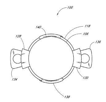
As best seen in Figure 4, the lens system 100 has an anterior portion 102
which is
anterior or forward of the line A-A (which represents a plane substantially
orthogonal to
the optical axis and intersecting first and second apices 112, 116) and a
posterior portion
104 which is posterior or rearward of the line A-A. The anterior portion 102
comprises
an anterior viewing element 106 and an anterior biasing element 108. The
anterior
biasing element 108 in turn comprises a first anterior translation member 110
which
extends from the anterior viewing element 106 to the first apex 112 and a
second anterior
translation member 114 which extends from the anterior viewing element 106 to
the
second apex 116. In the illustrated embodiment the first anterior translation
member 110
comprises a right arm 110a and a left arm 110b (see Figure 3). In addition,
the depicted
second anterior translation member 114 comprises a right arm 114a and a left
arm 114b.
However, in other embodiments either or both of the first and second anterior
translation
members 110, 114 may comprise a single arm or member, or more than two arms or
members.
As best seen in Figures 4, 5 and 7, the posterior portion 104 includes a
posterior
viewing element 118 and a posterior biasing element 120. The posterior biasing
element
120 includes a first posterior translation member 122 extending from the
posterior
viewing element 118 to the first apex 112 and a second posterior translation
member 124
extending from the posterior viewing element 118 to the second apex 116. In
the
illustrated embodiment, the first posterior translation member comprises a
right arm 122a
and a left arm 122b. Likewise, the depicted second posterior translation
member 124
comprises a right arm 124a and a left arm 124b. However, in other embodiments
either
or both of the first and second posterior translation members 122, 124 may
comprise a
single arm or member, or more than two arms or members.
In the embodiment shown in Figure 4, the anterior biasing element 108 and the
posterior biasing element are configured symmetrically with respect to the
plane A-A as
the lens system 100 is viewed from the side. As used herein to describe the
biasing
elements 108, 120, "symmetric" or "symmetrically" means that, as the lens
system 100 is
viewed from the side, the first anterior translation member 110 and the first
posterior
-20-
CA 02849167 2014-04-15
translation member 122 extend from the first apex 112 at substantially equal
first anterior
and posterior biasing angles 01, 02 with respect to the line A-A (which,
again, represents
the edge of a plane which is substantially orthogonal to the optical axis and
intersects the
first and second apices 112, 116) and/or that the second anterior translation
member 114
and the second posterior translation member 124 extend from the second apex
116 at
substantially equal second anterior and posterior biasing angles 03, 04 with
respect to the
line A-A. Alternative or asymmetric configurations of the biasing elements are
possible,
as will be discussed in further detail below. It should be further noted that
a symmetric
configuration of the biasing elements 108, 120 does not dictate symmetric
positioning of
the viewing elements with respect to the line A-A; in the embodiment shown in
Figure 4
the anterior viewing element 106 is closer to the line A-A than is the
posterior viewing
element.
Preferably, both the anterior viewing element 106 and the posterior viewing
element 118 comprise an optic or lens having refractive power. (As used
herein, the term
"refractive" or "refractive power" shall include "diffractive" or "diffractive
power") The
preferred power ranges for the optics are discussed in detail below. In
alternative
embodiments one or both of the anterior and posterior viewing elements 106,
118 may
comprise an optic with a surrounding or partially surrounding perimeter frame
member or
members, with some or all of the biasing elements/translation members attached
to the
frame member(s). As a further alternative, one of the viewing elements 106,
118 may
comprise a perimeter frame with an open/empty central portion or void located
on the
optical axis (see Figure 20 and discussion below), or a perimeter frame member
or
members with a zero-power lens or transparent member therein. In still further
variations, one of the viewing elements 106, 118 may comprise only a zero-
power lens or
transparent member.
In a presently preferred embodiment, a retention portion 126 is coupled to the
anterior portion 102, preferably at the anterior viewing element 106. The
retention
portion 126 preferably includes a first retention member 128 and a second
retention
member 130, although in alternative embodiments the retention portion 126 may
be
-21-
,
CA 02849167 2014-04-15
omitted altogether, or may comprise only one retention member or more than two
retention members. The first retention member 128 is coupled to the anterior
viewing
element 106 at a fixed end 128a and also includes a free end 128b opposite the
fixed end
128a. Likewise, the second retention member 130 includes a fixed end 130a and
a free
end 130b. The retention members 128, 130 are illustrated as being coupled to
the
anterior viewing element 106 at the upper and lower edges thereof; however,
the
retention members 128, 130 may alternatively be attached to the anterior
viewing element
106 at other suitable edge locations.
In the preferred embodiment, the posterior portion 104 includes a distending
portion 132, preferably attached to the posterior viewing element 118. The
preferred
distending portion 132 includes a first distending member 134 which in turn
includes a
fixed end 134a, a free end 134b opposite the fixed end 134a and preferably
also includes
an opening 134c formed therein. The preferred distending portion 132 also
comprises a
second distending member 136 with a fixed end 136a, a free end 136b and
preferably an
opening 136c formed therein. In alternative embodiments, the distending
portion 132
may be omitted altogether, or may comprise a single distending member or more
than
two distending members. To optimize their effectiveness, the preferred
location for the
distending members 134, 136 is 90 degrees away (about the optical axis) from
the apices
112, 116 on the posterior portion 104. Where the biasing elements form more
than two
apices (or where two apices are not spaced 180 degrees apart about the optical
axis), one
or more distending members may be positioned angularly midway between the
apices
about the optical axis. Alternatively, the distending member(s) may occupy
other
suitable positions relative to the apices (besides the "angularly midway"
positions
disclosed above); as further alternatives, the distending member(s) may be
located on the
anterior portion 102 of the lens system 100, or even on the apices themselves.
The
functions of the retention portion 126 and the distending portion 132 will be
described in
greater detail below.
-22-
I i
CA 02849167 2014-04-15
III. THE LENS SYSTEM: FUNCTION/OPTICS
The anterior and posterior biasing elements 108, 120 function in a springlike
manner to permit the anterior viewing element 106 and posterior viewing
element 118 to
move relative to each other generally along the optical axis. The biasing
elements 108,
120 bias the viewing elements 106, 118 apart so that the elements 106, 108
separate to
the accommodated position or accommodated state shown in Figure 4. Thus, in
the
absence of any external forces, the viewing elements are at their maximum
separation
along the optical axis. The viewing elements 106, 118 of the lens system 100
may be
moved toward each other, in response to a ciliary muscle force of up to 2
grams, to
provide an unaccommodated position by applying appropriate forces upon the
anterior
and posterior portions 102, 104 and/or the apices 112, 116.
When the lens system 100 is implanted in the capsular bag 58 (Figures 16-17)
the
above described biasing forces cause the lens system 100 to expand along the
optical axis
so as to interact with both the posterior and anterior aspects of the capsular
bag. Such
interaction occurs throughout the entire range of motion of the ciliary muscle
60. At one
extreme the ciliary muscle is relaxed and the zonules 62 pull the capsular bag
58 radially
so as to cause the bag to become more disk shaped. The anterior and posterior
sides of
the bag, in turn, apply force to the anterior and posterior portions 102, 104
of the lens
system 100, thereby forcing the viewing elements 106, 118 toward each other
into the
accommodated position. At the other extreme, the ciliary muscle contracts and
the
zonules 62 move inwardly to provide slack in the capsular bag 58 and allow the
bag to
become more football-shaped. The slack in the bag is taken up by the lens
system due to
the biasing-apart of the anterior and posterior viewing elements 106, 118. As
the radial
tension in the bag is reduced, the viewing elements 106, 118 move away from
each other
into an accommodated position. Thus, the distance between the viewing elements
106,
118 depends on the degree of contraction or relaxation of the ciliary muscle
60. As the
distance between the anterior and posterior viewing elements 106, 118 is
varied, the focal
length of the lens system 100 changes accordingly. Thus, when the lens system
100 is
implanted into the capsular bag (see Figures 16-17) the lens system 100
operates in
-23-
,
CA 02849167 2014-04-15
conjunction with the natural accommodation processes of the eye to move
between the
accommodated (Figure 16) and unaccommodated (Figure 17) states in the same
manner
as would a healthy "natural" lens. Preferably, the lens system 100 can move
between the
accommodated and unaccommodated states in less than about one second.
The entire lens system 100, other than the optic(s), thus comprises an
articulated
frame whose functions include holding the optic(s) in position within the
capsular bag
and guiding and causing movement of the optic(s) between the accommodated and
unaccommodated positions.
Advantageously, the entire lens system 100 may comprise a single piece of
material, i.e. one that is formed without need to assemble two or more
components by
gluing, heat bonding, the use of fasteners or interlocking elements, etc. This
characteristic increases the reliability of the lens system 100 by improving
its resistance
to material fatigue effects which can arise as the lens system experiences
millions of
accommodation cycles throughout its service life. It will be readily
appreciated that the
molding process and mold tooling discussed herein, lend themselves to the
molding of
lens systems 100 that comprise a single piece of material. However, any other
suitable
technique may be employed to manufacture single-piece lens systems.
In those embodiments where the optic(s) are installed into annular or other
perimeter frame member(s) (see discussion below), the articulated frame may
comprise a
single piece of material, to obtain the performance advantages discussed
above. It is
believed that the assembly of the optic(s) to the articulated frame will not
substantially
detract from the achievement of these advantages.
The lens system 100 has sufficient dynamic range that the anterior and
posterior
viewing elements 106, 118 move about 0.5-4 mm, preferably about 1-3 mm, more
preferably about 1-2 mm, and most preferably about 1.5 mm closer together when
the
lens system 100 moves from the accommodated state to the unaccommodated state.
In
other words the separation distance X (see Figures 9-10, 14-15) between the
anterior and
posterior viewing elements 106, 118, which distance may for present purposes
be defined
as the distance along the optical axis (or a parallel axis) between a point of
axial
-24-
CA 02849167 2014-04-15
intersection with the posterior face of the anterior viewing element 106 and a
point of
axial intersection with the anterior face of the posterior viewing element
118, decreases
by the amount(s) disclosed above upon movement of the lens system 100 to the
unaccornmodated state. Simultaneously, in the preferred mode the total system
thickness
Y decreases from about 3.0 - 4.0 mm in the accommodated state to about 1.5 -
2.5 mm in
the unaccommodated state.
As may be best seen in Figure 6, the first anterior translation member 110
connects to the anterior viewing element 106 via connection of the left and
right arms
110a, 110b to first and second transition members 138, 140 at attachment
locations 142,
144. The second anterior translation member 114 connects to the anterior
viewing
element 106 via connection of left and right arms 114a, 114b to the first and
second
transition members 138, 140 at attachment locations 146, 148. This is a
presently
preferred arrangement for the first and second anterior translation members
110, 114;
alternatively, the first and second anterior translation members 110, 114
could be
connected directly to the anterior viewing element 106, as is the case with
the connection
of the first and second posterior translation members 122, 124 to the
posterior viewing
element 118.
However the connection is established between the first and second anterior
translation members 110, 114 and the anterior viewing element 106, it is
preferred that
the attachment locations 142, 144 corresponding to the first anterior
translation member
110 be farther away from the first apex 112 than is the closest edge or the
periphery of
the anterior viewing element 106. This configuration increases the effective
length of the
first anterior translation member 110/arms 110a, 110b, in comparison to a
direct or
straight attachment between the apex 112 and the nearest/top edge of the
anterior viewing
element 106. For the same reasons, it is preferred that the attachment
locations 146, 148
associated with the second anterior translation member 114 be farther away
from the
second apex 116 than is the closest/bottom edge of the anterior viewing
element 106.
As best seen in Figure 7, the first posterior translation member 122 is
preferably
connected directly to the posterior viewing element 118 via attachment of the
left and
-25-
,
,
CA 02849167 2014-04-15
right arms 122a, 122b to the element 118 at attachment points 150, 152.
Likewise, the
second posterior translation member 124 is preferably directly connected to
the posterior
viewing element 118 via connection of the left and right arms 124a, 124b to
the element
118 at attachment points 154, 156, respectively. In alternative embodiments,
the first and
second posterior translation members 124, 122 can be connected to the
posterior viewing
element via intervening members as is done with the anterior viewing element
106. No
matter how these connections are made, it is preferred that the attachment
locations 150,
152 be spaced further away from the first apex 112 than is the nearest edge or
the
periphery of the posterior viewing element 118. Similarly, it is preferred
that the
attachment locations 154, 156 be spaced further away from the second apex 116
than is
the closest edge of the posterior viewing element 118.
By increasing the effective length of some or all of the translation members
110,
114, 122, 124 (and that of the arms 110a, 110b, 114a, 114b, 122a, 122b, 124a,
124b
where such structure is employed), the preferred configuration of the
attachment
locations 142, 144, 146, 148, 150, 152, 154, 156 relative to the first and
second apices
112, 116 enables the anterior and/or posterior viewing elements 106, 118 to
move with
respect to one another a greater distance along the optical axis, for a given
angular
displacement of the anterior and/or posterior translation members. This
arrangement thus
facilitates a more responsive spring system for the lens system 100 and
minimizes
material fatigue effects associated with prolonged exposure to repeated
flexing.
In the illustrated embodiment, the attachment location 142 of the first
anterior
translation member 110 is spaced from the corresponding attachment location
146 of the
second anterior translation member 114 along the periphery of the anterior
viewing
element, and the same relationship exists between the other pairs of
attachment locations
144, 148; 150, 154; and 152, 156. This arrangement advantageously broadens the
support base for the anterior and posterior viewing elements 106, 118 and
prevents them
from twisting about an axis parallel to the lateral axis, as the viewing
elements move
between the accommodated and unaccommodated positions.
-26-
i
CA 02849167 2014-04-15
It is also preferred that the attachment locations 142, 144 of the first
anterior
translation member 110 be located equidistant from the first apex 112, and
that the right
and left arms 110a, 110b of the member 110 be equal in length. Furthermore,
the
arrangement of the attachment locations 146, 148, arms 114a, 114b and second
apex
preferably mirrors that recited above regarding the first anterior translation
member 110,
while the apices 112, 116 are preferably equidistant from the optical axis and
are situated
180 degrees apart. This configuration maintains the anterior viewing element
106
orthogonal to the optical axis as the viewing element 106 moves back and forth
and the
anterior viewing element flexes.
For the same reasons, a like combination of equidistance and equal length is
preferred for the first and second posterior translation members 122, 124 and
their
constituent arms 122a, 122b, 124a, 124b and attachment points 150, 152, 154,
156, with
respect to the apices 112, 116. However, as shown the arms 122a, 122b, 124a,
124b need
not be equal in length to their counterparts 110a, 110b, 114a, 114b in the
first and second
anterior translation members 110, 114.
Where any member or element connects to the periphery of the anterior or
posterior viewing elements 106, 118, the member defines a connection geometry
or
attachment area with a connection width W and a connection thickness T (see
Figure 4
and the example illustrated therein, of the connection of the second posterior
translation
member 124 to the posterior viewing element 118). For purposes of clarity, the
connection width is defined as being measured along a direction substantially
parallel to
the periphery of the viewing element in question, and the connection thickness
is defined
as measured along a direction substantially perpendicular to the periphery of
the viewing
element. (The periphery itself is deemed to be oriented generally
perpendicular to the
optical axis as shown in Figure 4.) Preferably, no attachment area employed in
the lens
system 100 has a ratio of width to thickness less than 3. It has been found
that such a
geometry reduces distortion of the viewing element/optic due to localized
forces. For the
same reasons, it is also preferred that each of the translation members 110,
114, 122, 124
-27-
CA 02849167 2014-04-15
be connected to the periphery of the respective viewing elements at least two
attachment
areas, each having the preferred geometry discussed above.
Figures 17A and 17B show two preferred cross-sectional configurations which
may be used along some or all of the length of the translation members and/or
arms 110a,
110b, 114a, 114b, 122a, 122b, 124a, 124b. The shape is defined by a relatively
broad
and flat or slightly curved outer surface 182. It is intended that when in use
the outer
surface faces away from the interior of the lens system and/or toward the
capsular bag 58.
The remaining surfaces, proportions and dimensions making up the cross-
sectional shape
can vary widely but may advantageously be selected to facilitate manufacture
of the lens
system 100 via molding or casting techniques while minimizing stresses in the
arms
during use of the lens system.
Figures 17.C-17L depict a number of alternative cross-sectional configurations
which are suitable for the translation members and/or arms 110a, 110b, 114a,
114b, 122a,
122b, 124a, 124b. As shown, a wide variety of cross-sectional shapes may be
used, but
preferably any shape includes the relatively broad and flat or slightly curved
outer surface
182.
It is further contemplated that the dimensions, shapes, and/or proportions of
the
cross-sectional configuration of the translation members and/or arms 110a,
110b, 114a,
114b, 122a, 122b, 124a, 124b may vary along the length of the members/arms.
This may
be done in order to, for example, add strength to high-stress regions of the
arms, fine-tune
their spring characteristics, add rigidity or flexibility, etc.
As discussed above, each of the anterior viewing element 106 and the posterior
viewing element 118 preferably comprises an optic having refractive power. In
one
preferred embodiment, the anterior viewing element 106 comprises a biconvex
lens
having positive refractive power and the posterior viewing element 118
comprises a
convexo-concave lens having negative refractive power. The anterior viewing
element
106 may comprise a lens having a positive power advantageously less than 55
diopters,
preferably less than 40 diopters, more preferably less than 35 diopters, and
most
preferably less than 30 diopters. The posterior viewing element 118 may
comprise a lens
-28-
I I
CA 02849167 2014-04-15
having a power which is advantageously between -25 and 0 diopters, and
preferably
between -25 and -15 diopters. In other embodiments, the posterior viewing
element 118
comprises a lens having a power which is between -15 and 0 diopters,
preferably between
-13 and -2 diopters, and most preferably between -10 and -5 diopters.
Advantageously,
the total power of the optic(s) employed in the lens system 100 is about 5-35
diopters;
preferably, the total power is about 10-30 diopters; most preferably, the
total power is
about 15-25 diopters. (As used herein, the term "diopter" refers to lens or
system power
as measured when the lens system 100 has been implanted in the human eye in
the usual
manner.) It should be noted that if materials having a high index of
refraction (e.g.,
higher than that of silicone) are used, the optics may be made thinner which
facilitates a
wider range of motion for the optics. This in turn allows the use of lower-
power optics
than those specified above. In addition, higher-index materials allow the
manufacture of
a higher-power lens for a given lens thickness and thereby reduce the range of
motion
needed to achieve a given range of accommodation.
Some lens powers and radii of curvature presently preferred for use with an
embodiment of the lens system 100 with optic(s) having a refractive index of
about 1.432
are as follows: a +31 diopter, biconvex lens with an anterior radius of
curvature of 5.944
mm and a posterior radius of curvature of 5.944 mm; a +28 diopter, biconvex
lens with
an anterior radius of curvature of 5.656 mm and a posterior radius of
curvature of 7.788
mm; a +24 diopter, biconvex lens with an anterior radius of curvature of 6.961
mm and a
posterior radius of curvature of 8.5 mm; a -10 diopter, biconcave lens with an
anterior
radius of curvature of 18.765 mm and a posterior radius of curvature of 18.765
mm; a -8
diopter, concavo-convex lens with an anterior radius of curvature of between 9
mm and
9.534 mm and a posterior radius of curvature of 40 mm; and a -5 diopter,
concavo-
convex lens with an anterior radius of curvature of between 9 mm and 9.534 mm
and a
posterior radius of curvature of 20 mm. In one embodiment, the anterior
viewing
element comprises the +31 diopter lens described above and the posterior
viewing
element comprises the -10 diopter lens described above. In another embodiment,
the
anterior viewing element comprises the +28 diopter lens described above and
the
-29-
CA 02849167 2014-04-15
posterior viewing element comprises the -8 diopter lens described above. In
another
embodiment, the anterior viewing element comprises the +24 diopter lens
described
above and the posterior viewing element comprises the -5 diopter lens
described above.
The combinations of lens powers and radii of curvature specified herein
advantageously minimize image magnification. However, other designs and radii
of
curvature provide modified magnification when desirable.
The lenses of the anterior viewing element 106 and the posterior viewing
element
118 are relatively moveable as discussed above; advantageously, this movement
is
sufficient to produce an accommodation of at least one diopter, preferably at
least two
diopters and most preferably at least three diopters. In other words, the
movement of the
optics relative to each other and/or to the cornea is sufficient to create a
difference
between (i) the refractive power of the user's eye in the accommodated state
and (ii) the
refractive power of the user's eye in the unaccommodated state, having a
magnitude
expressed in diopters as specified above. Where the lens system 100 has a
single optic,
the movement of the optic relative to the cornea is sufficient to create a
difference in
focal power as specified above.
Advantageously, the lens system 100 can be customized for an individual
patient's needs by shaping or adjusting only one of the four lens faces, and
thereby
altering the overall optical characteristics of the system 100. This in turn
facilitates easy
manufacture and maintenance of an inventory of lens systems with lens powers
which
will fit a large population of patients, without necessitating complex
adjustment
procedures at the time of implantation. It is contemplated that all of the
lens systems in
the inventory have a standard combination of lens powers, and that a system is
fitted to a
particular patient by simply shaping only a designated "variable" lens face.
This custom-
shaping procedure can be performed to-order at a central manufacturing
facility or
laboratory, or by a physician consulting with an individual patient. In one
embodiment,
the anterior face of the anterior viewing element is the designated sole
variable lens face.
In another embodiment, the anterior face of the posterior viewing element is
the only
variable face. However, any of the lens faces is suitable for such
designation. The result
-30-
,
CA 02849167 2014-04-15
is minimal inventory burden with respect to lens power (all of the lens
systems in stock
have the same lens powers) without requiring complex adjustment for individual
patients
(only one of the four lens faces is adjusted in the fitting process).
IV. THE LENS SYSTEM: ALTERNATIVE EMBODIMENTS
Figure 17M depicts another embodiment of the lens system 100 in which the
anterior viewing element 106 comprises an optic with a smaller diameter than
the
posterior viewing element 118, which comprises an optic with a peripheral
positive-lens
portion 170 surrounding a central negative portion 172. This arrangement
enables the
user of the lens system 100 to focus on objects at infinity, by allowing the
(generally
parallel) light rays incident upon the eye from an object at infinity to
bypass the anterior
viewing element 106. The peripheral positive-lens portion 170 of the posterior
viewing
element 118 can then function alone in refracting the light rays, providing
the user with
focused vision at infinity (in addition to the range of visual distances
facilitated by the
anterior and posterior viewing elements acting in concert). In another
embodiment, the
anterior viewing element 106 comprises an optic having a diameter of
approximately 3
millimeters or less. In yet another embodiment, the anterior viewing element
106
comprises an optic having a diameter of approximately 3 millimeters or less
and a
refractive power of less than 55 diopters, more preferably less than 30
diopters. In still
another embodiment, the peripheral positive-lens portion 170 has a refractive
power of
about 20 diopters.
Figure 17N shows an alternative arrangement in which, the anterior viewing
element 106 comprises an optic having a central portion 176 with refractive
power, and a
surrounding peripheral region 174 having a refractive power of substantially
zero,
wherein the central region 176 has a diameter smaller than the optic of the
posterior
viewing element 118, and preferably has a diameter of less than about 3
millimeters.
This embodiment also allows some incident light rays to pass the anterior
viewing
element (though the zero-power peripheral region 174) without refraction,
allowing the
peripheral positive-lens portion 170 posterior viewing element 118 to function
alone as
described above.
-31-
CA 02849167 2014-04-15
Figures 18 and 19 depict another embodiment 250 of the intraocular lens. It is
contemplated that, except as noted below, this embodiment 250 is largely
similar to the
embodiment disclosed in FIGS. 3-17. The lens 250 features an anterior biasing
element
108 and posterior biasing element 120 which are arranged asymmetrically as the
lens
system 100 is viewed from the side. As used herein to describe the biasing
elements 108,
120, "asymmetric" or "asymmetrically" means that, as the lens system 250 is
viewed from
the side, the first anterior translation member 110 and the first posterior
translation
member 122 extend from the first apex 112 at unequal first anterior and
posterior biasing
angles 83, 82 with respect to the line B-B (which represents the edge of a
plane which is
substantially orthogonal to the optical axis and intersects the first and
second apices 112,
116) and/or that the second anterior translation member 114 and the second
posterior
translation member 124 extend from the second apex 116 at unequal second
anterior and
posterior biasing angles 63, 84 with respect to the line B-B.
In the embodiment shown in Figures 18-19, the first and second anterior
biasing
angles 63, 63 are greater than the corresponding first and second posterior
biasing angles
82, 84. This arrangement advantageously maintains the posterior viewing
element 118
and apices 112, 116 in a substantially stationary position. Consequently, the
moving
mass of the lens system 250 is reduced, and the anterior viewing element 106
can move
more quickly over a wider range along the optical axis under a given motive
force. (Note
that even where the posterior biasing element 120 and its constituent first
and second
posterior translation members 122, 124 are substantially immobile, they are
nonetheless
"biasing elements" and "translation members" as those terms are used herein.)
In another
embodiment, the anterior biasing element 108 and posterior biasing element 120
are
arranged asymmetrically in the opposite direction, i.e. such that the first
and second
anterior biasing angles 63, 63 are smaller than the corresponding first and
second posterior
biasing angles 82, 84. This arrangement also provides for a wider range of
relative
movement of the viewing elements, in comparison to a "symmetric" system.
It should be further noted that the viewing elements 106, 118 shown in Figures
18-19 are asymmetrically positioned in that the posterior viewing element 118
is closer to
-32-
,
CA 02849167 2014-04-15
the line B-B than is the anterior viewing element 106. It has been found that
this
configuration yields desirable performance characteristics irrespective of the
configuration of the biasing elements 108, 120. In alternative embodiments,
the viewing
elements 106, 118 may be positioned symmetrically with respect to the line B-
B, or they
may be positioned asymmetrically with the anterior viewing element 106 closer
to the
line B-B than the posterior viewing element 118 (see Figure 4 wherein the line
in
question is denoted A-A). Furthermore, the symmetry or asymmetry of the
biasing
elements and viewing elements can be selected independently of each other.
Figure 20 shows another embodiment 350 of an intraocular lens in which the
posterior viewing element 118 comprises an annular frame member defining a
void
therein, while the anterior viewing element 106 comprises an optic having
refractive
power. Alternatively, the posterior viewing element 118 could comprise a zero
power
lens or a simple transparent member. Likewise, in another embodiment the
anterior
viewing element 106 could comprise an annular frame member with a void therein
or a
simple zero power lens or transparent member, with the posterior viewing
element 118
comprising an optic having refractive power. As a further alternative, one or
both of the
anterior and posterior viewing elements 106, 118 may comprise an annular or
other
perimeter frame member which can receive a removable optic (or a "one-time
install"
optic) with an interference type fit and/or subsequent adhesive or welding
connections.
Such a configuration facilitates assembly and/or fine-tuning of the lens
system during an
implantation procedure, as will be discussed in further detail below.
V. THE LENS SYSTEM: ADDITIONAL FEATURES
Figure 21 depicts the function of the distending portion 132 in greater
detail. The
lens system 100 is shown situated in the capsular bag 58 in the customary
manner with
the anterior viewing element 106 and posterior viewing element 118 arranged
along the
optical axis. The capsular bag 58 is shown with a generally circular anterior
opening 66
which may often be cut into the capsular bag during installation of the lens
system 100.
The first and second distending members 134, 136 of the distending portion 132
distend
the capsular bag 58 so that intimate contact is created between the posterior
face of the
-33-
,
CA 02849167 2014-04-15
posterior viewing element and/or the posterior biasing element 120. In
addition, intimate
contact is facilitated between the anterior face of the anterior viewing
element 106 and/or
anterior biasing element 108. The distending members 134, 136 thus remove any
slack
from the capsular bag 58 and ensure optimum force coupling between the bag 58
and the
lens system 100 as the bag 58 is alternately stretched and released by the
action of the
ciliary muscle.
Furthermore, the distending members 134, 136 reshape the capsular bag 58 into
a
taller, thinner configuration along its range of accommodation to provide a
wider range
of relative motion of the viewing elements 106, 118. When the capsular bag 58
is in the
unaccommodated state, the distending members 134, 136 force the capsular bag
into a
thinner configuration (as measured along the optical axis) in comparison to
the
unaccommodated configuration of the capsular bag 58 with the natural lens in
place.
Preferably, the distending members 134, 136 cause the capsular bag 58 to taken
on a
shape in the unaccommodated state which is about 1.0-2.0 mm thinner, more
preferably
about 1.5 mm thinner, along the optical axis than it is with the natural lens
in place and in
the unaccommodated state.
With such a thin "starting point" provided by the distending members 134, 136,
the viewing elements 106, 118 of the lens system can move a greater distance
apart, and
provide a greater range of accommodation, without causing undesirable contact
between
the lens system and the iris. Accordingly, by reshaping the bag as discussed
above the
distending members 134, 136 facilitate a range of relative motion of the
anterior and
posterior viewing elements 106, 118 of about 0.5-4 mm, preferably about 1-3
mm, more
preferably about 1-2 mm, and most preferably about 1.5 mm.
The distending portion 132/distending members 134, 136 are preferably separate
from the anterior and posterior biasing elements 108, 120; the distending
members 134,
136 thus preferably play no part in biasing the anterior and posterior viewing
elements
106, 118 apart toward the accommodated position. This arrangement is
advantageous
because the apices 112, 116 of the biasing elements 108, 120 reach their point
of
minimum protrusion from the optical axis (and thus the biasing elements reach
their
-34-
,
CA 02849167 2014-04-15
minimum potential effectiveness for radially distending the capsular bag) when
the lens
system 100 is in the accommodated state (see Figure 16), which is precisely
when the
need is greatest for a taut capsular bag so as to provide immediate response
to relaxation
of the ciliary muscles. The preferred distending portion is "static" (as
opposed to the
"dynamic" biasing members 108, 120 which move while urging the viewing
elements
106, 118 to the accommodated position or carrying the viewing elements to the
unaccommodated position) in that its member(s) protrude a substantially
constant
distance from the optical axis throughout the range of motion of the viewing
elements
106, 118. Although some degree of flexing may be observed in the distending
members
134, 136, they are most effective when rigid. Furthermore, the thickness
and/or cross-
sectional profile of the distending members 134/136 may be varied over the
length of the
members as desired to provide a desired degree of rigidity thereto.
The distending portion 132/distending members 132, 134 advantageously reshape
the capsular bag 58 by stretching the bag 58 radially away from the optical
axis and
causing the bag 58 to take on a thinner, taller shape throughout the range of
accommodation by the eye. This reshaping is believed to facilitate a broad (as
specified
above) range of relative motion for the viewing elements of the lens system
100, with
appropriate endpoints (derived from the total system thicknesses detailed
above) to avoid
the need for unacceptably thick optic(s) in the lens system.
If desired, the distending members 134, 136 may also function as haptics to
stabilize and fixate the orientation of the lens system 100 within the
capsular bag. The
openings 134c, 136c of the preferred distending members 134,136 permit
cellular
ingrowth from the capsular bag upon positioning of the lens system 100
therein. Finally,
other methodologies, such as a separate capsular tension ring or the use of
adhesives to
glue the capsular bag together in selected regions, may be used instead of or
in addition
to the distending portion 132, to reduce "slack" in the capsular bag.
A tension ring can also act as a physical barrier to cell growth on the inner
surface
of the capsular bag, and thus can provide additional benefits in limiting
posterior capsule
opacification, by preventing cellular growth from advancing posteriorly on the
inner
-35-
, ,
CA 02849167 2014-04-15
surface of the bag. When implanted, the tension ring firmly contacts the inner
surface of
the bag and defines a circumferential barrier against cell growth on the inner
surface from
one side of the barrier to another.
Figure 21A shows an alternative configuration of the distending portion 132,
in
which the distending members 134, 136 comprise first and second arcuate
portions which
connect at either end to the apices 112, 116 to form therewith an integral
perimeter
member. In this arrangement it is preferred that the distending members and
apices form
an oval with height I smaller than width J.
Figure 21B shows another alternative configuration of the distending portion
132,
in which arcuate rim portions 137 interconnect the apices 112, 116 and the
free ends
134b, 136b of the distending members 134, 136. Thus is formed an integral
perimeter
member with generally higher lateral rigidity than the arrangement depicted in
Figure
21A.
Figure 21C shows another alternative configuration of the distending portion
132,
in which the distending members 134, 136 are integrally formed with the first
and second
posterior translation members 122, 124. The distending members 134, 136 and
translation members 122, 124 thus form common transition members 139 which
connect
to the periphery of the posterior viewing element 118.
Figure 22 shows the function of the retention portion 126 in greater detail.
It is
readily seen that the first and second retention members 128, 130 facilitate a
broad
contact base between the anterior portion of the lens system 100 and the
anterior aspect
of the capsular bag 58. By appropriately spacing the first and second
retention members
128, 130, the members prevent extrusion of the anterior viewing element 106
through the
anterior opening 66. It is also readily seen that where contact occurs between
the anterior
aspect of the capsular bag 58 and one or both of the retention members 128,
130, the
retention members also participate in force coupling between the bag 58 and
the lens
system 100 as the bag is stretched and released by the action of the ciliary
muscles.
As best seen in Figures 21 and 22, the anterior portion 102 of the lens system
100
forms a number of regions of contact with the capsular bag 58, around the
perimeter of
-36-
, ,
i
CA 02849167 2014-04-15
the anterior viewing element 106. In the illustrated embodiment, at least some
of these
regions of contact are located on the anteriormost portions of the anterior
biasing element
108, specifically at the transition members 138, 140, and at the retention
members 128,
130. The transition members and the retention members define spaces
therebetween at
the edges of the anterior viewing element 106 to permit fluid to flow between
the interior
of the capsular bag 58 and the portions of the eye anterior of the bag 58. In
other words,
the anterior portion of the lens system 100 includes at least one location
which is spaced
from and out of contact with the capsular bag 58 to provide a fluid flow
channel
extending from the region between the viewing elements 106, 118 to the
exterior of the
bag 58. Otherwise, if the anterior portion 102 of the lens system 100 seals
the anterior
opening 66 of the bag 58, the resulting prevention of fluid flow can cause the
aqueous
humor in the capsular bag to stagnate, leading to a clinically adverse event,
and can
inhibit the movement of the lens system 100 between the accommodated and
unaccommodated states.
If desired, one or both of the retention members 128, 130 may have an opening
129 formed therein to permit fluid flow as discussed above. (See Figure 21A.)
The retention members 128, 130 and the transition members 138, 140 also
prevent contact between the iris and the anterior viewing element 106, by
separating the
anterior opening 66 from the anterior face of the viewing element 106. In
other words,
the retention members 128, 130 and the transition members 138, 140 displace
the anterior
aspect of the capsular bag 58, including the anterior opening 66, anteriorly
from the
anterior viewing element 106, and maintain this separation throughout the
range of
accommodation of the lens system. Thus, if contact occurs between the iris and
the lens
system-capsular bag assembly, no part of the lens system will touch the iris,
only the
capsular bag itself, in particular those portions of the bag 58 overlying the
retention
members 128, 130 and/or the transition members 138, 140. The retention members
128,
130 and/or the transition members 138, 140 therefore maintain a separation
between the
iris and the lens system, which can be clinically adverse if the contacting
portion(s) of the
lens system are constructed from silicone.
-37-
,
CA 02849167 2014-04-15
As depicted in Figure 22A, one or more stop members or separation members 190
may be located where appropriate on the anterior and/or posterior biasing
elements 108,
120 to limit the convergent motion of the anterior and posterior viewing
elements 106,
118, and preferably prevent contact therebetween. As the lens system 100 moves
toward
the unaccommodated position, the stop member(s) located on the anterior
biasing
element 108 come into contact with the posterior biasing element 120 (or with
additional
stop member(s) located on thereon), and any stop member(s) located on the
posterior
biasing element 120 come into contact with the anterior biasing element 108
(or with
additional stop member(s) located thereon). The stop members 190 thus define a
point or
state of maximum convergence (in other words, the unaccommodated state) of the
lens
system 100/ viewing elements 106, 118. Such definition advantageously assists
in setting
one extreme of the range of focal lengths which the lens system may take on
(in those
lens systems which include two or more viewing elements having refractive
power)
and/or one extreme of the range of motion of the lens system 100.
The stop members 190 shown in Figure 22A are located on the first and second
anterior translation members 110, 114 of the anterior biasing element 108 and
extend
posteriorly therefrom. When the anterior and posterior viewing elements 106,
118 move
together, one or more of the stop members 190 will contact the posterior
translation
member(s) 122, 124, thereby preventing further convergent motion of the
viewing
elements 106, 118. Of course, in other embodiments the stop member(s) 190 can
be in
any suitable location on the lens system 100.
Figures 44-48 depict another embodiment of the lens system 100 having a number
of stop members or separation members 190. In this embodiment the stop members
190
include posts 190a and tabs 190b, although it will be apparent that any number
or
combination of suitable shapes may be employed for the stop members 190. Each
of the
stop members 190 has at least one contact surface 191, one or more of which
abuts an
opposing surface of the lens system 100 when the anterior and posterior
viewing
elements 106, 118 converge to a minimum separation distance SD (see Figure
47). In the
embodiment shown, one or more of the contact surfaces 191 of the posts 190a
are
-38-
, ,
CA 02849167 2014-04-15
configured to abut an opposing surface defined by a substantially flat
anterior perimeter
portion 193 of the posterior viewing element 118, when the viewing elements
106, 118
are at the minimum separation distance SD. One or more of the contact surfaces
191 of
the tabs 190b are configured to abut opposing surfaces defined by
substantially flat
anterior faces 195 of the distending members 134, 136, only if the viewing
elements 106,
118 are urged together beyond the minimum separation distance SD. This
arrangement
permits the tabs 190b to function as secondary stop members should the posts
190a fail to
maintain separation of the viewing elements.
In other embodiments all of the contact surfaces 191 of the posts 190a and
tabs
190b may be configured to contact their respective opposing surfaces when the
viewing
elements 106, 118 are at the minimum separation distance SD. In still further
embodiments, the contact surfaces 191 of the tabs 190b may be configured to
contact the
opposing surfaces when the viewing elements 106, 118 are at the minimum
separation
distance SD and the contact surfaces 191 of the posts 190a configured to
contact the
opposing surfaces only if the viewing elements 106, 118 are urged together
beyond the
minimum separation distance SD. In one embodiment, the minimum separation
distance
SD is about 0.1 - 1.0 mm; in another embodiment the minimum separation
distance SD is
about 0.5 mm.
When one of the contact surfaces abuts one of the opposing surfaces, the two
surfaces define a contact area CA (see Figure 48, depicting an example of a
contact area
CA defined when the contact surface 191 of a post 190a contacts an opposing
surface
defined by the perimeter portion 193 of the posterior viewing element 118).
Preferably,
the contact surface and opposing surface are shaped to cooperatively minimize
the size of
the contact area, to prevent adhesion between the contact surface and the
opposing
surface, which is often a concern when one or both of these surfaces has an
adhesive
affinity for the other. In the embodiment shown, this non-adhesive
characteristic is
achieved by employing a substantially hemispherical contact surface 191 and a
substantially flat opposing surface (perimeter portion 193). Of
course, other
configurations can be selected for the contact surface(s) 191, including
conical,
-39-
i
CA 02849167 2014-04-15
frustoconical, hemicylindrical, pyramidal, or other rounded, tapered or
pointed shapes.
All of these configurations minimize the contact area CA while permitting the
cross-
sectional area CS of the stop member 190 (such as the post 190a depicted) to
be made
larger than the contact area CA, to impart sufficient strength to the stop
member despite
the relatively small contact area CA. Indeed, when constructing the contact
surface(s)
191 any configuration may be employed which defines a contact area CA which is
smaller than the cross-sectional area CS of the stop member 190. As further
alternatives,
the contact surface(s) 191 may be substantially flat and the opposing
surface(s) may have
a shape which defines, upon contact with the opposing surface, a contact area
CA which
is smaller than the cross-sectional area CS of the stop member. Thus, the
opposing
surface(s) may have, for example, a hemispherical, conical, frustoconical,
hemicylindrical, pyramidal, or other rounded, tapered or pointed shape.
Other design features of the stop members 190 can be selected to maximize
their
ability to prevent adhesion of the contact surface(s) to the corresponding
opposing
surface(s), or adhesion to each other of any part of the anterior and
posterior portions
102, 104 of the lens system 100. For example, the contact and opposing
surfaces may be
formed from dissimilar materials to reduce the effect of any self-adhesive
materials
employed in forming the lens system 100. In addition the shape and/or material
employed in constructing one or more of the stop members 190 can be selected
to impart
a spring-like quality to the stop member(s) in question, so that when the stop
member is
loaded in compression as the viewing elements are urged together at the
minimum
separation distance, the stop member tends to exert a resisting spring force,
due to either
bending or axial compression (or both) of the stop member, which in turn
derive from the
elasticity of the material(s) from which the stop member is constructed, or
the shape of
the stop member, or both. This springlike quality is particularly effective
for inhibiting
adhesion of areas of the anterior and posterior portions 102, 104 other than
the contact
surface(s) and opposing surface(s).
As used herein, the term "adhesion" refers to attachment to each other of (i)
an
area of the anterior portion 102 of the lens system 100 and (ii) a
corresponding area of the
-40-
,
CA 02849167 2014-04-15
posterior portion 104 (other than the apices 112, 116), wherein such
attachment is
sufficiently strong to prevent, other than momentarily, the anterior and
posterior viewing
elements 106, 118 from moving apart along the optical axis under the biasing
force of the
anterior and/or posterior biasing elements 108, 120. If the areas in question
are formed of
different materials, adhesion may occur where at least one of the materials
has an
adhesive affinity for the other material. If the areas in question are formed
of the same
material, adhesion may occur where the material has an adhesive affinity for
itself.
In the embodiment shown, four posts 190a are positioned near the perimeter of
the anterior viewing element 106, equally angularly spaced around the optical
axis. In
addition, two tabs 190b are located on either side of the anterior viewing
element,
midway between the apices 112, 116 of the lens system. Naturally, the number,
type
and/or position of the stop members 190 can be varied while preserving the
advantageous
function of maintaining separation between the anterior and posterior portions
of the lens
system.
The illustrated embodiment employs stop members 190 which extend posteriorly
from the anterior portion 102 of the lens system 100, so that the contact
surfaces 191 are
located on the posterior extremities of the stop members 190 and are
configured to abut
opposing surfaces formed on the posterior portion 104 of the lens system 100.
However,
it will be appreciated that some or all of the stop members 190 may extend
anteriorly
from the posterior portion 104 of the lens system 100, so that their contact
surfaces 191
are located on the anterior extremities of the stop members 190 and are
configured to
abut opposing surfaces formed on the anterior portion 102 of the lens system
100.
VI. MOLD TOOLING
Figures 23-34 depict a mold system 500 which is suitable for molding the lens
system 100 depicted in Figure 3-17. The mold system 500 generally comprises a
first
mold 502, a second mold 504 and a center mold 506. The center mold 506 is
adapted to
be positioned between the first mold 502 and the second mold 504 so as to
define a mold
space for injection molding or compression molding the lens system 100. The
mold
system 500 may be formed from suitable metals, high-impact-resistant plastics
or a
-41-
,
CA 02849167 2014-04-15
combination thereof, and can be produced by conventional machining techniques
such as
lathing or milling, or by laser or electrical-discharge machining. The mold
surfaces can
be finished or modified by sand blasting, etching or other texturing
techniques.
The first mold 502 includes a first mold cavity 508 with a first anterior mold
face
510 surrounded by an annular trough 512 and a first perimeter mold face 514.
The first
mold 502 also includes a projection 516 which facilitates easier mating with
the second
mold 504.
The center mold 506 includes a first center mold cavity 518 which cooperates
with the first mold cavity 508 to define a mold space for forming the anterior
portion 102
of the lens system 100. The first center mold cavity 518 includes a central
anterior mold
face 520 which, upon placement of the center mold 506 in the first mold cavity
508,
cooperates with the first anterior mold face 510 to define a mold space for
the anterior
viewing element 106. In so doing, the first anterior mold face 510 defines the
anterior
face of the anterior viewing element 106 and the central anterior mold face
520 defines
the posterior face of the anterior viewing element 106. In fluid communication
with the
chamber formed by the first anterior mold face 510 and the central anterior
mold face 520
are lateral channels 522, 524 (best seen in Figure 31) which form spaces for
molding the
first and second transition members 138, 140, along with the arms 110a, 110b
of the first
anterior translation member 110 as well as the arms 114a, 114b of the second
anterior
translation member 114. The first center mold cavity 518 also includes
retention member
cavities 526, 528 which define spaces for molding the first and second
retention members
128, 130 to the anterior viewing element 106.
The second mold 504 includes a second mold cavity 530 with a second posterior
mold space 532, a generally cylindrical transition 534 extending therefrom and
connecting to a second perimeter mold face 536. Lateral notches 538, 540 (best
seen in
Figures 26 and 27) are formed in the second perimeter mold face 536. The
second mold
504 also includes an input channel 542 connected to an input channel opening
544 for
introducing material into the mold system 500. Also formed in the second mold
504 is
-42-
CA 02849167 2014-04-15
an output channel 546 and an output channel opening 548. A generally
cylindrical rim
550 is included for mating with the projection 516 of the first mold 502.
The center mold 506 includes a second center mold cavity 552 which cooperates
with the second mold cavity 530 to define a mold space for the posterior
portion 104 of
the lens system 100. The second center mold cavity 552 includes a central
posterior
mold face 554 which, upon placement of the center mold 506 in engagement with
the
second mold cavity 530, cooperates with the second posterior mold face 532 and
the
transition 534 to define a chamber for forming the posterior viewing element
118. In
fluid communication with the chamber formed by the central posterior mold face
554 and
the second posterior mold face 532 are lateral channels 556, 558, 560, 562
which provide
a mold space for forming the arms 122a, 122b of the first posterior
translation member
122 and the arms 124a, 124b of the second posterior translation member 124.
The
second center mold cavity 552 includes lateral projections 564, 566 which
coact with the
notches 538, 540 formed in the second mold cavity 530. The chambers formed
therebetween are in fluid communication with the chamber defined by the
central
posterior mold face 554 and the second posterior mold face 532 to form the
first and
second distending members 134, 136 integrally with the posterior viewing
element 118.
The center mold 506 includes a first reduced-diameter portion 568 and a second
reduced-diameter portion 570 each of which, upon assembly of the mold system
500,
defines a mold space for the apices 112, 116 of the lens system 100.
In use, the mold system 500 is assembled with the center mold 506 positioned
between the first mold 502 and the second mold 504. Once placed in this
configuration,
the mold system 500 is held together under force by appropriate techniques,
and lens
material is introduced into the mold system 500 via the input channel 542. The
lens
material then fills the space defined by the first mold 502, second mold 504,
and the
center mold 506 to take on the shape of the finished lens system 100.
The mold system 500 is then disassembled, and in one embodiment the lens
system 100 is left in position on the center mold 506 after removal of the
first and second
molds 502, 504. This technique has been found to improve the effectiveness of
any
-43-
,
1 ,
CA 02849167 2014-04-15
polishing/tumbling/deflashing procedures which may be performed (see further
discussion below). Whether or not these or any other additional process steps
are
performed, the lens system 100 is preferably removed from the center mold 506
while
maintaining the interconnection of the various components of the lens system
100.
In another embodiment, the lens system 100 or a portion thereof is formed by a
casting or liquid-casting procedure in which one of the first or second molds
is first filled
with a liquid and the center mold is placed then into engagement with the
liquid-filled
mold. The exposed face of the center mold is then filled with liquid and the
other of the
first and second molds is placed into engagement with the rest of the mold
system. The
liquid is allowed or caused to set/cure and a finished casting may then
removed from the
mold system.
The mold system 500 can advantageously be employed to produce a lens system
100 as a single, integral unit (in other words, as a single piece of
material). Alternatively,
various portions of the lens system 100 can be separately molded, casted,
machined, etc.
and subsequently assembled to create a finished lens system. Assembly can be
performed as a part of centralized manufacturing operations; alternatively, a
physician
can perform some or all of the assembly before or during the implantation
procedure, to
select lens powers, biasing members, system sizes, etc. which are appropriate
for a
particular patient.
The center mold 506 is depicted as comprising an integral unit with first and
second center mold cavities 518, 552. Alternatively, the center mold 506 may
have a
modular configuration whereby the first and second mold cavities 518, 552 may
be
interchangeable to adapt the center mold 506 for manufacturing a lens system
100
according to a desired prescription or specification, or to otherwise change
the power(s)
of the lenses made with the mold. In this manner the manufacture of a wide
variety of
prescriptions may be facilitated by a set of mold cavities which can be
assembled back-
to-back or to opposing sides of a main mold structure.
Figures 49-53 depict one embodiment of a method for manufacturing the center
mold 506. First, a cylindrical blank 1500 formed from any material (such as
Ultem)
-44-
, ,
CA 02849167 2014-04-15
suitable for use in the mold tooling, is loaded into a holder 1502 as shown in
Figure 49.
The holder 1502 has a main chamber 1504 which has an inner diameter
substantially
similar to that of the blank 1500, a smaller-diameter secondary chamber 1506
rearward of
the main chamber 1504, and a passage 1508 located rearward of the secondary
chamber
1506 and further defined by an annulus 1510. The holder also includes two or
more
holder bores 1512 which facilitate attachment of the holder 1502 to a blocker
(discussed
in further detail below). The blank is "blocked" in the holder by filling the
secondary
chamber 1506 and passage 1508 with water-soluble wax 1514.
Once the blank 1500 has been loaded and blocked into the holder 1502, the
holder
1502 is secured to a blocker 1516 by bolts or pins (not shown) which fit
snugly into the
holder bores 1512. The holder bores 1512 align precisely with corresponding
blocker
bores 1517, by virtue of a snug fit between the blocker bores 1517 and the
bolts/pins.
The blocker-holder assembly is then loaded into a conventional machine tool,
such as a
lathe and/or a mill, and one of the first and second center mold cavities 518,
552 (the
second cavity 552 is depicted in Figure 51) is machined from the exposed face
of the
blank 1500 using conventional machining techniques. The holder 1502 and blank
1500,
with the second center mold cavity 552 formed thereon, are then removed from
the
blocker 1516 as shown in Figure 51.
The main chamber 1504 is then filled with water-soluble wax 1520 forward of
the
second center mold cavity 552, and the wax 1514 is removed from the secondary
chamber 1506 and the passage 1508. Next the holder 1502 is fixed to the
blocker 1516
with the as-yet unmodified portion of the blank 1500 facing outward. Upon re-
loading
the holder-blocker assembly into the machine tool, a portion of the annulus
1510 is then
cut away to facilitate tool access to the blank 1500. A series of machining
operations are
then performed on the blank 1500 until the remaining mold cavity (the first
center mold
cavity 518 is depicted in Figure 53) has been formed. The completed center
mold 506
may then be removed from the holder 1502.
The machining technique depicted in Figures 49-53 is advantageous in that it
facilitates fabrication of the center mold 506 (with both the first and second
center mold
-45-
,
CA 02849167 2014-04-15
cavities 518, 552) from a single piece of material. While it is possible to
machine the
first and second center mold cavities 518, 552 from separate pieces of
material which are
subsequently glued together, such assembly creates a seam in the center mold
which can
retain contaminants and introduce those contaminants into the mold when
forming the
lens system 100. In addition, the assembly of the center mold 506 from two
halves
introduces errors wherein the first and second center mold cavities 518, 552
may be
angularly shifted with respect to each other about the optical axis, or
wherein the mold
cavities 518, 552 are non-concentric (i.e., shifted with respect to each other
in a direction
orthogonal to the optical axis). The method depicted in Figures 49-53
eliminates these
problems by retaining the blank 1500 in the holder 1502 throughout the
fabrication
process and by enforcing precise axial alignment, via forced alignment of the
bores 1512
with the blocker bores 1517, when machining of both mold cavities.
In another embodiment, the center mold 506 is formed by a molding process
rather than by machining. The center mold 506 may be molded from any of the
materials
disclosed herein as suitable for forming the lens system 100 itself, including
but not
limited to silicone, acrylics, polymethylmethacrylate (PMMA), block copolymers
of
styrene-ethylene-butylene-styrene (C-FLEX) or other styrene-base copolymers,
polyvinyl
alcohol (PVA), polyurethanes, hydrogels or any other moldable polymers or
monomers.
The lens system which is formed when employing the molded center mold 506
may itself be molded from the same material as the center mold 506. For
example, the
center mold 506 may be molded from silicone, and then the lens system 100 may
be
molded from silicone by using the mold system 500 with the molded silicone
center mold
506.
The center mold 506 can be molded by any suitable conventional techniques. A
polished, optical quality initial mold set can be used to make center molds
which in turn
will produce lens systems with optical quality surfaces on the posterior face
of the
anterior optic, and the anterior face of the posterior optic. Alternatively
(or additionally),
the molded center mold can be polished and/or tumbled to produce an optically-
accurate
center mold.
-46-
, ,
CA 02849167 2014-04-15
The molded center mold 506 offers several advantages over a machined center
mold. First, it is quicker, cheaper and easier to produce the center mold in
large
quantities by molding instead of machining. This in turn facilitates leaving
the lens
system in position on the center mold (see Figure 54) while the lens system is
tumbled,
polished and/or deflashed, without incurring undue expense. The presence of
the center
mold between the optics increases the effectiveness of the
tumbling/polishing/deflashing
by increasing the hoop strength of the lens system, so that the energy of the
impacting
tumbling beads is not dissipated in macroscopic deformation of the lens
system.
Molding also permits softer materials to be employed in forming the center
mold, and a
softer center mold is more resistant to damage from deflashing tools and
processes,
resulting in fewer center molds lost to such process-related damage.
VII. MATERIALS/SURFACE TREATMENTS
Preferred materials for forming the lens system 100 include silicone,
acrylics,
polymethylmethacrylate (PMMA), block copolymers of styrene-ethylene-butylene-
styrene (C-FLEX) or other styrene-base copolymers, polyvinyl alcohol (PVA),
polyurethanes, hydrogels or any other suitable polymers or monomers. In
addition, any
portion of the lens system 100 other than the optic(s) may be formed from
stainless steel
or a shape-memory alloy such as nitinol or any iron-based shape-memory alloy.
Metallic
components may be coated with gold to increase biocompatibility. Where
feasible,
material of a lower Shore A hardness such as 15A may be used for the optic(s),
and
material of higher hardness such as 35A may be used for the balance of the
lens system
100. Finally, the optic(s) may be formed from a photosensitive silicone to
facilitate post-
implantation power adjustment as taught in United States Patent No. 6,450,642,
issued
September 17, 2002, titled LENSES CAPABLE OF POST-FABRICATION POWER
MODIFICATION.
Methyl-methylacrylate monomers may also be blended with any of the non-
metallic materials discussed above, to increase the lubricity of the resulting
lens system
(making the lens system easier to fold or roll for insertion, as discussed
further below).
The addition of methyl-methylacrylate monomers also increases the strength and
transparency of the lens system.
-47-
,
i
CA 02849167 2014-04-15
The optics and/or the balance of the lens system 100 can also be formed from
layers of differing materials. The layers may be arranged in a simple sandwich
fashion,
or concentrically. In addition, the layers may include a series of polymer
layers, a mix of
polymer and metallic layers, or a mix of polymer and monomer layers. In
particular, a
nitinol ribbon core with a surrounding silicone jacket may be used for any
portion of the
lens system 100 except for the optics; an acrylic-over-silicone laminate may
be employed
for the optics. A layered construction may be obtained by pressing/bonding two
or more
layers together, or deposition or coating processes may be employed.
Where desired, the anterior optic may be formed from a material different from
that used to form the posterior optic. This may be done to take advantage of
differences
between the respective materials in refractive index, mechanical properties or
resistance
to posterior capsule pacification ("PCO"), or to achieve an appropriate
balance of
mechanical and optical properties. Additionally, the use of differing
materials can
increase resistance to intra-lenticular opacification ("ILO"). For example,
the material
forming the posterior optic may be selected for its resistance to
PCO, and/or for its rigidity (so as to form a relatively rigid base for the
biasing action of
the biasing elements 108, 120, thereby maximizing anterior displacement of the
anterior
biasing element). Thus, the posterior optic may be formed from acrylic; for
example, a
hydrophobic acrylic. The material forming the anterior optic may be selected
for its high
index of refraction, to keep to a minimum the size and weight of the anterior
optic (and
the lens system as a whole), thereby maximizing the range and speed of motion
of the
anterior optic in response to a given biasing force. To achieve these
properties the
anterior optic may be formed from silicone; for example, high-refractive-index
silicones
(generally, silicones with a refractive index greater than about 1.43, or
silicones with a
refractive index of about 1.46).
In other embodiments, the anterior optic may be formed from any suitable
material (including those disclosed herein), and the posterior optic may be
formed from
any suitable material (including those disclosed herein) other than the
material chosen to
form the anterior optic. In one embodiment the anterior optic is formed from
silicone and
-48-
,
CA 02849167 2014-04-15
the posterior optic is formed from acrylic; in another embodiment the anterior
optic is
formed from acrylic and the posterior optic is formed from silicone.
The optics may be considered to be formed from different polymeric materials
where no more than about 10 mole percent of recurring units of the polymer
employed in
the anterior optic are the same as the primary recurring units of the polymer
employed in
the posterior optic; and/or where no more than about 10 mole percent of
recurring units
of the polymer employed in the posterior optic are the same as the primary
recurring units
of the polymer employed in the anterior optic. In general, these conditions
are desirable
in order for the two materials to have sufficiently different material
properties. As used
herein, a "primary" recurring unit of a given polymer is the recurring unit
which is
present in such polymer in the greatest quantity by mole percentage.
In another embodiment, the optics may be considered to be formed from
different
polymeric materials where no more than about 10 mole percent of recurring
units of the
polymer employed in the anterior optic are of the same type as the primary
recurring units
of the polymer employed in the posterior optic; and/or where no more than
about 10 mole
percent of the recurring units of the polymer employed in the posterior optic
are of the
same type as the primary recurring units of the polymer employed in the
anterior optic.
As used herein, recurring units of the same "type" are in the same chemical
family (i.e.,
having the same or similar functionality) or where the backbone of the
polymers formed
by such recurring units is essentially the same.
In one embodiment, portions of the lens system 100 other than the optic(s) are
formed from a shape-memory alloy. This embodiment takes advantage of the
exceptional mechanical properties of shape-memory alloys and provides fast,
consistent,
highly responsive movement of the optic(s) within the capsular bag while
minimizing
material fatigue in the lens system 100. In one embodiment, one or both of the
biasing
elements 108, 120 are formed from a shape-memory alloy such as nitinol or any
iron-
based shape-memory alloy. Due to the flat stress-strain curve of nitinol, such
biasing
elements provide a highly consistent accommodation force over a wide range of
displacement. Furthermore, biasing elements formed from a shape-memory alloy,
-49-
,
CA 02849167 2014-04-15
especially nitinol, retain their spring properties when exposed to heat (as
occurs upon
implantation into a human eye) while polymeric biasing elements tend to lose
their spring
properties, thus detracting from the responsiveness of the lens system. For
similar
reasons, it is advantageous to use shape-memory alloys such as those discussed
above in
forming any portion of a conventional (non-accommodating) intraocular lens,
other than
the optic.
Where desired, various coatings are suitable for components of the lens system
100. A heparin coating may be applied to appropriate locations on the lens
system 100 to
prevent inflammatory cell attachment (ICA) and/or posterior capsule
pacification
(PC0); naturally, possible locations for such a coating include the posterior
biasing
element 120 and the posterior face of the posterior viewing element 118.
Coatings can
also be applied to the lens system 100 to improve biocompatibility; such
coatings include
"active" coatings like P-15 peptides or RGD peptides, and "passive" coatings
such as
heparin and other mucopolysaccharides, collagen, fibronectin and laminin.
Other
coatings, including hirudin, teflon, teflon-like coatings, PVDF, fluorinated
polymers, and
other coatings which are inert relative to the capsular bag may be employed to
increase
lubricity at locations (such as the optics and distending members) on the lens
system
which contact the bag, or Hema or silicone can be used to impart hydrophilic
or
hydrophobic properties to the lens system 100.
It is also desirable subject the lens system 100 and/or the mold surfaces to a
surface passivation process to improve biocompatibility. This may be done via
conventional techniques such as chemical etching or plasma treatment.
Furthermore, appropriate surfaces (such as the outer edges/surfaces of the
viewing
elements, biasing elements, distending members, retention members, etc.) of
the lens
system 100 can be textured or roughened to improve adhesion to the capsular
bag. This
may be accomplished by using conventional procedures such as plasma treatment,
etching, dipping, vapor deposition, mold surface modification, etc. As a
further means of
preventing ICA/PCO, a posteriorly-extending perimeter wall (not shown) may be
added
to the posterior viewing element 118 so as to surround the posterior face of
the posterior
-50-
CA 02849167 2014-04-15
optic. The wall firmly engages the posterior aspect of the capsular bag and
acts as a
physical barrier to the progress of cellular ingrowth occurring on the
interior surface of
the capsular bag. Finally, the relatively thick cross-section of the preferred
anterior
viewing element 118 (see Figures 9, 10) ensures that it will firmly abut the
posterior
capsule with no localized flexing. Thus, with its relatively sharp rim, the
posterior face
of the preferred posterior viewing element 118 can itself serve as a barrier
to cellular
ingrowth and ICA/PCO. In order to achieve this effect, the posterior viewing
element
118 is preferably made thicker than conventional intraocular lenses. As an
alternative or
supplement to a thick posterior viewing element, cell growth may be inhibited
by
forming a pronounced, posteriorly-extending perimeter rim on the posterior
face of the
posterior viewing element 118. Upon implantation of the lens system 100, the
rim firmly
abuts the inner surface of the capsular bag 58 and acts as a physical barrier
to cell growth
between the posterior face of the posterior viewing element 118 and the
capsular bag 58.
The selected material and lens configuration should be able to withstand
secondary operations after molding/casting such as polishing, cleaning and
sterilization
processes involving the use of an autoclave, or ethylene oxide or radiation.
After the
mold is opened, the lens should undergo deflashing, polishing and cleaning
operations,
which typically involve a chemical or mechanical process, or a combination
thereof.
Suitable mechanical processes include tumbling, shaking and vibration; a
tumbling
process may involve the use of a barrel with varying grades of glass beads,
fluids such as
alcohol or water and polishing compounds such as aluminum oxides. Process
rates are
material dependent; for example, a tumbling process for silicone should
utilize a 6"
diameter barrel moving at 30-100 RPM. It is contemplated that several
different steps of
polishing and cleaning may be employed before the final surface quality is
achieved.
In one embodiment, the lens system 100 is held in a fixture to provide
increased
separation between, and improved process effect on, the anterior and posterior
viewing
elements during the deflashing/polishing/cleaning operations. In another
embodiment,
the lens system 100 is everted or turned "inside-out" so that the inner faces
of the
viewing elements are better exposed during a portion of the
-51-
,
CA 02849167 2014-04-15
deflashing/polishing/cleaning. Figure 34A shows a number of expansion grooves
192
which may be formed in the underside of the apices 112, 116 of the lens system
100 to
facilitate eversion of the lens system 100 without damaging or tearing the
apices or the
anterior/posterior biasing elements 108, 120. For the same reasons similar
expansion
grooves may be formed on the opposite sides (i.e., the outer surfaces) of the
apices 112,
116 instead of or in addition to the location of grooves on the underside.
A curing process may also be desirable in manufacturing the lens system 100.
If
the lens system is produced from silicone entirely at room temperature, the
curing time
can be as long as several days. If the mold is maintained at about 50 degrees
C, the
curing time is reduced to about 24 hours; if the mold is preheated to 100-200
degrees C
the curing time can be as short as about 3-15 minutes. Of course, the time-
temperature
combinations vary for other materials.
VIII. MULTIPLE-PIECE AND OTHER EMBODIMENTS
Figure 35 is a schematic view of a two-piece embodiment 600 of the lens
system.
In this embodiment the anterior portion 102 and the posterior portion 104 are
formed as
separate pieces which are intended for separate insertion into the capsular
bag and
subsequent assembly therein. In one embodiment, each of the anterior and
posterior
portions 102, 104 is rolled or folded before insertion into the capsular bag.
(The insertion
procedure is discussed in further detail below.) The anterior portion 102 and
posterior
portion 104 are represented schematically as they may generally comprise any
anterior-
portion or posterior-portion structure disclosed herein; for example, they may
simply
comprise the lens system 100 shown in Figures 3-17, bisected along the
line/plane A-A
shown in Figure 4. The anterior portion 102 and posterior portion 104 of the
two-piece
lens system 600 will include first and second abutments 602, 604 which are
intended to
be placed in abutting relation (thus forming the first and second apices of
the lens system)
during the assembly procedure. The first and second abutments 602, 604 may
include
engagement members (not shown), such as matching projections and recesses, to
facilitate alignment and assembly of the anterior and posterior portions 102,
104.
-52-
CA 02849167 2014-04-15
As a further alternative, the anterior and posterior portions 102, 104 of the
lens
system 600 may be hingedly connected at one of the abutments 602, 604 and
unconnected at the other, to allow sequential (but nonetheless partially
assembled)
insertion of the portions 102, 104 into the capsular bag. The individual
portions may be
separately rolled or folded before insertion. The two portions 102, 104 are
"swung"
together and joined at the unconnected abutment to form the finished lens
system after
both portions have been inserted and allowed to unfold/unroll as needed.
Figure 36 depicts schematically another embodiment 700 of a two-piece lens
system. The lens system 700 is desirably similar to the lens system 600 shown
in Figure
35, except for the formation of relatively larger, curled abutments 702, 704
which are
assembled to form the apices 112, 116 of the system 700.
Figures 37 and 38 show a further embodiment 800 of the lens system, in which
the anterior and posterior biasing elements 108, 120 comprise integral "band"
like
members forming, respectively, the first and second anterior translation
members 110,
114 and the first and second posterior translation members 122, 124. The
biasing
elements 108, 120 also form reduced-width portions 802, 804 which meet at the
apices of
the lens system 800 and provide regions of high flexibility to facilitate
sufficient
accommodative movement. The depicted distending portion 132 includes three
pairs of
distending members 134, 136 which have a curved configuration but nonetheless
project
generally away from the optical axis.
Figures 38A and 38B depict another embodiment 900 of the lens system, as
implanted in the capsular bag 58. The embodiment shown in Figures 38A and 38B
may
be similar to any of the embodiments described above, except that the biasing
elements
108, 120 are dimensioned so that the apices 112, 116 abut the zonules 62 and
ciliary
muscles 60 when in the unaccommodated state as seen in Figure 38A. In
addition, the
lens system 900 is configured such that it will remain in the unaccommodated
state in the
absence of external forces. Thus, when the ciliary muscles 60 contract, the
muscles 60
push the apices 112, 116 closer together, causing the biasing elements 108,
120 to bow
out and the viewing elements 106, 118 to separate and attain the accommodated
state as
-53-
,
CA 02849167 2014-04-15
shown in Figure 38B. When the ciliary muscles 60 relax and reduce/eliminate
the force
applied to the apices 112, 116 the biasing elements 108, 120 move the lens
system 900 to
the unaccommodated state depicted in Figure 38A.
Figures 38C and 38D depict biasers 1000 which may be used bias the lens system
100 toward the accommodated or unaccommodated state, depending on the desired
operating characteristics of the lens system. It is therefore contemplated
that the biasers
1000 may be used with any of the embodiments of the lens system 100 disclosed
herein.
The bias provided by the biasers 1000 may be employed instead of, or in
addition to, any
bias generated by the biasing elements 108, 120. In one embodiment (see Figure
38C),
the biasers 1000 may comprise U-shaped spring members having apices 1002
located
adjacent the apices 112, 116 of the lens system 100. In another embodiment
(see Figure
38D), the biasers 1000 may comprise any suitable longitudinal-compression
springs
which span the apices 112, 116 and interconnect the anterior and posterior
biasing
elements 108, 120. By appropriately selecting the spring constants and
dimensions of the
biasers 1000 (in the case of U-shaped springs, the apex angle and arm length;
in the case
of longitudinal-compression springs, their overall length), the biasers 1000
can impart to
the lens system 100 a bias toward the accommodated or unaccommodated state as
desired.
The biasers 1000 may be formed from any of the materials disclosed herein as
suitable for constructing the lens system 100 itself. The material(s) selected
for the
biasers 1000 may be the same as, or different from, the material(s) which are
used to
form the remainder of the particular lens system 100 to which the biasers 1000
are
connected. The number of biasers 1000 used in a particular lens system 100 may
be
equal to or less than the number of apices formed by the biasing elements of
the lens
system 100.
Figure 38E depicts a further embodiment of the lens system 100 in which the
anterior translation members 110 and the posterior translation members 120 are
paired in
a number (in the example depicted, four) of separate positioners 1400 which
are radially
spaced, preferably equally radially spaced, about the optical axis. In the
depicted
-54-
CA 02849167 2014-04-15
embodiment, the anterior and posterior translation members 110, 120 connect
directly to
the periphery of the viewing elements 106, 118; however, in other embodiments
any of
the connection techniques disclosed herein may be employed. As shown, the
anterior
translation members 100 preferably extend anteriorly from the periphery of the
anterior
viewing element before bending and extending posteriorly toward the
apex/apices 112.
As discussed above, this configuration is advantageous for promotion of fluid
flow
through an opening formed in the anterior aspect of the capsular bag 58. It
has been
found that the lens configuration shown in Figure 38E is well suited for the
folding
technique shown in Figures 40A and 40B below. In additional embodiments, the
lens
system 100 shown in Figure 38E may incorporate any other suitable features of
the other
embodiments of the lens system 100 disclosed herein, such as but not limited
to the
distending members and/or retention members detailed above.
IX. IMPLANTATION METHODS
Various techniques may be employed in implanting the various embodiments of
the lens system in the eye of a patient. The physician can first access the
anterior aspect
of the capsular bag 58 via any appropriate technique. Next, the physician
incises the
anterior of the bag; this may involve making the circular opening 66 shown in
Figures 21
and 22, or the physician may make a "dumbbell" shaped incision by forming two
small
circular incisions or openings and connecting them with a third, straight-line
incision.
The natural lens is then removed from the capsular bag via any of various
known
techniques, such as phacoemulsification, cryogenic and/or radiative methods.
To inhibit
further cell growth, it is desirable to remove or kill all remaining
epithelial cells. This
can be achieved via cryogenic and/or radiative techniques, antimetabolites,
chemical and
osmotic agents. It is also possible to administer agents such as P15 to limit
cell growth
by sequestering the cells.
In the next step, the physician implants the lens system into the capsular
bag.
Where the lens system comprises separate anterior and posterior portions, the
physician
first folds or rolls the posterior portion and places it in the capsular bag
through the
anterior opening. After allowing the posterior portion to unroll/unfold, the
physician
adjusts the positioning of the posterior portion until it is within
satisfactory limits. Next
-55-
,
CA 02849167 2014-04-15
the physician rolls/folds and implants the anterior portion in a similar
manner, and aligns
and assembles the anterior portion to the posterior portion as needed, by
causing
engagement of mating portions, etc. formed on the anterior and posterior
portions.
Where the lens system comprises anterior and posterior portions which are
partially assembled or partially integral (see discussion above in the section
titled
MULTIPLE-PIECE AND OTHER EMBODIMENTS), the physician employs
appropriate implantation procedures, subsequently folding/rolling and
inserting those
portions of the lens system that are separately foldable/rollable. In one
embodiment, the
physician first rolls/folds one portion of the partially assembled lens system
and then
inserts that portion. The physician then rolls/folds another portion of the
partially
assembled lens system and the inserts that portion. This is repeated until the
entire
system is inside the capsular bag, whereupon the physician completes the
assembly of the
portions and aligns the lens system as needed. In another embodiment, the
physician first
rolls/folds all of the separately rollable/foldable portions of the partially
assembled lens
system and then inserts the rolled/folded system into the capsular bag. Once
the lens
system is in the capsular bag, the physician completes the assembly of the
portions and
aligns the lens system as needed.
It is contemplated that conventional intraocular lens folding devices,
injectors,
syringes and/or shooters can be used to insert any of the lens systems
disclosed herein. A
preferred folding/rolling technique is depicted in Figures 39A-39B, where the
lens system
100 is shown first in its normal condition (A). The anterior and posterior
viewing
elements 106, 118 are manipulated to place the lens system 100 in a low-
profile
condition (B), in which the viewing elements 106, 118 are out of axial
alignment and are
preferably situated so that no portion of the anterior viewing element 106
overlaps any
portion of the posterior viewing element 118, as viewed along the optical
axis. In the
low-profile position (B), the thickness of the lens system 100 is minimized
because the
viewing elements 106, 118 are not "stacked" on top of each other, but instead
have a
side-by-side configuration. From the low-profile condition (B) the viewing
elements
106, 118 and/or other portions of the lens system 100 can be folded or rolled
generally
-56-
,
CA 02849167 2014-04-15
about the transverse axis, or an axis parallel thereto. Alternatively, the
lens system could
be folded or rolled about the lateral axis or an axis parallel thereto. Upon
folding/rolling,
the lens system 100 is placed in a standard insertion tool as discussed above
and is
inserted into the eye.
When the lens system 100 is in the low-profile condition (B), the system may
be
temporarily held in that condition by the use of dissolvable sutures, or a
simple clip
which is detachable or manufactured from a dissolvable material. The sutures
or clip
hold the lens system in the low-profile condition during insertion and for a
desired time
after insertion. By temporarily holding the lens system in the low-profile
condition after
insertion, the sutures or clip provide time for fibrin formation on the edges
of the lens
system which, after the lens system departs from the low-profile condition,
may
advantageously bind the lens system to the inner surface of the capsular bag.
The physician next performs any adjustment steps which are facilitated by the
particular lens system being implanted. Where the lens system is configured to
receive
the optic(s) in "open" frame members, the physician first
observes/measures/determines
the post-implantation shape taken on by the capsular bag and lens system in
the
accommodated and/or unaccommodated states and select(s) the optics which will
provide
the proper lens-system performance in light of the observed shape
characteristics and/or
available information on the patient's optical disorder. The physician then
installs the
optic(s) in the respective frame member(s); the installation takes place
either in the
capsular bag itself or upon temporary removal of the needed portion(s) of the
lens system
from the bag. If any portion is removed, a final installation and assembly is
then
performed with the optic(s) in place in the frame member(s).
Where the optic(s) is/are formed from an appropriate photosensitive silicone
as
discussed above, the physician illuminates the optic(s) (either anterior or
posterior or
both) with an energy source such as a laser until they attain the needed
physical
dimensions or refractive index. The physician may perform an intervening step
of
observing/measuring/determining the post-implantation shape taken on by the
capsular
bag and lens system in the accommodated and/or unaccommodated states, before
-57-
r
CA 02849167 2014-04-15
determining any needed changes in the physical dimensions or refractive index
of the
optic(s) in question.
Figure 40 depicts a technique which may be employed during lens implantation
to
create a fluid flow path between the interior of the capsular bag 58 and the
region of the
eye anterior of the capsular bag 58. The physician forms a number of fluid-
flow
openings 68 in the anterior aspect of the capsular bag 58, at any desired
location around
the anterior opening 66. The fluid-flow openings 68 ensure that the desired
flow path
exists, even if a seal is created between the anterior opening 66 and a
viewing element of
the lens system.
Where an accommodating lens system is implanted, the openings 68 create a
fluid
flow path from the region between the viewing elements of the implanted lens
system,
and the region of the eye anterior of the capsular bag 58. However, the
technique is
equally useful for use with conventional (non-accommodating) intraocular
lenses.
Figures 40A and 40B illustrate another embodiment of a method of folding the
lens system 100. In this method the anterior viewing element 106 is rotated
approximately 90 degrees about the optical axis with respect to the posterior
viewing
element 118. This rotation may be accomplished by applying rotational force to
the
upper edge of the first transition member 138 and the lower edge of the second
transition
member 140 (or vice versa), as indicated by the dots and arrows in Figure 40A,
while
holding the posterior viewing element 118 stationary, preferably by gripping
or clamping
the distending members 134, 136. Alternatively, rotational force may be
applied in a
similar manner to a right edge of one of the retention members 128, 130 and to
a left
edge of the other of the retention members while holding the posterior viewing
element
118 stationary. As still further alternatives, the anterior viewing element
106 could be
held stationary while rotational force is applied to the posterior viewing
element 118, at
an upper edge of one of the distending members 134, 136 and at a lower edge of
the other
of the distending members; or both the anterior and posterior viewing elements
106, 118
could be rotated with respect to each other.
-58-
,
CA 02849167 2014-04-15
Preferably, the viewing elements 106, 118 are spread apart somewhat as the
rotation is applied to the lens system so that the translation members and
apices are
drawn into the space between the viewing elements 106, 118 in response to the
rotational
force. Once the anterior viewing element 106 has been rotated approximately 90
degrees
about the optical axis with respect to the posterior viewing element 118, the
lens system
100 takes on the configuration shown in Figure 40B, with the retention members
128,
130 generally radially aligned with the distending members 134, 136 and the
translation
members and apices disposed between the viewing elements 106, 118. This
configuration is advantageous for inserting the lens system 100 into the
capsular bag 58
because it reduces the insertion profile of the lens system 100 while storing
a large
amount of potential energy in the translation members. From the folded
configuration
the translation members thus exert a high "rebound" force when the lens system
has been
inserted to the capsular bag 58, causing the lens system to overcome any self-
adhesion
and spring back to the unfolded configuration shown in Figure 40A without need
for
additional manipulation by the physician.
Once the lens system 100 is in the folded configuration shown in Figure 40B,
it
may be further folded and/or inserted into the capsular bag 58 by any suitable
methods
presently known in the art or hereafter developed. For example, as shown in
Figure 40C
the folding method may further comprise inserting the folded lens system 100
between
the prongs 1202, 1204 of a clip 1200, preferably with the prongs 1202, 1204
oriented to
extend along the transition members 138, 140, or along the retention members
128, 130
and the distending members 134, 136.
Figures 40D - 40F illustrate the use of jaws 1250, 1252 of a pliers or forceps
to
fold the lens system 100 as it is held in the clip 1200. (Figures 40D - 40F
show an end
view of the clip-lens system assembly with the jaws 1250, 1252 shown in
section for
clarity.) As shown in Figures 40D and 40E, the edges of the jaws 1250, 1252
are urged
against one of the anterior and posterior viewing elements 106, 118 while the
jaws 1250,
1252 straddle the prong 1202 of the clip 1200. The resulting three-point load
on the lens
system 1200 causes it to fold in half as shown in Figure 40E. As the lens
system 100
-59-
,
I
CA 02849167 2014-04-15
approaches the folded configuration shown in Figure 40F, the jaws 1250, 1252
slide into
a pincer orientation with respect to the lens system 100, characterized by
contact between
the inner faces 1254, 1256 of the jaws 1250, 1252 and the anterior viewing
element 106
or posterior viewing element 118. With such a pincer orientation established,
the forceps
may be used to grip and compress the lens system with inward-directed pressure
and the
clip 1200 can be withdrawn, as shown in Figure 40F. With the lens system 100
thus
folded, it can be inserted to the capsular bag 58 by any suitable method
presently known
in the art or hereafter developed.
Figure 40G depicts a folding tool 1300 which may be employed to fold the lens
system 100 as discussed above in connection with Figures 40A and 40B. The tool
1300
includes a base 1302 with brackets 1304 which hold the lens system 100 to the
base 1302
by gripping the distending members 134, 136. Formed within the base 1302 are
arcuate
guides 1306. The tool further comprises a rotor 1308 which in turn comprises a
horizontal rod 1310 and integrally formed vertical rods 1312. The vertical
rods 1312
engage the arcuate guides 1306, both of which have a geometric center on the
optical axis
of the lens system 100. The vertical rods 1312 and the arcuate guides 1306
thus coact to
allow the horizontal rod to rotate at least 90 degrees about the optical axis
of the lens
system 100. The horizontal rod 1310 is fixed with respect to the anterior
viewing
element 106 of the lens system 100 so as to prevent substantially no relative
angular
movement between the rod 1310 and the anterior viewing element 106 as the rod
1310
(and, in turn, the anterior viewing element 106) rotates about the optical
axis of the lens
system 100. This fixed relationship may be established by adhesives and/or
projections
(not shown) which extend downward from the horizontal rod 1308 and bear
against the
upper edge of one of the transition members 138, 140 and against the lower
edge of the
other of the transition members as shown in Figure 40A. As an alternative or
as a
supplement to this arrangement, the projections may bear against the retention
members
128, 130 in a similar manner as discussed above.
Thus, when the rotor 1308 is advanced through its range of angular motion
about
the optical axis of the lens system 100, it forces the anterior viewing
element 106 to
-60-
,
CA 02849167 2014-04-15
rotate in concert therewith about the optical axis, folding the lens system as
discussed
above in connection with Figures 40A and 40B. It is further contemplated that
the
folding tool 1300 may comprise the lower half of a package in which the lens
system is
stored and/or shipped to a customer, to minimize the labor involved in folding
the lens
system at the point of use. Preferably, the lens system is stored in the tool
1300 in the
unfolded configuration, so as to avoid undesirable deformation of the lens
system.
X. THIN OPTIC CONFIGURATIONS
In some circumstances it is advantageous to make one or more of the optics of
the
lens system relatively thin, in order to facilitate rolling or folding, or to
reduce the overall
size or mass of the lens system. Discussed below are various optic
configurations which
facilitate a thinner profile for the optic; any one of these configurations
may be employed
as well as any suitable combination of two or more of the disclosed
configurations.
One suitable technique is to employ a material having a relatively high index
of
refraction to construct one or more of the optics. In one embodiment, the
optic material
has an index of refraction higher than that of silicone. In another
embodiment, the
material has an index of refraction higher than about 1.43. In further
embodiments, the
optic material has an index of refraction of about 1.46, 1.49 or 1.55. In
still further
embodiments, the optic material has an index of refraction of about 1.43 to
1.55. By
employing a material with a relatively high index of refraction, the curvature
of the optic
can be reduced (in other words, the radius/radii of curvature can be
increased) thereby
reducing the thickness of the optic without loss of focal power.
A thinner optic can also be facilitated by forming one or more of the surfaces
of
one or more of the optics as an aspheric surface, while maintaining the focal
power of the
optic. As shown in Figure 41, an aspheric, convex optic surface 1100 can be
formed with
the same radius of curvature (as a comparable-power spherical surface) at the
vertex 1102
of the surface 1100 and a longer radius of curvature (with a common center
point) at its
periphery 1104, creating a thinner optic without sacrificing focal power. This
contrasts
with a spherical optic surface 1106, which is thicker at its vertex 1108 than
is the
aspheric surface 1102. In one embodiment, the thickness of the optic is
reduced by about
19% at the vertex relative to a comparable-power spherical optic. It is
contemplated that
-61-
CA 02849167 2014-04-15
thinner, aspheric concave optic surfaces may be used as well. A further
advantage of an
aspheric optic surface is that it provides better image quality with fewer
aberrations, and
facilitates a thinner optic, than a comparable spherical surface.
Figure 42 depicts a further strategy for providing a thinner optic 1150. The
optic
1150 has a curved (spherical or aspheric) optic surface 1152 and a flat or
planar (or
otherwise less curved than a comparable refractive surface) diffractive optic
surface 1154
in place of a second curved surface 1156. The diffractive optic surface 1154
can
comprise any suitable diffraction grating, including the grooved surface
depicted or any
other diffractive surface presently known or hereafter developed, including
holographic
optical elements. By appropriately configuring the diffractive surface 1154 as
is well
known in the art, the optic 1150 can be made thinner than one having both
curved
surfaces 1152, 1154, while providing the same focal power. The use of the
diffractive
surface 1154 not only facilitates a thinner optic, but also reduces
aberrations in the
resulting image.
A further alternative for facilitating a thin, easy-to-fold optic is to
employ, in
place of a biconvex optic of refractive index greater than aqueous humor
(i.e., greater
than about 1.336), a biconcave optic of refractive index less than about
1.336, which is
thinner at the optical axis than the biconvex optic. By constructing the
biconcave optic of
material having a refractive index less than about 1.336, the biconcave optic
can be made
to have the same effective focal power, when immersed in aqueous humor, as a
given
biconvex optic.
Still another alternative thin optic, shown in Figure 43, is a biconcave optic
1160
of low refractive index (for example, about 1.40 or less or about 1.336 or
less) which is
clad with first and second cladding portions 1162, 1164 constructed of higher-
index
material (for example, about 1.43 or greater). Such an optic can be made to
have the
same effective focal power, when immersed in aqueous humor, as a thicker
biconvex
optic.
As a further alternative, one or more of the surfaces of the optics may be
formed
as a multifocal surface, with spherical and/or aspheric focal regions. A
multifocal surface
-62-
CA 02849167 2014-04-15
can be made with less curvature than a comparable-power single-focus surface
and thus
allows the optic to be made thinner. The additional foci provide added power
which
replaces or exceeds the power that is "lost" when the surface is reduced in
curvature. In
one embodiment, the multifocal optic is constructed as a concentric-ring,
refractive optic.
In another embodiment, the multifocal optic is implemented as a diffractive
multifocal
optic.
Although this invention has been disclosed in the context of certain preferred
embodiments and examples, it will be understood by those skilled in the art
that the present
invention extends beyond the specifically disclosed embodiments to other
alternative
embodiments and/or uses of the invention and obvious modifications and
equivalents
thereof. Thus, it is intended that the scope of the present invention herein
disclosed should
not be limited by the particular disclosed embodiments described above, but
should be
determined only by a fair reading of the claims that follow.
-63-