Note: Descriptions are shown in the official language in which they were submitted.
CA 02850189 2014-03-26
Attorney Docket No. 08927498CA
SYSTEM FOR DIAGNOSING BLOOD FLOW CHARACTERISTICS, METHOD THEREOF,
AND COMPUTER SOFTWARE PROGRAM
FIELD OF THE INVENTION
[0001] The present invention relates to a system for diagnosing blood flow
characteristics,
method thereof, and computer software program. More specifically, the present
invention relates
to a system for determining a possible appearance of lesion in a target
vascular site and its
potential growth based upon a diagnostic result of the blood flow
characteristics of the targeted
blood vessel, and furthermore, and predicting the effect of treatment, the
method of thereof, and
computer software program.
BACKGROUND OF THE INVENTION
[0002] Cardiovascular diseases appear in various types of lesions including
aneurysm,
atherosclerosis, and stenosis. These diseases are caused by pathological
changes of normal parts
with an influence of blood flow, and although the diseases would be fatal in
many cases
depending on their growth stages, it is extremely difficult to treat them
because such a treatment
may risk the patient's life span. For understanding these refractory
cardiovascular diseases, it is
beneficial to apply advanced engineering technology including fluid analysis
and structural
analysis, in addition to the fundamental medical approach of studying
underlying pathology.
[0003] For example, cerebral aneurysm is an angiopathy where a part of a
cerebral artery wall
protrudes outward, forming a shape similar to a balloon, and there are an
increasing number of
clinical cases of accidentally discovering an un-ruptured aneurysm while
conducting a brain
image diagnosis. A cerebral aneurysm appears due to the vulnerability of the
cerebral artery wall,
altering a part of the wall to develop a lump which is fragile due to the lack
of the tunica media,
and it is most likely a cause of subarachnoid hemorrhage because many cases of
cerebral
aneurysm tend to appear in the subarachnoid space. Therefore, a cerebral
aneurysm giving a high
potential of rupture needs to be treated proactively by conducting a proper
surgical treatment
such as a stent treatment.
[0004] However, the probability of the actual rupture of cerebral aneurysms is
reported to be
less than 1% annually for the size 10 mm or less; thus, considering the risk
of post-surgical
complication, preventive treatment would not be necessarily appropriate in
some cases, and
consequently rather than relying on surgical treatment alone, it is required
to determine a subject
to be treated by judging an aneurysm at a greater probability of rupture. For
this reason, there
have been research conducted on methods for diagnosing a cerebral aneurysm
based on its size
and shape, the family record, the blood pressure, and the habit of cigarette
smoking, and other
1/54
CA 02850189 2014-03-26
Attorney Docket No. 08927498CA
factors of the patient. Nevertheless, these indicators are not deterministic
factors of the diagnosis,
and developing a more effective diagnostic method is demanded.
[0005] Japanese Patent Application Publication No. 2010-207531 discloses MRI
equipment
that may diagnose the risk of aneurysmal rupture by analyzing the viscous
force of fluid that
exerts on the inner wall of cerebral aneurysm, i.e., by analyzing the
magnitude of wall shear
stress of the fluid. However, regarding the correlation between the magnitude
of the wall shear
stress and the growth of aneurysm there are several controversial arguments
where the diagnostic
results are contradicting each other. A first theory is the High Wall Shear
Stress (WSS) theory
which explains that cerebral aneurysm grows due to an appearance of an
endothelial cell fault
once the wall shear stress exceeds a certain threshold value which results in
the infiltration of
migratory cells, leading to reduce the mechanical strength of the aneurysm
wall. A second
theory is the Low WSS theory which explains that once the wall shear stress
drops below a
certain threshold value, platelets or white blood cells that adhere to the
endothelial cells lower
the endothelial function, resulting in the reduction of the mechanical
strength of the aneurysm
wall. Because those theories have explanations opposite to each other, the
magnitude of the wall
shear stress is not a direct measure of determining the growth and rupture of
the aneurysm.
[0006] There are other attempts to determine the rupturing risk by
investigating the magnitude
of the wall shear stress, e.g., a method for analyzing the blood flow either
experimentally or
computationally to extract the wall shear stress from medical images acquired
by MRI or CT.
However, as pointed out above, there is no conclusive correlation between the
magnitude of the
wall shear stress and the risk of rupture, and furthermore, the method of
using medical images
medical image is a methodology that is only based on the morphology of a
vascular lumen, and
thus provides no interpretation of the flow itself This is because the
observation of medical
images fails to allow us to obtain pathological information of cellular
conditions and
morphological information of aneurysmal wall thickness, which change locally
on the aneurysm
wall, while the magnitude of the wall shear stress itself also varies locally
on the aneurysm wall.
[0007] Considering the above issues, the present invention has been researched
and developed,
aiming the purpose that provides a method for determining a possible
appearance of lesion in a
target vascular site and its potential growth based upon a diagnostic result
of the blood flow
characteristics of the targeted blood vessel, and furthermore, and predicting
the effect of
treatment, a system thereof, and an accompanied software program.
SUMMARY OF THE INVENTION
[0008] The inventors of the present invention have reached to the conclusion
which establishes
a correlation between the information on aneurysm such as the morphology of
lumen, the
2/54
CA 02850189 2014-03-26
Attorney Docket No. 08927498CA
pathology, and the thickness, and a morphology of the shear stress vectors on
the vascular wall
of the aneurysm may be used to categorize the blood flow patterns into two
types, i.e., a
malignant flow pattern which would become a factor of appearing or growing a
lesion of the
vascular tissue, and a benign flow pattern which would hardly become the
factor; and then they
conducted tests and experiments diligently based upon the knowledge, to
implement the method,
the system, and the software program of the present invention.
[0009] According to the first main aspect of the present invention, there is
provided a
computer-based system for analyzing a blood flow at a target vascular site of
a subject by means
of a computer simulation, comprising:
a three-dimensional shape extraction unit, by a computer, for reading a
captured image at
the target vascular site and generating three-dimensional shape data of a
lumen of the target
vascular site;
a fluid analysis unit, by a computer, for determining state quantities of
blood flow at each
position of the lumen of the target vascular site by means of computation by
imposing boundary
conditions relating to blood flow to the three-dimensional shape data;
a blood flow characteristic determination unit for determining, from the state
quantities of
the blood flow determined by the fluid analysis unit, a wall shear stress
vector at each position of
the lumen wall surface of the target vascular site, determining relative
relationship between a
direction of the wall shear stress vector at a specific wall surface position
and directions of wall
shear stress vectors at wall surface positions surrounding the specific wall
surface position, and
from the morphology thereof, determining characteristics of the blood flow at
the specific wall
surface position and outputting the same as a determined result; and
a display unit, by a computer, for displaying the determined result of the
blood flow
characteristic which is graphically superposed onto a three-dimensional shape
model.
[0010] Here, according to an embodiment of the present invention, the blood
flow
characteristics determination unit determines whether the relative
relationship between the
direction of the wall shear stress vector at the specific position of the wall
surface and the
directions of the wall shear stress vectors at positions on the wall surface
surrounding the
specific position is "parallel", "confluent", "rotational", or "divergent",
and determines the blood
flow characteristics to be benign (or non-malignant) if the relative
relationship is "parallel",
otherwise malignant (or non-benign).
[0011] In this case, if the blood flow characteristics determination unit
determines that the
relative relationship between the direction of the wall shear stress vector at
the specific position
of the wall surface and the directions of the wall shear stress vectors at
positions of the wall
surface surrounding the specific position is "divergent", it is preferable
that the determination
3/54
CA 02850189 2014-03-26
Attorney Docket No. 08927498CA
unit determines that thinning of the vascular wall at the specific position
may occur, and the
display unit outputs the position of potential wall-thinning, superposed onto
the there-
dimensional shape model graphically.
[0012] Additionally, it is preferable that the blood flow characteristic
determination unit
computes a rotation: rot T, and a divergence: div T, which are scalar
quantities of a wall shear
stress vector field: 'I, from a relative angular relationship between the wall
shear stress vector t at
the specific position of the wall surface and a plurality of wall shear stress
vectors at positions of
the wall surface surrounding the specific position, defines these values as a
flow disturbance
index, and compares them with threshold values to determines the flow
disturbance index to be
"parallel", "confluent", "rotational", or "divergent"; wherein if the value of
rot t of the flow
disturbance index is either a negative or positive value outside a
predetermined threshold range,
it is determined as "rotational"; if the value of div t of the flow
disturbance index is a negative
value outside a predetermined threshold range, it is determined as
"confluent"; if the value of div
T of the flow disturbance index is a positive value outside a predetermined
threshold range, it is
determined as "divergent"; and if the values of rot t and div t of the flow
disturbance index are
both in a predetermined threshold range, it is determined as "parallel".
[0013] In this case, it is preferable that the blood flow characteristics
determination unit
regards the plurality of the wall shear stress vectors as unit vectors for
mathematical operations,
and the threshold value to be compared with the rot I' and the div r is zero.
[0014] Also, it is preferable that the blood flow characteristics
determination unit obtains the
numerical values of the rot r and div r of the flow disturbance index by
giving, as a weight
coefficient, an index value of a pressure that acts in a direction normal to
the specific wall
surface position, to the rot T and the div r values.
[0015] Furthermore, in this case, it is preferable that the blood flow
characteristics
determination unit obtains the index value of the pressure for calculating the
values of the rot
and the div t of the flow disturbance index by dividing the pressure at the
specific position of the
wall surface by a mean value of pressure on the wall surface of the target
vascular site.
[0016] Furthermore, the display unit displays preferably the numerical values
of the rot r
and/or the div r of the flow disturbance index with the three-dimensional
shape model on which
they are superposed.
[0017] According to another embodiment of the present invention, the blood
flow
characteristic determination unit computes a rotation rot T and a divergence
div T of a wall shear
stress vector field T from a relative relationship between a wall shear stress
vector 'r at a specific
position of the wall surface and a plurality of wall shear stress vectors at
positions of the wall
surface surrounding the specific position, compares these values as a flow
disturbance index with
4/54
CA 02850189 2014-03-26
Attorney Docket No. 08927498CA
threshold values, and determines that the blood flow characteristics is benign
(or non-malignant)
if the calculated values are within a threshold range; and the blood flow
characteristics is
malignant (or non-benign) if the calculated values are outside the threshold
range.
[0018] In this case, the blood flow characteristics determination unit
preferably regards the
plurality of the wall shear stress vectors as unit vectors for mathematical
operations, and the
threshold values to be compared with the rot t and the div t are zero.
[0019] Furthermore in this case, the blood flow characteristics determination
unit obtains the
numerical values of the rot and div r of the flow disturbance index by giving,
as a weight
coefficient, an index value of pressure that acts in a direction normal to the
specific wall surface
position, to the rot r and the div r values.
[0020] Furthermore, in this case, the blood flow characteristics determination
unit obtains the
index value of the pressure for calculating the values of the rot r and the
div r of the flow
disturbance index by dividing the pressure at the specific position of the
wall surface by a mean
value of pressure on the wall surface of the target vascular site.
[0021] The display unit preferably displays the numerical values of the rot r
and/or the div r of
the flow disturbance index with the three-dimensional shape model on which
they are
superposed.
[0022] The second aspect of the present invention provides a system which is
further
comprising: a surgical simulation unit, by a computer, for generating three-
dimensional data of
the target vascular site after a surgery by means of a simulation,
wherein the surgical simulation unit comprises:
a treatment method receiving unit, by a computer, for displaying the three-
dimensional
shape data produced by the there-dimensional shape extraction unit on a
computer display screen,
and receiving a specification of a lesion on display and a selection of a
surgical treatment method
for the lesion,
a modification method storage unit, by a computer, for pre-storing selectable
treatment
methods and methods for modifying the three-dimensional shape data for
respective treatment
methods, and
a modified three-dimensional shape data output unit, by a computer, for
reading out a
modification method from the modification method storage unit according to the
selection of a
treatment method, modifying the there-dimensional shape data related to the
specification of the
lesion by the selected method, and outputting the modified three-dimensional
shape data.
[0023] According to an embodiment of the present invention, the selectable
treatment methods
include coil embolization, wherein a method for modifying the three-
dimensional shape data for
the coil embolization comprises means to place a porous structure on a part of
the lumen of the
5/54
CA 02850189 2014-03-26
Attorney Docket No. 08927498CA
target vascular site on the three-dimensional data for simulating a state of
blocking the part of the
lumen of the target vascular site with the coil embolization. In this case,
the system preferably
further has means to adjust a coil filling ratio with an aperture ratio of the
porous structure.
[0024] According to another embodiment of the present invention, the
selectable treatment
methods include clipping, wherein a method for modifying the three-dimensional
shape data for
the clipping method comprises a program to remove one or more polygons which
configure a
surface of a part of the vascular lumen (i.e., a part that forms a lump), and
a program to
regenerate the removed surface with one or more different polygons for
simulating a state of
completely blocking the part of the vascular lumen.
[0025] According to yet another embodiment of the present invention, the
selectable treatment
methods include stent implantation, wherein the method for modifying the three-
dimensional
shape data appropriate to this treatment method has means for modifying an
uneven surface on a
part of the vascular lumen by moving or distorting polygons in order to
conduct a simulation of
controlling blood flow in a blood vessel by applying a stent.
[0026] According to yet another embodiment of the present invention, the
selectable treatment
methods include flow-diverting stent implantation, wherein the method for
modifying the three-
dimensional shape data appropriate to this treatment method has means for
forming a new
surface in part of vascular lumen, and means for defining a lattice structured
object on the newly
formed surface in order to conduct a simulation of restricting the blood flow
by applying the
flow-diverting stent implantation. In this case, it is preferred to have a
means for adjusting the
pore density with the aperture ratio of the lattice structured object.
[0027] According to the third main aspect of the present invention, the three-
dimensional
shape extraction unit in the system according to the first main aspect, has a
shape modification
unit for modifying the extracted three dimensional shape data, wherein the
shape modification
unit comprises:
a modification site specification unit, by a computer, for displaying the
three-
dimensional shape data produced by the there-dimensional shape extraction unit
on a computer
display screen, and receiving a specification of at least one polygon of a
part of the three-
dimensional shape data for which unevenness thereof is to be modified on the
display,
a polygon shifting unit, by a computer, for moving or distorting the at least
one polygon,
with its center of gravity as a starting point, outward or inward of the blood
vessel along a
direction normal to the vascular wall surface, and
a smoothing unit, by a computer, for detecting an acute angle part in the at
least one
polygon that is moved or distorted by the polygon shifting unit, and smoothing
out the acute
angle part.
6/54
CA 02850189 2014-03-26
Attorney Docket No. 08927498CA
[0028] According to the fourth main aspect the present invention, the fluid
analysis unit in the
system according to the first main aspect of the present invention comprises:
a computational condition storage unit, by a computer, for storing multiple
sets of
computational conditions including boundary conditions to calculate state
quantities of blood
flow that flows through the three-dimensional shape data, wherein the multiple
sets of the
computational conditions contain one or more different computational condition
values for a
calculation speed that a user requires, and
a computational unit, by a computer, for providing a user with a list of
possible
computational speed, reading out a set of computational condition values
relating to the selected
computational speed, calculating the blood flow state quantities based on the
computational
condition values included in the selected set, and outputting calculated
results.
[0029] According to an embodiment of the present invention, at least one set
of the multiple
sets of computational condition values contains computational condition values
which assumes a
steady blood flow when a user requires a fast calculation speed, and at least
another set of the
multiple sets of the computational condition values contains computational
condition values
which assumes a pulsatile blood flow when a user requires better calculation
accuracy rather
than calculation speed. In this case, the at least another set of
computational condition values
preferably contains a computational condition value under consideration of
transition from a
laminar flow to a turbulent flow within a pulsation cycle of the pulsatile
blood flow.
[0030] In addition, the computational unit further comprises: a first
processor for carrying out
calculations for which a user requires more computational speed, a second
processor for carrying
out calculations for which a user requires more computational accuracy, and a
processor
determination unit for determining which processor to be used according to a
choice made by a
user. In this case, the second processor conducts parallel analyses by
employing a plurality of
high speed arithmetic operation units.
[0031] The second processor is preferably installed in a separate location
which is connectable
with the system through a communication network, and, when the processor
determination unit
determines that the second processor is to be used, the processor
determination unit sends part or
all of the conditions required for computation to the second processor and
receives calculation
results via the communication network.
[0032] According to the fifth main point of view of the present invention, the
three-
dimensional shape extraction unit in the system according to the first aspect
the present invention,
has a labeling unit for labeling a target vascular site based on the three-
dimensional shape of the
extracted the target vascular site,
wherein the labeling unit comprises:
7/54
CA 02850189 2014-03-26
Attorney Docket No. 08927498CA
a storage unit, by a computer, for storing names of principal and other
vascular
elements contained in a specific target vascular site in conjunction with the
specific target
vascular site, and
a labeling result output unit, by a computer, for measuring cross-sectional
area of each
of vascular elements contained in a specific target vascular site in a
plurality of cross sections,
identifying a blood vessel with a largest median value of the area as a
principal blood vessel as
well as other vascular elements based on the determination of the principal
blood vessel, labeling
the names of the principal and other vascular elements, and then outputting
the labels together
with the three-dimensional shape model.
[0033] According to one embodiment, the fluid analysis unit changes a
computational
condition according to the labeling result. More specifically, the
computational condition is a
level of mesh detail in the analysis of blood flow state quantities, and the
level of mesh detail
varies for each vascular element.
[0034] Furthermore, the level of mesh detail is determined by the magnitude of
a median value
of the cross-sectional area from a plurality of levels that range from coarse
to fine.
[0035] The sixth main aspect of the present invention provides a computer
software program
for operating the systems of the first to fifth main aspects.
[0036] The seventh main aspect of the present invention provides a method for
operating the
systems of the first to fifth main aspects.
[0037] The characteristics of the present invention which are not described
above are disclosed
in the disclosure of embodiments of the present invention, and accompanied
figures shown
hereinafter in details so that those skilled in the art may work out the
present invention.
BRIEF DESCRIPTION OF THE DRAWINGS
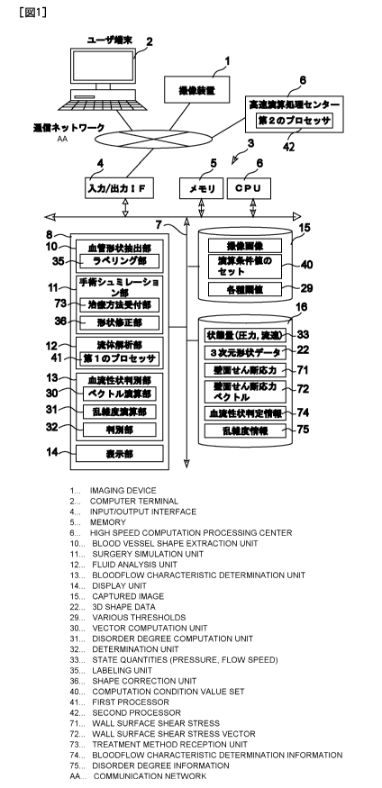
[0038]
Figure 1 shows a schematic diagram of an embodiment of the present invention.
Figure 2 depicts the graphical user interface of the vascular shape extraction
unit.
Figure 3 shows a flow chart of the vascular shape extraction unit.
Figure 4 illustrates a vascular image that explains the extraction of a
vascular shape image.
Figure 5 depicts the line-thinning step for vascular shapes.
Figure 6 illustrates labeling the name of blood vessels including the main
blood vessels.
Figure 7 shows processing of edging the extracted vascular shape.
Figure 8 shows a schematic diagram of overall shape of blood vessels in a
brain.
Figure 9 depicts the graphical user interface of the surgical simulation unit.
Figure 10 illustrates a schematic diagram of the surgical simulation unit.
8/54
CA 02850189 2014-03-26
Attorney Docket No. 08927498CA
Figure 11 depicts a simulation in the first surgical simulation mode.
Figure 12 depicts a simulation in the second surgical simulation mode.
Figure 13 depicts a simulation in the third surgical simulation mode.
Figure 14 illustrates an example of modification by applying the first
surgical simulation mode.
Figure 15 shows a schematic diagram of the fluid analysis unit.
Figure 16 shows a flowchart of processes performed by the fluid analysis unit.
Figure 17 depicts the graphical user interface of the fluid analysis unit.
Figure 18 explains the level of detail of mesh.
Figure 19 illustrates a diagram of the fluid shear stress.
Figure 20 illustrates a diagram of the fluid shear stress.
Figure 21 shows the global coordinate system for calculating the wall shear
stress.
Figure 22 shows the local coordinate system for calculating the wall shear
stress.
Figure 23 shows a graphical representation of superposition of shear stress
vectors on the three-
dimensional shape of blood vessels.
Figure 24 shows a graphical representation of the shear stress vectors and the
pressure which are
superposed on the three-dimensional shape of blood vessels.
Figure 25 explains the calculation of the flow disturbance index.
Figure 26 shows a diagram for interpreting the flow disturbance index.
Figure 27 shows the method for determining malignancy and benignancy with the
map of flow
disturbance index.
Figure 28 illustrates the method for determining wall thinning with the flow
disturbance index.
Figure 29 depicts the graphical user interface of the blood flow
characteristics determination unit.
Figures 30A to 30D show the displayed result of the effectiveness of the flow
disturbance index
on determining the aneurysm wall thinning process.
Figure 31 shows a schematic diagram of a surgical skill evaluation system of
another
embodiment of the present invention.
DETAILED DESCRIPTION OF THE INVENTION
[0039] Referring to figures herein, an embodiment of the present invention is
now described in
detail below. In the description hereinafter, a cerebral aneurysm is presented
as a cardiovascular
disease that may become a subject of diagnosis and treatment.
[0040] (System for diagnosing blood flow characteristics based on malignant/
benign blood
flow patterns)
As described above, the first main aspect of the present invention is to
provide a
diagnostic system for characterizing cerebral aneurysms. The present invention
associates the
9/54
CA 02850189 2014-03-26
Attorney Docket No. 08927498CA
morphology of shear stress vectors acting on the aneurysmal wall by blood
flow, with the
information on the luminal geometry, pathology, and wall thickness of aneurysm
in order to
categorize the vectors to either a "malignant blood flow pattern" which would
become a
potential risk of appearance of lesion or its growth or a "benign blood flow
pattern" which would
not become the potential risk. The morphology of the shear stress vectors
produced by the
simulation determines whether the vectors imply either a malignant blood flow
pattern or a
benign blood flow pattern. If it is a malignant blood flow pattern, it would
be a potential risk of
appearance or growth of a lesion, which may require considering a surgery
whereas if it is a
benign blood flow pattern, it would not be the potential risk, and may avoid a
risk of unnecessary
surgery.
[0041] (System for predicting treatment effect of blood vessel)
The second aspect of the present invention is to provide a system, e.g., a
system for
predicting the treatment effect of a cerebral aneurysm, which is determined to
have a malignant
blood flow pattern.
[0042] In other words, a method for determining the blood flow characteristics
to be malignant
or benign may be applied not only for pre-treated aneurysms, but also post-
treated aneurysms in
terms of predicting the treatment effect.
[0043] The surgical treatment for a cerebral aneurysm includes: 1) clipping,
2) coil
embolization, and 3) stent placement (flow-diverting stent).
[0044] The clipping method blocks the blood flow inside a cerebral aneurysm by
closing a
neck part of the aneurysm with a clip; i.e., it constructs a new vascular
morphology that does not
have the cerebral aneurysm. The coil embolization places a plural number of
coils in an
aneurysm to create thrombus in the lump for blocking the blood flow. The flow-
diverting stent
method places a mesh like object that is made of metal or other materials at
the neck of an
aneurysm to reduce the fluid flow through the lump and form a thrombus in it
for blocking the
flow.
[0045] Those treatment methods have a common feature of blocking the fluid
flow in a
cerebral aneurysm, and they reconstruct a new lump neck, i.e., a new vascular
shape by altering
the cerebral aneurysm artificially. A post-treatment complication may appear
as the
reconstructed vascular morphology gradually changes in the course of time. For
example, in a
case of the coil embolization treatment, the reconstructed lump neck may be
compressed into the
lumen by the fluid force, resulting in the reopening of a path between the
main blood vessel and
the lumen of lump, and thus a re-treatment is often required.
In such a case, first, the vascular morphology which is a three-dimensional
model created
by a computer is modified to create a new lump neck by a computer artificially
so that a
10/54
CA 02850189 2014-03-26
Attorney Docket No. 08927498CA
computer may construct a vascular morphology similar to one to be formed by
conducting an
actual surgery. Second, the morphology of the shear stress vectors acting on
the wall of the
newly created blood vessel is visualized by a simulation to apply the method
for determining if
the simulated blood flow pattern is malignant or benign so that the treatment
effect by the
surgery may be evaluated in advance. In other words, by applying the method
for determining
the malignant or the benign blood flow pattern, it is possible to predict a
direction of progress of
whether the vascular cells such as endothelial cells grow and adhere to the
part of the blood
vessel to reproduce the vascular tissue properly and regain the adequate
mechanical strength, and
those observations by simulation may contribute to the accurate prediction of
the treatment effect
to reduce a post-surgical complication and even death of a patient.
(Configuration of a system for determining the blood flow characteristics
diagnosis/predicting the treatment effect related to this embodiment)
Figure 1 shows a schematic diagram of a system for determining the blood flow
characteristics/predicting the treatment effect related to this embodiment.
The blood flow
characteristics determination/treatment effect prediction system corresponds
to the first and the
second aspects of the present invention, which has the following two
capabilities.
100461 (1) For considering if the subjective cerebral aneurysm has a
probability of an
appearance of lesion or its potential growth, the system determines
automatically whether the
target vascular site of a subject is either a benign blood flow pattern that
would not rupture the
cerebral aneurysm or a malignant (non-benign) blood flow that would rupture
the cerebral
aneurysm.
[0047] (2) When the cerebral aneurysm is to be surgically treated, by
conducting a surgical
simulation in order to predict the post-surgical blood flow, the system
determines automatically
whether the blood flow pattern would be either a benign blood flow that would
not develop a risk
of post-surgical complication or death, or a malignant blood flow that would
develop a risk of
post-surgical complication or death.
100481 In order to perform those functions, this system for diagnosing blood
flow
characteristics/predicting the treatment effect is installed at a site (e.g.,
a hospital) of a user such
as a doctor as shown in Figure 1, which equips with an image capture device 1
that takes images
of cerebral aneurysm and surrounding target vascular sites, a user terminal 2
with which a user
such as a doctor may operate the system, and a blood flow characteristics
diagnostic/treatment
effect prediction system server 3 which connects the image capture device 1
and the user
terminal 2 through a communication network (an in-hospital LAN, an out-of-
hospital WAN, or a
designated communication line).
11/54
CA 02850189 2014-03-26
Attorney Docket No. 08927498CA
[0049] Here, the image capture device 1 may be an instrument that acquires a
tomographic
image of the target vascular site, by using a Computed Tomography (CT)
scanner, an Magnetic
Resonance Imaging (MRI) system, a Digital Subtraction Angiography (DSA)
equipment, and
other medical instruments that acquire images of the target vascular site by
applying methods
such as the ultrasound Doppler and the near infrared imaging technology.
[0050] The aforementioned user terminal 2 may be a workstation consisting of a
standard
personal computer that runs a display software program such as a browser
capable of displaying
a graphical interface for establishing communication with a server of the
blood flow
characteristics determination/treatment effect prediction system.
[0051] The server 3 of the blood flow characteristics determination/treatment
effect prediction
system consist of a program storage unit 8 connected with a bus line 7 that
connects an
input/output interface 4 used for establishing communication with the
communication network, a
memory 5, and a CPU 6. The program storage unit 8 is configured with a
vascular shape
extraction unit (i-Vessel) 10 that produces a set of three-dimensional data of
a target vascular site
by using the image data acquired by the image capture device 1, a surgical
simulation unit (i-
Surgery) 11 that runs a surgical simulation by manipulating the three-
dimensional data, a fluid
analysis unit (i-CFD) 12 that computes the state quantities of the blood flow
at the target
vascular site, a blood flow characteristics determination unit (i-Flow) 13
that determines the
blood flow at the target vascular site whether it is benign or malignant, and
a display unit 14 that
has a user graphical interface produced by the system and a display screen to
show the image,
the analysis result and the determined outcome. There are two databases
connected with the bus
line 7: a simulation setting DB 15 that stores various setting information for
conducting the
simulation, and a simulation result DB 16 that stores outcomes of the
simulation and the analysis.
[0052] The components of the server 3 (the vascular shape extraction unit 10,
the surgical
simulation unit 11, the fluid analysis unit 12, and the blood flow
determination unit 13) are
actually constructed by computer software programs that are stored in a memory
area of a hard
drive of a computer, and the CPU 6 deploys the software programs from the hard
drive to the
memory 5 for executing the programs so that the components of the present
invention performs
their functions. A single computer may configure the server 3, or multiple
computers may
configure a distributed server as the server 3 as well.
[0053] In the above example, the server 3 of the blood flow characteristics
determination/treatment effect prediction system connects with a user terminal
2 in a hospital
through a communication network, and the server may be installed in a hospital
or in a high
speed process center 9 outside a hospital. In the latter case, the server is
preferably configured to
receive data and instructions from a number of user terminals 2 and image
capture devices 1 of
12/54
CA 02850189 2014-03-26
Attorney Docket No. 08927498CA
several hospital sites, and executes highly accurate fluid analysis using a
high speed processor,
and then feeds back the analysis outcome to the user terminals in each
hospital so that a user
such as a doctor may display the analysis outcome on screen for a patient and
other people on the
spot.
[0054] Referring to actual system operations, the capability of this blood
flow characteristics
determination/treatment effect prediction system is disclosed hereinafter.
(User graphical interface)
Figure 2 depicts the user graphical interface (GUI) 17 that is created by
employing the
display unit 14 of the server 3, and displayed on the user terminal 2. This
interface configures an
integrated interface function that operates the vascular shape extraction unit
(i-Vessel) 10, the
surgical simulation unit (i-Surgery) 11, the fluid analysis unit (i-CFD) 12,
and the blood flow
characteristics determination unit (i-Flow) together.
[0055] For example, Figure 2 shows an example when the vascular shape
extraction unit "i-
Vessel" 10, whose function is described below, is selected from the menu
located at the top of
the display screen. In a similar fashion, the interface (to be described
hereinafter) may switch
the function by selecting i-Surgery 11, i-CFD 12, or i-Flow 13.
[0056] There was no such integrated system in the prior art where simply
assembled individual
systems through separate interfaces were used. A conventional system is
anticipated to have
technological difficulties in practical clinical applications and
standardization of the analysis
conditions because: (1) a user has to employee a plural number of systems one
after another in
order to analyze a single case while spending at least several hours in a
workplace, and (2) each
system is designed to have large flexibility and versatility for engineering
work flows by
adjusting many and different parameters for setting up an analysis routine,
requiring user's
knowledge and skill to optimize the parameters, which may not be suitable for
medical
applications.
[0057] This embodiment of the blood flow characteristics
determination/treatment effect
prediction system needs to be used as part of medical treatment in an
extremely busy clinical
environment. Therefore, the time restriction imposed on a medical practitioner
and the
inconsistency of analytical conditions among different users and facilities
are major technical
issues to be solved. It also needs to consider the factor to be included that
a user, who is a
clinical doctor or a radiology technician, is not an engineer and unaware of
the knowledge of
fluid dynamics. The embodiment of this system integrates the system units and
a single interface
17 may execute an automatic control process, which eliminates the
technological issues
described above.
13/54
CA 02850189 2014-03-26
Attorney Docket No. 08927498CA
[0058] The embodiment of the system holds the optimal values of a group of the
operational
conditions for each application as a "module", which allows a user to carry
out an automatic
control process for a blood flow analysis required for a particular user's
application without
setting the group of the operational conditions.
(Vascular shape extraction unit)
Figure 3 shows a flow chart of the process steps of the vascular shape
extraction unit, and
Figures 4 to 9 illustrate vascular images that explain the process steps.
[0059] Step S1-1 inputs a set of image data, which an image capture device
acquired from the
target vascular site, in the DICOM format. Step S1-2 recognizes the
orientation of the image
(i.e., up, down, right, and left of the image) automatically or specifies the
orientation manually.
As described above, Figure 2 depicts the user interface of the vascular shape
extraction unit (i-
Vessel). The interface that recognizes the image orientation is the display
part 41 which is one
of four display parts 41 to 44 and located in the upper left corner of Figure
2. As the display
parts 42 and 43 show, when a three-dimensional vascular shape is visualized by
applying a
volume rendering method known to those skilled in the art, the orientation of
the blood vessel to
be displayed may be specified by pushing "Anterior (A)", "Posterior (P)",
"Left (L)", or "Right
(R)"of a button 18 so that the vascular image orientation is aligned with the
direction of
"Anterior (A)", "Posterior (P)", "Left (L)", or "Right (R)".
[0060] Next, on the same screen (Figure 2), an anatomical part is specified by
selecting, e.g., a
radio button 24 (Step S1-3). The anatomical part specified in this step is
used for labeling blood
vessels automatically in a step described hereinafter. For example, if a
cerebral aneurysm is
found in the right middle cerebral artery (MCA), "Right Anterior Circulation"
is selected.
Similarly, "Left Anterior Circulation", "Anterior Circulation", or "Posterior
Circulation" may be
also selected. The item 19 shown in Figure 3 indicates that the anatomical
part is stored in the
simulation setting DB 15.
[0061] Step S1-4 and following steps construct the three-dimensional vascular
morphology
(the three-dimensional shape data) by applying the threshold method or the
gradient method
combined with the region growing method (and other methods shown in Figure 2,
including: the
"Selection (where a user specifies a region of interest on screen to determine
a region containing
a targeted blood vessel from a three-dimensional structure that is extracted
by applying the
threshold method (or the gradient method))", "Connectivity (where the user
specifies the targeted
blood vessel to extract the targeted blood vessel by selectively taking
continuous voxels only)",
"Extension (which is an region growing method including the threshold method
(or the gradient
method) and the continuity of voxels, and adds blood vessels that need to be
used but deleted in
the blood vessel extraction Step )", and "Removal (where the user deletes the
blood vessels that
14/54
CA 02850189 2014-03-26
Attorney Docket No. 08927498CA
is not required)." For this purpose, Step S1-4 extracts a targeted vascular
region. The extraction
is executed by using e.g., the threshold or the gradient method.
[0062] Figure 4 shows an example of the extraction using the threshold method.
[0063] The threshold method uses, for instance, the absolute value or the
normalized relative
value of luminance. In this embodiment, the threshold setting unit 45 applies
the slider method
to select the histogram threshold value and changes the threshold value while
observing the
image on the display unit 42 to extract the characteristics that are intrinsic
to the vascular wall.
On the other hand, the gradient method calculates the luminance gradient of
the brightness from
the luminance distribution. After the extraction step, a user pushes the "Fix"
button 46 on the
screen shown in Figure 2 to activate the vascular shape extraction unit 10 to
remove noise from
the vascular surface by using the optimal threshold value for a given image
type (Step S 1-5),
and then construct three-dimensional shape data by dividing the region into
polygons to
complete extracting the targeted vascular region (Step S 1-6). Figure 4
depicts a schematic
diagram of extraction of vascular morphology in this step. These threshold
values are stored in
the simulation setting DB 15 (the item 29 shown in the figures attached
therein).
[0064] Then, a user presses the "Lesion" button 47 on the screen shown in
Figure 2 by using a
device such as the mouse to specify the lesion manually (Step SI-7). Step S1-8
executes the line
thinning routine to create the center lines of blood vessels. A user may
automatically perform the
line thinning routine by pushing the "Label" button on the screen of Figure 2.
There are various
well-known algorithms for the line thinning routine. Figure 5 shows the actual
line thinning step.
After acquiring the center lines, Step S1-9 divides the center lines into
multiple segments each of
which corresponds to a blood vessel. As shown in Figure 5, the segment-
division routine may be
performed by segmenting the center lines at vascular bifurcation points A, B,
C, D, etc. Figure 6
enlarges the segmented regions. In this figure, the segments (V1, V2, ...)
between two adjacent
bifurcations, A, B, C, ..., are called the blood vessel elements. Step1-10
obtains several cross-
sectional areas (as shown in Figure 6) that are perpendicular to the center
line of each blood
vessel segment, and then calculates the equivalent diameter of the cross-
sections for measuring
the shape 25 of each segment.
[0065] Step S1-11 labels the name of each blood vessel segment automatically.
Among the
several blood vessel segments V1, V2, V3, ..., the one that has the largest
median of the various
equivalent diameters calculated from the cross-sections 25 is determined to be
the main blood
vessel and labeled the name. (The mean value may not accurately represent the
main blood
vessel if there is an extraordinary large diameter due to a cerebral aneurysm
in the blood vessel.)
In this embodiment of the present invention, the labeling routine may be
executed automatically
as the anatomical lesion is specified. In other words, if the left anterior
circulation is selected,
15/54
CA 02850189 2014-03-26
Attorney Docket No. 08927498CA
the main blood vessel, (which is the blood vessel segment with the largest
median of the
equivalent diameters,) is labeled the "left internal carotid artery" whereas,
if a posterior
circulation is selected, the main blood vessel is labeled the "basilar
artery." These main blood
vessels are identified as the ones with the largest equivalent diameters.
Shape parameters other
than the equivalent diameter or their combinations may be applied for the
labeling routine. As
shown in Figure 3, the simulation setup DB 9 stores the anatomical lesion
information 19, the
names of the main blood vessel 20, and the names of branched blood vessels 21,
as related to
each other, which the labeling unit 35 of the vascular shape extraction unit
10 uses for the
automatic labeling routine.
[0066] Thus, Step S1-11 performs the aforementioned labeling routine for the
main blood
vessels V2, V3, ..., followed by tracking the branched blood vessels
individually to label the
names of blood vessels at each branch by identifying them according to the
information stored in
the DB 9. In the embodiment of the present invention, labeling the branched
blood vessels is
limited to carry out down to a 5 to 10 sub-layers from the main blood vessels.
As described
herein, once the name of the main blood vessel 20 is determined according to
the information
DB 19 of each anatomical lesion, the labeling routine of the branched blood
vessels may be
automatically performed by following the relation between the main blood
vessel name 20 and
the branched blood vessel names 21, which is stored in the database 9.
[0067] Next, Steps, S1-12 and S1-13, after labeling, construct the cross-
section of a blood
vessel by making the inlet and the outlet of the blood vessel perpendicular to
the central line
based on the orientation (the vertical and the horizontal directions) of an
image and the
anatomical lesion specified as the targeted blood vessel that is selected in
Step S1-2. Figure 7
illustrates the cross-sectional construction. Step S1-4 automatically outputs
polygon data as the
three-dimensional shape. At the same time, the shape data 22 of each blood
vessel (which is
called the labeling information 23), which are labeled automatically, are
calculated and recorded
into the simulation result DB 16 automatically (Figure 3). A user may confirm
if the process is
appropriate by checking the interface 17 displayed on screen. There may be a
case where
labeling is not processed properly in the automatic process. For example,
there is a case where a
patient with a congenital vascular malformation would not have a blood vessel
at a
corresponding location. In such a case, the diagnostic simulation system may
be configured so
that clicking on the falsely labeled blood vessel changes the name of the
selected blood vessel.
The names 20 and 21 of the setting DB21 may be also changed at this time.
After the manual
process, clicking the <End> button outputs the result automatically and
overwrites to update
DB15 and DB16. The name of a file output is configured according to the
patient ID that may
be extracted from the DICOM header information with which the file format may
be obtained,
16/54
CA 02850189 2014-03-26
Attorney Docket No. 08927498CA
which allows a user to eliminate inputting the file format manually. The
surgery simulation unit
11, the fluid analysis unit 12, and the blood flow characteristics
determination unit 13 have the
same file name protocol as described hereinafter.
100681 Figure 8 overviews a list of the names of cerebral blood vessels.
Figure 8 is for the
anterior and the posterior circulations. For example, the anterior
communicating artery, a lesion
where cerebral aneurysm often appears, runs across the left and the right
anterior circulations,
and hence it is necessary to target the overall anterior circulation for
analysis.
(Surgery simulation unit)
Figure 9 depicts a schematic diagram of the user graphical interface 17 of the
surgery
simulation unit 11; Figures 10 shows the operational flow chart of the surgery
simulation unit 11;
and Figures 11, 12, and 13 illustrate the surgical modes. Figure 14 is a
schematic diagram of the
shape modification unit 34 that modifies the three-dimensional morphological
unit for the
surgical simulation.
10069] In this example, the interface 17 shown in Figure 9 allows a user to
select a surgical
mode from the three predetermined modes, "Clipping/Coiling" 50 as the first
surgical mode,
"Stenting" 51 as the second surgical mode, or "Flow-diverting" as the third
surgical mode. With
this surgical mode selection, the surgical simulation unit 11 may produce the
optimal vascular
shape to reproduce the post-surgical blood flow.
[00701 In the aforementioned three modes, the first surgical simulation mode
cuts out a lesion
and reconstructs the vascular wall surface (Clipping/Coiling); the second
surgical simulation
mode reconstructs the vascular surface by smoothing the uneven surface of the
lesion (Stenting);
and the third surgical simulation mode places a lattice like object on an
arbitrary vascular cross-
section (Flow-diverting stent).
100711 The vascular shape modification method (the item 37 in Figure 15)
corresponding to
the first surgical simulation mode is a program group 50 (consisting of
<Positioning>,
<Removal>, <Recon>, <Shaping>, and <Label>) that simulates surgical clipping
or coil
embolization that completely closes an aneurysm lumen, in order to conduct a
pre-surgical
estimation of the fluid force which exerts on the neck of aneurysm formed by
the surgery. The
vascular shape modification method corresponding to the second simulation mode
is a collection
of programs 51 (consisting of <Positioning>, <Fitting>, <Shaping>, and
<Label>) which
simulates a stent placement that enlarges a vascular stenosis due to
arteriosclerosis by employing
a medical device such as a stent to conduct a pre-surgical estimation of the
fluid force which
exerts on the lesion formed by the surgery. The vascular shape modification
method
corresponding to the third surgical simulation is a collection of programs 52
(consisting of
<Positioning>, <Porosity>, <Shaping>, and <Label>) which simulates a treatment
of cerebral
17/54
CA 02850189 2014-03-26
Attorney Docket No. 08927498CA
aneurysm by using the flow-diverting stent to estimate the effect of reducing
the flow through
the aneurysm.
[0072] This simulation is conducted by actually modifying the three-
dimensional vascular
shape data, and the surgical simulation unit has the treatment receiving unit
73 and the shape
modification unit 34 as shown in Figure 15. Below is a description of the unit
configuration
along with their processing operations. The selectable surgical modes (which
are the first to the
third surgical simulation mode in this example) and the concrete methods for
modifying the
vascular morphology defined in relation to the surgical modes are stored in
the simulation setting
DB 15 as shown the items 36 and 37 in Figure 15.
[0073] First, on the screen of the user graphics interface 17, a user pushes
the <Surgery>
button 11 to display the vascular morphology that is created by the vascular
shape extraction unit
through the browser display of the user terminal 2. (Step2-0: the display part
54 on the upper left
corner of Figure 9.) When a user activates the first surgery simulation mode
(the item 50 in
Figure 9) on the interface 17, the treatment receiving unit 73 loads the
vascular shape
modification method 37 (which is the program group 50 consisting of
<Positioning>,
<Removal>, <Recon>, <Shaping>, and <Label>) from the setting DB 15, and the
user selects a
lesion by using the <Positioning> (Step 2-1). If the user selects
<Positioning>, the modified
lesion specification unit 38 displays the specified region on the user
interface 17. (The display
part of the upper right comer of Figure 9.) Because the three-dimensional
shape data are
polygon data that are a collection of minute triangles that configure the
surfaces and the ends
blood vessel surface and the ends of blood vessels, the specified region may
be enlarged or
shrunk for the purpose of the surgical simulation. If the user selects
<Removal>, it cuts out the
triangle element selected by the polygon moving unit 39 shown in Figure 15
(Step S2-2).
Pushing the <Recon> button reconstructs a surface on the dissected part by
using polygons.
Pushing the <Shaping> button activates the modification specification unit 38
and a user may
activate the modification specification unit 39 and operate the mouse to carry
out smoothing the
reconstructed surface (Step 2-3), and then <Label> defines labeling the new
surface (the
Labeling unit 35) (Step S2-4). The surface reconstruction may be executed by
calculating the
center of mass of the dissected region and connecting it with the vertexes of
the triangle elements
at the edge of the dissected region. For smoothing the surface, a user freely
move the center of
mass of the triangle to the normal direction of the outer (or inner)
peripheral direction of the
dissected surface by pushing the mouse wheel button, i.e., shifts the center
of mass which is the
unique point of the triangle to a different location to distort the triangle
artificially. A shape with
an acute angle by moving the center of mass may be smoothed out simultaneously
(by using the
aforementioned units 38 and 39).
18/54
CA 02850189 2014-03-26
Attorney Docket No. 08927498CA
[0074] With the user interface shown in Figure 9, a user uses the display
parts 55 and 56,
<<Post-surgery>>, at the left and right bottom to display an image of lesion
after surgery and
conduct surgical simulations using the program group. After completing the
labeling step,
<End> finalizes the shape, and similar to the vascular shape extraction unit,
polygon data are
stored automatically, updating the simulation result DB 16 (Step2-13: Updates
of labeling
information 23 and three-dimensional shape data 22). For comparing a plural
number of surgical
simulations by repeating the previous steps, there are the display parts of
<<Post-surgery>> at
the right bottom 55 and at the left bottom 56 <<Post-surgery #1 and #2>>. (The
comparison
display part of the present invention).
100751 Figure 11 depicts a diagram of an example of vascular shape
modification in the first
surgical simulation, and Figures 14A and B show the three-dimensional shape
before and after
the simulation (corresponding to before and after a treatment by clipping). As
shown herein,
deleting polygons that configure a shape of the cerebral aneurysm may
reproduce three-
dimensional vascular shape which exhibits the blood flow characteristics after
conducting the
clipping treatment. Therefore, a user may arbitrarily adjust the cross-
sectional shape of a
cerebral aneurysm that is constructed by a clipping treatment or a coil
embolization in order to
simulate and analyze the post-surgical blood flow.
[00761 In the second surgical simulation mode 51, similar to the
aforementioned simulation,
using <Positioning>, the lesion is selected and scaled-up and down (Step S2-5,
the display part
55). In the next step, with <Fitting>, the center of gravity of the lesion is
calculated, and using
the center as the starting point, a polygon is moved in the normal direction
to the vascular wall,
and a polynomial fitting interpolates the lesion morphology (Step S2-6). Then,
with <Shaping>,
smoothing out of the lesion is executed using the mouse (Step S2-7), and
finally, a method
similar to the aforementioned first surgical simulation performs the labeling
routine (Step S2-8).
Figure 12 illustrates a diagram of an example of the shape metrological
modification with the
second surgical simulation.
100771 In the third surgical simulation mode 52, a user uses <Positioning> to
construct a new
surface inside the three-dimensional vascular morphology (Step S2-9). Next,
for a specified
surface, <Porosity> defines a lattice-like object (Step S2-10), smoothing out
the surface by
applying a method similar to the aforementioned method (Step S2-11), and
executes labeling
(Step S2-12). The lattice-structured object used for the vascular shape
modification method 37
(Figure 15) attempts to simulate the flow-diverting stent. The lattice-
structured object is a
homogeneous porous media that a user may adjust the aperture ratio by using a
pull down menu.
The user may also create an inhomogeneous media by adjusting the aperture
ratio and the shape
of a pours media. Figure 13 shows a diagram of an example of shape
modification by the third
19/54
CA 02850189 2014-03-26
Attorney Docket No. 08927498CA
surgical simulation mode. In this figure, the lattice-like object is the item
25. The blood flow
simulation using the porous media may also simulate the blood flow after
conducting a coil
embolization surgery. The aforementioned coil embolization assumes a complete
embolization
in the lump. This actually corresponds to a condition of adequate embolization
in the lump when
the time subsequently elapsed after surgery. On the other hand, there is blood
flow in the coil
until it is completely blocked. Whether the blood flow may be simulated or not
is crucial to
determine the coil filling ratio (which is the volume ratio of the coil to the
lump). The above-
described flow-diverting stent uses the porous media as a two-dimensional
structure, which may
extend to the three-dimensional structure to simulate the condition
immediately after a coil
embolization. In other words, it is possible to add a function of simulating
the coil filling ratio
by using the aforementioned <Porosity> to place a porous media in the lump and
simulate the
coil filling ratio with the aperture ratio.
(Flow analysis unit)
In the next step, the fluid analysis unit 12 obtains the blood fluid velocity
and pressure
(which is the state variable 33) at each unit area of the target vascular site
using the three-
dimensional shape data of the target vascular site created by the vascular
shape extraction unit 10
(and the surgical simulation unit 11).
[0078] Figure 16 is the flow chart of processes that the fluid analysis unit
12 executes, and
Figure 17 shows an example of selecting "CFD" 12 from the menu of the user
graphic interface
17.
[0079] In Step S3-1, the fluid analysis unit 12 selects and reads the vascular
shape data for
calculation from the three-dimensional shape data of the target vascular site
which the vascular
shape extraction unit 10 (and the surgical simulation unit 11). The selected
data is displayed on
the display parts 58, 59, and 60 which locate the upper left corner of the
interface 17 as shown in
Figure 17. In this example, the display unit 58 displays the shape data of Pre-
Surgery, the
display part 59 displays the shape data of Post-Surgery#2, and the display
part 60 displays the
shape data of Post-Surgery#1.
[0080] In the next step, Step S3-2, a user selects a "module." As shown in
Figure 17, for
selecting a "module", there are three buttons displayed on the user graphic
interface 17 and
available for selection: "On-site" 26, "Quick" 27, and "Precision" 28.
[0081] The system configures a default set of mathematical operation values 40
(Figures 1 and
16) to execute computations with appropriate condition and precision after a
user selects a
module from the three modules. Considering the time restriction in the
clinical practice and the
user's non-expertise of the fluid analysis, this configuration of integrating
the analysis conditions
is realized to fulfill the demand from the workplace, and to achieve
reproducibility and
20/54
CA 02850189 2014-03-26
Attorney Docket No. 08927498CA
standardization of the analysis conditions. The mathematical operational
condition for "On-site"
adopts a steady flow analysis. The blood flow is an unsteady flow which is
called the pulsatile
flow produced by the cardiac pulsation. Calculation of an unsteady flow
executes an iterative
calculation that converges the solution at each time interval for the time-
varying flow, which
requires a large calculation load on the mathematical operation unit. On the
other hand, the
steady flow is not necessarily quite different from the pulsatile flow. In
particular, the cerebral
blood vessel is a region where the Reynolds number of the blood flow is
relatively small, whence
the blood flow is laminar in the pulsatile period, and does not have the
transient vortex observed
in turbulence with a large Reynolds number. In other words, the blood flow in
the pulsatile
period has a strong similarity in the variation of flow rate. Therefore, if a
blood flow
corresponding to the time-averaged flow may be reproduced, it is possible to
understand the flow
patter as the pulsatile flow. The On-site module is an analysis method that is
supported by the
experimental and analytical data of this approach.
[0082] On the other hand, "Quick" and "Precision" have the set of mathematical
operation
condition values 40 for the pulsatile flow. Unlike "Quick", "Precision" sets a
condition with a
capability of dealing with a change from the laminar to the turbulent
pulsatile flow. The set DB
15 pre-stores various conditions, including the level of detail of mesh, the
physical property of
blood, the wall boundary condition, the inlet boundary condition, the outlet
boundary condition
and the discretizing condition, as the set of mathematical operation condition
values 40. It would
often take several days for a single fast processor to complete analyzing
"Precision." In the
embodiment of the present invention, the first processor 41 of the fluid
analysis unit 12 executes
the relatively light process of the On-site while the second processor 42 of a
fast processing
center 9 in a remote area carries out the heavy process of the Precision. In
other words, the
precision module is configured in order to perform the following job flow: the
data for
processing the Precision task is automatically transferred to the process
center outside a hospital
through a telecommunication network, parallel-processed with a plural number
of fast processors,
and then retuned the analysis outcome to the hospital through the network.
[0083] In Step S3-3 and following steps, a user pushes the Run button 62 of
the interface 17
shown in Figure 17 to select the set of the mathematical operation values 40
for a selected
module, and automatically performs the calculation. Step S3-3 divides the
target vascular site
into a plural number of triangles of the finite element method based on the
three-dimensional
shape data. The embodiment of the present invention creates a mesh structure
using the level of
detail of mesh for the blood vessel size based on the vascular labeling
conducted by the vascular
shape extraction unit 10. In other words, in this embodiment, the set of the
mathematical
operational condition values 40 stores the level of detail of mesh for the
mesh dividing in
21/54
CA 02850189 2014-03-26
Attorney Docket No. 08927498CA
relation to the blood vessel name, or dynamically determines the level of
detail of mesh
according to the vascular cross-section. Thus, this system may read the level
of detail of mesh
out of the set DB 15 according to the labeling and use it for the mathematical
operation. That is,
each level of detail of mesh may be determined according to the module
selected and the
vascular type.
[0084] Figures 18A and 18B illustrate an example of varying the level of
detail of mesh per
blood vessel. In this example, the resolution for the ophthalmic artery of
diameter 1 mm is set to
be higher than the artery inside diameter 5 mm.
[0085] Dmesh Of this embodiment of the present invention is defined as
follows.
[0086] Dmesh =Dbase xKscale X Kmodule
.....s
where Dmesh i the level of detail of mesh (which is the representative
diameter Dmesh i -n this
embodiment), Dbase is the size of the base mesh (which is a constant
independent of the scale
factor), Kscale is a scale factor which varies according to the vascular size,
and Kmodule _s i a scale
factor which varies according to the module selected.
[0087] An ordinary finite element analysis does not consider the scale factor
defined above but
determined the mesh size by the base mesh alone. For this reason, the prior
art was unable to
include the variation of each vascular diameter. However, the embodiment of
the present
invention may overcome the technological issue of the prior art.
[0088] An example is described hereinafter. In the example, the fluid analysis
unit 12
calculates the equivalent vascular diameter D by using the blood vessel
volume, the length of the
central line of the blood vessel, and the approximate cylinder of the blood
vessel for quantitating
the vascular size.
[0089] 1) For using the On-site, Quick module,
Dbase = 0.1 mm
Kscale = 0.2 (if D (1.5mm)
Kscale = 1.0 (if D > 1.5mm)
Kmodule = I
(In other words, in this module, the mesh size of the arteriole having the
equivalent diameter D
less than 1/5 mm is only refined to 1/5 of the base mesh.)
[0090] 2) For using the Precision module,
Dbase = 0.1 mm
Kscale = 0.2 (if D < 1.5mm)
Kscale =1.0 (if D ? 1.5mm)
Kmoduie =0 .5
(In this example, Kmodute=0.5 and the mesh is refined overall.)
22/54
CA 02850189 2014-03-26
Attorney Docket No. 08927498CA
With the above method, the mesh size would change abruptly at a vascular
branch. The
discontinuous change of the mesh increases the morphological distortion, which
may lead to
degrade the convergence of computation. For overcoming this computational
issue, the
embodiment of the present invention creates the mesh by using the
aforementioned method, then
providing the upper limit for the mesh distortion and repeatedly carries out
the smoothing
process so that the maximum distortion settles within the threshold value.
[0091] The analysis method of the prior art was unable to change the mesh size
dynamically
for the vascular size, and hence it used the same level of detail of mesh for
both large and small
blood vessels. Although a mesh size which is adequate enough to analyzing a
large blood vessel
conversely shows poor analytical precision for a small blood vessel whereas a
mesh size
adequate enough to analyzing a small blood vessel creates the level of detail
of mesh
unnecessarily to prolong the time for analysis, the present invention solves
the technological
problem.
[0092] The following steps S3-4 to S3-8 read out the set of mathematical
operation conditions
40, which stores the physical property of blood, the boundary condition, and
the analysis
condition, from the aforementioned set DB 15, and Step S3-8 executes the
mathematical
operation based on these conditions. Specifically, the fluid analysis unit 12
solves a second
order nonlinear partial differential equation that describes the motion of
fluid, called the Navier-
Stokes equation, by applying the finite element method, and obtains the fluid
velocity and
pressure at each mesh. In this case, the solution of the finite element method
(the fluid velocity
U and the pressure P) is obtained in the three directions, the X-global, the Y-
global, and the Z-
global, of the global coordinate frame.
[0093] In the mathematical operation conditions 40, the physical property of
blood includes
the viscosity and density. The boundary condition is the fluid conditions at
the inlet and outlet of
the targeted lesion for analysis and the fluid condition applies the
statistical mean values of fluid
velocity and pressure.
[0094] Although the set condition selects a default condition automatically
according to the
selected module as described above, this embodiment of the present invention
preferably has an
additional capability of manually inputting a condition into the fluid
analysis unit 12 prior to
execution of the mathematical operation.
[0095] After the calculation starts automatically, Step S3-10 displays the
residual and the
calculation repeats until the result satisfies the predetermined converging
criterion. If the
residual does not satisfy the predetermined converging criterion even after
repeating the
maximum number of iterations permitted, the calculation is determined to be
non-converging
(Step S3-11). In this case, optimization of the mesh distortion will be
carried out (Step S3-12),
23/54
CA 02850189 2014-03-26
Attorney Docket No. 08927498CA
and the calculation will be resumed. Once the residual reaches within the
predetermined
converging range, the completion of calculation is displayed (Step S3-13). The
calculation result
(which is the state quantities (U and P)) is automatically stored in the DB 16
in a similar manner
described above.
[0096] The mathematical operation adopted herein is not only for the finite
element method
but also for numerical analyses of the differential equation of fluid flow,
such as the finite
volume method and the finite difference method.
(Blood flow characteristics determination device)
There is a software program that enables a computer to perform the following
functions
and installed in the blood flow characteristics determination unit 13. That
is, as shown in Figure
1, the blood flow characteristics determination unit 13 has the wall shear
stress vector calculation
unit 30 that obtains the fluid shear stress and its vector (the "wall shear
stress vector",
hereinafter) exerted on the vascular wall by the blood flow by using the fluid
velocity and
pressure for each mesh calculated by the fluid analysis unit, the flow
disturbance index
calculation unit 31 that obtains the numerical index (i.e., the flow
disturbance index) from the
wall shear stress vector, and the determination unit 32 that determines the
blood flow
characteristics at each mesh according to the flow disturbance index.
[0097] Figures 19 and 20 depict schematic diagrams to obtain the shear stress
vector r (x, y, z)
based on the fluid velocity U and the pressure P obtained at each mesh in the
wall shear stress
vector calculation unit 30.
[0098] As shown in Figure 19, the wall shear stress is the fluid viscous force
exerting on an
area element of the vascular lumen in the parallel direction, and the wall
shear stress vector is the
vector expression of the stress including the direction of the force. The
acting direction of the
wall shear stress vector is perpendicular to the pressure which is the fluid
force exerting on the
center of the mass of the area element in the normal direction of the area.
For describing the figure, it is necessary to understand the transformation
from the global
coordinate system to the local coordinate system. In other words, the pressure
P and the velocity
U for obtaining the shear stress vector are calculated in the global
coordinate system whereas the
shear stress force at a location on the vascular wall is in the tangential
direction of the wall
surface, and calculation of its magnitude requires transforming the pressure
and the velocity
from the global coordinate system to the local coordinate system of the
vascular wall.
[0099] Here, as shown in Figure 21, the global coordinate system of this
system of this
invention is a unique reference coordinate to show a universal position of
mesh nodes forming
the vascular surface and lumen. The finite element method and the finite
volume method
represent the subject for calculation as a set of geometrical elements (such
as triangle elements,
24/54
CA 02850189 2014-03-26
Attorney Docket No. 08927498CA
tetrahedral elements, and hexagonal elements). Each element has a vertex
called the node, and
the position information is retained using the global coordinate system such
as (X1 g, Y1 g, Z g),
(X2g, Y2g, Z2g), and (X3g, Y3g, Z3g)=
[00100] As shown in Figure 22, the local coordinate system is a frame of
reference that is
locally defined for each triangular element (or polygon) configuring the
vascular surface, and
usually, the center of gravity of the triangle is the origin and one axis
(i.e., the Z axis) is taken to
be the normal vector to the area. The local coordinates of each point of
contact of the area
element are (X1 1, Yli, Zli), (X21, Y21, Z21), and (X31, Y31, Z31). The
position in the global
coordinate system and that of the local coordinate system may be mutually
transformed by using
the position of the center of mass of the triangular element and the direction
of the area normal
vector.
[00101] A method for obtaining the wall shear stress is explained below.
[00102] The first step is calculation of the velocity and the pressure at each
node in the global
coordinate system by using the output from the fluid analysis unit 12 (i-CFD).
The next Step is
specification of a triangle where the wall shear stress vectors to be
calculated. The local
coordinate system is configured for the specified triangular element. In the
local coordinate
system, a position G where the shear stress vector is calculated is
determined. (For each
triangular element, the distance from the wall is usually kept to be constant,
e.g., 0.1 mm inward
from the wall.) The fluid velocity at the position G is zero because it
locates on the wall surface
as shown in Figure 20.
[00103] Letting Ut be the fluid velocity at a position from the position G by
a distance t, which
is assumed to be very small compared with the boundary layer thickness, in the
normal direction
(i.e., the Z direction of the local coordinate system), the fluid velocity Ut
is approximately
proportional to the distance n from the point G, and may be expressed as
Un = n = dUt/dZ.
[00104] According to the action-reaction law, the resistive force against
moving the point at
distance n at this fluid velocity has the same magnitude of the force that
fixes the point, and both
of them are proportional to the fluid velocity Ut and also inversely
proportional to the distance Z.
Therefore, the force t per unit area at the point G in contact with the fluid
becomes
= 11, = dUt/dZ.
That is, the wall shear stress vector is the product of the viscous
coefficient and the rate
of changing the velocity vector parallel to the area element in the normal
direction. There are
several methods for calculating the changing rate of the velocity vector
parallel to the area
element in the normal direction. For example, it is possible to obtain the
velocity at each point
of a plural number of points on the Zl axis by interpolating the surrounding
velocity vectors. In
25/54
CA 02850189 2014-03-26
Attorney Docket No. 08927498CA
this case, because the distance between the individual surrounding velocity
vector and the
candidate point is different, the interpolation requires a weight function of
the distance. Since
the surrounding velocity vector is expressed in the global coordinate system,
the velocity vector
after interpolation is expressed in the local coordinate system to calculate
the velocity component
parallel to the surface at each candidate point. When the changing rate in the
normal direction is
to be calculated later, the first order approximation using a single candidate
point near the wall
may be applied, or a higher order of mathematical differentiation where a
polynomial
approximation using a plural number of candidate points near the wall may be
calculated
followed by mathematical differentiation may be executed.
[00105] For calculating the aforementioned changing rate from velocity U(Xg,
Yg, Zg) in the
global coordinate system, the following approach may be applied: decomposing
the velocity
vector at the distance t in the local coordinate system (X1, Y1, Z1), and
solving r = g = dUt/dZ in
the coordinate (Xl, Y1) parallel to the wall surface in each local coordinate
system (the Z
component is zero).
[00106] In other words,
r(X1) = g = dUt(X1)/dZ,
and
r(Y1) = g = dUt(Y1)/dZ
are calculated.
The vector values r (Xl, Y1) in all local coordinate systems form the wall
shear stress
vector. Therefore, on an area element in contact with the wall surface, the
wall shear stress
vector has the x and the y components defined by the x and the y directions of
the area element.
[00107] Figure 23 illustrates the shear stress vectors along the vascular wall
by using the
method described above and attached to a three-dimensional shape model.
[00108] It should be noted that there is force exerting on the vascular wall
in the tangential
direction, and there is also the pressure P in the direction of collision
against the wall. The
pressure at the point G in the global coordinate system will be in the Z1
direction of the local
coordinate system after the coordinate transformation. Figure 24 superposes
the colorized
pressure values on Figure 23. Area with lighter color indicates higher
pressure.
[00109] The wall shear stress 71 and its vector 72 obtained for each polygon
in this manner are
stored in the simulation result DB 16.
[00110] (Flow disturbance index calculation unit)
Next, the flow disturbance index calculation unit 31 obtains the flow
disturbance index
by calculating the index numerically from the morphology of the wall shear
stress vectors. The
flow disturbance index is a numerical index that indicates the degree of
alignment of the wall
26/54
CA 02850189 2014-03-26
Attorney Docket No. 08927498CA
shear stress vector at a mesh with the surrounding wall shear stress vectors.
In other words, the
flow disturbance index is obtained by calculating each angle 8 between the
wall shear stress
vector of a mesh targeted for obtaining the flow disturbance index (the
"targeted mesh",
hereinafter) and the wall shear stress vector of another mesh adjacent to the
targeted mesh.
1001111 Figure 25 illustrates an example of the relationship among the shear
stress vector at an
area element G (which is shown as a point for illustrative purpose) and the
shear stress vectors at
surrounding eight area elements. In this example, the magnitudes of the shear
stress vectors are
not relevant but the directions, and hence they are expressed as unit vectors
to extract the
directions only. Although, strictly speaking, the area elements form a three-
dimensional
configuration, adjacent elements are very close and hence may be treated as a
two-dimensional
configuration. In other words, each wall shear stress vector is projected onto
a two-dimensional
plane for processing. Figure 25 illustrates a mapping of the area element G
and its surrounding
area elements onto the two-dimensional coordinate system.
[00112] In the embodiment of the present invention, the divergence ("div",
hereinafter) and the
rotation ("rot", hereinafter) operations of the vector analysis are calculated
for a targeted mesh in
order to obtain-a-numerical values of the morphology of wall shear stress
vectors.
That is, the components of the vector field t (i.e., the shear stress vector)
of a mesh in a
three-dimensional space may be expressed as the components at a point G(x, y)
which is mapped
into the two-dimensional orthogonal coordinate system (x, y), which is given
by the following
equation.
[00113] T(G) = (rx(x, y), ry(x, y))
[00114] Whence the "scalar field div t", which is called the "divergence of
the vector field r" is
defined by the following equation:
[00115] div r = arx /ax + ary /ay
Similarly, the "scalar field rot r", which is called the "rotation of the
vector field r" is
defined by the following equation:
[00116] rot r = ary /ax - au( /ay
Figure 26 depicts the relationship between the morphology of the wall shear
stress
vectors and the values of the aforementioned "divergence (div)" and "rotation
(rot)." The
morphology of the wall shear stress vectors has four categories: 1) parallel,
2) confluent, 3)
rotational, and 4) divergent.
[00117] For the parallel, (div, rot) = (0, 0), for the confluent, (div, rot) =
(negative value, 0), for
the rotational, (div, rot) = (0, positive or negative value), for the
divergent, (div, rot) = (positive
value, 0). The degree of confluent and divergent types can be quantified by
the div-value. That
is, for the confluent type, if its negative value increases in the negative
direction, the degree of
27/54
CA 02850189 2014-03-26
Attorney Docket No. 08927498CA
confluence also increases; and for the divergent type, if its positive value
increases in the
positive direction, the degree of divergence also increase. For the rotational
type, depending on
the direction of rotation, both the positive and the negative values appear
but their absolute
values may be a numerical parameter. If the flow disturbance index is defined
by the vector
quantity D = (div, rot), the magnitude may be used as the flow disturbance
index, i.e., as the flow
disturbance becomes smaller, the wall shear stress vector tends to align along
with other shear
stress vectors at surrounding meshes (becoming closer to the parallel type).
[00118] If there is a flow disturbance index, its magnitude (as compared with
the threshold
value) may be used to determine whether the blood flow is malignant or benign,
and furthermore,
comparing the numerical value of div with that of rot, the blood flow may be
categorized to the
confluent, the rotational, and or divergent type, which may be used to
determine whether the
aneurysmal wall is in a type of either atherosclerosis or wall thinning.
[00119] Figure 27 shows a map of the numerical values of div and rot. Namely,
this figure
shows the flow disturbance index (div, rot) for a typical example of the shear
stress vector. Here,
the typical example is an ideal mathematical pattern but not constructed from
a set of
experimental data. As described above, the magnitude of the shear stress
vector is converted to a
unit vector having the norm one, and thus the flow disturbance index is
already normalized,
which makes comparison among different patients possible. In other words, the
embodiment of
the present invention is capable of obtaining the index that may be evaluated
as the absolute
value of the flow disturbance index described above.
[00120] This embodiment of the present invention may combining the flow
disturbance index
with the pressure on the targeted mesh as the weight coefficient to make the
flow disturbance
index for accurately determining the damage to the targeted blood vessel
caused by the blood
flow pressurizing the vascular wall. This embodiment uses the normalized
pressure, i.e., the
pressure index even when the pressure is used. This embodiment calculates the
pressure index
by each pressure divided by the mean pressure in the lump. (The calculation is
a multiplication in
this case).
[00121] By the above argument, in a case where, for example, the divergence
type of the shear
stress vectors is formed by the blood flow collision, increase of the local
wall pressure may be
observed from the collision of the main flow but increase of the wall pressure
may not be
observed from the collision of the secondary flow separated from the main
flow. In such a case,
combining the morphology of the shear stress vectors with pressure may refine
the estimation,
and in particular effectively estimate a thinning part of cerebral aneurysm.
In other words, there
are several methods for indexing the pressure, and hence the method for
overlaying the index
28/54
CA 02850189 2014-03-26
Attorney Docket No. 08927498CA
with the flow disturbance which is calculated from the shear stress vector may
take
multiplication, division, or the power law, and multiple methods are possible.
(Determination unit)
The determination unit 32 determines if the flow at each mesh is malignant or
benign
according to the flow disturbance index of each mesh which is calculated by
the flow disturbance
index calculation unit 32. The conditions of the wall shear stress vector are:
parallel to the
surrounding wall stress vectors, confluent to the directions of the
surrounding wall shear stress
vectors, rotational along with the surrounding shear stress vectors, or
divergent from the
directions of the surrounding wall shear stress vectors. If the wall shear
stress vector at a mesh is
in the parallel condition, the blood flow characteristics at the mesh is
determined to be benign
whereas if the blood flow characteristics at a mesh is confluent, rotational,
or divergent, the
blood flow characteristics is malignant (not a benign flow) at the mesh.
[00122] Furthermore, the value of flow disturbance index in a malignant flow
may be used to
evaluate the degree of risk. The embodiment of the present invention estimates
that the risk is
higher when the value of flow disturbance index increases positively or
negatively. Here the
index used as the threshold value is determined in such a way that the
inventors of the present
invention trace the time variation of the wall shear stress vectors in a
cerebral aneurysm of a
patient and determine the threshold value empirically based on the correlation
between the wall
shear stress vectors and the actual vascular tissue of the cerebral aneurysm
sampled from the
patient, but the value may be changed in some case. The threshold value may
further be set
stepwise and the condition of the wall shear stress vector is set in several
steps in order to
determine the degree of the benign flow and/or the malignant flow.
[00123] As disclosed above, the embodiment of the present invention may
categorize the
condition of the vascular wall thickness (i.e., the lesion tendency) according
to the state of the
wall shear stress vector. If the wall shear stress vector is parallel, the
wall thickness is at a
normal level. If the wall shear stress vector is confluent or rotational,
there is a tendency where
blood cells and protein in blood plasma are easily deposited, and the blood
vessel turns to be an
atherosclerosis type and increases the wall thickness. Furthermore, if the
wall shear stress vector
is divergent, it is a wall thinning type where damage and reproduction fault
of endothelial cells
cause a tendency in which blood cells infiltrate, proliferate, and migrate
into the blood vessel,
degrading the mechanical strength of the vascular wall, and as a result,
decreasing the wall
thickness around the lesion. Figure 28 is a schematic diagram that shows the
concept of the
atherosclerosis-type and wall-thinning-type lesion.
[00124] Figure 29 shows the user graphic interface 17 that displays the result
of the blood flow
characteristics determination unit 13 (the vector operation unit 30, the index
calculation unit 31,
29/54
CA 02850189 2014-03-26
Attorney Docket No. 08927498CA
and the determination unit 32). As described previously, a user pushes the
<Load> button of the
interface 17to read the analytical data as the input. The user then selects
the items to be
displayed, from <Streamline> 61 to <Flow disturbance index>70 to complete the
display layout
of the interface. The user also may select parameters of blood vessel
resistance: <Pressure ratio>,
<Pressure loss coefficient>, and <Energy loss>. For displaying the parameters,
the user
determines the starting and the ending points for the central line of the
blood vessel, and then the
system automatically sets the volume of test and executes various
calculations. As a result, the
user interface 17 displays the result of the determination.
[00125] Figures 30A to D enlarge an example of determination. Referring to
these figures, the
effectiveness and the superiority of the <Flow disturbance index> are
explained hereinafter.
[00126] The system displays the wall shear stress, the pressure, and the flow
disturbance index,
each of which is normalized with the corresponding maximum value on the
aneurysm wall. On
the display, a thinner color indicates a larger value while a thicker color
means a smaller value.
For illustrative purpose, the wall shear stress (Figure 30A) has three wall-
thinning parts (PI, 2,
and 3) which are identified by observing the aneurysmal wall during a surgery
and analyzing the
wall thickness. Because at the part PI, the wall shear stress is low while at
the part P2 it is high,
there is no common characteristics over the three wall-thinning parts. On the
other hand, the
wall shear stress vector (Figure 30B) visualizes that the wall shear stress
vectors are in the
tendency of "divergent" at the 3 locations. In addition, at the locations,
Figure 30C indicates that
the pressure is also high. This means that the blood flow impinges with the
aneurysm wall.
Calculation of the flow disturbance index (divergent) reveals particularly
higher values of the
flow disturbance index (divergent) at the three locations of thinner walls as
shown in Figure 30D.
In this example, the location in black has the flow disturbance index 0
(parallel, i.e., a benign
flow), the location in grey has the flow disturbance index 1 (divergent, i.e.,
a malignant flow),
and the location in white has the flow disturbance index 2 (divergent, i.e., a
malignant flow).
[00127] In other words, there is a correlation between the thinner part and
the flow disturbance
index (divergent), and it is possible to determine the thinning part of an
aneurysmal wall of a
patient by applying the determination of the flow disturbance index
(divergent) prior to surgery.
[00128] As disclosed above, the determination unit of this system is capable
of determining
whether the blood flow characteristics at each mesh is malignant or benign
based on the flow
disturbance index, and the user interface may visualize the result. In
addition to the
determination result, the blood flow characteristics (including the
streamline, the fluid velocity,
and the pressure) at each mesh obtained by the fluid analysis unit is also
displayed visually. The
type and mode of displayed data is not particularly limited, and, for example,
it is possible to
visually recognize a region of high malignant flow density and other regions
in a patient's
30/54
CA 02850189 2014-03-26
Attorney Docket No. 08927498CA
cerebral aneurysm by colorizing begin and malignant flows at each mesh on the
surface of a
three-dimensional image of the cerebral aneurysm produced from three-
dimensional image data
of the cerebral aneurysm that are produced by the vascular shape extraction
device.
[00129] The flow disturbance index and the determination result of blood flow
characteristics
are stored in the simulation DB 16, as the items 74 and 75 in Figure 1. The
result of
determination is preferably stored so that the position (and the value) of a
malignant flow is
related to the value of the flow disturbance index.
[00130] In the flow disturbance index calculation unit 31, the time dependence
of the flow
disturbance index at each mesh may be also obtained as the flow disturbance
index. That is, after
calculating the flow disturbance index, the dynamical change of the flow
disturbance index is
calculated by using the time average of the flow disturbance index and its
variation, or the time-
series data, the derivative, or the frequency evaluation by using the Fourier
transform. In this
case, the characteristics determination means compares the calculated time
change with the pre-
determined threshold vale to determine if the flow is benign or malignant. In
other words, if the
time variation is smaller than the pre-determined threshold value, the blood
flow at a mesh is
benign, and on the other hand, if the time variation is larger than the pre-
determined threshold
value, the blood flow at the mesh is malignant. The threshold value is
empirically determined
based on the frequency of the heart pulsatile rate. The reason for this
criterion refers to
researches which discover that the shear stress on the vascular wall of
cerebral aneurysm
destroys vascular endothelial cells if the stress exerts at a frequency higher
than the heart
pulsatile rate.
[00131] The embodiment of the present invention discloses a system that
determines the
probability of rupture of cerebral aneurysms; however, the present invention
is not limited to the
aneurysm but also can be applied for other diseases in terms of determining
the possible
appearance of lesion in other blood vessels and its potential growth.
[00132] Furthermore, the vector operation unit may be configured as a single
mathematical
operation instrument that has the required functions. The mathematical
operation instrument
acquires the blood flow and the pressure at each unit area of the target
vascular site, and
calculates the wall shear stress vector on unit area of the vascular wall to
produce output data of
the wall shear stress vector, which may be displayed through the interface 17.
(Application to a surgery skill evaluation system)
The surgical simulation described by the embodiment of the present invention
may also be
applied to the following surgical skill evaluation system.
[00133] For example, a user who conducted a vascular anastomosis operation
using a blood
vessel simulation model may process the DICOM format data of the blood vessel
simulation
31/54
CA 02850189 2014-03-26
Attorney Docket No. 08927498CA
model on which vascular anastomosis is operated by uploading the data to the
sever 3 of this
system. The upload routine may be also performed by applying another means
such as the e-mail
communication.
[00134] In this case, the blood flow analysis for a vascular anastomosis model
is carried out but
at the same time, it is preferable for a user itself to edit the morphology of
the anastomotic part in
order to conduct a simulation to investigate the procedure of vascular
anastomosis or the loss of
energy due to the surgery for validating the surgical technique. Therefore, in
this case, in
addition to the configuration of the embodiment, the system needs to have a
unit for calculating
the energy loss.
[00135] For this purpose, as shown in Figure 31, in addition to the fluid
analysis unit 12, the
program storage unit of this system has an energy loss calculation unit 77,
vascular shape
modification unit 36, and a surgical skill evaluation unit 78.
[00136] The energy loss calculation unit computes the energy of the blood flow
at the inlet and
the outlet of the model under investigation and the energy loss by using the
state quantities that
the fluid analysis unit calculated. The energy loss is then converted to the
anastomotic stenosis
rate (or the degree of stenosis) by normalizing it for the cross-section and
the length of the blood
vessel. The vascular shape modification unit 36 uses the configuration of the
shape modification
unit 36 in order to check which part of the internal shape of the anastomotic
part needs to be
altered for obtaining more effect on the blood flow score. The surgical skill
evaluation unit 78
conducts the following evaluations based on the energy loss (or the
anastomotic stenosis rate (the
degree of stenosis)).
[00137] For evaluating the surgical skill during a course of training the
vascular anastomosis
operation, re-establishing a smooth blood flow is important. "Smooth" means
there is no
morphological existence of stenosis portion in the anastomotic lumen. A
stenosis portion in the
anastomotic lumen causes loss of energy. Hence in training the vascular
anastomosis operation,
it is ideal to perform anastomosis without stenosis in the anastomotic lumen.
In training the vascular anastomosis operation, the stenosis is considered to
be a lesion claimed
above. That is, unexperienced surgical skill causes the stenosis in the
anastomotic lumen,
bringing a circumstance where the post-surgical blood flow has large energy
loss.
[00138] The surgical simulation program possibly evaluates how to improve the
stenosis by
interpreting it as a lesion in the aforementioned embodiment of the present
invention. For
example, a user may arbitrarily edit the morphology of lesion, i.e., the
morphology of stenosis
(by using the vascular shape editorial functions including enlargement,
reduction, and deletion)
to investigate the cause and effect between the blood flow and the surgical
operation. Therefore,
in this example, the evaluation unit is configured to use an interface similar
to the
32/54
CA 02850189 2014-03-26
Attorney Docket No. 08927498CA
aforementioned embodiment to show the relationships between the surgical skill
and the lumen
morphology, and the lumen morphology and the blood flow promptly and
intuitively on a
computer display.
[00139] A vascular anastomosis operation using an automatic anatomic
instrument and
conventional suture consequently produces different anastomosis lumen. For
example, an
automatic anatomic instrument makes a T-shaped anatomic junction, and the
anatomic cross-
section is close to a circle. Thus, by enlarging or reducing the diameter of
the anatomic cross-
section, it is possible to simulate the result of an anastomosis operation.
[00140] It would be expected that by editing the anatomic lumen, a simulation
of removing part
that does not affect the post-surgical blood flow may be conducted to design a
new anatomic
operation and a new clinical discovery.
[00141] In addition, the configuration of the present invention is not limited
to the examples
depicted by the figures herein, and various modifications may be attainable
within the scope of
the present invention.
33/54