Note: Descriptions are shown in the official language in which they were submitted.
CA 02850333 2014-03-27
WO 2013/045886 PCT/GB2012/052172
1
Improvements in and relating to the reduction or removal of particles within
an
enclosed corporeal atmosphere
This invention relates to the reduction or removal of particles, such as
smoke particles, that are generated during intracorporeal procedures such as
medical or cosmetic procedures on the human body.
In this specification the word "particles" is intended to include smoke,
smoke particles, droplets or other matter suspended in a local atmosphere in
which a procedure is to be performed, either before, during or after the
procedure.
It is well known that particles generated during procedures such as
surgical procedures as a result of cutting flesh or cauterising wounds obscure
the view of the person performing the procedure and may be hazardous to the
health of surgical staff. In a general sense, particle removal methods, such
as
smoke removal methods usually comprise means by which the smoke is
physically removed by e.g. a vacuum and then vented externally of the
operating
theatre, or by filtering out the smoke particles and re-circulating air.
However, in
practice this may not be feasible or may be only partially achieved, meaning
that
health is at risk for those participating in the procedure and, more directly,
the
person carrying out the procedure can be hampered by the poor visibility
caused
by the presence of unwanted particles in the enclosed atmosphere, which may
typically be an artificially inflated area of a patients body, such as during
laparoscopic procedures where a suitably inert gas such as CO2 is introduced
into the patient via an access port to inflate the area of the patients body
where
the procedure is to be carried out prior to the procedure commencing.
CA 02850333 2014-03-27
WO 2013/045886 PCT/GB2012/052172
2
Even where cryosurgery is employed, frozen vapour, water droplets or
other matter can be generated which singly or collectively act like a fog
suspended in the local atmosphere, which again can obscure the view of those
involved in the procedure. In WO 2011/010148 apparatus and methods are
described for the removal or reduction of particles in an enclosed atmosphere
which employ a high voltage to ionise particles and thereby remove them,
partially or wholly, from the site of the procedure being undertaken and the
present invention is derived from the realisation that this technique can be
improved further by simplifying the process by which e.g. patients undergoing
surgery can be prepared in a manner by which the time taken to perform the
required procedure is kept to a minimum.
According to a first aspect of the invention there is provided an apparatus
for removing or reducing the number of particles in an enclosed atmosphere
during intracorporeal procedures, the apparatus comprising or including a
housing adapted to be placed against the body on which a procedure is to be
formed, such as a medical or cosmetic procedure, a first electrode external to
the housing for contacting the body, an elongated electrically insulated probe
extending from the housing and being insertable into an intracorporeal body
cavity in which a procedure is to be performed, and a second electrode at the
free end of the probe, and circuit means for generating voltage between said
first
and second electrodes sufficient to cause local ionisation of particles within
the
body cavity such that they migrate away from the second, electron discharge,
electrode, thereby removing or reducing the number of particles generated
CA 02850333 2014-03-27
WO 2013/045886 PCT/GB2012/052172
3
during the procedure from the enclosed atmosphere at or around the site of the
procedure.
With this arrangement, the apparatus can be substantially portable and
hence largely self contained, such as being battery operable, in which certain
parts of the apparatus, such as the probe and the second electrode, may be
replaceable, such as being disposable, so that the same apparatus can be
safely used with different patients without raising the potential for cross-
contamination. As such the device is constructed in such a way as to allow it
to
undergo repeated sterilisation cycles in order to guarantee sterility between
uses. The invention therefore lends itself to include a housing in which the
first
electrode, which may conveniently be annular or some other suitable shape, is
adapted to be placed onto the skin of a patient adjacent an area beneath the
skin where an intracorporeal procedure is to be performed, and with the probe
itself conveniently extending from the axis of the annulus so that it can be
inserted within an aperture in and through the skin of the patient to emerge
within an artificially inflated local atmosphere within the patient's body
around
the site where the procedure is to be performed, the length of the probe being
conveniently adjustable or of a chosen length whereby the second electrode is
not thereafter in direct contact with any part of the patient's body. As a
consequence, ionised particles in the enclosed atmosphere will thereafter
migrate away from the second electrode continuously as the procedure is being
performed, thereby ensuring or at least improving best visibility for the
person
carrying out the procedure.
CA 02850333 2014-03-27
WO 2013/045886 PCT/GB2012/052172
4
Although the means for generating ionising voltage is conveniently within
the housing it may instead be generated remote therefrom and, instead of being
battery powered, may be powered from mains electricity.
The second electrode may be of any convenient shape but, in particular,
it may be brush-like, to provide a relatively large surface area for improving
the
ionisation of particles in the immediately surrounding area.
The second electrode may alternatively be formed from any filament-type
structure.
The circuit means for generating voltage between the first and second
electrodes sufficient to cause local ionisation of particles within the body
cavity
may provide a voltage up to about 30KV, but preferably between 5KV and 15KV.
Where the circuit means is powered by a rechargeable battery the battery
may be recharged directly through contact with electrical conductors or
indirectly
by electro-magnetic induction.
The apparatus may further include an introducer tool, such as a tapered
solid needle generally of diameter less than that of a catheter, which may
therefore be mounted thereon from the sharp end but greater than the diameter
of the probe and attendant second electrode such that the needle can be used
to introduce the catheter into the body cavity of the patient and then
removed.
The probe is secured in the catheter in such as way as to provide an air-tight
seal thereby preventing unintentional loss of the gas used to inflate the
cavity.
In accordance with a second aspect of the invention there is provided a
method of removing or reducing the number of particles in an enclosed
atmosphere during intracorporeal procedures, the method including the steps
of,
CA 02850333 2014-03-27
WO 2013/045886 PCT/GB2012/052172
in any required order, providing an apparatus according to the first aspect of
the
invention and variations thereof and placing it against a body on which an
intracorporeal procedure is to performed such that the first electrode is
electrically connected to the body, inserting the second electrode into the
5 enclosed atmosphere and thereafter ionising particles therewithin such
that they
migrate away from the second electrode to thereby permit the procedure to be
performed with a total or a reduced number of particles being visible. The
ionisation is most preferably created by a negative corona although in
principle a
positive corona may also be used but will result in a lower efficiency of
particulate clearing.
The invention may be performed in various ways and an embodiment
thereof will now be described, by way of example only, reference being made to
the accompanying drawings, in which:-
Figure 1 shows a schematic representation of apparatus according to the
invention in use;
Figure 2 shows a simple circuit for powering the apparatus shown in
Figure 1;
Figure 3 shows a perspective view of the apparatus shown in Figure 1 in
more detail;
Figure 4 shows an underneath view of the apparatus shown in Figure 3;
Figure 5 shows a catheter introducer tool suitable for use with the probe
catheter of Figure 6; and
Figure 6 shows a probe catheter for use with the introducer tool of Figure
5.
CA 02850333 2014-03-27
WO 2013/045886 PCT/GB2012/052172
6
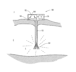
Referring firstly to Figure 1, there is shown particle removal apparatus
100 which is used to remove smoke particles S from the site of a body cavity C
of the patient P, during, for example, intracorporeal surgical procedures at
that
site. The apparatus 100 having a cylindrical housing 180 containing a high
voltage circuit 110 having poles A, B driven by a DC power source 170 in the
form of a rechargeable battery, although the power source may instead be a
non-rechargeable battery or even take the form of a transformer and associated
DC rectifier connected or connectable to mains electricity.
On the underside of the housing 180 is a first electrode 140, being
annular and, in the position shown, is resting on the outer skin of the
patient P,
where, in practice, an electrically conductive gel may be applied to the
patient or
the first electrode 140 in order to improve conductance therebetween.
Extending centrally downwards from the housing 180 is a tubular
insulated probe 130 on the free end of which is a brush-like second electrode
150 electrically connected to pole B of the circuit 110.
Referring now to Figure 2 there is shown a simple high voltage circuit for
powering the particle removal apparatus 100 and in which a low voltage DC
source is used to step up the voltage to a required higher voltage between the
poles A and B sufficient to cause a high voltage, low current, electric field
to
exist between the first electrode 140 via its contact with the patient P and
the
second electrode 150, thereby causing ionisation of particles in that region
such
that they migrate away from the second electrode 150. These particles can
collect on the body surfaces, to be later removed once the procedure is
complete.
CA 02850333 2014-03-27
WO 2013/045886 PCT/GB2012/052172
7
Figures 3 and 4 show respective upper and lower perspective views of
the apparatus 100. In Figure 3 the upper surface of the housing 180 includes
an
arcuate display 182 for displaying the status of the apparatus including, for
example, the charge state of the battery 170 and the functioning of the high
voltage circuit 110. Opposite to the display 180 is a set of controls 184 for
turning the device on and off and for testing it as required.
Figure 4 shows how the annular first electrode 140 is positioned around
the perimeter of the underside of the housing 180 and also shows the second
electrode 150 which, by virtue of being brush-like, provides a relatively
large
surface area for improving the ionisation of particles between the first and
second electrodes 140, 150 via the body of the patient P so that the enclosed
atmosphere in the cavity C can be kept substantially particle-free in a manner
to
be described.
Figure 5 shows a probe catheter introducer tool 200 in the form of a
needle having a solid stem 201, a finger grip 202 at one end and a sharp point
203 at the other. The probe catheter 310 shown in Figure 6 has a tubular stem
portion 311, a funnel-shaped upper end 312 and a blunt lower end 320.
With this arrangement, the catheter 310 can be mounted on the
introducer tool 200 which is then used to pierce an outer wall, such as an
abdominal wall, of the patient P, whereafter the tool 200 can be removed
leaving
the catheter 310 in place to receive the free end of the probe 130 and hence
the
second electrode 150 via the funnel 312, the probe 130 and the second
electrode 150 then emerging within the body cavity C from the lower end 320 of
the catheter 310.
CA 02850333 2014-03-27
WO 2013/045886 PCT/GB2012/052172
8
The outer diameter of the introducer tool and/or catheter is preferably
small, i.e. less than 5mm to avoid the need for post-operative sutures when it
has been removed. The outer surface of the probe 130 is preferably sealable
(for example by a tight interference fit at some point along its length, via
an o-
ring seal, a lever lock or other similar means), against the inner surface of
the
catheter to form a substantially air tight seal to prevent or inhibit gas from
escaping from the inflated body cavity.
The catheter length and/or the probe length is preferably adjustable and
depth indicators may be provided so that users can gauge how far into the body
the probe has been inserted. The depth indicators are preferably visible on
the
exterior of the probe 130 so that its depth can be ascertained using a
surgical
visualisation instrument such as a laparoscope or endoscope during the
surgical
procedure.
A catheter clasp may also be provided for locking the probe in relation to
the catheter so that depth/position of the probe can be temporarily fixed. The
catheter clasp can also be fixable to the patients body such that the probe
depth
and/or position is fixable relative to the patient's body.
One embodiment of the invention only has been described and illustrated,
and it will be readily apparent that other embodiments, modifications,
additions
and omissions are possible within the scope of the invention.
Although the battery 170 has been shown housed within the housing 180,
it will be appreciated that a means of powering the circuit 110 could be
provided
elsewhere. The battery 170 may be removable from the housing 180. In a
modification, the battery 170 may be charged via inductive link such that the
CA 02850333 2014-03-27
WO 2013/045886 PCT/GB2012/052172
9
apparatus 100 can be placed in a cradle and recharged without the need for
exposed recharging terminals, which could otherwise present a health risk. The
apparatus 100 may be a disposable item, in which case rechargeable batteries
need not be used. Capacitive type electrical storage could also be used
instead
of, or as well as, battery electrical storage.
Other refinements are possible within the scope of the claims. For
example, it is envisaged that the high voltage generating source would be
insulated from the outside world and only the two electrodes mentioned above
would be externally accessible. This allows for safer operation of the device
and
reduces the chances of electric shock. The embodiment described in the present
application describes the second electrode 150 as being mounted directly to
the
housing 180 upon which the first electrode 140 is also attached. However, it
will
be appreciated that such mounting of the second electrode 150 may be
detachable from the housing 180 rather than being permanently fixed to it and
this may be achieved using a suitable electrical connector and insulated
receptacle for use with the second electrode. Furthermore, a short electrical
cable may be deployed to allow the surgeon to place the housing 180 and the
first electrode 140 a short distance away from the site of insertion of the
second
electrode 150, but still within the sterile surgical field.
The brush-like second electrode 150 has "bristle" elements which are
each of a size that has been carefully chosen since metal bristles over a
thickness of around 100 microns can act like needles and consequently may
puncture internal organs if used within a body cavity. Likewise bristles
having a
thickness of less than about 50 microns tend to be too weak so bristles of
10
around 75 microns are about ideal. However, bristles between 10 and 100
microns can be satisfactory. It has been found that the number of bristles
does
not greatly affect the rate of smoke clearing with as few as ten bristles
performing satisfactorily. However for a useful, robust, and efficient
electrode,
bristles of around the size mentioned above and around 40 in number are
employed. The bristles have been found to work well when manufactured from
medical grade stainless steel, although other materials may be suitable.
The apparatus and method of the invention may be varied according to
requirements, having as its ultimate objective the removal or reduction of
e.g.
smoke particles at the site of a patient undergoing a medical or cosmetic
procedure whereby the person performing the procedure is afforded better
visibility therefor as a result of fewer particles being present in that
region than
would be the case without the apparatus and method of the invention.
This Patent application is intended to be interpreted in the light of W0201
1/010148.
CA 2850333 2017-09-14