Note: Descriptions are shown in the official language in which they were submitted.
CA 02850542 2014-03-28
WO 2013/049517
PCT/US2012/057839
THERAPEUTIC PEPTIDES
CLAIM OF PRIORITY
This application claims the benefit of U.S. Provisional Patent Application
Serial
No. 61/541,921, filed on September 30, 2011, the entire contents of which are
hereby
incorporated by reference.
GOVERNMENT SUPPORT
This invention was made with Government support under Grant No. P01
AI045757, awarded by the National Institutes of Health. The Government has
certain
rights in the invention.
SEQUENCE LISTING
The instant application contains a Sequence Listing which has been submitted
in
ASCII format via EFS-Web and is hereby incorporated by reference in its
entirety. Said
ASCII copy, created on September 27, 2012, is named 53293W01.txt and is 90,411
bytes
in size.
TECHNICAL FIELD
This invention relates to therapeutic compositions (e.g., peptides) related to
human subjects.
BACKGROUND
Human subjects exposed to a condition or disease offer a source of antibodies
with therapeutic potential and general methods for obtaining such antibodies
are known
in the art. However, methods for specifically obtaining antibodies with
therapeutic
potential are generally limited by the low frequency, slow proliferation rate,
and low
antibody secretion levels of B cells that express such antibodies. For
example, memory B
cells with defined specificity typically account for only one cell per million
peripheral
blood mononuclear cells or approximately one milliliter of blood (Lanzavecchia
et al.,
Curr. Opin. Immunol., 21:298-304 (2009): Yoshida et al., Immunol. Rev.,
237:117-139
(2010)). The frequency of antibodies with therapeutic potential is likely to
be even lower
1
CA 02850542 2014-03-28
WO 2013/049517
PCT/US2012/057839
in cancer patients, necessitating the development of novel approaches that
enable
isolation of such cells with high sensitivity and efficiency.
Conventional methods generally rely on conversion of memory B cells into
antibody secreting cells by in vitro culture and/or use of immunized animal
models (e.g.,
mice) (Crotty et al., J. Immunol., 171:4969-4973 (2003): Fecteau et al.,
Immunology,
128:e353-e365 (2009): Buisman et al., Vaccine, 28:179-186 (2009): Corti et
al., PLoS
One, 5:e8805 (2010)). For example, following in vitro culture for up to one
week,
antibodies can be measured in culture supernatants and frequencies of antibody
secreting
cells assessing using enzyme-linked immunosorbent spot (ELISPOT) assay.
Limitations
of such methods are reported (Henn et al., J. Immunol., 183:31777-3187 (2009):
Cao et
al., J. Immunol., Methods, 358:56-65 (2010)). For instances, in vitro culture
of memory
B cells alters the memory B cell phenotype to resemble plasma cells with
distinct
functional properties (Jiang et al., Eur. J. Immunol., 37:2205-2213 (2007):
Huggins et al.,
Blood, 109:1611-1619 (2007): Jourdan et al., Blood, 114:5173-5181 (2009)).
Limitations
for fluorescent antigen-based methods are also reported (Hofer et al.,
Immunol. Rev.,
211:295-302 (2006): Odendahl et al., Blood, 105:1614-1621 (2005); Kunkel et
al., Nat.
Rev. Immunol., 3:822-829 (2003): Scheid et al., Nature, 458:636-640 (2009): Wu
et al.,
Science, 329:856-861 (2010)).
Improved methods for specifically obtaining or targeting antibodies with
therapeutic potential are required.
MICA is a ligand for NKG2D, a C-type lectin-like, type II transmembrane
receptor expressed on most human NK cells, y6 T cells, and CD8+ T cells. Upon
ligation, NKG2D signals through the adaptor protein DAP10 to evoke perforin
dependent
cytolysis and to provide co-stimulation. In humans, the NKG2D ligands include
MHC
class I chain-related protein A (MICA), the closely related MICB, UL-16
binding proteins
(ULBP) 1-4, and RAE-1G While NKG2D ligands are not usually found on healthy
tissues, various forms of cellular stress, including DNA damage, may
upregulate ligand
expression, resulting in their frequent detection in multiple solid and
hematologic
malignancies, including melanoma. NKG2D activation through ligand positive
transformed cells contributes to extrinsic tumor suppression, since NKG2D
deficient and
2
CA 02850542 2014-03-28
WO 2013/049517
PCT/US2012/057839
wild type mice treated with anti-NKG2D blocking antibodies manifest enhanced
tumor
susceptibility. Immune escape may be achieved in patients, however, by the
shedding of
NKG2D ligands from tumor cells, which triggers internalization of surface
NKG2D and
impaired function of cytotoxic lymphocytes. Soluble NKG2D ligands may also
stimulate
the expansion of regulatory NKG2D+CD4+Foxp3- T cells that may antagonize anti-
tumor cytotoxicity through Fas ligand, IL-10, and TGF-13. MICA is a NKG2D
ligand
shed from tumor cells, i.e., released from the cell surface into the
surrounding medium,
and sera from cancer patients typically contain elevated levels of the soluble
form
(sMICA). MICA shedding is accomplished in part through interactions with the
protein
disulfide isomerase ERp5, which forms a disulfide bond with a critical
cysteine that
results in unfolding of the a3 domain, rendering it susceptible to proteolysis
by ADAM-
10/17 and MMP14.
Angiogenesis is the process of forming new capillaries from preexisting blood
vessels and has been implicated as a critical part of tumor growth and
dissemination.
Tumors stimulate angiogenesis to meet increasing oxygen and nutrient
requirements that
exceed those that can be met by diffusion alone. Consequently, tumors recruit,
remodel
and expand existing vascular to meet their metabolic demand. The dependence of
growing tumors on new blood vessel formation has made angiogenesis an
appealing
target for anti-cancer therapies. Many cytokines have been are believed to
play a role in
the regulation of angiogenesis, including vascular endothelial growth factor
(VEGF)
family members and the angiopoietins. The angiopioetins were discovered as
ligands for
the Ties, a family of tyrosine kinases that is selectively expressed in the
vascular
endothelium. There are four know angiopoietins: angiopoietin-1 ("Ang-1")
through
angiopoietin-4 ("Ang-4"). Studies have suggested that angiopoietins (e.g., Ang-
1 and
Ang-2) may be involved and tumor angiogenesis. With this information,
angiopoietins
have been identified as potential targets of immune-based cancer therapy.
There is a need to identify new agents that specifically recognize and bind
targets
of immune-based cancer therapy, such as MICA and angiopoietins. Such agents
would be
useful for diagnostic screening and therapeutic intervention in disease states
that are
associated with tumor development.
3
CA 02850542 2014-03-28
WO 2013/049517
PCT/US2012/057839
SUMMARY
The present disclosure provides compositions and methods related to antibodies
with therapeutic potential.
In some embodiments, the disclosure provides compositions comprising peptides
that immunospecifically bind to MHC class I polypeptide-related sequence A
(MICA), or
an epitope thereon. In some aspects, peptides of the compositions include
complementarity determining region (CDR) 3 of the VH of antibody ID 1, 6, 7, 8
or 9
shown in Table 1 having 5 or fewer conservative amino acid substitutions, and
CDR3 of
the VL of antibody ID 1, 6, 7, 8 or 9 shown in Table 1 having 5 or fewer
conservative
amino acid substitutions. In some aspects, such peptides include
complementarity
determining region (CDR) 3 of the VH of antibody ID 1, 6, 7, 8 or 9 shown in
Table 1,
and CDR3 of the VL of antibody ID 1, 6, 7, 8 or 9 shown in Table 1. In some
aspects,
peptides further include CDR2 of the VH of antibody ID 1, 6, 7, 8 or 9 shown
in Table 1
having 5 or fewer conservative amino acid substitutions, or CDR2 of the VL of
antibody
ID 1, 6, 7, 8 or 9 shown in Table 1 having 5 or fewer conservative amino acid
substitutions, or both. In some aspects, such peptides include complementarity
determining region CDR2 of the VH of antibody ID 1, 6, 7, 8 or 9 shown in
Table 1, or
CDR2 of the VL of antibody ID 1, 6, 7, 8 or 9 shown in Table 1, or both. In
some aspects,
peptides further include CDR1 of the VH of antibody ID 1, 6, 7, 8 or 9 shown
in Table 1
having 5 or fewer conservative amino acid substitutions, or CDR1 of the VL of
antibody
ID 1, 6, 7, 8 or 9 shown in Table 1 having 5 or fewer conservative amino acid
substitutions, or both. In some aspects, such peptides include complementarity
determining region CDR1 of the VH of antibody ID 1, 6, 7, 8 or 9 shown in
Table 1, or
CDR1 of the VL of antibody ID 1, 6, 7, 8 or 9 shown in Table 1, or both.
In some aspects, peptides are antibody or antibody fragment that include: a VH
chain with identity to SEQ ID NO:2, wherein regions corresponding to CDR1,
CDR2,
and CDR3 comprise CDR1, CDR2, and CDR3 of the VH of antibody ID 1 shown in
table
1 having 5 or fewer conservative amino acid substitutions, and regions within
SEQ ID
NO:2 corresponding to FR1, FR2, FR3, FR4, comprise amino acid sequences with
at
least 80%, 85%, 90%, 95%, 96%, 97%, 98, 99%, or 100% identity to FR1, FR2,
FR3,
4
CA 02850542 2014-03-28
WO 2013/049517
PCT/US2012/057839
FR4 of the VH of antibody ID 1 shown in table 1; and a VL chain with identity
to SEQ ID
NO:11, wherein regions corresponding to CDR1, CDR2, and CDR3 comprise CDR1,
CDR2, and CDR3 of the VL of antibody ID 1 shown in table 1 having 5 or fewer
conservative amino acid substitutions, and regions within SEQ ID NO:11
corresponding
to FR1, FR2, FR3, FR4, comprise amino acid sequences with at least 80%, 85%,
90%,
95%, 96%, 97%, 98, 99%, or 100% identity to FR1, FR2, FR3, FR4 of the VL of
antibody
ID 1 shown in table 1. In some aspects, peptides include an antibody or
antibody
fragment comprising a VH chain comprising SEQ ID NO:2 and a VL chain
comprising
SEQ ID NO:11. In some aspects, in addition the peptides, compositions further
include
one or more (e.g., 1 2, 3, 4, 5, 6, 7, 8, 9, 10, or less than 20) anti-cancer
therapeutics. In
some aspects, compositions are formulated as pharmaceutical compositions
(e.g., for
administration to a subject).
In some aspects, peptides are antibody or antibody fragment that include: a VH
chain with identity to SEQ ID NO:149, wherein regions corresponding to CDR1,
CDR2,
and CDR3 comprise CDR1, CDR2, and CDR3 of the VH of antibody ID 6 shown in
table
1 having 5 or fewer conservative amino acid substitutions within the CDR1,
CDR2, and
CDR3 regions, and regions within SEQ ID NO:149 corresponding to FR1, FR2, FR3,
FR4, comprise amino acid sequences with at least 80%, 85%, 90%, 95%, 96%, 97%,
98,
99%, or 100% identity to FR1, FR2, FR3, FR4 of the VH of antibody ID 6 shown
in table
1; and a VL chain with identity to SEQ ID NO:151, wherein regions
corresponding to
CDR1, CDR2, and CDR3 comprise CDR1, CDR2, and CDR3 of the VL of antibody ID 6
shown in table 1 having 5 or fewer conservative amino acid substitutions
within the
CDR1, CDR2, and CDR3 regions, and regions within SEQ ID NO:151 corresponding
to
FR1, FR2, FR3, FR4, comprise amino acid sequences with at least 80%, 85%, 90%,
95%, 96%, 97%, 98, 99%, or 100% identity to FR1, FR2, FR3, FR4 of the VL of
antibody ID 6 shown in table 1. In some aspects, peptides include an antibody
or
antibody fragment comprising a VH chain comprising SEQ ID NO:149 and a VL
chain
comprising SEQ ID NO:151. In some aspects, in addition the peptides,
compositions
further include one or more (e.g., 1 2, 3, 4, 5, 6, 7, 8, 9, 10, or less than
20) anti-cancer
5
CA 02850542 2014-03-28
WO 2013/049517
PCT/US2012/057839
therapeutics. In some aspects, compositions are formulated as pharmaceutical
compositions (e.g., for administration to a subject).
In some aspects, peptides are antibody or antibody fragment that include: a VH
chain with identity to SEQ ID NO:168, wherein regions corresponding to CDR1,
CDR2,
and CDR3 comprise CDR1, CDR2, and CDR3 of the VH of antibody ID 7 shown in
table
1 having 5 or fewer conservative amino acid substitutions within the CDR1,
CDR2, and
CDR3 regions, and regions within SEQ ID NO:168 corresponding to FR1, FR2, FR3,
FR4, comprise amino acid sequences with at least 80%, 85%, 90%, 95%, 96%, 97%,
98,
99%, or 100% identity to FR1, FR2, FR3, FR4 of the VH of antibody ID 7 shown
in table
1; and a VL chain with identity to SEQ ID NO:170, wherein regions
corresponding to
CDR1, CDR2, and CDR3 comprise CDR1, CDR2, and CDR3 of the VL of antibody ID 7
shown in table 1 having 5 or fewer conservative amino acid substitutions
within the
CDR1, CDR2, and CDR3 regions, and regions within SEQ ID NO:170 corresponding
to
FR1, FR2, FR3, FR4, comprise amino acid sequences with at least 80%, 85%, 90%,
95%, 96%, 97%, 98, 99%, or 100% identity to FR1, FR2, FR3, FR4 of the VL of
antibody
ID 7 shown in table 1. In some aspects, peptides include an antibody or
antibody
fragment comprising a VH chain comprising SEQ ID NO:168 and a VL chain
comprising
SEQ ID NO:170. In some aspects, in addition the peptides, compositions further
include
one or more (e.g., 1 2, 3, 4, 5, 6, 7, 8, 9, 10, or less than 20) anti-cancer
therapeutics. In
some aspects, compositions are formulated as pharmaceutical compositions
(e.g., for
administration to a subject).
In some aspects, peptides are antibody or antibody fragment that include: a VH
chain with identity to SEQ ID NO:186, wherein regions corresponding to CDR1,
CDR2,
and CDR3 comprise CDR1, CDR2, and CDR3 of the VH of antibody ID 8 shown in
table
1 having 5 or fewer conservative amino acid substitutions within the CDR1,
CDR2, and
CDR3 regions, and regions within SEQ ID NO:186 corresponding to FR1, FR2, FR3,
FR4, comprise amino acid sequences with at least 80%, 85%, 90%, 95%, 96%, 97%,
98,
99%, or 100% identity to FR1, FR2, FR3, FR4 of the VH of antibody ID 8 shown
in table
1; and a VL chain with identity to SEQ ID NO:188, wherein regions
corresponding to
CDR1, CDR2, and CDR3 comprise CDR1, CDR2, and CDR3 of the VL of antibody ID 8
6
CA 02850542 2014-03-28
WO 2013/049517
PCT/US2012/057839
shown in table 1 having 5 or fewer conservative amino acid substitutions
within the
CDR1, CDR2, and CDR3 regions, and regions within SEQ ID NO:188 corresponding
to
FR1, FR2, FR3, FR4, comprise amino acid sequences with at least 80%, 85%, 90%,
95%, 96%, 97%, 98, 99%, or 100% identity to FR1, FR2, FR3, FR4 of the VL of
antibody
ID 8 shown in table 1. In some aspects, peptides include an antibody or
antibody
fragment comprising a VH chain comprising SEQ ID NO:186 and a VL chain
comprising
SEQ ID NO:188. In some aspects, in addition the peptides, compositions further
include
one or more (e.g., 1 2, 3, 4, 5, 6, 7, 8, 9, 10, or less than 20) anti-cancer
therapeutics. In
some aspects, compositions are formulated as pharmaceutical compositions
(e.g., for
administration to a subject).
In some aspects, peptides are antibody or antibody fragment that include: a VH
chain with identity to SEQ ID NO:204, wherein regions corresponding to CDR1,
CDR2,
and CDR3 comprise CDR1, CDR2, and CDR3 of the VH of antibody ID 9 shown in
table
1 having 5 or fewer conservative amino acid substitutions within the CDR1,
CDR2, and
CDR3 regions, and regions within SEQ ID NO:204 corresponding to FR1, FR2, FR3,
FR4, comprise amino acid sequences with at least 80%, 85%, 90%, 95%, 96%, 97%,
98,
99%, or 100% identity to FR1, FR2, FR3, FR4 of the VH of antibody ID 9 shown
in table
1; and a VL chain with identity to SEQ ID NO:206, wherein regions
corresponding to
CDR1, CDR2, and CDR3 comprise CDR1, CDR2, and CDR3 of the VL of antibody ID 9
shown in table 1 having 5 or fewer conservative amino acid substitutions
within the
CDR1, CDR2, and CDR3 regions, and regions within SEQ ID NO:206 corresponding
to
FR1, FR2, FR3, FR4, comprise amino acid sequences with at least 80%, 85%, 90%,
95%, 96%, 97%, 98, 99%, or 100% identity to FR1, FR2, FR3, FR4 of the VL of
antibody
ID 9 shown in table 1. In some aspects, peptides include an antibody or
antibody
fragment comprising a VH chain comprising SEQ ID NO:204 and a VL chain
comprising
SEQ ID NO:206. In some aspects, in addition the peptides, compositions further
include
one or more (e.g., 1 2, 3, 4, 5, 6, 7, 8, 9, 10, or less than 20) anti-cancer
therapeutics. In
some aspects, compositions are formulated as pharmaceutical compositions
(e.g., for
administration to a subject).
7
CA 02850542 2014-03-28
WO 2013/049517
PCT/US2012/057839
In some embodiments, the disclosure provides compositions that include one or
more peptides that bind to angiopoietin or an epitope thereon. In some
aspects, peptides
of the compositions include complementarity determining region (CDR) 3 of the
VH of
antibody ID 2, 3, 4, 5 or lOshown in Table 1 having 5 or fewer conservative
amino acid
substitutions, and CDR3 of the VL of antibody ID 2, 3, 4 5 or 10 shown in
Table 1 having
5 or fewer conservative amino acid substitutions within the CDR1, CDR2, and
CDR3
regions. In some aspects, peptides can include complementarity determining
region
(CDR) 3 of the VH of antibody ID 2, 3, 4 or 5 or 10 shown in Table 1, and CDR3
of the
VL of antibody ID 2, 3, 4 or 5 or 10 shown in Table 1. In some aspects,
peptides can
further include CDR2 of the VH of antibody ID 2, 3, 4 or 5 or 10 shown in
Table 1
having 5 or fewer conservative amino acid substitutions, or CDR2 of the VL of
antibody
ID 2, 3, 4 or 5 or 10 shown in Table 1 having 5 or fewer conservative amino
acid
substitutions, or both. In some aspects, such peptides can include
complementarity
determining region CDR2 of the VH of antibody ID 2, 3, 4 or 5 or 10 shown in
Table 1,
or CDR2 of the VL of antibody ID 2, 3, 4 or 5 or 10 shown in Table 1, or both.
In some
aspects, peptides can further include CDR1 of the VH of antibody ID 2, 3, 4 or
5 or 10
shown in Table 1 having 5 or fewer conservative amino acid substitutions, or
CDR1 of
the VL of antibody ID 2, 3, 4, or 5 shown in Table 1 having 5 or fewer
conservative
amino acid substitutions, or both. In some aspects, such peptides can include
complementarity determining region CDR1 of the VH of antibody ID 2, 3, 4 or 5
or 10
shown in Table 1, or CDR1 of the VL of antibody ID 2, 3, 4 or 5 or 10 shown in
Table 1,
or both.
In some aspects, peptides include an antibody or antibody fragment comprising:
a
VH chain with identity to SEQ ID NO :20, wherein regions corresponding to
CDR1,
CDR2, and CDR3 comprise CDR1, CDR2, and CDR3 of the VH of antibody ID 2 shown
in table 1 having 5 or fewer conservative amino acid substitutions within the
CDR1,
CDR2, and CDR3 regions, and regions within SEQ ID NO:20 corresponding to FR1,
FR2, FR3, FR4, comprise amino acid sequences with at least 80%, 85%, 90%, 95%,
96%, 97%, 98, 99%, or 100% identity to FR1, FR2, FR3, FR4 of the VH of
antibody ID 2
shown in table 1; and a VL chain with identity to SEQ ID NO:29, wherein
regions
8
CA 02850542 2014-03-28
WO 2013/049517
PCT/US2012/057839
corresponding to CDR1, CDR2, and CDR3 comprise CDR1, CDR2, and CDR3 of the VL
of antibody ID 2 shown in table 1 having 5 or fewer conservative amino acid
substitutions within the CDR1, CDR2, and CDR3 regions, and regions within SEQ
ID
NO:29 corresponding to FR1, FR2, FR3, FR4, comprise amino acid sequences with
at
least 80%, 85%, 90%, 95%, 96%, 97%, 98, 99%, or 100% identity to FR1, FR2,
FR3,
FR4 of the VL of antibody ID 2 shown in table 1. In some aspects, the peptides
include
an antibody or antibody fragment comprising a VH chain comprising SEQ ID NO:20
and
a VL chain comprising SEQ ID NO:29.
In some aspects, the peptides an antibody or antibody fragment comprising: a
VH
chain with identity to SEQ ID NO:38, wherein regions corresponding to CDR1,
CDR2,
and CDR3 comprise CDR1, CDR2, and CDR3 of the VH of antibody ID 3 shown in
table
1 having 5 or fewer conservative amino acid substitutions within the CDR1,
CDR2, and
CDR3 regions, and regions within SEQ ID NO:38 corresponding to FR1, FR2, FR3,
FR4, comprise amino acid sequences with at least 80%, 85%, 90%, 95%, 96%, 97%,
98,
99%, or 100% identity to FR1, FR2, FR3, FR4 of the VH of antibody ID 3 shown
in table
1; and a VL chain with identity to SEQ ID NO:47, wherein regions corresponding
to
CDR1, CDR2, and CDR3 comprise CDR1, CDR2, and CDR3 of the VL of antibody ID 3
shown in table 1 having 5 or fewer conservative amino acid substitutions
within the
CDR1, CDR2, and CDR3 regions, and regions within SEQ ID NO:47 corresponding to
FR1, FR2, FR3, FR4, comprise amino acid sequences with at least 80%, 85%, 90%,
95%, 96%, 97%, 98, 99%, or 100% identity to FR1, FR2, FR3, FR4 of the VL of
antibody
ID 3 shown in table 1. In some aspects, peptides include an antibody or
antibody
fragment comprising a VH chain comprising SEQ ID NO:38 and a VL chain
comprising
SEQ ID NO:47.
In some aspects, peptides include an antibody or antibody fragment comprising:
a
VH chain with identity to SEQ ID NO:56, wherein regions corresponding to CDR1,
CDR2, and CDR3 comprise CDR1, CDR2, and CDR3 of the VH of antibody ID 4 shown
in table 1 having 5 or fewer conservative amino acid substitutions within the
CDR1,
CDR2, and CDR3 regions, and regions within SEQ ID NO:56 corresponding to FR1,
FR2, FR3, FR4, comprise amino acid sequences with at least 80%, 85%, 90%, 95%,
9
CA 02850542 2014-03-28
WO 2013/049517
PCT/US2012/057839
96%, 97%, 98, 99%, or 100% identity to FR1, FR2, FR3, FR4 of the VH of
antibody ID 4
shown in table 1; and a VL chain with identity to SEQ ID NO:65, wherein
regions
corresponding to CDR1, CDR2, and CDR3 comprise CDR1, CDR2, and CDR3 of the VL
of antibody ID 4 shown in table 1 having 5 or fewer conservative amino acid
substitutions within the CDR1, CDR2, and CDR3 regions, and regions within SEQ
ID
NO:65 corresponding to FR1, FR2, FR3, FR4, comprise amino acid sequences with
at
least 80%, 85%, 90%, 95%, 96%, 97%, 98, 99%, or 100% identity to FR1, FR2,
FR3,
FR4 of the VL of antibody ID 4 shown in table 1. In some aspects, peptides
include an
antibody or antibody fragment comprising a VH chain comprising SEQ ID NO:56
and a
VL chain comprising SEQ ID NO:65.
In some aspects, peptides include an antibody or antibody fragment comprising:
a
VH chain with identity to SEQ ID NO:74, wherein regions corresponding to CDR1,
CDR2, and CDR3 comprise CDR1, CDR2, and CDR3 of the VH of antibody ID 5 shown
in table 1 having 5 or fewer conservative amino acid substitutions within the
CDR1,
CDR2, and CDR3 regions, and regions within SEQ ID NO:74 corresponding to FR1,
FR2, FR3, FR4, comprise amino acid sequences with at least 80%, 85%, 90%, 95%,
96%, 97%, 98, 99%, or 100% identity to FR1, FR2, FR3, FR4 of the VH of
antibody ID 5
shown in table 1; and a VL chain with identity to SEQ ID NO:83, wherein
regions
corresponding to CDR1, CDR2, and CDR3 comprise CDR1, CDR2, and CDR3 of the VL
of antibody ID 5 shown in table 1 having 5 or fewer conservative amino acid
substitutions within the CDR1, CDR2, and CDR3 regions, and regions within SEQ
ID
NO:83 corresponding to FR1, FR2, FR3, FR4, comprise amino acid sequences with
at
least 80%, 85%, 90%, 95%, 96%, 97%, 98, 99%, or 100% identity to FR1, FR2,
FR3,
FR4 of the VL of antibody ID 5 shown in table 1. In some aspects, the peptides
include
an antibody or antibody fragment comprising a VH chain comprising SEQ ID NO:74
and
a VL chain comprising SEQ ID NO:83. In some aspects, the peptides
immunospecifically
bind to at least angiopoietin-2. In some aspects, the compositions further
include one or
more anti-cancer therapeutics. In some aspects, the compositions are
formulated as a
pharmaceutical composition.
CA 02850542 2014-03-28
WO 2013/049517
PCT/US2012/057839
In some aspects, peptides include an antibody or antibody fragment comprising:
a
VH chain with identity to SEQ ID NO:222, wherein regions corresponding to
CDR1,
CDR2, and CDR3 comprise CDR1, CDR2, and CDR3 of the VH of antibody ID 10
shown in table 1 having 5 or fewer conservative amino acid substitutions
within the
CDR1, CDR2, and CDR3 regions, and regions within SEQ ID NO:222 corresponding
to
FR1, FR2, FR3, FR4, comprise amino acid sequences with at least 80%, 85%, 90%,
95%, 96%, 97%, 98, 99%, or 100% identity to FR1, FR2, FR3, FR4 of the VH of
antibody ID 10 shown in table 1; and a VL chain with identity to SEQ ID
NO:224,
wherein regions corresponding to CDR1, CDR2, and CDR3 comprise CDR1, CDR2, and
CDR3 of the VL of antibody ID 10 shown in table 1 having 5 or fewer
conservative
amino acid substitutions within the CDR1, CDR2, and CDR3 regions, and regions
within
SEQ ID NO:224 corresponding to FR1, FR2, FR3, FR4, comprise amino acid
sequences
with at least 80%, 85%, 90%, 95%, 96%, 97%, 98, 99%, or 100% identity to FR1,
FR2,
FR3, FR4 of the VL of antibody ID 10 shown in table 1. In some aspects, the
peptides
include an antibody or antibody fragment comprising a VH chain comprising SEQ
ID
NO:222 and a VL chain comprising SEQ ID NO:224. In some aspects, the peptides
immunospecifically bind to at least angiopoietin-2. In some aspects, the
compositions
further include one or more anti-cancer therapeutics. In some aspects, the
compositions
are formulated as a pharmaceutical composition.
In some embodiments, the disclosure includes methods of treating cancer in a
subject. In some aspects, methods include administering to a subject a
composition of
any one of claims 1-27.
The present disclosure also provides provides methods of isolating human
antibodies from cancer patients following immunotherapy.
In some embodiments, the disclosure includes method of obtaining immune cells
directed against a self antigen from a subject, the method comprising
identifying a subject
exhibiting a positive immune response towards the self antigen, providing a
multimeric
form of the self antigen, contacting the multimeric form of the self antigen
with a sample
from the subject exhibiting a positive immune response towards the self
antigen, and
obtaining immune cells bound to the multimeric form of the self antigen.
11
CA 02850542 2014-03-28
WO 2013/049517
PCT/US2012/057839
In some embodiments, the disclosure includes method of obtaining immune cells
from a cancer patient directed against a self antigen, the method comprising
identifying a
subject exhibiting a positive immune response towards the self antigen;
providing a
multimeric form of the self antigen; contacting the multimeric form of the
self antigen
with a sample from the subject exhibiting a positive immune response towards
the self
antigen; and obtaining immune cells bound to the multimeric form of the self
antigen.
Unless otherwise defined, all technical and scientific terms used herein have
the
same meaning as commonly understood by one of ordinary skill in the art to
which this
invention belongs. Methods and materials are described herein for use in the
present
invention; other, suitable methods and materials known in the art can also be
used. The
materials, methods, and examples are illustrative only and not intended to be
limiting.
All publications, patent applications, patents, sequences, database entries,
and other
references mentioned herein are incorporated by reference in their entirety.
In case of
conflict, the present specification, including definitions, will control.
Other features and advantages of the invention will be apparent from the
following detailed description and figures, and from the claims.
DESCRIPTION OF DRAWINGS
FIG. 1 1 Nucleic acid sequence of the variable heavy (VH) chain of antibody ID
1
(anti-MHC class I polypeptide-related sequence A (MICA) antibody) (SEQ ID
NO:1).
FIG. 2 1 Amino acid sequence of VH chain of antibody ID 1 (anti-MICA antibody)
(SEQ ID NO:2).
FIG 3 1 Nucleic acid sequence of the variable light (VI) chain of antibody ID
1
(anti-MICA antibody) (SEQ ID NO:10).
FIG. 4 1 Amino acid sequence of VL chain of antibody ID 1 (anti-MICA antibody)
(SEQ ID NO:11).
FIG. 5 1 Nucleic acid sequence of the VH chain of antibody ID 2 (anti-
angiopoietin-2 antibody) (SEQ ID NO:19).
FIG. 6 1 Amino acid sequence of VH chain of antibody ID 2 (anti- angiopoietin-
2
antibody) (SEQ ID NO:20).
12
CA 02850542 2014-03-28
WO 2013/049517
PCT/US2012/057839
FIG. 71Nucleic acid sequence of the VL chain of antibody ID 2 (anti-
angiopoietin-2 antibody) (SEQ ID NO:28).
FIG. 81Amino acid sequence of VL chain of antibody ID 2 (anti- angiopoietin-2
antibody) (SEQ ID NO:29).
FIG. 91Nucleic acid sequence of the VH chain of antibody ID 3 (anti-
angiopoietin-2 antibody) (SEQ ID NO:37).
FIG. 10 Amino acid sequence of VH chain of antibody ID 3 (anti- angiopoietin-2
antibody) (SEQ ID NO:38).
FIG 11 Nucleic acid sequence of the VL chain of antibody ID 3 (anti-
angiopoietin-2 antibody) (SEQ ID NO:46).
FIG. 12 Amino acid sequence of VL chain of antibody ID 3 (anti- angiopoietin-2
antibody) (SEQ ID NO:47).
FIG 13 Nucleic acid sequence of the VH chain of antibody ID 4 (anti-
angiopoietin-2 antibody) (SEQ ID NO:55).
FIG. 14 Amino acid sequence of VH chain of antibody ID 4 (anti- angiopoietin-2
antibody) (SEQ ID NO:56).
FIG 15 Nucleic acid sequence of the VL chain of antibody ID 4 (anti-
angiopoietin-2 antibody) (SEQ ID NO:64).
FIG. 16 Amino acid sequence of VL chain of antibody ID 4 (anti- angiopoietin-2
antibody) (SEQ ID NO:65).
FIG 17 Nucleic acid sequence of the VH chain of antibody ID 5 (anti-
angiopoietin-2 antibody) (SEQ ID NO:73).
FIG. 18 Amino acid sequence of VH chain of antibody ID 5 (anti- angiopoietin-2
antibody) (SEQ ID NO:74).
FIG 19 Nucleic acid sequence of the VL chain of antibody ID 5 (anti-
angiopoietin-2 antibody) (SEQ ID NO:82).
FIG. 20 Amino acid sequence of VL chain of antibody ID 5 (anti- angiopoietin-2
antibody) (SEQ ID NO:83).
FIG. 21A-21F 1Illustrates exemplary methods for making antibodies from B-
cells.
(A) Antigen is expressed with a BirA tag for site-specific biotinylation and
13
CA 02850542 2014-03-28
WO 2013/049517
PCT/US2012/057839
tetramerization with fluorescently-labeled streptavidin. (B) B cells are
stained with
tetramer and a panel of monoclonal antibodies. Tetramer, ', class-switched
memory B
cells are single-cell sorted into PCR strips. (C) mRNA amplification is
performed with
T7 RNA polymerase. (D) Sequencing of PCR products is carried out using 300-
400bp
PCR products. (E) Overlap PCR is used for construction of full-length IgG1
heavy chain
and kappa/lambda light sequences which are cloned into separate vectors.
Vectors are
transiently transfected into CHO-S cells for expression of fully human
recombinant
antibodies. (F) Antibodies are tested for antigen binding and assessed for
potential
therapeutic properties.
FIGs. 22A-22B 1 Graphs showing comparison of monomeric and tetrameric
antigen for identification of memory B cells. (A) Mono-biotinylated TTCF or
CD80
antigens were directly labeled with Alexa-488 fluorophore; tetramers were
generated with
unlabeled streptavidin. Enriched B cells from each donor were split into three
fractions
and stained with control CD80 tetramer, TTCF monomer, or TTCF tetramer at the
same
total antigen concentration of 0.125 g/mL. FACS plots depict CD19 CD27' IgM-
class-switched memory B cells; numbers adjacent to the gate represent the
percentage of
the parental gate. (B) Frequencies of tetramer ' memory B cells detected in
three different
donors. Numbers are calculated as tetramer ' cells per lx106 CD19' memory B
cells.
FIGs. 23A-23B 1Line graphs showing high affinity binding of TTCF by
antibodies generated from plasmablasts and memory B cells. Saturation binding
experiments were carried out to determine the affinities of recombinant
antibodies.
TTCF antigen was labeled with europium, which emits a strong fluorescent
signal at
615nm upon incubation with a chelating reagent. Antibodies were immobilized in
a 96-
well plate and incubated with TTCF-europium (100nM to 4pM) for two hours at 37
C.
Fluorescent counts at 615nm were recorded and KD calculated using non-linear
regression
analysis. Control antibody (clone 8.18.C5) that was also produced in CHO-S
cells was
included in all experiments. (A) Recombinant TTCF Abs 1 and 2 were generated
from
TTCF tetramer ' plasmablasts (donor 1). (B) TTCF antibodies 3, 4, and 5
originated from
TTCF tetramer ' memory B cells of three different donors.
14
CA 02850542 2014-03-28
WO 2013/049517
PCT/US2012/057839
FIG. 24 Bar chart showing binding of anti-MICA antibodies to MICA-coated
luminex beads.
FIGs. 25A-250 1 Line graphs showing binding of anti-MICA antibodies to MICA-
coated beads.
FIGs 26A-26D 1 Bar graphs showing binding of four human angiopoietin 2
specific antibodies as well as a control antibody to three human angiopoietins
(angiopoietin-1, 2 and 4) and ang-like-3. Recombinant angiopoietins were
immobilized
in an ELISA plate and binding of human recombinant antibodies was detected
with
europium-labeled streptavidin.
FIGs. 27A-27C I Show graphs and a gel relating to isolation of angiopoietin-
specific antibodies from a lung cancer patient. (A) Angiopoietin-2 reactivity
of lung
cancer patient (L19) serum (diluted 1:1000) determined by ELISA. (B) FACS plot
showing PBMC sample (timepoint- 10/98) gated on CD19 ', CD27 ' IgM-B cells
with
CD19 on the X-axis and fluorescently-tagged angiopoietin-2 on the Y-axis. (C)
Heavy,
light chain, and hinge region PCR products from 10 angiopoietin-2 reactive
memory B-
cells isolated from patient L19. The 500 base pair marker is indicated on the
left.
FIG 28 Nucleic acid sequence of the variable heavy (VH) chain of antibody ID 6
(anti-MHC class I polypeptide-related sequence A (MICA) antibody) (SEQ ID
NO:148).
FIG 29 Amino acid sequence of VH chain of antibody 6 (anti-MICA antibody)
(SEQ ID NO:149).
FIG 30 Nucleic acid sequence of the variable light (VI) chain of antibody ID 6
(anti-MICA antibody) (SEQ ID NO:150).
FIG 31 Amino acid sequence of VL chain of antibody ID 6 (anti-MICA
antibody) (SEQ ID NO: 151).
FIG 32 Nucleic acid sequence of the variable heavy (VH) chain of antibody ID 7
(anti-MHC class I polypeptide-related sequence A (MICA) antibody) (SEQ ID
NO:167).
FIG 33 Amino acid sequence of VH chain of antibody ID 7 (anti-MICA
antibody) (SEQ ID NO:168).
FIG 34 Nucleic acid sequence of the variable light (VI) chain of antibody ID 7
(anti-MICA antibody) (SEQ ID NO:169).
CA 02850542 2014-03-28
WO 2013/049517
PCT/US2012/057839
FIG 35 Amino acid sequence of VL chain of antibody ID 7 (anti-MICA
antibody) (SEQ ID NO: 170).
FIG 36 Nucleic acid sequence of the variable heavy (VH) chain of antibody ID 8
(anti-MHC class I polypeptide-related sequence A (MICA) antibody) (SEQ ID
NO:185).
FIG. 37 Amino acid sequence of VH chain of antibody ID 8 (anti-MICA
antibody) (SEQ ID NO:186).
FIG 38 Nucleic acid sequence of the variable light (VL) chain of antibody ID 8
(anti-MICA antibody) (SEQ ID NO:187).
FIG. 39 Amino acid sequence of VL chain of antibody ID 8 (anti-MICA
antibody) (SEQ ID NO: 188).
FIG 40 Nucleic acid sequence of the variable heavy (VH) chain of antibody ID 9
(anti-MHC class I polypeptide-related sequence A (MICA) antibody) (SEQ ID
NO:203).
FIG 41 Amino acid sequence of VH chain of antibody ID 9 (anti-MICA
antibody) (SEQ ID NO:204).
FIG 42 Nucleic acid sequence of the variable light (VL) chain of antibody ID 9
(anti-MICA antibody) (SEQ ID NO:205).
FIG 43 Amino acid sequence of VL chain of antibody ID 9 (anti-MICA
antibody) (SEQ ID NO: 206).
FIG 44 Nucleic acid sequence of the VH chain of antibody ID 10 (anti-
angiopoietin-2 antibody) (SEQ ID NO:221).
FIG. 45 Amino acid sequence of VH chain of antibody ID 10 (anti- angiopoietin-
2 antibody) (SEQ ID NO:222).
FIG 46 Nucleic acid sequence of the VL chain of antibody ID 10 (anti-
angiopoietin-2 antibody) (SEQ ID NO:223).
FIG. 47 Amino acid sequence of VL chain of antibody ID 10 (anti- angiopoietin-
2 antibody) (SEQ ID NO :224).
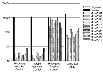
FIGs. 48A-G1 Line graphs showing assessment of MICA allele-specific binding
by recombinant anti-MICA antibodies.
FIG. 49 Line graph showing labeling of autologous tumor cells by anti-MICA
antibody CM24002 Ab2.
16
CA 02850542 2014-03-28
WO 2013/049517
PCT/US2012/057839
FIG. 501A series of FACS plot showing regulation of NKG2D by serum MICA.
Human NK cells were incubated with control serum from patient CM24002 and a
1:10
dilution for 48 hours. Indicated antibodies were added at the start of the
incubation at a
concentration of 10 ug/ml. NKG2D expression was assessed on CD56+ NK cells by
flow cytometry.
FIG. 511A series of FACS plot showing regulation of NKG2D by recombinant
MICA. Human NK cells were incubated with recombinant MICA at a concentration
of 2
ng/ml for 48 hours. Indicated antibodies were added at the start of the
incubation at a
concentration of 10 ug/ml. After 48 hours, NKG2D expression was assessed on
CD56+
NK cells by flow cytometry.
FIG. 521 Line graph demonstrating enhancement of cell-mediated toxicity by
anti-
MICA antibody CM24002 Ab2. Human NK cells were incubated with recombinant
MICA (2ng/m1) for 48 hours in the presence of indicated antibodies at 10
ug/ml. The
ability of NK cells (effectors) to kill K562 target cells was assessed by
measuring LDH
release following 4 hour incubation at the indicated ratios.
FIG. 531 Bar graph demonstration cell-mediated toxicity by anti-MICA
antibodies
CM24002 Ab2 and CM33322 Ab29. Human NK cells were incubated with recombinant
MICA (2ng/m1) for 48 hours in the presence of indicated antibodies at 10
ug/ml. The
ability of NK cells (effectors) to kill K562 target cells was assessed by
measuring LDH
release following 4 hour incubation. NKG2D blocking antibody or Fc blocking
antibody
was added during the 4 hr incubation of effector and target cells to assess
the contribution
of Fc receptor and NKG2D to cell-mediated toxicity.
FIG. 541A series of line graphs showing binding of MICA alpha 3 domain by
recombinant anti-MICA antibodies. Recombinant MICA alpha 3 domains were
biotinylated and captured on the surface of streptavidin-coated beads.
Indicated
antibodies were incubated at 10 g/m1 with the beads coated with the individual
recombinant protein for lhr. Beads were subsequently washed and incubated with
FITC-
conjugated anti-human IgG secondary antibody. FITC fluorescence was quantified
by
flow cytometry.
17
CA 02850542 2014-03-28
WO 2013/049517
PCT/US2012/057839
FIG. 55 Line graphs demonstrating labeling of tumor cells by anti-MICA
antibodies CM24002 Ab2 and CM33322 Ab29. Fluorescence was determined by flow
cytometry.
FIG. 56 Bar graph demonstrating MICA allelic specificity of anti-MICA
antibodies CM33322 Ab29 as determined by Luminex assay.
FIG. 57 Bar graphs showing binding of anti-angiopoietin 2 specific antibody
anti-Ang6 Ab2 as well as a control antibody to three human angiopoietins
(angiopoietin-
1, 2 and 4) and ang-like-3. Recombinant angiopoietins were immobilized in an
ELISA
plate and binding of human recombinant antibodies was detected with europium-
labeled
streptavidin.
DETAILED DESCRIPTION
The present disclosure is based, in part, on the observation that antibodies
directed
against therapeutic targets important in a disease can be obtained from human
subjects
exposed to the disease by labeling of B cells with a tetrameric form of the
antigen of
interest. As described in the background section above, prior methods are
limited at least
in that they are inefficient at identifying appropriate B cells in human
subjects and/or
because they induce any captured B cells to undergo phenotypic changes, thus
reducing
their value. In contrast, methods are described herein that allow capture of
rare memory
B cells directed against specific disease-related antigens. As described
below, the
methods require tetramerization of the disease-related antigen, which process,
as
demonstrated in the Examples below, enhances the identification of appropriate
memory
B cells. Specifically, methods herein permit more efficient capture of
appropriate
memory B cells for increased periods of time following initial exposure of a
subject to
the antigen. Methods herein also include antibodies (and peptides generated
from the
sequences of such antibodies) generated using genetic material obtained from
memory B
cells captured using the methods disclosed herein.
Described herein are human antibodies against MHC class I polypeptide-related
sequence A (MICA) and human antibodies targeted against angiopoietin-2. Both
types of
human antibodies were identified from patients who had received a cell-based
cancer
18
CA 02850542 2014-03-28
WO 2013/049517
PCT/US2012/057839
vaccine (GM-CSF transduced autologous tumor cells) by methods that entail the
use of
tetrameric antigens.
In some instances, the disclosure provides methods for specifically obtaining
or
targeting antibodies with therapeutic potential from select human subjects and
therapeutic
compositions resulting therefrom. These methods can include: obtaining or
targeting
immune cells in a human subject, wherein immune cells include but are not
limited to, for
example, B cells and/or memory B cells, isolating or purifying genetic
material (e.g.,
DNA and/or mRNA) from the obtained or targeted immune cells, and using the
isolated
or purified genetic material to produce therapeutic compositions, e.g.,
therapeutic
compositions disclosed herein. Further description of the methods is provided
under the
section entitled "Methods," below.
In some instances, the disclosure provides therapeutic compositions (e.g.,
including therapeutic peptides, including antibodies, antibody fragments,
antibody
derivatives, and/or antibody conjugates) related to antibodies present in
subjects that have
or had a condition or disease and that exhibited a positive immune response
towards the
condition or disease.
Therapeutic Compositions
In some instances, therapeutic compositions herein can interact with (e.g.,
bind,
bind specifically and/or bind immunospecifically) binding partners (e.g., an
immunogen(s), antigen(s), and/or epitope(s)) related to a disease or
condition, wherein
interaction between the therapeutic composition and the binding partners
results in a
positive immune response towards the condition or disease (e.g., a decrease in
the level
of disease or symptoms thereof in a subject).
In some instances, therapeutic compositions can include peptides that include
(e.g., comprise, consist essentially of, or consist of) at least one (e.g.,
one, two, three,
four, five, and/or six) complementarity determining region (CDR) of the
variable heavy
chain (VH) and/or variable light chain (VI) of antibody ID 1, 2, 3, 4, or 5,
6, 7, 8, 9 or 10,
shown in Table 1.
19
CA 02850542 2014-03-28
WO 2013/049517
PCT/US2012/057839
In some instances, therapeutic compositions can include peptides that include
(e.g., comprise, consist essentially of, or consist of) at least one (e.g.,
one, two, three,
four, five, and/or six) complementarity determining region (CDR) of the
variable heavy
chain (VH) and/or variable light chain (VL) of antibody ID 1, 2, 3, 4, 5, 6,
7, 8, 9 or 10,
shown in Table 1, and that interact with (e.g., bind, bind specifically and/or
bind
immunospecifically) to MHC class I polypeptide-related sequence A (MICA (e.g.,
UniGene Hs.130838)) (e.g., soluble MICA (sMICA)) and/or angiopoietin-2 (e.g.,
UniGene Hs.583870), including epitopes thereof.
In some instances, therapeutic compositions can include peptides that include
at
least one CDR of the VH and/or VL of antibody ID 1, 6, 7, 8 and/or 9 shown in
Table 1,
wherein the peptide binds (e.g., binds specifically and/or binds
immunospecifically) to
MICA (e.g., human MICA (e.g., soluble MICA (sMICA))). In some instances,
peptides
can include at least two CDRs, wherein the at least two CDRs are CDRs shown in
Table
1 for different antibodies.. In other words, CDRs (and FRs and/or AA
sequences) shown
in Table 1 for antibodies IDs 1, 6, 7, 8 and 9 are interchangeable and can be
combined to
generate peptides, so long as the peptides bind (e.g., bind specifically
and/or bind
immunospecifically) to MICA (e.g., human MICA (e.g., soluble MICA (sMICA))).
In
some instances, such peptides include CDR3 of the VH and/or VL of antibody ID
1, 6, 7,
8 and/or 9 shown in Table 1. In some instances, such peptides include CDR3 of
the VH
and VL of antibody ID 1, 6, 7, 8 and/or 9 and CDR1 and/or CDR2 of the VH
and/or VL of
antibody ID 1, 6, 7, 8 and/or 9 shown in Table 1. In some instances, such
peptides
include CDR1 CDR2, and CDR3 of the VH and/or VL of antibody ID 1, 6, 7, 8
and/or 9.
In some instances, such peptides include CDR1, CDR2, and CDR3 of the VH and/or
VL
of antibody ID 1, 6, 7, 8 and/or 9 and at least one of FR1 FR2 FR3, and/or FR4
of the VH
and/or VL of antibody ID 1, 6, 7, 8 and/or 9, shown in Table 1. In some
instances, such
peptides include one of SEQ ID NO:2, 149, 168, 186 or 204 and/or one of SEQ ID
NO:11, 151, 170, 188, or 206. In each instance, the peptide can bind (e.g.,
bind
specifically and/or bind immunospecifically) to MICA (e.g., human MICA (e.g.,
soluble
MICA (sMICA))). In some instances, the affinity of binding between the
peptides and
MICA can be between about 0.1nM to 1 M, for example, about lOnM.
CA 02850542 2014-03-28
WO 2013/049517
PCT/US2012/057839
In some instances, therapeutic compositions can include peptides that include
at
least one CDR of the VH and/or VL of antibody ID 6 shown in Table 1, wherein
the
peptide binds (e.g., binds specifically and/or binds immunospecifically) to
MICA (e.g.,
human MICA (e.g., soluble MICA (sMICA))). In some instances, such peptides
include
CDR3 of the VH and/or VL of antibody ID 6 shown in Table 1. In some instances,
such
peptides include CDR3 of the VH and VL of antibody ID 6 and CDR1 and/or CDR2
of the
VH and/or VL of antibody ID 6 shown in Table 1. In some instances, such
peptides
include CDR1 CDR2, and CDR3 of the VH and/or VL of antibody ID 6. In some
instances, such peptides include CDR1, CDR2, and CDR3 of the VH and/or VL of
antibody ID 6 and at least one of FR1 FR2 FR3, and/or FR4 of the VH and/or VL
of
antibody ID 6, shown in Table 1. In some instances, such peptides include SEQ
ID
NO:149 and/or SEQ ID NO:151. In each instance, the peptide can bind (e.g.,
bind
specifically and/or bind immunospecifically) to MICA (e.g., human MICA (e.g.,
soluble
MICA (sMICA))). In some instances, the affinity of binding between the
peptides and
MICA can be between about 0.1nM to 1 M, for example, about lOnM.
In some instances, therapeutic compositions can include peptides that include
at
least one CDR of the VH and/or VL of antibody ID 7 shown in Table 1, wherein
the
peptide binds (e.g., binds specifically and/or binds immunospecifically) to
MICA (e.g.,
human MICA (e.g., soluble MICA (sMICA))). In some instances, such peptides
include
CDR3 of the VH and/or VL of antibody ID 7 shown in Table 1. In some instances,
such
peptides include CDR3 of the VH and VL of antibody ID 7 and CDR1 and/or CDR2
of the
VH and/or VL of antibody ID 7 shown in Table 1. In some instances, such
peptides
include CDR1 CDR2, and CDR3 of the VH and/or VL of antibody ID 7. In some
instances, such peptides include CDR1, CDR2, and CDR3 of the VH and/or VL of
antibody ID 7 and at least one of FR1 FR2 FR3, and/or FR4 of the VH and/or VL
of
antibody ID 7, shown in Table 1. In some instances, such peptides include SEQ
ID
NO:168 and/or SEQ ID NO:170. In each instance, the peptide can bind (e.g.,
bind
specifically and/or bind immunospecifically) to MICA (e.g., human MICA (e.g.,
soluble
MICA (sMICA))). In some instances, the affinity of binding between the
peptides and
MICA can be between about 0.1nM to 1 M, for example, about lOnM.
21
CA 02850542 2014-03-28
WO 2013/049517
PCT/US2012/057839
In some instances, therapeutic compositions can include peptides that include
at
least one CDR of the VH and/or VL of antibody ID 8 shown in Table 1, wherein
the
peptide binds (e.g., binds specifically and/or binds immunospecifically) to
MICA (e.g.,
human MICA (e.g., soluble MICA (sMICA))). In some instances, such peptides
include
CDR3 of the VH and/or VL of antibody ID 8 shown in Table 1. In some instances,
such
peptides include CDR3 of the VH and VL of antibody ID 8 and CDR1 and/or CDR2
of the
VH and/or VL of antibody ID 8 shown in Table 1. In some instances, such
peptides
include CDR1 CDR2, and CDR3 of the VH and/or VL of antibody ID 8. In some
instances, such peptides include CDR1, CDR2, and CDR3 of the VH and/or VL of
antibody ID 8 and at least one of FR1 FR2 FR3, and/or FR4 of the VH and/or VL
of
antibody ID 8, shown in Table 1. In some instances, such peptides include SEQ
ID
NO:186 and/or SEQ ID NO:188. In each instance, the peptide can bind (e.g.,
bind
specifically and/or bind immunospecifically) to MICA (e.g., human MICA (e.g.,
soluble
MICA (sMICA))). In some instances, the affinity of binding between the
peptides and
MICA can be between about 0.1nM to 1 M, for example, about lOnM.
In some instances, therapeutic compositions can include peptides that include
at
least one CDR of the VH and/or VL of antibody ID 9 shown in Table 1, wherein
the
peptide binds (e.g., binds specifically and/or binds immunospecifically) to
MICA (e.g.,
human MICA (e.g., soluble MICA (sMICA))). In some instances, such peptides
include
CDR3 of the VH and/or VL of antibody ID 9 shown in Table 1. In some instances,
such
peptides include CDR3 of the VH and VL of antibody ID 9 and CDR1 and/or CDR2
of the
VH and/or VL of antibody ID 9 shown in Table 1. In some instances, such
peptides
include CDR1 CDR2, and CDR3 of the VH and/or VL of antibody ID 9. In some
instances, such peptides include CDR1, CDR2, and CDR3 of the VH and/or VL of
antibody ID 9 and at least one of FR1 FR2 FR3, and/or FR4 of the VH and/or VL
of
antibody ID 9, shown in Table 1. In some instances, such peptides include SEQ
ID
NO:204 and/or SEQ ID NO:206. In each instance, the peptide can bind (e.g.,
bind
specifically and/or bind immunospecifically) to MICA (e.g., human MICA (e.g.,
soluble
MICA (sMICA))). In some instances, the affinity of binding between the
peptides and
MICA can be between about 0.1nM to 1 M, for example, about lOnM.
22
CA 02850542 2014-03-28
WO 2013/049517
PCT/US2012/057839
In some instances, therapeutic compositions can include peptides that include
at
least one CDR of the VH and/or VL of antibody ID 2, 3, 4, 5, and/or 10 shown
in Table 1,
wherein the peptide binds (e.g., binds specifically and/or binds
immunospecifically) to
angiopoietin-2 (e.g., human angiopoietin-2). In some instances, peptides can
include at
least two CDRs, wherein the at least two CDRs are CDRs shown in Table 1 for
different
antibodies. In other words, CDRs (and FRs and/or AA sequences) shown in Table
1 for
antibodies IDs 2, 3 4, 5, and 10 are interchangeable and can be combined to
generate
peptides, so long as the peptides bind (e.g., bind specifically and/or bind
immunospecifically) to angiopoietin-2 (e.g., human angiopoietin-2). In some
instances,
such peptides include CDR3 of the VH and/or VL of antibody ID 2, 3, 4, 5,
and/or 10
shown in Table 1. In some instances, such peptides include CDR3 of the VH and
VL of
antibody ID 2, 3, 4, 5, and/or 10 and CDR1 and/or CDR2 of the VH and/or VL of
antibody
ID 2, 3, 4, 5, and/or 10 shown in Table 1. In some instances, such peptides
include
CDR1, CDR2, and CDR3 of the VH and/or VL of antibody ID 2, 3, 4, 5, and/or 10.
In
some instances, such peptides include CDR1 CDR2, and CDR3 of the VH and/or VL
of
antibody ID 2, 3, 4, 5, and/or 10 and at least one of FR1 FR2 FR3, and/or FR4
of the VH
and/or VL of antibody ID 2, 3, 4, 5, and/or 10 5, shown in Table 1. In some
instances,
such peptides include one of SEQ ID NO:20, 38, 56, 74, or 222 and/or one of
SEQ ID
NO:29, 47, 65, 83 or 224. In some instances, peptides include one of SEQ ID
NO:20, 38,
56, 74, or 222 and one of SEQ ID NO:29, 47, 65, 83 or 224. In each instance,
the peptide
can bind (e.g., bind specifically and/or bind immunospecifically) to
angiopoietin-2 (e.g.,
human angiopoietin-2 (e.g, UniGene Hs.583870)).
In some instances, therapeutic compositions can include peptides that include
at
least one CDR of the VH and/or VL of antibody ID 2 shown in Table 1, wherein
the
peptide binds (e.g., binds specifically and/or binds immunospecifically) to
angiopoietin-2
(e.g., human angiopoietin-2). In some instances, such peptides include CDR3 of
the VH
and/or VL of antibody ID 2 shown in Table 1. In some instances, such peptides
include
CDR3 of the VH and VL of antibody ID 2 and CDR1 and/or CDR2 of the VH and/or
VL of
antibody ID 2 shown in Table 1. In some instances, such peptides include CDR1,
CDR2,
and CDR3 of the VH and/or VL of antibody ID 2. In some instances, such
peptides
23
CA 02850542 2014-03-28
WO 2013/049517
PCT/US2012/057839
include CDR1 CDR2, and CDR3 of the VH and/or VL of antibody ID 2 and at least
one of
FR1 FR2 FR3, and/or FR4 of the VH and/or VL of antibody ID 2, shown in Table
1. In
some instances, such peptides include SEQ ID NO:20 and/or SEQ ID NO:29. In
each
instance, the peptide can bind (e.g., bind specifically and/or bind
immunospecifically) to
angiopoietin-2 (e.g., human angiopoietin-2). In some instances, the affinity
of binding
between the peptides and angiopoietin-2 can be between about 0.1nM to 1 M, for
example, about 1 OnM.
In some instances, therapeutic compositions can include peptides that include
at
least one CDR of the VH and/or VL of antibody ID 3 shown in Table 1, wherein
the
peptide binds (e.g., binds specifically and/or binds immunospecifically) to
angiopoietin-2
(e.g., human angiopoietin-2). In some instances, such peptides include CDR3 of
the VH
and/or VL of antibody ID 3 shown in Table 1. In some instances, such peptides
include
CDR3 of the VH and VL of antibody ID 3 and CDR1 and/or CDR2 of the VH and/or
VL of
antibody ID 3 shown in Table 1. In some instances, such peptides include CDR1,
CDR2,
and CDR3 of the VH and/or VL of antibody ID 3. In some instances, such
peptides
include CDR1 CDR2, and CDR3 of the VH and/or VL of antibody ID 3 and at least
one of
FR1 FR2 FR3, and/or FR4 of the VH and/or VL of antibody ID 3, shown in Table
1. In
some instances, such peptides include SEQ ID NO:38 and/or SEQ ID NO:47. In
each
instance, the peptide can bind (e.g., bind specifically and/or bind
immunospecifically) to
angiopoietin-2 (e.g., human angiopoietin-2). In some instances, the affinity
of binding
between the peptides and angiopoietin-2 can be between about 0.1nM to 1 M, for
example, about 1 OnM.
In some instances, therapeutic compositions can include peptides that include
at
least one CDR of the VH and/or VL of antibody ID 4 shown in Table 1, wherein
the
peptide binds (e.g., binds specifically and/or binds immunospecifically) to
angiopoietin-2
(e.g., human angiopoietin-2). In some instances, such peptides include CDR3 of
the VH
and/or VL of antibody ID 4 shown in Table 1. In some instances, such peptides
include
CDR3 of the VH and VL of antibody ID 4 and CDR1 and/or CDR2 of the VH and/or
VL of
antibody ID 4 shown in Table 1. In some instances, such peptides include CDR1,
CDR2,
and CDR3 of the VH and/or VL of antibody ID 4. In some instances, such
peptides
24
CA 02850542 2014-03-28
WO 2013/049517
PCT/US2012/057839
include CDR1 CDR2, and CDR3 of the VH and/or VL of antibody ID 4 and at least
one of
FR1 FR2 FR3, and/or FR4 of the VH and/or VL of antibody ID 4, shown in Table
1. In
some instances, such peptides include SEQ ID NO:56 and/or SEQ ID NO:65. In
each
instance, the peptide can bind (e.g., bind specifically and/or bind
immunospecifically) to
angiopoietin-2 (e.g., human angiopoietin-2). In some instances, the affinity
of binding
between the peptide and angiopoietin-2 can be between X-Y, for example, X-Y, X-
Y. In
some instances, the affinity of binding between the peptides and angiopoietin-
2 can be
between about 0.1nM to 1 M, for example, about lOnM.
In some instances, therapeutic compositions can include peptides that include
at
least one CDR of the VH and/or VL of antibody ID 5 shown in Table 1, wherein
the
peptide binds (e.g., binds specifically and/or binds immunospecifically) to
angiopoietin-2
(e.g., human angiopoietin-2). In some instances, such peptides include CDR3 of
the VH
and/or VL of antibody ID 5 shown in Table 1. In some instances, such peptides
include
CDR3 of the VH and VL of antibody ID 5 and CDR1 and/or CDR2 of the VH and/or
VL of
antibody ID 5 shown in Table 1. In some instances, such peptides include CDR1,
CDR2,
and CDR3 of the VH and/or VL of antibody ID 5. In some instances, such
peptides
include CDR1 CDR2, and CDR3 of the VH and/or VL of antibody ID 5 and at least
one of
FR1 FR2 FR3, and/or FR4 of the VH and/or VL of antibody ID 5, shown in Table
1. In
some instances, such peptides include SEQ ID NO:74 and/or SEQ ID NO:83. In
each
instance, the peptide can bind (e.g., bind specifically and/or bind
immunospecifically) to
angiopoietin-2 (e.g., human angiopoietin-2). In some instances, the affinity
of binding
between the peptides and angiopoietin-2 can be between about 0.1nM to 1 M, for
example, about 1 OnM.
In some instances, therapeutic compositions can include peptides that include
at
least one CDR of the VH and/or VL of antibody ID 10 shown in Table 1, wherein
the
peptide binds (e.g., binds specifically and/or binds immunospecifically) to
angiopoietin-2
(e.g., human angiopoietin-2). In some instances, such peptides include CDR3 of
the VH
and/or VL of antibody ID 10 shown in Table 1. In some instances, such peptides
include
CDR3 of the VH and VL of antibody ID 10 and CDR1 and/or CDR2 of the VH and/or
VL
of antibody ID 10 shown in Table 1. In some instances, such peptides include
CDR1,
CA 02850542 2014-03-28
WO 2013/049517
PCT/US2012/057839
CDR2, and CDR3 of the VH and/or VL of antibody ID 10. In some instances, such
peptides include CDR1 CDR2, and CDR3 of the VH and/or VL of antibody ID 10 and
at
least one of FR1 FR2 FR3, and/or FR4 of the VH and/or VL of antibody ID 10,
shown in
Table 1. In some instances, such peptides include SEQ ID NO:222 and/or SEQ ID
NO:224. In each instance, the peptide can bind (e.g., bind specifically and/or
bind
immunospecifically) to angiopoietin-2 (e.g., human angiopoietin-2). In some
instances,
the affinity of binding between the peptides and angiopoietin-2 can be between
about
0.1nM to 1 M, for example, about lOnM.
In some instances, peptides that bind to angiopoietin-2 can also bind to
angiopoietin-1 (e.g., Unigene Hs.369675) and/or angiopoietin-4 (e.g., Unigene
Hs.278973). For example, in some instances, peptides that bind to angiopoietin-
2 can
also bind specifically and/or immunospecifically relative to other antigens
(other than
angiopoietin-1) to angiopoietin-1. In some instances, peptides that bind to
angiopoietin-2
can also bind specifically and/or immunospecifically relative to other
antigens (other than
angiopoietin-4) to angiopoietin-4.
In some instances, therapeutic compositions can include peptides that include:
SEQ ID NO: 2 and/or SEQ ID NO:11; SEQ ID NO: 149 and/or SEQ ID NO:151; SEQ
ID NO: 168 and/or SEQ ID NO:170; SEQ ID NO: 186 and/or SEQ ID NO:188; SEQ ID
NO: 204 and/or SEQ ID NO:206; SEQ ID NO:20 and/or SEQ ID NO:29; SEQ ID NO:38
and/or SEQ ID NO:47; SEQ ID NO:56 and/or SEQ ID NO:65; SEQ ID NO:74 and/or
SEQ ID NO:83; and SEQ ID NO: 222 and/or SEQ ID NO:224.
26
TABLE 1
0
t..)
o
ID Target Vii I FRr CDR1** FR2* CDR2**
FR3* CDR3** FR4* A.A.# Nuc.
(...)
Vi.
Acid
Acid
,z
#4* cii
1¨,
--1
QVQLQQ GGSFTDH WSWIR INHSGVT NYNPS AKTG WGQGT
W Y (SEQ ID QAPGK
(SEQ ID LKSRLT LYYD LVTVSS SEQ SEQ
GAGLLKP NO: 4) GLEWIGE NO: 6)
ISVDTS DVW (SEQ ID ID ID
SETLALT (SEQ ID
KSQFSL GTFR NO: 9) NO: NO:
VH CAVS NO: 5) RLTSVT PRGG
2 1
(SEQ ID
AADTA FDS (see (see n
Human MICA NO: 3)
LYYC (SEQ ID FIG. FIG.
0
(SEQ ID
NO: 8) 2) 1) "
co
u-,
NO: 7)
0
u-,
DIVMTQS QSILYSSD LAWYQ WAS
IRESG QQYYSP FGQGTK .1,.
I.)
-1
PD NKNY HKPGQPP (SEQ ID
VPDRF PCS LEIQ SEQ SEQ I.)
0
H
SLAVSLG (SEQ ID KLLFY NO: 15)
SGGGSGT (SEQ ID (SEQ ID ID ID .1,.
I
VI, ERATINC NO: 13) (SEQ ID DFTLT NO: 17) NO: 18)
NO: NO: 0
UJ
I
KSS NO: 14)
ISSLQA 11 10 I.)
co
(SEQ ID
EDVAV (see (see
NO: 12)
YYC FIG. FIG.
(SEQ ID
4) 3)
NO: 16)
QVQLQES GGSISRS WSWVRQ IHHIGRS SYNPSLK CAKNGYY GQGTTVT
1-d
GPGLVEP NW PPGEGLE (SEQ ID SRVTMS
AMDVW VSS SEQ SEQ n
1-i
SGTLSLT (SEQ ID WIGE NO: 156)
VDKSQN (SEQ ID (SEQ ID ID ID
cp
Vii CTVS NO: 153) (SEQ ID QFSLRLT NO: 158) NO: 155)
NO: NO: t..)
o
,-,
(SEQ ID NO: 154)
SVTAAD 149 148 t..)
O-
-1
cio
(...)
,z
Human MICA
(SEQ ID FIG. FIG.
NO: 157) 28) 29) 0
t..)
EIVLTQS QSVSSDF LAWYQQ ATS FRATGIS CQHYRSS AQGTKL
o
,¨,
6
PGTLSLS (SEQ ID KPGQAPR (SEQ ID DRFSGSG
PPWYTF DMRRTV SEQ SEQ O-
4,.
PGERATL NO: 160) LLIY NO: 162)
SGTDFSL (SEQ ID AAPSV ID ID ,.tD
u,
,¨,
VI, S C RAS (SEQ ID
TINRLEP NO: 164) (SEQ ID NO: NO: -4
(SEQ ID NO: 161)
EDFAVYY NO: 165) 151 150
NO: 159)
(SEQ ID (see (see
NO: 163) FIG. FIG.
31) 30)
QVQLQES GASITNG WSWVRQ IYLNGNT NSNPSLK CAKNAAY GQGALVT
GPGLVKP AW
PPGKGLE (SEQ ID SRVIISVD NLEFW VSS SEQ SEQ n
SGTLSLT (SEQ ID WIGE
NO: 174) KSKNHFS (SEQ ID (SEQ ID ID ID 0
I.)
co
VI-I CAVS NO: 172) (SEQ ID
LTLNSVT NO: 176) NO: 177) NO: NO:
0
t..) (SEQ ID NO: 173)
AADTAV 168 167
.1,.
cio
I.)
NO: 171)
YY (see (see I.)
0
Human MICA
(SEQ ID FIG. FIG. H
FP
I
NO: 166) 33) 32) 0
UJ
I
EIVLTQS QTVSSPY VAWYQQ GAS
TRATGIP CQQYDRS GQGTKLE I.)
co
7 PGTLSLS (SEQ ID KRGQAP (SEQ ID DRFSGSG YYYTF
IK SEQ SEQ
PGERATL NO: 179) RLLIY
NO: 181) SGTDFTL (SEQ ID (SEQ ID ID ID
VI, S C RAS (SEQ ID
TISRLEP NO: 183) NO: 184) NO: NO:
(SEQ ID NO: 180)
EDFAVYY 170 169
NO: 178)
(SEQ ID (see (see
NO: 182) FIG. FIG. 1-d
n
35) 34)
QVQLQES DASMSD WSWIRQ MYSTGSP YYKPSLK CASGQHI GQGTLVT
cp
t..)
o
GPGLVKP YH AAGKGLE (SEQ ID GRVTMSI
GGWVPP VSS SEQ SEQ
t..)
SENLSLT (SEQ ID WIGR
NO: 192) DTSKNQ DFW (SEQ ID ID ID O-
u,
-4
VI-I CTVS NO: 190) (SEQ ID
FSLKLAS (SEQ ID NO: 195) NO: NO: clo
,.tD
(SEQ ID NO: 191)
V NO: 194) 186 185
NO: 189)
TAADTAI (see (see 0
t..)
Human MICA
YY FIG. FIG. o
,¨,
(...)
(SEQ ID
37) 36) O-
.6.
NO: 193)
,z
u,
,¨,
8 DIVMTQT EGLVYSD LSWFHQ KIS
NRFSGVP CMQATH GQGTKVE ¨1
PLSSPVT GDTY RPGQPPR (SEQ ID DRFSGSG
FPWTF VKR SEQ SEQ
LGQPASI (SEQ ID LLIY
NO: 199) AGTDFTL (SEQ ID (SEQ ID ID ID
VI, SCRSS NO: 197) (SEQ ID
KISRVEA NO: 201) NO: 202) NO: NO:
(SEQ ID NO: 198)
EDVGVY 188 187
NO: 196)
Y (see (see
(SEQ ID
FIG. FIG. n
NO: 200)
39) 38) 0
I.)
co
EVQLLES GFTFSSY LTWIRQA ISGSGNN YYADSVK CLGVGQ GHGIPVI
0
t..) GGGLVQP G PGKGLE T
GRFTISR (SEQ ID VSS SEQ SEQ
.1,.
GGSLRLS (SEQ ID WVSS (SEQ ID
DKVKKT NO: 212) (SEQ ID ID ID I.)
o
VH CAAS NO: 208) (SEQ ID
NO: 210) LYLQMD NO: 213) NO: NO: H
FP
I
(SEQ ID NO: 209)
SLTVGDT 204 203 0
UJ
I
NO: 207)
AVYY (see (see I.)
co
Human MICA
(SEQ ID FIG. FIG.
NO: 211)
41) 40)
DIVMTQT QSLVHRD LSWFLQ RIS
NRFSGVP CMQATQI GQGTKLE
9 PLSSPVT GNTY RPGQAPR (SEQ ID DRFSGSG
PNTF IK SEQ SEQ
LGQPASI (SEQ ID LLIY
NO: 217) AGTDFTL (SEQ ID (SEQ ID ID ID
VI, SCRSS NO: 215) (SEQ ID
KISRVEA NO: 219) NO: 220) NO: NO: 1-d
n
(SEQ ID NO: 216)
EDVGVY 206 205
NO: 214)
Y (see (see cp
t..)
o
(SEQ ID
FIG. FIG.
t..)
NO: 218)
43) 42) O-
u,
¨1
cio
(...)
,z
EVQLVES GFTFSSY MSWVRQ IYWSGGS YYADSVK ARGDYYG WGQGTL
GGGLVQP A (SEQ ID APGKGLE T (SEQ ID GRFTI SGAHFDY VTVSS
SEQ SEQ 0
t..)
GGSLRLS NO: 22) WVSG NO: 24)
SRDISKN (SEQ ID (SEQ ID ID ID o
,¨,
(...)
VI-I CAAS (SEQ ID
TLYLQM NO: 26) NO: 27) NO: NO: O-
4,.
(SEQ ID NO: 23)
NSLRAD 20 19 ,z
u,
,¨,
Angiopoietin- NO: 21)
D (see (see ¨1
2
TAVYYC FIG. FIG.
(SEQ ID
6) 5)
2 NO:
25)
DIVMTQT QSLVHSD LSWLQQ QIS (SEQ NRFSGVP MQGTQF FGQGTKV
PLSSPVT GNTY RPGQPPR ID NO:
DRFSGS PRT (SEQ EIK (SEQ SEQ SEQ
LGQPASI (SEQ ID LLIY 33)
GAGTDF ID NO: ID NO: ID ID n
I.)
(SEQ ID NO: 32)
EAEDVG 29 28 co
u-,
0
(...) NO: 30) V
YYC (see (see
.1,.
o
(SEQ ID
FIG. FIG. I.)
I.)
NO: 34)
8) 7) 0
H
FP
I
EVQLVES GFTFSNN MHWVR IRSDGNF RYADSM ARDYPYS WGQGTL
0
UJ
I
GGGLVQP W (SEQ QAPGKGL T (SEQ ID KGRFTI IDY (SEQ
VTVSS SEQ SEQ I.)
co
VI-I CAAS 40) (SEQ ID
STLYLQ 44) NO: 45) NO: NO:
(SEQ ID NO: 41)
MNSLRV 38 37
Angiopoietin- NO: 39)
ED (see (see
2
TGLYYC FIG. FIG.
(SEQ ID
10) 9) 1-d
n
3 NO:
43)
DIVMTQT QSLVHSN LSWLQQ EIS (SEQ KRVSGVP MQGKQL FGQGTKL
cp
t..)
o
PLSSPVT GNTY
RPGQPPR ID NO: DRFSGSG RT (SEQ EIK (SEQ
SEQ SEQ
t..)
LGQPASI (SEQ ID LLIY 51)
AGTDFTL ID NO: ID NO: ID ID O-
u,
¨I
VI, SCTSS NO: 49)
(SEQ ID KISRVEA 53) 54) NO: NO: cee
(...)
,z
(SEQ ID NO: 50)
EDVGVY 47 46
NO: 48) YC
(SEQ (see (see 0
t..)
ID NO:
FIG. FIG. o
,¨,
(...)
52)
12) 11) O-
4,.
EVQLVES GFILSNF MSWVRQ NFGGRE
YY ARGD WGQGILV ,z
u,
,¨,
GGGLVQP A (SEQ ID A NT (SEQ
ADSVKG YHGSGAH TVSS SEQ SEQ ¨1
GGSVRLS NO: 58) PGKGLD ID NO:
RFTI FDY (SEQ (SEQ ID ID ID
CAAS WVSG 60)
SRDSSKS ID NO: NO: 63) NO: NO:
(SEQ ID (SEQ ID
TLYLQM 62) 56 55
VH NO: 57) NO: 59)
NNLRAE (see (see
D
FIG. FIG.
Angiopoietin-
TAVYYC 14) 13) n
2
(SEQ ID 0
I.)
co
4 NO:
61)
0
u-,
(...)
.1,.
DIVMTQS QSLL LSWLHQ QIS (SEQ
NRF MQGTEFP FGQGTKV I.)
0
PLS
HSDGNT RPGQPPR ID NO: SGVPDRF RT (SEQ E IK
(SEQ SEQ SEQ H
FP
I
SPVILGQ Y (SEQ ID LLIY 69)
SGS ID NO: ID NO: ID ID 0
UJ
I
PASISCRS NO: 67) (SEQ ID
GTGTDF 71) 72) NO: NO: I.)
co
VI, S (SEQ ID NO: 68)
TLKISRV 65 64
NO: 66)
EAEDAGI (see (see
YYC (SEQ
FIG. FIG.
ID NO:
16) 15)
70)
EVQLVES GFTFR MSWVRR IGAESHD
HY AHHYYYG WGQ 1-d
n
GGG TSS (SEQ A
T (SEQ ID TDSAEG SRQKPKD GTMVSVS SEQ SEQ
LIQPGGS ID NO: PGKGLE NO: 78)
RFTI WGDAFD S (SEQ ID ID ID cp
t..)
o
LRLSCAT 76) WVSA
SKDYSK M (SEQ ID NO: 81) NO: NO:
t..)
VH S (SEQ ID (SEQ ID
NTVYLQ NO: 80) 74 73 O-
u,
¨1
Angiopoietin- NO: 75) NO: 77)
MNGLRV (see (see cee
(...)
,z
2
DD FIG. FIG.
TAIYYC 18) 17) 0
t..)
(SEQ ID o
,¨,
(...)
NO: 79) O-
4,.
DIQMTQS QDIS TW LTWYQQ GAS (SEQ TLEDGVP
QQ FGQ ,z
u,
,¨,
PSS (SEQ ID RAGKAP
ID NO: S SHSFPYT GTQLGIS SEQ SEQ ¨1
VSASVGD NO: 85) NLLIY 87)
RFSGSGS (SEQ ID (SEQ ID ID ID
RVTITCR (SEQ ID
GTD NO: 89) NO: 90) NO: NO:
VI, AS (SEQ NO: 86)
FTLTIDS 83 82
ID NO: LQPDDF
(see (see
84)
ATYYC FIG. FIG.
(SEQ ID 20) 19) n
NO: 88) 0
I.)
co
EVQLVES GFLISSYF MSWVRQ IYSDGST YYVDSVK CATRHLN GQGTLVT
0
(...)
GGGLIQP (SEQ ID APGKGPE (SEQ ID GRFTIST
YDGDHW VSSASTK SEQ SEQ
.1,.
GGSLRLS NO: 226) WVSV
NO: 228) DNSKNT (SEQ ID (SEQ ID ID ID I.)
0
CAAS (SEQ ID
LYLQMN NO: 230) NO: 175) NO: NO: H
FP
I
NTH (SEQ ID NO: 227)
SLRAEDT 222 221 0
UJ
I
Angiopoietin- NO: 225)
ARYY (see (see I.)
co
2
(SEQ ID FIG. FIG.
NO: 229) 45) 44)
DVVMTQ QSLVHSD LNWFHQ KVS
KRDSGV CMQGTH GQGTKVE
SPLSLPV GNTY RPGQSPR (SEQ ID PDRFSGS WPTF
IKRTVAA SEQ SEQ
TLGQPAS (SEQ ID RLIY NO: 234)
GSGSDFT (SEQ ID (SEQ ID ID ID
ISCRSS NO: 232) (SEQ ID
LKISRVE NO: 236) NO: 237) NO: NO: 1-d
n
VI, (SEQ ID NO: 233)
AEDVGIY 224 223
NO: 231)
Y (see (see cp
t..)
o
(SEQ ID FIG. FIG.
t..)
NO: 235) 47) 46) O-
u,
¨1
cio
(...)
,z
*
Sequences include sequences or variants with (e.g., with at least) 80%, 85%,
90%, 95%, 96%, 97%, 98, 99%, and/or 100%
0
sequence identity to the sequences shown.t..4
o
*.
Sequences can include one, two, three, four, five, less than five, or less
than ten conservative amino acid modifications. O-
4*.
o
#
Sequences include sequences or variants with (e.g., with at least) 80%, 85%,
90%, 95%, 96%, 97%, 98, 99%, and/or 100% u,
*.
-1
sequence identity to the sequences shown, e.g., within regions corresponding
to FR1, FR2, FR3, and/or FR4, and/or one, two, three,
four, five, less than 5, or less than ten conservative amino acid
modifications within regions corresponding to CDRs 1, 2, and/or 3.
##
Sequences include sequences or variants with (e.g., with at least) 80%, 85%,
90%, 95%, 96%, 97%, 98, 99%, and/or 100%
sequence identity to the sequences shown, wherein the sequences encode the
corresponding AA.
n
A.A.# shows the VH or VL amino acid sequence.
0
I.)
0
Nuc. Acid " shows the VH or VL nucleic acid sequence.
0
u-,
,...)
.1,.
,...) While CDR and FR regions are shown above, such regions can also
be defined according to Kabat (Sequences of Proteins of "
I.)
0
Immunological Interest (National Institutes of Health, Bethesda, Md., 1987 and
1991)). Amino acid numbering of antibodies or H
.1,.
1
0
antigen binding fragments is also according to that of Kabat.
us,
1
"
0
,-o
n
,-i
cp
t..4
o
*.
t..4
O-
u,
-1
cio
,...)
o
CA 02850542 2014-03-28
WO 2013/049517
PCT/US2012/057839
In some instances, therapeutic compositions can include peptides, including
for
example, antibodies, including full length and/or intact antibodies, or
antibody fragments.
An "antibody" is an immunoglobulin molecule capable of specific binding to a
target,
such as a carbohydrate, polynucleotide, lipid, polypeptide, etc., through at
least one
antigen recognition site, located in the variable region of the immunoglobulin
molecule.
As used herein, the term "antibody" encompasses not only intact polyclonal or
monoclonal antibodies, but also any antigen binding fragment (i.e., "antigen-
binding
portion") or single chain thereof, fusion proteins comprising an antibody, and
any other
modified configuration of the immunoglobulin molecule that comprises an
antigen
recognition site including. An antibody includes an antibody of any class,
such as IgG,
IgA, or IgM (or sub-class thereof), and the antibody need not be of any
particular class.
Depending on the antibody amino acid sequence of the constant region of its
heavy
chains, immunoglobulins can be assigned to different classes. There are five
major
classes of immunoglobulins: IgA, IgD, IgE, IgG, and IgM, and several of these
may be
further divided into subclasses (isotypes), e.g., IgGl, IgG2, IgG3, IgG4, IgAl
and IgA2.
The heavy-chain constant regions that correspond to the different classes of
immunoglobulins are called alpha, delta, epsilon, gamma, and mu, respectively.
The
subunit structures and three-dimensional configurations of different classes
of
immunoglobulins are well known. Exemplary antibodies and antibody fragments
include,
but are not limited to, monoclonal antibodies (including full-length
monoclonal
antibodies), polyclonal antibodies, multispecific antibodies formed from at
least two
different epitope binding fragments (e.g., bispecific antibodies), camelised
antibodies,
chimeric antibodies, single-chain Fvs (scFv), single-chain antibodies, single
domain
antibodies, domain antibodies, Fab fragments, F(ab')2 fragments, antibody
fragments that
exhibit the desired biological activity (e.g. the antigen binding portion),
disulfide-linked
Fvs (dsFv), and anti-idiotypic (anti-Id) antibodies (including, e.g., anti-Id
antibodies to
antibodies of the invention), intrabodies, and epitope-binding fragments of
any of the
above. Antibodies or antibody fragments can be human or humanized.
Fragments of antibodies are suitable for use in the methods provided so long
as
they retain the desired affinity and specificity of the full-length antibody.
Thus, a
34
CA 02850542 2014-03-28
WO 2013/049517
PCT/US2012/057839
fragment of an anti- MICA antibody or the anti-Angiopoietin antibody will
retain an
ability to bind to MICA or angiopoietin, respectively, in the Fv portion and
the ability to
bind the Fc receptor on dendritic cells in the FC portion. Such fragments are
characterized by properties similar to the corresponding full-length anti-MICA
antibody
or the anti-Angiopoietin antibody, that is, the fragments will specifically
bind a human
MICA antigen or the angiopoietin antigen, respectively, expressed on the
surface of a
human cell or the corresponding sMICA antigen that has been shed into the
media.
An Fv fragment is an antibody fragment which contains a complete antigen
recognition and binding site. This region consists of a dimer of one heavy and
one light
chain variable domain in tight association, which can be covalent in nature,
for example
in scFv. It is in this configuration that the three CDRs of each variable
domain interact to
define an antigen binding site on the surface of the VH-VL dimer.
Collectively, the six
CDRs or a subset thereof confer antigen binding specificity to the antibody.
However,
even a single variable domain (or half of an Fv comprising only three CDRs
specific for
an antigen) can have the ability to recognize and bind antigen, although
usually at a lower
affinity than the entire binding site.
Single-chain Fv or (scFv) antibody fragments comprise the VH and VL domains of
antibody, wherein these domains are present in a single polypeptide chain.
Generally the
Fv polypeptide further comprises a polypeptide linker between the VH and VL
domains,
which enables the scFv to form the desired structure for antigen binding.
The Fab fragment contains a variable and constant domain of the light chain
and a
variable domain and the first constant domain (CH1) of the heavy chain.
F(ab')2 antibody
fragments comprise a pair of Fab fragments which are generally covalently
linked near
their carboxy termini by hinge cysteines between them. Other chemical
couplings of
antibody fragments are also known in the art.
Diabodies are small antibody fragments with two antigen-binding sites, which
fragments comprise a VH connected to a VL in the same polypeptide chain (VH
and VL).
By using a linker that is too short to allow pairing between the two domains
on the same
chain, the domains are forced to pair with the complementary domains of
another chain
and create two antigen-binding sites.
CA 02850542 2014-03-28
WO 2013/049517
PCT/US2012/057839
Linear antibodies comprise a pair of tandem Fd segments (VH-CH1-VH-CH1)
which, together with complementary light chain polypeptides, form a pair of
antigen
binding regions. Linear antibodies can be bispecific or monospecific.
Antibodies and antibody fragments of the present disclosure can be modified in
the Fc region to provide desired effector functions or serum half-life. In
some instances,
the Fc region can be conjugated to PEG or albumin to increase the serum half-
life, or
some other conjugation that results in the desired effect. Alternatively,
where it is
desirable to eliminate or reduce effector function, so as to minimize side
effects or
therapeutic complications, certain other Fc regions may be used.
Human and humanized antibodies include antibodies having variable and constant
regions derived from (or having the same amino acid sequence as those derived
from)
human germline immunoglobulin sequences. Human antibodies may include amino
acid
residues not encoded by human germline immunoglobulin sequences (e.g.,
mutations
introduced by random or site-specific mutagenesis in vitro or by somatic
mutation in
vivo), for example in the CDRs and in particular CDR3.
A "CDR" of a variable domain are amino acid residues within the hypervariable
region that are identified in accordance with the definitions of the Kabat,
Chothia, the
cumulation of both Kabat and Chothia, AbM, contact, and/or conformational
definitions
or any method of CDR determination well known in the art. Antibody CDRs may be
identified as the hypervariable regions originally defined by Kabat et al.
See, e.g., Kabat
et al., 1992, Sequences of Proteins of Immunological Interest, 5th ed., Public
Health
Service, NIH, Washington D.C. The positions of the CDRs may also be identified
as the
structural loop structures originally described by Chothia and others. See,
e.g., Chothia et
al., 1989, Nature 342:877-883. Other approaches to CDR identification include
the
"AbM definition," which is a compromise between Kabat and Chothia and is
derived
using Oxford Molecular's AbM antibody modeling software (now Accelrys0), or
the
"contact definition" of CDRs based on observed antigen contacts, set forth in
MacCallum
et al., 1996, J. Mol. Biol., 262:732-745. In another approach, referred to
herein as the
"conformational definition" of CDRs, the positions of the CDRs may be
identified as the
residues that make enthalpic contributions to antigen binding. See, e.g.,
Makabe et al.,
36
CA 02850542 2014-03-28
WO 2013/049517
PCT/US2012/057839
2008, Journal of Biological Chemistry, 283:1156-1166. Still other CDR boundary
definitions may not strictly follow one of the above approaches, but will
nonetheless
overlap with at least a portion of the Kabat CDRs, although they may be
shortened or
lengthened in light of prediction or experimental findings that particular
residues or
groups of residues or even entire CDRs do not significantly impact antigen
binding. As
used herein, a CDR may refer to CDRs defined by any approach known in the art,
including combinations of approaches. The methods used herein may utilize CDRs
defined according to any of these approaches. For any given embodiment
containing
more than one CDR, the CDRs may be defined in accordance with any of Kabat,
Chothia,
extended, AbM, contact, and/or conformational definitions.
In some instances, amino acid sequences of the peptides disclosed herein can
be
modified and varied to create peptide variants (e.g., peptides with a defined
sequence
homology to the peptides disclosed herein), for example, so long as the
antigen binding
property of the peptide variant is maintained or improved relative to the
unmodified
peptide (antigen binding properties of any modified peptide can be assessed
using the in
vitro and/or in vivo assays described herein and/or techniques known in the
art).
While peptide variants are generally observed and discussed at the amino acid
level, the actual modifications are typically introduced or performed at the
nucleic acid
level. For example, variants with 80%, 85%, 90%, 95%, 96%, 97%, 98, or 99%
amino
acid sequence identity to the peptides shown in Table 1 can be generated by
modifying
the nucleic acids encoding SEQ ID NOs:1, 10, 19, 28, 37, 46, 55, 64, 73,
and/or 82 or
portions/fragments thereof, using techniques (e.g., cloning techniques) known
in the art
and/or that are disclosed herein.
Amino acid sequence modifications typically fall into one or more of three
classes: substitutional, insertional, or deletional modifications. Insertions
include amino
and/or terminal fusions as well as intra-sequence insertions of single or
multiple amino
acid residues. Insertions ordinarily will be smaller insertions than those of
amino or
carboxyl terminal fusions, for example, on the order of one to four residues.
Deletions
are characterized by the removal of one or more amino acid residues from the
protein
sequence. Typically, no more than about from 2 to 6 residues are deleted at
any one site
37
CA 02850542 2014-03-28
WO 2013/049517
PCT/US2012/057839
within the protein molecule. Amino acid substitutions are typically of single
residues, but
can occur at a number of different locations at once; insertions usually will
be on the
order of about from 1 to 10 amino acid residues; and deletions will range
about from 1 to
30 residues. Deletions or insertions can be made in adjacent pairs, i.e., a
deletion of 2
residues or insertion of 2 residues. Substitutions, deletions, insertions or
any combination
thereof may be combined to arrive at a final construct. The mutations must not
place the
sequence out of reading frame and preferably will not create complementary
regions that
could produce secondary mRNA structure. Substitutional modifications are those
in
which at least one residue has been removed and a different residue inserted
in its place.
In some instances, substitutions can be conservative amino acid substitutions.
In some
instances, peptides herein can include one or more conservative amino acid
substitutions
relative to a peptide shown in Table 1. For example, variants can include 1,
2, 3, 4, 5, 6,
7, 8, 9, 10, 11, 12, 13, 14, 15, 16, 17, 18, 19, 20, 20-30, 30-40, or 40-50
conservative
amino acid substitutions relative to a peptide shown in Table 1.
Alternatively, variants
can include 50 or fewer, 40 or fewer, 30 or fewer, 20 or fewer, 10 or fewer, 9
or fewer, 8
or fewer, 7 or fewer, 6 or fewer, 5 or fewer, 4 or fewer, 3 or fewer, or 2 or
fewer
conservative amino acid substitutions relative to a peptide shown in Table 1.
Such
substitutions generally are made in accordance with the following Table 2 and
are
referred to as conservative substitutions. Methods for predicting tolerance to
protein
modification are known in the art (see, e.g., Guo et al., Proc. Natl. Acad.
Sci., USA,
101(25):9205-9210 (2004)).
Table 2: Conservative Amino Acid Substitutions
Amino Acid Substitutions (others are known in the art)
Ala Ser, Gly, Cys
Arg Lys, Gln, His
Asn Gln, His, Glu, Asp
Asp Glu, Asn, Gln
Cys Ser, Met, Thr
Gln Asn, Lys, Glu, Asp, Arg
38
CA 02850542 2014-03-28
WO 2013/049517
PCT/US2012/057839
Glu Asp, Asn, Gin
Gly Pro, Ala, Ser
His Asn, Gin, Lys
Ile Leu, Val, Met, Ala
Leu Ile, Val, Met, Ala
Lys Arg, Gin, His
Met Leu, Ile, Val, Ala, Phe
Phe Met, Leu, Tyr, Trp, His
Ser Thr, Cys, Ala
Thr Ser, Val, Ala
Trp Tyr, Phe
Tyr Trp, Phe, His
Val Ile, Leu, Met, Ala, Thr
In some instances, substitutions are not conservative. For example, an amino
acid
in a peptide shown in Table 1 can be replaced with an amino acid that can
alter some
property or aspect of the peptide. In some instances, non-conservative amino
acid
substitutions can be made, e.g., to change the structure of a peptide, to
change the binding
properties of a peptide (e.g., to increase or decrease the affinity of binding
of the peptide
to an antigen and/or to alter increase or decrease the binding specificity of
the peptide to
the antigen).
In some instances, peptides and/or peptide variants can include or can be
fragments of the peptides shown in Table 1. Such fragments can include, for
example, 1,
2, 3,4, 5, 6, 7, 8, 9, 10, 11, 12, 13, 14, 15, 16, 17, 18, 19, 20, 21, 22, 23,
24, 25, 26, 27,
28, 29, 30, 31, 32, 33, 34, 35, 36 37, 38, 39, 40, 41, 42, 43, 44, 45, 46, 47,
48, 49, 50, 50-
100, 101-150, fewer amino acids than the CDRs, FRs, and/or AAs shown in Table
1, e.g.,
so long as the fragments retain at least at portion of the binding properties
of the full-
length peptide (e.g., at least 50%, 60%, 70%, 80%, 90%, or 100% of the binding
properties of the full-length peptide). Truncations can be made at the amino-
terminus,
the carboxy-terminus, and/or within the peptides herein.
39
CA 02850542 2014-03-28
WO 2013/049517
PCT/US2012/057839
In some instances, the interacting face of a peptide variant can be the same
(e.g.,
substantially the same) as an unmodified peptide, e.g., to alter (e.g.,
increase or decrease),
preserve, or maintain the binding properties of the peptide variant relative
to the
unmodified peptide. Methods for identifying the interacting face of a peptide
are known
in the art (Gong et al., BMC: Bioinformatics, 6:1471-2105 (2007); Andrade and
Wei et
al., Pure and Appl. Chem., 64(11):1777-1781 (1992); Choi et al., Proteins:
Structure,
Function, and Bioinformatics, 77(1):14-25 (2009); Park et al., BMC: and
Bioinformatics,
10:1471-2105 (2009).
Those of skill in the art readily understand how to determine the identity of
two
polypeptides (e.g., an unmodified peptide and a peptide variant). For example,
identity
can be calculated after aligning the two sequences so that the identity is at
its highest
level. Another way of calculating identity can be performed by published
algorithms.
Optimal alignment of sequences for comparison may be conducted by the local
identity
algorithm of Smith and Waterman, Adv. Appl. Math, 2:482 (1981), by the
identity
alignment algorithm of Needleman and Wunsch, J. Mol. Biol. 48:443 (1970), by
the
search for similarity method of Pearson and Lipman, Proc. Natl. Acad. Sci. USA
85:2444
(1988), by computerized implementations of these algorithms (GAP, BESTFIT,
FASTA,
and TFASTA in the Wisconsin Genetics Software Package, Genetics Computer
Group,
575 Science Dr., Madison, WI), or by inspection.
The same types of identity can be obtained for nucleic acids by, for example,
the
algorithms disclosed in Zuker, Science 244:48-52 (1989); Jaeger et al., Proc.
Natl. Acad.
Sci. USA 86:7706-10 (1989); Jaeger et al., Methods Enzymol. 183:281-306
(1989),
which are herein incorporated by reference for at least material related to
nucleic acid
alignment. It is understood that any of the methods typically can be used and
that in
certain instances the results of these various methods may differ, but the
skilled artisan
understands if identity is found with at least one of these methods, the
sequences would
be said to have the stated identity and to be disclosed herein.
In some instances, as described in more detail under the methods section
below,
therapeutic compositions disclosed herein can be produced using genetic
material (e.g.,
DNA and/or mRNA) isolated and/or purified from immune cells (e.g., B cells,
including
CA 02850542 2014-03-28
WO 2013/049517
PCT/US2012/057839
memory B cells) obtained using the methods disclosed herein. Once such genetic
material has been obtained, methods for using it to produce the therapeutic
compositions
disclosed herein are known in the art and/or are summarized below.
In some instances, peptides can include a detectable label. As used herein, a
"label" refers to a moiety that has at least one element, isotope, or
functional group
incorporated into the moiety which enables detection of the peptide to which
the label is
attached. Labels can be directly attached (ie, via a bond) or can be attached
by a linker
(e.g., such as, for example, a cyclic or acyclic, branched or unbranched,
substituted or
unsubstituted alkylene; cyclic or acyclic, branched or unbranched, substituted
or
unsubstituted alkenylene; cyclic or acyclic, branched or unbranched,
substituted or
unsubstituted alkynylene; cyclic or acyclic, branched or unbranched,
substituted or
unsubstituted heteroalkylene; cyclic or acyclic, branched or unbranched,
substituted or
unsubstituted heteroalkenylene; cyclic or acyclic, branched or unbranched,
substituted or
unsubstituted heteroalkynylene; substituted or unsubstituted arylene;
substituted or
unsubstituted heteroarylene; or substituted or unsubstituted acylene, or any
combination
thereof, which can make up a linker). Labels can be attached to a peptide at
any position
that does not interfere with the biological activity or characteristic of the
inventive
polypeptide that is being detected.
Labels can include: labels that contain isotopic moieties, which may be
radioactive or heavy isotopes, including, but not limited to, 2H5 3H5 13C5
14C5 15N5 31P5 32P5
35s5 67Ga, 99mTc (Tc-99m), "In, 12315 12515 169-.D 5
Y
and 186Re; labels that include immune or
immunoreactive moieties, which may be antibodies or antigens, which may be
bound to
enzymes {e.g., such as horseradish peroxidase); labels that are colored,
luminescent,
phosphorescent, or include fluorescent moieties (e.g., such as the fluorescent
label FITC);
labels that have one or more photoaffinity moieties; labels that have ligand
moieties with
one or more known binding partners (such as biotin-streptavidin, FK506-FKBP,
etc.).
In some instances, labels can include one or more photoaffinity moieties for
the
direct elucidation of intermolecular interactions in biological systems. A
variety of known
photophores can be employed, most relying on photoconversion of diazo
compounds,
azides, or diazirines to nitrenes or carbenes (see, e.g., Bayley, H.,
Photogenerated
41
CA 02850542 2014-03-28
WO 2013/049517
PCT/US2012/057839
Reagents in Biochemistry and Molecular Biology (1983), Elsevier, Amsterdam,
the entire
contents of which are incorporated herein by reference). In certain
embodiments of the
invention, the photoaffinity labels employed are o-, m- and p-azidobenzoyls,
substituted
with one or more halogen moieties, including, but not limited to 4-azido-
2,3,5,6-
tetrafluorobenzoic acid.
Labels can also be or can serve as imaging agents. Exemplary imaging agents
include, but are not limited to, those used in positron emissions tomography
(PET),
computer assisted tomography (CAT), single photon emission computerized
tomography,
x-ray, fluoroscopy, and magnetic resonance imaging (MRI); anti-emetics; and
contrast
agents. Exemplary diagnostic agents include but are not limited to,
fluorescent moieties,
luminescent moieties, magnetic moieties; gadolinium chelates (e.g., gadolinium
chelates
with DTPA, DTPA-BMA, DOTA and HP-DO3A), iron chelates, magnesium chelates,
manganese chelates, copper chelates, chromium chelates, iodine -based
materials useful
for CAT and x-ray imaging, and radionuclides. Suitable radionuclides include,
but are not
limited to, 12315 12515 13015 13115 13315 13515 475c, 72As, 725e5 90y 88y
97Ru, 100pd, 101mRh,
1195b, 128Ba, 197Hg, 211m5 212Bi, 212pb, io9pd, "In, 67Ga, 68Ga, 67 -u,
C
75Br, 77Br, 99mTc, 14C,
13N5 1505 32P 33P and 18F.
Fluorescent and luminescent moieties include, but are not limited to, a
variety of
different organic or inorganic small molecules commonly referred to as "dyes,"
"labels,"
or "indicators." Examples include, but are not limited to, fluorescein,
rhodamine, acridine
dyes, Alexa dyes, cyanine dyes, etc. Fluorescent and luminescent moieties may
include a
variety of naturally occurring proteins and derivatives thereof, e.g.,
genetically
engineered variants. For example, fluorescent proteins include green
fluorescent protein
(GFP), enhanced GFP, red, blue, yellow, cyan, and sapphire fluorescent
proteins, reef
coral fluorescent protein, etc. Luminescent proteins include luciferase,
aequorin and
derivatives thereof Numerous fluorescent and luminescent dyes and proteins are
known
in the art (see, e.g., U.S. Patent Publication 2004/0067503; Valeur, B.,
"Molecular
Fluorescence: Principles and Applications," John Wiley and Sons, 2002; and
Handbook
of Fluorescent Probes and Research Products, Molecular Probes, 9th edition,
2002).
42
CA 02850542 2014-03-28
WO 2013/049517
PCT/US2012/057839
The term "purified" as used herein, refers to other molecules, e.g.
polypeptide,
nucleic acid molecule that have been identified and separated and/or recovered
from a
component of its natural environment. Thus, in one embodiment the antibodies
of the
invention are purified antibodies wherein they have been separated from one or
more
components of their natural environment.
The term "epitope" as used herein refers to a protein determinant capable of
binding to an antibody. Epitopes usually consist of chemically active surface
groupings
of molecules such as amino acids or sugar side chains and usually have
specific three
dimensional structural characteristics, as well as specific charge
characteristics.
Conformational and non-conformational epitopes are distinguished in that the
binding to
the former but not the latter is lost in the presence of denaturing solvents.
In some instances, the disclosure provides nucleotide sequences corresponding
to
(e.g., encoding) the disclosed peptides (e.g., disclosed in Table 1). These
sequences
include all degenerate sequences related to the disclosed peptides, i.e., all
nucleic acids
having a sequence that encodes one particular peptide and variants and
derivatives
thereof Thus, while each particular nucleic acid sequence may not be written
out herein,
it is understood that each and every sequence is in fact disclosed and
described herein
through the disclosed polypeptide sequences.
In some instances, nucleic acids of the disclosed can include expression
vectors.
Examples of suitable vectors include, but are not limited to, plasmids,
artificial
chromosomes, such as BACs, YACs, or PACs, and viral vectors.
The provided vectors also can include, for example, origins of replication
and/or
markers. A marker gene can confer a selectable phenotype, e.g., antibiotic
resistance, on
a cell. The marker product is used to determine if the vector has been
delivered to the
cell and once delivered is being expressed. Examples of selectable markers for
mammalian cells are dihydrofolate reductase (DHFR), thymidine kinase,
neomycin,
neomycin analog G418, hygromycin, puromycin, and blasticidin. When such
selectable
markers are successfully transferred into a mammalian host cell, the
transformed
mammalian host cell can survive if placed under selective pressure. Examples
of other
markers include, for example, the E. coli lacZ gene, green fluorescent protein
(GFP), and
43
CA 02850542 2014-03-28
WO 2013/049517
PCT/US2012/057839
luciferase. In addition, an expression vector can include a tag sequence
designed to
facilitate manipulation or detection (e.g., purification or localization) of
the expressed
polypeptide. Tag sequences, such as GFP, glutathione S-transferase (GST),
polyhistidine, c-myc, hemagglutinin, or FLAGTM tag (Kodak; New Haven, CT)
sequences typically are expressed as a fusion with the encoded polypeptide.
Such tags
can be inserted anywhere within the polypeptide including at either the
carboxyl or amino
terminus.
In some instances, the disclosure includes cells comprising the nucleic acids
(e.g.,
vectors) and/or peptides disclosed herein. Cells can include, for example,
eukaryotic
and/or prokaryotic cells. In general, cells that can be used herein are
commercially
available from, for example, the American Type Culture Collection (ATCC), P.O.
Box
1549, Manassas, VA 20108. See also F. Ausubel et al., Current Protocols in
Molecular
Biology, John Wiley & Sons, New York, NY, (1998). Transformation and
transfection
methods useful in the generation of the cells disclosed herein are described,
e.g., in F.
Ausubel et al., Current Protocols in Molecular Biology, John Wiley & Sons, New
York,
NY, (1998).
Pharmaceutical Formulations
In some instances, therapeutic compositions disclosed herein can include other
compounds, drugs, and/or agents used for the treatment of cancer. Such
compounds,
drugs, and/or agents can include, for example, chemotherapy drugs, small
molecule drugs
or antibodies that stimulate the immune response to a given cancer. In some
instances,
therapeutic compositions can include, for example, one or more peptides
disclosed herein
and one or more of an anti-CTLA-4 antibody or peptide, an anti-PD-1 antibody
or
peptide, and/or an anti-PDL-1 antibody or peptide. For example, in some
instances,
therapeutic compositions disclosed herein can be combined with one or more
(e.g., one,
two, three, four, five, or less than ten) compounds.
In some instances, therapeutic compositions disclosed herein can include other
compounds including histone deacetylase inhibitors ("HDAC") inhibitors.
Examples of
HDAC inhibitors include, for example, hydroxamic acid, Vorinostat (Zolinza);
44
CA 02850542 2014-03-28
WO 2013/049517
PCT/US2012/057839
suberoylanilide hydroxamic acid (SAHA)(Merck), Trichostatin A (TSA), LAQ824
(Novartis), Panobinostat (LBH589) (Novartis), Belinostat (PXD101)(CuraGen),
ITF2357
Italfarmaco SpA (Cinisello), Cyclic tetrapeptide; Depsipeptide (romidepsin,
FK228)
(Gloucester Pharmaceuticals), Benzamide; Entinostat (SNDX-275/MS-275)(Syndax
Pharmaceuticals), MGCD0103 (Celgene), Short-chain aliphatic acids, Valproic
acid,
Phenyl butyrate, AN-9, pivanex (Titan Pharmaceutical), CHR-3996 (Chroma
Therapeutics), and CHR-2845 (Chroma Therapeutics).
In some instances, therapeutic compositions disclosed herein can include other
compounds including proteasome inhibitors, including, for example, Bortezomib,
(Millennium Pharmaceuticals), NPI-0052 (Nereus Pharmaceuticals), Carfilzomib
(PR-
171)(Onyx Pharmaceuticals), CEP 18770, and MLN9708
In some instances, the therapeutic compositions disclosed herein can include
alkylating agents such as mephalan and topoisomerase inhibitors such as
Adriamycin
(doxorubicin) have been shown to increase MICA expression, which could enhance
efficacy of an anti-MICA monoclonal antibody.
In some instances, therapeutic compositions disclosed herein can be formulated
for use as or in pharmaceutical compositions. Such compositions can be
formulated or
adapted for administration to a subject via any route, e.g., any route
approved by the
Food and Drug Administration (FDA). Exemplary methods are described in the
FDA's
CDER Data Standards Manual, version number 004 (which is available at
fda.give/cder/dsm/DRG/drg00301.htm).
In some instances, pharmaceutical compositions can include an effective amount
of one or more peptides. The terms "effective amount" and "effective to
treat," as used
herein, refer to an amount or a concentration of one or more peptides for a
period of time
(including acute or chronic administration and periodic or continuous
administration) that
is effective within the context of its administration for causing an intended
effect or
physiological outcome.
In some instances, pharmaceutical compositions can include one or more
peptides
and any pharmaceutically acceptable carrier, adjuvant and/or vehicle. In some
instances,
CA 02850542 2014-03-28
WO 2013/049517
PCT/US2012/057839
pharmaceuticals can further include one or more additional therapeutic agents
in amounts
effective for achieving a modulation of disease or disease symptoms.
The term "pharmaceutically acceptable carrier or adjuvant" refers to a carrier
or
adjuvant that may be administered to a patient, together with a peptide of
this invention,
and which does not destroy the pharmacological activity thereof and is
nontoxic when
administered in doses sufficient to deliver a therapeutic amount of the
compound.
Pharmaceutically acceptable carriers, adjuvants and vehicles that may be used
in
the pharmaceutical compositions of this invention include, but are not limited
to, ion
exchangers, alumina, aluminum stearate, lecithin, self-emulsifying drug
delivery systems
(SEDDS) such as d-a-tocopherol polyethyleneglycol 1000 succinate, surfactants
used in
pharmaceutical dosage forms such as Tweens or other similar polymeric delivery
matrices, serum proteins, such as human serum albumin, buffer substances such
as
phosphates, glycine, sorbic acid, potassium sorbate, partial glyceride
mixtures of
saturated vegetable fatty acids, water, salts or electrolytes, such as
protamine sulfate,
disodium hydrogen phosphate, potassium hydrogen phosphate, sodium chloride,
zinc
salts, colloidal silica, magnesium trisilicate, polyvinyl pyrrolidone,
cellulose-based
substances, polyethylene glycol, sodium carboxymethylcellulose, polyacrylates,
waxes,
polyethylene-polyoxypropylene-block polymers, polyethylene glycol and wool
fat.
Cyclodextrins such as a-, (3-, and y-cyclodextrin, may also be advantageously
used to
enhance delivery of compounds of the formulae described herein.
The pharmaceutical compositions of this invention may contain any conventional
non-toxic pharmaceutically-acceptable carriers, adjuvants or vehicles. In some
cases, the
pH of the formulation may be adjusted with pharmaceutically acceptable acids,
bases or
buffers to enhance the stability of the formulated compound or its delivery
form. The
term parenteral as used herein includes subcutaneous, intracutaneous,
intravenous,
intramuscular, intra¨articular, intraarterial, intrasynovial, intrasternal,
intrathecal,
intralesional and intracranial injection or infusion techniques.
Pharmaceutical compositions can be in the form of a solution or powder for
inhalation and/or nasal administration. Such compositions may be formulated
according
to techniques known in the art using suitable dispersing or wetting agents
(such as, for
46
CA 02850542 2014-03-28
WO 2013/049517
PCT/US2012/057839
example, Tween 80) and suspending agents. The sterile injectable preparation
may also
be a sterile injectable solution or suspension in a non-toxic parenterally
acceptable diluent
or solvent, for example, as a solution in 1,3-butanediol. Among the acceptable
vehicles
and solvents that may be employed are mannitol, water, Ringer's solution and
isotonic
sodium chloride solution. In addition, sterile, fixed oils are conventionally
employed as a
solvent or suspending medium. For this purpose, any bland fixed oil may be
employed
including synthetic mono- or diglycerides. Fatty acids, such as oleic acid and
its
glyceride derivatives are useful in the preparation of injectables, as are
natural
pharmaceutically-acceptable oils, such as olive oil or castor oil, especially
in their
polyoxyethylated versions. These oil solutions or suspensions may also contain
a long-
chain alcohol diluent or dispersant, or carboxymethyl cellulose or similar
dispersing
agents which are commonly used in the formulation of pharmaceutically
acceptable
dosage forms such as emulsions and or suspensions. Other commonly used
surfactants
such as Tweens or Spans and/or other similar emulsifying agents or
bioavailability
enhancers which are commonly used in the manufacture of pharmaceutically
acceptable
solid, liquid, or other dosage forms may also be used for the purposes of
formulation.
Pharmaceutical compositions can be orally administered in any orally
acceptable
dosage form including, but not limited to, capsules, tablets, emulsions and
aqueous
suspensions, dispersions and solutions. In the case of tablets for oral use,
carriers which
are commonly used include lactose and corn starch. Lubricating agents, such as
magnesium stearate, are also typically added. For oral administration in a
capsule form,
useful diluents include lactose and dried corn starch. When aqueous
suspensions and/or
emulsions are administered orally, the active ingredient may be suspended or
dissolved in
an oily phase is combined with emulsifying and/or suspending agents. If
desired, certain
sweetening and/or flavoring and/or coloring agents may be added.
Alternatively or in addition, pharmaceutical compositions can be administered
by
nasal aerosol or inhalation. Such compositions are prepared according to
techniques
well-known in the art of pharmaceutical formulation and may be prepared as
solutions in
saline, employing benzyl alcohol or other suitable preservatives, absorption
promoters to
47
CA 02850542 2014-03-28
WO 2013/049517
PCT/US2012/057839
enhance bioavailability, fluorocarbons, and/or other solubilizing or
dispersing agents
known in the art.
In some embodiments, the present disclosure provides methods for using any one
or more of the peptides or pharmaceutical compositions (indicated below as
'X')
disclosed herein in the following methods:
Substance X for use as a medicament in the treatment of one or more diseases
or
conditions disclosed herein (e.g., cancer, referred to in the following
examples as 'Y').
Use of substance X for the manufacture of a medicament for the treatment of Y;
and
substance X for use in the treatment of Y.
In some instances, therapeutic compositions disclosed herein can be formulated
for sale in the US, import into the US, and/or export from the US.
Methods
In some instances, methods can include selection of a human subject who has or
had a condition or disease and who exhibits or exhibited a positive immune
response
towards the condition or disease. In some instances, suitable subjects
include, for
example, subjects who have or had a condition or disease but that resolved the
disease or
an aspect thereof, present reduced symptoms of disease (e.g., relative to
other subjects
(e.g., the majority of subjects) with the same condition or disease), and/or
that survive for
extended periods of time with the condition or disease (e.g., relative to
other subjects
(e.g., the majority of subjects) with the same condition or disease), e.g., in
an
asymptomatic state (e.g., relative to other subjects (e.g., the majority of
subjects) with the
same condition or disease). In some instances, subjects can be selected if
they have been
vaccinated (e.g., previously vaccinated and/or vaccinated and re-vaccinated
(e.g.,
received a booster vaccine)) against a condition or disease.
The term "subject," as used herein, refers to any animal. In some instances,
the
subject is a mammal. In some instances, the term "subject", as used herein,
refers to a
human (e.g., a man, a woman, or a child). Samples for use in the methods can
include
serum samples, e.g., obtained from the selected subject.
48
CA 02850542 2014-03-28
WO 2013/049517
PCT/US2012/057839
In some instances, subject selection can include obtaining a sample from a
subject
(e.g., a candidate subject) and testing the sample for an indication that the
subject is
suitable for selection. In some instances, the subject can be confirmed or
identified, e.g.
by a health care professional, as having had or having a condition or disease.
In some
instances, exhibition of a positive immune response towards a condition or
disease can be
made from patient records, family history, and/or detecting an indication of a
positive
immune response. In some instances multiple parties can be included in subject
selection. For example, a first party can obtain a sample from a candidate
subject and a
second party can test the sample. In some instances, subjects can be selected
and/or
referred by a medical practitioner (e.g., a general practitioner). In some
instances, subject
selection can include obtaining a sample from a selected subject and storing
the sample
and/or using the in the methods disclosed herein. Samples can include, for
example, cells
or populations of cells.
In some instances, obtaining or targeting immune cells can include one or more
and/or combinations of, for example: obtaining or providing a tetrameric
immunogen that
can bind (e.g., bind specifically) to a target immune cell; contacting the
tetrameric
immunogen with a sample; detecting the tetrameric immunogen; determining
whether the
tetrameric immunogen is bound to a target immune cell; and, if the tetrameric
immunogen is bound to a target immune cell, then obtaining the target immune
cell.
Tetrameric immunogens can include immunogens related to a condition or disease
and/or that bind (e.g., bind specifically) to a target immune cell, e.g.,
wherein the target
immune cell is related to a selected condition or disease. Immunogens and
target immune
cells related to a condition or disease include, for example, immunogens or
immune cells
present in subjects with a certain condition or disease, but not subjects
without the
condition or disease; and/or immunogens or immune cells present at altered
levels (e.g.,
increased) in subjects with a certain condition or disease relative to
subjects without the
condition or disease. In some instances, immunogens or immune cells can be
cancer
specific. Immunogens can be soluble. Tetrameric immunogen can include
tetrameric
(including, e.g., tetramerized monomeric, dimeric, and/or trimeric antigen
immunogen
(e.g., antigen and/or epitope). In some instances, a tetrameric immunogen has
increased
49
CA 02850542 2014-03-28
WO 2013/049517
PCT/US2012/057839
binding to a cell relative to the level of binding between a non-tetrameric
form of the
immunogen to the cell under similar conditions. In some instances, a
tetrameric antigen
includes a detectable moiety, e.g., a streptavidin moiety. Tetramerization
methods are
known in the art and are disclosed herein.
Detecting tetrameric immunogen and/or determining whether tetrameric
immunogen is bound to a target cell can be performed using methods known in
the art
and/or disclosed herein. For example, methods can include flow cytometry.
Optimization methods for flow cytometry, including sorting and gating methods,
are
known in the art and/or are disclosed herein. In some instances, methods can
include
analysis of the level of binding, binding affinity, and/or binding specificity
between a
tetrameric immunogen bound to a target immune cell. For example, a target
immune cell
can be obtained if (e.g., only if) a pre-determined level of binding between a
tetrameric
immunogen and a target immune cell is determined. Pre-determined levels of
binding
can be specific levels and/or can be relative levels. Obtaining target immune
cells can
include obtaining, providing, identifying, selecting, purifying, and/or
isolating the target
immune cells. Such methods can include, for example, cell sorting methods,
cell
enrichment, and/or background reduction.
In some instances, obtaining immune cells directed against a self antigen can
include one or more and/or combinations of, for example, identifying a subject
exhibiting
a positive immune response towards the self antigen; obtaining or providing a
multimeric
form of the self antigen; contacting the multimeric form of the self antigen
with a sample
from the subject exhibiting a positive immune response towards the self
antigen;
obtaining immune cells bound to the multimeric form of the self antigen.
In some instances, methods can include obtaining immune cells directed against
a
self antigen from a cancer patient, can include one or more and/or
combinations of, for
example, identifying a subject exhibiting a positive immune response towards
the self
antigen; providing a multimeric form of the self antigen; contacting the
multimeric form
of the self antigen with a sample from the subject exhibiting a positive
immune response
towards the self antigen; and obtaining immune cells bound to the multimeric
form of the
self antigen.
CA 02850542 2014-03-28
WO 2013/049517
PCT/US2012/057839
Multimeric forms of a self antigen can include self antigens related to a
condition
or disease and/or that bind (e.g., bind specifically) to a target immune cell,
e.g., wherein
the target immune cell is related to a selected condition or disease. Self
antigens and
target immune cells related to a condition or disease include, for example,
antigens or
immune cells present in subjects with a certain condition or disease, but not
subjects
without the condition or disease; and/or immunogens or immune cells present at
altered
levels (e.g., increased) in subjects with a certain condition or disease
relative to subjects
without the condition or disease. In some instances, the condition or disease
can be a
cancer. In some embodiments, the cancer is melanoma, lung, breast, kidney,
ovarian,
prostate, pancreatic, gastric, and colon carcinoma, lymphoma or leukemia. In
some
instances, the self antigens or immune cells can be cancer specific. The self
antigens can
be soluble. Multimeric form of the self antigen can include a tetrameric form
(including,
e.g., tetramerized monomeric, dimeric, and/or trimeric antigen) of the self-
antigen (e.g.,
antigen and/or epitope). In some instances, a multimeric form of the self
antigen includes
a detectable moiety, e.g., a streptavidin moiety. Multimerization methods are
known in
the art and are disclosed herein.
Methods for isolating or purifying genetic material (e.g., DNA and/or mRNA)
from the obtained target immune cell are known in the art and are exemplified
herein.
Once such genetic material has been obtained, methods for using it to produce
the
therapeutic compositions disclosed herein are known in the art and/or are
summarized
below. As discussed above, genetic material can be varied, using techniques
known in
the art to create peptide variants disclosed herein.
Generating peptides from nucleic acids (e.g., cDNA) contained within or
obtained
from the target cell can include, for example, analysis, e.g., sequencing of
heavy and light
chain variable domains from target immune cells (e.g., single or isolated
identified target
immune cells). In some instances, methods can include generating fully human
antibodies, or fragments thereof (e.g., as disclosed above), and humanization
of non-
human antibodies. DNA can be readily isolated and/or sequenced from the
obtained
immune cells using conventional procedures (e.g., by using oligonucleotide
probes that
51
CA 02850542 2014-03-28
WO 2013/049517
PCT/US2012/057839
are capable of binding specifically to genes encoding the heavy and light
chains of
murine antibodies).
Once isolated, DNA can be placed into expression vectors, which are then
transfected into host cells such as Escherichia coli cells, simian COS cells,
Chinese
Hamster Ovary (CHO) cells, or myeloma cells that do not otherwise produce
antibody
protein, to obtain the synthesis of monoclonal antibodies in the recombinant
host cells.
Review articles on recombinant expression in bacteria of DNA encoding the
antibody
include Skerra et al., Curr. Opinion in Immunol., 5:256-262 (1993) and
Pluckthun,
Immunol. Revs., 130:151-188 (1992).
Recombinant expression of an antibody or variant thereof generally requires
construction of an expression vector containing a polynucleotide that encodes
the
antibody. The invention, thus, provides replicable vectors comprising a
nucleotide
sequence encoding an antibody molecule, a heavy or light chain of an antibody,
a heavy
or light chain variable domain of an antibody or a portion thereof, or a heavy
or light
chain CDR, operably linked to a promoter. Such vectors may include the
nucleotide
sequence encoding the constant region of the antibody molecule (see, e.g., US.
Patent
Nos. 5,981,216; 5,591,639; 5,658,759 and 5,122,464) and the variable domain of
the
antibody may be cloned into such a vector for expression of the entire heavy,
the entire
light chain, or both the entire heavy and light chains.
Once the expression vector is transferred to a host cell by conventional
techniques, the transfected cells are then cultured by conventional techniques
to produce
an antibody. Thus, the invention includes host cells containing a
polynucleotide
encoding an antibody of the invention or fragments thereof, or a heavy or
light chain
thereof, or portion thereof, or a single-chain antibody of the invention,
operably linked to
a heterologous promoter. In certain embodiments for the expression of double-
chained
antibodies, vectors encoding both the heavy and light chains may be co-
expressed in the
host cell for expression of the entire immunoglobulin molecule, as detailed
below.
Mammalian cell lines available as hosts for expression of recombinant
antibodies
are well known in the art and include many immortalized cell lines available
from the
American Type Culture Collection (ATCC), including but not limited to Chinese
hamster
52
CA 02850542 2014-03-28
WO 2013/049517
PCT/US2012/057839
ovary (CHO) cells, HeLa cells, baby hamster kidney (BHK) cells, monkey kidney
cells
(COS), human hepatocellular carcinoma cells (e.g., Hep G2), human epithelial
kidney
293 cells, and a number of other cell lines. Different host cells have
characteristic and
specific mechanisms for the post-translational processing and modification of
proteins
and gene products. Appropriate cell lines or host systems can be chosen to
ensure the
correct modification and processing of the antibody or portion thereof
expressed. To this
end, eukaryotic host cells which possess the cellular machinery for proper
processing of
the primary transcript, glycosylation, and phosphorylation of the gene product
may be
used. Such mammalian host cells include but are not limited to CHO, VERY, BHK,
Hela, COS, MDCK, 293, 3T3, W138, BT483, Hs578T, HTB2, BT20 and T47D, NSO (a
murine myeloma cell line that does not endogenously produce any functional
immunoglobulin chains), 5P20, CRL7030 and HsS78Bst cells. In one embodiment,
human cell lines developed by immortalizing human lymphocytes can be used to
recombinantly produce monoclonal antibodies. In one embodiment, the human cell
line
PER.C6. (Crucell, Netherlands) can be used to recombinantly produce monoclonal
antibodies.
In some instances, peptides disclosed herein can be generated synthetically.
Synthetic chemistry transformations and protecting group methodologies
(protection and
deprotection) useful in synthesizing peptides described herein are known in
the art and
include, for example, those such as described in R. Larock, Comprehensive
Organic
Transformations, VCH Publishers (1989); T.W. Greene and P.G.M. Wuts,
Protective
Groups in Organic Synthesis, 3d. Ed., John Wiley and Sons (1999); L. Fieser
and M.
Fieser, Fieser and Fieser's Reagents for Organic Synthesis, John Wiley and
Sons (1994);
and L. Paquette, ed., Encyclopedia of Reagents for Organic Synthesis, John
Wiley and
Sons (1995), and subsequent editions thereof
Peptides can also be made by chemical synthesis methods, which are well known
to the ordinarily skilled artisan. See, for example, Fields et al., Chapter 3
in Synthetic
Peptides: A User's Guide, ed. Grant, W. H. Freeman & Co., New York, N.Y.,
1992, p.
77. Hence, peptides can be synthesized using the automated Merrifield
techniques of
solid phase synthesis with the u-NH2 protected by either t-Boc or Fmoc
chemistry using
53
CA 02850542 2014-03-28
WO 2013/049517
PCT/US2012/057839
side chain protected amino acids on, for example, an Applied Biosystems
Peptide
Synthesizer Model 430A or 431.
One manner of making of the peptides described herein is using solid phase
peptide synthesis (SPPS). The C-terminal amino acid is attached to a cross-
linked
polystyrene resin via an acid labile bond with a linker molecule. This resin
is insoluble in
the solvents used for synthesis, making it relatively simple and fast to wash
away excess
reagents and by-products. The N-terminus is protected with the Fmoc group,
which is
stable in acid, but removable by base. Any side chain functional groups are
protected
with base stable, acid labile groups.
Longer peptides could be made by conjoining individual synthetic peptides
using
native chemical ligation. Alternatively, the longer synthetic peptides can be
synthesized
by well-known recombinant DNA techniques. Such techniques are provided in well-
known standard manuals with detailed protocols. To construct a gene encoding a
peptide
of this invention, the amino acid sequence is reverse translated to obtain a
nucleic acid
sequence encoding the amino acid sequence, preferably with codons that are
optimum for
the organism in which the gene is to be expressed. Next, a synthetic gene is
made,
typically by synthesizing oligonucleotides which encode the peptide and any
regulatory
elements, if necessary. The synthetic gene is inserted in a suitable cloning
vector and
transfected into a host cell. The peptide is then expressed under suitable
conditions
appropriate for the selected expression system and host. The peptide is
purified and
characterized by standard methods.
The peptides can be made in a high-throughput, combinatorial fashion, e.g.,
using
a high-throughput multiple channel combinatorial synthesizer available from
Advanced
Chemtech.
Peptide bonds can be replaced, e.g., to increase physiological stability of
the
peptide, by: a retro-inverso bonds (C(0)-NH); a reduced amide bond (NH-CH2); a
thiomethylene bond (S-CH2 or CH2-S); an oxomethylene bond (0-CH2 or CH2-0); an
ethylene bond (CH2-CH2); a thioamide bond (C(S)-NH); a trans-olefin bond
(CH=CH); a
fluoro substituted trans-olefin bond (CF=CH); a ketomethylene bond (C(0)-CHR)
or
54
CA 02850542 2014-03-28
WO 2013/049517
PCT/US2012/057839
CHR-C(0) wherein R is H or CH3; and a fluoro-ketomethylene bond (C(0)-CFR or
CFR-C(0) wherein R is H or F or CH3.
Peptides can be further modified by: acetylation, amidation, biotinylation,
cinnamoylation, farnesylation, fluoresceination, formylation, myristoylation,
palmitoylation, phosphorylation (Ser, Tyr or Thr), stearoylation,
succinylation and
sulfurylation. As indicated above, peptides can be conjugated to, for example,
polyethylene glycol (PEG); alkyl groups (e.g., C1-C20 straight or branched
alkyl groups);
fatty acid radicals; and combinations thereof.
In some instances, peptides can be purified by any method known in the art for
purification of an immunoglobulin molecule, for example, by chromatography
(e.g., ion
exchange, affinity, particularly by affinity for the specific antigens Protein
A or Protein
G, and sizing column chromatography), centrifugation, differential solubility,
or by any
other standard technique for the purification of proteins. Further, the
antibodies of the
present invention or fragments thereof may be fused to heterologous
polypeptide
sequences (refered to herein as "tags") described above or otherwise known in
the art to
facilitate purification.
An exemplary, non-limiting, overview of the methods is shown in FIG. 21.
Ordering is not implied.
Methods of Use
In some instances, the disclosure provides methods of treatment that include
administering to a subject a composition disclosed herein.
Provided herein are methods for treating and/or preventing cancer or symptoms
of
cancer in a subject comprising administering to the subject a therapeutically
effective
amount of a composition comprising a peptide that immunospecifically binds to
MHC
class I polypeptide-related sequence A (MICA), wherein the peptide comprises
complementarity determining region (CDR) 3 of the VH of antibody ID 1, 6, 7, 8
or 9
shown in Table 1 having 5 or fewer conservative amino acid substitutions, and
CDR3 of
the VL of antibody ID 1, 6, 7, 8 or 9 shown in Table 1 having 5 or fewer
conservative
amino acid substitutions. In some embodiments the cancer is a cancer
associated with
CA 02850542 2014-03-28
WO 2013/049517
PCT/US2012/057839
overexpression of MICA. In some embodiments, the cancer is melanoma, lung,
breast,
kidney, ovarian, prostate, pancreatic, gastric, and colon carcinoma, lymphoma
or
leukemia. In some embodiments, the cancer is melanoma. In some embodiments,
the
cancer is a plasma cell malignancy, for example, multiple myeloma (MM) or pre-
malignant condition of plasma cells. In some embodiments the subject has been
diagnosed as having a cancer or as being predisposed to cancer.
In some instances, the disclosure provides methods for treating and/or
preventing
cancer or symptoms of cancer in a subject comprising administering to the
subject a
therapeutically effective amount of a composition comprising an isolated
antibody which
specifically binds to MHC class I polypeptide-related sequence A (MICA),
wherein the
antibody comprises a heavy chain variable region (VH) comprising the VH CDR1,
VH
CDR2, and VH CDR3 as shown in the VH sequence of SEQ ID NO: 11, 149, 168, 186,
or 204 and a light chain variable region (VL) sequence of SEQ ID No: 4, 151,
170, 189,
or 206.
Also provided herein are methods for treating and/or preventing cancer or
symptoms of cancer in a subject comprising administering to the subject a
therapeutically
effective amount of a peptide that immunospecifically binds to angiopoietin,
wherein the
peptide comprises complementarity determining region (CDR) 3 of the VH of
antibody
ID 2, 3, 4 or 5 or 10 shown in Table 1 having 5 or fewer conservative amino
acid
substitutions, and CDR3 of the VL of antibody ID 2, 3, 4 or 5 shown in Table 1
having 5
or fewer conservative amino acid substitutions. In some embodiments the cancer
is a
cancer associated with overexpression of MICA. In some embodiments, the cancer
is
melanoma, lung, breast, kidney, ovarian, prostate, pancreatic, gastric, and
colon
carcinoma, lymphoma or leukemia. In some embodiments, the cancer is melanoma.
In
some embodiments, the cancer is a plasma cell malignancy, for example,
multiple
myeloma (MM) or pre-malignant condition of plasma cells. In some embodiments
the
subject has been diagnosed as having a cancer or as being predisposed to
cancer.
In some instances, the disclosure provides methods for treating and/or
preventing
cancer or symptoms of cancer in a subject comprising administering to the
subject a
therapeutically effective amount of a composition comprising an isolated
antibody which
56
CA 02850542 2014-03-28
WO 2013/049517
PCT/US2012/057839
specifically binds to angiopoietin (e.g., angiopoietin-2), wherein the
antibody comprises a
heavy chain variable region (VH) comprising the VH CDR1, VH CDR2, and VH CDR3
as shown in the VH sequence of SEQ ID NO: 20, 38, 56, 74, 222 and a light
chain
variable region (VL) sequence of SEQ ID No: 29, 47, 65, 83, or 224.
Symptoms of cancer are well-known to those of skill in the art and include,
without limitation, unusual mole features, a change in the appearance of a
mole,
including asymmetry, border, color and/or diameter, a newly pigmented skin
area, an
abnormal mole, darkened area under nail, breast lumps, nipple changes, breast
cysts,
breast pain, death, weight loss, weakness, excessive fatigue, difficulty
eating, loss of
appetite, chronic cough, worsening breathlessness, coughing up blood, blood in
the urine,
blood in stool, nausea, vomiting, liver metastases, lung metastases, bone
metastases,
abdominal fullness, bloating, fluid in peritoneal cavity, vaginal bleeding,
constipation,
abdominal distension, perforation of colon, acute peritonitis (infection,
fever, pain), pain,
vomiting blood, heavy sweating, fever, high blood pressure, anemia, diarrhea,
jaundice,
dizziness, chills, muscle spasms, colon metastases, lung metastases, bladder
metastases,
liver metastases, bone metastases, kidney metastases, and pancreatic
metastases,
difficulty swallowing, and the like.
The methods disclosed herein can be applied to a wide range of species, e.g.,
humans, non-human primates (e.g., monkeys), horses, cattle, pigs, sheep, deer,
elk, goats,
dogs, cats, mustelids, rabbits, guinea pigs, hamsters, rats, and mice.
The terms "treat" or "treating," as used herein, refers to partially or
completely
alleviating, inhibiting, ameliorating, and/or relieving the disease or
condition from which
the subject is suffering. In some instances, treatment can result in the
continued absence
of the disease or condition from which the subject is suffering.
In general, methods include selecting a subject at risk for or with a
condition or
disease. In some instances, the subject's condition or disease can be treated
with a
pharmaceutical composition disclosed herein. For example, in some instances,
methods
include selecting a subject with cancer, e.g., wherein the subject's cancer
can be treated
by targeting one or both of MICA and/or angiopoetin-2.
57
CA 02850542 2014-03-28
WO 2013/049517
PCT/US2012/057839
In some instances, treatments methods can include a single administration,
multiple administrations, and repeating administration as required for the
prophylaxis or
treatment of the disease or condition from which the subject is suffering. In
some
instances treatment methods can include assessing a level of disease in the
subject prior
to treatment, during treatment, and/or after treatment. In some instances,
treatment can
continue until a decrease in the level of disease in the subject is detected.
The terms "administer," "administering," or "administration," as used herein
refers
to implanting, absorbing, ingesting, injecting, or inhaling, the inventive
peptide,
regardless of form. In some instances, one or more of the peptides disclosed
herein can
be administered to a subject topically (e.g., nasally) and/or orally. For
example, the
methods herein include administration of an effective amount of compound or
compound
composition to achieve the desired or stated effect. Specific dosage and
treatment
regimens for any particular patient will depend upon a variety of factors,
including the
activity of the specific compound employed, the age, body weight, general
health status,
sex, diet, time of administration, rate of excretion, drug combination, the
severity and
course of the disease, condition or symptoms, the patient's disposition to the
disease,
condition or symptoms, and the judgment of the treating physician.
Following administration, the subject can be evaluated to detect, assess, or
determine their level of disease. In some instances, treatment can continue
until a change
(e.g., reduction) in the level of disease in the subject is detected.
Upon improvement of a patient's condition (e.g., a change (e.g., decrease) in
the
level of disease in the subject), a maintenance dose of a compound,
composition or
combination of this invention may be administered, if necessary. Subsequently,
the
dosage or frequency of administration, or both, may be reduced, as a function
of the
symptoms, to a level at which the improved condition is retained. Patients
may, however,
require intermittent treatment on a long-term basis upon any recurrence of
disease
symptoms.
In some instances, the disclosure provides methods for detecting immune cells
e.g., B cells and/or memory B cells, from a human subject. Such methods can be
used,
for example, to monitor the levels of immune cells e.g., B cells and/or memory
B cells, in
58
CA 02850542 2014-03-28
WO 2013/049517
PCT/US2012/057839
a human subject, e.g., following an event. Exemplary events can include, but
are not
limited to, detection of diseases, infection; administration of a therapeutic
composition
disclosed herein, administration of a therapeutic agent or treatment regimen,
administration of a vaccine, induction of an immune response. Such methods can
be used
clinically and/or for research.
EXAMPLES
The invention is further described in the following examples, which do not
limit
the scope of the invention described in the claims.
Methods are described herein that allow sensitive, specific, and reliable
detection
of rare memory B cells, with defined antigen specificity, from limited
quantities of
peripheral blood. Methods allowed visualization and isolation of memory B
cells months
to years after antigen had been cleared.
Proof of principle for the methods disclosed herein was established using
tetramers of tetanus toxin C-fragment (TTCF), as reported in detail in Franz
et al. (Blood,
118(2):348-357 (2011)), which reference is hereby incorporated by reference in
its
entirety.
TTCF (i.e., the 52 kDa, non-toxic, C-terminal fragment of TTCF) was selected
as
a model antigen because the majority of individuals have been vaccinated with
tetanus
toxoid and persistent IgG antibody titers are induced by the vaccine (Amanna
et al., N.
Engl. J. Med., 357:1903-1915, 2007). Accordingly, use of TTCF afforded a large
pool of
subjects in which the methods disclosed herein could be verified. One of skill
in the art
will appreciate, however, that the present methods can be adapted to include
any
disease-related antigen using routine skill. As demonstrated in the examples
below, such
adaption has been shown through the acquisition of antibodies directed against
MICA
and angiopoietin-2, which are cancer-related antigens.
Example 1: Antigen Expression and Tetramer Formation
As described in further detail below, TTCF was expressed in Eschericia coli
and a
BirA site was attached to the N-terminus for site-specific mono-biotinylation
by BirA
59
CA 02850542 2014-03-28
WO 2013/049517
PCT/US2012/057839
enzyme. A flexible linker was placed between the protein and the biotinylation
site to
prevent steric hindrance of antibody binding. TTCF was purified by anion-
exchange
chromatography, biotinylated with BirA, and separated from free biotin and
BirA by gel
filtration chromatography. TTCF tetramers were generated by incubating
fluorescently
tagged streptavidin with biotinylated TTCF antigen at a molar ratio of 1:4.
These
tetramers were then used along with a panel of mAbs for the identification of
tetanus
toxoid specific memory B cells.
TTCF was cloned in pET-15b (Novagen). Protein expression was induced in
BL21(DE3) Eschericia coli with 1mM isopropyl f3-D-1-thiogalactopyranoside
(IPTG) for
4 hours at 28 C. Cells were washed, lysed, and resulting supernatant was
collected.
TTCF was purified using a HIS-Select affinity column (Sigma). The His-tag was
removed proteolytically. Murine CD80 membrane proximal domain was produced
using
similar methods. Proteins were mono-biotinylated. For certain experiments,
Alexa-488
dye molecules (Molecular probes) were linked to primary amines on biotinylated
TTCF
or CD80.
Antigen tetramers were prepared by incubating biotinyated antigen with premium
grade PE labeled streptavidin (Molecular Probes) for at least 20 minutes on
ice at a molar
ratio of 4:1. Prior to use, tetramer preparations were centrifuged to remove
aggregates.
In some experiments, tetramers were formed with Alexa-fluor-488 tagged
antigens and
non-fluorescent streptavidin at a 4:1 ratio.
Example 2: Identification Methods
Methods were performed as described in Franz et al., Blood, 118(2):348-357
(2011).
Cells were sorted on a BD FACS Aria II cell sorter. Cells were single-cell
sorted.
Samples were first gated on CD19 cells that were negative for a panel of
exclusion
markers (CD3, CD14, CD16, 7AAD) then gated on plasmablasts, identified by high
levels of CD27 and an immediate level of CD19 expression, and finally on
tetramer '
CD19' cells.
CA 02850542 2014-03-28
WO 2013/049517
PCT/US2012/057839
Due to the low frequency of memory B cells, it was necessary to carefully
reduce
background as much as possible. B cells were first enriched by negative
selection
(cocktail of antibodies to CD2, CD3, CD14, CD16, CD56 and glycophorin A) to
remove
most cells that could non-specifically bind the tetramer. Enriched cells were
split evenly
and stained with TTCF or a control tetramer followed by labeling with CD19,
CD27 and
IgM to specifically select class-switched memory B cells. The gating strategy
considered
expression of CD19, lack of labeling with a panel of exclusion markers (CD3,
CD14,
CD16, 7AAD), expression of the memory marker CD27 and lack of IgM expression
as
evidence of class switching. Tetramer staining was plotted versus CD27
staining for
visualization of memory B cells with the antigen specificity of interest.
Tetramer-
positive B cells were directly sorted into PCR strips containing 3 1 mRNA
extraction
buffer.
Tubes were kept cold during sorting and sorted cells were frozen and stored at
-
80 C. CD19+ CD27+ IgM- B cells were used as positive controls.
A previously reported nest PCR protocol was used to amplify heavy and light
chain variable segments (Wang et al., J. Immunol. Methods., 244:217-225,
2000).
mRNA amplification was carried out under conditions suitable to minimize
contamination. Primers used included:
TAATACGACTCACTATAGGTTCGGGGAAGTAGTCCTTGACCAGG (SEQ
ID NO: 91);
TAATACGACTCACTATAGGGATAGAAGTTATTCAGCAGGCACAC (SEQ
ID NO:92);
TAATACGACTCACTATAGGCGTCAGGCTCAGRTAGCTGCTGGCCGC
(SEQ ID NO:93).
Nested RT-PCR was performed as described in Franz et al., Blood, 118(2):348-
357 (2011).
Negative controls were included to monitor and guard against contamination.
From a total of 35 single cells labeled with the TTCF tetramer, 32 heavy and
30 light
chain segments were amplified and directly sequence from gel-purified PCR
products,
corresponding to an overall PCR efficiency of 89%. Sequence analysis revealed
that
61
CA 02850542 2014-03-28
WO 2013/049517
PCT/US2012/057839
TTCF tetramer ' cells employed a variety of different VHD-JH gene segments,
without
dominance of one particular gene segment. Sequences observed supported that
clones
represented cells diversified by somatic hypermutation.
Antibody production and purification included cloning heavy and light variable
domain DNA into separate pcDNA3.3 expression vectors containing the bovine
prolactin
signal peptide sequence as well as full length IgG1 heavy or kappa light chain
constant
domains. Antibodies were expressed in CHO-S media (Invitrogen) supplemented
with
8mM Glutamax (Gibco) in 100m1 sinner flasks at 37 C with 8% CO2. One day
prior to
transfection, cells were split to 6x10⁵ cells/ml. On the day of transfection,
cells were
adjusted, were necessary, to 1 x10⁶ cells/ml. 25 iLig of heavy and light chain
plasmid
DNA were co-transfected using MAX transfection reagent (Invitrogen) and
transfected
cells were cultured for 6-8 days. Protein was obtained using Protein G
sepharose beads
and antibody was eluted using 100mM glycine pH2.5 and separated from beads
using
Spin-X centrifuge tubes. Purified antibody was exchanged into phosphate
buffered saline
(PBS) using Micro Bio-Spin columns (BioRad). Protein concentration was
assessed by
absorbance at 280nm.
For saturation binding assay, non-biotinylated, MonoQ purified TTCF was
labeled with europium and free europium was removed. 96-well flat bottom
plates were
coated overnight with 2Ong of antibody per well in 100mM NaHCO3 buffer at pH
9.6.
Blocking was performed with assay buffer supplemented with bovine serum
albumin
(BSA) and bovine gamma globulins. TTCF-europium was diluted in assay buffer
(100nM to 4pM) and 200 1 was added per well in triplicate. Plates were
incubated for 2
hours at 37 C and washed three times with 200 1 wash buffer (50mM Tris pH 8,
150mM NaC1, 20 ILLM EDTA, 0.05% Tween). 100 1 enhancement solution was added
to
each well and fluorescence counts measured using a Victor3 plate reader at
615nm.
Heavy and light chain variable domain sequences were analyzed using IMGTN-
Quest and JIONSOLVER software. Flow cytometry data were evaluated using FlowJo
analysis software. Statistical analyses were carried out using GraphPad Prism
5 software
using unpaired t-test. To determine antibody KD values, saturation binding
data were
fitted using GraphPad Prism 5 software using non-linear regression analysis.
62
CA 02850542 2014-03-28
WO 2013/049517
PCT/US2012/057839
Example 3: Multimerization Enhances Identification of Memory B Cells
Tetrameric and monomeric TTCF were compared. TTCF was fluorescently
labeled with Alexa-488 and then used in monomeric form or was converted to a
tetramer
using unlabeled streptavidin (see above). Enriched B cells were then incubated
with
tetrameric or monomeric TTCF-Alexa-488 at the same concentration. Control
protein
(CD80 membrane proximal domain) was labeled in the same way and also used as a
tetramer.
As shown in FIGs. 22A and 22B, TTCF labeled some memory B cells, but
frequencies identified with tetramer were substantially larger (1.6-7.3 fold)
using cells
from three donors. In one of the three donors TTCF specific memory B cells
could be
detected with the tetramer but not with the monomer.
These results demonstrate that antigen tetramers enable sensitive detection of
memory B cells based on the antigen specificity of their BCR, despite such
cells being
very rare in peripheral blood. Class-switched memory B cells specific for TTCF
were
brightly labeled by the appropriate tetrameric TTCF antigen, while background
labeling
with control tetramer was consistently low.
Example 4: Method/Antibody Validation
Fully human antibodies were generated by joining constant regions of IgG heavy
and kappa chains to isolated variable segments via overlap PCR. Antibodies
were
expressed in a transient, serum free mammalian expression system using CHO-S
cells for
a period of 6-8 days. Antibodies were purified using protein G and gel
filtration
chromatography.
As shown in FIG. 23, antibodies isolated from TTCF-specific plasmablasts
showed high binding affinities to TTCF antigen, with a KD of 2.2 nM (TTCF Ab
1) and
323 pM (TTCF Ab 2)(FIG. 23B. Antibodies isolated from memory B cells also
exhibited
high binding affinities, with KD of 382 pM, 228 pM, and 1.4 nM, for other
antibodies
(TTCF Abs 3, 4, and 5)(FIG. 23B).
63
CA 02850542 2014-03-28
WO 2013/049517
PCT/US2012/057839
These data support the specificity of the methods disclosed herein. Moreover,
the
specificity of the methods herein was demonstrated by the construction of five
anti-TTCF
antibodies from three different donors, all of which bound to TTCF with high
affinities.
Data herein also demonstrate that antigen tetramers enable sensitive detection
of
memory B cells long after clearance of the antigen from the host.
Example 5: Obtaining Anti-MICA Antibodies
Antibodies that immunospecifically bind to MICA were developed using the
methods herein.
Briefly, MICA antigen (UniGene Hs.130838) was expressed with a C-terminal
BirA tag (GLNDIFEAQKIEWHE (SEQ ID NO: 238)), which enables mono-
biotinylation of the antigen. Antigen was tetramerized with streptavidin (SA)
labeled with
R-Phycoerythrin (PE) at a molar ration of 4 MICA: 1 SA. Peripheral blood
mononuclear
cells were obtained from advanced stage melanoma patients who had been
vaccinated
with autologous tumor cells transduced with a GM-CSF expression vector (GVAX)
(PNAS 103: 9190, 2006), and subsequently treated with the anti-CTLA-4
monoclonal
antibody ipilimumab (YERVOYTM (available from Bristol Myers Squib)) Peripheral
blood mononuclear cells were quickly thawed, washed and resuspended at 5x10⁶
in
phosphate buffered saline (pH 7.2) supplemented with 2% fetal calf serum and
stained
with approximately 0.1ug/mltetramer for 30 minutes on ice. Antibodies were
added to
identify class-switched, memory B-cells (CD19+, CD27', and IgM-). A panel of
exclusion
antibodies labeling T-cells, natural killer-cells, marcrophages, and dead
cells were
included to reduce background tetramer staining (CD3, CD14, CD16, 7-AAD).
Single
B-cells that bound to the MICA tetramer were sorted into 8-tube-PCR strips
using the BD
FACS Aria II. The B-cell receptor (BCR) mRNA was amplified using a commercial
kit
from Epicentre Biotechnologies (catalog number: MBCL90310) using gene specific
primers shown below:
mRNA Amplification
IgG-T7: AATACGACTCACTATAGGTTCGGGGAAGTAGTCCTTGACCAGG
(SEQ ID NO:94)
64
CA 02850542 2014-03-28
WO 2013/049517
PCT/US2012/057839
Kappa-T7:
TAATACGACTCACTATAGGGATAGAAGTTATTCAGCAGGCACAC (SEQ ID
NO :95)
Lambda-T7:
TAATACGACTCACTATAGGCGTCAGGCTCAGRTAGCTGCTGGCCGC (SEQ ID
NO:96)
PCR One
VHL-1: TCACCATGGACTG(C/G)ACCTGGA (SEQ ID NO:97)
VHL-2: CCATGGACACACTTTG(C/T)TCCAC (SEQ ID NO:98)
VHL-3: TCACCATGGAGTTTGGGCTGAGC (SEQ ID NO:99)
VHL-4: AGAACATGAAACA(C/T)CTGTGGTTCTT (SEQ ID NO:100)
VHL-5: ATGGGGTCAACCGCCATCCT (SEQ ID NO:101)
VHL-6: ACAATGTCTGTCTCCTTCCTCAT (SEQ ID NO:102)
VkL-1: GCTCAGCTCCTGGGGCTCCTG (SEQ ID NO:103)
VkL-2: CTGGGGCTGCTAATGCTCTGG (SEQ ID NO:104)
VkL-3: TTCCTCCTGCTACTCTGGCTC (SEQ ID NO:105)
VkL-4: CAGACCCAGGTCTTCATTTCT (SEQ ID NO:106)
V1L-1: CCTCTCCTCCTCACCCTCCT (SEQ ID NO:107)
V1L-2: CTCCTCACTCAGGGCACA (SEQ ID NO:108)
V1L-3: ATGGCCTGGA(T/C)C(C/G)CTCTCC (SEQ ID NO:109)
CgII: GCCAGGGGGAAGAC(C/G)GATG (SEQ ID NO:110)
CkII: TTTCAACTGCTCATCAGATGGCGG (SEQ ID NO:111)
ClII: AGCTCCTCAGAGGAGGG(C/T)GG (SEQ ID NO:112)
PCR Two
VH-1: CAGGT(G/C)CAGCTGGT(G/A)CAGTC (SEQ ID NO:113)
VH-2: CAG(A/G)TCACCTTGAAGGAGTC (SEQ ID NO:114)
VH-3: (G/C)AGGTGCAGCTGGTGGAGTC (SEQ ID NO:115)
VH-4: CAGGTGCAGCTGCAGGAGTC (SEQ ID NO:116)
VH-5: GA(G/A)GTGCAGCTGGTGCAGTC (SEQ ID NO:117)
VH-6: CAGGTACAGCTGCAGCAGTC (SEQ ID NO:118)
CA 02850542 2014-03-28
WO 2013/049517
PCT/US2012/057839
Vk-1: CG(A/C)CATCC(A/G)G(A/T)TGACCCAGT (SEQ ID NO:119)
Vk-2: CGAT(A/G)TTGTGATGAC(C/T)CAG (SEQ ID NO:120)
Vk-3: CGAAAT(T/A)GTG(T/A)TGAC(G/A)CAGTCT (SEQ ID NO:121)
Vk-4: CGACATCGTGATGACCCAGT (SEQ ID NO:122)
V1-1 : CCAGTCTGTGCTGACTCAGC (SEQ ID NO:123)
V1-2: CCAGTCTGCCCTGACTCAGC (SEQ ID NO:124)
V1-3: CTCCTATGAGCTGAC(T/A)CAGC (SEQ ID NO:125)
CgIII: GAC(C/G)GATGGGCCCTTGGTGGA (SEQ ID NO:126)
CkIII: AAGATGAAGACAGATGGTGC (SEQ ID NO:127)
ClIII: GGGAACAGAGTGACCG (SEQ ID NO:128)
The primers and PCR cycling conditions used in PCR one and PCR two are
adapted from Wang and Stollar et al. (journal of immunological methods2000).
An alternate heavy chain variable region forward primer set was developed to
cover heavy chain variable region sequences potentially not adequately covered
by the
above primer set. The following alternate primers were generated:
PCR One
VHL1-58: TCACTATGGACTGGATTTGGA (SEQ ID NO:129)
VHL2-5: CCATGGACA(C/T)ACTTTG(C/T)TCCAC (SEQ ID NO:130)
VHL3-7: GTAGGAGACATGCAAATAGGGCC (SEQ ID NO:131)
VHL3-11: AACAAAGCTATGACATATAGATC (SEQ ID NO:132)
VHL3-13.1: ATGGAGTTGGGGCTGAGCTGGGTT (SEQ ID NO:133)
VHL3-13.2: AGTTGTTAAATGTTTATCGCAGA (SEQ ID NO:134)
VHL3-23: AGGTAATTCATGGAGAAATAGAA (SEQ ID NO:135)
VHL4-39: AGAACATGAAGCA(C/T)CTGTGGTTCTT (SEQ ID NO:136)
VHL4-61: ATGGACTGGACCTGGAGCATC (SEQ ID NO:137)
VHL-9: CCTCTGCTGATGAAAACCAGCCC (SEQ ID NO:138)
PCR Two
VH1-3/18: CAGGT(C/T)CAGCT(T/G)GTGCAGTC (SEQ ID NO:139)
VH1-45/58: CA(A/G)ATGCAGCTGGTGCAGTC (SEQ ID NO:140)
VH2-5: CAG(A/G)TCACCTTGA(A/G)GGAGTCTGGT (SEQ ID NO:141)
66
CA 02850542 2014-03-28
WO 2013/049517
PCT/US2012/057839
VH3-9/23/43: GA(A/G)GTGCAGCTG(T/G)TGGAGTC (SEQ ID NO:142)
VH3-16: GAGGTACAACTGGTGGAGTC (SEQ ID NO:143)
VH3-47: GAGGATCAGCTGGTGGAGTC (SEQ ID NO:144)
V4-34: CAGGTGCAGCTACAGCAGTG (SEQ ID NO:145)
V4-30-2/ 39: CAGCTGCAGCTGCAGGAGTC (SEQ ID NO:146)
VH7-4-1: CAGGTGCAGCTGGTGCAATC (SEQ ID NO:147)
Briefly, 2u1 cDNA generated via mRNA amplification was used as a template for
first-round PCR, with the following cycling conditions: 3 cycles of
preamplification
(94 C/45 seconds, 45 C/45 seconds, 72 C/105 seconds); 30 cycles of
amplification
(94 C/45 seconds, 50 C/45 seconds, 72 C/105 seconds); 10 minutes of final
extension at
72 C.
3u1 of first-round PCR product served as a template for the second round of
nested PCR. The same cycling conditions were used for the first round of PCR,
but the 3
cycles of preamplification were omitted. Both PCR steps were performed by the
use of
cloned Pfu polymerase AD (Agilent Technologies). PCR products were separated
on 1%
agarose gels and products of 300-400 nucleotides in size isolated with the use
of
Zymoclean DNA gel recovery kit (Zymo Research). Sequencing was performed by
the
use of forward and reverse primers used for the second-round nested PCR. A two-
step
nested PCR amplifies the BCR variable domains of heavy and light chains (see
above).
Peripheral blood mononuclear cells were obtained from advanced stage melanoma
patients who had been vaccinated with autologous tumor cells transduced with a
GM-
CSF expression vector (GVAX) (PNAS 103: 9190, 2006). The antibodies were
expressed
as full-length IgG1 antibodies in a transient CHO-S expression system.
Validation of anti-MICA antibody binding to MICA was performed using two
independent bead-based assays. The first assay used a commercially available
solution-based bead assay kit designed for detection of anti-MICA antibodies
reactive to
a variety of MICA alleles (One Lambda, catalog number LSMICA001). Varying
concentrations of the MICA antibody were incubated with beads, then washed,
and
incubated with an anti-human IgG antibody conjugated with phycoerythrin.
Following a
67
CA 02850542 2014-03-28
WO 2013/049517
PCT/US2012/057839
second wash step, beads were analyzed on a Luminex machine. A negative control
consisted of incubation of beads with anti-human IgG antibody conjugated with
phycoerythrin alone (no anti-MICA antibody). A positive control consisted of
incubation
of beads with a commercially available anti-MICA/MICB monoclonal antibody
(clone
6D4) directly conjugated to phycoerythrin (BioLegend catalog #320906). The
second
assay was developed internally using polystyrene beads conjugated with
streptavidin.
Beads were coated with monobiotinylated MICA protein, and incubated with
varying
concentrations of anti-MICA antibody, anti-TTCF antibody (isotype negative
control), or
BioLegend anti-MICA/MICB antibody directly conjugated to phycoerythrin
(positive
control). Beads incubated with anti-MICA antibody or anti-TTCF antibody were
washed
and then incubated with anti-human IgG antibody conjugated with A1exa488. To
determine background binding to the beads, the same incubation was performed
using
streptavidin-conjugated beads not coated with MICA protein for comparison.
Beads
were analyzed for binding to antibodies on a FACS Caliber flow cytometer.
As shown in FIGs. 24 and 25, anti-MICA antibodies (MICA-Ab12 and MICA-
Ab20) bind with high affinity to MICA. MICA-Ab20 corresponds to the anti-MICA
antibody ID-1 described in Table 1.
Example 6: Anti-MICAAntibodies
Additional anti- MICA antibodies with clinically relevant biological
properties
were developed using the methods herein. MICA-specific antibodies reactive to
common
alleles were identified in patients who had received a cellular cancer vaccine
(GM-CSF
transduced cancer cells, referred to as GVAX) and an antibody that blocks the
inhibitory
CTLA-4 receptor on T cells ipilimumab (YERVOYTM (available from Bristol Myers
Squib)). MICA tetramers were then used to isolate B cells from peripheral
blood
mononuclear cells of patients with the highest serum MICA reactivity. Heavy
and light
chain sequences were determined from these B cells by single cell PCR, as
outlined in the
in Example 5. This effort led to the identification of antibodies that
recognize alleles
common in the North American population.
68
CA 02850542 2014-03-28
WO 2013/049517
PCT/US2012/057839
CM24002 Ab2 (anti-MICA antibody ID-6 described in Table 1) is an antibody
isolated from a patient with acute myeloid leukemia (AML) who demonstrated a
significant clinical response to the GVAX + Ipilimumab combination therapy and
whose
plasma reacted strongly with MICA. The CM24002 Ab2 light chain (FIGs. 30 and
31)
and heavy chain (FIGs. 28 and 29) nucleotide and amino acid sequences are
shown, with
CDR1, CDR2 and CDR3 sequences underlined. An additional antibody with strong
binding was obtained from the same patient and is labeled as CM24002 Ab4 (anti-
MICA
antibody ID-7 described in Table 1) The CM24002 Ab4 light chain (FIGs. 34 and
35) and
heavy chain (FIGs. 23 and 32) nucleotide and amino acid sequences are shown,
with
CDR1, CDR2 and CDR3 sequences underlined.
CM33322 Abll (anti-MICA antibody ID-8 described in Table 1) and CM33322
Ab29 (anti-MICA antibody ID-9 described in Table 1) are antibodies isolated
from a
patient with metastatic melanoma who is a long-term responder (>15 years) to
the GVAX
+ Ipilimumab combination therapy. The CM33322 Abll light chain ((FIGs. 38 and
39)
and heavy chain (FIGs. 36 and 37) nucleotide and amino acid sequences are
shown, with
CDR1, CDR2 and CDR3 sequences underlined. The CM33322 Ab29 light chain ((FIGs.
42 and 43) and heavy chain (FIGs. 40 and 41) nucleotide and amino acid
sequences are
shown, with CDR1, CDR2 and CDR3 sequences underlined. Due to the long-term
clinical response of this patient, these antibodies are of particular
interest.
After initial identification, cloning, and expression of the antibodies of
interest,
the specificity of these antibodies for different MICA alleles was determined
with a
cytometric bead assay. Briefly, soluble, recombinant MICA alleles 002, 008,
009 and
MICB with a single BirA biotinylation site were expressed, purified, and
captured on
streptavidin beads. Indicated anti-MICA antibodies were then incubated with
the beads
coated with recombinant MICA at different concentrations for one hour, then
washed, and
incubated with a FITC-labeled anti-human IgG secondary antibody. Following a
second
wash step, quantification of bead-bound FITC fluorescence was completed by
flow
cytometry. MICA alleles 002, 008, 009 as well as the related MICB protein were
chosen
based on their prevalence in the North American population (FIG. 48). MICA
alleles 002,
008, 009 as well as the related MICB protein were also chosen based on their
generally
69
CA 02850542 2014-03-28
WO 2013/049517
PCT/US2012/057839
high prevalence worldwide. Importantly, CM24002 Ab2 and CM33322 Ab29 bound
strongly to all MICA alleles as well as to MICB. The other two antibodies
bound to a
subset of alleles: CM24002 Ab4 bound highly to MICA* 009 and MICB, and CM33322
Abll bound highly to MICA*002, MICA*008, and MICB. (FIGs. 48A-F) Specificity
was documented by use of a negative human control antibody generated with the
same
technology (specific for tetanus toxoid C-terminal fragment, TTCF) and a
positive
control antibody to MICA (a commercial murine antibody from BioLegend directed
against MICA). These studies identified CM24002 Ab2 and CM33322 Ab29 as
potential
candidates for clinical application.
Example 7: Binding of Anti-MICA Antibody to Autologous Tumor Cells
The ability of isolated anti-MICA antibody CM24002 Ab2 to bind to autologous
tumor cells was examined by flow cytometry (FIG. 49). Bone marrow obtained
from
patient CM24002 and tested binding to tumor cells by CM24002 Ab2. Tumor cells
were
then identified from the bone marrow sample as CD33+ CD34+ cells. The tumor
cells
were then stained with 10 g/ml with anti-MICA antibody CM24002 Ab2, positive
control commercial MICA antibody (BioLegend) or a negative control antibody
(TTCF
specific). As shown in FIG. 49, CM24002 Ab2 strongly bound to these cells.
CM24002
Ab2 did not display binding to non-tumor cells (CD16+ and CD3+ cells) and only
background binding to CD14+ cells, demonstrating anti-tumor specificity (data
not
shown).
Example 8. Anti-MICA Antibody Inhibition of NKG2D Receptor on NK Cells.
The ability of isolated anti-MICA antibody CM24002 Ab2 to prevent soluble
MICA-mediated down-regulation of is cognate receptor, NKG2D was examined.
Serum
from patient CM24002 was used at a 1:10 dilution and incubated with human NK
cells
for a period of 48 hours. CM24002 Ab2 (concentration of 10 g/m1), positive
control
commercial MICA antibody (BioLegend) or a negative control antibody (TTCF
specific)
were added to these cultures. NKG2D expression was assessed by flow cytometry
at
48hr (FIG. 50). Serum from patient CM24002 strongly down-regulated expression
of
CA 02850542 2014-03-28
WO 2013/049517
PCT/US2012/057839
NKG2D (thus disabling the function of this receptor). CM24002 Ab2 and the
positive
control MICA antibody partially restored NKG2D surface expression by NK cells.
To
demonstrate specificity, we repeated the above experiment by incubating cells
with
recombinant MICA at 2ng/m1 instead of patient serum (FIG. 51). CM24002 Ab2
completely prevented MICA-mediated down-regulation of NKG2D expression, while
the
negative control antibody (specific for TTCF) had no effect (FIG. 51). These
data
demonstrate that human MICA antibodies can prevent inhibition of the critical
NKG2D
receptor on human NK cells.
Example 9: Anti-MICAAntibody Cell-Mediated Cytotoxicity
To determine if CM24002 Ab2 enables cell-mediated cytotoxicity, human NK
cells (effector cells) were incubated for 48 hours with recombinant MICA
(2ng/m1) in the
presence of CM24002 Ab2, a negative control antibody (TTCF specific) or a
positive
control antibody (BioLegend), all at 10 g/ml. After 48 hours, cells were
washed and
incubated with K562 tumor cells at 20:1, 10:1, and 5:1 effector:target ratios
for 4 hours.
Specific lysis of target cells by NK cells was determined by release of a
cytosolic protein
(LDH) from K562 tumor cells. In the absence of MICA antibodies, there was no
killing
of K562 tumor cells by NK cells. However, CM24002 Ab2 greatly enhanced NK cell
mediated lysis of K562 tumor cells and was more effective than the positive
control
murine MICA antibody at all effector:target ratios (FIG. 52). It was further
demonstrated
that killing of K562 tumor cells was indeed mediated by the NKG2D pathway
(rather
than Fc receptors). The above experiment was repeated, with the addition two
experimental groups: a blocking antibody for NKG2D and human Fc block. In
addition,
CM33322 Ab29 was also tested. The data show that addition of CM24002 Ab2 and
CM33322 Ab29 enabled NK cell mediated cytotoxicity. Killing of K562 cells did
not
occur when a blocking NKG2D antibody was added, while the Fc blocking reagent
had
little effect (FIG. 53). These data show that CM24002 Ab2 and CM33322 Ab29
restore
the anti-tumor function of the NKG2D pathway.
Example 10: Binding of Anti-MICA Antibody to Alpha 3 MICA domain
71
CA 02850542 2014-03-28
WO 2013/049517
PCT/US2012/057839
The NKG2D receptor binds to the top alpha 1 and alpha 2 domains of MICA, and
antibodies that bind to the same site may compete with the NKG2D receptor and
thereby
block killing of tumor cells by NK cells. Antibodies that bind to the alpha 3
domain are
of particular interest because they cannot block NKG2D receptor binding. At
the same
time, such antibodies can interfere with proteolytic cleavage of MICA from the
tumor cell
surface. The ability of anti-MICA antibodies to the MICA alpha 3 domain was
assessed
using the previously described cytometric bead assay. The biotinylated
recombinant
protein was captured on streptavidin beads. Beads were then incubated with
antibodies
CM24002 Ab2, CM24002 Ab4, CM33322 Abll, CM33322 AB29, a negative control
antibody (TTCF specific) or a positive control antibody (BioLegend), at 10
g/m1
followed by a FITC-labeled anti-human IgG secondary antibody and
quantification of
bead-bound FITC fluorescence by flow cytometry (FIG. 54). As shown in FIG. 54,
CM33322 Ab29 bound to the MICA alpha 3 domain and is therefore of great
interest for
therapeutic applications.
Example 11: Binding of Anti-MICA Antibody to Tumor Cells
The potential of CM24002 Ab2 and CM33322 Ab29 to be used to target a broad
range of cancers was assessed. A panel of multiple myeloma (RPMI 8226 and Xg-
1),
ovarian cancer (OVCAR3), acute myeloid leukemia (U937), melanoma (K028), lung
cancer (1792 and 827), and breast cancer (MCF7) cells were tested for labeling
by
CM24002 Ab2 and CM33322 Ab29. The tumor cells were resuspended at a
concentration of lx106 cells/ml in PBS with 1% BSA and stained with the
CM24002 Ab2
and CM33322 Ab29, as well as positive and negative controls (murine MICA
antibody
and TTCF-specific antibody, respectively)(directly conjugated) at a
concentration of 10
ug/m1 for 1 hour at 4 C. Labeling was assessed by flow cytometry (FIG. 55).
CM24002
Ab2 and CM33322 Ab29 both bound every tumor cell type tested, with labeling
being
greater than the commercial positive control for the majority of tested cell
lines.
Example 11: MICA Allele Specificity of Anti-MICA antibody
72
CA 02850542 2014-03-28
WO 2013/049517
PCT/US2012/057839
The allelic specificity of CM33322 Ab29 was assessed using a commercially
available Luminex assay. The commercial test kit contains recombinant MICA
alleles
(MICA*001, *002, *007, *012, *017, *018, *027, *004, *009, and*015) directly
conjugated to Luminex beads, each with intrinsic fluorescent properties
enabling binding
to be assessed in a single sample. Luminex beads coated with the indicated
MICA alleles
were incubated with CM33322 Ab29, BioLegend positive control, and the negative
control (TTCF), at 10 ug/m1 for 1 hr, with subsequent incubation with PE-
conjugated
anti-human IgG secondary antibody. Fluorescence was determined following
incubation
for 60 minutes with the indicated antibodies and subsequent incubation with
anti-human
PE-conjugated secondary antibody using a Luminex 200 instrument (FIG. 56).
CM33322
Ab29 was able to bind to all alleles present in the commercial assay,
indicating that it
may be used in patients regardless of MICA genotype.
These data demonstrate the high biological activity of CM24002 Ab2 and
CM33322 Ab29 and their ability to restore NK cell mediated lysis of tumor
cells. These
data demonstrate that cancer patients who responded to immunotherapies
produced
MICA antibodies that restored the anti-tumor activity of NK cells. Together,
these results
highlight the therapeutic potential of anti-MICA antibodies to overcome immune
suppression and promote tumor destruction in cancer patients.
Example 12: Obtaining Anti-Angiopoietin-2 Antibodies
Antibodies that bind to angiopoietin-2 were developed using the methods
herein.
Briefly, biotinylated angiopoietin-2 (UniGene Hs.583870) was purchased from
R&D
Systems. Peripheral blood mononuclear cells were quickly thawed, washed and
resuspended at 5x10⁶ in phosphate buffered saline (pH 7.2) supplemented with
2% fetal
calf serum and stained with approximately 0.5ug/m1 angiopoietin-2 for 30
minutes on ice.
Cells were washed twice with 4m1 PBS/2% FCS. Then antibodies were added to
identify
class-switched, memory B-cells (CD19+, CD27+, and IgM-) as well as SA-PE to
label B-
cells with biotinylated angiopoietin on the surface. A panel of exclusion
antibodies
labeling T-cells, natural killer-cells, marcrophages, and dead cells were
included to
73
CA 02850542 2014-03-28
WO 2013/049517
PCT/US2012/057839
reduce background tetramer staining (CD3, CD14, CD16, 7-AAD). Single B-cells
that
bound to angiopoietin-2 were sorted into 8-tube-PCR strips using the BD FACS
Aria II.
The B-cell receptor (BCR) mRNA was amplified using a commercial kit from
Epicentre
Biotechnologies (catalog number: MBCL90310) using gene specific primers (see
above).
A two-step nested PCR amplifies the BCR variable domains of heavy and light
chains
(see above). Peripheral blood mononuclear cells were obtained from a patient
with
malignant non-small cell lung carcimonnoma who had been vaccinated with
autologous
tumor cells transduced with a GM-CSF expression vector (GVAX) (Cancer Res. 70:
10150, 2010). The antibodies were expressed as full-length IgG1 antibodies in
a transient
CHO-S expression system.
Validation of anti-angiopoietin-2 antibodies binding to angiopoietin-2 was
performed using ELISA assays. Briefly, angiopoietin-2 was coated overnight at
4 g/m1
in 100mM sodium bicarbonate buffer pH 9.6 in 96-well flat bottom plates
(PerkinElmer)
at 4 C. Plates were blocked with assay buffer containing bovine serum albumin
and
bovine gamma globulins (PerkinElmer) at room temperature for three hours.
Antibodies
were diluted in assay buffer at 2Oug/m1-0.16ug/m1 and incubated for 1 hour at
4 C. Plates
were washed three times with 200 1 wash buffer (50mM Tris pH8, 150mM NaC1,
20mM
EDTA, 0.05% Tween). 100 1 enhancement solution (PerkinElmer) was added to each
well and fluorescence counts measured using a Victor3 plate reader
(PerkinElmer) at a
wavelength of 615nm. Human angiopoietin-1 and -4 was also tested for binding
and
showed similar reactivity.
Relevant data is shown in FIGs. 27A-27C, that provide graphs and a gel
relating
to isolation of angiopoietin-specific antibodies from a lung cancer patient.
(A)
Angiopoietin-2 reactivity of lung cancer patient (L19) serum (diluted 1:1000)
determined
by ELISA. Dates of serum collection are shown on the X-axis. The control
protein
bovine serum albumin (BSA) was included as a negative control. (B) FACS plot
showing PBMC sample (timepoint- 10/98) gated on CD19+, CD27+ IgM-B cells with
CD19 on the X-axis and fluorescently-tagged angiopoietin-2 on the Y-axis. The
gate
indicates approximately where the sorting cut-off was made. Ten B-cells were
sorted
from this sample. (C) Heavy, light chain, and hinge region PCR products from
10
74
CA 02850542 2014-03-28
WO 2013/049517
PCT/US2012/057839
angiopoietin-2 reactive memory B-cells isolated from pateitn L19. Heavy (top)
and light
(bottom) chain PCR products after two rounds of nested PCR of approximately
350 base
pairs.
Example 13: Binding of Anti-Angiopoietin-2 Antibodies Against Human
Recombinant Angiopoietin Family Members
96 well plates were coated overnight with 4 iug/mL recombinant angiopoietin-1,
-
2, and -4 (R&D Systems) in sodium bicarbonate buffer at pH9.6. Plates were
subsequently blocked for 3 hours at room temperature with assay buffer (Perkin
Elmer)
containing bovine serum albumin (BSA) and bovine gamma-globulins. Antibodies
ID 2,
3, 4, and 5 (see Table 1), diluted between 20 iug/mL-0.16 iug/mL, were
incubated on
plates for 1 hour at 4 C with rotation. Plates were subsequently washed before
being
incubated with anti-human IgG-Europium antibody (Perkin Elmer). Fluorescent
counts
at 615 nm were obtained via plate reader. A negative control antibody (clone
8.18.C5)
was used to determine specificity. Data was determined in duplicate.
As shown in FIGs. 26A-26C, antibodies ID 2, 3, 4, and 5 (see Table 1)bind with
high specificity to angiopoietin-1 -2, and -4. Antibodies do not bind to Ang-
like-3, a
structurally-related protein (see FIG. 26D).
An additional anti- angiopoietin antibody, designated anti-Ang2 Ab6 (anti-MICA
antibody ID-10 described in Table 1) with clinically relevant biological
properties were
developed using the methods herein. Binding of anti-Ang2 Ab6 to human
recombinant
angiopoietin family members was analyzed as described above. Briefly, ELISA
plates
were coated with 4 ug/m1 of angiopoietins Ang-1, Ang-2, Ang4, and Ang-like-3
binding,
and detection by anti-Ang2 Ab6 was tested at 20 ug/ml. 4 ug/ml, 0.8 ug/ml, and
0.16
ug/ml. Europium conjugated anti-human IgG secondary was used, with europium
counts
measured after 45 minutes. As shown in FIG. 57, anti-Ang2 Ab6 binds to all
angiopoietins in a dose dependent manner.
OTHER EMBODIMENTS
It is to be understood that while the invention has been described in
conjunction
with the detailed description thereof, the foregoing description is intended
to illustrate
CA 02850542 2014-03-28
WO 2013/049517
PCT/US2012/057839
and not limit the scope of the invention, which is defined by the scope of the
appended
claims. Other aspects, advantages, and modifications are within the scope of
the
following claims.
76