Note: Descriptions are shown in the official language in which they were submitted.
CA 02851361 2014-04-07
WO 2013/055859 PCT/US2012/059660
Metal-Assisted and Microwave-Accelerated Evaporative Crystallization
CROSS REFERENCE TO RELATED APPLICATION
This application claims priority from U.S. Provisional Application No.
61/545,808 filed
October 11, 2011, the disclosure of which is incorporated herein by reference.
FIELD OF THE INVENTION
The present invention relates to methods for rapid crystallization of amino
acids, drug
molecules, proteins and DNA/peptides using metal and metal oxides in
particulate and thin film
forms in combination with microwave heating (e.g., 0.3 to 30 GHz) using, e.g.,
a microwave
power range of 1 W-30000 W. In particular, the present invention is directed
to a platform
technology, called metal-assisted and microwave-assisted evaporative
crystallization (MA-
MAEC), based on the combined use of (A) at least one metal or metal oxide in
particulate or thin
film form and (B) microwave heating for selective and rapid crystallization of
small molecules.
The MA-MAEC technique has the potential to selectively grow the desired
polymorphs of small
molecules "on-demand" in a fraction of the time as compared to the
conventional evaporative
crystallization.
BACKGROUND OF THE INVENTION
There has been an increased interest in the area of controlled crystal
formation in the
pharmaceutical industry; particularly in the area of crystal polymorphism and
solid form purity
(see Brittain, H. G., Effects of mechanical processing on phase composition.
Journal of
Pharmaceutical Sciences 2002, 91, (7), 1573-1580). Typically, the synthesized
drugs are
crystallized in the purest form possible and marketed in the forms of pills,
tablets, etc.
1
CA 02851361 2014-04-07
WO 2013/055859 PCT/US2012/059660
In addition, crystallization is also used for understanding the molecular
structures and
interactions of proteins to develop new drug treatments that target specific
human, animal, and
plant diseases (see Roberts, M. M.; Heng, J. Y. Y.; Williams, D. R., Protein
Crystallization by
Forced Flow through Glass Capillaries: Enhanced Lysozyme Crystal Growth.
Crystal Growth &
Design 2010, 10, (3), 1074-1083).
In particular, crystallography has become a very useful tool for scientists in
recent years
due to its success in contributing to the understanding of molecular
structures. While crystals of
all molecular types are helping to recognize biological significances,
proteins and amino acids
are the primary molecules that are being focused on today. Amino acids are of
particular
importance because of their solubility and stabilizing properties that allow
them to create
multitudes of distinctive proteins (see Ito, L.; Kobayashi, T.; Shiraki, K.;
Yamaguchi, H., Effect
of amino acids and amino acid derivatives on crystallization of hemoglobin and
ribonuclease A.
Journal of Synchrotron Radiation 2008, 15, 316-318). Along with this, they
also can serve as
either intermediate or end products of biological functions, and have a wide
range of applications
in the chemical, food, cosmetic, and pharmaceutical industries (see Ng, K. M.;
Harjo, B.;
Wibowo, C., Development of amino acid crystallization processes: L-glutamic
acid. Industrial &
Engineering Chemistry Research 2007, 46, (9), 2814-2822).
One can find numerous studies related to crystallization of small molecules in
the
literature. For example, Myerson and co-workers have been employing polarized
laser light
irradiation for the crystallization of different polymorphs of glycine (see
Garetz, B. A.; Matic, J.;
Myerson, A. S., Polarization switching of crystal structure in the
nonphotochemical light-
induced nucleation of supersaturated aqueous glycine solutions. Physical
Review Letters 2002,
89, (17), 175501). The same group also has demonstrated the use of self-
assembled monolayers
2
CA 02851361 2014-04-07
WO 2013/055859 PCT/US2012/059660
(SAMs) of alkane thiols on patterned gold thin films for size-controlled
crystallization of glycine
molecules through solvent evaporation (see Lee, A. Y.; Lee, I. S.; Dettet, S.
S.; Boerner, J.;
Myerson, A. S., Crystallization on confined engineered surfaces: A method to
control crystal size
and generate different polymorphs. Journal of the American Chemical Society
2005, 127, (43),
14982-14983). Ward and coworkers have employed nanoscale cylindrical pores to
control the
orientation of crystals formed by stereochemical inhibition (see Hamilton, B.
D.; Weissbuch, I.;
Lahav, M.; Hillmyer, M. A.; Ward, M. D., Manipulating Crystal Orientation in
Nanoscale
Cylindrical Pores by Stereochemical Inhibition. Journal of the American
Chemical Society 2009,
131, (7), 2588-2596). Zukoski and co-workers have demonstrated the selective
growth of 'y-
glycine crystals via concentrating micro-droplets of aqueous glycine solutions
through slow
evaporation-based crystallization platform (see He, G. W.; Bhamidi, V.;
Wilson, S. R.; Tan, R.
B. H.; Kenis, P. J. A.; Zukoski, C. F., Direct growth of gamma-glycine from
neutral aqueous
solutions by slow, evaporation-driven crystallization. Crystal Growth & Design
2006, 6, (8),
1746-1749).
In these reports, it was shown that the rapid evaporation of solvent produces
the unstable
3-form of glycine, while slowing the evaporation of solvent produced the
kinetically stable a-
form. Moreover, the generation of very slow super-saturation from water
results in the stable y-
form (see He, G. W.; Bhamidi, V.; Wilson, S. R.; Tan, R. B. H.; Kenis, P. J.
A.; Zukoski, C. F.,
Direct growth of gamma-glycine from neutral aqueous solutions by slow,
evaporation-driven
crystallization. Crystal Growth & Design 2006, 6, (8), 1746-1749). It was also
shown that the
distribution of glycine crystals can be affected by the surface (SAMs,
polymers, etc.) as well as
by the solution pH (see He, G. W.; Bhamidi, V.; Wilson, S. R.; Tan, R. B. H.;
Kenis, P. J. A.;
3
CA 02851361 2014-04-07
WO 2013/055859 PCT/US2012/059660
Zukoski, C. F., Direct growth of gamma-glycine from neutral aqueous solutions
by slow,
evaporation-driven crystallization. Crystal Growth & Design 2006, 6, (8), 1746-
1749).
However, no techniques exist for the rapid (i.e., in a matter of seconds) and
selective
formation of crystals, e.g., the stable a- and y-forms of glycine, without
using additives, SAMs of
alkane thiols or other engineered surfaces.
SUMMARY OF THE INVENTION
An object of the present invention is to provide a technique for the rapid and
selective
formation of crystals, e.g., the stable a- and y-forms of glycine, without
using additives, SAMs of
alkane thiols or other engineered surfaces.
The above and other objects are achieved by the present invention, which
includes the
following embodiments.
1. A method for rapid crystallization of functional group-containing
molecules
selected from the group consisting of amino acids, drug molecules, proteins
and DNAJpeptides,
the method comprising
(A) providing at least one metal or metal oxide in particulate or thin film
form to
provide (a) selective nucleation sites for crystallization of the functional
group-
containing molecules due to interactions of their functional groups and metal
surfaces or engineered metal surfaces and (b) a microwave-transparent medium
to
create a thermal gradient between the metal surfaces or engineered metal
surfaces
and a warmer solution containing functional group-containing molecules to be
crystallized, and
(B) conducting microwave heating to cause the functional group-containing
molecules to be crystallized.
4
CA 02851361 2014-04-07
WO 2013/055859 PCT/US2012/059660
2. The method according to embodiment 1, wherein at least one metal or
metal oxide
in particulate or thin film Bolin is silver, gold, copper, aluminum, zinc,
chromium, palladium,
nickel, rhodium, iron, platinum, tin, gallium, indium, cadmium, cobalt,
manganese, ruthenium, or
an oxide thereof.
3. The method according to embodiment 1, wherein at least one metal or
metal oxide
in particulate or thin film form is deposited onto a glass slide, polymeric
material, paper or
ceramic in a patterned fashion.
4. The method according to embodiment 1, wherein at least one metal or
metal oxide
in particulate or thin film form is deposited onto a glass slide, polymeric
material, paper or
ceramic in a random fashion.
5. The method according to embodiment 3, wherein the polymeric material is
selected from the group consisting of polyamide, polycarbonate, polyester,
polyetherimide,
polyimide, polynitrocellulose, polyethylene, polypropylene,
poly(ethylenevinylacetate), poly-2-
pentene, polyphenylene oxide, polyphenylene sulfide, polysulfone, and
polystyrene.
6. The method according to embodiment 4, wherein the polymeric material is
selected from the group consisting of polyarnide, polycarbonate, polyester,
polyetherimide,
polyimide, polynitrocellulose, polyethylene, polypropylene,
poly(ethylenevinylacetate), poly-2-
pentene, polyphenylene oxide, polyphenylene sulfide, polysulfone, and
polystyrene.
7. The method according to embodiment 1, wherein the metal surfaces or
engineered
metal surfaces comprise a single metal or metal oxide.
8. The method according to embodiment 1, wherein the metal surfaces or
engineered
metal surfaces comprise any combination of metals or metal oxides.
CA 02851361 2014-04-07
WO 2013/055859 PCT/US2012/059660
9. The method according to embodiment 1, wherein at least one metal or
metal oxide
is in particulate form and has a particle size in a range of 2 nanometers to
2000 nanometers.
10. The method according to embodiment 1, wherein at least one metal or
metal oxide
is in thin film form and has a thin film thickness in a range of 10 nanometers
to 2000
nanometers.
11. The method according to embodiment 1, further comprising metal surfaces
modified with a) compounds containing i) amine or thiol head groups, ii) 3
to16 methylene
groups, and iii) functional end groups selected from the group consisting of
amine, carboxyl,
hydroxyl, and ethyl, or b) compounds containing i) amine or thiol head groups
and ii) DNA or
peptide or polynucleic acid or any single amino acid as functional end groups.
12. The method according to embodiment 1, wherein the microwave heating is
at a
microwave frequency selected from microwave frequencies of 0.3 to 30 GHz using
a microwave
power range of 1 W-30000 W.
13. The method according to embodiment 12, wherein the microwave frequency
is
2.45 GHz.
14. The method according to embodiment 1, wherein the amino acids are
selected
from the group consisting of isoleucine, alanine, leucine, asparagine, lysine,
aspartic acid,
methionine, cysteine, phenylalanine, glutamic acid, threonine, glutamine,
tryptophan, glycine,
valine, proline, selenocysteine, serine, tyrosine, arginine, histidine,
ornithine, and taurine.
15. The method according to embodiment 1, wherein the amino acids are
selected
from the group consisting of glycine, alanine, arginine, and glutamic acid.
16. The method according to embodiment 1, wherein the drug molecules are
selected
from the group consisting of acetaminophen and ranitidine.
6
CA 02851361 2014-04-07
WO 2013/055859 PCT/US2012/059660
17. The method according to embodiment 1, wherein the proteins are selected
from
the group consisting of proteins found in humans and animals at their healthy
and diseased states.
18. The method according to embodiment 1, wherein the DNA and peptides are
selected from the group consisting of DNA and peptides found in humans and
animals at their
healthy and diseased states.
19. The method according to embodiment 1, wherein the functional groups are
selected from the group consisting of amine, thiol, ethyl, and hydroxyl.
20. The method according to embodiment 1, wherein the metal surfaces or
engineered
metal surfaces remain at room temperature after microwave heating.
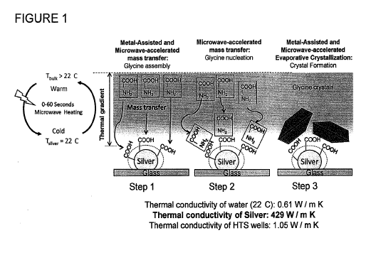
BRIEF DESCRIPTION OF THE DRAWINGS
Figure 1 is a schematic depiction of Metal-Assisted and Microwave-Accelerated
Evaporative Crystallization.
Figure 2 shows optical and SEM images for glycine crystals grown on blank
glass slides
from 3.2M solution, pH=6 (Top) at room temperature and (Bottom) using
microwave heating
(MW), wherein * indicates plate-like a-glycine.
Figure 3 shows optical and SEM images for glycine crystals grown on SIFs from
3.2M
solution, pH=6 (Top) at room temperature and (Bottom) using microwave heating.
Figure 4 shows optical images of L-Alanine crystals formed on blank glass
slides and
SIFs from 2.70 M solution at room temperature and using MA-MAEC technique. All
images
were taken with the same optical setup.
Figure 5 shows the time progression of the growth of L-alanine crystals on
blank glass
slides using MAEC technique at microwave power level 1.
7
CA 02851361 2014-04-07
WO 2013/055859 PCT/US2012/059660
Figure 6 shows the time progression of the growth of L-alanine crystals on
SIFs using
MA-MAEC technique at microwave power level 1.
Figure 7 shows a Raman spectrum of L-alanine crystallized on blank glass
slides at room
temperature and using the MA-MAEC technique (notation of functional groups at
peaks signifies
presence of functional group at the indicated wavelength).
Figure 8 shows a Raman spectrum of L-alanine crystallized on SIFs at room
temperature
and using the MA-MAEC technique (notation of functional groups at peaks
signifies presence of
functional group at the indicated wavelength).
Figure 9 shows an SEM image of Silver Island Films (SIFs) on blank glass
slides. SIF's
are ¨80nm in diameter.
Figure 10 shows SEM and optical images of glycine crystals grown from 3.2 M
pH=6
aqueous glycine solution on blank glass slides at room temperature (RT).
Figure 11 shows SEM and optical images of glycine crystals grown from various
aqueous
glycine solutions on blank glass slides using microwave heating (MW).
=
Figure 12 shows SEM and optical images of glycine crystals grown from 3.2 M
pH=6
aqueous glycine solution on SIFs at room temperature (RT).
Figure 13 shows SEM and optical images of glycine crystals grown from 3.2 M
pH=4
aqueous glycine solution on SIFs at room temperature (RT).
Figure 14 shows optical microscope images of glycine crystals grown from
various
aqueous glycine solutions on SIFs at room temperature (RT).
Figure 15 shows SEM images of glycine crystals grown from 3.2 M pH=4 aqueous
glycine solution on SIFs using microwave heating (MW).
8
CA 02851361 2014-04-07
WO 2013/055859 PCT/US2012/059660
Figure 16 shows SEM and optical images of glycine crystals grown from 3.2 M
pH=6
aqueous glycine solution on SIFs using microwave heating (MW).
Figure 17 shows optical microscope images of glycine crystals grown from
various
aqueous glycine solutions on SIFs using microwave heating (MW).
Figure 18 shows experimental 2-D (Left) and 1-D (Right) Powder X-Ray
Diffraction
patterns of glycine crystals grown from glycine solutions 3.2 M pH= 6 on glass
(A) at room
temperature (RT) and (B) using microwave heating (MW).
Figure 19 shows experimental 2-D (Left) and 1-D (Right) X-Ray Diffraction
patterns of
glycine crystals grown from glycine solutions 3.2 M pH= 6 on SIFs (A) at room
temperature
(RT) and (B) using microwave heating (MW).
Figure 20 shows experimental 2-D (Left) and 1-D (Right) patterns of glycine
crystals
grown from glycine solutions (A) 1.6 M pH= 9 on glass at room temperature (RT)
and (B) 3.2 M
pH= 9 on SIFs using microwave heating (MW).
Figure 21 shows simulated Powder X-Ray Diffraction patterns for a-, B- and y-
glycine
crystals.
Figure 22 shows simulated growth morphology of a-, B- and y- glycine crystals,
showing
the selected crystal faces, which were observed in the experimental data.
Figure 23 shows the time progression of the growth of L-alanine crystals on
blank glass
slides using MAEC technique at microwave power level 5.
Figure 24 shows the time progression of the growth of L-alanine crystals on
blank glass
slides using MAEC technique at microwave power level 10.
Figure 25 shows the time progression of the growth of L-alanine crystals on
blank glass
slides at room temperature.
9
CA 02851361 2014-04-07
WO 2013/055859 PCT/US2012/059660
Figure 26 shows the time progression of the growth of L-alanine crystals on
SIFs using
MA-MAEC technique at microwave power level 5.
Figure 27 shows the time progression of the growth of L-alanine crystals on
SIFs using
MA-MAEC technique at microwave power level 10.
Figure 28 shows the time progression of the growth of L-alanine crystals on
SIFs at room
temperature.
Figure 29 shows a powder X-ray diffraction pattern of L-alanine crystals.
DETAILED DESCRIPTION OF THE INVENTION
Any metal or metal oxide can be used in the present invention. Preferred
metals and
metal oxides are silver, gold, copper, aluminum, zinc, chromium, palladium,
nickel, rhodium,
iron, platinum, tin, gallium, indium, cadmium, cobalt, manganese, ruthenium,
and oxides thereof
In one embodiment of the present invention, these metals and metals oxides can
be used
alone.
In another embodiment of the present invention, two or more of these metals
and metal
oxides can be used at the same time.
Any suitable particle size can be used for the metal and metal oxide
particles. A
preferred particle size range for the metal and metal oxide particles is 2
nanometers to 2000
nanometers.
Any suitable film thickness can be used for the metal and metal oxide thin
films. A
preferred thin film thickness range for the metal and metal oxide thin films
is 10 nanometers to
2000 nanometers.
Surface engineering includes the modification of metal and metal oxides with
CA 02851361 2014-04-07
WO 2013/055859 PCT/US2012/059660
A) compounds containing i) amine or thiol head groups, ii) 3 to16 methylene
groups, and
iii) functional end groups (amine, carboxyl, hydroxyl, ethyl), or
B) compounds containing i) amine or thiol head groups, and ii) DNA or peptide
or
polynucleic acid or any single amino acids as functional end groups.
Any suitable microwave frequency can be used. A preferred microwave frequency
ranges from 0.3 to 30 GHz. A particularly preferred microwave frequency is
2.45 GHz.
Any suitable microwave power can be used. A preferred microwave power range is
1 W-
30000 W. A particularly preferred microwave range is 1 W-1200 W.
Amino acids which can be used in the present invention are isoleucine,
alanine, leucine,
asparagine, lysine, aspartic acid, methionine, cysteine, phenylalanine,
glutamic acid, threonine,
glutamine, tryptophan, glycine, valine, proline, selenocysteine, serine,
tyrosine, arginine,
histidine, ornithine, and taurine. Preferred amino acids include glycine,
alanine, arginine, and
glutamic acid.
Drug molecules which can be used in the present invention include all
commercially
available drug molecules and future molecules synthesized using organic
chemistry and drug
molecules derived from living organisms including bacteria and plants living
on land and in the
seas. Preferred drug molecules include acetaminophen and ranitidine.
Proteins which can be used in the present invention include all proteins found
in humans
and animals at their healthy and diseased states.
DNA/peptides which can be used in the present invention include all
DNA/peptides
found in humans and animals at their healthy and diseased states.
Any suitable solvent can be used for the amino acids, drug molecules,
proteins, and
DNA/peptides. A preferred solvent is deionized water or double-distilled
water.
11
CA 02851361 2014-04-07
WO 2013/055859 PCT/US2012/059660
The present invention provides rapid crystallization of amino acids, drug
molecules,
proteins and DNA/peptides. In this regard, crystallization is achieved in less
than 180 seconds
for samples smaller than 200 microliters in the case of amino acids and drug
molecules such as
in embodiments 14, 15 and 16 above, and crystallization is achieved in less
than 2 hours for
samples smaller than 200 microliters in the case of proteins and DNA/peptides
such as in
embodiments 17 and 18.
The present invention will now be described in further detail by way of the
following
examples, which should not be considered as limiting the present invention in
any way. In the
examples, power level 1, 5 and 10 means the application of 900 W in 10%, 50%
and 100% of the
total time, respectively.
EXAMPLE 1
The MA-MAEC technique was tested with a model amino acid, i.e., glycine.
Glycine has
three distinct polymorphs at ambient conditions: a, 13 and y (see Lee, A. Y.;
Lee, I. S.; Dettet, S.
S.; Boerner, J.; Myerson, A. S., Crystallization on confined engineered
surfaces: A method to
control crystal size and generate different polymorphs. Journal of the
American Chemical
Society 2005, 127, (43), 14982-14983). The formation of glycine crystals
mainly depends on the
type of solvent, pH and concentration. In the MA-MAEC technique used in this
example, metal
nano structures serves as 1) selective nucleation sites for the
crystallization of glycine due to the
interactions of primary amine (of glycine) and silver nanostructures and 2) a
microwave-
transparent medium for the creation of thermal gradient between a warmer
solution and the silver
nano structures that remain at room temperature after microwave heating (see
Aslan, K.; Geddes,
C. D., Microwave-accelerated metal-enhanced fluorescence: Platform technology
for ultrafast
12
CA 02851361 2014-04-07
WO 2013/055859 PCT/US2012/059660
and ultrabright assays. Analytical Chemistry 2005, 77, (24), 8057-8067). The
microwave heating
allows for the significant reduction in the time of crystallization process.
Figure 1 depicts the proposed mechanism for the MA-MAEC technique. In MA-MAEC,
upon exposure to microwave heating, a thermal gradient is created between the
solution and the
silver nanoparticles due to ¨620-fold difference in the thermal conductivity
of silver (429 W / m
K) and water (0.61 W / m K). This thermal gradient allows for the mass
transfer of glycine
molecules from the wanner solution to the cooler nanoparticles in an effort to
thennally
equilibrate the system. Subsequently, glycine molecules assemble either
directly (or by other
functional groups on silver) onto the silver nanoparticles (Figure 1, step 1).
With continued
microwave heating, mass transfer of glycine continues and the glycine
molecules assemble onto
the ones on the surface of silver nanoparticles in a process called nucleation
(Figure 1, step 2).
Crystal growth takes place as the solution evaporates and subsequent glycine
molecules assemble
on to one another until all glycine molecules crystallize (Figure 1, step 3).
Silver island films (SIFs) were deposited onto glass microscope slides by
allowing them
to soak in a heated silver nitrate/D-glucose solution as previously described
(see Aslan, K.;
Geddes, C. D., Microwave-accelerated metal-enhanced fluorescence: Platform
technology for
ultrafast and ultrabright assays. Analytical Chemistry 2005, 77, (24), 8057-
8067). Freshly
prepared SIFs (Figure 9) were used in all the experiments. The effect of
concentration and pH
on the crystallization of glycine in deionized water (no other solvent was
used) at constant
solution volume was studied. In this regard, aqueous solutions of glycine
(>99.5%, Sigma-
Aldrich, USA) with three different concentrations were prepared: 1.60, 3.20
and 4.0 M. The pH
of the glycine solutions was adjusted to 4 (acidic), 6 (neutral) and 9 (basic)
using 6M HC1 or 6M
NaOH. In the MA-MAEC experiments, a fixed volume (20 til) of freshly prepared
glycine
13
CA 02851361 2014-04-07
WO 2013/055859 PCT/US2012/059660
solution was pipetted onto SIFs-coated glass slides, which were then either
heated in a
conventional microwave oven (100% power level) or incubated at room
temperature. The time
taken for the solution to completely evaporate was recorded. In two control
samples, the
crystallization of glycine was carried out on blank glass slides with
microwave heating and on
blank glass slides at room temperature.
Glycine crystals formed on SIFs and glass slides were characterized by
microscopy
(optical microscope and scanning electron microscope, Figures 10-17) and
powder X-Ray
Diffraction (XRD) (see the Supporting Information below for the discussion of
X-ray
crystallography data). The crystal polymorph distribution was calculated using
the microscope
images of three different samples. Table 1 below summarizes the results for
the crystallization
of glycine using the MA-MAEC technique and control experiments. In this
regard, the crystal
morphology, crystal polymorph distribution (i.e., purity) and the total time
to evaporate different
glycine solutions are listed.
14
CA 02851361 2014-04-07
WO 2013/055859
PCT/US2012/059660
Table 1. Summary of results for the crystallization of glycine using MA-MAEC
technique and
control experiments.
SIFs- Microwave SIFs-Room Temperature
Crystal morphology / (purity) / time Crystal morphology / (purity) / time
CONCENTRATION CONCENTRATION
pH 1.6M 3.2M 1.61VI 3.2IVI 4.0NI
N/A # : a, y y cx, y V
4 - (10:90%) N/A* (10:90 %) (60:40 %) (100%)
43 G sec 24 sec 22 3 sec 12
0 Min 10 0 min 10 0 min
a a a, y ay a a
6 (100%) (100%) (25:35%) (70:30%) (100%) (100%)
57 t 6 sec 40 t 1 sec 50 1 sec 25 t
0 min 13 1 min 11 0 min
ct, v
9 (70; 30%) (30;40;30%): (100%) (15;60;25%)
(5:95%) (ND*)
53 6 sec 30 1 sec 30 Jec 24 2 min 21 min
17 0 min
Glass (No silver)-Microwave Glass
(No silver)-Room Temperature
Crystal morphology / (purity) / Utile Crystal morphology / (purity) /
CONCENTRATION CONCENTRATION
pH 1.6M 3.2M 4.0M 1.6M 3.2M 4.0M
N/A* WA 4 y y
4 ND* (100%) (100%) (100%)
55 6 sec 27 6 sec 20 1 sec 60 0 min 40 0 min 20 0 min
a, y a, y a, y a, y a
l'4)
6 (5:95 %) (50:50%) ND* (10:90%) (50:50%) (5;35;60%)
48 3 sec 33 3 sec 29 3,- 1 sec 461 0 min 42 0 min 12 0 min
N/A # a, y -
a,y,t3
9 (100%) ND* (10:90%) ND* (35;35;30%)
3 02 3 sec 21 2 sec 28 2 sec
40 0 min 4 0 min 13 0 min
# No crystals; % Not Determined;
Average of 3 samples
For a fixed volume of glycine solution, the total evaporation time on blank
glass slides at
room temperature (a control sample, evaporative crystallization) was recorded
to be between 12
(for 4M, pH=6) and 60 minutes (for 1.6M, pH=4). As the concentration of
glycine solution is
CA 02851361 2014-04-07
WO 2013/055859 PCT/US2012/059660
increased the total time of crystallization was decreased up to 4-fold, which
is due to the
presence of more glycine molecules in solution, driving the crystallization
process more rapidly.
In acidic and basic conditions, y-fonn of glycine was dominant, a-form of
glycine was observed
mostly at pH=6 as confirmed by XRD. Figures 2-Top and 10 show the optical
microscope and
SEM images of the glycine crystals formed on blank glass slides at room
temperature. As
expected, 'y-glycine is formed as needles (130-200 j.em in length) and a-
glycine (5-40 lam in
length) is foimed as bipyramids, which can be explained by a kinetically
controlled process
involving the presence of cyclic dimmers (see Weissbuch, I.; Lahav, M.;
Leiserowitz, L., Toward
stereochentical control, monitoring, and understanding of crystal nucleation.
Crystal Growth &
Design 2003, 3, (2), 125-150).
When identical glycine solutions on blank glass slides were exposed to
microwave
heating, glycine the solution completely evaporated in 20-55 seconds. However,
glycine crystals
were grown only for three out of nine solutions and the crystals were not well
organized as
compared to those grown at room temperature. That is, microwave heating of
glycine solution
on blank glass slides did not yield better crystals (Figure 2-Bottom, Figure
11, and Figure 18B.
Since primary amine (and thiol) groups have affinity towards silver
nanostructures,
glycine molecules are expected to assemble onto silver nanostructures through
amine groups
facing the silver surface. That is, silver nanostructures serve as selective
nucleation sites for the
crystallization of glycine, which increases the rate of crystallization and
potentially result in
selective polymorphism. Subsequently, the growth of glycine crystals at room
temperature was
carried out on SIFs. For a fixed volume of glycine solution, the total
evaporation time on SIFs at
room temperature was reduced by up to 5-fold (for 1.6 M, pH=4) as compared to
those on blank
glass slides at room temperature. Moreover, glycine crystals were grown on
SIFs for all nine
16
CA 02851361 2014-04-07
WO 2013/055859 PCT/US2012/059660
conditions and these crystals are well organized and larger (Fig. 3-Top and
Figs. 12-14) as
compared to those grown on blank glass slides. In this regard, the size of a-
glycine grown on
SIFs (up to ¨100 m in size) are ¨2-fold larger than those grown on blank
glass slides. This is
thought to be due to the presence of multiple silver nanostructures within
close proximity to one
another (Fig. 9), which affords for multiple crystal nucleation/growth
processes to occur
simultaneously.
It is also important to note that -y-glycine grown on SIFs reach lengths >1 mm
(Fig. 13),
which makes them a very promising candidate for non-linear optical
applications (see Bhat, M.
N.; Dharmaprakash, S. M., Effect of solvents on the growth morphology and
physical
characteristics of nonlinear optical gamma-glycine crystals. Journal of
Crystal Growth 2002,
242, (1-2), 245-252). In addition, a superior distribution of crystal
polymorphs was observed on
SIFs, where a desired type of polymorph can be grown in a relatively short
time. These
observations prove that the use of silver nanostructures (Metal-Assisted
Crystallization, MAC)
can significantly improve the crystallization process.
Despite the notable improvements afforded by MAC, the crystallization process
(for
complete evaporation of a 20 1..d solution) still requires up to 25 minutes to
be completed.
Subsequently, the effect of microwave heating on the crystallization process
on SIFs was
investigated (i.e, MA-MAEC). When identical glycine solutions on SIFs were
exposed to
microwave heating, the glycine solution completely evaporated in 22-57 seconds
(up to ¨60-fold
decrease as compared to glass at room temperature). Seven (out of 9) of the
glycine solutions
yielded well organized glycine crystals (Fig. 3-Bottom and Figs. 15-17). In MA-
MAEC the
heating of glycine solutions to higher temperatures (water is completely
evaporated) resulted in
the transformation of y-form into a- and I3-forms. This is due to the fact
that a- and y-glycine
17
CA 02851361 2014-04-07
WO 2013/055859 PCT/US2012/059660
are enantiotropically related and such transformation occurs at high
temperatures (see Lee, A. Y.;
Lee, I. S.; Dettet, S. S.; Boerner, J.; Myerson, A. S., Crystallization on
confined engineered
surfaces: A method to control crystal size and generate different polymorphs.
Journal of the
American Chemical Society 2005, 127, (43), 14982-14983). The existence of the
high energy 13-
form can be explained by the high super-saturation process resulted by rapid
evaporation of
water (see Lee, A. Y.; Lee, I. S.; Dettet, S. S.; Boerner, J.; Myerson, A. S.,
Crystallization on
confined engineered surfaces: A method to control crystal size and generate
different
polymorphs. Journal of the American Chemical Society 2005, 127, (43), 14982-
14983).
It is important to note that glycine crystals started to appear on SIFs before
the complete
evaporation (<1 min) of the aqueous glycine solution. That is, one can use the
MA-MAEC
technique without complete evaporation of the solvent, especially for the
separation of impurities
from the desired crystals.
In summary, the proof-of-principle of a platform technology, which involves
the use of
silver nanostructures with and without microwave heating to significantly
improve the
crystallization of organic small molecules, was demonstrated. In this regard,
the crystallization of
a model organic molecule (glycine) from a small volume aqueous solution using
microwave
heating was achieved in seconds. Glycine crystals grown on silver
nanostructures with and
without microwave heating were found be larger than those grown on blank glass
slides. The
MA-MAEC technique has the potential to selectively grow the desired polymorphs
of small
organic and biological molecules "on-demand" in a fraction of the time as
compared to the
conventional evaporative crystallization.
Supporting Information: The additional images of glycine crystals (Supporting
Inforniation 1) and powder XRD data (Supporting Infotmation 2) are discussed
below.
18
CA 02851361 2014-04-07
WO 2013/055859 PCT/US2012/059660
Supporting Information 1:
Figure 9 shows an SEM image of Silver Island Films (SIFs) on blank glass
slides. SIF's
are ¨80nm in diameter.
Figure 10 shows SEM and optical images of glycine crystals grown from 3.2 M
pH=6
aqueous glycine solution on blank glass slides at room temperature (RT).
Figure 11 shows SEM and optical images of glycine crystals grown from various
aqueous
glycine solutions on blank glass slides using microwave heating (MW).
Figure 12 shows SEM and optical images of glycine crystals grown from 3.2 M
pH=6
aqueous glycine solution on SIFs at room temperature (RT).
Figure 13 shows SEM and optical images of glycine crystals grown from 3.2 M
pH=4
aqueous glycine solution on SIFs at room temperature (RT).
Figure 14 shows optical microscope images of glycine crystals grown from
various
aqueous glycine solutions on SIFs at room temperature (RT).
Figure 15 shows SEM images of glycine crystals grown from 3.2 M pH=4 aqueous
glycine solution on SIFs using microwave heating (MW).
Figure 16 shows SEM and optical images of glycine crystals grown from 3.2 M
pH=6
aqueous glycine solution on SIFs using microwave heating (MW).
Figure 17 shows optical microscope images of glycine crystals grown from
various
aqueous glycine solutions on SIFs using microwave heating (MW).
Supporting Information 2:
Characterization of glycine crystals with powder X-ray diffraction (XRD) was
as follows.
XRD data for glycine crystals placed in a capillary tube with thin walls (0.02
mm) were collected
using an in-house X-ray generator (MicroMax 7, Rigaku/MSC, The Woodlands, TX)
and a
19
CA 02851361 2014-04-07
WO 2013/055859 PCT/US2012/059660
Raxis4++ image plate detector (Rigaku/MSC), which is housed at the Core
Facilities of the
Department of Pharmaceutical Sciences, University of Maryland School of
Pharmacy. The
distance between the detector and samples were kept constant at 75 mm. The
radiation source
was CuKa (wavelength: 0.54 nm). The 2-D XRD data was collected at 00 < < 120
at values of
00 < 20 < 40 .
The collected 2-D XRD data (in .0SC format) was converted to ".IMG" and ".PS"
foimats using ADXV software (see Pinard et al. below). 1-D Intensity vs. 20
plots was obtained
by fitting the ".IMG" files using FIT2D software (see Pinard et al. below).
The polymorph
reflections (e.g. a(020) were determined by comparing the peak locations in
the 20 plots for the
experimental (Figures 18-20) and simulated XRD patterns (Figure 21).
Simulated XRD patterns for a-, B-, and y-glycine were generated using Mercury
(Cambridge Crystallographic Data Center, Cambridge, United Kingdom, version
2.3). The
crystallographic parameters for glycine crystals (CIF files) were obtained
from published papers
(Ferrari, E. S.; Davey, R. J.; Cross, W. I.; GilIon, A. L.; Towler, C. S.
Crystal Growth & Design
2003, 3, 53-60; and Dawson, A.; Allan, D. R.; Belmonte, S. A.; Clark, S. J.;
David, W. I. F.;
McGregor, P. A.; Parsons, S.; Pulham, C. R.; Sawyer, L. Crystal Growth &
Design 2005, 5,
1415-1427).
Although optical microscopy and SEM images provide semi-quantitative
information
about the type of the glycine polymorphs due to the observable large size of
crystals, the XRD
data is more definitive. Figure 18 shows the 2-D XRD data for crystals grown
from a glycine
solution (3.2 M, pH=6) on glass at room temperature and using microwave
heating. The XRD
data also corroborate that the observation made by microscopy that a mixture
of a- and y-glycine
was grown on glass at room temperature and using microwave heating. The
intensity of
CA 02851361 2014-04-07
WO 2013/055859 PCT/US2012/059660
reflections from glycine crystals grown on glass at room temperature was
larger than those
grown using microwave heating, which indicates the larger number of crystals
grown at room
temperature, as again evidenced by SEM and optical microscope images. It is
important to note
that identical glycine solution was used. In Figure 18(A), the intensity of
peaks for a(011),
a(110) and a(020) are the largest indicating that glycine crystals are grown
preferentially along
these faces. Figure 22 (Top-Left) shows the depiction of the morphology for a-
glycine crystals
grown on glass at room temperature with these observed crystal faces. It is
also interesting to
note that bi-pyrimidal a-glycine crystals are formed through hydrogen bonding
that is strongest
in the bc- plane (011) and ab- plane (110). In addition, XRD data (Figure 18)
shows that y-
glycine was preferentially grown along the (101) face on glass slides.
Figure 19 shows that only a-glycine was grown on SIFs at room temperature and
using
microwave heating. It is important to remind that the crystallization on SIFs
occurred much
faster than on glass slides due to the presence of multiple silver
nanoparticles within close
proximity serving as nucleation/growth sites. This can be explained as in the
following: once the
initial glycine molecules are adsorbed onto silver nanoparticles through their
amine groups, the
subsequent glycine molecules are selectively assembled onto the first glycine
molecules through
the carboxylic acid groups (that is, Silver---[NH2¨COOF1]----[NH2¨0001-1]----
[NE12¨
COOF1]----). The assembling of glycine molecules occurs faster under microwave
heating due to
the temperature gradient between the solution and the silver nanoparticles.
Aslan, K.; Geddes, C.
D. Analyst 2008, 133, 1469-80. In this regard, it is also thought that
microwave heating lowers
the activation energy for the hydrogen bonding between glycine molecules,
effectively speeding
up the crystallization process. On the other hand, the assembly of glycine
molecules at room
temperature takes up to 20 minutes due to the absence of the driving force
(temperature gradient)
21
CA 02851361 2014-04-07
WO 2013/055859 PCT/US2012/059660
for the rapid transfer of glycine molecules from the solution to the
nucleation sites on the surface
of the silver nanoparticles.
It is also interesting to note a notable difference between the a-glycine
crystals grown on
glass at room temperature and on SIFs using microwave heating. As shown in the
XRD data
(Figures 18(A) and 19(B)), for a-glycine crystals grown on glass a strong peak
at ¨20
corresponding to the (110) face and a weak peak at ¨24 corresponding to the
(120) face appears.
Conversely, for a-glycine crystals grown on SIFs, the intensity for the peak
corresponding to the
(120) face is stronger and the peak at ¨20 corresponding to the (110) face is
not present. The
side-by-side comparison of the predicted a-glycine crystals morphology for
crystals grown on
glass at room temperature and on SIFs using microwave heating is shown in
Figure 22-Top.
Optical microscope and SEM images (Figures 10 and 16) show that the growth of
a-glycine
,
crystals on glass occurred preferentially in the z-direction (into the
solution; x-y is glass surface),
where glycine molecules were assembled onto smaller number of nucleation sites
on glass. In
comparison, the growth of a-glycine crystals on SIFs preferentially occurred
in the x-y direction
(on the surface), resulting in longer crystals due to the availability of
large number of
nucleation/growth sites (i.e., silver nanoparticles).
B-glycine crystals were also observed from some of the samples. Figure 20
shows the
XRD results for crystals grown from a 1.6 M, pl1=9 glycine solution on glass
at room
temperature and from a 3.2 M, pH=9 glycine solution on SIFs using microwave
heating. Once
again, the reflections from a-glycine and y-glycine were dominant, and in both
the samples
13(001) and 13(110) reflections were present. The presence of 13(001) and
13(110) reflections
indicate that 13-glycine crystals were grown as plates.
22
CA 02851361 2014-04-07
WO 2013/055859 PCT/US2012/059660
It is known that the heating of glycine solutions to higher temperatures
results in the
transformation of 7-form into a- and 13-forms. Lee, A. Y.; Lee, I. S.; Dettet,
S. S.; Boerner, J.;
Myerson, A. S. Journal of the American Chemical Society 2005, 127, 14982-
14983. This is due
to the fact that a- and 7-glycine are enantiotropically related and such
transformation occurs at
high temperatures. See Lee et al above. The existence of the high energy 13-
form can be
explained by the high supersaturation process resulted by rapid evaporation of
water. See Lee et
al above. The presence of 7-glycine on the surface after the crystallization
process ended
indicates the incomplete transformation of 7-glycine into a- and B-forms.
Figure 22-Middle and
Figure 22-Bottom show the predicted B- and 7-glycine crystals morphology for
crystals grown on
glass at room temperature and on SIFs using microwave heating.
Figure 18 shows experimental 2-D (Left) and 1-D (Right) Powder X-Ray
Diffraction
patterns of glycine crystals grown from glycine solutions 3.2 M pH=6 on glass
(A) at room
temperature (RT) and (B) using microwave heating (MW). The Greek letters on
the 1-D plots
indicate the type of glycine polymorph that the peak belongs, which was
determined by
comparing the simulated XRD pattern for all three polymorphs given in Figure
21. The Miller
indices corresponding to the peaks are also shown. The bell shape in the 1-D
plot is due to the
background signal as also observed in previous publications by others.
Hamilton, B. D.;
Hillmyer, M. A.; Ward, M. D. Crystal Growth & Design 2008, 8, 3368- 3375; and
Hamilton, B.
D.; Weissbuch, I.; Lahav, M.; Hillmyer, M. A.; Ward, M. D. Journal of the
American Chemical
Society 2009, 131, 2588-2596.
Figure 19 shows experimental 2-D (Left) and 1-D (Right) X-Ray Diffraction
patterns of
glycine crystals grown from glycine solutions 3.2 M pH= 6 on SIFs (A) at room
temperature
(RT) and (B) using microwave heating (MW). The Greek letters on the 1-D plots
indicate the
23
CA 02851361 2014-04-07
WO 2013/055859 PCT/US2012/059660
type of glycine polymorph that the peak belongs, which was detennined by
comparing the
simulated XRD patterns for all three polymorphs given in Figure 21. The Miller
indices
corresponding to the peaks are also shown.
Figure 20 shows experimental 2-D (Left) and 1-D (Right) patterns of glycine
crystals
grown from glycine solutions (A) 1.6 M pH= 9 on glass at room temperature (RT)
and (B) 3.2 M
pH= 9 on SIFs using microwave heating (MW). The Greek letters on the 1-D plots
indicate the
type of glycine polymorph that the peak belongs, which was deteiniined by
comparing the
simulated XRD patterns for all three polymorphs given in Figure 21. The Miller
indices
corresponding to the peaks are also shown.
Figure 21 shows simulated Powder X-Ray Diffraction patterns for a-, B- and y-
glycine
crystals. The Miller indices corresponding to the peaks are also shown.
Figure 22 shows simulated growth morphology of a-, B- and 7- glycine crystals,
showing
the selected crystal faces, which were observed in the experimental data.
Hydrogen bonds are
indicated as dashed lines.
This example is adapted from Pinard, M. A.; Aslan, K., Metal-Assisted and
Microwave-
Accelerated Evaporative Crystallization. Cryst Growth Des 2010, 10 (11), 4706-
4709, the
disclosure of which is incorporated herein by reference.
EXAMPLE 2
L-Alanine is an important amino acid that plays a key role in the molecular
structure of
many proteins. Crystallized forms of this molecule are currently in high
demand in chemical,
pharmaceutics, and food industries. However, the traditional evaporative
crystallization method
takes up to several hours to complete, and does not always consistently yield
usable crystals.
Using the metal-assisted and microwave-accelerated evaporative crystallization
(MA-MAEC)
24
CA 02851361 2014-04-07
WO 2013/055859 PCT/US2012/059660
technique, larger and better-organized L-Alanine crystals were formed in a
fraction of the time
using room temperature crystallization. This technique may be applicable to
organic molecules
other than amino acids, and thus will be able to produce the large amount of
molecular crystals
desired by industries today.
L-Alanine is one of the most abundant amino acids used in the synthesis of
proteins (see
Yamada, K.; Sato, A.; Shimizu, T.; Yamazaki, T.; Yokoyama, S., L-alanine
hydrochloride
monohydrate. Acta Crystallographica Section E-Structure Reports Online 2008,
64, 0806-
U1439). Because of its structural simplicity and importance in protein
construction, it is also a
key molecule in crystallization research. Furthermore, because hydrogen
bonding plays a large
role in alanine's molecular structure, research concerning this particular
amino acid can lead to a
better understanding of the structural dimensions of macromolecules such as
peptides and
proteins (see Mohan, R.; Kumar, K. S.; Raghavalu, T.; Mathivanan, V.;
Kovendhan, M.;
Sivakumar, B.; Kumar, G. R.; Raj, S. G., Structural, optical, spectral and
thennal studies of
nonlinear optical pure and deuterated L-alanine single crystals. Journal of
Crystal Growth 2008,
310, (6), 1182-1186). A number of studies have been conducted on the
properties of crystallized
L-Alanine, including studies about its vibrational spectra (see Machida, K.
K., A.; Saito, Y.;
Uno, T., Polarized Raman spectra and intermolecular potential of L-alanine
crystal. Spectrochim.
Acta, Part A 1978, 34, 909-914), morphology (see Lechuga-Ballesteros, D. R.-
H., N., Effects of
molecular structure and growth kinetics on the morphology of L-alanine
crystals. Int. Pharm
1995, 115, 151-160), and thermal properties (see Mohan, R.; Kumar, K. S.;
Raghavalu, T.;
Mathivanan, V.; Kovendhan, M.; Sivakumar, B.; Kumar, G. R.; Raj, S. G.,
Structural, optical,
spectral and thermal studies of nonlinear optical pure and deuterated L-
alanine single crystals.
Journal of Crystal Growth 2008, 310, (6), 1182-1186). However, a majority of
these studies
CA 02851361 2014-04-07
WO 2013/055859 PCT/US2012/059660
utilized the traditional room temperature evaporative crystallization method,
which can take up
to several days to complete.
In this Example, the application of metal-assisted and microwave-accelerated
evaporative
crystallization (MA-MAEC) to rapid crystallization of L-alanine, is used. The
MA-MAEC
technique is based on the combined use of microwave heating (for speeding up
the crystallization
process) and plasmonic nanostructures (silver island films, SIFs, as selective
nucleation sites) for
L-alanine crystal growth. The MA-MAEC technique is a promising new method for
rapid
molecular crystallization that significantly decreases the amount of time
required for complete
evaporation and crystallization to occur.
The effect of using SIFs and evaporative crystallization conditions (room
temperature
and microwave-accelerated) on the time of crystallization and type of crystals
of L-alanine were
studied. Table 2 below summarizes the results for the crystallization of L-
Alanine at room
temperature and using the MA-MAEC technique.
26
CA 02851361 2014-04-07
WO 2013/055859 PCT/US2012/059660
Table 2. Summary of results for the crystallization of 20 ul L-alanine from
2.70M solution on
glass slides and silver island films (SIFs) at room temperature and using MA-
MAEC technique.
1\1= 5 samples.
Glass SIFs Type of
Crystal
Room Temperature 50 3 min 41 13 min a
Microwave 6.5 1 7 1 min
Power Level 1 min a
Microwave 41 3 sec 45 6 sec
Power Level 5
Microwave 38 2 sec 22 3 sec
Power Level 10 a
For a fixed volume (20 ul) and concentration (2.70 M, pH=5.3) of L-Alanine
(minimum
of 5 samples were used), the crystallization process on blank glass slides and
SIFs took 50 3
minutes and 41 13 minutes on average at room temperature, respectively.
Complete L-Alanine
crystallization required 38 seconds to 6.5 minutes when using the microwave-
accelerated
evaporative crystallization (MAEC) technique on blank glass slides. Observable
crystals formed
on 25 of 31 blank glass slides, which is consistent with previously published
results for L-
glycine. Average crystallization time decreased as the microwave power level
was increased
when using the MA-MAEC technique. For example, crystallization of L-alanine
was completed
in only 22 seconds on SIFs when using the MA-MAEC technique at microwave power
level 10
and in 7 minutes on SIFs at microwave power level 1. It is also important to
note that all SIFs
surfaces yielded observable L-alanine crystals. The a-folin of L-Alanine
crystals was observed
27
CA 02851361 2014-04-07
WO 2013/055859 PCT/US2012/059660
by optical microscopy in all samples in this Example, which is similar to
observations made by
other groups (see Lechuga-Ballesteros, D. R.-H., N., Effects of molecular
structure and growth
kinetics on the morphology of L-alanine crystals. Int. I Pharm 1995, 115, 151-
160, and
Koyama, M.; Shiraishi, M.; Sasaki, K.; Kon-no, K., Preparation of L-Alanine
Crystals
Containing Gold Nanoparticles. Journal of Dispersion Science and Technology
2008, 29, (9),
1266-1271).
Figure 4 shows the visual comparison of L-alanine crystals formed using room
temperature and MA-MAEC techniques on both blank glass slides and on SIFs.
Crystals grown
using the MA-MAEC technique were consistently larger than those grown using
room
temperature crystallization. Crystal size ranged from 110 to 589 1.1m on blank
glass slides and
from 141 to 581 p.m on SIFs after complete evaporation. Crystals were believed
to have stopped
growing after complete evaporation of the aqueous solution because of a
decrease in
supersaturation of the solution (see Koyama, M.; Shiraishi, M.; Sasaki, K.;
Kon-no, K.,
Preparation of L-Alanine Crystals Containing Gold Nanoparticles. Journal of
Dispersion Science
and Technology 2008, 29, (9), 1266-1271). Consistent with previous research,
all a-crystals had
the largest face zone and were elongated along what was believed to be the c-
axis (see Lechuga-
Ballesteros, D. R.-H., N., Effects of molecular structure and growth kinetics
on the morphology
of L-alanine crystals. Int. I Pharm 1995, 115, 151-160).
As described in Pinard, M. A.; Asian, K., Metal-Assisted and Microwave-
Accelerated
Evaporative Crystallization. Cryst Growth Des 2010, 10 (11), 4706-4709, these
observations
were attributed to the fact that SIFs serve as selective nucleation sites for
L-alanine crystal
growth and as a microwave-transparent medium for the creation of thermal
gradient between the
wanner solution and the silver nanostructures that remain at room temperature
after microwave
28
CA 02851361 2014-04-07
WO 2013/055859 PCT/US2012/059660
heating. The microwave heating allows for the significant reduction in the
time of crystallization
process. It is well known that amine groups have affinity towards plasmonic
nanoparticles, such
as silver in particular (see Myerson, A. S.; Lee, A. Y.; Lee, I. S.; Dettet,
S. S.; Boerner, J.,
Crystallization on confined engineered surfaces: A method to control crystal
size and generate
different polymorphs. Journal of the American Chemical Society 2005, 127,
(43), 14982-14983).
Therefore, it is thought that the amine groups of L-alanine assemble onto
silver nanostructures,
becoming probable nucleation sites for the growth of crystals. This hypothesis
was tested by
comparison of crystal growth on blank glass slides and SIFs. Compared to L-
alanine crystals
fornied on blank glass slides at room temperature, crystals grown on SIFs were
more abundant
and had fewer imperfections. They also appeared to be more homogeneous in size
than crystals
grown on glass slides, where larger variation in the size of the crystals was
observed.
It is also important to note that the size distribution of the crystals grown
on blank glass
slides and SIFs using microwave power level 1 was homogeneous as compared to
heterogeneous
size distribution observed using microwave power levels 5 and 10. This is
attributed to the
excess microwave heating of the solution and the crystals formed during
microwave heating (at
power level 5 and 10). It is thought that excess microwave heating affects the
crystal nucleation
and growth by further increasing the rate of these processes.
In order to better understand the crystallization process during room
temperature and
microwave heating evaporation, optical images of the solution and the growing
crystals on blank
glass slides and SIFs were taken at time intervals as indicated in Figures 5,
6 and 23-28. In all
these experiments, microwave heating was stopped for a brief period of time (-
10 sec) to collect
optical images. Figure 5 (Glass_MW_PL1) shows the timed crystal growth
progression on glass
slides at microwave power level 1. Smaller crystals appeared by the time of
the first image (t=0
29
CA 02851361 2014-04-07
WO 2013/055859 PCT/US2012/059660
min) was taken. The crystal growth is clearly seen in the subsequent images,
where the crystals
seemed to grow to their final size at 4-7 min. These images also show that the
crystal movement
(t=0 to t=6 min) in solution, after which they rest in their final places
after the complete
evaporation of the solvent (at t=7 min). Similar observations were also made
for crystals grown
on glass slides using microwave power level 5 and 10 and room temperature (see
Figures 23-25).
Figure 6 (SIF_MW_PL1) shows the timed crystal growth progression on SIFs using
microwave power level 1. Crystals first started to appear on SIFs around 2 min
of microwave
heating, after which significant growth was observed until complete
evaporation at t=7 min. At
microwave power levels 1 and 5, significant improvement of the growth of
crystals was observed
on SIFs compared to glass slides. These crystals were much more abundant and
of better quality
than those grown using the MAEC technique on glass, which were imperfect and
scarce in
quantity. Crystal growth occurred on all SIFs samples of each microwave
heating condition, and
took only 22 seconds to 7 minutes for complete evaporation (microwave power
level 1 and 10,
respectively), proving that the same crystals can be grown using the MA-MAEC
technique over
10-fold faster than the traditional evaporative crystallization method. The
abundance of L-
alanine crystals formed using the MA-MAEC method can be explained by the
presence of silver
nanoparticles on the surface. SIFs served as nucleation sites that allowed for
the growth of
crystals in large quantities (see Pinard, M. A.; Asian, K., Metal-Assisted and
Microwave-
Accelerated Evaporative Crystallization. Cryst Growth Des 2010, 10, (11), 4706-
4709). In
comparison, the nucleation and growth of L-alanine crystals were random in
nature due to the
lack of functional surface groups on glass slides.
It is also important to note that when applying microwave heating to the L-
alanine
solution on both glass slides and SIFs, crystal organization improved when the
microwave was
CA 02851361 2014-04-07
WO 2013/055859 PCT/US2012/059660
stopped and started multiple times for imaging purposes, as compared to
uninterrupted
microwave heating of the same amount of time. This might be explained by the
high amount of
microwave energy being absorbed by the L-alanine solution in a short period of
time. The
amount of energy present may have been higher than required for crystal
growth, and thus may
have prevented the crystals from their normal growth.
Figures 7 and 8 shows the Raman spectra of L-alanine crystals grown on glass
slides and
SIFs at room temperature and using the MA-MAEC technique. Observable peaks
appear in the
same locations as those in previously published results (see Mohan, R.; Kumar,
K. S.;
Raghavalu, T.; Mathivanan, V.; Kovendhan, M.; Sivakumar, B.; Kumar, G. R.;
Raj, S. G.,
Structural, optical, spectral and thermal studies of nonlinear optical pure
and deuterated L-
alanine single crystals. Journal of Crystal Growth 2008, 310, (6), 1182-1186)
for L-alanine
grown on both glass and SIFs. This indicates that the crystals produced in
this Example possess
similar vibrational properties to other L-alanine crystals, and thus can be
deemed the type of L-
alanine crystals typically formed through room temperature evaporation from an
aqueous L-
alanine solution. Furthermore, since the Raman peaks are observed in identical
locations on
glass slides and SIFs, it can be concluded that the use of microwave heating
and SIFs accelerate
the crystallization process without altering the structural and vibrational
properties of the crystals
grown on them.
In summary, the results of this Example prove that the MA-MAEC technique is a
highly
effective method for rapid crystallization of L-alanine. Crystals produced
using microwave-
heating were larger in size than those grown at room temperature for both SIFs
and glass slides,
and were produced at a rate over 10-fold faster than that of the room
temperature method. The
presence of silver nanostructures on surfaces allowed for more selective
nucleation sites than on
31
CA 02851361 2014-04-07
WO 2013/055859 PCT/US2012/059660
blank glass slides, and therefore the simultaneous growth of more crystals was
able to occur.
Furthermore, the majority of crystals grown on SIFs was of better quality and
appeared with
fewer imperfections than those grown on glass. This Example demonstrates that
the use of the
MA-MAEC technique increases the efficiency of the crystallization of amino
acids.
Supporting Information: The additional images of L-alanine crystals and
experimental
details are discussed below.
Materials
Silver nitrate was purchased from Spectrum Chemical MFG Corp. Sodium
hydroxide,
ammonium hydroxide, D-glucose, and L-Alanine were purchased from Sigma-
Aldrich. All
chemicals were used as received.
Methods
Preparation of Silver Island Films. Silver island films were deposited onto
glass slides
(Corning). AgNO3 was precipitated by the addition of 5% NaOH, then quickly
redissolved by the
addition of NH4OH. The solution was then cooled to 5 C and blank glass slides
were immersed
in the solution for two minutes. D-glucose was added and the slides were
removed once they
were coated with a green color, after 5-7 minutes.
Preparation of L-Alanine Solution. A 2.70 M solution of L-alanine was prepared
by
dissolving appropriate amounts of L-alanine in double-distilled water
(Millipore), then heated to
60 C for up to 15 minutes, or until the solution appeared colorless and
transparent. The pH of the
prepared solution was slightly acidic at 5.3 (isoelectric point = 6) and was
used in all
experiments without changing the pH. The solution was stored in a 20 mL glass
vial (Corning) at
room temperature in between uses, and was heated to 60 C for 10 minutes before
each use.
32
CA 02851361 2014-04-07
WO 2013/055859 PCT/US2012/059660
Crystallization of L-Alanine. L-Alanine was deposited in 20 [LL drops onto
blank glass
slides (Corning) and SIFs, and was observed for crystallization at room
temperature and MA-
MAEC. Room temperature crystallization was carried out on an open laboratory
bench without
interference. The MAEC technique was performed in a conventional microwave
oven
(Frigidaire, 900 W) at microwave power levels 1, 5, and 10.
Timed images of growing crystals were recorded with a Swift Digital M1OL
Monocular
Microscope (Swift). The Raman spectra of L-alanine crystals were observed
using a Raman
spectrometer system (i-Raman from BW Tek, Inc. DE).
Figure 23 shows the time progression of the growth of L-alanine crystals on
blank glass
slides using MAEC technique at microwave power level 5. The actual length of
the crystals is
x4 of the lengths shown in the figure.
Figure 24 shows the time progression of the growth of L-alanine crystals on
blank glass
slides using MAEC technique at microwave power level 10. The actual length of
the crystals is
x4 of the lengths shown in the figure.
Figure 25 shows the time progression of the growth of L-alanine crystals on
blank glass
slides at room temperature. The actual length of the crystals is x4 of the
lengths shown in the
figure.
Figure 26 shows the time progression of the growth of L-alanine crystals on
SIFs using
MA-MAEC technique at microwave power level 5. The actual length of the
crystals is x4 of the
lengths shown in the figure.
Figure 27 shows the time progression of the growth of L-alanine crystals on
SIFs using
MA-MAEC technique at microwave power level 10. The actual length of the
crystals is x4 of
the lengths shown in the figure.
33
CA 02851361 2014-04-07
WO 2013/055859 PCT/US2012/059660
Figure 28 shows the time progression of the growth of L-alanine crystals on
SIFs at room
temperature. The actual length of the crystals is x4 of the lengths shown in
the figure.
Figure 29 shows a powder X-ray diffraction pattern of L-alanine crystals grown
in this
example.
This example is adapted from Alabanza, A. M.; Asian, K., Metal-Assisted and
Microwave-Accelerated Evaporative Crystallization: Application to L-Alanine.
Cryst Growth
Des 2011, 11(10), 4300-4304, the disclosure of which is incorporated herein by
reference.
While the invention has been described in detail and with reference to
specific
embodiments thereof, it will be apparent to one skilled in the art that
various changes can be
made without departing from the spirit and scope thereof.
34