Note: Descriptions are shown in the official language in which they were submitted.
CA 02851752 2014-04-10
2
1
Method for quantifying renal markers by urinary dosage.
1. Field of the invention
The field of the invention is that of uronephrology. More specifically, the
invention
pertains to a method of in vitro diagnosis of pathologies of the urinary
system.
2. Prior art
Kidney diseases can affect the different structural compartments of the
kidney: the
vessels, the glomeruli, the tubules or the interstitium. These disorders lead
to acute and/or
chronic kidney failure, their ultimate development being the total destruction
of the
functional units of the kidney which are replaced by an expansion of the extra
-cellular matrix,
i.e. renal fibrosis. These kidney diseases have various corresponding
etiologies: obstruction of
the excretory tracts, inflammation, auto-immunity, allergy, deposition
diseases, hypertension,
diabetes, vasculopathies, ischemia, toxicity, etc. Kidney diseases can affect
native kidneys or
allotransplants after a kidney transplant. In France, it is estimated that
there are 3,000 new
cases of transplants (kidney, heart, liver, bone marrow, lungs, etc) every
year. The systematic
follow-up of patients having received transplants has enabled the study of the
early stages of
renal diseases evolving towards fibrosis. The expression in the renal tissue
of epithelial-
mesenchymal transition (EMT) markers enables early detection of a fibrosing
disease in the
renal tissues, which can be caused by ischemia, the rejection or toxicity of
immunosuppressants, especially cyclosporin A (CsA) (Slattery et al, Am J
Pathol. 2005 August;
167(2): 395-407; Hertig et al, American Journal of Transplantation 2006,
Galichon et al,
Fibrogenesis Tissue Repair 2011; Galichon et al,. Transplantation, 2011).
EMT is a dynamic process during which the cells lose their epithelial
characteristics and
acquire mesenchymal characteristics. These modifications affect the morphology
of the cell as
well as its working. When EMT reaches the renal tubular cells, it progresses
towards fibrosis
and chronic renal failure (Hertig et al, J Am Soc Nephrol 2008). It is
therefore necessary to
monitor the appearance of this phenomenon among patients who have undergone
transplants in order to adapt or modify the immunosuppressant treatment.
At present, the reference method implemented for the detection and monitoring
of
any renal pathology is biopsy. Biopsy consists in removing a core of tissue
from the kidney by
CA 02851752 2014-04-10
2
transcutaneous, transvenous or surgical means. This sample is then subjected
to a histological
examination to detect possible signs of pathology (destruction, cell
infiltration or hypertrophy
of the glomerular, tubular, vascular or interstitial compartments).
This method however has numerous drawbacks. Taking samples is not without
risks for
the patient. Many complications have been observed such as hematuria,
obstructive renal
failure and even anuria, hematoma in the perirenal region, the appearance of
arterial and
venous fistulas and more rarely hemorrhage, loss of transplant, and death.
Apart from the
risks related to any invasive procedure, it can happen that the biopsies are
performed in a
region that does not represent the overall condition of the kidney and that,
therefore, the
patient's true situation is under-estimated or over-estimated because of this
sampling
procedure.
In addition, since biopsies cannot be done systematically at short intervals,
this
method does not enable early detection of the appearance of a pathology
either. Besides,
performing a renal biopsy is costly, complex, invasive and painful for the
patient.
It is therefore necessary to find a non-invasive, simple, economical, reliable
method of
diagnosis that enables early detection and entails the least possible risk for
the patient.
3. Goals of the invention
The invention is aimed at overcoming these drawbacks of the prior art.
More specifically, it is a goal of the invention, in at least one embodiment,
to provide a
method of early diagnosis of renal pathologies or of pathologies having renal
repercussions.
It is another goal of the invention, in at least one embodiment, to implement
a method
of non-invasive diagnosis.
It is yet another goal of the invention, in at least one embodiment, to
implement a
reliable and precise method of diagnosis.
It is another goal of the invention, in at least one embodiment of the
invention, to
implement a method of diagnosis that is simple to perform.
It is another goal of the invention, in at least one embodiment, to implement
a more
economical method of diagnosis.
It is another goal of the invention, in at least one embodiment, to implement
a method
for following up the efficacy and tolerance of a treatment.
Finally, it is another goal of the invention, in at least one embodiment, to
implement a
CA 02851752 2014-04-10
3
method of diagnosis that is less costly for the patient.
4. Summary of the invention
These goals as well as others that shall appear here below are achieved
entirely or at
least partly by means of a method of in vitro diagnosis of pathologies in a
patient.
According to the invention, such a method comprises the following steps:
a) obtaining a urine sample from said patient,
b) detecting, in said sample, at least one marker of said pathology and at
least one
specific marker of the urothelial cells and/or the urothelial microparticles,
and
c) determining a threshold of expression of said at least one marker of said
pathology by normalization of said marker of said pathology by said at least
one specific marker of the urothelial cells and/or the urothelial
microparticles.
Thus, the invention relies on the use of cells and microparticles contained in
the urine
in order to extract therefrom the genetic material and to compare the
expression of a gene of
interest, correlated with a pathology, with the expression of a specific gene
of the urine cells
unaffected by the pathology.
Urine indeed contains a small quantity of urothelial cells arising out of the
normal
renewal of the epithelium of the urinary excretory tracts. It can also contain
quantities,
variable depending especially on the presence of a renal pathology, of
leukocytes, renal
tubular or glomerular cells, blood as well as microparticles.
The term "microparticles" is understood to mean complex vesicular structures
that can
be released by most cells during the activation process or apoptosis. They are
formed by a
bilayer membrane of phospholipids exposing transmembrane proteins and
receptors, and
they enclose cytosolic constituents such as enzymes, transcription factors and
mRNA coming
from their mother cells.
The term "urothelial cells" is understood to mean transitional epithelial
cells forming
the human urothelium, from the pelvis up to the urethra. These cells have
various shapes:
cylindrical, kite-shaped, umbrella-shaped and balloon-shaped.
The term "specific marker of urothelial cells" is understood to mean a gene
specifically
expressed by the urothelial cells or urothelial microparticles, whether it is
within the cells or
on the surface, the level of synthesis of this gene by said cell being
independent of the
pathologies that can effect the renal cells. This notion is therefore
different from the notion of
CA 02851752 2014-04-10
4
a housekeeping gene, the expression of which is ubiquitous whatever the cell
type, the
function of the cell or its state.
Although this is rare, it is possible using current-day methods of molecular
biology or
biochemistry to extract nucleic acids and proteins from the cell and the
microparticles
contained in the patient's urine. In order to eliminate the bias related to
the quantity of cells
and microparticles in the sample, it is common practice to express the
expression of the gene
of interest as a function of the expression of a housekeeping gene such as
GAPDH
(glyceraldehyde 3 phosphate dehydrogenase) genes, 185 ribosomal RNA, the
cyclophilin A or
B, the P-catenin or again HPRT (hypoxanthine-guanine
phosphoribosyltransferase) (Absolute
quantification of mRNA using real-time reverse transcription polymerase chain
reaction, S.A.
Bustin, Journal of Molecular Endocrinology, 2000, 25, 169-193).
However, this method does not take account of the cellular specificity of the
pathology. Thus, the modification of the expression of a gene linked to a
pathology affecting a
precise cell type can be masked by the fact that the housekeeping gene coming
from other
cell types present in the urine is strongly expressed.
One of the contributions of the invention is therefore that it normalizes the
expression
of the gene of interest by the expression of a gene independent of the cell
types affected. This
step of normalization is used to determine a threshold of expression of the
pathological
marker by means of a urine marker independent of the quantitative and
qualitative variations
of the urine cells of renal origin. It is then possible to know whether this
marker is expressed
strongly or, on the contrary, weakly. This characteristic makes it possible to
obtain a
diagnostic test that is reliable, precise and reflects the patient's state of
health with greater
exactness. Thus, the method according to the invention can be used for
diagnostic purposes
to monitor the progress of a pathology.
In addition, working from a urine sample has many advantages:
- the sample is easy to access;
- collecting the sample is non-invasive and painless;
- collecting the sample is economical.
The simplicity of this method also makes it possible to implement it in all
types of
laboratories without making use of special technical qualifications or
equipment other than
that commonly used. The absence of any particular investment for implementing
this method
thus reduces the costs of analysis.
CA 02851752 2014-04-10
Finally, this method of normalization can be applied to different pathologies,
renal or
non-renal, provided that these pathologies modify the expression and/or
quantity of
urothelial cells and/or microparticles excreted and present in the urine.
The invention furthermore pertains to a method of in vitro diagnosis in which
the step
5 b)
comprises the detection of the product of transcription of said at least one
specific marker
of the urothelial cells and/or urothelial microparticles and of the product of
transcription of
said at least one marker of said pathology.
The study of the products of transcription is more reliable than the study of
the
presence of a gene in the genome of the cell. It is indeed well known that the
presence of a
gene in the genome of a cell cannot necessarily be correlated with its
expression in said cell,
since the regulation of the expression of a particular gene is subject to
numerous parameters.
The detection of the product of transcription therefore makes it possible to
obtain a more
precise and more reliable result.
Another object of the invention is a method in which the step b) is
implemented by
means of a technique of amplification of nucleic acids chosen from the group
comprising RT-
PCR, quantitative PCR, final-point PCR, semi-quantitative PCR or their
combination.
The term PCR (Polymerase Chain Reaction) is understood to mean the technique
in
which a fragment of target DNA is replicated in vitro in numerous copies. The
term RT-PCR
(Reverse Transcriptase Polymerase Chain Reaction) is understood to mean in
vitro synthesis of
a complementary DNA from extracted messenger RNAs . The term "quantitative
PCR", also
known as real-time PCR, is understood to mean the technique of in vitro
replication of a
fragment of target DNA additionally enabling measurement of the initial
quantity of this
target fragment. Semi- quantitative PCR can be distinguished from quantitative
PCR in that
the PCR is interrupted at several points enabling the initial quantity of DNA
to be evaluated.
This type of PCR is useful when the quantity of DNA is unusually low. Final-
point PCR
associates the Northern Blot technique with classic PCR in order to evaluate
the initial
quantity of DNA by comparison of the bands on agarose gel.
These methods, which cost little to implement, have the advantage of giving a
fast and
reliable result compatible with the requirements of a diagnostic test.
The invention furthermore pertains to a method of in vitro diagnosis in which
the step
b) is implemented using a nucleic acid hybridization technique chosen from the
group
comprising in situ hybridization (ISH), fluorescence in situ hybridization
(FISH) or hybridization
CA 02851752 2014-04-10
6
with marking by fluorescence (FISH), biochip hybridization, the Northern Blot
method or the
Southern Blot method.
The invention also pertains to a method of in vitro diagnosis in which the
step b) is
implemented through a method of sequencing of the nucleic acids.
Yet another object of the invention is a method of in vitro diagnosis in which
the
pathology is a renal pathology chosen from the group comprising renal
fibrosis, a phenotypic
change of the renal epithelial cells, a transplant rejection, a cancer, the
glomerular diseases
(diabetes, extramembranous glomerulonephritis, minimal glomerular lesions,
segmentary and
focal hyalinosis, etc), the tubular diseases (acute tubular necrosis,
expression of epithelial-to-
mesenchymal transition markers, atrophy, cellular rejection, obstruction of
the excretory
tracts, etc), the interstitial diseases (inflammation, fibrosis) and the
vascular kidney diseases
(arterial hypertension, thrombotic microangiopathy, humoral rejection, etc).
The term "epithelial- mesenchymal transition" (EMT) refers to a biological
process that
enables a polarized epithelial cell, interacting normally with the basal
membrane, to
undertake numerous biochemical transformations that enable it to acquire a
mesenchymal
cell phenotype, including increased migratory capacity, an invasive character,
increased
resistance to apoptosis and massive increase in the components of the extra-
cellular matrix
(Kalluri R, Weinberg RA, "The basics of epithelial-to-mesenchymal transition",
J Clin Invest. 119
(2009) 1420-1428). The epithelial phenotypic changes are the EMT markers (for
example
vimentin and 13-catenin in the tubular epithelium) that can be studied in the
tissues in a
clinical situation (Hertig A. et al.n "Early epithelial phenotypic changes
predict graft fibrosis", J
Am Soc Nephrol. 19 (2008) 1584-1591).
Another object of the invention is a method during which said patient has
received an
organ transplant and said renal pathology is the presence of an interstitial
fibrosis, a tubular
atrophy or epithelial- mesenchymal transition in the renal transplant.
The method according to the invention therefore enables the efficient and
early
detection of the emergence of a renal pathology such as inflammation or EMT-
inducing
epithelial phenotypic changes in the kidney.
Yet another object of the invention is a method of in vitro diagnosis in which
at least
one specific genetic marker of said renal pathology is chosen from the group
comprising the
human genes CD45 (SEQ ID 1), CD68 (SEQ ID 2), and VIM (SEQ ID 3) as well as
the genes
CA 02851752 2014-04-10
7
having a sequence homology of at least 80%, preferably at least 85%,
preferably at least 90%,
preferably at least 95%, preferably at least 99% with these human genes.
By way of a precise indication, the human gene CD45 is also symbolized as
PTPRC
(Protein Tyrosine Phosphatase Receptor type C). In the following description,
the human gene
CD45 or PTRPC will be designated equally by the symbol CD45 or PTPRC.
The inventors have surprisingly discovered that the expression of these genes
is
considerably increased in the urine of patients having undergone clinically
stable kidney
transplants but for which, however, the biopsy of the transplant reveals the
presence of
epithelial phenotypic changes. This over-expression can be correlated with the
presence of
epithelial phenotypic changes arising during an epithelial- mesenchymal
transition and
tubular-interstitial diseases in the biopsies of renal allotransplants, these
biopsies being
performed in the context of a systematic screening three months after the
transplant. It is
possible, as understood in the invention, to search for the expression of only
one gene, which
is a specific marker of a pathology. However, in order to refine the
diagnosis, it is preferable to
search for a combination of different genes, the expression of which in urine
takes account of
the presence of a particular renal pathology. For example, we can cite
research of the
expression of CD45, or PTPRC, and CD68 genes, normalized by uroplakin to
detect the
presence of inflammatory cells in the transplant. It is also possible to
search for the expression
of certain tumor markers. Examples that can be cited are markers for clear-
cell carcinoma, the
search for racemase, caveolin-1 (SEQ ID 29), ROR1 (SEQ ID 30), CD10 (SEQ ID
31), keratin 7,
vimentin (SEQ ID 3), TP53 (SEQ ID 26) in the context of monitoring a renal
cancer.
A "homologous sequence" or a "sequence homology" between nucleotide sequences
is determined by linear comparison of the nucleotide sequences using the
software BLAST
(Basic Local Alignment Search Tool), using the algorithm blastn available on
the NCBI site:
(http://blast.ncbi.nlm.nih.gov/Blast.cgi?PROGRAM=blastn&BLAST_PROGRAMS=megaBlas
t&P
AGE_TYPE=BlastSearch&SHOW_DEFAULTS=on&LINK_LOC=blasthome).
The parameters chosen for this analysis are the following:
¨ database: human genomic plus transcript (Human G +T);
¨ no exclusion of models or samples of environmental sequences;
¨ optimizing of the program: blastn (somewhat similar sequences);
¨ short queries = automatically adjusted parameters for input sequence;
¨ expect threshold = 10;
CA 02851752 2014-04-10
8
¨ word size = 11;
¨ max match in a query range = 0.
With respect to the scoring parameters, these parameters are fixed by default
(match/mismatch scores = 2-3; gap costs = existence: 5, extension: 2).
Finally, no filter is
applied.
According to another advantageous embodiment, said pathology is a pathology
modifying the quantity of cells and/or microparticles excreted in the urine.
Preferably, said pathology modifying the quantity of cells and/or
microparticles
excreted in the urine is chosen from the group comprising glomerular diseases
such as
segmentary and focal hyalinosis, tubular diseases such as acute tubular
necrosis, epithelial
phenotypic changes, cell rejection and interstitial diseases such as acute
transplant rejection,
Sjogren's syndrome and sarcoidosis.
Thus, the method according to the invention enables the reliable, speedy and
non-
invasive detection of the development of non-renal diseases through the
collection of urine
samples from the patient, when the pathologies modify the profile of gene
expression and/or
the quantity of cells excreted in the urine.
Tubular necrosis takes the form of an increase in the number of tubular cells
in the
urine due to a major desquamation of the walls of the renal tubular
epithelium. Segmentary
or focal hyalinosis is accompanied by a major quantity of podocytes in the
urine. The increase
in the number of leukocytes is a sign of acute rejection of a transplant.
Advantageously, said at least one specific marker of urothelial cells is
chosen from the
group comprising the human genes uroplakin 1A (SEQ ID 4), uroplakin 18 (SEQ ID
5), uroplakin
2 (SEQ ID 6), uroplakin 3A (SEQ ID 7),uroplakin 3B (SEQ ID 8), uroplakin 3BL
(SEQ ID 9), Bcas1
(SEQ ID 10), CEP152 (SEQ ID 11), CRABP2 (SEQ ID 12), DNASE1 (SEQ ID 13), KRT20
(SEQ ID 14),
PLEKHF1 (SEQ ID 15), PLEKHG4B (SEQ ID 16), RCN1 (SEQ ID 17), SEMA5B (SEQ ID
18), SULT2A1
(SEQ ID 19), TFF1 (SEQ ID 20), VILL (SEQ ID 21), ZNF720 (SEQ ID 22) as well as
genes having
sequence homology of at least 80%, preferably at least 85%, preferably at
least 90%,
preferably at least 95%, preferably at least 99% with these genes.
Among the cells present in the urine, only the urothelial cells express these
genes.
These genes are specifically and constantly expressed by the urothelial cells
and/or
microparticles, independently of the renal pathologies. Naturally, these genes
are present in
the genetic material contained in the nucleus of each cell of the organism.
However, the
= CA 02851752 2014-04-10
9
genes are not expressed in the same way by all the cells of the organism. In
other words, not
all the genes are transcribed from DNA to m RNA and then translated from mRNA
into protein
in all the cells forming the human body. However, the inventors have
discovered that, among
the cells and microparticles contained in urine, these genes are specifically
expressed by the
urothelial cells and that that they are so expressed constantly. They
therefore constitute
referentials of choice for normalizing genes linked to a pathology
specifically affecting the
renal cells or modifying their quantity in the urine. The inventors wish to
emphasize that the
above-mentioned genes can in fact be detected in other cell types, for example
when this
detection is based on a simple search for the presence of a gene in the total
DNA and not in
the genes expressed by a cell type. These genes can also be expressed by other
types of cells.
However, their interest in the present invention is related to the fact that,
among all the cell
types that can be found is a patient's urine sample, only the urothelial cells
express these
genes, independently of pathological conditions. Consequently, as understood
in the
invention, the notion of a specific marker of the urothelial cells or of the
urothelial
microparticles must be distinguished from the notion of a
housekeepinghousekeeping gene.
Indeed, housekeepinghousekeeping genes are genes expressed by all the cells
whatever their
cell type and their function. On the contrary, the term "specific marker of
the urothelial cells"
corresponds to genes expressed solely by the urothelial cells among all the
cells that can be
found in a urine sample.
Besides, these genes have been identified by the inventor as genes that can be
used to
obtain an excellent statistical correlation (p value < 0.01) relative to this
method of
normalization by the housekeeping genes (18S RNA, GAPDH, etc). The p value
obtained for
each gene is indicated in Table 1 below.
Table 1 ¨Specific markers of the urothelial cells
Name of the
p-value
gene
UPK1A 1.37. 1040
-
UPK1B 1.40. 10-12
UPK2 2.11. 10-12
UPK3A 4.04. 10-9
Bcas1 4. 10-5
PLEKHF1 2.17. 10-5
=
= = CA 02851752 2014-04-10
KRT20 3.05. 10-5
ZNF720 7.80. 10-5
UPK3B 1.74. 10-4
RCN1 1.97. 10-4
TFF1 2.54. 10-4
Preferably, said at least one specific marker of the urothelial cells or of
the urothelial
microparticles is chosen from the group of genes comprising the human genes
UPK1A, UPK1B,
UPK2 and UPK3A as well as the genes having a sequence homology of at least
80%, preferably
at least 85%, preferably at least 90%, preferably at least 95%, preferably at
least 99% with
5 these genes.
The term "normalization" is understood to mean the elimination of biases
related to
errors of measurement or manipulation and independent of the biological
variations. The step
of normalization therefore enables the production of more reliable results.
Normalizing by
using specific genes of the cells affected by the pathology being sought
eliminates the bias
10 related to the proportion of these same cells in the urine sample.
Indeed, the quantitative and
qualitative composition of urine is not fixed either because of the rate of
flow of urine or
because of the disease. The risk of wrongly estimating the true progress of
the pathology in
the patient is therefore real and could prove to be detrimental to his
therapeutic treatment.
For example, when the quantitative PCR technique was used, the normalization
consisted of a passage from the logarithmic scale (corresponding to the raw
result) to a linear
scale in two steps:
(1) CP specific marker of urothelial cells ¨ CP
pathological marker = ACP,
the Cp, or crossing point, being the number of cycles of amplification before
detection of the
fluorescent signal by the apparatus.
ACp
(2) Level of expression of the normalized pathological marker = 2
When a migration on gel is implemented to display results as is the case for
the classic
PCR followed by migration on agarose gel or for the Northern and Southern Blot
techniques,
the operator must measure the optical densities of each migration band with
any usual
software (Image JTM, UN-SCAN-IT GeITM) and make a report of it to determine
the threshold of
expression of the gene of interest in the patient. The threshold of expression
is then
computed as follows:
Level of expression of the normalized pathological marker = optical density of
pathological
marker/optical density of urothelial cell marker
= CA 02851752 2014-04-10
11
Another object of the invention is a method of in vitro diagnosis further
comprising a
step for comparing the threshold of expression of said marker of a renal
pathology with a
threshold of expression unchanged by the disease.
The comparison with a healthy patient makes it possible to note the positive
or
negative influence of a pathology on the regulation of the expression of a
normally expressed
gene. It is done as follows:
(ACp patient - ACp healthy individual)
Regulation of a gene = 2
Apart from the comparison with a healthy patient, it is possible to monitor
the
progress of a transplant or a pathological state through the method of the
invention. It is
indeed possible to keep the previous results of a patient and to use them as a
referential. This
internal normalization eliminates the bias due to the differences between
individuals. It also
enables the medical follow-up of the patient and the monitoring of his illness
or of the
transplant.
The invention further comprises an in vitro diagnostic kit for the detection
of
pathologies from a urine sample coming from a patient, the kit comprising at
least one pair of
primers for the detection, in said sample, of at least one specific marker of
a pathology and at
least one pair of primers for the detection, in said sample, of at least one
specific marker of
the urothelial cells.
Advantageously, said pathology is a renal fibrosis or a phenotypic change of
the renal
epithelial cells, and said at least one specific marker of a renal pathology
is chosen from the
group comprising the human genes CD45, CD68 and VIM.
In a preferred embodiment, said at least one specific marker of urothelial
cells is
chosen from the group comprising the human genes uroplakin 1A, uroplakin 1B,
uroplakin 2,
uroplakin 3A, uroplakin 3B, uroplakin 3BL, Bcas1, CEP152, CRABP2, DNASE1,
KRT20, PLEKHF1,
PLEKHG4B, RCN1, SEMA5B, SULT2A1, TFF1, VILL, ZNF720 as well as genes having a
sequence
homology of at least at least 80%, preferably at least 85%, preferably at
least 90%, preferably
at least 95%, preferably at least 99% with these human genes.
Another object of the invention lies in the use of the in vitro diagnostic kit
for the
detection of a renal pathology, said renal pathology being a fibrosis or a
phenotypic change of
the renal epithelial cells.
= CA 02851752 2014-04-10
12
5. List of figures
Other features and advantages of the invention shall appear more clearly from
the
following description of a preferred embodiment, given by way of a simple
illustratory and
non-exhaustive example and from the appended drawings, of which:
- Figure 1 is a graph showing the correlation of the EMT scores with the
result of the
urine PCR for vimentin (VIM) normalized by GAPDH.
Figure 2 illustrates the correlation of the same EMT scores with the same
results of
urine PCR for vimentin (VIM) when the results are normalized by the uroplakin
1A gene
UPK1A.
- Figure 3 is a graph representing the correlation of the EMT scores with
the results of
the urine PCR for CD68 normalized by GAPDH.
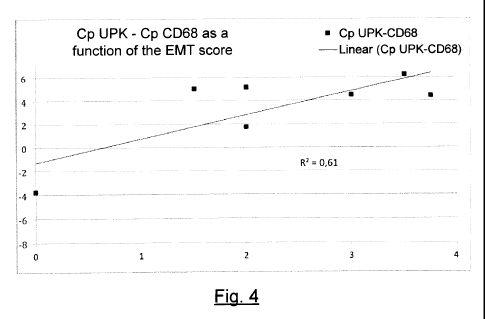
- Figure 4 shows the correlation of the same EMT scores with the same
results of urine
PCR for CD68 when these results are normalized by the uroplakin 1A gene UPK1A.
- Figure 5 represents the correlation of the EMT scores with the results of
the urine PCR
of CD45 normalized by GAPDH.
- Figure 6 represents the correlation of the same EMT scores with the same
results of
urine PCR for CD45 when these results are normalized by the uroplakin 1A gene
UPK1A.
- Figure 7 is a graph representing the number of identified genes
corresponding to the
terms "kidney" and "the inter-cell junction" according to the method of the
invention.
- Figure 8 is a graph representing the significance of gene enrichment with
respect to
the term "kidney" and the term "inter-cell junction" according to the method
of
normalization.
6. Description of one embodiment of the invention
The general principle of the invention relies on the comparison of the
expression of a
gene correlated with a pathological phenomenon, designated as a marker of a
pathology or
pathological marker, with the expression of a reference gene, the level of
expression of which
in the urothelial cells is independent of the cells affected by the pathology.
This marker is
designated as a specific marker of the urothelial cells.
= CA 02851752 2014-04-10
13
Example 1: Diagnosis of the epithelial phenotypic changes of the renal
transplant
through the method of the invention
In order to evaluate the sensitivity of the diagnostic method according to the
invention, renal biopsies are carried out on patients who have undergone an
organ transplant
and have been treated with CsA. At the same time, a sample of their urine is
collected. In
these samples, a search is made by quantitative PCR for markers associated
with a phenotypic
change of the renal epithelial cells. In order to demonstrate the superiority
of the method
according to the invention, the results of quantitative PCR are normalized
according to a
urothelial reference gene, in compliance with the method according to the
invention, and
according to a housekeepinghousekeeping gene, in compliance with the classic
method
described in the literature.
Control biopsies on transplant patients are analyzed by the Anatomopathology
Laboratory of the Hopital Tenon (Paris). A search is made for the protein
expression of
vimentin and 13-catenin, which are EMT markers, according to methods of
immunohistochemistry well known to those skilled in the art. The EMT score is
determined as
a function of the percentage of renal tubules expressing the EMT markers, i.e.
vimentin and 13-
catenin (Hertig et al, American Journal of Transplantation 2006). These scores
are expressed
as follows:
- score = 0: no EMT;
- score = 1: <10% of the renal tubules in the biopsy present EMT markers;
- score = 2: 10-24% of the renal tubules in the biopsy present EMT markers;
- score = 3: 25-50% of the renal tubules in the biopsy present EMT
markers;
- score = 4 : >50% of the renal tubules in the biopsy present EMT markers.
The patient is considered to be positive for EMT when the score is greater
than or
equal to 2.
1. Collection of urine and preparation of cellular lysate
50m1 of fresh urine is collected from these patients in a Falcon tube. The
collection is
done during the three weeks preceding the biopsy in order to prevent the
presence of red
blood cells in the urine. The selected patients have no trace of urinary
infection, and have not
had any residual diuresis before the transplant. The day's first miction is
not used.
CA 02851752 2014-04-10
14
The urine sample is centrifuged at 2000 rpm for 20 minutes, at ambient or room
temperature (Tamb). A volume of 2 ml of supernatant is stored at -80 C. The
rest of the
supernatant is discarded. The cell pellet containing cells and minerals is
taken into a volume of
15 ml of buffer solution PBS1X. The cell suspension is again centrifuged to
remove debris for
10 minutes at 2000 rpm, at Tamb. The supernatant is discarded, the pellet is
drained by the
overturning of the tube and then re-suspended in 150 1.11 of lysis buffer RLT,
supplemented
with 1% by volume of 13-mercaptoethanol (14.3 M solution). The buffer RLT is
provided by the
Qiagen laboratories in the RNeasy Micro Kit. At this step, the lysate thus
obtained can be
kept at -80 C or directly used to extract RNA.
2. Extraction of RNA
The RNA messengers (mRNA) are extracted from the cells present in the urine
sample.
The markers indicating pathology, fibrosis or EMT are generally expressed only
when these
phenomena appear. To study their transcription is therefore more relevant than
to look for
their presence in the genome.
The RNA is extracted from the cellular lysate, prepared as described here
above, using
the RNeasy Micro Kit (Qiagen) according to the protocol provided by the
manufacturer.
More specifically, the protocol followed is the protocol "Tissues obtained by
micro-dissection".
Briefly, a volume of 70% sterile ethanol is added to the homogenized lysate
according to the
indications of the protocol. The lysate is deposited entirely or partly in a
RNeasy column
provided with the kit. The columns are centrifuged for 15 seconds at a
rotational speed of
over 10,000 rpm at 4 C. The flow-through is discarded. The column is washed
with buffer RW1
provided with the kit. The RNA is eluted and then recovered in 14 ill of water
without RNAse.
3. Complementary DNA synthesis
The reverse transcription of the RNA extracted here above is achieved by means
of the
QuantiTect Reverse Transcription kit (Qiagen). Briefly, the RNA solution
produced previously
is added to the gDNA Wipeout Buffer provided with the kit and then incubated
at 42 C, for 2
minutes. This step eliminates the residual genomic DNA. The reverse
transcription mix (RT
Primer Mix) contains nucleic bases, reverse transcriptase (Quantiscript
Reverse
Transcriptase) and the reaction buffer (Quantiscript RT Buffer). It is added
to the RNA
solution. The mixture is incubated for 15 minutes at 42 C, so that reverse
transcription is
= CA 02851752 2014-04-10
achieved. The mixture is then incubated for 3 minutes at 95 C, in order to
deactivate the
reverse transcriptase. The solution of complementary DNA thus ready can be
preserved or
diluted to 1/10th before analysis.
5 4. Quantitative PCR
Quantitative PCR is used to evaluate the initial quantity of transcription
products in the
cells. It therefore makes it possible to determine whether a gene is over-
regulated or under-
regulated.
Briefly, a reaction mixture is prepared containing:
10 - 5 !al of SYBR Green Master Mix 2X (Roche Laboratories)/well,
- 0.25 I of each primer at 10 M (Roche Laboratories) /well,
- 1.5 I of sterile water/well.
7 I of this reaction mixture is deposited per well in a 96-well plate (Roche
Laboratories). 3 I
of complementary DNA from each patient, diluted to 1/10th, is added to each
well. The plate is
15 then centrifuged at 1500 g for 2 minutes and then introduced into the
LightCycler 480
automaton (Roche Laboratories) for amplification.
The pairs of primers used are provided by the Roche Laboratories:
Table 2- Sequences of pairs of primers of human genes vimentin, CD45 (or
PTPRC), GAPDH
and uroplakin 1A
Gene Sense primer Anti-sense primer
VIM gaccagctaaccaacgacaaa gaagcatctcctcctgcaat
CD45 agttattgttatgctgacagaactgaa tgctttccttctccccagta
CD68 gtccacctcgacctgctct
cactggggcaggagaaact
Uroplakin 1A ggtagccagttttggtgtgg
agcatgagcaccaggtacg
(UPK1A)
GAPDH agccacatcgctcagacac
gcccaatacgaccaaatcc
Vimentin is a protein belonging to the family of intermediate filaments. Its
gene
symbol is VIM. It takes part in the cytoskeleton. CD45 or PTPRC is a
transmembrane protein
tyrosine phosphatase normally expressed by the leukocytes. CD68 is a
glycoprotein normally
expressed by macrophages and monocytes.
= CA 02851752 2014-04-10
16
The genes of vimentin (VIM), CD68 and CD45 (PTPRC), in this case our genes of
interest, seem to be particularly over-expressed in the renal tubules and/or
renal interstitium
during fibrosing diseases of the transplant, which are manifested also in
immunohistochemistry by the presence of EMT in renal biopsies. The GAPDH gene
is used as a
reference housekeeping gene, and is expressed in all types of nucleated cells
without
distinction. The uroplakin 1A gene is used as a reference gene specific to the
urothelial cells.
The following is the amplification program:
- 1 pre-incubation cycle (5 minutes at 95 C)
- 45 amplification cycles (15 seconds at 60 C; 15 seconds at 72 C)
- 1 fusion curve (5 seconds at 96 C, 1 minute at 60 C, then slow heating from
0.06 C/s to reach 96 C with 10 acquisitions / C).
- 1 cooling cycle (30 seconds at 40 C).
The plate containing amplified DNA is withdrawn from the automaton and then
preserved at 4 C. The raw data are retrieved from the automaton for
normalization.
5. Normalization
The raw data are normalized according to the reference method used to compute
the
initial quantity of DNA during a quantitative PCR. Briefly, the results of
each patient were
normalized and then linearized as follows:
relative to a housekeepinghousekeeping gene (GAPDH) :
CP GAPDH CP gene of interest = ACP/
Gene of interest / GAPDH = 2 CA p
- relative to uroplakin, which is the specific marker of the urothelial
cells:
CP UPK CP gene of interest = ACP,
Gene of interest/uroplakin = 2 cAp
Cp being the number of amplification cycles before detection of the
fluorescent signal
by the apparatus.
6. Results
Figures 1 and 2 present the correlation of the results of the quantitative PCR
on the
vimentin gene in correlation with the EMT scores. In figure 1, the correlation
coefficient R2 is
very low and the slope of the regression line is zero. It is therefore
impossible to conclusively
=
CA 02851752 2014-04-10
17
relate the expression of vimentin to the presence of epithelial phenotypic
change in the renal
cells. However, the normalization of the results by the uroplakin in
compliance with the
method of the invention significantly improves the regression coefficient.
Furthermore, the
slope of the regression line becomes positive, thus clearly and unequivocally
correlating the
expression of the vimentin gene with increasingly higher EMT scores. These
results are
therefore consistent with the results of biopsies.
Similarly, according to figure 3, the slope of the regression line relating
the expression
of CD68 with the presence of EMT is very low. This would mean that the
expression of CD68 is
not correlated with the appearance of epithelial phenotypic changes in the
kidney. Now, the
expression of CD68 is positively regulated in renal fibrosing diseases (Anders
et al, Kidney Int.,
2011). It is therefore clear that the classic method of normalization by a
housekeeping gene
leads to false negatives.
On the contrary, figure 4 shows that normalization by uroplakin considerably
improves
the test. The slope of the regression straight line becomes positive and the
expression of CD68
is positively regulated during the phenomena of epithelial phenotypic changes.
The result is in
accordance with the anatomopathological examination on the control biopsies.
With respect to the expression of CD45 (PTPRC), the over-expression of CD45
(PTPRC)
in the renal tissue is associated with an unfavorable development of the
kidney
allotransplants (Scherer et al, Nephrol Dial. Transplant., 2009). The
comparison of figures 5
and 6 shows that the PCR test using urine is considerably improved when the
normalization is
done by uroplakin.
In conclusion, the normalization of the results relative to GAPDH does not
enable any
efficient discrimination between patients showing EMT and "healthy" patients,
and this is the
case whatever the gene of interest studied. As indicated by the slopes,
respectively zero and
negative, of the regression lines of figures 1 and 3, the normalization by the
GAPDH
housekeeping gene leads to false negatives. The clinical specialist therefore
cannot rely on the
results of this type of analysis. Resorting to a confirmation biopsy therefore
remains
inevitable.
The test is considerably improved when uroplakin is used as the reference gene
for
normalizing the results. According to figures 2, 4 and 6, the expression of
vimentin, CD68 and
CD45 (PTPRC) is regulated positively in phenomena of EMT in the kidney. This
corresponds to
what has been effectively observed in immunohistochemistry in biopsies on
patients. Thus,
=
CA 02851752 2014-04-10
18
the method according to the invention reflects the patient's real situation.
It also enables
precise and reliable monitoring and diagnosis of the appearance of epithelial
phenotypic
changes in the kidney.
It is therefore clear that the method according to the invention gives results
that are
reliable, precise and consistent with the patient's real situation.
Furthermore, this method is
painless for the patient, swift, simple and economical to implement. It is
furthermore
perfectly suited to the monitoring of the appearance of EMT in a patient who
has undergone
an organ transplant.
Example 2: Detection of genes expressed in the urine of 26 patients showing or
not
showing epithelial phenotypic changes in a biopsy of a transplant, and
comparison of the
results obtained with the classic method of normalization and the method
according to the
invention
26 clinically stable patients had urine samples taken as described in example
1 before
biopsy of the transplant. Of the 26 patients analyzed, 12 showed no signs of
EMT, while 14
showed signs of EMT. Cell pellets were prepared from these urine samples as
described in
Example 1. These cell pellets were then sent to the firm Miltenyi Biotech Gmbh
for extraction
of RNA, complementary DNA reverse transcription, amplification, or
incorporation of the DNA
fluorescent marker, quality controls and hybridization on complementary DNA
microarrays
from Agilent . These microarrays are used to make a quantitative study of the
expression of
the genes representing the totality of the human genes. The transcriptome of
each patient
was therefore analyzed on a microarray. In other words, one microarray
corresponds to one
patient. The level of expression of the genes is expressed in intensity of
fluorescence after
adjustment on internal fluorescence references present in each microarray:
these are raw
data. The median corresponds here to the luminosity emitted and recorded in
the gene
situated on the median of the list of genes analyzed on the complementary DNA
microarray.
Normalization by the median eliminates the bias related to the preparation of
each of
the microarrays. An example of bias related to the preparation of the
microarray is the
quantity of fluorescent marker incorporated in the patient's DNA or the
temperature to which
the microarray is exposed. Normalization by the median is done by applying the
following
formula to each gene tested on the complementary DNA microarray for the given
patient:
. = CA 02851752 2014-04-10
19
Normalized value = (raw value)/(median of the values of all the genes tested
on the
microarray)
A generalized linear model of a binomial family was created by using the R
software:
modek-glm(EPC¨x, family=binomial, offset=blood+SFN)
where:
¨ model is the model,
¨ EPC is the predicted binary categorical variable (indicating the presence
or non-
presence of epithelial phenotypic changes in the biopsy),
¨ x represents each gene tested on the complementary DNA microarray tested
separately in this model,
¨ the blood and SFN co-variables are respectively the mean value of the
hemoglobin
genes and the mean value of stratum n on the basis of measurements of
expression
delivered by the complementary DNA microarray of a given individual.
Taking blood and stratifin as co-variables makes it possible to takes account
of the
contamination of the collected sample by blood and/or non-renal epithelial
cells.
The significance (p value) of the improvement of the prediction of the EMT
variable by
the introduction of x into the module is given by the following command:
a nova(model,test= "Chisq")[2,5].
The genes, the obtained p value of which is < 0.05, were selected for each
method of
normalization and used as a list of genes for the functional study of
enrichment by means of
the DAVID software (david.abcc.ncifcrf.gov). The totality of the genes
assessed by the
complementary DNA microarray was used as a reference list (background). These
results are
presented in figures 7 and 8 as well as in Table 3 here below:
Table 3 ¨ Enrichment of the terms "kidney" and "intercell junction" according
to the
normalization method
Value p
Number ofadjusted by
Enrichment
Normalization Data base Term genesthe
coefficient
identified
Bonferroni
method
None UP_TISSUE Kidney 8 2.68 7.15E-01
GOTERM CC GO:0005911¨c
None
¨ 1 2.31 1.00E+00
FAT
ell-cell
CA 02851752 2014-04-10
junction
Median UP_TISSUE Kidney 172 0.90
1.00E+00
GO:0005911¨c
GOTERM CC
Median
FAT¨ ell-cell 22 0.80 1.00E+00
junction
18S UP_TISSUE Kidney 220 1.15
9.98E-01
GOTERM
GO:0005911¨c
CC
18S
FAT¨ ell-cell 24 1.01 1.00E+00
junction
UPK1A UP_TISSUE Kidney 222 1.42 1.13E-05
GOTERM CC GO:0005911¨c
UPK1A
FAT¨ ell-cell 57 2.70 1.39E-09
junction
The inventors made observations firstly of the enrichment of the term "kidney"
in the
UP_TISSUE data base in order to evaluate the consistency of the results
obtained by
implementing the method of normalization according to the invention relative
to the organ
5 studied, and secondly of the enrichment of the "cell-cell junction" (GO
:0005911¨cell-cell
junction) in the GOTERM_CC_FAT data base in order to evaluate the consistency
of the results
relative to the pathology studied, in this case EMT.
Table 3 and the figures 7 and 8 which are taken from Table 3 show that the
normalization by uroplakin 1A is used to obtain the most significant
enrichment for these two
10 terms, an enrichment which remains significant solely for normalization
by uroplakin 1A when
correction by the Bonferroni method is applied to take account of multiple
tests.
Normalization by uroplakin 1A identifies the greatest number of genes
belonging to these two
terms as associated with the presence of epithelial phenotypical changes in
the renal biopsy.
15 7. Applications
The method according to the invention has thus demonstrated its efficiency in
the
early detection of the appearance of phenotypic changes. Other applications in
the detection
of EMT can be obtained by the method of the invention. The detection of acute
rejection of a
renal transplant can be diagnosed through the method according to the
invention. In this
20 case, the pathological markers sought will be the markers related to the
activation of the
immune cells in the kidney such as granzyme B (SEQ ID 23), perforin (SEQ ID
24), the
interferons or Fas-Ligand (SEQ ID 25).
CA 02851752 2014-04-10
21
The detection of tubular, podocyte or inflammatory cells could be used,
through the
method according to the invention, for the diagnosis of any kidney disease.
The progress of
renal cancer in a patient could also be monitored through the method according
to the
invention through the detection of the markers TP53 (SEQ ID 26), MIB1 (SEQ ID
27), AgNOR,
CD44 (SEQ ID 28), racemase, CD10 (SEQ ID 31), keratin 7, vimentin, caveolin-1
(SEQ D 29) and
ror1 (SEQ ID 30).