Note: Descriptions are shown in the official language in which they were submitted.
CA 02851800 2016-04-15
SENSING ZONE FOR SPATIALLY RELEVANT ELECTRICAL INFORMATION
CROSS-REFERENCE TO RELATED APPLICATION
[0001]
TECHNICAL FIELD
[0002] This disclosure relates to a sensing zone that can be used to
obtaining
spatially relevant electrical information, such as for one or more regions of
an
anatomical structure.
BACKGROUND
[0003] Body surface mapping (BSM) is well known art in
electrocardiography.
BSM involves recording electrocardiograms from several locations on the body
surface. The principle of body surface mapping is to obtain the heart's
electrical
activity in a spatially comprehensive manner as possible.
[0004] Electrocardiographic mapping (ECM) is a technology that is used to
determine heart electrical data from non-invasively measured body surface
electrical
signals, such as measured from BSM or other non-invasive electrical sensors.
The
resulting heart electrical data can be utilized to generate an output, such as
a
graphical map of heart electrical activity.
SUMMARY
[0005] This disclosure relates to a sensing zone that can be used to
obtaining
spatially relevant electrical information, such as for one or more regions of
an
anatomical structure. The sensing zone can provide very sensitive and specific
data
pertaining to the electrical activity of the heart, globally and regionally.
This has
several applications, including to facilitate electrocardiographic mapping
(ECM) and
analysis.
1
CA 02851800 2014-04-10
WO 2013/056050
PCT/US2012/059957
[0006] For example, one or more sensing zones can be determined for a
selected region of interest. Electrical activity thus can be sensed for the
sensing
zone, such as by using an application-specific arrangement of electrodes. The
electrical activity for a given predetermined sensing zone on the body surface
can
provide a surrogate estimate for electrical activity of the region of
interest, which be
displayed in a graphical map for the region of interest. in other examples,
the
electrical activity for the sensing zone can be mapped via
electrocardiographic
mapping on to a cardiac envelope such as to display reconstructed electrical
activity
for the region of-interest.
[0007] As one example, a computer-implemented method can include
identifying a region of interest for an anatomical structure located within a
patient's
body. A zone on a body surface of the patient can be determined, based on
analysis of electrical activity for the region of interest relative to
electrical activity on
the body surface, such that electrical activity for the zone on the body
surface
provides a surrogate estimate for electrical activity of the region of
interest.
[0008] As another example, a non-transitory computer-readable medium
having instructions stored thereon, the instructions being executable by a
processor
to perform a method. The method can include accessing electrical data measured
from at least a predetermined sensing zone on a body surface of a patient that
is
spaced apart from a given region of interest of the heart. A surrogate
estimate for
electrical activity of the given region of interest can be determined based on
the
electrical data for the predetermined sensing zone on the body surface.
[0009] As another example, a non-transitory computer-readable medium
having instructions stored thereon, the instructions being executable by a
processor
to perform a method. The method can include accessing electrical data measured
from at least a predetermined sensing zone on a body surface of a patient that
is
spaced apart from a given region of interest of the heart. Electrical activity
for the
given region of interest of the heart can be determined based on geometry data
and
the electrical data for the predetermined sensing zone on the body surface. A
graphical map of electrical activity can be generated for the given region of
interest
of the heart based on the reconstructed electrical activity.
[0010] As yet another example, a non-transitory computer-readable medium
having instructions executable by a processor. The instructions can include a
channel detector to determine at least one input channel expected to affect
mapping
2
CA 02851800 2014-04-10
WO 2013/056050
PCT/US2012/059957
of body surface electrical activity within a sensing zone that comprises a
proper
subset of available input channels. A resolution calculator can compute
coefficients
of a transformation matrix for each of the at least one input channel in the
sensing
zone. An evaluator can identify a low resolution anatomical spatial region
based on
an evaluation of the coefficients.
BRIEF DESCRIPTION OF THE DRAWINGS
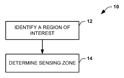
[0011] FIG. 1 is a flow diagram that illustrates an example of a method to
determine
a sensing zone for a region of interest.
[0012] FIG. 2 is a flow diagram illustrating an example of a method of
performing
electrocardiographic mapping.
[0013] FIG. 3 depicts an example of a system to perform electrocardiographic
mapping.
[0014] FIG. 4 depicts examples of baseline graphical maps.
[0015] FIG. 5 depicts examples of graphical maps post-therapy.
[00161 FIG. 6 depicts an example of a workflow to be utilized to determine a
sensing
zone.
[0017] FIG. 7 depicts a comparative example of reconstructed heart electrical
activity.
[0018] FIG. 8 depicts an example of a resolution calculation system.
[0019] FIG. 9 depicts an example of a graphical user interface that can be
utilized in
conjunction with the system of FIG. 8.
[0020] FIG. 10 depicts a simulated example comparison of reconstructed heart
electrical activity for a given arrangement of electrodes in a sensing zone
with and
without bad channels.
[0021] FIG. 11 depicts an example of reconstructed heart electrical activity
comparing graphical maps generated from data simulating measurements made with
and without bad sensing channels.
[0022] FIG. 12 depicts another example of reconstructed heart electrical
activity
comparing graphical maps generated from data simulating measurements made with
and without bad sensing channels.
[0023] FIGS. 13A, 13B and 13C demonstrate examples of different types of maps
that can be generated.
3
CA 02851800 2014-04-10
WO 2013/056050
PCT/US2012/059957
[0024] FIG. 14 depicts an example of a computing environment in which the
systems and methods disclosed herein can be implemented.
DETAILED DESCRIPTION
[0025] This disclosure provides systems and methods for determining one or
more sensing zones that can be utilized to facilitate evaluating cardiac
function. In
some examples electrocardiographic mapping (ECM). Also disclosed is an
approach to provide a design of electrode arrangements that can be used to
acquire
data for ECM, such as can be an application-specific arrangement of
electrodes.
[0026] In some examples, systems and methods disclosed herein can
compute a transfer matrix A-1 having coefficients that relates heart
electrical
potentials VH and body surface potentials VB, such as follows VH=A-lx VB. By
using
this matrix A-1, a proper subset of body surface channels and a relevant
contributions
to each and every node on the heart can be identified. That is, for each node
on a
cardiac envelope, the matrix A-1 can identify a set of electrode locations,
which
defines a zone, having a greatest contribution to the respective node. For a
given
region of interest (ROI) of the heart, which can include one or more nodes, a
corresponding the matrix A-1 can be computed to identify a set of electrode
locations
(a zone) having the greatest contribution to the nodes that define the ROI. As
used
herein, the terms "region" and "ROI" as applied to a given surface of an
anatomical
structure or envelope means something less than the entire surface. For
example,
some well known regions of the heart include the right-ventricular (RV)
freewall
region, the anterior left ventricular (LV) region, the lateral left
ventricular region, the
apex, the left ventricular base and the like.
[0027] The =set of body surface channels contributing the greatest amount
for
a given ROI form a group of channels that are referred to herein as a sensing
zone.
As one example, the sensing zone can correspond to a critical set of
electrodes
necessary and sufficient to generate an accurate ECM.
[0028] As another example, electrical activity for a predetermined sensing
zone on the body surface provides a surrogate estimate for electrical activity
of the
region of interest. Thus, electrical activity measured from a given sensing
zone can
be utilized to understand and characterize electrical activity of the
corresponding
ROI. Moreover electrical activity measured for a plurality of different
sensing zones
4
CA 02851800 2014-04-10
WO 2013/056050
PCT/US2012/059957
can be analyzed to provide spatially and temporally consistent electrical
information
for multiple ROls of the heart and even across the entire heart. For instance,
the
analysis can include computing electrical function characteristics, such as
activation
time, repolarization time, synchrony and the like, from the measured body
surface
electrical signals for each respective sensing zone. The computed electrical
characteristics can be analyzed to understand relative cardiac electrical
function
among different ROls of the heart, for example, by providing a graphical map
of the
measured body surface electrical signals, such as a potential map, or a map of
the
computed electrical characteristics.
[0029] The sensing zone can vary depending on an ROI that is selected
and/or application for which the ECM is being generated. For example, in some
applications (e.g., cardiac resynchronization therapy (CRT)), a greater level
of
accuracy may be desired such that the sensing zone can be determined to
include a
number of electrode sensing locations sufficient to afford the desired
accuracy. In
other situations, an even greater reduced subset of electrode sensing
locations may
be adequate. The sensing zone can include a contiguous set of body surface
electrode sensing locations. Additionally or alternatively, the sensing zone
can
include non-contiguous clusters of one or more body surface electrode sensing
locations. The distribution and arrangement of electrode sensing locations for
a
given sensing zone can vary depending on the selected ROI. The ROI can range,
for example, from a single node of a cardiac envelope to the entire heart.
[0030] As a further example, the set of body surface electrode sensing
locations corresponding to the sensing zone can provide a reduced set of
electrodes
necessary and sufficient for ECM computations for each given ROI. As a result,
application specific or special purpose vests and arrangements of sensing
electrodes
can be provided for use in ECM, such as one or more electrode arrangements
configured with electrodes in different sensing zones for corresponding
different
applications. For example, a simplified sensing vest with an arrangement of
electrodes can be provided for one or more ROls on the heart, such as one or
more
ventricles. Such special purpose arrangement of sensing electrodes can have
fewer
electrodes than a general purpose vest that can include electrodes covering
the
entire torso in an evenly distributed arrangement. Additionally or
alternatively, an
arrangement of electrodes can be provided for a predetermined sensing zone
configured for reconstructing heart electrical activity for one or more atria
of a
CA 02851800 2014-04-10
WO 2013/056050
PCT/US2012/059957
patient. Similar specially designed vests can also be configured for other
ROls of
different anatomical or geometrical structures.
[0031] As a further example, by knowing the sensing zone for a selected
ROI,
systems and methods disclosed herein can readily determine if a bad channel
exists
outside of the sensing zone it will have little if any impact on ECM results.
Thus,
there may be no need to correct for such a bad channel. In contrast, if a bad
channel or electrode is determined to exist within a desired sensing zone, an
appropriate warning can be generated to prompt the user to take corrective
action.
The impact of one or more bad channels on the resulting resolution of maps
that can
be generated from the measured body surface electrical activity can also be
ascertained. For example, one or more low resolution regions on the heart can
be
determined and displayed in a corresponding graphical map.
[0032] Additionally, by understanding the body surface electrode locations
corresponding to the sensing zone, systems and methods disclosed herein can be
programmed to provide guidance to facilitate application of other sensing and
therapy devices (e.g., defibrillator patches, guidance electrodes and the
like) to
locations that reside outside of a sensing zone or minimize overlap with a
sensing
zone, as appropriate. Additionally, if a sensing zone is known for a given
application,
a reduced set of sensing data can be acquired (since there are fewer channels)
to
facilitate the resulting computations in translating the body surface
electrical signals
to corresponding reconstructed cardiac electrical signals on a cardiac
envelope.
Such computations thus can employ a set of contributing transfer matrix
coefficients
determined according to the electrode sensing locations. In other examples,
the
measured electrical activity for the sensing zone (or other electrical
characteristics
computed therefrom) can be utilized as a surrogate estimate of electrical
activity for
the corresponding ROI on the heart.
[0033] FIG. 1 is a flow diagram 10 demonstrating an example method for
determining a sensing zone corresponding to a given ROI. The method can
include
a computer implemented-method that employs instructions, executable by a
processor, such as can be stored in a non-transitory machine readable medium
(e.g., volatile or non-volatile memory). The method can also involve the use
of
electrodes for sensing electrical activity, which can be positioned on a
patient for
measuring electrical activity for one or more sensing zone.
6
CA 02851800 2014-04-10
WO 2013/056050
PCT/US2012/059957
[0034] In FIG. 1, the method 10 includes identifying a ROI, at 12. The ROI
can be identified in response to a user input such as entered via a user input
device
(e.g., mouse, keyboard, touchscreen) of a computer implementing the method.
For
instance, the user input can include access functions to select one or more
ROI of
the heart, to acquire body surface electrical data, to initiate a mapping
function or the
like. As disclosed herein, the ROI can correspond to an anatomic region or
surface
area of an anatomical structure within the patient's body, such as patient's
heart or
some other geometric construct (e.g., a cardiac envelope). For instance, an
ROI can
include the ventricles or atria or other region of interest, which may vary
according to
application requirements.
[0035] In the example of FIG. 1, after the ROI has been ascertained, a
corresponding sensing zone can be determined at 14. The sensing zone can be
determined at 14 to encompass one or more body surface locations on which
electrodes are positioned sufficient to ensure accurate inverse reconstruction
of the
body surface electrical measurements to corresponding cardiac electrical
activity for
the identified ROI. The determination can be made based on understanding the
relative contribution of each of a plurality of body surface electrodes to
nodes within
the region of interest. The relative contribution of body surface electrodes
to the
location within the identified region of interest thus can be utilized to
identify the
sensing zone via a corresponding transfer matrix A-1. The sensing zone for a
selected ROI can be determined from data from one patient.
[0036] In some examples, the ROI can be further localized by using
techniques. For example, such localization techniques can include, in response
to
applying a therapy (e.g., electrical stimulation pulse) to a known anatomical
region
and correlating the mapped body surface electrical measurements, an earliest
activation signal and/or time can be computed to identify a corresponding
sensing
zone for the corresponding ROI at which the stimulation was applied.
[0037] It is to be understood and appreciated that the actions illustrated
in
FIG. 1, in other embodiments, may occur in different orders than shown. For
example, the region of interest can be identified at 12 based on the zone that
has
been determined at 14. This can be implemented to identify an ROI of a
resultant
map (e.g., corresponding to a low resolution region) that may be adversely
affected
by bad channels determined to reside in the zone. For instance, a bad channel
can
result from a missing electrode, an inadequate attachment of an electrode on
the
7
CA 02851800 2014-04-10
WO 2013/056050
PCT/US2012/059957
body surface, low signal-to-noise ratio, manually identifying a channel as bad
or
missing or combinations of these or other factors. As disclosed herein, one or
more
such low resolution area can be identified by evaluating coefficients
calculated for
the channels corresponding to electrodes that reside in the zone. The
specificity
and/or sensitivity of such evaluation may be fixed or it can be user
programmable.
[00381 Once the sensing zone has been adequately determined, a simplified
arrangement of electrodes can be utilized for analysis. For example, the
simplified
arrangement of electrodes can be utilized for generating one or more graphical
maps
based on electrical activity sensed by the arrangement of electrodes, such as
demonstrated in FIG. 2. In some examples, the sensed electrical activity can
be
analyzed to compute ECM. In other examples, the sensed electrical activity can
be
analyzed to generate body surface maps for the sensing zone, which provides a
surrogate estimate for the corresponding ROI of the heart.
[0039] In the example of FIG. 2, the method 20 includes providing an
arrangement of electrodes according to the determined sensing zone,
demonstrated
at 22. The arrangement of electrodes can be configured as part of a simplified
or
custom vest design in which the arrangement of electrodes corresponds to a
distribution of electrodes within the sensing zone. In other examples, the
sensing
zone can correspond to a proper subset of electrodes selected from an array of
electrodes, such as may be integrated into a sensing vest or similar
construct. If the
sensing zone can be known a priori, such as to provide an application-specific
arrangement of electrodes, a corresponding transfer matrix A-1 can be utilized
to
translate the body surface electrical signals to a corresponding ROI.
[0040] Once the arrangement of electrodes has been positioned on the
patient's body surface, including for a given sensing zone, body surface
electrical
measurements can be acquired at 24. Since the arrangement of electrodes for
the
given sensing zone is less than a full complement of body surface electrodes
surrounding a patient's entire torso, as is done for traditional ECM, the
amount of
data acquired for ECM can be reduced as to facilitate subsequent processing
for
visualization.
[0041] At 26, a graphical map can be generated for the ROI based on the
acquired body surface electrical measurements for the sensing zone. By way of
example, a map of body surface electrical activity for the sensing zone can be
generated to provide a surrogate estimate for electrical activity of the
corresponding
8
CA 02851800 2014-04-10
WO 2013/056050
PCT/US2012/059957
ROI. For instance the surface map can be a potential map for the given zone or
a
plurality of different zones on the body surface where measurements are made
via
the electrode arrangement (at 22). Alternatively or additionally, the map can
include
a characterization of electrical activity that is computed from the body
surface
electrical measurements for the zone. As yet another example, the map can
include
a characterization of electrical activity that is computed from the body
surface
electrical measurements for a plurality of different zones, such as to provide
a
surrogate estimate of corresponding electrical characteristics for a plurality
of
respective ROls. The electrical characteristics can include body surface
activation
times, relative synchrony for the body surface signals, a repolarization or
depolarization time calculated from the body surface signals. These and other
characteristics for the body surface can provide surrogate estimates for the
respective ROls without requiring imaging or solving the inverse solution.
[0042] As another example, the graphical map can be generated based on
reconstructed electrical signals corresponding to heart electrical activity.
As
disclosed herein the reconstructed electrical signals can be generated based
on
transformation matrix designed for the solving the inverse problem for the
body
surface electrical measurements for the sensing zone (or set of plural zones)
and for
reconstruction at each ROI (e.g., on the heart). The map thus can display
reconstructed potentials in a potential map for the ROI over one or more fime
intervals. The maps can also include, for example, activation maps,
repolarization
maps, dominant frequency maps or maps of other electrical characteristics
computed for the ROI based the reconstructed electrical activity.
p0431 As mentioned above, since the number electrodes for the sensing zone
can be significantly reduced relative to the traditional ECM vest, other
objects (e.g.,
patches, electrodes, or the like) can be positioned on a patient's chest
concurrently
with the sensing electrodes during the acquisition of body surface
measurements (at
24). If such other objects are placed outside of the sensing zones, such
objects
would not affect the accuracy of the results. Additionally, if such objects
were placed
within a sensing zone, an appropriate indication or warning could be provided
to the
user about the adversely affected electrodes. In this way, the user is
afforded an
opportunity to take corrective action as may be appropriate.
[0044] By way of example, if defibrillator patches are required to be
applied to
a patient's body in particular locations and if a region of interest or
regions can be
9
CA 02851800 2016-04-15
identified in advance, the systems and methods shown and described herein can
ascertain which electrodes are part of the sensing zone (or zones) such that
if body
surface electrodes are removed from the patient's body in order to position
the
defibrillator patches, the effect can be determined depending on whether or
not the
removed electrodes were in the sensing zone. For instance, information can be
computed and provided to the user (e.g., a graphical map) demonstrating areas
affected
that may experience a decrease in resolution for one or more ROI. Thus,
guidance can
be provided to facilitate placements of such defibrillator patches or other
objects
depending upon the region of interest. In some cases, the region of low
resolution may
reside outside a ROI that is being evaluated such that it can be ignored. In
other
examples, the low resolution area may be within or overlap with the ROI such
that
corrective action (e.g., re-location of electrodes) may be needed before
analysis of
electrical activity for the ROI.
[0045] Additionally or alternatively, as mentioned, specific vest designs can
be created
for particular applications such as may provide openings or access to the body
surface
to facilitate placements of defibrillator patches or other objects on the
patient's torso
while retaining the set of electrodes in the sensing zone.
[0046] FIG. 3 demonstrates an example of a system 100 that can be utilized for
performing electrical mapping of electrical activity. The system 100 can also
be
employed to determine a sensing zone for a selected ROI or to identify an
anatomical
region based on a selected sensing zone. For instance, the system 100 can
perform the
assessment of a patient's heart 102 in real time as part of a diagnostic or
treatment
procedure. Alternatively, the system 100 can operate offline based on stored
data.
[0047] The system 100 can include a measurement system 104 to acquire
electrophysiology information for the patient 106. In the example of FIG. 3, a
sensor
array 108 includes one or more electrodes that can be utilized for recording
patient
electrical activity. As one example, the sensor array 108 can correspond to an
arrangement of body surface electrodes that are distributed over and around
the
patient's torso for measuring electrical activity associated with the
patient's heart (e.g.,
as part of an ECM procedure). An example of a non-invasive sensor array that
can be
used is shown and described in International application No.
PCT/US2009/063803,
which was filed 10 November 2009. This non-invasive sensor array corresponds
to one
example of
CA 02851800 2014-04-10
WO 2013/056050
PCT/US2012/059957
a full complement of sensors that can include one or more sensing zones. As
another example, the sensor array 108 can include an application-specific
arrangement of electrodes corresponding to a single sensing zone or multiple
discrete sensing zones. The application-specific arrangement of electrodes can
be a
proper subset of electrodes selected from the full complement of electrodes,
such as
disclosed herein. Additionally, invasive sensors (not shown) can be used in
conjunction with the body surface sensor array 108.
[0048] The measurement system 104 receives sensed electrical signals from
the corresponding sensor array 108. The measurement system 104 can include
appropriate controls and signal processing circuitry for providing
corresponding
electrical measurement data 110 that describes electrical activity detected by
the
sensors in the sensor array 108. The measurement data 110 can be stored in
memory as analog or digital information. Appropriate time stamps can be
utilized for
indexing the temporal relationship between the respective measurement data 110
to
facilitate the evaluation and analysis thereof. For instance, each of the
sensors in
the sensor array 108 can simultaneously sense body surface electrical activity
and
provide corresponding measurement data 110 for a user selected time interval.
[0049] An output system 112 is configured to process the electrical
measurement data, including for one or more sensing zones on the patient's
body
106, and to generate an output 114. The output system 112 can be implemented
as
machine-readable instructions that, when executed by a processor, perform the
methods and functions disclosed herein. The output 114 can present information
for
one or more ROls based on the electrical measurement data 110. The output 14
can be stored in memory and provided to a display 116 or other type of output
device. As disclosed herein, the type of output 114 and information presented
can
vary depending on, for example, application requirements of the user.
[0050] As an example, the output system 112 can include a map generator
118 that is programmed to generate map data 120 representing a graphical map
of
electrical activity for one or more ROI based on the electrical measurement
data 110
for one or more respective zone. As disclosed herein, the graphical map can
represent body surface electrical activity that provides a surrogate estimate
for one
or more ROI, reconstructed electrical activity for a cardiac envelope (e.g.,
an
epicardial surface), or other electrical characteristics computed therefrom
and
visualized as a corresponding map. The map generator 118 can generate the map
11
CA 02851800 2014-04-10
WO 2013/056050
PCT/US2012/059957
data 120 to visualize such map spatially superimposed on the ROI of a
graphical
representation of the heart.
[0051] In some examplesõ the output system 112 includes a reconstruction
component 122 that reconstructs heart electrical activity by combining the
measurement data 110 with geomer data 116 through an inverse algorithm to
reconstruct the electrical activity onto a cardiac envelope, such as an
epicardial
surface or other envelope. Examples of inverse algorithms are disclosed in
U.S.
Patent Nos. 7,983,743 and 6,772,004, each of which is incorporated herein by
reference. The reconstruction component 122 for example computes coefficients
for
the transfer matrix K1 to determine heart electrical activity based on the
body surface
electrical activity represented by the electrical measurement data 110.
[0052] The map generator 118 can employ the reconstructed electrical data
computed via the inverse method to produce corresponding map data 120 based on
reestructed heart electrical activity computed by the reconstruction component
114
for each ROI. The map data 120 can represent electrical activity of the heart
102,
such as corresponding to a plurality of reconstructed electrograms distributed
over
each ROI for which the sensing zone(s) measured the body surface electrical
activity. Alternatively or additionally, an analysis system 124 can compute
other
electrical characteristics from the reconstructed electrograms, such as
disclosed
herein. The map generator 118 can in turn produce graphical maps of such
electrical characteristics for each ROI.
[0053] As a further example, the geometry data 116 may be in the form of
graphical representation of the patient's torso, such as image data acquired
for the
patient 106. Such image processing can include extraction and segmentation of
anatomical features, including one or more organs and other structures, from a
digital image set. Additionally, a location for each of the electrodes in the
sensor
array 108 can be included in the geometry data 116, such as by acquiring the
image
while the electrodes are disposed on the patient and identifying the electrode
locations in a coordinate system through appropriate extraction and
segmentation.
The resulting segmented image data can be converted into a two-dimensional or
three-dimensional graphical representation that includes the volume of
interest for
the patient.
[0054] By way of further example, the patient geometry data 172 can be
acquired using nearly any imaging modality (e.g., x-ray, computed tomography,
12
CA 02851800 2014-04-10
WO 2013/056050
PCT/US2012/059957
magnetic resonance imaging, ultrasound or the like) based on which a
corresponding representation can be constructed, such as described herein.
Such
imaging may be performed concurrently with recording the electrical activity
that is
utilized to generate the measurement data 110 or the imaging can be performed
separately (e.g., before the measurement data has been acquired).
[0055] Aspnother example, the geometry data 116 can correspond to a
mathematical model of a torso that has been constructed based on image data
for
the patient's organ. A generic model can also be utilized to provide the
geometry
data 116. The generic model further may be customized (e.g., deformed) for a
given
patient, such as based on patient characteristics include size image data,
health
conditions or the like. Appropriate anatomical or other landmarks, including
locations
for the electrodes in the sensor array 108 can be represented in the geometry
data
116 to facilitate registration of the electrical measurement data 110 and
performing
the inverse method thereon via the reconstruction component 114. The
identification
of such landmarks can be done manually (e.g., by a person via image editing
software) or automatically (e.g., via image processing techniques). Where the
electrical measurement data 110 is for a given sensing zone that can provide
surrogate electrical activity for a corresponding ROI of the heart, the
geometry data
and the transformation matrix utilized for reconstructing electrical signals
on the
heart can likewise be application-specific to facilitate computations.
[0056] In other examples, the map generator 118 can employ BSM/surrogate
code 126 for generating the map data 120 directly based on the non-invasive
body
surface electrical activity (e.g., corresponding to the measurement data 110)
without
involving reconstruction by solving the inverse solution. In this example, the
map
data 120 provides a surrogate estimate of cardiac electrical activity for one
or more
ROI. For instance, the surrogate map data 120 can include measured electric
potentials for a given sensing zone to provide a surrogate potential map for
the
respective ROI associated with such zone. Alternatively, BSM/surrogate code
126
can employ the analysis methods 124 to compute other electrical
characteristics for
the sensing zone directly from the measured data 110 without solving the
inverse
problem for reconstructing the signals on the respective ROI. The type of
analysis
124 applied to the electrical measurement data 110, if any, for generating the
surrogate estimate data can be selected in response to a user input.
13
CA 02851800 2014-04-10
WO 2013/056050
PCT/US2012/059957
[0057] The map generator 118 can thus generate the map data 120 based on
the surrogate estimate data without the complex computations associated with
solving the inverse problem. The map data 120 for the surrogate estimate
(e.g.,
electrical potentials or other characteristics computed for the zone) can
include a
map that shows variations across the sensing zone. In other examples, the
electrical
information for a given sensing zone can be aggregated spatially, temporally
or both
spatially and temporally, for the given zone to produce a value or range of
values for
a given ROI of the heart. As a further example, surrogate estimates of
electrical
characteristics can be determined for a plurality of different sensing zones,
which
can be used by the map generator118 to generate map data for each respective
ROI
of the heart. The map generator can generate and update maps to provide a
visualization of the surrogate estimates in substantially real time, such as
to facilitate
providing real-time intraoperative guidance during a procedure, such as
disclosed
herein.
[0058] As another example, an ROI selector 128 can be employed to select
an ROI in response to a user input. The ROI can be selected as one of a
plurality of
predetermined anatomical regions. Alternatively, or additionally, the ROI can
be
traced on a graphical user interface of the anatomy containing the ROI, such
as in
response to a user input (e.g., via a mouse, touchscreen). The selected ROI
can be
utilized for determining a corresponding sensing zone.
[0059] In some examples, a sensing zone function 130 can compute the
sensing zone for the selected ROI based on the map data 120. For example, the
sensing zone function 128 can determine the sensing zone for a given ROI based
on
a comparison of map data computed for a plurality of different subsets of
electrical
measurement data relative to map data computed for a full set measurement data
(e.g., using a fully compliment of electrodes around the ROI. The comparison
can
employ from statistical analysis to ascertain a minimized sensing zone that is
closest
match to the map data. The comparison of map data can be performed
automatically by the sensing zone function 130 and/or manual review of
respective
maps can be performed via the display to select a suitable sensing zone.
[0060] In other examples, the ROI selector 128 can be programmed to
identify
an ROI based on a given sensing zone that has been determined by the zone
function 130. For example, the map generator 118 can provide a graphical map
for
the identified ROI superimposed on a graphical representation of a heart. As
14
CA 02851800 2014-04-10
WO 2013/056050
PCT/US2012/059957
disclosed herein (see, e.g., FIG. 9) the ROI can correspond to an anatomical
region
that is expected to be adversely affected by measurements in a given sensing
zone
of the patient's body surface (e.g., corresponding to bad channels). This
analysis
can be performed in situations when the full complement of body surface
electrodes
is being used as well as in situations when an application-specific
arrangement of
electrodes is being used. The guidance provided by the map thus can afford a
user
the opportunity understand how the measurements in the zone may affect
resulting
analysis. As a result, a user can have an opportunity to correct the problem
(e.g., a
set of bad channels), if appropriate, or proceed knowing how subsequent
analysis of
the selected ROI may be affected.
[0061] In view of the foregoing, an application-specific arrangement of
electrodes can be designed and/or produced to measure given electrical
activity of a
given respective sensing zone. Such application-specific arrangement of
electrodes
can be configured to include a spatial distribution of electrodes that reside
only within
the computed sensing zone. Such an application specific arrangement of
electrodes
can be implemented, for example, in the form of patches (e.g., single or
multiple
pieces). For example, a patch for CRT can be configured to allowing for left
arm
access for a pectoral pocket. The right side can remain free (uncovered) if it
does
not include a corresponding sensing zone design and if a right pocket is
desired.
The sensing zone can be thus used for application specific vest designs.
Additionally or alternatively, the sensing zone can be used to evaluate the
impact of
not having sensors in sensing zones, including in real time, such as during a
procedure.
[0062] As another example, the application specific arrangement of
electrodes
can be implemented as a complete or partial band that can cover and wrap
around a
portion of the patient's chest that includes one or more sensing zone. Such
band
can include one or more clusters of electrodes or arrays distributed along one
or
more predetermined sensing zones. In other examples, small patches of
electrode
clusters can be configured for placement in application-specific sensing
zones. One
or more such patches could be used for acquiring body surface electrical data.
[0063] The particular configuration and size of a given application-
specific
arrangement of electrodes, including patches, bands or the like, can vary
depending
on the geometry and location of the sensing zone that is determined for each
respective application. Additionally, some application-specific arrangements
of
CA 02851800 2014-04-10
WO 2013/056050
PCT/US2012/059957
electrodes can be configured for multiple sensing zones. Placement of the
patches
can be guided based on manual measurements of the patient's anatomy.
Additionally, imaging, which can be performed previously or during a
procedure, may
further be utilized to guide placement of the electrodes. For example,
contributions
of individual electrodes can be determined (e.g., by the measurement system
104
and/or the output system 112) with respect to points along one or more ROls to
provide additional feedback to the user for adjusting the position of the
application-
specific arrangement of electrodes.
[0064] Alternatively, a more extensive arrangement of electrodes up to
covering the full torso can be utilized and the measurement system 104 and/or
the
output system 112 can remove measurement data from channels that is outside
the
zone. That is, the application-specific arrangement of electrodes for a given
sensing
zone can be implemented by constructing a physical arrangement of electrodes
for
the zone and/or by configuring the system to process a proper subset of
channels
corresponding to the zone. In either case, the computational complexity of
signal
processing and map generation can be reduced relative to traditional systems
that
process the entire compliment of channels. The application-specific
application zone
of channels can not only facilitate resulting analysis of electrical activity
for the ROI,
but do so while maintaining a high degree of accuracy for many applications
and
procedures (e.g., CRT).
[0065] As mentioned above, due to the reduced computational complexity
afforded by an application-specific zone, the system 100 can be utilized
intraprocedurally for real-time analysis. For instance, the system can be used
before, during and after providing a therapy or while programming a therapy
delivery
system to achieve a desired therapeutic effect.
[0066] By way of example, a therapy system 132 can be configured to apply a
therapy to the heart via a delivery device 134. In some examples, the therapy
device
134 can be an electrically conductive structure, such as an electrode or
antenna for
providing electrical or radiofrequency therapy. Alternatively, the device 134
can be
configured to apply a thermal therapy (e.g., heating or cooling) to the heart
102. The
particular type and configuration of the delivery device 134 can depend on the
mode
of therapy, delivery site and application requirements. The therapy system can
include a control 136 configured to control application of therapy, such as in
response to a user input. For instance, a trigger (e.g., a switch or button)
can be
16
CA 02851800 2014-04-10
WO 2013/056050
PCT/US2012/059957
activated by a user to initiate application of therapy. Various therapy
parameters can
also be set in response to the user input to control the therapy. For the
example of
electrical stimulation (e.g., for CRT), the parameters can include amplitude,
cycle
time or the like and further will vary depending on the type of therapy.
[0067] In the following example, a therapy delivery device (e.g., an
electrode
134) can be positioned in or on a patient's heart 102, such as part of a
minimally
invasive catheter procedure. As disclosed herein, the sensor arrayl 08 is
configured
for measuring activity for at least a predetermined sensing zone or a
plurality of
different sensing zones. Prior to delivering a therapy, which may be before or
after
the device 134 has been positioned in the patient's body 106, one or more
measurements (over respective time intervals) of body surface electrical
activity can
be made with the sensor array 108. The pre-therapy measurements of electrical
activity can be stored as baseline electrical measurement data 110 for each
zone
over one or more pre-therapy time intervals.
[0068] The acquired electrical measurements for each of the plurality of
different zones can provide a baseline surrogate estimate of electrical
activity (e.g.,
for each corresponding spatial ROI of the heart. The analysis function 124 can
also
compute surrogate estimates for electrical characteristics (e.g., activation-
repolarization time etc.) for each respective ROI which can also be stored in
memory
as part of the baseline data for the procedure. In some examples, the analysis
function 124 may be programmed to compute the estimate an indication of
relative
synchrony for a plurality of ROls based on a comparison of activation and/or
depolarization times for each of the ROls relative to its respective baseline.
[0069] After the baseline data has been acquired, therapy can be applied to
the heart 102 via the therapy device 132. The measurement system 104 can
measure body surface electrical activity from the patient's body during
delivery of
therapy and/or after the therapy is applied to provide corresponding
measurement
data. The BSM/surrogate function 126 can provide a surrogate estimate of
electrical
activity for each of a plurality of corresponding spatial ROls of the heart
based on
intra- and/or post-therapy data that was acquired. This can be repeated over a
plurality of different available therapy parameters.
[0070] As a further example, the system can be utilized to implement a
method for targeted analysis of one or more R01s, including intraprocedurally
and in
real time. For example, the system 100 can be utilized to calibrate and
identify the
17
CA 02851800 2014-04-10
WO 2013/056050
PCT/US2012/059957
zones on the body surface that correspond to key anatomical regions. For
instance,
the anatomical region can include one or more stimulation sites, such as may
have
been identified as potential responders to CRT. A user can thus employ the
electrode 134 and pace at various locations (e.g., in response to a user input
to
initiate stimulation). The analysis methods 124 of the output system 112 can
compute a corresponding earliest activation 'zone' on the body surface in
response
to the stimulation (e.g., pacing). This can be performed directly on body
surface
electrical measurement data, for example, without solving the inverse problem
and
reconstructing the electrical data. In other examples, the analysis 124 can be
performed on reconstructed electrical data for the heart. The zone function
130 can
in turn identify the input channels on the body surface that provide the
earliest
activation time, thereby specifying relationship between the zone and the
pacing site.
As an example, a simple protocol that can be used in current venous lead
placement
approaches is to pace the potential veins that are amenable to lead placement
to
determine the relative correspondence to electrical sensors of the array 108,
which
correspond to a zone on the body surface. This will enable targeted further
analysis
pertaining to these zones, such as over a range of parameters and pacing
protocols.
This process can be utilized with other types of lead placement and cardiac
therapies.
[0071] By way of example, FIGS. 4 and 5 depict graphical maps surrogate
electrical activity that can be generated based on the systems and methods
disclosed herein, such as part of a CRT procedure. In FIG. 4, baseline body
surface
activation maps 140 are shown for different ROls for surrogate regions of the
heart,
such as based on electrical measurements acquired for a predetermined sensing
zone (or zones) before applying a therapy. Also shown in FIG. 4 are different
ROls
142 and 143 for surrogate heart regions, such as can be selected as disclosed
herein.
[0072] FIG. 5 demonstrates the same types of surrogate maps 146 generated
for activation times computed from body surface measurements that have been
acquired following delivery of therapy as disclosed herein. For example, the
therapy
can include electrical or other stimulation, such as part of a CRT procedure.
A
comparison between the maps depicted FIGS. 4 and 5 demonstrates an improved
CRT response for each of the ROls 142 and 143.
18
CA 02851800 2014-04-10
WO 2013/056050
PCT/US2012/059957
[0073] Referring back to FIG. 3, the BS/surrogate function 126 can further
be programmed to compare the pre-therapy baseline surrogate estimate (e.g., of
FIG. 4) relative to the post-therapy surrogate estimate of electrical activity
(e.g., of
FIG. 5). The comparison can be made between the pre-therapy and post-therapy
surrogate estimates for the same sensing zone such as to provide an indication
of
the change in the cardiac electrical activity for a given therapy relative to
the baseline
measurements. Such comparison can also be performed for electrical
characteristics computed by the analysis function 124 based on the pre-therapy
baseline results and post therapy measured electrical activity for the given
sensing
zone. This can further be performed for each of a plurality of different
respective
sensing zones that provide surrogate estimates for different defined regions
of the
heart. In addition to comparing the baseline surrogate electrical
characteristics
relative to the intra- and/or post therapy surrogates, the analysis function
126 can
compare the different electrical characteristics for the same ROI or a
combination of
ROls for different therapy parameters and protocols. The therapy parameters
can
be stored with the electrical measurement data that is acquired during or
after
application of therapy. In this way, a user can review the results to help
identify
which therapy parameters and protocols help achieve a desired result.
[0074] The map generator 118 can also generate a map to depict the pre- and
post-therapy surrogate estimates. As disclosed above, the surrogate estimates
for
each sensing zone can be displayed in graphical maps superimposing the
estimated
values on the respective anatomic ROls of the heart. The pre- and post-therapy
maps (e.g., shown in FIGS. 4 and 5) can be displayed simultaneously at the
display
116 such as in separate windows. Additionally, or alternatively, a comparative
map
(not shown) can be generated and visualized as a graphical map on the display
116.
[0075] As further example, the analysis function 124 can be programmed to
calculate pseudo activation times of non-invasively measured body surface
electrical
activity. For instance, the activation times can be calculated using a dv/dt
method or
another method (e.g., directional activation, such as disclosed in PCT
Application
No. PCT/US11/51954). The pseudo activation time can be computed for pre-
therapy
(e.g., baseline) and post-therapy body surface electrical data. Even without
reconstructing the electrical data onto a cardiac envelope via solving the
inverse
problem, corresponding patterns of the pseudo activation times for one or more
zones can be analyzed and visualized as a graphical map to provide a broad
global
19
CA 02851800 2014-04-10
WO 2013/056050
PCT/US2012/059957
estimate of sequence of activation, conduction velocity, early and late
activation or
each of a plurality of different sensing zones. Slow or blocked conduction can
also
be roughly estimated from such line patterns on the body surface as well. This
can
be used, for example, to delineate the approximately location of the septum or
even
the engagement of the conduction system.
[0076] Continuing with the example of providing therapy to the heart via
the
device 134, body surface activation maps or potential map patterns for each
sensing
zone can be generated to provide surrogate estimates of synchrony information
that
can be utilized to help tune therapy parameters. The synchrony information for
a
given sensing zone can be derived from measurements made at different time
intervals, each having unique different therapy parameters, can be compared to
provide an indication how the corresponding ROI responds to the different
therapy
parameters. The comparison can be made by computing the difference between
electrical characteristics in each zone between the baseline data and data
acquired
for each of the different therapy parameters. Alternatively or additionally,
graphical
maps can be presented concurrently on the display 116 for comparison by the
user.
[0077] As an example, the average timing change for the same given zone
can be compared over a plurality of different pacing protocols and pacing
parameters. The activation times or potential patterns can be compared for the
same ROI. Alternatively or additionally, activation times or potential
patterns can be
compared for a combination of plural ROls between baseline maps and paced maps
including CRT.
[0078] In addition to comparing different surrogate estimates derived for
the
same ROI over different therapy parameters, as mentioned above, the output
system
100 can be utilized to facilitate spatial analysis of surrogate estimates of
electrical
information among a spatially diverse set of sensing zones. Such analysis can
help
characterize relative timing information for different spatial regions of the
heart. For
example, the electrical measurement data 110 can include body surface
potentials
acquired for a plurality of different predetermined sensing zones for a
plurality of
different time intervals. Each measurement time interval can correspond to a
unique
combination of therapy protocol and therapy parameters. The BSM/surrogate
function 122 thus can compute a surrogate estimate of electrical activity or
electrical
characteristics based on a comparison of relative timing information computed
based
on the measured body surface electrical data for a combination of different
zones at
CA 02851800 2014-04-10
WO 2013/056050
PCT/US2012/059957
each measurement interval. By tracking changes in the relative timing (e.g.,
activation timing, or average timing, synchrony or dyssynchrony) for the
different
body surface zones with respect to different therapy protocols and parameters,
the
analysis function 126 can provide an estimate in improvement of cardiac
function for
the corresponding ROls of the heart. The map generator 118 can generate
graphical maps to demonstrate and visualize (e.g., via graphical map) the
improvements to the user. Due to the reduced processing associated with the
approach disclosed herein, the visualization can be generated in substantially
real
time graphical maps that may be update dynamically during testing.
[0079] As another example, the direction of the changes in timing can also
be
employed to estimate improvement or reduction in synchrony or dyssynchrony.
The
activation times or potential patterns for can be evaluated to estimate
cardiac
dyssynchrony and classify the patient as 'dyssynchronous' based on threshold
values determined from normal body surface maps. The activation times or
potential
patterns can be compared for the same region or a combination of regions for
various combinations of pacing protocols and device parameters. The resulting
timing information and direction of changes in timing can be implemented
(e.g., as
manual or automated feedback to the therapy system 132) to help control
selection
of the therapy parameters. QRST intervals or other measures of activation-
repolarization measures could be also quantified based on surrogate estimates
of
electrical activity and compared similar to as disclosed above.
[0080] In view of the foregoing, the output system 118 can be programmed to
provide various temporal and spatial comparisons of surrogate estimates of
electrical
characteristics derived from electrical activity measured for one or a
combination of
sensing zones over a plurality of different time intervals. Different time
intervals can
correspond to different therapy parameters and protocols, such that the
comparative
data can be analyzed to determine which combination of parameters will achieve
a
desired therapeutic effect as disclosed herein. While the examples disclosed
above
in relation to programming therapy delivery were described in the context of
using
surrogate estimates (in the absence of solving the inverse problem), the same
methods can be implemented on reconstructed electrograms for one or more
sensing zone over a range of therapy parameters and protocols.
[0081] As an example, another method to calibrate and identify the spatial
zones on the body surface that correspond to key anatomical regions (e.g,
21
CA 02851800 2014-04-10
WO 2013/056050
PCT/US2012/059957
pertaining to CRT) is to pace at various known locations and note the
corresponding
earliest activation 'zone' on the body surface. The activation time for each
of the
body surface electrodes can be computed, and the set of electrodes determined
having the earliest activation time in response to the pacing stimulus can
define a
corresponding sensing zone for the stimulated anatomical region. For instance,
a
percentage of those electrodes exhibiting the earliest activation time can be
defined
as a respective sensing zone for the stimulated region. An example of a simple
protocol that can be used in current venous lead placement approaches is to
pace
the potential veins that are amenable to lead placement to determine the
relative
correspondence on the body surface. This will enable targeted analysis
pertaining to
these zones.
[00821 Further accuracy and refinement can be obtained by applying known
anatomical constraints obtained from simple chest x-ray or fluoroscopic
imaging to
estimate the approximate location of the heart. Such anatomical location
constraints
can include, for example, center of the heart, apex of the heart, anterior
septum
(LAD), posterior septum (PDA), outflow tracts, valves and the like.
[0083] FIG. 6 demonstrates an example of a workflow 200 that can be
utilized
for evaluating contributions of a selected region of interest for a patient's
heart. In
the workflow, at 202 ECM data is acquired. The ECM data can include body
surface
electrical data as well as geometry data such as can be acquired via a
corresponding imaging modality. Alternatively, the geometry data can
correspond to
a model of a patient's heart. The model can be a generic model, a generic
model
adjusted based on some patient specific data or a model derived from patient-
specific imaging data. For instance, a generic model can be adjusted based on
manual measurements of the patient (e.g., chest measurements, patient weight
or
the like), based on measurements from imaging (e.g., x-ray, fluoroscopy, and
ultrasound) or a combination of patient information.
[00841 In the example workflow of FIG. 6, at 204, a region of interest of
a
patient's heart is also selected. The region of interest can correspond to any
region
of a patient's heart for which it is desirable to understand the contribution
of a
corresponding sensing zone to ECM analysis. In the example of FIG. 6 there are
two generally parallel paths that are utilized for comparing and evaluating
the effect
of excluding electrode locations (and corresponding electrode signals) that do
not
significantly affect the accurate calculation of maps. Parallel does not
require
22
CA 02851800 2014-04-10
WO 2013/056050
PCT/US2012/059957
concurrent operations but instead indicates that the paths are sufficiently
separate as
to generate map data for different subsets taken from a common set of
electrical
measurements. In the following example, one subset is a proper subset of the
other.
[0085] With the acquired ECM data, at 206 potentials for a heart envelope
can
be computed. As used herein, the heart envelope can refer to any surface
(e.g.,
actual or virtual) that may reside on, inside or outside a patient's heart
that is spaced
radially inwardly from the body surface at which the ECM data is acquired (at
202).
In one example, the heart envelope can correspond to the outer three-
dimensional
surface of the epicardium, which may be the actual outer surface or an
approximation thereof. At 208, corresponding map data can be generated based
on
the computed potentials for the heart envelope.
[0086] In the other processing path, at 210, a sensing zone at the body
surface for the selected ROI is identified, such as disclosed herein. At 212,
the
potentials for a heart envelope can be computed based on measurement data
excluding the signals from sensors residing in the sensing zone that has been
identified (at 210) for the ROI. At 214, corresponding map data can be
generated
from the heart electrical activity computed at 212.
[0087] At 216, the respective maps (generated at 208 and 214) can be
compared. The comparison can be made via one or more statistical methods. At
218, the contribution of the sensing zone for the ROI can be determined based
on
the comparison. For example, if there is a significant difference in the
resulting
maps, then the impact of removing the sensing zone for the selected region of
interest is significant, which can be quantified by the statistical methods.
On the
other hand, if the comparison indicates that the maps are sufficiently
similar, then the
identified sensing zone for the ROI can be removed from the analysis while
retaining
sufficient accuracy in the resulting ECM data. Analysis similar to FIG. 6 can
be
repeatedly performed as part of the analysis for determining a configuration
an
arrangement of electrodes for a variety of specially adapted purposes.
[0088] By way of example, FIG. 7 demonstrates a comparative example of
reconstructed heart electrical activity for a heart envelope (e.g., the
epicardial
surface). In this example, a graphical map 220 for one reconstruction
demonstrates
results from using a full arrangement of electrodes (e.g., a full vest) 222.
For
example, the electrodes 222, for example, includes an arrangement of
electrodes
completely surrounding a patient's torso in a generally evenly distributed
manner. In
23
CA 02851800 2014-04-10
WO 2013/056050
PCT/US2012/059957
some examples, there can be greater than about 200 electrodes 222 in the form
of a
vest.
[0089] Another representation of reconstructed heart electrical activity,
demonstrated at 224, corresponds to ECM data computed for an application-
specific
arrangement of electrodes. That is the graphical map 224 can be generated
(e.g.,
by the reconstruction method 120) based on electrical measurement data
provided
from an arrangement of electrodes configured for a predetermined sensing zone
based upon the methods disclosed herein. The sensing zone in this example
includes electrodes 228, demonstrated as darker objects, distributed mainly
across
front, left and back locations of the patient's body.
[0090] A visual inspection of the reconstructed ECM for the full vest,
demonstrated at 220, and the reconstruction 224 for the application-specific
arrangement of electrodes demonstrates very similar results. The arrangement
of
electrodes for the application-specific arrangement of electrodes corresponds
to a
much simpler vest having a proper subset of electrodes 228 from the full vest
222
corresponding to a determined sensing zone for a selected ROI. In the example
of
FIG. 7, for example, the application-specific arrangement of electrodes can
correspond to a vest having an ROI configured for performing CRT.
[0091] FIG. 8 demonstrates an example of a system 150 for indicating one or
more ROls affected by contributions of electrodes residing within a given
sensing
zone. In some examples, the sensing zone can correspond to locations on the
body
surface where one or more bad channels reside or locations that can be
otherwise
identified, such as in response to a user input.
[0092] In the example of FIG. 8, a channel detector 152 is configured to
identify a subset of channels, corresponding to one or more sensing zones
expected
to adversely affect mapping of body surface electrical activity acquired for
the zone.
The channel detector 152 can identify the zone in response to a user input,
based on
measurement data (e.g., electrical measurement data 110 of FIG. 3) or a
combination thereof. In some examples, the channel detector 154 can designate
the
sensing zone as including a set of bad channels. A bad channel can correspond
to a
body surface location at which an electrode does not adequately contact the
body
surface, which may be intentional or unintentional. Additionally or
alternatively, a
bad channel can correspond to channel having a signal to noise ratio that is
below a
threshold.
24
CA 02851800 2014-04-10
WO 2013/056050
PCT/US2012/059957
[0093] A resolution calculator 154 is configured to compute an indication
of
the contribution that each electrode in the identified sensing zone (channels
provided
by the channel detector 152) has on the inverse solution for each point on the
heart.
The contribution, for example, can be determined by the resolution calculator
computing coefficients for the transformation matrix 158 for each of the
sensing
channels for the zone for each point on the cardiac envelope.
[0094] An evaluator 160 can analyze the computed coefficients (e.g., an
absolute value thereof) to generate ROI data 162. For example, the evaluator
160
can employ a specificity parameter 164 and a sensitivity parameter 166 to
control
how the ROI 162 is determined. The sensitivity parameter 166 can be a user
programmable threshold that can be set and compared to the absolute value of
the
computed transformation matrix coefficients. If a coefficient exceeds the
threshold
for a given node on the cardiac envelope, the electrode can be identified for
the node
as having a sufficient contribution to the reconstructed data at such node.
There can
be one or more coefficients at a given point on the envelope that can exceed
the
threshold. For instance, increasing the threshold value increases the spatial
sensitivity for identifying a low resolution region on the cardiac envelope as
it
requires a greater spatial contribution. The sensitivity parameter can set a
minimum
number of hits for the computed coefficients needed to designate a low
resolution
area. Increasing the number of hits can increase the overall specificity for
precisely
identifying a low resolution area. Thus by setting the specificity and
sensitivity
parameters appropriately for a given sensing system, one or more low
resolution
region on the heart can be determined and used to inform the user accordingly.
[0095] in some examples, the specificity and sensitivity parameters 164 and
166 can be pre-programmed (e.g., to default values) for system 150 (as well as
the
system 100 of FIG. 3) to determine circumstances when bad channels on the vest
tend to create artifacts in the reconstruction. The information can be
presented to
the user as a notice or warning, such as afford an opportunity to take
corrective
action, such as by adjusting the electrodes to improve the sensing ability or
changing
the contribution of electrodes to the ROI. After corrective action is taken,
the system
150 can recompute coefficients, such as in response to a user input, to
determine if
one or more low resolution region still exists. In some examples, a user can
opt not
to take corrective action, such as where the low resolution region is outside
of a ROI
considered important to the user for a given analysis. Alternatively or
additionally, a
CA 02851800 2014-04-10
WO 2013/056050
PCT/US2012/059957
user can adjust one or more of the sensitivity or specificity parameters to
reevaluate
the impact of the identified sensing zone (e.g., bad channels) on the inverse
solution.
Additional analysis by the evaluator may include comparing a computed low
resolution region with respect to a user-selected ROI, such that an overlap
between
such regions can result in generating a warning or other notification.
[0096] A map generator 168 can provide map data 170 for rendering a
visualization based on the ROI data 162. For instance, the map data can be
utilized
for providing a graphical map of a low resolution region that is superimposed
on a
graphical map of a heart. The map generator 168 can correspond to the map
generator 118 of FIG. 3, and the graphical map can be a three-dimensional map.
[0097] FIG. 9 depicts an example of a graphical user interface (GUI) 172
that
can be utilized for accessing the functions and methods of the system 150
demonstrated in FIG. 8. The GUI 172 can include multiple windows for
displaying
graphical representations of relevant data, such as can be associated with non-
invasive measurement of body surface electrical activity. The GUI 172 also
provides
an interactive visualization that can be utilized for demonstrating one or
more low
resolution regions. For example the GUI 172 can include a graphical map 174 of
a
low resolution region 176 superimposed on a heart. The low resolution region
can
be computed according to the method of FIG. 8. The GUI can also include an
interactive graphical display 178 of each of an arrangement of electrodes,
demonstrated as including right and left front panels and a back panel. Other
numbers and arrangements of electrodes can also be utilized, including the
application specific electrode arrangements disclosed herein.
[0098] The electrode display 178 can provide relevant status information of
electrodes. For instance the electrode display can identify bad channels by
graphically or otherwise differentiating the bad channels from other electrode
channels. The designation of channels can be described by a scale 180, such as
designating channels as good, bad, bad but editable and missing. Signals
utilized
for mapping the measured body surface potentials to the heart can also be
displayed. A user can also manually designate a given channel as bad, which
will
result in the measurement data for such electrode being omitted from
processing
while it is designated as a bad channel. In the example of FIG. 9, the group
of
channels in the right front panel, demonstrated at 182, has been designated as
bad
26
CA 02851800 2014-04-10
WO 2013/056050
PCT/US2012/059957
channels but editable. In this example, the resulting low resolution region
176 thus
identifies the region of the heart that is adversely affected by the bad
electrodes 182.
[0099] As a further example, FIG. 10 demonstrates graphical maps 230 and
232 representing of reconstructed heart electrical activity that has been
computed
from different sets of non-invasive body surface electrical measurements. In
the
example of FIG. 10, the graphical map 230 demonstrates reconstructed ECM data
in
the form of a potential map without any bad nodes (i.e., without compromising
electrodes in a corresponding sensing zone). Also demonstrated is another
graphical potential map 232 corresponding to reconstruction performed with
patches
applied to a patient's body surface, but outside of the sensing zone. For
example,
the patches can include other sensors and/or defibrillation patches that have
been
attached to the patient's torso. These other patches thus can overlap with
and/or
replace sensor electrodes from the arrangement of electrodes, as shown by
shading
in the electrode display GUI at 234. A comparison between the different maps
demonstrates that the reconstruction is accurate.
[00100] By way of further example, FIGS. 11 and 12 demonstrate simulated
examples of reconstructed graphical maps for different sensing zones that have
been determined for a selected region of interest of a patient's heart. For
instance,
the maps can be produced via inverse reconstruction for the entire heart
surface (or
other cardiac envelope) based on electrical measurement and geometry data
acquired for all electrodes, as disclosed herein. Each of these examples
demonstrates the effect of removing sensed body surface channels,
corresponding
to electrode sensing locations, demonstrated in these examples as bad
channels.
[00101] FIG. 11 demonstrates effects of bad channels on a reconstructed
potential map for a given sensing zone 248, such as can be determined for an
arrangement of electrodes 249 from a transformation matrix for a given ROI, as
disclosed herein. In FIG. 11, four graphical maps are depicted at 250, 252,
254 and
256. Each of the maps 250, 252, 254 and 256 have been generated based on the
same electrical measurement data, but with different contributions from
certain
sensing zones.
[00102] For example, the map 250 demonstrates a resulting graphical map
where no bad channels exist, such as can be produced via reconstruction for
the
entire heart surface based on electrical measurement data acquired for all
electrodes
249. The map 252 demonstrates a situation where bad channels form the sensing
27
CA 02851800 2014-04-10
WO 2013/056050
PCT/US2012/059957
zone 248 of an arrangement of electrodes that has been determined to be
critical
(e.g., for sensing electrical activity at a selected ROI). That is, the map
252 is
generated in the absence of electrical measurement data from the sensing zone
248.
The map 254 demonstrates a resulting graphical map where a cluster of bad
channels 260 (Bad Channel Cluster 1) overlaps with the critical sensing zone
248.
In contrast, the map 256 demonstrates a resulting graphical map where a
cluster of
bad channels 262 (Bad Channel Cluster 2) resides outside of the critical
sensing
zone 258. In contrast to the maps 252 and 254, the resulting map 256 is
substantially similar to the graphical map 250. This is because the bad
channels are
outside or mostly outside of the determined sensing zone 248. That is,
different
channels are removed from the inputs to the algorithm used to generate the
heart
electrical activity from the body surface electrical measurements. Thus, the
results
in the examples of FIG. 11 demonstrate a distinct difference between removing
electrodes from inside or outside of the sensing zone 248.
[00103] The example, of FIG. 12 is similar to FIG. 11, in that it
demonstrates
the effects of removing channels (e.g., bad channels) relative to a given
sensing
zone 264, such as can be determined for an arrangement of electrodes from a
transformation matrix for a selected ROI, as disclosed herein. In contrast to
the
example of FIG. 11, however, the sensing zone for the selected ROI is
generally
evenly distributed across the arrangement of electrodes. In the example of
FIG. 12,
contribution of corresponding coefficients in the transfer matrix A-1 are also
evenly
distributed across the arrangement of electrode sensing locations.
[00104] In the example of FIG. 12, the map 266 demonstrates a resulting
graphical map computed based on electrical measurements where no bad channels
exist. The map 268 is generated in the absence of electrical measurement data
from
the sensing zone 264 (e.g., electrical measurement data for the sensing zone
264
are removed). The map 270 demonstrates a resulting graphical map where a
cluster
of bad channels 272 (Bad Channel Cluster 1) overlaps with the sensing zone
264. In
contrast, the map 274 demonstrates a resulting graphical map where a cluster
of bad
channels 276 (Bad Channel Cluster 2) resides outside of the critical sensing
zone
264.
[00105] A comparison of the graphical maps 266, 268, 270 and 274
demonstrates that the resulting difference between the "gold standard"
graphical
map 266 for the graphical representation of reconstructed electrical activity
for the
28
CA 02851800 2014-04-10
WO 2013/056050
PCT/US2012/059957
full set of electrodes relative to the other examples in which electrode
clusters have
been removed from the analysis are relatively small. This suggests that the
impact
of removing electrodes for certain ROls may have little impact on the
resulting
computations for heart electrical activity data.
[00106] FIGS. 13A, 138 and 13C demonstrate examples of different types of
maps that can be generated from a common set of electrical measurement data,
such as can be generated by the map generator 118 of FIG. 3. FIG. 13A shows a
resulting isochrone map of voltage potentials reconstructed (by solving the
inverse
problem) onto the surface of a heart based on body surface electrical
measurements
and patient geometry data (e.g., from a computed tomography scan). FIG. 13B
depicts a simulated graphical map of isochrones that is reconstructed
generated
based on electrical body surface electrical measurements for a predetermined
sensing zone and patient geometry data. For instance, the body surface
measurements can be obtained using an application-specific arrangement of
electrodes determined for a desired ROI, as disclosed herein. FIG. 13C depicts
a
simulated graphical map of isochrones that is projected onto the heart (in the
absence of solving the inverse problem) based on electrical body surface
electrical
measurements for a predetermined sensing zone and patient geometry data. A
comparison of the resulting maps of FIGS. 13A, 13B and 13C demonstrates the
efficacy of using an application-specific arrangement of electrodes, without
or without
inverse reconstruction.
[00107] In view of the foregoing structural and functional description,
those
skilled in the art will appreciate that portions of the invention may be
embodied as a
method, data processing system, or computer program product. Accordingly,
these
portions of the present invention may take the form of an entirely hardware
embodiment, an entirely software embodiment, or an embodiment combining
software and hardware, such as shown and described with respect to the
computer
system of FIG. 14. Furthermore, portions of the invention may be a computer
program product on a computer-usable storage medium having computer readable
program code on the medium. Any suitable computer-readable medium may be
utilized including, but not limited to, static and dynamic storage devices,
hard disks,
optical storage devices, and magnetic storage devices.
[00108] Certain embodiments of the invention have also been described
herein
with reference to block illustrations of methods, systems, and computer
program
29
CA 02851800 2014-04-10
WO 2013/056050
PCT/US2012/059957
products. It will be understood that blocks of the illustrations, and
combinations of
blocks in the illustrations, can be implemented by computer-executable
instructions.
These computer-executable instructions may be provided to one or more
processor
of a general purpose computer, special purpose computer, or other programmable
data processing apparatus (or a combination of devices and circuits) to
produce a
machine, such that the instructions, which execute via the processor,
implement the
functions specified in the block or blocks.
[00109] These computer-executable instructions may also be stored in
computer-readable memory that can direct a computer or other programmable data
processing apparatus to function in a particular manner, such that the
instructions
stored in the computer-readable memory result in an article of manufacture
including
instructions which implement the function specified in the flowchart block or
blocks.
The computer program instructions may also be loaded onto a computer or other
programmable data processing apparatus to cause a series of operational steps
to
be performed on the computer or other programmable apparatus to produce a
computer implemented process such that the instructions which execute on the
computer or other programmable apparatus provide steps for implementing the
functions specified in the flowchart block or blocks.
[00110] In this regard, FIG. 14 illustrates one example of a computer
system
300 that can be employed to execute one or more embodiments of the invention,
such as including acquisition and processing of sensor data, processing of
image
data, as well as analysis of transformed sensor data and image data associated
with
the analysis of cardiac electrical activity. Computer system 300 can be
implemented
on one or more general purpose networked computer systems, embedded computer
systems, routers, switches, server devices, client devices, various
intermediate
devices/nodes or stand alone computer systems. Additionally, computer system
300
can be implemented on various mobile clients such as, for example, a personal
digital assistant (PDA), laptop computer, pager, and the like, provided it
includes
sufficient processing capabilities.
[00111] Computer system 300 includes processing unit 301, system memory
302, and system bus 303 that couples various system components, including the
system memory, to processing unit 301. Dual microprocessors and other multi-
processor architectures also can be used as processing unit 301. System bus
303
may be any of several types of bus structure including a memory bus or memory
CA 02851800 2014-04-10
WO 2013/056050
PCT/US2012/059957
controller, a peripheral bus, and a local bus using any of a variety of bus
architectures. System memory 302 includes read only memory (ROM) 304 and
random access memory (RAM) 305. A basic input/output system (BIOS) 306 can
reside in ROM 304 containing the basic routines that help to transfer
information
among elements within computer system 300.
[00112] Computer system 300 can include a hard disk drive 307, magnetic
disk
drive 308, e.g., to read from or write to removable disk 309, and an optical
disk drive
310, e.g., for reading CD-ROM disk 311 or to read from or write to other
optical
media. Hard disk drive 307, magnetic disk drive 308, and optical disk drive
310 are
connected to system bus 303 by a hard disk drive interface 312, a magnetic
disk
drive interface 313, and an optical drive interface 314, respectively. The
drives and
their associated computer-readable media provide nonvolatile storage of data,
data
structures, and computer-executable instructions for computer system 300.
Although the description of computer-readable media above refers to a hard
disk, a
removable magnetic disk and a CD, other types of media that are readable by a
computer, such as magnetic cassettes, flash memory cards, digital video disks
and
the like, in a variety of forms, may also be used in the operating
environment; further,
any such media may contain machine-readable instructions that can be executed
by
a processor (e.g., processing unit 301) for implementing one or more functions
and
methods disclosed herein.
[00113] A number of program modules may be stored in drives and RAM 305,
including operating system 315, one or more application programs 316, other
program modules 317, and program data 318. The application programs and
program data can include functions and methods programmed to acquire, process
and display electrical data from one or more sensors, such as shown and
described
herein.
[00114] The application programs and program data can include functions and
methods programmed to determine a sensing zone as disclosed herein. The
application programs and program data can also include functions and methods
programmed to generate an electrocardiographic map using a transformation
matrix
configured to reconstruct electrical signals for a region of interest from a
predetermined proper subset of body surface channels for a predetermined
sensing
zone.
31
CA 02851800 2014-04-10
WO 2013/056050
PCT/US2012/059957
[00115] As an example, the application programs 316 and program data 318
can be configured to implement a computer-implemented method that can identify
a
ROI and determine a zone on a body surface of the patient based on analysis of
electrical activity for the ROI relative to electrical activity on the body
surface. The
resulting electrical activity for the zone on the body surface can provides a
surrogate
estimate for electrical activity of the region of interest as disclosed
herein. In other
examples, the electrical activity measured at the body surface can be used to
reconstruct electrical activity onto a cardiac envelope, such as a surface of
an
internal organ (e.g., the heart or brain). The electrical activity, whether it
be a
surrogate estimate or reconstructed on to the cardiac envelope can be
presented in
a graphical map via the output device 324). The method can also be stored in a
non-transitory machine-readable media 302, 304, 306, 307, 308, 310 and/or 340.
[00116] In addition to mapping electrical activity and related computed
electrical
characteristics, as disclosed herein, the computer system 100 can also be
configured store and execute instructions to compute a low resolution region
of
interest based on a set of designated input channels (e.g., bad channels), as
disclosed with respect to FIGS. 8 and 9.
[00117] A user may enter commands and information into computer system
300 through one or more input devices 320, such as a pointing device (e.g., a
mouse, touch screen), keyboard, microphone, joystick, game pad, scanner, and
the
like. For instance, the user can employ input device 320 to edit or modify a
domain
model. These and other input devices 320 are often connected to processing
unit
301 through a corresponding port interface 322 that is coupled to the system
bus,
but may be connected by other interfaces, such as a parallel port, serial
port, or
universal serial bus (USB). One or more output devices 324 (e.g., display, a
monitor, printer, projector, or other type of displaying device) is also
connected to
system bus 303 via interface 326, such as a video adapter.
[00118] Computer system 300 may operate in a networked environment using
logical connections to one or more remote computers, such as remote computer
328. Remote computer 328 may be a workstation, computer system, router, peer
device, or other common network node, and typically includes many or all the
elements described relative to computer system 300. The logical connections,
schematically indicated at 330, can include a local area network (LAN) and a
wide
area network (WAN).
32
CA 02851800 2014-04-10
WO 2013/056050
PCT/US2012/059957
[00119] When used in a LAN networking environment, computer system 300
can be connected to the local network through a network interface or adapter
332.
When used in a WAN networking environment, computer system 300 can include a
modem, or can be connected to a communications server on the LAN. The modem,
which may be internal or external, can be connected to system bus 303 via an
appropriate port interface. In a networked environment, application programs
316 or
program data 318 depicted relative to computer system 300, or portions
thereof, may
be stored in a remote memory storage device 340.
(00120] What have been described above are examples. It is, of course, not
possible to describe every conceivable combination of components or
methodologies, but one of ordinary skill in the art will recognize that many
further
combinations and permutations are possible. Accordingly, the disclosure is
intended
to embrace all such alterations, modifications, and variations that fall
within the
scope of this application, including the appended claims. As used herein, the
term
"includes" means includes but not limited to, the term "including" means
including but
not limited to. The term "based on" means based at least in part on.
Additionally,
where the disclosure or claims recite "a," "an," "a first," or "another"
element, or the
equivalent thereof, it should be interpreted to include one or more than one
such
element, neither requiring nor excluding two or more such elements.
33