Note: Descriptions are shown in the official language in which they were submitted.
CA 02851828 2014-04-10
WO 2013/063530 PCT/US2012/062313
METHODS FOR DRUG DELIVERY
CROSS-REFERENCE
This application claims the benefit of priority under 35 U.S.C. section 119(e)
to US Provisional
Application 61/553,003, filed October 28, 2011; and US Provisional Application
61/680,847,
filed August 8, 2012; the contents of which are incorporated by reference in
their entirety.
BACKGROUND OF THE INVENTION
[0001] Numerous cancer-related therapeutics are under preclinical, phase I or
phase II clinical
trial and evaluations at any particular time; however, most of them will fail
to advance. In fact,
numerous drug candidates fail in the preclinical test, and it is estimated
that more than 90% of
cancer-related therapeutics will fail phase I or II clinical trial evaluation.
The failure rate in
phase III trials is almost 50%, and the cost of new drug development from
discovery through
phase Ill trials is between $0.8 billion and $1.7 billion and can take between
eight and ten years.
[0002] In addition, many subjects fail to respond even to standard drugs that
have been shown to
be efficacious. For reasons that are not currently well understood or easily
evaluated, individual
subjects may not respond to standard drug therapy. One significant challenge
in the field of
oncology is to exclude treatment selection for individual subjects having cell
autonomous
resistance to a candidate drug to reduce the risk of unnecessary side effects.
A related problem is
that excessive systemic concentrations are required for many oncology drug
candidates in efforts
to achieve a desired concentration at a tumor site, an issue compounded by
poor drug penetration
in many under-vascularized tumors (Tunggal et al., 1999 Clin. Canc. Res.
5:1583).
[0003] The present invention addresses these and similar needs, and offers
other related
advantages.
SUMMARY OF THE INVENTION
[0004] In one aspect, the present disclosure provides a method of delivering a
plurality of agents
to a solid tissue of a subject, comprising:
(a) inserting a plurality of microdialysis probes to the solid tissue;
and
-1-
CA 02851828 2014-04-10
WO 2013/063530 PCT/US2012/062313
(b) delivering the plurality of agents to said solid tissue through the
plurality of
microdialysis probes. The method may further comprise evaluating at least one
effect of the
plurality of agents on the solid tissue.
[0005] In another aspect, the present disclosure provides a method of
delivering two or more
agents to a solid tissue of a subject, comprising: (a) administering at least
one of said two or
more agents to said subject systemically; and (b) delivering at least one of
said two or more
agents to said solid tissue with at least one microdialysis probe or at least
one needle, wherein
said agent(s) administered in (a) is different from said agent(s) delivered in
(b). In some case,
step (a) is performed prior to step (b). In some other cases, step (a) is
performed after step (b).
The method may further comprise evaluating at least one effect of the agents
on the solid tissue.
[0006] In some embodiments, the agent(s) delivered in (a) or (b) is selected
from the group
consisting of an anti-angiogenic agent, a kinase inhibitor, an inhibitor of
metabolic pathway
targets that are preferentially expressed in cancer cells, or an epigenetic
modifier. In some other
embodiments, the agent(s) delivered in (a) or (b) comprises a small molecule
anti-cancer agent.
In some embodiments, the agent(s) delivered in (a) comprises an antibody or
antibody drug
conjugate. In some embodiments, the agent(s) delivered in (b) comprises a
small interfering
RNA, an antisense RNA or a small molecule anti-cancer agent. At least one of
the agents
delivered in step (b) may be delivered at different concentrations to
different regions of the solid
tissue. Alternatively, at least one of the agents delivered in step (b) may be
delivered in multiple
doses to a same region of the solid tissue. The agent(s) administered in step
(a) and the agent(s)
delivered in step (b) may have a synergistic effect on the solid tissue. The
agent(s) may be
present at a concentration below the therapeutic effective concentration.
[0007] The microdialysis probes may have different shapes. In some
embodiments, at least one
of the plurality of microdialysis probes is Y-shaped. In a further embodiment,
each of the
plurality of microdialysis probes is Y-shaped. In some other embodiments, at
least one of the
plurality of microdialysis probes is linear. In a further embodiment, each of
the plurality of
microdialysis probes is linear.
[0008] The agents may be delivered by diffusing through the microdialysis
probes. The diffusion
may be driven by concentration gradient (e.g., from a higher concentration to
a lower
concentration). In some embodiments, the diffusion may be driven by a
solubility gradient (e.g.,
from a less soluble solution to a more soluble solution or from a more soluble
solution to a less
-2-
CA 02851828 2014-04-10
WO 2013/063530 PCT/US2012/062313
soluble solution). Alternatively, the diffusion may be driven by active
transportation. In some
embodiments, the agents are delivered by flowing a solution of the agents
through the
microdialysis probes. The flow rate may be at least about 0.1 [1.1/min. In
some embodiments, the
flow rate is between about 0.1 [il/min and about 10 [1.1/min. In a further
embodiment, the flow
rate is between about 1 [il/min and about 2 [1.1/min. In some other
embodiments, the flow rate is
about 0.5, 0.8, 1, 1.2, 1.4, 1.6, 1.8, 2.0, 2.2, 2.5 3.0, 3.5, 4.0, 4.5, 5.0,
5.5, 6.0, 6.5, 7.0, 7.5, 8.0,
8.5, 9.0, 9.5, or 10.0 [Wmin.
[0009] The plurality of agents may flow through the microdialysis probes in a
continuous
fashion. The flow may be carried out with a peristaltic pump or a syringe
pump. The flow may
span a pre-determined period of time. The pre-determined period of time may be
at least about 1
hour, 2 hours, 3 hours, 4 hours, 6 hours, 12 hours, 18 hours, 24 hours, 36
hours, 48 hours, 72
hours, or 96 hours. The pre-determined period of time may be in a range of
about one hour to
about one year.
[0010] Upon insertion into a solid tissue, at least a portion of the
microdialysis probe spanning
the solid tissue may comprise a semi-permeable membrane. In some embodiments,
the entire
section of the microdialysis probe spanning the solid tissue comprises a semi-
permeable
membrane.
[0011] The insertion of microdialysis probes may be directed by an array
guide. The insertion of
microdialysis probes may be directed by an arthroscopic device. The insertion
of microdialysis
probes may be carried out with a needle array device. The needle array device
may comprise at
least two, at least five, or at least ten needles. Each of the needles may be
configured to receive
one microdialysis probe. The needle array device may further comprise at least
one actuator for
controlling needle insertion.
[0012] In some embodiments, at least three, at least five, or at least ten
microdialysis probes are
inserted. Each microdialysis probe may contain a different agent.
Additionally, at least two
microdialysis probes may contain a same agent at a same or different
concentrations.
[0013] In another aspect, the present disclosure provides a method of
delivering one or more
agents to a solid tissue, comprising: (a) inserting one or more needles to the
solid tissue; and (b)
delivering one or more agents to the solid tissue by withdrawing one or more
needles from the
solid tissue and injecting one or more agents into the solid tissue. The
method may further
comprise a step of evaluating an effect of one or more agents on the solid
tissue.
-3-
CA 02851828 2014-04-10
WO 2013/063530 PCT/US2012/062313
[0014] The needle may be a porous needle or an end port needle. In some
embodiments, the
needle is a porous needle. In some other embodiments, the needed is an end
port needle.
[0015] The rate of injecting one or more agents may be at least about 0.1,
0.2, 0.3, 0.4, 0.5, 0.6,
0.8, 1.0, 1.2, 1.5, 1.8, 2.0, 2.5, 3.0, 3.5, 4.0, 4.5, 5.0, 6.0, 7.0, 8.0,
9.0, 10.0, 12.0, 15.0, 20.0
nl/min. In some embodiments, the rate of injecting one or more agents is
between about 0.1
nl/min and about 5.0 nl/min. In some other embodiments, the rate of injecting
one or more
agents is about 0.1, 0.5, 1.0, or 2.0n1/min.
[0016] The rate of withdrawing one or more needles may be at least about 0.1,
0.2, 0.3, 0.4, 0.5,
0.6, 0.7, 0.8, 0.9, 1.0, 1.2, 1.5, 1.8, 2.0, 2.5, 3.0, 3.5, 4.0, 4.5, 5.0,
6.0, 7.0, 8.0, 9.0, 10.0, or 15.0
mm/min. In some embodiments, the rate of withdrawing one or more needles is
between about
0.1mm/min and about 5.0 mm/min. In some other embodiments, the rate of
withdrawing one or
more needles is about 0.1, about 0.5, about 1.0, or about 2.0 mm/min.
[0017] The insertion and withdrawal of needles may be directed by a fixed
guide or an
arthroscopic device. The fixed guide may comprise a stereotactic device. The
withdrawal of
needles and injection of agents may be carried out simultaneously or
sequentially. In an
exemplary embodiment, the withdrawal of needles and injections of agents are
carried out
simultaneously.
[0018] The insertion may be carried out with a needle array device. The needle
array device may
comprise at least two, at least five, or at least ten needles. The needle
array device may comprise
a plurality of reservoirs. In some embodiments, the needle array device
comprises at least three,
at least five, or at least ten reservoirs. Each of the reservoirs may be in a
separate fluid
communication with a separate needle. Each of the reservoirs may contain a
different agent from
the agent in any other reservoirs. In some cases, at least two of the
reservoirs contain a same
agent at different concentrations. The needle array may further comprise an
actuator and/or
controller. The controller may be operably linked or separated from the
actuator. The controller
may control the dosage of an agent to be delivered to the solid tissue.
[0019] With regard to any one of above mentioned aspects, the agent(s) is
either (i) undetectable
outside the solid tissue, or (ii) if detectable outside the solid tissue, the
agent(s) is present at less
than a minimal dose. Alternatively, the agent(s) is introduced in an amount
that is less than a
minimal dose required to produce a detectable effect in a subject when
delivered systemically.
Alternatively, the agent(s) is present in the solid tissue at a
therapeutically effective
-4-
CA 02851828 2014-04-10
WO 2013/063530 PCT/US2012/062313
concentration. The therapeutically effective concentration in the solid tissue
may be achieved by
dosing the agent orally.
[0020] With regard to any one of above mentioned aspects, the microdialysis
probes or the
needles may be inserted along an axis. Upon insertion, the agents may be
delivered along the
axis. The axis may be one of a plurality of parallel axes within the solid
tissue. In some
embodiments, there are 3, 4, 5, 6, 7, 8, 9, 10, 12, 14, 15, 18, or even 20
parallel axes.
[0021] With regard to any one of above mentioned aspects, the solid tissue may
comprise a
tumor. The tumor may be selected from the group consisting of a benign tumor
and a malignant
tumor. The tumor may be selected from the group consisting of a primary tumor,
an invasive
tumor and a metastatic tumor. The tumor may comprise at least one cancer cell
selected from the
group consisting of a prostate cancer cell, a breast cancer cell, a colon
cancer cell, a lung cancer
cell, a brain cancer cell, and an ovarian cancer cell. The tumor may comprise
a cancer selected
from the group consisting of adenoma, adenocarcinoma, squamous cell carcinoma,
basal cell
carcinoma, small cell carcinoma, large cell undifferentiated carcinoma,
chondrosarcoma, and
fibrosarcoma. Additionally, the solid tissue may be selected from the group
consisting of brain,
liver, lung, kidney, prostate, ovary, spleen, lymph node, thyroid, pancreas,
heart, skeletal muscle,
intestine, larynx, esophagus, and stomach.
[0022] With regard to any one of above mentioned aspects, the evaluating may
be performed in
vitro or in vivo. In some embodiments, the evaluating is selected from the
group consisting of
histology sectioning; collecting and analyzing at least one biomarker for
tumor cell death, cell
signal changes, or proliferation/mitotic changes; detecting the effect of said
one or more agents
on the proliferative gradient or multiple microenvironments of said solid
tissue; detecting the
activity or toxicity of each of the plurality of agents in separate regions of
the solid tissue;
detecting the activity or toxicity of one agent at different concentrations on
adjacent positions
within a solid tissue; and detecting the activity or toxicity of at least two
of the plurality of
agents in a same region of the solid tissue. When the activity or toxicity of
at least two of the
plurality of agents in a same region of the solid tissue is detected, the
activity or toxicity from
different agents may be synergistic or additive. In some other embodiments,
the evaluating
comprises imaging said solid tissue. The imaging may comprise radiographic
imaging, magnetic
resonance imaging, positron emission tomogoraphy, or biophotonic imaging. The
imaging may
occur before, during, or after introduction of the agents.
-5-
CA 02851828 2014-04-10
WO 2013/063530 PCT/US2012/062313
[0023] With regard to any one of above mentioned aspects, the plurality of
agents may comprise
an agent selected from the group consisting of a protein, a peptide, a
peptidomimetic, an
antibody, a small molecule, a small interfering RNA-encoding polynucleotide, a
nanoparticle, a
GCMS tag molecule, a gene therapy agent, an antisense RNA-encoding
polynucleotide, a
fluorescent dye, a positive control, a negative control, a small molecule anti-
cancer agent, or a
ribozyme-encoding polynucleotide. In some embodiments, the plurality of agents
comprise a
chemotherapeutic agent. In a further embodiment, the chemotherapeutic agent
comprises a small
molecule agent. In a still further embodiment, the small molecule agent has a
molecular weight
of less than 103 daltons. In some other embodiments, the plurality of agents
comprise an anti-
cancer agent. In some cases, two or more agents are delivered simultaneously
to a same region
within said solid tissue. In some cases, two or more agents are delivered
sequentially through a
microdialysis probe to a same region within said solid tissue.
[0024] With regard to any one of above mentioned aspects, the method may
further comprise
marking sites of insertions. In some embodiments, the sites of insertions are
marked by residual
color markers attached to the probes after delivering the agents. In some
other embodiments, the
sites of insertions are marked by at least one position marker. In a further
embodiment, the at
least one position marker comprises a dye. The dye may be a fluorescent dye.
[0025] With regard to any one of above mentioned aspects, an agent may be
delivered to a same
region of the solid tissue in multiple doses. Any two of the multiple doses
may be separated by a
selected period of time. The selected period of time may be at least about 10
minutes, 20
minutes, 30 minutes, 40 minutes, 60 minutes, 80 minutes, 90 minutes, 120
minutes, 6 hours, 12
hours, 24 hours, 36 hours, 48 hours, 72 hours, or 96 hour. The selected period
of time may be in
a range of about one hour to about three months.
[0026] With regard to any one of above mentioned aspects, the agents may
comprise a cancer
therapeutic agent and the evaluating may comprise detecting the presence or
absence of a drug
response and/or at least one biomarkers. Examples of drug response or
biomarker may include,
but are not limited to, cell apoptosis, downstream protein phosphorylation,
gene expression
markers, metabolic markers and other IHC markers. Cell apoptosis may be
detected in a region
of within about 0.001, 0.005, 0.01, 0.05, 0.1, 0.2, 0.5, 1.0, 2.0, 3.0, 4.0 or
about 5 mm from the
site of delivery. Cell apoptosis may be detected in a region of about 0.001-
0.1,0.1- 0.5, 0.5-1.0,
or 1.0-5.0 mm from the site of delivery. The threshold for selecting or
deselecting of an agent
-6-
CA 02851828 2014-04-10
WO 2013/063530 PCT/US2012/062313
based on cell apoptosis may depend upon the cancer therapeutic agent used and
the nature or size
of the tumor. In some embodiments, the cancer therapeutic agent is deselected
from further
evaluation if less than about 1%, about 3%, about 5%, about 10%, about 15%,
about 20%, about
25%, about 30%, about 35%, about 40%, about 45%, about 50%, about 55%, about
60%, about
65%, about 70%, about 75%, about 80%, about 85%, about 90%, about 95%, or
about 100% cell
apoptosis is observed comparing to a control without the cancer therapeutic
agent. In some other
embodiments, the cancer therapeutic agent is selected for further evaluation
if more than about
1%, about 3%, about 5%, about 10%, about 15%, about 20%, about 25%, about 30%,
about
35%, about 40%, about 45%, about 50%, about 55%, about 60%, about 65%, about
70%, about
75%, about 80%, about 85%, about 90%, about 95%, or about 100% cell apoptosis
is observed
comparing to a control without the cancer therapeutic agent.
[0027] With regard to any one of above mentioned aspects, the subject may be
an animal or a
human. Upon evaluating at least one effect of the agent(s) on the solid
tissue, the agent(s) may be
selected, deselected or prioritized based on the evaluation. In some
embodiments, the agent(s) is
selected for a clinical trial based on the evaluation. In some other
embodiments, the agent(s) is
deselected from a clinical trial based on the evaluation. The subject may be
one of a plurality of
subjects. Upon evaluating at least one effect of the agent(s) on the solid
tissue of the plurality of
subjects, some subjects may be selected, deselected or prioritized based on
the evaluation. In
some embodiments, some subjects are selected for a clinical trial of an agent
based on the
evaluation. In some other embodiments, some subjects are deselected from a
clinical trial of an
agent based on the evaluation. Non-limiting examples of the effect include the
presence or
absence of a change of physiological state of the solid tissue and the
presence of absence of a
biomarker.
[0028] In another aspect, the present disclosure provides a device for
delivering a plurality of
agents to a solid tissue of a subject, comprising a plurality of microdialysis
probes. The device
may further comprise any one of the followings: (1) a plurality of needles,
each configured to
receive one of said plurality of microdialysis probes; (2) at least one
controller, operatively
coupled to said plurality of needles; and (3) a guiding device to guide the
insertion of said
plurality of needles to said solid tissue. The device may comprise at least 3,
4, 5, 6, or 10
microdialysis probes or needles. In some embodiments, controller is a
computer. The computer
-7-
CA 02851828 2014-04-10
WO 2013/063530 PCT/US2012/062313
may be used to control the insertion of microdialysis probe and injection of
agents. In some
further embodiments, the computer is part of a cloud computing system.
[0029] In another aspect, the present invention provides a device, comprising
a top block having
a first plurality of holes sized to allow a needle to pass through the top
block, and a bottom block
having a second plurality of holes sized to allow a needle to pass through the
bottom block,
wherein the top and bottom blocks are in a substantially parallel arrangement
and wherein the
first and second plurality of holes are positioned so as to allow one or more
needles to pass
through a hole in the top block and the bottom block in a path substantially
vertical to the plane
of both blocks. The device may further comprise at least one adjustable leg,
wherein the at least
one adjustable leg is attached to the bottom block. The number of legs may be
at least 1, 2, 3, 4,
5, 6, 7, 8, 9, 10, 12, 14, 16, 18, 20, or even more. In an exemplary
embodiment, there are 4 legs.
In some embodiments, the legs are vertically and horizontally adjustable. The
bottom block and
the top block may be independently stationary or movable. In some cases, the
bottom block of
the device is stationary and the top block can move vertically relative to the
bottom block. In
some further embodiments, the top block moves along guide rods attached to the
bottom block.
Movement of any blocks may be controlled. For example, a system may be
attached to the
device to control vertical movement of the top block.
[0030] In some embodiments, the first and second pluralities of holes are
arranged in
substantially parallel rows. The device may further comprise at least one
needle. In some
embodiments, a control attachment is attached to the at least one needle. The
control attachment
may stop the insertion of the at least one needle, thereby controlling depth
of needle insertion
into the solid tissue. Additionally, the device may further comprise at least
one spring, wherein
the at least one spring is in substantial contact with an adjustable leg and
the bottom block.
INCORPORATION BY REFERENCE
[0031] All publications, patents, and patent applications mentioned in this
specification are
herein incorporated by reference to the same extent as if each individual
publication,
patent, or patent application was specifically and individually indicated to
be
incorporated by reference.
-8-
CA 02851828 2014-04-10
WO 2013/063530 PCT/US2012/062313
BRIEF DESCRIPTION OF THE FIGURES
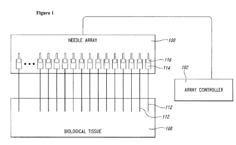
[0032] Figure 1 is a schematic diagram of a needle array assembly for
injecting biological tissue
with agents according to various embodiments.
[0033] Figure 2 shows an exemplary device embodying principles of the present
invention.
[0034] Figure 3 shows an example of a platform for tumor stabilization using
springs embodying
principles of the present invention.
[0035] Figure 4 shows a top view of needles with a control attachment
embodying principles of
the present invention.
[0036] Figure 5 shows an exemplary device with a drive mechanism for
controlling vertical
movement of a top block embodying principles of the present invention.
[0037] Figure 6 shows diagrammatically a portion of a tumor illustrating
principles of the
invention.
[0038] Figure 7 shows an example of targeting the viable EBC-1 tumor
epithelium expressing
the target of interest (c-Met) using a linear array of microdialysis probe
embodying
principles of the present invention.
[0039] Figure 8 shows a schematic example of monitoring multiple
zones/microenvironments in
a solid tumor using long microdialysis membranes in vivo embodying principles
of the
present invention.
[0040] Figure 9 shows a diagrammatic view of dose determination using
microdialysis probes
embodying principles of the present invention. By running a continuous loop of
drug for
a fixed time, the total dialysate from tubing can be collected and analyzed
using HPLC,
fluorescence/absorbance, etc. to determine the amount of therapeutic agents
delivered
through passive diffusion.
[0041] Figure 10 shows a diagrammatic view of testing the efficacy of anti-
cancer drugs given in
a particular sequence embodying principles of the present invention. In a
first dose, cell
cycle/signaling is activated in a contact-inhibited low proliferation zone.
After some time
and clearing of the first drug from the microdialysis tubing, a second drug
that arrests and
kills cells that are now actively dividing is administered.
[0042] Figure 11 shows a schematic view of targeting both the proliferative
zone and other zones
in a solid tumor model using the extrusion/injection technique embodying
principles of
the present invention.
-9-
CA 02851828 2014-04-10
WO 2013/063530 PCT/US2012/062313
[0043] Figure 12 shows fluorescent imaging of near infrared (NlR) dye
delivered in tumor using
a microdialysis probe. A, B, C and D designate various cross-sections
embodying
principles of the present invention.
[0044] Figure 13a and 13b show drug delivered through microdialysis probe
induces spatially
restricted drug specific tumor cell death embodying principles of the present
invention.
[0045] Figure 14 shows results from two injection methods with respect to
efficiency (14a),
signal uniformity (14b) and column length (14c) embodying principles of the
present
invention.
[0046] Figure 15 shows average number of positive regions per section of three
different
injection methods embodying principles of the present invention.
[0047] Figure 16 shows average variance within section of three different
injection methods.
[0048] Figure 17 shows results evaluating different injection methods with
simplified
experimental systems embodying principles of the present invention.
[0049] Figure 18 shows fluorescent microscopy images of three different
injection methods
embodying principles of the present invention.
DETAILED DESCRIPTION OF THE INVENTION
General Overview
[0050] Clinical trials for therapeutic agents, including cancer therapeutic
agents, are incredibly
expensive and time consuming. It is therefore very important to effectively
screen for agents that
have relatively greater potential as early in the process as possible. Agents
subjected to such
screening are sometimes referred to as candidate agents. One screening method
involves growing
tumor cells in an artificial environment on plastic cell culture plates with a
growth medium, then
placing each candidate agent in a respective cell culture plate or Dish. The
cell cultures are later
evaluated for indications of cell growth. Agents that appear to have impeded
growth of cancer
cells and/or engage with a biological target may then be advanced for further
study.
[0051] However, this method is only marginally effective, for several reasons.
First, cell culture
poorly mimics tumors growing in a patient. Only the most general information
can be gleaned
from such studies because the test conditions do not remotely resemble the
conditions in which
the cancer normally lives and grows, and in which it is treated
therapeutically. Screening tests
like that described are therefore ineffective with these. Second, the process
of immortalizing a
tumor for laboratory use can alter the response characteristics of a tumor.
The process involves
-10-
CA 02851828 2014-04-10
WO 2013/063530 PCT/US2012/062313
essentially pureeing the tumor, which completely destroys any structural
differentiation, and may
render the cancer susceptible to some agents that would have no effect on the
same strain in vivo,
resulting in a false positive, even though such agents might be useless for
treating the cancer in
patients. Third, the same reduction process can also produce false negatives,
in which some
agents may fail to inhibit cell growth in vitro, but would be effective in
treating the same cancer
in vivo. Finally, even where general efficacy of an agent in treating a
particular cancer type,
subtype, variant, strain or the like has been demonstrated, it is not uncommon
for the cancer of a
particular patient to be wholly unresponsive to the agent.
[0052] The inventors have recognized the need to accurately position an agent
and/or control an
amount of the agent to be delivered in a solid tissue in vivo and later
identify the locations of the
agent in the solid tissue. If such accuracy could be achieved, significant
benefits in research and
therapy could be realized. For example, many tumors are heterogeneous in
nature with among
other differences, a quiescent inner zone and a proliferative outer zone. It
may be important to
target the proliferating zone of solid tumors to assess drugs that target
mitosis and mitotic
checkpoints and/or pathways which are more active in proliferating zones, for
example, C-Met
and AKT. Therefore, methods allowing evaluation of therapeutic agents across
an entire solid
tumor may be highly valuable.
[0053] The present disclosure provides methods and devices for delivering an
agent to a solid
tissue, and in particular to a solid tumor in vivo. Often, one or more agents
are delivered to a
solid tissue with improved accuracy, uniformity and dosage control.
Thereafter, the agents
remain in the solid tissue for a selected period of time. The effects of the
agents on the solid
tissue are then monitored in vivo or in vitro. Based on the observed effects,
each of the agents is
selected or deselected for further studies or consideration of treatment for a
patient, on whose
solid tissue the candidate drugs have been assessed.
[0054] The agents are usually dissolved in solution and delivered to a target
site within a solid
tissue. The volume of fluid that is delivered can be vanishingly small, much
less than would be a
minimal dose required to produce a detectable effect in a subject when
delivered systemically.
Depending on the agent, the effect may nevertheless be detected on the very
small region
immediately surrounding the delivery site. Accordingly, candidate effective
agents can be
injected into a tumor, for example, in situ, without danger of harming the
subject. Additionally, a
-11-
CA 02851828 2014-04-10
WO 2013/063530 PCT/US2012/062313
significant number of different agents can be simultaneously delivered to
respective delivery
axes within the tumor.
[0055] The procedures described herein can be employed to resolve a number of
the problems
and difficulties that contribute to the cost and delay of developing effective
cancer therapies. For
example, because the candidate agents are delivered in vivo, the tumor is not
otherwise disturbed
and drug concentrations can approximate levels achieved through systemic
administration of the
drug, and so its reaction to each agent will tend to be indicative of its
reaction if exposed to that
agent in therapeutically effective quantities. The incidence of false
positives and false negatives
is significantly reduced. Second, because relatively large numbers of agents
can be delivered to a
tumor without significant danger to the subject, it is practical to use the
procedure to screen large
numbers of candidate agents early in the testing process, perhaps eliminating
those that show the
least promise, flagging the most promising agents for additional study, or
prioritizing candidates
for further study. Third, again because of the large number of agents that can
be delivered to a
tumor, potential study subjects can be screened for response to particular
agents, reducing or
eliminating the number of subjects with idiosyncratic responses. Fourth,
because the agents are
delivered locally to a solid tissue, systemic exposure of the agent can be
avoided.
[0056] Accordingly, for example, certain embodiments contemplate direct drug
delivery to a
solid tissue at low flow rates with low shear forces that eliminate or reduce
mechanochemical
damage to tissues while permitting precisely targeted therapeutic agent
delivery to defined focal
sites. Significantly higher concentrations of the agents may be achieved
within the solid tissue
than would be the case if the agents were delivered systemically. In other
words, the amount of
agents required to achieve desired pharmacological effect would be lower, and
in some case
much lower, than would be the case if the agents were delivered systemically.
In some cases, the
agents are undetectable outside the solid tissue. In some other cases, less
than 10% of the agents
are detected outside the solid tissue (e.g., in the systemic circulation). In
some other cases, upon
delivery, the agents are present in a solid tissue at therapeutically
effective concentrations.
Therapeutically effective concentrations of the agents in a solid tissue can
be achieved by dosing
the agents orally. However, systemic exposure of the agents, often at high
concentrations, is
required. Hence, problems (e.g., toxicity, detrimental side-effects, etc.)
associated with
administering excessively high systemic concentrations of the agents in order
to obtain
-12-
CA 02851828 2014-04-10
WO 2013/063530 PCT/US2012/062313
therapeutically effective concentrations in a desired solid tissue are
overcome by the presently
disclosed embodiments.
[0057] In one aspect, the present disclosure provides methods of delivering
and evaluating one
or more agents in a solid tissue with one or more microdialysis probes. The
number of
microdialysis probes inserted may be at least 2, 3, 4, 5, 6, 7, 8, 9, 10, 12,
14, 15, 20, 25 or 30. In
some cases, at least some of the microdialysis probes are inserted
simultaneously. In some other
cases, at least some of the microdialysis probes are inserted sequentially.
The microdialysis
probes may be inserted along an axis. The axis may be one of a plurality o
parallel axis. The
number of axes may be at least 3, at least 4, at least 5, at least 6, at least
7, at least 8, at least 9, at
least 10, at least 12, or at least 15. The number of axes may be about 3, 4,
5, 6, 7, 8, 9, 10, 12, or
15. After the insertion, the agents may be delivered to the solid tissue by
diffusing through the
microdialysis probes. The agents may diffuse through the membrane region of
the microdialysis
probes, thus delivering the agents to a column-shaped region along a delivery
axis within the
solid tissue.
[0058] Each of the microdialysis probes may contain a different agent from any
other probes.
Alternatively, some of the microdialysis probes may contain a same agent as at
least another
microdialysis probe. When two or more microdialysis probes contain a same
agent in perfusate,
concentrations of the agent in different probes may be the same or different.
[0059] The agents may be delivered to the solid tissue in a continuous
fashion. In some cases,
the delivery may last about 10, 20, 30, 40, 50, 60, 70, 80, 90, 100, 110, 120,
130, 140, 150, 160,
170, 180, 200, 220, 240, 260, 280, 300, 320, 340, 360, 400, 450, 500, 550,
600, 650, 700, 750,
800, 850, 900, 1000, 1100, 1200, 1300, 1400, 1500, 1600, 1700, 1800, 2000,
2200, 24000, 2600,
2800, 3000 minutes, 3 days, 4 days, 5 days, 6 days, 1 week, 2 weeks, 3 weeks,
4 weeks, 5
weeks, 6 weeks, 2 months, 3 months, 4 months, 6 months, 8 moths, 9 months, 1
years or 2 years.
[0060] The agents may be delivered to the solid tissue in multiple doses. At
least two of the
multiple doses may be separated by a pre-determined period of time. The pre-
determined period
of time may be about 10, 20, 30, 40, 50, 60, 70, 80, 90, 100, 110, 120, 130,
140, 150, 160, 170,
180, 200, 220, 240, 260, 280, 300, 320, 340, 360, 400, 450, 500, 550, 600,
650, 700, 750, 800,
850, 900, 1000, 1100, 1200, 1300, 1400, 1500, 1600, 1700, 1800, 2000, 2200,
24000, 2600,
2800, 3000 minutes, 3 days, 4 days, 5 days, 6 days, 1 week, 2 weeks, 3 weeks,
4 weeks, 5
weeks, 6 weeks, 2 months, 3 months, 4 months, 6 months, 8 moths, 9 months, 1
years or 2 years.
-13-
CA 02851828 2014-04-10
WO 2013/063530 PCT/US2012/062313
[0061] The flow rate of an agent in each microdialysis probe may be
independently controlled.
The flow rate may be independently at least 0.0010min. In some embodiments,
the flow rate is
independently in the range of about 0.001[Wmin to about 5 1/min. In some
embodiments, the
flow rate is independently about 0.001, 0.01, 0.05, 0.1, 0.2, 0.3, 0.4, 0.5,
0.6, 0.7, 0.8, 0.9, 1.0,
1.1, 1.2, 1.3, 1.4, 1.5, 1.6, 1.7, 1.8, 1.9, 2.0, 2.1, 2.2, 2.3, 2.4, 2.5,
2.6, 2.7, 2.8, 2.9, 3.0, 3.1, 3.2,
3.3, 3.4, 3.5, 3.6, 3.7, 3.8, 3.9, 4.0, 4.1, 4.2, 4.3, 4.4, 4.5, 4.6, 4.7,
4.8, 4.9, or 5.0 II/min
[0062] The microdialysis probes may be inserted with a needle array device.
The needle array
device may contain a plurality of needles. In some cases, the needle array
device has 2, 3, 4, 5, 6,
7, 8, 9, 10, 11, 12, 13,14, 15, 16, 17, 18, 19, 20, 21, 22, 23, 24, 25, 26,
27, 28, 29, 30 or even
more needles. Each of the needles may be configured to receive at least one
microdialysis probe.
In some cases, each needle is inserted along one of a plurality of parallel
axes. Upon insertion
and placement of microdialysis probes in the solid tissue, the needles are
then withdrew, leaving
behind the microdialysis probes in the solid tissue.
[0063] The method may further comprise a step of evaluating an effect of the
agents on the solid
tissue. In some cases, such evaluation comprises detecting the activity or
toxicity of each of the
agents in separate regions of the solid tissue. In some other cases, such
evaluation comprises
detecting the activity or toxicity of two or more of the agents in a same
region of the solid tissue.
When two or more agents are delivered to a same region, they may be delivered
simultaneously
or sequentially. In some other cases, such evaluation comprises detecting the
activity or toxicity
of a same agent with different concentrations in adjacent regions of the solid
tissue. In a further
embodiment, such evaluation comprises detecting the activity or toxicity of
three or more of
agents in a same region of the solid tissue.
[0064] In another aspect, the present disclosure provides a method of
delivering two or more
agents to a solid tissue of a subject, comprising: (a) administering at least
one of said two or
more agents to said subject systemically; and (b) delivering at least one of
said two or more
agents to said solid tissue with at least one microdialysis probe or at least
one needle, wherein
said agent(s) administered in (a) is different from said agent(s) delivered in
(b). In some case,
step (a) is performed prior to steno (b). In some other cases, step (a) is
performed after step (b).
The method may further comprise a step of evaluating an effect of the agents
on the solid tissue.
The agent(s) in step (a) may be administered orally or via injection.
-14-
CA 02851828 2014-04-10
WO 2013/063530 PCT/US2012/062313
[0065] In another aspect, the present disclosure provides a method of
delivering one or more
agents to a solid tissue, comprising: (a) inserting one or more needles to the
solid tissue; and (b)
delivering the one or more agents to the solid tissue by withdrawing the one
or more needles
from the solid tissue and injecting the one or more agents into the solid
tissue. The method may
further comprise evaluating an effect of one or more agents on the solid
tissue.
[0066] The injection of one or more agents and withdrawal of one or more
needles may be
carried out sequentially or simultaneously. In some embodiments, the injection
of one or more
agents and withdrawal of one or more needles are carried out simultaneously.
Additionally, since
the agents are delivered to an empty space instead of being forced through a
solid tissue, the
likelihood of agents cross-contamination is much reduced.
[0067] The rate of injecting one or more agents and the rate of withdrawal of
one or more
needles may be separately controlled. The rate of injection may be in a range
of 0.1-5.0 p.1/min.
In some embodiments, the rate of injection is at least about 0.1 [1.1/min,
about 0.2 [1.1/min, about
0.3 p.1/min, about 0.4 [1.1/min, about 0.5 [1.1/min, about 0.6 [1.1/min, about
0.7 [1.1/min, about 0.8
1.i1/min, about 0.9 pl/min, about 1.0 pl/min, about 1.1 pl/min, about 1.2
pl/min, about 1.3 pl/min,
about 1.4 [1.1/min, about 1.5 [1.1/min, about 1.6 [1.1/min, about 1.7
[1.1/min, about 1.8 [1.1/min, about
1.9 pl/min, about 2.0 pl/min, about 2.1 pl/min, about 2.2 pl/min, about 2.3
pl/min, about 2.4
[Wmin, about 2.5 [1.1/min, about 2.6 [1.1/min, about 2.7 [1.1/min, about 2.8
[1.1/min, about 2.9 p.1/min,
about 3.0 [1.1/min, about 3.1 [1.1/min, about 3.2 [1.1/min, about 3.3
[1.1/min, about 3.4 [1.1/min, about
3.5 p.1/min, about 3.6 [1.1/min, about 3.7 [1.1/min, about 3.8 [1.1/min, about
3.9 [1.1/min, about 4.0
1.i1/min, about 4.1 pl/min, about 4.2 pl/min, about 4.3 pl/min, about 4.4
pl/min, about 4.5 pl/min,
about 4.6 [1.1/min, about 4.7 [1.1/min, about 4.8 [1.1/min, about 4.9 p.1/min
or about 5.0 p.1/min. In
some other embodiments, the rate of injection is about 0.1 [1.1/min, about 0.2
[1.1/min, about 0.3
[Wmin, about 0.4 [1.1/min, about 0.5 [1.1/min, about 0.6 [1.1/min, about 0.7
[1.1/min, about 0.8 p.1/min,
about 0.9 1[1.1/min, about 1.0 [1.1/min, about 1.1 [1.1/min, about 1.2
[1.1/min, about 1.3 .1/min, about
1.4 p.1/min, about 1.5 [1.1/min, about 1.6 [1.1/min, about 1.7 [1.1/min, about
1.8 [1.1/min, about 1.9
1.i1/min, about 2.0 pl/min, about 2.1 pl/min, about 2.2 pl/min, about 2.3
pl/min, about 2.4 pl/min,
about 2.5 [1.1/min, about 2.6 [1.1/min, about 2.7 [1.1/min, about 2.8
[1.1/min, about 2.9 [1.1/min, about
3.0 pl/min, about 3.1 pl/min, about 3.2 pl/min, about 3.3 pl/min, about 3.4
pl/min, about 3.5
1.i1/min, about 3.6 pl/min, about 3.7 pl/min, about 3.8 pl/min, about 3.9
pl/min, about 4.0 pl/min,
about 4.1 [1.1/min, about 4.2 [1.1/min, about 4.3 [1.1/min, about 4.4
[1.1/min, about 4.5 [1.1/min, about
-15-
CA 02851828 2014-04-10
WO 2013/063530 PCT/US2012/062313
4.6 .1/min, about 4.7 .1/min, about 4.8 .1/min, about 4.9 t1/min or about
5.0 [Wmin. In some
other embodiments, the rate of injection is in a range of 0.1-1.0 .1/min, 0.5-
1.5 [1.1/min, 1.0-2.0
[1.1/min, 2.0-3.0 [Ll/min, 3.0-4.0 [1.1/min or 4.0-5.0 [Ll/min.
[0068] The rate of withdrawal of one or more needles may be in a range of 0.1-
10 mm/min. In
some embodiments, the rate of withdrawal of one or more needles is at least
about 0.1 mm/min,
about 0.2 mm/min, about 0.3 mm/min, about 0.4 mm/min, about 0.5 mm/min, about
0.6
mm/min, about 0.7 mm/min, about 0.8 mm/min, about 0.9 mm/min, about 1.0
mm/min, about
1.1 mm/min, about 1.2 mm/min, about 1.3 mm/min, about 1.4 mm/min, about 1.5
mm/min,
about 1.6 mm/min, about 1.7 mm/min, about 1.8 mm/min, about 1.9 mm/min, about
2.0
mm/min, about 2.1 mm/min, about 2.2 mm/min, about 2.3 mm/min, about 2.4
mm/min, about
2.5 mm/min, about 2.6 mm/min, about 2.7 mm/min, about 2.8 mm/min, about 2.9
mm/min,
about 3.0 mm/min, about 3.1 mm/min, about 3.2 mm/min, about 3.3 mm/min, about
3.4
mm/min, about 3.5 mm/min, about 3.6 mm/min, about 3.7 mm/min, about 3.8
mm/min, about
3.9 mm/min, about 4.0 mm/min, about 4.1 mm/min, about 4.2 mm/min, about 4.3
mm/min,
about 4.4 mm/min, about 4.5 mm/min, about 4.6 mm/min, about 4.7 mm/min, about
4.8
mm/min, about 4.9 mm/min, about 5.0 mm/min, about 5.1 mm/min, about 5.2
mm/min, about
5.3 mm/min, about 5.4 mm/min, about 5.5 mm/min, about 5.6 mm/min, about 5.7
mm/min,
about 5.8 mm/min, about 5.9 mm/min, about 6.0 mm/min, about 6.1 mm/min, about
6.2
mm/min, about 6.3 mm/min, about 6.4 mm/min, about 7.0 mm/min, about 8.0
mm/min, about
9.0 mm/min or 10.0 mm/min. In some other embodiments, the rate of withdrawal
of one or more
needles is about 0.1 mm/min, about 0.2 mm/min, about 0.3 mm/min, about 0.4
mm/min, about
0.5 mm/min, about 0.6 mm/min, about 0.7 mm/min, about 0.8 mm/min, about 0.9
mm/min,
about 1.0 mm/min, about 1.1 mm/min, about 1.2 mm/min, about 1.3 mm/min, about
1.4
mm/min, about 1.5 mm/min, about 1.6 mm/min, about 1.7 mm/min, about 1.8
mm/min, about
1.9 mm/min, about 2.0 mm/min, about 2.1 mm/min, about 2.2 mm/min, about 2.3
mm/min,
about 2.4 mm/min, about 2.5 mm/min, about 2.6 mm/min, about 2.7 mm/min, about
2.8
mm/min, about 2.9 mm/min, about 3.0 mm/min, about 3.1 mm/min, about 3.2
mm/min, about
3.3 mm/min, about 3.4 mm/min, about 3.5 mm/min, about 3.6 mm/min, about 3.7
mm/min,
about 3.8 mm/min, about 3.9 mm/min, about 4.0 mm/min, about 4.1 mm/min, about
4.2
mm/min, about 4.3 mm/min, about 4.4 mm/min, about 4.5 mm/min, about 4.6
mm/min, about
4.7 mm/min, about 4.8 mm/min, about 4.9 mm/min, about 5.0 mm/min, about 5.1
mm/min,
-16-
CA 02851828 2014-04-10
WO 2013/063530 PCT/US2012/062313
about 5.2 mm/min, about 5.3 mm/min, about 5.4 mm/min, about 5.5 mm/min, about
5.6
mm/min, about 5.7 mm/min, about 5.8 mm/min, about 5.9 mm/min, about 6.0
mm/min, about
6.1 mm/min, about 6.2 mm/min, about 6.3 mm/min, about 6.4 mm/min, about 7.0
mm/min,
about 8.0 mm/min, about 9.0 mm/min or 10.0 mm/min. In some other embodiments,
the rate of
withdrawal of one or more needles is in a range of 0.1-1.0 mm/min, 0.5-1.5
mm/min, 1.0-2.0
mm/min, 1.5-2.5 mm/min, 2.0-3.0 mm/min, 2.5-3.5 mm/min, 3.0-4.0 mm/min, 3.5-
4.5 mm/min
or 4.0-5.0 mm/min.
[0069] In another aspect, the present disclosure provides a device for
delivering a plurality of
agents to a solid tissue of a subject, comprising a plurality of microdialysis
probes. The device
may further comprise any one of the followings: (1) a plurality of needles,
each configured to
receive one of said plurality of microdialysis probes; (2) at least one
controller, operatively
coupled to said plurality of needles; and (3) a guiding device to guide the
insertion of said
plurality of needles to said solid tissue. The device may comprise at least 3,
4, 5, 6, or 10
microdialysis probes or needles. In some embodiments, controller is a
computer. The computer
may be used to control the insertion of microdialysis probe and injection of
agents. In some
further embodiments, the computer is part of a cloud computing system.
[0070] In another aspect, the present invention provides a device for
controlling needle insertion
into and withdrawal from a solid tissue, comprising: (a) a positioning
mechanism; (b) a depth-
control mechanism; and (c) a needle withdrawal mechanism.
[0071] In yet another aspect, there is provided a device for delivery of at
least one agent to a
solid tissue, comprising one bottom block and one top block in a substantially
parallel
arrangement, each having a plurality of holes. The plurality of holes in the
bottom and top block
may guide the insertion of needles. In some cases, the size of holes may be
controlled to allow
needles of a certain size to pass through. The device may lead to improved
accuracy of needle
insertion and exquisite control of delivery of the at least one agent to a
solid tissue.
[0072] As used herein the term "synergistic activity or toxicity" refers to
coordinated activity or
toxicity of two or more agents so that the combined action is greater than the
sum of each agent
acting separately. The coordinated activity or toxicity may be at least about
10%, about 20%,
about 30%, about 40%, about 50%, about 60%, about 70%, about 80%, about 90% or
even
higher than the sum of each agent acting separately.
-17-
CA 02851828 2014-04-10
WO 2013/063530 PCT/US2012/062313
[0073] As used herein the term "additive activity or toxicity" refers to
activity or toxicity of two
or more agents so that the combined action is about equal to the sum of each
agent acting
separately.
[0074] As used herein the term "about" refers to 10 % and includes 1% and
0.1%.
[0075] As used herein the term "therapeutically effective concentrations"
refers to the
concentrations of agents in a solid tissue when a desirable pharmacological
effect is observed in
the solid tissue. For example, for an anti-cancer drug which is delivered
orally, the drug needs to
go through an absorption process to get into the systemic circulation. After
absorption, the drug
then enters or accumulates in the solid tissue. The concentrations of the drug
in the solid tissue
and in the systemic circulation may be the same or different when a desirable
pharmacological
effect is observed.
[0076] As used herein the term "pre-determined period of time" or "selected
period of time"
refers to any time within a range of 1 minute to 2 years. In some embodiments,
the pre-
determined period of time or selected period of time is about 0.1, 0.2, 0.5,
1, 2, 3, 4, 5, 6, 7, 8, 9,
10, 11, 12, 15, 18, 24, 36, 48, 72, 96, 120, 144, 168, or 192 hours. In some
other embodiments,
the pre-determined period of time or selected period of time is in a range of
about 24-72 hours.
Microdialysis Probes
[0077] The present invention provides methods for the administration of an
agent to a solid
tissue through the use of one or more microdialysis probes. In some cases, the
microdialysis
probe has an inlet-tubing, an outlet-tubing and a membrane region. The
solution in the inlet-
tubing is termed "perfusate" while the solution in the outlet tubing is termed
"dialysate". The
inlet- and outlet-tubings may be made of a material suitable for microdialysis
application. In
some embodiments, the material is fused silica. In some other cases, the
microdialysis probe has
an inlet-tubing and a membrane region without an outlet-tubing. In this
design, an agent may be
actively pumped across the membrane region.
[0078] The inventors have recognized the advantages of using microdialysis
probes as a delivery
tool, which include: (1) microdialysis probes are an enclosed system, not
dependent upon
delivery of a liquid volume, thus eliminating many of the microfluidic
engineering hurdles; (2)
the semi-permeable membrane surrounding the probe allows liquid to be filled
and distributed
evenly along probe membrane when injecting into a solid tissue; (3) initial
delivery and
biodistribution of agents are highly restricted and dependent upon passive
diffusion forces, not
-18-
CA 02851828 2014-04-10
WO 2013/063530 PCT/US2012/062313
deposition/delivery of a liquid; (4) true "microdosing" of agents can be
achieved by controlling
time, flow rate and concentration of perfusate; (5) multiple or timed dosing
over an extended
periods of time can be achieved by leaving probes in the solid tissue; (6) the
amount of agents
delivered can be accurately determined by analyzing the amount of agent in
perfusate and
dialysate; (7) the length of the probe/semi-permeable membrane can be
customized to target
various size tumors or length of targeting zone within a tumor; (8) an array
of linear
microdialysis probes can be designed to target the proliferating zone in solid
tumor xenografts,
as well as avoiding the central regions necrosis; (9) better sampling of
multiple zones, including
the entire dimension of a solid tumor, to look for efficacy differences using
linear probe arrays
can be achieved; and (10) collection and analysis of dialysate at various time
points following
dosing may allow development and analysis of markers of tumor cell death, cell
signal changes,
or proliferation/mitotic changes. In addition, microdialysis probes can be
used to coax contact-
inhibited cells into cycling in order to kill them using checkpoint
inhibition/DNA damage, or
activate cell signal pathways that have been shut down in non-proliferative
zones.
[0079] A microdialysis probe may be suitable for containing, administering,
delivering and
transporting contents. The contents may be an aqueous solution comprising a
pharmaceutical
composition comprising one or more agents. The agents within a single
microdialysis probe may
be the same or a mixture of different types of agents. Within a plurality of
microdialysis probes,
each microdialysis probe may contain the same agent as another probe, or
different agents as
another probe. In some embodiments, every microdialysis probe contains agents
that are unique
from the agents contained in other microdialysis probes.
[0080] A microdialysis probe may have different shapes. In some cases, the
microdialysis probe
has a "Y" shape. In some other cases, the microdialysis probe has a linear
shape. The linear
shape may allow the microdialysis probe to penetrate across different sections
of a tumor.
[0081] The membrane of a microdialysis probe may be semi-permeable. The
membrane may
permit the transport of some but not all solutes. In some embodiments, the
membrane permits the
transport of solutes with a molecule weight of less than 1 million Daltons. In
a further
embodiments, the membrane permits the transport of solutes with a molecule
weight in the range
of 5,000 Daltons to 1 million Daltons. In another further embodiment, the
membrane permits the
transport of solutes with a molecule weight of less than 1,000 Daltons.
-19-
CA 02851828 2014-04-10
WO 2013/063530 PCT/US2012/062313
[0082] The movement of a substance or an agent from one side to another side
of a membrane
may be driven by concentration gradient. In some cases, the movement of a
substance or an
agent from one side to another side of a membrane is driven only by
concentration gradient. A
substance or an agent may move from an area of higher concentration to an area
of lower
concentration through the semi-permeable membrane. In some cases, the agent
diffuses from a
microdialysis probe into a solid tissue. In some other cases, a solute in a
solid tissue diffuses into
a microdialysis probe. The solute can be collected and/or analyzed from
dialysate. Alternatively,
the movement of a substrate or an agent may be driven by active transporter,
irrespective of
concentration gradient. For example, in nature, some cells use active
transporter to accumulate
molecules, such as ions, glucose and amino acids. Alternatively, the movement
of a substrate or
an agent may be driven by solubility difference. The substrate or agent may
have a higher
solubility on one side of the membrane than the solubility on the other side.
In some cases, the
substrate or agent moves from a higher concentration side to a lower
concentration side. In some
embodiments, the substrate or agent moves from a lower concentration side to a
higher
concentration side. In some cases, the movement of a substance or an agent
from one side to
another side of a membrane is driven by a combination of any one of
concentration gradient,
active transportation, and solubility difference.
[0083] The membrane may be biocompatible. The membrane may be essentially
physiologically
inactive or does not trigger physiological events. In some embodiments, the
membrane may not
cause inflammation, immune response, infection, or any other sort of
rejections within a solid
tissue.
[0084] The membrane may be flexible. The flexibility of the membrane will
permit the insertion
of the membrane section into the solid tissue with minimal damage to the
tissue. Yet, the
membrane may have certain strength to maintain its integrity before, during or
after the insertion.
In some embodiments, the membrane is both flexible and durable.
[0085] The membrane material may be polymeric or co-polymeric. The polymeric
or co-
polymeric material may be linear or cross-linked. Non-limiting examples of
membrane materials
include PE (polyethylene), Kevlar, cuprophane, polyethersulfone, polyamine,
polyamide,
polycarbonate, polycarbamate, polyurethane, polyester, polyether, polyolefin,
polysilicon oxide,
cellulose acetate, and polyaromatic materials.
-20-
CA 02851828 2014-04-10
WO 2013/063530 PCT/US2012/062313
[0086] The membrane material may be porous. In some embodiments, the average
pore size is
less than about 1, 5, 10, 20, 30, 40, 50, 100, 200, 500, 1000, 2000, 5000, or
10000 nanometers. In
some other embodiments, the average pore size is more than about 1, 5, 10, 20,
30, 40, 50, 100,
200, or 500 nanometers. In some other embodiments, the average pore size is in
a range of 1-10,
1-40, 1- 100, 1-200, or 1-500 nanometers. In some other embodiments, all pores
of a membrane
has a substantially similar pore size.
[0087] The pore size may control the rate of diffusion. The pore size may be
modulated to
control the rate of diffusion. A membrane may be made with a selected average
pore size for the
purpose of controlling the rate of diffusion. Different pharmaceutical
compositions of agents can
diffuse through the membrane at varying rates, controlled in part by the
physical and chemical
properties of the pharmaceutical compositions, agents, and membrane materials.
In some
embodiments, the selected pore size permits the transport of solutes with a
molecule weight of
less than 1 million Daltons. In a further embodiments, the selected pore size
permits the transport
of solutes with a molecule weight in the range of 5,000 Daltons to 1 million
Daltons. In another
further embodiment, the selected pore size permits the transport of solutes
with a molecule
weight of less than 1,000 Daltons. In addition, membranes with varying average
pore sizes can
be made and tested experimentally to find a pore size that provides a
desirable diffusion rate for
a specific pharmaceutical composition or agent.
[0088] A pharmaceutical composition or agent may be delivered to a
microdialysis probe by
using a pump, such as a peristaltic pump or syringe pump. The use of a pump
can lead to
controlled delivery. For example, the agent or pharmaceutical composition can
be delivered
through a microdialysis probe in a continuous fashion. Alternatively, the
agent or pharmaceutical
composition can be delivered in several doses. The time interval between any
two doses can be
controlled. Furthermore, the flow rate may be individually controlled for each
microdialysis
probe. The flow rate may be in a range of about 0.1 to about 5 microliter/min.
The flow rate may
be about 0.1, 0.2, 0.3, 0.4, 0.5, 0.6, 1.0, 1.5, 2.0, 2.5, 3.0, 3.5, 4.0, 4.5,
or about 5 microliter/min.
[0089] A microdialysis probe may be inserted into a solid tissue directly or
indirectly. The
indirectly insertion may comprise the steps of: (1) insertion of a
microdialysis probe into a
needle; (2) insertion of the needle into a solid tissue; and (3) withdrawal of
the needle from the
solid tissue, therefore leaving the microdialysis probe in the solid tissue.
In some cases, a
plurality of microdialysis probes are inserted into a solid tissue with a
plurality of needle along a
-21-
CA 02851828 2014-04-10
WO 2013/063530 PCT/US2012/062313
plurality of axes into a solid tissue. Each of the plurality of needles holds
one of the plurality of
microdialysis probes. In some embodiments, the plurality of axes are a
plurality of parallel axes.
In some embodiments, the plurality of needles are part of a needle array
device. The needle array
device may comprise at least 2, 5, 10 or even more needles.
[0090] In addition, the present invention provides a microdialysis probe which
has an inlet-
tubing without an outlet-tubing. In some embodiments, the terminal end of the
probe is
surrounded by a semi-permeable membrane. In this design, the microdialysis
probe may act as a
diffuser in which liquid and small molecules are actively pumped across the
semi-permeable
membrane.
[0091] The insertion of a microdialysis probe may be guided. In some
embodiments, the
insertion of a microdialysis probe is guided by a fixed guide to direct the
insertion of a
microdialysis probe into a selected region of a solid tissue. In some
embodiments, the insertion
of a microdialysis probe is guided by an arthroscopic device.
[0092] The present invention also provide a method of monitoring drug
metabolism and
response in a solid tissue. For example, without being limiting, a
microdialysis probe may be a
part of closed loop. The membrane section of the microdialysis probe may span
the solid tissue.
By running a continuous flow of a solution of an agent through the
microdialysis probe for a
selected period of time, the agent may be delivered to the solid tissue. After
another selected
period of time, another solution (e.g. saline) may be flown through the
microdialysis probe.
Solutes in the solid tissue, for example without being limiting, may be
collected in dialysate and
analyzed. Non-limiting examples of solutes include biomarkers, agents
delivered to the solid
tissue and metabolites of the agents delivered to solid tissue. By analyzing
the presence or
absence and/or concentration of solutes, the efficacy of the agents on the
solid tissue may be
determined.
Target Tissues
[0093] In some embodiments, the present disclosure exemplifies a method for
screening agents
in a solid tissue. Solid tissues are well known to the medical arts and may
include any cohesive,
spatially discrete non-fluid defined anatomic compartment that is
substantially the product of
multicellular, intercellular, tissue and/or organ architecture, such as a
three-dimensionally
defined compartment that may comprise or derive its structural integrity from
associated
connective tissue and may be separated from other body areas by a thin
membrane (e.g.,
-22-
CA 02851828 2014-04-10
WO 2013/063530 PCT/US2012/062313
meningeal membrane, pericardial membrane, pleural membrane, mucosal membrane,
basement
membrane, omentum, organ-encapsulating membrane, or the like). Non-limiting
exemplary solid
tissues may include brain, liver, lung, kidney, prostate, ovary, spleen, lymph
node (including
tonsil), thyroid, pancreas, heart, skeletal muscle, intestine, larynx,
esophagus and stomach.
Anatomical locations, morphological properties, histological characterization,
and invasive
and/or non-invasive access to these and other solid tissues are all well known
to those familiar
with the relevant arts. In some embodiments, the tissue is, or is suspected of
being, cancerous,
inflamed, infected, atrophied, numb, in seizure, or coagulated. In some
embodiments, the tissue
is, or is suspected of being, cancerous. In some embodiments, the tissue is
cancerous.
[0094] In some embodiments, the present method is directed to cancer, and the
target tissue
comprises a tumor, which may be benign or malignant, and comprises at least
one cancer cell
selected from the group consisting of a prostate cancer cell, a breast cancer
cell, a colon cancer
cell, a lung cancer cell, a brain cancer cell, and an ovarian cancer cell. In
certain embodiments,
the tumor comprises a cancer selected from adenoma, adenocarcinoma, squamous
cell
carcinoma, basal cell carcinoma, small cell carcinoma, large cell
undifferentiated carcinoma,
chondrosarcoma and fibrosarcoma. Art-accepted clinical diagnostic criteria
have been
established for these and other cancer types, such as those promulgated by the
U.S. National
Cancer Institute (Bethesda, MD, USA) or as described in DeVita, Hellman, and
Rosenberg's
Cancer: Principles and Practice of Oncology (2008, Lippincott, Williams and
Wilkins,
Philadelphia/ Ovid, New York); Pizzo and Poplack, Principles and Practice of
25 Pediatric
Oncology (Fourth edition, 2001, Lippincott, Williams and Wilkins,
Philadelphia/Ovid, New
York); and Vogelstein and Kinzler, The Genetic Basis of Human Cancer (Second
edition, 2002,
McGraw Hill Professional, New York). Other non-limiting examples of typing and
characterization of particular cancers are described, e.g., in Ignatiadis et
al. (2008 PathobioL
75:104); Curr. Drug Discov. Technol. 5:9); and Auman et al. (2008 Drug Metab.
Rev. 40:303).
In certain embodiments the selected region of tissue is a portion of a tumor
in a subject, and in
certain further embodiments the subject is one of a preclinical model OR a
human patient.
[0095] Certain embodiments contemplate a subject or biological source that is
a human subject
such as a patient that has been diagnosed as having or being at risk for
developing or acquiring
cancer according to art-accepted clinical diagnostic criteria, such as those
of the U.S. National
Cancer Institute (Bethesda, MD, USA) or as described in DeVita, Hellman, and
Rosenberg's
-23-
CA 02851828 2014-04-10
WO 2013/063530 PCT/US2012/062313
Cancer: Principles and Practice of Oncology (2008, Lippincott, Williams and
Wilkins,
Philadelphia/ Ovid, New York); Pizzo and Poplack, Principles and Practice of
Pediatric
Oncology (Fourth edition, 2001, Lippincott, Williams and Wilkins,
Philadelphia/ Ovid, New
York); and Vogelstein and Kinzler, The Genetic Basis of Human Cancer (Second
edition, 2002,
McGraw Hill Professional, New York); certain embodiments contemplate a human
subject that
is known to be free of a risk for having, developing or acquiring cancer by
such criteria.
[0096] Certain other embodiments contemplate a non-human subject or biological
source, for
example a non-human primate such as a macaque, chimpanzee, gorilla, vervet,
orangutan,
baboon or other non-human primate, including such non-human subjects that may
be known to
the art as preclinical models, including preclinical models for solid tumors
and/or other cancers.
Certain other embodiments contemplate a non-human subject that is a mammal,
for example, a
mouse, rat, rabbit, pig, sheep, horse, bovine, goat, gerbil, hamster, guinea
pig or other mammal;
many such mammals may be subjects that are known to the art as preclinical
models for certain
diseases or disorders, including solid tumors and/or other cancers (e.g.,
Talmadge et al., 2007
Am. J. Pathol. 170:793; Kerbel, 2003 Canc. Biol. Therap. 2(4 Suppl 1):5134;
Man et al., 2007
Canc. Met. Rev. 26:737; Cespedes et al., 2006 Clin. TransL Oncol. 8:318). The
range of
embodiments is not intended to be so limited, however, such that there are
also contemplated
other embodiments in which the subject or biological source may be a non-
mammalian
vertebrate, for example, another higher vertebrate, or an avian, amphibian or
reptilian species, or
another subject or biological source. A transgenic animal is a non-human
animal in which one or
more of the cells of the animal includes a nucleic acid that is non-endogenous
(i.e., heterologous)
and is present as an extrachromosomal element in a portion of its cell or
stably integrated into its
germ line DNA (i.e., in the genomic sequence of most or all of its cells). In
certain embodiments
of the present invention, the tissue of a transgenic animal may be targeted.
[0097] Methods of the current invention are suitable for administering agents
to a variety of
animal tissues; thus the methods have medical and veterinary uses. In some
embodiments, the
animal tissue is soft tissue. Non-limiting examples of soft tissue include
muscle, adipose, skin,
tendons, ligaments, blood, and nervous tissue. In some embodiments, the animal
is a reptile, an
amphibian, an ayes, or a mammal. In some embodiments, the animal is a mammal.
In some
embodiments, the animal is a mouse. In some embodiments, the animal is a
human. In some
-24-
CA 02851828 2014-04-10
WO 2013/063530 PCT/US2012/062313
embodiments, the animal is a pet, a companion, a guardian, a working animal, a
breeding animal,
a service animal, a racing animal, a farm animal, a herded animal, or a
laboratory animal.
[0098] In some embodiments, the target tissue does not exhibit features of a
disease, but may be
used to assess the response of an individual tissue to one or more compounds.
In some cases, one
or more agents may be administered to produce an altered physiologic state
within a tissue. An
altered physiologic state can be any detectable parameter that directly
relates to a condition,
process, pathway, dynamic structure, state or other activity in a solid tissue
(and in some
embodiments in a solid tumor) including in a region or a biological sample
that permits detection
of an altered (e.g., measurably changed in a statistically significant manner
relative to an
appropriate control) structure or function in a biological sample from a
subject or biological
source. The methods of the present invention thus pertain in part to such
correlation where an
indicator of altered physiologic state can be, for example, a cellular or
biochemical activity,
including as further non-limiting examples, cell viability, cell
proliferation, apoptosis, cellular
resistance to anti-growth signals, cell motility, cellular expression or
elaboration of connective
tissue-degrading enzymes, cellular recruitment of angiogenesis, or other
criteria as provided
herein.
[0099] Altered physiologic state can further refer to any condition or
function where any
structure or activity that is directly or indirectly related to a solid tissue
function has been
changed in a statistically significant manner relative to a control or
standard, and can have its
origin in direct or indirect interactions between a solid tissue constituent
and an introduced agent,
or in structural or functional changes that occur as the result of
interactions between
intermediates that can be formed as the result of such interactions, including
metabolites,
catabolites, substrates, precursors, cofactors and the like. Additionally,
altered physiologic state
can include altered signal transduction, respiratory, metabolic, genetic,
biosynthetic or other
biochemical or biophysical activity in some or all cells or tissues of a
subject or biological
source. As non-limiting examples, altered biological signal transduction, cell
viability, cell
proliferation, apoptosis, cellular resistance to anti-growth signals, cell
motility, cellular
expression or elaboration of connective tissue-degrading enzymes, cellular
recruitment of
angiogenesis, or other criteria including induction of apoptotic pathways and
formation of
atypical chemical and biochemical crosslinked species within a cell, whether
by enzymatic or
non-enzymatic mechanisms, can all be regarded as indicative of altered
physiologic state.
-25-
CA 02851828 2014-04-10
WO 2013/063530 PCT/US2012/062313
Agents
[00100] In some embodiments, the agents comprise an agent that is selected
from (a) a
gene therapy agent; (b) a chemotherapy agent; (c) a small molecule; (d) an
antibody; (e) a
protein; (f) one of a small interfering RNA and an encoding polynucleotide;
(g) one of an
antisense RNA and an encoding polynucleotide; (h) one of a ribozyme and an
encoding
polynucleotide; (i) a detectable label; (j) one of a therapeutic protein, a
peptide, polypeptide, and
a peptidomimetic; (k) an anti-angiogenic agent; (1) an epigenetic modifier;
(m) an antibody-drug
conjugates; (n) a kinase inhibitor; and (o) an inhibitor of metabolic pathway
targets that are
preferentially expressed in cancer cells. In certain further embodiments, the
detectable label is
selected from a radiolabel, a radio-opaque label, a fluorescent label, a
colorimetric label, a dye,
an enzymatic label, a GCMS tag, avidin, and biotin. In certain embodiments,
the agents are
selected from (i) a gene therapy agent that comprises at least one operably
linked promoter, (ii) a
small interfering RNA-encoding polynucleotide that comprises at least one
operably linked
promoter; (iii) an antisense RNA encoding polynucleotide that comprises at
least one operably
linked promoter; and (iv) a ribozyme-encoding polynucleotide that comprises at
least one
operably linked promoter. In certain further embodiments, the operably linked
promoter is
selected from a constitutive promoter and a regulatable promoter. In certain
still further
embodiments, the regulatable promoter is selected from an inducible promoter,
a tightly
regulated promoter and a tissue-specific promoter. Example of anti-angiogenic
agent includes,
but is not limited to, bevacizumab and others in development. Example of
epigenetic modifier
includes, but is not limited to, azacitididne and decitabine and others in
development. The small
molecule may be an agent with significant cytotoxicity.
[00101] Agents may be dissolved or suspended in an aqueous solution as a
mixture or
colloid that may be delivered to a target tissue. When used to refer to agent
delivered through
microdialysis probes or needles, the term agent is to be read broadly to read
on any substance
capable of flowing through such a microdialysis probe or needle, including
liquids, gases,
colloids, suspended solids, etc.
[00102] In some embodiments, the agents are candidate oncology agents.
Selection of
candidate oncology agents is understood and determinable by one skilled in the
relevant arts
(see, e.g., Berkowet al., eds., The Merck Manual, 16th edition, Merck and Co.,
Rahway; N.J.,
1992; Goodman et al., eds., Goodman and Gilman's The Pharmacological Basis of
Therapeutics,
-26-
CA 02851828 2014-04-10
WO 2013/063530 PCT/US2012/062313
10th edition, Pergamon Press, Inc., Elmsford, N.Y., (2001); De Vita, Hellman,
and Rosenberg's
Cancer: Principles and Practice of Oncology (2008, Lippincott, Williams and
Wilkins,
Philadelphia/ Ovid, New York); Pizzo and Poplack, Principles and Practice of
Pediatric
Oncology (Fourth edition, 2001, Lippincott, Williams and Wilkins,
Philadelphia/ Ovid, New
York); Avery's Drug Treatment: Principles and Practice of Clinical
Pharmacology and
Therapeutics, 3rd edition, ADIS Press, LTD., Williams and Wilkins, Baltimore,
MD. (1987),
Ebadi, Pharmacology, Little, Brown and Co., Boston, (1985); Osolci al., eds.,
Remington's
Pharmaceutical Sciences, 18th edition, Mack Publishing Co., Easton, PA (1990);
Katzung, Basic
and Clinical Pharmacology, Appleton and Lange, Norwalk, CT (1992)).
Therapeutic agents can
be selected from resources that disclose listings of investigational
therapeutics, for instance, the
National Institutes of Health (Bethesda, MD) which maintains a database of
ongoing and planned
clinical trials at its "ClinicalTrials.gov" website.
[00103] Agents for use in screening methods and in methods of rating for
development
into therapeutic agents can be provided as "libraries" or collections of
compounds, compositions
or molecules. Such molecules typically include compounds known in the art as
"small
molecules" and having molecular weights less than 105 daltons, less than 104
daltons, or less
than 103 daltons.
[00104] For example, a plurality of members of a library of test compounds
can be
introduced as therapeutic agents to a region of a solid tumor of known tumor
type in each one or
a plurality of subjects having a tumor of the known tumor type, by
distributing each of the
therapeutic agents to a plurality of positions along an axis within the region
in each subject, and
after a selected period of time (e.g., a range of time, a minimum time period
or a specific time
period) the region of solid tumor in which the candidate agents have been
introduced can be
imaged or removed from each subject, and each region compared by detecting an
effect (if any)
of each agent on the respective position within the region, for instance, by
determining whether
an altered physiologic state is present as provided herein, relative to
positions in the region that
are treated with control agents as provided herein, which would either produce
no effect
(negative control) or a readily detectable effect (positive control).
[00105] Agents further can be provided as members of a combinatorial
library, which can
include synthetic agents prepared according to a plurality of predetermined
chemical reactions
performed in a plurality of reaction vessels. For example, various starting
compounds can be
-27-
CA 02851828 2014-04-10
WO 2013/063530
PCT/US2012/062313
prepared employing one or more of solid-phase synthesis, recorded random mix
methodologies
and recorded reaction split techniques that permit a given constituent to
traceably undergo a
plurality of permutations and/or combinations of reaction conditions. The
resulting products
comprise a library that can be screened followed by iterative selection and
synthesis procedures,
such as a synthetic combinatorial library of peptides (see e.g.,
PCT/US91/08694,
PCT/US91/04666, which are hereby incorporated by reference in their
entireties) or other
compositions that can include small molecules as provided herein (see e.g.,
PCT/US94/08542,
EP 0774464, U.S. 5,798,035, U.S. 5,789,172, U.S. 5,751,629, which are hereby
incorporated by
reference in their entireties). Those having ordinary skill in the art will
appreciate that a diverse
assortment of such libraries can be prepared according to established
procedures, and tested for
their influence on an indicator of altered mitochondrial function, according
to the present
disclosure. Other agents can be proteins (including therapeutic proteins),
peptides,
peptidomimetics, polypeptides, and gene therapy agents (e.g., plasmids, viral
vectors, artificial
chromosomes and the like containing therapeutic genes or polynucleotides
encoding therapeutic
products, including coding sequences for small interfering RNA (siRNA),
ribozymes and
antisense RNA) which in certain further embodiments can comprise an operably
linked promoter
such as a constitutive promoter or a regulatable promoter, such as an
inducible promoter (e.g.,
IPTG inducible), a tightly regulated promoter (e.g., a promoter that permits
little or no detectable
transcription in the absence of its cognate inducer or depressor) or a tissue-
specific promoter.
Methodologies for preparing, testing and using these and related agents are
known in the art.
See, e.g., Ausubel (Ed.), Current Protocols in Molecular Biology (2007 John
Wiley & Sons,
NY); Rosenzweig and Nabel (Eds), Current Protocols in Human Genetics (esp. Ch.
13 therein,
"Delivery Systems for Gene Therapy", 2008 John Wiley & Sons, NY); Abell,
Advances in
Amino Acid Mimetics and Peptidomimetics, 1997 Elsevier, NY.
[00106] In
some embodiments, the agent is a small molecule agent. As used herein, the
term "small molecule agent" means an agent with a molecule weight less than
about 1000
daltons, less than about 800 daltons, or less than about 500 daltons. In some
further
embodiments, the small molecule agent is an anti-cancer agent. The anti-cancer
agent may be an
approved anti-cancer drug currently on the market, an anti-cancer drug
currently in clinical trials,
an anti-cancer drug withdrawn from clinical trials or market due to toxicity
or lack of efficacy, or
an early stage anti-cancer drug in the development.
-28-
CA 02851828 2014-04-10
WO 2013/063530 PCT/US2012/062313
[00107] Other agents can be antibodies, including naturally occurring,
immunologically
elicited, chimeric, humanized, recombinant, and other engineered antigen-
specific
immunoglobulins and artificially generated antigen-binding fragments and
derivatives thereof,
such as single-chain antibodies, minibodies, Fab fragments, bi-specific
antibodies and the like.
See, e.g., Coligan et al. (Eds.), Current Protocols in Immunology (2007 John
Wiley & Sons,
NY); Harlow and Lane, Antibodies: A Laboratory Manual (1988 Cold Spring Harbor
Press, Cold
Spring Harbor, NY); Harlow and Lane, Using Antibodies (1999 Cold Spring Harbor
Press, Cold
Spring Harbor, NY).
[00108] Pharmaceutically acceptable carriers for therapeutic use are well
known in the
pharmaceutical art, and are described, for example, in Remingtons
Pharmaceutical Sciences.
Mack Publishing Co. (A.R. Gennaro edit. 1985). For example, sterile saline and
phosphate-
buffered saline at physiological pH can be used. Preservatives, stabilizers,
dyes and other
ancillary agents can be provided in the pharmaceutical composition. For
example, sodium
benzoate, sorbic acid and esters of p-hydroxybenzoic acid can be added as
preservatives. In
addition, antioxidants and suspending agents can be used. "Pharmaceutically
acceptable salt"
refers to salts of drug compounds derived from the combination of such
compounds and an
organic or inorganic acid (acid addition salts) or an organic or inorganic
base (base addition
salts). The agents, including drugs, contemplated for use herein can be used
in either the free
base or salt forms, with both forms being considered as being within the scope
of the certain
present invention embodiments.
[00109] The pharmaceutical compositions that contain one or more agents can
be in any
form which allows for the composition to be administered to a subject.
According to some
embodiments, the composition will be in liquid form and the route of
administration will
comprise administration to a solid tissue as described herein. The term
parenteral as used herein
includes transcutaneous or subcutaneous injections, and intramuscular,
intramedullar and
intrastemal techniques.
[00110] The pharmaceutical composition is formulated so as to allow the
active
ingredients contained therein to be bioavailable upon administration of the
composition to a
subject such as a human subject. Compositions that will be administered to a
subject can take
the form of one or more doses or dosage units, where for example, a pre-
measured fluid volume
can comprise a single dosage unit, and a container of one or more compositions
(e.g., drugs) in
-29-
CA 02851828 2014-04-10
WO 2013/063530 PCT/US2012/062313
liquid form can hold a plurality of dosage units. A dose of an agent includes
all or a portion of a
therapeutically effective amount of a particular agent that is to be
administered in a manner and
over a time sufficient to attain or maintain a desired concentration range of
the agent, for
instance, a desired concentration range of the agent in the immediate vicinity
of a delivery
microdialysis probe or needle in a solid tissue, and where the absolute amount
of the agent that
comprises a dose will vary according to the agent, the subject, the solid
tissue and other criteria
with which the skilled practitioner will be familiar in view of the state of
the medical and
pharmaceutical and related arts. In certain embodiments, at least two doses of
the agent can be
administered, and in certain other embodiments 3, 4, 5, 6, 7, 8, 9, 10 or more
doses can be
administered.
[00111] A liquid pharmaceutical composition as used herein, whether in the
form of a
solution, suspension or other like form, can include one or more of the
following adjuvants:
sterile diluents such as water for injection, saline solution, physiological
saline, Ringer's solution,
saline solution (e.g., normal saline, or isotonic, hypotonic or hypertonic
sodium chloride), fixed
oils such as synthetic mono or digylcerides which can serve as the solvent or
suspending
medium, polyethylene glycols, glycerin, propylene glycol or other solvents;
antibacterial agents
such as benzyl alcohol or methyl paraben; antioxidants such as ascorbic acid
or sodium bisulfite;
chelating agents such as ethylenediaminetetraacetic acid; buffers such as
acetates, citrates or
phosphates and agents for the adjustment of tonicity such as sodium chloride
or dextrose. The
parenteral preparation can be enclosed in ampoules, disposable syringes or
multiple dose vials
made of glass or plastic. In some embodiments, physiological saline is the
adjuvant. An
injectable pharmaceutical composition can be sterile. It can also be desirable
to include other
components in the preparation, such as delivery vehicles including but not
limited to aluminum
salts, water-in-oil emulsions, biodegradable oil vehicles, oil-in-water
emulsions, biodegradable
microcapsules, hydrogels, and liposomes.
[00112] While any suitable carrier known to those of ordinary skill in the
art can be
employed in the pharmaceutical compositions of this invention, the type of
carrier will vary
depending on the mode of administration and whether a conventional sustained
drug release is
also desired. For parenteral administration, such as supplemental injection of
drug, the carrier
can comprise water, saline, alcohol, a fat, a wax or a buffer. Biodegradable
microspheres (e.g.,
polylactic galactide) can also be employed as carders for the pharmaceutical
compositions of this
-30-
CA 02851828 2014-04-10
WO 2013/063530 PCT/US2012/062313
invention. Suitable biodegradable microspheres are disclosed, for example, in
U.S. Patent Nos.
4,897,268 and 5,075,109. In some embodiments, the microsphere be larger than
approximately
25 microns, while other embodiments are not so limited and contemplate other
dimensions.
[00113] Pharmaceutical compositions can also contain diluents such as
buffers,
antioxidants such as ascorbic acid, low molecular weight (less than about 10
residues)
polypeptides, proteins, amino acids, carbohydrates including glucose, sucrose
or dextrins,
chelating agents such as EDTA, glutathione and other stabilizers and
excipients. Neutral
buffered saline or saline mixed with nonspecific serum albumin are exemplary
appropriate
diluents. In some embodiments, an agent (e.g., a therapeutic drug or a
candidate drug) is
formulated as a lyophilizate using appropriate excipient solutions (e.g.,
sucrose) as diluents.
Position Markers
[00114] Certain embodiments contemplate direct delivery of multiple agents,
candidate
drugs, imaging agents, positional markers, indicators of efficacy and
appropriate control
compositions to a plurality of spatially defined locations along parallel axes
in a solid tissue,
such as a solid tumor, followed, after a desired time interval, by excision of
the treated tissue and
evaluation or analysis of the tissue for effects of the treatments. Indicators
of efficacy can be, for
example, detectable indicator compounds, nanoparticles, nanostructures or
other compositions
that comprise a reporter molecule which provides a detectable signal
indicating the physiological
status of a cell, such as a vital dye (e.g., Trypan blue), a colorimetric pH
indicator, a fluorescent
compound that can exhibit distinct fluorescence as a function of any of a
number of cellular
physiological parameters (e.g., pH, intracellular Ca2' or other
physiologically relevant ion
concentration, mitochondrial membrane potential, plasma membrane potential,
etc., see
Haugland, The Handbook: A Guide to Fluorescent Probes and Labeling
Technologies (10th Ed.)
2005, Invitrogen Corp., Carlsbad, CA), an enzyme substrate, a specific
oligonucleotide probe, a
reporter gene, or the like. Control compositions can be, for example, negative
controls that have
been previously demonstrated to cause no statistically significant alteration
of physiological
state, such as sham injection, saline, DMSO or other vehicle or buffer
control, inactive
enantiomers, scrambled peptides or nucleotides, etc.; and positive controls
that have been
previously demonstrated to cause a statistically significant alteration of
physiological state, such
as an FDA-approved therapeutic compound.
-31-
CA 02851828 2014-04-10
WO 2013/063530 PCT/US2012/062313
[00115] In some embodiments, a pharmaceutical formulation further comprises
a dye.
The dye can be imaged after administration of the pharmaceutical composition
to an animal
tissue to observe the distribution and activity of a therapeutic agent present
in the same
pharmaceutical composition. In some embodiments, the dye is a fluorescent dye.
In some
embodiments, the dye is a radioactive dye.
[00116] In some embodiments, the excised tissue can be cut into a plurality
of serial
histological sections along parallel planes that are substantially normal
(e.g., perpendicular or
deviating from perpendicular by as much as 1, 2, 3, 4, 5, 6, 7, 8, 9, 10, 11,
12, 13, 14, 15, 20, 25,
30, 35 or more degrees) to the parallel axes, for analysis by any of a number
of known
histological, histochemical, immunohistological, histopathologic, microscopic
(including
morphometric analysis and/or three-dimensional reconstruction), cytological,
biochemical,
pharmacological, molecular biological, immunochemical, imaging or other
analytical techniques,
which techniques are known to persons skilled in the relevant art. See, e.g.,
Bancroft and
Gamble, Theory and Practice of Histological Techniques (6th Ed.) 2007
Churchill Livingstone,
Oxford, UK; Kieman, Histological and Histochemical Methods: Theory and
Practice, 2001 Cold
Spring Harbor Laboratory Press, Cold Spring Harbor, NY; and M.A. Hayat (Ed.),
Cancer
Imaging - Vols. 1 and 2, 2007 Academic Press, NY, each of which is
incorporated by reference
herein in its entirety. Imaging can be performed before, during or after
dispenser needles are
inserted into the solid tissue. Positional markers are known and include, as
non-limiting
examples, metal or plastic clips, fluorescent quantum dots, India ink, metal
or plastic beads,
dyes, stains, tumor paint (Veiseh et al., 2007 Canc. Res. 67:6882) or other
positional markers,
and can be introduced at desired positions. Markers can include any
subsequently locatable
source of a detectable signal, which can be a visible, optical, colorimetric,
dye, enzymatic,
GCMS tag, avidin, biotin, radiological (including radioactive radiolabel and
radio-opaque),
fluorescent or other detectable signal.
[00117] In some embodiments, microdialysis probes are used as position
markers. After
delivering an agent to a solid tissue, the solid tissue may be sectioned along
parallel planes that
are substantially normal to the insertion axes of maicrodialysis probes. The
residual
microdialysis probe may serve as position markers.
[00118] A detectable marker thus comprises a unique and readily
identifiable gas
chromatography/mass spectrometry (GCMS) tag molecule. Numerous such GCMS tag
-32-
CA 02851828 2014-04-10
WO 2013/063530 PCT/US2012/062313
molecules are known to the art and can be selected for use alone or in
combination as detectable
identifier moieties. By way of illustration and not limitation, various
different combinations of
one, two or more such GCMS tags can be added to individual reservoirs of the
device described
herein in a manner that permits the contents of each reservoir to be
identified on the basis of a
unique GCMS "signature", thereby permitting any sample that is subsequently
recovered from an
injection region to be traced back to its needle of origin for identification
purposes. Examples of
GCMS tags include a, a, a-trifluorotoluene, a-methylstyrene, o-anisidine, any
of a number of
distinct cocaine analogues or other GCMS tag compounds having readily
identifiable GCMS
signatures under defined conditions, for instance, as are available from SPEX
CertiPrep Inc.
(Metuchen, NJ) or from SigmaAldrich (St. Louis, MO), including Supelcot
products described
in the Supelcot 2005 gas chromatography catalog and available from
SigmaAldrich.
[00119] Certain other embodiments contemplate the use of colored
microdialysis probe or
needle as position markers. For example, when microdialysis probes are used
for delivering
agents, colored wax attached to the probes can be pulled through a solid
tissue to mark injection
zones for the following histology sectioning analysis.
Devices
[00120] In one aspect, the present disclosure provides a device for
delivering a plurality of
agents to a solid tissue of a subject, comprising a plurality of microdialysis
probes. The device
may further comprise any one of the followings: (1) a plurality of needles,
each configured to
receive one of said plurality of microdialysis probes; (2) at least one
controller, operatively
coupled to said plurality of needles; and (3) a guiding device to guide the
insertion of said
plurality of needles to said solid tissue. The device may comprise at least 3,
4, 5, 6, or 10
microdialysis probes or needles. In some embodiments, controller is a
computer. The computer
may be used to control the insertion of microdialysis probe and injection of
agents. In some
further embodiments, the computer is part of a cloud computing system.
[00121] In another aspect, the present disclosure provides methods of
delivery of one or
more agents with a needle array device. In some cases, the needle array device
is used for
inserting of a plurality of microdialysis probes into a solid tissue. In some
other cases, the needle
array device is used for delivering a plurality of agent by (1) inserting a
plurality of needles into
a solid tissue; and (2) withdrawing the one or more needles from and injecting
the one or more
agents into the solid tissue, such that the one or more agents are delivered
to the solid tissue
-33-
CA 02851828 2014-04-10
WO 2013/063530 PCT/US2012/062313
[00122] The needle array device may comprise a plurality of needles and a
plurality of
reservoirs. The needle array device may comprise at least 1, 2, 3, 4, 5, 6, 7,
8, 9, 10, 11, 12, 13,
14, 15, 16, 17, 18, 20, 25, 30 or even more needles. The needle array device
may comprise at
least 1, 2, 3, 4, 5, 6, 7, 8, 9, 10, 11, 12, 13, 14, 15, 16, 17, 18, 20, 25,
30 or even more reservoirs.
In some embodiments, each one of the plurality of reservoirs is in a separate
fluid
communication with a respective one of a plurality of needles. The needle
array device may
further comprise one or more actuators. The one or more actuators may be
driven to produce
negative or positive pressure. The needle array device may further comprise
one or more
controllers. The controllers may control the depth of needle insertion, and
thus the depth of
microdialysis probe insertion. One example of needle array devices is
described in WO
2009/023798 to Bahrami et al. and is herein incorporated by reference in its
entirety.
[00123] Referring to FIG. 1 , a needle array assembly 100 is shown,
including a plurality
of needles 112, a plurality of reservoirs 114, a plurality of delivery
actuators such as, in the
present example, plungers 116, and a controller 102. Each of the plurality of
needles 112 is fixed
in position relative to the others of the plurality of needles, and the
plungers are likewise
operatively coupled so as to be fixed in position and simultaneously actuable.
Each of the
plurality of needles 112 is in fluid communication with a respective one of
the plurality of
reservoirs 114, and each of the plurality of plungers includes a first end
positioned in a respective
one of the plurality of reservoirs 114. The controller 102 is operatively
coupled to second ends of
each of the plurality of plungers 116. The controller is configured to control
actuation of the
plungers within the reservoir with respect to speed, distance, and direction
of movement.
[00124] Despite recently progress on the injection of agents with a needle
array device,
there remains a need to reduce potential platform variability. Platform
variability could lead to
missing injection, unequal agent deposition, cross-contamination or a
combination thereof.
Potential sources of platform variability may include: (a) tumor environment;
(b) injection
system; (c) operator technique. Among those, sources from injection system and
operator
technique may be fixable and controllable. Improvement on these two aspects
could lead to
improved methods, for example, improved precision and narrow biodistribution,
for delivering
an agent.
-34-
CA 02851828 2014-04-10
WO 2013/063530 PCT/US2012/062313
[00125] In one aspect, the present invention provides a device for
controlling needle
insertion into and withdrawal from a solid tissue, comprising: (a) a
positioning mechanism; (b) a
depth-control mechanism; and (c) a needle withdrawal mechanism.
[00126] A position mechanism is a mechanism to guide the insertion of a
needle. The
insertion of a needle may be guided by a hole. The hole may guide a needle to
a site of insertion.
The physical boundary set by the hole may improve the accuracy of needle
insertion. In some
embodiment, each hole accommodates just one needle.
[00127] A depth-control mechanism is a mechanism to control the depth of a
needle
insertion. The depth-control mechanism may be adjustable to control the depth
of a needle
insertion. In some embodiments, an attachment is attached to one end of the
needle. The position
of the attachment along the needle may be adjustable. The attachment may stop
the further
insertion of the needle upon in contact with a hole.
[00128] The needle withdrawal mechanism may comprise a drive mechanism. The
drive
mechanism may be controllable. It may move at certain controllable speed. The
drive mechanism
may be operatively connected to the position mechanism and control the speed
of needle
withdrawal.
[00129] In another aspect, there is provided a device for delivery of at
least one agent to a
solid tissue, comprising one bottom block and one top block in a substantially
parallel
arrangement, each having a plurality of holes. The plurality of holes in the
bottom and top block
may guide the insertion of needles. In some cases, the size of holes may be
controlled to allow
needles of a certain size to pass through. The device may lead to improved
accuracy of needle
insertion and exquisite control of delivery of the at least one agent to a
solid tissue.
[00130] The examples and devices described herein are meant to be
illustrative and not to
limit scope of the present invention.
[00131] FIG. 2 depicts one type of device embodying principles of the
present invention.
The device assembly comprises guiding rod 201, needle with control attachment
202, top block
203, bottom block 204, platform 206, leg 205 and holes 207 in the top and the
bottom blocks.
The top block 203 and the bottom block 204 are in a substantially parallel
arrangement. The top
block 203 and the bottom block 204 can be made of a variety of materials,
including but are not
limited to, metals and plastics. They may be transparent and may have a range
of thickness.
Each block may have multiple holes depicted as 207. The holes in each block
may have a
-35-
CA 02851828 2014-04-10
WO 2013/063530 PCT/US2012/062313
variety of arrangement. In a particular embodiment, the holes within each
block form
substantially parallel rows. The number of holes can be controlled to allow a
specific number of
needles to be inserted. The number of hole(s) in each block could be 1, 2, 3,
4, 5, 6, 7, 8, 9, 10,
11, 12, 13, 14, 15, 16, 17, 8, 19, 20, 21, 22, 23, 24, 25, 26, 27, 28, 29, 30,
35, 40, 50, 60, 70, 80,
90, 100, 150, 200, 300, 400, and 500. The number of holes may be 1000 or even
2000 if needed.
[00132] The present invention is intended to be useful with any and all
standard sized
needles. The size of holes 207 can be independently controlled to allow
needles of specific
gauge to pass through them. Generally, holes in the top block and the bottom
block are aligned
in such a way that the insertion trajectory of needles is substantially
perpendicular to their planes.
In some cases, two holes, one in the top block and one in the bottom block,
defining the insertion
trajectory are the same size. In addition, the size of holes within a block
could be uniform or
different. When the size of the holes is uniform, needles of the same size are
used. When the
size of the holes is different, needles of different sizes are used.
[00133] As depicted in FIG. 2, guiding rods 201 are used to guide the
movement of the top
block. The number of guiding rods may be any number between 1 and10,
inclusive. In FIG. 2,
two guiding rods 201 are permanently attached to the bottom block 204. The
guiding rods 201
control the movement trajectory of the top block 203. In some cases, the
guiding rods 201 are
substantially perpendicular to the bottom block 204 and/or the top block 203.
In some cases,
guiding rods are substantially parallel to each other. In some cases, the top
block 203 moves to
and from the bottom block 204 vertically. Besides the permanent attachment
option as shown in
FIG. 2, a variety of other ways of attaching the rods can be envisioned and is
well within the
scope of the present invention. For example, without being limiting, the rods
can be attached to
the bottom block via clamps, which are permanently attached to the side of the
bottom block. In
this particular configuration, the guiding rod 201 and the top block 203 can
be readily
disassembled from the device of needed.
[00134] Additionally depicted in FIG. 3 is a platform 206 underneath the
bottom block
204. The platform 206 provides support for the stationary bottom block 204.
The platform can
have a variety of shape and configurations so long as it provides support for
the bottom block.
One exemplary example of platform is shown in FIG. 3. The platform is
comprised of 4 legs
205, each attached to one side of the bottom block 204. The other end of the
leg 205 is attached
to a supporting surface 208. The legs can be cylindrical, rectangular or
square. The legs can be
-36-
CA 02851828 2014-04-10
WO 2013/063530 PCT/US2012/062313
of any shape so long as they provide support for the bottom block 204. In a
particular
embodiment, the legs are vertically and horizontally adjustable. After placing
a solid tissue or a
subject in the device, the adjustable platform allows substantially improved
tissue and/or subject
stabilization during inserting of needles and injection of at least one agent.
This leads to greater
insertion precision, narrower biodistribution and less sample cross
contamination, among others,
compared to injections without the device. The number of legs can be changed.
The number of
legs may be any number between 1-12, inclusive.
[00135] Turing to FIG. 3, it depicts one configuration for achieving solid
tissue and/or
subject stabilization according to an embodiment. Spring 301 is in substantial
contact with leg
205 and one side of the bottom block 204. If the legs are appropriately
adjusted, upon placing a
solid tissue or a subject in the device, the tension from the spring could
firmly hold a solid tissue
and/or subject during the needle insertion and agent injection process.
[00136] FIG. 4 shows a top view of needles with the control attachment 402.
In this
figure, the control attachment 402 is substantially square and is attached
around the needles.
Upon contacting the upper surface of holes in the top block 103, the control
attachment 102 stops
further insertion of the needles. The position of attachment of control
attachment 102 to the
needle is one of the key factors for controlling the depth of insertion. Since
the function of a
control attachment is to stop the insertion of a needle, a control attachment
can be of any size,
shape, attachment configuration, and material so long as it can stop further
insertion of needles.
[00137] FIG. 5 depicts one particular type of device embodying principles
of the present
invention. In addition to the components outlined in FIG.2, a drive mechanism
501 for
controlling vertical movement of the top block 203 is shown. The drive
mechanism 501 may
serve two functions: (1) setting the position of the top block 203 prior to
needle insertion; (2)
withdrawing the top block 203 and needle with an control attachment away from
a solid tissue or
subject. The rate of withdrawal may be controlled.
[00138] According to one embodiment, there is provided a method of
operating a device
described herein. The drive mechanism 501 sets the position of the top block
203. A variety of
factor, for example, without being limiting, the length of needle, the height
of the bottom block
204, the size of a solid tissue and the depth of intended insertion, may be
considered to determine
a suitable position for the top block 203. The distance between the top block
203 and the bottom
block 204 is not particularly limited. The distance may be 0, or at least
0.01, 0.02, 0.03, 0.04,
-37-
CA 02851828 2014-04-10
WO 2013/063530 PCT/US2012/062313
0.05, 0.06, 0.07, 0.08, 0.09, 0.1, 0.2, 0.3, 0.4, 0.5, 0.6, 0.7, 0.8, 0.9,
1.0, 1.5, 2.0, 2.5, 3.0, 3.5, 4.0,
4.5, 5.0, 5.5, 6.0, 7.0, 8.0, 9.0, 10.0, 15, or 20 mm. The distance may be
less than 0.01, 0.02,
0.03, 0.04, 0.05, 0.06, 0.07, 0.08, 0.09, 0.1, 0.2, 0.3, 0.4, 0.5, 0.6, 0.7,
0.8, 0.9, 1.0, 1.5, 2.0, 2.5,
3.0, 3.5, 4.0, 4.5, 5.0, 5.5, 6.0, 7.0, 8.0, 9.0, 10.0, 15, or 20 mm.
Alternatively, the distance may
be about 0, 0.01, 0.02, 0.03. 0.04, 0.05, 0.06, 0.07, 0.08, 0.09, 0.1, 0.2,
0.3, 0.4, 0.5, 0.6, 0.7, 0.8,
0.9, 1.0, 1.5, 2.0, 2.5, 3.0, 3.5, 4.0, 4.5, 5.0, 5.5, 6.0, 7.0, 8.0, 9.0,
10.0, 15, or 20 mm. When the
distance between two blocks is zero, the top block lays on the top of the
bottom block. After
setting the position of the top block, the drive mechanism 501 keeps the top
block stationary. A
solid tissue or a subject is placed underneath the bottom block. In a
particular embodiment, the
solid tissue or subject is placed substantially within the boundary set by all
legs. For example,
one side of the solid tissue or subject is placed against the bottom portion
of leg 205 and/or the
supporting floor 208. The other side is placed against the bottom block 204
through adjusting
legs 205. The placement of a solid tissue or a subject may occur before or
after setting a suitable
position for the top block 203. The platform 206 is adjusted to provide
suitable stabilization for
the solid tissue or subject. Needles are inserted through holes in the top
block 203 and the
bottom block 204. The path of needle insertion is guided by the holes. In some
cases, the
needles are a part of a needle array device.
[00139] The present invention does not limit the type or the shape of
needle array so long
as the shape of needle array matches the configuration of holes defined by the
top block and the
bottom block. Furthermore, the present invention does not limit the type of
needle to be used as
long as a control attachment is attached to the needle. Any of the needles may
be independently
selected from gauge 14, 16, 18, 20, 21, 22, 23, 24, 25, 26, 27, 28, 29 or 30.
The needles may be
end-port needles or porous needles. In some cases, the needles are end-port
needles. In some
cases, the needles are porous needles. In some cases, the needles are a
mixture of end-port
needles and porous needles. In some cases, all the needles are gauge 26. When
a needle array
device is used, one end of the needle is attached to the device. The device
and its use has been
described in US patent applications 2010/0330589A1 by Bahrami et al.,
published on December
30, 2010; and 2011/0230839A1 by Bahrami et al., published on September 22,
2011.
[00140] After insertion of needles, the top block 203 is lifted away from
the bottom block
204 at a selected speed controlled by the drive mechanism 501. Simultaneously,
at least one
agent, typically dissolved and/or admixed with at least one suitable solvent,
is injected through
-38-
CA 02851828 2014-04-10
WO 2013/063530 PCT/US2012/062313
needles into respective locations within a solid tissue. The rate of lifting
the top block 203 and
the rate of injection can be independently controlled. The choice of each rate
is likely
determined by a variety of factors, such as for example, but is not limited
to, the type of solid
tissue, the size of needle, the viscosity of the solvent and the permeability
of the at least one
agent. In some embodiments, the rate of movement of the top block is at least
0.1, 0.15, 0.2,
0.25, 0.3, 0.35, 0.4, 0.45, 0.5, 0.55, 0.6, 0.65, 0.7, 0.75, 0.8, 0.85, 0.9,
1.0, 1.1, 1.2, 1.4, 1.6, 1.8,
2.0, 2.2, 2.4, 2.6, 2.8, 3.0, 4.0, 5.0, 6.0, 7.0, 8.0, 9.0, 10, 11, 12, 13,
14, 15, 18, or 20 mm/min. In
some embodiments, the rate of movement of the top block is less than 0.1,
0.15, 0.2, 0.25, 0.3,
0.35, 0.4, 0.45, 0.5, 0.55, 0.6, 0.65, 0.7, 0.75, 0.8, 0.85, 0.9, 1.0, 1.1,
1.2, 1.4, 1.6, 1.8, 2.0, 2.2,
2.4, 2.6, 2.8, 3.0, 4.0, 5.0, 6.0, 7.0, 8.0, 9.0, 10, 11, 12, 13, 14, 15, 18,
or 20 mm/min. In some
embodiments, the rate of movement of the top block is about 0.1, 0.15, 0.2,
0.25, 0.3, 0.35, 0.4,
0.45, 0.5, 0.55, 0.6, 0.65, 0.7, 0.75, 0.8, 0.85, 0.9, 1.0, 1.1, 1.2, 1.4,
1.6, 1.8, 2.0, 2.2, 2.4, 2.6, 2.8,
3.0, 4.0, 5.0, 6.0, 7.0, 8.0, 9.0, 10, 11, 12, 13, 14, 15, 18, or 20 mm/min.
In some cases, the rate
of injecting the at least one agent is at least 0.1, 0.15, 0.2, 0.25, 0.3,
0.35, 0.4, 0.45, 0.5, 0.55, 0.6,
0.65, 0.7, 0.75, 0.8, 0.85, 0.9, 0.95, 1.0, 1.1, 1.2, 1.3, 1.5, 1.8, 2.0, 2.5,
3.0, 3.5, 4.0, 4.5, 5, 6, 7, 8,
9, 10, 15, 20, 25, 30, 40, 50 il/min or even more. In some other cases, the
rate of injecting the at
least one agent is less than 0.1, 0.15, 0.2, 0.25, 0.3, 0.35, 0.4, 0.45, 0.5,
0.55, 0.6, 0.65, 0.7, 0.75,
0.8, 0.85, 0.9, 0.95, 1.0, 1.1, 1.2, 1.3, 1.5, 1.8, 2.0, 2.5, 3.0, 3.5, 4.0,
4.5, 5, 6, 7, 8, 9, 10, 15, 20,
25, 30, 40 or 50 jul/min. In some other cases, the rate of injecting the at
least one agent is about
0.1, 0.15, 0.2, 0.25, 0.3, 0.35, 0.4, 0.45, 0.5, 0.55, 0.6, 0.65, 0.7, 0.75,
0.8, 0.85, 0.9, 0.95, 1.0,
1.1, 1.2, 1.3, 1.5, 1.8, 2.0, 2.5, 3.0, 3.5, 4.0, 4.5, 5, 6, 7, 8, 9, 10, 15,
20, 25, 30, 40 or 50 il/min.
[00141] According to various embodiments, a region of tissue is left in
place for some
period of time before being resected. For example, 48-72 hours following
delivery is thought to
be generally sufficient for a tumor to exhibit a detectable response. In other
cases, the wait
period may be minutes, hours, days, or weeks. In addition, the tissue region
may be imaged
using known methods to precisely locate the target region of tissue prior to
insertion of the
needles. The region may be imaged repeatedly before and after delivery of the
plurality of
agents to the region of tissue. The number of repeats may be about 2, 3, 4, 5,
6, 7, 8, 9, 10, 11,
12, 13, 14, 15, 16, 17, 18, 19, 20 or even more.
[00142] According to some embodiments, solid tissue into which at least one
agent has
been delivered is subsequently resected from the subject and evaluated. For
example, in a case
-39-
CA 02851828 2014-04-10
WO 2013/063530
PCT/US2012/062313
where the target tissue is a cancerous tumor, the plurality of agents injected
therein can include
some agents whose efficacy or effect on such tumors is under investigation. By
injecting the
various agents in vivo then waiting a selected period before removing the
tumor, the effect of the
agents on the tumor in situ can be investigated. This preserves the tumor
microenvironment and
distinguishes this method from current ex vivo or in vitro therapeutics
evaluation methods.
Assuming that the needles used are configured to deliver a substantially equal
amount of agents
at any given location along their length, the agent delivered by each of the
needles is evenly
distributed to the surrounding tissue along the delivery axis on which the
respective needle was
positioned during the delivery of the agent to a solid tissue. Over time, each
agent permeates
outward from its delivery axis to a greater or lesser degree, depending on
factors such as, for
example, the density of the surrounding tissue, the viscosity and composition
of the agent, the
wettability of the tissue by the respective agent, etc. Typically, the
portions of the tissue into
which the agents spread are approximately column-shaped regions coaxial with
the respective
delivery axes.
Methods
[00143]
Through the use of the methods described herein, which includes configuration
(e.g., by placing at least one positional marker in one or more known
locations of the multiple
microdialysis probes or needles in a manner that permits ready identification
of the effects at a
particular location, if any, of the contents released from a particular needle
at the tissue location
these and related embodiments thus contemplate methods of simultaneously
delivering and
comparing the relative therapeutic efficacies and/or toxicities of a large
number of candidate
therapeutic agents. Such applications can find uses in drug screening and drug
discovery, such
as in preclinical animal models to identify and functionally characterize
potential new
therapeutics. For instance, a plurality of siRNAs can be administered
intratumorally and their
relative abilities to knock down expression of a desired target gene can be
compared. Other
similar embodiments can find uses in clinical contexts, for example, to
"deselect", or eliminate
from consideration, known therapeutic agents that have no effect in a
particular tumor, thereby
advantageously advancing the therapeutic management of a subject by avoiding
the loss of time
and the undesirable side-effects that can be associated with administering an
ineffectual
treatment regimen.
-40-
CA 02851828 2014-04-10
WO 2013/063530 PCT/US2012/062313
[00144] In certain other embodiments the evaluating comprises
differentiating a degree of
the effect of at least one of the plurality of agents on different sections of
the solid tissue
according to different characteristics of the different sections of the solid
tissue. In certain other
embodiments the evaluating comprises comparing a first effect of at least a
first one of the
plurality of agents on the solid tissue with a second effect of at least a
second one of the plurality
of agents on the solid tissue. In certain other embodiments the evaluating
comprises, with
respect to at least one of the plurality of agents, assessing at least one of
efficacy, activity, and
toxicity on the region of solid tissue. In certain other embodiments the
method comprises
deselecting at least one of the plurality of agents based on the evaluating.
In certain other
embodiments the method comprises selecting at least one of the agents based on
the evaluating.
In certain other embodiments the method comprises prioritizing at least two of
the plurality of
agents based on the evaluating. In certain other embodiments the method
comprises distributing
the plurality of agents to a plurality of positions, each along a respective
one of a plurality of
parallel axes within a region of solid tissue within each of a plurality of
subjects. In certain
further embodiments the method comprises one of (i) selecting at least one of
the plurality of
agents based on the evaluating, (ii) deselecting at least one of the plurality
of agents based on the
evaluating, and (iii) prioritizing at least two of the plurality of agents
based on the evaluating. In
certain other embodiments the method comprises one of (i) selecting at least
one of the plurality
of subjects based on the evaluating, (ii) deselecting at least one of the
plurality of subjects based
on the evaluating, and (iii) prioritizing at least two of the plurality of
subjects based on the
evaluating. In certain other embodiments the evaluating comprises determining
a level of altered
physiologic state of the solid tissue near at least one of the plurality of
parallel axes.
[00145] In certain embodiments there is provided a method of screening
subjects for
eligibility to participate in a clinical trial of one or more agents,
comprising (a) introducing one
or more agents to a region of solid tissue in one or more subjects in vivo by
distributing each of
said agents to a plurality of positions along an axis within the region in
each subject; (b)
removing the region of solid tissue from each of said subjects; and (c)
evaluating each region
removed in (b) for an effect of each agent on the respective position along
the axis within the
region, wherein either (i) for any given agent or agents presence of a
detectable effect of said
agent or agents on the solid tissue region from the subject indicates
eligibility of the subject for
participation in a clinical trial of the agent or agents, (ii) for any given
agent or agents absence of
-41-
CA 02851828 2014-04-10
WO 2013/063530 PCT/US2012/062313
a detectable effect of said agent or agents on the solid tissue region from
the subject indicates
ineligibility of the subject for participation in a clinical trial of the
agent or agents, or (iii) both (i)
and (ii).
[00146] In certain embodiments there is provided a method of rating a
candidate agent for
development into a therapeutic agent for treating a solid tumor, comprising
(a) introducing one
or more agents to a region of a solid tumor of known tumor type in each one or
more subjects
having a tumor of the known tumor type, by distributing each of said candidate
agents to a
plurality of positions along an axis within the region in each subject; (b)
removing the region of
solid tumor from each of said subjects; and (c) comparing each region removed
in (b) for an
effect of each candidate agent on the respective position along the axis
within the region,
wherein an agent that results in a greater beneficial effect when introduced
to the tumor receives
a more favorable rating for development into a therapeutic agent for treating
the solid tumor,
and an agent that results in a lesser beneficial effect when introduced to the
tumor receives a less
favorable rating for development into a therapeutic agent for treating the
solid tumor.
[00147] The present invention provides compositions and methods that are
useful for the
classification and/or stratification of a subject or subject population,
including for use in drug
discovery and in pharmacogenomics. In these and related embodiments,
correlation of one or
more indicia of an altered physiological state with a position at which a
given candidate agent
has been introduced in a solid tumor can be used to gauge the subject's
responsiveness to, or the
potential efficacy of, a particular therapeutic treatment; related embodiments
contemplate this
approach for "deselection", or elimination from consideration as potential
therapies, of candidate
agents in which no evidence of an altered physiological state is detected at a
site of introducing
in the tumor.
[00148] As described herein, determination of levels of at least one
indicator of altered
physiologic state can also be used to stratify a subject population for
eligibility to participate in a
clinical trial. These and related embodiments are contemplated as usefully
providing advantages
associated with evaluation of candidate therapeutic compounds at an earlier
stage of
development than is currently the case. For instance, it is not currently
standard clinical trial
practice to establish biomarker parameters (which can be the basis for
exclusion of subjects)
prior to Phase Ill studies, whereas the embodiments described herein can
provide useful results
even in the absence of established biomarker criteria, for example, at Phase
II. Accordingly it is
-42-
CA 02851828 2014-04-10
WO 2013/063530 PCT/US2012/062313
envisioned that through the practice of certain presently disclosed
embodiments, relevant
information on the properties of a candidate agent can be obtained earlier in
a solid tumor
oncology drug development program than has previously been the case, including
in a manner
which can time-efficiently and cost-effectively permit elimination from a
clinical trial of subjects
for whom no response or benefit can be expected based on a nonresponder result
for a particular
candidate agent.
[00149] For example, stratification of a subject population according to
levels of at least
one indicator of altered physiologic state, determined as described herein,
can provide a useful
marker with which to correlate the efficacy of any candidate therapeutic agent
being used in
cancer subjects, and/or to classify subjects as responders, nonresponders or
possible responders.
Data Acquisition and Analysis
[00150] In some embodiments it is contemplated that the target region in a
solid tissue can
be imaged using known techniques to evaluate the effects of the agents. The
imaging can be by
any suitable process or method, including, for example, radiographic imaging,
magnetic
resonance imaging, positron emission tomogoraphy, biophotonic imaging, etc. In
some
embodiments, the target region can be imaged repeatedly before, during, and
after the delivery
process.
[00151] Upon imaging, the level of the reporting signal can be quantified
by methods
known to one of skill in the art. Observation and/or quantification of the
reporting signal can be
used to make informed research and health care decisions regarding the use and
efficacy of a
therapeutic agent. Non-limiting examples of decisions that can be made on such
observations
include fluid volume quality control, positional tracking, and drug
biodistribution. Such
experiments can be performed on a lower mammal, for example, a mouse, to
provide reporting
signals that can be used to make informed predictions regarding the activity
of a potential
therapeutic agent in a human. Animal studies of this type can be used to avoid
the inherent
uncertainty and inaccuracies that arise by conducting drug efficacy studies in
cells in controlled
environments instead of in the native environment.
[00152] Quantification of fluorescence signals can be accomplished by any
method known
in the art. Fluorescence signals can be compared with a standard or a control
to determine up-
regulation or down-regulation of a biological pathway. Such observations can
be used to make
predictions regarding the therapeutic value of drug candidates.
-43-
CA 02851828 2014-04-10
WO 2013/063530 PCT/US2012/062313
[00153] Certain embodiments described herein relate to introducing an agent
into a solid
tissue in a subject, and/or excising all or a portion of a solid tissue from a
subject, and/or
obtaining one or more biological samples from a solid tissue that can be in a
subject, and/or
screening one or more subjects for clinical trial eligibility, and/or any
number of other methods
that can involve a subject, which includes a subject or biological source.
[00154] The subject or biological source can be a human or non-human
animal, a
transgenic or cloned or tissue-engineered (including through the use of stem
cells) organism, a
primary cell culture or culture adapted cell line including but not limited to
genetically
engineered cell lines that can contain chromosomally integrated or episomal
recombinant nucleic
acid sequences, immortalized or immortalizable cell lines, somatic cell hybrid
cell lines,
differentiated or differentiatable cell lines, transformed cell lines and the
like. In some
embodiments of the invention, the subject or biological source can be
suspected of having or
being at risk for having a malignant condition, and in some embodiments of the
invention the
subject or biological source can be known to be free of a risk or presence of
such disease.
[00155] Some embodiments as disclosed herein relate to a method for
selective delivery of
a fluid-phase agent to a solid tissue. As also noted above, such selective
delivery obviates the
need for excessive systemic concentrations of therapeutic or candidate agents
in order to achieve
therapeutically effective concentrations in the desired solid tissue, thereby
avoiding clinically
detrimental toxicities to uninvolved tissues and also avoiding undesirable
side-effects. Related
embodiments contemplate the testing of currently non-approved candidate agents
through such
selective delivery to a solid tissue. Without wishing to be bound by theory,
according to these
embodiments, direct effects of the candidate agent on the solid tissue (e.g.,
solid tumor) can be
evaluated by in vivo administration followed by ex vivo analysis of excised
tissue, without
threatening the health of the subject, because the dose used for direct
administration into the
solid tissue is far lower than the minimal dose that would otherwise be
administered
systemically. (The minimal dose is the smallest amount of the agent that will
produce a desired
physiologic effect in the subject.) Given the minute volumes and low pressures
of the present
modes of fluid administration, and full or partial patency of the solid tissue
as a physical property
that promotes retention of the administered fluid (also determinable by
existing methodologies,
e.g., by imaging and/or by use of a detectable label as a tracer), the agent
that is selectively
administered to the solid tissue according to the present disclosure is either
undetectable outside
-44-
CA 02851828 2014-04-10
WO 2013/063530 PCT/US2012/062313
the solid tissue, or if detectable outside the solid tissue, the agent is
present at less (in a
statistically significant manner) than the minimal dose.
[00156] Such considerations pertain in related embodiments, wherein
detection in a solid
tissue of an altered physiologic state subsequent to introducing an agent or a
plurality of agents
includes detecting a degree of permeation of the agent(s) through the solid
tissue, detecting a
degree of absorption of the agent(s) in the tissue, detecting a
physicochemical effect of the
agent(s) on the tissue, and/or detecting a pharmacological effect of the
agent(s) on the tissue.
Assays, including fluorescence assays, of drug permeation or penetration in
solid tissues are
known in the art and have been described (e.g., Kerr et al., 1987 Canc.
Chemother. Pharmacol.
19:1 and references cited therein; Nederman et al., 1981 In Vitro 17:290;
Durand, 1981 Canc.
Res. 41:3495; Durand, 1989 JNCI 81:146; Tunggal et al., 1999 Chin. Canc. Res.
5:1583) and can
be configured further according to the present disclosure, for instance,
through the detection in
histological serial sections of a detectable label that has been co-
administered to the solid tissue,
prior to excision and sectioning, with an agent of interest.
[00157] In such embodiments, permeation or penetration refers to the area
of retention of
an agent in the solid tissue in the immediate vicinity of the needle from
which the agent was
introduced exclusive of perfusion (entry into and dispersion via any blood
vessel), and can
include retention of the agent in extracellular space or extracellular matrix
or in association with
a cell membrane or intracellularly. Permeation can be distinct from a
physicochemical effect,
which refers to microscopically detectable mechanical disruption of tissue
that results from the
needle insertion or fluid injection itself, or from non-biological mechanical
or chemical tissue
disruption caused by the agent (e.g., damage to cell membranes or
disintegration of cell-cell
junctions). Pharmacological effects include statistically significant
alterations of a cell or tissue
physiological state that are detectable as consequences of the molecular
mechanism of action of
the agent, for example, cytoskeletal reorganization, extension or withdrawal
of cellular
processes, or evidence of biological signal transduction as can be detected
using any of a number
of known cytological, biochemical, molecular biological or other read-outs.
Comparison of
serial sections can permit distinguishing the nature of the effect that is
detected histologically.
[00158] Some embodiments include those in which the solid tissue comprises
a tumor,
wherein agent delivery can be made to, and/or sample retrieval can be made
from, the solid
tumor. It will be appreciated by persons familiar with the art from the
disclosure herein that in
-45-
CA 02851828 2014-04-10
WO 2013/063530 PCT/US2012/062313
the course of practicing certain embodiments described herein, a selected
region of a tumor can
comprise the site into which the needles of the presently described devices
are inserted,
introduced or otherwise contacted with the tumor. The region can be selected
on any number of
bases, including based on imaging that can be conducted before, during or
after a step of needle
insertion, introduction or contacting, or based on imaging conducted before,
during or after
excising the solid tissue from a subject, or based on other criteria including
but not limited to
anatomic location, accessibility in the course of a surgical procedure, degree
of vascularization
or other criteria.
[00159] Solid tumors of any type are contemplated as being suitable for
intervention using
the devices described herein. In some embodiments, the solid tumor can be a
benign tumor or a
malignant tumor, which can further be a primary tumor, an invasive tumor or a
metastatic tumor.
Certain embodiments contemplate a solid tumor that comprise one of a prostate
cancer cell, a
breast cancer cell, a colon cancer cell, a lung cancer cell, a brain cancer
cell and an ovarian
cancer cell, but the invention is not intended to be so limited and other
solid tumor types and
cancer cell types can be used. For example, the tumor can comprise a cancer
selected from
adenoma, adenocarcinoma, squamous cell carcinoma, basal cell carcinoma, small
cell carcinoma,
large cell undifferentiated carcinoma, chondrosarcoma and fibrosarcoma, or the
like. As also
noted elsewhere herein, art-accepted clinical diagnostic criteria have been
established for these
and other cancer types, such as those promulgated by the U.S. National Cancer
Institute
(Bethesda, MD, USA) or as described in DeVita, Hellman, and Rosenberg's.
Cancer: Principles
and Practice of Oncology (2008, Lippincott, Williams and Wilkins,
Philadelphia/ Ovid, New
York); Pizzo and Poplack, Principles and Practice of Pediatric Oncology
(Fourth edition, 2001,
Lippincott, Williams and Wilkins, Philadelphia/ Ovid, New York); and
Vogelstein and Kinzler,
The Genetic Basis of Human Cancer (Second edition, 2002, McGraw Hill
Professional, New
York). Other non-limiting examples of typing and characterization of
particular cancers are
described, e.g., in Ignatiadis et al. (2008 Pathobiol. 75:104); Kunz (2008
Curr. Drug Discov.
Technol. 5:9); and Auman et al. (2008 Drug Metab. Rev. 40:303).
[00160] According to certain presently contemplated embodiments, the
efficacy of a
therapeutic agent can be identified by detecting an altered physiologic state
as provided herein,
including by assessing any of a number of biological parameters characteristic
of a cancer cell
such as those reviewed by Hanahan and Weinberg (2000 Cell 100:57) and in the
references cited
-46-
CA 02851828 2014-04-10
WO 2013/063530 PCT/US2012/062313
therein. There are characteristics of cancer cells that are useful in
determining the effect of a
candidate agent on one or more traits exhibited by cancer cells, and
detectable by any of a variety
of techniques known to the art for determining one or more of (i) an ability
to evade apoptosis,
(ii) acquisition of self-sufficiency in growth signals, (iii) insensitivity to
growth-inhibitory
signals, (iv) acquisition of tissue invasive and metastatic phenotype, (v)
unlimited replicative
potential, and (vi) sustained angiogenesis. Persons skilled in the art are
familiar with multiple
approaches for detecting the presence of these alterations of physiologic
state, which can be
adapted to a particular excised tumor system. See, e.g., Bonificano et al.
(Eds.) Current Protocols
in Cell Biology, 2007 John Wiley & Sons, NY; Ausubel et al. (Eds.) Current
Protocols in
Molecular Biology, 2007 John Wiley & Sons, NY; Coligan et al. (Eds.), Current
Protocols in
Immunology, 2007 John Wiley & Sons, NY; Robinson et al. (Eds), Current
Protocols in
Cytometry, 2007 John Wiley & Sons, NY. Non-limiting examples of parameters
that can be
assayed to identify an altered physiologic state include assays of cell
viability, cell division,
apoptosis, necrosis, cell surface marker expression, cellular activation
state, cellular elaboration
of extracellular matrix (ECM) components or of ECM-degrading enzymes,
morphometric
analysis, extension or retraction of cellular processes, cytoskeletal
reorganization, altered gene
expression, e.g., by in situ hybridization of immunohistochemistry (e.g.,
Shibata et al., 2002 J.
Anat. 200:309) intracellular phosphoprotein localization (e.g., Gavet et al.,
1998 J Cell Sci
111:3333), and the like.
[00161] In some cases, the selection/deselection of an agent is based on
cell apoptosis.
The threshold for selecting or deselecting of an agent based on cell apoptosis
may depend upon
the cancer therapeutic agent used, and/or the nature or size of the tumor. For
example, the
experiment may be carried out by simultaneously delivering a fluidic solution
containing an
agent and a control (the same solution without the agent control) to adjacent
positions of a solid
tissue. After a selected period of time, the effect of the agent or control on
cell apoptosis is then
compared. In some embodiments, the cancer therapeutic agent is deselected from
further
evaluation if less than about 1%, about 3%, about 5%, about 10%, about 15%,
about 20%, about
25%, about 30%, about 35%, about 40%, about 45%, about 50%, about 55%, about
60%, about
65%, about 70%, about 75%, about 80%, about 85%, about 90%, about 95%, or
about 100% cell
apoptosis is observed comparing to the control without the cancer therapeutic
agent. In some
other embodiments, the cancer therapeutic agent is selected for further
evaluation if more than
-47-
CA 02851828 2014-04-10
WO 2013/063530 PCT/US2012/062313
about 1%, about 3%, about 5%, about 10%, about 15%, about 20%, about 25%,
about 30%,
about 35%, about 40%, about 45%, about 50%, about 55%, about 60%, about 65%,
about 70%,
about 75%, about 80%, about 85%, about 90%, about 95%, or about 100% cell
apoptosis is
observed comparing to the control without the cancer therapeutic agent.
[00162] The present disclosure provides methods of evaluating an effect of
an anti-cancer
or an anti-tumor agent on a solid tissue of a subject, in particular solid
tumor. In some
embodiments, the evaluation is bases on the analysis of an effect of the
agents on a region of
within about 15, 14, 13, 12, 11, 10, 9, 8, 7, 6, 5,4, 3,2.5, 2,2.0, 1.9, 1.8,
1.7, 1.6, 1.5, 1.4, 1.3,
1.2, 1.1, 1.0, 0.9, 0.8, 0.7, 0.6, 0.5, 0.4, 0.3, 0.2, 0.1 or 0.05 mm of the
site of agent delivery. The
effect may be an altered physiological state, the present or absence of a
biomarker or cell
apoptosis. Based on the evaluation, the agent and/or the subject may be
selected or deselected for
further studies.
[00163] The present disclosure also relates to methods of distributing at
least one agent at
different concentrations to adjacent positions within a solid tissue. In some
embodiments, one
agent is distributed at 2, 3, 4, 5, 6, or even more different concentrations
to adjacent positions
within the solid tissue. After a selected period of time, the solid tissue is
resected and evaluated.
The selected period of time may be at least 6, 12, 18, 24, 36, 48, 72, 96
hours or even longer.
Based on the evaluation, a minimal concentration of the agent to have an
effect on the solid
tissue may be determined. For example, in the case of a tumor, a minimal
concentration for a
potential anti-tumor agent to have an anti-tumor effect may be determined by
injecting the agent
at different concentrations into the tumor. This information may help
physicians to design
optimal dosing regimen for a patient.
[00164] FIG. 6 illustrates one embodiment of the present invention. A
portion of tumor
620 has been sectioned into a plurality of slices 622 along planes that lie
substantially normal to
the delivery axes. Column-shaped delivery regions 624 define the regions of
permeation of the
respective agents, and extend perpendicular to the planes of the sections 622.
[00165] Many of the regions 624 may not be easily detectable to a user, so
generally at
least two readily detectable position markers 624a, 624b are among the agents
injected, at widely
separated locations. In some cases, the detectable position markers are
coinjected with at least
one additional agent. The user can then overlay a template on which the
locations of each of the
delivery axes is marked, aligning the indicated marker positions of the
template with the
-48-
CA 02851828 2014-04-10
WO 2013/063530 PCT/US2012/062313
detectable position markers 624a, 624b of a given section 622, thereby
locating the remaining
delivery regions 624. The position markers 624a, 624b can be any composition
that is detectable
by a user. Various exemplary position markers are described in detail
elsewhere in this
disclosure. According to an embodiment, the position markers are selected to
resist permeation
and diffusion into the surrounding tissue and to remain concentrated in a
narrow column, as
shown for example at 624a, so as to be detectable for an extended period after
the injection
procedure, and to provide an accurate guide for positioning the template.
Alternatively, the
position markers 624a, 624b may be a color stain coated on a needle or a
microdialysis probe.
The insertion of the needle or microdialysis probe may lead to stain of a
solid tissue at the site of
insertion. Additionally, in the case of microdialysis probe, a colored string
may be attached to a
microdialysis probe. After delivering an agent to a solid tissue, the
microdialysis probe is pulled
through the solid tissue, leading to the staining of the site of injection by
the colored wax string.
[00166] In addition to position markers, control agents may also be among
the agents
injected. For example, a negative control can comprise a substance used as a
vehicle in others of
the agents, and a positive control can comprise a compound of most or all of
the agents delivered
individually at other delivery axes.
[00167] Following sectioning of the tumor 620, a user conducts selected
assays on
delivery regions 624 of various sections 622 of the tumor 620, as described in
more detail later.
One benefit of the devices and methods disclosed herein is that, in addition
to evaluating the
efficacy of a given agent on the tumor, the efficacy of agents at various
delivery regions 624 can
be evaluated and compared. Additionally, the effect of a given agent on
various parts of the
tumor can be evaluated, both vertically and horizontally. By comparing the
effect of an agent in a
delivery region 624c at section 622a, for example, with its effect in the same
region 624c at
sections 622b and 622c, the effect of that agent on different tissue
compositions that may occur
vertically can be differentiated. Similarly, the same agent can be delivered
at several delivery
axes in the array, e.g., 624c and 624d, and the relative effects at those
locations in a given section
622 can then be compared, providing horizontal differentiation. As is well
known in the art,
biological tissue is rarely homogeneous over even relatively small distances.
A given agent
might have substantially no effect on some tissue structures of a tumor, but
might, on the other
hand, be extremely effective on others. Such differential effects can be
detected and evaluated as
described above.
-49-
CA 02851828 2014-04-10
WO 2013/063530 PCT/US2012/062313
[00168] Another valuable aspect that can be evaluated is the effect of
multiple agents in
regions where they interact within the tissue. Delivery regions 624e and 624f
are spaced more
closely together than the others, resulting in the respective agents
interacting in a region 324ef
where the respective delivery regions overlap.
Biomarkers
[00169] The present disclosure exemplifies a method for evaluating changes
in the
physiological status of tumor cells or tumorigenic cells by measuring the
biomarkers secreted by
the cells. Cells may communicate and respond to physiological cues by
secreting the biomarkers
that can be soluble factors including autocrines, paracrines, or endocrines.
Tumor cells or
tumorigenic cells may secrete a plurality of biomarkers that are known in the
medical arts before,
during or after a change of the physiological status. The biomarkers can be
proteins, peptides,
amino acids, RNA, DNA, nucleic acids, proteoglycans, lipids, small organic
molecules, small
inorganic molecules, or ions. In some embodiments, the biomarkers can be
measured in
transcriptional levels as gene expressions or in protein levels. By measuring
and detecting the
biomarkers described herein over time, and relating the measurement to the
biomarkers known in
the medical art, thereby the physiological status or the changes in the
physiological status of the
tumor cells or tumorigenic cells, such as cell death, cell proliferation, cell
signaling process or
cellular responses, can be determined.
[00170] The death of tumor cells or tumorigenic cells can be via apoptosis
or necrosis.
Apoptosis is a process of programmed cell death, and may be activated via
either the death
receptor-mediated extrinsic pathway or the mitochondria-directed intrinsic
pathway. Non-
limiting examples of biomarkers of apoptosis that can be measured in gene
expressions or
protein levels include: activated caspase family such as caspases 2, 3, 7, 8,
9 and 10; tumor
protein 53 (p53), phosphor-p53, p'73, cyclin-dependent kinase inhibitor 1 (p21-
waf1), and
phosphor-H2AX/Ser 139 (pH2AX); B-cell lymphoma 2 (Bc1-2) family members such
as Bc1-2,
B-cell lymphoma-extra large (Bcl-XL), Bcl-xs, Bcl-W, and induced myeloid
leukemia cell
differentiation protein (Mcl-1); pro-apoptotic protein family such as Bc1-
2¨associated X protein
(Bax), and Bc1-2 homologous antagonist/killer (Bak); Bc1-2 homology (BH)
domain family such
as BH1, BH2, BH3, BH4, Bc1-2-associated death promoter (Bad), p53 upregulated
modulator of
apoptosis (PUMA), NOXA, Bc1-2 modifying factor (Bmf), Bc1-2 interacting killer
(Bik), Bc1-2 -
related ovarian killer (Bok), Bc1-2 interacting mediator of cell death (Bim),
and BH3 interacting-
-50-
CA 02851828 2014-04-10
WO 2013/063530 PCT/US2012/062313
domain death agonist (Bid); modulators of apoptosis proteins such as apoptotic
protease
activating factor 1 (APAF-1), apoptosis inducing factor (AIF), inhibitors of
apoptosis (TAP) such
as cIAP1, cIAP2, Cp-IAP, Op-TAP, XIAP, NAIP, survivin, and second mitochondria-
derived
activator of caspases (SMAC); markers to measure extent of DNA oxidative
damage such as 8-
hydroxy-2-deoxyguanosine and 3-nitrotyrosine; other biomarkers related to
apoptosis such as
cytochrome c, N-hydroxy-L-arginine (NOHA), 14-3-3 protein, tumor necrosis
factor (TNF)-
related apoptosis inducing ligand (TRAIL), reactive oxygen species (ROS),
externalized
phosphatidylserine, cytokeratins, poly(ADP-ribose) polymerase, nucleosomal
DNA, apoptosis
antigen 1 (Apo-1), TNF receptor superfamily, member 6 (Fas), Fas ligand
(FasL), Fas-associated
death domain protein (FADD), phosphorylated-FADD, glutathione-S-transferase-
isoenzyme z
(Gst-z), P-galactosidase, phosphorylated retinoblastoma suppressor protein and
the like.
[00171] Necrosis is a premature death of cells or tissues, and may be
caused by factors
external to the cells or tissues. Other physiological events such as
inflammatory responses of the
cells may be triggered with necrosis. Non-limiting examples of biomarkers
related to necrosis of
tumor cells or tumorigenic cells that can be measured in gene expressions or
protein levels
include tumor necrosis factor (TNF), cachexin, cachectin, lymphotoxin,
cyclophilin A,
interleukin-1, 2, 3,4, 5, 6, 7, 8, 9, 10, 11, 12, 13, 14, 15, 16 and 17,
alphal-antitrypsin, copeptin,
myeloperoxidase, FLICE-like inhibitory protein (FLIP), transducer and
activator of transcription
(STAT), tumor necrosis factor receptor superfamily, member 19 (TROY),
cyclooxygenase
(COX)-1, COX-2, cell death factors, macrophage inflammatory proteins,
macrophage activating
factors, macrophage migration inhibitory factors, neuroleukin, immunologic
suppressor factors,
transfer factors, oncostatin, osteopontin, interferon type I, interferon
gamma, interleukin 1
receptor antagonist protein, CD70, CD30, CD40, 4-1BB ligand, ectodysplasins, B-
cell activating
factor, receptor activator of nuclear factor kappa-B ligand (RANKL),
lymphotoxin and the like.
[00172] In addition to measuring the biomarkers that can be related to cell
death, the
current disclosure further provides a method to measure biomarkers that can be
measured in gene
expressions or protein levels to relate to the proliferation/growth or mitotic
activities of tumor
cells or tumorigenic cells. Non-limiting examples of biomarkers described
herein include Akt
protein kinase B, Wilms tumor marker, retinoblastoma (Rb), Ki-67,
proliferating cell nuclear
antigen (PCNA), serine/threonine kinase, mammalian target of rapamycin (mTOR),
neurotrophin, protein Mis18 beta, myostatin, cyclin dependent kinases (Cdk) 1,
2, 4, and 6,
-51-
CA 02851828 2014-04-10
WO 2013/063530 PCT/US2012/062313
cyclin dependent kinase comples 2 (Cdc2 p34), cyclin D1, cyclin D2, cyclin D3,
cyclin E, cyclin
A, growth differentiation factors 1, 2, 3, 5, 6, 9, 10 and 15 and the like.
[00173] The physiological status of a cell may be heavily modulated by a
plurality of
signal transduction pathways. Signal transduction occurs when an extracellular
signaling
molecule or a ligand binds to and further activates a cell surface receptor,
thereby altering
intracellular molecules creating a response. In some preferred aspects, the
biomarkers related to
signal transduction changes of tumor cells or tumorigenic cells can be
measured in gene
expressions or protein levels. The biomarkers described herein can participate
in the signaling
pathways as growth factors, enzymes, signaling factors, ligands, intermediate
molecules
generated in biological pathways, hormones, nutrients, transmembrane proteins,
extracellular
matrix proteins, intracellular components, downstream factors of protein
phosphorylation and the
like. Non-limiting examples of signal transduction biomarkers include human
epidermal growth
factor receptor (HER) family molecules such as HER1, 3, and 4;
phosphatidylinositol 3-kinases
(PI3K) / protein kinase B (Akt) signaling pathway molecules as PI3K/AKT,
microtubule-
associated protein kinase (MAPK) / extracellular signal-regulated kinase (ERK)
pathway
molecules such as MAPK, mitogen-activated protein kinase (MEK), Ras, proto-
oncogene
serine/threonine-protein kinase (RAF), ERK1 and 2; hedgehog pathway proteins
such as sonic
hedgehog, desert hedgehog, indian hedgehog, hedgehog-interacting protein,
smoothened protein
(SMO), Gli-1, Gli-2, Gli-3, and forkhead box 0 (Fox0)-1; Wnt signal
transduction pathway
modulators such as Wntl, 2, 2B, 3, 3A, 4, 5A, 5B, 6, 7A, 7B, 8A, 8B, 9A, 9B,
10A, 10B, 11, 16,
Wntl-inducible-signaling pathway protein 1 (Wisp-1), Wisp-2, and P-catenin;
parathyroid
hormone-related proteins such as hypercalcemic hormone of malignancy,
parathyroid hormone
like tumor factor; phosphatase and tensin homolog (PTEN), serine/threonine-
protein kinase
(SGK3), eukaryotic translation initiation factor 4E-binding protein 1 (4E-
BP1), tymidine kinase,
growth hormone, pyruvate dehydrogenase lipoamide kinase isozyme 1 (PDK1),
citrate, nitride
oxide, P70 S6 kinase, glycogen synthase kinase 3 (GSK-3), Src homology 2
domain containing
(SHC)-transforming protein 1, CD117, platelet-derived growth factor receptor
(PDGFR)-a,
PDGFR-13, vascular endothelial growth factor receptor-2 (VEGFR-2), epidermal
growth factor
receptor (EGFR), matrix metalloproteinase (MMP)-1, CD9, keratin 7, p27,
parafibromin, BMI1
polycomb ring finger oncogene (Bmi-1), 14-3-3G, cystatin-SA, epididymal
secretory protein E4,
whey acidic protein (WAP) four-disulfide core domain protein 2 (WFDC2),
adiponectin, leptin,
-52-
CA 02851828 2014-04-10
WO 2013/063530 PCT/US2012/062313
resistin, agouti signaling protein, agouti-related protein, angiopoietins,
angiostatic proteins,
cysteine-rich protein 61, nephroblastoma overexpressed protein, peptide PHI,
peptide YY,
insulin, glucose, pituitary hormones, placental hormones, relaxin, secretin,
urocortins, urotensins,
vasoactive intestinal peptide, autocrine motility factor, beta-
thromboglobulin, leukemia
inhibitory factor, leukocyte migration-inhibitory factors, lymphotoxin-alpha,
endothelin, enphrin,
bradykinin, kininogens, tachykinins, chemokines such as chemokine C, CC, CXC,
CX3C and the
like.
[00174] In certain aspects, the biomarkers capable of triggering a signal
transduction
pathway, in turn altering a cellular response can be a growth factor. Non-
limiting examples of
growth factors that can be measured in gene expressions or protein levels to
relate tumor cells or
tumorigenic cells to a physiological status include erythropoietin (EPO),
angiopoietin (Ang),
stem cell factor (SCF), vascular endothelial growth factor (VEGF), fibroblast
growth factor
(FGF), nerve growth factor (NGF), hematopoietic cell growth factor, hepatocyte
growth factor,
hepatoma-derived growth factor, migration-stimulating factor, autocrine
motility factor,
epidermal growth factor (EGF), insulin-like growth factor 1 (IGF-1),
transforming growth factor
(TGF), cartilage growth factor (CGF), keratinocyte growth factor (KGF),
skeletal growth factor
(SGF), osteoblast-derived growth factor (BDGF), cytoline growth factor (CGF),
colony
stimulating factor (CSF), integrin modulating factor (IMF), platelet-derived
growth factor
(PDGF), calmodulin, bone morphogenic proteins (BMP), tissue inhibitor matrix
metalloproteinase (TIMP), and the like.
[00175] In certain embodiments, the biomarkers are immunohistochemistry
(IHC)
markers. Non-limiting examples of IHC markers that can be measured include
hematopoetic
markers, breast markers, carcinoma or mesothelial markers, colon markers,
central nervous
system markers, infectious disease markers, keratin or epithelial markers,
lung markers,
melanocytic markers, neuroendocrine markers/other hormones, other organ-
related markers,
prognostic other markers, prostate markers, stromal markers or tumor markers.
Hematopoetic
markers include, but not limited to: annexin Al, BCL2 follicular lymphoma
marker, BCL6
follicle center B cell marker, CD10, CD20, CD23, CD79a, cyclin D1, hairy cell
leukemia
marker, multiple myeloma oncogene 1, PAX-g B cell transcriptional factor, ZAP
70, CD34,
CD68, CD99, CD117, glycophorin-A, myeloperoxidase, terminal deoxynucleotidyl
transferase,
von willebrand factor VIII, anaplastic lymphoma kinase-1, CD15, CD30, fascin,
CD45, CD138,
-53-
CA 02851828 2014-04-10
WO 2013/063530 PCT/US2012/062313
kappa immunoglobulin light chains, lambda immunoglobulin light chains, plasma
cell p63,
CD1a, CD2, CD3, CD4, CD5, CD7, CD8, CD43, CD56, CD57 and granzyme B. Breast
markers
include, but not limited to: Akt protein kinase, cytokeratin 5, p63,
epithelial antigen, cathepsin D,
cytokeratin 8, HMW cytokeratin high molecule weight, cytokeratin 5/6,
cytokeratin 7,
cytokeratin 19, cytokeratin 20, E-cadherin, estrogen receptor, HER2/neu, Ki67
cell proliferation
marker, p53 tumor suppressor gene protein, progesterone receptor and smooth
muscle actin.
Carcinoma or medothelial markers include, but not limited to: BER-EP4
epithelial antigen,
calretinin, ERA epithelial related antigen, cervical or gynecological markers,
p16 tumor
suppressor gene protein, ProEx C biomarker, TAG72 and wilms tumor marker.
Colon markers
include, but not limited to: epidermal growth factor receptor, CDX2,
microsatellite instability
marker such as MLH1, MSH2, MSH6, PMS2 and p53. CNS markers include, but not
limited to:
human glial fibrillary acidic protein and neurofilament. Infectious disease
markers include, but
not limited to: cytomegalovirus, herpes simplex virus type I, II, pylori H and
varicella zoster
virus. Keratin and epithelian markers include, but not limited to: cytokeratin
5/6, cytokeratin 7,
cytokeratin 8/18, cytokeratin 19, cytokeratin 20, cytokeratin high molecular
weight, caldesmon
smooth muscle, p63, collagen 9, smooth muscle myosin, cytokeratin cocktail and
epithelial
membrane antigen. Lung markers include, but not limited to: 34BE12, HMW
cytokeratin high
molecular weight, excision repair cross complementing polypeptide,
synaptophysin and thyroid
transcription factor-1. Melanocytic markers include, but not limited to: HMB
melanoma
associated marker 45, melanoma cocktail, melanoma associated marker 1, s100
protein and
tyrosinase. Neuro endocrine markers and other hormones include, but not
limited to: androgen
receptor, calcitonin, chromogranin A, G cell antral pyloric mucosa, neuron-
specific enolase,
somatostatin and synaptophysin. Other organ-related markers include, but not
limited to: CEA
carcinoembryonic antigen, calectin-3, gross cyctic disease fluid protein 15,
hepatocyte antigen,
adrenal cortical inhibin and renal cell carcinoma marker. Prostate markers
include, but not
limited to: PIN2 cocktail, PIN4 cocktail, prostate specific antigen, prostatic
acid phosphorase and
p504s gene product. Stromal markers include, but not limited to: CD31,
podoplanin, DOG1
derived from GIST1, desmin filament protein, factor XIIIa fibrohistocytic,
human herpesvirus
type 8, muscle specific actin, myogenin muscle marker, myoglobin cardiac and
skeletal marker,
s100 protein, smooth muscle actin, smooth muscle myosin and vimentin. Tumor
markers
-54-
CA 02851828 2014-04-10
WO 2013/063530 PCT/US2012/062313
indluce, but not limited to: alpha detoprotein, Ca 19-9 CI, Ca-125 epitheliod
malign marker and
survivin.
[00176] In some embodiments, the biomarkers that can be measured in gene
expressions
or protein levels are metabolites or metabolic biomarkers. Non-limiting
examples of metabolites
or metabolic biomarkers include: adenosine triphosphate (ATP), adenosine
diphosphate (ADP),
adenosine monophosphate (AMP), cyclic adenosine monophosphate (cAMP),
Guanosine-5'-
triphosphate (GTP), Guanosine-5'-diphosphate (GDP), Guanosine-5'-monophosphate
(GMP),
nicotinamide adenine dinucleotide phosphate (NADP), NADPH, nicotinamide
adenine
dinucleotide (NAD), NADH, proliferating cell nuclear antigen, glucose, glucose-
6-phosphate,
fructose-6-phosphate, fructose 1,6-b phosphate, ribose-5-phosphate, erythrose-
4-phosphate,
xylulose 5-phosphate, glyceraldehyde-3-phosphate, sedoheptulose 7-phosphate, 3
ribulose-5-
phosphate, 1 ribose-5-phosphate, phosphoenolpyruvate, 2-phosphoglycerate, 3-
phosphoglycerate, 1, 3-phosphoglycerate, dihydroxyacetone phosphate, malate,
oxaloacetate,
ketoglutarate, lactate, glutamine, alanine, glutamate, pyruvate, fatty acids,
acetyl-coA, citrate,
glycerol, uric acid, cholesterols, eicosanoids, glycolipids, phospholipids,
shpingolipids, steoid,
triacylglycerols, albumin, insulin, diols, Ros, NO, bilirubin, phosphor-
creatine, ketone bodies, L-
ornithine, argininosuccinate, fumarate, L-arginine, urea, carbamoyl phosphate,
ornithine,
citrulline, histidine, isoleucine, leucine, lysine, methionine, phenylanine,
threonine, tryptophan,
valine, asparagines, aspartic acid, cysteine, glutamic acid, glycine, proline,
selenocysteine,
serine, taurine, tyrosine, citric acid and the like.
[00177] In some embodiments, the biomarkers could be ions. Non-limiting
examples
include hydrogen, potassium, sodium, calcium, chloride, magnesium,
bicarbonate, phosphate,
hydroxyl, iodine, copper, iron, zinc, sulfate and the like.
EXAMPLES
Example 1
[00178] FIG. 7. shows an example of targeting the viable EBC-1 tumor
epithelium
expressing the target of interest (c-Met) using a linear array of
microdialysis probes. The length
of the probe/membrane can be controlled, allowing delivery of the therapeutic
agents mainly to
the proliferative zone of the tumor. The image is of an H&E stained slice from
an EBC-1 cell
line xenograft. EBC-1 cells are a lung cancer cell line with a c-Met
amplification. These
xenografts grow rapidly in nude mice and develop central regions of necrosis
and a-cellularity as
-55-
CA 02851828 2014-04-10
WO 2013/063530 PCT/US2012/062313
shown in white. To assess the action of a compound meant to target c-met it is
necessary to
direct the compound to the actively proliferating zone near the periphery of
the tumor. The
drawing demonstrates how microdialysis probes can be strung through the tumor
and placed in
only the peripheral area f the tumor, thus allowing for proper assessment of a
compound(s)
activity on only the tissue of interest and not regions of the tumor
irrelevant to the action of the
compound.
Example 2
[00179] FIG. 8 shows an example of sampling multiple
zones/microenvironments in solid
xenograft tumors using long microdialysis membranes. Through the use of long
microdialysis
membranes, the entire dimension of the solid tumor and the proliferative
gradient and multiple
microenvironments are dosed. This represents a more complete 3-dimensional
dosing than
current techniques. In this image the outer circle represents the typically
more proliferative zone
of a tumor and the inner circle represents the often less active and more
tightly packed center of a
tumor. Here the drawing shows how longer microdialysis probes can be strung
through the
entire length of the tumor, thus allowing for delivery of compound into each
of the various
tissues/zones of a single tumor to evaluate differential effects of a compound
or multiple
compounds given variations in local tumor environment.
Example 3
[00180] FIG. 9 shows a diagrammatic view of dose determination using
microdialysis
probes. By running a continuous loop of drug for a fixed time, the total
dialysate from tubing can
be collected and analyzed using HPLC, fluorescence/absorbance, etc. to
determine the amount of
therapeutic agents delivered through passive diffusion. In this drawing the
tumor is represented
by the two shaded circles, one inside the other. The microdialysis probe is
shown as the column
strung from one side of the tumor to the other with a closed loop of tubing
connected to the
microdialysis probe and passing through a peristaltic pump represented by the
wheel at the
bottom. This set up allows for a known concentration of compound to be
introduced into the
closed system. In this system one can deliver compound either passively or
actively to the tumor
as well as collect signaling molecules from the tumor into the closed loop
system. Thus after a
given amount of time the fluid in the closed system can be collected and
analyzed to determine
exact amounts of drug delivered to the tumor, by determining the difference in
starting
-56-
CA 02851828 2014-04-10
WO 2013/063530 PCT/US2012/062313
concentration and ending concentration, as well as changes over time in
molecules extruded from
the tumor into the microdialysis probe.
Example 4
[00181] FIG. 10 shows a diagrammatic of a multiple dosing system utilizing
microdialysis
membranes. In this case the probe is targeted to the interior non-
proliferative zone of the tumor.
The first dose through the probe would deliver compounds designed to activate
cell cycling in
these previously arrested cells. The second dose of a different compound would
then be
delivered at some time in the future to assess the effects on those cells that
have reentered the
cell cycle. This technique allows for the engineering of new cell states
within the tumor that may
arise during natural tumor progression, and the subsequent assessment of
compound efficacy on
those new cell states.
Example 5
[00182] FIG. 11 shows a diagrammatic view of targeting the proliferative
zone in solid
tumor models using the extrusion/injection technique. Fixed guide keeps tumor
from pulling up
with needles during the extrusion injection; depth and length of drug
placement are dictated by
insertion and extrusion/delivery distances. In this drawing the shaded circles
inside each other
represent the tumor. Shown here are needles represented by the vertical lines
running through
the shaded boxes labeled "fixed guide" and "extrusion array head". These
needles are attached
to the "extrusion array head and are passing through holes in the "fixed
guide" in the same
orientation as the needles. This setup allows for the parallel placement of
multiple needles and
multiple columns of drug as well as precise placement of those needles into
various zones of the
tumor. Placement of the needles is accomplished through the attached
sterotacxic device, which
is attached to the "extrusion array head", that can be raised/lowered in
micrometer increments
depending on where in the tumor one desires to place compound. The "fixed
guide" which the
needles pass through assures that both the needles stay in the same
orientation to each other as
well as securing the tumor in place as the needles are moved through it.
Example 6
[00183] FIG. 12 shows an example of microdialysis probe inserted along a
particular axis
within the tumor. The entrance points were marked on the outside of solid
tumor, and the mass
was imaged via IVIS Spectrum for VivoTAG 680-S from Perkin Elmer, 24 hrs post-
injection.
-57-
CA 02851828 2014-04-10
WO 2013/063530 PCT/US2012/062313
Example 7
[00184] FIG. 13 shows placement of an agent in the solid tissue with clear
manifestation
of triggered cellular response. There was a even biomarker expression pattern
around the
delivery axis, demonstrating even agent distribution to the surrounding
tissue. Little or no
evidence of tissue disruption due to insertion was observed. The membrane
maintained its
integrity through insertion and tissue processing, including microtome
sectioning. The solid
tumor is a mouse xenograft of Human Lymphoma Ramos cell line. The biomarkers
are DAPI for
nuclear stain and Cleaved Caspase 3 Fluorophore 555 for cell death in response
to Vincristine as
delivery agent.
Example 8
[00185] FIG. 14 shows comparison of results from a standard injection
method and an
exemplary injection method embodying principles of the present invention with
respect to
efficiency (14a), signal uniformity (14b) and column length (14c). The "new"
method referred in
the present example is the exemplary injection method embodying principles of
the present
invention involving withdrawing needles from a solid tissue and injecting an
agent
simultaneously into the tissue with an end port needle as described in details
below. The
"standard" method referred in the present example involves inserting a porous
needle to a solid
tissue and injecting an agent as described in details below. Injected agent is
VivoTAG 680-S
from Perkin Elmer. Method of detection is via the IVIS Spectrum from Perkin
Elmer. Tissues
injected are H2122 or RH30 cell line xenographs in nude mice.
Experimental Details for "Standard" method
26 Gauge porous needle with 5 mm long porous region
Flow rate of 0.70 [IL/min
No vertical retraction of needles
microliter injection volume
Experimental Details for "new" method
25 Gauge end port needle from BD Biosciences
Flow rate of 0.70 [IL/min
Needle withdrawing rate of lmm/min
5 microliter injection volume
-58-
CA 02851828 2014-04-10
WO 2013/063530 PCT/US2012/062313
[00186] FIG 14a. shows the "efficiency" of injection methods as defined by
the number of
tumors which display each of the 4 points of injection in each 2mm slice from
the "top" or dorsal
and "bottom" or ventral halves of the tumor.
[00187] FIG 14b. shows the "intratumoral signal uniformity" as defined by
how consistent
the signal intensity is between injection points within the same 2mm tumor
section and between
different 2mm sections. In essence it represents the range of signal
intensities through the tumor
with 100% being all spots show the same intensity and 0% being no 2 spots have
the same signal
intensity. Measurements were done using Living Image software (Perkin Elmer).
[00188] FiG14c. shows the vertical length (mm) of the fluorescent column
within the
tumor as measured by the distance between the first slice of the tumor showing
signal at a given
point to the last slice of the same tumor showing signal at that same point.
Example 9
[00189] FIG. 15 shows comparison of average number of visible points of
injection out of
a maximum of 4 points of injection, for each method listed below. Injected
agent is VivoTag 680
S from Perkin Elmer. Method of detection is IVIS Spectrum by Perkin Elmer,
Tissue injected is
either H2122 or RH30 cell line xenografts in nude mice.
[00190] FIG. 16 shows comparison average variance of fluorescent signal
intensity
between different injection points within the same section of a tumor, for
each of the injection
methods listed below Signal intensity was measured using Living Image software
from Perkin
Elmer. Tumor sections analyzed were the same as in FIG 15
Experimental Details
Method A:
25 Gauge end port needle from BD Biosciences
Injection rate of 0.70 [IL/min
Needle withdrawing rate of 1 mm/min
Method B:
26 Gauge porous needle with 3 mm long porous region
Injection rate of 0.70 [IL/min
Needle withdrawing rates of 1 mm/min
microliter injection volume
-59-
CA 02851828 2014-04-10
WO 2013/063530 PCT/US2012/062313
Method C:
26 Gauge porous needle with 5mm long porous region
Injection rate of 0.70 [IL/min
No vertical retraction of needles
microliter injection volume
Example 10
Experimental Details
[00191] FIG.17 shows results evaluating different injection methods with
simplified
experimental systems.
Fluid Dynamics Simulations
[00192] Comsol multiphysics fluid dynamics software was used for
simulation. Variables
such as flow rate, pore size, pore number, needle length, fluid viscosity,
etc. were manipulated to
determine effect on fluid deposition outside the needle. Modeling was similar
to what has been
shown in: S. Mokhtari, V. Kudriavtsev, MDanna, "Flow Uniformity and Pressure
Variation in
Multi-outlet Flow Distribution Pipes", ASME Vol. PVP-355, /Ed. by K.K. Panahi,
in Advances
in Analytical, Experimental and Computational Technologies in Fluids,
Structures, Transients
and Natural Hazards, ASIV1E Pressure Vessels and Piping Conference, July 1997,
pp.113-122.
Real Time Visualization of Injections into Gel Slabs
[00193] Injection was done in real time and visualized with a Canon EOS
Rebel T3i using
a Canon EF-S 60mm Macro Lens. Dyes injected were all standard off the shelf
food coloring.
FD&C Blue No. 1, Brilliant Blue FCF, EU# E133,
FD&C Green No.3, Fast Green FCF, EU# E143,
FD&C Red No. 3, Erythrosine, EU# E12,7
Gelatin used for injections is commonly known as "ballistics gel" and is
designed to simulate
animal tissue.
General injection conditions
[00194] Flow rates between 0.70 [IL/min and 250 [IL/min,
[00195] Needle withdrawing rates between 0.5 mm/min and 1 mm/20sec, as well
as no
retraction of needle,
[00196] Injection volumes of 3-5 microliters,
-60-
CA 02851828 2014-04-10
WO 2013/063530 PCT/US2012/062313
[00197] Needle designs tested included 25 Gauge end port needle from BD
Biosciences,
23 Gauge end port needle from BD Biosciences, 26 Gauge porous needles with 5
mm porous
region, 26 Gauge porous needles with 3mm porous region. Other factors
varied/assessed were
the amount of pressure applied by the array onto the top of the gel.
Injections were repeated at
least 5 times each and visually assessed as to consistency and uniform fluid
distribution down the
vertical column, as well as physical disruption of the gel and outflow/leak
from the site of
injection.
[000117] Method 1 was carried out at a fluid flow rate of 0.70 [IL/min, a
needle
withdrawing rate of 1 mm/min and 5 microliter injection volume with 25 Gauge
end port needle
from BD Biosciences. Method 2 were carried out at a fluid flow rate of 0.70
[IL/min, a needle
withdrawing rate of 1 mm/min and 5 microliter injection volume with 26 Gauge
porous needle
with 3 mm long porous region. Method 3 was carried out at a fluid flow rate of
0.70 [IL/min and
microliter injection volume without vertical needle retraction with 26 Gauge
porous needle
with 5 mm long porous region. Images were of either H2122 or RH30 cell line
xenografts in
nude mice injected with VivoTag 680 S (Perkin Elmer) and visualized using the
IVIS Spectrum
(Perkin Elmer).
Example 11
[00198] FIG. 18 shows fluorescent and bright field images of three
different injection
methods. Injections were carried out in either H2122 or RH30 cell line
xenographs in nude mice.
Injected agent is VivoTAG 680-S (Perkin Elmer). Imaging and signal detection
was done using
the IVIS Spectrum (Perkin Elmer).
[00199] The first two rows were images from a method carried out at a fluid
flow rate of
0.70 [IL/min, a needle withdrawing rate of 1 mm/min and 5 microliter injection
volume with 25
Gauge end port needle from BD Biosciences. The third and fourth rows were
images from a
method carried out at a fluid flow rate of 0.70 [IL/min, a needle withdrawing
rate of 1 mm/min
and 5 microliter injection volume with 26 Gauge porous needle with 3 mm long
porous region.
The fifth and sixth rows were images from a method carried out at a fluid flow
rate of 0.70
[IL/min and 5 microliter injection volume without vertical needle retraction
with 26 Gauge
porous needle with 5 mm long porous region.
[00200] Each row is showing sequential 2 mm sections from one tumor
injected using a
given method described above. From left to right the sections start from the
dorsal side of the
-61-
CA 02851828 2014-04-10
WO 2013/063530 PCT/US2012/062313
tumor (i.e. the face of the tumor which the needles first puncture) and move
to the ventral side of
the tumor.
Example 12
[00201] The insertion of microdialysis probe/s was directed by a needle
guide. The needle
was configured to receive one microdialysis probe. Needle gage was selected
according to probe
design (linear or Y-shaped as described above). To place a microdialysis probe
along the desired
axis the following steps was followed: 1) insertion of a microdialysis probe
into the needle
without the hub as to load the needle, with all of the probe front components
(outlet tubing in
linear probe) concealed inside the needle; 2) insertion of the guide needle
into the solid tumor by
penetrating the skin and tissue with a sharp end of the needle (for some
organisms it may be
necessary to guide the needle into the tumor subcutaneously so that none of
the inlet tubing is
left exposed. The needle may perforate all the way through the solid tumor if
the linear probe is
to be placed with out-let tubing for perfusate collection; or for general
linear probe placement,
the needle may create a tunnel through which the probe can be moved through
until desired
placement is achieved); 3) withdrawal of the needle from the solid tissue,
therefore leaving the
microdialysis probe in the solid tissue (in case of the linear probe
placement, the outlet tubing is
held in place during the needle retrieval so that the probe may remain in
place; the in-let tubing is
secured in place on terminal probes (no out-let tubing) during the needle
removal; the needle
slides all the way off the in-let tubing); 4) attachment of tubing adaptor/s
onto the in-let tubing of
the probe; 5) performing active pumping across the semi-permeable membrane
with a peristaltic
pump at the flow rates described above; 6) disconnection of the pump tubing
and adjustment of
the length of the in-let and out-let probe tubing by cutting, so it is
suitable to remain on the
organism; 7) for additional dosing, the tubing adaptor may be reattached to
the in-let tubing and
connected again to the pump. The probe may remain in the solid tumor for
histological
processing or be pulled out of the tumor.
[00202] While preferred embodiments of the present invention have been
shown and
described herein, it will be obvious to those skilled in the art that such
embodiments are provided
by way of example only. Numerous variations, changes, and substitutions will
now occur to
those skilled in the art without departing from the invention. It should be
understood that various
alternatives to the embodiments of the invention described herein may be
employed in practicing
-62-
CA 02851828 2014-04-10
WO 2013/063530 PCT/US2012/062313
the invention. It is intended that the following claims define the scope of
the invention and that
methods and structures within the scope of these claims and their equivalents
be covered thereby.
-63-