Note: Descriptions are shown in the official language in which they were submitted.
CA 02852154 2014-04-14
WO 2013/060855 PCT/EP2012/071277
1
SPECIFICATION
TITLE: A medical implant, a kit and a method of manufacturing a 3D fabric of
strands for forming a
medical implant
BACKGROUND OF THE INVENTION
Field of the Disclosure
This disclosure pertains in general to the field of medical implants and
methods therefore. More
particularly, the disclosure relates to a method of manufacturing a medical
implant or structures for a
1 0 medical implant. Even more particularly the disclosure relates to
manufacturing of structures for
cardiovascular interventions, in particular embodiments provided as left
atrial appendage occluders or left
auricular appendix occluders.
Description of the Prior Art
An occluder is a medical product or implant used for occluding defects e.g. in
the human heart.
Defects may occur in various regions of the heart and have different forms.
Defects in the septum of the
atrium are common.
The occluders can be inserted using minimally invasive cardiac catheter
techniques, more
precisely by means of a transvenous, catheter-interventional access.
Being projections from the atria, auricles are parts of the heart and not
defects.
In the case of patients who are susceptible to atrial fibrillation or
suffering from arrhythmia, the auricle may
be the origin of blood clots. Thus, occluding the left auricle can prevent the
creation of thrombi and reduce
the risk of a stroke.
There are some left atrial appendage (LAA) occluders known for this purpose.
However, it may be difficult to make the LAA occluders stay in the right
position once implanted. The LAA
occluder in US2011054515 A solves this by the use of barbs. Another LAA
occluder is known from
EP2263553 A. In this document, the positioning of the occluder is secured by
the use of hooks.
However, the use of hooks or barbs for securing the positioning of an occluder
may damage the body
tissue surrounding the barbs or hooks. This is in particular true for LAA
defects which often have a very
thin surrounding tissue. Aneurism penetration is another issue to be avoided
in an example. Penetration of
surrounding tissue by barbs may cause undesired leakage through the puncture
site, e.g. into the interior
of the endocardial sack surrounding the heart muscle.
JP 2004/049806 A discloses a stent for insertion into a tubular-shaped organ,
such as a
blood vessel of a human body. The stent has a U-shaped member, which is
entwined with a peripheral
section of an intersection (refer to abstract). The purpose of the U-shaped
member is to hold the different
sections together, not to secure the position of the stent.
Thus, there is a need for another mechanism for securing a position of the
occluder. This
is particularly important for LAA occluders, since the left atrial wall is
rather thin and should preferably not
be perforated.
Hence, an improved occluder, which upon implantation does not damage the
surrounding
4 0 body tissue, would be advantageous.
SUMVARY OF THE INVENTION
Accordingly, embodiments of the present disclosure preferably seek to
mitigate, alleviate or
eliminate one or more deficiencies, disadvantages or issues in the art, such
as the above-identified, singly
CA 02852154 2014-04-14
WO 2013/060855 PCT/EP2012/071277
2
or in any combination by providing a method of manufacturing a kit and a
medical implant, according to
the appended patent claims.
According to one aspect of the disclosure, a method of manufacturing a 3D
fabric of strands for
forming a medical implant is provided. The method comprises intertwining the
strands along a length of
the 3D fabric for forming a primary 3D fabric structure. The intertwining is
non-continuous. For example,
the intertwining can be interrupted along the length, i.e. the braiding
procedure can be halted, for forming a
secondary structure of the 3D fabric without intertwining.
According to another aspect of the disclosure, a kit for manufacturing a
medical implant with a
non-continuous method is provided. The kit comprises a plurality of strands
for braiding. It also comprises
a braiding cylinder with a braiding head of an appropriate diameter, which is
adaptable to a braiding
machine. Furthermore, a crown-shaped holder for holding a plurality of strand
loops is comprised in the kit.
The kit also comprises a ring for fixation of strand loops.
Further embodiments of the disclosure are defined in the dependent claims,
wherein features
for the second and subsequent aspects of the disclosure are as for the first
aspect mutatis mutandis.
Some embodiments of the disclosure provide for that no damage is done to body
tissue by
elements used for securing the position of the medical implant, since no barbs
or hooks are used for this
purpose.
Some embodiments of the disclosure provide for that pericardial effusion is
avoided.
Some embodiments of the disclosure provide for that the medical implant is
retrievable without
injuring, since no barbs or hooks are used.
Some embodiments of the disclosure provide for prevention of slipping or
unwanted movement
of the medical implant.
Some embodiments of the disclosure provide for avoiding perforation of the
thin left atrial wall,
since no barbs or hooks are used.
Some embodiments of the disclosure provide for that shaping of strand loops
can be made
accurately, fast and/or easily.
Some embodiments of the disclosure provide for easily connecting the medical
implant to e.g. a
guide wire and/or for easy retrieval of the medical implant.
Some embodiments of the disclosure provide for a sinking of the coupling
towards the centre of
the medical implant, when the medical implant is compressed.
Some embodiments of the disclosure provide for fast, accurate and/or easy
manufacturing.
The use of a coating outside an external surface of a medical implant provides
for a lower
friction of the medical implant in e.g. a catheter.
Some embodiments of the disclosure also provide for an improved occlusion.
Some embodiments of the disclosure also provide for improved sealing of a
defect, such as a
heart defect.
Some embodiments of the disclosure also provide for an improved
endothelialization.
Some embodiments of the disclosure also provide for slowing down the blood
flow through the
defect.
Some embodiments of the disclosure also provide for an advantageous and/or
easier delivery
of the medical implant, since the use of a coating outside an external surface
of a medical implant may
make the medical implant glide or slide easier through a delivery catheter.
Some embodiments of the disclosure also provide for enabling an initial
controllable fluid
retention.
CA 02852154 2014-04-14
WO 2013/060855 PCT/EP2012/071277
3
Some embodiments of the disclosure also provide for that an inflow of blood to
different areas
of a medical implant is controlled or controllable.
Some embodiments of the disclosure also provide for that the flow is
efficiently restricted by
covering at least substantially the full diameter of both ends of the medical
implant.
Some embodiments of the disclosure also provide for that integration of the
medical implant
with surrounding blood is enhanced.
Some embodiments of the disclosure also provide for that the coating or the
non-fibrous
membrane is free of tension, so that pre-mature fatigue thereof can be avoided
and thus a reliable
ingrowth is allowed for.
Some embodiments of the disclosure also contribute to facilitation of
expansion into an
expanded shape, since the coating elastically contributes to expansion into
the expanded shape, i.e. by
making the coating elastic and by applying the coating to the medical implant,
while the medical implant is
in its expanded shape, the coating on the external surface of the medical
implant is prone to contribute to
force the medical implant into its expanded shape.
Some embodiments of the disclosure also provide for facilitation of the
delivery of the medical
implant through a catheter, since the coating is prone to contribute to force
the medical implant into its
contracted shape if the coating is applied to the medical implant while the
medical implant is in its
contracted shape.
Some embodiments of the disclosure also provide for that the occlusion is not
abrupt upon
2 0 implantation.
Some embodiments of the disclosure also provide for that a certain blood flow
may still occur
after implantation and gradually decline upon blood coagulation and/or
endothelialization of the implanted
medical implant.
Some embodiments of the disclosure also provide for that friction of the
medical device is
lowered, e.g. during delivery through a catheter.
Some embodiments of the disclosure also provide for that cellular
biocompatibility is maximized.
Some embodiments of the disclosure also provide for a medical implant, which
is easier and
cheaper to manufacture than a medical implant having patches inside, since no
sewing is necessary.
Some embodiments of the disclosure also provide for a less time consuming
manufacturing of a
medical implant.
Some embodiments of the disclosure also provide for a very flexible medical
implant.
Some embodiments of the disclosure also provide for a medical implant with a
particularly large
expansion/contraction ratio.
It should be emphasized that the term "comprise/comprises/comprising" when
used in this
specification is taken to specify the presence of stated features, integers,
steps or components but does
not preclude the presence or addition of one or more other features, integers,
steps, components or
groups thereof.
BRIEF DESCRIPTION OF THE DRAWINGS
4 0 These and other aspects, features and advantages of which embodiments
of the disclosure are
capable of will be apparent and elucidated from the following description of
embodiments of the present
disclosure, reference being made to the accompanying drawings, in which
Fig. 1 is a lateral view of a braiding head with a crown-shaped holder and a
ring;
Fig. 2 is a top view of a medical implant;
CA 02852154 2014-04-14
WO 2013/060855
PCT/EP2012/071277
4
Fig. 3 is a lateral view of a medical implant;
Fig. 4 is a view of a medical implant from below;
Fig. 5 is a top view of another medical implant;
Fig. 6 is a lateral view of a medical implant;
Fig. 7 is a top view of a medical implant;
Fig. 8 is a lateral view of a medical implant with a membrane;
Fig. 9 is a view of a thread used for attaching a membrane to a medical
implant;
Fig. 10 is a top view of a medical implant with strand loops;
Fig. 11 is a detailed view of strand loops of a medical implant;
Fig. 12 is a view of different knots used for attaching a membrane to a
medical implant;
Fig. 13 is lateral view of a medical implant with a coupling;
Fig. 14 is a lateral view of a medical implant with a membrane;
Fig. 15 is a view of a thread used for attaching a membrane to a medical
implant;
Fig. 16 is a detailed view of triangular strand loops of a medical implant;
Fig. 17 is a top view of a medical implant;
Fig. 18 is a view of different knots for a medical implant;
Fig. 19 is a lateral view of a medical implant with a coupling;
Fig. 20 is a detailed view of a coupling of a medical implant;
Fig. 21 is a lateral view of a medical implant being manufactured;
Fig. 22 is a lateral view of another medical implant being manufactured;
Fig. 23a is a view from above at an angle of a braiding machine;
Fig. 23b is a detailed view of bobbins and a braiding head of a braiding
machine;
Fig. 24 is a detailed view of a braiding head;
Fig. 25a shows the concave shape of an medical implant;
Fig. 25b shows a medical implant with an outer membrane;
Fig. 26a is a lateral view of a medical implant with a coating outside the
medical implant;
Fig. 26b is a lateral view of a medical implant with a non-fibrous membrane
outside the medical
implant;
Fig. 27a is a top view of a medical implant;
Fig. 27b is a top view of a medical implant having a coating with perforations
evenly distributed;
Fig. 27c is a top view of a medical implant having a coating with
perforations, wherein the
density of perforations in some areas is higher than the density of
perforations in other areas;
Fig. 27d is a top view of a medical implant having a coating with
perforations, wherein a
diameter of perforations in some areas is larger than a diameter of
perforations in other areas;
Fig. 28 is a lateral view of a medical implant, which has been provided with a
coating at both
ends;
Fig. 29 is a top view of a medical implant with a coating covering only a part
of the medical
implant in a radial direction;
Fig. 30 is a lateral view of a medical implant in an expanded shape;
Fig. 31 is a lateral view of a medical implant in a contracted shape;
Fig. 32 is a top view of a medical implant, provided with a coating, wherein
the coating forms a
pattern;
Fig. 33 is a lateral view of a medical implant being coated by dipping;
Fig. 34 is a lateral view of a medical implant being coated by spraying; and
CA 02852154 2014-04-14
WO 2013/060855 PCT/EP2012/071277
Fig. 35 is a schematic sketch of a medical implant being coated by a process,
such as electro-
spinning or Nano-spinning.
DESCRIPTION OF THE PREFERRED EM3ODIIVENTS
5 Specific embodiments of the disclosure will now be described with
reference to the
accompanying drawings. This disclosure may, however, be embodied in many
different forms and should
not be construed as limited to the embodiments set forth herein; rather, these
embodiments are provided
so that this disclosure will be thorough and complete, and will fully convey
the scope of the invention to
those skilled in the art. The terminology used in the detailed description of
the embodiments illustrated in
the accompanying drawings is not intended to be limiting of the disclosure. In
the drawings, like numbers
refer to like elements.
The following description focuses on an embodiment of the present disclosure
applicable to
medical implants and in particular to a left atrial appendage (LAA) occluder.
However, it will be
appreciated that the disclosure is not limited to this application but may be
applied to many other medical
implants including for example Filters, Stents, Vascular Occluders, Products
for treatment of aneurysm,
Plugs and Occlusion systems for other applications, such as atrial septal
defect (ASD) occluders, Patent
foramen ovale (PFO) occluders, paravalvular leakage (PLD) occluders and
ventricular septal defect (VSD)
occluders.
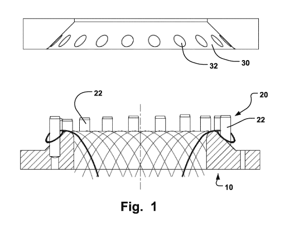
In Fig. 1, a braiding head 10 of a braiding machine for braiding a medical
implant is shown. The
2 0 braiding head 10 is equipped with a crown-shaped holder 20. The crown-
shaped holder comprises a
number of pins 22 distributed evenly around the crown-shaped holder 20. On top
of the braiding head 10,
a ring 30 can be placed. The ring 30 has holes 32 corresponding to the pins 22
of the crown-shaped
holder 20.
One embodiment is depicted in Fig. 2. Fig. 2 is a top view of a medical
implant 1, such as an
LAA occluder. This figure shows a medical implant 1 comprising strand loops
40. The strand loops 40
prevent the medical implant 1 from slipping and/or moving from the position
once implanted, since the
strand loops can fixate the medical implant to a body wall. The strand loops
40 are loops made from
strands. The strands may be made of shape-memory materials, metal, super
elastic alloys, Nitinol, or
polymers, such as biodegradable polymers. Thus, the strands may be wires. If
the strands are made of
Nitinol, then the strands may be heat-treated and a very flexible self-
expanding wire-mesh can be
obtained.
Fig. 3 is a lateral view of a medical implant 1. In this embodiment the loops
40 are located at
one side of the medical implant and the coupling 50 on the opposite side of
the medical implant 1.
However in another embodiment, the strand loops 40 may be positioned on the
same side of the medical
implant as the coupling 50.
Fig. 4 is a view of a medical implant from below and depicts another
embodiment of this
disclosure. In this embodiment the strand loops 40 are of a shape that extends
out of a plane
perpendicular to the longitudinal axis of the medical implant 1. More
precisely, the peripheral edges are
bent out of the perpendicular direction towards an end of the device. The
shape looks in the illustration like
4 0 triangular strand loops that may be rounded at the corners. They may be
in examples. However, the
illustration of embodiments given is round or oval shaped in a desired 3D
shape. By shaping the strand
loops 40 in this manner, the stabilization of the medical implant 1 will be
further improved, and thus the
medical implant 1 is further prevented from slipping and/or moving from the
position once implanted. By
the use of thus shaped strand loops, the retention may be improved. The bent
peripheral edges of the
CA 02852154 2014-04-14
WO 2013/060855 PCT/EP2012/071277
6
strand loops 40 provide for a defined outwardly oriented spring like force
when implanted. The spring force
is preferably much lower than the force of the medical device for returning to
an expanded shape from a
collapsed shape. In addition, the loops provide for a controllable spring
force irrespective of the main body
of the implant 1 being fully expanded.
The spring force of the loops may be provided in a radial direction and an
axial direction thanks
to the advantageous bending of the peripheral edge. As the number and shape of
the loops may be
varied, a large flexibility and adaptability to different defects to be
occluded is provided in a cautious and
reliable manner by embodiments of the device 1.
Fig. 5 is a top view of another medical implant. In this embodiment the strand
loops 40 are of a
round shape. By giving the strand loops 40 a round shape, the strand loops 40
are less prone to break.
Fig. 6 is a lateral view of a medical implant, in this case an LAA occluder,
showing another
embodiment. A longitudinal section 82 of the medical implant in this figure is
of the form of a frustum of a
hollow cone-shaped cylinder. The strand loops 40 surround the rim of the
hollow cone-shaped cylinder
and are extendable outwardly from the hollow cone-shaped cylinder
substantially perpendicularly to a
centre axis of the hollow cone-shaped cylinder. The cone shape provides for a
reliable positioning
avoiding embolization of the device in a defect, such as the LAA. The
principle may be compared to a cork
that has a larger diameter at one end. The larger diameter of the LAA occlude
shown in Fig. 6 is upon
implantation positioned at or towards the opening of the LAA.
The medical implant may also be of another shape. It can consist of different
sections, whereof
2 0 some sections are cone-shaped and other sections are disc-shaped.
Fig. 7 is a top view of a medical implant. In one embodiment, shown in Fig. 7,
half the strands,
which will also be used for the longitudinal section 82, form a cover for the
medical implant on the proximal
side 172. The remaining strands, which will also be used for the longitudinal
section 82, project outwards
on the edge of the cover as strand loops 40. Thus, on the proximal side 172,
strands which form the
strand loops 40 are not integrated in the braid. On a distal side, opposite to
the proximal side 172, as well
as along the longitudinal section 82, all strands from the outer edge up to
the rotation axis are braided or
intertwined. Thus, the configuration or distribution of strands to strand
loops can be said to be 2:1.
However, it is also possible to have other distributions, such as 1:1, 1:2,
1:3, 3:1 etc. It is also possible to
make a medical implant, which has only strand loops on the proximal side 172,
i.e. forming an open mesh
or a round longitudinal braid, closed on one side only.
Fig. 8 is a lateral view of a medical implant with a membrane 100. The
membrane 100 may be
attached to the medical implant 1 with a strand or thread 102. In this
embodiment, the membrane is
attached to the outer surface of the medical implant, on the same side as the
coupling 50. The membrane
100 can cover the whole circumference of the medical implant 1 and is then
also stitched to the medical
implant 1 with a seam 104. The use of membranes or inner membranes results in
improved occlusion and
rapid endothelialisation. The use of a membrane also results in an ideal
closure of e.g. the left atrial
appendage, since it seals the gap instantly.
Fig. 9 is a view of a thread used for attaching a membrane to a medical
implant. The thread 102
is wound around at least one strand of the medical implant.
Fig. 10 is a top view of a medical implant 1 with strand loops 40. The strand
loops 40 are
located all around the medical implant 1. Also the membrane 100 can be seen in
Fig. 10. Here the
membrane 100 covers the whole circumference of the medical implant 1.
CA 02852154 2014-04-14
WO 2013/060855 PCT/EP2012/071277
7
Fig. 11 shows the strand loops 40 of the medical implant 1 more in detail. The
strand loops 40
in Fig. 11 have a rounded shape, i.e. a somewhat rectangular shape with
rounded corners. Rounded
corners are tissue friendly avoiding deep tissue penetration and possible
ruptures or leakages.
Fig. 12 is a view of different knots used for attaching a membrane to a
medical implant. The
thread 102 can be secured to a strand of the medical implant 1 with a single
knot or with a double knot.
The ends of the thread 102 can be thermally treated to provide further
reliability of the fixation.
Fig. 13 is lateral view of a medical implant with a coupling 50. The coupling
50 is in the shape of
a ball pivot. The ball pivot can be connected to a flexible pusher, which can
be used to move the medical
implant in a sheath e.g. for delivery of the medical implant and/or retrieval
of the medical implant prior to
being decoupled. The coupling 50 is in this embodiment sunk or lowered into
the medical implant 1 and
will not impede blood flow at the target site, e.g. in a body vessel, where it
is situated after having been
delivered. In this embodiment, the medical implant has been braided so as to
form a hollow space for the
coupling 50. The advantage offered by the lowered coupling 50 is that the
medical implant is not
lengthened when the shaft is compressed, i.e. when the medical implant is
radially compressed. Thus, the
coupling is not moving out of its position, and will therefore not impede
blood flow. In another embodiment
according to Fig. 14, a proximal side 172 is instead given a concave shape to
assure a sinking of the
coupling 50, when the medical implant is radially compressed. With the
coupling 50, the flexibility during
delivery is increased, since the medical implant 1 is retrievable.
In an embodiment depicted in Fig. 14, the medical implant has a membrane 171.
The
membrane is an inner membrane 170. The inner membrane may be a special thermo-
treated PET-knit
fabric. The inner membrane 170 is attached to the inner surface of the medical
implant 1. The inner
membrane 170 may be attached to the medical implant 1 with a strand or thread.
In this embodiment, the
inner membrane 170 is attached to the inner surface of the medical implant 1,
on the same side of the
medical implant 1, as the coupling 50, i.e. the proximal side 172 of the
medical implant 1, and extending in
a longitudinal direction along the longitudinal sides 174 of the medical
implant 1. The inner membrane 170
can cover the whole circumference of the medical implant 1 and is then also
stitched to the medical
implant with seams 176, 178. The use of membranes and/or inner membranes
results in improved
occlusion and rapid endothelialisation. In another embodiment depicted in fig.
25b, an outer membrane
300 is attached outside the medical implant in a similar manner as the inner
membrane 170. Also the
outer membrane 300 may be a special thermo-treated PET-knit fabric. In one
embodiment, the medical
implant is covered with membranes both on the inside and the outside of the
braiding. As an alternative of
using membranes, the medical implant may instead be covered or coated using
Nano-spinning or a
dipping method. The coating as well as the membranes may be made of a
biocompatible and implantable
material, such as PTE, PTFE or PUR. The inner and/or outer membrane or coating
may be provided as a
non-fibrous film membrane that may have an initial controllable fluid
retention by perforations or
microperforations thereof. The membrane may cover the entire expanded diameter
of the implant.
Alternatively, it may only cover portions thereof. The portions may be as
small as the cell structure of the
fabric of the implant 1. For instance one or more cells of a braiding may be
provided with a coating
extending the space between adjacent strand portions forming the cells.
In this manner, different perfusion rates may be adjusted to different areas
of the device. It may
for instance be desired to obtain an inflow of blood into the inner of the
expanded device from a distal end
thereof to enhance integration of the device with surrounding blood upon
clotting thereof. A reduced or
prohibited outflow of blood through the proximal end may however be provided
by a tighter membrane or
larger diameter/surface/cells of the device being covered than those of
another section of the implant 1.
CA 02852154 2014-04-14
WO 2013/060855 PCT/EP2012/071277
8
The coating or external membrane may be affixed to the implant 1 in its
expanded shape. In this
manner, the coating or membrane is free of tension which advantageously avoids
pre-mature fatigue
thereof allowing for a reliable ingrowth.
The coating or external membrane may alternatively be affixed to the implant 1
in its collapsed
shape.
Patterns of covered cells may be provided to efficiently control a desired
flow pattern upon
implantation. In this manner, the ocdusion is not abrupt upon implantation. A
certain blood flow may still
occur after implantation and gradually decline upon blood coagulation and/or
endotheliazation of the
implanted device.
It should be noted that the aforementioned principles of coatings/membranes
may be provided
with other implants than the examples shown herein, e.g. ASD, PFO, PLD or VSD
ocduders.
Fig. 15 is a view of a thread used for attaching a membrane to a medical
implant. The thread
102 is wound around at least one strand of the medical implant for attaching
the membrane to the medical
implant.
Fig. 16 is a detailed view of strand loops 40 of a medical implant. The view
in Fig. 16
corresponds to the area in Fig. 17 marked with F.
Fig. 17 is a top view of a medical implant 1. The triangular strand loops 40
are located all along
the perimeter of the medical implant 1. Also in Fig. 17, the inner membrane
170 can be seen.
Fig. 18 is a view of different knots for a medical implant. The thread 102 can
be secured to a
strand of the medical implant 1 with a single knot or with a double knot. The
ends of the thread 102 can be
thermally treated.
Fig. 19 is a lateral view of a medical implant with a coupling 50. As can be
seen from this figure,
the braiding of the medical implant 1 is at the proximal side 172 of the
medical implant 1 formed so that
the proximal side 172 can be sunk in towards a centre of the medical implant
1.
Thus, the coupling 50 extends less from the medical implant 1 and will impede
the blood flow in
the atrium to a lower extent, at the target site where it is situated after
having been delivered, since the
proximal side 172 forms the ending towards the atrium.
Fig. 20 is a detailed view of a coupling of a medical implant 1. The coupling
50 is formed by
welding the strand ends together after the braiding machine has finished the
braiding or intertwining of the
medical implant. The coupling 50 is formed by welding it into a ball pivot.
The strands are merging into the
welded clot in a not-straight, i.e. not parallel manner in the example shown
in Fig. 20. The ball pivot can be
connected to a socket of a flexible pusher, which can be used to move the
medical implant in a sheath for
delivery. The pusher may be able to rotate the medical implant 360 degrees,
when the ball pivot is
connected to the socket. The braiding or intertwining of the medical implant 1
can be compressed so that
the medical implant can be inserted into the sheath. After leaving the sheath,
the medical implant 1
independently reassume the predetermined shape and ensure an interlocking
hold.
Fig. 21 is a lateral view of a medical implant 1 being manufactured. This
figure shows the
medical implant 1, after being completed. In the figure, the strands 252 are
shown. The strands 252 are
only for illustrative purposes shown flaring out at the end of the bundle. In
practice, the bundle of strands
has parallel strands. These strands 252 are cut to an appropriate length and
welded together to form the
coupling 50 shown in e.g. Fig. 19.
Fig. 22 is a lateral view of a medical implant being manufactured. This figure
shows the medical
implant 1, after being completed. In the figure, the strands 252 are shown.
The braiding of the medical
implant has here been sunk down into the medical implant so as to accommodate
a hollow space for the
CA 02852154 2014-04-14
WO 2013/060855
PCT/EP2012/071277
9
coupling 50. The strands 252 are cut to an appropriate length and welded
together to form the coupling 50
shown in e.g. Fig. 13.
Fig. 23a is a view from above at an angle of a braiding machine 270. The
braiding machine is a
round braiding machine and has a plurality of bobbins 272 arranged in a circle
with a braiding head 274 of
a braiding cylinder arranged inside the circle of the bobbins 272. The number
of bobbins may vary
according to the type of braiding machine used.
Fig. 23b is a detailed view of the bobbins 272 and a braiding head 274 of a
braiding machine.
The bobbins 272 are used for keeping the strands.
Fig. 24 is a detailed view of a braiding head 274. One embodiment of the
disclosure is a method
of manufacturing a 3D fabric of strands for forming a medical implant.
Examples of such medical implants
are septal, ventricular or auricle appendage occluders. The method comprises
intertwining the strands
along a length of the 3D fabric for forming a primary 3D fabric structure. An
example of such a primary
structure is the structure 42 shown in fig. 4. The intertwining is non-
continuous. First, a portion, such as
the proximal side 172 (shown in fig. 6), of the primary structure is formed by
intertwining strands. Then the
intertwining is interrupted along the length,i.e. the braiding procedure is
halted. This interruption may occur
after the bobbins 272 have been rotated a quarter of a turn. Thereafter a
secondary structure of the 3D
fabric is formed without intertwining. The forming of the secondary structure
is performed by making strand
loops 40. The making of the strand loops 40 is facilitated by the use of a
crown-shaped holder 20 (shown
in Fig. 1). The crown-shaped holder 20 has a plurality of pins 22 distributed
around it. The strands are
circled around the pins 22 so as to encircle the pins 22 and form strand loops
40. The pins 22 may have a
circular shape, an oval shape, a triangular shape or any other suitable shape.
Although pins 22 of the
same crown-shaped holder 20 normally have the same shape, it is possible to
have a crown-shaped
holder with pins 22 having different shapes. Thereafter, the intertwining is
continued. A ring 30 of a similar
size as the crown-shaped holder 20 is placed over the crown-shaped holder 20
for fixation of the strand
loops, since the ring 30 is provided with holes 32 in positions along the ring
30 corresponding to positions
of the pins 22 of the crown-shaped holder 20. Instead of intertwining, the
primary structure may be formed
by interlacing, interweaving, or braiding. The strand loops 40 prevent
slipping or unwanted movement of
the medical implant. Since the strand loops 40 are used instead of hooks or
barbs for fixation of the
medical implant after implantation, there will be no damage to body tissue as
there may be if hooks or
barbs are used. Thus, the risk of perforation of the thin left atrial wall is
considerably lowered.
Furthermore, pericardial effusion is avoided. Moreover, the medical implant
may be retrievable without
injuring body tissue or heart structure, since no barbs or hooks are used.
Although no barbs or hooks are
used, the medical implant 1 is easily fixated to a body wall and a very little
gap or no gap is left in the
target cavity while being sufficiently retained to allow for reliable ingrowth
in a minimum of time.
In one embodiment, the bobbins of the braiding machine are driven in a certain
position. The
advance of the strands are set to an appropriate length of lay, e.g. the
gradient of the strand windings is
set, and the appropriate length of braid or intertwining is set. A braiding
cylinder appropriate for the braid
size is chosen from braiding cylinders with different diameters. The braiding
cylinder with a braiding head
274 actuated by a feed gear mechanism is arranged in the centre of the
machine. Then the strands are
4 0 wound onto the bobbin coils; and the strands are routed over the thread
disengagement system of the
bobbins and pretensioned. A coupling used to hold the strand sections to be
braided is attached to the end
of the thread.
The strand sections required for the braid length are provided. The method of
manufacturing
comprises connecting a first end of a strand to a bobbin 272 of a round
braiding machine with a plurality of
CA 02852154 2014-04-14
WO 2013/060855 PCT/EP2012/071277
bobbins 272 and a second end of said strand to a diametrically opposing bobbin
272 of said round
braiding machine for a plurality of strands and arranging the middle sections
of said plurality of strands in a
fixed sequence over a braiding head 274 in a crisscrossed manner, i.e. there
is a crisscrossed placement
in a fixed sequence for half of the strands. The braiding head 274 of the
braiding cylinder is equipped with
5 pins for putting on strands in an ordered fashion. The pins are
subdivided depending on the number of
bobbins and the diameter of the braiding cylinder. The braiding head 274 of
the cylinder may be
semicircular or have planar surfaces that are rounded on the edges.
Thereafter a braiding procedure is started. After a portion of the medical
implant has been
braided or intertwined, the braiding procedure is halted. A crown-shaped
holder 20 for holding a plurality of
1 0 strand loops 40 is placed at the braiding head 274. The crown-shaped
holder 20 is held centrally by a
screw so that there is only a small space between the crown-shaped holder 20
and the braiding head 274,
i.e. the crown-shaped holder 20 is placed at a certain axial distance from the
braiding head 274.
Thereafter the remaining strand sections are individually bent in the middle
sections in order to
form strand loops 40. The remaining strand sections are introduced into a
space between the braiding
head 274 and the crown-shaped holder 20 from below. Thereafter, the strand
loops 40 are guided
separately through the space between the crown-shaped holder 20 and the
braiding head 274. The strand
loops 40 are placed over pins 22 of the crown-shaped holder 20. The strand
ends are routed to the
bobbins 272. Thereafter the strand ends are attached to the bobbins 272, i.e.
the strand ends are
connected to the clamp system of every second bobbin. Thus, the strand ends
being crossed on the
2 0 braiding head 274 and the strand loops 40 attached to the crown-shaped
holder 20 are connected in
regular correspondence with the bobbins 272. A ring 30 is placed on top of the
crown-shaped holder 20 for
fixation of the strand loops 40. When the ring 30 has been placed on top of
the strand loops 40, the strand
loops 40 are pressed down so as to be held. Then the braiding procedure is
continued until an intended
strand length has been braided. The strand ends are detached from the bobbins
272. Thereafter, the
strand ends are attached to the ring 30 with fixation means, such as an
adhesive strip. The braided
material may be thermally treated together with the ring 30 and the crown-
shaped holder 20 for shaping of
the medical implant. The thermal treatment serves for shaping, with the braids
being introduced into a
device that is operative in prescribing the shape of the medical implant.
Certain tools are used for this
shaping and the medical implant is shaped into a conical or truncated shape in
a longitudinal direction. A
medical implant 1 with a conical shape can be seen in fig. 25a. Due to the
conical shape of the medical
implant, higher radial forces from the body walls are possible. Therefore, the
risk of perforation is lowered.
Other possible shapes of the occluder are elongated, round, cylindrical, flat
or dumbbell-
shaped.
Finally the strand ends, preferably all the strand ends, are welded together,
by at least partly
melting a length of the plurality of strands to form a defined ball pivot as a
coupling 50. The method of
manufacturing provides accurate, fast and easy shaping of strand loops 40.
The medial device manufactured by the above-mentioned method is rotationally
symmetrical,
and may be of a closed mesh-structure. When the medical implant is implanted,
it is slight radial
compressed. However, no proximal change in length occurs.
The medical implants are available in different sizes over a large range, with
the length
corresponding to substantially 1/3 of the nominal diameter. The length may
vary between e.g. 10-22 mm,
and the diameter between e.g. 15-39 mm. It is even possible to combine
different wire gauges in one
braid. The medical implants can be held in the auricle as a result of radial
forces. They are distinguished
CA 02852154 2014-04-14
WO 2013/060855 PCT/EP2012/071277
11
by simple handling, and self-centering in the shunt. Since the braiding of the
medical implant is highly
flexible, the medical implant adapts well to the complex shape of the left
atrial appendage.
Although the strand loops 40 are shaped so as not to damage body tissue,
should barbs be
needed for positioning of the medical implant, then the loops can be severed
to form sharp barbs. Thus a
perforation of the tissue is possible.
Although, the strand loops 40 are depicted in the figures as situated in one
row, it is possible to
have strand loops 40 in multiple rows, e.g. two rows.
The strand loops 40 may be situated either on the proximal side 172 or the
opposite side, i.e.
the distal side. It is further possible to have strand loops 40 on both the
proximal side 172 and the distal
side. This results in a fixation of the implanted medical device in both
directions.
In an embodiment of the disclosure according to Fig. 26a, a medical implant 1
is provided with a
coating 11. The coating 11 has been applied to an external surface of the
medical implant 1. The medical
implant has been coated on the outside by a method, in which the medical
implant 1 has been dipped in a
solution with a specific viscosity, while the medical implant 1 was in an
expanded shape. By applying the
coating 11 to the medical device 1, while the medical device 1 is in an
expanded shape, the coating 11 is
free of tension, which advantageously avoids pre-mature fatigue thereof and
thus allows a reliable
ingrowth. Application of a coating 11 to a medical implant 1, while the
medical device 1 is in an expanded
shape, may also be advantageous for other reasons, such as the fact that the
medical implant 1 can be
made very flexible and that a particularly large expansion/contraction ratio,
i.e. a ratio of a size or diameter
of the medical implant 1 in its expanded shape and the size or diameter of the
medical implant 1 in its
contracted shape, can be obtained for the medical implant 1.
By making sure that the solution has a specific viscosity, the coating can be
made non-fibrous.
The specific viscosity is a viscosity, which takes on a value, which is in an
interval, where the solution for
the coating is non-fibrous or not fibrous. Thus, the coating will be made
fibrous. This may be
advantageous, since e.g. a lower friction towards a catheter is achieved. By
having a lower friction towards
the walls of a delivery catheter, the delivery is facilitated and made
smoother, i.e. the medical implant
slides or glides more smoothly through the delivery catheter. In one
embodiment only one end 13, and not
the side 14 of the medical implant 1, which side 14 encircles the medical
implant 1, is dipped into the
solution. In another embodiment, the end 13 and part of or the whole side 14
are dipped into the solution,
so as to be provided with coating. Thereby, a large portion of the medical
implant 1 is covered with the
coating 11. In yet another embodiment, only the ends of the medical implant 1
are dipped into the solution,
i.e. the end 13 and the other end 15 are dipped into the solution, but the
side 14 is not dipped into the
solution. Thereby, the medical implant is covered at both ends. This can be
done by first dipping the end
13 into the solution, then retracting the medical implant 1 from the solution.
Thereafter, the medical implant
is turned around and with the other end 15 facing the solution, the medical
device is again dipped into the
solution. A coating applied to the medical device 1 provides for an improved
occlusion, improved sealing
of a defect, such as a heart defect, an improved endothelialization and/or for
slowing down the blood flow
through the defect.
In an embodiment according to Fig. 26b, a medical implant 1 is instead
provided with a non-
fibrous membrane 17 externally, i.e. on the outside of the medical implant 1.
Such a non-fibrous
membrane 17 may be sewed onto the medical implant 1 by stitching it onto the
medical implant 1 along
the medical implant's circumference with stitches 19. In some embodiments, the
non-fibrous membrane 17
only covers one end 13, and not the side 14, of the medical implant 1. In
another embodiment, the end 13
and the side 14 are provided with a non-fibrous membrane 17. In yet another
embodiment, only the ends
CA 02852154 2014-04-14
WO 2013/060855 PCT/EP2012/071277
12
of the medical implant 1 are provided with a non-fibrous membrane 17, i.e. the
end 13 and the other end
15 are provided with a non-fibrous membrane 17, whereas the side 14 is not
provided with any non-fibrous
membrane 17. Thereby, the medical implant is covered at both ends 13, 15. A
non-fibrous membrane 17
applied to the medical device 1 provide for an improved occlusion, improved
sealing of a defect, such as a
heart defect, an improved endothelialization and/or for slowing down the blood
flow through the defect.
However, a coating 11 offers an advantage over the non-fibrous membrane 17,
since there is no stitching,
no sewing or even any clips needed for attaching and/or affixing the coating
to the medical implant 1.
Thus, the applying of a coating 11 instead of a non-fibrous membrane 17 may
offer the advantage of
providing easier and cheaper manufacturing. When compared to providing
membranes or patches inside
the medical implant 1, this advantage may be even greater, since the applying
of membranes or patches
inside such a medical implant 1 is even more time-consuming and complicated
than just applying a non-
fibrous membrane on an outside of the medical implant 1, because the membranes
or patches has to first
be put inside the medical implant 1 and then sewed or stitched onto the
medical implant, while being
inside the medical implant 1.
According to an embodiment, depicted in fig. 27a, one end 20 of the medical
implant 1 is
completely covered with a coating 22. Thus, an improved occlusion, an improved
sealing of a defect, such
as a heart defect, an improved endothelialization and/or a slowing down of the
blood flow through the
defect is achieved. However, in order to provide some body liquid to pass
through the medical implant 1
once implanted, the coating may be provided with perforations or
microperforations. This is shown in fig.
2 0 27b, which depicts a situation where one end 20 of the medical implant
1 has been covered with a coating
22 and where the coating has been perforated, i.e. provided with perforations
24 or microperforations.
Such perforations or microperforations may be provided by a process, such as
mechanical perforation or
laser perforation, i.e. laser cutting. The use of laser perforation offers the
advantage of a better
consistency of the hole size, i.e. the perforation size, than the use of
mechanical perforation. By providing
the coating 22 with perforations or microperforations, an initial controllable
body liquid retention is enabled.
Furthermore, the integration of the medical implant 1 may be enhanced and/or
facilitated by the use of
such perforations 24 or microperforations, since the body liquid is allowed to
enter into the interior of the
medical implant 1. A limited blood flow may actually pass through the medical
implant 1 after implantation.
However, this limited blood flow will gradually decline upon blood coagulation
and/or endothelialization of
the implanted medical implant. Thus, by the use of perforations 24 or
microperforations, the occlusion is
not abrupt, but formed gradually over time.
In one embodiment, the perforations 24 or microperforations of the coating 22
are uniformly
distributed over the area of the coating 22. However, in other embodiments,
the perforations 24 or
microperforations are randomly distributed. In yet another embodiment,
depicted in fig. 27d, a first central
area 28 of the coating 22, corresponding to a first area of the medical
implant is provided with perforations
of a larger size, such as a diameter, than perforations of a second peripheral
area 26 of the coating 22,
corresponding to a second area of the medical implant 1, so that the inflow to
different areas is controlled.
As an alternative, the first central area 28 of the coating 22, corresponding
to a first area of the medical
implant 1, is provided with a higher number of perforations or a higher
density of perforations than a
second peripheral area 26 of the coating 22, corresponding to a second area of
the medical implant, so
that the inflow to different areas is controlled. This is depicted in fig.
27c.
In fig. 28, another kind of medical implant 1 or occluder is shown. This
medical implant 1
comprises a first disc-shaped section 30, a tubular middle section 32 and a
second disc-shaped section
34. In this embodiment, the one depicted in fig. 28, only the ends of the
medical implant 1 are coated or
CA 02852154 2014-04-14
WO 2013/060855 PCT/EP2012/071277
13
provided with non-fibrous membranes, i.e. the outer end side 36 of the first
disc-shaped section 30 and the
outer end side 38 of the second disc-shaped section 34 are coated or provided
with non-fibrous
membranes. The application of a coating can e.g. be performed by dipping both
outer end sides 36, 38
into a solution, with a specific viscosity. Thereby, the medical implant is
covered at both ends. In another
embodiment, only the outer end side 36 of the first disc-shaped section 30 is
provided with a coating 22. In
yet another embodiment, the full length of the medical implant 1 is provided
with a coating 22. In some
embodiments, the tubular middle section 32 is provided with a coating 22,
whereas the disc-shaped
sections 34, 36 are not provided with a coating.
In yet another embodiment, depicted in fig. 29, a coating 22 or non-fibrous
film membrane is
arranged at an end 20 of a medical implant 1 so as to obtain an inflow of
blood, into the inner of the
medical implant 1, after implantation and thus in an expanded shape, from a
distal end of the medical
implant 1 for enhancing integration of the medical implant with surrounding
blood upon clotting thereof.
This can e.g. be achieved by providing the end 20 with a coating 22, which
covers substantially the whole
end 20, but does not cover the section 40 of the end 20. Thus, an inflow of
blood, into the inner of the
medical implant 1 can be obtained through the section 40. The section 40 may
be centred or situated at
any other position at the end 20. As an alternative, the section 40 may
instead by surrounding the coating
22, i.e. the coating 22 is applied only to a central portion of the end 20.
Fig. 30 shows a medical implant 1 in an expanded shape. This is the shape the
medical implant
1 preferably has after being implanted. This is also the shape, the medical
implant resiliently returns to, i.e.
it can also be called the relaxed shape. However, in order to deliver a
medical implant 1 into a target site
inside a mammal body, the medical implant 1 needs to be put through a narrow
delivery catheter. In order
for the medical implant 1 to fit into a narrow delivery catheter, the medical
implant 1 will have to take on
another shape. This other shape is here called the contracted shape. It could
also be called a delivery
shape. The medical implant in its contracted shape can be seen in fig. 31. The
coating 22 is in one
embodiment applied to the medical implant 1, while the medical implant 1 is in
the contracted shape.
Thereby, the coating 22 will be prone to contribute to force the medical
implant into its contracted shape
and thus provide for facilitation of the delivery of the medical implant 1
through a catheter.
Fig. 32 is a top view of a medical implant, provided with a coating 22,
wherein the coating 22
forms a pattern. Such a pattern can be any pattern, which is advantageous for
control of a desired flow
pattern through the medical implant 1 upon implantation. In the embodiment
according to fig. 32, the
medical implant 1 is provided with a coating 22 in some sections, i.e. one
section 72 of the end portion 20
of the medical implant 1 is provided with a coating 22, whereas all adjacent
sections 70 are not provided
with a coating and likewise all sections being adjacent to a non-coated
section 70 are provided with a
coating 22. By the use of such a pattern of covered sections 72 or cells an
efficient control of a desired
flow pattern through the medical implant 1 upon implantation is established.
The sections or cells may be
large and thus cover large portions of the medical implant or as small as a
gap between adjacent strands
of the mesh, which makes up the medical implant 1. The pattern may be formed
be first applying a coating
22 by a method, such as dipping the medical implant 1 into a solution, and
thereafter removing parts of the
coating 22 so as to form a pattern of coating 22.
A step of a method of producing a medical implant for occluding an opening in
a body is shown
in Fig. 33, which is a lateral view of a medical implant being coated by
dipping. In some embodiments,
such a method comprises producing a body mesh of strands forming a plurality
of adjacent cells delimited
by the strands. Some or all of these cells may be provided with a coating.
Therefore, in some
embodiments, the method further comprises applying a polymer, such as
polyurethane,
CA 02852154 2014-04-14
WO 2013/060855 PCT/EP2012/071277
14
polytetrafluoroethylene (PTFE) or expanded polytetrafluoroethylene (ePTFE), to
at least part of an
external surface of the medical implant 1, such as the outer end side 36 of
the first disc-shaped section 30
of the medical implant 1. The use of e.g. PTFE or ePTFE may be especially
advantageous, since these
materials may provide for low friction. The polymer may be applied to the
medical implant 1 by e.g.
dipping, spraying, electro-spinning, electro-spraying or Nano-spinning. As
another alternative, instead of
coating the medical implant 1, a non-fibrous film membrane may be sewed onto
the external surface of the
medical implant 1. Such a non-fibrous film membrane may be manufactured by
applying a film on a
substrate and then removing the substrate. E.g. a film can be applied by
dipping a substrate into a solution
and thereafter removing the substrate. As an alternative, a substrate can be
sprayed and thereafter
1 0 removed from the coating formed by the spray, so that a non-fibrous
film membrane is obtained.
In figure 33, the process of dipping a medical implant into a solution 80 of a
specific viscosity is
shown. Thus, the polymer is applied to the medical implant 1 by dipping the
medical implant 1 into a
solution 80 of a specific viscosity, so that a non-fibrous coating is applied
and affixed to an external
surface of the medical implant 1. In one embodiment only an outer end side 36
of a first disc-shaped
section 30 of the medical implant is dipped into the solution. In another
embodiment, the outer end side 36
of the first disc-shaped section 30 and an outer end side 38 of a second disc-
shaped section 34 of the
medical implant 1 are dipped into the solution. In other embodiments, further
parts of the medical implant 1
may be dipped into the solution. As an example, the side 14 (shown in fig.
27a) can be dipped into the
solution. As another example, substantially the whole medical implant 1 may be
dipped into the solution.
Which parts of the medical implant 1 that are dipped into the solution may
depend on what kind of medical
device 1 is to be applied with coating, i.e. it may depend on whether the
medical implant is e.g. an atrial
septal defect (ASD) occluder, a Patent foramen ovale (PFO) occluder, a
paravalvular leakage (PLD)
occluder, a ventricular septal defect (VSD) occluder or some other medical
implant.
Alternatively, the coating can be applied to the medical implant 1 by spraying
the medical
implant with a spray 90, which is of a specific viscosity, so that a non-
fibrous coating 92 is applied and
affixed to an external surface of the medical implant 1. This alternative is
shown in Fig. 34, which is a
lateral view of a medical implant 1 being coated by spraying. In one
embodiment only an outer end side 36
of a first disc-shaped section 30 of the medical implant is sprayed. In
another embodiment, the outer end
side 36 of the first disc-shaped section 30 and an outer end side 38 of a
second disc-shaped section 34 of
the medical implant 1 are sprayed. In other embodiments, further parts of the
medical implant 1 may be
sprayed. As an example, the side 14 (shown in fig. 26a) can be sprayed. As
another example,
substantially the whole medical implant 1 may be sprayed. Which parts of the
medical implant 1 that are
sprayed may depend on what kind of medical device 1 is to be applied with
coating, i.e. it may depend on
whether the medical implant is e.g. an atrial septal defect (ASD) occluder, a
Patent foramen ovale (PFO)
occluder, a paravalvular leakage (PLD) occluder, a ventricular septal defect
(VSD) occluder or some other
medical implant.
Other alternatives of applying a coating to the medical implant 1 are e.g.
electro-spinning,
electro-spraying or Nano-spinning. Fig. 35 is a schematic sketch of a medical
implant being coated by a
process, such as electro-spinning or Nano-spinning. In the figure, a polymer
or composite solution 90 is
contained in a syringe pump 92. The syringe pump 92 comprises a spinneret,
such as a hypodermic
syringe needle or a metallic needle, which is connected to a high-voltage
direct current power supply 96.
The direct current power supply 96 is also connected to ground 100 and on the
ground side, a collector 98
is connected to the direct current power supply 96. The medical implant 1 to
be coated would typically be
placed in connection with the collector 98. The polymer solution 90 is loaded
into the syringe 92 and
CA 02852154 2014-04-14
WO 2013/060855 PCT/EP2012/071277
extruded, as droplets, from the tip of the needle 94 at a constant rate by the
syringe pump 92. A
sufficiently high voltage must then be applied to the droplets, so that the
droplets become charged. Since
electrostatic repulsion counteracts the surface tension, the droplets are
stretched: At a critical point, a
stream of liquid erupts from the surface. This point of eruption is known as
the Taylor cone. If the
5 molecular cohesion of the liquid is sufficiently high, stream breakup
does not occur and a charged liquid jet
is formed. Alternatively, if stream breakup occurs, the droplets are instead
electro-sprayed.
As the jet dries in flight, the mode of current flow changes from ohmic to
convective as the
charge migrates to the surface of the strand. The jet is then elongated by a
whipping process caused by
electrostatic repulsion initiated at small bends in the strand, until it is
finally deposited on the grounded
10 collector. The elongation and thinning of the strand resulting from this
bending instability leads to the
formation of uniform strands. Such uniform strands may have nanometer-scale
diameters.
In some embodiments, a method of producing a medical implant for occluding an
opening in a
body comprises producing a body mesh of strands forming a plurality of
adjacent cells delimited by the
strands. The producing of a body mesh of strands may be performed by
intertwining strands along a
15 length of a 3D fabric for forming a primary 3D fabric structure. The
intertwining may be non-continuous,
such as interrupted along the length, for forming a secondary structure of the
3D fabric without
intertwining. A round braiding machine may be used for forming the primary and
secondary fabric
structures. In one embodiment, the method may comprise connecting a first end
of a strand to a bobbin of
a round braiding machine with a plurality of bobbins and a second end of the
strand to a diametrically
opposing bobbin of the round braiding machine for a plurality of strands and
arranging middle sections of
the plurality of strands in a fixed sequence over a braiding head in a
crisscrossed manner. It may further
comprise starting a braiding procedure, halting the braiding procedure,
placing a crown-shaped holder for
holding a plurality of strand loops at the braiding head, bending remaining
strand sections individually in
the middle sections in order to form strand loops, introducing the remaining
strand sections into a space
between the braiding head and the crown-shaped holder from below, placing the
strand loops over pins of
the crown-shaped holder, routing the strand ends to the bobbins, attaching the
strand ends to the bobbins,
placing a ring on top of the crown-shaped holder for fixation of the strand
loops, continuing the braiding
procedure until an intended strand length has been braided, detaching the
strand ends from the bobbins,
attaching the strand ends to the ring with fixation means and/or treating the
braided material, the ring and
the crown-shaped holder thermally for shaping of the medical implant. It may
also comprise welding the
strand ends together, by at least partly melting a length of the plurality of
strands to form a defined ball
pivot. Thereby, shaping of loops can be made accurately, fastly and/or easily.
The medical implant 1 may also in one embodiment be constructed so that the
ends of the
medical implant 1 folds inwards for delivery, i.e. when the medical implant 1
is in its contracted shape, the
coating 22 is on the inside and covered and/or protected by the sides of the
medical implant 1, which sides
are close to or touching the inside wall of a delivery catheter, in which it
is delivered. The ends of the
medical implant 1 are in this embodiment somewhat conically shaped or funnel-
shaped, so that the
medical implant 1 folds into its contracted shape with its coated ends covered
on the outside with the sides
of the medical device 1. The two disc-shaped sections 30, 34 of the medical
device 1 may also be cupped
4 0 away from each other.
The present disclosure has been described above with reference to specific
embodiments.
However, other embodiments than the above described are equally possible
within the scope of the
disclosure. Different method steps than those described above, may be provided
within the scope of the
disclosure. The different features and steps of the disclosure may be combined
in other combinations than
CA 02852154 2014-04-14
WO 2013/060855
PCT/EP2012/071277
16
those described. The scope of the disclosure is only limited by the appended
patent claims. More
generally, those skilled in the art will readily appreciate that all
parameters, dimensions, materials, and
configurations described herein are meant to be exemplary and that the actual
parameters, dimensions,
materials, and/or configurations will depend upon the specific application or
applications for which the
teachings of the present disclosure is/are used.