Note: Descriptions are shown in the official language in which they were submitted.
Automated Tissue Engineering System Comprising
Sensors Linked to a Microprocessor
Field of the Invention
This invention relates to devices, methods and systems for the
automated culture, proliferation, differentiation, production and maintenance
of tissue engineered products. Such systems, methods and products find
use in various clinical and laboratory settings.
Background of the Invention
Throughout this application, various references are cited in parentheses
to describe more fully the state of the art to which this invention pertains.
During the past several years, researchers have developed and used
different cell culture and tissue engineering techniques for the culture and
production of various types of cellular implants. Such systems are described
for example in U.S. Patent Nos. 5,041,138, 5,842,477, 5,882,929,
5,891,455, 5,902,741, 5,994,129, 6,048,721 and 6,228,635. Bioreactor
systems have also been developed for the culture of cells and cellular
implants and are described for example in U.S. Patent Nos. 5,688,687,
5,728,581, 5,827,729 and 6,121,042.
The aforementioned methods and systems generally employ
conventional laboratory culturing techniques using standard culture
equipment for cell seeding of selected cell populations onto scaffolds. As
such, the generated implants simply comprise proliferated cell populations
grown on a type of biopolymer support where any manipulation of the
cellular environment is limited to endogenous cell production of cytokines
present in any standard cell culture, and application of shear and/or physical
stresses due to circulation of cell culture media and physical manipulation of
the support onto which the cells are seeded. The systems do not address
nor are they capable of generating a tissue implant that comprises
proliferated and differentiated cells representative of developing tissues in
CA 2853267 2017-09-21
CA 02853267 2014-06-03
- 2 -
vivo and further integrated within a selected scaffold that can be
successfully
integrated in vivo. Moreover, known methods and systems are not capable
of multi-functionally carrying out all of the steps of biopsy tissue digestion
to
yield disassociated cells, subsequent cell seeding on a proliferation
substrate, cell number expansion, controlled differentiation, tissue formation
and production of a tissue implant within a single automated tissue
engineering system. This is primarily because known culture systems are
not sophisticated in that they are not capable of automatically evaluating and
manipulating the changing environment surrounding the developing implant
such that cells progressively proliferate and differentiate into a desired
implant.
Furthermore, conventional culture methods and systems are labor
intensive and suffer from the drawbacks of contamination and varying
degrees of culturing success due to human error and lack of continual
performance evaluation. Conventional culture systems require that most of
the initial steps in the preparation of cells for seeding (i.e. tissue
digestion,
cell selection) is performed manually which is time consuming, unreliable in
terms of the quality of the tissue produced, and prone to culture
contamination problems. The systems are incapable of supporting the
automated preparation of tissue engineered implants from primary or
precursor cells due to inherent design limitations that restrict the cell and
tissue culture process, the inability to adequately monitor and modify the
environment to support tissue development, and the absence of techniques
to enable the implementation of effective quality control measures.
Thus, there remains a real and unmet need for an improved system
for in vitro and ex vivo tissue engineering that can consistently meet the
operational requirements associated with the different steps in the
development and production of tissue engineered implants. Of particular
importance is the ability to create functional tissue constructs where the
cells
present are active, differentiated and already expressing extracellular
matrix.
This involves more than, and is strikingly different to, the simple simulation
of
the mature in vivo environment present at the host site. This is because the
CA 02853267 2014-06-03
- 3 -
preparation of functional de novo tissue fundamentally requires that the cells
progress through a series of developmental stages as part of an ex vivo
sequence.
In order to address both clinical and research requirements, new
devices, methods and systems have been developed that obviate several of
the disadvantages and limitations of conventional ex vivo culturing
techniques and systems.
Summary of the Invention
The present invention is directed to a user-friendly automated system
for cell culture and tissue engineering that can be used in a variety of
clinical
and research settings for both human and veterinary applications.
As used herein, "tissue engineering" may be defined as "the
application of principles and methods of engineering and life sciences
toward fundamental understanding and development of biological substitutes
to restore, maintain and improve tissue functions". This definition is
intended
to include procedures where the biological substitutes are cells or
combinations of different cells that may be implanted on a substrate or
scaffold formed of biocompatible materials to form a tissue, in particular an
implantable tissue construct. Furthermore, it is noted that the cells involved
in the tissue engineering processes may be autologous, allogenic or
xenogenic.
The tissue engineering system of the present invention is designed to
perform all activities under sterile operating conditions. The system is fully
automated, portable, multifunctional in operation and performs/provides one
or more of the following:
- sterile reception/storage of tissue biopsy;
- automated monitoring of digestion process
- digestion of biopsy tissue to yield disassociated cells;
- cell sorting and selection, including safe waste collection;
- cell seeding on or within a proliferation substrate or scaffold
CA 02853267 2014-06-03
-4-
- proliferation of cells to expand cell populations;
- cell washing and cell collection;
- cell seeding on or within a tissue engineering scaffold or matrix;
- cell differentiation to allow specialization of cellular activity;
- tissue formation;
- mechanical and/or biochemical stimulation to promote tissue
maturity;
- harvesting the tissue engineered constructs /implants for
reconstructive
surgery; and
- storage and transportation of implantable tissue.
The tissue engineering system of the present invention may be pre-
programmed to perform each of the above noted steps, individually,
sequentially or in certain predetermined partial sequences as desired and
required. Furthermore, each of these steps, or any combination thereof, are
accomplished within one or more bioreactors on a tissue engineering
module. In operation, the tissue engineering system is pre-programmed and
automatically controlled thus requiring minimal user intervention and, as a
result, enhances the efficiency and reproducibility of the cell culture and/or
tissue engineering process while minimizing the risks of contamination. The
tissue engineering system of the invention and components thereof are
operable under conditions of microgravity and/or zero gravity where such
system and components are used for space research.
The system of the present invention is designed such that primary or
precursor cells can be isolated from a donor tissue for further propagation,
differentiation and production of a tissue implant. Alternatively, cell lines
may also be used either alone or in combination with other cell sources.
In accordance with the invention, is an automated tissue engineering
system, the system comprising a housing that supports at least one
bioreactor that facilitates physiological cellular functions and the
generation
of tissue constructs from cell and tissue sources. The housing also supports
a flusicl containment system that is in fluid communication with the
bioreactor.
CA 02853267 2014-06-03
- 5 -
Associated with the housing and/or the bioreactor are sensors that monitor
physiological parameters of fluid provided in the fluid containment system. A
microprocessor disposed within the housing is linked to the bioreactor and
the fluid containment system and functions to control their functioning. The
microprocessor may also independently control environmental conditions
within the system.
In accordance with another aspect of the invention there is provided a
system for cell and tissue engineering comprising portable, sterile tissue
engineering modules having one or more bioreactors which provide the
basis for tissue digestion, cell seeding on a proliferation substrate, cell
proliferation, cell seeding on a differentiation scaffold, cell
differentiation, and
tissue formation with subsequent maturation into functional tissue for
implantation. The bioreactor is operatively connected with a media flow and
reservoir system for the delivery of reagents and the collection of waste
fluids in a non-reflux manner. The bioreactor and/or the media flow system
optionally include gas exchange components that utilize semi-permeable
membranes to allow the transfer of gaseous products thereby controlling
levels of dissolved gases in the media. The tissue engineering module
operatively interacts with a central microprocessor controlled base unit that
automatically monitors the progression of the cell culture or tissue
engineering process and adjusts the environmental conditions to meet the
requirements of the different stages of cell culture and tissue development
within the bioreactor. Deviations from ideal conditions are sensed by a
variety of sensors present within the bioreactor and the signals generated
are monitored by the central microprocessor. As such, changes in
environmental conditions such as but not limited to pH, temperature and
dissolved gases can be automatically monitored and altered as required. In
addition, the status of cell proliferation is indirectly assessed by detection
of
metabolic turnover as a function of time (e.g. pH, 02, CO2, lactic acid and
glucose consumption). Further to the control of processing conditions by the
central microprocessor, the tissue engineering module itself may optionally
include a secondary onboard microprocessor that operates in unison with
CA 02853267 2014-06-03
- 6 -
the central microprocessor. The tissue engineering module microprocessor
expands the data processing capabilities of the tissue engineering system by
performing specific functions directly onboard the tissue engineering module,
thereby minimizing the demands on the central microprocessor.
Various growth factors, cytokines, experimental agents,
pharmaceuticals, chemicals, culture fluids and any combinations thereof may
be loaded and stored within any of the reservoirs located on the tissue
engineering module and automatically transferred to the one or more
bioreactors according to a pre-programmed sequence or as required by the
developing tissue implant. The individual tissue engineering modules are
removable from the system for transport without compromising the sterility of
the tissue engineered constructs present within the bioreactor. Such
removal does not affect the processing of any other modules present within
the tissue engineering system. Furthermore, the tissue engineering module
may be considered to be disposable following the completion of a tissue
engineering sequence, as this practice prevents contamination arising from
prior use.
In various embodiments of the invention, the device and system can
be used to digest tissues obtained by surgical biopsy. In another
embodiment, cells can be filtered and a particular population selected and
isolated. In another embodiment, digested cells can be proliferated to
expand the population of the cells. In still a further embodiment, cells can
be
seeded and cultivated on a desired scaffold or substrate (also referred to as
a matrix). In yet .a further embodiment, cells can be differentiated on and/or
throughout a desired scaffold or substrate until suitable tissue formation is
obtained. In yet a further embodiment, the tissue may be stimulated to
promote tissue maturity. In yet another embodiment, a tissue implant is
produced that is *suitable for reconstructive surgery. In still a further
embodiment, cell sampling can be done at each stage of cellular proliferation
and developmental progression in a sterile manner without adverse effects
on the culture itself. Each of the aforementioned embodiments can be done
alone or sequentially as desired. Tracking of such processing events can be
CA 02853267 2014-06-03
- 7 -
performed by the central microprocessor and/or the module-based
microprocessor for incorporation into quality control records.
In one aspect, the tissue engineering system optionally uses a
synthetic biomaterial compound,SkeliteTm, described in Applicant's U.S.
Patent No. 6,323,146 to enhance biological performance. Briefly, SkeliteTM
is an isolated bioresorbable biomaterial compound comprising calcium,
oxygen and phosphorous, wherein a portion of at least one of said elements
is substituted with an element having an ionic radius of approximately 0.1 to
0.6 Angstroms. In one embodiment, Skelite TM may be used to enhance cell
proliferation through its use as a coating on the walls of the bioreactor, as
a
thin film on the proliferation substrate, or as a three-dimensional and
thereby
high surface area proliferation scaffoldThe use of SkeliteTM in the
proliferation stage may be demonstrated to:
- increase the rate of proliferation;
- increase the cell yield following the proliferation step;
- reduce the surface area required for a target cell yield;
- reduce the problematic tendency of cell phenotype dedifferentiation
during proliferation; and
- enhance the binding of growth factors to the proliferation substrate.
In a further embodiment, SkeliteTM may be used as a resorbable
scaffold to enhance the differentiation of cells and the subsequent formation
of tissue constructs. The use of SkeliteTM in the differentiation stage may be
demonstrated to:
- increase productivity by improving the reliability of the differentiation
stage;
- increase the integrity and hence biological viability of the tissue
construct;
- allow flexibility in construct configuration based on various scaffold
formats;
- allow the stages of proliferation, differentiation and tissue formation
to occur on a common substrate;
CA 02853267 2014-06-03
-8-
- enhance the binding of growth factors to the differentiation scaffold;
and
- improve tissue construct handling properties during surgical
implantation.
In another aspect, the present invention provides a method and
system for the preparation of tissue constructs through the automated steps
of digestion, proliferation, seeding and differentiation of primary or
precursor
cells that originate from a patient thus eliminating immunological and disease
transmission issues. An implant may be formed from the controlled
cultivation of various cell types, including but not limited to chondrocytes,
stromal cells, osteoblasts, nerve cells, epithelial cells stem cells and
mixtures
thereof.
The system of the invention in an embodiment, incorporates one or
more detachable, portable, and independently operable tissue engineering
modules that support one or more bioreactors, media reservoirs and
fluid/media flow system. Each module, and hence the bioreactor(s), is under
the automated control of a central microprocessor. The module and
associated bioreactor(s) may be configured for various specialized
applications such as, but not limited to:
- sterile reception/storage of tissue biopsy;
- automated mixing and delivery of digestion reagents;
- automated monitoring of digestion process;
- digestion of biopsy tissue to yield disassociated cells;
- cell sorting and selection, including safe waste collection;
- cell washing and cell collection;
- cell seeding on or within a proliferation substrate or scaffold;
- automated mixing and delivery of proliferation reagents;
- proliferation of cells to expand cell populations;
- automated monitoring of cell conditions, including detection of
confluence;
- controlled cell release from the proliferation substrate or scaffold;
CA 02853267 2014-06-03
-9-
- repeated proliferation steps on selected surface area sizes to
increase cell numbers;
- channeling of cell population toward one or more tissue engineering
scaffolds or matrices;
- cell seeding on or within the tissue engineering scaffold or matrix;
- automated mixing and delivery of differentiation reagents;
- automatic monitoring of cell/tissue culture conditions;
- cell differentiation to allow specialization of cellular activity;
- tissue formation;
- mechanical and/or biochemical stimulation to promote tissue
maturity;
- harvesting the tissue engineered constructs /implants for
reconstructive
surgery; and
- storage and transportation of cells and/or implantable tissue.
When two or more bioreactors are provided within the system either
supported directly within the housing of the system or supported on a tissue
engineering module insertable into the housing, the bioreactors may be
provided connected in series and individually operable and controlled by the
microprocessor or alternatively, may be operated and controlled
independently depending on the user's programming of the microprocessor
and the desired result to be achieved. Furthermore, when two or more
bioreactors are provided within the system, the bioreactors and internal
chambers may be connected such that there is an exchange of cells and/or
tissues from bioreactor to bioreactor.
The bioreactor can be manufactured in various sizes and
configurations as required to support varying numbers and sizes of
proliferation and differentiation scaffolds or substrates. The bioreactor may
be incorporated as part of the structural components of the tissue
engineering module. Alternately, the bioreactor may be detachable as a
separate component to the remaining components of tissue engineering
module. If present as a discrete component, the bioreactor may be
CA 02853267 2014-06-03
- 10 -
packaged separately in a sterile package and joined to the tissue
engineering module using sterile access techniques at the time of use.
Furthermore, the sterile access techniques enable the bioreactor to be
detached from the module, upon completion of the tissue engineering
process, for easy transport to the operating room in preparation for the
retrieval of a newly formed implantable tissue construct.
The bioreactor and/or the tissue engineering module may be rotated
or agitated within the overall tissue engineering system via control
actuators.
Rotation may enable the beneficial use of gravity to effect specific
bioprocessing sequences such as sedimentation-based cell seeding and
fluid exchange within the bioreactor.
The tissue engineering module may be bar coded or provided with a
memory chip for rapid and accurate tracking both within the tissue
engineering system and externally as part of the clinical or experimental
environment. Such tracking technology as incorporated within the tissue
engineering device also enables electronic tracking via clinic-based
information systems for patient records. This ensures that the tissue
engineering module and hence the associated cells or tissue implants are
properly coded to ensure administration to the correct patient and that the
process is recorded for hospital billing purposes. The module and/or
bioreactor may also utilize a bar code and/or memory chip in a similar
manner for rapid and accurate patient and sample tracking.
According to an aspect of the present invention is an automated
tissue engineering system comprising;
- a housing;
- at least one bioreactor supported by said housing, said bioreactor
facilitating physiological cellular functions and/or the generation of one or
more tissue constructs from cell and/or tissue sources;
- a fluid containment system supported by said housing and in fluid
communication with said bioreactor,
- one or more sensors associated with one or more of said housing,
bioreactor or fluid containment system for monitoring parameters related to
CA 02853267 2014-06-03
- 11 -
said physiological cellular functions and/or generation of tissue constructs;
and
- a microprocessor linked to one or more of said sensors.
According to another aspect of the present invention is an automated
tissue engineering system comprising;
- a housing;
- at least one tissue engineering module removably accomodated
within said housing, said tissue engineering module comprising a support
structure that holds at least one bioreactor, said bioreactor facilitating
physiological cellular functions and/or the generation of one or more tissue
constructs from cell and/or tissue sources, a fluid containment system in
fluid
communication with said bioreactor, and one or more sensors for monitoring
parameters related to said cell culture and/or tissue engineering functions;
and
- a microprocessor disposed in said housing and linked to said tissue
engineering module, said microprocessor controlling the operation of said
tissue engineering module.
According to a further aspect of the invention is portable and
sterilizable tissue engineering module, the module comprising;
- a structural support holding' at least one bioreactor, said bioreactor
facilitating cell culture and tissue engineering functions;
- a fluid containment system in fluid communication with said
bioreactor; and
- one or more sensors for monitoring parameters related to said cell culture
and tissue engineering functions.
In aspects of this embodiment, the bioreactor comprises a bioreactor
housing having one or more inlet ports and one or more outlet ports for
media flow and at least one chamber defined within said bioreactor housing
for receiving cells and/or tissues and facilitating said cell culture and
tissue
CA 02853267 2014-06-03
- 12 -
engineering functions. The chamber may be selected from the group
consisting of a cell culture/proliferation chamber, cell
differentiation/tissue
"formation chamber, tissue digestion chamber and combinations thereof.
Furthermore, the chamber houses one or more substrates and/or scaffolds.
In embodiments of the invention, two or more chambers may be provided
operably connected within the bioreactor and be operably connected.
Alternatively, the two or more bioreactors may be independently operable or
co-operatively operable. In still further aspects, the chambers and/or
bioreactors are operably connected to provide for the exchange of fluids,
cells and/or tissues between the chambers and/or bioreactors.
The scaffold for use in the present invention is selected from the group
consisting of a porous scaffold, a porous scaffold with gradient porosity, a
porous reticulate scaffold, a fiberous scaffold, a membrane encircled scaffold
and combinations thereof. Chambers may also be further subdivided into
zones. For example, a differentiation/tissue formation chamber may be
provided with a plurality of zones to contain several scaffolds. Funnels or
similar passageways may be provided between chambers within a
bioreactor. Furthermore, one or more filters may be provided at any location
within a bioreactor.
According to still another aspect of the present invention is a
bioreactor that provides an environment for cell culture and/or tissue
engineering functions selected from the group consisting of storage of tissue
biopsy, digestion of tissue biopsy, cell sorting, cell washing, cell
concentrating, cell seeding, cell proliferation, cell differentiation, cell
storage,
cell transport, tissue formation, implant formation, storage of implantable
tissue, transport of implantable tissue and combinations thereof.
According to still another aspect of the present invention is a
bioreactor for facilitating and supporting cellular functions and generation
of
implantable tissue constructs, said bioreactor comprising;
- a bioreactor housing;
CA 02853267 2014-06-03
-13-
- one or more inlet ports and one or more outlet ports for media flow;
- at least one chamber defined within said bioreactor housing for
facilitating and supporting cellular functions and/or the generation of one or
more tissue constructs from cell and/or tissue sources; and
- one or more sensors for monitoring parameters related to said
cellular functions and/or generation of tissue constructs within said at least
one chamber.
In embodiments of the invention, the bioreactor housing comprises a
lid, where the lid may be a detachable lid or integral with the bioreactor
housing.
Cells and tissues may be selected from bone, cartilage, related bone
and cartilage precursor cells and combinations thereof. More specifically,
cells suitable for use in the bioreactor, module and system of the invention
are selected from but not limited to the group consisting of embryonic stem
cells, adult stem cells, osteoblastic cells, pre-osteoblastic cells,
chondrocytes, nucleus pulposus cells, pre-chondrocytes, skeletal progenitor
cells derived from bone, bone marrow or blood, including stern cells, and
combinations thereof. The cells or tissues may be of an autologous,
allogenic, or xenogenic origin relative to the recipient of an implant formed
by
the cell culture and tissue engineering functions of the invention.
According to another aspect of the invention is a tissue implant
produced within a bioreactor of the present invention.
According to yet another aspect of the present invention is a tissue
implant produced by the tissue engineering system of the present invention.
According to another aspect of the present invention is a tissue
engineered implantable construct for repair of bone trauma wherein the
- CA 02853267 2014-06-03
- 14 -
implant comprises a porous scaffold of a bone biomaterial in combination
with active bone cells and tissue engineered mineralized matrix.
According to another aspect of the present invention is a tissue
engineered implant comprising:
- a cartilage zone comprising tissue engineered cartilage that is
devoid of any mineral-based scaffold;
- a bone biomaterial zone comprising a porous scaffold; and
- an interfacial zone between said cartilage zone and said bone
biomaterial zone.
The cartilage zone promotes lateral integration with the host cartilage
while the bone biomaterial zone promotes lateral and vertical integration with
the subchondral bone plate when implanted in vivo. The interfacial zone
provides the structural union between the cartilage zone and the bone
biomaterial zone. The cartilage zone may additionally incorporate a
secondary non-mineral scaffold that assists with the formation of tissue
engineered cartilage and allows for the development of a shaped surface
profile in keeping with the particular anatomical characteristics present at
the
site of implantation.
According to another aspect of the present invention is a method for
digesting a tissue biopsy, the method comprising;
- loading a tissue biopsy within a bioreactor connected with a media
reservoir and flow system, said bioreactor having one or more sensors to
detect physiological conditions within said bioreactor to a microprocessor
- providing tissue digestion enzymes; and
- monitoring and maintaining suitable digestion conditions within said
bioreactor for a sufficient period of time for a desired level of tissue
digestion.
According to another aspect of the present invention is a method for
the rfroliferation of cells, said method comprising;
CA 02853267 2014-06-03
- 15 -
- seeding cells onto a proliferation substrate or scaffold supported
within a bioreactor connected with a media reservoir and flow system, said
bioreactor having one or more sensors to detect physiological conditions
within said bioreactor to a microprocessor; and
- monitoring and maintaining suitable culturing conditions within said
bioreactor for a sufficient period of time for a desired level of cell
proliferation.
According to another aspect of the present invention is a method for
the differentiation of cells, said method comprising;
- seeding cells onto a differentiation substrate or scaffold supported
within a bioreactor connected with a media reservoir and flow system, said
bioreactor having one or more sensors to detect physiological conditions
within said bioreactor to a microprocessor; and
- monitoring and maintaining suitable culturing conditions within said
bioreactor for a sufficient period of time for a desired level of cell
differentiation.
According to another aspect of the present invention is a method for
digesting a tissue biopsy to provide primary cells, including precursor cells
such as stem cells, and then proliferating and differentiating the cells to
enable the formation of a tissue implant, the method comprising;
- loading a tissue biopsy within a bioreactor connected with a media
reservoir and flow system, said bioreactor having one or more sensors to
detect and relay physiological conditions within said bioreactor to a
microprocessor;
- providing tissue digestion enzymes;
- monitoring and maintaining suitable digestion conditions within said
bioreactor for a sufficient period of time to obtain disassociated cells;
- seeding the disassociated cells onto a proliferation substrate or
scaffold supported within a bioreactor connected with a media reservoir and
CA 02853267 2014-06-03
- 16 -
flow system, said bioreactor having one or more sensors to detect
physiological conditions within said bioreactor to a microprocessor;
- monitoring and maintaining suitable culturing conditions within said
bioreactor for a sufficient period of time to obtain the desired level of cell
proliferation and expansion;
- releasing the expanded cells from the proliferation substrate or
scaffold;
- seeding the expanded cells onto a differentiation substrate or
scaffold supported within a bioreactor connected with a media reservoir and
flow system, said bioreactor having one or more sensors to detect and relay
physiological conditions within said bioreactor to a microprocessor; and
- monitoring and maintaining suitable culturing conditions within said
bioreactor for a sufficient period of time to obtain a tissue implant.
According to another aspect of the present invention is a method for
providing a skeletal implant, the method comprising;
- seeding osteogenic and/or osteoprogenitor cells onto a porous
scaffold of a bone biomaterial supported within a bioreactor connected with a
media reservoir and flow system, said bioreactor having one or more
sensors to detect physiological conditions within said bioreactor to a
microprocessor; and
- monitoring and maintaining suitable conditions within said bioreactor
for a sufficient period of time to allow the osteogenic and/or osteoprogenitor
cells to proliferate and/or differentiate throughout the scaffold to provide a
tissue implant for orthopedic applications.
According to still another aspect of the invention is a method for
providing a cartilage implant, the method comprising;
- seeding chondrogenic and/or chondroprogenitor cells onto a porous
scaffold of a biomaterial supported within a bioreactor connected with a
media reservoir and flow system, said bioreactor having one or more
CA 02853267 2014-06-03
- 17 -
sensors to detect physiological conditions within said bioreactor to a
microprocessor; and
- monitoring and maintaining suitable conditions within said bioreactor
for a sufficient period of time to allow the chondrogenic and/or
chondroprogenitor cells to proliferate and/or differentiate throughout the =
scaffold to provide a cartilage implant.
According to still another aspect of the invention is a method for
washing cells, the method comprising:
- loading a cell suspension containing one or more undesired
chemicals into a chamber;
- continuously recirculating the cell suspension from the chamber
through a cross-flow filtration module that comprises a membrane
impermeable to said cells but permeable to said undesired chemicals to
provide a washed cell suspension; and
- collecting the washed cell suspension_
According to yet another aspect of the invention is a method for
enrichment of cells, the method comprising:
- loading a cell suspension containing excessive cell suspension
volume into a chamber;
- continuously recirculating the cell suspension from the chamber
through a cross-flow filtration module that comprises a membrane
impermeable to the cells but allowing the excessive cell suspension volume
to be removed and collected.
According to yet another aspect of the invention is a method for
providing an implant for re-establishing the inner nucleus of a spinal disc,
the
method comprising;
- seeding nucleus pulposus cells within a scaffold a porous scaffold of
a biomaterial supported within a bioreactor connected with a media
reservoir and flow system, said bioreactor having one or more sensors to
CA 02853267 2014-06-03
18
detect physiological conditions within said bioreactor to a microprocessor,
and
- monitoring and maintaining suitable conditions within said bioreactor
for a sufficient period of time to allow proliferation and/or differentiation
of the
nucleus pulposus cells and the expression of extracellular matrix
components characteristic of the nucleus pulposus.
According to still a further aspect of the present invention is a method
for the preparation of quality assessment samples for use in clinical tissue
engineering, said method comprising;
- parallel preparation of primary and secondary implants using the
system of the invention as described herein, where the primary implant is for
implantation and one or more secondary implants are for testing purposes to
infer the calibre of the primary implant.
According to an aspect of the present invention there is provided an
automated tissue engineering system comprising;
- a housing;
- one or more bioreactors within said housing, said bioreactors
facilitating physiological cellular functions and/or the generation of one or
more cell populations and/or tissue constructs from cell and/or tissue
sources;
- a fluid containment system in fluid communication with said
bioreactors,
- one or more sensors associated with one or more of said housing,
bioreactors or fluid containment system, said one or more sensors for
monitoring parameters related to said physiological cellular functions and/or
generation of cell populations and/or tissue constructs; and
- a microprocessor linked to one or more of said sensors, said
sensors sensing deviations from culturing conditions and generating signals
that are monitored by said microprocessor such that said microprocessor
automatically monitors the cell culture or tissue engineering process and
adjusts the environmental conditions to meet the requirements of the cell
culture and/or tissue development within the bioreactor in response to the
sensor-generated signals and in this manner customizes the internal
environment of said bioreactors during culturing.
According to a further aspect of the present invention there is
provided an automated tissue engineering system comprising;
CA 02853267 2014-06-03
18a
- a housing;
- at least one tissue engineering module removably accommodated
within said housing, said tissue engineering module comprising a support
structure that holds one or more multifunctional bioreactors, said bioreactors
facilitating different cell culture and/or tissue engineering functions, a
fluid
containment system in fluid communication with said bioreactors, and one or
more sensors for monitoring parameters related to said cell culture and/or
tissue engineering functions; and
- a central microprocessor disposed within said housing and linked to
said sensors of said tissue engineering module, said microprocessor
controlling the operation of said tissue engineering module a such that said
sensors sense deviations from culturing conditions and generate signals that
are monitored by said microprocessor such that said microprocessor
automatically monitors the cell culture or tissue engineering process and
adjusts the environmental conditions to meet the requirements of the cell
culture and/or tissue development within the bioreactor in response to the
sensor-generated signals and in this manner customizes the internal
environment of said bioreactors during culturing.
According to a further aspect of the present invention there is
provided a portable and sterilizable tissue engineering module, the module
comprising;
- a structural support holding one or more multifunctional bioreactors,
said bioreactors facilitating cell culture and tissue engineering functions
selected from the group consisting of;
- sterile reception/storage of tissue biopsy;
- mixing and delivery of digestion reagents;
- monitoring of a digestion process;
- digestion of biopsy tissue to yield disassociated cells;
- cell sorting and selection,
- cell seeding on or within a proliferation substrate or scaffold
- mixing and delivery of proliferation reagents
- proliferation of cells to expand cell populations;
- monitoring of cell conditions;
- detection of confluence;
- controlled release from a proliferation substrate or scaffold;
- cell washing and cell collection;
- cell seeding on or within a tissue engineering scaffold or matrix;
CA 02853267 2014-06-03
18b
- mixing and delivery of differentiation reagents;
- monitoring of cell/tissue culture conditions;
- cell differentiation to allow specialization of cellular activity;
- tissue formation;
- mechanical and/or biochemical stimulation to promote tissue
maturity;
- waste collection;
- harvesting tissue engineered constructs /implants; and
- storage and transportation of cells and/or tissue.
- a fluid containment system in fluid communication with said
bioreactors; and
- one or more sensors associated with said one or more
multifunctional bioreactors, said sensors monitoring parameters related to
said cell culture and tissue engineering functions, said sensors being in
communication with a central microprocessor, said sensors sensing
deviations from culturing conditions and generating signals that are
monitored by said microprocessor such that said microprocessor
automatically monitors the cell culture or tissue engineering process and
adjusts the environmental conditions to meet the requirements of the cell
culture and/or tissue development within the bioreactor in response to the
sensor-generated signals and in this manner customizes the internal
environment of said bioreactors during culturing.
According to a further aspect of the present invention there is
provided a bioreactor for facilitating and supporting cellular functions
and/or
the generation of tissue constructs, said bioreactor comprising;
- a bioreactor housing;
- one or more inlet ports and one or more outlet ports for media flow;
- at least one chamber defined within said bioreactor housing for
facilitating and supporting cellular functions and/or the generation of one or
more tissue constructs from cell and/or tissue sources; and
- one or more sensors for monitoring parameters related to said
cellular functions and/or generation of tissue constructs within said at least
one chamber, said sensors being in communication with a central
microprocessor linked to one or more of said sensors, said sensors sensing
deviations from culturing conditions and generating signals that are
monitored by said microprocessor such that said microprocessor
CA 02853267 2014-06-03
=
18c
automatically monitors the cell culture or tissue engineering process and
adjusts the environmental conditions to meet the requirements of the cell
culture and/or tissue development within the bioreactor in response to the
sensor-generated signals and in this manner customizes the internal
environment of said bioreactors during culturing.
According to a further aspect of the present invention there is
provided a method for the automated proliferation of cells, said method
comprising;
- seeding cells onto a proliferation substrate or scaffold supported
within a bioreactor connected with a media reservoir and flow system, said
bioreactor having one or more sensors to detect physiological conditions
within said bioreactor for assessment by a central microprocessor; and
- monitoring and maintaining suitable culturing conditions within said
bioreactor for a sufficient period of time for a desired level of cell
proliferation.
According to a further aspect of the present invention there is
provided a method for the automated differentiation of cells, said method
comprising;
- seeding cells onto a differentiation substrate or scaffold supported
within a bioreactor connected with a media reservoir and flow system, said
bioreactor having one or more sensors to detect physiological conditions
within said bioreactor for automataic assessment by a microprocessor; and
monitoring and maintaining suitable culturing conditions within said
bioreactor
for a sufficient period of time for a desired level of cell differentiation.
According to a further aspect of the present invention there is
provided a method for the production of a tissue construct, said method
comprising;
- seeding cells onto a scaffold supported within a bioreactor, said
bioreactor connected with a media reservoir and flow system, said bioreactor
having one or more sensors to detect physiological conditions within said
bioreactor to a microprocessor; and
- monitoring and maintaining suitable culturing conditions within said
bioreactor for a sufficient period of time for said cells to express
extracellular
matrix that provides structural support for the tissue construct.
According to a further aspect of the present invention there is
provided an automated method for digesting a tissue biopsy to provide
primary cells, including precursor cells, and further proliferating and
CA 02853267 2014-06-03
1 8d
differentiating the cells to enable the formation of a tissue implant, the
method comprising;
- loading a tissue biopsy within a bioreactor connected with a media
reservoir and flow system, said bioreactor having one or more sensors to
detect physiological conditions within said bioreactor for assessment by a
microprocessor;
- providing tissue digestion enzymes;
- monitoring and maintaining suitable digestion conditions within said
bioreactor for a sufficient period of time to obtain disassociated cells;
- seeding the disassociated cells onto a proliferation substrate or
scaffold supported within a bioreactor connected with a media reservoir and
flow system, said bioreactor having one or more sensors to detect
physiological conditions within said bioreactor for assessment by a
microprocessor;
- monitoring and maintaining suitable culturing conditions within said
bioreactor for a sufficient period of time to obtain the desired level of cell
proliferation and expansion;
- releasing the expanded cells from the proliferation substrate or
scaffold;
- seeding the expanded cells onto a differentiation substrate or
scaffold supported within a bioreactor connected with a media reservoir and
flow system, said bioreactor having one or more sensors to detect
physiological conditions within said bioreactor for assessment by a
microprocessor; and
- monitoring and maintaining suitable culturing conditions within said
bioreactor for a sufficient period of time to obtain a tissue implant.
According to a further aspect of the present invention there is
provided a method for providing a skeletal implant, the method comprising;
- seeding osteogenic and/or osteoprogenitor cells onto a porous
scaffold of a bone biomaterial supported within a bioreactor connected with a
media reservoir and flow system, said bioreactor having one or more
sensors to detect physiological conditions within said bioreactor for
assessment by a microprocessor; and
- monitoring and maintaining suitable conditions within said bioreactor
for a sufficient period of time to allow the osteogenic and/or osteoprogenitor
cells to proliferate and/or differentiate throughout the scaffold to provide a
CA 02853267 2014-06-03
18e
tissue implant for orthopedic applications.
According to a further aspect of the present invention there is
provided a method for providing a cartilage implant, the method comprising;
- seeding chondrogenic and/or chondroprogenitor cells onto a porous
scaffold of a biomaterial supported within a bioreactor connected with a
media reservoir and flow system, said bioreactor having one or more
sensors to detect physiological conditions within said bioreactor for
assessment by a microprocessor; and
- monitoring and maintaining suitable conditions within said bioreactor
for a sufficient period of time to allow the chondrogenic and/or
chondroprogenitor cells to proliferate and/or differentiate throughout the
scaffold to provide a cartilage implant.
According to a further aspect of the present invention there is
provided a method for providing an implant for re-establishing the inner
nucleus of a spinal disc, the method comprising;
- seeding nucleus pulposus cells within a scaffold a porous scaffold of
a biomaterial supported within a bioreactor connected with a media reservoir
and flow system, said bioreactor having one or more sensors to detect
physiological conditions within said bioreactor to a microprocessor; and
- monitoring and maintaining suitable conditions within said bioreactor
for a sufficient period of time to allow proliferation and/or differentiation
of the
nucleus pulposus cells and the expression of extracellular matrix
components characteristic of the nucleus pulposus.
According to a further aspect of the present invention there is
provided an automated system for cell culture and/or tissue engineering
comprising;
- one or more bioreactors for facilitating physiological cellular
functions and/or the generation of one or more cell populations and/or tissue
constructs from cell and/or tissue sources;
- one or more fluid reservoirs in fluid communication with said
bioreactors,
- one or more sensors associated with each of said one or more
bioreactors for monitoring parameters related to said physiological cellular
functions and/or generation of cell populations and/or tissue constructs; and
- a microprocessor linked to one or more of said sensors, said
sensors sensing deviations from culturing conditions and generating signals
CA 02853267 2014-06-03
18f
that are monitored by said microprocessor such that said
microprocessor automatically monitors the cell culture or tissue
engineering process and adjusts the environmental conditions to meet
the requirements of the cell culture and/or tissue development within
the bioreactor in response to the sensor-generated signals and in this
manner customizes the internal environment of said bioreactors during
culturing.
According to a further aspect of the present invention there is
provided an automated cell culture and/or tissue engineering system
comprising;
- one or more bioreactors each of said bioreactors comprising
two or more chambers therein for facilitating physiological cellular
functions and/or generation of one or more cell populations and/or
tissue constructs in a sequential and/or concurrent processing manner
within and/or between said chambers and/or bioreactors;
- one or more sensors for monitoring parameters related to said
physiological cellular functions and/or generation of cell populations
and/or tissue constructs in said sequential and/or concurrent
processing manner.
The tissue engineering system of the present invention in
various embodiments is under the control of one or more
microprocessors that may be preprogrammed in order that the user
can select a specific type of environment (or sequence of
environments) within the bioreactor such as tissue digestion, cell
proliferation, cell differentiation and/or tissue construct formation. This
eliminates operator intervention and reduces the possibility of
inadvertent contamination.
According to a further aspect of the present invention there is
provided a method for the automated digestion of a tissue biopsy, the
method comprising;
- loading a tissue biopsy within a bioreactor connected with a
media reservoir and flow system, said bioreactor having one or more
sensors to detect physiological conditions within said bioreactor for
assessment by a microprocessor;
- providing tissue digestion enzymes within said bioreactor; and
- monitoring and maintaining digestion conditions within said
18g
bioreactor for a sufficient period of time for a desired level of tissue
digestion.
According to a further aspect of the present invention there is
provided a method for washing cells, said method comprising:
- loading a cell suspension containing one or more undesired
chemicals into a chamber;
- continuously recirculating the cell suspension from the
chamber through a cross-flow filtration module that comprises a
membrane impermeable to said cells but permeable to said undesired
chemicals to provide a washed cell suspension; and
- collecting the washed cell suspension.
According to yet a further aspect of the invention, there is
provided a method for enrichment of cells, said method comprising:
- loading a cell suspension containing excessive cell suspension
volume into a chamber;
- continuously recirculating the cell suspension from the
chamber through a cross-flow filtration module that comprises a
membrane impermeable to the cells but allowing the excessive cell
suspension volume to be removed and collected.
According to yet a further aspect of the invention, there is
provided An automated method for washing cells, the method
comprising:
- loading a cell suspension containing one or more undesired
agents into a chamber associated with one or more sensors for
monitoring environmental parameters and metabolic parameters
related to said cell suspension, said sensors being linked to a
microprocessor;
- continuously recirculating the cell suspension from the
chamber through a cross-flow filtration module that comprises a
membrane impermeable to cells of said cell suspension but permeable
to said undesired agents to provide a washed cell suspension, wherein
a certain percentage of media is removed during cross flow filtration to
reduce at least one of the suspension volume and dilution of any of
said undesired agents present, provided the removal of permeate is
compensated by the supply of fresh medium, wherein said
microprocessor automatically monitors the continuous recirculating cell
CA 2853267 2017-09-21
18h
suspension to reduce cells becoming entrapped within the membrane
of the cross-flow module, and further senses deviations from desired
environmental and metabolic parameters and adjusts the
environmental conditions to meet and customize the requirements of
the cell suspension; and
- collecting the washed cell suspension.
The tissue engineering system of the invention can be provided
as a "kit". In this manner the device, tissue engineering module(s),
bioreactor(s) . and various components thereof can be packaged and
sold together along with instructions and quality control techniques.
The system of the present invention is ideal for clinical use in
hospitals, and in particular surgical settings where due to trauma and/or
disease, a tissue-engineered implant is desired. Using the present system,
tissue engineered implantable constructs can be safely prepared from
autologous tissue obtained via patient biopsy, allogenic cells or xenogenic
cells. The specifications of such tissue engineered implantable constructs
CA 2853267 2017-09-21
CA 02853267 2014-06-03
- 19 -
can be matched to the type, size and condition of the implantation site.
Furthermore, the implant as generated by the present system contains active
cells that promote integration with the host thereby improving patient
recovery.
In practice, using an autologous cell model, a tissue biopsy can be
obtained from the patient and placed directly into the bioreactor present on
the tissue engineering module while in the operating room. A specific
bioreactor design is selected depending on the type and size of the tissue
construct desired. At the completion of the tissue engineering process, the
tissue construct produced can be transported still contained in the sterile
bioreactor to the operating room for implantation back into the patient. The
system is ideal for providing "customized" autologous tissue implants in a
safe and therapeutically effective manner.
The system and methods of the present invention are not limited to
providing automated cell culture techniques. The tissue engineering system
described moves well beyond the cell expansion used in cell therapy. The
tissue engineering system may be used to create functional tissue constructs
where the cells present are active, differentiated and already expressing
extracellular matrix. Consequently, the tissue constructs so produced are in
a high state of development and thereby accelerate the rate and improve the
quality of tissue repair at the implant site.
The system of the invention is also suitable for pharmacological
research. Specifically, the system finds use in the area of drug
development. New potential drugs and molecules can be tested on cells
and tissues to determine effects on cellular events and tissue development.
Such testing can be done on a patient's own cells/tissues to assess and
possibly avoid adverse side effects prior to administration. Alternatively,
specialized cell lines or tissues can be used with the system as a key tool in
the drug discovery process. The system can be programmed to monitor and
assess various physiological conditions of the cells/tissues present within
the
bioreactor and thus provide a fast indication of the biological effects of a
selected drug or molecule.
CA 02853267 2014-06-03
- 20 -
The system may also be used for research and development studies
where conventional tissue engineering techniques are difficult to use and
practice, and/or in conditions requiring extensive diagnostic recording. For
example, microgravity studies involving tissue engineering are difficult to
conduct due to the unique properties of this environment. Traditional cell
and tissue culture techniques are simply not viable in this environment due
to fluid containment issues and the absence of gravity-based transport of
cells. The system and methods of the invention are easily adaptable to the
microgravity environment as the system is completely sealed to prevent fluid
loss and the migration of cells as part of the tissue engineering process can
be achieved by controlled fluid flow.
Other features and advantages of the present invention will become
apparent from the following detailed description, examples and drawings. It
should be understood, however, that the detailed description, specific
examples and drawings while indicating embodiments of the invention are
given by way of illustration only, since various changes and modifications
within the spirit and scope of the invention will become apparent to those
skilled in the art from said detailed description.
Brief Description of the Drawings
The present invention will be further understood from the following
description with reference to the figures, in which:
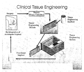
Figure 1 illustrates a general methodology for clinical tissue
engineering as applied to the example of cartilage repair using autologous
chondrocytes;
Figure 2 shows an integrated tissue engineering device of the present
invention;
Figure 3 shows a further embodiment of the tissue engineering device
of Figure 2;
Figure 4 shows a further embodiment of the tissue engineering device
of Figure 2;
=
CA 02853267 2014-06-03
- 21 -
Figure 5 shows a cut-away view of the tissue engineering device of
Figure 2 illustrating some of the internal components and a tissue
engineering module for insertion into the device;
Figure 6 shows an enlarged cut-away view of the tissue engineering
device of Figure 2 illustrating an inserted tissue engineering module;
Figure 7 shows an enlarged perspective view of the tissue
engineering module and interface with components of the device housing;
Figure 7(a) shows an enlarged perspective view of the bioreactor and
pump unit;
Figure 7 (b) shows an enlarged perspective view of the pump unit and
the associated pump tubing;
Figure 8 shows a perspective view of the reverse side of the tissue
engineering module of Figure 7 and the internal configuration of the flow
plate that attaches thereto;
Figure 9 shows an enlarged perspective view of the mixing and micro-
loading components associated with the instrumented bioreactor design;
Figure 10 shows the basic tissue engineering fluid flow schematic;
Figure 11 shows a further embodiment of the basic tissue engineering
fluid flow schematic;
Figure 12 shows alternate bioreactor, proliferation substrate or
scaffold, differentiation scaffold and process monitoring designs, as
applicable to different tissue engineering scenarios;
Figure 13 shows an enlarged perspective view of the bioreactor of the
tissue engineering module, illustrating the internal configuration of the
bioreactor and the flow path of fluids;
Figure 14 shows a further embodiment of the bioreactor of the tissue
engineering module, illustrating the internal configuration of the bioreactor;
Figure 15 shows a rotatable bioreactor design;
Figure 16 shows the sterile sampling embodiment of the tissue
engineering module;
Figure 17 shows a further embodiment of the tissue engineering fluid
flow schematic;
CA 02853267 2014-06-03
- 22 -
Figure 18 shows yet a further embodiment of the tissue engineering
fluid flow schematic; and
Figure 19 shows a bioreactor design suitable for tissue digestion and
cell collection;
= Figure 20 shows a bioreactor design suitable for cell proliferation;
Figure 21 shows a bioreactor design suitable for cell differentiation
and tissue construct formation;
Figure 22 shows yet a further embodiment of the tissue engineering
fluid flow schematic; and
Figure 23 shows a further embodiment of the tissue engineering
module with separate bioreactors for tissue digestion / cell collection, cell
proliferation, and cell differentiation / tissue formation.
Detailed Description of the Invention
The present invention is directed to an integrated, automated tissue
engineering device for the ex vivo processing of cells, particularly
autologous
cells, to enable cell proliferation, cell differentiation and tissue formation
in an
efficient and consistent manner requiring minimal human intervention. The
tissue constructs developed within the device may be integrated into a host
to assist in tissue reconstruction procedures and subsequent patient
recovery. Furthermore, the invention provides automated methods for tissue
engineering using a variety of cells from a number of different sources (for
example autologous cells obtained via patient biopsy, allogenic cells or
xenogenic cells). Furthermore, the cells may be precursor cells, primary
cells, cells from an immortal cell line and combinations thereof.
The general methodology and principle for clinical tissue engineering
incorporating the tissue engineering system and methods of the present
invention is illustrated in Figure 1, using autologous cartilage tissue
engineering as a representative example. In such example, cells (i.e.
chondrocytes) are obtained from a surgical biopsy of a patient and either
manually or automatically seeded onto a suitable substrate or scaffold (i.e. a
Skelite TM support). The chondrocytes and the support are present within the
CA 02853267 2014-06-03
- 23 -
bioreactor portion of an automated tissue engineering module, with the
module forming part of a clinical base station of the tissue engineering
system. A central microprocessor is present within the tissue engineering
system and controls and customizes the internal environment of the
bioreactor, and hence facilitates tissue growth therein, resulting in the
stimulation of cell growth within and onto the support to generate an implant.
Sensors within the bioreactor provide feedback to the microprocessor to
ensure that the cells are seeded, expanded and differentiated in a desired
and controlled manner to provide an autologous tissue implant. Once the
implant is generated, it is removed from the bioreactor for surgical
implantation into the patient. The present system provides an advantageous
way to provide autologous tissue engineered implants in a sterile, safe,
convenient and efficacious manner. Furthermore, the ability to prepare
tissue engineered implants in a clinical setting allows considerable
flexibility
in the locations for undertaking the tissue engineering process. While the
system can be used in a centralized location, the design and operation of the
system enables clinical use at regional centers. Such widespread availability
precludes the transportation of biological material to and from centralized
cell/tissue processing facilities, thereby improving the cost effectiveness
and
efficiency of the tissue engineering process while avoiding shipment,
tracking and regulatory complications.
In accordance with an embodiment of the present invention is a tissue
engineering system as shown in Figure 2 and generally indicated with
reference numeral 100. The system 100 (may alternatively be referred to as
a device) comprises a housing 102 having an insertion slot 104 for receiving
a tissue engineering module. The insertion slot 104 has a movable door 106
and a locking mechanism 108. A user interface 110 such as a touch screen,
key pad or combination of both is provided for control of system operation
and for the display of system status. A data storage system 112 is present
which permits the recording of information via a variety of mediums known to
those of skill in the art (i.e. ZIP, CDROM, diskette, flashcard). A
computer/communications link 114 provides the capability to upload new
CA 02853267 2014-06-03
=
- 24 -
software, modify control parameters using an external computer, download
data as well as troubleshoot and test the device. This link also permits the
system to be connected to electronic information systems present at the
clinic. The system 100 is powered with a power input 116. Figure 3 shows a
further embodiment of the system 100 having several bay doors 106 to
accommodate several tissue engineering modules. Figure 4 shows a further
embodiment of the system 100 having bay doors 106 orientated in a
horizontal manner to allow for the preferential orientation of the tissue
engineering module relative to the gravity vector.
Figure 5 shows the internal configuration of the system 100
represented in Figures 2 and 3 with the vertical orientation of the bay doors
for vertical insertion of a tissue engineering module. A tissue engineering
module 118 is shown for insertion within the insertion slot 104 of the bay
door 106. The tissue engineering module 118 slides into the system
housing 102 via a guide rail system 120. Upon insertion, the module 118
engages with one or more pump units 122 (i.e. peristaltic, piston, diaphragm
or rotary), electrical connectors 124 (i.e. DIN, AMP, PCB, breadboard
socket), and valve actuators 126 (i.e. servo motor, linear drive, linear
actuator). Any suitable guide system to allow the module to be inserted
properly into the system may be contemplated as is understood by one of
skill in the art.
As better seen in Figure 6, where the tissue engineering module 118
is inserted into the housing 102, a series of valve actuators 126 interface
with valves (shown in more detail in Figures 7 and 7a) on the module to
provide flow control. The electrical connectors 124 provide electrical
connection between the module 118 and a central microprocessor unit
(CPU) 128 via an electronic back-plane 130. The CPU 128 controls the
operational sequence, the transport of fluids and gases, the management of
process data, the monitoring of system status, the user interface, and the
external data communication port. The CPU 128 provides control through
electrical links with active and passive electrical components present on the
back-plane 130 and each of the inserted tissue engineering modules 118.
CA 02853267 2014-06-03
- 25 -
Temperature sensors 132 (i.e. thermocouple, RTD or thermistor), gas
sensors 134 (i.e. 02 and CO2) and an environment control unit (ECU) 136
are controlled by the CPU 128 to maintain the environment (i.e. temperature
and gas atmosphere) within the housing 102 using standard methods known
to those skilled in the art. The environment can be adjusted to meet the
requirements of the tissue engineering process, including storage of
reagents at refrigeration temperature (i.e. 4 C), the simulation of nominal
body temperature (i.e. 37 C), and the availability of gaseous mixtures for
transport into and out of the module 118 in the event that the module is
equipped with gas exchange components (i.e. membranes). Gaseous
conditions are monitored by the gas sensors 134 located within the housing
102 and the data is sent to the CPU 128 via the electronic back-plane 130.
Gas input(s) to the ECU can be via gas supply inlet 140 provided within the
housing 102 configured with standard fittings. In other embodiments, gases
may be housed within the ECU. Gases for use within the device include but
are not limited to oxygen, carbon dioxide, nitrogen and mixtures thereof. In
order to adequately contain such gases within the housing 102, the bay door
106 is configured to provide for a hermetic seal when closed. The housing
102 is insulated with insulating material 142 such as styrofoam, aerogel,
fiberglass and the like to allow for the efficient regulation of internal
temperatures (i.e. 4 C to 37 C).
While the tissue engineering system of the present invention is
generally shown to comprise a boxed shaped housing, it is understood by
one of skill in the art that the housing may be made of various configurations
so long as it may accommodate the components as described herein. For
example, this includes but is not limited to open configurations that may not
require a top and/or side portions.
The tissue engineering module 118 is illustrated in more detail in
Figures 7-9. The tissue engineering module 118 comprises a rigid structural
spine 200 to which is affixed a bioreactor 202. The bioreactor 202
comprises a bioreactor housing that has a lid 204 and may be customized
with respect to the substrate(s) or scaffold(s) contained therein to enable
CA 02853267 2014-06-03
- 26 -
tissue digestion, cell culture, cell proliferation, cell differentiation,
tissue
implant formation and combinations thereof. The bioreactor lid may be
detachable or alternatively made integral to the bioreactor housing. The
bioreactor 202 may be separately detachable and disposable relative to the
structural spine 200. To enable such detachment, the bioreactor 202 and
the structural spine 200 may use fluid disconnect fittings that include the
provision for self sealing of input and output lines to avoid loss of fluids
and
to prevent contamination of the contents of the bioreactor. The entire tissue
engineering module may be considered to be disposable following the
completion of a tissue engineering sequence, as this practice prevents
contamination arising from prior use. Alternately, only selected components
of the module 118 may be considered as disposable due to contact with
fluids, leaving non-contamination prone components available for re-use.
As seen in Figures 7, 7a and 7b, a fluid containment system 206 is
affixed onto the structural spine 200 of the tissue engineering module 118.
The fluid containment system 206 is comprised of a sterile series of flexible
reservoirs 208 and flexible tubing 210 for supplying and retrieving types of
tissue and cell culture fluids and pharmaceuticals to and from the bioreactor
202. The reservoirs 208 may be of varying configuration and number as
required and may contain different types of cell and tissue culture media,
growth factors, pharmaceutical agents and may also contain waste media
and/or media samples from the bioreactor 202. Fluids are loaded or
removed from the fluid containment system 206 via a series of fluid access
ports 212. Tubing 210 is present to provide fluid connection between the
various reservoirs 208 and the fluid control components, such as the fluid
flow control valves 214. The fluid flow control valves 214 are opened and
closed by valve actuators 126. Similarly, the pump unit 122 interfaces with
disposable pump components present on the module. These pump
components may be pistons, diaphragms, rotary elements or peristaltic
tubing 218, provided that the operation of these components does not
generate harsh conditions, such as excessive shear stress, that compromise
cell viability during the transfer of cell suspensions. The pump unit 122 and
CA 02853267 2014-06-03
- 27 -
the valve actuators 126 reside within the housing 102, Alternately, the
actuators and pump unit may form part of the tissue engineering module,
however, this may result in disposal of these components following patient
use. Fluid is transferred out of the reservoirs 208 by the programmed action
of the pump unit 122 on the pump tubing 218. Fluid travels from a flexible
fluid reservoir 208 to a fluid valve 214 via tubing 210. A fluid flow plate
220
(as shown in Figure 8) directs fluid flow between different flow control
valves
214 and the pump tubing 218 of the pump unit 122. Fluid is returned to a
selected empty reservoir 208 for storage. A flexible printed circuit board
(PCB) 222 provides the electronic interface for electronic components (i.e.
sensors) present on the structural spine 200 and/or the bioreactor 202. In
the event that a sensor indicates that a monitored parameter (e.g., pH) is
outside acceptable levels, the CPU triggers a control intervention such as
replacing the media within the bioreactor.
The tissue engineering module may optionally include a
microprocessor 224 to enable data processing and data storage directly on
the module. This information may transferred to the central CPU 128 while
the module is inserted into the housing 102 and retained in electronic
memory for later access once the module is removed. In addition to the data
stored via the microprocessor or memory chip resident on the tissue
engineering module, the module may also optionally include a bar code 226,
magnetic strip 228, electronic memory (not shown) and/or ID label 230 to
facilitate administrative tracking within the clinic.
As seen in Figure 8, the fluid flow plate 220 is secured to the
structural spine 200 of the tissue engineering module 118. The technique
for attachment of the fluid flow plate may be, but is not limited to, a press
fit,
snap fit, ultrasonic weld, solvent bond and the like, recognizing that the
technique adopted must allow for sealing of the assembly to avoid loss of
fluids and to prevent contamination. As shown in the disassembled view in
Figure 8a, the fluid flow plate 220 has an integral fluid pathway 232 to
provide a means for directing flow associated with the actuation of the fluid
valves 214. New flow paths may be accommodated via revisions to the
CA 02853267 2014-06-03
- 28 -
pathway present on the flow plate 220. In one embodiment, the fluid plate
220 may be integrally formed into the structural spine 200 to form a single
component. A fluid heating and mixing chamber 234 is included to ensure
fluids that are directed to the bioreactor are at the correct temperature and
are adequately mixed so as to not disrupt the biological processes underway
in the bioreactor. Furthermore, a thermoelectric element 236 is present on
the tissue engineering module 118 to vary the temperature within the
bioreactor 202 compared with the operational temperature of the module, as
defined by the operation of the ECU 136. Such a temperature change may
be necessary to simulate nominal physiological conditions within the
bioreactor, while the remaining components of the tissue engineering
module, particularly the reagents and/or samples, are at a reduced
temperature (i.e. refrigeration) to maintain physical, chemical and/or
biological viability. Power and control of the thermoelectric element is
performed by the CPU 128. In addition to sensors present on the bioreactor,
a sensor 238 present on the tissue engineering module provides feedback to
the CPU 128. The sensor and thermoelectric connections are made via the
electrical cabling 240 and connector 124.
Figure 9 shows the mixing and micro-loading aspects of the tissue
engineering module 118. The bioreactor 202 has a mixing drive 260
operably connected with a mixing actuator 262 and to the mixing diaphragm
264. The mixing diaphragm is incorporated as part of the bioreactor 202 or
the bioreactor lid 204, as shown. In operation, the mixing drive 260 in
combination with the mixing actuator 262 provide translation or pulsing of the
mixing diaphragm to effect controlled mixing of the contents of the bioreactor
202. Ideally, the nature of the mixing is such to avoid high fluid shear that
could compromise the physical integrity of cells present within the
bioreactor.
For certain tissue engineering protocols, moderate levels of fluid shear are
actually beneficial for the successful development of tissue constructs. In
addition to the mixing components, an impact drive 266 and impact actuator
268 are present. These components serve to apply a controlled impact to
the bioreactor assembly at the conclusion of the proliferation sequence to
CA 02853267 2014-06-03
- 29 -
assist with the release of cells from a proliferation substrate or scaffold
resident within the bioreactor. Also provided is a micro-loading drive 270 in
operable connection with a micro-loading actuator 272 and micro-loading
diaphragm 274. The micro-loading diaphragm 274 is incorporated as part of
the bioreactor 202 or the bioreactor lid 204, as shown. The location and
orientation of the micro-loading diaphragm is such to enable intimate contact
with the substrate or scaffold and any associated cells or tissues present in
the bioreactor 202. The application of micro-loads is known to be
advantageous for certain tissue engineering protocols. The mixing drive
260, impact drive 266, and micro-loading drive 270 may be any of a series of
electromechanical devices such as solenoids, linear drives, rotational drives,
or piezo electric components. Furthermore, it is possible for the mixing drive
260, impact drive 266, micro-loading drive 270, and the related actuators to
be mounted on the housing 200. Alternatively, the drives and actuators may
be mounted on the tissue engineering module or the bioreactor provided that
the design of the drives is consistent with the disposable nature of the
tissue
engineering module. In addition to the provision of mechanical stimulation,
the bioreactor may also be configured to introduce electrical and/or chemical
stimulation of the tissue construct. In particular, electric fields may be
generated in the region of the bioreactor to enhance cell transport and/or
tissue formation. Methods of generation of electric fields are known to those
of skill in the art and include but are not limited to the provision of
electric
coils.
Figure 10 illustrates a basic fluid flow schematic for the tissue
engineering module 118 in which there is a single cell or tissue culture
chamber present within the bioreactor 202, (refer to descriptions of Figures
12 and 17 for further information on the multi-chamber bioreactors). The
flow path links the bioreactor 202 to reservoirs 208 that supply fluid and
collect waste. The fluid access ports 212 may be used to load reagents or
remove samples or waste fluid. Flow is generated by the operation of pump
unit 122 with flow direction defined by actuation of specific flow control
valves 214. Perfusion to the bioreactor can be either continuous or pulsatile,
CA 02853267 2014-06-03
- 30 -
provided that the associated flow does not result in high fluid shear in
regions containing cells, as such conditions could damage the cells or an
emerging tissue construct. A recirculation loop 280 is provided to allow the
fluid contents of the bioreactor to be either monitored or modified by
external
components, such as an in-line gas exchange membrane 282, without
necessitating the delivery of new fluid from the fluid reservoirs 208.
Components of the tissue engineering module 118 dedicated to storing
fluids, (i.e. reservoirs 208), are kept refrigerated at approximately 4 C to
facilitate storage of fluids that would otherwise degrade at the elevated
temperatures used to simulate body temperature (i.e. 37 C). According to a
preprogrammed routine, the CPU 128 controls the operation of fluid valve(s)
214 to allow fluid stored in a reservoir 208 to be delivered via the pump unit
122 into a heating and mixing chamber 234 prior to entry into the bioreactor
chamber 300 (shown in detail in Figures 13, 14 and 15). Fluids are supplied
to the bioreactor via the inlet port 302 and removed via outlet port 304. To
simulate normal body temperatures for optimal cell and tissue culture
performance, the bioreactor 202, the pump unit 122 and the heating and
mixing chamber 234 are maintained at approximately 37 C by the operation
of a thermoelectric element 236. It will be obvious to one skilled in the art
that alternate thermal regulation devices may be used to obtain the desired
thermal profiles for the tissue engineering module 118.
Figure 11 illustrates a variation on the basic fluid flow schematic
where the fluid flow control valves are substituted for multiple pump units
122. This configuration provides enhanced operational redundancy and a
reduced component count_ Operation of such a system requires that any
dormant pump unit prevents unregulated pass-through flow, as such an
occurrence would compromise the controlled delivery of fluids.
Figures 12a - 12d illustrate various bioreactor configurations and
alternate formats for the substrates and scaffolds used for the proliferation
and differentiation steps involved in the operation of the tissue engineering
system. Figure 12a shows a series of interchangeable bioreactor designs
that address different bioprocessing scenarios. The Type I scenario is
CA 02853267 2014-06-03
- 31 -
indicative of a basic single chamber 300 within a bioreactor 202 that
accommodates a proliferation scaffold or substrate 310, or a differentiation
scaffold or substrate (not shown) and is ideally suited to either
proliferation
or differentiation. Cells are either manually seeded onto the scaffold 310 or
automatically delivered via the fluid pathway of the tissue engineering
module.
The Type II scenario involves a multi-chamber bioreactor that
provides for the use of a scaffold 310 (or substrate) for proliferation of the
cell population and an implantable differentiation scaffold 312 that promotes
the formation of a tissue construct. The culture / proliferation chamber 300
is
connected to the differentiation / tissue formation chamber 306 via a funnel
314. The funnel serves to channel the cells released from the proliferation
scaffold 310 into the implantable differentiation scaffold 312. The use of a
filter 316 in several locations within the bioreactor serves to regulate the
size
of the cells or cell aggregates that can freely pass from one chamber to the
next. A filter 316a is present upstream of the proliferation scaffold with the
purpose of regulating the incoming cell population for the cell expansion
step. Another filter 316b is present upstream of the differentiation scaffold
again to control the cell population entering this step of the tissue
engineering sequence. In addition, there is a further filter 316c over the
outlet port 304 to prevent the loss of cells from the differentiation/tissue
formation chamber during operations involving fluid transfer through the
bioreactor. The filter 316 can be a filter membrane or mesh or similar type
filtering material as is known to those of skill in the art.
The Type Ill scenario combines tissue digestion with subsequent
proliferation, differentiation and tissue construct formation. In this
scenario,
a tissue biopsy 320 is loaded into a digestion chamber 322 present within
the bioreactor 202. Digestion of the tissue biopsy occurs through the
delivery of digestion enzymes into the bioreactor from one of the fluid
reservoirs 208 present on the tissue engineering module. Disassociated
cells exit the digestion chamber 322 under the influence of gravity
sedimentation and/or fluid flow through the culture / proliferation chamber
CA 02853267 2014-06-03
- 32 -
300, and subsequently collect on the proliferation scaffold 310. Transfer of
tissue aggregates out of the digestion chamber 322 is precluded by the
presence of a filter membrane/mesh 316a in the flow path between the
digestion chamber 322 and the culture / proliferation chamber 300.
Following proliferation, the cells are released and transferred to the
implantable differentiation scaffold 312 via the cell funnel 314. Again,
membrane/mesh filters are present both upstream 316b and downstream
316c of the implantable differentiation scaffold 312 to ensure that the
correct
cell population are seeded on the scaffold and that cells are not
inadvertently
lost to waste during fluid transfer operations.
In the preceding scenarios, various configurations of the proliferation
substrate 310 or scaffold are possible, as illustrated for example in Figure
12b. For example, one configuration is a porous scaffold 310a having a
relatively even pore gradient. A pore gradient scaffold 310b is a porous
scaffold having a pore gradient where the pore size decreases as cells travel
through the scaffold. This promotes a more homogeneous distribution of
cells throughout the scaffold at the conclusion of the cell seeding process. A
pore gradient scaffold with reversed orientation 310c may be used.
Alternatively, a fiber filter scaffold 310d, may be used which is a fibrous
matrix typical of organic compounds such as collagen. It is also possible to
utilize a contained suspension of micro-carriers (e.g. CytodexTM) as the
proliferation substrate. Furthermore, the bioreactor may have an optical
probe 324 (shown in conjunction with the porous scaffold 310a) supported
by the CPU 128 to enable the inspection of the cell seeding process
occurring within the proliferation scaffold and to further assess the
proliferation events, particularly progress toward attaining a confluent cell
layer.
As with proliferation, there are a variety of implantable differentiation
scaffolds 312 that may be formed in differing configuration and of diverse
materials (i.e. inorganic mineral-based scaffolds such calcium phosphate,
organic biopolymer scaffolds such as collagen, etc.) and employed in the
tissue engineering process. Figure 12c illustrates a multi-zone
- CA 02853267 2014-06-03
- 33 -
differentiation/tissue formation chamber 306 that comprises up to three
implantable differentiation scaffolds 312, all of which may simultaneously
proceed toward tissue construct formation. This allows for the preparation of
different sizes of implantable tissue and for the use of alternate implantable
differentiation scaffolds to assess and maximize tissue yield. For example,
scaffold 312a is a porous reticulate formed from a bone biomaterial such as
Skelite TM for use in bone and cartilage applications where the tissue
construct requires hard tissue anchoring within bone. The scaffold 312a
may be further enhanced through the use of a scaffold membrane/mesh 326
that encircles the implant to create a membrane encircled scaffold 312b such
that the loss of cells out of the scaffold 312a during the cell seeding
process
is minimized, thereby making the tissue engineering process more efficient.
The membrane may preferably only partially encircle the scaffold or
alternatively, more fully encircle the scaffold. While the primary purpose of
the scaffold membrane/mesh 326 is to contain the cells on and within the
implantable differentiation scaffold 312, careful selection of the properties
of
the scaffold membrane/mesh 326 as is understood by one of skill in the art
either allows or limits the passage of specific molecular entities that may
have a marked influence on the tissue engineering process at the cellular
level.
A further embodiment is a gradient porosity and membrane encircled
scaffold 312c that combines the advantages of the scaffold membrane/mesh
326 with a pore gradient. The gradient is configured to deliberately cause
the cells to collect on the top surface with only minimal propagation into the
scaffold. A degree of porosity in the surface is considered advantageous for
tissue stability and for the supply of nutrients to the developing tissue via
the
scaffold surface. This approach results in the development of a bipolar
tissue construct with distinct stratified zones. The top zone is essentially
comprised of de novo tissue. The bottom zone is essentially free of cells or
tissue and remains as an open porous scaffold. The middle interfacial zone
represents the structurally stable transition between the open scaffold and
the de novo tissue layer. Such a bipolar tissue construct is ideal for the
CA 02853267 2014-06-03
= , =
- 34 -
repair of focal defects in articular cartilage as the top layer is tissue
engineered cartilage that provides for lateral integration with the host
cartilage, while the bottom layer provides for lateral and axial integration
with
the subchondral bone. Integration of the bottom layer with the surrounding
subchondral bone may be further enhanced by the application of bone
marrow to the open scaffold at the time of surgical implantation. In cartilage
repair applications, it is important that the mineral-based scaffold does not
extend to the articular surface, as this may compromise joint function.
Accordingly, a secondary non-mineral scaffold (not shown in the figures)
may be employed in the top zone of de novo cartilage to assist with the
formation of tissue constructs of sufficient size to treat large cartilage
lesions
(i.e. up to 10 cm2 in diameter and 2-3 mm in thickness). Furthermore, the
secondary scaffold can be configured to generate shaped constructs that
have articular surface profiles that more closely match the particular
anatomical characteristics present at the site of implantation. Candidate
materials for the secondary scaffold are synthetic biopolymers (e.g. PGA,
PLA) or natural biopolymers (e.g. alginate, agarose, fibrin, collagen,
hyaluronic acid). These secondary scaffolds may be in the form of hydrogels
or three-dimensional preformed scaffolds.
Alternate techniques for the preparation of bipolar tissue constructs
are possible within the tissue engineering system. The implantable
differentiation scaffold 312 may be partially infiltrated with a bioresorbable
polymer that limits cell seeding to certain regions of the scaffold. This
creates a preferential zone of new tissue formation during the preparation of
the tissue construct. Upon implantation, the polymer is resorbed thereby
leaving voids in the porous scaffold that promote anchorage within the host
tissue. A further configuration involves an implantable scaffold with
relatively
open porosity that is positioned away from the exit of the
differentiation/tissue formation chamber. During cell seeding, this open
space provides for the collection of cells that migrate through the open
scaffold. As cells are accumulated within the differentiation/tissue formation
chamber, both the open space and a portion of the scaffold become
CA 02853267 2014-06-03
- 35 -
infiltrated with cells and thereby create a preferential zone of new tissue
formation. The resulting tissue construct comprises a de novo tissue zone
that is devoid of the scaffold, a middle transition zone or interfacial zone
containing both de novo tissue and the scaffold, and a region of the porous
scaffold that is open and essentially free of cells or tissue.
Figure 12d illustrates a bioreactor monitoring scheme whereby
sensors 216 (i.e. temperature, pH, dissolved gases, etc) are integrated into
the lid 204 of the bioreactor 202 to provide feedback to the CPU 128 of the
progress of the tissue engineering process. In addition, a CCD camera 330
may be employed to monitor the optical properties of the proliferation
scaffold 310 (or substrate) for evidence of impending confluence (e.g. optical
density and/or light scattering as a function of cell density) such that cell
release is timed to maximize the cell yield from the proliferation step.
Figure 13 better shows the flow path and fluid circulation within the
bioreactor 202. The bioreactor 202 is shown to have an inlet port 302, an
outlet port 304 and an internal cavity defining a basic chamber 300. Fluid
flows from the fluid flow plate 220 into the bioreactor 202 via the inlet port
302 and exits through the outlet port 304. The bioreactor lid 204 attaches to
the bioreactor 202. A variety of different mounting hardware may be used to
hold the bioreactor lid 204 and bioreactor 202 together. The chamber 300
may be designed to accommodate one or more substrates or scaffolds 310.
Furthermore, the bioreactor 202 may be subdivided into separate chambers
that permit the steps of tissue digestion, proliferation, differentiation, and
tissue formation. Each chamber may be configured with inlet and outlet
ports that are independently controlled via flow control valves for greater
control over the tissue engineering sequence. Circulation of fluid is effected
by the activation of one or more flow control valves 214 and the pump unit
122, based on control signals from the CPU 128. Depending upon the
specific valves activated, operation of the pump unit 122 moves fluid from
one of the fluid reservoirs 208 into the bioreactor 202 or permits
recirculation
of the fluid within the bioreactor. For biological processes that require
stable
dissolved gas concentrations, recirculation is advantageous as it enables the
CA 02853267 2014-06-03
- 36 -
fluid to be passed across a membrane that facilitates gas exchange. The
nature of the exchange is based on the dissolved concentrations in the
bioreactor versus the external conditions established by the ECU 136. The
bioreactor lid 204 is shown to have a sampling port 332 and sensor probes
216 that are operably connected to the interior chamber of the bioreactor.
Alternatively, the sampling port may be provided elsewhere on the bioreactor
housing. The sampling port allows the removal or addition of materials into
and out from the bioreactor. The sampling port may be replaced or
augmented with a gas exchange membrane as required.
Figure 14 illustrates a multi chamber bioreactor 202 with the
bioreactor lid 204 removed for clarity. The inlet port 302 is connected to a
tissue digestion chamber 322. The configuration of the tissue digestion
chamber 322 permits a patient biopsy to be loaded into the bioreactor for
subsequent automated digestion to yield disassociated cells. The circulation
path within the digestion chamber promotes the gentle agitation of the
biopsy to prevent stagnant areas that could potentially lead to excessive
exposure of the biopsy tissue to circulating digestion enzymes. Furthermore,
the inlet and/or outlet of the digestion chamber may house a filter
membrane/mesh 316 (not shown) of varying porosity to provide for cell
sorting and to preclude the release of partially digested tissue aggregates.
The bioreactor contains a second chamber 300 that accommodates a
proliferation substrate or scaffold 310 for receiving cells for proliferation.
The
proliferation scaffold may be formed in various geometries that support both
two-dimensional and three-dimensional proliferation, and may be comprised
of various biocompatible materials that promote cell proliferation, such as
calcium phosphate biomaterials (for example SkeliteTm), biopolymers, or
natural matrices (for example collagen). Cells delivered from the tissue
digestion chamber 322, or via the optional cell inoculation port 334, become
dispersed on or within the proliferation substrate or scaffold 310 and
proliferate, thereby increasing the cell population for subsequent cell
differentiation and tissue formation. Note that the process may be
terminated following proliferation if the goal is to only expand the cell
CA 02853267 2014-06-03
- 37 -
population without further differentiation. An implantable differentiation
scaffold 312 is present within a differentiation/tissue formation chamber 306
at the base of the bioreactor 202. As with the proliferation scaffold 310, the
implantable differentiation scaffold 312 may be formed in different
geometries and may be composed of a variety of biocompatible materials
that are properly selected to meet the biological requirements of the implant
site, (for example Skelite TM is an ideal candidate implant for skeletal
sites).
In operation, cells are released from the proliferation substrate or
scaffold 310 through an automated sequence, such as the delivery of
enzymes (for example trypsin) and the timed application of impact to the
bioreactor via the impact drive 266 (not shown). The cell suspension
migrates under the controlled flow conditions present in the bioreactor into
the implantable scaffold 312 via the cell funnel 314, whereupon the cells
become resident and initiate the differentiation and tissue formation
sequence. Upon conclusion of this sequence, the tissue so formed may be
removed from the bioreactor for subsequent implantation. One skilled in the
art would understand that the particular embodiment of the bioreactor of
Figure 14 is only a representative design example. The bioreactor, in
general, can be configured in various ways with respect to overall shape,
size and internal configuration without adverse effect on function. For
example, a gas exchange membrane 336 present on the bioreactor may be
a separate and discrete component that is connected in-line with one or
more fluid delivery tubes 210 or the flow plate 220 of the tissue engineering
module 118. Furthermore, the chambers of the bioreactor may be isolated
from each other via control valves to avoid the necessity for all fluids to
pass
through all chambers. When required the passageways between chambers
may be opened to effect the transfer of fluids and cell suspensions. An
example of such a variation that enables increased flexibility in
bioprocessing conditions and sequences is illustrated in Figure 17. An
alternate configuration to enable controlled exposure of the implantable
differentiation scaffold 312 to the contents of the bioreactor is the use of a
shuttle 318 that isolates the implantable scaffold until cell seeding is
required
CA 02853267 2014-06-03
- 38 -
as part of the differentiation step. To enable cell seeding, the shuttle 318
moves the implantable scaffold into the fluid flow from a protected location
within the bioreactor. Various configurations of the shuttle are possible,
including rotation-based movement or the use of a removable barrier that
isolates the implantable scaffold until cell seeding is required.
Figure 15 illustrates a rotational bioreactor design that takes
advantage of the orientation of the gravity vector to effect cell transport by
sedimentation at different stages in the tissue engineering process. Note
that while this figure illustrates rotation of the bioreactor, the technical
objective may be equally attained by rotation of the tissue engineering
module or indeed by rotation of the entire housing 102. As shown in Figure
15, the bioreactor 202 is attached to a rotational shaft 350 which is affixed
to
the structural spine 200 of the tissue engineering module 118. This provides
a mechanism for the rotation of the bioreactor 202 in order that cell seeding
via sedimentation can occur on to selected proliferation surfaces within the
culture/proliferation chamber 300. The proliferation surfaces of the
bioreactor may be optionally coated with biomaterials that enhance
proliferation (for example SkeliteTm), or a dedicated proliferation substrate
or
scaffold may be inserted into the chamber 300 to provide this role. As an
alternative to the use of the digestion chamber 322, a second inoculation
port 352 is provided at the side of the bioreactor 202 to enable direct cell
seeding. Cells may be initially seeded on a proliferation surface 354 which is
relatively small (Figure 15a). Based on elapsed proliferation time or the
detection of confluence, the cells may be automatically released and the
bioreactor rotated via the rotational shaft 350 such that the cells released
from the proliferation surface 354 will sediment on to the increased area of
surface 356 (Figure 15b), allowing further proliferation. At the completion of
the secondary proliferation step, the expanded cells are released and the
bioreactor is again rotated to permit seeding of the implantable scaffold 312
(Figure 15c). Thus the rotational shaft 350 and associated flexible tubes 358
allow the bioreactor 202 to be rotated as required to maximize the use of
gravity sedimentation in sequential proliferation stages. It is within the
scope
CA 02853267 2014-06-03
- 39 -
of the present invention to use the rotational shaft in a manner to agitate or
shake the bioreactor where such conditions are desirable.
Referring now to Figure 16, the tissue engineering module 118 may
be adapted to include techniques for the sterile sampling of suspended cells,
tissue culture fluids, and/or waste products. In this embodiment, a syringe
manifold 400 and sterile offloading ports 402 are integrated into the
structural spine 200 of the tissue engineering module 118. Microbore tubing
406 links the syringe manifold to the bioreactor 202 via the sampling port
332. Syringes 404 are connected to offloading ports 402 at the manifold 400
to enable the collection and removal of fluid samples or cell suspensions for
subsequent analysis without compromising the operation, integrity or sterility
of the tissue engineering process. An alternate sampling technique is also
provided whereby a fusible bioreactor sampling line 408 is connected to the
bioreactor lid 204. As this line is physically linked to the interior of the
bioreactor and is in close proximity to the biological events underway
therein,
the line contains fluid of substantially the same composition as that present
within the bioreactor. Consequently, a representative sample of the
bioreactor fluid may be obtained by fusing the ends of the sampling line and
then removing the line from the tissue engineering module for subsequent
analysis. It will be obvious to one skilled in the art that such a fusible
line
can be used as the basis for a sampling technique through the automatic
operation of sealing components within the housing 102.
Figure 17 illustrates a more complex fluid flow schematic for the
tissue engineering module 118 in which the different requirements for
digestion, proliferation and differentiation are accommodated by separate
bioreactor chambers. These chambers may be present within a series of
discrete bioreactors or combined within a single bioreactor that maintains
separate control over the conditions in each chamber. A tissue digestion
chamber 322 is present that accommodates a tissue biopsy 320. A
proliferation chamber 300 is present that is configured to accept cells from
the digestion chamber 322 and allows seeding of a proliferation substrate or
scaffold 310. A differentiation/tissue formation chamber 306 is also present
CA 02853267 2014-06-03
- 40 -
that is configured to accept the expanded cell numbers from the proliferation
chamber 300 and allows seeding of an implantable scaffold 312.
Tissue engineering reagents (i.e. media, enzyme solutions, washing
solutions, etc.) are loaded in fluid reservoirs 208a ¨ 208e. Waste products
are collected in fluid reservoir 208f, which can be manually aspirated for
sampling purposes using access port 212f. Additional fluid reservoirs may
form part of the fluid reservoir system 206 and be accommodated on the
tissue engineering module as required for different tissue engineering
processes. Fluid flow through the system is directed by the operation of fluid
pumps 122a ¨ 122k, flow control valves 214a ¨ 214c, and uni-directional
flow valves 410a ¨ 410c (i.e. fluid flow check valves). Furthermore, pumps
212a ¨ 212k are configured to operate as active pumps or passive valves
(open/closed), according to control inputs from a central microprocessor.
Filters 316a ¨ 316d are used to selectively control the movement of cell
suspensions within the system and to limit the passage of cell aggregates
during washing and transition stages of the tissue engineering process.
Levels of dissolved gasses in the media are maintained via the in-line gas
exchange membranes 282a and 282b. Optional syringes 404a and 404b
are present to allow cell collection or media sampling via sterile offloading
ports 402a and 402b.
In operation, a tissue biopsy 320 is inserted into the tissue digestion
chamber 322 between filters 316a and 316b. A digestion medium containing
enzymes is pumped into the tissue digestion chamber 322 from the fluid
reservoir system 206 to initiate the digestion process. Digestion may be
enhanced by gentle agitation of the digestion medium within the digestion
chamber via a mixing diaphragm to maximize reagent exposure to the biopsy.
The digestion medium may be continuously or periodically re-circulated via
pump 122g. During recirculation, the fluid flow is directed into the bottom of
the digest chamber, against the gravity vector, in order to suspend and
tumble the tissue biopsy, thereby maximizing the effectiveness of the
digestion process. Filter 316a prevents migration of cells and cell aggregates
into the fluid pathway. The recirculation path includes the in-line gas
CA 02853267 2014-06-03
- 41 -
exchange membrane 282a which provides for consistent levels of dissolved
gases in the digestion medium. Introduction of a washing solution,
contained in the fluid reservoir system 206, into the bottom of the digestion
chamber 322 flushes the digestion chamber and effectively washes the
digestion medium from both the disassociated cells and any residual cell
aggregates. Following a single or multiple washing procedures, the
application of reverse flow transfers the cell suspension to either the
proliferation chamber 300 or the optional syringe 404a for external inspection
or analysis. The transfer of partially digested tissue out of the digestion
chamber is precluded by filter 316b that is sized to allow passage of
disassociated cells and retention of cell aggregates.
Cells generated from the biopsy digestion process or available via
direct loading of a cell suspension are seeded through fluid flow and/or
gravity sedimentation onto a proliferation substrate or scaffold 310 present
within the proliferation chamber 300. Following a quiescent period to allow
attachment of the cells to the proliferation substrate or scaffold 310 (for
the
example of attachment dependent cells), a proliferation medium is
introduced into the proliferation chamber 300 from the fluid reservoir system
206. This medium is periodically replaced with fresh proliferation medium
from the reservoir system 206 at specific times during the proliferation
phase. In between the medium replacement steps, the fluid within the
proliferation chamber is continuously or periodically recirculated under the
control of pumps 122g, 122h and 122i, plus control valves 214a and 214b.
The fluid delivery and recirculation paths include the in-line gas exchange
membrane 282a which provides for consistent levels of dissolved gases in
the proliferation medium. During a medium replacement step, the supply of
fresh medium from the fluid reservoir system 206 is balanced by the removal
of fluid to the waste reservoir 208f via pump 122f. Thus, through a
combination of periodic medium replacement steps and controlled
recirculation, the tissue engineering system maintains optimal conditions
within the proliferation chamber throughout the proliferation process.
CA 02853267 2014-06-03
- 42 -
Once the cell culture approaches confluence, the media within the
proliferation chamber 300 is evacuated into the waste reservoir 208f by
pump 122f. In this process, the removal of fluid from the proliferation
chamber is balanced by incoming sterile air delivered via a sterile filter
port
on the proliferation chamber (not shown) or by incoming PBS wash solution
from the fluid reservoir system 206. The cells are washed extensively by two
consecutive washing steps with the PBS wash solution to remove residual
proliferation medium. The cells are subsequently released from the
proliferation substrate or scaffold 310 through an automated sequence, such
as the delivery of enzymes (for example trypsin) and the timed application of
impact to the bioreactor via an impact drive. Following cell release, the
enzymatic process may be stopped by the delivery of media containing
serum that inhibits enzyme activity. In order to collect the cells for
eventual
seeding on to the implantable scaffold 312 within the differentiation/tissue
formation chamber 306, the cell suspension is transferred from the
proliferation chamber 300 to the filter 316c. The filter 316c prevents the
passage of cells but allows the media to continue via valve 214b to the
waste reservoir 208f under the control of pump 122f. The collected cells are
then released from the filter 316c by the application of reverse flow and are
delivered to either the differentiation/tissue formation chamber 306 or the
optional syringe 404b for external inspection or analysis.
Cell seeding on to the implantable differentiation scaffold 312 is
achieved by transferring the cells from the filter 316c to the top surface of
the
scaffold via pump 122j. The loss of cells away from the scaffold is minimized
by the optional use of a scaffold membrane or mesh 326. Following cell
seeding, fresh differentiation media may be introduced into the
differentiation/tissue formation chamber 306 through a secondary input by
the operation of pump 122k. This secondary input is located away from that
region of the implantable scaffold that is seeded with cells so as to minimize
the potential for damaging sheer stresses that could compromise the
formation of cell aggregates. The differentiation medium is periodically
replaced with fresh differentiation medium from the reservoir system 206 at
CA 02853267 2014-06-03
,
-43 -
specific times during the differentiation phase. In between the medium
replacement steps, the fluid within the differentiation/tissue formation
chamber is continuously or periodically recirculated under the control of
pumps 122j or 122k, plus control valve 214b. The path for the delivery of
both fresh differentiation medium and recirculated medium includes the in-
line gas exchange membrane 282b which provides for consistent levels of
dissolved gases in the differentiation medium. During a medium
replacement step, the supply of fresh medium from the fluid reservoir system
206 is balanced by the removal of fluid to the waste reservoir 208f via pump
122f. Environmental conditions within the differentiation/tissue formation
chamber are monitored and controlled for the period necessary for the
successful formation of the tissue construct, at which time the
differentiation/tissue formation chamber of the bioreactor is opened and the
construct retrieved for subsequent clinical or research use.
Figure 18 illustrates a variation on the fluid flow schematic of Figure
17 where the proliferation scaffold or substrate 310 within the proliferation
chamber 300 is replaced with a planar proliferation substrate of relatively
large surface area. The orientation of the substrate is such that cell
sedimentation under gravity evenly distributes the cells over the
proliferation
surface. Provided that the correct orientation of the proliferation chamber is
maintained, the proliferation substrate may be in the form of a rigid polymer
culture plate or a flexible wall container.
Figure 19 shows a tissue digestion bioreactor 500 that contains a
tissue digestion chamber 322 of an appropriate size to accommodate one or
more tissue samples such as a tissue biopsy 320. The bioreactor 500
consists of four primary components: a bioreactor base 502 that substantially
forms the tissue digestion chamber 322, a removable bioreactor lid 504, port
filter 316b, and optional port filter 316a (not shown).
The bioreactor lid 504 provides for a media port 506 with an optional
port filter 316a (not shown) and an air outlet port 508. The bioreactor base
500 accommodates filter 316b that allows passage of disassociated cells out
CA 02853267 2014-06-03
. ,
- 44 -
of the tissue digestion chamber 322, via media port 510, and retention of
tissue aggregates and biopsy debris.
Following insertion of the tissue biopsy 320, the bioreactor is filled
under automated control with an enzyme solution through port 506 or port
510. The addition of enzyme solution to the tissue digestion chamber 322 is
balanced by air escaping through port 508. Biopsy digestion takes place
under continuous or intermittent recirculation of the enzyme solution, thereby
keeping the released cells in suspension and maximizing the exposure of the
biopsy to the enzyme reagents. During recirculation, the enzyme solution
enters the bioreactor through port 510 and leaves via port 506. This creates
a fluid flow path in a direction opposite to the gravity vector such that the
biopsy is suspended and tumbled to maximize the effectiveness of the
enzyme reagents. Digestion may be enhanced by gentle agitation of the
digestion medium within the digestion chamber via a mixing diaphragm (not
shown). Port 508 may be closed during any recirculation steps, as air
bubbles present in the fluid flow system are trapped in the upper half of the
bioreactor, above the inlet 512 of port 506. Upon completion of the digestion
sequence, the application of reverse flow of either air or medium through
port 506 transfers the disassociated cells through port 510 to either a
proliferation chamber or a cell collection vessel.
Figure 20 shows a proliferation bioreactor 520 that provides for a
proliferation chamber 300. The bottom of the proliferation chamber consists
of proliferation substrate 310 suitable for cell attachment and growth. To
adjust or maintain the levels of dissolved gases in the medium, a gas
permeable membrane (not shown) may be incorporated to the top surface of
the proliferation chamber that allows the transport of gases such as oxygen
and CO2. Separation walls 522 divide the internal space of the proliferation
chamber into a channel system that forces medium to follow a predefined
pathway from the inlet port 524 to the outlet port 526.
The design of the proliferation bioreactor design has several important
operational features. Relatively uniform cell seeding can be obtained by the
infusion of a cell suspension through the channel system. Furthermore, the
CA 02853267 2014-06-03
,
- 45 -
channel configuration ensures that media flow is well distributed over the
whole proliferation surface, thereby reducing potential low-flow regions that
may compromise local cell vitality due to reduced nutritional supply or waste
product removal. At the conclusion of the proliferation sequence, continuous
or intermittent recirculation of an appropriate enzyme solution through the
channel system induces uniform cell detachment due to the effect of the
enzyme reaction and the low-level sheer stresses generated by the fluid
flow. Accordingly, cell harvest is achieved without the need for mechanical
shaking or rotation of the proliferation chamber.
Figure 21 shows a differentiation bioreactor 530 designed to promote
cell differentiation and subsequent tissue construct formation. The
bioreactor consists of four primary components: a bioreactor base 532 that
substantially forms a differentiation/tissue formation chamber 306, a
removable bioreactor lid 534, a permeable membrane tube 326, and a
differentiation scaffold 312. The permeable membrane tube 326 tightly
encircles the scaffold reticulate to form a tissue growth compartment 536
above the scaffold. The tissue growth compartment may extend within the
scaffold according to the pore size of the scaffold and the placement of the
scaffold within the membrane tube. The membrane tube is also affixed to
the inlet 540 of port 542, such that the membrane is physically located
centrally within the differentiation/tissue formation chamber 306. This
divides the bioreactor into two independent compartments, a cell and tissue
growth compartment 536 and an outer cell-free medium compartment 538,
all within the overall differentiation/tissue formation chamber 306. The pore
size of the membrane tube is selected on the basis of being impermeable for
cells but permeable for nutrients, waste products, growth factors, etc.,
within
the culture medium. If desired, membrane pore size can be chosen in a
manner to exclude molecules of a certain molecular weight from passing
through the membrane.
The bioreactor lid 534 has two air outlets ports 542 and 544, and one
media inlet port 546. The bioreactor base 532 accommodates two further
ports 548 and 550. The inlet port 546 is required for loading a cell
CA 02853267 2014-06-03
=
- 46 -
suspension into the tissue growth compartment 536 and for the perfusion of
the emerging tissue construct with culture medium. During the delivery of
the cell suspension into the empty tissue growth compartment, entrapped air
is allowed to exit through port 542. In a similar fashion, the outer cell free
compartment 538 is loaded with media via port 548 or port 550 and
entrapped air may escape via port 544.
The design of the differentiation bioreactor allows direct perfusion of
the tissue construct through media delivery to port 546 or indirect media
supply to the surrounding cell free compartment 538 via port 548. Typically,
ports 542 and 544 are closed during perfusion and port 550 serves as a
media outlet; however, various alternate media supply scenarios are
possible based on specific tissue engineering requirements. An important
aspect of the media perfusion strategy is that the permeable membrane 326,
which forms part of the tissue growth compartment, allows fresh culture
medium to permeate into the tissue growth compartment without any loss of
cells away from the scaffold. Furthermore, nutrition is provided to the cells
from essentially all directions without restrictions from any impermeable
bioreactor walls.
Figure 22 illustrates a further embodiment of the fluid flow schematic
in which the bioreactors of Figures 19 - 21 may be employed. A tissue
digestion chamber 322 is present that accommodates a tissue biopsy. A
proliferation chamber 300 is present that is configured to accept cells from
the digestion chamber 322 and allows seeding of a proliferation substrate. A
bubble trap 560 removes air bubbles from the input line to the proliferation =
chamber and therefore prevents these bubbles from entering the
proliferation chamber 300 and potentially compromising localized cell
populations. A reservoir 562 is present to accept the expanded cell numbers
from the proliferation chamber 300 and to serve as a temporary holding
container during a cell washing and cell concentration procedure performed
with the aid of a cross flow filtration module 564. A differentiation/tissue
formation chamber 306 is also present that is configured to accept the cells
CA 02853267 2014-06-03
-47 -
from reservoir 562 after the washing and concentration step and allows
seeding of an implantable scaffold 312.
Tissue engineering reagents (i.e. media, enzyme solutions, washing
solutions, etc.) are stored in fluid reservoirs 208a ¨ 208e. Waste products
are collected in fluid reservoir 208f. Fluid flow through the system is
directed
by the operation of fluid pumps 122a and 122b, flow control valves 214a ¨
214v according to control inputs from a central microprocessor. Air filters
566a ¨ 566c allow the transfer of air into or out of the system as required
during operation without compromising system sterility. Furthermore, in-line
gas exchange membranes (not shown) may be deployed at various locations
within the fluid flow paths to facilitate the control of dissolved gases in
the
culture medium.
In operation, a tissue biopsy 320 is inserted into the tissue digestion
chamber 322. A digestion medium containing enzymes is pumped into the
tissue digestion chamber 322 from a fluid reservoir 208 to initiate the
digestion process. The digestion medium may be continuously or
periodically re-circulated via pump 122a, thereby keeping the released cells
in suspension and maximizing reagent exposure to the biopsy. Introduction
of a proliferation culture medium from one of the fluid reservoirs 208 into
the
top of the digestion chamber 322 transfers the cell suspension to the
proliferation chamber 300 and simultaneously dilutes the enzyme solution to
a concentration that is tolerable for cell proliferation in the in the
proliferation
chamber 300. The transfer of partially digested tissue out of the digestion
chamber is precluded by port filter 316b that is sized to allow passage of
disassociated cells and retention of cell aggregates. Cells generated from
the biopsy digestion process are homogeneously distributed throughout the
proliferation chamber 300 either by the recirculation of the cell suspension
via the activation of valves 214h, 214J, 2141 and the pump 122a, or by the
automated application of gentle shaking of the proliferation bioreactor.
Following a quiescent period to allow attachment of the cells to the
proliferation substrate, the proliferation medium is periodically or
continuously replaced with fresh proliferation medium from one of the fluid
CA 02853267 2014-06-03
- 48 -
reservoirs 208. Durihg a medium replacement step, the supply of fresh
medium from the fluid reservoir system 208 is balanced by the removal of
fluid to the waste reservoir 208f via valve 214i.
Once the cell culture approaches confluence, the media within the
proliferation chamber 300 is evacuated into the waste reservoir 208f. In this
process, the removal of fluid from the proliferation chamber is balanced by
incoming sterile air delivered via a sterile filter 566a or by incoming PBS
wash solution from one of the fluid reservoirs 208.
The cells are subsequently released from the proliferation substrate
through an automated sequence, such as the delivery of enzymes (for
example trypsin) and the timed recirculation of the cell suspension or the
timed application of impact or agitation to the bioreactor via an impact
drive.
In order to remove the enzymes and to collect the cells in a relatively small
volume of medium for subsequent transfer to the cell differentiation chamber
306, the cell suspension is transferred from the proliferation chamber 300 to
the reservoir 562. The cell suspension is then continuously recirculated via
valves 214m, 214j, 214q and pump 122a through the cross-flow filtration
module 564. The membrane in the cross flow filtration module 564 prevents
the loss of cells but allows a certain percentage of media (permeate) to be
removed via valve 214o to the waste reservoir 208f. The consequence is a
reduction of the suspension volume and/or dilution of any enzymes present,
provided the removal of permeate is compensated by the supply of fresh
medium from one of the fluid reservoirs 208. The continuous flow reduces
the potential for cells to become entrapped within the membrane of the
cross-flow module 564.
Cell seeding on to the implantable differentiation scaffold 312 is
achieved by transferring the washed cells from the reservoir 562 to the top
surface of the scaffold via the valves 214m, 214j, 214p, and pump 122a.
The loss of cells away from the scaffold is minimized by the optional use of a
scaffold membrane or mesh 326. Following cell seeding, fresh differentiation
media may be introduced into the differentiation/tissue formation chamber
306 through the operation of pump 122b. The differentiation medium is
CA 02853267 2014-06-03
=
- 49 -
periodically or continuously replaced with fresh differentiation medium from
the reservoir system. During a medium replacement step, the supply of fresh
medium from one of the fluid reservoirs 208 is balanced by the removal of
fluid to the waste reservoir 208f via valve 214u. In between the medium
replacement steps, the fluid within the differentiation/tissue formation
chamber is continuously or periodically recirculated under the control of
pump 122b, valve 214t, and either valve 214r for perfusion through the
tissue construct or valve 214s for delivery outside the scaffold membrane
326. This secondary fluid delivery path outside the scaffold membrane is
located away from that region of the implantable scaffold that is seeded with
cells so as to minimize the potential for damaging sheer stresses that could
compromise the formation of cell aggregates. As with the previous
embodiments of the fluid flow schematic, environmental conditions within the
differentiation/tissue formation chamber are monitored and controlled for the
period necessary for the successful formation of the tissue construct, at
which time the differentiation/tissue formation chamber of the bioreactor is
opened and the construct retrieved for subsequent clinical or research use.
Figure 23 illustrates an embodiment of the invention where the tissue
engineering module as described herein comprises three bioreactors. Figure
23 illustrates the combined use of the tissue digestion bioreactor of Figure
19 having an internal tissue digestion chamber 322, with the proliferation
bioreactor of Figure 20 having a proliferation chamber 300, and the
differentiation bioreactor of Figure 21 having a differentiation chamber 306.
These bioreactors are operably connected on a tissue engineering module
to provide for the automated steps involved in the sequence of tissue
digestion, cell proliferation, cell differentiation, and tissue formation.
It is understood by one of skill in the art that the automated tissue
engineering system may comprise one or more bioreactors as supported to
a housing either by a structural support or by equivalent means. When
comprising two or more bioreactors, the bioreactors may be operatively
connected or alternatively, independently operable and/or co-operatively
operable. Furthermore, each bioreactor may comprise a different internal
CA 02853267 2014-06-03
,
- 50 -
chambers or the same type of chambers. In a further embodiment, the
chambers and/or bioreactors are operably connected to provide for the
exchange of fluids, cells and/or tissues between the chambers and/or the
bioreactors.
The automated tissue engineering system of the invention is easy to
prepare for use. The following sequence is a representative example for the
preparation of a cartilage implant based on the use of the tissue engineering
system of the present invention for the repair of focal defects in articular
cartilage. For this application, the stages of tissue digestion, cell
proliferation
and cell differentiation / tissue formation are required. The three stages of
the tissue engineering process may be accomplished by way of a single
bioreactor with multiple chambers or three separate and discrete bioreactors,
as shown in Figures 17, 18 and 22.
Prior to initiating the tissue engineering sequence, the following
reagent compositions are loaded into the reservoirs 208a through 208e in
the tissue engineering module via the reservoir injection ports 212. Reagent
A is utilized for the digestion of chondrocytes derived from small human
articular cartilage biopsies. Reagents B, D and E are utilized for cell
proliferation. Reagent C is utilized for differentiation and tissue construct
formation.
= Reagent A - Digestion Medium: DMEM/F-12, 5% FCS or autologous
serum, 1pg/m1 Insulin, 50pg/mlAscorbic Acid, 1001U/100pg/m1
Pen/Strep, 2.5% Hepes Buffer, 0.1% (1mg/m1) Pronase and 0.025%
(0.25mg/m1) Collagenase, pH 7.4
= Reagent B - Proliferation Medium: DMEM/F-12, 10% FCS or
autologous serum, 10pg/m1 Ascorbic Acid, 1001U/100pg/m1
Pen/Strep, 2.5% Hepes Buffer, pH 7.4
= Reagent C - Differentiation Medium: DMEM/F-12, 10% FCS or
autologous serum, lpg/m1 Insulin, 50pg/m1 ascorbic acid,
1001U/100pg/m1 Pen/Strep, 2.5% Hepes Buffer, pH 7.4
= Reagent D - PBS Wash Solution: 137mM NaCI, 3.7mM KCI, 8mM
Na2HP0e2H20, 1.5mM KH2PO4, in H20, pH 7_4
CA 02853267 2014-06-03
- 51 -
= Reagent E - Cell Release Solution: lx Trypsin solution
The above reagents are nominally stable for periods up to several
weeks when stored at 4 C on the tissue engineering module within the system
enclosure. Enzymes may be stored lyophilized within the tissue engineering
module and hydrated at the time of use. This allows custom enzyme tailoring
to the specific tissue engineering application.
A human cartilage biopsy (100-500 mg) is obtained through an
arthroscopic surgery from a non-load bearing area on the upper medial femoral
condyle. Prior to loading the biopsy into the digestion chamber, the biopsy is
weighed and the mass recorded for subsequent data entry into the
programming sequence for the base unit. Following mass determination, the
biopsy is placed within the digestion chamber and the bioreactor is closed
ready for the tissue engineering module to be inserted into the base unit of
the
16 tissue engineering system. Once the tissue engineering module is
installed,
the CPU of the base unit is then programmed via the user interface according
to the size of the biopsy and the tissue engineering sequence desired.
On initiation of the programmed automated sequence,
pronase/collagenase digestion of the biopsy is commenced by an infusion of
Reagent A into the digestion chamber of the bioreactor through the activation
of the required flow valves and the operation of the fluid delivery pump.
Digestion is performed at 37 C over a 16 hour period under continuous or
intermittent recirculation of Reagent A to keep cells in suspension and to to
maximize reagent exposure to the biopsy. This may be followed by two
consecutive washing steps in Reagent D. At the end of this digestion
sequence, approximately 200,000 to 500,000 cells per 100 mg of biopsy tissue
are obtained.
At this point a sample of the digested cells may be retrieved via the
sampling port in order to assess cell number and vitality. This biological
assessment is typically assessed outside the system by way of a
hemocytometer after staining with trypan blue.
CA 02853267 2014-06-03
,
- 52 -
Under the automated control of the base unit, the disassociated cells
are delivered on to the proliferation substrate or scaffold present in the
proliferation chamber of the bioreactor in order to establish a cell seeding
density between 2000 cells/cm2 and 15000 cells/cm2. To effect continued
proliferation toward confluence, Reagent B is supplied from a reservoir on the
tissue engineering module according to a preprogrammed flow profile. The
temperature and pH of the medium are monitored to detect deviations from
37 C and pH 7.4, respectively. In addition, the status of cell proliferation
is
indirectly assessed by detection of metabolic turnover as a function of time
(e.g. pH, 02, CO2, lactic acid and glucose consumption). The level of
confluence is further supported by optical monitoring via CCD camera linked to
the proliferation probe embedded within the proliferation chamber. Once
impending confluence is determined either empirically or by way of sensor-
based monitoring, the cells are washed extensively by two consecutive
washing steps with Reagent D to remove all culture medium.
Detachment of propagated cells from the proliferation substrate or
scaffold is initiated by the transfer of Reagent E from a reservoir within the
tissue engineering module into the proliferation chamber. This trypsin
solution
is present for 5 minutes within the bioreactor whereupon the reaction is
stopped by the automatic addition of Reagent B which contains FCS or
autologous serum that inhibits enzyme activity. Cell release from the
proliferation substrate or scaffold is further enhanced by the application of
low
frequency impact to the bioreactor via the impact drive or recirculation of
the
trypsin solution. Once released, a cell washing and filtration step is
performed
in order to remove the trypsin and to concentrate the cell suspension for
subsequent transfer on to the scaffold present in the differentiation / tissue
formation bioreactor.
For this application, a bipolar configuration is ideal as this provides for
cartilage layer at the articular surface that is connected to a porous
scaffold
layer, formed of a bone biomaterial such as SkeliteTM, for integration with
the
subchondral bone. The preparation of the bipolar construct may be achieved
through one of several alternate procedures. The differentiation scaffold may
CA 02853267 2014-06-03
- 53 -
be formed with a pore density gradient that preferentially traps cells at one
end
creating a region of high cell concentration which promotes the formation of
the
cartilage layer. Alternately, the scaffold may be previously coated on one end
with fibrin gel to preclude cell attachment and cartilage matrix formation in
this
region. With either approach, the loss of cells away from the scaffold is
minimized by the optional use of an encircling membrane or mesh. The flow
rate for cell delivery is low to ensure fluid shear does not damage the
proliferated cell population. Following the completion of the cell seeding
step,
fluid flow through the differentiation/tissue formation chamber is stopped to
enable the formation of cell aggregates, as this is known to be crucial in
terms
of successful differentiation. Following this important step, perfusion of
Reagent C is performed over the period necessary for tissue formation and
maturation in order to optimally supply cells with nutrients and to remove
waste
products. After this culture period, the cells will have produced
extracellular
matrix that is substantially identical to that of native human articular
cartilage.
The properties of the tissue formed can be confirmed by independent external
biochemical methods such as collagen typing via SDS-PAGE and gene
expression. As a final step in the process, the tissue engineering system
provides notification by way of the user interface that the sequence is
complete
and the tissue engineering module may be removed to harvest the implant.
The tissue engineering module or a detachable form of the bioreactor may be
transported to the operating room whereupon the bioreactor lid is removed in a
sterile field and the implant retrieved for surgical use.
It should be noted that the system of the invention is not limited to a
particular type of cell or tissue. For example, a skeletal implant may be
prepared for use in the reconstruction of bone defects. In this application,
bone marrow could be used as the source of the primary and/or precursor cells
required for the tissue engineering process. Accordingly, there is no
requirement to perform tissue digestion; hence, the bioreactor may be of the
type. that only supports proliferation and differentiation. Depending on the
available cell population and the required size of the implant, even
proliferation
may not be required. In this case, the configuration of the bioreactor may be
CA 02853267 2014-06-03
- 54 -
directed to the single stage of cell differentiation and ongoing tissue
formation.
The final tissue construct would be comprised of an implantable scaffold,
which
may be composed of a bone biomaterial such as SkeliteTM, with active bone
cells lining the open pores of the scaffold and actively laying down new
mineralized matrix (osteoid). Such an implant would be quickly integrated at
the implant site thereby accelerating the recovery process.
As a further example of the flexibility of the system, tissue engineered
blood vessels may be prepared using culture expanded endothelial cells
seeded onto flexible scaffolds of a tubular geometry in the final
differentiation
stage.
The integrated tissue engineering system of the present invention has
several advantages compared to methods and systems of the prior art. In
particular, the turnkey operation of the device enables complex tissue
engineering procedures to be performed under automated control in the
clinic, thereby precluding the need to transport cells to centralized
facilities
for biological processing. The system is simple to use and obviates the
existing time consuming and expensive human tissue culture procedures
which often lead to implant contamination and failure. The tissue
engineering modules and associated subsystem assemblies may be
customized for the type of cell or tissue to be cultured and may be fabricated
from any suitable biocompatible and sterilization tolerant material. The
entire tissue engineering module or specific components thereof are
replaceable and may be considered disposable. The tissue engineering
module may be provided in a single-use sterile package that simplifies
system set-up and operation in clinical settings.
It is understood by those skilled in the art that the tissue engineering
module and device of the present invention can be fabricated in various
sizes, shapes and orientation. The device can be fabricated to incorporate a
single tissue engineering module or multiple modules in vertical or horizontal
formats. Accordingly, the subassemblies can be made to correspond to the
spatial format selected for the tissue engineering device. As such, different
types of tissue engineering can be simultaneously conducted in a single
CA 02853267 2014-06-03
- 55 -
device with each tissue engineering sequence being automatically monitored and
controlled on an individual basis. It is also within the scope of the
invention to have a
plurality of automated tissue engineering systems operating and networked
under the
control of a remote computer.