Note: Descriptions are shown in the official language in which they were submitted.
CA 02853734 2014-04-25
WO 2013/063613 PCT/US2012/062498
METHODS AND COMPOSITIONS RELATED TO INTRACELLULAR
NEUTRALIZATION BY IgG
This application claims benefit of U.S. Provisional Application No. 61/553,024
filed on October
28, 2011, which is incorporated herein in its entirety. This invention was
made with government
support under R0 1A1065892, R21 AI067965, R21 AI073139, DK56597, and R37
AI041239-
06A1, awarded by the National Institutes of Health. The government has certain
rights in the
invention.
I. BACKGROUND
1. Antibodies, including mucosal antibodies, provide a primary line of defense
against
pathogen invasion. Most pathogens (>90%) initiate their infections at the
apical domain,
although the basolateral domain is also targeted in some cases. Receptor-
mediated endocytosis
of viruses and post-endocytic membrane fusion has long been accepted as a cell
entry
mechanism for many viruses. For example, influenza virus replication begins
with
hemagglutinin (HA) binding to the cellular receptor in apical surface of
airway epithelial cells,
after which the viruses are internalized inro endosomes. Traditionally, IgG is
thought to
function extracellular by preventing virion attachment of or penetration into
polarized
epithelium. Due to the traditional notion vaccine, as well as therapeutic and
neutralizing
antibody design strategy has only focused on antigens thought to be targeted
by naturally
occurring antibodies. That is antigens on extracellular pathogens. However,
such design
strategy in effect means that only the small number of antigens that are
expressed on the surface
of a pathogen in an extracellular environment are targeted. Moreover, the vast
majority of
antigens, primarily available in the intracellular environment, are neglected.
What are needed
are antibodies and vaccines that can target antigens that are available to the
intracellular
environment.
II. SUMMARY
2. Disclosed are methods and compositions related to antibodies specific for a
non-
surface expressed antigen or an antigenic determinant that is only accessible
to an antibody
through a conformational change of the antigen. In one aspect, the disclosed
compositions and
antibodies can be used as part of a vaccine or passive immunotherapy.
3. Also disclosed herein are method of treating or inhibiting a disease or
condition
comprising administering to a subject one or more of the antibodies disclosed
herein.
______________________________________ I __
CA 02853734 2014-04-25
WO 2013/063613 PCT/US2012/062498
4. In another aspect, disclosed herein are methods of diagnosing a disease or
condition
or detecting exposure to an antigen in a subject comprising obtaining a tissue
sample from the
subject and contacting the tissue with one or more antibodies of claim 1,
wherein the one or
more antibodies comprise a detectable label, wherein detection of the one or
more antibodies
indicates the subject has the disease or condition or has been exposed to the
pathogen.
III. BRIEF DESCRIPTION OF THE DRAWINGS
5. The accompanying drawings, which are incorporated in and constitute a
part of this
specification, illustrate several embodiments and together with the
description illustrate the
disclosed compositions and methods.
6. Figure 1 shows the neutralization of influenza PR8 virus in MDCK-FcRn cells
by Y8
mAb. Cells (1 x 10⁵/well) were grown in a 0.4-[tm transwell insert and allowed
to polarize.
Figure lA shows the neutralization of PR8 virus by Y8 transcytosis. Y8 mAb or
lgG2a isotype
(400 lig/mL) was added to the basolateral chamber for 2 h at 37 C;
subsequently, PR8 virus
(100 pfu/cell) was added to the apical chamber for 1.5 h at 4 C, then switched
to 37 C for 45
min. Cells in both chambers were completely washed of residual IgG to remove
adherent virus
particles. Monolayers were then incubated for an additional 24 h at 37 C. The
amount of PR8
virus in the apical medium was analyzed by TCID50 assay. Figure 1B shows that
the
neutralization of PR8 virus by Y8 mAb is dependent on IgG transcytosis. Y8 mAb
(400 lig/mL)
was added to the basolateral chamber of MDCK-FcRn, MDCK-FcRn-GFP, or control
cells for 2
h at 37 C. PR8 virus was subsequently added to the apical side for 1.5 h at 4
C, and then cells
were switched to 37 C for another 45 min to allow for infection. The remaining
procedures
were performed as in 1B.
7. Figure 2 shows that PR8 HA-specific Y8 mAb protected mice from virus
infection.
(A and B) Severity of infection in mice challenged with PR8 virus. Groups of
five WT and
FcRn-K0 mice were intraperitoneally injected with 100 lig Y8 mAb or control
IgG. One group
of five mice was mock-injected with PBS solution. Four hours later, mice were
intranasally
challenged with 500 pfu of PR8 virus. The mice were monitored for 10 d. FcRn-
K0 mice were
injected daily with 25-57.5 lig Y8 or control IgG to compensate for IgG
catabolism. Figure 2A
shows the survival rate was assessed by recording whether the mice died from
the infection.
Percentage of mice protected on the indicated days was calculated as the
number of mice
surviving divided by the number of mice in each group and averaged over three
similar
experiments (n = 15). The mice were also weighed daily to monitor illness, as
defined by
percent weight loss (32B). For virus titration, lungs were harvested at day 1
(2C) or day 5 (2D)
after infection and homogenized. The amount of PR8 virus in the supernatant
was analyzed by
2
CA 02853734 2014-04-25
WO 2013/063613 PCT/US2012/062498
TCID50. Data shown are the means of three independent experiments, with five
mice per group
("P < 0.01).
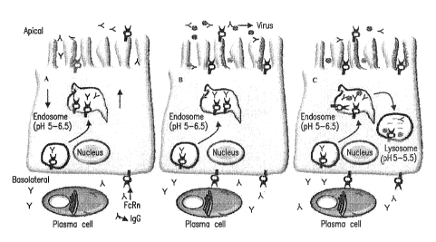
8. Figure 3 shows a model for IgG-mediated intracellular neutralization by
FcRn in
polarized epithelial cells. Figure 4A shows that FcRn transports IgG
bidirectionally. Figure 4B
shows that IgG is transcytosed and secreted into the lumen, where it can
combine antigens to
form immune complexes. Figure 4C shows that in a cell that has been infected
by a virus, a
transcytotic vesicle containing antiviral IgG has the opportunity to meet
virus. IgG neutralizes
the virus inside vesicles and therefore aborts viral replication by delivery
of these particles to
lysosomes for degradation.
IV. DETAILED DESCRIPTION
9. Before the present compounds, compositions, articles, devices, and/or
methods are
disclosed and described, it is to be understood that they are not limited to
specific synthetic
methods or specific recombinant biotechnology methods unless otherwise
specified, or to
particular reagents unless otherwise specified, as such may, of course, vary.
It is also to be
understood that the terminology used herein is for the purpose of describing
particular
embodiments only and is not intended to be limiting.
A. Definitions
10. As used in the specification and the appended claims, the singular forms
"a," "an"
and "the" include plural referents unless the context clearly dictates
otherwise. Thus, for
example, reference to "a pharmaceutical carrier" includes mixtures of two or
more such carriers,
and the like.
11. Ranges can be expressed herein as from "about" one particular value,
and/or to
"about" another particular value. When such a range is expressed, another
embodiment includes
from the one particular value and/or to the other particular value. Similarly,
when values are
expressed as approximations, by use of the antecedent "about," it will be
understood that the
particular value forms another embodiment. It will be further understood that
the endpoints of
each of the ranges are significant both in relation to the other endpoint, and
independently of the
other endpoint. It is also understood that there are a number of values
disclosed herein, and that
each value is also herein disclosed as "about" that particular value in
addition to the value itself
For example, if the value "10" is disclosed, then "about 10" is also
disclosed. It is also
understood that when a value is disclosed that "less than or equal to" the
value, "greater than or
equal to the value" and possible ranges between values are also disclosed, as
appropriately
understood by the skilled artisan. For example, if the value "10" is disclosed
the "less than or
3
CA 02853734 2014-04-25
WO 2013/063613 PCT/US2012/062498
equal to 10"as well as "greater than or equal to 10" is also disclosed. It is
also understood that
the throughout the application, data is provided in a number of different
formats, and that this
data, represents endpoints and starting points, and ranges for any combination
of the data points.
For example, if a particular data point "10" and a particular data point 15
are disclosed, it is
understood that greater than, greater than or equal to, less than, less than
or equal to, and equal
to 10 and 15 are considered disclosed as well as between 10 and 15. It is also
understood that
each unit between two particular units are also disclosed. For example, if 10
and 15 are
disclosed, then 11, 12, 13, and 14 are also disclosed.
12. In this specification and in the claims which follow, reference will be
made to a
number of terms which shall be defined to have the following meanings:
13. "Optional" or "optionally" means that the subsequently described event or
circumstance may or may not occur, and that the description includes instances
where said event
or circumstance occurs and instances where it does not.
14. Throughout this application, various publications are referenced. The
disclosures of
these publications in their entireties are hereby incorporated by reference
into this application in
order to more fully describe the state of the art to which this pertains. The
references disclosed
are also individually and specifically incorporated by reference herein for
the material contained
in them that is discussed in the sentence in which the reference is relied
upon.
B. Compositions
15. Disclosed are the components to be used to prepare the disclosed
compositions as
well as the compositions themselves to be used within the methods disclosed
herein. These and
other materials are disclosed herein, and it is understood that when
combinations, subsets,
interactions, groups, etc. of these materials are disclosed that while
specific reference of each
various individual and collective combinations and permutation of these
compounds may not be
explicitly disclosed, each is specifically contemplated and described herein.
Thus, if a class of
molecules A, B, and C are disclosed as well as a class of molecules D, E, and
F and an example
of a combination molecule, A-D is disclosed, then even if each is not
individually recited each is
individually and collectively contemplated meaning combinations, A-E, A-F, B-
D, B-E, B-F, C-
D, C-E, and C-F are considered disclosed. Likewise, any subset or combination
of these is also
disclosed. Thus, for example, the sub-group of A-E, B-F, and C-E would be
considered
disclosed. This concept applies to all aspects of this application including,
but not limited to,
steps in methods of making and using the disclosed compositions. Thus, if
there are a variety of
additional steps that can be performed it is understood that each of these
additional steps can be
4
CA 02853734 2014-04-25
WO 2013/063613 PCT/US2012/062498
performed with any specific embodiment or combination of embodiments of the
disclosed
methods.
16. Disclosed herein it is shown that the neonatal Fc receptor (FcRn) shuttles
the IgG
antibody across mucosal surfaces. The FcRn was initially thought to transport
maternal IgG to
human fetuses through the placenta or to newborns via the intestine in
neonatal life. It is shown
herein that FcRn can function beyond neonatal life because of the functional
expression of FcRn
in adult tissues. By transcytosing IgG across the vascular endothelium at all
stages of life, FcRn
ensures the extravascular bioavailability of IgG. Finally, by transcytosing
IgG across the
mucosal epithelium, FcRn provides a line of humoral defense at the mucosal
surfaces. The
functional discovery of FcRn explains why IgG, but not IgA, is a major Ig in
the lung and
genital tract.
17. In addition to its transcytotic function, FcRn plays a role in serum IgG
homeostasis
by recycling IgG away from a catabolic pathway in vascular endothelium, thus
extending its
lifespan in circulation and ensuring long-lasting protective immunity after
infection. A hallmark
of FcRn is that it binds IgG at acidic pH (<6.5) and releases IgG at neutral
or higher pH. In the
majority of cell types, FcRn resides primarily in early acidic endosomal
vesicles; FcRn binds to
IgG that enters the cell by pinocytosis or endocytosis. Subsequently, FcRn
efficiently recycles
IgG back to the plasma membrane or transcytoses it to the opposite plasma
membrane, where
the near-neutral pH of the extracellular environment causes IgG release from
FcRn. Any
pinocytosed or endocytosed proteins, including IgG, that are not rescued in
this manner are
efficiently trafficked to the lysosomes for degradation.
18. Epithelial monolayers lining the mucosal surfaces polarize into two
separate plasma
membrane domains, the apical and basolateral, which are separated by
intercellular tight
junctions at the apical poles. The vast mucosal surfaces represent major sites
of potential attack
by invading pathogens. Most pathogens (>90%) initiate their infections at the
apical domain,
although the basolateral domain is also targeted in some cases. Receptor-
mediated endocytosis
of viruses and postendocytic membrane fusion has long been accepted as a cell
entry mechanism
for many viruses. For enveloped viruses, fusion of the viral lipid bilayer
with the membrane of
an acidic endosome is generally catalyzed by a "fusion protein" on the viral
surface. Influenza
A virus infection begins with the interaction of virions with cell surface
sialic acid residues
primarily mediated by hemagglutinin (HA). After binding virions are
internalized through
endocytic pathways the acidic pH within the endosomes induces a conformational
change in the
viral proteins such as, HA, which in turn triggers fusion between the viral
envelope and the
endosomal membranes. Subsequently, the low pH induces further conformational
changes in
______________________________________ 5 __
CA 02853734 2014-04-25
WO 2013/063613 PCT/US2012/062498
the viral matrix and viral ribonucleoprotein (yRNP) which are ejected into the
cytoplasm and the
yRNP is actively imported into the nucleus. Viral proteins produced in the
cytoplasm assemble
with replicated viral RNA and bud from the cell membrane.
19. The internalization of the pathogen and subsequent conformational changes
makes
available antigens that are not present in the extracellular milieu.
Accodingly, disclosed herein
are antibodies specific for a non-surface expressed antigen or an antigenic
determinant that is
only accessible to an antibody through a conformational change of the antigen.
The antibodies
can be isolated or part of a composition such as a vaccine, passive
immunization, or passive
immunotherapy. It is understood and herein contemplated that the antibodies
disclosed herein
either isolated or as part of a vaccine or larger composition are of the IgG
isotype to facilitate
internalization by FcRn. It is further understood that the disclose antibodies
can be neutralizing
antibodies.
20. "Antigen" means any native or foreign substance that is capable of
eliciting an
immune response. Preferably, the antigen will elicit an antibody, plasma cell,
plasmablast, or B-
cell response. Such antigens can include but are not limited to peptides
and/or proteins from a
subject, virus, bacteria, yeast, or parasite, including but not limited to
toxins. Antigens can also
include vaccines (e.g., peptides, proteins, killed pathogens, or attenuated
pathogens administered
in a pharmaceutically acceptable carrier either prophylactically or
therapeutically), bio-warfare
agents, and native peptides, polypeptides, and proteins.
21. Since viral, bacterial, fungal, and parasitic antigens, including viral
antigens such as,
HA, vary among strains and are continuously changing, a vaccine produced
against one strain
will be less effective or ineffective against other strains. This is highly
challenging, because
multiple strains circulate in the population each flu season, and new strains
are continually
emerging. Indeed, availability of strain-matched vaccines usually lags behind
these antigenic
changes. For example, the ultimate goal of developing a "universal" flu
vaccine that protects
against almost all strains of flu is highly desirable and needed. In one
aspect, disclosed herein
are antibodies, vaccines, and compositions that target conserved parts of
viral, bacterial, fungal,
parasitic, and cancer antigens. Consequently, these antigens can be recognized
by the immune
system from strain to strain.
22. It is understood and herein contemplated that the antibodies disclosed
herein can bind
to antigens that are internal or otherwise unavailable when a virus, bacteria,
fungi, or parasite is
in the extracellular environment. Thus, in one aspect, disclosed herein are
antibodies specific
for an antigen that is present in or on the surface of a pathogen or encoded
by a pathogen.
Anti-viral Antibodies
6
CA 02853734 2014-04-25
WO 2013/063613 PCT/US2012/062498
23. In one aspect, the pathogen can be a virus and the antigen a viral
antigen. Disclosed
herein are antibodies and compositions comprising said antibodies, such as,
for example,
vaccines, passive immunotherapy, and passive immunizations wherein the
antibody is specific
for a viral antigen from a virus selected from the group consisting of Herpes
Simplex virus-1,
Herpes Simplex virus-2, Varicella-Zoster virus, Epstein-Barr virus,
Cytomegalovirus, Human
Herpes virus-6, Variola virus, Vesicular stomatitis virus, Hepatitis A virus,
Hepatitis B virus,
Hepatitis C virus, Hepatitis D virus, Hepatitis E virus, Rhinovirus,
Coronavirus, Influenza virus
A (including H1N1 or other Swine HO, Influenza virus B, Measles virus,
Polyomavirus, Human
Papilomavirus, Respiratory syncytial virus, Adenovirus, Coxsackie virus,
Dengue virus, Mumps
virus, Poliovirus, Rabies virus, Rous sarcoma virus, Reovirus, Yellow fever
virus, Ebola virus,
Marburg virus, Lassa fever virus, Eastern Equine Encephalitis virus, Japanese
Encephalitis
virus, St. Louis Encephalitis virus, Murray Valley fever virus, West Nile
virus, Rift Valley fever
virus, Rotavirus A, Rotavirus B, Rotavirus C, Sindbis virus, Simian
Immunodeficiency virus,
Human T-cell Leukemia virus type-1, Hantavirus, Rubella virus, Simian
Immunodeficiency
virus, Human Immunodeficiency virus type-1, and Human Immunodeficiency virus
type-2. In a
further aspect, the viral antigen can be a viral nonstructural protein,
strucutural protein,
regulatory protein or accessory protein. Thus, the viral antigen can be a
viral glycoprotein (GP),
portal protein, tegument protein, capsid protein, DNA polymerase, RNA
polymerase, reverse
transcriptase, protease, integrase, DNA-binding protein, nucleoprotein (NP),
nuclear matric
protein, envelope protein (ENV), nuclear antigen, membrane protein, proteins
encoded by viral
early genes, group specific antigen (gag) protein, hemagglutinin (HA),
neuraminidase (NA), or
matrix protein. Specific examples of viral antigens include but are not
limited to ENV, GP160
(HIV) GP120 (HIV), GP41 (HIV), EBNA-1, EBNA-2, EBNA-3, LMP-1, LMP-2, El, E2,
E3,
E4, E5, E6, E7, NSP1, NSP2, NSP3, NSP4, NSP5, NSP10, NSP14, NSP15, NSP16,
N5P29,
G35P, G38P, G39P, zygocin protein, VP5 protein, 3AB protein, L4-22K protein,
L4-100K
protein, ORF 17 protein, S7 protein, S9 protein, S10 protein, HBXIP protein,
UL3.5 protein,
virus-infected-associated antigen protein, 3ABC protein, Cng protein, 2 BC
protein, p58 protein,
A4OR protein, vpu protein, VPX protein, BPLF1 protein, NEF protein, SGTA
protein, UL102
protein, p121 protein, VP35 protein, SPP1 Pac region protein, pX protein, N
protein,
agnoprotein, sigma NS protein, phage repressor proteins, U(S)3 protein kinase,
ToxR protein,
LexA protein, lambda CI repressor protein, Mu Ner protein, and Tat proteins.
Anti-bacterial Antibodies
24. Similarly, the pathogen can be a bacteria and the antigen a bacterial
antigen.
Disclosed herein are antibodies and compositions comprising said antibodies,
such as, for
7
CA 02853734 2014-04-25
WO 2013/063613 PCT/US2012/062498
example, vaccines, passive immunotherapy, and passive immunizations wherein
the antibody is
specific for a bacterial antigen from a bacterium selected from the group
consisting of M.
tuberculosis, M. bovis, M. bovis strain BCG, BCG substrains, M. avium, M.
intracellulare, M.
africanum, M. kansasii, M. marinum, M. ulcerans, M. avium subspecies
paratuberculosis,
Nocardia asteroides, other Nocardia species, Legionella pneumophila, other
Legionella species,
Salmonella typhi, other Salmonella species, Shigella species, Yersinia pestis,
Pasteurella
haemolytica, Pasteurella multocida, other Pasteurella species, Actinobacillus
pleuropneumoniae, Listeria monocytogenes, Listeria ivanovii, Brucella abortus,
other Brucella
species, Cowdria ruminantium, Chlamydia pneumoniae, Chlamydia trachomatis,
Chlamydia
psittaci, Coxiella burnetti, other Rickettsial species, Ehrlichia species,
Staphylococcus aureus,
Staphylococcus epidermidis, Streptococcus pneumoniae, Streptococcus pyogenes,
Streptococcus
agalactiae, Bacillus anthracis, Escherichia coli, Vibrio cholerae,
Campylobacter species,
Neiserria men ingitidis, Neiserria gonorrhea, Pseudomonas aeruginosa, other
Pseudomonas
species, Haemophilus influenzae, Haemophilus ducreyi, other Hemophilus
species, Clostridium
tetani, other Clostridium species, Yersinia enterolitica, and other Yersinia
species. In another
apsect, the antigen comprises a bacterial surface protein including but not
limited to bacterial
oligosaccharide, polysaccharide, or lipopolysaccharide; a protein associated
with fimbrial
structure and biogenesis, antimicrobial resistance, heavy metal transport,
bacterial adhesion,
extracytoplasmic substrate trafficking, or secreted hydrolases;
exopolysaccharide; humic acid;
N-acetylmuramic acid (NAM); N-acetylglucosamine (NAG); teichoic acids
including ribitol
teichoic acid and glycerol teichoic acid; 0-antigen; Lipid A; pilin proteins;
Porin; MA0829; or
SbsB. In yet another aspect, the antigen can be a a component of a microbial
biofilm, examples
of which include but are not limited to exopolysaccharide, humic acid or other
humic
substances.
Anti-parasitic Antibodies
25. In another aspect, the pathogen can be a parasite and the antigen a
parasitic antigen.
Disclosed herein are antibodies and compositions comprising said antibodies,
such as, for
example, vaccines, passive immunotherapy, and passive immunizations wherein
the antibody is
specific for a parasitic antigen from a parasite selected from the group
consisting of Toxoplasma
gondii, Plasmodium falciparum, Plasmodium vivax, Plasmodium malariae, other
Plasmodium
species, Trypanosoma brucei, Trypanosoma cruzi, Leishmania major, other
Leishmania species,
Schistosoma mansoni, other Schistosoma species, and Entamoeba histolytica. For
example, the
antigen can be parasitophorous vacuole membrane-enclosed merozoite structures,
galactose-
______________________________________ 8 __
CA 02853734 2014-04-25
WO 2013/063613 PCT/US2012/062498
inhibitable adherence protein, TSOL 16, MSP1, AMA1, Tryptophan rich antigens,
MIC1,
MAGI, or SAG1.
Anti-fungal Antibodies
26. Also disclosed, the pathogen can be a fungus and the antigen a fungal
antigen.
Disclosed herein are antibodies and compositions comprising said antibodies,
such as, for
example, vaccines, passive immunotherapy, and passive immunizations wherein
the antibody is
specific for a fungal antigen from a fungielected from the group consisting of
Candida albicans,
Cryptococcus neoformans, Histoplama capsulatum, Aspergillus fumigatus,
Coccidiodes immitis,
Paracoccidiodes brasiliensis, Blastomyces dermitidis, Pneomocystis carnii,
Penicillium
marneffi, and Alternaria alternata. For example, the fungal antigen can be
Dsel, Intl,
glucuronoxylomannan capsular polysaccharide, mannose polymers (mannan),
galactomannan,
Asp f 16 and Asp f 9, 0-glycosylhydroases,r3-endoglucanases, CRH-like
proteins, Enolase,
pyruvate decarboxylase, aldolase, pyruvate carboxylase, transketolase,
phosphoglucomutase,
HSP 30, 60, 80 and 90, AHP1, Elongation factor 1, Leishmanial elongation
factor 4a,
Phosphoglucomutase, Ribosomal L10 protein, PEP2, formate dehydrogenase,
Histone H3, or
Chitin.
Antibodies to antigens present on pathogens at mucosal surfaces
Many of the viral, bacterial, fungal, and parasitic infections to which the
disclosed
antibodies are raised are infections of mucosal surfaces. Typically, mucosal
antibody provides a
primary line of defense against pathogen invasion. The current dogma for
antibody-mediated
mucosal immunity is that polymeric IgA receptor (pIgR)-mediated transcytosis
of dimeric IgA
(dIgA) crosses epithelial barrier and releases secretory IgA (S-IgA) into
mucosal secretions. For
many years, IgA has been considered as a major antibody in seeding mucosal
immunity. The
role of IgG in mucosal immunity has been largely neglected although IgG is a
major dominant
isotype in the lung. Intriguingly, acidic endosomes appear to be the primary
compartment in
which FcRn resides and functions, and endocytosed virions initiate fusion of
their envelopes
within these compartments. Therefore, the endosome is an ideal site for the
transcytosed IgG to
meet internalized virions within polarized epithelial cells. Thus, FcRn
traffics extracellular
virus-specific IgG to the endosomes of epithelial cells, where it prevents
virus replication. To
show this, an mAb, Y8-10C2 (Y8)õ traditionally considered to be "non-
neutralizing" IgG, is in
fact capable of blocking viral infection in polarized epithelial cells via a
mechanism which is
dependent on FcRn-mediated IgG transport. It is intriguing that Y8 mAb binds
to the globular
but not the fusion domain of the stalk region of influenza HA. By binding to
low pH-induced
monomeric HA molecules, Y8 mAb prevented a structural transition of HA
required for
9
CA 02853734 2014-04-25
WO 2013/063613 PCT/US2012/062498
membrane fusion. Thus, Y8 mAb prevents viral membrane fusion and the
subsequent entry of
viral contents into the cytosol, finally resulting in the transport of virions
to the lysosome for
destruction.
27. In one aspect, disclosed herein are antibodies specific for antigens
present on
pathogens at mucosa' surfaces including non-neutralizing antibodies, wherein
the isotype is
changed from IgA to IgG.
Anti-cancer Antibodies
28. It is understood and herein contemplated that the antibodies disclosed
herein can also
be useful in treating and or diagnosing a cancer. Thus, disclosed herein are
antibodies specific
for a non-surface expressed antigen or an antigenic determinant that is only
accessible to an
antibody through a conformational change of the antigen wherein the antigen is
encoded by a
cancer. Accordingly, in one aspect, disclosed herein are antibodies specific
for a non-surface
expressed antigen or an antigenic determinant that is only accessible to an
antibody through a
conformational change of the antigen wherein the antigen is encoded by a
cancer and the cancer
is selected from the group of cancers consisting of lymphomas (Hodgkins and
non-Hodgkins), B
cell lymphoma, T cell lymphoma, myeloid leukemia, leukemias, mycosis
fungoides, carcinomas,
carcinomas of solid tissues, squamous cell carcinomas, adenocarcinomas,
sarcomas, gliomas,
blastomas, neuroblastomas, plasmacytomas, histiocytomas, melanomas, adenomas,
hypoxic
tumors, myelomas, AIDS-related lymphomas or sarcomas, metastatic cancers,
bladder cancer,
brain cancer, nervous system cancer, squamous cell carcinoma of head and neck,
neuroblastoma/glioblastoma, ovarian cancer, skin cancer, liver cancer,
melanoma, squamous cell
carcinomas of the mouth, throat, larynx, and lung, colon cancer, cervical
cancer, cervical
carcinoma, breast cancer, epithelial cancer, renal cancer, genitourinary
cancer, pulmonary
cancer, esophageal carcinoma, head and neck carcinoma, hematopoietic cancers,
testicular
cancer, cob-rectal cancers, prostatic cancer, or pancreatic cancer. It is
understood and herein
contemplated that the cancer antigen can be a oncogenic protein. Furthermore,
it is
contemplated herein that the cancer antigen to which the dislosed antibody is
specific can be a
growth factor or mitogen, including but not limited to c-Sis, PDGF, CSF-1,
EGF, PMA, IGF-1,
IGF-2, IL-1, IL-2, IL-6, IL-8, estrogens, androgens, VEGF or FGF.
Alternatively, the disclosed
antibodies can be specific for a tyrosine kinase, including but not limited to
Src-family proteins,
Syk-ZAP-70, BTK, pp125, E6 and E7 from Human papillomavirus, or JAK family
proteins or a
serine/threonine kinase, including but not limited to Raf, cyclin-dependent
kinases, protein
kinase A (PKA), protein kinase B (AKT), protein kinase C (PKC),
phosphatidylinositol 3-kinase
(PI3K), mTOR, mitogen-activated protein kinases (MAPKs), ERK1, ERK2, ERK3,
ERK4,
CA 02853734 2014-04-25
WO 2013/063613 PCT/US2012/062498
ERK5, ERK6, ERK7, JNKs, p38, MKK1, MKK2, RSK kinase, ASK1, TAK1, MLK3, TAOK1,
Ca2+/calmodulin-dependent protein kinases (CaM Kinase), ribosomal S6 kinase or
IRAK1. In
another aspect, the disclosed antibodies can be specific for a regulatory
GTPase, including but
not limited to Ras, Rho, Rab, Arf, Ran, Ral, or Rac or a transcription factor,
including but not
limited to myc or c-Myc, a STAT family protein, a HOX family protein, NF-KB,
AP-1, SP1,
NF-1, Oct-1, ATF/CREB, C/EBP, Elk-1, c-Jun, c-Fos or steroid recpetors. It is
further
contemplated herein that the disclosed antibodies can be specific for an
antigen that is a protein
target that has been pathologically phosphorylated or dephosphorylated. For
example, when
AKT is phosphorylated it is activated. When it is constitutively
phosphorylated it can result in
hyperproliferation and cancer. Therefore, phosphor-AKT is an example of one
such antigen.
Likewise, hyperpohsphoylated retinoblastoma protein (Rb) is a useful antigen
to target in order
to decrease proliferation in cancer. Likewise, phosphorylation of
intercellular tyrosines of
receptor tyrosine kinases, like EGFR, FGFR, and VEGFR, results in the
activation of signal
transduction, the net result of which often has a bearing on survival and
proliferation of the cell.
This phosphorylation site is also an adequate antigen target. Conversely,
peptidyl-prolyl
cis/trans isomerase (Pin 1) has been implicated in multiple types of cancer
and is oncogenic
when it is hypophosphorylated. Thus, hypophosphorylated Pinl is also a useful
antigen.
Antibodies to Allergens
29. In addition to antibodies disclosed herein that are specific for
pathogenic antigens or
cancer antigens, it is contemplated herein that the disclosed antibodies can
be specific for an
allergen. such antibodies are useful in passive immunotherapies and passive
immunizations, for
example, in sensitization therapy, as a mechanism for stifling an allergic
response, or Rh
incompatibility. Accordingly, in one aspect, disclosed herein are antibodies
specific for a non-
surface expressed antigen or an antigenic determinant that is only accessible
to an antibody
through a conformational change of the antigen wherein the antigen is an
allergen selected from
the allergens from group consisting of house Mites Mite, House Dust
Dermatophagoides farinae
Mite, House Dust Dermatophagoides pteronyssinus Mite, Acarus siro Food/Storage
Mite, House
Dust Blomia tropicalis Mite, Storage Chortoglyphus arcuates Mite, House Dust
Euroglyphus
maynei Mite, Lepidoglyphus Food/Storage destructor Mite, Tyrophagus
Food/Storage
putrescentiae Mite, House Dust Glycyphagus domesticus Venoms Bumble Bee Bombus
spp.
Venom European Hornet Vespa crabro Venom Honey Bee Apis mellifera. Venom Mixed
Hornet Dolichovespula Venom spp Mixed Paper Polistes spp. Wasp Venom Mixed
Yellow
Vespula spp. Jacket Venom White (bald)- Dolichovespula faced Hornet maculate
Venom
Yellow Hornet Dolichovespula Venom arenaria Insects Ant, Carpenter Camponotus
______________________________________ 11 __
CA 02853734 2014-04-25
WO 2013/063613 PCT/US2012/062498
pennsylvanicus Ant, Fire Solenopsis invicta Ant, Fire Solenopsis richteri
Cockroach, Periplaneta
American Americana Cockroach, Blattella German germanica Cockroach, Blatta
orientalis
Oriental Horse Fly Tabanus spp. House Fly Musca domestica Mayfly Ephemeroptera
spp.
Mosquito Culicidae sp. Moth Heterocera spp. Epithelia, Dander, Hair & Feathers
Canary
Feathers Serinus canaria Cat Epithelia Felis catus (domesticus) Cattle
Epithelia Bos Taurus
Chicken Feathers Gallus gallus (domesticus) Dog Epithella, Canis familiaris
Mixed Breeds
Duck Feathers Anas platyrhynchos Gerbil Epithelia Meriones unguiculatus Goat
Epithelia Capra
hircus Goose Feathers Anser domesticus Guinea Pig Cavia porcellus Epithelia
(cobaya) Hamster
Epithelia Mesocricetus auratus Hog Epithelia Sus scrofa Horse Epithelia Equus
caballus Mouse
Epithelia Mus musculus Parakeet Feathers Psittacidae spp. Pigeon Feathers
Columba fasciata
Rabbit Epithelia Oryctolagus cuniculus Rat Spithelia Rettus norvegicus Wool,
Sheep Ovis aries
Dander Cat Felis catus dander/Antigen (domesticus) Dog Dander, Canis
familiaris Mixed-Breed
Poodle Dander Canis familiaris Fungi Acremonium Cephalosporium strictum
acremonium
Alternaria Alternaria alternate tenuis Aspergillus Aspergillus amstelodami
glaucus Aspergillus
flavus Aspergillus furmigatus Aspergillus nidulans Aspergillus niger
Aspergillus ten-eus
Aspergillus versicolor Aureobasidium Pullularia pullulans pullulans Bipolaris
Drechslera
sorokiniana sorokiniana, Helminthosporium sativum Botrytis cinerea Candida
albicans
Chaetomium globosum Cladosporium herbarum Cladosporium Hormodendrum
sphaerospermum hordei Drechslere Curvularia spicifera spicifera Epicoccum
Epicoccum nigrum
purpurascens Epidermophyton floccosum Fusarium moniliforme Fusarium solani
Geotrichum
Oospora lactis candidum Gliocladium Gliocladium viride deliquescens
Helminthosporium
Spondylocladium solani atrovirens Microsporum Microsporum canis lanosum Mucor
Mucor
mucedo circinelloides f. circinelloides Mucor Mucor circinelloides f.
racemosus lusitanicus
Mucor plumbeus Mycogone perniciosa Neurospora Neurospora intermedia sitophila,
Monilia
sitophila Nigrospora oryzae Paecilomyces variotii Penicillium brevi- compactum
Penicillium
camembertii Penicillium chrysogenum Penicillium digitatum Penicillium expensum
Penicillium
notatum Penicillium roquefortii Phoma betae Phomma Phoma herbarum pigmentivora
Rhigopus
oryzae Rhizopus arrhizus Rhizopus Rhizopus stolonifer nigricans Rhodotorula
Rhodotorula
mucilaginosa rubra var. mucilaginosa Saccharomyces cerevisiae Scopulariopsis
brevicaulis
Serpula lacrymans Merulius lacrymans Setosphaeria Exserohilum rostrata
rostratum,
Helminthosporium halodes Stemphylium botryosum Stemphylium solani Trichoderma
Trichoderma harzianum viride Trichophyton Trichophyton mentagrophytes
interdigitale
Trichophyton rubrum Trichothecium Cephalothecium roseum roseum Smuts Barley
Smut
Ustilago nuda Bermuda Grass ustilago Smut cynodontis Corn Smut Ustilago maydis
Johnson
12
CA 02853734 2014-04-25
WO 2013/063613 PCT/US2012/062498
Grass Sporisorium Smut cruentum Oat Smut Ustilago avenae Wheat Smut Ustilago
tritici Grass
Pollens Bahia Paspalum notatum Bermuda Cynodon dactylon Blue, Canada Poa
compressa
Brome, Smooth Bromus inermis Canary Phalaris arundinacea Corn Zea mays
Couch/Quack
Elytrigia repens (Agropyron repens) Johnson Sorghum , halepense Kentucky Blue
Poa pratensis
Meadow Fescue Festuca pratensis (elatior) Oat, Cultivated Avena sativa Orchard
Dactylis
glomerata Red Top Agrostis gigantean (alba) Rye, Cultivated Secale cereale
Rye, Giant Wild
Leymus (Elymus) condensatus Rye, Italian Lolium perenne ssp. multiflorum Rye,
Perennial
Lolium perenne Sweet Vernal Anthoxanehum odoratum Timothy Phleum pratense
Velvet
Holcus lanatus Wheat, Cultivated Triticum aestivum Wheatgrass, Elymus Western
(Agropyron)
smithii Weed Pollens Allscale Atriplex polycarpa Baccharis Baccharis
halimifolia Baccharis
Baccharis sarothroides Burrobrush Hymenoclea salsola Careless Weed Amaranthus
hybridus
Cocklebur Xanthium strumarium (commune) Dock, Yellow Rumex crispus Dog Fennel
Eupatorium capillifolium Goldenrod Solidago spp. Hemp, Western Amaranthus
Water
tuberculatus (Acnida tamariscina) Iodine Bush Allenrolfea occidentalis
Jerusalem Oak
Chenopodium botrys Kochia/Firebush Kochia scoparia Lambs Quarter Chenopodium
album
Marsh Elder, Iva xanthifolia Burweed Marsh Elder, Iva angustifolia Nan-owleaf
Marsh Elder,
Iva annua Rough (ciliata) Mexican Tea Chenopodium ambrosioides Mugwort,
Artemisia
Common vulgaris Mugwort, Artemisia Darkleaved ludoviciana Nettle Unica dioica
Palmer's
Amaranthus Amaranth palmeri Pigweed, Amaranthus Redroot/Rough retroflexus
Pigweed,
Spiny Amaranthus spinosus Plantain, English Plantago lanceolata Poverty Weed
Iva axillaris
Quailbrush Atriplex lentiformis Rabbit Bush Ambrosia deltoidea Ragweed, Desert
Ambrosia
dumosa Ragweed, False Ambrosia acanthicarpa Ragweed, Giant Ambrosia trifida
Ragweed,
Short Ambrosia artemisiifolia Ragweed, Slender Ambrosia confertiflora Ragweed,
Ambrosia
Southern bidentata Ragweed, Ambrosia Western psilostachya Russian Thistle
Salsola kali
(pestifer) Sage, Coastal Artemisia californica Sage, Pasture Artemisia frigida
Sagebrush,
Artemisia Common tridentate Saltbush, Annual Atriplex wrightii Shadscale
Atriplex
confertifolia Sorrel, Red/Sheep Rumex acetosella Wingscale Atriplex canescens
Wormwood,
Artemisia annua Annual Tree Pollens Acacia Acacia spp. Alder, European Alnus
glutinosa
Alder, Red Alnus rubra Alder, Tag Alnus incana ssp. rugosa Alder, White Alnus
rhombifolia
Ash, Arizona Fraxinus velutina Ash, Green/Red Fraxinus pennsylvanica Ash,
Oregon Fraxinus
latifolia Ash, White Fraxinus americana Aspen Populus tremuloides Bayberry
Myrica cerifera
Beech, American Fagus grandifolia (americana) Beefwood/Austral Casuarina ian
Pine
equisetifolia Birch, Betula lenta Black/Sweet Birch, European Betula pendula
White Birch,
Red/River Betula nigra Birch, Spring Betula occidentalis (fontinalis) Birch,
White Betula
______________________________________ 13 __
CA 02853734 2014-04-25
WO 2013/063613 PCT/US2012/062498
populifolia Box Elder Acer negundo Cedar, Japanese Cryptomeria japonica Cedar,
Mountain
Juniperus ashei (sabinoides) Cedar, Red Juniperus virginiana Cedar, Salt
Tamarix gallica
Cottonwood, Populus Black balsamifera ssp. trichocarpa Cottonwood, Populus
Eastern deltoides
Cottonwood, Populus Fremont fremontii Cottonwood, Rio Populus Grande wislizeni
Cottonwood, Populus Western monilifera (sargentii) Cypress, Arizona Cupressus
arizonica
Cypress, Bald Taxodium distichum Cypress, Italian Cupressus sempervirens Elm,
American
Ulmus americana Elm, Cedar Ulmus crassifolia Elm, Siberian Ulmus pumila
Eucalyptus
Eucalyptus globulus Hackberry Celtis occidentalis Hazelnut Corylus americana
Hazelnut,
Corylus European avellana Hickory, Pignut Carya glabra Hickory, Carya ovata
Shagbark
Hickory, Carya laciniosa Shellbark Hickory, White Carya alba Juniper, Oneseed
Juniperus
monosperma Juniper, Pinchot Juniperus pinchotii Juniper, Rocky Juniperus
Mountain
scopulorum Juniper, Utah Juniperus osteosperma Juniper, Western Juniperus
occidentalis Locust
Blossom, Robinia Black pseudoacacia Mango Blossom Mangifera indica Maple,
Coast Acer
macrophyllum Maple, Red Acer rubrum Maple, Silver Acer saccharinum Maple,
Sugar Acer
saccharum Melaleuca Melaleuca quinquenervia (leucadendron) Mesquite Prosopis
glandulosa
(julifiora) Mulberry, Paper Broussonetia papyrifera Mulberry, Red Moms rubra
Mulberry,
White Moms alba Oak, Quercus Arizona/Gambel gambeiji Oak, Black Quercus
velutina, Oak,
Bur Quercus macrocarpa Oak, California Quercus Black kelloggii Oak, California
Quercus Live
agrifolia Oak, California Quercus lobata White/Valley Oak, English Quercus
robur Oak, Holly
Quercus ilex Oak, Post Quercus stellata Oak, Red Quercus rubra Oak, Scrub
Quercus dumosa
Oak, Virginia Quercus Live virginiana Oak, Water Quercus nigra Oak, Western
Quercus
White/Gany garryana Oak, White Quercus alba Olive Olea europaea Olive, Russian
Elaeagnus
angustifolia Orange Pollen Citrus sinensis Palm, Queen Arecastrum
romanzoffianum (Cocos
plumosa) Pecan Carya illinoensis Pepper Tree Schinus molle Pepper Schinus
Tree/Florida
terebinthifolius Holly Pine, Loblolly Pinus taeda Pine, Eastern Pinus strobus
White Pine,
Longleaf Pinus palustris Pine, Ponderosa Pinus ponderosa Pine, Slash Pinus
elliottii Pine,
Virginia Pinus virginiana Pine, Western Pinus monticola White Pine, Yellow
Pinus echinata
Poplar, Lombardy Populus nigra Poplar, White Populus alba Privet Ligustrum
yulgare Sweet
Gum Liquidambar styraciflua Sycamore, Platanus Eastern occidentalis Sycamore,
Platanus
Oriental orientalis Sycamore, Platanus Western racemosa Sycamore/London
Platanus Plane
acerifolia Walnut, Black Juglans nigra Walnut, Juglans California Black
californica Walnut,
English Juglans regia Willow, Arroyo Salix lasiolepis Willow, Black Salix
nigra Willow, Pussy
Salix discolor Flowers: Wild & Cultivated Daisy, Ox-Eye Chrysanthemum
leucanthemum
Dandelion Taraxacum officinale Sunflower Helianthus annuus Cultivated Farm
Plant Pollens
______________________________________ 14
CA 02853734 2014-04-25
WO 2013/063613 PCT/US2012/062498
Alfalfa Medicago sativa Castor Bean Ricinus communis Clover, Red Trifolium
pratense
Mustard Brassica spp. Sugar Beet Beta vulgaris Plant Food Almond Prunus dulcis
Apple Malus
pumila Apricot Prunus armeniaca Banana Musa paradisiaca (sapientum) Barley
Hordeum
vulgare Bean, Lima Phaseolus lunatus Bean, Navy Phaseolus vulgaris Bean, Pinto
Phaseolus sp.
Bean, Red Kidney Phaseolus sp. Bean, Phaseolus String/Green vulgaris
Blackberry Rubus
allegheniensis Blueberry Vaccinium sp. Broccoli Brassica oleracea var.
botrytis Buckwheat
Fagopyrum esculentum Cabbage Brassica oleracea var. capitata Cacao Bean
Theobroma cacao
Cantaloupe Cucumis melo Carrot Daucus carota Cauliflower Brassica oleracea
var. botrytis
Celery Apium graveolens var. dulce Cherry Prunus sp. Cinnamon Cinnamomum verum
Coffee
Coffee arabica Corn Zea mays Cranberry Vaccinium macrocarpon Cucumber Cucumis
sativus
Garlic Allium sativum Ginger Zingiber officinale Grape Vitis sp. Grapefruit
Citrus paradisi
Hops Humulus lupulus Lemon Citrus limon Lettuce Lactuca sativa Malt Mushroom
Agaricus
campestris Mustard Brassica sp. Nutmeg Myristica fragrans Oat Avena sativa
Olive, Green Olea
europaea Onion Allium cepa var. cepa Orange Citrus sinensis Pea, Blackeye
Vigna unguiculata
Pea, Green Pisum sativum (English) Peach Prunus persica Pear Pyrus communis
Pepper, Black
Piper nigrum Pepper, Green Capsicum annuum var. annuum Pineapple Ananas
comosus Potato,
Sweet Ipomoea batatas Potato, White Solanum tuberosum Raspberry Rubus idaeus
var. idaeus
Rice Oryza sativa Rye Secale cereale Sesame Seed Sesamum orientale (indicum)
Soybean
Glycine max Spinach Spinacia oleracea Squash, Yellow Cucurbita pepo var.
melopepo
Strawberry Fragaria chiloensis Tomato Lycopersicon esculentum (lycopersicum)
Turnip
Brassica rapa var. rapa Vanilla Bean Vanilla planifolia Watermelon Citrullus
lanatus var. lanatus
Wheat, Whole Triticum aestivum Fish & Shellfish Bass, Black Micropterus sp.
Catfish Ictalurus
punctatus Clam Mercenaria mercenaria Codfish Gadus morhua Crab Callinectes
sapidus
Flounder Platichthys sp. Halibut Hippoglossus sp. Lobster Homarus americanus
Mackerel
Scomber scombrus Oyster Crassostrea virginica Perch Sebastes marinus Salmon
Salmo salar
Sardine Clupeiformes Scallop Pectan magellanicus Shrimp Penaeus sp. Trout,
Lake Salvelinus
sp. Tuna Fish Thunnus sp. Animal Foods Beef Bos taurus Lamb Ovis aries Pork
Sus scrofa
Poultry Products Chicken Gallus gallus Egg, Chicken, Gallus gallus. White Egg
(Gallus gallus),
Yolk (Meleagris gallopavo), Casein, Brazil Nut Bertholletia excels, Cashew Nut
Anacardium
occidentale, Coconut Cocos nucifera, Filbert/Hazelnut Corylus Americana,
Peanut Arachis
hypogaea, Pecan Carya illinoensis, Walnut, Black Juglans nigra Walnut, English
Juglans regia,
and latex.
Antibodies to Toxins
15 _______________________________________
CA 02853734 2014-04-25
WO 2013/063613 PCT/US2012/062498
1. It is understood and herein contemplated that the antibodies disclosed
herein can bind
to antigens that are associated with a toxin. Thus, in one aspect, disclosed
herein are antibodies
specific for an antigen that is present in or on the surface of a toxin (such
as an antigenic
determinant on the toxin that is only accessible to an antibody through a
conformational change
of the antigen) or encoded by a toxin. Such antigens include but are not
limited to Abrin,
Conotoxins Diacetoxyscirpenol Bovine spongiform encephalopathy agent, Ricin,
Saxitoxin,
Tetrodotoxin, epsilon toxin, Botulinum neurotoxins, Shigatoxin, Staphylococcal
enterotoxins, T-
2 toxin, Diphtheria toxin, Tetanus toxoid, and pertussis toxin.
1. Antibodies
(1) Antibodies Generally
2. The term "antibodies" is used herein in a broad sense and includes both
polyclonal
and monoclonal antibodies. In addition to intact immunoglobulin molecules,
also included in
the term "antibodies" are fragments or polymers of those immunoglobulin
molecules, and
human or humanized versions of immunoglobulin molecules or fragments thereof,
as long as
they are chosen for their ability to interact with viral, bacterial, fungal,
or parasitic antigens such
that viral, bacterial, fungal, or parasitic infection, replication, or
survival is inhibited; the ability
to interact with cancer antigens such that metastasis or cancer progression is
inhibited; or the
ability to interact with allergens. The antibodies can be tested for their
desired activity using the
in vitro assays described herein, or by analogous methods, after which their
in vivo therapeutic
and/or prophylactic activities are tested according to known clinical testing
methods. There are
five major classes of human immunoglobulins: IgA, IgD, IgE, IgG and IgM, and
several of these
may be further divided into subclasses (isotypes), e.g., IgG-1, IgG-2, IgG-3,
and IgG-4; IgA-1
and IgA-2. One skilled in the art would recognize the comparable classes for
mouse. The heavy
chain constant domains that correspond to the different classes of
immunoglobulins are called
alpha, delta, epsilon, gamma, and mu, respectively.
3. The term "monoclonal antibody" as used herein refers to an antibody
obtained from a
substantially homogeneous population of antibodies, i.e., the individual
antibodies within the
population are identical except for possible naturally occurring mutations
that may be present in
a small subset of the antibody molecules. The monoclonal antibodies herein
specifically include
"chimeric" antibodies in which a portion of the heavy and/or light chain is
identical with or
homologous to corresponding sequences in antibodies derived from a particular
species or
belonging to a particular antibody class or subclass, while the remainder of
the chain(s) is
identical with or homologous to corresponding sequences in antibodies derived
from another
16
CA 02853734 2014-04-25
WO 2013/063613 PCT/US2012/062498
species or belonging to another antibody class or subclass, as well as
fragments of such
antibodies, as long as they exhibit the desired antagonistic activity.
4. Monoclonal antibodies may be prepared using hybridoma methods, such as
those
described by Kohler and Milstein, Nature, 256:495 (1975) or Harlow and Lane.
Antibodies, A
Laboratory Manual. Cold Spring Harbor Publications, New York, (1988). In a
hybridoma
method, a mouse or other appropriate host animal, is typically immunized with
an immunizing
agent to elicit lymphocytes that produce or are capable of producing
antibodies that will
specifically bind to the immunizing agent. Alternatively, the lymphocytes may
be immunized in
vitro. Preferably, the immunizing agent comprises one of the viral, bacterial,
parasitic, or fungal
antigens; one of the cancer antigens, or one of the allergens disclosed
herein. Traditionally, the
generation of monoclonal antibodies has depended on the availability of
purified protein or
peptides for use as the immunogen. More recently DNA based immunizations have
shown
promise as a way to elicit strong immune responses and generate monoclonal
antibodies. In this
approach, DNA-based immunization can be used, wherein DNA encoding a portion
of one of
the viral, bacterial, parasitic, or fungal antigens; one of the cancer
antigens, or one of the
allergens disclosed herein expressed as a fusion protein with human IgG is
injected into the host
animal.
5. An alternate approach to immunizations with either purified protein or DNA
is to use
antigen expressed in baculovirus. The advantages to this system include ease
of generation,
high levels of expression, and post-translational modifications that are
highly similar to those
seen in mammalian systems. Use of this system involves expressing domains of
an antibody as
fusion proteins. The antigen is produced by inserting a gene fragment in-frame
between the
signal sequence and the mature protein domain of the antibody nucleotide
sequence. This results
in the display of the foreign proteins on the surface of the virion. This
method allows
immunization with whole virus, eliminating the need for purification of target
antigens.
6. Generally, either peripheral blood lymphocytes ("PBLs") are used in methods
of
producing monoclonal antibodies if cells of human origin are desired, or
spleen cells or lymph
node cells are used if non-human mammalian sources are desired. The
lymphocytes are then
fused with an immortalized cell line using a suitable fusing agent, such as
polyethylene glycol,
to form a hybridoma cell (Goding, "Monoclonal Antibodies: Principles and
Practice" Academic
Press, (1986) pp. 59-103). Immortalized cell lines are usually transformed
mammalian cells,
including myeloma cells of rodent, bovine, equine, and human origin. Usually,
rat or mouse
myeloma cell lines are employed. The hybridoma cells may be cultured in a
suitable culture
medium that preferably contains one or more substances that inhibit the growth
or survival of
______________________________________ 17
CA 02853734 2014-04-25
WO 2013/063613 PCT/US2012/062498
the unfused, immortalized cells. For example, if the parental cells lack the
enzyme
hypoxanthine guanine phosphoribosyl transferase (HGPRT or HPRT), the culture
medium for
the hybridomas typically will include hypoxanthine, aminopterin, and thymidine
("HAT
medium"), which substances prevent the growth of HGPRT-deficient cells.
Preferred
immortalized cell lines are those that fuse efficiently, support stable high
level expression of
antibody by the selected antibody-producing cells, and are sensitive to a
medium such as HAT
medium. More preferred immortalized cell lines are murine myeloma lines, which
can be
obtained, for instance, from the Salk Institute Cell Distribution Center, San
Diego, Calif and the
American Type Culture Collection, Rockville, Md. Human myeloma and mouse-human
heteromyeloma cell lines also have been described for the production of human
monoclonal
antibodies (Kozbor, J. Immunol., 133:3001 (1984); Brodeur et al., "Monoclonal
Antibody
Production Techniques and Applications" Marcel Dekker, Inc., New York, (1987)
pp. 51-63).
The culture medium in which the hybridoma cells are cultured can then be
assayed for the
presence of monoclonal antibodies directed against one of the viral,
bacterial, parasitic, or fungal
antigens; one of the cancer antigens, or one of the allergens disclosed
herein. Preferably, the
binding specificity of monoclonal antibodies produced by the hybridoma cells
is determined by
immunoprecipitation or by an in vitro binding assay, such as radioimmunoassay
(RIA) or
enzyme-linked immunoabsorbent assay (ELISA). Such techniques and assays are
known in the
art, and are described further in the Examples below or in Harlow and Lane
Antibodies, A
Laboratory Manul Cold Spring Harbor Publications, New York, (1988).
7. After the desired hybridoma cells are identified, the clones may be
subcloned by
limiting dilution or FACS sorting procedures and grown by standard methods.
Suitable culture
media for this purpose include, for example, Dulbecco's Modified Eagle's
Medium and RPMI-
1640 medium. Alternatively, the hybridoma cells may be grown in vivo as
ascites in a mammal.
8. The monoclonal antibodies secreted by the subclones may be isolated or
purified
from the culture medium or ascites fluid by conventional immunoglobulin
purification
procedures such as, for example, protein A-Sepharose, protein G,
hydroxylapatite
chromatography, gel electrophoresis, dialysis, or affinity chromatography.
9. The monoclonal antibodies may also be made by recombinant DNA methods, such
as
those described in U.S. Pat. No. 4,816,567. DNA encoding the monoclonal
antibodies can be
readily isolated and sequenced using conventional procedures (e.g., by using
oligonucleotide
probes that are capable of binding specifically to genes encoding the heavy
and light chains of
murine antibodies). The hybridoma cells serve as a preferred source of such
DNA. Once
isolated, the DNA may be placed into expression vectors, which are then
transfected into host
18 _______________________________________
CA 02853734 2014-04-25
WO 2013/063613 PCT/US2012/062498
cells such as simian COS cells, Chinese hamster ovary (CHO) cells,
plasmacytoma cells, or
myeloma cells that do not otherwise produce immunoglobulin protein, to obtain
the synthesis of
monoclonal antibodies in the recombinant host cells. The DNA also may be
modified, for
example, by substituting the coding sequence for human heavy and light chain
constant domains
in place of the homologous murine sequences (U.S. Pat. No. 4,816,567) or by
covalently joining
to the immunoglobulin coding sequence all or part of the coding sequence for a
non-
immunoglobulin polypeptide. Optionally, such a non-immunoglobulin polypeptide
is
substituted for the constant domains of an antibody or substituted for the
variable domains of
one antigen-combining site of an antibody to create a chimeric bivalent
antibody comprising one
antigen-combining site having specificity for one of the viral, bacterial,
parasitic, or fungal
antigens; one of the cancer antigens, or one of the allergens disclosed herein
and another
antigen-combining site having specificity for a different antigen.
10. In vitro methods are also suitable for preparing monovalent antibodies.
Digestion of
antibodies to produce fragments thereof, particularly, Fab fragments, can be
accomplished using
routine techniques known in the art. For instance, digestion can be performed
using papain.
Examples of papain digestion are described in WO 94/29348 published Dec. 22,
1994, U.S. Pat.
No. 4,342,566, and Harlow and Lane, Antibodies, A Laboratory Manual, Cold
Spring Harbor
Publications, New York, (1988). Papain digestion of antibodies typically
produces two identical
antigen binding fragments, called Fab fragments, each with a single antigen
binding site, and a
residual Fc fragment. Pepsin treatment yields a fragment, called the F(ab')2
fragment, that has
two antigen combining sites and is still capable of cross-linking antigen.
11. The Fab fragments produced in the antibody digestion also contain the
constant
domains of the light chain and the first constant domain of the heavy chain.
Fab' fragments differ
from Fab fragments by the addition of a few residues at the carboxy terminus
of the heavy chain
domain including one or more cysteines from the antibody hinge region. The
F(ab')2 fragment is
a bivalent fragment comprising two Fab' fragments linked by a disulfide bridge
at the hinge
region. Fab'-SH is the designation herein for Fab' in which the cysteine
residue(s) of the
constant domains bear a free thiol group. Antibody fragments originally were
produced as pairs
of Fab' fragments which have hinge cysteines between them. Other chemical
couplings of
antibody fragments are also known.
12. An isolated immunogenically specific paratope or fragment of the antibody
is also
provided. A specific immunogenic epitope of the antibody can be isolated from
the whole
antibody by chemical or mechanical disruption of the molecule. The purified
fragments thus
obtained are tested to determine their immunogenicity and specificity by the
methods taught
19
CA 02853734 2014-04-25
WO 2013/063613 PCT/US2012/062498
herein. Immunoreactive paratopes of the antibody, optionally, are synthesized
directly. An
immunoreactive fragment is defined as an amino acid sequence of at least about
two to five
consecutive amino acids derived from the antibody amino acid sequence.
13. One method of producing proteins comprising the antibodies is to link two
or more
peptides or polypeptides together by protein chemistry techniques. For
example, peptides or
polypeptides can be chemically synthesized using currently available
laboratory equipment
using either Fmoc (9-fluorenylmethyloxycarbonyl) or Boc (tert -
butyloxycarbonoyl) chemistry.
(Applied Biosystems, Inc., Foster City, CA). One skilled in the art can
readily appreciate that a
peptide or polypeptide corresponding to the antibody, for example, can be
synthesized by
standard chemical reactions. For example, a peptide or polypeptide can be
synthesized and not
cleaved from its synthesis resin whereas the other fragment of an antibody can
be synthesized
and subsequently cleaved from the resin, thereby exposing a terminal group
which is
functionally blocked on the other fragment. By peptide condensation reactions,
these two
fragments can be covalently joined via a peptide bond at their carboxyl and
amino termini,
respectively, to form an antibody, or fragment thereof (Grant GA (1992)
Synthetic Peptides: A
User Guide. W.H. Freeman and Co., N.Y. (1992); Bodansky M and Trost B., Ed.
(1993)
Principles of Peptide Synthesis. Springer-Verlag Inc., NY. Alternatively, the
peptide or
polypeptide is independently synthesized in vivo as described above. Once
isolated, these
independent peptides or polypeptides may be linked to form an antibody or
fragment thereof via
similar peptide condensation reactions.
14. For example, enzymatic ligation of cloned or synthetic peptide segments
allow
relatively short peptide fragments to be joined to produce larger peptide
fragments, polypeptides
or whole protein domains (Abrahmsen L et al., Biochemistry, 30:4151 (1991)).
Alternatively,
native chemical ligation of synthetic peptides can be utilized to
synthetically construct large
peptides or polypeptides from shorter peptide fragments. This method consists
of a two step
chemical reaction (Dawson et al. Synthesis of Proteins by Native Chemical
Ligation. Science,
266:776-779 (1994)). The first step is the chemoselective reaction of an
unprotected synthetic
peptide-alpha-thioester with another unprotected peptide segment containing an
amino-terminal
Cys residue to give a thioester-linked intermediate as the initial covalent
product. Without a
change in the reaction conditions, this intermediate undergoes spontaneous,
rapid intramolecular
reaction to form a native peptide bond at the ligation site. Application of
this native chemical
ligation method to the total synthesis of a protein molecule is illustrated by
the preparation of
human interleukin 8 (IL-8) (Baggiolini M et al. (1992) FEBS Lett. 307:97-101;
Clark-Lewis I et
CA 02853734 2014-04-25
WO 2013/063613 PCT/US2012/062498
al., J.Biol.Chem., 269:16075 (1994); Clark-Lewis Jet al., Biochemistry,
30:3128 (1991);
Rajarathnam K et al., Biochemistry 33:6623-30 (1994)).
15. Alternatively, unprotected peptide segments are chemically linked where
the bond
formed between the peptide segments as a result of the chemical ligation is an
unnatural (non-
peptide) bond (Schnolzer, M et al. Science, 256:221 (1992)). This technique
has been used to
synthesize analogs of protein domains as well as large amounts of relatively
pure proteins with
full biological activity (deLisle Milton RC et al., Techniques in Protein
Chemistry IV. Academic
Press, New York, pp. 257-267 (1992)).
16. As used herein, the term "antibody" or "antibodies" can also refer to a
human
antibody and/or a humanized antibody. Many non-human antibodies (e.g., those
derived from
mice, rats, or rabbits) are naturally antigenic in humans, and thus can give
rise to undesirable
immune responses when administered to humans. Therefore, the use of human or
humanized
antibodies in the methods serves to lessen the chance that an antibody
administered to a human
will evoke an undesirable immune response.
(2) Human antibodies
17. The disclosed human antibodies can be prepared using any technique. The
disclosed
human antibodies can also be obtained from transgenic animals. For example,
transgenic,
mutant mice that are capable of producing a full repertoire of human
antibodies, in response to
immunization, have been described (see, e.g., Jakobovits et al., Proc. Natl.
Acad. Sci. USA,
90:2551-255 (1993); Jakobovits et al., Nature, 362:255-258 (1993); Bruggermann
et al., Year in
Immunol., 7:33 (1993)). Specifically, the homozygous deletion of the antibody
heavy chain
joining region (J(H)) gene in these chimeric and germ-line mutant mice results
in complete
inhibition of endogenous antibody production, and the successful transfer of
the human
germ-line antibody gene array into such germ-line mutant mice results in the
production of
human antibodies upon antigen challenge. Antibodies having the desired
activity are selected
using Env-CD4-co-receptor complexes as described herein.
(3) Humanized antibodies
18. Antibody humanization techniques generally involve the use of recombinant
DNA
technology to manipulate the DNA sequence encoding one or more polypeptide
chains of an
antibody molecule. Accordingly, a humanized form of a non-human antibody (or a
fragment
thereof) is a chimeric antibody or antibody chain (or a fragment thereof, such
as an sFy, Fv, Fab,
Fab', F(ab')2, or other antigen-binding portion of an antibody) which contains
a portion of an
antigen binding site from a non-human (donor) antibody integrated into the
framework of a
human (recipient) antibody.
21 ______________________________________
CA 02853734 2014-04-25
WO 2013/063613 PCT/US2012/062498
19. To generate a humanized antibody, residues from one or more
complementarity
determining regions (CDRs) of a recipient (human) antibody molecule are
replaced by residues
from one or more CDRs of a donor (non-human) antibody molecule that is known
to have
desired antigen binding characteristics (e.g., a certain level of specificity
and affinity for the
target antigen). In some instances, Fy framework (FR) residues of the human
antibody are
replaced by corresponding non-human residues. Humanized antibodies may also
contain
residues which are found neither in the recipient antibody nor in the imported
CDR or
framework sequences. Generally, a humanized antibody has one or more amino
acid residues
introduced into it from a source which is non-human. In practice, humanized
antibodies are
typically human antibodies in which some CDR residues and possibly some FR
residues are
substituted by residues from analogous sites in rodent antibodies. Humanized
antibodies
generally contain at least a portion of an antibody constant region (Fc),
typically that of a human
antibody (Jones et al., Nature, 321:522-525 (1986), Reichmann et al., Nature,
332:323-327
(1988), and Presta, Curr. Opin. Struct. Biol., 2:593-596 (1992)).
20. Methods for humanizing non-human antibodies are well known in the art. For
example, humanized antibodies can be generated according to the methods of
Winter and
co-workers (Jones et al., Nature, 321:522-525 (1986), Riechmann et al.,
Nature, 332:323-327
(1988), Verhoeyen et al., Science, 239:1534-1536 (1988)), by substituting
rodent CDRs or CDR
sequences for the corresponding sequences of a human antibody. Methods that
can be used to
produce humanized antibodies are also described in U.S. Patent No. 4,816,567
(Cabilly et al.),
U.S. Patent No. 5,565,332 (Hoogenboom et al.), U.S. Patent No. 5,721,367 (Kay
et al.), U.S.
Patent No. 5,837,243 (Deo et al.), U.S. Patent No. 5,939,598 (Kucherlapati et
al.), U.S. Patent
No. 6,130,364 (Jakobovits et al.), and U.S. Patent No. 6,180,377 (Morgan et
al.).
(4) Administration of antibodies
21. Administration of the antibodies can be done as disclosed herein. Nucleic
acid
approaches for antibody delivery also exist. The broadly neutralizing anti-
viral, anti-bacterial,
anti-parasitic, anti-fungal, anti-cancer, or anti-allergens disclosed herein
antibodies and antibody
fragments can also be administered to patients or subjects as a nucleic acid
preparation (e.g.,
DNA or RNA) that encodes the antibody or antibody fragment, such that the
patient's or
subject's own cells take up the nucleic acid and produce and secrete the
encoded antibody or
antibody fragment. The delivery of the nucleic acid can be by any means, as
disclosed herein,
for example.
Further compositions
22
CA 02853734 2014-04-25
WO 2013/063613
PCT/US2012/062498
It is understood that the antibodies disclosed herein can be administered
alone or as single active
ingredient in a composition. It is further contemplated herein that the
disclosed antibodies may
be administered in a composition comprising one or more additional active
ingredients. For
example, disclosed herein are compositions comprising the antibodies disclosed
herein and one
or more T cell determinants and/or one or more antibodies to extracellular
antigens.
2. Pharmaceutical carriers/Delivery of pharamceutical products
22. As described above, the disclosed antibodies can be administered directly
or as part
of a larger composition. In addition to the disclosed antibodies, the
compositions can also be
administered in vivo in a pharmaceutically acceptable carrier. By
"pharmaceutically acceptable"
is meant a material that is not biologically or otherwise undesirable, i.e.,
the material may be
administered to a subject, along with the nucleic acid or vector, without
causing any undesirable
biological effects or interacting in a deleterious manner with any of the
other components of the
pharmaceutical composition in which it is contained. The carrier would
naturally be selected to
minimize any degradation of the active ingredient and to minimize any adverse
side effects in
the subject, as would be well known to one of skill in the art.
23. The compositions may be administered orally, parenterally (e.g.,
intravenously), by
intramuscular injection, by intraperitoneal injection, transdermally,
extracorporeally, topically or
the like, including topical intranasal administration or administration by
inhalant. As used
herein, "topical intranasal administration" means delivery of the compositions
into the nose and
nasal passages through one or both of the nares and can comprise delivery by a
spraying
mechanism or droplet mechanism, or through aerosolization of the nucleic acid
or vector.
Administration of the compositions by inhalant can be through the nose or
mouth via delivery by
a spraying or droplet mechanism. Delivery can also be directly to any area of
the respiratory
system (e.g., lungs) via intubation. The exact amount of the compositions
required will vary
from subject to subject, depending on the species, age, weight and general
condition of the
subject, the severity of the allergic disorder being treated, the particular
nucleic acid or vector
used, its mode of administration and the like. Thus, it is not possible to
specify an exact amount
for every composition. However, an appropriate amount can be determined by one
of ordinary
skill in the art using only routine experimentation given the teachings
herein.
24. Parenteral administration of the composition, if used, is generally
characterized by
injection. Injectables can be prepared in conventional forms, either as liquid
solutions or
suspensions, solid forms suitable for solution of suspension in liquid prior
to injection, or as
emulsions. A more recently revised approach for parenteral administration
involves use of a
23
CA 02853734 2014-04-25
WO 2013/063613 PCT/US2012/062498
slow release or sustained release system such that a constant dosage is
maintained. See, e.g.,
U.S. Patent No. 3,610,795, which is incorporated by reference herein.
25. The materials may be in solution, suspension (for example, incorporated
into
microparticles, liposomes, or cells). These may be targeted to a particular
cell type via
antibodies, receptors, or receptor ligands. The following references are
examples of the use of
this technology to target specific proteins to tumor tissue (Senter, et al.,
Bioconjugate Chem.,
2:447-451, (1991); Bagshawe, K.D., Br. J. Cancer, 60:275-281, (1989);
Bagshawe, et al., Br. J.
Cancer, 58:700-703, (1988); Senter, et al., Bioconjugate Chem., 4:3-9, (1993);
Battelli, et al.,
Cancer Immunol. Immunother., 35:421-425, (1992); Pietersz and McKenzie,
Immunolog.
Reviews, 129:57-80, (1992); and Roffler, et al., Biochem. Pharmacol, 42:2062-
2065, (1991)).
Vehicles such as "stealth" and other antibody conjugated liposomes (including
lipid mediated
drug targeting to colonic carcinoma), receptor mediated targeting of DNA
through cell specific
ligands, lymphocyte directed tumor targeting, and highly specific therapeutic
retroviral targeting
of murine glioma cells in vivo. The following references are examples of the
use of this
technology to target specific proteins to tumor tissue (Hughes et al., Cancer
Research, 49:6214-
6220, (1989); and Litzinger and Huang, Biochimica et Biophysica Acta, 1104:179-
187, (1992)).
In general, receptors are involved in pathways of endocytosis, either
constitutive or ligand
induced. These receptors cluster in clathrin-coated pits, enter the cell via
clathrin-coated
vesicles, pass through an acidified endosome in which the receptors are
sorted, and then either
recycle to the cell surface, become stored intracellularly, or are degraded in
lysosomes. The
internalization pathways serve a variety of functions, such as nutrient
uptake, removal of
activated proteins, clearance of macromolecules, opportunistic entry of
viruses and toxins,
dissociation and degradation of ligand, and receptor-level regulation. Many
receptors follow
more than one intracellular pathway, depending on the cell type, receptor
concentration, type of
ligand, ligand valency, and ligand concentration. Molecular and cellular
mechanisms of
receptor-mediated endocytosis has been reviewed (Brown and Greene, DNA and
Cell Biology
10:6, 399-409 (1991)).
a) Pharmaceutically Acceptable Carriers
26. The compositions, including antibodies, can be used therapeutically in
combination
with a pharmaceutically acceptable carrier or excipient.
27. Suitable carriers and their formulations are described in Remington: The
Science and
Practice of Pharmacy (19th ed.) ed. A.R. Gennaro, Mack Publishing Company,
Easton, PA
1995. Typically, an appropriate amount of a pharmaceutically-acceptable salt
is used in the
formulation to render the formulation isotonic. Examples of the
pharmaceutically-acceptable
24
CA 02853734 2014-04-25
WO 2013/063613 PCT/US2012/062498
carrier include, but are not limited to, saline, Ringer's solution and
dextrose solution. The pH of
the solution is preferably from about 5 to about 8, and more preferably from
about 7 to about
7.5. Further carriers include sustained release preparations such as
semipermeable matrices of
solid hydrophobic polymers containing the antibody, which matrices are in the
form of shaped
articles, e.g., films, liposomes or microparticles. It will be apparent to
those persons skilled in
the art that certain carriers may be more preferable depending upon, for
instance, the route of
administration and concentration of composition being administered.
28. Pharmaceutical carriers are known to those skilled in the art. These most
typically
would be standard carriers for administration of drugs to humans, including
solutions such as
sterile water, saline, and buffered solutions at physiological pH. The
compositions can be
administered intramuscularly or subcutaneously. Other compounds will be
administered
according to standard procedures used by those skilled in the art.
29. Pharmaceutical compositions may include carriers, thickeners, diluents,
buffers,
preservatives, surface active agents and the like in addition to the molecule
of choice.
Pharmaceutical compositions may also include one or more active ingredients
such as antimicrobial
agents, antiinflammatory agents, anesthetics, and the like.
30. The pharmaceutical composition may be administered in a number of ways
depending
on whether local or systemic treatment is desired, and on the area to be
treated. Administration may
be topically (including ophthalmically, vaginally, rectally, intranasally),
orally, by inhalation, or
parenterally, for example by intravenous drip, subcutaneous, intraperitoneal
or intramuscular
injection. The disclosed antibodies can be administered intravenously,
intraperitoneally,
intramuscularly, subcutaneously, intracavity, or transdermally.
31. Preparations for parenteral administration include sterile aqueous or non-
aqueous
solutions, suspensions, and emulsions. Examples of non-aqueous solvents are
propylene glycol,
polyethylene glycol, vegetable oils such as olive oil, and injectable organic
esters such as ethyl
oleate. Aqueous carriers include water, alcoholic/aqueous solutions, emulsions
or suspensions,
including saline and buffered media. Parenteral vehicles include sodium
chloride solution,
Ringer's dextrose, dextrose and sodium chloride, lactated Ringer's, or fixed
oils. Intravenous
vehicles include fluid and nutrient replenishers, electrolyte replenishers
(such as those based on
Ringer's dextrose), and the like. Preservatives and other additives may also
be present such as,
for example, antimicrobials, anti-oxidants, chelating agents, and inert gases
and the like.
32. Formulations for topical administration may include ointments, lotions,
creams, gels,
drops, suppositories, sprays, liquids and powders. Conventional pharmaceutical
carriers, aqueous,
powder or oily bases, thickeners and the like may be necessary or desirable.
_____________________________________ 25 __
CA 02853734 2014-04-25
WO 2013/063613 PCT/US2012/062498
33. Compositions for oral administration include powders or granules,
suspensions or
solutions in water or non-aqueous media, capsules, sachets, or tablets.
Thickeners, flavorings,
diluents, emulsifiers, dispersing aids or binders may be desirable..
34. Some of the compositions may potentially be administered as a
pharmaceutically
acceptable acid- or base- addition salt, formed by reaction with inorganic
acids such as
hydrochloric acid, hydrobromic acid, perchloric acid, nitric acid, thiocyanic
acid, sulfuric acid,
and phosphoric acid, and organic acids such as formic acid, acetic acid,
propionic acid, glycolic
acid, lactic acid, pyruvic acid, oxalic acid, malonic acid, succinic acid,
maleic acid, and fumaric
acid, or by reaction with an inorganic base such as sodium hydroxide, ammonium
hydroxide,
potassium hydroxide, and organic bases such as mono-, di-, trialkyl and aryl
amines and
substituted ethanolamines.
b) Therapeutic Uses
35. Effective dosages and schedules for administering the compositions may be
determined empirically, and making such determinations is within the skill in
the art. The
dosage ranges for the administration of the compositions are those large
enough to produce the
desired effect in which the symptoms of the disorder are effected. The dosage
should not be so
large as to cause adverse side effects, such as unwanted cross-reactions,
anaphylactic reactions,
and the like. Generally, the dosage will vary with the age, condition, sex and
extent of the
disease in the patient, route of administration, or whether other drugs are
included in the
regimen, and can be determined by one of skill in the art. The dosage can be
adjusted by the
individual physician in the event of any counterindications. Dosage can vary,
and can be
administered in one or more dose administrations daily, for one or several
days. Guidance can
be found in the literature for appropriate dosages for given classes of
pharmaceutical products.
For example, guidance in selecting appropriate doses for antibodies can be
found in the
literature on therapeutic uses of antibodies, e.g., Handbook of Monoclonal
Antibodies, Ferrone
et al., eds., Noges Publications, Park Ridge, N.J., (1985) ch. 22 and pp. 303-
357; Smith et al.,
Antibodies in Human Diagnosis and Therapy, Haber et al., eds., Raven Press,
New York (1977)
pp. 365-389. A typical daily dosage of the antibody used alone might range
from about 1 lag/kg
to up to 100 mg/kg of body weight or more per day, depending on the factors
mentioned above.
36. Following administration of a disclosed composition, such as the
antibodies
disclosed herein, for treating, inhibiting, or preventing a viral infection,
bacterial infection,
fungal infection, parasitic infection, cancer, or an allergic reaction, the
efficacy of the
therapeutic antibody can be assessed in various ways well known to the skilled
practitioner. For
instance, one of ordinary skill in the art will understand that a composition,
such as an antibody,
26
CA 02853734 2014-04-25
WO 2013/063613 PCT/US2012/062498
disclosed herein is efficacious in treating or inhibiting an influenza
infection in a subject by
observing that the composition reduces viral load or prevents a further
increase in viral load.
37. The compositions that inhibit viral infection, bacterial infection, fungal
infection,
parasitic infection, cancer, or an allergic reactions disclosed herein may be
administered
prophylactically to patients or subjects who are at risk for viral, bacterial,
fungal, or parasitic
exposure; cancer, an allergic reaction, or exposure to a toxin.
3. Nucleic acids
38. There are a variety of molecules disclosed herein that are nucleic acid
based,
including for example the nucleic acids that encode, for example the
antibodies disclosed herein.
The disclosed nucleic acids are made up of for example, nucleotides,
nucleotide analogs, or
nucleotide substitutes. Non-limiting examples of these and other molecules are
discussed
herein. It is understood that for example, when a vector is expressed in a
cell, that the expressed
mRNA will typically be made up of A, C, G, and U. Likewise, it is understood
that if, for
example, an antisense molecule is introduced into a cell or cell environment
through for example
exogenous delivery, it is advantagous that the antisense molecule be made up
of nucleotide
analogs that reduce the degradation of the antisense molecule in the cellular
environment.
a) Nucleotides and related molecules
39. A nucleotide is a molecule that contains a base moiety, a sugar moiety and
a
phosphate moiety. Nucleotides can be linked together through their phosphate
moieties and
sugar moieties creating an internucleoside linkage. The base moiety of a
nucleotide can be
adenin-9-y1 (A), cytosin-l-yl (C), guanin-9-y1 (G), uracil-1-y1 (U), and
thymin-l-yl (T). The
sugar moiety of a nucleotide is a ribose or a deoxyribose. The phosphate
moiety of a nucleotide
is pentavalent phosphate. An non-limiting example of a nucleotide would be 3'-
AMP (3'-
adenosine monophosphate) or 5'-GMP (5'-guanosine monophosphate). There are
many varieties
of these types of molecules available in the art and available herein.
40. A nucleotide analog is a nucleotide which contains some type of
modification to
either the base, sugar, or phosphate moieties. Modifications to nucleotides
are well known in
the art and would include for example, 5-methylcytosine (5-me-C), 5-
hydroxymethyl cytosine,
xanthine, hypoxanthine, and 2-aminoadenine as well as modifications at the
sugar or phosphate
moieties. There are many varieties of these types of molecules available in
the art and available
herein.
41. Nucleotide substitutes are molecules having similar functional properties
to
nucleotides, but which do not contain a phosphate moiety, such as peptide
nucleic acid (PNA).
Nucleotide substitutes are molecules that will recognize nucleic acids in a
Watson-Crick or
27
CA 02853734 2014-04-25
WO 2013/063613 PCT/US2012/062498
Hoogsteen manner, but which are linked together through a moiety other than a
phosphate
moiety. Nucleotide substitutes are able to conform to a double helix type
structure when
interacting with the appropriate target nucleic acid. There are many varieties
of these types of
molecules available in the art and available herein.
42. It is also possible to link other types of molecules (conjugates) to
nucleotides or
nucleotide analogs to enhance for example, cellular uptake. Conjugates can be
chemically
linked to the nucleotide or nucleotide analogs. Such conjugates include but
are not limited to
lipid moieties such as a cholesterol moiety. (Letsinger et al., Proc. Nall.
Acad. Sci. USA, 1989,
86, 6553-6556). There are many varieties of these types of molecules available
in the art and
available herein.
43. A Watson-Crick interaction is at least one interaction with the Watson-
Crick face of
a nucleotide, nucleotide analog, or nucleotide substitute. The Watson-Crick
face of a
nucleotide, nucleotide analog, or nucleotide substitute includes the C2, Ni,
and C6 positions of a
purine based nucleotide, nucleotide analog, or nucleotide substitute and the
C2, N3, C4 positions
of a pyrimidine based nucleotide, nucleotide analog, or nucleotide substitute.
44. A Hoogsteen interaction is the interaction that takes place on the
Hoogsteen face of a
nucleotide or nucleotide analog, which is exposed in the major groove of
duplex DNA. The
Hoogsteen face includes the N7 position and reactive groups (NH2 or 0) at the
C6 position of
purine nucleotides.
b) Functional Nucleic Acids
45. Functional nucleic acids are nucleic acid molecules that have a specific
function,
such as binding a target molecule or catalyzing a specific reaction.
Functional nucleic acid
molecules can be divided into the following categories, which are not meant to
be limiting. For
example, functional nucleic acids include antisense molecules, aptamers,
ribozymes, triplex
forming molecules, and external guide sequences. The functional nucleic acid
molecules can act
as affectors, inhibitors, modulators, and stimulators of a specific activity
possessed by a target
molecule, or the functional nucleic acid molecules can possess a de novo
activity independent of
any other molecules.
46. Functional nucleic acid molecules can interact with any macromolecule,
such as
DNA, RNA, polypeptides, or carbohydrate chains. Thus, functional nucleic acids
can interact
with the mRNA of any of the disclosed nucleic acids, such as viral
polymerases, integrase,
reverse transcriptases, glycoproteins, or capsid proteins; bacterial cell wall
proteins and the like
disclosed herein, and oncoggenes. Often functional nucleic acids are designed
to interact with
other nucleic acids based on sequence homology between the target molecule and
the functional
28
CA 02853734 2014-04-25
WO 2013/063613 PCT/US2012/062498
nucleic acid molecule. In other situations, the specific recognition between
the functional
nucleic acid molecule and the target molecule is not based on sequence
homology between the
functional nucleic acid molecule and the target molecule, but rather is based
on the formation of
tertiary structure that allows specific recognition to take place.
47. Antisense molecules are designed to interact with a target nucleic acid
molecule
through either canonical or non-canonical base pairing. The interaction of the
antisense
molecule and the target molecule is designed to promote the destruction of the
target molecule
through, for example, RNAseH mediated RNA-DNA hybrid degradation.
4. Antioxidants
48. Generally, antioxidants are compounds that get react with, and typically
get
consumed by, oxygen. Since antioxidants typically react with oxygen,
antioxidants also
typically react with the free radical generators, and free radicals. ("The
Antioxidants¨The
Nutrients that Guard Your Body" by Richard A. Passwater, Ph. D., 1985, Keats
Publishing Inc.,
which is herein incorporated by reference at least for material related to
antioxidants). The
compositions can contain any antioxidants, and a non-limiting list would
included but not be
limited to, non-flavonoid antioxidants and nutrients that can directly
scavenge free radicals
including multi-carotenes, beta-carotenes, alpha-carotenes, gamma-carotenes,
lycopene, lutein
and zeanthins, selenium, Vitamin E, including alpha-, beta- and gamma-
(tocopherol,
particularly .alpha.-tocopherol, etc., vitamin E succinate, and trolox (a
soluble Vitamin E
analog) Vitamin C (ascoribic acid) and Niacin (Vitamin B3, nicotinic acid and
nicotinamide),
Vitamin A, 13-cis retinoic acidõ N-acetyl-L-cysteine (NAC), sodium ascorbate,
pyrrolidin-
edithio-carbamate, and coenzyme Q10; enzymes which catalyze the destruction of
free radicals
including peroxidases such as glutathione peroxidase (GSHPX) which acts on
H202 and such as
organic peroxides, including catalase (CAT) which acts on H202, superoxide
dismutase (SOD)
which disproportionates 02H202 ; glutathione transferase (GSHTx), glutathione
reductase (GR),
glucose 6-phosphate dehydrogenase (G6PD), and mimetics, analogs and polymers
thereof
(analogs and polymers of antioxidant enzymes, such as SOD, are described in,
for example, U.S.
patent Ser. No. 5,171,680 which is incorporated herein by reference for
material at least related
to antioxidants and antioxidant enzymes); glutathione; ceruloplasmin;
cysteine, and cysteamine
(beta-mercaptoethylamine) and flavenoids and flavenoid like molecules like
folic acid and
folate. A review of antioxidant enzymes and mimetics thereof and antioxidant
nutrients can be
found in Kumar et al, Pharmac. Ther. Vol 39: 301, 1988 and Machlin L. J. and
Bendich,
F.A.S.E.B. Journal Vol. 1:441-445, 1987 which are incorporated herein by
reference for material
related to antioxidants.
29 _______________________________________
CA 02853734 2014-04-25
WO 2013/063613 PCT/US2012/062498
49. Flavonoids, also known as "phenylchromones," are naturally occurring,
water-
soluble compounds which have antioxidant characteristics. Flavonoids are
widely distributed in
vascular plants and are found in numerous vegetables, fruits and beverages
such as tea and wine
(particularly red wine). Flavonoids are conjugated aromatic compounds. The
most widely
5. Nucleic Acid Delivery
50. In the methods described above which include the administration and uptake
of
exogenous DNA into the cells of a subject (i.e., gene transduction or
transfection), the disclosed
nucleic acids can be in the form of naked DNA or RNA, or the nucleic acids can
be in a vector
for delivering the nucleic acids to the cells, whereby the antibody-encoding
DNA fragment is
under the transcriptional regulation of a promoter, as would be well
understood by one of
25 51. As one example, vector delivery can be via a viral system, such as a
retroviral vector
system which can package a recombinant retroviral genome (see e.g., Pastan et
al., Proc. Natl.
Acad. Sci. U.S.A. 85:4486, 1988; Miller et al., Mol. Cell. Biol. 6:2895,
1986). The recombinant
retrovirus can then be used to infect and thereby deliver to the infected
cells nucleic acid
encoding a broadly neutralizing antibody (or active fragment thereof). The
exact method of
______________________________________ 30 __
CA 02853734 2014-04-25
WO 2013/063613 PCT/US2012/062498
24:738-747, 1996). Physical transduction techniques can also be used, such as
liposome
delivery and receptor-mediated and other endocytosis mechanisms (see, for
example,
Schwartzenberger et al., Blood 87:472-478, 1996). This disclosed compositions
and methods
can be used in conjunction with any of these or other commonly used gene
transfer methods.
52. As one example, if the antibody-encoding nucleic acid is delivered to the
cells of a
subject in an adenovirus vector, the dosage for administration of adenovirus
to humans can
range from about 107 to 109 plaque forming units (pfu) per injection but can
be as high as 1012
pfu per injection (Crystal, Hum. Gene Ther. 8:985-1001, 1997; Alvarez and
Curiel, Hum. Gene
Ther. 8:597-613, 1997). A subject can receive a single injection, or, if
additional injections are
necessary, they can be repeated at six month intervals (or other appropriate
time intervals, as
determined by the skilled practitioner) for an indefinite period and/or until
the efficacy of the
treatment has been established.
53. Parenteral administration of the nucleic acid or vector, if used, is
generally
characterized by injection. Injectables can be prepared in conventional forms,
either as liquid
solutions or suspensions, solid forms suitable for solution of suspension in
liquid prior to
injection, or as emulsions. A more recently revised approach for parenteral
administration
involves use of a slow release or sustained release system such that a
constant dosage is
maintained. For additional discussion of suitable formulations and various
routes of
administration of therapeutic compounds, see, e.g., Remington: The Science and
Practice of
Pharmacy (19th ed.) ed. A.R. Gennaro, Mack Publishing Company, Easton, PA
1995.
54. As described above, the compositions can be administered in a
pharmaceutically
acceptable carrier and can be delivered to the subject's cells in vivo and/or
ex vivo by a variety
of mechanisms well known in the art (e.g., uptake of naked DNA, liposome
fusion,
intramuscular injection of DNA via a gene gun, endocytosis and the like).
55. If ex vivo methods are employed, cells or tissues can be removed and
maintained
outside the body according to standard protocols well known in the art. The
compositions can
be introduced into the cells via any gene transfer mechanism, such as, for
example, calcium
phosphate mediated gene delivery, electroporation, microinjection or
proteoliposomes. The
transduced cells can then be infused (e.g., in a pharmaceutically acceptable
carrier) or
homotopically transplanted back into the subject per standard methods for the
cell or tissue type.
Standard methods are known for transplantation or infusion of various cells
into a subject.
C. Methods of Treating or Inhibiting Disease
56. The antibodies and compositions disclosed herein can be used, for example,
to bind
antigens or antigenic determinants typically not available to naturally
occurring antibodies and
31
CA 02853734 2014-04-25
WO 2013/063613 PCT/US2012/062498
provide a therapeutic or prophylactic benefit to a subject receiving the
antibody or compositions.
Thus, in one aspect, disclosed herein are methods of treating or inhibiting a
disease or condition
comprising administering to a subject one or more antibodies, wherein each
antibody separately
specific for a non-surface expressed antigen or an antigenic determinant that
is only accessible
to an antibody through a conformational change of the antigen. The antibodies
can be
neutralizing or non-neutralizing antibodies.
57. Treatment," "treat," or "treating" mean a method of reducing the effects
of a disease
or condition. Treatment can also refer to a method of reducing the disease or
condition itself
rather than just the symptoms. The treatment can be any reduction from native
levels and can be
but is not limited to the complete ablation of the disease, condition, or the
symptoms of the
disease or condition. Therefore, in the disclosed methods, "treatment" can
refer to a 10%, 20%,
30%, 40%, 50%, 60%, 70%, 80%, 9u,-so z/0,
or 100% reduction in the severity of an established
disease or the disease progression. For example, a disclosed method for
reducing the effects of
prostate cancer is considered to be a treatment if there is a 10% reduction in
one or more
symptoms of the disease in a subject with the disease when compared to native
levels in the
same subject or control subjects. Similarly, a disclosed method of treating or
inhibiting a
pathogenic infection is considered to be a treatment if there is a 10%
reduction in one or more
symptoms of the disease in a subject with the disease when compared to native
levels in the
same subject or control subjects. Thus, the reduction can be a 10, 20, 30, 40,
50, 60, 70, 80, 90,
100%, or any amount of reduction in between as compared to native or control
levels. It is
understood and herein contemplated that "treatment" does not necessarily refer
to a cure of the
disease or condition, but an improvement in the outlook of a disease or
condition.
58. "Inhibit," "inhibiting," and "inhibition" mean to decrease an activity,
response,
condition, disease, or other biological parameter. This can include but is not
limited to the
complete ablation of the activity, response, condition, or disease. This may
also include, for
example, a 10% reduction in the activity, response, condition, or disease as
compared to the
native or control level. Thus, the reduction can be a 10, 20, 30, 40, 50, 60,
70, 80, 90, 100%, or
any amount of reduction in between as compared to native or control levels.
59. It is understood and herein contemplated that the disclosed methods can be
used to
treat or inhibit any disease or conditions such as a pathogenic infection
(e..g, viral infection,
bacterial infection, fungal infection, parasitic infection), cancer, or an
allergic reaction.
60. In one aspect, disclosed herein are methods of treatment or inhibition
wherein the
pathogenic infection is a viral infection. It is understood that the viral
infection can be any viral
infection for which an antibody has been raised and administered to the
subject. Thus, for
32
CA 02853734 2014-04-25
WO 2013/063613 PCT/US2012/062498
example, disclosed herein are methods of treating or inhibiting a disease or
condition comprising
administering to a subject one or more antibodies, wherein each antibody
separately specific for
a non-surface expressed antigen or an antigenic determinant that is only
accessible to an
antibody through a conformational change of the antigen, and wherein the
disease is a viral
infection selected from the group consisting of Herpes Simplex virus-1, Herpes
Simplex virus-2,
Varicella-Zoster virus, Epstein-Barr virus, Cytomegalovirus, Human Herpes
virus-6, Variola
virus, Vesicular stomatitis virus, Hepatitis A virus, Hepatitis B virus,
Hepatitis C virus, Hepatitis
D virus, Hepatitis E virus, Rhinovirus, Coronavirus, Influenza virus A
(including H1N1 or other
Swine HO, Influenza virus B, Measles virus, Polyomavirus, Human Papilomavirus,
Respiratory
syncytial virus, Adenovirus, Coxsackie virus, Dengue virus, Mumps virus,
Poliovirus, Rabies
virus, Rous sarcoma virus, Reovirus, Yellow fever virus, Ebola virus, Marburg
virus, Lassa
fever virus, Eastern Equine Encephalitis virus, Japanese Encephalitis virus,
St. Louis
Encephalitis virus, Murray Valley fever virus, West Nile virus, Rift Valley
fever virus,
Rotavirus A, Rotavirus B, Rotavirus C, Sindbis virus, Simian Immunodeficiency
virus, Human
T-cell Leukemia virus type-1, Hantavirus, Rubella virus, Simian
Immunodeficiency virus,
Human Immunodeficiency virus type-1, and Human Immunodeficiency virus type-2.
In another
aspect, disclosed herein are methods of treating and inhibiting a viral
infection wherein the
antigen is a viral glycoprotein (GP), portal protein, tegument protein, capsid
protein, DNA
polymerase, RNA polymerase, reverse transcriptase, protease, integrase, DNA-
binding protein,
nucleoprotein (NP), nuclear matric protein, envelope protein (ENV), nuclear
antigen, membrane
protein, proteins encoded by viral early genes, group specific antigen (gag)
protein,
hemagglutinin (HA), neuraminidase (NA), or matrix protein. Specific examples
of viral
antigens include but are not limited to ENV, GP160 (HIV) GP120 (HIV), GP41
(HIV), EBNA-
1, EBNA-2, EBNA-3, LMP-1, LMP-2, El, E2, E3, E4, E5, E6, E7, NSP1, NSP2, NSP3,
NSP4,
NSP5, NSP10, NSP14, NSP15, NSP16, N5P29, G35P, G38P, G39P, zygocin protein,
VP5
protein, 3AB protein, L4-22K protein, L4-100K protein, ORF 17 protein, S7
protein, S9 protein,
S10 protein, HBXIP protein, UL3.5 protein, virus-infected-associated antigen
protein, 3ABC
protein, Cng protein, 2 BC protein, p58 protein, A4OR protein, vpu protein,
VPX protein,
BPLF1 protein, NEF protein, SGTA protein, UL102 protein, p121 protein, VP35
protein, SPP1
Pac region protein, pX protein, N protein, agnoprotein, sigma NS protein,
phage repressor
proteins, U(S)3 protein kinase, ToxR protein, LexA protein, lambda CI
repressor protein, Mu
Ner protein, and Tat proteins.
61. In one aspect, disclosed herein are methods of treatment or inhibition
wherein the
pathogenic infection is a bacterial infection. It is understood that the viral
infection can be any
33
CA 02853734 2014-04-25
WO 2013/063613
PCT/US2012/062498
bacterial infection for which an antibody has been raised and administered to
the subject. Thus,
for example, disclosed herein are methods of treating or inhibiting a disease
or condition
comprising administering to a subject one or more antibodies, wherein each
antibody separately
specific for a non-surface expressed antigen or an antigenic determinant that
is only accessible
to an antibody through a conformational change of the antigen, and wherein the
disease is a
bacterial infection selected from the group consisting of M. tuberculosis, M.
bovis, M. bovis
strain BCG, BCG substrains, M. avium, M. intracellulare, M. africanum, M.
kansasii, M.
marinum, M. ulcerans, M. avium subspecies paratuberculosis, Nocardia
asteroides, other
Nocardia species, Legionella pneumophila, other Legionella species, Salmonella
typhi, other
Salmonella species, Shigella species, Yersinia pestis, Pasteurella
haemolytica, Pasteurella
multocida, other Pasteurella species, Actinobacillus pleuropneumoniae,
Listeria
monocytogenes, Listeria ivanovii, Brucella abortus, other Brucella species,
Cowdria
ruminantium, Chlamydia pneumoniae, Chlamydia trachomatis, Chlamydia psittaci,
Coxiella
burnetti, other Rickettsial species, Ehrlichia species, Staphylococcus aureus,
Staphylococcus
epidermidis, Streptococcus pneumoniae, Streptococcus pyogenes, Streptococcus
agalactiae,
Bacillus anthracis, Escherichia coli, Vibrio cholerae, Campylobacter species,
Neiserria
meningitidis, Neiserria gonorrhea, Pseudomonas aeruginosa, other Pseudomonas
species,
Haemophilus influenzae, Haemophilus ducreyi, other Hemophilus species,
Clostridium tetani,
other Clostridium species, Yersinia enterolitica, and other Yersinia species.
In one aspect, the
antigens against which antibodies are raised in the disclosed methods can be a
bacterial surface
protein including but not limited to bacterial oligosaccharide,
polysaccharide, or
lipopolysaccharide; a protein associated with fimbrial structure and
biogenesis, antimicrobial
resistance, heavy metal transport, bacterial adhesion, extracytoplasmic
substrate trafficking, or
secreted hydrolases; exopolysaccharide; humic acid; N-acetylmuramic acid
(NAM); N-
acetylglucosamine (NAG); teichoic acids including ribitol teichoic acid and
glycerol teichoic
acid; 0-antigen; Lipid A; pilin proteins; Porin; MA0829; or SbsB. In yet
another aspect, the
antigen can be a a component of a microbial biofilm, examples of which include
but are not
limited to exopolysaccharide, humic acid or other humic substances.
62. In another apect, the disclosed methods can be used to treat or inhibit a
parasitic
infection. Thus, for example, disclosed herein are methods wherein the disease
is a parasitic
infection, and wherein the parasitic infection is an infection with a parasite
selected from the
group consisting of Toxoplasma gondii, Plasmodium falciparum, Plasmodium
vivax,
Plasmodium malariae, other Plasmodium species, Trypanosoma brucei, Trypanosoma
cruzi,
Leishmania major, other Leishmania species, Schistosoma mansoni, other
Schistosoma species,
______________________________________ 34 __
CA 02853734 2014-04-25
WO 2013/063613 PCT/US2012/062498
and Entamoeba histolytica. It is understood and herein contemplated that the
disclosed methods
of inhibiting or treating a parasitic infection can comprise administering an
antibody to a
parasitic antigen including but not limited to parasitophorous vacuole
membrane-enclosed
merozoite structures, galactose-inhibitable adherence protein, TSOL 16, MSP1,
AMA1,
Tryptophan rich antigens, MIC1, MAGI, and SAG1.
63. In another apect, the disclosed methods can be used to treat or inhibit a
fungal
infection. Thus disclosed herein are disclosed herein are methods of treating
or inhibiting a
disease or condition comprising administering to a subject one or more
antibodies, wherein each
antibody separately specific for a non-surface expressed antigen or an
antigenic determinant that
is only accessible to an antibody through a conformational change of the
antigen, and wherein
the disease is a fungal infection, and wherein the fungal infection is an
infection with a fungi
selected from the group consisting of Candida albicans, Cryptococcus
neoformans, Histoplama
capsulatum, Aspergillus fumigatus, Coccidiodes immitis, Paracoccidiodes
brasiliensis,
Blastomyces dermitidis, Pneomocystis carnii, Pen icillium marneffi, and
Alternaria alternata. In
one aspect, the fungal antigens against which antibodies are raised in the
disclosed methods can
be Dsel, Intl, glucuronoxylomannan capsular polysaccharide, mannose polymers
(mannan),
galactomannan, Asp f 16 and Asp f 9, 0-glycosylhydroases,r3-endoglucanases,
CRH-like
proteins, Enolase, pyruvate decarboxylase, aldolase, pyruvate carboxylase,
transketolase,
phosphoglucomutase, HSP 30, 60, 80 and 90, AHP1, Elongation factor 1,
Leishmanial
elongation factor 4a, Phosphoglucomutase, Ribosomal L10 protein, PEP2, formate
dehydrogenase, Histone H3, or Chitin.
64. The principals governing the disclosed methods of treating or inhibiting
pathogenic
infections are equally applicable to treat any disease where uncontrolled
cellular proliferation
occurs such as cancers. Thus, in one aspect, disclosed herein are methods of
treating or
inhibiting a disease or condition comprising administering to a subject one or
more antibodies,
wherein each antibody separately specific for a non-surface expressed antigen
or an antigenic
determinant that is only accessible to an antibody through a conformational
change of the
antigen, and wherein the disease is a cancer selected from the group of
cancers consisting of
lymphomas (Hodgkins and non-Hodgkins), B cell lymphoma, T cell lymphoma,
myeloid
leukemia, leukemias, mycosis fungoides, carcinomas, carcinomas of solid
tissues, squamous cell
carcinomas, adenocarcinomas, sarcomas, gliomas, blastomas, neuroblastomas,
plasmacytomas,
histiocytomas, melanomas, adenomas, hypoxic tumors, myelomas, AIDS-related
lymphomas or
sarcomas, metastatic cancers, bladder cancer, brain cancer, nervous system
cancer, squamous
cell carcinoma of head and neck, neuroblastoma/glioblastoma, ovarian cancer,
skin cancer, liver
CA 02853734 2014-04-25
WO 2013/063613 PCT/US2012/062498
cancer, melanoma, squamous cell carcinomas of the mouth, throat, larynx, and
lung, colon
cancer, cervical cancer, cervical carcinoma, breast cancer, epithelial cancer,
renal cancer,
genitourinary cancer, pulmonary cancer, esophageal carcinoma, head and neck
carcinoma,
hematopoietic cancers, testicular cancer, cob-rectal cancers, prostatic
cancer, or pancreatic
cancer. In one aspect, the disclosed antibodies for use in the methods
described herein can be
directed to cancer antigens including but not limited to c-Sis, PDGF, CSF-1,
EGF, PMA, IGF-1,
IGF-2, IL-1, IL-2, IL-6, IL-8, estrogens, androgens, VEGF, FGF, Src-family
proteins, Syk-ZAP-
70, BTK, pp i25, E6 and E7 from Human papillomavirus, JAK family proteins,
Raf, cyclin-
dependent kinases, protein kinase A (PKA), protein kinase B (AKT), protein
kinase C (PKC),
phosphatidylinositol 3-kinase (P13 K), mTOR, mitogen-activated protein kinases
(MAPKs),
ERK1, ERK2, ERK3, ERK4, ERK5, ERK6, ERK7, JNKs, p38, MKK1, MKK2, RSK kinase,
ASK1, TAK1, MLK3, TAOK1, Ca2+/calmodulin-dependent protein kinases (CaM
Kinase),
ribosomal S6 kinase, IRAK1, Ras, Rho, Rab, Arf, Ran, Ral, Rac, myc or c-Myc, a
STAT family
protein, a HOX family protein, NF-M3, AP-1, SP1, NF-1, Oct-1, ATF/CREB, C/EBP,
Elk-1, c-
Jun, c-Fos or steroid recpetors.
65. In addition to the disclosed method of treating pathogenic diseases and
cancers, the
antibodies disclosed herein can also be used to inhibit or treat (i.e.,
reduce) an allergic reaction.
Thus disclosed herein are methods of treating or inhibiting a disease or
condition comprising
administering to a subject one or more antibodies, wherein each antibody
separately specific for
a non-surface expressed antigen or an antigenic determinant that is only
accessible to an
antibody through a conformational change of the antigen, and wherein the
condition is an
allergic reaction to an antigen selected from the allergens from group
consisting of house Mites
Mite, House Dust Dermatophagoides farinae Mite, House Dust Dermatophagoides
pteronyssinus
Mite, Acarus siro Food/Storage Mite, House Dust Blomia tropicalis Mite,
Storage
Chortoglyphus arcuates Mite, House Dust Euroglyphus maynei Mite, Lepidoglyphus
Food/Storage destructor Mite, Tyrophagus Food/Storage putrescentiae Mite,
House Dust
Glycyphagus domesticus Venoms Bumble Bee Bombus spp. Venom European Hornet
Vespa
crabro Venom Honey Bee Apis mellifera. Venom Mixed Hornet Dolichovespula Venom
spp
Mixed Paper Polistes spp. Wasp Venom Mixed Yellow Vespula spp. Jacket Venom
White
(bald)- Dolichovespula faced Hornet maculate Venom Yellow Hornet
Dolichovespula Venom
arenaria Insects Ant, Carpenter Camponotus pennsylvanicus Ant, Fire Solenopsis
invicta Ant,
Fire Solenopsis richteri Cockroach, Periplaneta American Americana Cockroach,
Blattella
German germanica Cockroach, Blatta orientalis Oriental Horse Fly Tabanus spp.
House Fly
Musca domestica Mayfly Ephemeroptera spp. Mosquito Culicidae sp. Moth
Heterocera spp.
36
CA 02853734 2014-04-25
WO 2013/063613 PCT/US2012/062498
Epithelia, Dander, Hair & Feathers Canary Feathers Serinus canaria Cat
Epithelia Felis catus
(domesticus) Cattle Epithelia Bos Taurus Chicken Feathers Gallus gallus
(domesticus) Dog
Epithella, Canis familiaris Mixed Breeds Duck Feathers Anas platyrhynchos
Gerbil Epithelia
Meriones unguiculatus Goat Epithelia Capra hircus Goose Feathers Anser
domesticus Guinea
Pig Cavia porcellus Epithelia (cobaya) Hamster Epithelia Mesocricetus auratus
Hog Epithelia
Sus scrofa Horse Epithelia Equus caballus Mouse Epithelia Mus musculus
Parakeet Feathers
Psittacidae spp. Pigeon Feathers Columba fasciata Rabbit Epithelia Oryctolagus
cuniculus Rat
Spithelia Rettus norvegicus Wool, Sheep Ovis aries Dander Cat Felis catus
dander/Antigen
(domesticus) Dog Dander, Canis familiaris Mixed-Breed Poodle Dander Canis
familiaris Fungi
Acremonium Cephalosporium strictum acremonium Alternaria Alternaria alternate
tenuis
Aspergillus Aspergillus amstelodami glaucus Aspergillus flavus Aspergillus
furmigatus
Aspergillus nidulans Aspergillus niger Aspergillus terreus Aspergillus
versicolor Aureobasidium
Pullularia pullulans pullulans Bipolaris Drechslera sorokiniana sorokiniana,
Helminthosporium
sativum Botrytis cinerea Candida albicans Chaetomium globosum Cladosporium
herbarum
Cladosporium Hormodendrum sphaerospermum hordei Drechslere Curvularia
spicifera spicifera
Epicoccum Epicoccum nigrum purpurascens Epidermophyton floccosum Fusarium
moniliforme
Fusarium solani Geotrichum Oospora lactis candidum Gliocladium Gliocladium
viride
deliquescens Helminthosporium Spondylocladium solani atrovirens Microsporum
Microsporum
canis lanosum Mucor Mucor mucedo circinelloides f. circinelloides Mucor Mucor
circinelloides
f. racemosus lusitanicus Mucor plumbeus Mycogone perniciosa Neurospora
Neurospora
intermedia sitophila, Monilia sitophila Nigrospora oryzae Paecilomyces
variotii Penicillium
brevi- compactum Penicillium camembertii Penicillium chrysogenum Penicillium
digitatum
Penicillium expensum Penicillium notatum Penicillium roquefortii Phoma betae
Phomma
Phoma herbarum pigmentivora Rhigopus oryzae Rhizopus arrhizus Rhizopus
Rhizopus
stolonifer nigricans Rhodotorula Rhodotorula mucilaginosa rubra var.
mucilaginosa
Saccharomyces cerevisiae Scopulariopsis brevicaulis Serpula lacrymans Merulius
lacrymans
Setosphaeria Exserohilum rostrata rostratum, Helminthosporium halodes
Stemphylium
botryosum Stemphylium solani Trichoderma Trichoderma harzianum viride
Trichophyton
Trichophyton mentagrophytes interdigitale Trichophyton rubrum Trichothecium
Cephalothecium roseum roseum Smuts Barley Smut Ustilago nuda Bermuda Grass
ustilago
Smut cynodontis Corn Smut Ustilago maydis Johnson Grass Sporisorium Smut
cruentum Oat
Smut Ustilago avenae Wheat Smut Ustilago tritici Grass Pollens Bahia Paspalum
notatum
Bermuda Cynodon dactylon Blue, Canada Poa compressa Brome, Smooth Bromus
inermis
Canary Phalaris arundinacea Corn Zea mays Couch/Quack Elytrigia repens
(Agropyron repens)
37
CA 02853734 2014-04-25
WO 2013/063613 PCT/US2012/062498
Johnson Sorghum , halepense Kentucky Blue Poa pratensis Meadow Fescue Festuca
pratensis
(elatior) Oat, Cultivated Avena sativa Orchard Dactylis glomerata Red Top
Agrostis gigantean
(alba) Rye, Cultivated Secale cereale Rye, Giant Wild Leymus (Elymus)
condensatus Rye,
Italian Lolium perenne ssp. multiflorum Rye, Perennial Lolium perenne Sweet
Vernal
Anthoxanehum odoratum Timothy Phleum pratense Velvet Holcus lanatus Wheat,
Cultivated
Triticum aestivum Wheatgrass, Elymus Western (Agropyron) smithii Weed Pollens
Allscale
Atriplex polycarpa Baccharis Baccharis halimifolia Baccharis Baccharis
sarothroides
Burrobrush Hymenoclea salsola Careless Weed Amaranthus hybridus Cocklebur
Xanthium
strumarium (commune) Dock, Yellow Rumex crispus Dog Fennel Eupatorium
capillifolium
Goldenrod Solidago spp. Hemp, Western Amaranthus Water tuberculatus (Acnida
tamariscina)
Iodine Bush Allenrolfea occidentalis Jerusalem Oak Chenopodium botrys
Kochia/Firebush
Kochia scoparia Lambs Quarter Chenopodium album Marsh Elder, Iva xanthifolia
Burweed
Marsh Elder, Iva angustifolia Narrowleaf Marsh Elder, Iva annua Rough
(ciliata) Mexican Tea
Chenopodium ambrosioides Mugwort, Artemisia Common vulgaris Mugwort, Artemisia
Darkleaved ludoviciana Nettle Urtica dioica Palmer's Amaranthus Amaranth
palmeri Pigweed,
Amaranthus Redroot/Rough retroflexus Pigweed, Spiny Amaranthus spinosus
Plantain, English
Plantago lanceolata Poverty Weed Iva axillaris Quailbrush Atriplex lentiformis
Rabbit Bush
Ambrosia deltoidea Ragweed, Desert Ambrosia dumosa Ragweed, False Ambrosia
acanthicarpa
Ragweed, Giant Ambrosia trifida Ragweed, Short Ambrosia artemisiifolia
Ragweed, Slender
Ambrosia confertiflora Ragweed, Ambrosia Southern bidentata Ragweed, Ambrosia
Western
psilostachya Russian Thistle Salsola kali (pestifer) Sage, Coastal Artemisia
californica Sage,
Pasture Artemisia frigida Sagebrush, Artemisia Common tridentate Saltbush,
Annual Atriplex
wrightii Shadscale Atriplex confertifolia Sorrel, Red/Sheep Rumex acetosella
Wingscale
Atriplex canescens Wormwood, Artemisia annua Annual Tree Pollens Acacia Acacia
spp.
Alder, European Alnus glutinosa Alder, Red Alnus rubra Alder, Tag Alnus incana
ssp. rugosa
Alder, White Alnus rhombifolia Ash, Arizona Fraxinus velutina Ash, Green/Red
Fraxinus
pennsylvanica Ash, Oregon Fraxinus latifolia Ash, White Fraxinus americana
Aspen Populus
tremuloides Bayberry Myrica cerifera Beech, American Fagus grandifolia
(americana)
Beefwood/Austral Casuarina ian Pine equisetifolia Birch, Betula lenta
Black/Sweet Birch,
European Betula pendula White Birch, Red/River Betula nigra Birch, Spring
Betula occidentalis
(fontinalis) Birch, White Betula populifolia Box Elder Acer negundo Cedar,
Japanese
Cryptomeria japonica Cedar, Mountain Juniperus ashei (sabinoides) Cedar, Red
Juniperus
virginiana Cedar, Salt Tamarix gallica Cottonwood, Populus Black balsamifera
ssp. trichocarpa
Cottonwood, Populus Eastern deltoides Cottonwood, Populus Fremont fremontii
Cottonwood,
38 _______________________________________
CA 02853734 2014-04-25
WO 2013/063613 PCT/US2012/062498
Rio Populus Grande wislizeni Cottonwood, Populus Western monilifera
(sargentii) Cypress,
Arizona Cupressus arizonica Cypress, Bald Taxodium distichum Cypress, Italian
Cupressus
sempervirens Elm, American Ulmus americana Elm, Cedar Ulmus crassifolia Elm,
Siberian
Ulmus pumila Eucalyptus Eucalyptus globulus Hackberry Celtis occidentalis
Hazelnut Corylus
americana Hazelnut, Corylus European avellana Hickory, Pignut Carya glabra
Hickory, Carya
ovata Shagbark Hickory, Carya laciniosa Shellbark Hickory, White Carya alba
Juniper, Oneseed
Juniperus monosperma Juniper, Pinchot Juniperus pinchotii Juniper, Rocky
Juniperus Mountain
scopulorum Juniper, Utah Juniperus osteosperma Juniper, Western Juniperus
occidentalis Locust
Blossom, Robinia Black pseudoacacia Mango Blossom Mangifera indica Maple,
Coast Acer
macrophyllum Maple, Red Acer rubrum Maple, Silver Acer saccharinum Maple,
Sugar Acer
saccharum Melaleuca Melaleuca quinquenervia (leucadendron) Mesquite Prosopis
glandulosa
(julifiora) Mulberry, Paper Broussonetia papyrifera Mulberry, Red Moms rubra
Mulberry,
White Moms alba Oak, Quercus Arizona/Gambel gambeiji Oak, Black Quercus
velutina, Oak,
Bur Quercus macrocarpa Oak, California Quercus Black kelloggii Oak, California
Quercus Live
agrifolia Oak, California Quercus lobata White/Valley Oak, English Quercus
robur Oak, Holly
Quercus ilex Oak, Post Quercus stellata Oak, Red Quercus rubra Oak, Scrub
Quercus dumosa
Oak, Virginia Quercus Live virginiana Oak, Water Quercus nigra Oak, Western
Quercus
White/Gany garryana Oak, White Quercus alba Olive Olea europaea Olive, Russian
Elaeagnus
angustifolia Orange Pollen Citrus sinensis Palm, Queen Arecastrum
romanzoffianum (Cocos
plumosa) Pecan Carya illinoensis Pepper Tree Schinus molle Pepper Schinus
Tree/Florida
terebinthifolius Holly Pine, Loblolly Pinus taeda Pine, Eastern Pinus strobus
White Pine,
Longleaf Pinus palustris Pine, Ponderosa Pinus ponderosa Pine, Slash Pinus
elliottii Pine,
Virginia Pinus virginiana Pine, Western Pinus monticola White Pine, Yellow
Pinus echinata
Poplar, Lombardy Populus nigra Poplar, White Populus alba Privet Ligustrum
vulgare Sweet
Gum Liquidambar styraciflua Sycamore, Platanus Eastern occidentalis Sycamore,
Platanus
Oriental orientalis Sycamore, Platanus Western racemosa Sycamore/London
Platanus Plane
acerifolia Walnut, Black Juglans nigra Walnut, Juglans California Black
californica Walnut,
English Juglans regia Willow, Arroyo Salix lasiolepis Willow, Black Salix
nigra Willow, Pussy
Salix discolor Flowers: Wild & Cultivated Daisy, Ox-Eye Chrysanthemum
leucanthemum
Dandelion Taraxacum officinale Sunflower Helianthus annuus Cultivated Farm
Plant Pollens
Alfalfa Medicago sativa Castor Bean Ricinus communis Clover, Red Trifolium
pratense
Mustard Brassica spp. Sugar Beet Beta vulgaris Plant Food Almond Prunus dulcis
Apple Malus
pumila Apricot Prunus armeniaca Banana Musa paradisiaca (sapientum) Barley
Hordeum
vulgare Bean, Lima Phaseolus lunatus Bean, Navy Phaseolus vulgaris Bean, Pinto
Phaseolus sp.
______________________________________ 39 __
CA 02853734 2014-04-25
WO 2013/063613 PCT/US2012/062498
Bean, Red Kidney Phaseolus sp. Bean, Phaseolus String/Green vulgaris
Blackberry Rubus
allegheniensis Blueberry Vaccinium sp. Broccoli Brassica oleracea var.
botrytis Buckwheat
Fagopyrum esculentum Cabbage Brassica oleracea var. capitata Cacao Bean
Theobroma cacao
Cantaloupe Cucumis melo Carrot Daucus carota Cauliflower Brassica oleracea
var. botrytis
Celery Apium graveolens var. dulce Cherry Prunus sp. Cinnamon Cinnamomum verum
Coffee
Coffee arabica Corn Zea mays Cranberry Vaccinium macrocarpon Cucumber Cucumis
sativus
Garlic Allium sativum Ginger Zingiber officinale Grape Vitis sp. Grapefruit
Citrus paradisi
Hops Humulus lupulus Lemon Citrus limon Lettuce Lactuca sativa Malt Mushroom
Agaricus
campestris Mustard Brassica sp. Nutmeg Myristica fragrans Oat Avena sativa
Olive, Green Olea
europaea Onion Allium cepa var. cepa Orange Citrus sinensis Pea, Blackeye
Vigna unguiculata
Pea, Green Pisum sativum (English) Peach Prunus persica Pear Pyrus communis
Pepper, Black
Piper nigrum Pepper, Green Capsicum annuum var. annuum Pineapple Ananas
comosus Potato,
Sweet Ipomoea batatas Potato, White Solanum tuberosum Raspberry Rubus idaeus
var. idaeus
Rice Oryza sativa Rye Secale cereale Sesame Seed Sesamum orientale (indicum)
Soybean
Glycine max Spinach Spinacia oleracea Squash, Yellow Cucurbita pepo var.
melopepo
Strawberry Fragaria chiloensis Tomato Lycopersicon esculentum (lycopersicum)
Turnip
Brassica rapa var. rapa Vanilla Bean Vanilla planifolia Watermelon Citrullus
lanatus var. lanatus
Wheat, Whole Triticum aestivum Fish & Shellfish Bass, Black Micropterus sp.
Catfish Ictalurus
punctatus Clam Mercenaria mercenaria Codfish Gadus morhua Crab Callinectes
sapidus
Flounder Platichthys sp. Halibut Hippoglossus sp. Lobster Homarus americanus
Mackerel
Scomber scombrus Oyster Crassostrea virginica Perch Sebastes marinus Salmon
Salmo salar
Sardine Clupeiformes Scallop Pectan magellanicus Shrimp Penaeus sp. Trout,
Lake Salvelinus
sp. Tuna Fish Thunnus sp. Animal Foods Beef Bos taurus Lamb Ovis aries Pork
Sus scrofa
Poultry Products Chicken Gallus gallus Egg, Chicken, Gallus gallus. White Egg
(Gallus gallus),
Yolk (Meleagris gallopavo), Casein, Brazil Nut Bertholletia excels, Cashew Nut
Anacardium
occidentale, Coconut Cocos nucifera, Filbert/Hazelnut Corylus Americana,
Peanut Arachis
hypogaea, Pecan Carya illinoensis, Walnut, Black Juglans nigra Walnut, English
Juglans regia,
and latex. Also disclosed are methods further comprising switching isotype of
antibody form
IgE to IgG.
66. The antibodies disclosed herein can also be used to treat a subject having
been
exposed to a toxin. Thus, in one aspect, disclosed herein are methods of
treating or inhibiting a
disease or condition comprising administering to a subject one or more
antibodies, wherein each
antibody separately specific for a non-surface expressed antigen or an
antigenic determinant that
is only accessible to an antibody through a conformational change of the
antigen, and wherein
CA 02853734 2014-04-25
WO 2013/063613 PCT/US2012/062498
the antigens include but are not limited to Abrin, Conotoxins
Diacetoxyscirpenol Bovine
spongiform encephalopathy agent, Ricin, Saxitoxin, Tetrodotoxin, epsilon
toxin, Botulinum
neurotoxins, Shigatoxin, Staphylococcal enterotoxins, T-2 toxin, Diphtheria
toxin, Tetanus
toxoid, and pertussis toxin.
D. Methods of diagnosing or detecting exposure
67. It is understood and herein contemplated that one use of the disclosed
antibodies is
for the diagnosis of a disease or condition or the detection of exposure to an
antigen. As the
antibodies bind to antigens, the use of a labeled antibody allows for the
ability to detect when an
antibody has bound a target. In this case, the target can be a viral antigen,
bacterial antigen,
fungal antigen, parasitic antigen, cancer antigen, allergen, or toxin. The
detection of the
presence of the labeled antibody would indicate exposure to the target antigen
or provide a
diagnosis. Thus, disclosed herein are methods of diagnosing a disease or
condition in a subject
or detecting exposure of a subject to an antigen associated with a disease,
condition, or toxin
comprising obtaining a tissue sample from the subject and contacting the
tissue with one or more
antibodies, wherein each antibody separately specific for a non-surface
expressed antigen or an
antigenic determinant that is only accessible to an antibody through a
conformational change of
the antigen, wherein the one or more antibodies comprise a detectable label,
wherein detection
of the one or more antibodies indicates the subject has the disease or
condition or indicates
exposure to the antigen associated with a disease, condition, or toxin.
68. It is understood and herein contemplated that the tissue sample can
include any tissue
that can reasonably be extracted from a subject influding, but not limited to
blood (including
peripheral blood and peripheral blood mononuclear cells), tissue biopsy
samples (e.g., spleen,
liver, bone marrow, thymus, lung, kidney, brain, salivary glands, skin, lymph
nodes, and
intestinal tract), and specimens acquired by pulmonary lavage (e.g.,
bronchoalveolar lavage
(BAL)). It is further understood that the disclosed methods can utilize a
labeled antibody
labeled in any available way the facilitates detection and any method of
immunodetection
known in the art
69. It is understood that the disclosed diagnostic or detection methods can be
used to
diagnosis or detect exposure to pathogenic infections (e.g., viral, bacterial,
fungal, or parasitic
infections), cancers, or exposure to toxins. Thus, in one aspect, disclosed
herein are methods of
diagnosis or detection wherein the pathogenic infection is a viral infection
selected from the
group consisting of Herpes Simplex virus-1, Herpes Simplex virus-2, Varicella-
Zoster virus,
Epstein-Barr virus, Cytomegalovirus, Human Herpes virus-6, Variola virus,
Vesicular
stomatitis virus, Hepatitis A virus, Hepatitis B virus, Hepatitis C virus,
Hepatitis D virus,
_____________________________________ 41 __
CA 02853734 2014-04-25
WO 2013/063613 PCT/US2012/062498
Hepatitis E virus, Rhinovirus, Coronavirus, Influenza virus A (including H1N1
or other Swine
HO, Influenza virus B, Measles virus, Polyomavirus, Human Papilomavirus,
Respiratory
syncytial virus, Adenovirus, Coxsackie virus, Dengue virus, Mumps virus,
Poliovirus, Rabies
virus, Rous sarcoma virus, Reovirus, Yellow fever virus, Ebola virus, Marburg
virus, Lassa
fever virus, Eastern Equine Encephalitis virus, Japanese Encephalitis virus,
St. Louis
Encephalitis virus, Murray Valley fever virus, West Nile virus, Rift Valley
fever virus,
Rotavirus A, Rotavirus B, Rotavirus C, Sindbis virus, Simian Immunodeficiency
virus, Human
T-cell Leukemia virus type-1, Hantavirus, Rubella virus, Simian
Immunodeficiency virus,
Human Immunodeficiency virus type-1, and Human Immunodeficiency virus type-2.
70. In one aspect, the disclosed methods of detecting or diagnosing a viral
infection
comprise contacting a tissue sample with an antibody against a viral antigen.
Thus, also
disclosed are methods wherein the antigen against which the detecting antibody
is specific is a
viral glycoprotein (GP), portal protein, tegument protein, capsid protein, DNA
polymerase,
RNA polymerase, reverse transcriptase, protease, integrase, DNA-binding
protein, nucleoprotein
(NP), nuclear matric protein, envelope protein (ENV), nuclear antigen,
membrane protein,
proteins encoded by viral early genes, group specific antigen (gag) protein,
hemagglutinin (HA),
neuraminidase (NA), or matrix protein. Specific examples of viral antigens
include but are not
limited to ENV, GP160 (HIV) GP120 (HIV), GP41 (HIV), EBNA-1, EBNA-2, EBNA-3,
LMP-
1, LMP-2, El, E2, E3, E4, E5, E6, E7, NSP1, NSP2, NSP3, NSP4, NSP5, NSP10,
NSP14,
N5P15, NSP16, N5P29, G35P, G38P, G39P, zygocin protein, VP5 protein, 3AB
protein, L4-
22K protein, L4-100K protein, ORF 17 protein, S7 protein, S9 protein, S10
protein, HBXIP
protein, UL3.5 protein, virus-infected-associated antigen protein, 3ABC
protein, Cng protein, 2
BC protein, p58 protein, A4OR protein, vpu protein, VPX protein, BPLF1
protein, NEF protein,
SGTA protein, UL102 protein, p121 protein, VP35 protein, SPP1 Pac region
protein, pX
protein, N protein, agnoprotein, sigma NS protein, phage repressor proteins,
U(S)3 protein
kinase, ToxR protein, LexA protein, lambda CI repressor protein, Mu Ner
protein, and Tat
proteins.
71. Also disclosed are methods of detecting exposure to or diagnosis of a
pathogenic
infection wherein the antigen or pathogenic infection is a bacterial
infection. For example,
disclosed herein are methods wherein the bacterial infection is an infection
with the bacteria
selected from the group consisting of M. tuberculosis, M. bovis, M. bovis
strain BCG, BCG
substrains, M. avium, M. intracellulare, M. africanum, M. kansasii, M.
marinum, M. ulcerans,
M. avium subspecies paratuberculosis, Nocardia asteroides, other Nocardia
species, Legionella
pneumophila, other Legionella species, Salmonella typhi, other Salmonella
species, Shigella
42
CA 02853734 2014-04-25
WO 2013/063613 PCT/US2012/062498
species, Yersinia pestis, Pasteurella haemolytica, Pasteurella multocida,
other Pasteurella
species, Actinobacillus pleuropneumoniae, Listeria monocytogenes, Listeria
ivanovii, Bruce/la
abortus, other Bruce/la species, Cowdria ruminantium, Chlamydia pneumoniae,
Chlamydia
trachomatis, Chlamydia psittaci, Coxiella burnetti, other Rickettsial species,
Ehrlichia species,
Staphylococcus aureus, Staphylococcus epidermidis, Streptococcus pneumoniae,
Streptococcus
pyogenes, Streptococcus agalactiae, Bacillus anthracis, Escherichia coli,
Vibrio cholerae,
Campylobacter species, Neiserria meningitidis, Neiserria gonorrhea,
Pseudomonas aeruginosa,
other Pseudomonas species, Haemophilus influenzae, Haemophilus ducreyi, other
Hemophilus
species, Clostridium tetani, other Clostridium species, Yersinia enterolitica,
and other Yersinia
species. In one aspect, the bacterial antigen against which the detecting
antibody is specific can
be a bacterial surface protein including but not limited to bacterial
oligosaccharide,
polysaccharide, or lipopolysaccharide; a protein associated with fimbrial
structure and
biogenesis, antimicrobial resistance, heavy metal transport, bacterial
adhesion, extracytoplasmic
substrate trafficking, or secreted hydrolases; exopolysaccharide; humic acid;
N-acetylmuramic
acid (NAM); N-acetylglucosamine (NAG); teichoic acids including ribitol
teichoic acid and
glycerol teichoic acid; 0-antigen; Lipid A; pilin proteins; Porin; MA0829; or
SbsB. In yet
another aspect, the antigen can be a a component of a microbial biofilm,
examples of which
include but are not limited to exopolysaccharide, humic acid or other humic
substances.
72. In another apect, disclosed herein are methods of diagnosing an infection
or detecting
antigenic exposure wherein the infection or antigen is a parasite. Examples of
parasites that can
be detected or diagnosed using the disclosed methods include parasites
selected from the group
consisting of Toxoplasma gondii, Plasmodium falciparum, Plasmodium vivax,
Plasmodium
malariae, other Plasmodium species, Trypanosoma brucei, Trypanosoma cruzi,
Leishmania
major, other Leishmania species, Schistosoma mansoni, other Schistosoma
species, and
Entamoeba histolytica. Also disclosed are methods wherein the antigen is
parasitophorous
vacuole membrane-enclosed merozoite structures, galactose-inhibitable
adherence protein,
TSOL 16, MSP1, AMA1, Tryptophan rich antigens, MIC1, MAGI, or SAG1.
73. In another aspect, disclosed herein are methods of detecting exposure to a
fungus or
diagnosing a fungal infection wherein the fungi is selected from the group
consisting of Candida
albicans, Cryptococcus neoformans, Histoplama capsulatum, Aspergillus
fumigatus,
Coccidiodes immitis, Paracoccidiodes brasiliensis, Blastomyces dermitidis,
Pneomocystis
carnii, Pen icillium marneffi, and Alternaria alternata. It is understood and
herein contemplated
that fungal antigen against which the detecting antibody is specific can be
Dsel, Intl,
glucuronoxylomannan capsular polysaccharide, mannose polymers (mannan),
galactomannan,
43
CA 02853734 2014-04-25
WO 2013/063613 PCT/US2012/062498
Asp f 16 and Asp f 9, 0-glycosylhydroases,r3-endoglucanases, CRH-like
proteins, Enolase,
pyruvate decarboxylase, aldolase, pyruvate carboxylase, transketolase,
phosphoglucomutase,
HSP 30, 60, 80 and 90, AHP1, Elongation factor 1, Leishmanial elongation
factor 4a,
Phosphoglucomutase, Ribosomal L10 protein, PEP2, formate dehydrogenase,
Histone H3, or
Chitin.
74. Also disclosed herein are methods of detecting exposure to an antigen,
wherein the
antigen is derived from or is a toxin selected form the group consisting of
Abrin, Conotoxins
Diacetoxyscirpenol Bovine spongiform encephalopathy agent, Ricin, Saxitoxin,
Tetrodotoxin,
epsilon toxin, Botulinum neurotoxins, Shigatoxin, Staphylococcal enterotoxins,
T-2 toxin,
Diphtheria toxin, Tetanus toxoid, and pertussis toxin.
1. Immunoassays and fluorochromes
75. Each of the detection and diagnostic methods described above utilize
immunodetection through the use of label antibodies. The steps of various
useful
immunodetection methods have been described in the scientific literature, such
as, e.g., Maggio
et al., Enzyme-Immunoassay, (1987) and Nakamura, et al., Enzyme Immunoassays:
Heterogeneous and Homogeneous Systems, Handbook of Experimental Immunology,
Vol. 1:
Immunochemistry, 27.1-27.20 (1986), each of which is incorporated herein by
reference in its
entirety and specifically for its teaching regarding immunodetection methods.
Immunoassays, in
their most simple and direct sense, are binding assays involving binding
between antibodies and
antigen. Many types and formats of immunoassays are known and all are suitable
for detecting
the disclosed biomarkers. Examples of immunoassays are enzyme linked
immunosorbent assays
(ELISAs), radioimmunoassays (RIA), radioimmune precipitation assays (RIPA),
immunobead
capture assays, Western blotting, dot blotting, gel-shift assays, Flow
cytometry, protein arrays,
multiplexed bead arrays, magnetic capture, in vivo imaging, fluorescence
resonance energy
transfer (FRET), and fluorescence recovery/localization after photobleaching
(FRAP/ FLAP).
76. In general, immunoassays involve contacting a sample suspected of
containing a
molecule of interest (such as the disclosed biomarkers) with an antibody to
the molecule of
interest or contacting an antibody to a molecule of interest (such as
antibodies to the disclosed
biomarkers) with a molecule that can be bound by the antibody, as the case may
be, under
conditions effective to allow the formation of immunocomplexes. Contacting a
sample with the
antibody to the molecule of interest or with the molecule that can be bound by
an antibody to the
molecule of interest under conditions effective and for a period of time
sufficient to allow the
formation of immune complexes (primary immune complexes) is generally a matter
of simply
bringing into contact the molecule or antibody and the sample and incubating
the mixture for a
44
CA 02853734 2014-04-25
WO 2013/063613 PCT/US2012/062498
period of time long enough for the antibodies to form immune complexes with,
i.e., to bind to,
any molecules (e.g., antigens) present to which the antibodies can bind. In
many forms of
immunoassay, the sample-antibody composition, such as a tissue section, ELISA
plate, dot blot
or Western blot, can then be washed to remove any non-specifically bound
antibody species,
allowing only those antibodies specifically bound within the primary immune
complexes to be
detected.
77. Immunoassays can include methods for detecting or quantifying the amount
of a
molecule of interest (such as the disclosed biomarkers or their antibodies) in
a sample, which
methods generally involve the detection or quantitation of any immune
complexes formed
during the binding process. In general, the detection of immunocomplex
formation is well
known in the art and can be achieved through the application of numerous
approaches. These
methods are generally based upon the detection of a label or marker, such as
any radioactive,
fluorescent, biological or enzymatic tags or any other known label.
78. As used herein, a label can include a fluorescent dye, a member of a
binding pair,
such as biotin/streptavidin, a metal (e.g., gold), or an epitope tag that can
specifically interact
with a molecule that can be detected, such as by producing a colored substrate
or fluorescence.
Substances suitable for detectably labeling proteins include fluorescent dyes
(also known herein
as fluorochromes and fluorophores) and enzymes that react with colorometric
substrates (e.g.,
horseradish peroxidase). The use of fluorescent dyes is generally preferred in
the practice of the
invention as they can be detected at very low amounts. Furthermore, in the
case where multiple
antigens are reacted with a single array, each antigen can be labeled with a
distinct fluorescent
compound for simultaneous detection. Labeled spots on the array are detected
using a
fluorimeter, the presence of a signal indicating an antigen bound to a
specific antibody.
79. Fluorophores are compounds or molecules that luminesce. Typically
fluorophores
absorb electromagnetic energy at one wavelength and emit electromagnetic
energy at a second
wavelength. Representative fluorophores include, but are not limited to, 1,5
IAEDANS; 1,8-
ANS; 4- Methylumbelliferone; 5-carboxy-2,7-dichlorofluorescein; 5-
Carboxyfluorescein (5-
FAM); 5-Carboxynapthofluorescein; 5-Carboxytetramethylrhodamine (5-TAMRA); 5-
Hydroxy
Tryptamine (5-HAT); 5-ROX (carboxy-X-rhodamine); 6-Carboxyrhodamine 6G; 6-CR
6G; 6-
JOE; 7-Amino-4-methylcoumarin; 7-Aminoactinomycin D (7-AAD); 7-Hydroxy-4- I
methylcoumarin; 9-Amino-6-chloro-2-methoxyacridine (ACMA); ABQ; Acid Fuchsin;
Acridine
Orange; Acridine Red; Acridine Yellow; Acriflavin; Acriflavin Feulgen SITSA;
Aequorin
(Photoprotein); AFPs - AutoFluorescent Protein - (Quantum Biotechnologies) see
sgGFP,
sgBFP; Alexa Fluor 3SOTM; Alexa Fluor 430TM; Alexa Fluor 488TM; Alexa Fluor
532TM; Alexa
CA 02853734 2014-04-25
WO 2013/063613 PCT/US2012/062498
Fluor 546TM; Alexa Fluor 568TM; Alexa Fluor 594TM; Alexa Fluor 633TM; Alexa
Fluor 647TM;
Alexa Fluor 660TM; Alexa Fluor 680TM; Alizarin Complexon; Alizarin Red;
Allophycocyanin
(APC); AMC, AMCA-S; Aminomethylcoumarin (AMCA); AMCA-X; Aminoactinomycin D;
Aminocoumarin; Anilin Blue; Anthrocyl stearate; APC-Cy7; APTRA-BTC; APTS;
Astrazon
Brilliant Red 4G; Astrazon Orange R; Astrazon Red 6B; Astrazon Yellow 7 GLL;
Atabrine;
ATTO- TAGTm CBQCA; ATTO-TAGTm FQ; Auramine; Aurophosphine G; Aurophosphine;
BAO 9 (Bisaminophenyloxadiazole); BCECF (high pH); BCECF (low pH); Berberine
Sulphate;
Beta Lactamase; BFP blue shifted GFP (Y66H); Blue Fluorescent Protein; BFP/GFP
FRET;
Bimane; Bisbenzemide; Bisbenzimide (Hoechst); bis- BTC; Blancophor FFG;
Blancophor SV;
BOBOTM -1; BOBOTm-3; Bodipy492/515; Bodipy493/503; Bodipy500/510; Bodipy;
505/515;
Bodipy 530/550; Bodipy 542/563; Bodipy 558/568; Bodipy 564/570; Bodipy
576/589; Bodipy
581/591; Bodipy 630/650-X; Bodipy 650/665-X; Bodipy 665/676; Bodipy Fl; Bodipy
FL ATP;
Bodipy Fl-Ceramide; Bodipy R6G SE; Bodipy TMR; Bodipy TMR-X conjugate; Bodipy
TMR-
X, SE; Bodipy TR; Bodipy TR ATP; Bodipy TR-X SE; BO-PROTM -1; BO-PROTM -3;
Brilliant
Sulphoflavin FF; BTC; BTC-5N; Calcein; Calcein Blue; Calcium Crimson - ;
Calcium Green;
Calcium Green-1 Ca2+ Dye; Calcium Green-2 Ca2+; Calcium Green-5N Ca2+; Calcium
Green-
C18 Ca2+; Calcium Orange; Calcofluor White; Carboxy-X-rhodamine (5-ROX);
Cascade
BlueTM; Cascade Yellow; Catecholamine; CCF2 (GeneBlazer); CFDA; CFP (Cyan
Fluorescent
Protein); CFP/YFP FRET; Chlorophyll; Chromomycin A; Chromomycin A; CL-NERF;
CMFDA; Coelenterazine; Coelenterazine cp; Coelenterazine f; Coelenterazine
fcp;
Coelenterazine h; Coelenterazine hcp; Coelenterazine ip; Coelenterazine n;
Coelenterazine 0;
Coumarin Phalloidin; C-phycocyanine; CPM I Methylcoumarin; CTC; CTC Formazan;
Cy2TM;
Cy3.1 8; Cy3.5 TM; Cy3 TM; Cy5.1 8; CyS.STM; CySTM; Cy7TM; Cyan GFP; cyclic
AMP
Fluorosensor (FiCRhR); Dabcyl; Dansyl; Dansyl Amine; Dansyl Cadaverine; Dansyl
Chloride;
Dansyl DHPE; Dansyl fluoride; DAPI; Dapoxyl; Dapoxyl 2; Dapoxyl 3'DCFDA; DCFH
(Dichlorodihydrofluorescein Diacetate); DDAO; DHR (Dihydorhodamine 123); Di-4-
ANEPPS;
Di-8-ANEPPS (non-ratio); DiA (4-Di 16-ASP); Dichlorodihydrofluorescein
Diacetate (DCFH);
DiD- Lipophilic Tracer; DiD (Di1C18(5)); DIDS; Dihydorhodamine 123 (DHR); Dil
(Di1C18(3)); I Dinitrophenol; Di0 (Di0C18(3)); DiR; DiR (Di1C18(7)); DM-NERF
(high pH);
DNP; Dopamine; DsRed; DTAF; DY-630-NHS; DY-635-NHS; EBFP; ECFP; EGFP; ELF 97;
Eosin; Erythrosin; Erythrosin ITC; Ethidium Bromide; Ethidium homodimer-1
(EthD-1);
Euchrysin; EukoLight; Europium (111) chloride; EYFP; Fast Blue; FDA; Feulgen
(Pararosaniline); FIF (Formaldehyd Induced Fluorescence); FITC; Flazo Orange;
Fluo-3; Fluo-
46
CA 02853734 2014-04-25
WO 2013/063613 PCT/US2012/062498
4; Fluorescein (FITC); Fluorescein Diacetate; Fluoro-Emerald; Fluoro-Gold
(Hydroxystilbamidine); Fluor-Ruby; FluorX; FM i43TM; FM 4-46; Fura RedTM (high
pH); Fura
RedTm/Fluo-3; Fura-2; Fura-2/BCECF; Genacryl Brilliant Red B; Genacryl
Brilliant Yellow
10GF; Genacryl Pink 3G; Genacryl Yellow 5GF; GeneBlazer; (CCF2); GFP (S65T);
GFP red
shifted (rsGFP); GFP wild type' non-UV excitation (wtGFP); GFP wild type, UV
excitation
(wtGFP); GFPuy; Gloxalic Acid; Granular blue; Haematoporphyrin; Hoechst 33258;
Hoechst
33342; Hoechst 34580; HPTS; Hydroxycoumarin; Hydroxystilbamidine (FluoroGold);
Hydroxytryptamine; Indo-1, high calcium; Indo-1 low calcium;
Indodicarbocyanine (DiD);
Indotricarbocyanine (DiR); Intrawhite Cf; JC-1; JO J0-1; JO-PRO-1; LaserPro;
Laurodan; LDS
751 (DNA); LDS 751 (RNA); Leucophor PAF; Leucophor SF; Leucophor WS; Lissamine
Rhodamine; Lissamine Rhodamine B; Calcein/Ethidium homodimer; LOLO-1; LO-PRO-
1;;
Lucifer Yellow; Lyso Tracker Blue; Lyso Tracker Blue-White; Lyso Tracker
Green; Lyso
Tracker Red; Lyso Tracker Yellow; LysoSensor Blue; LysoSensor Green;
LysoSensor
Yellow/Blue; Mag Green; Magdala Red (Phloxin B); Mag-Fura Red; Mag-Fura-2; Mag-
Fura-5;
Mag-lndo-1; Magnesium Green; Magnesium Orange; Malachite Green; Marina Blue; I
Maxilon
Brilliant Flavin 10 GFF; Maxilon Brilliant Flavin 8 GFF; Merocyanin;
Methoxycoumarin;
Mitotracker Green FM; Mitotracker Orange; Mitotracker Red; Mitramycin;
Monobromobimane;
Monobromobimane (mBBr-GSH); Monochlorobimane; MPS (Methyl Green Pyronine
Stilbene);
NBD; NBD Amine; Nile Red; Nitrobenzoxedidole; Noradrenaline; Nuclear Fast Red;
i Nuclear
Yellow; Nylosan Brilliant lavin E8G; Oregon GreenTM; Oregon GreenTM 488;
Oregon GreenTM
500; Oregon GreenTM 514; Pacific Blue; Pararosaniline (Feulgen); PBFI; PE-Cy5;
PE-Cy7;
PerCP; PerCP-Cy5.5; PE-TexasRed (Red 613); Phloxin B (Magdala Red); Phorwite
AR;
Phorwite BKL; Phorwite Rev; Phorwite RPA; Phosphine 3R; PhotoResist;
Phycoerythrin B
[PE]; Phycoerythrin R [PE]; PKH26 (Sigma); PKH67; PMIA; Pontochrome Blue
Black; POPO-
1; POPO-3; P0-PRO-1; PO- I PRO-3; Primuline; Procion Yellow; Propidium lodid
(P1);
PyMPO; Pyrene; Pyronine; Pyronine B; Pyrozal Brilliant Flavin 7GF; QSY 7;
Quinacrine
Mustard; Resorufin; RH 414; Rhod-2; Rhodamine; Rhodamine 110; Rhodamine 123;
Rhodamine 5 GLD; Rhodamine 6G; Rhodamine B; Rhodamine B 200; Rhodamine B
extra;
Rhodamine BB; Rhodamine BG; Rhodamine Green; Rhodamine Phallicidine;
Rhodamine:
Phalloidine; Rhodamine Red; Rhodamine WT; Rose Bengal; R-phycocyanine; R-
phycoerythrin
(PE); rsGFP; 565A; 565C; 565L; 565T; Sapphire GFP; SBFI; Serotonin; Sevron
Brilliant Red
2B; Sevron Brilliant Red 4G; Sevron I Brilliant Red B; Sevron Orange; Sevron
Yellow L;
5gBFPTM (super glow BFP); 5gGFPTM (super glow GFP); SITS (Primuline; Stilbene
Isothiosulphonic Acid); SNAFL calcein; SNAFL-1; SNAFL-2; SNARF calcein;
SNARF1;
47 _______________________________________
CA 02853734 2014-04-25
WO 2013/063613 PCT/US2012/062498
Sodium Green; SpectrumAqua; SpectrumGreen; SpectrumOrange; Spectrum Red; SPQ
(6-
methoxy- N-(3 sulfopropyl) quinolinium); Stilbene; Sulphorhodamine B and C;
Sulphorhodamine Extra; SYTO 11; SYTO 12; SYTO 13; SYTO 14; SYTO 15; SYTO 16;
SYTO 17; SYTO 18; SYTO 20; SYTO 21; SYTO 22; SYTO 23; SYTO 24; SYTO 25; SYTO
40; SYTO 41; SYTO 42; SYTO 43; SYTO 44; SYTO 45; SYTO 59; SYTO 60; SYTO 61;
SYTO 62; SYTO 63; SYTO 64; SYTO 80; SYTO 81; SYTO 82; SYTO 83; SYTO 84; SYTO
85; SYTOX Blue; SYTOX Green; SYTOX Orange; Tetracycline; Tetramethylrhodamine
(TRITC); Texas RedTM; Texas Red-XTM conjugate; Thiadicarbocyanine (DiSC3);
Thiazine Red
R; Thiazole Orange; Thioflavin 5; Thioflavin S; Thioflavin TON; Thiolyte;
Thiozole Orange;
Tinopol CBS (Calcofluor White); TIER; TO-PRO-1; TO-PRO-3; TO-PRO-5; TOTO-1;
TOTO-
3; TriColor (PE-Cy5); TRITC TetramethylRodaminelsoThioCyanate; True Blue; Tru
Red;
Ultralite; Uranine B; Uvitex SFC; wt GFP; WW 781; X-Rhodamine; XRITC; Xylene
Orange;
Y66F; Y66H; Y66W; Yellow GFP; YFP; YO-PRO-1; YO- PRO 3; YOY0-1;YOY0-3; Sybr
Green; Thiazole orange (interchelating dyes); semiconductor nanoparticles such
as quantum
dots; or caged fluorophore (which can be activated with light or other
electromagnetic energy
source), or a combination thereof
80. A modifier unit such as a radionuclide can be incorporated into or
attached directly to
any of the compounds described herein by halogenation. Examples of
radionuclides useful in
this embodiment include, but are not limited to, tritium, iodine-125, iodine-
131, iodine-123,
iodine-124, astatine-210, carbon-11, carbon-14, nitrogen-13, fluorine-18. In
another aspect, the
radionuclide can be attached to a linking group or bound by a chelating group,
which is then
attached to the compound directly or by means of a linker. Examples of
radionuclides useful in
the apset include, but are not limited to, Tc-99m, Re-186, Ga-68, Re-188, Y-
90, Sm-153, Bi-
212, Cu-67, Cu-64, and Cu-62. Radiolabeling techniques such as these are
routinely used in the
radiopharmaceutical industry.
81. The radiolabeled compounds are useful as imaging agents to diagnose
neurological
disease (e.g., a neurodegenerative disease) or a mental condition or to follow
the progression or
treatment of such a disease or condition in a mammal (e.g., a human). The
radiolabeled
compounds described herein can be conveniently used in conjunction with
imaging techniques
such as positron emission tomography (PET) or single photon emission
computerized
tomography (SPECT).
82. Labeling can be either direct or indirect. In direct labeling, the
detecting antibody
(the antibody for the molecule of interest) or detecting molecule (the
molecule that can be bound
by an antibody to the molecule of interest) include a label. Detection of the
label indicates the
48
CA 02853734 2014-04-25
WO 2013/063613 PCT/US2012/062498
presence of the detecting antibody or detecting molecule, which in turn
indicates the presence of
the molecule of interest or of an antibody to the molecule of interest,
respectively. In indirect
labeling, an additional molecule or moiety is brought into contact with, or
generated at the site
of, the immunocomplex. For example, a signal-generating molecule or moiety
such as an
enzyme can be attached to or associated with the detecting antibody or
detecting molecule. The
signal-generating molecule can then generate a detectable signal at the site
of the
immunocomplex. For example, an enzyme, when supplied with suitable substrate,
can produce
a visible or detectable product at the site of the immunocomplex. ELISAs use
this type of
indirect labeling.
83. As another example of indirect labeling, an additional molecule (which can
be
referred to as a binding agent) that can bind to either the molecule of
interest or to the antibody
(primary antibody) to the molecule of interest, such as a second antibody to
the primary
antibody, can be contacted with the immunocomplex. The additional molecule can
have a label
or signal-generating molecule or moiety. The additional molecule can be an
antibody, which
can thus be termed a secondary antibody. Binding of a secondary antibody to
the primary
antibody can form a so-called sandwich with the first (or primary) antibody
and the molecule of
interest. The immune complexes can be contacted with the labeled, secondary
antibody under
conditions effective and for a period of time sufficient to allow the
formation of secondary
immune complexes. The secondary immune complexes can then be generally washed
to remove
any non-specifically bound labeled secondary antibodies, and the remaining
label in the
secondary immune complexes can then be detected. The additional molecule can
also be or
include one of a pair of molecules or moieties that can bind to each other,
such as the
biotin/avadin pair. In this mode, the detecting antibody or detecting molecule
should include the
other member of the pair.
84. Other modes of indirect labeling include the detection of primary immune
complexes
by a two step approach. For example, a molecule (which can be referred to as a
first binding
agent), such as an antibody, that has binding affinity for the molecule of
interest or
corresponding antibody can be used to form secondary immune complexes, as
described above.
After washing, the secondary immune complexes can be contacted with another
molecule
(which can be referred to as a second binding agent) that has binding affinity
for the first
binding agent, again under conditions effective and for a period of time
sufficient to allow the
formation of immune complexes (thus forming tertiary immune complexes). The
second binding
agent can be linked to a detectable label or signal-genrating molecule or
moiety, allowing
49
CA 02853734 2014-04-25
WO 2013/063613 PCT/US2012/062498
detection of the tertiary immune complexes thus formed. This system can
provide for signal
amplification.
85. Immunoassays that involve the detection of as substance, such as a protein
or an
antibody to a specific protein, include label-free assays, protein separation
methods (i.e.,
electrophoresis), solid support capture assays, or in vivo detection. Label-
free assays are
generally diagnostic means of determining the presence or absence of a
specific protein, or an
antibody to a specific protein, in a sample. Protein separation methods are
additionally useful for
evaluating physical properties of the protein, such as size or net charge.
Capture assays are
generally more useful for quantitatively evaluating the concentration of a
specific protein, or
antibody to a specific protein, in a sample. Finally, in vivo detection is
useful for evaluating the
spatial expression patterns of the substance, i.e., where the substance can be
found in a subject,
tissue or cell.
86. Provided that the concentrations are sufficient, the molecular complexes
([Ab¨Ag]n)
generated by antibody¨antigen interaction are visible to the naked eye, but
smaller amounts may
also be detected and measured due to their ability to scatter a beam of light.
The formation of
complexes indicates that both reactants are present, and in
immunoprecipitation assays a
constant concentration of a reagent antibody is used to measure specific
antigen ([Ab¨Ag]n),
and reagent antigens are used to detect specific antibody ([Ab¨Ag]n). If the
reagent species is
previously coated onto cells (as in hemagglutination assay) or very small
particles (as in latex
agglutination assay), "clumping" of the coated particles is visible at much
lower concentrations.
A variety of assays based on these elementary principles are in common use,
including
Ouchterlony immunodiffusion assay, rocket immunoelectrophoresis, and
immunoturbidometric
and nephelometric assays. The main limitations of such assays are restricted
sensitivity (lower
detection limits) in comparison to assays employing labels and, in some cases,
the fact that very
high concentrations of analyte can actually inhibit complex formation,
necessitating safeguards
that make the procedures more complex. Some of these Group 1 assays date right
back to the
discovery of antibodies and none of them have an actual "label" (e.g. Ag-enz).
Other kinds of
immunoassays that are label free depend on immunosensors, and a variety of
instruments that
can directly detect antibody¨antigen interactions are now commercially
available. Most depend
on generating an evanescent wave on a sensor surface with immobilized ligand,
which allows
continuous monitoring of binding to the ligand. Immunosensors allow the easy
investigation of
kinetic interactions and, with the advent of lower-cost specialized
instruments, may in the future
find wide application in immunoanalysis.
______________________________________ 50 __
CA 02853734 2014-04-25
WO 2013/063613 PCT/US2012/062498
87. Immunohistochemistry and flow cytometry allow for the direct visualization
of
internal and external antigenic determinants through the binding of
antibodies. It is
contemplated herein that the disclosed antibodies can be used to detect the
presence of an
antigen intracellularly and thereby provide a diagnosis or indicate antigenic
exposure.
Additionally, the antibodies herein can be used in a research capacity to
determine intracellular
protein expression comprising administering to a cell a labeled IgG antibody
to a protein,
wherein the method does not comprise permeablizing the cell prior to
administration of the
antibody.
88. Protein arrays are solid-phase ligand binding assay systems using
immobilized
proteins on surfaces which include glass, membranes, microtiter wells, mass
spectrometer plates,
and beads or other particles. The assays are highly parallel (multiplexed) and
often miniaturized
(microarrays, protein chips). Their advantages include being rapid and
automatable, capable of
high sensitivity, economical on reagents, and giving an abundance of data for
a single
experiment. Bioinformatics support is important; the data handling demands
sophisticated
software and data comparison analysis. However, the software can be adapted
from that used for
DNA arrays, as can much of the hardware and detection systems.
89. One of the chief formats is the capture array, in which ligand-binding
reagents, which
are usually antibodies but can also be alternative protein scaffolds, peptides
or nucleic acid
aptamers, are used to detect target molecules in mixtures such as plasma or
tissue extracts. In
diagnostics, capture arrays can be used to carry out multiple immunoassays in
parallel, both
testing for several analytes in individual sera for example and testing many
serum samples
simultaneously. In proteomics, capture arrays are used to quantitate and
compare the levels of
proteins in different samples in health and disease, i.e. protein expression
profiling. Proteins
other than specific ligand binders are used in the array format for in vitro
functional interaction
screens such as protein-protein, protein-DNA, protein-drug, receptor-ligand,
enzyme-substrate,
etc. The capture reagents themselves are selected and screened against many
proteins, which can
also be done in a multiplex array format against multiple protein targets.
90. For construction of arrays, sources of proteins include cell-based
expression systems
for recombinant proteins, purification from natural sources, production in
vitro by cell-free
translation systems, and synthetic methods for peptides. Many of these methods
can be
automated for high throughput production. For capture arrays and protein
function analysis, it is
important that proteins should be correctly folded and functional; this is not
always the case, e.g.
where recombinant proteins are extracted from bacteria under denaturing
conditions.
______________________________________ 51 __
CA 02853734 2014-04-25
WO 2013/063613 PCT/US2012/062498
Nevertheless, arrays of denatured proteins are useful in screening antibodies
for cross-reactivity,
identifying autoantibodies and selecting ligand binding proteins.
91. Protein arrays have been designed as a miniaturization of familiar
immunoassay
methods such as ELISA and dot blotting, often utilizing fluorescent readout,
and facilitated by
robotics and high throughput detection systems to enable multiple assays to be
carried out in
parallel. Commonly used physical supports include glass slides, silicon,
microwells,
nitrocellulose or PVDF membranes, and magnetic and other microbeads. While
microdrops of
protein delivered onto planar surfaces are the most familiar format,
alternative architectures
include CD centrifugation devices based on developments in microfluidics
(Gyros, Monmouth
Junction, NJ) and specialised chip designs, such as engineered microchannels
in a plate (e.g.,
The Living ChipTM, Biotrove, Woburn, MA) and tiny 3D posts on a silicon
surface (Zyomyx,
Hayward CA). Particles in suspension can also be used as the basis of arrays,
providing they are
coded for identification; systems include colour coding for microbeads
(Luminex, Austin, TX;
Bio-Rad Laboratories) and semiconductor nanocrystals (e.g., QD0t5TM, Quantum
Dot, Hayward,
CA), and barcoding for beads (UltraPlexTM, SmartBead Technologies Ltd,
Babraham,
Cambridge, UK) and multimetal microrods (e.g., NanobarcodesTM particles,
Nanoplex
Technologies, Mountain View, CA). Beads can also be assembled into planar
arrays on
semiconductor chips (LEAPS technology, BioArray Solutions, Warren, NJ).
92. Immobilization of proteins involves both the coupling reagent and the
nature of the
surface being coupled to. A good protein array support surface is chemically
stable before and
after the coupling procedures, allows good spot morphology, displays minimal
nonspecific
binding, does not contribute a background in detection systems, and is
compatible with different
detection systems. The immobilization method used are reproducible, applicable
to proteins of
different properties (size, hydrophilic, hydrophobic), amenable to high
throughput and
automation, and compatible with retention of fully functional protein
activity. Orientation of the
surface-bound protein is recognized as an important factor in presenting it to
ligand or substrate
in an active state; for capture arrays the most efficient binding results are
obtained with
orientated capture reagents, which generally require site-specific labeling of
the protein.
93. Both covalent and noncovalent methods of protein immobilization are used
and have
various pros and cons. Passive adsorption to surfaces is methodologically
simple, but allows
little quantitative or orientational control; it may or may not alter the
functional properties of the
protein, and reproducibility and efficiency are variable. Covalent coupling
methods provide a
stable linkage, can be applied to a range of proteins and have good
reproducibility; however,
orientation may be variable, chemical derivatization may alter the function of
the protein and
52
CA 02853734 2014-04-25
WO 2013/063613 PCT/US2012/062498
requires a stable interactive surface. Biological capture methods utilizing a
tag on the protein
provide a stable linkage and bind the protein specifically and in reproducible
orientation, but the
biological reagent must first be immobilized adequately and the array may
require special
handling and have variable stability.
94. Several immobilization chemistries and tags have been described for
fabrication of
protein arrays. Substrates for covalent attachment include glass slides coated
with amino- or
aldehyde-containing silane reagents. In the VersalinxTM system (Prolinx,
Bothell, WA)
reversible covalent coupling is achieved by interaction between the protein
derivatised with
phenyldiboronic acid, and salicylhydroxamic acid immobilized on the support
surface. This also
has low background binding and low intrinsic fluorescence and allows the
immobilized proteins
to retain function. Noncovalent binding of unmodified protein occurs within
porous structures
such as HydroGelTM (PerkinElmer, Wellesley, MA), based on a 3-dimensional
polyacrylamide
gel; this substrate is reported to give a particularly low background on glass
microarrays, with a
high capacity and retention of protein function. Widely used biological
coupling methods are
through biotin/streptavidin or hexahistidine/Ni interactions, having modified
the protein
appropriately. Biotin may be conjugated to a poly-lysine backbone immobilised
on a surface
such as titanium dioxide (Zyomyx) or tantalum pentoxide (Zeptosens,
Witterswil, Switzerland).
95. Array fabrication methods include robotic contact printing, ink-jetting,
piezoelectric
spotting and photolithography. A number of commercial arrayers are available
[e.g. Packard
Biosciences] as well as manual equipment [V & P Scientific]. Bacterial
colonies can be
robotically gridded onto PVDF membranes for induction of protein expression in
situ.
96. At the limit of spot size and density are nanoarrays, with spots on the
nanometer
spatial scale, enabling thousands of reactions to be performed on a single
chip less than lmm
square. BioForce Laboratories have developed nanoarrays with 1521 protein
spots in 85sq
microns, equivalent to 25 million spots per sq cm, at the limit for optical
detection; their readout
methods are fluorescence and atomic force microscopy (AFM).
97. Fluorescence labeling and detection methods are widely used. The same
instrumentation as used for reading DNA microarrays is applicable to protein
arrays. For
differential display, capture (e.g., antibody) arrays can be probed with
fluorescently labeled
proteins from two different cell states, in which cell lysates are directly
conjugated with
different fluorophores (e.g. Cy-3, Cy-5) and mixed, such that the color acts
as a readout for
changes in target abundance. Fluorescent readout sensitivity can be amplified
10-100 fold by
tyramide signal amplification (TSA) (PerkinElmer Lifesciences). Planar
waveguide technology
(Zeptosens) enables ultrasensitive fluorescence detection, with the additional
advantage of no
53
CA 02853734 2014-04-25
WO 2013/063613 PCT/US2012/062498
intervening washing procedures. High sensitivity can also be achieved with
suspension beads
and particles, using phycoerythrin as label (Luminex) or the properties of
semiconductor
nanocrystals (Quantum Dot). A number of novel alternative readouts have been
developed,
especially in the commercial biotech arena. These include adaptations of
surface plasmon
resonance (HTS Biosystems, Intrinsic Bioprobes, Tempe, AZ), rolling circle DNA
amplification
(Molecular Staging, New Haven CT), mass spectrometry (Intrinsic Bioprobes;
Ciphergen,
Fremont, CA), resonance light scattering (Genicon Sciences, San Diego, CA) and
atomic force
microscopy [BioForce Laboratories].
98. Capture arrays form the basis of diagnostic chips and arrays for
expression profiling.
They employ high affinity capture reagents, such as conventional antibodies,
single domains,
engineered scaffolds, peptides or nucleic acid aptamers, to bind and detect
specific target ligands
in high throughput manner.
99. Antibody arrays have the required properties of specificity and acceptable
background, and some are available commercially (BD Biosciences, San Jose, CA;
Clontech,
Mountain View, CA; BioRad; Sigma, St. Louis, MO). Antibodies for capture
arrays are made
either by conventional immunization (polyclonal sera and hybridomas), or as
recombinant
fragments, usually expressed in E. coli, after selection from phage or
ribosome display libraries
(Cambridge Antibody Technology, Cambridge, UK; BioInvent, Lund, Sweden;
Affitech, Walnut
Creek, CA; Biosite, San Diego, CA). In addition to the conventional
antibodies, Fab and scFv
fragments, single V-domains from camelids or engineered human equivalents
(Domantis,
Waltham, MA) may also be useful in arrays.
100. The term "scaffold" refers to ligand-binding domains of proteins, which
are
engineered into multiple variants capable of binding diverse target molecules
with antibody-like
properties of specificity and affinity. The variants can be produced in a
genetic library format
and selected against individual targets by phage, bacterial or ribosome
display. Such ligand-
binding scaffolds or frameworks include `Affibodies' based on Staph. aureus
protein A
(Affibody, Bromma, Sweden), `Trinectins' based on fibronectins (Phylos,
Lexington, MA) and
`Anticalins' based on the lipocalin structure (Pieris Proteolab, Freising-
Weihenstephan,
Germany). These can be used on capture arrays in a similar fashion to
antibodies and may have
advantages of robustness and ease of production.
101. Nonprotein capture molecules, notably the single-stranded nucleic acid
aptamers
which bind protein ligands with high specificity and affinity, are also used
in arrays
(SomaLogic, Boulder, CO). Aptamers are selected from libraries of
oligonucleotides by the
SelexTM procedure and their interaction with protein can be enhanced by
covalent attachment,
54 _______________________________________
CA 02853734 2014-04-25
WO 2013/063613
PCT/US2012/062498
through incorporation of brominated deoxyuridine and UV-activated crosslinking
(photoaptamers). Photocrosslinking to ligand reduces the crossreactivity of
aptamers due to the
specific steric requirements. Aptamers have the advantages of ease of
production by automated
oligonucleotide synthesis and the stability and robustness of DNA; on
photoaptamer arrays,
universal fluorescent protein stains can be used to detect binding.
102. Protein analytes binding to antibody arrays may be detected directly or
via a
secondary antibody in a sandwich assay. Direct labelling is used for
comparison of different
samples with different colours. Where pairs of antibodies directed at the same
protein ligand are
available, sandwich immunoassays provide high specificity and sensitivity and
are therefore the
method of choice for low abundance proteins such as cytokines; they also give
the possibility of
detection of protein modifications. Label- free detection methods, including
mass spectrometry,
surface plasmon resonance and atomic force microscopy, avoid alteration of
ligand. What is
required from any method is optimal sensitivity and specificity, with low
background to give
high signal to noise. Since analyte concentrations cover a wide range,
sensitivity has to be
tailored appropriately; serial dilution of the sample or use of antibodies of
different affinities are
solutions to this problem. Proteins of interest are frequently those in low
concentration in body
fluids and extracts, requiring detection in the pg range or lower, such as
cytokines or the low
expression products in cells.
103. An alternative to an array of capture molecules is one made through
'molecular
imprinting' technology, in which peptides (e.g., from the C-terminal regions
of proteins) are
used as templates to generate structurally complementary, sequence-specific
cavities in a
polymerizable matrix; the cavities can then specifically capture (denatured)
proteins that have
the appropriate primary amino acid sequence (ProteinPrintTM, Aspira
Biosystems, Burlingame,
CA).
104. Another methodology which can be used diagnostically and in expression
profiling is the ProteinChip0 array (Ciphergen, Fremont, CA), in which solid
phase
chromatographic surfaces bind proteins with similar characteristics of charge
or hydrophobicity
from mixtures such as plasma or tumour extracts, and SELDI-TOF mass
spectrometry is used to
detection the retained proteins.
105. Large-scale functional chips have been constructed by immobilizing large
numbers of purified proteins and used to assay a wide range of biochemical
functions, such as
protein interactions with other proteins, drug-target interactions, enzyme-
substrates, etc.
Generally they require an expression library, cloned into E. coli, yeast or
similar from which the
expressed proteins are then purified, e.g. via a His tag, and immobilized.
Cell free protein
CA 02853734 2014-04-25
WO 2013/063613 PCT/US2012/062498
transcription/translation is a viable alternative for synthesis of proteins
which do not express
well in bacterial or other in vivo systems.
106. For detecting protein-protein interactions, protein arrays can be in
vitro
alternatives to the cell-based yeast two-hybrid system and may be useful where
the latter is
deficient, such as interactions involving secreted proteins or proteins with
disulphide bridges.
High-throughput analysis of biochemical activities on arrays has been
described for yeast
protein kinases and for various functions (protein-protein and protein-lipid
interactions) of the
yeast proteome, where a large proportion of all yeast open-reading frames was
expressed and
immobilised on a microarray. Large-scale `proteome chips' promise to be very
useful in
identification of functional interactions, drug screening, etc. (Proteometrix,
Branford, CT).
107. As a two-dimensional display of individual elements, a protein array can
be used
to screen phage or ribosome display libraries, in order to select specific
binding partners,
including antibodies, synthetic scaffolds, peptides and aptamers. In this way,
'library against
library' screening can be carried out. Screening of drug candidates in
combinatorial chemical
libraries against an array of protein targets identified from genome projects
is another
application of the approach.
108. A multiplexed bead assay, such as, for example, the BDTM Cytometric Bead
Array, is a series of spectrally discrete particles that can be used to
capture and quantitate
soluble analytes. The analyte is then measured by detection of a fluorescence-
based emission
and flow cytometric analysis. Multiplexed bead assay generates data that is
comparable to
ELISA based assays, but in a "multiplexed" or simultaneous fashion.
Concentration of
unknowns is calculated for the cytometric bead array as with any sandwich
format assay, i.e.
through the use of known standards and plotting unknowns against a standard
curve. Further,
multiplexed bead assay allows quantification of soluble analytes in samples
never previously
considered due to sample volume limitations. In addition to the quantitative
data, powerful
visual images can be generated revealing unique profiles or signatures that
provide the user with
additional information at a glance.
E. Methods of using the compositions as research tools
109. The disclosed compositions can be used in a variety of ways as research
tools.
For example, the disclosed antibodies, being internalized through FcRn can be
used to study the
intracellular protein expression, such as, for example, IFN-y or other
cytokine expression in
activated T cells. Thus, disclosed herein are methods of determining
intracellular protein
56
CA 02853734 2014-04-25
WO 2013/063613 PCT/US2012/062498
expression comprising administering to a cell a labeled IgG antibody to a
protein, wherein the
method does not comprise permeablizing the cell prior to administration of the
antibody.
110. The disclosed antibodies and compositions can be used as discussed herein
as
either reagents in micro arrays or as reagents to probe or analyze existing
microarrays. The
disclosed compositions can be used in any known method for isolating or
identifying single
nucleotide polymorphisms. The antibodies and compositions can also be used in
any known
method of screening assays, related to chip/micro arrays. The antibodies and
compositions can
also be used in any known way of using the computer readable embodiments of
the disclosed
antibodies and compositions.
111. Throughout this application, various publications are referenced. The
disclosures
of these publications in their entireties are hereby incorporated by reference
into this application
in order to more fully describe the state of the art to which this invention
pertains. The
references disclosed are also individually and specifically incorporated by
reference herein for
the material contained in them that is discussed in the sentence in which the
reference is relied
upon.
112. It will be apparent to those skilled in the art that various
modifications and
variations can be made in the present invention without departing from the
scope or spirit of the
invention. Other embodiments of the invention will be apparent to those
skilled in the art from
consideration of the specification and practice of the invention disclosed
herein. It is intended
that the specification and examples be considered as exemplary only, with a
true scope and spirit
of the invention being indicated by the following claims.
F. Examples
113. The following examples are put forth so as to provide those of ordinary
skill in
the art with a complete disclosure and description of how the compounds,
compositions, articles,
devices and/or methods claimed herein are made and evaluated, and are intended
to be purely
exemplary and are not intended to limit the disclosure. Efforts have been made
to ensure
accuracy with respect to numbers (e.g., amounts, temperature, etc.), but some
errors and
deviations should be accounted for. Unless indicated otherwise, parts are
parts by weight,
temperature is in C or is at ambient temperature, and pressure is at or near
atmospheric.
1. Example 1: FcRn-Mediated IgG Neutralization of Influenza Virus.
Transcytosis of the FcRn-IgG complex can be faithfully recapitulated in
polarized
Madin-Darby canine kidney (MDCK) cells stably transfected with rat FcRn and
132m (MDCK-
FcRn). Additionally, MDCK is a classic model cell line for replicating
influenza virus. Y8
mAb can only detect PR8 HA in conformational forms induced by an acidic pH.
MDCK-FcRn
57
CA 02853734 2014-04-25
WO 2013/063613 PCT/US2012/062498
was used as a cell-based model system. Y8 mAb or an irrelevant IgG was added
to the
basolateral chamber of MDCK-FcRn to initiate transcytosis. Subsequently, PR8
virus was
added to the apical side to initiate infection. Viral yields were measured in
the apical medium
24 h later by a 50% tissue culture infective dose (TCID50) assay. The results
showed that mAb
Y8 reduced the yield of PR8 virus approximately 100-fold, but not in an MDCK-
vector or IgG
control monolayer (Fig. 1A). The extent of viral replication was further
assessed by examining
the expression of the influenza nucleoprotein (NP) gene. FcRn-mediated
transcytosis of Y8
IgG, but not control IgG, significantly reduced the expression level of NP
gene. These data
further show that the intracellular inhibition of virus replication is
dependent on the
transepithelial flux of IgG.
Two additional experiments show that the intracellular neutralization of
influenza virus
by Y8 mAb in MDCK-FcRn cells was dependent on FcRn-mediated IgG transcytosis.
First,
MDCK cells expressing a chimeric FcRn and GFP are unable to transcytose IgG.
Y8 mAb
added to the basolateral chamber of MDCK-FcRn-GFP monolayers did not
significantly reduce
virus titers in comparison with those of control IgG-treated cells (Fig. 1B).
In contrast, MDCK-
FcRn cells produced significantly fewer viral progeny when incubated
basolaterally with Y8
mAb.
Second, the virus titers in MDCK-FcRn/IgG cells that were pretreated with
nocodazole
reached comparable levels to those observed in untreated MDCK-vector cells.
Thus, the
intracellular neutralization of virus by Y8 mAb is dependent on FcRn-mediated
IgG transport by
polarized epithelial cells.
2. Example 2: Colocalization of FcRn, Virus, and IgG in Endosomal
Compartment.
FcRn binding to IgG and Y8 mAb binding to PR8 virions both occur only at
acidic pH.
Acidic conditions also cause fusion between the endosomal membrane and the
viral envelope.
Y8 mAb transports into these endosomal compartments by FcRn, which interact
with virus
particles endocytosed from apical epithelial surfaces. To test this, confocal
analysis of confluent
MDCK-FcRn cells that were incubated with Y8 mAb and further infected with
biotin-labeled
PR8 virus was performed. The staining appeared in punctuate and vesicular
patterns. Pair-wise
colocalization of PR8 virus or FcRn with Y8 mAb showed significant
colocalization in all cases.
Furthermore, both Y8 mAb and PR8 virus colocalized with the early endosomal
marker EEA1
in three-color confocal experiments. Most importantly, the colocalization
results were
confirmed by staining MDCK-FcRn cells that were inoculated basolaterally with
Y8 mAb and
apically infected with PR8. A Z-stack reconstructed view showed that the
colocalization only
58
CA 02853734 2014-04-25
WO 2013/063613 PCT/US2012/062498
occurred on the apical sides, demonstrating that Y8 IgG had been transported
from the
basolateral to apical domain.
3. Example 3: Y8 mAb Neutralizes Viral Replication by Blocking
Trafficking of Influenza yRNPs to the Nucleus.
When influenza particles are endocytosed into endosomes, the acidic pH
triggers fusion
between the viral envelope and the endosomal membranes and release of yRNPs
into the
cytoplasm, with subsequent travel to the nucleus to initiate replication. The
Y8 mAb acts by
preventing viral envelope fusion with the endosomal membrane, preventing the
trafficking of
yRNPs to the nucleus. To test this, infected cells were stained with mAb anti-
EEA1, an early
endosome marker, or anti-NP protein to visualize yRNP trafficking to the
nucleus. PR8 NP
proteins were detected in the nucleus in control IgG-treated cells 1 h after
infection, but not in
the Y8 mAb-treated cells. Interestingly, the overall density of NP staining
was significantly
increased in control IgG-treated cells. NP was made rapidly in cells that were
infected with this
amount of virions, so the observed staining represents newly synthesized NP.
To further investigate the fate of virus particles, anti-lysosome-associated
membrane
glycoprotein-2 (LAMP-2), a lysosomal marker, and anti-NP mAbs were used to
follow virion
trafficking to nuclear or lysosomal sites. Transport of the virus particles to
the lysosomes was
negligible in control IgG-treated MDCK-FcRn cells during the incubation
periods indicated.
However, the colocalization of LAMP-2 and virus particles became more
prominent at 45 min in
Y8 mAb-treated cells, showing that this antibody promotes the trafficking of
virus particles into
the lysosomes. Pearson correlation coefficient analysis indicated a
significant colocalization of
viral NP protein with endosomal, lysosomal, and nucleus markers. Taken
together, these data
support that the Y8 mAb prevents influenza virus entry into the nucleus, by
retaining the virus in
endocytic compartments and by inhibiting the fusion of virus envelope and
endosomal
membranes, ultimately resulting in the delivery of these particles to
lysosomes for degradation.
4. Example 4: Prophylactic Efficacy of Y8 mAb Against PR8 Influenza
Challenge in Vivo.
Given FcRn expressed in the airway and mediated IgG transport across the
airway
mucosal barrier, it was of interest to know whether passive transfer of Y8 mAb
confers
protection from PR8 infection in mice. Groups of five WT and five FcRn-K0 mice
each
received 100 ug of purified Y8 mAb via an intraperitoneal injection. Control
groups of WT
mice received isotype-matched IgG or sterile PBS solution. All mice were
intranasally
challenged 4 h later with a lethal dose (500 pfu) of PR8 virus. Because the
serum half-life of
IgG is greatly reduced in FcRn-K0 mice, the FcRn KO mice were injected daily
with 25-57.5
59 _______________________________________
CA 02853734 2014-04-25
WO 2013/063613 PCT/US2012/062498
ug of Y8 mAb to compensate for IgG degradation. This supplementation strategy
was first
confirmed by injecting biotin-labeled IgG in a pilot experiment. In this way,
the concentrations
of Y8 mAb were expected to be similar between WT and FcRn-K0 mice. Survival
rates (Fig.
2A) and body weight losses (Fig. 2B) were then monitored. All WT animals that
received Y8
mAb survived, whereas only 40% of the animals in the FcRn-K0 group survived (P
< 0.05).
The majority of animals that received irrelevant IgG or PBS solution died of
infection within 6 d
after challenge. Therefore, the administration of Y8 mAb in the WT mice was
clearly associated
with a survival benefit compared with control animals. Although the FcRn-K0
mice receiving
the Y8 mAb showed a trend toward increased survival, the increase was not
significantly
different from control animals. In addition, WT animals treated with Y8 mAb
did not
significantly lose body weight, whereas the mean weight loss in the control
group was
approximately 30% by the time the mice died or were euthanized (P < 0.01).
FcRn-K0 mice
that received Y8 mAb showed similar decreases in body weight as the control
animals, with a
25% mean weight loss (Fig. 2B). In addition, all animals were assessed for
viral load in the
lungs at day 1 (Fig. 2C) or day 5 (Fig. 2D) after infection, after necropsy.
The levels of virus in
the lungs of WT mice, but not FcRn-K0 mice treated with Y8 mAb, were 2.5 to 3
log10 TCID50
lower than that in the control group (both P < 0.01).
Pathological results were in accordance with the findings described earlier.
No lesions
were present in the lungs of mock-infected mice. In H36-4 mAb-treated animals,
the loss of
infectivity attributable to the combined inhibition of attachment and
inhibition of fusion was
sufficient to account for the extent of neutralization caused by relatively
low concentrations of
H36-4 mAb. WT animals that received Y8 mAb showed much less pulmonary
pathology, such
as edema or hemorrhagic appearance, or showed such lesions at a lower grade of
severity,
compared with control antibody- or PBS solution-treated animals: Examination
of lungs in mice
that received Y8 mAb on day 6 or 8 after infection revealed that mice did not
develop apparent
inflammatory changes although a slightly increased lymphocytic perivascular
cuffing was
observed. Examination of lungs in mice that received H36-4 mAb on day 6 or 8
after infection
revealed a similar level of resolution. In contrast, FcRn KO mice that
received Y8 developed
peribronchiolar pneumonia that increased in severity, and a necrotizing
bronchitis and
bronchiolitis also appeared at this time point. Mice that received PBS
solution and irrelevant
IgG had continued peribronchiolar pneumonia and necrotizing bronchiolitis at
day 6 after
infection; the pneumonia was more widespread. The unprotected animals all died
at day 6 to 7
after infection. Overall, these findings show that prophylactically
administered Y8 mAb confers
CA 02853734 2014-04-25
WO 2013/063613 PCT/US2012/062498
protection against lethal PR8 challenge, prevents mortality and viral
replication, and reduces
pulmonary pathology in an FcRn-dependent manner.
5. Example 5: Discussion
IgG is the predominant Ig isotype present in the lungs. In the context of
influenza virus,
passive immunization by vertical acquisition or passive transfer demonstrates
a clear role for
virus-specific murine or humanized IgG antibodies in prophylaxis and therapy
in both animal
models as well as in infant humans. However, the precise cellular mechanisms
by which these
antibodies protect against viral infection and/or propagation remain elusive.
Although the direct
neutralization of viral particles is believed to be the primary function of
antibodies in antiviral
immunity, IgG is also efficient at fixing complement and binding to Fc
receptors on cells.
Indeed, "nonneutralizing" antibody-dependent cellular cytotoxicity has been
demonstrated in
viral infection. However, these functions all require extracellular
interactions, which do not
occur between Y8 mAb and HA because this antibody is unable to bind HA at
neutral pH.
Herein, it is shown that anti-influenza IgG antibodies, traditionally
considered to be
nonneutralizing IgG, are in fact capable of blocking viral infection in
polarized epithelial cells
via a mechanism that is dependent on FcRn-mediated transport of IgG.
To directly determine whether Y8 interferes within influenza infection, the
intracellular
neutralizing potential of Y8 mAb was evaluated by mimicking the mucosa'
epithelial barrier in
vitro. Y8, but not control IgG, significantly reduced PR8 viral replication,
showing that the
blocking of viral replication by Y8 is dependent on transepithelial
transcytosis of IgG by FcRn.
Furthermore, Y8 colocalized with virions and FcRn inside endosomes, consistent
with the
intracellular colocalization of these proteins. Most importantly, Y8 mAb did
not bind the PR8
virus at neutral pH, excluding the possibility that the Y8 neutralized virus
in an extracellular
environment. Therefore, the Y8 mAb interrupted viral replication during its
encounter with viral
particles in acidic endosomal compartments. The capacity of Y8 mAb to inhibit
PR8 virus
replication and reduce lung inflammation was further examined when Y8 mAb was
administered
to WT and FcRn-K0 mice. Y8 mAb in WT mice provided strong protection from
lethality,
prevented weight loss, provided a significant reduction in pulmonary virus
titers, and largely
reduced virus-induced inflammation in the lungs. However, it should be noted
that Y8 conferred
some (albeit significantly less) protection from postinfection lethality and
weight loss in FcRn-
KO mice. This FcRn-independent effect is a result of fluid-phase uptake of the
Y8 mAb into
cells and endosomal entry in vivo. Overall, these results show a mechanism in
which FcRn
mediates the intracellular transport of anti-influenza IgG antibodies for
endosomal neutralization
sites in polarized epithelial cells.
______________________________________ 61
CA 02853734 2014-04-25
WO 2013/063613
PCT/US2012/062498
An acidic pH aids FcRn binding to Y8 and for Y8 to interact with HA. Y8 mAb
was
shown to bind to internalized virus but not to virus absorbed to the cell
surface. This finding is
consistent with the fact that Y8 can bind to HA only following conformational
changes caused
by a low pH such as those that occur inside endosomes. Y8 is therefore
sterically blocking an
interaction between the endosomal membrane and a region of the influenza HA
responsible for
fusion. As such, FcRn organize IgG in the endosome in an orientation that
facilitates the
interaction with viral HA. Alternatively, FcRn simply increase the endosomal
concentrations of
IgG to levels that more effectively block viral fusion.
FcRn-mediated transport of IgG can be divided into several steps: IgG
pinocytosis from
the basolateral membrane to basolateral recycling endosomes, translocation
from basolateral
early endosomes to apical recycling endosomes (ARE), and finally, IgG
recycling between the
ARE and the apical plasma membrane. Therefore, transcytosis results in the
accumulation of
FcRn/Y8 mAb in the ARE. Intracellular neutralization results from the fusion
of transcytotic
vesicles containing Y8 mAb with vesicles containing endocytosed virions,
showing that Y8
mAb blocks acid-induced fusion of the viral and endosome membranes required
for yRNP entry
into the cytoplasm and nucleus. Disclosed herein, inhibition of this fusion
process was strongly
shown by the fact that NP antigen from Y8 IgG-neutralized virus, unlike that
of nonneutralized
virus, did not accumulate in the nucleus; instead, it was enriched in the
lysosomes. Live cell
imaging analyses of endothelial cells provides strong evidence that FcRn
traffic its ligands to the
lysosomes. FcyRI and FcyRIII engagement by IgG-bacteria immune complexes
target
intracellular bacteria to lysosomes in macrophages for degradation by a
process strictly
dependent on protein kinases involved in FcR intracellular signaling. Although
these
mechanisms are found in endothelial cells or macrophages, it is interesting to
determine whether
similar intracellular signaling and trafficking pathways operate in polarized
epithelial cells to
target antibody-coated viruses to lysosomes. Furthermore, although a highly pH-
dependent
mAb was used to demonstrate intracellular neutralization, a pH independent IgG
that binds HA
at a location that prevents a conformational change required for fusion
functions in both
extracellular neutralization and, upon encountering virus within endosomes,
intracellular
neutralization.
It is intriguing that Y8 mAb binds to the globular but not the fusion domain
of the stalk
region of influenza HA. Membrane fusion mediated by the influenza HA requires
the concerted
action of at least HA trimmers. By binding to low pH-induced monomeric HA
molecules, Y8
mAb prevents a structural transition of HA required for fusion. Thus, Y8 mAb
neutralizes
intracellularly because it blocks fusion and egress from endosomes, resulting
in the transport of
62
CA 02853734 2014-04-25
WO 2013/063613 PCT/US2012/062498
virions to the lysosome for destruction. Other IgG antibodies with a broader
spectrum of action
or directed against the HA stalk regions containing the fusion domain work
similarly, or even
more effectively, by FcRn-dependent intracellular neutralization mechanisms at
the mucosal
surface. For example, antibodies can broadly recognize a highly conserved
influenza virus
epitope in the stalk regions of influenza HA; a vaccine based on the conserved
HA stalk domain
provided full protection against death and partial protection against disease
following lethal viral
challenge. Thus, heterotypic immunity involves several distinct immunological
pathways, and
the results herein illustrate that FcRn-mediated IgG transcytosis contributes
to an intracellular
neutralization mechanism.
The current paradigm for antibody-mediated mucosal immunity is that polymeric
IgA
receptor-mediated transcytosis of dimeric IgA releases secretory IgA into
mucosal secretions by
proteolytic cleavage. This transport process makes it possible for secretory
IgA to block the
binding of viruses to their entry receptors on the cell surface and to
neutralize intracellular
viruses. Disclosed herein, FcRn-mediated IgG transcytosis provides a mechanism
for
eliminating intracellular pathogens without destroying epithelial integrity
(Fig. 3). This
protective mechanism is determined by intracellular interactions between IgG
and viral proteins
enabled by the FcRn-mediated transport of IgG, by the specificity of the IgG
for a particular
viral component, and by the life cycle of the virus within mucosal epithelial
cells. Similar
intracellular neutralization mechanisms are applicable for HIV as well as
bacteria. Recently, a
cytosolic IgG receptor, tripartite motif-containing 21, bound and targeted
incoming antibody-
virus complexes to the proteasome via its E3 ubiquitin ligase activity. FcRn
efficiently delivers
IgG to intracellular vesicles. Thus provides an endosomal route for access to
this cytosolic
receptor.
6. Example 6: Characterization of Influenza PR8-Specific Y8 mAb.
Mouse IgG2a Y8 mAb binds to the globular domain of HA. Its cognate epitope is
located at the interface of adjacent subunits. For this reason, Y8 mAb can
bind influenza virus
PR8 HA monomers but not native trimers. The Y8 mAb was further characterized
in
comparison with another HA-specific neutralizing mAb, H36-4, which can bind HA
native
trimers. Confluent MDCK cells were infected with influenza virus for 1.5 h at
4 C to allow
virus attachment to the cell surface and then at 37 C for 30 min to permit
viral endocytosis.
The monolayers were stained with Y8 or H36-4 mAb with or without
permeabilizing the cells.
H36-4 mAb incubation resulted in a granular appearance of fluorescence
staining; in contrast,
antibody Y8 remained unreactive with virus particles adsorbed to the cell
surface. H36-4, but
63
CA 02853734 2014-04-25
WO 2013/063613 PCT/US2012/062498
not Y8 mAb, can react with surface virus. When the infected cells were warmed
to 37 C for 30
min before staining, both the H36-4 and Y8 mAbs reacted with the virus
particles in
permeabilized cells, as shown by the presence of discrete fluorescent spots in
the cytoplasm,
showing the Y8 mAb only recognizes intracellular HA. To further evaluate the
difference of
binding activity between the Y8 and H36-4 mAb, influenza virus PR8 was
incubated with Y8 or
H36-4 mAb in pH 5.0 or 7.4 buffer, readjusted to pH 7.4, and followed by HI
assay. Treatment
of purified influenza virus at pH 5.0 leads to irreversible conformational
alterations in HA
proteins. H36-4 mAb had potent HI activity at acidic and neutral pH; however,
the Y8 mAb had
HI activity only at an acidic pH. These results demonstrate that, unlike H36-4
mAb, Y8 mAb
can only detect PR8 HA in conformational alterations induced by an acidic pH.
Therefore, the
pH sensitivity of Y8 mAb provides a unique tool to investigate the potential
of FcRn-mediated
IgG transport to block viral infection in epithelial cells.
7. Example 7: Neutralization of Influenza PR8 Virus by Y8 mAb Is
Dependent on IgG Transcytosis.
Nocodazole, a reversible inhibitor of microtubule polymerization, has been
shown to
efficiently block IgG transcytosis. Preincubation of MDCK-FcRn monolayers with
33 lam
nocodazole abolished the apically directed transport of Y8 IgG in a
transcytosis assay.
Likewise, the virus titers in MDCK-FcRn/IgG cells that were pretreated with
nocodazole
reached comparable levels as those observed in untreated MDCK-vector cells.
Although it was
not tested directly, MDCK cells return to a normal cell state following
nocodazole removal,
thus, the 2-h nocodazole pretreatment does not significantly impact normal
viral replication
during the subsequent 24-h incubation.
8. Example 8: In Vivo Transcytosis of Y8 mAb.
By Western blot analysis, FcRn is highly expressed in respiratory epithelial
cells. It was
subsequently tested whether murine FcRn can mediate IgG transport across the
airway mucosa'
barrier in WT vs. FcRn-KO mice. Biotin-IgG was administered into WT (100 rig)
or FcRn-KO
(200 [ig) mice i.p. The rationale for injecting twofold more IgG into FcRn-KO
mice is both
endogenous and injected IgG exhibit fast clearance in these mice; thus, more
IgG is required in
FcRn-KO mice to obtain comparable exposure levels. As a specificity control,
200 lig of
chicken IgY-biotin was also injected into the WT mice. Lung bronchoalveolar
lavage (BAL)
samples were taken 24 h after each injection and subjected to avidin blot
analysis. IgG was
detected in the BAL of WT mice. The failure to detect IgG in FcRn KO mice and
chicken IgY
in WT mice is consistent with specific transport of IgG by across alveolar
tissue by FcRn in
vivo.
64
CA 02853734 2014-04-25
WO 2013/063613 PCT/US2012/062498
9. Example 9: Expressions of FcRn and pIgR in Mouse Airway Tissues.
Although both IgA and IgG are transcytosed by Fe receptors, IgG is the major
Ig isotype
detected in human bronchoalveolar fluid. The differential expression levels of
FcRn and pIgR,
which transcytoses IgA through the polarized epithelial cells, explains this
discrepancy in the
BAL. The levels of mouse FcRn and pIgR expression in the lung and trachea of
adult mice was
examined by immunofluorescence staining and Western blot analysis. The liver
and intestine
were used as controls. FcRn was detected in the epithelial cells of both
trachea and lung.
However, the pIgR was barely detectable in the lung alveolar epithelial cells,
although it was
detected in the bronchial and tracheal epithelial cells. The pIgR was abundant
in the epithelial
cells of the liver and small intestine. However, mouse FcRn was detected in
only the liver.
These results explain why large amounts of IgG, but not IgA, appear in the
BAL. This
observation has biological significance for antibody-mediated immunity against
respiratory
infections in lung tissues.
10. Methods
a) Cells, Antibodies, and Mice.
Madin-Darby canine kidney (MDCK) type II cell line was a gift from Keith
Mostov.
Cells were grown in DMEM complete medium supplemented with 10 mM Hepes, 10%
FCS, 1%
L-glutamine, nonessential amino acids (Invitrogen), and 1%
penicillin/streptomycin (Invitrogen
Life Technologies). When necessary, media were also complemented with 400
[tg/mL of G418
(Invitrogen). Cells were grown in 5% CO2 at 37 C. mAb anti-EEA1 was obtained
from BD
Biosciences. Goat anti-mouse polymeric IgA receptor (pIgR) was from R&D
Systems. Mouse
anti-canine LAMP-2 was from AbD Serotec, and anti-3-tubulin antibody was
obtained from the
Developmental Studies Hybridoma Bank, developed under the auspices of the
National Institute
of Child Health and Human Development and maintained by the University of
Iowa. ZO-1-
specific antibody was obtained from Invitrogen. Alexa Fluor-conjugated 488,
555, and Alexa
Fluor 633 goat anti-mouse or rabbit antibodies were purchased from Molecular
Probes.
Affinity-purified rabbit IgG against mouse FcRn was used. HRP-conjugated
donkey anti-rabbit
or rabbit anti-mouse antibody was purchased from Pierce Biotechnology Affinity-
purified
mouse IgG and chicken IgY were obtained from Rockland Immunochemicals. Sulfo-
NHS-LC-
biotin was from Pierce.
b) Virus and mAb.
Influenza A virus (strain A/Puerto Rico/8/1934 H1N1) was a gift from Peter
Palese
(Mount Sinai School of Medicine, New York, NY). Influenza PR8 virus was grown
in 10- to
11-d-old embryonated chicken eggs. Purification of the virus was performed by
differential
______________________________________ 65 __
CA 02853734 2014-04-25
WO 2013/063613 PCT/US2012/062498
centrifugation and sedimentation through a sucrose gradient. PR8HA-specific
hybridoma
Y8(IgG2a) and H36-4 (IgG2a) were from Cone!! Institute for Medical Research,
and the NP-
specific hybridoma HB-65(IgG2a) was from the American Type Culture Collection.
Purification
of mAb from cell culture medium was done by affinity chromatography using
Protein A
(Pierce), dialyzed against PBS solution and stored in PBS solution at -80 C.
Influenza virus (2
mg/mL) was biotinylated by using 150 ,M sulfo-NHS-SS-biotin (Pierce) according
to
manufacturer's instructions, and free biotin was completely removed by
desalting column
(Pierce).
c) SDS/PAGE, Western Blot, and Avidin Blot.
Protein concentrations were determined by Bradford assay (Bio-Rad
Laboratories).
Then, proteins or biotin-labeled proteins were resolved on a 12% SDS/PAGE gel
under reducing
conditions. Proteins were transferred onto a nitrocellulose membrane
(Schleicher and Schuell).
The membranes were blocked with 5% nonfat powered milk, probed separately with
anti-FcRn,
anti-pIgR, or fl-tublin Ab for 1 h, and followed by incubating with HRP-
conjugated rabbit anti-
mouse IgG, HRP-conjugated donkey anti-rabbit IgG or HRP-avidin, respectively.
All blocking,
incubating, and washing were performed in PBS solution with 0.05% Tween 20.
Proteins were
visualized by the ECL method (Pierce).
d) TCID50 Assay and Hemagglutination Inhibition Assay.
TCID50 was determined in MDCK cells. Samples were serially diluted 10-fold in
Opti-
MEM I (Invitrogen). MDCK cells were plated 1 d before PR8 infection in 96-well
plates.
MDCK confluent monolayers were then infected with the diluted virus for 1 h at
37 C.
Infected cells were subsequently washed and incubated with fresh medium
supplemented with 1
lig/mL TPCK-trypsin (Sigma) for 72 h. Supernatant were collected and the
endpoint viral titer
was determined by a hemagglutination assay.
The antiviral activity of PR8 HA specific mAb was measured by standard
hemagglutination inhibition (HI) assay with minor modifications. Approximately
four
hemagglutination units of PR8 viruses were incubated at pH 5.0 or pH 7.4 Opti-
MEM for 2 h at
4 C. Spin desalting columns were used to exchange the buffer to neutral pH
(7.4). Y8 or H36
mAbs were serially diluted 10-fold in V-bottom 96-well plates and incubated
for 1 h at room
temperature with viruses treated at different pH values. Subsequently, 1%
chicken red blood
cells were added and incubated for 30 min at room temperature. The highest
serum dilution that
inhibited hemagglutination was considered the HI titer of the mAb.
66
CA 02853734 2014-04-25
WO 2013/063613 PCT/US2012/062498
e) In Vitro and in Vivo Transcytosis.
IgG transcytosis in MDCK monolayers was measured. MDCK cells expressing rat
FcRn
or FcRn-GFP, and MDCK-vector control, were grown onto 0.4-um pore size trans-
well filter
inserts (Corning Costar) to form a monolayer exhibiting transepithelial
electrical resistances
(300 n/cm2). Transepithelial electrical resistance was measured by using a
volt-ohm meter
equipped with planar electrodes (World Precision Instruments). Monolayers were
equilibrated
in Hanks balanced salt solution. IgG, IgG-biotin, or IgY-biotin (400 mg/mL)
were applied to the
basolateral compartment in pH 7.4 serum-free DMEM (Invitrogen) supplemented
with 10 mM
Hepes, 10 mM sodium pyruvate, 1% L-glutamine, 1% nonessential amino acids, and
1%
penicillin/streptomycin, and incubated for 2 h at 37 C. Transported proteins
were sampled
from the apical chamber and analyzed by SDS/PAGE under reducing conditions.
Proteins were
visualized by Western blot-ECL or avidin blot-ECL analysis. For in vivo IgG or
IgY transport,
200 mg of biotinylated mouse IgG or chicken IgY in 100 ii,L of PBS solution
were injected i.p.
into mice. Lung lavages were collected 24 h later. The transported IgG-biotin
or IgY-biotin
antibody was analyzed by SDS/PAGE and Western blot-ECL or avidin blot-ECL
analysis
(Pierce).
I) Intracellular Neutralization of PR8 Virus by Y8 mAb.
Y8 IgG (400 mg/mL) or an irrelevant murine IgG2a antibody was applied to the
lower
compartment when MDCK-FcRn or MDCK-vector cell monolayers become polarized on
insert
filters. Cells were incubated with IgG antibody for 2 h at 37 C.
Subsequently, PR8 virus (100
pfu/cell) was inoculated into the apical chamber for 1.5 h at 4 C; then,
cells were warmed to 37
C for an additional 45 min to allow infection. Inserts were completely washed
to remove the
residual antibody or virus. Cells were incubating for additional 24 h at 37
C, at which time the
apical supernatants were removed. The apical supernatants were tested for
virus titers by
TCID50.
g) Nocodazole Treatment.
MDCK transfectants (1 x 10⁵) were seeded onto the transwell to allow
polarization.
Cells were preincubated with or without nocodazole (33 ilm) for 2 h;
nocodazole was then
removed from the chambers. Y8 mAb (400 mg/mL) was subsequently added into the
basolateral
chamber to allow transport for 2 h. PR8 virus was added to the apical chamber
for 45 min to
allow infection. Cells were completely washed to remove the IgG or virus and
incubated for an
additional 24 h at 37 C. The amount of PR8 virus in the apical medium was
analyzed by a
TCID50 assay.
67
CA 02853734 2014-04-25
WO 2013/063613 PCT/US2012/062498
h) RT-PCR Analysis.
For total RNA extraction, cells were pelleted and resuspended in TRIzol
reagent
(Invitrogen Life Technologies). The influenza PR8 NP gene was amplified by
primers (5'-AT-
CATGGCGTCTCAAGGCAC-3'(SEQ ID NO: 1), 5'-TCCTGTATATAGGTC-CTC-3' (SEQ
ID NO: 2)) with an One-Step RT-PCR kit (Qiagen). The RNA was also amplified by
using
GAPDH-specific primers (5'-GGAG-AAAGCTGCCAAATATG-3' (SEQ ID NO: 3), 5'-
TACCAGGAAATGAGCT-TGAC-3' (SEQ ID NO: 4)) as an internal control to monitor the
quality of the RNA purification and cDNA synthesis. The PCR products were
analyzed by 1.5%
agarose gel electrophoresis and stained with ethidium bromide.
i) Immunofluorescence and Confocal Microscopy.
Immunofluorescence staining of cells or frozen tissue sections was performed.
Briefly,
cells were cultivated on coverslips for 24 h and subsequently incubated with
Y8 for 2 h at 37 C.
Next, antibody-treated cells were incubated with biotin-labeled virus for 30
min. The cells
were rinsed in PBS solution, fixed in 3.7% paraformaldehyde (Sigma) in PBS
solution for 30
min at 4 C, and quenched with 10% glycine for 10 min. After two washings with
PBS solution,
the coverslips were permeabilized in PBS solution containing 0.2% Triton X-100
for 20 min.
The frozen tissues were embedded in optimal cutting temperature media,
serially sectioned,
fixed in acetone for 5 min at -20 C, and air-dried for 30 min. Both cells and
tissue sections
were blocked with 10% normal goat serum for 30 min and stained with affinity-
purified primary
antibodies in PBS solution with 0.05% Tween 20 with 3% BSA for 1 h, followed
by Alexa Fluor
555- or Alexa Fluor 488-conjugated anti-IgG antibodies of the corresponding
species in
blocking buffer. Biotinylated virus was detected by using streptavidin
conjugates labeled with
Alexa Fluor 488 (Molecular Probes). After each step, cells were washed at
least three times
with PBS solution containing 0.05% Tween-20. Coverslips were mounted on slides
with
ProLong antifade reagent (Molecular Probes) and examined by using a Zeiss LSM
510 confocal
fluorescence microscope. The images were processed by using LSM Image Examiner
software
(Zeiss). Quantitative colocalization measurement was performed by using Zeiss
LSM 510
Examiner Software. Pearson correlation coefficient was calculated for
describing the
colocalization correlation of the intensity distributions between two
channels.
j) Analysis of Intracellular Distribution of Nucleoprotein Protein
After PR8 Infection in Presence or Absence of Y8 mAb.
MDCK-FcRn cells were cultivated on coverslips for 24 h. Cells were treated
with 400
ng/mL Y8 mAb or isotype-matched IgG for 1 h. Cells were infected with PR8
virus at a
multiplicity of infection of 100 pfu/cell at 4 C for 1.5 h . After they were
washed with cold
68
CA 02853734 2014-04-25
WO 2013/063613 PCT/US2012/062498
PBS solution three times, cells were shifted to 37 C in culture medium and
collected at 10, 30,
45, 60, 120, or 240 min. Cells were stained with primary anti-EEA1, LMAP-2,
and anti-NP
mAb. Other staining procedures are the same as the described for
immunofluorescence and
confocal microscopy. Quantitative co-localization measurements were performed
by using
Zeiss LSM 510 Examiner Software. Pearson correlation coefficients were
calculated for
describing the colocalization correlation of intensity distributions between
the two channels. In
the quantitative experiment with MDCK-FcRn cells, 10 cells per view field were
analyzed. P <
0.05 was considered as significant.
k) Passive Protection of WT and FcRn KO Mice Against PR8 Virus
by mAb.
Groups of five mice were injected i.p. with 100 [IL of PBS solution with 100
i.tg Y8 or
mouse IgG 4 h before challenge to allow distribution and equilibration of
antibody to all tissues
before virus inoculation. One group of five mice was mock-immunized with PBS
solution
following the same schedule. Mice were inoculated with 500 pfu PR8 viruses
intranasally under
an anesthesia induced with 100 [IL of 40 mg/mL tribromoethanol (Avertin;
Sigma). Mice were
kept on their backs under the influence of anesthesia for 45 min to allow
infection. Mice were
monitored for 10 d for illness and death. Body weight changes were recorded on
a daily basis.
For virus titration in the lung, viruses were inoculated into MDCK cells and
cultured for 3 d, and
TCID50 values were measured.
1) Pathology.
To assess pulmonary inflammation after PR8 virus infection, lungs were taken
from
experimental mice to examine the gross pathologic changes after biopsy. Lungs
were also
immediately placed in 10% neutral buffered formalin and sent to American
HistoLabs, where
they were embedded in paraffin and stained with H&E to visualize cellular
inflammation. Slides
were coded and read "blind."
G. References
Bachi T, Gerhard W, Yewdell JW (1985) Monoclonal antibodies detect different
forms of
influenza virus hemagglutinin during viral penetration and biosynthesis. J
Virol 55:307-313.
Baker K, et al. (2009) Immune and non-immune functions of the (not so)
neonatal Fc receptor,
FcRn. Semin Immunopathol 31:223-236.
Barbey-Martin C, et al. (2002) An antibody that prevents the hemagglutinin low
pH fusogenic
transition. Virology 294:70-74.
Bomsel M, et al. (1998) Intracellular neutralization of HIV transcytosis
across tight epithelial
barriers by anti-HIV envelope protein dlgA or IgM. Immunity 9:277-287.
69 _______________________________________
CA 02853734 2014-04-25
WO 2013/063613 PCT/US2012/062498
Burns JW, Siadat-Pajouh M, Krishnaney AA, Greenberg HB (1996) Protective
effect of
rotavirus VP6-specific IgA monoclonal antibodies that lack neutralizing
activity. Science
272:104-107.
Casadevall A (1998) Antibody-mediated protection against intracellular
pathogens. Trends
Microbiol 6:102-107.
Corthesy B, Kraehenbuhl J-P (1999) Antibody-mediated protection of mucosa'
surfaces. Curr
Top Microbiol Immunol 236:93-111.
Danieli T, Pelletier SL, Henis YI, White JM (1996) Membrane fusion mediated by
the influenza
virus hemagglutinin requires the concerted action of at least three
hemagglutinin trimers. J Cell
Biol 133:559-569.
Ekiert DC, et al. (2009) Antibody recognition of a highly conserved influenza
virus epitope.
Science 324:246-251.
Florese RH, et al. (2006) Evaluation of passively transferred, nonneutralizing
antibody-
dependent cellular cytotoxicity-mediating IgG in protection of neonatal rhesus
macaques against
oral SIVmac251 challenge. J Immunol 177:4028-4036.
Forthal DN, Moog C (2009) Fc receptor-mediated antiviral antibodies. Curr Opin
HIV AIDS
4:388-393.
Gan Z, Ram S. Vaccaro C, Ober RJ, Ward ES (2009) Analyses of the recycling
receptor, FcRn,
in live cells reveal novel pathways for lysosomal delivery. Traffic 10:600-
614.
Israel EJ, Wilsker DF, Hayes KC, Schoenfeld D, Simister NE (1996) Increased
clearance of IgG
in mice that lack beta 2-microglobulin: Possible protective role of FcRn.
Immunology 89:573-
578.
Jerdeva GV, et al. (2010) Comparison of FcRn- and pIgR-mediated transport in
MDCK cells by
fluorescence confocal microscopy. Traffic 11:1205-1220.
Joller N, et.al. (2010) Antibodies protect against intracellular bacteria by
Fc receptor-mediated
lysosomal targeting. Proc Natl Acad Sci USA 107:20441-20446.
Kaetzel CS, Robinson JK, Chintalacharuvu KR, Vaerman JP, Lamm ME (1991) The
polymeric
immunoglobulin receptor (secretory component) mediates transport of immune
complexes
across epithelial cells: A local defense function for IgA. Proc Natl Acad Sci
USA 88:8796-8800.
Li Z, et al. (2011) Transfer of IgG in the female genital tract by MHC class 1-
related neonatal Fc
receptor (FcRn) confers protective immunity to vaginal infection. Proc Natl
Acad Sci USA
108:4388-4393.
Mallery DL, et al. (2010) Antibodies mediate intracellular immunity through
tripartite motif-
containing 21 (TRIM21). Proc Natl Acad Sci USA 107:19985-19990.
Mazanec MB, Kaetzel CS, Lamm ME, Fletcher D, Nedrud JG (1992) Intracellular
neutralization
of virus by immunoglobulin A antibodies. Proc Natl Acad Sci USA 89:6901-6905.
Momose F, Kikuchi Y, Komase K, Morikawa Y (2007) Visualization of microtubule-
mediated
transport of influenza viral progeny ribonucleoprotein. Microbes Infect 9:1422-
1433.
70 _______________________________________
CA 02853734 2014-04-25
WO 2013/063613 PCT/US2012/062498
Nicholls JM, Chan RW, Russell RJ, Air GM, Peiris JS (2008) Evolving
complexities of
influenza virus and its receptors. Trends Microbiol 16:149-157.
Ober RJ, Martinez C, Vaccaro C, Zhou J, Ward ES (2004) Visualizing the site
and dynamics of
IgG salvage by the MHC class 1-related receptor, FcRn. J Immunol 172:2021-
2029.
Raghavan M, Bonagura VR, Morrison SL, Bjorkman PJ (1995) Analysis of the pH
dependence
of the neonatal Fc receptor/immunoglobulin G interaction using antibody and
receptor variants.
Biochemistry 34:14649-14657.
Renegar KB, Small PA, Jr., Boykins LG, Wright PF (2004) Role of IgA versus IgG
in the
control of influenza viral infection in the murine respiratory tract. J
Immunol 173:1978-1986.
Reuman PD, Paganini CM, Ayoub EM, Small PA, Jr. (1983) Maternal-infant
transfer of
influenza-specific immunity in the mouse. J Immunol 130:932-936.
Reynolds HY (1987) Identification and role of immunoglobulins in respiratory
secretions. Eur J
Respir Dis Suppl 153:103-116.
Rojas R, Apodaca G (2002) Immunoglobulin transport across polarized epithelial
cells. Nat Rev
Mol Cell Biol 3:944-955.
Roopenian DC, Akilesh S (2007) FcRn: The neonatal Fc receptor comes of age.
Nat Rev
Immunol 7:715-725.
Roopenian DC, et al. (2003) The MHC class I-like IgG receptor controls
perinatal IgG transport,
IgG homeostasis, and fate of IgG-Fc-coupled drugs. J Immunol 170:3528-3533.
Rust MJ, Lakadamyali M, Zhang F, Zhuang X (2004) Assembly of endocytic
machinery around
individual influenza viruses during viral entry. Nat Struct Mol Biol:
11:5670573.
Simmons CP, et al. (2007) Prophylactic and therapeutic efficacy of human
monoclonal
antibodies against H5N1 influenza. PLoS Med 4:e178.
Spiekermann GM, et al. (2002) Receptor-mediated immunoglobulin G transport
across mucosa'
barriers in adult life: Functional expression of FcRn in the mammalian lung. J
Exp Med
196:303-310.
Steel J, et al. (2010) Influenza virus vaccine based on the conserved
hemagglutinin stalk
domain. mBio 1:pii:e00018-10.
Sui J, et al. (2009) Structural and functional bases for broad-spectrum
neutralization of avian
and human influenza A viruses. Nat Struct Mol Biol 16:265-273.
Tesar DB, Tiangco NE, Bjorkman PJ (2006) Ligand valency affects transcytosis,
recycling and
intracellular trafficking mediated by the neonatal Fc receptor. Traffic 7:1127-
1142.
Tucker SP, Compans RW (1993) Virus infection of polarized epithelial cells.
Adv Virus Res
42:187-247.
Ward ES, Ober RJ (2009) Chapter 4: Multitasking by exploitation of
intracellular transport
functions the many faces of FcRn. Adv Immunol 103:77-115.
______________________________________ 71 __
CA 02853734 2014-04-25
WO 2013/063613
PCT/US2012/062498
White JM, Wilson IA (1987) Anti-peptide antibodies detect steps in a protein
conformational
change: Low-pH activation of the influenza virus hemagglutinin. J Cell Biol
105:2887-2896.
Ye L, et al. (2008) The MHC class II-associated invariant chain interacts with
the neonatal Fc
gamma receptor and modulates its trafficking to endosomal/lysosomal
compartments. J
Immunol 181:2572-2585.
Yewdell JW, Gerhard W, Bachi T (1983) Monoclonal anti-hemagglutinin antibodies
detect
irreversible antigenic alterations that coincide with the acid activation of
influenza virus
A/PR/834-mediated hemolysis. J Virol 48:239-248.
Yoshida M, et al. (2006) Neonatal Fc receptor for IgG regulates mucosa' immune
responses to
luminal bacteria. J Clin Invest 116:2142-2151.
_____________________________________ 72 __