Note: Descriptions are shown in the official language in which they were submitted.
- 1 -
MUTATIONS OF HISTONE PROTEINS ASSOCIATED WITH
PROLIFERATIVE DISORDERS
CROSS-REFERENCE TO RELATED APPLICATIONS AND DOCUMENTS
FIELD OF THE INVENTION
The present application shows that non-conservative substitutions in histone
proteins (such as the H3.3
encoded by the H3F3a gene) are associated with proliferation-associated
disorders, such as cancer. This
novel class of non-conservative histone proteins can be used for diagnostic
and prognostic applications,
provide a basis for therapeutic applications and enable the screening of the
usefulness of agents for the
prevention, treatment and/or alleviation of symptoms associated to the
disorder.
BACKGROUND
Brain tumors are currently the leading cause of cancer-related mortality and
morbidity in children.
Glioblastoma multiforme (GBM) is a highly aggressive brain tumor and the first
cancer to be
comprehensively profiled by The Cancer Genome Atlas (TOGA) consortium. While
GBM is less common
in a pediatric setting than in adults, affected children show dismal outcomes
similar to adult patients, and
the vast majority will die within a few years of diagnosis despite aggressive
therapeutic approaches.
Tumors arise de novo (primary GBM) and are morphologically indistinguishable
from their adult
counterparts. A number of comprehensive studies have identified transcriptome-
based subgroups and
indicator mutations in adult GBM, and have thus enabled its molecular sub-
classification. In contrast,
while it has been demonstrated the presence of distinct molecular subsets of
childhood GBM and
described different genetic alterations compared to adult cases, the pediatric
disease remains
understudied. There is currently insufficient information to improve disease
management, and since
conventional treatments universally fail, there is a crucial need to identify
relevant targets for the design of
novel therapeutic agents.
It would be highly desirable to be provided with a biological marker for the
prognostic and diagnostic of
proliferation-associated disorders such as cancer. It would also be highly
desirable to be provided with a
potential candidate target for evaluating the usefulness of agents in the
prevention, treatment and/or
alleviation of symptoms associated with a proliferation-associated disorder.
It would further be desirable
to be provided with a novel therapy for the prevention, treatment and/or
alleviation of symptoms
associated with a proliferation-associated disorder.
CA 2854255 2019-02-01
CA 02854255 2014-05-01
WO 2013/075237 PCT/CA2012/050834
- 2 -
BRIEF SUMMARY
The present application concerns the relationship between the presence of
variations in amino acid
sequence of histone proteins in subjects afflicted with a proliferation-
associated disorder. This relationship
provides a rationale for supporting diagnostic, prognostic and theranostic
applications in which those
variations are used as predictive markers. This relationship further provides
a rationale for supporting
therapeutic applications for the prevention and/or treatment of the
proliferation-associated disorders. This
relationship also provides a rationale for the screening of therapeutic agents
for the treatment and/or
prevention of proliferation-associated disorders.
In accordance with a first aspect, the present application provides a
polypeptide having the amino acid
sequence of SEQ ID NO: 6, SEQ ID NO: 7 or SEQ ID NO: 8. In an embodiment, the
polypeptide has or
consists of the amino acid sequence of SEQ ID NO: 1, SEQ ID NO: 2, SEQ ID NO:
3 or SEQ ID NO: 4.
There is also provided a fragment of the polypeptide of claim 1 or 2, wherein
the fragment is recognized
by an antibody (i) specific for the H3.3 polypeptide having the SEQ ID NO: 1,
SEQ ID NO: 2, SEQ ID NO:
3, SEQ ID NO: 4, SEQ ID NO: 6, SEQ ID NO: 7 and/or SEQ ID NO: 8 and (ii)
lacking specificity towards
SEQ ID NO: 5. There is further provided a polynucleotide encoding the
polypeptide or the fragment
described herein. There is also provided an antibody (in an embodiment a
monoclonal antibody) which
specifically recognized the H3.3 polypeptides described herein.
In accordance with a second aspect, the present application provides a method
of assessing the disease
status of a proliferation-associated disorder in a subject. Broadly, the
method comprises: (a) providing a
biological sample from the subject containing a H3.3 polypeptide or a H3.3-
encoding polynucleotide; (b)
determining the sequence identity of the H3.3 polypeptide or the encoded H3.3
polypeptide at a residue
corresponding to position 27 and/or 34 of SEQ ID NO: 5; and (c) characterizing
the subject based on
such determination. The subject is characterized has having a poor disease
status if the sequence
identity of the H3.3 polypeptide or the encoded H3.3 polypeptide at the
residue corresponding to position
27 is different from a lysine and/or at the residue corresponding to position
34 is different from a glycine.
In an embodiment, the disease status is a predisposition to the proliferation-
associated disorder and the
poor disease status is associated with an increased likelihood of the
proliferation-associated disorder in
the subject. In another embodiment, the disease status is a diagnosis of the
proliferation-associated
disorder and the poor disease status is associated with the presence of the
proliferation-associated
disorder in the subject. In yet another embodiment, the disease status is a
sub-classification of the
proliferation-associated disorder and the poor disease status is associated
with the association of the
subject with a more aggressive class of the proliferation-associated disease.
In still another embodiment,
the disease status is a re-occurrence of the proliferation-associated disorder
and the poor disease status
is associated with the re-occurrence of the proliferation-associated disorder
in the subject. In yet another
embodiment, the subject has received at least one dose of an adjuvant therapy.
In still another
embodiment, the method further comprises determining the presence of a
methionine residue
corresponding to position 27. In yet another embodiment, the method further
comprises determining the
presence of an arginine residue corresponding position 34. In still another
embodiment, the method
CA 02854255 2014-05-01
WO 2013/075237 PCT/CA2012/050834
- 3 -
further comprises determining the presence of a valine residue corresponding
to position 34. In an
embodiment, the proliferation-associated disorder is cancer, and in still
another embodiment, the cancer
is a glioma (such as, for example, a glioblastoma multiforme and/or a diffuse
intrinsic pontine glioma). In
still another embodiment, the subject is less than 20 less than 18, less than
16, less than 14 or less than
12 years of age.
In a third aspect, there is provided a kit for the assessment of a disease
status of cancer in a subject. The
kit can comprise a reagent capable of specifically recognizing a H3.3
polypeptide having an amino acid
different from a lysine at a location corresponding to position 27 of SEQ ID
NO: 5 and/or the H3.3
polypeptide having an amino acid residue different from a glycine at a
location corresponding to position
34 of SEQ ID NO: 5. Alternatively (or in combination), the kit can comprise a
H3.3-encoding
polynucleotide encoding H3.3 polypeptide having an amino acid different from a
lysine at a location
corresponding to position 27 of SEQ ID NO: 5 and/or the H3.3 polypeptide
having an amino acid residue
different from a glycine at a location corresponding to position 34 of SEQ ID
NO: 5. In one embodiment,
the reagent comprises a first antibody or a fragment thereof capable of
specifically recognizing the H3.3
protein having the amino acid different from a lysine at a location
corresponding to position 27 of SEQ ID
NO: 5, for example, the H3.3 polypeptide having a methionine residue at a
location corresponding
position 27 of SEQ ID NO: 5. In another embodiment, the reagent comprises a
second antibody or
fragment thereof capable of specifically recognizing the H3.3 polypeptide
having the amino acid residue
different from a glycine at a location corresponding to position 34 of SEQ ID
NO: 5, for example, the H3.3.
polypeptide having an arginine and/or a valine residue at a location
corresponding to position 34 of SEQ
ID NO: 5. In an embodiment, the reagent comprises a first probe capable of
hybridizing to a first
polynucleotide encoding the H3.3 polypeptide having the amino acid different
from a lysine at a location
corresponding to position 27 of SEQ ID NO: 5, for example, a first
polynucleotide encoding the H3.3
polypeptide having a methionine residue at a location corresponding position
27 of SEQ ID NO: 5. In still
.. another embodiment, the reagent comprises a second probe capable of
hybridizing to a second
polynucleotide encoding the H3.3 polypeptide having the amino acid residue
different from a glycine at a
location corresponding to position 34 of SEQ ID NO: 5, for example, a second
polynucleotide encoding
the H3.3. protein having an arginine and/or a valine residue at a location
corresponding to position 34 of
SEQ ID NO: 5.
In a fourth aspect, the present application provides a method of preventing,
treating and/or alleviating the
symptoms associated with a proliferation-associated disorder in a subject in
need thereof. Broadly, the
method comprises increasing the proportion of a wild-type H3.3 with respect to
a non-conservative H3.3
variant in a tumor so as to prevent, treat and/or alleviate the symptoms
associated with the proliferation-
associated disorder in the subject. In an embodiment, the method further
comprises administering to the
subject a polynucleotide encoding the polypeptide of SEQ ID NO: 5 and/or the
polypeptide of SEQ ID NO:
5. Various embodiments of the proliferation-associated disorder have been
described above and do apply
herein.
CA 02854255 2014-05-01
WO 2013/075237 PCT/CA2012/050834
- 4 -
In a fifth aspect, the present application provides an H3.3-based agent for
the prevention, treatment
and/or alleviation of symptoms associated with a proliferation-associated
disorder in a subject, wherein
the agent increases the proportion of a wild-type H3.3 with respect to a non-
conservative H3.3 variant in a
tumor. In an embodiment, the H3.3-based agent is a polypeptide of SEQ ID NO: 5
and/or a H3.3-
encoding polynucleotide encoding the polypeptide of SEQ ID NO: 5. Various
embodiments of the
proliferation-associated disorder have been described above and do apply
herein.
In a sixth aspect, the present application provides a method for the screening
of agents useful in the
prevention, treatment and/or alleviation of symptoms of a proliferation-
associated disorder. Broadly, the
method comprises combining the agent with an H3.3-based reagent; measuring a
parameter of the H3.3-
based reagent in the presence of the agent to provide a test value; comparing
the test value with a control
value to determine if the test value is higher than, equal to or lower than
the control value, wherein the
control value is associated with a lack of prevention, treatment and/or
alleviation of symptoms of the
proliferation-associated disorder; characterizing the usefulness of the agent
based on the comparison. In
an embodiment, the H3.3-based reagent is a wild-type H3.3-based reagent. In
yet another embodiment,
the agent is considered useful in the treatment, prevention and/or alleviation
of symptoms of the
proliferation-associated disorder when the test value is higher than the
control value. In still another
embodiment, the H3.3-based reagent is a non-conservative H3.3 variant-based
reagent. In another
embodiment, the agent is considered useful in the treatment, prevention and/or
alleviation of symptoms of
the proliferation-associated disorder when the test value is lower than the
control value. In still a further
embodiment, the H3.3-based reagent is an H3.3 polypeptide. In another
embodiment, the parameter of
the H3.3-based reagent is the level of expression of the H3.3 polypeptide In
yet another embodiment, the
H3.3-based reagent is a polynucleotide encoding an H3.3 polypeptide. In still
another embodiment, the
parameter of the H3.3-based reagent is the level of expression of the
polynucleotide encoding the H3.3
polypeptide. In an embodiment, the H3.3-based reagent is in a cell, such as,
for example, a glial cell.
.. Various embodiments of the proliferation-associated disorder have been
described above and do apply
herein.
BRIEF DESCRIPTION OF THE DRAWINGS
Having thus generally described the nature of the invention, reference will
now be made to the
accompanying drawings, showing by way of illustration, a preferred embodiment
thereof, and in which:
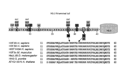
Figure 1 shows the most frequently identified mutations in pediatric GBM. (A)
Most frequent somatic
mutations in 48 pediatric glioblastoma tumors. * Mutations in bold and marked
with * appear to be
homozygous. Sample PGBM19 additionally has a DAXX mutation C629Sfs, while
PGBM21 has no
ATRX mutation but has the DAXX mutation shown. 4 Sample PGBM22 has a third
ATRX mutation,
p.D2136N, and a third NF1 mutation, p.A887T. Mutations identified in genes
listed in this table were
confirmed by Sanger sequencing, and were not present in dbSNP nor in the 1000
genomes dataset (Oct.
2011), except for the TP53 SNP at R273, which has been previously associated
with cancer. Detailed
description of the mutations in affected samples is provided in Table 5. (B)
Three recurrent non-
- 5 -
synonymous single nucleotide variants (SNVs) were observed in H3F3A. A
schematic representation of
the K27M, G34R and G34V mutations is shown in the context of the common post-
translational
modifications of the H3.3 N-terminal tail, which regulates the histone code
and histone functions.
Mammalian cells express three major types of non-centromeric histone H3
variants, H3.1, H3.2, and
H3.3. H3.3 has 136 amino acids, and is highly conserved across all eukaryotes.
The amino acid
sequence shows high conservation across species and from mammals to plants,
including the residues
subject to mutation in pediatric GBM (see multiple alignment of amino acids 11
to 60). (C) Schematic
representation of the missense mutations, frameshift deletions and stopgain
SNVs observed in ATRX in
the 48 whole exome sequencing WES samples. (D) Schematic of the overlap
between mutations
affecting ATRX-DAXX (observed in 15 samples), H3F3A (observed in 15 samples)
and TP53 (observed
in 26 samples). Eight samples had mutations in the three genes. (See multiple
alignment of amino acids
11 to 60, SEQ ID N0,14 for H. sapiens H3F3A, H. sapiens H3F3B, M. musculus
H3F3c and D.
melanogaster His3.3B; SEQ ID NO:15 for H. sapiens HIST1H3A, SEQ ID NO:16 for
S. pombe Hht3; SEQ
ID NO:17 for A. thaliana AT1G13370).
Figure 2 provides a correlation of ATRX mutation and lack of protein
expression in pediatric GBM
samples. Immunohistochemical staining of ATRX in samples analyzed by whole
exome sequencing
shows correlation between ATRX negative staining in tumor cells and presence
of an ATRX mutation.
ATRX is expressed in two samples with wild-type ATRX following whole exome
sequencing (A for sample
PGBM27 and B for PGBM26). ATRX is not expressed in PGBM14 (C) where mutations
in ATRX were
identified following whole exome sequencing.
Figure 3. (A) H3F3A mutations in a set of 784 gliomas from all ages and
grades. Sanger sequencing was
performed on DNA obtained from patients with low grade (I and II) and high
grade (III and IV) gliomas
from several countries in Europe and from Canada and shows that H3F3A
mutations are exclusive to
high grade tumors and the vast majority occur in glioblastoma (GBM) and in the
pediatric setting. 0:
oligodendroglioma, AO: anaplastic oligodendroglioma, OA: oligoastrocytoma,
AOA: anaplastic
oligoastrocytoma, A: diffuse astrocytoma grade II, AA: anaplastic astrocytoma.
PA; pilocytic astrocytoma.
(B) H3.3 mutations are specific to pediatric and young adult glioblastoma
(GBM). Schematic
representation of the occurrence of H3.3 mutations across age groups shows
that K27M mutations occur
mainly in younger patients (median age 11 years) and G34R/V mutations occur in
older children and
young adults (median age 20 years). No mutations were identified in older
patients with GBM. (C)
Comparison of the most frequently mutated genes in pediatric and adult GBM
shows that H3F3A, ATRX
and DAXX mutations are largely specific to pediatric disease. Except for
similarities in the mutation rate
for TP53 and PDGFRa with the previously identified proneural adult GBM
subgroup, the rate and type of
genes mutated were distinct between pediatric and adult GBM whatever the
molecular subgroup (Figure
4). (D) ATRX and DAXX immunohistochemical staining of a pediatric GBM tissue
microarray (TMA)
comprising 124 samples. View of the TMA slide and an example of a negative and
of a positive core at
high magnification to show specific nuclear staining (or lack thereof) for
DAXX and ATRX. No gender bias
for ATRX loss was observed. Overall survival and progression-free survival
were similar in patients with
CA 2854255 2017-11-20
- 5a -
and without loss of ATRX and/or DAXX (data not shown). (E) Differential
association of K27M and
G34RN H3F3A mutations with ATRX mutations. G34RN-H3.3 mutations were always
associated with
CA 2854255 2017-11-20
- 6 -
ATRX mutations, while a non significant overlap was observed for K27M (two-
sided Fisher's exact test,
p=0.0016).
Figure 4 shows that pediatric GBM is molecularly distinct from the four
molecular subgroups previously
identified in adult GBM. (A) Comparison of the mutation rate of the previously
identified most frequently
mutated genes in adult glioblastoma to pediatric glioblastoma shows that
pediatric GBM mutational profile
does not overlap with any of the previously described four molecular adult GBM
molecular subgroups.
Similarities to the pro-neural subgroup include TP53 and PDGFRa mutations,
however clear differences
exist for IDH mutations and NF1 mutations that occur at respectively a much
lower frequency and higher
frequency in children. (B) Bar graph showing percentage of sample with
mutations in function of genes.
For each gene, pediatric GBM tumors (bar on the right side) are compared to
adult GBM tumors (bar on
the left side). * indicates statistical significance.
Figure 5. (A) Lysine 36 is methylated in a G34V mutant (GBM 14). Cell lysates
from GBM14 (which
harbours G34V mutation) and from GBM24, a pediatric GBM cell line (SF188) and
normal human
astrocytes (NHA; all wild type for H3.3) were analyzed, with a Western blot,
with antibodies recognizing
the three methylated forms of K36 and the methylated form of K27. Even though
we cannot differentiate
H3.3K36me3 from global H3K36me3 levels, results indicate increased methylation
of K36 met me2 and
me3 in the sample carrying the G34V mutation. (B) Unsupervised hierarchical
clustering of 27 of the GBM
samples analyzed by whole exome sequencing shows that K27M and G34RN H3.3
mutants have
specific gene expression profiles. Gene expression profiles were generated on
Affymetrix U133Plus2.01-m
arrays. Clustering was based on the top 100 genes by standard deviation from
autosomal genes detected
as present in >10% of samples, and showed a clear distinction between K27M and
G34RN mutant
cases. (C) Genes involved in development and differentiation show H3.3
mutation-specific expression
patterns. Analysis for enrichment of Gene Ontology (GO) terms amongst the top
differentially-expressed
genes revealed 'Multicellular Organismal Development' (GO:0007275) to be the
most highly enriched
category (17/99 recognised gene IDs, p=0.01). Several of these show H3.3
mutation-specific expression
patterns. Results (log2 expression in function of K27 mutants, G34 mutants and
H3.3 wild-type) are
shown for the MYT1 gene (top left panel), SFRP2 gene (top right panel), FZD7
(lower left panel) and
DLX2 (lower right panel). * and ** indicates statistical significance. (D)
Alternate lengthening of telomere
is associated with the presence of mutant H3F3A/ATRX/P53 in pediatric GBM. We
assessed ALT using
two surrogate markers: Telomere-specific fluorescence in situ hybridization
(normal glia, left panel; ALT
negative, middle panel and ALT positive, right panel as well as in Figure 6)
and telomere-specific
Southern blotting of high molecular weight genomic DNA (Figure 7). Both
methods show ALT to be
associated with mutant H3F3A, ATRX and TP53. Representative images of ALT
positive and negative
staining of a pediatric GBM tissue microarray and a control brain are provided
(upper panel).
Figure 6. Comparison of frequently mutated genes indicates that pediatric GBM
is distinct from the four
previously identified molecular subgroups in adult GBM. (A) Comparison of gene
expression (associated
with various pathways) in pediatric GBM as well as in four subgroups of adult
GBM (proneural, neural,
classical and mesenchymal). Mutations in H3F3A, ATRX and DA)0( we identified
in
CA 2854255 2019-02-01
- 7 -
this study and show to be specific to pediatric GBM. IDH1 mutations have
already been previously shown
to be representative of a subgroup of adult GBM (proneural) and not pediatric
GBM. Mutations in
PDGFRA and TP53 had similar rates to adult proneural GBM while NF1 and RBI
mutations were more
similar to mesenchymal subgroup. (B) Bar graphs representative of the rate of
mutations for specific
genes (IDH1, EGFR, DGFRA, NF1, PIK3CA, PIK3R1, PTEN, TP53, RB1) between
pediatric GBM and
the proneural (top left graph), mesenchymal (lower left graph), neural (top
right graph) and classical
(lower right graph) adult GBM. For each gene in each graph, mutation rate of
pediatric GBM is shown as
the left bar while the mutation rate for the adult GBM is shown as the right
bar. * indicates statistically
significance as measured by the Fisher t-test (p<0.05).
Figure 7. (A) Overlap of TP53, ATRX and H3F3A mutations and ALT in tumor
cells. Samples with
H3F3A/ATRX/TP53 mutations determined by whole exome sequencing and for which
fixed material was
available were subjected to immunohistochemical staining for ATRX (left
panels) and p53 (middle
panels). Lack of ATRX expression was present across the vast majority of tumor
cells. Aberrant p53
staining was present in more than 80% of cells lacking ATRX expression, both
in K27M and G34R H3.3
mutant samples. This indicates that at least 30% of tumor cells have
concomitant mutations in these three
genes. Representative staining of two samples (GBM4 and GBM14) is shown.
Telomere-specific
fluorescence (left panels) in situ hybridization indicates the presence of ALT
in tumors cells. (B) Overlap
of absent DAXX and ATRX protein expression in tumor cells in a sample. Six
samples showed
concomitant lack of ATRX (right panels) and DAXX (left panels) expression
following
immunohistochemical staining of the pediatric GBM the tissue microarray (as
well as Figure 3D). These
samples stained positively for other markers (GFAP) showing that lack of
staining was not due to tissue
processing (data not shown).
Figure 8. Alternative Lengthening of Telomeres (ALT) is associated with
ATRX/H3F3/TP53 mutations.
Previous groups have shown measurement of telomerase expression or activity
not to be reliable to
assess ALT. We assessed ALT using two surrogate markers: telomere-specific
fluorescence in situ
hybridization (Figure 5D and Figure 7). In this figure, telomere-specific
Southern blotting of high molecular
weight genomic DNA is shown for various samples. Telomere Restriction Length
(TRF) assay was used
to assess the presence of ALT in 32 pediatric GBM. Tumors were blotted
according to their TP53
mutations, H3F3A mutations and ATRX mutations. Tumors demonstrating ALT are
marked as A in red.
ALT tumors demonstrate abnormally long telomeres (>21 kb in length) which are
not seen in telomerase
positive tumors or normal tissues. Most ALT tumors had concomitant TP53
mutations and there was
significant enrichment for ALT among ATRX/H3F3 mutant tumors (p=0.003).
Overall ALT tumors were
strongly associated with ATRX/H3F3/TP53 mutations (p=0.0002, Fisher's exact
test). These data suggest
that TP53 mutations are typically necessary but require additional alterations
in chromatin modulating
genes for ALT formation.
Figure 9. Single nucleotide polymorphism (SNP) array profiling reveals
differences in copy number
aberrations (CNAs) in ATRX/DNOUH3F3A-mutated pediatric glioblastoma. Focal
losses or gains
comprising genes relevant in pediatric GBM overlapped with previous reports.
Samples were split into a
CA 2854255 2019-02-01
CA 02854255 2014-05-01
WO 2013/075237 PCT/CA2012/050834
- 8 -
group with relatively stable genomes (<10 CNAs) and a group with more unstable
genomes (>=10 CNAs).
Results are shown as the number of tumors in function of genome stability and
mutations at ATRX, DAXX
and H3F3A. Samples with a mutation in at least one of ATRX, TP53 and H3F3A
were significantly
associated with an unstable genome (p=0.0207, Fisher's exact test).
Figure 10. Comparison of the most frequent focal amplification and deletion in
genes involved in
glioblastoma shows major differences between adult GBM and pediatric GBM. (A)
Results are shown as
percent of samples with focal amplification (upward arrow) or percent of
samples with homozygous
deletions (downward arrow) for the CDKN2A, MDM2, TP53, CDK4, CDK6, RB1, EGFR,
PDGFRA, NF1,
PIK3Ca, PIK3R1 or the PTEN genes for adult GBM samples (left bar) and
pediactric GBM (right bar). *
indicates statistical significance. (B) Table associated with Figure 10A
listing, for each gene, the type of
mutation (DEL = deletion, AMP = amplification), the number of adult GBM
samples bearing the mutation
in function of the total number of samples of adult GBM (#aGBM/n), the
percentage of adult GBM bearing
the mutation (%aGBM), the number of pediatric GBM samples bearing the mutation
in function of the total
number of samples of pediatric GBM (#pGBM/n), the percentage of pediatric GBM
bearing the mutation
(%pGBM) and the p-value associated with the comparison between adult and
pediatric GBM (p-value).
Figure 11. Representative high-resolution melting curves for the
identification of H3.3 mutants bearing a
mutation at K27M or 334R. Results are shown as relative signal intensity in
function of temperature.
Figure 12. Norton blot, using a K27M H3.3 probe, of cell culture supernatant
of cell lines ( a mutated
(K27MGBM cell line) or wild-type (H3.3 WT GBM cell line) and of plasma of
patient (K27M plasma
patient) or of a control individual (plasma from normal control).
Figure 13. Western blot of histone extracts of cells lines expressing wild
type H3.3 (SF-188 EV), K27M
H3.3 (SF-188 Myc(K27M)) or G34R H3.3 (SF-188 Myc(G34R)). Top panel shows
results obtained with a
monoclonal antibody recognizing the wild-type H3.3 polypeptide. Middle panel
shows results obtained
with a monoclonal antibody specific for the K27M H3.3 polypeptide. Lower panel
shows results obtained
with a monoclonal antibody specific for the Myc polypeptide.
Figure 14. Evidence that oncogenic transcripts can be extracted and enriched
from minimal amounts of
biological material containing tumour extravesicles (EVs/oncosomes). Detection
of BRAF/KIAA in plasma
of juvenile pilocytic astrocytoma patients by nested RT-PCR. The material was
extracted from either
unfractionated plasma or from the EV fraction of each sample. Starting
material was 250 pL for all lanes.
Figure 15. (A) Graphical representation of Epigenetic and Biological Subgroups
of glioblastoma
reviewing, per mutated gene, the DNA methylation pattern, the gene expression,
the IHC protein marker,
the age distribution, the tumor location as well as the patient survival (in
months). (B) Neuroatonomical
and age specificity of IDH, H3.3-K27M and G34R in the brain GBM. K27M occurs
mainly in the
brainstem and the thalamus (70%-80% of all GBM in these locations). It is
inconsistently associated with
ATRX mutations. G34V-R occurs mainly in the cerebral hemispheres similar to
IDH mutations that have a
predilection for the frontal cortex. Both are strongly and significantly
associated with ATRX mutations.
- 9 -
SETD2 mutations are in the brain hemispheres and partly overlap with IDH
mutations in a sample. The
size of the shape illustrating each mutation is proportional to the %
identified in our studies. (C)
Cumulative survival (%) in function of overall survival (years) for patients
expressing wilt-type H3.3 or
K27M H3.3 polypeptide (p=0.027). Thalamic and pontine high grade gliomas
carrying K27MH3.3
mutations have universal rapid poor outcome.
Figure 16. Methylation profiling reveals the existence of six epigenetic GBM
subgroups. Heatmap of
methylation levels in six GBM subgroups identified by unsupervised k-means
consensus clustering, and
control samples as indicated
Figure 17. Whole genome bisulphite sequencing (WGBS) compared to IIlumina 450K
Human Methylation
array data. A region in chromosome 10 showing classifying difference in
methylation (from 450K
unsupervised clustering analyses) between major mutation types is highlighted.
We carried out
methylation quantitation from WGBS in IDH1 (top track) and H3.3 G34R (second
track from top) mutation
carrying tumors in parallel with 450K assay (3rd and 4th track). Despite low
coverage (3-4x genome-wide
in this pilot experiment) the correlation between 450K and WGBS IS HIGH at
identical CpGs. Advantages
offered by WGBS are highlighted at four putative regulatory elements (in blue
for promoter distal
elements, and in pink for promoter associated elements) for SKID1 gene. A
highly significant difference
between tumor types was observed for only 2 CpG sites in 450K analyses
(promoter associated region,
highlighted by arrows), whereas WGBS shows complete hypo vs. hypomethylation
between H3.3 G34R
and IDH1 harboring tumors at multiple regulatory regions showing active
enhancer/promoter mark
(H3K27ac) and DNasel hypersensitivity in ENCODE cell lines. The high
resolution and coverage of
WGBS allows "indexing" of putative regulatory elements differing between
tumors supporting integrative
analyses of regulatory differences and gene networks perturbed by pediatric
HGA associated mutations.
Figure 18. Tumors arising in the brain of K27MH3.3 injected mice (SB model)
monitored using the built in
luciferase (green indicative of increased cellular activity) indicative that
K27MH3.3 is oncogenic by itself
and promotes tumor formation.
DETAILED DESCRIPTION
Definitions
Throughout this application, various terms are used and some of them are more
precisely defined herein.
Agonist. This term, as used herein, refers to an agent that mimics or
upregulates (e.g., increases,
potentiates or supplements) the expression and/or activity of a wild-type H3.3
protein (having the amino
acid sequence as set forth in SEQ ID NO: 5). An agonist can be the wild-type
protein itself and/or a
nucleic acid molecule encoding the wild-type protein. An agonist can also be a
compound that
upregulates expression of a wild-type h3.3 gene or which increases at least
one activity of a wild-type
H3.3 protein. An agonist can also be a compound which increases the biological
activity of the wild-type
H3.3 protein via direct interaction, e.g. a binding partner.
CA 2854255 2019-02-01
CA 02854255 2014-05-01
WO 2013/075237 PCT/CA2012/050834
- 10 -
Biological sample. A biological sample is a sample of an individual's bodily
fluid, cells or tissues. The
biological sample comprises either a H3.3 polypeptide and/or a polynucleotide
encoding the H3.3
polypeptide. In this present application, the biological sample can be derived
from a tumor tissue and may
even comprise tumor cells. Alternatively or in combination, the biological
sample can be derived from the
individual's bodily fluid (such as blood, for example plasma or cerebrospinal
fluid). In an embodiment, the
biological sample comprises a cell having a H3.3 polypeptide and/or a
polynucleotide encoding the H3.3
polypeptide. In another embodiment, the biological sample is a cell-free
DNA/RNA sample having a H3.3
a polynucleotide encoding the H3.3 polypeptide. The biological sample can be
used without prior
modification in the various methods described herein. Optionally, the
biological sample can be treated
(mechanically, enzymatically, etc.) prior to the assays described herein. In
one embodiment, the
microvesicles from the biological sample are obtained and used in the assays
described herein.
Exemplary methods for obtaining microvesicles are described in WO 2012/051622.
H3.3. The H3.3 polypeptide (also referred to Histone 3) is a regulator of
chromatin configuration and is
encoded by the H3F3A gene. The GenBank accession number of the human mRNA
sequence of this
polypeptide is NM_002107 The GenBank accession number of the human polypeptide
sequence of this
protein is NP_002098. It is worth noting that the protein is post-
translationally modified to remove the first
methionine residue presented in the GenBank listing. There are at least two
copies of the H3F3A gene in
the human genome, which differ in their nucleotide sequence but produce
proteins with the identical
amino acid sequence. As known in the art, histone H3 is one of the five main
histone proteins involved in
the structure of chromatin in eukaryotic cells. Featuring a main globular
domain and a long N-terminal tail,
H3 is involved with the structure of the nucleosomes of the "beads on a
string" structure. Histone H3 is
the most extensively modified of the five known histones. The N-terminal tail
of histone H3 protrudes from
the globular nucleosome core and can undergo several different types of post-
translational modification
that influence cellular processes. These modifications include the covalent
attachment of methyl or acetyl
groups to lysine and arginine amino acids and the phosphorylation of serine or
threonine.
Pharmaceutically effective amount or therapeutically effective amount. These
expressions refer to an
amount (dose) effective in mediating a therapeutic benefit to a patient (for
example prevention, treatment
and/or alleviation of symptoms of a proliferation associated disorder). It is
also to be understood herein
that a "pharmaceutically effective amount" may be interpreted as an amount
giving a desired therapeutic
effect, either taken in one dose or in any dosage or route, taken alone or in
combination with other
therapeutic agents.
Pharmaceutically acceptable salt. This expression refers to conventional acid-
addition salts or base-
addition salts that retain the biological effectiveness and properties of the
therapeutic agent described
herein. They are formed from suitable non-toxic organic or inorganic acids or
organic or inorganic bases.
Sample acid-addition salts include those derived from inorganic acids such as
hydrochloric acid,
hydrobromic acid, hydroiodic acid, sulfuric acid, sulfamic acid, phosphoric
acid and nitric acid, and those
derived from organic acids such as p-toluenesulfonic acid, salicylic acid,
methanesulfonic acid, oxalic
acid, succinic acid, citric acid, malic acid, lactic acid, fumaric acid, and
the like. Sample base-addition
CA 02854255 2014-05-01
WO 2013/075237 PCT/CA2012/050834
- 1 1 -
salts include those derived from ammonium, potassium, sodium and, quaternary
ammonium hydroxides,
such as e.g., tetramethylammonium hydroxide. The chemical modification of an
agent into a salt is a well
known technique which is used in attempting to improve properties involving
physical or chemical
stability, e.g., hygroscopicity, flowability or solubility of compounds.
Prevention, treatment and alleviation of symptoms. These expressions refer to
the ability of a method or
an agent to limit the development, progression and/or symptomology of a
proliferation-associated
disorders. Broadly, the prevention, treatment and/or alleviation of symptoms
can encompass the
reduction of proliferation of the cells (e.g. by reducing the total number of
cells in an hyperproliferative
state and/or by reducing the pace of proliferation of cells). Symptoms
associated with proliferation-
associated disorder include, but are not limited to: local symptoms which are
associated with the site of
the primary cancer (such as lumps or swelling (tumor), hemorrhage, ulceration
and pain), metastatic
symptoms which are associated to the spread of cancer to other locations in
the body.(such as enlarged
lymph nodes, hepatomegaly, splenomegaly, pain, fracture of affected bones, and
neurological
symptoms), and systemic symptoms (such as weight loss, fatigue, excessive
sweating, anemia and
paraneoplastic phenomena).
Proliferation-associated disorders. These disorders form a class of diseases
where cells proliferate more
rapidly, and usually not in an ordered fashion. The proliferation of cells
cause an hyperproliferative state
that may lead to biological dysfunctions, such as the formation of tumors
(malignant or benign). One of
the proliferation-associated disorder is cancer. Also known medically as a
malignant neoplasm, cancer is
a term for a large group of different diseases, all involving unregulated cell
growth. In cancer, cells divide
and grow uncontrollably, forming malignant tumors, and invade nearby parts of
the body. The cancer may
also spread to more distant parts of the body through the lymphatic system or
bloodstream. In an
embodiment, the cancer is a glioma (e.g. a cancer of the gial cells located in
the brain or spine). In
another embodiment, the cancer is associated with the involvement of
isocitrate dehydrogenase or IDH
(such as, for example, breast cancer, acute myeloid leukemia, chronic myeloid
leukemia). IDH mutations
occur in a variety of cancers of the central nervous system: adult low grade
gliomas, secondary
glioblastoma as well as oligodendrogliomas. IDH mutations also occur in acute
myeloid leukemia,
myelodysplastic syndroms and myeloproliferative neoplasms, gliomas and, at
lower frequencies, in
prostate cancer, acute lymphoblastic leukemia and breast cancer.
Reaction vessel. The reaction vessel is a discrete unit where a biological
sample comprising a H3.3-
based reagent (H3.3 polypeptide or polynucleotide encoding same) is placed.
The reaction vessel also
includes the discrete unit where the agent is combined with the H3.3-based
reagent, and it can be an in
vitro or in vivo environment. Suitable in vitro environments can include, for
example, a cell-free
environment where a H3.3-based reagent is combined in a reaction media
comprising the appropriate
reagents to enable the assessment of the parameter associated with H3.3 to be
monitored.
CA 02854255 2014-05-01
WO 2013/075237 PCT/CA2012/050834
- 12 -
Whole exome sequencing. This sequencing technique refers to the determination
of nucleotide
sequences of exons in individuals. As shown below, this technique was
successfully used to identify
variations in the H3.3 amino acid sequence that were associated with a
proliferation-associated disorder.
H3.3 non-conservative variants
The present application provides novel non-conservative variants of H3.3 whose
expression is associated
with proliferative disorder. More specifically, the non-conservative H3.3
variants are expressed in cells
and tissues afflicted by the proliferative disorder. The non-conservative
variants are distinct in at least one
of two positions when compared to the wild-type H3.3 protein (whose sequence
is presented in SEQ ID
NO: 5). Some of specific non-conservative variants presented herewith are
encoded at the following
chromosomic regions: chr1:226252135, chr1:226252155 and chr1:226252156.
A first non-conservative H3.3 variant (e.g. SEQ ID NO: 6) concerns a
polypeptide having a residue
different from lysine at a location corresponding to position 27 of the wild-
type H3.3. It is known that the
lysine at position 27 of the wild-type H3.3 protein is capable of being
methylated. This first non-
conservative H3.3 variant is preferably not being capable of being methylated
at position 27.
Consequently, the non-conservative H3.3 variant preferably does not bear a
lysine or an arginine residue
at position 27. Such non-conservative H3.3 variant can bear any other
naturally occurring amino acid, and
preferably a methionine residue (SEQ ID NO: 1) at position 27. In an
embodiment, the first non-
conservative H3.3 variant has or comprise the amino acid sequence of SEQ ID
NO: 6 or SEQ ID NO: 1.
In another embodiment, the first non-conservative H3.3 variant consists of the
amino acid sequence of
SEQ ID NO: 6 or SEQ ID NO: 1.
A second non-conservative H3.3 variant (e.g. SEQ ID NO: 7) concerns a
polypeptide having a residue
different from glycine at a location corresponding to position 34 of the wild-
type H3.3. It is thought that the
glycine at position 34 of the wild-type H3.3 protein does not interfere with
the methylation of the lysine
residue located at position 36. This second non-conservative H3.3 variant is
preferably capable of
interfering with the methylation of the lysine residue at position 34. Such
non-conservative H3.3 variant
can bear any naturally occurring amino acid other than a glycine, and,
preferably, an arginine or valine
residue (SEQ ID NO: 2 or SEQ ID NO: 3). In an embodiment, the second non-
conservative H3.3 variant
has or comprise the amino acid sequence of SEQ ID NO: 7, SEQ ID NO: 2 or SEQ
ID NO: 3. In another
embodiment, the second non-conservative H3.3 variant consists of the amino
acid sequence of SEQ ID
NO: 7, SEQ ID NO: 2 or SEQ ID NO: 3.
A third non-conservative H3.3 variant (e.g. SEQ ID NO: 8) concerns a
polypeptide having a residue
different from lysine at a location corresponding to position 27 of the wild-
type H3.3 and a residue
different from glycine at a location corresponding to position 34 of the wild-
type H3.3. This third non-
conservative variant preferably does not bear an arginine residue at position
26. An exemplary sequence
of the third non-conservative variants bears a methionine residue at position
27 and an arginine or a
valine residue at position 34 (as shown in SEQ ID NO: 4). In an embodiment,
the third non-conservative
CA 02854255 2014-05-01
WO 2013/075237 PCT/CA2012/050834
- 13 -
H3.3 variant has or comprise the amino acid sequence of SEQ ID NO: 8 or SEQ ID
NO: 4. In another
embodiment, the third non-conservative H3.3 variant consists of the amino acid
sequence of SEQ ID NO:
8 or SEQ ID NO: 4.
The present application also provides fragments of the non-conservative H3.3
variants described herein.
These fragments contain less amino acids than the wild-type H3.3 but are
recognized specifically by
antibodies which fail to recognize the wild-type H3.3. In an embodiment, these
fragments bear epitopes
corresponding to positions 27 and/or 34 that are specifically recognized by
antibodies which fail to
recognize wild-type H3.3. These fragments encompass at least amino acid
residues corresponding to
positions 27 to 34. In an embodiment, the fragments are recognized by
antibodies specific for SEQ ID
.. NO: 6, SEQ ID NO: 7 or SEQ ID NO: 8 and that fail to specifically recognize
the polypeptide presented in
SEQ ID NO: 5. In another embodiment, the fragments are recognized by
antibodies specific for SEQ ID
NO: 1, SEQ ID NO: 2, SEQ ID NO: 3 or SEQ ID NO: 4 and that fail to
specifically recognize the
polypeptide presented in SEQ ID NO: 5.
Nucleic acid polynucleotide molecules are also contemplated herein and encodes
the H3.3 non-
conservative variants may be derived from a variety of sources including DNA,
cDNA, synthetic DNA,
synthetic RNA, derivatives, mimetics or combinations thereof. Such sequences
may comprise genomic
DNA, which may or may not include naturally occurring introns, genic regions,
nongenic regions, and
regulatory regions. Moreover, such genomic DNA may be obtained in association
with promoter regions
or poly (A) sequences. The sequences, genomic DNA, or cDNA may be obtained in
any of several ways.
Genomic DNA can be extracted and purified from suitable cells by means well
known in the art.
Alternatively, mRNA can be isolated from a cell and used to produce cDNA by
reverse transcription or
other means. The nucleic acids described herein are used in certain
embodiments of the methods of the
present invention for production of RNA, proteins or polypeptides, through
incorporation into host cells,
tissues, or organisms. In one embodiment, DNA containing all or part of the
coding sequence for the H3.3
variant polypeptides are incorporated into vectors for expression of the
encoded polypeptide in suitable
host cells.
Antibodies (as well as antigen-binging fragment thereof) specific for the H3.3
non-conservative variants
are also contemplated.
Predictive methods and associated commercial packages
The diagnostic and prognostic methods described herein are designed to capture
the relationship
between H3.3's amino acid identity (at specific positions) and proliferation-
associated disorders to
generate valuable information about the individual that is being tested. Once
an individual has been
diagnosed by one of the methods described herein, this individual can be
treated according to the
therapeutic regimen that is considered useful depending on its disease status.
In the diagnostic and prognostic applications, a biological sample is first
provided from the subject that is
being tested. In an embodiment, the biological sample comprises a subject's
own cell. In another
CA 02854255 2014-05-01
WO 2013/075237 PCT/CA2012/050834
- 14 -
embodiment, the biological sample is cell-free DNA (cfDNA), which is thought
to be released from dying
cells, as DNA fragments or nucleosomes. CfDNA often maintains sequence and
methylation
characteristics of the parental cancer cells. Alternatively, oncogenic DNA,
mRNA, miRNA and proteins
may become a cargo of extracellular vesicles (EVs), which originate from
viable cells as a product of
exosome biogenesis or membrane blebbing. It was previously reported that
oncogene containing EVs
(oncosomes) are released at a high rate from glioma cells in vivo. These
oncosomes may contain intact
oncogenic EGFRvIll, and we showed it can exert biological effects upon
intercellular transfer and similar
observations were reported for other transforming protein, mRNA and DNA
species. It is possible that
what is known as cfDNA is also, at least in part, contained in oncosomes, as
suggested by detection of c-
MYC and G12V-H-Ras sequences in this material. EVs protect this material from
degradation and
preserve it in blood, thereby allowing remote access to the mutational status,
functional state and identity
of the cells of origin. Enrichment of this material in the EV compartment, as
compared to total plasma, can
offer an opportunity to increase the sensitivity of detection and multiplexing
capacity.
In diagnostic and prognostic applications, a biological sample of an
individual is placed in a reaction
vessel. The biological sample comprises an H3.3 polypeptide (e.g. either an
H3.3-encoding nucleic acid
molecule and/or an H3.3 polypeptide). In the assays, the reaction vessel can
be any type of container that
can accommodate the determination of the nucleic/amino acid identity of the
H3.3 polypeptide.
Once the biological sample has been placed in the reaction vessel, the amino
acid sequence identity of
the H3.3 polypeptide and/or the nucleic acid sequence identity of the H3.3-
encoding nucleic acid
molecule is determined. This assessment may be made directly in the reaction
vessel (by using a probe)
or on a sample of such reaction vessel. The determination of the sequence
identity of the H3.3
polypeptide (either directly or via the H3.3-encoding molecule nucleic acid
molecule) can be made either
at the DNA level, the RNA level and/or the polypeptide level.
It is not necessary to determine the sequence identity of the complete H3.3
polypeptide and/or H3.3-
encoding nucleic acid molecule. In the methods presented herein, it is
important to determine the
sequence identity of the H3.3 polypeptide and/or H3.3-encoding nucleic acid
molecule in at least one of
two positions. The first position corresponds to the amino acid residue 27 of
the wild-type H3.3 protein
(e.g. SEQ ID NO: 5). At this first position, the wild-type H3.3 protein
presents a lysine residue. The
second position corresponds to the amino acid residue 34 of the wild-type H3.3
protein (e.g. SEQ ID NO:
5). At this second position, the wild-type H3.3 protein presents a glycine
residue. In an embodiment, the
sequence identity is determined at one of the two positions (e.g. residue 27
or 34). In another
embodiment, the sequence identity is determined at both positions (e.g.
residue 27 and 34). In yet a
further embodiment, the sequence identity can be first determined at one of
the two positions (e.g.
residues 27 or 34) and then determined at the other position (e.g. residues 27
or 34). As it will be
appreciated by those skilled in the art, the determination of sequence
identity can be made at the nucleic
acid level and/or at the polypeptide level.
-15-
The determination step can rely on the addition of a qualifier specific to the
sequence to be determined.
The qualifier can, for example, specifically bind to a sequence or subsequence
of amino acids of the H3.3
protein. In those instances, the association between the qualifier and the
H3.3 protein can be used to
provide the sequence identity of the H3.3 protein. For example, the qualifier
can be an antibody or a
fragment thereof capable of specifically recognizing a methionine at a
position corresponding to residue
27 of the H3.3 protein. If a methionine residue is present at position
corresponding to residue 27, the
antibody will specifically bind to the H3.3 polypeptide in the reaction vessel
and this association will
indicate the presence of a methionine at a position corresponding to residue
27 in the H3.3 protein. In
another embodiment, the qualifier can be an antibody or a fragment thereof
that can specifically
recognize a lysine residue at a position corresponding to residue 27 of the
H3.3 protein. In such
embodiment, specific binding between the antibody and the H3.3-based reagent
indicates that the
polypeptide bears a lysine residue at a position corresponding to residue 27.
However, the absence of
binding between this lysine-specific antibody and the H3.3 protein (in
conditions where binding between
the anti-lysine antibody and its cognate ligand would otherwise be observed),
indicates that the H3.3
protein does not bear, at position 27, a lysine residue.
If the measurement of the parameter is performed at the nucleotide level, then
the nucleic acid sequence
of the H3.3 gene, transcript (e.g. mRNA) or corresponding cDNA can be
assessed. Various methods of
determining the nucleic acid sequence of a nucleic acid molecule are known to
those skilled in the art and
include, but are not limited to, chemical sequencing (e.g. Maxam¨Gilbert
sequencing), chain termination
.. methods (e.g. Sanger sequencing, and dye-terminator sequencing),
restriction digestion-based
sequencing (e.g. RFLP), hybridization-based sequencing (e.g. DNA micro-array,
RNA micro array,
Molecular Beacon probes, TaqManTm probes), mass spectrometry-based sequencing,
next generation
sequencing (e.g. Whole exome sequencing, Massively Parallel Signature
Sequencing or MPSS, Polony
sequencing, pyrosequencing, llluminaTM (Solexa) sequencing, SOLiDTM
sequencing, ion semiconductor
sequencing, DNA nanoball sequencing, HelioscopeTM single molecule sequencing,
Single Molecule
SMRTTm sequencing, Single Molecule real time (RNAP) sequencing, and Nanopore
DNA sequencing). As
indicated above, it is not necessary to sequence the complete nucleic acid
molecule encoding the H3.3
protein, only the nucleic acid identity of the bases encoding the amino acid
at a position corresponding to
residues 27 and/or 34 is required.
If the measurement of the parameter is performed at the polypeptide level, an
assessment of the amino
acid identity of the H3.3 level of expression can be performed. In an
embodiment, this determination can
be done through an antibody-based technique (such as an Western Blot, FACS or
an ELISA), a micro-
array, mass spectrometry, protein sequencing, etc.
In addition, an assessment of H3.3 biological activity can be performed as an
indirect indicator of amino
acid sequence identity at positions corresponding to residues 27 and/or 34.
H3.3 is a histone protein and
its post-translational modifications influences its biological activity (e.g.
regulation of gene expression).
For example, in native wild-type H3.3 polypeptide, the lysine at position 27
can be methylated and, in
return, this methylation modifies the biological activity of H3.3 (e.g. favors
a closed chromatin
CA 2854255 2019-02-01
CA 02854255 2014-05-01
WO 2013/075237 PCT/CA2012/050834
- 16 -
configuration) and ultimately limits and/or lowers gene expression. In an H3.3
polypeptide bearing, at
position 27 a residue different from a lysine (for example a methionine),
methylation may be limited or
reduced and as such H3.3 is not able of mediating its activity of limiting or
shutting off gene expression. In
another example, in native wild-type H3.3 polypeptide, the relatively small
and uncharged glycine residue
at position 34 does not prevent the recognition of the modified lysine at
position 36 and, as such, favors a
closed chromatin configuration. However, the presence of a relatively bulky
arginine or a charged valine
residue at position 34 probably prevents the recognition of the modified
lysine at position 36 and limits the
ability of H3.3 to mediate its biological activity. As such, it is also
possible to determine the amino acid
identity of the H3.3 protein by either measuring or determining the presence
or absence of post-
transcriptional modifications (such as methylation, citrullination,
acetylation, phosphorylation,
SUMOylation, ubiquitination, and/or ADP-ribosylation) at specific residues. It
is also possible to determine
the amino acid identity of the H3.3 protein by measuring the ability of the
H3.3-based reagent to limit or
suppress gene expression.
As known in the art, H3.3 forms a complex with the ATRX and DAXX proteins and
such complex
localizes H3.3 in the pericentric heterochromatin and telomeres regions. As
shown herein, the mutations
associated with H3.3 are associated with a lower expression of ATRX and DAXX
as well as the absence
of the complex when measured by histochemistry. Consequently, in a further
assay format, H3.3's
biological activity can be indirectly measured by quantifying the expression
of ATRX and/or DAXX or by
determining the presence or absence of the ATRX-DAXX-H3.3 complex in tumor
tissues.
The interaction between more than one molecule can also be detected, e.g.,
using a fluorescence assay
in which at least one molecule is fluorescently labeled. One example of such
an assay includes
fluorescence energy transfer (FET or FRET for fluorescence resonance energy
transfer). A fluorophore
label on the first "donor" molecule is selected such that its emitted
fluorescent energy will be absorbed by
a fluorescent label on a second "acceptor" molecule, which in turn is able to
fluoresce due to the
absorbed energy. Alternately, the "donor" protein molecule may simply utilize
the natural fluorescent
energy of tryptophan residues. Labels are chosen that emit different
wavelengths of light, such that the
"acceptor" molecule label may be differentiated from that of the "donor".
Since the efficiency of energy
transfer between the labels is related to the distance separating the
molecules, the spatial relationship
between the molecules can be assessed. In a situation in which binding occurs
between the molecules,
the fluorescent emission of the "acceptor" molecule label in the assay should
be maximal. A FET binding
event can be conveniently measured through standard fluorometric detection
means well known in the art
(e.g., using a fluorimeter).
Another example of a fluorescence assay is fluorescence polarization (FP). For
FP, only one component
needs to be labeled. A binding interaction is detected by a change in
molecular size of the labeled
component. The size change alters the tumbling rate of the component in
solution and is detected as a
change in FP.
- 17 -
In another embodiment, the measuring step can rely on the use of real-time
Biomolecular Interaction
Analysis (BIA). "Surface plasmon resonance" or "BIA" detects biospecific
interactions in real time, without
labeling any of the interactants (e.g., BlAcore Tm). Changes in the mass at
the binding surface (indicative
of a binding event) result in alterations of the refractive index of light
near the surface (the optical
phenomenon of surface plasmon resonance (SPR)), resulting in a detectable
signal which can be used as
an indication of real-time reactions between biological molecules.
In another assay format, H3.3's biological activity can be indirectly measured
by quantifying the
expression levels of its target genes whose expression is modulated by the
presence and activity of the
variant H3.3. Such genes can be, for example, those listed in Table 7.
Once the sequence identity has been determined, the information is extracted
from the reaction vessel is
compared to residues at corresponding positions in a control sequence (e.g.
wild-type H3.3 or SEQ ID
NO: 5). In diagnostic and prognostic application, it must be determined if the
sequence of the H3.3 protein
is identical or different from the wild-type sequence (e.g. SEQ ID NO: 5) at
positions corresponding to
residues 27 and/or 34. The presence of a discrepancy between the sequenced
H3.3 protein and the wild-
type sequence at positions corresponding to residues 27 and/or 34 is
associated with a poor disease
status. For example, if in the sample, it is determined that the residue
corresponding to position 27 of the
wild-type H3.3 is a methionine, then the comparison indicates that the tested
H3.3 is different from the
wild-type H3.3 (e.g. that the residue is not a lysine) and the individual is
characterized as being
associated with a poor disease status. In another example, if in the sample,
it is determined that the
residue corresponding to position 34 of the wild-type H3.3 is a an arginine or
a valine, then the
comparison indicates that the tested H3.3 is different from the wild-type H3.3
(e.g. that the residue is not
a glycine) and the individual is characterized as being associated with a poor
disease status.
In an embodiment, the comparison can be made by an individual. In another
embodiment, the
comparison can be made in a comparison module. Such comparison module may
comprise a processor
and a memory card to perform an application. The processor may access the
memory to retrieve data.
The processor may be any device that can perform operations on data. Examples
are a central
processing unit (CPU), a front-end processor, a microprocessor, a graphics
processing unit (PPU/VPU), a
physics processing unit (PPU), a digital signal processor and a network
processor. The application is
coupled to the processor and configured to determine the presence or absence
of a discrepancy between
the sequence of tested H3.3 with respect to sequence of the wild-type H3.3. An
output of this comparison
may be transmitted to a display device. The memory, accessible by the
processor, receives and stores
data, such as sequence identity of the H3.3 protein (either directly or the
encoded H3.3 from the nucleic
acid molecule) or any other information generated or used. The memory may be a
main memory (such as
a high speed Random Access Memory or RAM) or an auxiliary storage unit (such
as a hard disk, a floppy
disk or a magnetic tape drive). The memory may be any other type of memory
(such as a Read-Only
Memory or ROM) or optical storage media (such as a videodisc or a compact
disc).
CA 2854255 2019-02-01
CA 02854255 2014-05-01
WO 2013/075237 PCT/CA2012/050834
- 18 -
Once the comparison between the sequence of the tested H3.3 protein and the
wild-type H3.3 is made,
then it is possible to characterize the individual. This characterization is
possible because, as shown
herein, mutations of H3.3 at positions corresponding to residues 27 and/or 34
are associated with a poor
disease status.
In an embodiment, the characterization can be made by an individual. In
another embodiment, the
characterization can be made with a processor and a memory card to perform an
application. The
processor may access the memory to retrieve data. The processor may be any
device that can perform
operations on data. Examples are a central processing unit (CPU), a front-end
processor, a
microprocessor, a graphics processing unit (PPU/VPU), a physics processing
unit (PPU), a digital signal
processor and a network processor. The application is coupled to the processor
and configured to
characterize the individual being tested. An output of this characterization
may be transmitted to a display
device. The memory, accessible by the processor, receives and stores data,
such as sequences of the
tested H3.3 or any other information generated or used (such as the sequence
identity of the wild-type
H3.3). The memory may be a main memory (such as a high speed Random Access
Memory or RAM) or
an auxiliary storage unit (such as a hard disk, a floppy disk or a magnetic
tape drive). The memory may
be any other type of memory (such as a Read-Only Memory or ROM) or optical
storage media (such as a
videodisc or a compact disc).
The methods described herein are useful for determine the predisposition of an
individual to a
proliferation-associated disorder. As shown herein, the presence of variations
at positions 27 and/or 34 of
the H3.3 protein are associated with a population of individuals afflicted
with a proliferation-associated
disorder (e.g. cancer). As such, the determination of amino acid variations at
positions 27 and/or 34 can
be useful in predicting the likelihood of disease in tested individuals.
The methods presented herein can also be useful for diagnosing a proliferation-
associated disorder in an
individual. As shown herein, the presence of variations at positions 27 and/or
34 are associated with
disease tissue of a population of individuals afflicted with a proliferation-
associated disorder (e.g. cancer).
As further shown herein, even the variation occur within the afflicted tissue,
it is possible to detect it in the
blood stream of afflicted individuals. As such, the presence of amino acid
residues variations at a location
corresponding to positions 27 and/or 34 of the wild-type H3.3 can be useful in
determining the presence
or absence of the proliferation-associated disease in tested individuals.
.. The methods presented herein can also be useful in classifying individuals
already diagnosed with a
proliferation-associated disorder. As shown herein, the presence of variations
at positions 27 and/or 34
are associated with the most aggressive forms of diseases (e.g. Grade III and
Grade IV cancers). As
such, the determination of amino acid variations at positions 27 and/or 34 can
be useful in determining
the grade of the disease and, optionally, this information can be used to
optimize the therapeutic regimen.
The methods presented herein can also be useful in determining the re-
occurrence of a proliferation-
associated disorder in individuals previously diagnosed (and, optionally
treated) with the disorder. As
CA 02854255 2014-05-01
WO 2013/075237 PCT/CA2012/050834
- 19 -
shown herein, the presence of variations at positions 27 and/or 34 are
associated with the presence of an
hyperproliferative tissue. As such, determination of the presence of amino
acid variations at positions 27
and/or 34 can be useful in determining the presence or absence of the
proliferation-associated disease in
tested individuals and, optionally, this information can be used to optimize
the appropriate therapeutic
regimen. For example, if an individual has been treated up until the point
where the variants of the H3.3
proteins could no longer be detected in its biological fluids, the methods
described herein can be used to
monitor the re-occurrence of the disease and this information can be further
used to determine the
necessity of treating the individual with, for example, an adjuvant therapy.
Optionally, the methods described herein can also include the determination of
variations in other
.. proliferation-associated disorder associated polypeptides. As shown herein,
variations in sequence
identity and/or expression of proteins associated with chromatin remodeling
(such as, for example, ATRX,
DAXX and IDH1) have been shown to be associated with poor disease status and
as such can be used
as complementary variations to confirm poor disease status. In an embodiment,
the present methods are
performed after the determination in variations in the IDH1 polypeptide has
been performed. In another
embodiment, variations in the H3.3 protein are first performed and then
variations in the IDH1 polypeptide
are characterized. Some of these variations are presented in Tables 3 and 5.
In addition, variations in
sequence identity of proteins associated with cell signaling (such as, for
example, PDGFR1, EFGR, NF1,
PIK3CA, PIK3R1 and PTEN) have also been shown to be associated with poor
disease status (Table 6)
and can be optionally used in the methods described herein. Some of these
variations are presented in
Table 5. Further, variations in sequence identity of proteins associated with
cell cycle (such as, for
example, P53, CDKN2A and RB1) have also been shown to be associated with poor
disease status
(Table 6) and can be optionally used in the methods described herein. Some of
these variations are
presented in Table 5.
The present application also provides diagnostic and prognostic systems for
performing the
characterizations and methods described herein. These systems comprise a
reaction vessel for placing
the biological sample, a processor in a computer system, a memory accessible
by the processor and an
application coupled to the processor. The application or group of applications
is(are) configured for
receiving a sequence identity of the H3.3 polypeptide (either directly or
encoded by a H3.3-encoding
nucleic acid); comparing the sequence identity to the sequence of a wild-type
H3.3 (at positions 27 and/or
.. 34) and/or characterizing the individual in function of this comparison.
The present application also provides a software product embodied on a
computer readable medium.
This software product comprises instructions for characterizing the individual
according to the methods
described herein. The software product comprises a receiving module for
receiving a sequence identity of
a H3.3 polypeptide (either directly or from a H3.3-encoding nucleic acid
molecule) from a biological
.. sample; a comparison module receiving input from the measuring module for
determining if the sequence
identity is identical to the sequence of a wild-type H3.3 protein; a
characterization module receiving input
from the comparison module for performing the characterization based on the
comparison.
CA 02854255 2014-05-01
WO 2013/075237 PCT/CA2012/050834
- 20 -
In an embodiment, an application found in the computer system of the system is
used in the comparison
module. A measuring module extracts/receives information from the reaction
vessel with respect to the
sequence identity of the H3.3 protein. The receiving module is coupled to a
comparison module which
receives the value(s) of the sequence identity of the H3.3 protein and
determines if this value is identical
or different from the sequence of a wild-type H3.3 protein. The comparison
module can be coupled to a
characterization module.
In another embodiment, an application found in the computer system of the
system is used in the
characterization module. The comparison module is coupled to the
characterization module which
receives the comparison and performs the characterization based on this
comparison.
In a further embodiment, the receiving module, comparison module and
characterization module are
organized into a single discrete system. In another embodiment, each module is
organized into different
discrete system. In still a further embodiment, at least two modules are
organized into a single discrete
system.
Commercial packages. The present application also provides commercial packages
or kits for assessing
disease status of a proliferation associated disorder. The commercial package
comprises reagents for
detecting the sequence identity in at least one position corresponding to
amino acid residues 27 and/or
34 of the wild-type H3.3 protein (SEQ ID NO: 5). In some embodiment, the
reagent is an antibody or a
combination of antibodies specific for either the wilt-type H3.3 polypeptide
or the non-conservative H3.3.
polypeptide. In another embodiment, the reagent is a probe or a combination of
probes specific for a
polynucleotide encoding either the wilt-type H3.3 polypeptide or the non-
conservative H3.3. polypeptide.
Nucleic acid probes. The nucleic acid probes that can be used in the present
methods and commercial
packages are that can specifically detect a modification at the nucleic acid
level which will result in a
variation at positions corresponding to residues 27 and/or 34 of the wild-type
H3.3 protein (SEQ ID NO:
5). In an embodiment, the probes can specifically hybridize to a nucleic acid
sequence encoding the
residues at positions 27 and/34 and binding provides information with respect
to the sequence identity.
Nucleic acid hybridization involves contacting a probe and target nucleic acid
under conditions where the
probe and its complementary target can form stable hybrid duplexes through
complementary base
pairing. It is generally recognized that nucleic acids are denatured by
increasing the temperature or
decreasing the salt concentration of the buffer containing the nucleic acids.
Under low stringency
conditions (e.g., low temperature and/or high salt) hybrid duplexes (e.g.,
DNA:DNA, RNA:RNA, or
RNA:DNA) will form even where the annealed sequences are not perfectly
complementary. Thus,
specificity of hybridization is reduced at lower stringency. Conversely, at
higher stringency (e.g., higher
temperature or lower salt) successful hybridization tolerates fewer
mismatches. One of skill in the art will
appreciate that hybridization conditions may be selected to provide any degree
of stringency as described
in Sambrook et al. (1989, Molecular Cloning: A Laboratory Manual, 2d Ed., Cold
Spring Harbor
Laboratory Press, Cold Spring Harbor, NY). In some embodiments, high
stringency hybridizations
CA 02854255 2014-05-01
WO 2013/075237 PCT/CA2012/050834
-21 -
conditions can be observed when, once the nucleic acid probe and its target
have been incubated, the
complex is washed in a 0.1X SSC solution at 65 C.
Alternatively or in combination, the probes that are complementary to the H3.3-
encoding polynucleotide
or fragments thereof refer to oligonucleotides that are capable of hybridizing
under stringent conditions to
at least part of the nucleotide sequences of the H3.3-encoding polynucleotide.
Such hybridizable
oligonucleotides will typically exhibit at least about 75% sequence identity
at the nucleotide level to said
genes, preferably about 80% or 85% sequence identity or more preferably about
90%, 95%, 98% or more
sequence identity to the H3.3-encoding polynucleotide.
In another embodiment, the probes can serve to amplify a fragment of the
nucleic acid encoding the
residues H3.3 protein at corresponding position 27 and/or 34. Such amplified
fragment can then either be
submitted to sequence or to hybridization to provide sequence identity.
Antibodies. The antibodies that can be used in the present methods and
commercial packages are those
that specifically recognize the epitopes created with the variations in amino
acid identity at positions
corresponding to residues 27 and/or 34 of the H3.3 protein. The antibodies can
recognize either one or
both epitopes. In an embodiment, the antibodies specifically recognize a
methionine residue at a position
corresponding to residue 27. In yet another embodiment, the antibodies
specifically recognize an arginine
or a valine residue at a position corresponding to residue 34. The antibodies
can be polyclonal or
monoclonal.
In an embodiment, the antibody does not specifically recognized the wild-type
H3.3 polypeptide. In still
another embodiment, the antibody specifically recognizes at least one of the
non-conservative H3.3
variant polypeptides described herein. For example, the antibody specifically
can recognize the non-
conservative H3.3 variant having a residue different from lysine at a residue
corresponding to position 27
of SEQ ID NO: 5. In such example, the antibody can specifically recognizes the
non-conservative H3.3
variant having a methionine at a residue corresponding to position 27 of SEQ
ID NO: 5. In another
example, the antibody specifically can recognize the non-conservative H3.3
variant having a residue
different from glycine at a residue corresponding to position 34 of SEQ ID NO:
5. In such example, the
antibody can specifically recognizes the non-conservative H3.3 variant having
an arginine or a valine at a
residue corresponding to position 34 of SEQ ID NO: 5.
Naturally occurring immunoglobulins have a common core structure in which two
identical light chains
(about 24 kD) and two identical heavy chains (about 55 or 70 kD) form a
tetramer. The amino-terminal
portion of each chain is known as the variable (V) region and can be
distinguished from the more
conserved constant (C) regions of the remainder of each chain. Within the
variable region of the light
chain is a C-terminal portion known as the J region. Within the variable
region of the heavy chain, there is
a D region in addition to the J region. Most of the amino acid sequence
variation in immunoglobulins is
confined to three separate locations in the V regions known as hypervariable
regions or complementarity
determining regions (CDRs) which are directly involved in antigen binding.
Proceeding from the amino-
CA 02854255 2014-05-01
WO 2013/075237 PCT/CA2012/050834
- 22 -
terminus, these regions are designated CDR1, CDR2 and CDR3, respectively. The
CDRs are held in
place by more conserved framework regions (FRs). Proceeding from the amino-
terminus, these regions
are designated FR1, FR2, FR3, and FR4, respectively.
As used herein, the term antibody also includes antibody derivatives. Antibody
derivatives include, but are
.. not limited to, humanized antibodies. As used herein, the term "humanized
antibody" refers to an
immunoglobulin that comprises both a region derived from a human antibody or
immunoglobulin and a
region derived from a non-human antibody or immunoglobulin. The action of
humanizing an antibody
consists in substituting a portion of a non-human antibody with a
corresponding portion of a human
antibody. For example, a humanized antibody as used herein could comprise a
non-human region
variable region (such as a region derived from a murine antibody) capable of
specifically recognizing the
variant H3.3 protein and a human constant region derived from a human
antibody. In another example,
the humanized immunoglobulin can comprise a heavy chain and a light chain,
wherein the light chain
comprises a complementarity determining region derived from an antibody of non-
human origin which
specifically bind to a variant H3.3 protein and a framework region derived
from a light chain of human
origin, and the heavy chain comprises a complementarity determining region
derived from an antibody of
non-human origin which specifically binds the a variant H3.3 protein and a
framework region derived from
a heavy chain of human origin.
As used herein, the present application also relates to fragments of the
antibodies described herein. As
used herein, a "fragment" of an antibody (e.g. a monoclonal antibody) is a
portion of an antibody that is
.. capable of specifically recognizing the same epitope as the full version of
the antibody. In the present
patent application, antibody fragments are capable of specifically recognizing
the variant H3.3 protein.
Antibody fragments include, but are not limited to, the antibody light chain,
single chain antibodies, Fv,
Fab, Fab and F(ab')2 fragments. Such fragments can be produced by enzymatic
cleavage or by
recombinant techniques. For instance, papain or pepsin cleavage can be used to
generate Fab or F(ab')2
fragments, respectively. Antibodies can also be produced in a variety of
truncated forms using antibody
genes in which one or more stop codons have been introduced upstream of the
natural stop site. For
example, a chimeric gene encoding the heavy chain of an F(ab')2 fragment can
be designed to include
DNA sequences encoding the CH1 domain and hinge region of the heavy chain.
Antibody fragments can
also be humanized. For example, a humanized light chain comprising a light
chain CDR (i.e. one or more
CDRs) of non-human origin and a human light chain framework region. In another
example, a humanized
immunoglobulin heavy chain can comprise a heavy chain CDR (i.e., one or more
CDRs) of non-human
origin and a human heavy chain framework region. The CDRs can be derived from
a non-human
immunoglobulin.
The polyclonal antibody composition obtained by this method can be used for
other purposes. The
polyclonal antibody composition can be used directly as it is generated by the
method, or can be further
processed prior to its use. For example, the polyclonal antibody composition
can be further fragmented,
humanized, linked to another agent, etc.
CA 02854255 2014-05-01
WO 2013/075237 PCT/CA2012/050834
- 23 -
The antibody composition can be coupled (i.e., physically linked) to a
detectable substance. Examples of
detectable substances include various enzymes, prosthetic groups, fluorescent
materials, luminescent
materials, bioluminescent materials, and radioactive materials. Examples of
suitable enzymes include
horseradish peroxidase, alkaline phosphatase, beta-galactosidase, or
acetylcholinesterase; examples of
suitable prosthetic group complexes include streptavidin/biotin and
avidin/biotin; examples of suitable
fluorescent materials include umbelliferone, fluorescein, fluorescein
isothiocyanate, rhodamine,
dichlorotriazinylamine fluorescein, dansyl chloride or phycoerythrin; an
example of a luminescent material
includes luminol; examples of bioluminescent materials include luciferase,
luciferin, and aequorin, and
examples of suitable radioactive materials include 1251 131, I 35,
S or 3H. Alternatively, the antibody
composition can be coupled to a chemotherapeutic agent; a toxin (e.g., an
enzymatically active toxin of
bacterial, fungal, plant, or animal origin, or fragments thereof); a
radioactive isotope (i.e., a
radioconjugate). Exemplary toxins include diphtheria A chain, nonbinding
active fragments of diphtheria
toxin, exotoxin A chain (from Pseudomonas aeruginosa), ricin A chain, abrin A
chain, modeccin A chain,
alpha-sarcin, Aleurites fordii proteins, dianthin proteins, Phytolaca
americana proteins (e.g. PAPI, PAPII,
and PAP-S), momordica charantia inhibitor, curcin, crotin, sapaonaria
officinalis inhibitor, gelonin,
mitogellin, restrictocin, phenomycin, enomycin, and the tricothecenes.
Therapeutic methods
The present application does hereby provide that non-conservative
substitutions in H3.3 variants most
likely gain a novel biological function. As shown in the Experimental section
below, the K27M and the
G34R variants of H3.3 remain most likely unmethylated at residues 27 and 36,
respectively. This lack of
methylation limits the ability of the H3.3 variants to keep the chromatin in a
closed configuration and,
ultimately leads to the expression of various genes (some of which are listed
in Table 7) as well as the
elongation of telomeres. As also shown herein, the presence of the non-
conservative H3.3 variants are
limited to the afflicted cells or tissues (e.g. tumor). Consequently, it is
expected that the increased
expression of wild-type H3.3 in the tumor or the decrease of the expression of
the non-conservative H3.3
variants would shift the balance of chromatin configuration towards a closed
one and would limit gene
expression associated with the presence of the non-conservative H3.3 variants.
It is also expected that
the increased expression of wild-type H3.3 in the tumor or the decrease of the
expression of the non-
conservative H3.3 variants would limit the lengthening of the telomeres and
may even shorten the
.. telomeres. In return, this shift in chromatin configuration and telomere
lengthening is thought to be useful
for the prevention, treatment and/or alleviation of symptoms associated with a
proliferation-associated
disorder, such as cancer.
The agents that can be administered for this purpose include, but are not
limited to, small molecules,
peptides, antibodies, nucleic acids, analogs thereof, multimers thereof,
fragments thereof, derivatives
thereof and combinations thereof.
In an embodiment, nucleic acid molecules encoding the wild-type H3.3 (SEQ ID
NO: 5) could be
administered to an individual. Their expression in the individual, preferably
in the vicinity of the tumor or
CA 02854255 2014-05-01
WO 2013/075237 PCT/CA2012/050834
- 24 -
directly in the tumor can lead to an increase presence of the wild-type H3.3
polypeptide in the tumor to
provide therapeutic benefits. These nucleic acid molecules can be inserted
into any of a number of well-
known vectors for their introduction in target cells and subjects as described
below. The nucleic acids can
be introduced into cells, ex vivo or in vivo, through the interaction of the
vector and the target cell. The
nucleic acid molecules encoding the H3.3 polypeptide, under the control of a
promoter, then express the
encoded protein, thereby mitigating the effects of the non-conservative H3.3
variants present in the
tumor. In an embodiment, the nucleic acid molecule are targeted for expression
in a glial cell.
In another embodiment, it is possible to administer directly the wild-type
H3.3 protein to the afflicted
individual. Preferably, the wild-type H3.3 protein is administered intra-
tumorally and is formulated to reach
the nucleus of the cells (preferably the glial cells).
In still another embodiment, peptide mimetics can mimic the three-dimensional
structure of the wild-type
H3.3 polypeptide and can be used in the present methods. Such peptide mimetics
may have significant
advantages over naturally occurring peptides, including, for example: more
economical production,
greater chemical stability, enhanced pharmacological properties (half-life,
absorption, potency, efficacy,
etc.), altered specificity (e.g. a broad-spectrum of biological activities),
reduced antigenicity and others. In
one form, mimetics are peptide-containing molecules that mimic elements of
protein secondary structure.
The underlying rationale behind the use of peptide mimetics is that the
peptide backbone of proteins
exists chiefly to orient amino acid side chains in such a way as to facilitate
molecular interactions, such as
those of antibody and antigen. A peptide mimetic is expected to permit
molecular interactions similar to
the natural molecule. In another form, peptide analogs are commonly used in
the pharmaceutical industry
as non-peptide drugs with properties analogous to those of the template
peptide. Peptide mimetics that
are structurally similar to therapeutically useful peptides may be used to
produce an equivalent
therapeutic or prophylactic effect.
Optionally or in combination, it is possible to limit and even shut down the
expression of the non-
conservative H3.3 variants in the present methods. As an example, an antisense
nucleic acid or
oligonucleotide is wholly or partially complementary to, and can hybridize
with, a target nucleic acids
encoding the non-conservative H3.3 variant polypeptide (either DNA or RNA) is
administered to the
individual. For example, an antisense nucleic acid or oligonucleotide can be
complementary to 5 or 3'
untranslated regions, or can overlap the translation initiation codon (5
untranslated and translated
regions) of at least one nucleic acid molecule encoding for a non-conservative
H3.3 variant. As non-
limiting examples, antisense oligonucleotides may be targeted to hybridize to
the following regions:
mRNA cap region; translation initiation site; translational termination site;
transcription initiation site;
transcription termination site; polyadenylation signal; 3' untranslated
region; 5' untranslated region; 5'
coding region; mid coding region; 3' coding region; DNA replication initiation
and elongation sites.
Preferably, the complementary oligonucleotide is designed to hybridize to the
most unique 5' sequence of
a nucleic acid molecule encoding for a non-conservative H3.3 variant,
including any of about 15-35
nucleotides spanning the 5' coding sequence.
CA 02854255 2014-05-01
WO 2013/075237 PCT/CA2012/050834
- 25 -
In another embodiment, oligonucleotides can be constructed which will bind to
duplex nucleic acid (i.e.,
DNA:DNA or DNA:RNA), to form a stable triple helix containing or triplex
nucleic acid. Such triplex
oligonucleotides can inhibit transcription and/or expression of a nucleic acid
encoding a non-conservative
H3.3 variant. Triplex oligonucleotides are constructed using the base-pairing
rules of triple helix formation.
In yet a further embodiment, oligonucleotides can be used in the present
method. In the context of this
application, the term "oligonucleotide" refers to naturally-occurring species
or synthetic species formed
from naturally-occurring subunits or their close homologs. The term may also
refer to moieties that
function similarly to oligonucleotides, but have non-naturally-occurring
portions. Thus, oligonucleotides
may have altered sugar moieties or inter-sugar linkages. Exemplary among these
are phosphorothioate
and other sulfur containing species which are known in the art. In preferred
embodiments, at least one of
the phosphodiester bonds of the oligonucleotide has been substituted with a
structure that functions to
enhance the ability of the compositions to penetrate into the region of cells
where the RNA whose activity
is to be modulated is located. It is preferred that such substitutions
comprise phosphorothioate bonds,
methyl phosphonate bonds, or short chain alkyl or cycloalkyl structures. In
accordance with other
preferred embodiments, the phosphodiester bonds are substituted with
structures which are, at once,
substantially non-ionic and non-chiral, or with structures which are chiral
and enantiomerically specific.
Persons of ordinary skill in the art will be able to select other linkages for
use in the practice of the
invention. Oligonucleotides may also include species that include at least
some modified base forms.
Thus, purines and pyrimidines other than those normally found in nature may be
so employed. Similarly,
modifications on the furanosyl portions of the nucleotide subunits may also be
affected, as long as the
essential tenets of this invention are adhered to. Examples of such
modifications are 2'-0-alkyl- and 2'-
halogen-substituted nucleotides. Some non-limiting examples of modifications
at the 2 position of sugar
moieties which are useful in the present invention include OH, SH, SCH3, F,
OCH3, OCN, 0(CH2), NH2
and 0(CH2)nCH3, where n is from 1 to about 10. Such oligonucleotides are
functionally interchangeable
with natural oligonucleotides or synthesized oligonucleotides, which have one
or more differences from
the natural structure. All such analogs are comprehended herewith so long as
they function effectively to
hybridize with at least one nucleic acid molecule encoding a non-conservative
H3.3 variant to inhibit the
function thereof.
Alternatively, expression vectors derived from retroviruses, adenovirus,
herpes or vaccinia viruses or from
various bacterial plasmids may be used for delivery of nucleotide sequences to
the targeted organ, tissue
or cell population. Methods which are well known to those skilled in the art
can be used to construct
recombinant vectors which will express nucleic acid sequence that is
complementary to the nucleic acid
sequence encoding a non-conservative H3.3 polypeptide.
RNA interference (RNAi) is a post-transcriptional gene silencing process that
is induced by a miRNA or a
dsRNA (a small interfering RNA; siRNA), and has been used to modulate gene
expression. RNAi can be
used in the therapeutic method describe herewith. Generally, RNAi is being
performed by contacting cells
with a double stranded siRNA ou a small hairpin RNA (shRNA). However,
manipulation of RNA outside of
cells is tedious due to the sensitivity of RNA to degradation. It is thus also
encompassed herein a
CA 02854255 2014-05-01
WO 2013/075237 PCT/CA2012/050834
- 26 -
deoxyribonucleic acid (DNA) compositions encoding small interfering RNA
(siRNA) molecules, or
intermediate siRNA molecules (such as shRNA), comprising one strand of an
siRNA be used.
Accordingly, the present application provides an isolated DNA molecule, which
includes an expressible
template nucleotide sequence of at least about 16 nucleotides encoding an
intermediate siRNA, which,
when a component of an siRNA, mediates RNA interference (RNAi) of a target
RNA. The present
application further concerns the use of RNA interference (RNAi) to modulate
the expression of nucleic
acid molecules encoding the non-conservative H3.3 variants in target cells.
While the therapeutic
applications are not limited to a particular mode of action, RNAi may involve
degradation of messenger
RNA (e.g., mRNA of genes of non-conservative H3.3 variants) by an RNA induced
silencing complex
(RISC), preventing translation of the transcribed targeted mRNA.
Alternatively, it may also involve
methylation of genomic DNA, which shuts down transcription of a targeted gene.
The suppression of
gene expression caused by RNAi may be transient or it may be more stable, even
permanent.
"Small interfering RNA" or siRNA can also be used in the present methods. It o
refers to any nucleic acid
molecule capable of mediating RNA interference "RNAi" or gene silencing. For
example, siRNA can be
double stranded RNA molecules from about 10 to about 30 nucleotides long that
are named for their
ability to specifically interfere with protein expression (e.g. the non-
conservative H3.3 variant protein
expression). In one embodiment, siRNAs of the present invention are 12-28
nucleotides long, more
preferably 15-25 nucleotides long, even more preferably 19-23 nucleotides long
and most preferably 21-
23 nucleotides long. Therefore preferred siRNA are 12, 13, 14, 15, 16, 17, 18,
19, 20, 21, 22, 23, 24, 25,
26, 27, 28 nucleotides in length. As used herein, siRNA molecules need not to
be limited to those
molecules containing only RNA, but further encompass chemically modified
nucleotides and non-
nucleotides. siRNA can be designed to decrease expression of non-conservative
H3.3 variants in a target
cell by RNA interference. siRNAs can comprise a sense region and an antisense
region wherein the
antisense region comprises a sequence complementary to an mRNA sequence for a
nucleic acid
molecule encoding non-conservative H3.3 variants and the sense region
comprises a sequence
complementary to the antisense sequence of the gene's mRNA. An siRNA molecule
can be assembled
from two nucleic acid fragments wherein one fragment comprises the sense
region and the second
fragment comprises the antisense region of siRNA molecule. The sense region
and antisense region can
also be covalently connected via a linker molecule. The linker molecule can be
a polynucleotide linker or
a non-polynucleotide linker.
A ribozyme (from ribonucleic acid enzyme, also called RNA enzyme or catalytic
RNA) is an RNA
molecule that catalyzes a chemical reaction. Some ribozymes may play an
important role as therapeutic
agents, as enzymes which target defined RNA sequences, as biosensors, and for
applications in
functional genomics and gene discovery. Ribozymes can be genetically
engineered to specifically cleave
a transcript of a gene from a nucleic acid molecule encoding non-conservative
H3.3 variant whose
expression is upregulated with the disease.
The delivery of the gene or genetic material into the cell (encoding partly or
wholly the wild-type H3.3 or a
sequence that will lower the expression of a non-conservative H3.3 variant) is
the first step in gene
CA 02854255 2014-05-01
WO 2013/075237 PCT/CA2012/050834
- 27 -
therapy treatment of any disorder. A large number of delivery methods are well
known to those of skill in
the art. Preferably, the nucleic acids are administered for in vivo or ex vivo
gene therapy uses. Non-viral
vector delivery systems include DNA plasmids, naked nucleic acid, and nucleic
acid complexed with a
delivery vehicle such as a liposome. Viral vector delivery systems include DNA
and RNA viruses, which
have either episomal or integrated genomes after delivery to the cell.
The use of RNA or DNA based viral systems for the delivery of nucleic acids
take advantage of highly
evolved processes for targeting a virus to specific cells in the body and
trafficking the viral payload to the
nucleus. Viral vectors can be administered directly to patients (in vivo) or
they can be used to treat cells in
vitro and the modified cells then administered to patients (ex vivo).
Conventional viral based systems for
the delivery of nucleic acids could include retroviral, lentiviral,
adenoviral, adeno-associated and herpes
simplex virus vectors for gene transfer. Viral vectors are currently the most
efficient and versatile method
of gene transfer in target cells and tissues. Integration in the host genome
is possible with the retrovirus,
lentivirus, and adeno-associated virus gene transfer methods, often resulting
in long term expression of
the inserted transgene. Additionally, high transduction efficiencies have been
observed in many different
cell types and target tissues.
In applications where transient expression of the nucleic acid is preferred,
adenoviral based systems are
typically used. Adenoviral based vectors are capable of very high transduction
efficiency in many cell
types and do not require cell division. With such vectors, high titer and
levels of expression have been
obtained. This vector can be produced in large quantities in a relatively
simple system. Adeno-associated
virus ("AAV") vectors are also used to transduce cells with target nucleic
acids, e.g., in the in vitro
production of nucleic acids and peptides, and for in vivo and ex vivo gene
therapy procedures.
Recombinant adeno-associated virus vectors (rAAV) are a promising alternative
gene delivery systems
based on the defective and nonpathogenic parvovirus adeno-associated type 2
virus. All vectors are
derived from a plasmid that retains only the AAV 145 bp inverted terminal
repeats flanking the transgene
expression cassette. Efficient gene transfer and stable transgene delivery due
to integration into the
genomes of the transduced cell are key features for this vector system.
Replication-deficient recombinant adenoviral vectors (Ad) are predominantly
used in transient expression
gene therapy; because they can be produced at high titer and they readily
infect a number of different cell
types. Most adenovirus vectors are engineered such that a transgene replaces
the Ad E1a, E1b, and E3
genes; subsequently the replication defective vector is propagated in human
293 cells that supply the
deleted gene function in trans. Ad vectors can transduce multiple types of
tissues in vivo, including non-
dividing, differentiated cells such as those found in the liver, kidney and
muscle tissues. Conventional Ad
vectors have a large carrying capacity.
In many gene therapy applications, it is desirable that the gene therapy
vector be delivered with a high
degree of specificity to a particular tissue type, such as for example, the
glial cells. A viral vector is
typically modified to have specificity for a given cell type by expressing a
ligand as a fusion protein with a
CA 02854255 2014-05-01
WO 2013/075237 PCT/CA2012/050834
- 28 -
viral coat protein on the viruses outer surface. The ligand is chosen to have
affinity for a receptor known
to be present on the cell type of interest.
Gene therapy vectors can be delivered in vivo by administration to an
individual subject, typically by
systemic administration (e.g., intravenous, intratumoral, intraperitoneal,
intramuscular, subdermal, or
intracranial infusion) or topical application. Alternatively, vectors can be
delivered to cells ex vivo, such as
cells explanted from an individual patient (e.g., lymphocytes, bone marrow
aspirates, and tissue biopsy)
or universal donor hematopoietic stem cells, followed by re-implantation of
the cells into the subject,
usually after selection for cells which have incorporated the vector.
In one embodiment, stem cells are used in ex vivo procedures for cell
transfection and gene therapy. The
advantage to using stem cells is that they can be differentiated into other
cell types in vitro, or can be
introduced into a mammal (such as the donor of the cells) where they will
engraft at an appropriate
location (such as in the bone marrow). Methods for differentiating CD34+ cells
in vitro into clinically
important immune cell types using cytokines such as for example GM-CSF, IFN-y
and TNF-a are known.
Stem cells are isolated for transduction and differentiation using known
methods. For example, stem cells
can be isolated from bone marrow cells by panning the bone marrow cells with
antibodies which bind
unwanted cells, such as CD4+ and CD8+ (T cells), 0D45+ (panB cells), GR-1
(granulocytes), and lad
(differentiated antigen presenting cells).
Administration is by any of the routes normally used for introducing a
molecule into ultimate contact with
blood or tissue cells. The nucleic acids molecules described herein can be
administered in any suitable
manner, preferably with the pharmaceutically acceptable carriers or
excipients. The terms
"pharmaceutically acceptable carrier", "excipients" and "adjuvant" and
"physiologically acceptable vehicle"
and the like are to be understood as referring to an acceptable carrier or
adjuvant that may be
administered to a patient, together with a compound of this invention, and
which does not destroy the
pharmacological activity thereof. Further, as used herein "pharmaceutically
acceptable carrier or
"pharmaceutical carrier are known in the art and include, but are not limited
to, 0.01-0.1 M and preferably
0.05 M phosphate buffer or 0.8% saline. Additionally, such pharmaceutically
acceptable carriers may be
aqueous or non-aqueous solutions, suspensions, and emulsions. Examples of non-
aqueous solvents are
propylene glycol, polyethylene glycol, vegetable oils such as olive oil, and
injectable organic esters such
as ethyl oleate. Aqueous carriers include water, alcoholic/aqueous solutions,
emulsions or suspensions,
including saline and buffered media. Parenteral vehicles include sodium
chloride solution, Ringer's
dextrose, dextrose and sodium chloride, lactated Ringer's or fixed oils.
Intravenous vehicles include fluid
and nutrient replenishers, electrolyte replenishers such as those based on
Ringer's dextrose, and the like.
Preservatives and other additives may also be present, such as, for example,
antimicrobials, antioxidants,
collating agents, inert gases and the like.
As used herein, "pharmaceutical composition" means therapeutically effective
amounts (dose) of the
agent together with pharmaceutically acceptable diluents, preservatives,
solubilizers, emulsifiers,
CA 02854255 2014-05-01
WO 2013/075237 PCT/CA2012/050834
- 29 -
adjuvants and/or carriers. A "therapeutically effective amount" as used herein
refers to that amount which
provides a therapeutic effect for a given condition and administration
regimen. Such compositions are
liquids or lyophilized or otherwise dried formulations and include diluents of
various buffer content (e.g.,
Tris-HCI, acetate, phosphate), pH and ionic strength, additives such as
albumin or gelatin to prevent
absorption to surfaces, and detergents (e.g., Tween 2QTM, Tween 801m, Pluronic
F68TM, bile acid salts).
The pharmaceutical composition can comprise pharmaceutically acceptable
solubilizing agents (e.g.,
glycerol, polyethylene glycerol), anti-oxidants (e.g., ascorbic acid, sodium
metabisulfite), preservatives
(e.g., thimerosal, benzyl alcohol, parabens), bulking substances or tonicity
modifiers (e.g., lactose,
mannitol), covalent attachment of polymers such as polyethylene glycol to the
protein, complexation with
metal ions, or incorporation of the material into or onto particulate
preparations of polymeric compounds
such as polylactic acid, polyglycolic acid, hydrogels, etc, or onto liposomes,
microemulsions, micelles,
unilamellar or multilamellar vesicles, erythrocyte ghosts, or spheroplasts.
Such compositions will influence
the physical state, solubility, stability, rate of in vivo release, and rate
of in vivo clearance. Controlled or
sustained release compositions include formulation in lipophilic depots (e.g.,
fatty acids, waxes, oils). Also
comprehended by the invention are particulate compositions coated with
polymers (e.g., poloxamers or
poloxamines).
Suitable methods of administering such nucleic acids are available and well
known to those of skill in the
art, and, although more than one route can be used to administer a particular
composition, a particular
route can often provide a more immediate and more effective reaction than
another route. The preventive
or therapeutic agents of the present invention may be administered, either
orally or parenterally,
systemically or locally. For example, intravenous injection such as drip
infusion, intramuscular injection,
intraperitoneal injection, subcutaneous injection, suppositories, intestinal
lavage, oral enteric coated
tablets, and the like can be selected, and the method of administration may be
chosen, as appropriate,
depending on the age and the conditions of the patient. The effective dosage
is chosen from the range of
0.01 mg to 100 mg per kg of body weight per administration. Alternatively, the
dosage in the range of 1 to
1000 mg, preferably 5 to 50 mg per patient may be chosen. The preventive or
therapeutic agents are
preferably administered locally to the tumor (e.g. intrathecally,
intratumorally).
Screening methods
As shown herein, in a tumor, there is an imbalance between the levels of wild-
type H3.3 and the non-
conservative H3.3 variants. More specifically, the tumor expresses higher
levels of the non-conservative
H3.3 variants that the wild-type H3.3 proteins. This has been shown to cause
an increase in telomeric
length. This is also thought to cause an increase in gene expression. As such,
the present application
provides screening applications to determine the usefulness of an agent in the
treatment, prevention
and/or alleviation of symptoms of a proliferation-associated disorder. The
agent can be considered useful
if they increase the expression of the wild-type H3.3 protein, particularly at
the level of the cells of the
tumor. The agent can also be considered useful if they decrease the expression
of the non-conservative
H3.3 variants, particularly at the level of the cells of the tumor. The agent
can further be considered useful
CA 02854255 2014-05-01
WO 2013/075237 PCT/CA2012/050834
- 30 -
if they increase the expression of the wild-type H3.3 protein and decrease the
expression of the non-
conservative H3.3 variants, particularly at the level of the cells of the
tumor.
In screening applications, an agent to be screened is placed in a reaction
vessel and is supplemented
with an H3.3-based reagent. In the assays, the reaction vessel can be any type
of container that can
accommodate the measurement of an H3.3-based reagent's parameter. As used
herein, the H3.3-based
reagent is either a polynucleotide encoding the H3.3 protein, a H3.3
polypeptide and/or a the promoter or
regulator region of the H3.3 gene.
For screening applications, a suitable in vitro environment for the screening
assay described herewith can
be a cultured cell. Such cell should be able to maintain viability in culture.
Consequently, the cultured
cell(s) should (i) express a polynucleotide encoding H3.3 (ii) express a H3.3-
encoding polynucleotide
and/or (iii) comprise the H3.3 promoter region. The cell is preferably derived
from a brain tissue (primary
cell culture or cell line) and even more preferably is a glial cell. If a
primary cell culture is used, the cell
may be isolated or in a tissue-like structure. A further suitable environment
is a non-human model, such
as an animal model. If the characterization of the agent occurs in a non-human
model, then the model
(such as a rodent or a worm) is administered with the agent. Various dosage
and modes of administration
maybe used to fully characterize the agent's ability to prevent, treat and/or
alleviate the symptoms of a
proliferation-associated disorder.
Once the biological sample or the agent has been placed in the reaction vessel
with the H3.3-based
reagent, a measurement or value of a parameter of the H3.3-based reagent is
made. This assessment
may be made directly in the reaction vessel (by using a probe) or on a sample
of such reaction vessel.
The measurement of the parameter of the H3.3-based reagent can be made either
at the DNA level, the
RNA level and/or the polypeptide level.
The measuring step can rely on the addition of a quantifier specific to the
parameter to be assessed to
the reaction vessel or a sample thereof. The quantifier can specifically bind
to a parameter of a H3.3-
based reagent that is being assessed, such as, for example, a nucleotide
product encoding H3.3 or a
H3.3 polypeptide. In those instances, the amount of the quantifier that
specifically bound (or that did not
bind) to the H3.3-based reagent can be determined to provide a measurement of
the parameter of the
H3.3-based reagent. In another embodiment, the quantifier can be modified by a
parameter of the H3.3-
based reagent, such as, for example, H3.3's biological activity. In this
specific instance, the amount of
.. modified (or unmodified) quantifier will be determine to provide a
measurement of the parameter of the
H3.3-based reagent. In an embodiment, the signal of the quantifier can be
provided by a label that is
either directly or indirectly linked to the quantifier.
Various parameters of the H3.3-based reagent can be measured. For example,
when the H3.3-based
reagent is a H3.3 polypeptide or fragment thereof, the parameter that is
measured can be the
polypeptide's biological activity, the polypeptide quantity and/or stability.
When the H3.3-based reagent is
a nucleotide encoding a H3.3 polypeptide or fragment thereof, the parameter
can be the level of
CA 02854255 2014-05-01
WO 2013/075237 PCT/CA2012/050834
-31 -
expression and/or stability of the H3.3-encoding nucleotide. Even though a
single parameter is required to
enable the characterization of the the agent, it is also provided that more
than one parameter of the H3.3-
based reagent may be measured and even that more than one H3.3-based reagents
may be used in the
characterization.
If the measurement of the parameter is performed at the nucleotide level, then
the transcription activity of
the promoter or regulator associated with the H3.3 gene can be assessed. This
assessment can be
made, for example, by using a reporter vector (such as a luciferase reporter
based assay). Such reporter
vectors can include, but are not limited to, the promoter region of the H3.3
gene (or fragments thereof)
operably linked to a nucleotide encoding a reporter polypeptide (such as, for
example, H3.3, p-
galactosidase, green-fluorescent protein, yellow-fluorescent protein, etc.).
Upon the addition of the agent
in the reaction vessel, the promotion of transcription from the promoter of
the H3.3 gene is measured
indirectly by measuring the transcription of the reporter polypeptide. In this
particular embodiment, the
quantifier is the reporter polypeptide and the signal associated to this
quantifier that is being measured
will vary upon the reporter polypeptide used. Alternatively or
complementarily, the stability and/or the
.. expression level of the H3.3-encoding nucleotide can be assessed by
quantifying the amount of a H3.3-
encoding nucleotide (for example using qPCR or real-time FOR) or the stability
of such nucleotide.
In another assay format, the expression of a nucleic acid encoding H3.3 in a
cell or tissue sample is
monitored directly by hybridization to the nucleic acids specific for H3.3. In
another assay format, cell
lines or tissues can be exposed to the agent to be tested under appropriate
conditions and time, and total
RNA or mRNA isolated, optionally amplified, and quantified.
In another assay format, H3.3's biological activity can be indirectly measured
by quantifying the
expression levels of its target genes whose expression is modulated by the
presence and activity of H3.3.
Some of the target genes associated with H3.3's biological activity are
presented in Table 7. In another
embodiment, H3.3's activity is measured indirectly by measuring the expression
of at least one gene
.. presented in Table 7. In another embodiment, H3.3's activity is measured
indirectly by measuring the
expression of at least five genes presented in Table 7. In a further
embodiment, H3.3's activity is
measured indirectly by measuring the expression of at least ten genes
presented in Table 7.
If the measurement of the parameter is performed at the polypeptide level, an
assessment of the H3.3
level of expression can be performed. In an embodiment, specifically the level
of expression of the H3.3
polypeptide is measured for example, through an antibody-based technique (such
as a Western blot, an
ELISA or a FACS), a micro-array, spectrometry, etc. In one embodiment, this
assay is performed utilizing
antibodies specific to H3.3 or target molecules but which do not interfere
with binding of the H3.3 to its
target molecule (such as, for example, ATRX or DAXX). Such antibodies can be
directed to the surface,
and unbound target or the H3.3-based reagent trapped on the surface by
antibody conjugation. Methods
for detecting such complexes, in addition to those described above for the GST-
immobilized complexes,
include immunodetection of complexes using antibodies reactive with the H3.3-
based reagent or target
CA 02854255 2014-05-01
WO 2013/075237 PCT/CA2012/050834
- 32 -
molecule, as well as enzyme-linked assays which rely on detecting an enzymatic
activity associated with
the H3.3-based reagent or target molecule.
In addition, an assessment of H3.3's biological activity can be performed.
H3.3 is a chromatin regulator
that modulates gene expression in general. One of H3.3's biological activity
is to bind to other partners as
.. well as to associate with DNA.
The evaluation of H3.3's biological activity can be made in vitro. The
reaction mixture can include, e.g. a
co-factor, a substrate (such as DNA) or other binding partner or potentially
interacting fragment thereof.
Exemplary binding partners include ATRX, DAXX, or interacting fragments
thereof. Preferably, the
binding partner is a direct binding partner. This type of assay can be
accomplished, for example, by
coupling one of the components, with a label such that binding of the labeled
component to the other can
be determined by detecting the labeled compound in a complex. A component can
be labeled with 1251,
35S, 14C, or 3H, either directly or indirectly, and the radioisotope detected
by direct counting of
radioemmission or by scintillation counting. Alternatively, a component can be
enzymatically labeled with,
for example, horseradish peroxidase, alkaline phosphatase, or luciferase, and
the enzymatic label
detected by determination of conversion of an appropriate substrate to
product. Competition assays can
also be used to evaluate a physical interaction between a test compound and a
target.
Cell-free screening assays usually involve preparing a reaction mixture of the
target protein and the test
compound under conditions and for a time sufficient to allow the two
components to interact and bind,
thus forming a complex that can be removed and/or detected.
The interaction between two molecules can also be detected, e.g., using a
fluorescence assay in which at
least one molecule is fluorescently labeled. One example of such an assay
includes fluorescence energy
transfer (FET or FRET for fluorescence resonance energy transfer). A
fluorophore label on the first
"donor" molecule is selected such that its emitted fluorescent energy will be
absorbed by a fluorescent
label on a second "acceptor" molecule, which in turn is able to fluoresce due
to the absorbed energy.
Alternately, the "donor" protein molecule may simply utilize the natural
fluorescent energy of tryptophan
residues. Labels are chosen that emit different wavelengths of light, such
that the "acceptor" molecule
label may be differentiated from that of the "donor". Since the efficiency of
energy transfer between the
labels is related to the distance separating the molecules, the spatial
relationship between the molecules
can be assessed. In a situation in which binding occurs between the molecules,
the fluorescent emission
of the "acceptor" molecule label in the assay should be maximal. A FET binding
event can be
conveniently measured through standard fluorometric detection means well known
in the art (e. g., using
a fluorimeter).
Another example of a fluorescence assay is fluorescence polarization (FP). For
FP, only one component
needs to be labeled. A binding interaction is detected by a change in
molecular size of the labeled
.. component. The size change alters the tumbling rate of the component in
solution and is detected as a
change in FP.
CA 02854255 2014-05-01
WO 2013/075237 PCT/CA2012/050834
- 33 -
In another embodiment, the measuring step can rely on the use of real-time
Biomolecular Interaction
Analysis (BIA). "Surface plasmon resonance" or "BIA" detects biospecific
interactions in real time, without
labeling any of the interactants (e.g., BlAcore). Changes in the mass at the
binding surface (indicative of
a binding event) result in alterations of the refractive index of light near
the surface (the optical
phenomenon of surface plasmon resonance (SPR)), resulting in a detectable
signal which can be used as
an indication of real-time reactions between biological molecules.
In one embodiment, the H3.3-based reagent is anchored onto a solid phase. The
H3.3-based reagent-
related complexes anchored on the solid phase can be detected at the end of
the reaction, e. g. , the
binding reaction. For example, the H3.3-based reagent can be anchored onto a
solid surface, and the test
compound, (which is not anchored), can be labeled, either directly or
indirectly, with detectable labels
discussed herein. Examples of such solid phase include microtiter plates, test
tubes, array slides, beads
and micro-centrifuge tubes. In one embodiment, a H3.3 chimeric protein can be
provided which adds a
domain that allows one or both of the proteins to be bound to a matrix.
Following incubation, the vessels
are washed to remove any unbound components, the matrix immobilized in the
case of beads, complex
determined either directly or indirectly, for example, as described above.
Alternatively, the complexes can
be dissociated from the matrix, and the level of H3.3 binding or activity
determined using standard
techniques.
In order to conduct the assay, the non-immobilized component (agent or
biological agent) is added to the
coated surface containing the anchored component. After the reaction is
complete, unreacted
components are removed (e.g. by washing) under conditions such that any
complexes formed will remain
immobilized on the solid surface. The detection of complexes anchored on the
solid surface can be
accomplished in a number of ways. Where the previously non-immobilized
component is pre-labeled, the
detection of label immobilized on the surface indicates that complexes were
formed. Where the previously
non-immobilized component is not pre-labeled, an indirect label can be used to
detect complexes
anchored on the surface, e.g., using a labeled antibody specific for the
immobilized component (the
antibody, in turn, can be directly labeled or indirectly labeled with, e.g. a
labeled anti-Ig antibody).
Alternatively, cell free assays can be conducted in a liquid phase. In such an
assay, the reaction products
are separated from unreacted components, by any of a number of standard
techniques, including but not
limited to: differential centrifugation; chromatography (gel filtration
chromatography, ion-exchange
chromatography) and/or electrophoresis. Such resins and chromatographic
techniques are known to one
skilled in the art. Further, fluorescence energy transfer may also be
conveniently utilized, as described
herein, to detect binding without further purification of the complex from
solution.
To identify agents that modulate the interaction between H3.3 and its binding
partner(s), for example, a
reaction mixture containing the H3.3-based reagent and the binding partner is
prepared, under conditions
and for a time sufficient, to allow the two products to form complex. In order
to test if an agent which
facilitates the interaction between H3.3 and its binding partner, the reaction
mixture can be provided in
the presence and absence of the test agent. The test agent can be initially
included in the reaction
CA 02854255 2014-05-01
WO 2013/075237 PCT/CA2012/050834
- 34 -
mixture, or can be added at a time subsequent to the addition of the target
and its cellular or extracellular
binding partner. Control reaction mixtures are incubated without the test
agent or with vehicle. The
formation of any complexes between the target product and the cellular or
extracellular binding partner is
then detected. The formation of a complex in the reaction mixture containing
the test compound, but not
in the control reaction, indicates that the test agent facilitates the
interaction of the H3.3-based reagent
and the interactive binding partner. In an embodiment, it is possible to
detect the formation of the H3.3-
based complex indirectly by measuring the level of expression of a reporter
gene whose expression is
modulated by the presence (or absence) of the complex.
These assays can be conducted in a heterogeneous or homogeneous format.
Heterogeneous assays
involve anchoring either the H3.3-based reagent or the binding partner onto a
solid phase, and detecting
complexes anchored on the solid phase at the end of the reaction. In
homogeneous assays, the entire
reaction is carried out in a liquid phase. In either approach, the order of
addition of reactants can be
varied to obtain different information about the agents being tested. For
example, test agents that
interfere with the interaction between the H3.3-based reagent and the binding
partners, e.g. , by
competition, can be identified by conducting the reaction in the presence of
the test substance.
Alternatively, test agents that facilitates preformed complexes, can be tested
by adding the test
compound to the reaction mixture prior to complexes have been formed. The
various formats are briefly
described below.
In a heterogeneous assay system, either the H3.3-based reagent or the binding
partner, is anchored onto
a solid surface (e.g. a microtiter plate), while the non-anchored species is
labeled, either directly or
indirectly. The anchored species can be immobilized by non-covalent or
covalent attachments.
Alternatively, an immobilized antibody specific for the species to be anchored
can be used to anchor the
species to the solid surface.
In order to conduct the assay, the partner of the immobilized species is
exposed to the coated surface
with or without the agent. After the reaction is complete, unreacted
components are removed (e.g. by
washing) and any complexes formed will remain immobilized on the solid
surface. Where the non-
immobilized species is pre-labeled, the detection of label immobilized on the
surface indicates that
complexes were formed. Where the non-immobilized species is not pre-labeled,
an indirect label can be
used to detect complexes anchored on the surface; e.g., using a labeled
antibody specific for the initially
non-immobilized species (the antibody, in turn, can be directly labeled or
indirectly labeled with, e.g., a
labeled anti-Ig antibody). Depending upon the order of addition of reaction
components, agents that
enable complex formation or that promote the stability of preformed complexes
can be detected.
Alternatively, the reaction can be conducted in a liquid phase in the presence
or absence of the agent, the
reaction products separated from unreacted components, and complexes detected;
e.g., using an
immobilized antibody specific for one of the binding components to anchor any
complexes formed in
solution, and a labeled antibody specific for the other partner to detect
anchored complexes. Again,
CA 02854255 2014-05-01
WO 2013/075237 PCT/CA2012/050834
- 35 -
depending upon the order of addition of reactants to the liquid phase, test
compounds that enable
complex or that promote the stability of preformed complexes can be
identified.
In an alternate embodiment, a homogeneous assay can be used. For example, a
preformed complex of
the H3.3-based reagent and the interactive cellular or extracellular binding
partner product is prepared in
that either the target products or their binding partners are labeled, but the
signal generated by the label
is quenched due to complex formation. The addition of agent that favors the
formation of the complex will
result in the generation of a signal below the control value. In this way,
agents that modulate H3.3-binding
partner interaction can be identified.
In yet another aspect, the H3.3-based reagent can be used as "bait proteins"
in a two-hybrid assay or
three-hybrid assay, to identify other proteins, which bind to or interact with
H3.3 binding proteins and are
involved in H3.3's biological activity. Such binding partners can be
activators or inhibitors of signals or
transcriptional control.
In another embodiment, the assay for selecting compounds which interact with
H3.3 can be a cell-based
assay. Useful assays include assays in which a marker of chromatin
configuration or telomere length is
measured. The cell-based assay can include contacting a cell expressing a H3.3-
based reagent with an
agent and determining the ability of the test compound to modulate (e.g.
stimulate or inhibit) the activity of
a H3.3, and/or determine the ability of the agent to modulate expression of a
H3.3, e.g. by detecting H3.3-
encoding nucleic acids (e.g. mRNA) or related proteins in the cell.
Determining the ability of the agent to
modulate H3.3 activity can be accomplished, for example, by determining the
ability of the H3.3 to bind to
or interact with the agent, and by determining the ability of the agent to
modulate heart remodeling/heart
disease. Cell-based systems can be used to identify compounds that increase
the expression and/or
activity and/or effect of H3.3. Such cells can be recombinant or non-
recombinant, such as cell lines that
express the H3.3 gene. In some embodiments, the cells can be recombinant or
non-recombinant cells
which express a H3.3-binding partner. Exemplary systems include mammalian or
yeast cells that express
a H3.3 (for example from a recombinant nucleic acid). In utilizing such
systems, cells are exposed to
agents suspected of increasing expression and/or activity of a H3.3. After
exposure, the cells are
assayed, for example, for H3.3 expression or activity. A cell can be from a
stable cell line or a primary
culture obtained from an organism (for example an organism treated with the
agent).
In addition to cell-based and in vitro assay systems, non-human organisms,
e.g. transgenic non-human
organisms or a model organism, can also be used. A transgenic organism is one
in which a heterologous
DNA sequence is chromosomally integrated into the germ cells of the animal. A
transgenic organism will
also have the transgene integrated into the chromosomes of its somatic cells.
Organisms of any species,
including, but not limited to: yeast, worms, flies, fish, reptiles, birds,
mammals (e.g. mice, rats, rabbits,
guinea pigs, pigs, micro-pigs, and goats), and non-human primates (e.g.
baboons, monkeys,
chimpanzees) may be used in the methods described herein.
CA 02854255 2014-05-01
WO 2013/075237 PCT/CA2012/050834
- 36 -
A transgenic cell or animal used in the methods described herein can include a
transgene that encodes,
e.g. an H3.3 polypeptide, fragment or variant. The transgene can encode a
protein that is normally
exogenous to the transgenic cell or animal, including a human protein, e.g. a
human H3.3 or one of its
biding partner. The transgene can be linked to a heterologous or a native
promoter. Methods of making
transgenic cells and animals are known in the art.
In another assay format, the specific activity of H3.3, normalized to a
standard unit, may be assayed in a
cell-free system, a cell line, a cell population or animal model that has been
exposed to the agent to be
tested and compared to an unexposed control cell-free system, cell line, cell
population or animal model.
The specific activity of an H3.3-activating reagent can also be assessed using
H3.3-deficient systems
(H3.3 knockout cells or animals) as a control.
Once the measurement has been made, it is extracted from the reaction vessel,
and the value of the
parameter of the H3.3-based reagent is compared to a control value. In an
embodiment, the control value
is associated with a lack of proliferation-associated disorder.
In an embodiment, when the control value is associated with a lack of a
proliferation-associated disorder,
the H3.3-based reagent can be derived from a wild-type H3.3. In such assay
format, agents useful in the
prevention, treatment and/or alleviation of symptoms of a proliferation
associated disorder are able to
increase the expression and/or stability of nucleic acid molecule encoding the
wild-type H3.3 or the
expression and/or the activity of the wild-type H3.3 protein. In an
embodiment, the agent identified as
useful does not increase the expression and/or stability of nucleic acid
molecule encoding the non-
conservative H3.3 variants nor the expression and/or the activity of the non-
conservatives H3.3 variants.
In another embodiment, the identified agent is capable of limiting and even
reducing the expression
and/or stability of nucleic acid molecule encoding the non-conservative H3.3
variants nor the expression
and/or the activity of the non-conservatives H3.3 variants. In such assay
format, the agent is considered
not to be useful if the test value is equal to or lower than the control
value.
.. In an embodiment, when the control value is associated with a lack of a
proliferation-associated disorder,
the H3.3-based reagent can be derived from a non-conservative H3.3 variant. In
such assay format,
agents useful in the prevention, treatment and/or alleviation of symptoms of a
proliferation associated
disorder are able to decrease the expression and/or stability of nucleic acid
molecule encoding the non-
conservative H3.3 variants or the expression and/or the activity of the non-
conservative H3.3 variants. In
an embodiment, the agent identified as useful does not decrease the expression
and/or stability of nucleic
acid molecule encoding the wild-type H3.3 proteins nor the expression and/or
the activity of the wild-type
H3.3 proteins. In another embodiment, the identified agent is capable of
increasing the expression and/or
stability of nucleic acid molecule encoding the wild-type H3.3 proteins nor
the expression and/or the
activity of the wild-type H3.3 proteins. In such assay format, the agent is
considered not to be useful if the
test value is equal to or higher than the control value.
In another embodiment, the control value is associated with a proliferation-
associated disorder.
CA 02854255 2014-05-01
WO 2013/075237 PCT/CA2012/050834
- 37 -
In an embodiment, when the control value is associated with a proliferation-
associated disorder, the H3.3-
based reagent can be derived from a wild-type H3.3. In such assay format,
agents useful in the
prevention, treatment and/or alleviation of symptoms of a proliferation
associated disorder are able to
increase the expression and/or stability of nucleic acid molecule encoding the
wild-type H3.3 or the
expression and/or the activity of the wild-type H3.3 protein. In an
embodiment, the agent identified as
useful does not increase the expression and/or stability of nucleic acid
molecule encoding the non-
conservative H3.3 variants nor the expression and/or the activity of the non-
conservatives H3.3 variants.
In another embodiment, the identified agent is capable of limiting and even
reducing the expression
and/or stability of nucleic acid molecule encoding the non-conservative H3.3
variants nor the expression
and/or the activity of the non-conservatives H3.3 variants. In such assay
format, the agent is considered
not to be useful if the test value is equal to or lower than the control
value.
In an embodiment, when the control value is associated with a proliferation-
associated disorder, the H3.3-
based reagent can be derived from a non-conservative H3.3 variant. In such
assay format, agents useful
in the prevention, treatment and/or alleviation of symptoms of a proliferation
associated disorder are able
to decrease the expression and/or stability of nucleic acid molecule encoding
the non-conservative H3.3
variants or the expression and/or the activity of the non-conservative H3.3
variants. In an embodiment,
the agent identified as useful does not decrease the expression and/or
stability of nucleic acid molecule
encoding the wild-type H3.3 proteins nor the expression and/or the activity of
the wild-type H3.3 proteins.
In another embodiment, the identified agent is capable of increasing the
expression and/or stability of
.. nucleic acid molecule encoding the wild-type H3.3 proteins nor the
expression and/or the activity of the
wild-type H3.3 proteins. In such assay format, the agent is considered not to
be useful if the test value is
equal to or higher than the control value.
In an embodiment, the comparison can be made by an individual. In another
embodiment, the
comparison can be made in a comparison module. Such comparison module may
comprise a processor
and a memory card to perform an application. The processor may access the
memory to retrieve data.
The processor may be any device that can perform operations on data. Examples
are a central
processing unit (CPU), a front-end processor, a microprocessor, a graphics
processing unit (PPU/VPU), a
physics processing unit (PPU), a digital signal processor and a network
processor. The application is
coupled to the processor and configured to determine the effect of the agent
on the parameter of the
H3.3-based reagent with respect to the control value. An output of this
comparison may be transmitted to
a display device. The memory, accessible by the processor, receives and stores
data, such as measured
parameters of the H3.3-based reagent or any other information generated or
used. The memory may be a
main memory (such as a high speed Random Access Memory or RAM) or an auxiliary
storage unit (such
as a hard disk, a floppy disk or a magnetic tape drive). The memory may be any
other type of memory
(such as a Read-Only Memory or ROM) or optical storage media (such as a
videodisc or a compact disc).
Once the comparison between the parameter of the H3.3-based reagent and the
control value is made,
then it is possible to characterize the agent. This characterization is
possible because, as shown herein,
CA 02854255 2014-05-01
WO 2013/075237 PCT/CA2012/050834
- 38 -
(i) wild-type H3.3 is less (or not) present in tumors and (ii) non-
conservative H3.3 variants are only
expressed in tumors.
In an embodiment, the characterization can be made by an individual. In
another embodiment, the
characterization can be made with a processor and a memory card to perform an
application. The
processor may access the memory to retrieve data. The processor may be any
device that can perform
operations on data. Examples are a central processing unit (CPU), a front-end
processor, a
microprocessor, a graphics processing unit (PPU/VPU), a physics processing
unit (PPU), a digital signal
processor and a network processor. The application is coupled to the processor
and configured to
characterize the agent being screened. An output of this characterization may
be transmitted to a display
device. The memory, accessible by the processor, receives and stores data,
such as measured
parameters of the H3.3-based reagent or any other information generated or
used. The memory may be a
main memory (such as a high speed Random Access Memory or RAM) or an auxiliary
storage unit (such
as a hard disk, a floppy disk or a magnetic tape drive). The memory may be any
other type of memory
(such as a Read-Only Memory or ROM) or optical storage media (such as a
videodisc or a compact disc).
The screening methods described herein can be used to determine an agent's
ability to prevent, treat or
alleviate the symptoms of a proliferation-associated disorder. The premise
behind this screening method
is that non-conservative H3.3 variants's activity or expression is upregulated
during disease. As such, by
assessing if an downregulation of H3.3's activity or expression made by the
agent, it can be linked to its
ability to prevent, treat or alleviate the symptoms of a proliferation-
associated disorder. In these methods,
the control value may be the parameter of the H3.3-based reagent in the
absence of the agent. In this
particular embodiment, the parameter of the H3.3-reagent can be measured prior
to the combination of
the agent with the H3.3-based reagent or in two replicates of the same
reaction vessel where one of the
screening system does not comprise the agent. The control value can also be
the parameter of the H3.3-
based reagent in the presence of a control agent that is known not to
prevent/treat/alleviate the symptoms
of a proliferation-associated disease. Such control agent may be, for example,
a pharmaceutically inert
excipient. The control value can also be the parameter of the H3.3-based
reagent obtained from a
reaction vessel comprising cells or tissues from a healthy subject that is not
afflicted by a proliferation-
associated disorder. The ability of the agent is determined based on the
comparison of the value of the
parameter of the H3.3-based reagent with respect to the control value.
The present application also provides screening systems for performing the
characterizations and
methods described herein. These systems comprise a reaction vessel for placing
the the agent
(screening system) and the H3.3-based reagent, a processor in a computer
system, a memory accessible
by the processor and an application coupled to the processor. The application
or group of applications
is(are) configured for receiving a test value of a level of an H3.3-based
reagent in the presence of the
agent; comparing the test value to a control value and/or characterizing the
agent in function of this
comparison.
CA 02854255 2014-05-01
WO 2013/075237 PCT/CA2012/050834
- 39 -
The present application also provides a software product embodied on a
computer readable medium.
This software product comprises instructions for characterizing the agent
according to the methods
described herein. The software product comprises a receiving module for
receiving a test value of a level
of an H3.3-based reagent in the presence of an agent; a comparison module
receiving input from the
measuring module for determining if the test value is lower than, equal to or
higher than a control value; a
characterization module receiving input from the comparison module for
performing the characterization
based on the comparison.
In an embodiment, an application found in the computer system of the system is
used in the comparison
module. A measuring module extracts/receives information from the reaction
vessel with respect to the
level of the H3.3-based reagent. The receiving module is coupled to a
comparison module which receives
the value(s) of the level of the H3.3-based reagent and determines if this
value is lower than, equal to or
higher than a control value. The comparison module can be coupled to a
characterization module.
In another embodiment, an application found in the computer system of the
system is used in the
characterization module. The comparison module is coupled to the
characterization module which
receives the comparison and performs the characterization based on this
comparison.
In a further embodiment, the receiving module, comparison module and
characterization module are
organized into a single discrete system. In another embodiment, each module is
organized into different
discrete system. In still a further embodiment, at least two modules are
organized into a single discrete
system.
In the screening assay provided herewith, a full length nucleotide sequence
encoding the H3.3
polypeptide or a fragment thereof can be used. A "fragment" of a H3.3-encoding
nucleotide sequence that
encodes a biologically active portion (e.g. for the wild-type H3.3 - that
retains H3.3's closed chromatin
configuration and for the non-conservative variants ¨ that do no retain the
H3.3 closed chromatin
configuration) of the H3.3 protein and will encode at least 5, 10, 12, 25, 30,
50, 75, 100, 125 or 135
contiguous amino acids, or up to the total number of amino acids present in a
full-length H3.3
polypeptide. Fragments of the H3.3-encoding nucleotide sequence that are
useful as specific
hybridization probes and/or as specific FOR primers generally need not encode
a biologically active
portion of the H3.3 polypeptide.
In the methods provided herewith, it is also possible to use the promoter of
the H3.3 gene operably linked
to a reporter gene. The reporter gene can encoded a protein that can be
detected in the reaction vessel.
The reporter gene can be, for example, the H3.3 gene itself or any other gene
encoding a protein that can
be detected in the reaction vessel (for example the yellow fluorescent protein
or the p-galactosidase
protein).
The H3.3 polypeptide or a biologically active fragment of the H3.3 polypeptide
that retains its
characteristic chromatin configuration activity can also be used in the
screening assay. "Fragments" or
"biologically active portions" of the H3.3 polypeptide include polypeptide
fragments comprising amino acid
CA 02854255 2014-05-01
WO 2013/075237 PCT/CA2012/050834
- 40 -
sequences sufficiently identical to or derived from the amino acid sequence of
the H3.3 polypeptide and
exhibiting at least one activity of the H3.3 polypeptide, but which include
fewer amino acids than the full-
length H3.3 polypeptide. Typically, biologically active portions comprise a
domain or motif with at least
one activity of the H3.3 polypeptide. A biologically active portion of the
H3.3 polypeptide can be a
polypeptide that is, for example, 5, 10, 15, 25, 30, 40, 50, 100, 125 or 135
or more amino acids in length.
Such biologically active portions can be prepared by recombinant techniques
and evaluated for one or
more of the functional activities of a native H3.3 polypeptide.
The methods described herein can also rely on a H3.3 polypeptide chimeric or
fusion proteins as an
H3.3-based reagent. As used herein, the "chimeric protein" or "fusion protein"
comprises the H3.3
polypeptide operably linked to a non-H3.3 polypeptide. A "non-H3.3
polypeptide" is intended to refer to a
polypeptide having an amino acid sequence corresponding to a protein that is
not substantially identical
to the H3.3 polypeptide, e.g., a protein that is different from the H3.3
polypeptide. The non-H3.3
polypeptide can derived from the same or a different organism/species with
respect to the H3.3
polypeptide. Within the H3.3 polypeptide fusion protein, the H3.3 polypeptide
can correspond to entirety
or a portion of the H3.3 polypeptide. The non-H3.3 polypeptide can be fused to
the N-terminus or C-
terminus of the H3.3 polypeptide. In an embodiment, the non-H3.3 polypeptide
provides a flag which can
facilitates the measurement of the level of expression and/or activity of the
H3.3 polypeptide.
The present invention will be more readily understood by referring to the
following examples which are
given to illustrate the invention rather than to limit its scope.
EXAMPLE I ¨ MATERIAL AND METHODS
Samples Characteristics and Pathological Review. All samples were obtained
with informed consent after
approval of the Institutional Review Board of the respective hospitals they
were treated in and were
independently reviewed by senior pediatric neuropathologists (SA, AK)
according to the WHO guidelines.
Forty-nine pediatric grade IV astrocytomas (glioblastoma GBM) patients between
the age of 1 and 20
years were included in the study. Clinical characteristics of patients are
summarized in Table 1. Samples
were taken at the time of the first surgery, prior to further treatment as
needed. Tissues were obtained
from the London/Ontario Tumor Bank the Pediatric Cooperative Health Tissue
Network, the Montreal
Children's Hospital and from collaborators in Hungary and Germany. Seven
hundred and eighty-five
glioma samples from all grades and histological diagnoses across the entire
age range in this study were
obtained from collaborators across Europe and North America.
Alignment and variant calling for whole exome sequencing. We followed standard
manufacturer protocols
to perform target capture with the Illumina TruSeq-rm exome enrichment kit and
sequencing of 100 bp
paired end reads on Illumina HiseqTM. We generated approximately 10 Gb of
sequence for each subject
such that >90% of the coding bases of the exome defined by the consensus
coding sequence (CODS)
project were covered by at least 10 reads. We removed adaptor sequences and
quality trimmed reads
using the FastxTM toolkit (http://hannonlab.cshl.edu/fastx_toolkit/) and then
used a custom script to ensure
-41 -
that only read pairs with both mates present were subsequently used. Reads
were aligned to hg19 with
BWA1, and duplicate reads were marked using Picard
(http://picard.sourceforge.net/) and excluded from
downstream analyses. Single nucleotide variants (SNVs) and short insertions
and deletions (indels) were
called using samtools (http://samtools.sourceforge.net/) pileup and varFilter2
with the base alignment
quality (BAQ) adjustment disabled, and were then quality filtered to require
at least 20% of reads
supporting the variant call. Variants were annotated using both Annovar3 and
custom scripts to identify
whether they affected protein coding sequence, and whether they had previously
been seen in
dbSNP131, the 1000 genomes pilot release (Nov. 2010), or in approximately 160
exomes previously
sequenced at our center.
Somatic mutation identification for whole exome sequencing. A variant called
in a tumor was considered
to be a candidate somatic mutation if the matched normal sample had at least
10 reads covering this
position and had zero variant reads, and the variant was not reported in
dbSNP131 or the 1000 genomes
pilot release (Nov. 2010). For the resulting 117 candidate somatic mutations,
we manually examined the
alignment of each to check for sequencing artifacts and alignment errors.
Fifteen variants were easily
identified as sequence-specific error artifacts commonly seen shortly
downstream of GGC sequences on
Illumina sequencers. Once genes of interest were identified (H3F3A, ATRX,
DAXX, TP53, NF1), we
examined positions in these genes in the 34 tumor samples where less than 20%
of the reads supported
the variant. This identified only two additional variants, both in sample GBM-
245-SP, where there were
low read counts for frameshift insertions in both ATRX (6/32 reads) and DAXX
(8/47 reads).
Immunohistochemistry and immunoblotting. Formalin-fixed, paraffin-embedded
sections of pediatric GBM
and TMA (4 pm) were immunohistochemically stained for ATRX and DAXX proteins.
Unstained sections
were subjected to antigen retrieval in 10mM citrate buffer (pH6.0) for 10
minutes at sub-boiling
temperatures. Individual slides were incubated overnight at 4 C with rabbit
anti-ATRX (1:750 dilution,
Sigma, Cat. #: HPA001906) or rabbit anti-DAXX (1:100 dilution, Sigma, Cat. #:
HPA008736) antibodies.
Following incubation with the primary antibody, secondary biotin-conjugated
donkey anti-rabb:t antibodies
(Jackson) were applied for 30 minutes, After washing with PBS, slides were
developed with
diaminobenzidine (Dako, Mississauga, ON, Canada) as the chromogen. All slides
were counterstained
using Harris haematoxylin. The criterion for positive staining was described
previously by Heaphy et at.
(Altered telomeres in tumors with ATRX and DAXX mutations. Science 333 (6041),
425 (2011)). IHC
staining on TMA was scored by three individuals independently, including a
pathologist. To test the level
of mono-, di- and tri-methylated H3 at position K36, cell lysates from tumor
cells were analysed by
Western Blot. Antibodies against H3K36me3 (Abcam, Cat. #: ab9050), H3K36me2
(Abcam, Cat. #:
ab9049), H3K36me1 (Abcam, Cat. #: ab9048) and H3.3 (Abcam, Cat. #: ab97968)
were used, with
conditions suggested by the manufacturer.
The blot was hybridized with a (TTAGGG)3 (SEQ ID NO:13) telomere probe at 42 C
for 3 hours and
washed in 2X SSC/0.1% sodium dodecyl sulfate.
CA 2854255 2017-11-20
- 42 -
o
w
=
,--
t..,
,
-a
Table 1. Presentation of the characteristics of the 48 samples analyzed by
whole exome sequencing. OS = overall survival , PFS = progression-free
survival. uri
n.)
c..)
--.1
Sample ID Age Gender Tumor location Death OS (months)
Recurrence PFS (months) GEP-Affi SNP 2.5M IIlumina
PGBM1 13 F thalamic NA NA NA NA
YES NO
PGBM2 5 M left temporo-parietal NO 55 NO
55 YES NO
PGBM3 11 M intraventribular (1-11) YES 14 YES
12 YES NO
PGBM4 10 M thalamus+lateral ventricle YES 12 YES
7 YES NO
a
PGBM5 9 F NA YES 13 YES 9
YES NO
PGBM6 11 M thalamus YES 7 NO 6
YES NO 0
iv
OD
PGBM8 6 F NA NO 117 NO 117
NO NO ()I
.1,
iv
PGBM9 8 F NA NA NA NA NA
YES NO ul
ul
PGBM10 11 M NA NO 8 NA NA
NO NO iv
0
PGBM11 13 M multiforme YES 7 NO 6
YES NO
FP.
I
PGBM12 14 M left temporal lobe NA NA NA NA
YES NO 0
ul
1
PGBM13 14 M occipital lobe NA NA NA NA
YES NO 0
I-.
PGBM14 15 M right temporo-parietal NA NA NA
NA YES NO
PGBM15 13 M NA NA NA NA NA
YES NO
PGBM16 20 F parietal occipital YES 34 YES 22
NO YES
PGBM17 17 M left frontal and axial NO 27 NO
27 NO YES
PGBM18 14 M temporal lobe YES 13 YES 5
NO YES 0:
n
PGBM19 20 M NA YES 18 NO 11
YES YES 1-3
PGBM20 11 M NA YES 37 YES 23
NO NO n
(.4
oe
(.4
4-
- 43 -
o
w
=
,--
t.4
,
-1
Sample ID Age Gender Tumor location Death OS (months)
Recurrence PFS (months) GEP-Affi SNP 2.5M Illumine uri
n.)
c..)
PGBM21 14 F temporal lobe YES 11 YES 10
NO YES
PGBM22 NA NA NA YES 12 YES 10
YES YES
PGBM23 13 M NA YES 5 YES 5
YES YES
PGBM24 14 M NA YES 10 YES 9
YES YES
PGBM25 12 M temporal lobe YES 6 YES 4
YES YES
PGBM26 14 M NA YES 10 YES 9
YES YES
C)
PGBM27 9 F NA YES NA YES 7
YES YES
PGBM28 14 M left temporo-parietal YES NA YES 7
NO YES 0
iv
OD
PGBM29 15 M NA NA NA NA NA
YES YES ()I
.1,
N)
PGBM30 6 M thalamic YES 12 YES 8
YES YES (n
PGBM31 7 F NA YES 17 YES 15
NO YES iv
0
I-.
PGBM32 4 M NA NA NA NA NA
YES NO .1..
1
PGBM33 12 M NA NA NA YES NA
NO YES 0
1
PGBM34 12 F NA YES 14 YES 5
YES YES 0
I-.
PGBM35 7,3 M parietal lobe YES 8 YES 4
YES YES
PGBM36 7 M NA NA NA NA NA
NO YES
PGBM37 7 M left cerebellar NO 27 NO 27
NO YES
PGBM38 11 M NA YES 25 YES 15
NO YES
FOB M39 12 F parietal lobe YES 36 YES 18
NO YES oci
n
PGBM40 14 F thalamus NO 14 NA NA
NO YES 1-3
ri
(.4
-a
cc
(.4
4,
- 44 -
o
w
=
,--
t.4
,
-1
Sample ID Age Gender Tumor location Death OS (months)
Recurrence PFS (months) GEP-Affi SNP 2.5M Illumine uri
n.)
c..)
PGBM41 7 F left thalamus YES NA YES 7
NO NO
PGBM42 2 F NA YES 8 NA NA
NO YES
PGBM43 16 F multiforme YES 12 YES 8
NO YES
PGBM44 6 F NA NO 24 NA NA
YES YES
PGBM45 9 M right frontal YES 8 NA NA
NO YES
PGBM46 14 M NA YES 7 NA NA
YES YES
PGBM47 14 F NA NO 16 NA NA
YES YES a
PGBM48 2 M NA YES 5 NA NA
YES YES 0
iv
OD
PGBM49 5,4 M frontal lobe NO 17 NO 17
YES YES ()I
.1,
iv
ul
ul
iv
0
I-.
FP.
I
0
Ui
I
0
I-.
.:1
n
1-
ri
(.4
oe
(.4
4-
CA 02854255 2014-05-01
WO 2013/075237 PCT/CA2012/050834
- 45 -
Gene Expression Profiling. Total RNA from frozen samples were hybridized to
the Affymetrix-HG-U133
plus 2.0 genechips (Affymetrix, Santa Clara, CA). Array quality assurance was
determined using 13-actin
and GAPDH 3'/5' ratio, as recommended by the manufacturer.
Genome-wide SNP Array. DNA from 31 of the 49 pediatric GBM tumors analyzed by
whole exome
sequencing was hybridized to IIlumina Human OmniTM 2.5M Single Nucleotide
Polymorphism (SNP)
arrays, according to the manufacturer's protocol. Copy Number Alterations were
analyzed using IIlumina
GenomeStudioTm Data Analysis Software (IIlumina) as previously described by
Peiffer et a/. (High-
resolution genomic profiling of chromosomal aberrations using lnfinium whole-
genome genotyping.
Genome Res 16(9), 1136-1148 (2006)). Statistical analysis of Fisher's exact
test was performed using
GraphPad PrismTM software.
Telomere specific fluorescence in situ hybridization (FISH). Telomere specific
FISH was done using a
standard formalin-fixed parrafin embedded FISH protocol (as described in
Heaphy et al. (supra)), using
an FITC PNA Telomere probe from Dako
(http://www.dako.com/de/ar42/p107840/prod_).
Analysis of Alternative Lengthening of Telomere length (ALT). ALT was
determined by Telomere
Restriction Fragment analysis using the non-radioactive chemiluminescent assay
kit (TeloTAGGGTm
Telomere Length Assay, Roche Diagnostics GmbH, Mannheim, Germany). Briefly,
extracted DNA
samples (1-2 mg of tumor DNA) were digested with the restriction enzymes Rsal
and Hinfl at 37 C for 2
hours and run on 0.8% agarose gels at 10 V for 18 hours. A biotinylated gamma
DNA molecular weight
marker was used as DNA length standard. High- and low-molecular-weight DNA
were run as positive
controls. The DNA samples were depurinated in 0.25 M HCI, denatured in 0.4 M
NaOH/3 M NaCI, and
transferred to a positively charged nylon membrane Hybond-NTM (Amersham
Pharmacia Biotech, Little
Chalfont, England, UK) by capillary blotting over 12 hours. The membrane was
washed in saline¨sodium
citrate buffer. The blot was hybridized with a (TTAGGG)3 telomere probe at 42
C for 3 hours and washed
in 2X SSC/0.1% sodium dodecyl sulfate. Chemiluminescent detection was
performed according to the
Detection Kit (Roche Diagnostics). Detection was performed on an X-ray
Hyperfilm ECTM. To address the
issue of tissue heterogeneity, mean TRF lengths were calculated as
e(0Di)/e(0Di/Li). The final number
represents the mean molecular size of 36 equal intervals of telomeric smears
in the range of 2 to 20 kb,
as defined by DNA length standard. ODi reflects the measured intensity of
luminescence in each of the
35 intervals. As reported in the literature, TRF lengths were recorded as
telomere lengths.
EXAMPLE II ¨ CHARACTERIZATION OF HISTONE PROTEIN-ASSOCIATED MUTATIONS
The material and methods used in this Example are those presented in Example
I.
To decipher the molecular pathogenesis of pediatric gliobastoma multiforme
(GBM), we undertook a
comprehensive mutation analysis in protein-coding genes by performing whole-
exome sequencing (WES)
on 48 well-characterized pediatric GBMs, including 6 patients for whom we had
matched non-tumor
CA 02854255 2014-05-01
WO 2013/075237 PCT/CA2012/050834
- 46 -
(germline) DNA. Samples from the tumor core containing more than 90%
neoplastic tissue were collected
from patients aged between 3 and 20 years (Table 1). Coding regions of the
genome were enriched by
capture with the IIlumina TruSee kit and sequenced with 100 bp paired-end
reads on an IIlumina
HiSeqTM 2000 platform. The median coverage of each base in the targeted
regions was 61-fold, and 91%
of the bases were represented by at least 10 reads (Table 2). We identified 87
somatic mutations in 80
genes among the 6 tumors for which we had matched constitutive DNA. The
mutation count per tumor
ranged from 3 to 31, with a mean of 15 (Table 3). This is much lower than the
rate observed using Sanger
sequencing in other solid tumors including adult GBM17, but somewhat higher
than in another pediatric
brain tumor, medu11ob1astoma22 (Table 4). Relevant mutations (as defined
below) were validated by
Sanger sequencing.
Initially, we focused on the distribution of somatic, non-silent protein-
coding mutations in the six tumors
with matched germline DNA. Four samples had recurrent heterozygous mutations
in H3F3A, which
encodes the replication-independent histone variant H3.3. Both mutations were
single nucleotide variants
(SNVs), in two samples changing lysine 27 to methionine (K27M), and in two
samples changing glycine
34 to arginine (G34R) (Figure 1A, Table 3). These mutations seem particularly
interesting since histone
genes are highly conserved throughout eukaryotes (Figure 1B), and to our
knowledge no human
disorders have specifically been associated with mutations in histones,
including H3.3. Both mutations
are at or very near positions in the N-terminal tail of the protein that
undergo important post-translational
modifications associated with either transcriptional repression (K27) or
activation (K36) (Figure 1B). All
four samples additionally harboured mutations in ATRX, which encodes a member
of a
transcription/chromatin remodeling complex required for the incorporation of
H3.3 at pericentric
heterochromatin and at telomeres, as well as at several transcription factor
binding sites. We extended
our WES analysis to 42 additional tumor samples and focused on ATRX and H3F3A,
as well as DAXX
(since the gene product heterodimerizes with ATRX and participates in H3.3
recruitment to DNA). A total
of 15 samples had heterozygous H3.3 mutations (9 K27M, 5 G34R, 1 G34V) and 14
samples had a
mutation in ATRX, including frameshift insertions/deletions (6 samples), gains
of a stop codon (4
samples), and missense SNVs (4 samples). Nearly all of the ATRX mutations
occurred either within the
carboxy-terminal helicase domain or led to truncation of the protein upstream
of this domain (Figure 10).
Mutations were accompanied by an absence of detectable ATRX protein by
immunohistochemistry in
samples for which paraffin material was available (Figure 2). Two samples had
heterozygous DAXX
mutations, simultaneous with an ATRX mutation in one sample (Figure 1A, Table
3). Overall, 21 of 48
samples (44%) had a mutation in at least one of these three genes. Notably, we
also identified TP53
mutations in 26 samples (25 somatic, 1 germline in PGBM26), which overlapped
significantly with
samples that had ATRX, DAXX and/or H3F3A mutations (18/21 cases, 86%, Figure
1D; p=1.1x10⁻⁴,
permutation test). A list of all mutations discovered by WES in selected genes
associated with GBM is
provided in Table 5.
- 47 -
o
Table 2. Sequencing data and coverage of the samples analyzed.
t=-4
o
1-.
(..,
--.
Sample Bases sequenced (after
Median # of reads per base Median # of reads per base in CODS after
CODS bases with at least 10 o
-.1
quality filtering) in CODS duplicate removal
reads (%) (A
n.4
C=4
--.1
PGBM1 13 505 091 987 94
85 92,4
PGBM2 17 119 601 726 109
70 91,3
PGBM3 17 792 909 823 111
72 91,0
PGBM4 13 363 577 977 64
52 90,5
PGBM4-blood 14 066 040 787 74
59 91,9
PGBM5 14 504 723 839 75
43 88,9 a
PGBM6 12 287 727 427 59
46 88,1 0
N)
PGBM6-blood 13 999 868 369 67
53 88,9 OD
Ui
.I,
PGBM8 12 897 621 735 109
88 93,5 iv
(n
ul
PGBM9 12 045 904 509 104
85 93,1 n)
0
PGBM10 11 619 534 201 100
82 93,1
FP.
I
PGBM11 16 935 710 296 112
104 93,9 0
Ui
I
PGBM12 18 612 864 498 95
56 91,1 0
I-.
PGBM13 10 904 833 155 51
41 87,3
PGBM13-blood 13 552 900 813 73
58 91,9
PGBM14 15 701 377 658 86
53 91,0
PGBM14-blood 10 213 821 624 50
30 83,4
PGBM15 10 582 247 277 86
67 92,1
0:
PGBM16 11 521 709 389 106
80 92,3 n
1-
PGBM17 12 870 074 056 68
57 91,9 n
PGBM18 16 596 170 697 113
104 94,3
PGBM19 12 687 545 184 65
53 91,3 n.4
7=-5
PGBM20 13 400 490 858 69
56 91,5 (..1
o
ce:
PGBM21 16 068 676 400 102
95 93,8 (.4
4,
- 48 -
o
Sample Bases sequenced (after
Median # of reads per base Median # of reads per base in CODS after
CODS bases with at least 10 (,..
o
quality filtering) in CODS duplicate removal
reads (%) 1--
(..,
,
o
PGBM22 11 061 729 809 93
74 92,8
(A
(.4
PGBM23 14 088 721 409 68
44 89,1 C=4
--.1
PGBM24 10 190 203 445 49
41 87,9
PGBM25 12 094 215 054 62
52 90,5
PGBM26 19 718 043 045 123
81 91,7
PGBM27 17 672 965 295 98
62 90,5
PGBM28 13 235 175 617 88
65 92,0
PGBM29 4 376 261 391 38
22 83,9 0
PGBM30 11 331 964 823 100
78 92,5 0
N)
PGBM31 9 457 007 712 48
29 85,4 OD
Ui
.I,
PGBM32 15 996 029 641 101
94 94,1 iv
(n
Ui
PGBM33 10 601 377 271 89
72 92,7 n)
0
PGBM34 9 753 010 363 46
27 83,7 H
FP.
I
PGBM35 16 828 099 833 115
68 91,3 0
Ui
I
PGBM36 12 731 008 997 68
38 88,2 0
I-.
PGBM37 20 336 444 728 134
78 92,9
PGBM38 9 929 368 120 51
29 85,5
PGBM39 13 628 886 633 65
52 89,7
PGBM40-blood 13 251 854 585 71
59 91,4
PGBM40 11 824 281 050 58
46 87,2
0:
PGBM41-blood 14 095 936 522 68
53 88,5 n
1-
PGBM41 17 799 081 592 120
78 91,3 n
PGBM42 12 032 711 376 64
54 89,9
PGBM43 14 904 682 891 88
80 92,4 (.4
7=-5
PGBM44 14 651 870 734 102
94 93,6 (.44
o
cc
PGBM45 17 328 188 664 90
60 91,7 (.4
4,
- 49 -
Sample Bases sequenced (after
Median # of reads per base Median # of reads per base in CODS after
CODS bases with at least 10
quality filtering) in CODS duplicate removal
reads (%)
PGBM46 10 137 136 155 82
69 92,7
PGBM47 10 003 457 301 80
66 92,3
PGBM48 12 519 238 552 111
91 93,4
PGBM49 11 864 031 610 103
81 92,5
Table 3A. Somatic mutations identified in the 6 paired tumor/normal samples
examined by WES. Tumor variants were considered to be somatic when matched
normal had more than >= 10 reads and 0 variant reads.
0
OD
# Somatic mutations
cri
Normal has >= 0 Normal has >= 5 Normal has >= 10
Sample reads reads reads**
0
PGBM6 20 17 13
0
PGBM13 32 32 31
cri
0
PGBM4 14 12 12
PGBM39 19 19 16
PGBM14 29 18 14
PGBM40 10 6 3
**Variants shown in Table 3B below
(.4
7::"5
- 50 -
o
Table 3B. Characterization of the variants of Table 3A.
(,..
o
1-4
(4,
--õ
Sample Gene Transcript accession Nucleotide variant
Amino acid change Mutation type ..
-a
(A
PGBM6 AHNAK NM_001620.1 c.10565C>T p.(Pro3522Leu)
nonsynonymous SNV (.4
Co.)
--.1
PGBM39 AHRR NM_020731.4 c.496G>A p.(Asp166Asn)
nonsynonymous SNV
PGBM14 ATRX NM_000489.3 c.5269G>T p.(G1u1757*)
stopgain SNV
PGBM4 ATRX NM_000489.3 c.3168deIG p.(Lys1057Argfs*61)
frameshift deletion
PGBM13 ATRX NM_000489.3 c.5215C>T p.(Arg1739*)
stopgain SNV
PGBM6 ATRX NM_000489.3 c.5399T>C p.(Met1800Thr)
nonsynonymous SNV
PGBM13 BMPER NM_133468.3 c.1476G>T p.(Lys492Asn)
nonsynonymous SNV
a
PGBM39 BRAF NM_004333.4 c.1799T>A p.(Va1600Glu)
nonsynonymous SNV
0
PGBM13 C13orf40 NM_001146197.1 c.3703G>C p.(G1u1235GIn)
nonsynonymous SNV "
OD
()I
PGBM13 C20orf195 NM_024059.2 c.16G>T p.(Ala6Ser)
nonsynonymous SNV
iv
(n
PGBM13 C8orf73 NM_001100878.1 c.1933G>A p.(Asp645Asn)
nonsynonymous SNV ul
PGBM13 CD5L NM_005894.2 c.568C>T p.(Arg190Cys)
nonsynonymous SNV n)
0
I-.
PGBM13 CHMP7 NM_152272.3 c.1012G>T p.(Asp338Tyr)
nonsynonymous SNV .p.
1
0
PGBM13 CMYA5 NM_153610.3 c.2674C>T p.(Arg892*)
stopgain SNV ul
1
PGBM39 COL19A1 NM_001858.4 c.1969A>T p.(Thr657Ser)
nonsynonymous SNV 0
H
PGBM13 CR2 NM_001006658.2 c.1559G>A p.(Arg520His)
nonsynonymous SNV
PGBM14 CSMD3 NM_198123.1 c.1352C>A p.(Ala451Asp)
nonsynonymous SNV
PGBM39 DSPP NM_014208.3 c.3447A>C p.(G1u1149Asp)
nonsynonymous SNV
PGBM6 DUSP6 NM_001946.2 c.848G>A p.(Arg283GIn)
nonsynonymous SNV
PGBM40 ElF4E1B NM_001099408.1 c.140G>A p.(Gly47G1u)
nonsynonymous SNV 0:
n
PGBM14 FBXVV7 NM 033632.2 c.566_567del p.(Lys189Serfs*66)
frameshift deletion 1-3
n
PGBM4 FCGBP NM_003890.2 c.14369G>A p.(Gly4790Asp)
nonsynonymous SNV
PGBM6 FGFR1 NM_023110.2 c.1966A>G p.(Lys656G1u)
nonsynonymous SNV
(.4
PGBM39 GNAS NM_001077490.1 c.644C>T p.(Ser215Phe)
nonsynonymous SNV -a
(.44
PGBM39 GPR172A NM_024531.3 c.1052G>A p.(Gly351Asp)
nonsynonymous SNV o
ce:
(.4
PGBM4 GRIPAP1 NM_020137.3 c.2414A>G p.(Lys805Arg)
nonsynonymous SNV 4,
- 51 -
o
Sample Gene Transcript accession Nucleotide variant
Amino acid change Mutation type (,..
o
1-4
PGBM4 GYS2 NM_021957.3 c.1889C>T p.(Thr630Met)
nonsynonymous SNV (4,
--õ
o
PGBM4 H3F3A NM_002107.4 c.83A>T p.(Lys28Met)
nonsynonymous SNV -a
(A
(.4
PGBM6 H3F3A NM_002107.4 c.83A>T p.(Lys28Met)
nonsynonymous SNV Co.)
--.1
PGBM13 H3F3A NM_002107.4 c.103G>A p.(Gly35Arg)
nonsynonymous SNV
PGBM14 H3F3A NM_002107.4 c.103G>A p.(Gly35Arg)
nonsynonymous SNV
PGBM13 HMX3 NM_001105574.1 c.622G>T p.(Gly208Cys)
nonsynonymous SNV
PGBM13 HOOK1 NM_015888.4 c.206A>G p.(Asp69Gly)
nonsynonymous SNV
PGBM14 KCNS2 NM 020697.2 c.395_397de1 p.(G1u133del)
nonframeshift deletion
PGBM13 KIAA1217 NM_019590.3 c.3988G>A p.(Va11330Met)
nonsynonymous SNV a
PGBM4 KIAA1826 NM_032424.1 c.9040>T p.(Arg302*)
stopgain SNV 0
iv
PGBM39 KRT27 NM_181537.3 c.167G>A p.(Gly56G1u)
nonsynonymous SNV OD
Ui
.I,
PGBM40 LOXL4 NM_032211.6 c.247G>T p.(Ala83Ser)
nonsynonymous SNV iv
(n
Ui
PGBM6 LPHN2 NM_012302.2 c.3287C>A p.(Pro1096GIn)
nonsynonymous SNV iv
PGBM39 LRP1 NM_002332.2 c.22180>T p.(Pro740Ser)
nonsynonymous SNV 0
H
FP.
I
PGBM13 LSP1 NM_002339.2 c.970G>A p.(Gly324Arg)
nonsynonymous SNV 0
ul
1
PGBM39 LUM NM_002345.3 c.5470>T p.(Leu183Phe)
nonsynonymous SNV 0
I-.
PGBM4 LYPD5 NM_001031749.2 c.695G>A p.(Arg232G1n)
nonsynonymous SNV
PGBM14 MARK1 NM_018650.3 c.1259G>A p.(Arg420G1n)
nonsynonymous SNV
PGBM14 MFGE8 NM 005928.2 c.118_120de1 p.(G1u40del)
nonframeshift deletion
PGBM40 MTF1 NM_005955.2 c.1532C>A p.(Ala511Glu)
nonsynonymous SNV
PGBM13 MTUS2 NM 001033602.2 c.1472C>T p.(Thr491Met)
nonsynonymous SNV
0:
PGBM13 MY05C NM_018728.3 c.4626C>A p.(Asp1542G1u)
nonsynonymous SNV n
1-
PGBM39 NCAM2 NM_004540.3 c.2230A>G p.(Ser744Gly)
nonsynonymous SNV n
PGBM4 NDST2 NM 003635.3 c.329G>A p.(Arg110His)
nonsynonymous SNV
PGBM6 NF1 NM_001042492.2 c.3735_3744del p.(Phe1247Glyfs*16)
frameshift deletion (.4
-a
PGBM6 NF1 NM_001042492.2 c.6746_6748de1 p.(Va12251del)
nonframeshift deletion (.44
o
PGBM13 NLRP2 NM_017852.3 c.13790>T p.(Ala460Val)
nonsynonymous SNV cc
(.4
4,
- 52 -
o
Sample Gene Transcript accession Nucleotide variant
Amino acid change Mutation type (,..
o
1-4
PGBM6 OR1E1 NM_003553.2 c.4370>T p.(Ala146Val)
nonsynonymous SNV (4,
--õ
o
PGBM13 0R4C6 NM_001004704.1 c.662G>T p.(Cys221Phe)
nonsynonymous SNV
(A
(.4
PGBM6 0R51A7 NM_001004749.1 c.136C>T
p.(Leu46Phe) nonsynonymous SNV Co.)
--.1
PGBM4 PCDHB14 NM_018934.2 c.1966G>A p.(Ala656Thr)
nonsynonymous SNV
PGBM14 PHF3 NM 015153.2 c.310_312de1 p.(G1u106del)
nonframeshift deletion
PGBM4 PIK3C2A NM_002645.2 c.4580>T
p.(Ala153Val) nonsynonymous SNV
PGBM13 PRIC285 NM_001037335.2 c.48420>A
p.(Asp1614G1u) nonsynonymous SNV
PGBM6 PTEN NM_000314.4 c.634-2A>C splicing
splicing
PGBM13 PTGDR NM_000953.2 c.146G>T p.(Cys49Phe)
nonsynonymous SNV a
PGBM39 RAB23 NM_016277.3 c.5510>T p.(Thr184Met)
nonsynonymous SNV 0
iv
PGBM13 RANBP2 NM_006267.4 c.7106G>A p.(Arg2369His)
nonsynonymous SNV OD
Ui
.I,
PGBM13 RERE NM 001042681.1 c.8C>T p.(Ala3Val)
nonsynonymous SNV iv
(n
Ui
PGBM13 RGMA NM_020211.2 c.1248G>T p.(Arg416Ser)
nonsynonymous SNV iv
0
PGBM13 RHOBTB1 NM_014836.4 c.15020>T p.(Pro501Leu)
nonsynonymous SNV H
FP.
I
PGBM13 RYR2 NM_001035.2 c.131300>T p.(Ser4377Leu)
nonsynonymous SNV 0
i
PGBM39 SDHA NM_004168.2 c.772G>C p.(Gly258Arg)
nonsynonymous SNV 0
I-.
PGBM14 SESN3 NM_144665.2 c.649_650de1 p.(Asp217Serfs*19)
frameshift deletion
PGBM13 SFXN4 NM_213649.1 c.9710>A p.(Ser324Tyr)
nonsynonymous SNV
PGBM14 TKT NM_001135055.2 c.1644C>T p.(Trp548Cys)
nonsynonymous SNV
PGBM6 TMC2 NM_080751.2 c.2173C>A p.(Pro725Thr)
nonsynonymous SNV
PGBM13 TMEM132D NM 133448.2 c.89G>T p.(Gly30Val)
nonsynonymous SNV
oci
PGBM6 TNP2 NM_005425.4 c.62C>T p.(Pro21Leu)
nonsynonymous SNV n
PGBM14 TP53 TP53 NM_000546.4 c.8170>T p.(Arg273Cys)
nonsynonymous SNV n
PGBM14 TP53 NM_000546.4 c.743G>A p.(Arg248G1n)
nonsynonymous SNV
PGBM4 TP53 NM_000546.4 c.785deIG p.(Gly262Valfs*83)
frameshift deletion (.4
-a
PGBM13 TP53 NM_000546.4 c.767deIC p.(Thr256Asnfs*89)
frameshift deletion cipi
o
cc
PGBM39 TRIM28 NM_005762.2 c.499G>A p.(Va1167Met)
nonsynonymous SNV (.4
4,
- 53 -
o
Sample Gene Transcript accession Nucleotide variant
Amino acid change Mutation type w
o
1--
PGBM13 TTN NM_133378.4 c.240600>A p.(Phe8020Leu)
nonsynonymous SNV c..,
,
o
PGBM4 UBE2I NM_194261.2 c.28G>A p. (Alai OThr)
nonsynonymous SNV
uri
n.)
PGBM13 UBE3A NM_000462.3 c.1619T>G p.(Leu540Arg)
nonsynonymous SNV C=4
--.1
PGBM14 URB2 NM_014777.2 c.156G>T p.(Leu52Phe)
nonsynonymous SNV
PGBM39 USP26 NM_031907.1 c.21381>A p.(11e713Asn)
nonsynonymous SNV
PGBM13 ZCCHC4 NM_024936.2 c.100G>T p.(Ala34Ser)
nonsynonymous SNV
PGBM14 ZCCHC5 NM_152694.2 c.1085A>T p.(GIn362Leu)
nonsynonymous SNV
PGBM39 ZNF622 NM_033414.2 c.525G>T p.(G1u175Asp)
nonsynonymous SNV
PGBM39 ZNF622 NM_033414.2 c.327G>C p.(Met10911e)
nonsynonymous SNV a
0
N)
Table 4. Comparison of somatic mutation rate in pediatric GBM with adult GBM
and four other types of cancer. Summary of somatic mutations in pediatric
OD
Ui
.I,
glioblastoma and 5 cancer types from Parsons et al.
iv
01
Ui
_______________________________________________________________________________
________________________________ ND
0
Pediatric Adult Medulloblastoma Pancreas Colorectal
Breast
FP
Glioblastoma Glioblastoma 1
0
Number of samples analyzed 6 21 22
24 11 11 I'
0
Number of mutated genes 80 685
218 1007 769 1026 H
Number of nonsilent mutations 87 748
183 1163 849 1112
Missense 71(81.6%) 622(83.2%)
130(71.0%) 974 (83.7%) 722 (85%) 909 (81.7%)
Nonsense 4 (4.6%) 43
(5.7%) 18 (9.8%) 60 (5.2%) 48 (5.7%) 64 (5.8%)
Insertion 0 3 (0.4%) 5
(2.7%) 4 (0.3%) 4 (0.5%) 5 (0.4%)
Deletion 10 (11.5%) 46
(6.1%) 14 (7.7%) 43 (3.7%) __ 27(3.2%) 78 (7.0 A
Duplication 0 7 (0.9%) 7
(3.8%) 31(2.7%) 18 (2.1%) 3(0.3
Splice site or untranslated region 2 (2.3%) 27
(3.6%) 9 (4.9%) 51(4.4%) 30 (3.5%) 53 (4.8
Average number of nonsilent mutations per sample 15 36 8
48 77 11-
tv
Observed/expected number of nonsense alterations 1
2,48 1,18 1,25 1, -II
o
Total number of substitutions# 77 937
199 1486 893 11 Le
Zz
- 54 -
Pediatric Adult Medulloblastoma Pancreas Colorectal
Break.,
Glioblastoma Glioblastoma
Substitutions at C:G base pairs
C:G to T:A** 40 (50.6%) 601 (64.1%)
109(54.8%) 798 (53.8%) 534 (59.8%) 422 (36.5
C:G to G:C** 3(3.8%) 67(7.2%) 12
(6.0%) 142 (9.6%) 61(6.8%) 325 (28.1 Z.,
C:G to A:T** 21(26.6%) 114 (12.1%)
41(20.6%) 246 (16.6%) 130 (14.6%) 175 (15.1%)
Substitutions at T:A base pairs
T:A to C:G** 5 (6.3%) 87 (9.3%) 19
(9.5%) 142 (9.6%) 69 (7.7%) 102 (8.8%)
T:A to G:C** 3 3.8%) 24 (2.6%) 14
(7.0%) 79 (5.3%) 59 (6.6%) 57 (4.9%)
T:A to A:T** 7 (8.9%) 44 (4.7%) 4
(2.0%) 77 (5.2%) 40 (4.5%) 76 (6.6%) a
Substitutions at specific dinucleotides
0
5'-CpG-3'** no data 404 (43.1%)
85 (42.7%) 563 (37.9%) 427 (47.8%) 195 (16.9%)
()I
5'-TpC-3'** no data 102(10.9%)
14(7.0%) 218(14.7%) 99(11.1%) 395(34.1%)
0
FP.
0
Lri
0
oe
- 55 -
C
Table 5. Mutations in selected genes H3F3A, ATRX, DAXX, IDH1, PDGFRA, EGFR,
TP53 fof the samples analyzed by whole exome sequencing. w
o
1-4
c4,
--õ
o
Sample Gene Transcript accession Nucleotide
variant Amino acid change Mutation type
uri
PGBM1
n.)
H3F3A NM_002107.4 c.83A>T p.(Lys27Met)
Missense c4)
--.1
PGBM2 H3F3A NM_002107.4 c.83A>T p.(Lys27Met)
Missense
PGBM3 H3F3A NM_002107.4 c.83A>T p.(Lys27Met)
Missense
PGBM5 H3F3A NM_002107.4 c.83A>T p.(Lys27Met)
Missense
PGBM6 H3F3A NM_002107.4 c.83A>T p.(Lys27Met)
Missense
PGBM4 H3F3A NM_002107.4 c.83A>T p.(Lys27Met)
Missense
PGBM8 H3F3A NM_002107.4 c.83A>T p.(Lys27Met)
Missense a
PGBM9 H3F3A NM_002107.4 c.83A>T p.(Lys27Met)
Missense 0
N)
PGBM10 H3F3A NM_002107.4 c.83A>T p.(Lys27Met)
Missense co
()I
PGBM11 H3F3A NM_002107.4 c.103G>A p.(Gly34Arg)
Missense
iv
ol
PGBM14 H3F3A NM_002107.4 c.103G>A p.(Gly34Arg)
Missense C.,,
PGBM12 H3F3A NM_002107.4 c.103G>A p.(Gly34Arg)
Missense 10)
I-.
FP
PGBM13 H3F3A NM_002107.4 c.103G>A p.(Gly34Arg)
Missense I
0
PGBM15 H3F3A NM_002107.4 c.103G>A p.(Gly34Arg)
Missense C.,,
'
0
PGBM16 H3F3A NM_002107.4 c.104G>T p.(Gly34Val)
Missense I-.
PGBM1 ATRX NM_000489.3 c.3364delT
p.(Cys1122Valfs*8) Frameshift indel
PGBM4 ATRX NM_000489.3 c.3168deIG
p.(Lys1057Argfs*61) Frameshift indel
PGBM6 ATRX NM_000489.3 c.5399T>C p.(Met1800Thr)
Missense
PGBM11 ATRX NM_000489.3 c.4179_4182de1
p.(Ser1394Asnfs*95) Frameshift indel
PGBM12 ATRX NM_000489.3 c.5178_5179insA
p.(G1u1727Argfs*7) Frameshift indel
n
PGBM13 ATRX NM_000489.3 c.52150>T p.(Arg1739*)
Nonsense 1-3
n
PGBM14 ATRX NM_000489.3 c.5269G>T p.(G1u1757*)
Nonsense
PGBM15 ATRX NM_000489.3 c.6761A>G p.(His2254Arg)
Missense n.)
PGBM16 ATRX NM_000489.3 c.6331C>T p.(Arg2111*)
Nonsense
cm
o
PGBM17 ATRX NM_000489.3 c.4766G>T p.(Gly1589Val)
Missense We
4,
- 56 -
C
Sample Gene Transcript accession Nucleotide
variant Amino acid change Mutation type V:
PGBM18 ATRX NM_000489.3 c.42760>T p.(Arg1426*)
Nonsense Cb:
- - ,
PGBM19 ATRX NM 0004893 c.4745_4746insA
p.(Lys1584Glufs*17) Frameshift indel F-3
(A
PGBM20 ATRX NM 000489.3 c.7327A>G p.(Asn2443Asp)
Missense
= - 4
PGBM22 ATRX NM 0004893 c.4745_4746insA
p.(Lys1584Glufs*17) Frameshift indel
PGBM22 ATRX NM_000489.3 c.3904delA
p.(Arg1302Glufs*44) Frameshift indel
PGBM22 ATRX NM_000489.3 c.6406G>A p.(Asp2136Asn)
Missense
PGBM19 DAXX NM_001350.4 c.1885_1886insC
p.(Cys629Serfs*29) Frameshift indel
PGBM21 DAXX NM_001350.4 c.712C>T p.(Arg238*)
Nonsense
PGBM17 IDH1 NM _005896.2 c.395G>A p.(Arg132His)
Missense a PGBM18 IDH1 NM _005896.2 c.395G>A
p.(Arg132His) Missense 0
PGBM23 IDH1 NM _005896.2 c.395G>A p.(Arg132His)
Missense I W)
( J 1
PGBM29 IDH1 NM 005896.2 c.395G>A p.(Arg132His)
Missense t
(n
PGBM2 PDGFRA NM_006206.4 c.1154A>T p.(Lys385Met)
Missense in
N)
PGBM16 PDGFRA NM_006206.4 c.1154A>T p.(Lys385Met)
Missense 19
PGBM34 PDGFRA NM_006206.4 c.2525_2527de1
p.(Asp842_11e843delinsVal) Nonframeshift indel
0
PGBM12 PDGFRA NM_006206.4 c.2545T>G p.(Tyr849Asp)
Missense T
0
PGBM22 EGFR NM 005228.3 c.2165C>T p.(Ala722Val)
Missense
PGBM27 EGFR NM_005228.3 c.2950G>A p.(Asp984Asn)
Missense
PGBM1 Tp53 NM_000546.4 c.916C>T p.(Arg306*)
Nonsense
PGBM1 Tp53 NM 000546.4 c.455_459de1
p.(Pro152Argfs*27) Frameshift indel
_
PGBM2 Tp53 NM_000546.4 c.6370>T p.(Arg213*)
Nonsense
PGBM3 Tp53 NM_000546.4 c.393_395de1 p.(Asn131del)
Nonframeshift indel A
PGBM4 Tp53 NM_000546.4 c.785deIG
p.(Gly262Valfs*83) Frameshift indel n
PGBM8 Tp53 NM_000546.4 c.8170>T p.(Arg273Cys)
Missense
PGBM9 Tp53 NM_000546.4 c.818G>C p.(Arg273Pro)
Missense n.
PGBM 11 Tp53 NM_000546.4 c.488A>G p.(Tyr163Cys)
Missense --(i
PGBM12 Tp53 NM_000546.4 c.10240>T p.(Arg342*)
Nonsense cc
(.4
4,
- 57 -
C
Sample Gene Transcript accession Nucleotide
variant Amino acid change Mutation type w
o
000546.4 c.524G>A p.(Arg175His) Missense
1-4
PGBM12 Tp53 NM
(..,
_
,
o
PGBM13 Tp53 NM 000546.4 c.767deIC
p.(Thr256Asnfs*89) Frameshift indel -a
_
(A
PGBM14 Tp53 NM 000546.4 c.8170>T p.(Arg273Cys)
Missense n.)
Co.)
--.1
PGBM14 Tp53 NM _000546.4 c.743G>A
p.(Arg248GIn) Missense
PGBM15 c.548_549
insGCCCCCACCATGAGCGCTGCT
TP53 NM _000546.4 (SEQ ID NO: 9)
p.(Asp184_Asp393delinsProProPro) Nonframeshift indel
PGBM16 Tp53 NM _000546.4 c.10240>T
p.(Arg342*) Nonsense
PGBM17 Tp53 NM _000546.4 c.659A>G
p.(Tyr2200ys) Missense
PGBM18 Tp53 NM _000546.4 c.5860>T
p.(Arg196*) Nonsense a
PGBM18 Tp53 NM 000546.4 c.817C>T p.(Arg273Cys)
Missense 0
_
iv
PGBM19 Tp53 NM 000546.4 c.800G>A p.(Arg267G1n)
Missense co
_
Ui.I,
PGBM19 Tp53 NM 000546.4 c.689C>T p.(Thr2301Ie)
Missense iv
()I
_
ul
PGBM20 Tp53 NM 000546.4 c.742C>T p.(Arg248Trp)
Missense iv _ 0
PGBM21 Tp53 NM 000546.4 c.7990>T p.(Arg267Trp)
Missense H
FP.
I
PGBM21 Tp53 NM 000546.4 c.455C>T p.(Pro152Leu)
Missense 0
_
ul
I
PGBM22 Tp53 NM 000546.4 c.1009C>T p.(Arg337Cys)
Missense 0
_
I-.
PGBM22 Tp 53 N M _000546.4 c.524G>A
p.(Arg175His) Missense
PGBM23 Tp53 NM _000546.4 c.761T>G
p.(11e254Ser) Missense
PGB M 24 Tp 53 N M _000546.4 c.5860>T
p.(Arg196*) Nonsense
PGBM25 Tp53 NM _000546.4 c.10240>T
p.(Arg342*) Nonsense
PGBM26 Tp53 NM 000546.4 c.524G>A p.(Arg175His)
Missense _ 00
PGBM27 Tp 53 N M 000546.4 c.751A>C
p.(11e251Leu) Missense n
_
1-
PGBM28 Tp53 NM 000546.4 c.8180>A p.(Arg273His)
Missense n
PGBM29 Tp 53 N M _000546.4 c.291>G
p.(Va110Gly) Missense
PGBM30 Tp53 NM 000546.4 c.733G>A p.(Gly245Ser)
Missense n.)
_
-a
PGBM5 NF1 NM 000267.3 c.6787_6790del
p.(Tyr2264Thrfs*5) Frameshift indel (.44
o
_ oe
PGBM6 NF1 NM 000267.3 c.3735 3744del
p.(Phe1247Glyfs*16) Frameshift indel (.4
_ _
4,
- 58 -
C
Sample Gene Transcript accession Nucleotide
variant Amino acid change Mutation type w
o
1--
PGBM6 NF1 NM 000267.3 c.6683_6685de1 p.(Va12230del)
Nonframeshift indel c..,
,
_
o
PGBM10 NF1 NM 000267.3 c.2970delA p.(Met991*)
Nonsense
uri
_
n.)
PGBM18 NF1 NM 000267.3 c.1866T>A p.(Cys622*)
Nonsense C=4
--.1
PGBM18 NF1 NM 000267.3 c.4466delT
p.(Leu1489Hisfs*64) Frameshift indel
_
PGBM19 NF1 NM _000267.3 c.13180>T
p.(Arg440*) Nonsense
PGBM21 NF1 NM _000267.3 c.58390>T
p.(Arg1947*) Nonsense
PGBM22 NF1 NM _000267.3 c.78460>T
p.(Arg2616*) Nonsense
PGBM22 NF1 NM 000267.3 c.2659G>A p.(Ala887Thr)
Missense
_
PGBM22 NF1 NM _000267.3 c.13810>T
p.(Arg461*) Nonsense a
PGBM25 NF1 NM 000267.3 c.4575deIG
p.(Gly1526Valfs*27) Frameshift indel 0
_
iv
PGBM26 NF1 NM 000267.3 c 6787 6790del
p.(Tyr2264Thrfs*5) Frameshift indel co
cri _
.1,
PGBM28 NF1 NM 000267.3 c.2026_2027insC
p.(11e679Aspfs*21) Frameshift indel iv
01
PGBM32 NF1 NM
in
000267.3 c.4879A>T p.(Thr1627Ser) Missense _
iv
0
PGBM33 NF1 NM 000267.3 c.1641+1G>T
Splicing H
FP.
I
0
VI
I
0
I-.
.:1
n
1-
ri
(N.
oe
(.4
4-
CA 02854255 2014-05-01
WO 2013/075237 PCT/CA2012/050834
- 59 -
H3F3A, ATRX or DAXX were not part of the 600 genes sequenced by The Cancer
Genome Atlas (TCGA)
glioblastoma project, and no H3F3A mutations were identified in 22 adult GBM
samples sequenced by
Parsons et a/. (An integrated genomic analysis of human glioblastoma
multiforme. Science 321, 1807-
1812, doi:1164382). To investigate whether H3F3A mutations are specific to GBM
and/or pediatric
disease, we sequenced this gene in 784 glioma samples from all grades and
histological diagnoses
across the entire age range (Figure 3A). H3.3 mutations were highly specific
to GBM and were much
more prevalent in the pediatric setting (32/90, 36%), although they also
occurred rarely in young adults
with GBM (11/318, 3%) (Figure 3B). K27M-H3.3 mutations occurred mainly in
younger patients (median
age 11 years, range 5-29) and thalamic GBM (Table 1), while G34R- or G34V-H3.3
mutations occurred in
older patients (median age 20 years, range 9-42) and in tumors of the cerebral
hemispheres (Figure 3B).
Further comparison of our dataset with adult GBM databases indicated limited
overlap in frequently
mutated genes between pediatric GBM and any of the four previously described
adult GBM subtypes
(Figure 30, Figure 4, Table 6 ).
Somatic mutations in ATRX and DAXX have recently been reported in a large
proportion (43%) of
pancreatic neuroendocrine tumors (PanNETs), a rare form of pancreatic cancer
with a 10-year overall
survival of ¨40%, and no reported association with TP53 or H3F3A mutations. A
follow-up study found
ATRX mutations in a series of cancers, including GBM, where ATRX (but not
DAXX) mutations were
identified in 3/21 pediatric GBMs (14%) and 8/122 adult GBMs (7%). To further
evaluate the prevalence
of ATRX and DAXX mutations in pediatric GBM, we performed immunostaining for
these proteins on a
well-characterized tissue microarray (TMA) with samples from 124 pediatric GBM
patients. Lack of
immunopositivity for ATRX was seen in 35% of cases (40/113 scored, 22 females
and 18 males) and for
DAXX in 6% (7/124 scored) (Figure 3D, Figure 2). Overall, 37% of samples had
lost nuclear expression of
either factor, corroborating our WES findings. Strikingly, ATRX-DAXX mutations
(as assessed by direct
sequencing or loss of protein expression) were found in 100% of G34-H3.3
mutant cases in the larger
cohort of GBMs (13/13) where sufficient material was available (p=1.4x10⁻⁸,
permutation test). The
overlap of ATRX mutations with K27M-H3.3-mutated samples was not significant
in either the exome data
set (3/9 samples, p=0.58) or the full set of GBM screened (5/13, p=0.40)
(Figure 3E).
- 60 -
o
Table 6. Comparison of genes mutated in each of the 4 described adult GBM
molecular subgroups (Verhaak et a/. (Integrated genomic analysis identifies
kL.)
=
clinically relevant relevant subtypes of glioblastoma characterized by
abnormalities in PDGFRA, IDH1, EGFR, and NF1. Cancer Cell 17, 98-110) and in
pediatric GBM c..,
--.
o
shows that GBM in children has limited molecular overlap with adult GBM.
*Fisher's two tailed test to compare between pediatric GBM and different
subtypes of
uri
kL.)
c..)
adult GBM (Proneural, Neural, Classical, Mesenchymal). N/A = not available.
--.1
adult GBM Gene-expression based molecular subtypes
Pediatric GBM Proneural Neural
Classical Mesenchymal
No.of No.of p- No.of p- No.of p- No. of
P-
Pathway Gene tumors % tumors % value* tumors %
value* tumors % value* tumors % value* a
Chromatin H3F3A 33/91 36 NA NA NA NA NA NA NA NA NA NA NA NA 0
iv
Remodelling
co
ATRX 59/191 31 NA NA NA NA NA NA NA NA NA NA NA NA ol
.1,
iv
DAXX 2/70 3 NA NA NA NA NA NA NA NA NA NA NA NA 01
in
IDH1 8/84 10 11/37 30 0,0124 1/19 5 1
0/22 0 0,2006 0/38 0 0,0564 ,n2
Cell Signalling EGFR 2/49 4 6/37 16 0,0703 5/19
26 0,0155 7/22 32 0,0029 2/38 5 1 H'
Fp.
1
PDGFRA 4/49 8 4/37 11 0,7208 0/19 0
0,5702 0/22 0 0,3033 0/38 0 0,1283
1
NF1 13/49 26 2/37 5
0,011 3/19 16 0,5261 1/22 5 0,0499 14/38 37 0,3542 IS'
PIK3CA 3/49 6 3/37 8 1 1/19 5 1 1/22 5 1 1/38 3 0,6286
P1K3R1 5/49 10 7/37 19 0,3477 2/19 11 1 1/22 5 0,6583 0/38 0 0,0652
PTEN 3/49 6 6/37 16 0,1646 4/19 21 0,0892 5/22 23 0,0971 12/38 32 0,0032
Cell Cycle TP53 27/49 55 20/37 54 1 4/19 21 0,0149
0/22 0 <0.0001 12/38 -- 32 -- 0,0325
CDKN2A 3/49 6 NA NA NA NA NA NA NA NA NA
NA NA NA 00
n
RB1 5/49 10 1/37 3 0,2302 1/19 5
1 0/22 0 0,3151 5/38 13 0,742 Lt.
n
(.4
oe
(.4
4-
CA 02854255 2014-05-01
WO 2013/075237 PCT/CA2012/050834
-61 -
The histone code ¨ post-translational modifications of specific histone
residues ¨ regulates virtually all
processes that act on or depend on DNA, including replication and repair,
regulation of gene expression,
and maintenance of centromeres and telomeres. Accordingly, although recurrent
histone mutations have
not previously been reported in cancer, mutations in genes affecting histone
post-translational
modifications are increasingly described. H3.3 is a universal, replication-
independent histone
predominantly incorporated into transcription sites and telomeric regions, and
associated with active and
open chromatin. This role is conserved in the single histone H3 present in
yeast, indicating its importance
throughout evolution. It functions as a neutral replacement histone, but also
participates in the epigenetic
transmission of active chromatin states and is associated with chromatin
assembly factors in large scale
replication-independent chromatin remodeling events.
The non-random recurrence of the exact same mutation in different tumors, and
the absence of truncating
mutations, suggest that H3F3A mutations are most likely gain-of-function
events. Lysine 27 is a critical
residue of histone 3 and its variants, and methylation at this position
(H3K27me) is commonly associated
with transcriptional repression. In contrast, H3K36 methylation or acetylation
typically promotes gene
transcription. Without wishing to be bound to theory, the terminal CH3 of
methionine substituted at residue
27 likely mimics methylated H3K27, as the closest natural mimics to methylated
lysine are leucine and
methionine. Further, we also speculate that the positive charge and/or steric
bulk of arginine or valine
substituted for glycine 34 might prevent the recognition and subsequent
removal of a lysine modification
at K36. We identified increased levels of H3K36me3 in cells carrying the G34V-
H3.3 mutation (PGBM14)
compared to other cells, supporting this hypothesis (Figure 5A). Thus, whilst
their morphological
phenotype is very similar (K27M and G34R/V mutant tumors are histologically
indistinguishable), the two
H3.3 variants are expected to act through a different set of genes. This
indeed appears to be the case
when looking at expression profiles of GBMs harbouring these two mutations.
Unsupervised hierarchical
clustering of gene expression from 27 of the WES cohort samples for which
sufficient RNA was available
revealed a clear separation in the expression of K27M versus G34R/V mutant
samples (Figure 6). Further
analysis of just those samples harbouring an H3F3A mutation additionally
showed a clear distinction in
the expression pattern of these two variants (Figure 5B, Table 7). Amongst
these differentially expressed
genes were several linked to brain development which showed a clear mutation-
specific expression
pattern when comparing both between K27 & G34 mutants and with H3.3 wild-type
GBMs, including
DLX2, SFRP2, FZD7 & MYT1 (Figure 5C).
- 62 -
o
Table 7. Top 100 differentially expressed genes by standard deviation, used
for unsupervised hierarchical clustering, ordered as presented in Figure 5B.
(,..
o
1-4
(4,
--õ
o
-a
G34 Mutant K27 Mutant
(A
(N.
PGBM14 PGBM15 PGBM16 PGBM11 PGBM2 PGBM4 PGBM6 PGBM8 PGBM9 SD Mean(G34)-
Mean(K27) C=4
--.1
FOXG1 11,79 11,92 11,32 11,89 4,11 0,00 4,17 3,17
1,72 4,96 9,09
SP8 5,47 9,88 7,95 9,13 0,00 0,68 0,00 2,07
0,00 4,20 7,56
DLX6-AS1 2,72 10,83 10,20 9,38 0,00 2,70 2,29 0,00
0,00 4,58 7,28
DLX2 4,03 10,96 9,26 9,25 1,26 3,69 1,20 0,38 0,00
4,28 7,07
DLX1 5,07 11,45 10,02 9,88 2,74 4,87 0,68 2,51
0,93 4,12 6,76
DLX6 3,74 10,05 9,02 7,35 1,32 1,58 2,23 0,85 0,00
3,80 6,35 a
C14orf23 7,70 6,80 5,78 7,34 0,77 0,00 2,26 0,49
2,20 3,16 5,76 0
iv
OD
DLX5 3,87 9,48 7,36 6,77 0,26 0,58 3,55 1,20 0,93
3,39 5,57 ol
.1,
FZD7 8,71 8,54 10,49 9,06 2,98 1,81 3,77 6,37 4,04
3,14 5,41 iv
(ri
in
PCK1 5,40 6,79 7,78 3,58 0,00 0,00 1,26 0,00 2,91
3,03 5,05 iv
0
NPY 7,01 6,22 11,08 11,00 3,20 3,64 4,57 3,87
3,94 3,08 4,99
1
MOXD1 5,62 6,93 10,28 10,20 6,64 3,36 1,26 4,53
4,49 2,98 4,20 0
in
1
TRD@ 8,31 4,15 6,92 10,72 1,38 4,89 1,89 5,04 3,83
2,99 4,12 0
I-.
NEUROD1 3,32 8,05 3,05 9,67 0,00 3,49 1,26 3,41
4,91 3,05 3,41
CES1 10,48 2,46 4,04 5,86 2,83 0,00 4,14 4,65 0,00
3,19 3,39
L00441179 1,38 8,96 5,35 8,56 3,04 0,26 3,79 2,32
4,41 2,99 3,30
KIRREL3 0,85 5,77 7,36 8,79 0,68 3,68 1,26 2,56
4,69 2,93 3,12
L00100292909 4,04 7,45 9,93 5,33 0,00 4,17 1,38 6,41
6,58 3,05 2,98
0:
TFPI2 0,38 5,25 9,53 6,41 0,00 0,00 0,85 8,74 2,87
3,81 2,90 n
L00100192378 1,14 1,14 8,73 7,59 6,04 7,39 0,00 5,00
2,83 0,00 3,40 2,83 n
HLA-DQA1 4,52 3,95 6,79 5,76 0,14 0,14 4,96 9,56
0,00 3,33 2,30
HES5 2,89 9,44 8,08 6,58 1,07 8,77 2,00 5,55 5,86
3,02 2,09 (N.
-a
PLN 0,58 10,65 4,64 7,78 4,03 0,00 3,83 6,28
5,30 3,32 2,03 (.44
o
cc
HLA-DQB1 0,93 3,98 5,51 5,79 0,58 0,26 0,93 8,58
0,26 3,07 1,93 (.4
4,
- 63 -
o
w
G34 Mutant K27 Mutant
o
1-4
(...,
PGBM14 PGBM15 PGBM16 PGBM11 PGBM2 PGBM4 PGBM6 PGBM8 PGBM9 SD Mean(G34)-
Mean(K27) ,
o
-a
L00100271840 3,83 6,14 2,94 8,37 1,96 1,14 0,68 5,57
8,06 2,88 1,84 CA
N
Co4
AQP9 9,75 1,54 0,58 3,96 1,58 2,07 3,02 6,13
1,68 2,91 1,06 --4
CXCL14 9,76 5,11 4,93 5,49 1,77 1,20 12,02 7,85
6,34 3,48 0,49
OGDHL 5,28 0,49 0,49 8,73 0,77 6,28 6,78 1,32
2,85 3,15 0,14
SLC14A1 1,20 0,68 6,00 2,26 7,90 0,49 0,00 5,75
0,14 3,01 -0,32
CNGA3 0,00 1,43 6,83 5,15 8,57 0,00 1,14 3,35
6,10 3,17 -0,48
DDIT4L 11,53 5,58 2,89 3,14 8,93 3,38 8,05 7,96
4,80 3,02 -0,84
COL6A2 4,22 4,24 4,43 0,00 0,26 0,58 8,39 6,61
5,35 2,95 -1,02 a
CHI3L1 13,30 4,92 7,45 9,87 8,21 8,10 13,68 12,75
7,74 3,05 -1,21 0
iv
co
MET 8,83 5,51 4,41 6,11 3,61 12,84 4,77 11,84
4,14 3,45 -1,22 ()I
.1,
iv
ASCL1 1,20 11,78 10,24 9,84 11,13 9,68 7,69
8,75 10,35 3,15 -1,25 01
in
C8orf34 0,85 5,86 4,73 3,63 10,63 1,63 4,11 7,12
3,52 2,95 -1,64 iv
0
SLC6A15 1,38 7,21 2,79 0,49 6,18 7,81 0,00 2,63
6,80 3,08 -1,72
FP
I
CRABP1 0,85 4,82 0,49 1,20 0,00 4,46 1,07 4,93
8,57 2,89 -1,97 0
in
1
C1orf192 1,77 0,68 5,78 0,00 9,55 0,85 1,00 7,12
1,81 3,41 -2,01 0
I-.
I L8 6,79 5,48 5,28 4,15 2,63 3,29 10,42 11,62
9,20 3,21 -2,01
AKR1C1 12,64 3,91 4,78 4,50 9,00 9,68 5,31 9,40
9,05 3,03 -2,03
SLC39Al2 0,93 2,96 3,83 4,12 8,75 0,38 6,07 7,77
2,20 2,91 -2,07
FSTL5 0,68 3,93 0,38 4,90 10,54 5,79 0,00 4,83
2,29 3,35 -2,22
LTF 10,01 0,00 5,68 5,88 10,11 0,00 8,63 11,40
8,30 4,22 -2,30 00
NEFL 4,12 5,89 0,58 1,72 3,91 10,69 1,63 7,85
2,89 3,27 -2,31 n
1-
C2orf40 2,70 6,16 3,97 5,22 10,44 4,54 0,58 9,94
9,27 3,41 -2,44 n
CDH13 1,00 0,00 6,84 1,93 6,38 0,00 6,89 6,15
5,92 3,07 -2,63
C7orf57 2,74 0,00 3,00 0,00 9,32 1,43 0,00 6,90
2,98 3,25 -2,69 tv
-a
CCL20 6,00 0,77 1,49 0,00 1,81 0,77 7,35 8,71
5,38 3,28 -2,74 cri
o
oe
KCNA5 2,61 0,00 0,14 1,26 7,63 0,00 5,40 0,38
5,45 2,91 -2,77 f..4
4,
- 64 -
o
w
G34 Mutant K27 Mutant
o
1-4
(..,
PGBM14 PGBM15 PGBM16 PGBM11 PGBM2 PGBM4 PGBM6 PGBM8 PGBM9 SD Mean(G34)-
Mean(K27) ,
o
-a
GRIA2 7,99 1,72 9,15 10,53 10,14 11,82 10,06
9,04 9,80 2,90 -2,82 (11
N
Co.)
SERPINA3 11,80 1,00 9,61 9,56 12,19 7,09 13,37
11,61 9,99 3,71 -2,86 --4
CDH19 0,00 1,96 5,28 7,79 9,32 5,22 7,31 5,76
5,66 2,87 -2,90
SLC44A5 0,00 9,04 5,74 8,13 8,92 8,93 7,99 7,96
9,93 3,01 -3,02
BCHE 1,63 9,34 9,31 9,49 10,45 10,93 10,57
10,45 10,73 2,91 -3,18
L0C157503 0,00 4,22 0,00 6,86 8,43 7,50 3,52 4,99
5,51 3,01 -3,22
SCN7A 0,00 3,07 2,87 7,23 8,69 2,41 8,44 6,54
6,71 3,07 -3,27
OTX2 0,00 5,19 2,49 0,00 8,92 0,00 8,56 6,81
1,68 3,70 -3,27 a
DPP10 3,10 10,40 2,74 2,23 8,04 8,03 7,10 7,95
8,36 2,95 -3,28 0
iv
co
AKR1C2 12,37 1,20 2,29 2,04 8,07 9,35 3,64 9,28
8,41 4,03 -3,28 ()I
.1,
iv
PAK7 0,00 6,65 3,05 2,26 7,67 8,49 3,12 5,31
6,94 2,86 -3,31 (n
in
CALB1 7,79 0,00 1,14 2,61 3,32 7,24 6,17 7,92
6,57 3,02 -3,36 iv
0
RALGAPA2 0,00 0,38 3,57 3,90 7,38 8,85 0,00 5,57
5,08 3,24 -3,41
FP.
I
STMN2 4,09 10,37 2,38 8,83 7,94 11,89 9,93 9,05
10,51 3,13 -3,45 0
in
1
DCC 0,00 7,97 1,89 5,32 6,13 9,29 7,47 6,39
7,94 3,04 -3,65 0
I-.
GRIN2A 2,89 0,77 2,41 0,68 9,86 1,54 4,00 5,29
6,17 2,99 -3,69
CTNNA2 1,26 9,33 6,22 4,86 11,04 8,95 8,03 8,75
8,92 2,94 -3,72
KLRC4 4,25 0,58 0,00 2,91 1,00 6,99 8,50 5,69
7,25 3,17 -3,95
PCDH7 2,00 7,03 2,32 10,51 10,94 8,51 8,87 9,09
9,70 3,32 -3,96
ALDH1A3 0,14 5,89 0,00 1,07 6,85 2,20 6,51 6,33
6,78 3,04 -3,96 00
RALYL 0,93 5,21 3,90 2,32 8,76 8,21 2,77 7,45
8,43 2,99 -4,04 n
1-
TTC9B 0,68 2,85 0,68 0,85 1,07 7,62 6,68 4,89
6,42 2,89 -4,07 n
TMEFF2 0,85 8,22 3,58 5,65 8,61 9,90 7,58 7,85
9,37 2,96 -4,08
INSM1 0,38 0,68 0,00 0,00 0,00 8,04 1,26 5,33
7,61 3,41 -4,18 tv
-a
UGT8 0,49 4,48 5,46 8,07 8,90 9,54 7,29 8,46
10,00 3,03 -4,21 (.44
o
oe
RIT2 0,58 6,96 4,67 6,08 9,99 10,69 7,24 7,15
9,00 3,04 -4,24 (.4
4,
- 65 -
o
w
G34 Mutant K27 Mutant
o
1-4
(..,
PGBM14 PGBM15 PGBM16 PGBM11 PGBM2 PGBM4 PGBM6 PGBM8 PGBM9 SD Mean(G34)-
Mean(K27) ,
o
-a
OGN 0,00 3,10 0,00 0,14 12,07 0,14 3,56 6,22
3,29 3,99 -4,25 (11
N
Co.)
H19 3,32 2,00 4,80 4,38 7,12 6,50 11,57 5,65
8,60 2,89 -4,26 --4
NEGRI 0,00 8,91 1,81 7,28 10,16 9,38 7,69 8,15
8,46 3,52 -4,27
SHISA6 4,07 2,85 4,24 0,26 9,25 4,38 8,80 5,71
7,55 2,91 -4,28
KLRC3 4,94 0,14 2,70 4,41 4,55 8,46 7,81 7,09
8,83 2,89 -4,30
FAM 19A5 0,00 2,54 7,12 4,71 8,87 8,64 8,25 6,31
9,57 3,23 -4,74
SLC7A2 5,08 0,14 1,93 0,38 7,67 5,77 5,45 7,69
6,82 2,98 -4,80
SUSD5 6,98 3,51 0,14 4,36 9,03 8,53 9,60 8,14
7,99 3,15 -4,91 a
ODZ2 2,58 6,04 1,20 4,12 8,22 7,21 10,37 7,23
9,07 3,05 -4,93 0
iv
co
DLK1 6,85 5,16 0,00 2,43 6,68 3,92 11,63 9,52
11,05 3,93 -4,95 ()I
.1,
iv
MYT1 1,63 4,14 0,93 0,26 4,15 9,02 6,90 5,78
7,93 3,14 -5,02 (n
in
KCND2 2,72 5,26 2,20 6,52 9,36 9,93 8,35 8,46
9,99 2,99 -5,04 iv
0
KCNJ9 0,77 3,70 0,93 0,00 8,38 7,11 7,67 2,83
6,00 3,23 -5,05
FP.
I
LHFPL3 6,18 4,49 3,49 7,94 11,34 10,58 10,26 9,37
11,40 2,99 -5,07 0
in
1
FAM5C 5,73 2,35 0,26 5,26 8,02 9,32 8,23 8,06
9,05 3,16 -5,14 0
I-.
DBC1 1,63 2,10 1,96 3,29 9,49 7,00 10,00 6,35
6,28 3,23 -5,57
OPCML 0,49 4,28 4,80 5,00 9,81 9,14 8,76 8,25
10,58 3,32 -5,67
CA10 3,51 2,20 2,14 2,04 6,50 8,97 8,03 8,05
9,60 3,18 -5,76
GPR17 1,07 2,93 0,00 0,00 6,49 5,31 5,19 8,68
8,63 3,43 -5,86
CADM2 1,43 4,88 0,26 5,59 9,30 9,03 9,14 8,26
9,30 3,54 -5,97 00
NXPH1 0,14 4,79 3,55 3,88 8,34 10,20 8,91 8,69
9,68 3,48 -6,08 n
1-
SFRP2 3,49 2,51 2,38 0,49 10,24 7,76 7,35 7,95
8,62 3,44 -6,17 n
OLIG1 4,75 2,93 5,34 4,13 8,65 12,51 11,25 10,75
12,15 3,78 -6,77
MEGF11 0,00 0,85 1,43 2,49 8,21 7,80 8,33 7,09
10,24 3,91 -7,14 tv
-a
(.44
o
oe
(.4
4,
CA 02854255 2014-05-01
WO 2013/075237 PCT/CA2012/050834
- 66 -
ATRX loss, frequently observed in this study, has recently been shown to be
associated with alternative
lengthening of telomeres (ALT) in PanNETs and GBMs. We performed telomere-
specific fluorescence in
situ hybridization (FISH) on the samples with K27M or G34R/V mutations
identified by WES for which we
had slides available (Figure 7) and on the pediatric GBM TMA (Figure 50).
These experiments showed
that ALT is strongly correlated with ATRX loss (37/47 samples with ALT showed
ATRX loss, p<0.001).
However, some samples with nuclear ATRX staining still showed ALT, indicating
that additional defects
may also account for elongated telomeres in GBM. Hence, we compared telomere
length in 14 pediatric
GBM samples carrying mutations in ATRX, and/or H3F3A with 18 samples having no
mutations in these
genes by telomere restriction fragment length (TRF) analysis (Figure 5E,
Figure 8). The presence of ALT
was best explained by the simultaneous presence of ATRX/H3F3A/TP53 mutations
(p=0.0002, Fisher's
exact test). Tumors without ATRX/H3F3A/TP53 mutations almost invariably showed
shorter telomeres
than are observed with ALT, as seen in telomerase-positive gliomas.
Genetic stability was also assessed through evaluating DNA copy number
aberrations (CNAs) in 31 of the
48 tumors using IIlumina SNP arrays containing ¨2.5 million oligonucleotides
(Table 1). We identified a
total of 254 alterations, including 119 high-level focal amplifications and 22
homozygous deletions (Tables
8 and 9). Loss of heterozygosity (whole chromosome changes, broad and focal
heterozygous deletions,
Table 9) was common in pediatric GBM samples, as we have previously reported.
The focal gains and
losses we identified in our study, as well as genes most frequently affected
by these CNAs, showed a
high degree of overlap with other published pediatric datasets. The number of
CNAs per tumor was
higher in samples with H3F3A/ATRX-DAXX/TP53 mutations (Figure 9).
Tables 8 and 9 present the single nucleotide polymorphism (SNP) array
profiling which reveals
differences in copy number aberrations (CNAs) in ATRX/DAXX/H3F3A-mutated
pediatric glioblastoma.
Thirty one pediatric GBM DNA samples were analyzed using the IIlumina Human
0mni2.STM SNP array
and visualized with Illumine GenomeStudioTM software. Copy number aberrations
(CNAs) were quantified
visually as previously described (Paugh, B. S. et al. Integrated molecular
genetic profiling of pediatric
high-grade gliomas reveals key differences with the adult disease. J Clin
Oncol 28, 3061-3068; Maher, E.
A. et a/. Marked genomic differences characterize primary and secondary
glioblastoma subtypes and
identify two distinct molecular and clinical secondary glioblastoma entities.
Cancer Res 66, 11502-11513
(2006); lwase, S. et a/. ATRX ADD domain links an atypical histone methylation
recognition mechanism
to human mental-retardation syndrome. Nat Struct Mol Biol 18, 769-776). CNAs
were further sub-
categorized into whole chromosome, broad or focal areas of gain, homozygous
deletion and loss of
heterozygosity (LOH). The number of CNAs of all types was counted for each
sample and associated
with its H3F3A, ATRX, DAXX and TP53 mutation status. The average number of
CNAs per sample was
24.5 (an average of 8.2 gains and 16.3 losses).
Recurrent point mutations in IDH1 (mainly R132H) are gain of function
mutations commonly identified in
secondary GBM and the lower-grade tumors from which they develop (86-98% of
these astrocytomas),
and typically occur in younger adults. Strikingly, IDH1 and H3F3A mutations
were mutually exclusive in
our sequencing cohort (p=1.6x10⁻⁴). Neomorphic enzyme activity resulting from
IDH1 mutation leads to
CA 02854255 2014-05-01
WO 2013/075237 PCT/CA2012/050834
- 67 -
the production of high quantities of the onco-metabolite 2-hydroxyglutarate (2-
HG). Without wishing to be
bound to theory, it is speculated that the increased 2-HG inhibits histone
demethylases, specifically
inducing increased methylation of both H3K27 and H3K36, the two residues
affected directly (K27) or
indirectly (K36) by the mutations in H3F3A uncovered in this study.
Furthermore, overlap of H3F3A and
1P53 mutations in children with GBM (all of the G34R/V and 82% of K27M mutants
also harbour TP53
mutations) mirrors the large overlap of IDH1 mutations with 1P53 mutations in
the proneural adult GBM
sub-group. Thus, mutations which directly (H3F3A), or indirectly (IDH1) affect
the methylation of H3.3 K27
or H3.3 K36, in combination with TP53 mutations, characterize the pathogenesis
of pediatric and young
adult GBM.
Our data indicate a central role of H3.3/ATRX-DAXX perturbation in pediatric
GBM. The main chaperone
protein for loading of H3.3 at active and repressed genes and at transcription
factor binding sites is
HIRA44, while the ATRX-DAXX complex mediates H3.3 deposition at telomeres and
near specific active
genes. Assuming that HIRA-dependent recruitment of H3.3 is preserved
(mutations in HIRA were not
identified in our dataset), mutant H3.3 recruitment would occur at locations
across the chromosome and
induce specific patterns of chromatin remodelling to yield distinct gene
expression profiles. Additional loss
of ATRX may act to reduce H3.3 incorporation at a subset of genes important in
oncogenesis, preventing
mutant H3.3 from altering their transcription. ATRX loss will also impair H3.3
loading at telomeres and
disrupt their heterochromatic state, which in turn will lead to telomere
destabilization and increased
homologous recombination, facilitating alternative lengthening of telomeres
(ALT), aneuploidy, or
chromosome mis-segregation. ALT allows escape from senescence and promotes
survival of ATRX-
DAXX mutant cells, while the added loss of p53 function, also a common finding
in the present study, will
prevent apoptosis (as ATRX or DAXX deficiency otherwise leads to p53-dependent
apoptosis), and
seems to further promote ALT as identified herein. We suggest that the
combined effects of these
mutations would thus have profound effects on chromatin remodeling.
- 68 -
C
Table 8. Numbers of CNAs of each type identified in each tumour sample
analyzed. w
o
1--
_______________________________________________________________________________
___________________________________ c..,
--.
GAINS DELETIONS LOH
CNA o
H3F3A ATRX DAX511
Grouping
Whole Total Whole Total Whole
Total Total Total Group Mut Mut Mutt!)
ID Broad Focal Broad Focal Broad Focal
YIN YIN Y/N-4
Chr. Gains Chr. Deletions Chr. LOH
Losses CNAs 1/2
PGBM1 0 2 2 4 0 0 0 0 1 3 2 6
6 10 2 Y Y N
PGBM2 3 1 2 6 0 0 0 0 4 10 2
16 16 22 2 Y N N
PGBM3 2 10 7 19 0 0 0 0 3 22 5
30 30 49 2 Y N N
PGBM4 0 15 9 24 0 0 0 0 4 17 3
24 24 48 2 Y Y N
PGBM5 0 4 0 4 0 0 0 0 2 19 4 25 25 29 2 Y N N p,
PGBM6 0 3 1 4 0 0 0 0 0 5 6 11 11 15 2 Y Y N 2
co
PGBM11 0 7 11 18 0 0 2 2 0 16 2
18 20 38 2 Y Y N '71
.1,
N)
PGBM12 1 2 0 3 0 0 3 3 1 22 8 31 34 37 2 Y Y N 01
Ui
PGBM13 1 2 1 4 0 0 0 0 1 4 1 6
6 10 2 Y Y N 1\)
0
I¨.
PGBM14 0 3 1 4 0 0 0 0 11 10 1 22 22 26 2 Y Y N
a,
1
0
PGBM18 0 2 1 3 0 0 0 0 1 3 2 6 6 9 1 N Y N in
I
0
PGBM19 1 0 0 1 0 0 0 0 17 3 0 20 20 21 2 N Y Y
I¨.
PGBM20 1 5 4 10 0 0 0 0 1 23 17 41 41 51 2 N Y N
PGBM21 0 4 3 7 0 0 0 0 13 7 1 21 21 28 2 N N Y
PGBM23 0 1 0 / 0 0 0 0 0 3 0 3 3 4 1 N N N
PGBM24 0 10 1 // 0 0 2 2 8 15 3 26 28 39 2 N N N
PGBM25 0 6 11 17 0 1 4 5 11 14 3
28 33 50 2 N N N A
,-
PGBM26 2 4 11 17 0 0 0 0 8 5 3 16
16 33 2 N N N --
n
PGBM27 3 5 14 22 0 1 0 1 9 14 5 28 29 5/ 2 N N N
PGBM31 0 7 5 12 0 0 0 0 1 3 0 4
4 16 2 N N N ri
PGBM32 0 2 2 4 0 0 0 0 0 2 3 5
5 9 1 N N --c:-5
N cil
o
PGBM34 3 11 8 22 0 1 1 2 4 18 2
24 26 48 2 N N N .erii
- 69 -
_______________________________________________________________________________
___________________________________ C
GAINS DELETIONS LOH
CNA
H3F3A ATRX DAXV
Grouping
Whole Total Whole Total Whole
Total Total Total Group Mut Mut Mutc.74
ID Broad Focal Broad Focal Broad Focal
1/2'
YIN YIN Y/N F..,
Chr. Gains Chr. Deletions Chr.
LOH Losses CNAs uri
n.)
PGBM35 0 1 1 2 0 0 0 0 3 1 0 4
4 6 1 N N N t
PGBM36 0 0 2 2 0 0 0 0 0 1 2 3 3 5 1 N N N
PGBM37 0 1 0 1 0 0 0 0 0 5 2 7 7 8 1 N N N
PGBM39 3 1 0 4 0 0 3 3 3 2 12 17 20 24 2 N N N
PGBM40 0 0 0 0 0 0 0 0 0 0 0 0 0 0 1 N N N
PGBM41 0 0 0 0 0 0 0 0 0 0 1 1
1 1 1 N N N
C)
PGBM42 0 0 0 0 0 0 0 0 0 1 0 1 1 1 1 N N N
0
PGBM43 0 5 16 21 0 0 4 4 5 18 9 32 36 57 2 N N N N)
co
PGBM45 0 1 6 7 0 0 0 0 0 1 6 7 7 14 2 N N N ()I
.1,.
N)
Totals 20 115 119 254 0 3 19 22 111 267
105 483 505 759 C-,,
in
Mean 0,65 3,71 3,84 8,19 0,00 0,10 0,61 0,71 3,58 8,61 3,39 15,58
16,29 24,48 n)
0
I-.
FP.
I
0
C.,,
I
0
I-.
.:1
n
,-
ri
(.4
oe
(.4
4-
- 70 -
o
Table 9. CNA regions identified in each tumour sample analyzed.
(,..
o
1-4
_______________________________________________________________________________
_________________________________ (4,
--õ
o
Sample GAINS DELETIONS
LOH
_______________________________________________________________________________
_________________________________ uri
n.)
c..)
Whole Chr Broad Focal Whole Broad Focal
Whole Broad Focal --.1
Chr
Chr
PGBM1 11q13.1-11q25 11q22.3
16 10q21.3-10q26.3 11q14.1
17p13.2-17p13.3 18q23 15q
15q26.3
Xp21.2-Xp22.33
PGBM2 4 10q25.3-10q26.3
5p14.3-5p12 3 5q 5p15.31
8 13q34
7 5p15.2-5p14.3 5p15.33 a
19
9 10p 0
iv
co
18
10q (si
.1,
N)
12q24.31-12q24.33 01
in
13q14.11-13q34
iv
0
I-.
14q
i
0
17p13.1-17p13.3 in
i
0
21q
22q
PGBM3 7 1q 1q31 2-1q31.3
4 1p 5q31.3
X 9p 5q21.1
8 2p14-2q37.3 5q33.1-5q34
14q11.2-14q24.2 6p24.3 10 3p
14q24.3
14q24.3-14q31.3 6p12.1-6p11.2
___ 5q11.2-5q21.3 17q11.2 oo
_______________________________________________________________________________
_________________________________ n
1-
16p 17q21.31
5q22.2-5q23.2 ___ 11q14.1 n
18p 17q11.2
5q23.2-5q31.1
20p12.3-20p13 11p14.1-11p13
5q31.1-5q32 _____ (.4
CE5
21q
5q35.1-5q35.3 (.44
_______________________________________________________________________________
_________________________________ o
oe
12q15-12p13.33
7q31.31-7q32.1 (.4
_______________________________________________________________________________
_________________________________ 4,
- 71 -
_______________________________________________________________________________
_________________________________ o
Sample GAINS DELETIONS
LOH w
_______________________________________________________________________________
_________________________________ o
1--
Whole
Whole c..,
,
Whole Chr Broad Focal Broad Focal
Broad Focal o
Chr
Chr -a
_______________________________________________________________________________
_________________________________ uri
n.)
11p12-11p11.2
8q11.1-8q12.1 Co.)
--.1
8q13.2-8q21.13
9q
11p14.1-11p15.5
11p11.2
12q15-12q24.33
a
12p13.31-12p12.3
0
12q12-12q13.13 iv
OD
Ui
13q
iv
14q31.3-14q32.33 (n
in
17q21.33-17p13.3 iv
0
I-.
17q21.33-17q25.3 .p.
1
0
22q
in
1
_______________________________________________________________________________
____________________________________ 0
PGBM4 1p13.3-1q44 4q12
9 3q11.2-3q25.33 7p22.1-7p22.3 I-.
2p15-2p25.3 4q31.21
11 4q12-4q25 7q31.2-7q31.31
3q25.33-3q29 4q31.23
16 4q32.3-4q35.2 8p11.22-8p11.21
3q26.32-3q29 4q34.1
20 5q21.1-5q35.3
4q25-4q26 7q31.2
6q13-6q27
_______________________________________________________________________________
_________________________________ 0:
4q26-4q28.2 8q24.21
7p21.3-7p21.1 n
_______________________________________________________________________________
_________________________________ 1-
4q31.3-4q32.1 10q26.13
7p21.1-7p11.2 ___ n
4q32.1-4q32.3 10q26.2
7q35.1-7q36.3
10q23.31-10q23.33 20p13
8p12-8p23.3 (.4
_______________________________________________________________________________
_________________________________ -a
12q12-12q13.13
8q (.44
_______________________________________________________________________________
_________________________________ cc
12q23.3-12q24.31
10q21.2-10q22.3 (.4
_______________________________________________________________________________
_________________________________ 4,
- 72 -
_______________________________________________________________________________
_________________________________ C
Sample GAINS DELETIONS
LOH w
_______________________________________________________________________________
_________________________________ o
1--
Whole
Whole c..,
,
Whole Chr Broad Focal Broad Focal
Broad Focal o
Chr
Chr -a
_______________________________________________________________________________
_________________________________ uri
n.)
14q11.2
12p Co.)
--.1
14q21.1-14q21.2
12q
14q21.2-14q21.3
13q
16q11.2-16q23.1
14q
17p
22q
C)
PGBM5 15q11.2-15q14
1 2p 13q31.1
0
15q25.1-15q26.3
6 2q11.2-2q12.3 13q31.3 iv
co
17q21.32-17q25.3
2q31.1-2q34 13q32.1-13q32.2 ()I
d,
N)
19p13.11-19p12
2q34-2q37.3 17q11.2 01
Ui
3p21.31-3p26.3 iv
0
I-.
5q
.p.
1
0
9p21.2-9p24.3 ()I
I
0
10p14-10p15.3 H
10p12.1-10q26.3
11p
12q13.11-12q24.33
14q23.3-14q32.33
_______________________________________________________________________________
_________________________________ 0:
15q15.1-15q22.2 n
_______________________________________________________________________________
_________________________________ 1-
16q
n
17p
17q11.2-17q21.32 n.)
_______________________________________________________________________________
_________________________________ CE5
19q
(.44
_______________________________________________________________________________
_________________________________ oe
21q11.2-21q21.1 (.4
_______________________________________________________________________________
_________________________________ 4,
- 73 -
_______________________________________________________________________________
_________________________________ o
Sample GAINS DELETIONS
LOH w
_______________________________________________________________________________
_________________________________ o
1--
Whole
Whole c..,
,
Whole Chr Broad Focal Broad Focal
Broad Focal o
Chr
Chr -a
_______________________________________________________________________________
_________________________________ uri
n.)
21q21.2-21q21.3
Co.)
--.1
PGBM6 1q 10q22.3
1p31.1-1p11.2 4q26-4q27
9p23-9p21.1
9p23-9p24.3 10q23.1
9q31.1-9q34.3
9p21.1-9q31.1 10q23.1
10q23.31-10q25.1
10q23.1
19q
10q22.3
_______________________________________________________________________________
_____________________________________ a
10q22.3
_______________________________________________________________________________
_____________________________________ 0
PGBM11 2p24.2-2p25.3 5q31.3
10q23.31 1q31.1-1q44 2q22.2-2q22.1 iv
OD
Ui
2q24.2-2q37.3 7p21.3 Xp11.3
1q24.2-1q21.1 5q13.1
iv
(n
5p15.2-5p15.33 7p22.2
9p22.1-9p24.3 in
iv
5q13.2-5q31.3 7p22.3
9p21.3-9p21.2 0
I-.
7p21.1-7p21.3 7p22.3
10q23.31-10q26.3 .p.
1
0
10p12.31-10p15.3 9p21.3
10q21.1-10q23.1 in
1
0
17q24.2-17q25.3 10q22.3
11p I-.
10p11.22-
12q
10p11.21
16q21-16q22.1 13q
20p13
16p11.2-16q24.3
Xp11.22-Xp11.21 17p11.2-17p13.3 ___________ 0:
n
18q11.2-18q12.2
1-3
_______________________________________________________________________________
_________________________________ n
18q12.2-18q23
20p
_______________________________________________________________________________
_________________________________ (.4
20q
-a
_______________________________________________________________________________
_________________________________ (.44
21q
oe
_______________________________________________________________________________
_________________________________ (.4
_______________________________________________________________________________
_________________________________ 4,
- 74 -
Sample GAINS DELETIONS
LOH
Whole
Whole
Whole Chr Broad Focal Broad Focal
Broad Focal
Chr
Chr
_______________________________________________________________________________
_________________________________ JI
PGBM12 7 3q26.31-3q29 4q31.23
8 1q23.2-1q43 5q34
22q 13.2-22q 13. 33 10q22.1 2p 11q14.1
17p13.3
3q 12. 1-3q26. 1 11q22.1
3q26. 1-3q26. 31
11q22.1
4q
11q22.1-11q24.3
8p21.1-8p12
15q26.3
8p11.22-8p11.21
16q24.3
0
9p24. 1-9p24. 3
18q23
co
()I
9p24. 1-9p13. 1
9q31. 1-9q21. 11
9q31.1-9q33.2
0
9q33.2-9q34.3
0
10p
0
10q11.21-10q25.1
10q25.1-10q26.3
14q
14q31.3-14q32.31
15q
16p
18q21.2-18q23
19p13.11-19p13.3
19q13.32-19q13.43
(.4
CE5
(.44
PGBM13 20 1q 5q13.2-5q13.3
17 3p12. 1-3q26. 1 6q12
12p12. 3-12 p13. 33 4p15. 31-4p16. 3
4,
- 75 -
_______________________________________________________________________________
_________________________________ C
Sample GAINS DELETIONS
LOH w
_______________________________________________________________________________
_________________________________ o
1--
Whole
Whole c..,
,
Whole Chr Broad Focal Broad Focal
Broad Focal o
Chr
Chr -a
_______________________________________________________________________________
_________________________________ uri
n.)
c..)
4 p15. 31-4q35.2
--.1
11q24.3-11q25
PGBM14 4p15. 33-4q 13. 1
4q32. 1-4q32. 3 2 3q 18q23
4q26-4q35.2
6 4q 13. 1-4q26
17q 12-17q25. 3
7 9p
C)
8 9q
0
13q
1\)
OD
(Ji
11 14q
N)
(n
12 15q
in
N)
16 19q13.42-19q13.43
0
I-.
FP
18 21q
1
0
19 22q
ul
1
0
PGBM18 1q 13q 13. 3
10 3q27.2-3q29 8p23.2-8p23.3
9q
9p 12q24.33
22q12.1-22q13.33
PGBM19 X
1 13q
0:
2 14q
n
1-
3 15q
n
4
5
(.4
-a
(.44
6
cc
(.4
8
4,
- 76 -
_______________________________________________________________________________
_________________________________ C
Sample GAINS DELETIONS
LOH w
_______________________________________________________________________________
_________________________________ o
1--
Whole
Whole c..,
,
Whole Chr Broad Focal Broad Focal
Broad Focal o
Chr
Chr -a
_______________________________________________________________________________
_________________________________ uri
n.)
c..)
9 --.1
11
12
16
17
C)
19
0
20 1\)
co
in
21
N)
01
22 in
N)
PGBM20 19 10p12.2-10p15.3 2 p24.3-2 p24.2
9 1p 5q23.2 0
I-.
FP
15q25.1-15q26.3 2q14.2
3q11.2-3q28 6p24.3 I
0
in
16p13.12-16p13.3 2q14.3
5q31.1-5q34 6p22.3 I
0
18p11.31-18p11.21 15q23
5q35.2-5q35.3 6p21.2-6p21.1 I-.
20p13-20q11.23
6p22.3-6p21.32 6p21.1
6q12-6q13
6p12.1-6p11.2
6q13-6q14.1
6q15
6q23.3-6q24.2
6q22.31
0:
8p23.1-8p23.3
6q24.3-6q25.1 n
1-
10q25.3-10q26.3
6q25.3 n
11p12-11q25
6q25.3
11p13-11p12
6q26 (.4
CE5
(.44
11p14.1-11p15.5
6q27 o
oe
(.4
12p12.1-12p13.33
6q27 4,
- 77 -
Sample GAINS DELETIONS
LOH
Whole
Whole
Whole Chr Broad Focal Broad Focal
Broad Focal
Chr
Chr
_______________________________________________________________________________
_________________________________ JI
14q11.2-14q23.3
11p14.111p13
14q23.3-14q31.3
18q12.3
16p13.12-16q24.3
18q21.1
17p
17q
18q11.2-18q12.1
18q11.1-18q12.3
0
18q12.3
1\)
OD
()I
22q
PGBM21 2q11.2-2q21.2 20p12.1-20p11.23
1 6p21.1-6p25.3 Xp22.32
6p21.1-6q27 22q11.23
2 13q 0
17q24.2-17q24.3 Xp22.33
3 14q
0
20q12-20q13.33
4 15q
0
20p13-20q12
8 21q
9 22q
11
16
17
18
19 (.4
(.44
PGBM23 7q
13q
17p13.1-17p13.3
4,
- 78 -
_______________________________________________________________________________
_________________________________ o
Sample GAINS DELETIONS
LOH w
_______________________________________________________________________________
_________________________________ o
1--
Whole
Whole c..,
,
Whole Chr Broad Focal Broad Focal
Broad Focal o
Chr
Chr -a
_______________________________________________________________________________
_________________________________ uri
n.)
c..)
19q
--.1
PGBM24 2p24.1-2p25.3 12p13.2-12p13.1 9p21.3
3 3q28-3q29 11p14.3
2q24.1-2q33.2 15q14
4 9p 11p14.2-11p14.1
2q33.2-2q35
5 9q34.13-9q34.3 11p11.2
4q26-4q31.22
6 10p15.3-10q26.12
7p14.1-7p11.2
8 11p15.2-11p15.5
C)
7q11.23-7q36.3
12 11p15.2-11p15.1
0
10q26.12-10q26.3
17 11p14.1-11p13 "
OD
()I
13q21.31-13q34
18 11p13-11p12
iv
(n
15q23-15q26.3
13q12.11-13q21.31 in
n)
16p13.3
14q12-14q32.33 0
I-.
FP
15q11.2-15q23
1
0
in
16p11.2-16p13.3
1
0
16q12.1-16q24.3
I-.
19q13.2-19q13.43
22q
9p21.3-
PGBM25 4q26-4q28.1 2q33.2-2q33.3 9p21.2 2q22.1-2q22.2
1 4q32.3-4q35.2 4p14-4p13
7p 3q13.32 3q13.2
2 5q15-5q35.3 4q28.2 0:
n
8p 3q26.33-3q27.1 9p23
3 6p-6q13 17p13.3 1-3
n
8q24.13-8q24.22 4q12 Xp21.2
9 6p12.1-6p11.2
10q26.3 8q11.21-8q11.23
10 6q13-6q25.3 (.4
15q25.1 8q24.22
11 6q25.3-6q27 -a
(.44
9p24.3-9p24.2
16 8q12.1-8q23.3 cc
(.4
4,
- 79 -
_______________________________________________________________________________
_________________________________ o
Sample GAINS DELETIONS
LOH w
_______________________________________________________________________________
_________________________________ o
1--
Whole
Whole c..,
,
Whole Chr Broad Focal Broad Focal
Broad Focal o
Chr
Chr -a
_______________________________________________________________________________
_________________________________ uri
n.)
c..)
10q21.1
17 8q24.22-8q24.3 --.1
10q25.1
18 12q
10q25.3
19 13q
10q26.2-10q26.3
20 14q
15q14-15q26.3
21q
C)
22q
0
PGBM26 16 9q33.3-9q34.3
5p13.2 2 1q42.13-1q44 19p13.2 "
OD
()I
20 12q24.31-12p13.33 7q21.2
3 13q 19p13.2
iv
(n
14q 10p12.2
4 15q 19p12-19p11 in
n)
21q 13q14.3
5 19p13.3 o
I-.
FP.
I 13q21.1
6 22q o
in
13q31.1
8 1
o
13q33.1
10 I-.
13q33.2
17
13q34
19p13.12
19p13.11-
19p13.12
0:
n
10p15.1- 1-3
PGBM27 2 5q12.1-5p15.33 2p25.1
10p15.3 3 1q42.2-1q43 5q22.3 n
7 9q31.3-9q21.33 2p24.3-2p24.2 4
2p24.1-2p24.2 5q12.2-5q12.3
16 9q21.33-9q21.13 2p24.1
6 2p21-2p22.3 17q21.32 (.4
-a
12p 5q23.1
8 5q 18q23 (.44
o
cc
(.4
21q 5q22.2
11 5q14.2-5q22.2 18q23 4,
- 80 -
_______________________________________________________________________________
_________________________________ o
Sample GAINS DELETIONS
LOH w
_______________________________________________________________________________
_________________________________ o
1--
Whole
Whole c..,
,
Whole Chr Broad Focal Broad Focal
Broad Focal o
Chr
Chr -a
_______________________________________________________________________________
_________________________________ uri
n.)
c..)
7p11.2
17 5p13.3-5p13.2 --.1
7q21.13
18 7q34-7q11.21
7q21.2
19 9q33.3-9q32
7q33
X 9q21.13-9p24.3
7q33
10q25.1-10q26.2
10p15.1
11q13.2-11q23.1
C)
11q12.1-11q11
11p15.4-11p15.5
0
15q26.3
15q 1\)
OD
U'I
19q13.43
22q
iv
(n
PGBM31 3q28-3q29 5p15.33
17 3p24.3-3p26.3 VI
ND
8q21. 13-8q24. 12 9p13.3
3p14.2-3q26.31 0
I-.
FP.
I 9p21.3-9p24.3 9q21.33
8p 0
in
9p21.2-9p13.3 18q22.2-18q22.3
1
0
9q 19p13.3
I-.
20q13.2-20q13.33
22q11.23-22q11.21
PGBM32 1q 6q14.1
1p 6q12
6p12.3-6p25.3 22q 11.23-22q 12. 1
6q14.1-6q27 6q13
0:
6q13
n
1-
9p21.3- n
PGBM34 3 1q32.1-1q21.2 1p36.22-1p36.21
9p21.1 6q12 10 1p 19p13.3
4 1p36.21-1p33 1p32.2-1p32.1 11
2q14.3-2p23.1 19p13.2 (.4
7 8q21.13-8q24.3 1p31.1
12 2q34-2q37.3 -a
(.44
9q21.11-9q31.3 1p13.3
17 3p25.3-3p26.3 cc
(.4
4,
- 81 -
_______________________________________________________________________________
_________________________________ C
Sample GAINS DELETIONS
LOH w
_______________________________________________________________________________
_________________________________ o
1--
Whole
Whole c..,
,
Whole Chr Broad Focal Broad Focal
Broad Focal o
Chr
Chr -a
_______________________________________________________________________________
_________________________________ uri
n.)
19p13.2-19p12 19p13.3
4q28.1-4q23.1 Co.)
--.1
22q11.21-
20p13-20q11.21 22q11.22
5q23.1-5q35.3
20q11.22-20q13.12 22q11.21
6p
21q 22q11.21
6q
22q13.2-22q13.31
8p12-8p23.3
Xp21.3-Xq27.3
9p a
Xp21.3-Xp22.33
13q 0
N)
14q
OD
Lri
.I,
15q11.2-15q22.2
iv
(n
C.,,
15q22.2-15q26.3
n)
0
19p12-19q12
H'
.p.
1
20q13.12-20q13.2
0
C.,,
1
22q
0
I-.
Xq27.3-Xq28
PGBM35 17q21.32-17q25.3
10q11.22 9 22q
16
18
PGBM36 14q32.33
22q 7p22.1 0:
n
18q23
7q22.1 1-3
n
PGBM37 1q
2q14.1-2q31.3 9q34.3
6q12-6q27
20p12.1 (.4
8q24.22-8q24.3
-a
(.44
9p21.1-9p24.3
cc
(.4
4,
- 82 -
_______________________________________________________________________________
_________________________________ o
Sample GAINS DELETIONS
LOH w
_______________________________________________________________________________
_________________________________ o
1--
Whole
Whole c..,
,
Whole Chr Broad Focal Broad Focal
Broad Focal o
Chr
Chr -a
_______________________________________________________________________________
_________________________________ uri
n.)
c..)
16q22.1-16q24.3
--.1
PGBM39 3 21q 9p21.3
10 12p13.31-12p13.33 5p15.32-5p15.33
7 9p21.3
12 12p12.3-12p11.23 5p15.2
19 9p21.3
18 5p15.1
5p14.3
9p23
C)
9p23
0
9p22.3
"
OD
()I
9p22.3-9p22.2
.1,
iv
(n
9p21.2
in
n)
9p21.2-9p13.2
0
I-.
FP
12p13.31
1
0
in
12p12.3
1
0
ZERO
H'
PGBM40 ALTERATIONS
PGBM41
8p11.22
22q11.23-22q13.33
PGBM42
(CN)
PGBM43 5p14.3-5p13.3 4q12 4q34.3
1 2p 4p12
0:
6q15-6p25.3 4q12 7p14.1
4 3q13.33-3q29 5p15.32-5p15.31 :1
9p13.3-9q34.3 4q12 11q22.3-
11q23.1 7 5q11.2-5q21.3 5q31.3-5q32 n
9p24.1-9p24.3 5p15.33 20q13.31
11 9p22.1-9p21.2 6q24.3
(.4
10p12.1-10p13 5p15.33-5p15.32
18 10q 9p23-9p22.3 -a
(.44
5p15.2
12q21.2-12q23.1 10p11.23
cc
(.4
5p15.1
13q 12q24.31- 4,
- 83 -
Sample GAINS DELETIONS
LOH
Whole
Whole
Whole Chr Broad Focal Broad Focal
Broad Focal
Chr
Chr
JI
12q24.32
Co.)
5p12
14q 16q23.2-16q23.3
7q31.2-7q31.31
15q 19p13.3
9p13.3
16q
9p21.1
17p
10q23.33-10q24.1
17q11.2-17q23.2
10p11.21-10p11.1
19q
0
10p11.22
20p 1\)
OD
()I
13q13.3
20q
19p13.3
22q
Xp
0
Xq11.2-Xq21.31
0
PGBM45 1q 2p24.3
1p32.2-1p34.1 2p23.3-2p24.1
0
2p25.1
3p12.3-3p12.1
2p25.1
6p25.2-6p25.3
2p25.3
9p23
7p11.2
10p15.1-10p14
9p24.3
12q21.1-12q21.2
(.4
7::"5
CA 02854255 2014-05-01
WO 2013/075237 PCT/CA2012/050834
- 84 -
EXAMPLE III ¨ TOOLS FOR THE IDENTIFICATION HISTONE PROTEIN-ASSOCIATED
MUTATIONS
We have developed several high resolution melting (HRM)-based and digital FOR
protocols to analyse
tumor and plasma DNA (and RNA). This technique allows detection of the variant
nucleotide-based
molecules (such as those derived from the H3.3 gene) with the resolution of
0.3% - 0.001%, or with the
sensitivity of up to 1 in 5 000 molecules (A. Narayan et aL, Cancer Res 72,
3492 (Jul 15, 2012)).
Representative results from the HRM-based protocol is shown in Figure 11. DNA
was obtained from HGA
tumour samples with a known H3.3 status (as assessed by whole exon sequencing
or Sanger). A 100%
correlation was obtained in 300 samples.
We are able to detect circulating free DNA of the mutant forms of H3.3 in the
plasma and in purified
microvesicles from the plasma. H3.3 mRNA in conditioned media from a cell line
(wild-type or bearing a
mutation at H3.3. K27M) and from the plasma of a patient (bearing a mutation
at H3.3 K27M) or a normal
control (expressing wild-type H3.3) were extracted and probe for the presence
of H3.3 mutant mRNA. As
show on Figure 12, mutant H3.3 mRNA was detected in the culture supernatant of
cell lines bearing a
K27M mutation and the plasma of the patient bearing the K27M mutation but not
in the cell culture
supernatant of the WT cell line or the control patient.
We also were able to generate monoclonal antibodies derived in mouse
hybridomas that recognize the
mutant forms of H3.3. The mouse monoclonal antibodies were produced by
Genescript usually the
following peptides as antigens: KAARKSAPSTGGVKKC (SEQ ID NO: 10, wild-type
H3.3 polypeptide),
CATKAARMSAPST (SEQ ID NO: 11, K27M H3.3 polypeptide) or CSAPSTGRVKKPH (SEQ ID
NO: 12,
.. G24R H3.3 polypeptide). Histone extracts (1 pg/lane) obtained from cells
expressing the wild-type H3.3
(SF-188 EV), the mutated K27M H3.3 (SF-188 Myc(K27M)) or the G34R H3.3 (SF-188
Myc(G34R)) were
loaded on 12% PAGE-SDS gels. The gels were transferred, using TurboblotTm
transfer (Low MW
program) on PVDF membranes. The membranes were incubated 1h in a blocking
solution (5% SM). The
membranes were then washed 3 times for 5 min in a 0.1% TBST solution. The
primary antibodies (a
antiH3.3 wild type antibody, a rat anti- K27M H3.3 antibody or a rabbit anti-
mouse Myc antibody) were
added and the membranes were incubated overnight. The membranes were then
washed 3 times for 5
min in a 0.1% TBST solution. The secondary antibodies were added and the
membranes were incubated
for 1 h. The membranes were then washed 3 times for 5 min in a 0.1% TBST
solution. ECL as added and
the membranes were incubates for 5 min before the StormTivi scanning. Results
as shown on Figure 13.
Concentration of tumor-derived molecular cargo in EVs (oncosomes) offers a
unique opportunity to
increase the robustness of plasma-based DNA and mRNA testing. Such EVs may
also contain oncogenic
mRNA (and microRNA) species, and proteins, including H3.3. We have validated
RT-PCR approaches
and protein extraction protocols to detect oncogenic transcripts/protein in
the cargo of oncosomes
released from HGA. We show we can detect in oncosomes mutant
transcripts/proteins for EGFRvIll. We
also detect oncogenic transcripts (H3.3K27M) in the plasma containing
oncosomes released from HGA in
as little as 250 uL of plasma derived EVs. While in some instances it is
possible to directly extract mRNA
from plasma aliquots, the prior purification of the EV/exosome fraction
affords greater reproducibility,
CA 02854255 2014-05-01
WO 2013/075237 PCT/CA2012/050834
- 85 -
several fold enrichment potential and combinatorial read out (mutant RNA and
its transcriptional targets
from the same cellular source). Figure 14 illustrates the enrichment of signal
in purified oncosomes
versus plasma for the mutant KIAA1549-BRAF transcript in low grade gliomas.
EXAMPLE IV ¨ADDITIONAL CLINICAL VALIDATION
Using an independent cohort of ¨790 gliomas across age, group and grade, we
further showed H3.3
mutations to be specific to high-grade tumors, to be prevalent in children
(incidence <3.4% in adult HGA),
and to have neuroanatomical and age specificities (Figure 15). Indeed, our
findings indicate potentially a
developmental origin of pediatric HGA: K27M mutations occur in younger
children and target the
brainstem and the thalamus (70-80% of all HGA cases in these regions, tumors
of the midline). They
overlap with TP53 mutations in 80% of cases and with ATRX mutations in only
50% of cases, mainly in
older children. G34V-R mutations are mainly found in HGA within the cerebral
hemispheres, occur in
older children and young adults and almost always overlap with mutant
TP53/ATRX. Also, important to
this study, our results indicate universally worse prognosis and rapid death
(within 18 months) for K27M
tumors, which behave like DIPG whatever their localization within the brain
(Figure 15). We also showed
that ATRX mutations characterize adult IDH-mutant gliomas of the astrocytic
lineage arguing for the
importance of an ATRX & IDH1/2 & TP53 mutant phenotype in their early
development and progression.
Collectively, these findings indicate that defects in chromatin remodeling are
central and that age and
brain-location specific defects in chromatin structure underlie the genesis of
pediatric and young adult
HGA.
We also investigated a subset of childhood HGAs (n=59) and a cohort of young
adult cases (n=77) using
the IIlumina 450k InfiniumTm Methylation Array. Adult samples were enriched
for tumors carrying IDH1 or
H3.3 mutations. Results are shown in Figure 16. We also screened all samples
with available DNA for
mutations in H3F3A, IDH1 and 1P53. Notably, 88% of IDH1-mutated tumors (23/26)
were found in the
cluster 1 extending the results previously described to a pediatric setting.
Most strikingly, however,
H3F3A K27 and G34 mutations were exclusively distributed to cluster 2 (18/18)
and cluster 3 (18/18),
respectively (Figure 16). This data was obtained at low resolution (IIlumina
450K assay measuring ¨1% of
CpG sites in the genome). We also compared low coverage (2-7x per strand)
methyIC-seq to IIlumina
450K to interrogate inter-individual CpG-methylation variation and its
correlation to other functional
genomic data (RNA-seq). Overall, more than 100 times greater inter-individual
methylation variation was
revealed by methyIC-seq even at the lowest sequencing depth, while agreement
between the two
methods remained high (Figure 17). Unbiased methylation seq observed
substantially higher genome
methylation as compared to estimates based on targeted 450K analyses.
Furthermore, expression
phenotypes correlate substantially better with methyIC-seq than with IIlumina
450K.
In a mouse model, the tumorigenicity potential of non-conservative H3.3
variants has been investigated.
As show on Figure 18, within 3 weeks pups injected with K27M-H3.3 (3/10) or
G34R-H3.3 (2/7) alone
generate tumors. We generated transgenic mice expressing H3.3 mutants under
the control of an
astrocytic lineage promoter (GFAP, mature astrocytes and nestin, neural stem
cells).
CA 02854255 2014-05-01
WO 2013/075237 PCT/CA2012/050834
- 86 -
While the invention has been described in connection with specific embodiments
thereof, it will be
understood that it is capable of further modifications and this application is
intended to cover any
variations, uses, or adaptations of the invention following, in general, the
principles of the invention and
including such departures from the present disclosure as come within known or
customary practice within
the art to which the invention pertains and as may be applied to the essential
features hereinbefore set
forth, and as follows in the scope of the appended claims.