Note: Descriptions are shown in the official language in which they were submitted.
CA 02854352 2016-09-06
LATTICE
FIELD OF THE INVENTION
[0002] The invention relates generally to medical implants for supporting,
maintaining, or repairing a lumen, passageway or opening in a living body and
to
methods of using them. In particular, the invention relates to medical devices
that
are designed to be inserted endoluminally into a body.
BACKGROUND OF THE INVENTION
[0003] Medical stents are generally known. One use for medical stents is to
expand a body lumen, such as a blood vessel, which has contracted in diameter
through, for example, the effects of lesions called atheroma or the occurrence
of
cancerous tumors. Atheroma refers to lesions within arteries that include
plaque
accumulations that can obstruct blood flow through the vessel. Over time, the
plaque can increase in size and thickness and can eventually lead to
clinically
significant narrowing of the artery, or even complete occlusion. When expanded
against the body lumen, which has contracted in diameter, the medical stents
provide a tube-like support structure inside the body lumen. Stents, in
combination
with coverings, also can be used for the endovascular repair of aneurysms, an
abnormal widening or ballooning of a portion of a body lumen which can be
related
to weakness in the wall of the body lumen. Various stent designs are known in
the
art. Stents typically are tubular, and are expandable or self-expand from a
relatively
small diameter to a larger diameter.
1
CA 02854352 2014-05-01
WO 2013/074663
PCT/US2012/065066
SUMMARY OF THE INVENTION
100041 A prosthesis according to this application is suitable for implantation
into various body vessels or openings and can be adjusted in accordance to the
size
(length or diameter) of said body vessel or opening. Further, the prosthesis
in
accordance with the instant invention is an endovascular prosthesis resistant
to
dilation and creep that can be configured to radially or longitudinally expand
under
the action of the distensive force in a sloped or in a stepped manner. The
prosthesis
is provided with or without one or more stents, one or more grafts, or a
combination
of stents and grafts.
100051 In one embodiment, a prosthesis is provided with a lattice, which
defines a plurality of openings. The lattice is resistant to dilation and
creep and can
be configured to radially expand under the action of the distensive force in a
sloped
or in a stepped manner. The lattice comprises at least two circumferential
segments.
The circumferential segments are oriented at an angle of between about 450 and
about 900 with respect to the longitudinal axis of the prosthesis. When not
compacted the prosthesis and lattice expands radially into an enlarged first
diametrical dimension, wherein the full expansion of the prosthesis is
constrained by
the lattice. At least one circumferential segment of the lattice is resistant
to further
expansion. The prosthesis and lattice can be adjusted to a further enlarged
second
diametrical dimension when distensive force is applied to the lattice and the
circumferential segment resistant to further expansion is plastically deformed
(i.e.
stretch with little or no recoil) or ruptured. If the circumferential segment
is plastically
deformed, the lattice expands in a sloped manner. If the circumferential
segment
ruptures, the lattice expands in a stepped manner. Once the prosthesis expands
radially into an enlarged second diametrical dimension, at least one
circumferential
segment of the lattice is resistant to further expansion. An embodiment
comprises at
least two continuous longitudinal segments, and at least two continuous
circumferential segments, wherein the longitudinal and circumferential
segments
define the plurality of openings. In such an embodiment, the longitudinal
segments
are substantially parallel to a longitudinal axis of the prosthesis.
[0006] In another embodiment, a prosthesis has a lattice that can be
configured to longitudinally expand under the action of the distensive force
in a
sloped or in a stepped manner in which longitudinal segments are plastically
2
CA 02854352 2014-05-01
WO 2013/074663
PCT/US2012/065066
deformed or ruptured. In another embodiment, said prosthesis has a lattice
that can
be configured to radially and longitudinally expand.
100071 In another embodiment, a prosthesis has a multi-layer lattice, which
defines a plurality of openings. The lattice is resistant to dilation and
creep and can
be configured to radially expand under the action of the distensive force in a
sloped
or in a stepped manner. The lattice forms a unitary tubular structure having a
first
expanded diameter when the prosthesis is not radially constrained. At least
one
layer within the lattice is under load when the prosthesis is not radially
constrained.
Such layer is resistant to further radial expansion of the prosthesis. The
prosthesis
can be adjusted to a second expanded diameter that is greater than the first
diameter when a distensive force is applied to the lattice. At a prescribed
pressure,
the distensive force causes the layer of the lattice that is resistant to
further radial
expansion of the prosthesis to rupture or plastically deform. If the layer is
plastically
deformed, the lattice expands in a sloped manner. If the layer is ruptured,
the lattice
expands in a stepped manner. The lattice then expands radially to the second
expanded diameter. At least one layer within the lattice is under load at the
second
expanded diameter. Such layer is resistant to further radial expansion of the
prosthesis. The number of layers having varied expanded diameters within the
lattice is not particularly limited. The expansion using the distensive force
with
prescribed pressure can continue to rupture or plastically deform individual
layers or
several layers at the same time if they all have the same expanded diameter
until all
the layers are ruptured or plastically deformed and the indwelling prosthesis
is
allowed to achieve its full, unconstrained diameter. Alternatively, the
prosthesis can
reach a built in "hard-stop" at which point no further expansion is allowed by
the
lattice.
[0008] In another embodiment, a prosthesis has a multi-layer lattice that can
be configured to longitudinally expand in either a stepped or a sloped manner,
yet
resist dilation or creep. In another embodiment, a prosthesis has a multi-
layer lattice
that can be configured to radially and/or longitudinally expand. In another
embodiment, a prosthesis has a multi-layer lattice that can be configured to
radially
and/or longitudinally expand in partially stepped and partially sloped manner
in
which, for example, the segments in one layer are broken and the segments in
another layer are plastically deformed.
3
CA 02854352 2014-05-01
WO 2013/074663
PCT/US2012/065066
100091 In another embodiment, a prosthesis is an accessory prosthesis having
a lattice with or without a stent frame at one or both ends. The stent frame
may be
balloon expandable or self-expanding. The lattice defines a plurality of
openings and
has at least two continuous longitudinal segments and at least two continuous
circumferential segments. The lattice can be configured to radially and/or
longitudinally expand in a stepped or a sloped manner. The accessory
prosthesis
can be deployed in a prescribed lumen prior to the deployment of the primary
prosthesis and the primary prosthesis can be deployed within it. The function
of the
accessory prosthesis is to constrain the primary prosthesis at a reduced size,
yet
allow diametrical adjustment as necessary.
[0010] In another embodiment, a prosthesis has a drug eluting lattice. The
lattice has at least one layer with a therapeutic agent that is disposed in
between two
nonpermeable layers. The therapeutic agent is sealed within the lattice
between the
two nonpermeable layers. The lattice also defines a plurality of openings and
the
therapeutic agent is sealed within the lattice at the inner walls of the
lattice openings.
As the prosthesis experiences a distensive force, the nonpermeable layers
expand,
for instance, radially into an enlarged diametrical dimension, while the inner
walls of
the openings fail, break, crack or tear to allow the therapeutic agent to be
released.
10011] In another embodiment, a, prosthesis is provided that is configured to
have pulsatile compliance. The prosthesis has a stent (i.e. a self-expanding
stent),
and can have a distal end and/or proximal end flared such that a diameter at
an end
of the stent is greater than a diameter defined in the center portion of the
stent. The
prosthesis further has a lattice defining a plurality of openings. These two
components of the prosthesis have large differences in mechanical properties.
The
lattice can be very elastic or flexible, and the stent is typically very stiff
in
comparison. Thus, the combination produces an elastic response within the
physiological pressure range of a natural vessel such as a blood vessel
including for
example a diseased blood vessel. In an embodiment, the combination can produce
a non-linear elastic response within the physiological pressure range of a
natural
vessel. This characteristic of pulsatile expansion and contraction of host
vessels
requires fine mechanical compliance of the prosthesis, i.e., a close mimicking
by the
prosthetic device of the mechanics and timing of the natural vessel distending
and
reshaping under change in blood pressure. An elastomeric lattice covering on
the
4
CA 02854352 2014-05-01
WO 2013/074663
PCT/US2012/065066
outer surface of a stent embodiment provides an elastic constraining force to
the
stent (i.e. inward force) while the stent can provide an expansion force (i.e.
outward
force). This can be beneficial in avoiding draping of the lattice covering
into the
luminal space of the stent while it may additionally provide pulsatile
compliance.
100121 In another embodiment, a lattice includes a generally tubular member
containing a plurality of openings and a luminal (inner) and exterior (outer)
surface.
The openings may each have a size of less than about 2.0 mm, 1.0 mm, or even
less than 0.5 mm. The generally tubular member comprises a composite material
that has an expanded fluoropolymer membrane and preferably an elastomer. The
fluoropolymer may be expanded polytetrafluoroethylene. In exemplary
embodiments, the expanded fluoropolymer membrane includes serpentine fibrils.
In
at least one exemplary embodiment, the expanded fluoropolymer membrane may
include a plurality of serpentine fibrils.
10013] An embodiment of an endovascular prosthesis can comprise a
generally tubular lattice comprising at least two circumferential segments
that are
oriented at an angle of between about 45 degrees and about 90 degrees with
respect to the longitudinal axis of the generally tubular lattice; wherein the
generally
tubular lattice is adapted to expand radially into an enlarged first
diametrical
dimension and at least one circumferential segment of the lattice being
resistant to
further expansion, and wherein the generally tubular lattice can be adjusted
to a
further enlarged second diametrical dimension when distensive force is applied
thereto and the circumferential segment resistant to further expansion is
plastically
deformed or broken. The lattice can further comprise at least two longitudinal
segments that are substantially parallel to the axis of the generally tubular
lattice and
wherein said at least two longitudinal segments and said at least two
circumferential
segments define a plurality of openings.
[0014] An alternative embodiment of an endovascular prosthesis comprises a
lattice defining a plurality of openings; the lattice comprising (ii) at least
two
circumferential segments that are oriented at an angle of between about 45
degrees
and about 90 degrees with respect to the longitudinal axis of the prosthesis;
wherein at least one circumferential segment has excess length when the
prosthesis
expands radially into an enlarged first diametrical dimension and at least one
circumferential segment of the lattice being resistant to further expansion.
CA 02854352 2014-05-01
WO 2013/074663
PCT/US2012/065066
[0015] An embodiment of an endovascular prosthesis can comprise
a multi-layer lattice resistant to dilation and creep; each layer of the
lattice defines a
plurality of openings wherein when the prosthesis expands radially into an
enlarged
first diametrical dimension and at least one layer of the lattice being
resistant to
further expansion, and wherein the prosthesis can be adjusted to a further
enlarged
second diametrical dimension when distensive force is applied thereto and the
layer
resistant to further expansion is compromised.
[0016] Another embodiment of an endovascular prosthesis comprises a lattice
defining a plurality of openings and having a generally tubular form; the
lattice
comprising (i) at least two longitudinal segments that are substantially
parallel to a
longitudinal axis of the lattice, and (ii) at least two circumferential
segments that are
oriented at an angle with respect to the longitudinal axis; wherein at least
one
circumferential or longitudinal segment has an excess length when the lattice
expands radially into an enlarged first diametrical dimension or
longitudinally into an
enlarged first linear dimension and at least one circumferential or
longitudinal
segment of the lattice being resistant to further expansion.
[0017] An embodiment of an endovascular prosthesis having a therapeutic
lattice reservoir comprises a lattice having at least two layers nonpermeable
to
therapeutic agents; and a reservoir layer disposed there between comprising
one or
more therapeutic agents; the lattice defines a plurality of openings having an
inner
wall and the therapeutic agent is sealed within the reservoir layer at the
inner wall of
the openings; wherein as the prosthesis is adjusted to an enlarged diametrical
dimension by a distensive force applied thereto, the inner wall of the
openings is
adapted to be resistant to dilation allowing the therapeutic agent to be
released.
[0018] An embodiment of an endovascular prosthesis with pulsatile
compliance comprises a stent having one or more ends; and a lattice defining a
plurality of openings covering the stent; wherein a combination of the stent
and the
lattice produces an elastic response within a physiological pressure range of
a
diseased blood vessel.
[0019] Another embodiment of a multi-layer lattice endovascular prosthesis
comprises a multi-layer lattice resistant to physiological pressures; each
layer of the
lattice defines a plurality of openings wherein when the prosthesis expands
radially
into an enlarged first diametrical dimension and at least one layer of the
lattice being
6
CA 02854352 2014-05-01
WO 2013/074663
PCT/US2012/065066
resistant to further expansion, and wherein the prosthesis can be adjusted to
a
further enlarged second diametrical dimension when distensive force is applied
thereto and the layer resistant to further expansion is compromised.
[0020] An embodiment of a lattice comprises a generally tubular member
having a plurality of openings therein and a luminal surface and an exterior
surface,
wherein said member comprises a composite material including a least one
fluoropolymer membrane and an elastomer, and wherein said fluoropolymer
membrane includes serpentine fibrils.
[0021] The devices described herein have various uses. An exemplary use is
in a method of treating stenosis in a vessel. For example, the device is a
stent with a
lattice having an insertion configuration with a reduced profile and a
deployed
configuration with an enlarged profile greater than the insertion profile.
This stent is
inserted into the vasculature of the patient. The stent is then positioned and
deployed within the vessel.
[0022] Numerous variations and modifications of these exemplary prostheses
and methods of using them are contemplated. Additional features and advantages
of the invention will be set forth in the description or can be learned by
practice of the
invention. These features and other advantages of the invention will be
realized and
attained by the structure particularly pointed out in the written description
and claims
hereof as well as the appended drawings.
It is to be understood that both the foregoing general description and the
following detailed description are exemplary and explanatory, and are intended
to
provide further explanation of the invention as claimed.
BRIEF DESCRIPTION OF THE DRAWINGS
[0023] The accompanying drawings are included to provide a further
understanding of the invention, and are incorporated in and constitute a part
of this
specification, illustrate embodiments of the invention, and together with the
description serve to explain the principles of the invention.
[0024] In the drawings:
[0025] FIG. 1A is a plan view of a stent with a square-shaped lattice
covering;
[0026] FIG. 1B is a close-up view of the stent illustrated in FIG. 1A;
7
CA 02854352 2014-05-01
WO 2013/074663
PCT/US2012/065066
[0027] FIG. 'IC is a plan view of a stent with a diamond-shaped lattice
covering;
100281 FIG. 2A is a full view of a stent with a square-shaped lattice
covering;
[0029] FIG. 2B is a close-up view of a stent at one of its ends with a square
shape lattice;
100301 FIG. 2C is a close-up view of a stent at one of its ends with a diamond
shape lattice;
[0031] FIG. 3A is a partial close-up view of a lattice prior to a micro-
catheter
advancing through a lattice opening;
[0032] FIG. 3B is a partial close-up view of a lattice as a micro-catheter is
advanced through a lattice opening;
[0033] FIG. 3C is a partial close-up view of a lattice after a micro-catheter
is
advanced through a lattice opening;
[0034] FIG. 4A is a partial close-up of a lattice;
[0035] FIG. 4B is a partial close-up of a lattice;
[0036] FIG. 4C is a partial close-up of the lattice of Figure 4B applied to
the
lattice of 4A;
[0037] FIG. 4D is a partial close-up of the lattice openings in the lattice of
Figure 4C;
[0038] FIGs. 5A-5C illustrate a partial close-up of a lattice with
circumferential
segments of varying length during radial expansion;
[0039] FIGs. 6A-6C illustrate a partial close-up of a lattice with
longitudinal
segments of varying length during longitudinal expansion;
100401 FIGs. 7A-7C illustrate a partial close-up of each layer within a multi-
layer lattice;
[0041] FIG. 7D is a partial close-up of a multi-layer lattice;
[0042] FIG. 8A is a plot of a diameter of the lattice that is configured to
expand
in a sloped manner as a function of the distensive pressure.
[0043] FIG. 8B is a plot of a diameter of the lattice that is configured to
expand
in a stepped manner as a function of the distensive pressure.
10044] FIG. 9A is a partial close-up of a drug-eluting lattice;
[0045] FIG. 9B is a partial close-up of a drug-eluting lattice during
radial/longitudinal expansion;
8
CA 02854352 2015-12-07
100461 FIG. 10 illustrate preparation and deployment steps of a drug-eluting
lattice;
100471 FIG. 11A is an accessory lattice;
[0048] FIGs. 11B-11D illustrate the deployment steps of an accessory lattice;
[00491 FIG. 12 illustrate the preparation and deployment of a prosthesis with
pulsatile compliance;
100501 FIG. 13 is a prosthesis having a constrained mid-section by a lattice
structure;
100511 FIG. 14 is a schematic illustration of an exemplary, idealized
serpentine
fibril; and
100521 FIG. 15 is a scanning electron micrograph of the surface of an
elastomeric composite material with the copolymer removed.
DETAILED DESCRIPTION OF THE ILLUSTRATED EMBODIMENTS
100531 Unless defined otherwise, all technical and scientific terms used
herein
have the same meaning as commonly understood by one of ordinary skill in the
art
to which the invention belongs. In the drawings, the thickness of the lines,
layers,
and regions may be exaggerated for clarity. Like numbers found throughout the
figures denote like elements.
100541 A prosthesis is a device adapted to be inserted into a body and then
deployed within the body such as within the carotid artery. The prosthesis has
a
stent with a framework of struts or relatively rigid sections. Alternatively,
the
prosthesis has a graft with, for example, a flexible, cylindrical tubing
supported by a
plurality of circumferential ring-like scaffold elements. In yet another
alternative, the
prosthesis has a stent and a graft to form a stent-graft. Examples of such
devices
are described in U.S. Pat. No. 6,361,637 to Martin et al. and U.S. Patent
Publication
20070198077 to Cully, et at.
Most generally, prostheses assist in structurally supporting the host
vessel lumen, maintaining patency through the vessel, passageway or opening,
repairing vessels having an intimal flap or dissection, or isolating sections
of a host
vessel lumen, such as aneurysms. In another embodiment, said prosthesis are
vascular grafts, e.g. GORE-TEX6 Vascular Grafts, which are used, inter alia,
to
create a conduit for repeated blood access during hemodialysis or as conduits
9
CA 02854352 2014-05-01
WO 2013/074663
PCT/US2012/065066
between vessels. According to one embodiment of the invention, any of the
prosthesis mentioned above can be customized to fit a particular anatomy,
including
adjusting its length and inside diameters. In another embodiment, said
prosthesis
can also be tapered along all or a portion of its length so that the inside
diameter
changes along the length.
[0055] Coverings can be provided for a stent, a graft, or a stent-graft.
Alternatively, coverings can be used independently. The use of coverings in
combination with the stent, the graft, or the stent-graft can help, for
example, to
minimize or at least reduce the risk of introduction of emboli into a
bloodstream,
resist tissue encroachment into the lumen defined by the stent, reduce
pressure on a
weakened part of a blood vessel to reduce the risk of vessel rupture, and/or
to create
a conduit for attaching at least two vessels. Coverings can be made from
continuous
materials with no holes visible without magnification.
[0056] Various coverings can be provided independently or on the interior or
exterior surfaces of the stent, the graft, the stent-graft, or both. A
prosthesis
embodiment can have a covering attached to the luminal (interior) or exterior
surface
of the stent, the graft, or the stent-graft. The covered prosthesis can be
used to
isolate cells, aneurysms, vessel wall defects, and the like. Suitable covering
materials include bioabsorbable polymer (such as polylactic acid,
poly(trimethylene
carbonate) or PGA/TMC), fluoropolymer (such as fluorinated ethylene propylene
or
FEP, polytetrafluoroethylene or PTFE and expanded fluoropolymer, such as
expanded polytetrafluoroethylene or ePTFE), fluoroelastomer (for example,
TFE/PMVE copolymers), polyester (such as polyethylene terephthalate or PET),
polyethylene, polypropylene, polyurethane, metal mesh (such as a woven or cut
nitinol sheet) silicone, etc.
[0057] The covering material can form a lattice having a plurality of
openings.
In an embodiment, the covering lattice material having a plurality of openings
is
attached to one or more surfaces of a stent, graft, or stent graft. In such an
embodiment, the covering lattice material can partially cover one or more
surfaces of
the stent, graft, or stent graft.
[0058] A lattice covering can have various uses. The lattice covering can be
attached to one surface or multiple surfaces of a stent, graft, or stent
graft. For
example, a lattice covered stent can provide plaque stabilization and
scaffolding,
CA 02854352 2014-05-01
WO 2013/074663
PCT/US2012/065066
while simultaneously allowing perfusion of blood from the inner lumen of the
stent if
the openings are sized appropriately. This can be beneficial, for example, to
perfuse
side branch blood vessels. Alternatively, the relatively small lattice
openings can be
provided (for example about 40 or 50 pm) to relieve pressure from weakened
portions of a blood vessel (for example, to treat a cerebral aneurysm). The
relatively
small lattice openings also can be useful for preventing encroachment of
tissue from
the patient into the inner lumen of the stent (for example, when the stent is
placed
near cancerous tissue), while still permitting side branch perfusion.
100591 FIGs. 1A and 1B illustrate two kinds of coverings, which can be termed
to be lattices 200, which are attached to structures, which can be termed to
be stents
100. These lattices are unitary structures. A series of interconnected,
continuous
segments define one or more patterns of openings in the lattice. The width of
the
lattice segments ranges between about 0.02 mm and about 0.2 mm, between about
0.02 mm and about 0.1 mm, or about 0.05 mm. The thickness of the lattice
segments ranges between about 0.02 mm and about 0.2 mm, between about 0.02
mm and about 0.1 mm, or about 0.05 mm. The lattice opening size is the
diameter
of the largest inscribed circle, and ranges between about 40 pm and about 1
mm,
between about 50 pm and about 800 pm, between about 100 pm and about 750 pm,
or between about 200pm and about 500 pm. The lattice opening size can be the
size of the smallest kerf width of a laser. A lattice opening for use in an
application
such as aneurysm exclusion can be between about 10 pm and about 40 pm,
between about 12 pm and about 30 pm, or between about 15 pm and about 20 pm.
100601 The lattice openings can be arranged in various regular and irregular
patterns to provide diametrically stable functionality. The openings can have
various
shapes, such as triangles, squares, diamonds, parallelograms, hexagons,
circles, or
any other geometric shape, or combinations of shapes. FIGs. 1A and 1C show
illustrative square and diamond-shaped openings, respectively.
100611 The square-shaped lattice of FIGS. 1A and 1B have a series of
continuous longitudinal segments (204) that extend in a direction that is
substantially
parallel to a longitudinal axis of the prosthesis, and a series of continuous
circumferential segments (201) that extend in a direction that is at an angle
approximately transverse to the longitudinal axis of the prosthesis. In FIG.
1B, the
11
CA 02854352 2014-05-01
WO 2013/074663
PCT/US2012/065066
square-shaped openings have four equal or substantially equal sides and its
interior
angles are all at or approximately right angles (90 ).
[0062] The arrangement of the square-shaped lattice of FIG. 1B can provide
longitudinal segments with substantially constant length in an insertion or
constrained configuration (when the prosthesis, such as a stent, has a reduced
profile), and in a deployed configuration (when the prosthesis, such as a
stent, has
an enlarged profile greater than the insertion profile). For example, as
compared
with overall length of longitudinal lattice segments in the deployed
configuration, the
longitudinal segments of the lattice can have lengths 5% in the insertion
configuration, 4% in the insertion configuration or 2% in the insertion
configuration.
[0063] Alternatively, the lattice covering can have parallelogram-shaped
openings. Continuous longitudinal segments extend in a direction that is
substantially parallel to the longitudinal axis of the prosthesis, such as a
stent.
Continuous circumferential segments extend at an angle with respect to the
longitudinal axis that is greater than 0 and less than about 90 with respect
to the
longitudinal axis. For example, the circumferential segments can be oriented
at an
angle of about 45 with respect to the longitudinal axis. In an embodiment, a
parallelogram-shaped lattice can be positioned with respect to a stent so that
one or
more of the longitudinal segments extend along the length of the closed cell
connectors.
[0064] Further, the lattice covering can have diamond-shaped openings as
shown in FIG. 1C. Two sets of continuous circumferential segments extend at
different angles with respect to the longitudinal axis of the prosthesis. For
example,
a first set of the circumferential segments is oriented at an angle of about
45 with
respect to the longitudinal axis, while a second set of the circumferential
segments is
oriented at an angle of about -45 and about -90 with respect to the
longitudinal
axis. In the lattice depicted in FIG. 1C, there are no longitudinal segments.
[0065] Yet still more lattice opening shapes can be obtained, such a
triangles,
or trapezoids, with additional lattice segments. For example, the lattice can
have two
sets of circumferential segments, as well as longitudinal segments. One set of
the
circumferential segments can be oriented at an angle of between about 45 and
about 90 with respect to the longitudinal axis, while a second set of the
12
CA 02854352 2014-05-01
WO 2013/074663
PCT/US2012/065066
circumferential segments can be oriented at an angle of between about -450 and
about -900 with respect to the longitudinal axis.
[0066] When the lattice is provided as a covering for a stent, longitudinal
and/or circumferential lattice segments can be positioned to extend along one
or
more stent struts. For example, in FIG. 2B, longitudinal segments of the
square-
shaped openings extend along one of the closed cell connectors of the
circumferential member, and are longitudinally aligned with it. The number of
longitudinal segments of the lattice covering can be the same as or greater
than the
number of the closed cell connectors in each of the circumferential members.
One,
some, or all of the longitudinal members can be joined with the closed cell
connectors. Similarly, other shaped openings of the lattice can be aligned so
that
one or more sides extend along the length of one or more connector struts
within the
stent.
100671 The number of attachments between a stent and the lattice covering
can be varied depending on various factors, such as the size of the stent
openings,
the size of the lattice openings, and the orientation of the lattice with
respect to the
stent. In FIGs. 2B and 2C, the closed cells of the stent have a larger
dimension along
the longitudinal axis, and a shorter dimension transverse to the longitudinal
axis. In
FIG. 2B, the square-shaped lattice covering is oriented with fewer lattice
openings
across the larger dimension of the closed cell, and an equal or fewer lattice
openings
across the smaller dimension of the closed cell. In FIG. 2C, the diamond-
shaped
lattice covering is oriented with more lattice openings across the smaller
dimension
of the closed cell than in FIG. 2B.
[0068] A substantially uniform lattice opening pattern is shown in FIGs. 1A-
1C.
In those lattices, the size and shape of the openings is substantially uniform
throughout. However, the lattice opening pattern also can be irregular.
Lattice
openings can be provided in one portion and not in the balance of the lattice.
For
example, a first arc of the lattice can have openings along the entire length
of the
lattice while a second arc opposite of the first arc is substantially without
openings.
Alternatively, the lattice openings can be provided in along a spiral with
respect to
the longitudinal axis. Further still, the lattice can have a perfusion region
within
which the openings are provided and an excluding region devoid of openings,
thus,
13
CA 02854352 2014-05-01
WO 2013/074663
PCT/US2012/065066
configured to allow orientation of the perfusion region to be determined
endovascularly.
[0069] Alternatively, the lattice openings can have several patterns. The
openings of similar size and shape can be grouped together to have at least
two sets
of openings with each set having a predetermined size and shape, or uniformly
distributed throughout the lattice. For example, lattice openings
corresponding to the
circumferential members can be square-shaped as depicted in FIG. 1A, while the
lattice openings corresponding to the helical element can be diamond-shaped as
depicted in FIG. 1C.
100701 Alternatively, the lattice can have three sets of openings distributed
along the length of the lattice, one at the proximal end, one at the distal
end and one
in-between. The openings of the proximal set, for example, can have diamond-
shaped openings with a nominal diameter of about 300 pm as measured by the
largest inscribed circle. The openings of the distal set, for example, can
also have
diamond-shaped openings but with a nominal diameter of about 500 pm as
measured by the largest inscribed circle. On the other hand, the openings of
the
central set, those that span between the proximal and distal sets, can have
squared-
shaped openings with a nominal diameter of about 100 pm as measured by the
largest inscribed circle. Other permutations, sets, and groupings are also
envisioned. For example, in addition to the square or diamond-shaped lattice
openings, one or more large oval openings adapted to allow for side branch
perfusion can be provided.
[0071] The lattice can be produced by laser cutting, such as a CO2 laser, from
a longitudinally wrapped tube of, for example, six layers of biaxially-
oriented film
made from one suitable covering material or from a combination of suitable
covering
materials to produce a unitary structure, not woven. Such a lattice could have
a
nominal thickness between about 10 pm and about 250 pm, between about 20 pm
and about 60 pm, or between about 35 pm and about 50 pm. Other films can be
used together with the biaxially-oriented films or in place of them to form
the lattice.
For example, uniaxially-oriented or multiaxially-oriented films can be used.
These
films can be wrapped longitudinally as described above, or can be wrapped in
other
configurations. For example, the films can be helically wound to form the
tubular
structure. Other methods of lattice preparation are also envisioned in
accordance
14
CA 02854352 2015-12-07
with the procedures described in U.S. Pat. Pub. No. 2008/0119943 to Armstrong
et
al., or U.S. Pat. 7,306,729 to Bacino et al.
Alternatively, a lattice can also be formed from a
fiber by techniques such a knitting, weaving, or crocheting.
[00721 Conformability of the stent with and without the lattice can be
measured according various known test methods. For example, ISO 25539-2 (2008)
describes one protocol for assessing the ability of medical devices to conform
to
vessel walls = Most
generally, the test method measures the smallest radius of curvature that a
stent can
withstand without kinking. A more conformable stent will have greater ability
to
conform to bends having a smaller radius of curvature without kinking, and a
less
conformable stent will have a lesser ability to conform to such bends without
kinking.
[0073] Flexibility of the stent with and without the lattice can be assessed
by a
three-point bend test on deployed stents. One method for such testing is set
forth in
ASTM F2606-08.
Most generally, after the stent is placed into a specific three-point bend
fixture, the
amount of force required to bend the stent is measured. The resulting load-
deflection curves can be used to assess flexibility of stents. A more flexible
stent will
have greater ability to bend at lower forces, and a less flexible stent will
have a
lesser ability to bend at lower forces.
[0074] The stent, stent graft, and/or vascular graft and the lattice can be
sized
to be the same or different. For instance, the lattice covering a stent as
shown in
FIGs. 1A, 1C, 2A and 2B does not notably constrain the stent. For example, the
stent has an outer diameter of about 8 mm, and the lattice has an inner
diameter of
about 8 mm.
10075] Alternatively, however, the lattice can resist full expansion of the
stent,
e.g. a self-expanding stent, depending upon lattice geometry and material
chosen.
This can be achieved by over-sizing the stent with respect to the lattice
covering.
The stent can have an outer diameter that is oversized with respect to the
lattice
covering in an amount of about 10% to about 100%, between about 20% and about
70%, or between 30% and about 50%. For example, the self-expanding stent can
have an outer diameter of about 10 mm, and the lattice can have an inner
diameter
of about 8 mm. An effect of oversizing the stent as compared to the lattice
(in this
CA 02854352 2015-12-07
example to about 20%) is to provide a final self-expanding device that resists
forces
tending to collapse the deployed stent. The amount of force needed to reduce
the
diameter of the deployed stent is higher when an oversized self-expanding
stent is
used as compared with the same stent that Is not oversized.
100761 In addition to oversizing the stent as compared with the lattice, the
lattice can be made from a rapidly recovering distensible material that is
capable of
being stretched and then recovering. A rapidly recovering distensible material
for the
lattice can be made according to various known techniques, such as in
accordance
with the procedures described in U.S. Pat. Nos. 4,877,661 and 5,026,513 to
House
et al. The
lattice made from rapidly recovering distensible material can have a rapid
recovery of
greater than about 5.5%, greater than about 15%, or greater than about 30%.
For
example, the stent can be sized to have an outer diameter of about 8 mm, and
the
rapidly recovering distensible lattice can be sized to have an inner diameter
of about
6 mm. Although the above embodiments describes a stent and lattice, other
prosthesis can be used in combination with a lattice, including, but not
limited to
stent-graft and vascular grafts
[0077] The lattice can have longitudinal and/or circumferential lattice
segments of varying length that are configured to provide resistance to
dilation and
creep and expand in a sloped or a stepped manner. The terms "dilation" and
"creep"
as used herein are meant to denote chronic time-dependent radial or
longitudinal
expansion of the prosthesis in response to physiological or stent-induced
stress on
the prosthesis. The segments in the lattice can be configured to plastically
deform or
rupture depending on the prescribed diametrical dimension and the applied
pressure. The lattice covering 200 shown in FIG. 5A is not constrained and
expands
radially into an enlarged first diametrical dimension dl. A circumferential
segment
201 of the lattice 200 is under load and resistant to further expansion,
whereas
circumferential segments 202 and 203 are tension-free. The circumferential
segment 202 is constructed with excess length (shown as a hump) that allows
the
circumferential segment 202 to expand to an enlarged second diametrical
dimension
d2. The circumferential segment 203 is also constructed with excess length
(shown
as two humps) that allows the circumferential segment 203 to expand to an
enlarged
third diametrical dimension d3. The lattice 200 can be adjusted to a further
enlarged
16
CA 02854352 2014-05-01
WO 2013/074663
PCT/US2012/065066
second diametrical dimension d2 as shown in FIG.5B, when distensive force is
applied to the lattice 200. When the distensive force reaches a prescribed
pressure,
the circumferential segment 201 ruptures. Alternatively, instead of rupturing,
the
circumferential segment 201 can be plastically deformed. A balloon catheter
can be
used to exert the distensive force. Once the lattice 200 expands radially into
an
enlarged second diametrical dimension d2, the circumferential segment 202
assumes the load. However, the circumferential segment 202 is resistant to
further
expansion, whereas circumferential segment 203 is still relaxed. The lattice
200 can
be adjusted to a further enlarged third diametrical dimension d3 as shown in
FIG.5C,
when distensive force is applied to the lattice 200. When the distensive force
reaches a prescribed pressure, the circumferential segment 202 ruptures and
the
circumferential segment 203 assumes the load. Alternatively, the
circumferential
segment 201 can be plastically deformed instead of rupturing. Although, only
three
segments of varying length are shown in FIGs. 5A-5C, the number can range from
2
to 1000. The width of the segments can also vary depending on the pressure at
which the segments are desired to be plastically deformed or ruptured.
[0078] FIGs. 8A and 8B show a relationship between the pressure applied
during expansion and the diameter of the lattice. As depicted in FIG. 8A, if
the
segments are plastically deformed, the lattice expands in a sloped manner. For
example, a lattice can have a diameter of 8 mm and it holds such diameter
until the
pressure reaches about 6 atm. Once 6 atm is exceeded, the lattice begins to
plastically deform. Continued application of pressure results in continued
diametrical
increase until the lattice ruptures or, as shown in FIG. 8A, it reaches a
"hard-stop"
built into the lattice, e.g., diameter of 12 mm. As depicted in FIG. 8B, if
the segment
ruptures, the lattice expands in a stepped manner, thus allowing for discreet
diametrical steps. For example, a lattice can have a diameter of 8 mm and it
holds
such a diameter until the pressure reaches about 6 atm. Once 6 atm is
exceeded,
certain segments that are resistant to further expansion break and the lattice
instantly expands to a diameter of about 10 mm. Again, the lattice holds such
a
diameter until the pressure reaches about 8 atm. Once 8 atm is exceeded,
certain
segments that are resistant to further expansion break and the lattice
instantly
expands to a diameter of about 12 mm.
17
CA 02854352 2014-05-01
WO 2013/074663
PCT/US2012/065066
100791 In addition to expanding the whole prosthesis, only a portion can be
radially or longitudinally expended that can provide a high degree of accuracy
during
implementation. Any portion of the prosthesis can be expanded to create any
shape,
such as a dog bone shape, an hour glass shape, or a taper. For example, the
proximal and distal ends of the prosthesis can be expanded to retain a dog
bone
shape shown in FIG. 13 that is resistant to dilation and creep. The prosthesis
can be
tapered along all or a portion of its length so that the diameter changes
along the
length. A tapered length section may be located closer to either end of the
prosthesis, or the taper may exist as a uniform, gradual taper extending
between the
prosthesis ends.
10080] A lattice covering can allow an adjustment in its length. The lattice
200
shown in FIG. 6A has longitudinal segments of varying length that are
substantially
parallel to a longitudinal axis of the prosthesis. The lattice 200 shown in
FIG. 6A is
not constrained and expands longitudinally into an enlarged first linear
dimension Ii.
A longitudinal segment 204 of the lattice 200 is under load and resistant to
further
expansion, whereas longitudinal segments 205 and 206 are not under load. The
lattice 200 can be adjusted to a further enlarged second linear dimension 12
as
shown in FIG.6B, when a force is applied to the lattice 200. When the force
reaches
a prescribed pressure, the longitudinal segment 204 ruptures and the lattice
200
expands to a further enlarged second linear dimension 12. Alternatively,
instead of
rupturing, the longitudinal segment 204 can be plastically deformed. The
process
can be once again repeated as the linear expansion continues into the third
enlarged
linear dimension 13 shown in FIG. 6C.
[0081] In addition to providing the lattice that can have lattice segments of
varying length configured to expand in a sloped or a stepped manner, the
lattice can
also have a stent or stent frame attached at either end of the lattice, or the
lattice can
be interposed between two stents or stent frames. By incorporating the lattice
between two stent frames, such device can function as an "accessory"
prosthesis
that can constrain the primary prosthesis deployed within it, yet allow
diametrical
adjustment as deemed necessary. Herein, the term "primary prosthesis" is
defined
as the main device chosen as therapy for the treatment site. An accessory
prosthesis 300 is shown in FIG. 11A. The prosthesis 300 has a lattice 200
interposed
between two stent frames 150 at its distal and proximal ends. The accessory
18
CA 02854352 2014-05-01
WO 2013/074663
PCT/US2012/065066
prosthesis can be deployed in a prescribed lumen prior to the deployment of
the
primary prosthesis as depicted in FIG. 11B. A delivery system of such a device
may
be by means of mechanical or hydraulic distension or a sheath-type delivery
system
if the device is configured to self-expand. Deployment of the accessory
prosthesis
can be immediately prior to the deployment of the primary prosthesis or as a
staged
procedure. In the staged procedure, the accessory stent can be deployed one
day,
two days, one week, two weeks or any other prescribed time before the
deployment
of the primary prosthesis. If deployed immediately prior to the deployment of
the
primary prosthesis, both the accessory prosthesis and the primary prosthesis
can be
provided on the same catheter, yet spaced axially apart. Once the accessory
prosthesis lattice device is deployed, the catheter system can then be
advanced and
the primary prosthesis deployed within it. Such a setup can reduce procedural
time
and radiation exposure by eliminating catheter exchanges while also minimizing
introductory profile. As shown in FIG. 11C, a stent 100 is deployed within the
accessory prosthesis 300. If necessary, the accessory prosthesis 300 can be
radially
expanded with the stent 100 as shown in FIG. 11D. The open structure of
lattice 200
allows intended host biological response and interaction with the abluminal
surface
of the primary prosthesis. For instance, if the abluminal surface of the
primary
prosthesis is coated with a drug or has an engineered microstructure to
accelerate
cellular ingrowth, the lattice will minimally inhibit these functions.
[0082] A lattice covering can stretch or deform when advancing a catheter or
other tool from a deployment system through its sidewall to allow crossing for
deployment of a side branch device or other device. The lattice can
substantially
return to its structure, size and shape once the side branch or additional
device is
deployed and that deployment system removed from the lattice. FIG. 3A is a
partial
view of a lattice covering prior to micro-catheter advancement. FIG 3B is a
partial
view of the lattice with a micro-catheter advancing through one of the lattice
openings and showing the opening deforming to take the shape of the outer
diameter of the micro-catheter. FIG 3C is a partial view of the same lattice
in FIG 3B
after the micro-catheter is removed and shows that the lattice opening has
substantially returned to its original size and shape. In another method, a
balloon
catheter is advanced through one of the lattice openings instead of a micro-
catheter.
The balloon is deployed to size the opening for placement of a side branch
stent,
19
CA 02854352 2014-05-01
WO 2013/074663
PCT/US2012/065066
graft or stent graft. In sizing, the lattice opening can deform to take the
shape of the
outer diameter of the balloon. Once the side branch stent, graft or stent
graft is
placed into the balloon sized opening of the lattice, the lattice opening
conforms to
the shape of the side branch stent, graft or stent graft.
[0083] A lattice covering can be formed from longitudinal strips of any of the
covering materials described herein including by bonding or weaving into a
basket
weave, mesh, or lattice pattern that define a plurality of openings.
[0084] Optionally, a stent, graft, or stent-graft can be covered with multiple
layers of coverings. A lattice can be formed by two or more layers of lattice
coverings. Two or more layers can be bonded together with openings aligned or
offset. One or more of the layers can have elastic properties. As used herein,
the
term "elastic" refers to the property of a material to be elongated upon the
application
of a force and that returns to its approximate original dimensions upon the
release of
the force due to the retraction force of the material. Two lattice coverings
as shown
in FIGs. 4A and 4B can be layered such that the openings are offset or
staggered as
shown in FIG. 4C. The resulting open area, as shown in FIG. 4D, may provide
smaller trans-mural porosity than may be achieved by utilizing a single
lattice
covering.
10085] One or more of the layers within a lattice can have same or different
expanded diameter. The lattice having multiple layers with different expanded
diameters can be configured to expand in a stepped rather than sloped manner
while
providing resistance to dilation and creep. A least one layer in such a
lattice has a
fully expanded diametrical dimension diameter that is greater than at least
one other
layer in the same lattice. When not constrained the lattice can expand
radially into
an enlarged diametrical dimension that is lesser of two. At this level of
expansion, at
least one layer is under tension and is resistant to further expansion. The
lattice,
however, can be adjusted to a further enlarged second diametrical dimension
when
distensive force is applied to the lattice and at a prescribed pressure, the
layer that is
resistant to further expansion fails. For example, the linking segments within
the
layer are plastically deformed or ruptured. Once the lattice expands radially
into an
enlarged second diametrical dimension, at least one other layer assumes the
load
and is resistant to further expansion.
CA 02854352 2014-05-01
WO 2013/074663
PCT/US2012/065066
[0086] As depicted in FIGs. 7A-7D, the lattice 200 has three layers 200a,
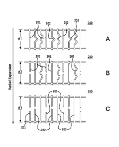
200b, and 200c. Each layer has at least two longitudinal segments (204, 205,
206)
and at least two circumferential segments (201, 202, and 203). Each
circumferential
segment 201 of layer 200a has a fully expanded dimension x. Each
circumferential
segment 202 of layer 200b is built at dimension x but has a fully expanded
dimension y. Each circumferential segment 203 of layer 200c is built at
dimension x
but has a fully expanded dimension z. The relationship between the illustrated
dimensions is z> y > x. When not constrained the lattice 200 can expand
radially
into an enlarged diametrical dimension x. At this level of expansion, layer
200a is
under tension and is resistant to further expansion. The lattice 200 can be
adjusted
to a further enlarged second diametrical dimension y when distensive force is
applied to the lattice 200. When the prescribed pressure is exceeded, layer
200a of
the lattice 200 fails, i.e., ruptures or plastically deforms. For example, the
circumferential segments 201 within layer 200a are plastically deformed or
ruptured.
The lattice 200 expands radially into an enlarged second diametrical dimension
y.
Layer 200b of the lattice 200 assumes the load and is resistant to further
expansion.
Once again, the lattice 200 can be adjusted to a further enlarged third
diametrical
dimension z when distensive force is applied to the lattice. When the
prescribed
pressure is exceeded, layer 200b of the lattice 200 fails. Lattice 200 expands
radially into an enlarged third diametrical dimension z. Once the lattice 200
expands
radially into an enlarged third diametrical dimension z, layer 200c of the
lattice 200
assumes the load and is resistant to further expansion.
[0087] Alternatively, the multi-layer lattice can be configured to radially
and/or
longitudinally expand in a partially stepped and a partially sloped manner.
With
reference to FIG. 7D, for example, the segments in layer 200a are broken, the
segments in layer 200b are plastically deformed and the segments in layer 200c
are
broken.
[0088] The prosthesis is provided that is configured to have pulsatile
compliance. The characteristic of pulsatile expansion and contraction of
vessels
requires fine mechanical compliance of the prosthesis, i.e., a close mimicking
by the
prosthetic device of the mechanics and timing of the natural vessel distending
and
reshaping under change in blood pressure. Such prosthesis has a stent. The
stent
can be flared at one or more ends. For example, both ends of the stent can be
21
CA 02854352 2014-05-01
WO 2013/074663
PCT/US2012/065066
flared. That is, a diameter at an end of the stent is greater than a diameter
defined
at the center of the stent. The prosthesis further has a lattice defining a
plurality of
openings. These two components of the prosthesis have large differences in
mechanical properties. The lattice is very flexible or elastic, and the stent
is typically
is very stiff in comparison. Thus, the combination produces a non-linear
elastic
response within the physiological pressure range of a natural vessel. The
lattice can
be made from a rapidly recovering distensible material and/or a material with
elastic
properties, for example a composite material, including at least one
fluoropolymer
membrane and elastomer. FIG 12 illustrates the combination of a stent 100 with
at
least one end flared combined with a lattice 200, which can be deployed in a
vessel
to produce a non-linear elastic response to physiological pressure between
diameters d and d'.
[00891 A lattice can be imbibed with PVA (polyvinyl alcohol) or other
materials
(e.g., gold, platinum/iridium, or the like) to aid the physician during
imaging (e.g.,
ultrasound, fluoroscopy, MRI, or the like). A lattice can be imbibed with one
or more
therapeutic agents. The term "imbibed or imbibing" as used herein is meant to
describe any means for at least partially filling a portion of the pores of a
porous
material such as ePTFE or the like. This can be done during manufacturing by,
for
example imbibing, or it can be done during catheter flushing which may imbibe
or
coat one or more therapeutic agents into or onto the lattice. Imbibing or
coating of a
therapeutic agent can result in release of the agent over time. One skilled in
the art
can select suitable therapeutic agents including without limitation:
sirolimus,
dexamethoasone, paclitaxel, phosphorylcholine, everolimus, or like agents. As
used
herein, a therapeutic agent can be a drug or other pharmaceutical product such
as a
non-genetic agents, genetic agents, cellular material, etc. Some examples of
suitable non-genetic therapeutic agents include but are not limited to: anti-
thrombogenic agents such as heparin, heparin derivatives, vascular cell growth
promoters, growth factor inhibitors, paclitaxel, etc. Where an agent includes
a
genetic therapeutic agent, such a genetic agent may include but is not limited
to:
DNA, RNA and their respective derivatives and/or components: hedgehog
proteins,
etc. Where a therapeutic agent includes a cellular material, the cellular
material may
include but is not limited to: cells of human origin and/or non-human origin
as well as
their respective components and/or derivatives thereof. Where the therapeutic
agent
22
CA 02854352 2014-05-01
WO 2013/074663
PCT/US2012/065066
includes a polymer agent, the polymer agent may be a poly-styrene-
polyisobutylene-
polystyrene triblock copolymer (SIBS), polyethylene oxide, silicone rubber
and/or any
other suitable substrate. In at least one embodiment the polymer agent can be
biodegradable such as PLA, PLGA, etc. A therapeutic agent can also be a
coating
material as described herein.
[0090] A lattice can be imbibed with one or more therapeutic agents that can
be released during distension. As shown in FIGs. 9A and 10, this can be done
during manufacturing by preparing a multi-layer lattice 200 with a reservoir
layer 211
having a therapeutic agent. The reservoir layer 211 is disposed between at
least two
layers 210, such as ePTFE, that are nonpermeable to the therapeutic agent. The
openings 212 in the lattice can be produced by laser cutting, such as a CO2
laser.
During laser cutting, the polymer adhesive used in manufacture of a multi-
layer
lattice, such as FEP or TFE/PMVE, reflows and seals the inner walls of the
openings
212, holding the therapeutic agent within the reservoir layer 211. To avoid
any
negative thermal effect on the therapeutic agent during manufacturing, the
laser
used for cutting openings in the lattice is substantially focused and the
layers can be
joined together by compression and the polymer adhesive reflow at the inner
walls of
the openings 212. As the prosthesis expands during deployment by means of
mechanical or hydraulic distension, the nonpermeable layers 210 expand, for
instance, radially into an enlarged diametrical dimension. Even in expanded
state,
the nonpermeable layers 210 typically do not allow the release of the
therapeutic
agent. In contrast, the inner walls of the openings 212 are compromised
immediately upon expansion. As shown in FIG. 9B and 10, the inner walls fail,
break,
crack or tear to allow the therapeutic agent 211a to be released. The cracks
in the
inner walls typically develop across the entire lattice that helps to achieve
a high rate
of release throughout the lattice by providing a conduit through which the
therapeutic
agent can easily and quickly diffuse from the reservoir layer.
[0091] A lattice can also be imbibed with an alginate. The alginate can be
imbibed throughout the lattice or selectively to one or more portions of the
lattice.
The alginate can be cross-linked by delivering divalent or trivalent cations
(for
example, calcium) though a catheter or the prosthesis delivery system to the
prosthesis delivery site. The cross-linked alginate portion of the lattice can
be used
to relieve pressure from weakened portions of a blood vessel (for example, to
treat a
23
CA 02854352 2014-05-01
WO 2013/074663
PCT/US2012/065066
cerebral aneurysm) or to occlude other openings or vessels adjacent to the
sidewall
of the stent. A lattice can be imbibed with calcium. An alginate can be
delivered to
the calcium imbibed lattice through the prosthesis delivery system or by
another
catheter system to cause crosslinking on or in close proximity to the lattice.
A stent
with a calcium imbibed lattice can be placed over an aneurysm neck and then
one
can introduce the alginate through the lattice and into the aneurysm. While
flowing
through the calcium imbibed lattice, the alginate can react with the calcium
to cause
formation of a gel in the aneurysm sac.
[0092] In Figs. 1A and 1B, the lattice is shown to be generally uniform.
Alternatively, the lattice covering can be varied along its length. For
example, the
size of the openings, the orientation of the openings and their shapes need
not be
uniform throughout the lattice covering. A portion of the lattice covering can
have
square-shaped openings and another portion of the lattice covering can have
diamond-shaped openings.
[0093] These coverings can be joined to a stent, graft, or stent-graft over
all or
over only a portion of the device length. The coverings can be joined
intermittently.
For example, a lattice covering can be joined only at the ends of the stent,
graft, or
stent-graft, at the closed cell portions of the stent, or only at the closed
cell
connectors. The covering can be on the outside of the stent, graft, or stent-
graft; it
can be on the inside of the stent, graft, or stent-graft; or it can be on
both.
[0094] The attachment of the lattice covering to a stent, graft, or stent-
graft
can be accomplished by mechanical means such as fiber, friction fit, braiding
a
lattice into the stent, graft, or stent-graft, or discrete mechanical
attachment points
(clips, etc.). The covering also can be attached by a single longitudinal
strip. These
components also can be bonded together through heat treatment (such as,
sintering
of the materials together) or through use of a wrap (for instance a tube,
tape, or
membrane) around the outside of the covering and stent, graft, or stent-graft
(either
continuous or discontinuous), that is adhered through either a thermoplastic
or
thermoset adhesive. The covering also can be attached to the stent, graft, or
stent-
graft by adhering the two together through use of a suitable adhesive. The
covering
can also be held in place through friction or as an interference fit. The
covering can
be held down at one or both ends. Combinations of these methods also can be
24
CA 02854352 2015-12-07
used. These methods and combinations of these methods can be used to attach
the
stent and covering while under inert gas conditions as commonly known in the
art.
[0095] Among suitable biocompatible adhesives are thermoplastic adhesives
such as fluorinated ethylene propylene (FEP), polyurethane, cyanoacrylate,
thermoplastic fluoropolymer, including flouroelastomers such as those
disclosed in
U.S. Pat. No. 7,049,380 [TFE/PMVE], etc. Thermoset adhesives are also useful,
such as silicone including room temperature vulcanizing (RTV) silicone.
[0096] For example, where the covering is a PTFE lattice; fluorinated ethylene
propylene (FEP) can be used as an adhesive. Such a coating can be applied by
various methods including extrusion over the covering, powder coating with
powdered FEP that is subsequently melted to flow over the lattice surface, or
running
the covering through a bath of molten FEP optionally followed by pulling the
covering
through a die to achieve uniformity of the coating. Alternatively, the stent
can be
provided with a coating of adhesive such as by powder coating with FEP in a
continuous or discontinuous fashion, or through use of an FEP wrap (for
instance a
tube, tape, or membrane). In an embodiment, FEP can attach the lattice to the
external surface of a stent by covering all surfaces of the stent.
100971 A covering can be provided that allows the stent, graft, or stent-graft
to
be embedded within the covering material, such as through use of a silicone or
other
elastomeric material.
[0098] Coverings can be coextensive with the length of the stent, graft, or
stent-graft, as shown in FIGs. 1A-1C and 2A-2C, or they can be either longer
or
shorter than the stent, graft, or stent-graft. Coverings can also cover only a
portion of
the stent, or can cover separately two or more portions of the stent. If
multiple
portions are covered, coverings can also overlap on the stent, graft, or stent-
graft.
For instance, one portion of the stent can be covered, while another portion
remains
uncovered as described in U.S. Pat. No. 6,673,102 to Vonesh et al.
In one embodiment, the
uncovered portion of the stent-graft in U.S. Pat. No. 6,673,102 is constrained
by a
lattice, wherein said lattice covered stent can be diametrically adjusted
according to
any one of the methods described above. Such a device allows for custom sizing
of
the prosthesis in order to adjust the prosthesis to a unique anatomy.
CA 02854352 2014-05-01
WO 2013/074663
PCT/US2012/065066
100991 Additionally, the lattice covering and the stent, graft, or stent-graft
or
both can be provided with additional treatment or therapeutic agents, such as
drugs,
radiation, radiopaque markers or coatings, or other agents to enhance
visualization
in-vivo. For example, various coatings can be provided on all or some of the
covering, the stent, graft, or stent-graft, or both. Suitable coating
materials include
fluoroelastomer, ceramic, silicone, polyethylene, carbon, gold, heparin,
hydrogel,
lubricious coatings, antibiotics, anticoagulant agents, anti-inflammatory
agents,
antimetabolic agents, antimicrobial agents, antimigratory agents, antiplatelet
agents,
antiproliferative agents, antisense agents, cytostatic agents, nitric oxide
releasing
agents, pro-endothelial agents, selective gene delivery vectors, super oxide
dismutases, super oxide dismutases mimics, vasoactive agents, and combinations
thereof, such as, for example, actinomycin-D, ciclosporin, clobetasol,
dexamethasone, estradiol, everolimus, heparin, paclitaxel, pimecrolimus,
rapamycin,
sirolimus, tacrolimus, and derivatives of these compounds. Coating materials
can
provide numerous benefits, including protecting the underlying stent material,
providing a substrate for delivery of drugs or other therapeutic substances,
isolating
the stent material from interaction with surrounding cells, improving
fluoroscopic
visualization. Coatings can be applied in any material-appropriate manner,
such as
dip-coating, spray-coating, electro-deposit, or chemical vapor deposition.
[00100] Such a prosthesis can be used to treat various body lumens,
including, the aortoiliac, carotid, cerebral, coronary, hepatic,
infrainguinal,
mesenteric, renal, splenic, subclavian, and superior mesenteric arteries and
veins as
well as other bodily conduits such as the common bile duct, pancreatic duct,
urethra
intestines and colon. Such a prosthesis' configuration allows it to conform to
the
native anatomy of blood vessels or other body lumens, while also enhancing the
stent's fatigue performance and crush-resistance.
1001011 For example, a prosthesis as described herein can be used for
treating stenosis in a carotid artery of a patient. A prosthesis is provided
having an
insertion configuration with a reduced profile and a deployed configuration
with an
enlarged profile greater than the insertion profile. For example, the
prosthesis can
have a nitinol stent which is capable of self-expanding to the deployed
configuration
when a constraint is removed. The prosthesis is inserted into the vasculature
of the
26
CA 02854352 2015-12-07
patient. The prosthesis is then positioned and deployed within the patient's
artery, for
example, at a position where plaque has caused a narrowing of the artery.
1001021 The prosthesis can be implanted by a catheter delivery system or
surgically (e.g. implanting a vascular graft). If the prosthesis is implanted
by a
catheter, the prosthesis can be radially compressed and placed within a sheath
(or
any constraining device). The sheath can be subsequently mounted on a 3F to
25F
introducer-sheath compatible delivery system, depending on the prosthesis
and/or
the anatomy to which said prosthesis will be delivered. To aid visualization
during
delivery and deployment, one or more radiopaque markers can be integrated into
the
delivery system. For example, one radiopaque marker, such as BaSO4, can be
placed into the polymer used for the distal tip of the catheter. Another
radiopaque
marker, such as a platinum / iridium band, can be incorporated into the sheath
material to indicate progression of the sheath retraction during stent
deployment.
Additionally, two markers, such as gold, platinum, or tantalum, can be placed
adjacent to the proximal and distal ends of the compressed stent to aid in
positioning.
1001031 Exemplary deployment systems that can be used in conjunction with
the prosthesis disclosed herein include U.S. Pat. Nos. 6,139,572; 6,352,561
and
7,198,636.
1001041 It can be beneficial to use the disclosed coverings independently, on
the stent, on the graft, or on the stent-graft hybrid. For example, a covering
can
provide a scaffold to reduce the risk of introduction of emboli being released
into a
bloodstream. A covering also can resist tissue encouragement into the lumen
defined by the stent. Further, a covering can help to reduce pressure on a
weakened part of a blood vessel, which in turn can reduce the risk of vessel
rupture.
[001051 For example, for carotid applications, the stent with a lattice (see
FIGs. 1A and 1B) can be useful for treating carotid stenosis. The lattice
covered
stent has flexibility and can conform to the anatomy by distending the stent
and
lattice to the desired size and shape of the vessel.
1001061 The method for doing so includes several steps. First, a prosthesis
including a lattice and a stent is provided. Second, the prosthesis is
inserted into the
patient while the prosthesis is in an insertion configuration with a reduced
profile.
Third, the prosthesis is moved through the patient's vasculature and
positioned with
27
CA 02854352 2014-05-01
WO 2013/074663
PCT/US2012/065066
the portion of the carotid artery to be treated. Fourth, the prosthesis is
deployed so
that it assumes an enlarged profile greater than the insertion profile. Fifth,
a
distending pressure is applied to the stent and lattice to distend the stent
to fit the
anatomy of the vessel. Said distending force can be applied, for example, via
a
medical balloon.
1001071 In this method, the lattice and the stent are configured and
positioned
after deployment so that the stent provides scaffolding necessary to hold the
artery
open and ensure adequate blood flow, while the lattice in combination with the
stent
simultaneously provides the correct size and shape.
1001081 The lattice openings can further provide perfusion to a side branch
vessel in this application when properly positioned. For example, a lattice
can have
a perfusion region with openings and an excluding region substantially without
the
openings. By determining the orientation of the perfusion region
endovascularly, the
lattice covered stent can be positioned so that the perfusion region allows
side
branch perfusion. Orientation can be determined by fluoroscopic visualization
of one
or more radiopaque markers incorporated within the lattice.
1001091 Also, a lattice covered stent can be used in conjunction with balloon
catheters and/or guidewires, for example, to provide perfusion to a side
branch
vessel. After initially deploying the lattice covered stent as above, a
balloon catheter
can be endovascularly introduced into a one of the openings of the lattice,
and
expanded to permanently distend or disrupt the lattice covering. This allows
endovascular modification of the size and shape of at least that one opening.
Again,
this can help to provide side branch perfusion among other uses.
[001101 In another embodiment, lattice coverings comprising fluoropolymer
membranes that exhibit high elongation while substantially retaining the
strength
properties of the fluoropolymer membrane are utilized to at least partially
cover the
stent, graft, or stent-graft. As discussed above, the coverings can be
provided
independently or on the interior or exterior surfaces of the stent, the graft,
or the
stent-graft. The term "elongation" or "elongated" as used herein is meant to
denote
the increase in length in response to the application of a tensile force. Such
membranes characteristically possess serpentine fibrils, such as the idealized
serpentine fibril exemplified in Figure 14. As depicted generally in Figure
14, a
serpentine fibril curves or turns generally one way in the direction of arrow
10 then
28
CA 02854352 2014-05-01
WO 2013/074663
PCT/US2012/065066
generally another way in the direction of arrow 20. It is to be understood
that the
amplitude and/or frequency of the serpentine-like fibrils as exemplified in
FIG. 1 may
vary. In one embodiment, the fluoropolymer membranes are expandable
fluoropolymer membranes. Non-limiting examples of expandable fluoropolymers
include, but are not limited to, expanded PTFE, expanded modified PTFE, and
expanded copolymers of PTFE. Patents have been filed on expandable blends of
PTFE, expandable modified PTFE, and expanded copolymers of PTFE, such as
U.S. Patent No. 5,708,044 to Branca; U.S. Patent No. 6,541,589 to Baillie;
U.S.
Patent No. 7,531,611 to Sabol et al.; U.S. Patent Application No. 11/906,877
to Ford;
and U.S. Patent Application No. 12/410,050 to Xu etal.
1001111 The high elongation is enabled by forming relatively straight fibrils
into
serpentine fibrils that substantially straighten upon the application of a
force in a
direction opposite to the compressed direction. The creation of the serpentine
fibrils
can be achieved through a thermally-induced controlled retraction of the
expanded
polytetrafluoroethylene (ePTFE), through wetting the article with a solvent,
such as,
but not limited to, isopropyl alcohol or Fluorinere (a perfluorinated solvent
commercially available from 3M, Inc., St. Paul, MN), or by a combination of
these
two techniques. As used herein, the term "controlled retraction" refers to
causing
articles to shorten in length in at least one direction by the application of
heat, by
wetting with a solvent, or by any other suitable means or combinations thereof
in
such a way as to inhibit folding, pleating, or wrinkling of the subsequent
article visible
to the naked eye.
[001121 The retraction of the article does not result in visible pleating,
folding,
or wrinkling of the ePTFE, unlike what occurs during mechanical compression.
The
retraction also can be applied to very thin membranes, unlike known methods.
During the retraction process, the fibrils not only become serpentine in shape
but
also may also increase in width. Upon retraction, the expanded fluoropolymer
membrane possesses serpentine fibrils. These retracted membranes
characteristically possess serpentine fibrils and are wrinkle free.
1001131 The precursor materials can be biaxially expanded ePTFE
membranes. In one embodiment, materials such as those made in accordance with
the general teachings of U.S. Patent No. 7,306,729 to Bacino, eta!, are
suitable
precursor membranes, especially if small pore size articles are desired. These
29
CA 02854352 2014-05-01
WO 2013/074663
PCT/US2012/065066
membranes may possess a microstructure of substantially only fibrils. In some
exemplary embodiments, the membranes may possess a microstructure of
substantially only serpentine fibrils. In at least one embodiment, the
fluoropolymer
membranes include a plurality of serpentine fibrils. As used herein, the
phrase
"plurality of serpentine fibrils" is meant to denote the presence of 2 or
more, 5 or
more, 10 or more, or 15 or more serpentine fibrils in the fluoropolymer
membrane
within a field of view as taught below. The serpentine fibrils have a width of
about
1.0 micron or less, and in some embodiments, about 0.5 microns or less. In one
embodiment, the serpentine fibrils have a width from about 0.1 to about 1.0
microns,
or from about 0.1 to about 0.5 microns. The precursor membrane may or may not
be amorphously locked. The precursor membrane may also be at least partially
filled, coated, or otherwise combined with additional materials.
[00114] The precursor membrane may be restrained in one or more directions
during the retraction process in order to prescribe the desired amount of
elongation
of the final article. The amount of elongation is directly related to, and
determined
by, the amount of retraction.
[00115] In one embodiment, retraction can be achieved in a uniaxial tenter
frame by positioning the rails at a distance less than the width of the
precursor
membrane prior to the application of heat or solvent or both. When using a
biaxial
tenter frame, one or both of the sets of grips, pins, or other suitable
attachment
means can similarly be positioned at a distance less than the dimensions of
the
precursor membrane. It is to be appreciated that these retraction means differ
from
the mechanical compression taught by the House and Sowinski patents noted
above.
[00116] The precursor membranes described above can be imbibed with an
elastomeric material prior, during, or subsequent to retraction to form a
composite.
The term "imbibed or imbibing" as used herein is meant to describe any means
for at
least partially filling at least a portion of the pores of a porous material
such as
ePTFE or the like. The term "all or at least a portion of the pores" as used
herein is
meant to denote that the elastomer is present in at least a portion of all or
nearly all
of the pores of the ePTFE membrane. In the absence of such elastomeric
materials,
fluoropolymer articles having serpentine fibrils do not exhibit appreciable
recovery
after elongation. Suitable elastomeric materials may include, but are not
limited to,
CA 02854352 2014-05-01
WO 2013/074663
PCT/US2012/065066
PMVE-TFE (perfluoromethylvinyl ether-tetrafluoroethylene) copolymers, PAVE-TFE
(perfluoro (alkyl vinyl ether)-tetrafluoroethylene) copolymers, silicones,
polyurethanes, and the like. It is to be noted that PMVE-TFE and PAVE-TFE are
fluoroelastomers. Other fluoroelastomers include suitable elastomeric
materials as
identified by those of skill in the art. The resultant retracted article
possesses high
elongation while substantially retaining the strength properties of the
fluoropolymer
membrane.
1001171 As one exemplary embodiment, a lattice of the type shown in FIGs.
1A and 2B having square-shaped openings may be prepared. It is to be
understood
that any shaped openings may be formed in the lattice and that square-shaped
openings described herein are merely meant to be representative. To form such
a
lattice, a mandrel may be wrapped with an elastomeric composite material, such
as
the elastomeric composite material described below in Example 7. The composite
material is free of wrinkles. The film-mandrel assembly may then be placed
into an
oven at a temperature of about 320 C for about 12 minutes to bond the layers.
After
bonding, the assembly may be removed from the oven and permitted to cool at
room
temperature to provide an ePTFE tube. Next, a pattern of regular square
openings
may be cut into the ePTFE tube, such as with a CO2 laser. The openings can
have a
size less than about 2.0 mm, about 1.0 mm, or about 0.5 mm. Additionally, the
width
of the lattice segments may be greater than about 0.01 mm or about 0.05 mm
(see
FIG. 1B). This square shaped lattice may then be placed into a convection oven
at
about 370 C for approximately 12 minutes. During heating, the material
shrinks to
form squares that can be approximately 2.0 mm, about 1.0 mm, or about 0.5 mm,
respectively, in diameter and inscribed circle and lattice segments that can
be
approximately 0.01 mm or approximately 0.05 mm wide, respectively. It is to be
appreciated that any suitable means for attaching the elastomeric composite
material
to a stent or other support structure may be used and is considered to be
within the
scope of the invention.
[001181 A lattice made with the elastomeric composite material can be
designed to be extended or elongated longitudinally or radially. In addition,
the
lattice may be expanded and contracted radially without creating folds which
drape
into the lumen. For instance, the lattice can be over-distended, such as when
an
over-sized catheter is placed through it, and the composite material will
return
31
CA 02854352 2014-05-01
WO 2013/074663
PCT/US2012/065066
(contract) without wrinkling or folding. For purposes of this invention, the
entire
device is considered to be "wrinkle-free" if within a 1 cm length of the
device, the
graft portion is devoid of wrinkles and folds when viewed by the naked eye. It
is to be
noted that 1 cm length of the device should be used unless the entire length
of the
device is less than 1 cm. In that instance, the entire device should be
utilized to
determine if the device is "wrinkle-free". It is to be noted that the terms
"free of
folds", "devoid of folds", and "fold free" are used interchangeably herein.
[00119] Once the serpentine fibrils in the elastomeric composite material are
extended to a substantially straight orientation, the strength of the
fluoropolymer
membrane is substantially that of the original fluoropolymer membrane. Also,
the
elastomeric composite material can be elongated at a relatively low tensile
stress
until reaching a point at which a high tensile stress is required for further
elongation.
Further, the composite material exhibits high elongation while substantially
retaining
the strength properties of the fluoropolymer membrane. Additionally, with
longitudinal elongation, a lattice covered stent can bend in a tight radius
without the
inner diameter of curvature buckling.
[00120] Further, should a stent graft be implanted undersized, no folds are
present in the lattice covering. Additionally, if needed, the covered stent
may be
expanded beyond the nominal stent diameter. The ability of the lattice
covering to
remain wrinkle-free results in less or no material infolding, which, in turn,
permits the
covered stent devce to have a smaller profile (e.g., a reduction in delivery
profile of
at least 1 Fr).
[00121] Having generally described this invention, a further understanding can
be obtained by reference to certain specific examples illustrated below which
are
provided for purposes of illustration only and are not intended to be all
inclusive or
limiting unless otherwise specified.
[00122] These methods of using the stent disclosed herein are exemplary and
not limiting. Further uses will be recognized by a skilled artisan.
Testing Methods
[00123] It should be understood that although certain methods and equipment
are described below, any method or equipment determined suitable by one of
ordinary skill in the art may be alternatively utilized.
32
CA 02854352 2015-12-07
Mass, Thickness, and Density
[00124] Membrane samples are die cut to form rectangular sections about
2.54 cm by about 15.24 cm to measure the weight (using a Mettler-Toledo
analytical
balance model AG204) and thickness (using a Kafer Fz1000/30 snap gauge). Using
these data, density is calculated with the following formula: p = m/(w*rt), in
which: p
= density (g/cm3), m = mass (g), w = width (cm), I = length (cm), and t =
thickness
(cm). The average of three measurements is reported.
Matrix Tensile Strength (MTS) of Membranes
[00125] Tensile break load is measured using an INSTRON 122 tensile test
machine equipped with flat-faced grips and a 0.445 kN load cell. The gauge
length
is about 5.08 cm and the cross-head speed is about 50.8 cm/min. The sample
dimensions are about 2.54 cm by about 15.24 cm. For highest strength
measurements, the longer dimension of the sample is oriented in the highest
strength direction. For the orthogonal MTS measurements, the larger dimension
of
the sample is oriented perpendicular to the highest strength direction. Each
sample
is weighed using a Mettler Toledo Scale Model AG204, then the thickness is
measured using the Kafer FZ1000/30 snap gauge; alternatively, any suitable
means
for measuring thickness may be used. The samples are then tested individually
on
the tensile tester. Three different sections of each sample are measured. The
average of the three maximum loads (i.e., peak force) measurements is
reported.
The longitudinal and transverse matrix tensile strengths (MTS) are calculated
using
the following equation: MTS= (maximum load/cross-section area)*(bulk density
of
PTFE)I (density of the porous membrane), where the bulk density of the PTFE is
taken to be about 2.2 g/cm3.
Elongation Testing
[00126] Elongation of the retracted article can be measured by any suitable
application of tensile force, such as, for example, by the use of a tensile
testing
machine, by hand, or by applying internal pressure to a tubular article. In
the instant
invention, elongation is performed at a rate of about 10% per second in all
directions
that are elongated. Elongation is calculated as the final length minus the
initial
length, divided by the initial length, and is reported as a percentage.
33
*Trademark
CA 02854352 2015-12-07
Scanning Electron Microscopy
1001271 Scanning electron micrographs are created choosing magnifications
suitable for identifying fibrils. Articles that have been retracted in
accordance with
the teachings of invention may require elongation in the direction of
retraction in
order to identify the serpentine fibrils. For the purposes of identifying the
number of
serpentine fibrils, a field of view of 7 microns by 7 microns of the sample is
to be
employed.
In addition, for the purpose of characterizing fibril width, measurements
should be made for serpentine fibrils that are substantially separated from
each other
and do not band together or otherwise form series of fibrils paralleling each
other
within the membrane. To determine the fibril width, a line is drawn through
the SEM
image to bisect it. The SEM image should be of sufficient magnification such
that at
least 5 serpentine fibrils and not more than 20 serpentine fibrils are clearly
visible
within the SEM image. Starting from one edge of the bisected image, the width
of
the first five consecutive serpentine fibrils that intersect the bisecting
line are
measured. The measurements are made where the fibril intersects the bisecting
line. Next, the five measurements are averaged and the average measurement is
recorded.
Removal of Elastomer from a Lattice Material
1001281 For lattice materials containing elastomer, the elastomer can be
dissolved or degraded and rinsed away using an appropriate solvent in order to
measure or examine desired properties.
1001291 For instance, the fluoroelastomer component of a lattice material can
be partially or substantially removed to enable SEM imaging of the ePTFE
structure.
The samples are submerged in 95 g of Fluorinert Electronic Liquid FC-72 (3M
Inc.,
St. Paul, MN) and allowed to soak without agitation. After approximately one
hour,
the fluorinated solvent is poured off and replaced with 95 g of fresh solvent.
This
process is repeated for a total of 5 soaking cycles, the first 4 cycles for
approximately
1 hour, and the 5th cycle for approximately 24 hours. Alternatively, to aid in
the
removal of elastomer, the sample can also be agitated using an ultrasonic
cleaner
(e.g. Branson 200 Ultrasonic Cleaner (Model ¨ B200)).
*Trademark 34
CA 02854352 2014-05-01
WO 2013/074663
PCT/US2012/065066
EXAMPLES
EXAMPLE 1
1001301 A lattice of the type shown in FIGs. 1A and 2B with square-shaped
openings is prepared. A mandrel is wrapped with an ePTFE film with a
discontinuous FEP coating to a thickness of approximately 0.05 mm. The film-
mandrel assembly is placed into an oven at 320 C for 12 minutes to bond the
layers. The assembly is removed from the oven and allowed to cool at room
temperature to provide an ePTFE tube. Using a CO2 laser, a pattern of regular
square openings is cut into the tube. The openings are square-shaped with a
size of
less than about 0.5 mm. The width of the lattice segments is greater than
about 0.05
mm (see FIG. 18). The prepared square shaped lattice is placed in a convection
oven set at 370 C for 12 minutes. The material shrinks during heating to form
squares that are approximately 0.5 mm diameter inscribed circle and lattice
segments that are approximately 0.05 mm wide.
EXAMPLE 2
1001311 A lattice of the type shown in FIGs. 1B and 2C with diamond-shaped
openings is prepared. An oversized mandrel that is approximately 25% larger
than
the nominal stent diameter is wrapped with an ePTFE film with a discontinuous
FEP
coating to a thickness of approximately 0.05 mm.
[00132] The film-mandrel assembly is placed into an oven at 320 C for 12
minutes to bond the layers. The assembly is removed from the oven and allowed
to
cool atroom temperature to provide an ePTFE tube. Using a CO2 laser, a pattern
of
slits approximately 40% longer than the final inscribed circle diameter are
oriented
transverse to the longitudinal axis of the mandrel are cut into the tube. The
tube with
slits is removed from the mandrel and tensioned over the nominal stent
diameter
mandrel and the slits open to form diamond shapes. The tube ends are
temporarily
fixed to length on the mandrel by ePTFE tape. The assembly is then placed into
a
convection oven set at 370 C for 12 minutes. The material shrinks to form
diamonds that are approximately 0.5 mm diameter inscribed circle and lattice
segments are approximately 0.05 mm wide.
CA 02854352 2014-05-01
WO 2013/074663
PCT/US2012/065066
EXAMPLE 3
[00133] A stent is powder coated with a thin layer of FEP powder (DuPont
FEP Fluoropolymer Resin, Product Type 5101) in a tabletop blender within which
the
stent is suspended. After the stent is placed within the blender with FEP
powder, the
blender is activated. The powder disperses into the volume of the blender
chamber
and the stent is powder coated. After approximately 3 seconds, the stent is
removed, and is placed into a convection oven set at 320 C for 5 minutes.
After this
time, the stent is removed and allowed to air cool.
[00134] The stent is then placed on a mandrel having an outer diameter
approximately equal to the inner diameter of the stent. The mandrel is covered
on its
outer diameter with polyimide film. To temporarily fix the stent to the
mandrel, the
stent is placed in a convection oven set at 320 C for 4 minutes.
[00135] After removal from the oven and cooling of the stent and mandrel
assembly, a square-shaped opening lattice according to Example 1 is coaxially
positioned over the stent.
[00136] The lattice is axially tensioned over the stent and comes in full
contact
with the outer diameter of the stent. The covering ends are temporarily fixed
to
length on the mandrel by ePTFE tape. A temporary layer of ePTFE film is then
tightly wrapped around the assembly. The perforated covering is then placed
within
a convection oven set at 320 C oven for 12 minutes to adhere the covering to
the
stent. After removal from the oven and being allowed to cool to ambient
temperature, the temporary film wrapping is removed, and the stent and lattice
covering are removed from the mandrel. The lattice is then trimmed flush with
the
end of the stent.
EXAMPLE 4
[00137] The stent is powder coated as described in Example 3 above. The
prepared diamond-shaped opening lattice of Example 2 is coaxially positioned
over
the stent. The lattice is axially tensioned over the stent, causing it to
decrease in
diameter and to come in full contact with the outer diameter of the stent. The
lattice
ends are temporarily fixed to length on the mandrel by ePTFE tape. A temporary
layer of ePTFE film is then tightly wrapped around the assembly. The lattice
is then
placed within a convection oven set at 320 C oven for 12 minutes. After
removal
36
CA 02854352 2014-05-01
WO 2013/074663
PCT/US2012/065066
from the oven and being allowed to cool to ambient temperature, the temporary
film
wrapping is removed, and the stent and lattice covering are removed from the
mandrel. The lattice is then trimmed flush with the end of the stent.
EXAMPLE 5
100138] A lattice of the type shown in FIGs. 5A-5C and 6A-6C with square-
shaped openings and varied length segments is prepared. A mandrel is wrapped
with an ePTFE film with a discontinuous FEP coating to a thickness of
approximately
0.05 mm. The film-mandrel assembly is placed into an oven at 320 C for 12
minutes to bond the layers. The assembly is removed from the oven and allowed
to
cool at room temperature to provide an ePTFE tube. Using a CO2 laser, (1) a
pattern of irregular parallelogram openings is cut into the tube (see FIGs. 5A
and
6A). The openings are shaped to provide segments with no excess length or with
some excess length, e.g., segment with no bends, one bend, or two bends. The
openings have a size of less than about 0.5 mm in one dimension and less than
about 0.17 mm in the second dimension. The width of the lattice segments is
greater than about 0.05 mm (see FIG. 1B). The prepared lattice is placed in a
convection oven set at 370 C for 12 minutes. The material shrinks during
heating to
form parallelograms with or without excess length that are approximately 0.5
mm in
the long dimension and 0.17 in the short dimension and lattice segments that
are
approximately 0.05 mm wide.
EXAMPLE 6
1001391 A drug-eluting lattice of the type shown in FIGs. 9A and 10 with
square-shaped openings is prepared. A mandrel is wrapped with an ePTFE film
with
a discontinuous FEP coating to a thickness of approximately 0.05 mm with at
least
one reservoir layer comprising a therapeutic agent. Using a CO2 laser, a
pattern of
regular square openings is cut into the tube. During laser cutting, the FEP
used in
manufacture of a multi-layer lattice, reflows and seals the inner walls of the
openings
holding the therapeutic agent within the reservoir layer. The openings are
square-
shaped with a size of less than about 0.5 mm. The prepared square shaped
lattice
is placed in a convection oven set at 370 C for 12 minutes. The material
shrinks
37
CA 02854352 2014-05-01
WO 2013/074663
PCT/US2012/065066
during heating to form squares that are approximately 0.5 mm diameter
inscribed
circle and lattice segments that are approximately 0.05 mm wide.
EXAMPLE 7
1001401 An exemplary elastomeric composite material was made in the
following manner.
Precursor Membrane
[00141] A biaxially expanded ePTFE membrane that had been amorphously
locked and had the following properties was obtained: thickness = 0.002 mm,
density = 0.837 g/cc, matrix tensile strength in the strongest direction = 475
MPa,
matrix tensile strength in the direction orthogonal to the strongest direction
= 390
MPa, elongation at maximum load in the strongest direction = 68%, and
elongation
at maximum load in the direction orthogonal to the strongest direction = 86%.
Upon
tensioning by hand, the membrane did not noticeably retract upon the release
of the
tension.
Retracted Membrane
[00142] A roll of precursor membrane, wherein the length direction
corresponded with the weakest direction of the membrane, was restrained in the
clamps of a heated, uniaxial tenter frame and fed into the heated chamber of
the
tenter frame. The oven temperature was set to about 270 C. The rails of the
tenter
frame within the heated chamber were angled inward in order to allow membrane
shrinkage to about 39% of its original width in response to the heat. The line
speed
was set to provide a dwell time of about 1.5 minutes within the heated
chamber.
[00143] The initial and final widths of the membrane were 1625 mm and 632
mm, respectively. The retracted membrane had the following properties:
thickness
= .003 mm, density = 1.36 g/cc, matrix tensile strength in the strongest
direction of
the precursor membrane = 158 MPa, matrix tensile strength in the direction
orthogonal to the strongest direction of the precursor membrane = 409 MPa,
elongation at maximum load in strongest direction of the precursor membrane =
301%, and elongation at maximum load in the direction orthogonal to the
strongest
direction of the precursor membrane = 85%.
38
CA 02854352 2014-05-01
WO 2013/074663
PCT/US2012/065066
Extruded Elastomer
[00144] A copolymer comprising tetrafluoroethylene (TEE) and
perfluoro(methyl vinylether) (PMVE) as described in U.S. Patent No. 7,049,380
to
Chang, et al. was obtained with a PMVETTFE ratio of 2:1. The copolymer was
extruded at about 350 C into a thin film. The film had the following
properties:
thickness = 0.025 mm and width = 115 mm.
Elastomeric Composite Material
[00145] The extruded elastomer was fed onto the surface of the retracted
membrane and spooled with a 0.064 mm thick high density polyethylene release
film. The elastomeric composite material had the following properties:
thickness =
0.033 mm and width = 115 mm.
[00146] The fibrils of the membrane were noted to have a serpentine shape as
shown in Figure 15, a scanning electron micrograph of the surface of an
elastomeric
composite material with the copolymer removed taken at 10,000x.
[00147] In addition to being directed to the teachings described above and
claimed below, devices and/or methods having different combinations of the
features
described above and claimed below are contemplated. As such, the description
is
also directed to other devices and/or methods having any other possible
combination
of the dependent features claimed below.
[00148] Numerous characteristics and advantages have been set forth in the
preceding description, including various alternatives together with details of
the
structure and function of the devices and/or methods. The disclosure is
intended as
illustrative only and as such is not intended to be exhaustive. It will be
evident to
those skilled in the art that various modifications may be made, especially in
matters
of structure, materials, elements, components, shape, size and arrangement of
parts
including combinations within the principles of the invention, to the full
extent
indicated by the broad, general meaning of the terms in which the appended
claims
are expressed. To the extent that these various modifications do not depart
from the
spirit and scope of the appended claims, they are intended to be encompassed
therein.
39