Note: Descriptions are shown in the official language in which they were submitted.
LEAN ELECTROLYTE FOR BIOCOMPATIBLE PLASMAELECTROLYTIC
COATINGS ON MAGNESIUM IMPLANT MATERIAL
CROSS-REFERENCE TO RELATED APPLICATIONS
[0001] This application claims priority to U.S. Provisional Patent Application
No. 61/556,563,
filed November 7, 2011.
FIELD OF THE INVENTION
[0002] The present disclosure is directed, at least in part, to a method of
producing ceramic layers
on magnesium and its alloys, a magnesium implant with a ceramic layer made by
the method, and a
magnesium implant having a biocompatible ceramic layer substantially free of
material which
impairs the biocompatibility of said biocompatible ceramic layer.
BACKGROUND OF THE INVENTION
[0003] Traditional methods of osteosynthesis and osteotomy used permanent
metal implants made
of steel or titanium. However, since these durable metal implants represent a
foreign body, patients
receiving them are potentially at a greater risk of a local inflammation.
Moreover, while these
implants tend to permanently protect healing bones against mechanical
exposure, this stress
shielding-effect actually forestalls the stabilization of the bone tissue that
needs mechanical loads to
obtain and maintain its rigidity. One solution to this problem requires a
follow up surgery to remove
the permanent metal implants. But such follow up surgeries increase the risk
of re-fracture of the
healing bones, and/or cause the patients to suffer unnecessary inconveniences,
including delayed
recovery and incurrence of additional expenses.
[0004] Alternative implants using metallic magnesium and certain magnesium
alloys have been
shown to be biodegradable and potentially suitable for medical applications.
However, because of
the electrochemical activity of magnesium, the corrosion rates of such
implants are highly dependent
on factors such as implant composition, type of environment or site of
implantation, and the surface
condition of the implant (treated or untreated). When exposed to air the
surface of untreated
magnesium implants reacts with oxygen, building up a layer of magnesium
hydroxide on the surface,
thereby slowing down further chemical reactions. In saline media, such as in
the environment of the
human organism, untreated magnesium implants initially corrode very rapidly,
producing high
amounts of hydrogen gas and magnesium hydroxide. Uncontrolled corrosion of
magnesium
1
CAN_DMS' \126128511\1
CA 2854667 2019-03-21
CA 02854667 2014-05-05
WO 2013/070669 PCT/US2012/063815
implants can cause premature failure of loaded implants due to stress
corrosion cracking and/or due
to corrosion fatigue. Moreover, because of the initial high gas release
subcutaneous gas cavities
might form. Thus, a need exists for magnesium based implants with improved
corrosion
performance.
[0005] The initial high gas release and the formation of gas bubbles in vivo
can potentially be
avoided by application of a coating to the surface of the magnesium implants
prior to implantation.
The coating would retard the rate of corrosion of the metal implants, thereby
stabilizing the rate of
gas release due to corrosion of the implants. Several attempts to improve
corrosion performance of
magnesium have been reported, including coating by anodization in solutions of
concentrated
alkaline hydroxides, or in solutions of hydrofluoric acid or acid fluoride
salts.
[0006] Anodization of magnesium using base solutions of concentrated alkaline
hydroxides is
generally provided through the supply of a DC current at a range of 50 volts
to 150 volts. A coating
is formed on the magnesium through the formation of sparks within the bath.
The tracking of the
sparks across the surface of the magnesium element slowly places the coating
onto the magnesium.
The use of sparks throughout the process leads to a relatively high current
usage and to significant
heat absorption by the bath itself. Therefore, cooling may be necessary to
reduce the temperature of
the bath during the anodization process.
[0007] Use of hydrofluoric acid or acid fluoride salts in anodization of
magnesium results in the
formation of a protective layer of magnesium fluoride on the magnesium
surface. This protective
layer is not soluble in water and thus prevents further reaction of the
magnesium metal.
[0008] Other methods for anodization of magnesium or alloys of magnesium
incorporate other
species into the film as it is farmed on the surface of the magnesium. Some
anodization processes
use silicates and others use various ceramic materials.
[0009] However, many of the reported magnesium coatings might be toxic.
Therefore, a need
exists for biocompatible coating compositions and coating processes will
produce resorbable
biomaterial onto the surface of magnesium implants that cannot completely
prevent the degradation
process, so the performance of the implants can be modulated by how the
implant is coated and/or
the corrosion characteristic of the base material used to coat the implants.
BRIEF SUMMARY OF THE INVENTION
2
CA 02854667 2014-05-05
WO 2013/070669 PCT/US2012/063815
[0010] An aspect of the present disclosure provides for a method of producing
ceramic layers on
magnesium and its alloys. An exemplary method in accordance with the present
invention
comprises the steps of: (a) immersing an implant and a metal sheet into the
aqueous electrolyte bath,
said aqueous electrolyte bath consisting essentially of: ammoniac (NH3),
diammonium hydrogen
phosphate ((NH4)2HPO4) and urea (CH4N20), and wherein the implant is made of
magnesium or its
alloy; (b) performing a anodic oxidation by passing a current between the
implant, the metal sheet
and through the aqueous electrolyte bath, wherein the implant is connected to
a positive pole of a
current source and the metal sheet is connected to a negative pole of the
current source; (c) applying
a current density selected to form sparks on said implant, to thereby form a
ceramic layer on said
implant. In an embodiment, the ammoniac concentration at 25 vol. % ranges from
1.0 mol/L to 6.0
mol/L, the diammonium hydrogen phosphate concentration ranges from 0.05 mol/L
to 0.2 mol/L;
and the urea concentration ranges from 0.01 mol/L to 1.0 mol/L.
[0011] Another exemplary method in accordance with the present invention
comprises the steps
of: (a) immersing an implant and a metal sheet into the aqueous electrolyte
bath, said aqueous
electrolyte bath consisting of: ammoniac, diammonium hydrogen phosphate and
urea, and wherein
the implant is made of magnesium or its alloy; (b) performing a anodic
oxidation by passing a
current between the implant, the metal sheet and through the aqueous
electrolyte bath, wherein the
implant is connected to a positive pole of a current source and the metal
sheet is connected to a
negative pole of the current source; (c) applying a current density selected
to form sparks on said
implant, to thereby form a ceramic layer on said implant. In an embodiment,
the ammoniac
concentration at 25 vol. % ranges from 1.0 mol/L to 6.0 mol/L, the diammonium
hydrogen
phosphate concentration ranges from 0.05 mol/L to 0.2 mol/L; and the urea
concentration ranges
from 0.01 mol/L to 1.0 mol/L.
[0012] In an embodiment, the aqueous electrolyte bath has a pH value ranging
from 10.3 to 11.6
and a temperature ranging from 18 C to 22 C. In another embodiment, the
current density is at
least 1 A/dm2. In another embodiment, the current density ranges from 1 A/dm2
to 3 A/dm2. In yet
another embodiment, the coating is selectively applied to the implant by
electrically insulating areas
of the surface which are not to be coated. In another embodiment, electric
insulation of the areas
which are not to be coated is achieved by applying a lacquer, film or foil or
the like which can be
removed after the coating process (e.g. by manual delamination).
3
CA 02854667 2014-05-05
WO 2013/070669 PCT/US2012/063815
[0013] Another aspect of the present disclosure provides for a magnesium
implant with a ceramic
layer made by exemplary methods according to the present invention. In an
exemplary embodiment
of said magnesium implant with a ceramic layer, said layer is an oxide,
hydroxide or phosphate
ceramic layer or a combination thereof and has a thickness of up to 50 Inn. In
another embodiment
of the magnesium implant with a ceramic layer, said ceramic layer has a
thickness ranging from 2
lam to 20 [tm. In another embodiment of the magnesium implant with a ceramic
layer, said ceramic
layer selected from the group consisting of: MgO, Mg(OH)2, Mg3(PO4)2 and
oxides of alloying
elements of magnesium. In yet another embodiment of the magnesium implant with
a ceramic layer,
said ceramic layer improves bone tissue adhesion compared to non-coated
magnesium implant and is
substantially free of substances which impair biocompatibility. In an
embodiment of the magnesium
implant with a ceramic layer, said magnesium implant is substantially free of
substances which
impair biocompatibility. In one such embodiment, said substances comprise an
amine
decomposition product.
100141 According to another exemplary embodiment of the magnesium implant of
the present
invention, said magnesium implant has a biocompatible ceramic layer
substantially free of material
which impairs the biocompatibility of said biocompatible ceramic layer, said
biocompatible ceramic
layer having a thickness of up to 50 tim. In one embodiment, said
biocompatible ceramic layer
includes a component selected from the group consisting of MgO, Mg(OH)2,
Mg3(PO4)2, oxides of
alloying elements of magnesium and combinations thereof. In one such
embodiment, said material
which impairs the biocompatibility of said biocompatible ceramic layer
comprises an amine
decomposition product.
100151 In an embodiment of the magnesium implant with a ceramic layer, said
implant delays and
reduces hydrogen release, compared to a magnesium implant without said
biocompatible oxide
ceramic layer, when immersed in a simulated body fluid. In yet another
embodiment of the
magnesium implant with a ceramic layer, said hydrogen release is reduced with
respect to the
corroded mass of magnesium compared to a magnesium implant without said
ceramic layer by 10 %
to 50 % over an immersion period of up to 40 days.
BRIEF DESCRIPTION OF THE DRAWINGS
100161 The foregoing summary, as well as the following detailed description of
the invention, will
be better understood when read in conjunction with the appended drawings. For
the purpose of
4
CA 02854667 2014-05-05
WO 2013/070669 PCT/US2012/063815
illustrating the invention, there are shown in the drawings embodiments which
are presently
preferred. It should be understood, however, that the invention can be
embodied in different forms
and thus should not be construed as being limited to the embodiments set forth
herein.
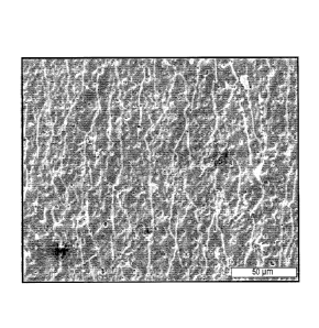
[0017] Figure 1 is an SEM image of a coating according to an embodiment of the
invention with
coarse pores;
[0018] Figure 2 is an SEM image of a coating according to another embodiment
of the invention
with fine pores;
[0019] Figure 3 illustrates the position of implanted strength retention
plates according to an
embodiment of the invention on a miniature pig nasal bone;
[0020] Figure 4 illustrates a 3-point-bending test of a degraded rectangular
plate according to an
embodiment of the invention;
[0021] Figure 5 shows average gas release rate of coated and non-coated
rectangular plates
according to certain embodiments of the invention immersed in simulated body
fluid (SBF) for up to
12 weeks (average of 6 tests per data point);
[0022] Figure 6 shows gas release as a function of weight loss of coated and
non-coated
rectangular plates according to certain embodiments of the invention immersed
in SBF for up to 12
weeks (average of 6 tests per data point);
[0023] Figure 7 shows an X-ray image of a non-coated magnesium plate according
to an
embodiment of the invention implanted in a miniature pig after 1 week;
[0024] Figure 8 shows an X-ray image of a coated magnesium plate according to
an embodiment
of the invention implanted in a miniature pig before euthanasia at 12 weeks;
[0025] Figure 9 shows decrease of yield strength for in vitro and in vivo
degraded rectangular
plates according to certain embodiments of the invention;
[0026] Figure 10 shows in vitro degradation behavior of non-coated WE43
magnesium alloy
samples and WE43 magnesium alloy samples coated according to certain
embodiments of the
invention, during immersion in simulated body fluid (SBF);
CA 02854667 2014-05-05
WO 2013/070669 PCT/US2012/063815
[0027] Figure 11 shows average accumulated gas release of tensioned WE43
magnesium alloy
samples, treated in accordance with certain embodiments of the invention,
during immersion in SBF;
[0028] Figure 12 shows strength retention (remaining bending force)
measurements of tensioned
WE43 magnesium alloy samples, treated in accordance with certain embodiments
of the invention,
during immersion in SBF;
[0029] Figure 13 shows failure times as a function of coating variants (6
specimens per variant)
according to certain embodiments of the invention on WE43 magnesium alloy
samples;
100301 Figure 14A shows an example WE43 magnesium alloy sample, treated in
accordance with
an embodiment of the invention, after plastic deformation around a 16 mm
diameter cylinder;
[0031] Figure 14B shows an example WE43 magnesium alloy sample, treated in
accordance with
an embodiment of the invention, after tensioning and positioning in a sample
holder;
[0032] Figure 15A shows an example WE43 magnesium alloy sample, treated in
accordance with
an embodiment of the invention, positioned in a holder with screw fixation for
strength retention
testing;
[0033] Figure 15B shows an example WE43 magnesium alloy sample, treated in
accordance with
an embodiment of the invention, in a holder with screw fixation for strength
retention testing prior to
immersion in SBF;
[0034] Figure 15C shows the WE43 magnesium alloy sample of Figure 15B after
six weeks of
immersion in SBF;
[0035] Figure 15D shows the WE43 magnesium alloy sample of Figure 15C after
removal from
the holder;
[0036] Figures 16A-16D show example bone plate configurations in accordance
with some
embodiments of the invention; and
[0037] Figures 17A and 17B show other example bone plate configurations in
accordance with
further embodiments of the invention.
DETAILED DESCRIPTION OF THE INVENTION
6
[0038] The present subject matter will now be described more fully hereinafter
with reference to
the accompanying Figures and Examples, in which representative embodiments are
shown. The
present subject matter can, however, be embodied in different forms and should
not be construed as
limited to the embodiments set forth herein. Rather, these embodiments are
provided to describe and
enable one of skill in the art. Unless otherwise defined, all technical and
scientific terms used herein
have the same meaning as commonly understood by one of ordinary skill in the
art to which the
subject matter pertains.
[0039] During the degradation of metallic magnesium implant, hydrogen gas and
magnesium
hydroxide are formed by the corrosion reaction. If the amount of released gas
surpasses the
absorption and diffusion capacity of the surrounding tissue, gas bubbles might
form and are often
visible on X-rays. The bare metal surface causes an initial increased release
of gas right after
implantation, but soon after the metal surface is covered with degradation
products, the gas release
rate stabilizes and might be low enough to allow sufficient gas transport. The
application of a
coating could avoid the initial high gas release and the formation of gas
bubbles. Also, an adequate
coating should effectively avoid premature failure of loaded implants due to
stress corrosion
cracking and/or corrosion fatigue. Moreover, a coating should be biocompatible
and be obtainable
without the use of toxic or potentially harmful substances.
[0040] Accordingly, an aspect of the present invention provides a method of
producing ceramic
layers on magnesium and its alloys. In some embodiments of the invention, the
method includes
exposing a magnesium or magnesium alloy implant to an aqueous electrolyte
comprising, consisting
of, or consisting essentially of: ammoniac, diammonium hydrogen phosphate and
urea. In an
embodiment, said method comprises (a) immersing an implant and a metal sheet
into an aqueous
electrolyte bath, said aqueous electrolyte bath consisting essentially of:
ammoniac, diammonium
hydrogen phosphate and urea, said implant being made of magnesium or its
alloy; (b) performing an
anodic oxidation by passing a current between said implant, said metal sheet
and through said
aqueous electrolyte bath, wherein said implant is connected to a positive pole
of a current source and
said metal sheet is connected to a negative pole of said current source; (c)
applying a current density
selected to form sparks on said implant, to thereby form a ceramic layer on
said implant. For the
purpose of this application, consisting essentially of shall mean that in
addition to the recited
components, the aqueous electrolyte bath may include other components that do
not materially affect
7
CAN_DMS: \126128511\1
CA 2854667 2019-03-21
CA 02854667 2014-05-05
WO 2013/070669 PCT/US2012/063815
the characteristics of the ceramic layer of the magnesium implant. In some
embodiments, such
characteristics may include one or more of bone tissue adhesion of the
implant, biocompatibility,
absence of amine decomposition products, and reduced hydrogen gas evolution
each compared to an
uncoated magnesium implant.
[0041] In an embodiment, the ammoniac concentration at 25 vol. % ranges from
1.0 mol/L to 6.0
mol/L. In another embodiment, the diammonium hydrogen phosphate concentration
ranges from
0.05 mol/L to 0.2 mol/L. In another embodiment the urea concentration ranges
from 0.01 mol/L to
1.0 mol/L. In an embodiment, the ammoniac concentration at 25 vol. % ranges
from 1.0 mol/L to
6.0 of and the diammonium hydrogen phosphate concentration ranges from 0.05
mol/L to 0.2 mol/L.
In an embodiment, the ammoniac concentration at 25 vol. % ranges from 1.0
mol/L to 6.0 of and the
urea concentration ranges from 0.01 mol/L to 1.0 mol/L. In an embodiment, the
diammonium
hydrogen phosphate concentration ranges from 0.05 mol/L to 0.2 mol/L and the
urea concentration
ranges from 0.01 mol/L to 1.0 mol/L.
[0042] In another exemplary embodiment, the present invention provides a
method of producing
ceramic layers on magnesium and its alloys, said method comprises (a)
immersing an implant and a
metal sheet into an aqueous electrolyte bath, said aqueous electrolyte bath
consisting of: ammoniac,
diammonium hydrogen phosphate and urea, said implant being made of magnesium
or its alloy; (b)
perfoHning an anodic oxidation by passing a current between said implant, said
metal sheet and
through said aqueous electrolyte bath, wherein said implant is connected to a
positive pole of a
current source and said metal sheet is connected to a negative pole of said
current source; (c)
applying a current density selected to form sparks on said implant, to thereby
form a ceramic layer
on said implant.
[0043] In an embodiment, the ammoniac concentration at 25 vol. % ranges from
1.0 mol/L to 6.0
mol/L. In another embodiment, the diammonium hydrogen phosphate concentration
ranges from
0.05 mol/L to 0.2 mol/L. In another embodiment the urea concentration ranges
from 0.01 mol/L to
1.0 mol/L. In an embodiment, the ammoniac concentration at 25 vol. % ranges
from 1.0 mol/L to
6.0 mol/L and the diammonium hydrogen phosphate concentration ranges from 0.05
mol/L to 0.2
mol/L. In an embodiment, the ammoniac concentration at 25 vol. % ranges from
1.0 mol/L to 6.0
mol/L and the urea concentration ranges from 0.01 mol/L to 1.0 mol/L. In an
embodiment, the
8
CA 02854667 2014-05-05
WO 2013/070669 PCT/US2012/063815
diammonium hydrogen phosphate concentration ranges from 0.05 mol/L to 0.2
mol/L and the urea
concentration ranges from 0.01 mol/L to 1.0 mol/L.
[0044] In some embodiments of the methods, the ammoniac concentration at 25
vol. % is selected
from the group consisting of 1.0 mol/L, 1.1 mol/L, 1.2 mol/L, 1.3 mol/L, 1.4
mol/L, 1.5 mol/L, 1.6
mol/L, 1.7 mol/L, 1.8 mol/L, 1.9 mol/L, 2 mol/L, 2.1 mol/L, 2.2 mol/L, 2.3
mol/L, 2.4 mol/L, 2.5
mol/L, 2.6 mol/L, 2.7 mol/L, 2.8 mol/L, 2.9 mol/L, 3 mol/L, 3.1 mol/L, 3.2
mol/L, 3.3 mol/L, 3.4
mol/L, 3.5 mol/L, 3.6 mol/L, 3.7 mol/L, 3.8 mol/L, 3.9 mol/L, 4 mol/L, 4.1
mol/L, 4.2 mol/L, 4.3
mol/L, 4.4 mol/L, 4.5 mol/L, 4.6 mol/L, 4.7 mol/L, 4.8 mol/L, 4.9 mol/L, 5
mol/L, 5.1 mol/L, 5.2
mol/L, 5.3 mol/L, 5.4 mol/L, 5.5 mol/L, 5.6 mol/L, 5.7 mol/L, 5.8 mol/L, 5.9
mol/L, 6 mol/L, and
values in between. In some embodiments, the ammoniac concentration at 25 vol.
% is at least 1.0
mol/L. In some embodiments, the ammoniac concentration at 25 vol. % is greater
than 1.0 mol/L.
In some embodiments, the ammoniac concentration at 25 vol. % is less than 6
mol/L. In some
embodiments, the ammoniac concentration at 25 vol. % is no more than 6 mol/L.
[0045] In some embodiments of the methods, the diammonium hydrogen phosphate
concentration
is selected from the group consisting 0.05 mol/L, 0.06 mol/L, 0.07 mol/L, 0.08
mol/L, 0.09 mol/L,
0.1 mol/L, 0.11 mol/L, 0.12 mol/L, 0.13 mol/L, 0.14 mol/L, 0.15 mol/L, 0.16
mol/L, 0.17 mol/L,
0.18 mol/L, 0.19 mol/L, 0.2 mol/L, and values in between. In some embodiments,
the diammonium
hydrogen phosphate concentration is at least 0.05 mol/L. In some embodiments,
the diammonium
hydrogen phosphate concentration is greater than 0.05 mol/L. In some
embodiments, the
diammonium hydrogen phosphate concentration is less than 0.2 mol/L. In some
embodiments, the
diammonium hydrogen phosphate concentration is no more than 0.2 mol/L.
[0046] In some embodiments of the methods, the urea concentration is selected
from the group
consisting of 0.01 mol/L, 0.02 mol/L, 0.03 mol/L, 0.04 mol/L, 0.05 mol/L, 0.06
mol/L, 0.07 mol/L,
0.08 mol/L, 0.09 mol/L, 0.1 mol/L, 0.11 mol/L, 0.12 mol/L, 0.13 mol/L, 0.14
mol/L, 0.15 mol/L,
0.16 mol/L, 0.17 mol/L, 0.18 mol/L, 0.19 mol/L, 0.2 mol/L, 0.21 mol/L, 0.22
mol/L, 0.23 mol/L,
0.24 mol/L, 0.25 mol/L, 0.26 mol/L, 0.27 mol/L, 0.28 mol/L, 0.29 mol/L, 0.3
mol/L, 0.31 mol/L,
0.32 mol/L, 0.33 mol/L, 0.34 mol/L, 0.35 mol/L, 0.36 mol/L, 0.37 mol/L, 0.38
mol/L, 0.39 mol/L,
0.4 mol/L, 0.41 mol/L, 0.42 mol/L, 0.43 mol/L, 0.44 mol/L, 0.45 mol/L, 0.46
mol/L, 0.47 mol/L,
0.48 mol/L, 0.49 mol/L, 0.5 mol/L, 0.51 mol/L, 0.52 mol/L, 0.53 mol/L, 0.54
mol/L, 0.55 mol/L,
0.56 mol/L, 0.57 mol/L, 0.58 mol/L, 0.59 mol/L, 0.6 mol/L, 0.61 mol/L, 0.62
mol/L, 0.63 mol/L,
9
CA 02854667 2014-05-05
WO 2013/070669 PCT/US2012/063815
0.64 mol/L, 0.65 mol/L, 0.66 mol/L, 0.67 mol/L, 0.68 mol/L, 0.69 mol/L, 0.7
mol/L, 0.71 mol/L,
0.72 mol/L, 0.73 mol/L, 0.74 mol/L, 0.75 mol/L, 0.76 mol/L, 0.77 mol/L, 0.78
mol/L, 0.79 mol/L,
0.8 mol/L, 0.81 mol/L, 0.82 mol/L, 0.83 mol/L, 0.84 mol/L, 0.85 mol/L, 0.86
mol/L, 0.87 mol/L,
0.88 mol/L, 0.89 mol/L, 0.9 mol/L, 0.91 mol/L, 0.92 mol/L, 0.93 mol/L, 0.94
mol/L, 0.95 mol/L,
0.96 mol/L, 0.97 mol/L, 0.98 mol/L, 0.99 mol/L, 1 mol/L, and values in
between. In some
embodiments, the urea concentration is at least 0.01 mol/L. In some
embodiments, the urea
concentration is greater than 0.01 mol/L. In some embodiments, the urea
concentration is less than 1
mol/L. In some embodiments, the urea concentration is no more than 1 mol/L.
[0047] In an embodiment, the aqueous electrolyte bath has a pH value ranging
from about 6 to
about 14, from about 6 about 13, from about 6 to about 12, from about 6 to
about 11, from about 6 to
about 10, from about 6 to about 9, from about 6 to about 8, or from about 6 to
about 7. In another
embodiment, the aqueous electrolyte bath has a pH value ranging from about 7
to about 14, from
about 7 about 13, from about 7 to about 12, from about 7 to about 11, from
about 7 to about 10, from
about 7 to about 9, or from about 7 to about 8. In another embodiment, the
aqueous electrolyte bath
has a pH value ranging from about 8 to about 14, from about 8 about 13, from
about 8 to about 12,
from about 8 to about 11, from about 8 to about 10, or from about 8 to about
9. In another
embodiment, the aqueous electrolyte bath has a pH value ranging 9 to about 14,
from about 9 about
13, from about 9 to about 12, from about 9 to about 11, or from about 9 to
about 10. In another
embodiment, the aqueous electrolyte bath has a pH value ranging 10 to about
14, from about 10
about 13, from about 10 to about 12, or from about 10 to about 11. In another
embodiment, the
aqueous electrolyte bath has a pH value ranging 11 to about 14, from about 11
about 13, or from
about 11 to about 12. In some embodiments, the aqueous electrolyte bath has a
pH value of greater
than 6. In some embodiments, the aqueous electrolyte bath has a pH value of at
least 6, at least 7, at
least 8, at least 9, at least 10, or at least 11. In some embodiments, the
aqueous electrolyte bath has a
pH value of less than 14, less than 13, or less than 12. In some embodiments,
the aqueous
electrolyte bath has a pH value of no more than 14. In yet another embodiment,
the aqueous
electrolyte bath has a pH value ranging from 10.3 to 11.6.
[0048] In an embodiment, the aqueous electrolyte bath has a temperature
ranging from about 0 C
to about 5 C, from about 10 C to about 15 C, from about 20 C to about 25
C, from about 30 C
to about 35 C, from about 40 C to about 45 C, from about 45 C to about 50
C, from about 0 C
to about 5 C, from about 0 C to about 10 C, from about 0 C to about 15 C,
from about 0 C to
CA 02854667 2014-05-05
WO 2013/070669 PCT/US2012/063815
about 20 C, from about 0 C to about 25 C, from about 0 C to about 30 C to
about 35 C, from
about 0 C to about 40 C, from about 0 C to about 45 C, from about 0 C to
about 45 C, from 0
C to about 50 C, from about 5 C to about 10 C, from about 5 C to about 15
C, from about 5 C
to about 20 C, from about 5 C to about 25 C, from about 5 C to about 30
C, from about 5 C to
about 35 C, from about 5 C to about 40 C, from about 5 C to about 45 C,
from 5 C to about 50
C, from about 10 C to about 15 C, from about 10 C to about 20 C, from
about 10 C to about 25
C, from about 10 C to about 30 C, from about 10 C to about 35 C, from
about 10 C to about 40
C, from about 10 C to about 45 C, from 10 C to about 50 C, from about 15
C to about 20 C,
from about 15 'V to about 25 C, from about 15 C to about 30 C, from about
15 C to about 35 C,
from about 15 C to about 40 C, from about 15 C to about 45 C, from 15 C
to about 50 C, from
about 20 C to about 25 C, from about 20 C to about 30 C, from about 20 C
to about 35 C, from
about 20 C to about 40 C, from about 20 C to about 45 C, from 20 C to
about 50 C, from about
25 C to about 30 C, from about 25 C to about 35 C, from about 25 C to
about 40 C, from about
25 'V to about 45 C, from 25 C to about 50 C, from about 30 C to about 35
C, from about 30 C
to about 40 C, from about 30 C to about 45 C, from 30 C to about 50 C,
from about 35 C to
about 40 C, from about 35 C to about 45 C, from about 35 C to about 45 C,
from 35 C to about
50 C, from about 40 C to about 45 C, from 40 C to about 50 C, or from 45
C to about 50 C.
In another embodiment, the aqueous electrolyte bath has a temperature ranging
from 18 C to 22 C.
100491 In an embodiment, the current density ranges from 1 A/dm2 to 1.2 A/dm2,
from 1 A/dm2 to
1.3 A/dm2, from 1 A/dm2 to 1.4 A/dm2, from 1 A/dm2 to 1.5 A/dm2, from 1 A/dm2
to 1.6 A/dm2,
from 1 A/dm2 to 1.7 A/dm2, from I A/dm2 to 1.8 A/dm2, from 1 A/dm2 to 1.9
A/dm2, from 1 A/41[112
to 2 A/dm2, from 1 A/dm2 to 2.1 A/dm2, from 1 A/dm2 to 2.2 A/dm2, from 1 A/dm2
to 2.3 A/dm2,
from 1 A/dm2 to 2.4 A/dm2, from 1 A/dm2 to 2.5 A/dm2, from 1 A/dm2 to 2.6
A/dm2, from 1 A/dm2
to 2.7 A/dm2, from 1 A/dm2 to 2.8 A/dm2, from 1 A/dm2 to 2.9 A/dm2, or from 1
A/dm2 to 3 A/dm2.
In another embodiment, the current density is at least 1 A/dm2. In some
embodiments, the current
density is greater than 1 A/dm2. In some embodiments, the current density is
less than 3 A/dm2. In
some embodiments, the current density is no more than 3 A/dm2.
[0050] In an embodiment, a method of the present invention provides for
forming a ceramic
coating on selected portions of the surface area of the implant. In an
embodiment, selected portions
of the surface area of the implant are electrically insulated to allow
selective anodization of the
regions of the surface of the implant that are not electrically insulated. In
an embodiment, the
11
CA 02854667 2014-05-05
WO 2013/070669 PCT/US2012/063815
electric insulation of the areas which are not to be coated is achieved by
applying a lacquer, film or
foil or the like to the desired regions of the surface area of the implant,
and subsequent to the coating
process, the applied lacquer, film or foil is removed (by manual delamination,
for example).
[0051] It will be understood by those of ordinary skill in the art that a wide
variety of coating
patterns may be designed and applied to implants. Those of ordinary skill in
the art that would also
know that the position and dimensions of the selectively coated regions of the
surface area of the
implant may be varied to modulate the corrosion performance the coated
implant. For example, the
selectively coated regions of the implant would be expected to degrade at a
slower rate than the
uncoated regions because the coat the reactants must first penetrate the coat
or erode it before
reaching the coated surface of the reactive surface of the implant.
[0052] In an embodiment of the magnesium implant with a ceramic layer, said
ceramic layer
comprises an oxide, hydroxide, phosphate or combinations thereof. In an
embodiment of the
magnesium implant with a ceramic layer, said ceramic layer comprises an oxide.
In an embodiment
of the magnesium implant with a ceramic layer, said ceramic layer comprises a
hydroxide. In an
embodiment of the magnesium implant with a ceramic layer, said ceramic layer
comprises
phosphate. In an embodiment of the magnesium implant with a ceramic layer,
said ceramic layer
comprises an oxide and a hydroxide. In an embodiment of the magnesium implant
with a ceramic
layer, said ceramic layer comprises an oxide and a phosphate. In an embodiment
of the magnesium
implant with a ceramic layer, said ceramic layer comprises a hydroxide and a
phosphate. In another
embodiment of the magnesium implant with a ceramic layer, said ceramic layer
comprises an oxide,
a hydroxide and a phosphate. In another embodiment of the magnesium implant
with a ceramic
layer, said ceramic layer is selected from the group consisting of: MgO,
Mg(OH)2, Mg3(PO4)2 and
oxides of alloying elements of magnesium.
[0053] In an embodiment of the magnesium implant with a ceramic layer, said
ceramic layer has a
thickness of up to 50 pm. In an embodiment of the magnesium implant with a
ceramic layer, said
ceramic layer has a thickness ranging from about 1 ?Am to about 5 m, from
about 10 t_tm to about 15
p.m, from about 20 p.m to about 25 p.m, from about 30 1-tin to about 35 m,
from about 401AM to
about 45 !Am, from about 45 p.m to about 50 p.m, from about 1 m to about
51ifil, from about 1 i_tm
to about 10 p.m, from about 1 p.m to about 15 [tm, from about 1 p.m to about
20 pm, from about 1
pint to about 25 1.tm, from about 1 iint to about 30 pm to about 35 p.m, from
about 1 p.m to about 40
12
CA 02854667 2014-05-05
WO 2013/070669 PCT/US2012/063815
m, from about 1 gm to about 45 pm, from about 1 gm to about 45 V1111, from 1
pm to about 50 pm,
from about 5 m to about 101.1m, from about 5 pm to about 15 pLm, from about 5
m to about 20
In, from about 5 p.m to about 25 1.IM, from about 5 p.m to about 30 pm, from
about 5 gm to about
35 m, from about 5 m to about 40 p.m, from about 5 gm to about 45 vim, from
5 m to about 50
p.m, from about 10 p.m to about 15 p.m, from about 10 i.tm to about 20 m,
from about 10 p.m to
about 25 p.m, from about 10 pm to about 30 p.m, from about 10 m to about 35
pm, from about 10
pm to about 40 gm, from about 10 p.m to about 45 gm, from 10 m to about 50
m, from about 15
pm to about 20 pm, from about 15 m to about 25 pm, from about 15 m to about
30 m, from
about 15 VIM to about 35 m, from about 15 pm to about 40 p.m, from about 15
i.tm to about 45 m,
from 15 pm to about 50 m, from about 20 gm to about 25 p.m, from about 20 m
to about 30 pm,
from about 20 p.m to about 35 gm, from about 20 m to about 40 gm, from about
20 p.m to about 45
VIM, from 20 gm to about 50 VIM, from about 25 gm to about 30 pm, from about
25 p.m to about 35
m, from about 25 pm to about 40 m, from about 25 p.m to about 45 pm, from 25
p.m to about 50
m, from about 30 p.m to about 35 gm, from about 30 jam to about 40 gm, from
about 30 p.m to
about 45 gm, from 30 pm to about 50 pm, from about 35 gm to about 40 pm, from
about 35 p.m to
about 45 p.m, from about 35 jim to about 45 m, from 35 p.m to about 50 pm,
from about 40 gm to
about 45 !Am, from 40 p.m to about 50 VIM, or from 45 i_tm to about 50 jam. In
another embodiment,
the magnesium implant with a ceramic layer, said ceramic layer has a thickness
ranging from 2 [tm
to 20 !Am. In some embodiments, the ceramic layer is at least or greater than
1 p.m in thickness, at
least or greater than 2 gm in thickness, at least or greater than 5 gm in
thickness, at least or greater
than 10 gm in thickness, at least or greater than 15 pm in thickness, at least
or greater than 20 p.m in
thickness, at least or greater than 25 p.m in thickness, at least or greater
than 30 m in thickness, at
least or greater than 35 gm in thickness, at least or greater than 40 p.m in
thickness, at least or greater
than 45 p.m in thickness, or at least or greater than 50 in in thickness. In
some embodiments, the
ceramic layer is no more than 50 pm in thickness.
[0054] The magnesium implant with a ceramic layer made by the methods of the
present
invention advantageously has a ceramic layer that not only improves bone
tissue adhesion, but also
is substantially free of substances which impair the biocompatibility. In an
embodiment, the
biocompatible ceramic layer is substantially free of material which impairs
the biocompatibility of
said biocompatible ceramic layer. In an embodiment, said biocompatible ceramic
layer typically
will have a thickness of up to 50 m. In one such embodiment, said material
which impairs the
biocompatibility of said biocompatible ceramic layer comprises an amine
decomposition product. In
13
CA 02854667 2014-05-05
WO 2013/070669 PCT/US2012/063815
another embodiment, biocompatible ceramic layer includes a component selected
from the group
consisting of MgO, Mg(OH)2, Mg3(PO4)2, oxides of alloying elements of
magnesium and
combinations thereof. Another advantage of the magnesium implant with a
ceramic layer made by
the methods of the present invention is that said implant delays and/or
reduces hydrogen release,
compared to a magnesium implant without said biocompatible ceramic layer, when
immersed in a
simulated body fluid, for example.
[0055] Accordingly, in an embodiment of the magnesium implant with a ceramic
layer according
to the present invention, said ceramic layer reduces hydrogen release with
respect to the corroded
mass of magnesium compared to a magnesium implant without said ceramic layer
by 10 % to 50 %
over an immersion period of up to 40 days. In an embodiment, said ceramic
coated magnesium
implant reduces hydrogen release with respect to the corroded mass of
magnesium compared to a
magnesium implant without said ceramic layer by from about 10 % to about 15 %,
from about 10 %
to about 20 %, from about 10 % to about 25 %, from about 10 % to about 30 %,
from about 10 % to
about 35 %, from about 10 % to about 40 %, from about 10 % to about 45 %, from
10 % to about 50
%, from about 15 % to about 20%, from about 15 % to about 25 %, from about 15
% to about 30 %,
from about 15 % to about 35 %, from about 15 % to about 40 %, from about 15 %
to about 45 %,
from 15 % to about 50 %, from about 20 % to about 25 %, from about 20 % to
about 30 %, from
about 20 % to about 35 %, from about 20 % to about 40 %, from about 20 % to
about 45 %, from 20
% to about 50 %, from about 25 % to about 30 %, from about 25 % to about 35 %,
from about 25 %
to about 40 %, from about 25 % to about 45 %, from 25 % to about 50 %, from
about 30 % to about
35 %, from about 30 % to about 40 %, from about 30 % to about 45 %, from 30 %
to about 50 %,
from about 35 % to about 40 %, from about 35 % to about 45 %, from about 35 %
to about 45 %,
from 35 % to about 50 %, from about 40 % to about 45 %, from 40 % to about 50
%, or from 45 %
to about 50 % over an immersion period of from 5 days to 10 days, from 5 days
to 15 days, from 5
days to 20 days, from 5 days to 25 days, from 5 days to 30 days, from 5 days
to 35 days, from 5 days
to 40 days, from 10 days to 15 days, from 10 days to 20 days, from 10 days to
25 days, from 10 days
to 30 days, from 10 days to 35 days, from 10 days to 40 days, from 15 days to
20 days, from 15 days
to 25 days, from 15 days to 30 days, from 15 days to 35 days, from 15 days to
40 days, from 20 days
to 25 days, from 20 days to 30 days, from 20 days to 35 days, from 20 days to
40 days, from 25 days
to 30 days, from 25 days to 35 days, from 25 days to 40 days, from 30 days to
35 days, from 30 days
to 40 days, or from 35 days to 40 days.
14
CA 02854667 2014-05-05
WO 2013/070669 PCT/US2012/063815
[0056] The materials and implants according to embodiments of the present
invention may be
configured for use as any medical implants known in the art constructed from
magnesium or its
alloys. In some embodiments, implants of the present invention are useful as
bone implants, fixation
devices, and/or for osteosynthesis. In some embodiments, the implants of the
present invention are
configured to be biodegradable. In some embodiments, the present invention
includes a bone plate
made from the materials disclosed herein. In some embodiments, the bone plate
of the present
invention is constructed from magnesium or its alloys. In some embodiments,
the bone plate is
entirely or at least partially coated with a coating or ceramic layer as
described herein. In some
embodiments, the bone plate is only partially coated. Bone plates according to
some embodiments
of the present invention are configured for attachment to one or more bones or
bone fragments and
may have any general shape known in the art suitable for bone fixation,
osteosynthesis, compression
and/or bone fusion. In some embodiments, the bone plates include one or more
fixation holes for
receiving a bone screw, tack, nail, or other fixation device for attachment to
bone. In some
embodiments, the bone plates may have a substantially linear or longitudinal
configuration. In some
embodiments, for example, the bone plate may have a plurality of fixation
holes that are arranged
substantially linearly or in a single row. In other embodiments, the bone
plate may include a
plurality of fixation holes that are arranged in a plurality of rows, for
example, in a two dimensional
array.
[0057] Figures 16A-16D illustrate example bone plates 100, 110, 120, and 130
according to
embodiments of the invention, showing different possible configurations. Bone
plates 100, 110,
120, and 130 may include one or more holes for receiving fixation devices, for
example, bone
screws 102, 112, 122, and 132. In some embodiments, bone plates 100, 110, 120,
and 130 are made
from magnesium or a biocompatible magnesium alloy and may be entirely or at
least partially coated
with a ceramic coating or layer as described herein. In some embodiments, bone
plates 100, 110,
120, and 130 are only partially coated. In some embodiments, bone screws 102,
112, 122, and 132
are made from the same materials as bone plates 100, 110, 120, and 130,
respectively. In some
embodiments, bone screws 102, 112, 122, and 132 are made from magnesium or a
biocompatible
magnesium alloy and may be entirely or at least partially coated with a
ceramic coating or layer as
described herein. In some embodiments, the portions of bone plates 100, 110,
120, and 130 and/or
bone screws 102, 112, 122, and 132 to be coated are coated by exposure to an
aqueous electrolyte
bath containing, consisting of, or consisting essentially of ammoniac,
diammonium hydrogen
phosphate, and urea as described herein.
10058] Figures 17A and 17B illustrate further example bone plates 140 and 150
according to
embodiments of the invention. In some embodiments, bone plates 140 and 150
respectively include
holes 142 and 152 for receiving fixation devices (not shown), such as a bone
screw, nail, or tack. In
some embodiments, bone plates 140 and 150 may further include countersinking
144 and 154
around holes 142 and 152. In some embodiments, bone plates 140 and 150 may be
constructed from
magesium or a biocompatible magnesium alloy. In some embodiments, bone plates
140 and 150 are
entirely or at least partially coated with a ceramic coating or layer as
described herein. In some
embodiments, bone plates 140 and 150 are only partially coated. For example,
in some
embodiments, the internal surfaces of holes 142 and 152 remain uncoated. In
some embodiments,
countersinking 144 and 154 remain uncoated. In some embodiments, the portions
of bone plates 140
and 150 to be coated are coated by exposure to an aqueous electrolyte bath
containing, consisiting
of, or consisting essentially of ammoniac, diammonium hydrogen phosphate, and
urea as described
herein.
[0059] Other example bone plate configurations that may be used according to
some embodiments
of the present invention may be found in U.S. Patent Application Publication
Nos. US 2003/0004515
Al and US 2008/0009872 Al.
[0060] These and other aspects of the present invention will be further
appreciated upon
consideration of the following Examples, which are intended to illustrate
certain particular
embodiments of the invention but are not intended to limit its scope, as
defined by the claims.
[0061] Example 1: Lean electrolyte compositions
[0062] Coatings were made on rectangular magnesium plates with 10 cm2 surface
area immersed
in selected electrolyte compositions, using a direct current of 0.16 A, a
maximum tension of 400 V
and a coating time of 10 minutes. The electrolyte compositions used are as
follows:
[0063] Composition of electrolyte A: 0.13 mol/L diammonium hydrogen phosphate,
1.07 mol/L
ammoniac (25%), and 0.50 mol/L urea.
[0064] Composition of electrolyte B: 0.05 mol/L diammonium hydrogen phosphate,
5.36 mol/L
ammoniac (25%), and 0.50 mol/L urea.
16
CAN_DMS. \126128511\1
CA 2854667 2019-03-21
CA 02854667 2014-05-05
WO 2013/070669 PCT/US2012/063815
[0065] Figure 1 shows an SEM image of a coating on a magnesium plate with
coarse pores
produced using electrolyte A, after plastic deformation. Figure 2 shows an SEM
image of a coating
on a magnesium plate with fine pores produced using electrolyte B, after
plastic deformation. The
composition of the electrolyte was the major parameter for the pore size as
all other parameters were
identical between the two samples.
[0066] The size and distribution of the pores may be important for the failure
behavior of the
implant. After plastic deformation and elastic tensioning, the sample with the
coarse pores (Figure
1) shows broader cracks than the sample with fine pores (Figure 2) where the
cracks are finer and
more evenly distributed. It is presumed that corrosion attack may be more
localized with the coarser
pores, which might also act as stress risers.
[0067] Example 2: In vivo degradation
[0068] Experiment:
[0069] All animal experiments were conducted in accordance with the Swiss
animal protection
law. Fourteen skeletally mature miniature pigs each with an age of 30 to 36
months and an average
weight of 53 7 kg were used in this preliminary study.
[0070] The midface of the miniature pig is approached by a T-type incision
where as a median cut
of 11-12 cm length was started about 2 cm below the lower orbits. After
exposing the frontal bone,
a soft tissue pocket was created with a rasp, big enough to accommodate the
two rectangular plates
and deep enough to profit of the straight portion of the nasal bone. Pre-
bending of the plates could
therefore be avoided. Figure 3 illustrates the positioning of implanted plates
10a and 10b on a pig
skull 12 in accordance with this Example. Each miniature pig received either
two coated or two
non-coated magnesium plates. 'fhe coated plates were coated in accordance with
Example 3 below.
[0071] In addition to the post-operative X-rays of the head, intemiediate
radiographs (Philips
BVPulsera) were taken at 1, 4, 8, and 12 weeks. Figure 7 shows an X-ray image
of a non-coated
magnesium plate implanted in a miniature pig after 1 week. Figure 8 shows an X-
ray image of a
coated magnesium plate implanted in a miniature pig at 12 weeks before
euthanasia. The animals
were sacrificed after 12 and 24 weeks. After euthanasia, a computed X-ray
tomography (CT) was
made. A medial incision of about 10 cm length was made along the longitudinal
axis of the nose and
the implants were removed. The pH of the implant bed was determined using pII
sensitive strip
17
(Merck 1.09557.0003, pH range 6.4 ¨ 8.0) which was moistened with distilled
water before use. The
removed plates were stored in 70% ethanol in a tightly sealed glass bottle.
After transportation to the
mechanical testing site, the magnesium plates were removed from the glass
bottles, dabbed with
paper towel and dried in air.
[0072] Energy dispersive X-ray spectroscopy (EDX) measurements were carried
out in a Zeiss
EV060 scanning electron microscope (SEM) using a THERMO Scientific ultra dry
EDX detector.
The measured spectra were analyzed for the elements C, 0, Mg, P, Ca, Y, Zr,
Nd, Gd, Dy, Er, Yb,
Na and K. Chlorine (CI) was excluded from the analysis as it could not be
detected on any of the
spectra. Three areas Of about 100 i_trn x 100 Rm were measured on each sample
to determine the
EDX-spectra. The weight loss was determined after brushing off the degradation
products with a
nail brush. Additionally, the plates were immersed in 40% hydrofluoric acid
for at least 5 minutes as
described by A. Krause et al. ("Degradation behavior and mechanical properties
of magnesium
implants in rabbit tibiae" Journal of Materials Science 2010, 45, 624-632),
cleaned in distilled water
and ethanol and dried with an air blower.
[0073] Results:
[0074] The occurrence of gas bubbles might be taken as an indicator for the in
vivo degradation.
As the exposed surface of the magnesium plates is very large (2x9 cm2), a
daily release of about 5 ml
might be expected when using the in vitro gas release rate of 0.3 ml/cm2 per
day. If this amount of
gas could not be transported away, gas bubbles would form in the thick soft
tissue on top of the
plates. Intermediate X-rays were used to check the occurrence of gas bubbles
and the integrity of the
rectangular plates. For the non-coated plates, gas bubbles could be observed
in most of the animals
after 1 week. The large observed gas bubble in the case of one animal
disappeared by week 4. For
the coated plates, the occurrence of gas bubbles was delayed. First signs of
gas pockets often
occurred around the thread holes and started to appear by week 4. No signs of
loose tissue could be
seen around the titanium control plates. The additional CT images show the
situation after
euthanasia and before the removal of the plates. The plates did not seem to be
much corroded upon
removal. The plates removed at 24 weeks showed larger areas with white
corrosion products than
the plates at 12 weeks. The two sides of the plates were not equally corroded;
the top side in contact
with the soft tissue seemed more corroded than the bottom side in contact with
the frontal bone. The
plates seemed well integrated to the surrounding tissue as a lateral step
seemed to have formed in the
18
CAN_DMS: \126128511\1
CA 2854667 2019-03-21
CA 02854667 2014-05-05
WO 2013/070669 PCT/US2012/063815
bone. On one animal of each 24 week group, the pH of the implant beds was
determined after
removal of the plate. No difference in pH could be found for the coated and
non-coated groups
compared to the titanium reference. pH values of 7.0 - 7.2 were typically
found. The white, enamel-
like degradation products seemed more compact and more adherent compared to
the in vitro
situation. As a consequence, the brushing off of the degradation products was
not sufficient and
additional bathing in hydrofluoric acid was used to determine the total weight
loss. For both kind of
plates, the average weight loss was about 5-6% after 12 weeks and increased to
13-14% after 24
weeks. The results of the EDX analysis of the in vivo degradation products
prior to the brush off
showed significantly higher calcium and phosphor contents for the coated
magnesium plates for each
milligram of corroded metal and are summarized in Table I below.
[0075] Table 1: EDX analysis of degraded implant surface before brushing off
degradation
products.
Chemical elements [wt%] Non-coated Non-coated Coated
Coated
12 weeks 24 weeks 12 weeks 24
weeks
Carbon 11 6 16 14 26 11 18
8
Oxygen 42 5 42 9 31 5 33
3
Magnesium 13 2 13 5 3.3 1.3 4.2
1.4
Calcium 2.5 0.7 2.6 2.2 14 6 12+7
Phosphor 3.8 1.4 4.4 4.1 12 3 10 4
Yttrium, Zirconium & rare earths 28 2 22 1 2 13 7 23
16
[0076] Example 3: Alloy and coating
[00771 Based on the composition of the magnesium alloy WE43 (chemical
composition: Mg-Y-
Nd heavy rare earths), a new alloy was developed. Implants from the same lot
were used for all
experiments (lot MI0018B, T5 heat treated, 6.4 x 19 mm extrusion profile). The
rectangular plates
with 60 mm x 6.0 mm x 1.50 mm were machined dry (w/o lubricant) using hard
metal tools. All
edges were rounded with a radius of 0.5 mm. A total of 36 plates were tested,
half of the plates
without a coating and the other half with a plasmaelectrolytic coating from
AHC (Kerpen,
Germany). A standard MAGOXIDTM electrolyte was used and a direct current of
1.4 A/dm2 for up
19
to 400 V was applied to generate the coating. Non-coated plates initially
weighted 940 + 5 mg. The
MAGOXIDTM coating had a typical thickness of 101.tm and accounted for 15 mg of
additional mass.
The total surface of a plate was 9 em2. The plates were cleaned with
ultrasound assistance in 90 -
100% ethanol, dried in air, packaged in pairs of two in a double vacuum pouch
and 7-sterilized with
a dose of 25 ¨ 30 kGy.
[0078] Example 4: In vitro immersion testing
[0079] Experiment:
[0080] Coated and non-coated samples were each tested inside a separate
immersion unit
containing 250 ml of simulated body fluid (SBF). Coated samples were prepared
in accordance with
Example 3 above. An immersion unit consisted of a graduated glass cylinder
with 25 mm inner
diameter and 240 mm length and a 250 ml plastic bottle. Each magnesium sample
was put inside the
glass cylinder which was then filled with SBF. The plastic bottle was put
upside down over the
glass cylinder. The cylinder/bottle assembly was quickly tilted to avoid the
flowing out of the liquid
and the remaining SBF was poured into the gap between bottle and glass
cylinder. Finally, the lid of
the bottle - which had a 33 mm hole - was slid over the glass cylinder to fix
the assembly. The
bottles were put inside a tempered water bath at 37 C.
[0081] The simulated body fluid was prepared from stock solutions as described
by L. Muller and
F. A. Muller ["Preparation of SBF with different HCO3- content and its
influence on the composition
of biomimetic apatites" Acta Biomaterialia 2 (2006) 181-9] with TRIS buffer
and the recipe for a
HCO3 - content of 27 mmol/L. The addition of NaN3 was omitted as no bacterial
growth was
observed and as N2 release into the medium could be avoided. The medium was
changed once a
week. Identical material lots, coatings and geometries were used for the in
vitro and the in vivo
degradation tests. The samples were immersed for 4, 8 and 12 weeks. The gas
release was
determined by regular visual inspection of the graded glass cylinders with a
precision of about +1
ml. The average mass loss was determined at the end of the immersion period by
brushing off the
corrosion products with a common nail brush.
[0082] Results:
[0083] The average gas release during immersion in SBF can be seen in Figure
5, which shows a
graph depicting the average gas release rate of coated and non-coated
rectangular plates immersed in
CAN_DMS= \126128511\1
CA 2854667 2019-03-21
CA 02854667 2014-05-05
WO 2013/070669 PCT/US2012/063815
SBF for up to 12 weeks (average of 6 tests per data point). The non-coated
samples started to
release gas directly after immersion. Initial gas release rates were highest
during the first couple of
days (> 1 ml/cm2 per day) and then stabilized around 0.3 ml/cm2 per day. On
the other hand, the
coated samples showed nearly no gas release during the first two weeks. The
gas release rates then
started to increase and stabilized around 0.2 ml/cm2 per day. The degradation
of non-coated
magnesium samples was uniform over the entire immersion time. Some localized
corrosion seemed
to occur for the coated magnesium samples at 12 weeks which might be
associated with a slight
increase in the gas release rate around day 65 (9 - 10 weeks). The mass loss
of the samples -
determined by brushing off the powder-like white corrosion products - could be
put into relation
with the observed gas release as shown in Figure 6, which is a graph depicting
gas release as a
function of weight loss of coated and non-coated plates immersed in SBF for up
to 12 weeks
(average of 6 tests per data point). For the non-coated samples, about 1 ml of
gas is released for 1
mg of corroded magnesium as theoretically expected from the overall corrosion
reaction. For the
coated magnesium, however, less gas was released than expected; only around
0.6 ml of gas could
be collected
[0084] Example 5: Mechanical testing
[0085] Experiment:
[0086] The 3-point-bending tests of the in vivo and in vitro degraded samples
from Examples 2
and 4 were made using a small Zwick/Roell universal testing machine (type
BZ2.5/TN1S) with a test
device according to ISO EN 178. Figure 4 illustrates a depiction of the 3-
point-bending test showing
a sample 20 positioned on two support brackets 22a and 22b being bent by a
downward moving
plunger 24. A span of 40 mm was used for all the plates. The support brackets
had a radius of 2
mm. The plunger was 4 mm in diameter and was moved downwards at a rate of 1
mm/min. The test
was stopped after 10 mm of displacement. Forces were recorded with a precision
of +0.5% (2 kN
force gauge).
[0087] Results:
[0088] The measured maximum bending force, bending stress, yield strength and
flexural
modulus are given in Table IT for the non-coated and in Table III for the
coated implants below.
Each value averages 6 samples, from individual bottles for the in vitro case
and from 3 different
21
CA 02854667 2014-05-05
WO 2013/070669 PCT/US2012/063815
animals in the in vivo case (pairs of two). Figure 9 is a graph further
showing the decrease of yield
strength over time for in vitro and in vivo degraded coated and non-coated
rectangular plates.
100891 Table II: Strength retention of non-coated rectangular magnesium plates
after in vitro and
in vivo degradation.
Mechanical Maximum Maximum Flexural Yield strength
property bending force flexural strength modulus [MPa]
Time point [N] [MP a] [GPa]
Titanium 213 972 100 805
Non degraded 91.6 1.2 402 5 41.5 0.5 336 2
In vitro 4 weeks 82.9 3.1 361 16 32.0 1 2.0 285 12
In vitro 8 weeks 61.3 1 9.0 279 38 24.4 3.7 224 30
In vitro 12 weeks 48.9 8.7 241 30 18.7 3.8 192 24
In vivo 12 weeks 86.7 4.6 367 20 29.5 2.4 270 13
In vivo 24 weeks 80.0 6.4 346 33 28.0 4.8 264 26
10090] Table III: Strength retention of coated rectangular magnesium plates
after in vitro and in
vivo degradation.
Mechanical property Maximum Maximum flexural Flexural Yield
strength
Time point bending force [N] strength [MPa] modulus
[GPa] [MPa]
Non degraded 91.5 2.0 393 7 39.2 0.6 316
4
In vitro 4 weeks 102.4 5.6 437 24 35.5 0.8 308
3
In vitro 8 weeks 73.6 3.5 332 15 28.6 1.9 252 12
In vitro 12 weeks 60.2 20.8 280 92 24.7 3.6 213
57
In vivo 12 weeks 92.7 3.9 394 16 33.3 1.8 290
12
In vivo 24 weeks 83.9 3.9 363 21 27.9 3.9 268
12
10091] All in vivo degraded plates could be deformed to the final bending
position without
breaking. In addition to those 3-point-bending tests on in vivo and in vitro
degraded plates, the
chosen 3-point-bending setup with a constant span of 40 mm was verified with a
series of
22
CA 02854667 2014-05-05
WO 2013/070669 PCT/US2012/063815
rectangular plates to check if the changed dimensions of the degraded plates
would give correct
strength measurements. A uniform degradation was "simulated" by decreasing the
thickness and
width of the plates in 0.2 mm steps down to a thickness of 0.5 mm and to a
width of 5.0 mm.
According to theory, the bending force F is expected to depend on the
thickness d and on the width b
as follows:
cy
= bj1j2
3 L with the span L and the bending stress ab
When assuming a constant bending stress, ab = MPa , an excellent fit between
the measured
maximum forces (results not shown) and the theoretical values was obtained (AF
< 2 N), This
relation might be used to calculate the core thickness of a degraded plate and
to assess the unifolinity
of degradation.
[0092] Example 6: Anodic Oxidation
[0093] Experiment:
[0094] The magnesium implant of WE43 alloy used in this experiment had a
surface of 0.1 dm2.
It was degreased, pickled and rinsed with aseptic water. The WE43 alloy was
treated with an
aqueous electrolyte bath consisting of:
1.07 mol/L ammoniac (25%)(80 ml/L);
0.13 mol/L diammonium hydrogen phosphate; and
0.5 mol/L urea.
[0095] The magnesium implant was hung into the aqueous electrolyte bath and
the positive pole
was connected to a D.C. current source. A sheet of stainless steel was also
put inside the aqueous
electrolyte bath and was connected to the negative pole of the D.C. current
source. The current
density was set to 1.4 A/dm2. The "ceramization" of the magnesium implant was
carried out for 8
minutes. The final voltage was set to 360 V.
[0096] Results:
[0097] The obtained ceramic layer had a thickness of 11 m. The "ceramized"
magnesium
implant was taken out of the electrolyte bath and was rinsed well with
aseptic, de-ionised water and
23
CA 02854667 2014-05-05
WO 2013/070669 PCT/US2012/063815
subsequently dried. Chemical analysis of the produced ceramic layer on the
WE43-magnesium
implant showed MgO, Mg(OH)2 and small amounts of Mg3(PO4)2, Yttrium oxide and
oxides of rare
earth elements.
[0098] Other magnesium wrought alloys such as WE54, ZK40, ZK, 60, AZ31 as well
as
magnesium cast alloys such as AZ91, AM50, AS41 can similarly be ceramized
(with stainless steel
and platinum as cathode materials, for example) with the procedure of Example
6.
[0099] Example 7: In vitro degradation behavior of WE43 samples
[00100] Experiment:
[00101] In vitro degradation behavior of non-coated and coated WE43 magnesium
alloy samples
during immersion in simulated body fluid (SBF) is shown Figure 10. Magnesium
WE43 samples
with coatings from three different electrolytes exhibit a significantly
reduced hydrogen release
compared to non-coated WE43 alloy samples. The three electrolytes contained
the following:
Electrolyte 1: diammonium hydrogen phosphate and ammoniac.
Electrolyte 2: diammonium hydrogen phosphate, ammoniac, and urea.
Electrolyte 3: citric acid, boric acid, phosphoric acid, and ammoniac.
[00102] Example 8: Gas release and strength retention of tensioned WE43
samples immersed in
SBF
[00103] Experiment:
[00104] Rectangular samples of WE43 alloy (60 mm x 8.0 mm x 0.50 mm) were dry
machined
(w/o lubricant) using hard metal tools. A portion of the samples were coated
with a
plasmaelcctrolytic coating from AHC (Kerpen, Germany). The electrolyte
compositions used for the
plasmaelectrolytic coating are variations of the standard MAGOXIDTM
electrolyte. A direct current
of 1.4 A/dm2 for up to 400 V was applied to generate the coating. Other sample
lots were coated
using different lean electrolytes comprising varying percentages of diammonium
hydrogen
phosphate, ammoniac (at 25 vol. % concentration), and urea, the ratios of
which are shown in Table
IV below.
24
CA 02854667 2014-05-05
WO 2013/070669 PCT/US2012/063815
[00105] The rectangular samples were manually deformed by bending the ends
around a cylinder
with a 16 mm diameter. The amount of bending is defined by the span of the two
ends of the
rectangular sample in a relaxed state. A span of about 42 mm was applied to
the samples as shown
in Figure 14A, which depicts an example of a manually deformed sample 30. The
bent samples
were then put under tension by inserting each of the ends of a sample into
slots spaced about 12 mm
apart in a UHMWPE sample holder. Figure 14B shows example sample 30 after the
ends of which
have been inserted into slots 34a and 34b of sample holder 32 under tension.
[00106] Immersion tests of the tensioned were perfomied by placing tensioned
samples inside
separate immersion units containing 250 ml of SBF in a manner similar to the
process described in
Example 4 for a total of six weeks. The 250 ml of SBF was exchanged once a
week. Gas levels
were recorded twice on working days and occurrence of failure was visually
checked for the
samples.
[00107] Strength retention tests were also carried out on immersed samples
using sample holders
with screw fixation (Figure 15A-15D). During the weekly SBF changes, the screw
of the holder is
loosened and the spring force above the holder is measured by a push pin
(indicated by the arrow in
Figure 15A). The samples did not need to be removed from the holder during the
procdure. The
samples were left in the SBF for 6 weeks irrespective of eventual failure
(breakage) of the sample
(e.g., shown in Figures 15C and 15D).
[00108] Results:
[00109] All the tested coatings had an excellent adherence to the base
material and did not
delaminate during the large plastic defomiation applied to the samples.
Plastic deformation did
introduce microcracks into the coating which broadened during the additional
tensioning and
allowed greater access to the corrosive SBF medium. Despite the severe testing
conditions, the gas
release rates of the lean electrolyte-coated samples were found to be between
about 0.2 ml/cm2 per
day and about 0.4 ml/cm2 per day, and were generally below the values for the
non-coated base
material which ranged from about 0.4 ml/cm2 per day to about 0.6 ml/cm2 per
day. The average
accumulated gas release of the lean electrolyte-coated rectangular samples
under tension and
immersed in SBF over time is shown in the graph of Figure 11. The strength
retention
measurements of the lean electrolyte-coated samples is shown in Figure 12,
which is a graph
depicting remaining bending force as a function of immersion time.
CA 02854667 2014-05-05
WO 2013/070669 PCT/US2012/063815
[00110] The failure times of tensioned rectangles during immersion in SBF are
shown in the box
plot of Figure 13, which illustrates differences between the various coatings.
Four of the five non-
coated samples failed after 32 days of immersion. The specimens coated with
the lean electrolytes
showed a larger degree of variance with some samples withstanding the 42 days
of immersion
without failing while others failed prior to the non-coated samples. The
strength retention
(remaining bending force) testing, which was applied only to the coated
samples, may have
accelerated the failure of the coated samples compared to the non-coated
samples. Moreover,
variations in the manual force applied to bend and tension the samples may
have contributed to the
wider scatter of results. Additional time to failure and gas release rate data
for the lean electrolyte-
coated samples are provided in Table IV below.
1001111 Table IV:
-
electrolyte diammonium ammoniac urea time to gas hydrogen
average
hydrogen failure release release rate
time to
block phosphate [Vo] [Vo]
linear [mg/cm2day] failure
[days] regression
[days]
rd
48.15 14.81 37.04 16.9 1.67
4.1 48.15 14.81 37.04 5.8 2.65 0.22 21.6
48.15 14.81 37.04 42.0 2.13
38.61 35.91 25.48 6.7 2.29
5.1 38.61 35.91 25.48 19.7 2.20 0.23 10.7
38.61 35.91 25.48 5.8 2.26
20.00 80.00 0.00 29.2 3.36
6.1 20.00 80.00 0.00 42.00 2.43 0.29 37.1
20.00 80.00 0.00 40.00 2.89
83.33 16.67 0.00 42.0 2.06
7.1 83.33 16.67 0.00 12.7 2.19 0.22 32.2
83.33 16.67 0.00 42.0 2.26
26
CA 02854667 2014-05-05
WO 2013/070669 PCT/1JS2012/063815
17.24 13.79 68.97 42.0 1.06
8.1 17.24 13.79 68.97 42.0 2.54 0.23 41.3
17.24 13.79 68.97 40.0 3.26
18.52 44.44 37.04 42.0 2.50
9.1 18.52 44.44 37.04 42.0 3.48 0.29 30.3
18.52 44.44 37.04 7.0 2.66
52.00 48.00 0.00 2.0 1.19
10.1 52.00 48.00 0.00 15.8 2.52 0.18 19.9
52.00 48.00 0.00 42.0 1.64
48.15 14.81 37.04 42.0 2.44
4.2 48.15 14.81 37.04 42.0 2.87 0.26 42.0
48.15 14.81 37.04 42.0 2.36
52.00 48.00 0.00 42.0 2.41
10.2 52.00 48.00 0.00 34.7 3.00 0.28 39.6
52.00 48.00 0.00 42.0 2.99
17.24 13.79 68.97 24.7 3.35
8.2 17.24 13.79 68.97 26.7 3.74 0.32 31.1
17.24 13.79 68.97 42.0 2.57
38.61 35.91 25.48 42.0 2.80
5.2 38.61 35.91 25.48 42.0 2.71 0.28 42.0
38.61 35.91 25.48 42.0 2.79
18.52 44.44 37.04 37.0 4.15
9.2 18.52 44.44 37.04 41.0 3.18 0.36 39.7
18.52 44.44 37.04 41.0 3.53
6.1 20.00 80.00 0.00 42.0 3.46 0.33 38.9
27
CA 02854667 2014-05-05
WO 2013/070669 PCT/US2012/063815
20.00 80.00 0.00 34.1 3.48
20.00 80.00 0.00 40.7 2.81
83.33 16.67 0.00 42.0 2.55
7.2 83.33 16.67 0.00 27.8 2.93 0.26 37.3
83.33 16.67 0.00 42.0 2.28
[00112] It should be understood that various changes, substitutions, and
alterations can be made
herein without departing from the spirit and scope of the invention as defined
by the appended
claims. It should also be apparent that individual elements identified herein
as belonging to a
particular embodiment may be included in other embodiments of the invention.
Moreover, the scope
of the present application is not intended to be limited to the particular
embodiments of the process,
machine, manufacture, and composition of matter, means, methods and steps
described in the
specification. As one of ordinary skill in the art will readily appreciate
from the disclosure herein,
processes, machines, manufacture, composition of matter, means, methods, or
steps, presently
existing or later to be developed that perform substantially the same function
or achieve substantially
the same result as the corresponding embodiments described herein may be
utilized according to the
present invention.
28