Note: Descriptions are shown in the official language in which they were submitted.
CA 02855356 2014-05-09
WO 2013/071142 PCT/US2012/064496
BIOMARICERS OF RESPONSE TO PROTEASOME INHIBITORS
Related Applications
[0001] This application claims priority to U.S. Provisional Application
number 61/558,474
filed on November 11, 2011 and to U.S. Provisional Application number
61/721,818 filed on
November 2, 2012. The entire contents of the foregoing applications are
incorporated herein by
reference.
Sequence Listing
[0002] This application contains a Sequence Listing which is submitted
herewith in
electronically readable format. The electronic Sequence Listing file was
created on November 8,
2012, is named "sequencelisting.txt" and has a size of 26.4 kb (27,099 bytes).
The entire
contents of the Sequence Listing in the electronic sequencelisting.txt file
are incorporated herein
by this reference.
Background
[0003] Cells become cancerous when their genotype or phenotype alters in a
way that there is
uncontrolled growth that is not subject to the confines of the normal tissue
environment. One or
more genes is mutated, amplified, deleted, overexpressed or underexpressed.
Chromosome
portions can be lost or moved from one location to another. Some cancers have
characteristic
patterns by which genotypes or phenotypes are altered.
[0004] Many genes have mutations which are associated with cancer. Some genes
have
multiple sites where mutations can occur. Many cancers have mutations in
and/or mis-
expression of more than one gene. Gene mutations can facilitate tumor
progression, tumor
growth rate or whether a tumor will metastasize. Some mutations can affect
whether a tumor
cell will respond to therapy.
100051 A variety of agents treat cancers. Cancers of the blood and bone marrow
often are
treated with steroids/glucocorticoids, imids, proteasome inhibitors and
alkylating agents.
Cancers of other tissues often are treated with alkylating agents,
topoisomerase inhibitors, kinase
inhibitors, microtubule inhibitors, angiogenesis inhibitors or other agents.
Some patients respond
to one therapy better than another, presenting the potential for a patient to
follow multiple
therapeutic routes to effective therapy. Valuable time early in a patient's
treatment program can
be lost pursuing a therapy which eventually is proven ineffective for that
patient. Many patients
- 1 -
CA 02855356 2014-05-09
WO 2013/071142 PCT/US2012/064496
cannot afford the time for trial-and-error choices of therapeutic regimens.
Expedient and
accurate treatment decisions lead to effective management of the disease.
Summary
[0006] The present disclosure relates to prognosis and planning for
treatment of solid tumors
by measurement of at least one characteristic of a marker provided herein.
Markers were
identified in solid tumor samples from xenografts of human cancer cells by
associating their
characteristics, e.g., size, sequence, composition, activity or amount, with
outcome of subsequent
treatment of the host animal with proteasome inhibition therapy. The markers
are predictive of
whether there will be a favorable outcome (e.g., good response, long time-to-
progression and/or
long term survival) after treatment of patients with a proteasome inhibitor,
such as a peptidyl
boronic acid or peptidyl epoxy ketone. Testing samples comprising tumor cells
to determine the
presence, amounts or changes of genetic markers identifies particular patients
who are expected
to have a favorable outcome with treatment, e.g., with a proteasome inhibitor,
e.g., a peptidyl
boronic acid, and whose disease may be managed by standard or less aggressive
treatment, as
well as those patients who are expected have an unfavorable outcome with the
treatment and
may require an alternative treatment to, a combination of treatments and/or
more aggressive
treatment with a proteasome inhibitor to ensure a favorable outcome and/or
successful
management of the disease.
[0007] In one aspect, the invention provides kits useful in determination
of characteristics,
e.g., amounts, presence or changes, of the markers. In another aspect, the
invention provides
methods for determining prognosis and treatment or disease management
strategies. In these
aspects, the characteristic, e.g., size, sequence, composition, activity or
amount of marker in a
sample comprising tumor cells is measured. In one embodiment, the tumor is a
solid tumor, e.g.,
non-hematological tumor, e.g., non-small cell lung cancer, colon cancer,
pancreatic cancer,
breast cancer, ovarian cancer, melanoma, head and neck carcinoma, prostate
cancer or renal cell
carcinoma.
[0008] In various embodiments, the characteristic, e.g., size, sequence,
composition, activity
or amount of marker DNA, the size, sequence, composition or amount of marker
RNA and/or the
size, sequence, composition, activity or amount of marker protein
corresponding to a marker
gene with one or more mutation, e.g., somatic mutation, described herein is
measured. Useful
information leading to the prognosis or treatment or disease management
strategies is obtained
- 2 -
CA 02855356 2014-05-09
WO 2013/071142 PCT/US2012/064496
when assays reveal information about a marker gene, e.g., whether the gene is
mutated, or not,
the identity of the mutation, and/or whether the RNA or protein amount of a
mutated gene or
genes indicates overexpression or underexpression. In one embodiment, the
strategy is
determined for proteasome inhibitor, e.g., peptidyl boronic acid, e.g.,
bortezomib (VELCADEO)
or ixazomib citrate (MLN9708), therapy.
100091 A marker gene useful to test for determination of non-hematological
tumor, i.e., solid
tumor, prognosis or treatment or disease management strategy, e.g., using a
proteasome inhibitor,
is v-Ki-ras2 Kirsten rat sarcoma viral oncogene homolog (KRAS). The marker
gene includes
mutations or alterations whose presence in marker DNA or whose effects, e.g.,
on marker RNA
and/or protein characteristics, e.g., amounts, size, sequence, activity or
composition, can provide
information for determination of prognosis or treatment or disease management.
In some
embodiments, a gene or a mutant or modified form thereof useful as a marker
gene, is associated
with one or more markers, e.g., a DNA, an RNA and/or protein characteristic,
e.g., size,
sequence, composition, activity or amount, e.g., in a sample comprising tumor
cells, which is
different than a normal DNA, RNA and/or protein. Described herein are examples
of
modifications of this gene, referred to as a "marker gene" whose mutation can
provide such
information.
10010] In some embodiments, a marker gene useful to test for determination
of non-
hematological tumor, i.e., solid tumor, prognosis or treatment or disease
management strategy,
e.g., using a proteasome inhibitor, is glucose transporter 4 (GLUT4).
[0011] The mutation of a marker gene of the present invention can provide
information about
outcome after treatment, e.g., with a proteasome inhibitor, e.g., a peptidyl
boronic acid or
peptidyl epoxy ketone. By examining a characteristic, e.g., size, sequence,
composition, activity
or amount of one or more of identified markers in a tumor, it is possible to
determine which
therapeutic agent, combination of agents, dosing and/or administration regimen
is expected to
provide a favorable outcome upon treatment. By examining the characteristic,
e.g., size,
sequence, composition, activity or amount of one or more of the identified
markers or marker
sets in a cancer, it is also possible to determine which therapeutic agent,
combination of agents,
dosing and/or administration regimen is less likely to provide a favorable
outcome upon
treatment. By examining the characteristic, e.g., size, sequence, composition,
activity or amount
of one or more of the identified markers, it is therefore possible to
eliminate ineffective or
- 3 -
CA 02855356 2014-05-09
WO 2013/071142 PCT/US2012/064496
inappropriate therapeutic agents or regimens. Importantly, these
determinations can be made on
a patient-by-patient basis. Thus, one can determine whether or not a
particular therapeutic
regimen is likely to benefit a particular patient or type of patient, and/or
whether a particular
regimen should be started or avoided, continued, discontinued or altered.
100121 The present invention is directed to methods of identifying and/or
selecting a cancer
patient who is expected to demonstrate a favorable outcome upon administration
of a therapeutic
regimen, e.g., a therapeutic regimen comprising a proteasome inhibitor, such
as a peptidyl
boronic acid or peptidyl epoxy ketone treatment. Additionally provided are
methods of
identifying a patient who is expected to have an unfavorable outcome upon
administration of
such a therapeutic regimen. These methods typically include measuring,
determining, receiving,
storing or transmitting information about the characteristic, e.g., size,
sequence, composition,
activity or amount of one or more markers or mutation of marker gene in a
patient's tumor (e.g.,
in a patient's cancer cells, e.g., non-hematological cancer cells, e.g., solid
tumor cells), optionally
comparing that to the characteristic, e.g., size, sequence, composition,
activity or amount of a
reference marker, and in a further embodiment, identifying or advising whether
the result from
the sample corresponds to a favorable outcome of a treatment regimen, e.g., a
proteasome
inhibitor, such as a peptidyl boronic acid or peptidyl epoxy ketone treatment
regimen.
10013] Additionally provided methods include therapeutic methods which
further include the
step of beginning, continuing, or commencing a therapy accordingly where the
presence of a
mutation in a marker gene or the characteristic, e.g., size, sequence,
composition, activity or
amount, of a patient's marker or markers indicates that the patient is
expected to demonstrate a
favorable outcome with the therapy, e.g., the proteasome inhibitor, such as a
peptidyl boronic
acid or peptidyl epoxy ketone, therapeutic regimen. In addition, the methods
include therapeutic
methods which further include the step of stopping, discontinuing, altering or
halting a therapy
accordingly where the presence of a mutation in a marker gene or the
characteristic, e.g., size,
sequence, composition, activity or amount of a patient's marker indicates that
the patient is
expected to demonstrate an unfavorable outcome with the treatment, e.g., with
the proteasome
inhibitor, such as a peptidyl boronic acid or peptidyl epoxy ketone, regimen,
e.g., as compared to
a patient identified as having a favorable outcome receiving the same
therapeutic regimen. In
another aspect, methods are provided for analysis of a patient not yet being
treated with a
therapy, e.g., an proteasome inhibitor, e.g., a peptidyl boronic acid or
peptidyl epoxy ketone, and
- 4 -
CA 02855356 2014-05-09
WO 2013/071142 PCT/US2012/064496
identification and prediction of treatment outcome based upon the presence of
a mutation in a
marker gene or characteristic, e.g., size, sequence, composition, activity or
amount, of one or
more of a patient's marker described herein. Such methods can include not
being treated with
the therapy, e.g., proteasome inhibitor, e.g., a peptidyl boronic acid or
peptidyl epoxy ketone
therapy, being treated with therapy, e.g., proteasome inhibitor, being treated
with a peptidyl
boronic acid or peptidyl epoxy ketone therapy in combination with one more
additional
therapies, being treated with an alternative therapy to a proteasome
inhibitor, such as a peptidyl
boronic acid or peptidyl epoxy ketone therapy, or being treated with a more
aggressive dosing
and/or administration regimen of a therapy, e.g., proteasome inhibitor, e.g.,
a peptidyl boronic
acid or peptidyl epoxy ketone inhibitor, e.g., as compared to the dosing
and/or administration
regimen of a patient identified as having a favorable outcome to standard
proteasome inhibitor,
e.g., a peptidyl boronic acid or peptidyl epoxy ketone therapy. Thus, the
provided methods of
the invention can eliminate ineffective or inappropriate use of therapy, e.g.,
proteasome inhibitor,
e.g., peptidyl boronic acid or peptidyl epoxy ketone therapy regimens.
10014] Additional methods include methods to determine the activity of an
agent, the efficacy
of an agent, or identify new therapeutic agents or combinations. Such methods
include methods
to identify an agent as useful, e.g., as a proteasome inhibitor, e.g., a
peptidyl boronic acid or
peptidyl epoxy ketone, for treating a cancer, e.g., a non-hematological
cancer, i.e., a solid tumor
cancer (e.g., non-small cell lung cancer, colon cancer, pancreatic cancer,
breast cancer, ovarian
cancer, melanoma, head and neck carcinoma, prostate cancer or renal cell
carcinoma), based on
its ability to affect the presence of a mutation in a marker gene or
characteristic, e.g., size,
sequence, composition, activity or amount of a marker or markers of the
invention. In some
embodiments, an inhibitor which decreases or increases the presence of a
mutation in a marker
gene or characteristic, e.g., size, sequence, composition, activity or amount
of a marker or
markers provided (i.e., in a cell population, the inhibitor selects against
cells comprising the
mutation characteristic or selects for cells comprising the mutation or
characteristic, respectively)
in a manner that indicates favorable outcome of a patient having cancer would
be a candidate
agent for the cancer. In another embodiment, an agent which is able to
decrease the viability of
a tumor cell comprising a marker indicative of an unfavorable outcome would be
a candidate
agent for the cancer.
-5-.
CA 02855356 2014-05-09
WO 2013/071142 PCT/US2012/064496
[0015] The present invention is also directed to methods of treating a
cancer patient, with a
therapeutic regimen, e.g., a proteasome inhibitor, e.g., a peptidyl boronic
acid or peptidyl epoxy
ketone therapy regimen (e.g., alone, or in combination with an additional
agent such as a
chemotherapeutic agent, e.g., a glucocorticoid agent, a microtubule inhibitor,
an alkylating agent,
a kinase inhibitor or a topoisomerase inhibitor), which includes the step of
selecting for treatment
a patient whose marker characteristic, e.g., size, sequence, composition,
activity or amount
indicates that the patient is expected to have a favorable outcome with the
therapeutic regimen,
and treating the patient with the therapy, e.g., proteasome inhibitor, e.g., a
peptidyl boronic acid
therapy. In some embodiments, the method can include the step of selecting a
patient whose
marker characteristic, e.g., size, sequence, composition, activity or amount
or amounts indicates
that the patient is expected to have a favorable outcome and administering a
therapy other than a
proteasome inhibitor therapy that demonstrates similar expected progression-
free survival times
as the proteasome inhibitor, e.g., a peptidyl boronic acid therapy.
[0016] Additional methods of treating a cancer patient include selecting
patients that are
unlikely to experience a favorable outcome upon treatment with a cancer
therapy (e.g.,
proteasome inhibitor, e.g., a peptidyl boronic acid or peptidyl epoxy ketone
therapy). Such
methods can further include one or more of: administering a higher dose or
increased dosing
schedule of a therapy, e.g., proteasome inhibitor, e.g., a peptidyl boronic
acid or peptidyl epoxy
ketone as compared to the dose or dosing schedule of a patient identified as
having a favorable
outcome with standard therapy; administering a cancer therapy other than a
proteasome inhibitor,
e.g., a peptidyl boronic acid or peptidyl epoxy ketone therapy; administering
a proteasome
inhibitor, e.g., a peptidyl boronic acid or peptidyl epoxy ketone agent in
combination with an
additional agent such as a chemotherapeutic agent, e.g., a glucocorticoid
agent, a microtubule
inhibitor, an alkylating agent, a kinase inhibitor or a topoisomerase
inhibitor. Further provided
are methods for selection of a patient having aggressive disease which is
expected to demonstrate
more rapid time to progression or short term survival.
[0017] Additional methods include a method to evaluate whether to treat or
pay for the
treatment of cancer, e.g., non-hematological cancer, i.e., solid tumor cancer
(e.g., non-small cell
lung cancer, colon cancer, pancreatic cancer, breast cancer, ovarian cancer,
melanoma, head and
neck carcinoma, prostate cancer or renal cell carcinoma) by reviewing the
amount of a patient's
marker or markers for indication of outcome to a cancer therapy, e.g., a
proteasome inhibitor,
- 6 -
CA 02855356 2014-05-09
WO 2013/071142 PCT/US2012/064496
e.g., a peptidyl boronic acid or peptidyl epoxy ketone therapy regimen, and
making a decision or
advising on whether payment should be made.
[0018] The entire contents of all publications, patent applications,
patents and other
references mentioned herein are incorporated by reference.
[0019] Other features and advantages of the invention will be apparent from
the following
detailed description, drawings and from the claims.
Drawings
[0020] Figure 1. Correlation of mutation status of KRAS to sensitivity of
tumor xenografts to
MLN2238.
[0021] Figure 2. Antitumor activity of MLN2238 in representative xenografts in
comparison
with vehicle control. A. PHTX132Lu primary NSCLC xenograft (wild type KRAS),
B. HCT-
116 xenograft (mutant KRAS).
[0022] Figure 3. Antitumor activity of MLN2238 in xenografts of isogenic
SW48 cell lines.
A. SW48 xenograft (wild type KRAS), B. SW48-KrasG13D xenograft (recombinantly
mutated
KRAS-G13D), C. SW48-KrasG12V xenograft (recombinantly mutated KRAS-G12V).
[0023] Figure 4. Western blot of GLUT4 protein from KRAS wild type and mutant
cells.
FIG.4A, GLUT4 levels in cells grown in vitro; FIG. 4B, GLUT4 levels in tumor
xenografts.
Detailed Description
[0024] One of the continued problems with therapy in cancer patients is
individual
differences in response to therapies. While advances in development of
successful cancer
therapies progress, only a subset of patients respond to any particular
therapy. With the narrow
therapeutic index and the toxic potential of many available cancer therapies,
such differential
responses potentially contribute to patients undergoing unnecessary,
ineffective and even
potentially harmful therapy regimens. If a designed therapy could be optimized
to treat
individual patients, such situations could be reduced or even eliminated.
Furthermore, targeted
designed therapy may provide more focused, successful patient therapy overall.
Accordingly,
there is a need to identify particular cancer patients who are expected to
have a favorable
outcome when administered particular cancer therapies as well as particular
cancer patients who
may have a favorable outcome using more aggressive and/or alternative cancer
therapies, e.g.,
alternative to previous cancer therapies administered to the patient. It would
therefore be
-7..
CA 02855356 2014-05-09
WO 2013/071142 PCT/US2012/064496
beneficial to provide for the diagnosis, staging, prognosis, and monitoring of
cancer patients,
including, e.g., non-hematological cancer patients, e.g., patients with solid
tumors (e.g., non-
small cell lung cancer, colon cancer, pancreatic cancer, breast cancer,
ovarian cancer, melanoma,
head and neck carcinoma, prostate cancer or renal cell carcinoma) who would
benefit from
particular cancer inhibition therapies as well as those who would benefit from
a more aggressive
and/or alternative cancer inhibition therapy, e.g., alternative to a cancer
therapy or therapies the
patient has received, thus resulting in appropriate preventative measures.
10025] The present invention is based, in part, on the recognition that
mutation of a marker
gene can be associated with sensitivity of a cell comprising the mutated gene
to a proteasome
inhibitor, e.g., a peptidyl boronic acid. In some embodiments, the marker gene
is involved in the
Rat Sarcoma (RAS) signaling pathway, e.g., a gene whose mutation enables
activation of the
pathway. RAS is an oncogenic GTPase whose active GTP-bound state activates
pathways (e.g.,
the mitogen-activated protein (MAP) kinase cascade) involved in
tutmorigenesis. Proteins,
including tumor suppressors, facilitate hydrolysis of RAS-bound GTP to GDP to
inactivate RAS
and thus limit the signaling from a RAS oncogene. A mutation in a gene
involved in this
checkpoint, either in an oncogene upstream of RAS (e.g., p210BCR-ABL or erbB),
in a RAS
oncogene (e.g., HRAS, KRAS or NRAS), in a RAS-associated tumor suppressor
(neurofibromatosis 1(NF1)), and/or in a GTPase-activating protein (e.g.,
RASGAP), can enable
activation of RAS signaling pathways. A protein encoded by a marker gene for
sensitivity to a
proteasome inhibitor can be a RAS protein. KRAS is an example of a marker
gene. In the GTP-
bound state of RAS proteins, e.g., NRAS, 1-fRAS and KRAS, the RAS signaling
occurs, and in
the GDP-bound state, the signaling is abrogated. A mutated RAS protein can
prolong its time in
the GTP-bound state and the resulting signaling pathway activation can lead to
proliferation of
cells harboring the mutated gene. A marker gene can exhibit one or more
mutations, e.g.,
somatic mutations, whose presence can affect expression or activity of the
encoded gene product.
In some embodiments, there can be more than one mutation in a marker gene in a
tumor cell or
tumor. In additional embodiments, there can be marker gene mutations in cells
which have
mutations in one or more additional genes, including mutations that can lead
to tumorigenesis,
but the additional mutated gene(s) may not be a marker gene as considered
herein. In some
embodiments, the mutation is an activating mutation. In other embodiments, the
mutation affects
- 8 -
CA 02855356 2014-05-09
WO 2013/071142 PCT/US2012/064496
the expression of the marker gene. In other embodiments, a mutation can result
in an altered
interaction of the encoded gene product with a cellular binding partner.
100261 In one aspect, the invention provides a method for determining
whether to treat with a
proteasome inhibitor a patient having a solid tumor selected from the group
consisting of a lung
tumor and a colon tumor, the method comprising the steps of: a) measuring at
least one
characteristic of at least one marker associated with at least one marker gene
in a patient sample
comprising tumor cells, wherein one marker gene is v-Ki-ras2 Kirsten rat
sarcoma viral
oncogene homolog (KRAS); b) identifying whether the at least one
characteristic measured in
step a) is informative for outcome of treatment with the proteasome inhibitor;
and c)
determining to treat the patient with the proteasome inhibitor if the
informative characteristic
indicates that the tumor cells comprise wild type KRAS. In some embodiments,
the method
comprises determining to treat the patient with a proteasome inhibitor if the
informative
characteristic indicates that the tumor cells comprise wild type KRAS or KRAS
with a mutation
at codon 146. In some embodiments, the method to determine whether to treat
with a
proteasome inhibitor is performed in vitro.
[0027] In another aspect, the invention provides a method for determining
whether to
continue proteasome inhibitor treatment of a solid tumor in a patient
comprising: a) treating a
patient having a solid tumor with a proteasome inhibitor; b) obtaining a
sample comprising
tumor cells from the patient; c) measuring at least one characteristic of at
least one marker
associated with at least one marker gene in the sample, wherein at least one
marker gene is
KRAS; d) comparing the results of the measurements in c) to a reference; and
e) determining to
continue treatment with the proteasome inhibitor if the comparison indicates
that the solid tumor
cells in the sample comprise wild type KRAS; wherein the patient has a solid
tumor selected
from the group consisting of a lung tumor and a colon tumor. In some
embodiments, the method
comprises determining to continue to treat the patient with a proteasome
inhibitor if the if the
comparison indicates that the solid tumor cells comprise wild type KRAS or
KRAS with a
mutation at codon 146. In some embodiments, the method to determine whether to
continue to
treat with a proteasome inhibitor is performed in vitro.
[0028] In another aspect, the invention provides a kit comprising a
stabilizer to add to a
sample comprising tumor cells and a reagent to measure at least one
characteristic of at least one
- 9 -
CA 02855356 2014-05-09
WO 2013/071142 PCT/US2012/064496
marker in a sample, wherein the result of the measurement indicates whether
there is a mutation
in at least one marker gene, wherein at least one marker gene is KRAS.
[0029] In another aspect, the invention provides a kit comprising at least
two reagents to
measure at least one characteristic of at least two markers in a sample,
wherein the result of the
measurement indicates whether there is a mutation in at least one marker gene,
wherein at least
one marker gene is KRAS and wherein the sample comprises solid tumor cells
wherein the solid
tumor cells are selected from the group consisting of lung tumor cells and a
colon tumor cells.
[0030] In another aspect, the invention provides a method for predicting
sensitivity of a solid
tumor cell to a proteasome inhibitor, comprising: a) assessing whether the
cell expresses mutated
KRAS; and b) predicting sensitivity of the cell to a proteasome inhibitor,
wherein expression of
mutated KRAS is predictive of poor sensitivity to the proteasome inhibitor,
wherein the solid
tumor cell is selected from the group consisting of a lung tumor cell and a
colon tumor cell. In
one embodiment, the mutated KRAS does not have a mutated codon 146.
[0031] In another aspect, the invention provides a method for treating a
patient having a solid
tumor comprising wild type KRAS status, comprising the step of administering
to the patient a
therapeutically effective amount of a proteasome inhibitor, wherein the solid
tumor is selected
from the group consisting of a lung tumor and a colon tumor. In one
embodiment, the solid
tumor comprises wild type KRAS or KRAS with mutated codon 146.
[0032] In another aspect, the invention provides the use of a proteasome
inhibitor in the
manufacture of a medicament to treat a solid tumor selected from the group
consisting of a lung
tumor and a colon tumor, wherein the solid tumor has a wild type KRAS. In one
embodiment,
the solid tumor has wild type KRAS or KRAS with mutated codon 146.
[0033] In another aspect, the invention provides the use of a proteasome
inhibitor for treating
a lung tumor or a colon tumor in a patient whose lung tumor or colon tumor has
wild type
KRAS. In one embodiment, the tumor has wild type KRAS or KRAS with mutated
codon 146.
[0034] In another aspect, the invention provides a method for identifying a
proteasome
inhibitor as suitable for use in treating a patient with a non-hematological
cancer, comprising: a)
contacting a tumor cell in a xenograft comprising at least one mutation in at
least one marker
gene with a test proteasome inhibitor, wherein at least one marker gene is
KRAS; b) assessing
the effect of the test proteasome inhibitor on the viability of the cell; and
c) determining that the
test proteasome inhibitor is suitable for use in treating a patient with a non-
hematological cancer
- 10 -
CA 02855356 2014-05-09
WO 2013/071142 PCT/US2012/064496
if it decreases the viability of the cell. In one embodiment, the mutation is
not at codon 146 of
KRAS.
[0035] In another aspect, the invention provides a method for paying for
the treatment of
cancer with a proteasome inhibitor comprising: a) recording whether KRAS is
mutated, in a
sample comprising solid tumor cells from a patient, wherein the patient has
lung cancer or colon
cancer, and b) authorizing payment of the proteasome inhibitor treatment if
KRAS is wild type.
In one embodiment, the method comprises authorizing payment if KRAS is wild
type or mutated
at codon 146.
[0036] A method of identifying a non-hematological cancer patient who will be
nonresponsive to treatment with a proteasome inhibitor, comprising determining
the presence or
absence of at least one KRAS mutation in a sample comprising tumor cells from
the patient,
wherein the patient has a non-hematological cancer selected from the group
consisting of lung
cancer and colon cancer, whereby the presence of at least one KRAS mutation
indicates that the
patient will not respond to the proteasome inhibitor. In one embodiment, the
mutation is not in
codon 146.
[0037] In another aspect, the invention provides a method of identifying a
non-hematological
cancer patient who will have a favorable outcome to treatment with a
proteasome inhibitor,
comprising determining the presence or absence of at least one KRAS mutation
in a sample
comprising tumor cells from the patient, wherein the patient has a non-
hematological cancer
selected from the group consisting of lung cancer and colon cancer, whereby
the presence of
wild type KRAS or a mutation in codon 146 indicates that the patient will
respond to the
proteasome inhibitor.
[0038] In some embodiments, the mutation is identified by measuring in the
tumor cells, or in
an extract prepared therefrom, a characteristic of a marker associated with
the KRAS marker
gene. In some embodiments, the method comprises determining the tumor cell
KRAS sequence.
[0039] An additional embodiment of the present invention is based on the
identification, in a
tumor cell or tumor, whose sensitivity or resistance to a proteasome inhibitor
is correlated to a
mutational status of a RAS marker gene, of an additional marker gene, a
glucose transporter,
e.g., GLUT4. In one embodiment, a characteristic, e.g., composition, activity
or amount, e.g.,
expression, in a tumor cell or tumor, of a glucose transporter, e.g., GLUT4,
can be correlated to a
mutational status or characteristic, e.g., size, sequence, composition,
activity or amount, of the
-11-
CA 02855356 2014-05-09
WO 2013/071142 PCT/US2012/064496
KRAS marker gene. In some embodiments, a glucose transporter marker
characteristic
correlated with sensitivity to a proteasome inhibitor is amount. In some
embodiments, a glucose
transporter marker, e.g., mRNA or protein, has an informative characteristic
amount of low or
normal expression in a tumor cell whose RAS marker gene has a mutational
status which is
indicative of a favorable outcome upon treatment of the tumor with a
proteasome inhibitor. In
some embodiments, a glucose transporter marker, e.g., mRNA or protein, has an
informative
characteristic of low or normal expression in a tumor cell which has an
informative characteristic
of expression of wild type KRAS. In one embodiment, a method of the invention
includes the
steps of identifying the KRAS mutational status and measuring the expression
of GLUT4. In
some embodiments, a patient whose tumor, e.g., a solid tumor, comprises wild
type KRAS and
low or normal GLUT4 expression is predicted to have a favorable outcome of
treatment with a
proteasome inhibitor, e.g., a peptidyl boronic acid or a peptidyl epoxy
ketone. In another
embodiment, a patient whose tumor, e.g., a solid tumor, comprises mutant KRAS
and high,
higher than normal, or higher than a reference level of GLUT4 expression is
predicted to have an
unfavorable outcome of treatment with a proteasome inhibitor, e.g., a peptidyl
boronic acid or a
peptidyl epoxy ketone.
[0040] The identification and/or measurement of a mutation in a marker gene
or characteristic
of a marker can be used to determine whether a favorable outcome can be
expected by treatment
of a tumor, e.g., with a proteasome inhibitor, e.g., a peptidyl boronic acid
or peptidyl epoxy
ketone therapy or whether an alternative therapy to and/or a more aggressive
therapy with, e.g., a
proteasome inhibitor, e.g., a peptidyl boronic acid or peptidyl epoxy ketone
inhibitor may
enhance the response. For example, the compositions and methods provided
herein can be used
to determine whether a patient is expected to have a favorable outcome to a
proteasome inhibitor,
e.g., a peptidyl boronic acid or peptidyl epoxy ketone therapeutic agent
dosing or administration
regimen. Based on these identifications, the present invention provides,
without limitation: 1)
methods and compositions for determining whether a proteasome inhibitor, e.g.,
a peptidyl
boronic acid or peptidyl epoxy ketone therapy regimen will or will not be
effective to achieve a
favorable outcome and/or manage the cancer; 2) methods and compositions for
monitoring the
effectiveness of a proteasome inhibitor, e.g., a peptidyl boronic acid or
peptidyl epoxy ketone
therapy (alone or in a combination of agents) and dosing and administrations
used for the
treatment of tumors; 3) methods and compositions for treatments of tumors
comprising, e.g.,
- 12 -
CA 02855356 2014-05-09
WO 2013/071142 PCT/US2012/064496
proteasome inhibitor, e.g., a peptidyl boronic acid or peptidyl epoxy ketone
inhibition therapy
regimen; 4) methods and compositions for identifying specific therapeutic
agents and
combinations of therapeutic agents as well as dosing and administration
regimens that are
effective for the treatment of tumors in specific patients; and 5) methods and
compositions for
identifying disease management strategies.
[0041] Proteasome inhibition represents an important strategy in cancer
treatment. The
proteasome is a multi-enzyme complex present in all cells which play a role in
degradation of
proteins involved in regulation of the cell cycle. For example, King et al.
(Science 274:1652-
1659 (1996)) demonstrated that the ubiquitin-proteasome pathway plays an
essential role in
regulating cell cycle, neoplastic growth and metastasis. A number of key
regulatory proteins,
including p53, cyclins, and the cyclin-dependent kinases p21 and p27KIPI, are
temporally
degraded during the cell cycle by the ubiquitin-proteasome pathway. The
ordered degradation of
these proteins is required for the cell to progress through the cell cycle and
to undergo mitosis.
Furthermore, the ubiquitin-proteasome pathway is required for transcriptional
regulation.
Palombella et al. (International Patent Application Publication No. WO
95/25533) teach that the
activation of the transcription factor NF-KB is regulated by proteasome-
mediated degradation of
the inhibitor protein IKB. In turn, NF-KB plays a central role in the
regulation of genes involved
in the immune and inflammatory responses. For example, Read et aL (Immunity
2:493-506
(1995)) demonstrated that the ubiquitin-proteasome pathway is required for
expression of cell
adhesion molecules, such as E-selectin, ICAM-1, and VCAM-1. Additional
findings further
support the role for proteasome inhibition in cancer therapy, as Zetter
(Seminars in Cancer
Biology 4:219-229 (1993)) found that cell adhesion molecules are involved in
tumor metastasis
and angiogenesis in vivo, by directing the adhesion and extravasation of tumor
cells to and from
the vasculature to distant tissue sites within the body. Moreover, Beg and
Baltimore (Science
274:782 (1996)) found that NF-KB is an anti-apoptotic factor, and inhibition
of NF-KB activation
makes cells more sensitive to environmental stress and cytotoxic agents.
Bortezomib
(VelcadeC) is a first-in-class peptidyl boronic acid proteasome inhibitor.
[0042] As used herein, the term "proteasome" refers to a subcellular complex
which
participates in protein homeostasis by degrading proteins no longer needed by
a cell or defective
proteins and which are targeted for degradation by being tagged with ubiquitin
or a ubiquitin-like
- 13 -
CA 02855356 2014-05-09
WO 2013/071142 PCT/US2012/064496
protein. The proteasome comprises a core complex with proteases which mediate
the protein
degradation.
[0043] As used herein, the term "proteasome inhibitor" refers to any
substance which directly
inhibits enzymatic activity of the 20S or 26S proteasome in vitro or in vivo.
Proteasome
inhibitors, their pharmacological properties and use in treating disease,
including oncological
diseases and inflammatory diseases are reviewed in Ruggeri et al. (2009) Adv.
Pharmacol.
57:91-135. In some embodiments, the proteasome inhibitor is a peptidyl boronic
acid.
Examples of peptidyl boronic acid proteasome inhibitors suitable for use in
the methods of the
invention are disclosed in Adams et al., U.S. Patent Nos. 5,780,454 (1998),
6,066,730 (2000),
6,083,903 (2000); 6,297,217 (2001), 6,465,433 (2002), 6,548,668 (2003),
6,617,317 (2003), and
6,747,150 (2004), each of which is hereby incorporated by reference in its
entirety, including all
compounds and formulae disclosed therein. In some embodiments, the peptidyl
boronic acid
proteasome inhibitor is selected from the group consisting of: N (4
morpholine)carbonyl-f3-(1-
naphthyl)-L-alanine-L-leucine boronic acid; N (8 quinoline)sulfonyl- 1 -(1-
naphthyl)-L-alanine-
L-alanine-L-leucine boronic acid; N (pyrazine)carbonyl-L-phenylalanine-L-
leucine boronic acid,
and N (4 morpholine)¨carbonyl-[0-(2-pyridylmethy1)1-L-tyrosine-L-leucine
boronic acid. In
one embodiment, the proteasome inhibitor is N (pyrazine)carbonyl-L-
phenylalanine-L-leucine
boronic acid (bortezomib; VELCADEO; formerly known as MLN34I or PS-341). In
another
embodiment, the proteasome inhibitor is disclosed in U.S. Patent No.
7,442,830, for example,
[(1R)-1({[(2,4-dichlorobenzoyl)amino]acety1{-amino)-3-methylbutyl]boronic acid
(MLN2238)
or a boronate ester thereof, e.g., a citrate ester thereof, e.g., as disclosed
in PCT Publication No.
W02009154737 (ixazomib citrate, MLN9708). Ixazomib citrate, e.g., MLN9708,
which can be
administered orally, has anti-tumor activity in a range of hematological and
solid tumor
xenograft models (Kupperman et al. (2010) Cancer Res. 70:1970-1980). MLN9708
is a citrate
ester, which rapidly hydrolyzes to the active form, M1LN2238 upon exposure to
aqueous solution
or plasma. In another embodiment, the peptide boronic acid is disclosed in
U.S. Patent No.
7,915,236, for example [(1R)-1-[[(2S,3R)-3-hydroxy-2-[(6-phenyl-pyridine-2-
carbonypamino]-
1-oxo-butyllamino]-3-methylbutyl] boronic acid (delanzomib). The entire
contents of each of the
foregoing patent publications are incorporated herein by reference.
[00441 Further examples of peptidyl boronic acid proteasome inhibitors are
disclosed in
Fleming and Li, International Patent Publications WO 2010/036357 and WO
2011/123502, both
- 14 -
CA 02855356 2014-05-09
WO 2013/071142 PCT/US2012/064496
of which are herein incorporated by reference in their entirety, including all
compounds and
formulae disclosed therein.
10045] In some embodiments, proteasome inhibitor is characterized by a
compound of
formula (4
CI 0 Z1
H
0 CH3
CI CH3
(1);
or a pharmaceutically acceptable salt or a pharmaceutical composition or a
boronic acid
anhydride thereof, wherein:
Z1 and Z2 are each independently hydroxy, alkoxy, aryloxy, or aralkoxy; or Z1
and Z2
together form a moiety derived from a boronic acid complexing agent.
[00461 As used herein, the term "boronic acid" refers to a chemical
compound containing a
-B(OH)2 moiety. In some embodiments, boronic acid compounds can form
oligomeric
anhydrides by dehydration of the boronic acid moiety. For example, Snyder et
al., J Am. Chem.
Soc. 80:3611 (1958), reports oligomeric arylboronic acids.
[00471 As used herein, the term "boronic acid anhydride" refers to a chemical
compound
formed by combination of two or more molecules of a boronic acid compound,
with loss of one
or more water molecules. When mixed with water, the boronic acid anhydride
compound is
hydrated to release the free boronic acid compound. In various embodiments,
the boronic acid
anhydride can comprise two, three, four, or more boronic acid units, and can
have a cyclic or
linear configuration. Non-limiting examples of oligomeric boronic acid
anhydrides of peptide
boronic acids compound of the invention are illustrated below:
w cy(w w
I I
H0- -OH
(1)
- 15 -
CA 02855356 2014-05-09
WO 2013/071142 PCT/US2012/064496
(V1K 0)-BV\I
(2)
[0048] In formulae (1) and (2) directly above, the variable n is an integer
from 0 to about 10,
preferably 0, 1, 2, 3, or 4. In some embodiments, the boronic acid anhydride
compound
comprises a cyclic trimer ("boroxine") of formula (2), wherein n is 1. The
variable W has the
formula (3):
CI 0
1:10
Nr
0 -CH3
CI CH3 (3)-
[0049] In some embodiments, at least 80% of the boronic acid present in the
boronic acid
anhydride compound exists in a single oligomeric anhydride form. In some
embodiments, at
least 85%, 90%, 95%, or 99% of the boronic acid present in the boronic acid
anhydride
compound exists in a single oligomeric anhydride form. In certain preferred
embodiments, the
boronic acid anhydride compound consists of, or consists essentially of, a
boroxine having
formula (3).
[0050] The boronic acid anhydride compound preferably can be prepared from the
corresponding boronic acid by exposure to dehydrating conditions, including,
but not limited to,
recrystallization, lyophilization, exposure to heat, and/or exposure to a
drying agent.
Nonlimiting examples of suitable recrystallization solvents include ethyl
acetate,
dichloromethane, hexanes, ether, acetonitrile, ethanol, and mixtures thereof.
[0051] In some embodiments, Zi and Z2 together form a moiety derived from a
boronic acid
complexing agent as disclosed in Olhava and Danca, U.S. Patent Nos. 7,442,830,
7,867,662, and
8,003,819 all of which are herein incorporated by reference in their entirety.
For purposes of the
invention, the term "boronic acid complexing agent" refers to any compound
having at least two
functional groups, each of which can form a covalent bond with boron.
Nonlimiting examples of
suitable functional groups include amino, hydroxyl, and carboxyl. In some
embodiments, at
least one of the functional groups is a hydroxyl group. The term "moiety
derived from a boronic
- 16 -
CA 02855356 2014-05-09
WO 2013/071142 PCT/US2012/064496
acid complexing agent" refers to a moiety formed by removing the hydrogen
atoms from two
functional groups of a boronic acid complexing agent.
[0052] As used herein, the terms "boronate ester" and "boronic ester" are
used
interchangeably and refer to a chemical compound containing a ¨B(Z1)(Z2)
moiety, wherein at
least one of Z1 or Z2 is alkoxy, aralkoxy, or aryloxy; or Z1 and Z2 together
form a moiety derived
from a boronic acid complexing agent having at least one hydroxyl group.
[0053] In some embodiments, Z1 and Z2 are each hydroxy and the compound of
formula (/) is
characterized by formula (//):
CI 0 H OH
N B.
Nr OH
0 CH3
CI CH3
(//);
or a pharmaceutically acceptable salt or a pharmaceutical composition or a
boronic acid
anhydride thereof.
[0054] The compound of formula (//), [(1R)-1({[(2,4-
dichlorobenzoyl)amino]acetyl{-
amino)-3-methylbutyl]boronic acid (MLN2238) is disclosed in Olhava and Danca,
U.S. Patent
No. 7,442,830, herein incorporated by reference in its entirety.
[0055] In some other embodiments, Z1 and Z2 together form a moiety derived
from a
compound having at least two hydroxyl groups separated by at least two
connecting atoms in a
chain or ring, said chain or ring comprising carbon atoms and, optionally, a
heteroatom or
heteroatoms which can be N, S, or 0, wherein the atom attached to boron in
each case is an
oxygen atom.
[0056] As employed herein, the term "compound having at least two hydroxyl
groups" refers
to any compound having two or more hydroxyl groups. For purposes of the
invention, the two
hydroxyl groups preferably are separated by at least two connecting atoms,
preferably from
about 2 to about 5 connecting atoms, more preferably 2 or 3 connecting atoms.
For convenience,
the term "dihydroxy compound" may be used to refer to a compound having at
least two
hydroxyl groups, as defined above. Thus, as employed herein, the term
"dihydroxy compound"
is not intended to be limited to compounds having only two hydroxyl groups.
The moiety
derived from a compound having at least two hydroxyl groups may be attached to
boron by the
- 17 -
CA 02855356 2014-05-09
WO 2013/071142 PCT/US2012/064496
oxygen atoms of any two of its hydroxyl groups. Preferably, the boron atom,
the oxygen atoms
attached to boron, and the atoms connecting the two oxygen atoms together form
a 5- or 6-
membered ring.
[0057] For purposes of the present invention, the boronic acid complexing
agent preferably is
pharmaceutically acceptable, i.e., suitable for administration to humans. In
some preferred
embodiments, the boronic acid complexing agent is a sugar, as described, e.g.,
in Plamondon et
aL, WO 02/059131 and Gupta etal., WO 02/059130. The term "sugar" includes any
polyhydroxy carbohydrate moiety, including monosaccharides, disaccharides,
polysaccharides,
sugar alcohols and amino sugars. In some embodiments, the sugar is a
monosaccharide,
disaccharide, sugar alcohol, or amino sugar. Non-limiting examples of suitable
sugars include
glucose, sucrose, fructose, trehalose, mannitol, sorbitol, glucosamine, and N-
methylglucosamine.
In certain embodiments, the sugar is mannitol or sorbitol. Thus, in the
embodiments wherein the
sugar is mannitol or sorbitol, Z1 and Z2 together form a moiety of formula C61-
11206, wherein the
oxygen atoms of the two deprotonated hydroxyl groups form covalent attachments
with boron to
form a boronate ester compound. In certain embodiments, Z1 and Z2 together
form a moiety
derived from D-mannitol as disclosed in U.S. Patent Nos. 7,442,830, herein
incorporated by
reference in its entirety.
[0058] In some embodiments, the boronic acid complexing agent is an alpha-
hydroxycarboxylic acid or a beta-hydroxycarboxylic acid, as described, e.g.,
in Elliott et al., WO
09/154737, herein incorporated by reference in its entirety. In some
embodiments, the boronic
acid complexing agent is selected from the group consisting of glycolic acid,
malic acid,
hexahydromandelic acid, citric acid, 2-hydroxyisobutyric acid, 3-
hydroxybutyric acid, mandelic
acid, lactic acid, 2-hydroxy-3,3-dimethylbutyric acid, 2-hydroxy-3-
methylbutyric acid, 2-
hydroxyisocaproic acid, beta-hydroxyisovaleric acid, salicylic acid, tartaric
acid, benzilic acid,
glucoheptonic acid, maltonic acid, lactobionic acid, galactaric acid, embonic
acid, 1-hydroxy-2-
naphthoic acid, and 3-hydroxy-2-naphthoic acid. In certain embodiments, the
boronic acid
complexing agent is citric acid.
[0059] In certain embodiments, wherein the alpha-hydroxy carboxylic acid or
beta-hydroxy
carboxylic acid is citric acid, the compound of formula (/) is characterized
by formula (II1-A) or
(111-B):
-18-
CA 02855356 2014-05-09
WO 2013/071142 PCT/US2012/064496
T 0 H
..... 2
CI 0 0 ..
H i
N B.
40 NThr N! 0
H n
,...., 7.1.,..CH3CO2H
CI CH3 (III-A);
0
Cl 0
H C) co2H
40
N B. N N! 0
H
0 -rCH3 CO2H
Cl CH3 (III-B);
or a mixture thereof or a pharmaceutical composition thereof.
[0060] In certain embodiments, wherein the alpha-hydroxy carboxylic acid or
beta-hydroxy
carboxylic acid is citric acid, the compound of formula (I) is characterized
by formula (III-A):
40 To 21.1
CI 0 0
H r
N B.
0 N /er 0)c
H n
=-= 7.....r.CH3CO2H
CI CH3 (III-A);
or a pharmaceutical composition thereof
[0061] The compound of formula (III-A), 2,2'-{2-[(1R)-1-
({ [(2,5dichlorobenzoyDamino]acetyllamino)-3-methylbutyl]-5-oxo-1,3,2-
dioxaborolane-4,4-
diy1}diacetic acid (MLN9708, ixazomib citrate) is disclosed in Elliott et al.,
WO 09/154737,
herein incorporated by reference in its entirety.
[0062] Additionally, proteasome inhibitors include peptide aldehyde
proteasome inhibitors
(Stein et al., U.S. Patent No. 5,693,617 (1997); Siman et al., international
patent publication WO
91/13904; Iqbal et at, J. Med. Chem. 38:2276-2277 (1995); and Iinuma et at,
international
patent publication WO 05/105826, each of which is hereby incorporated by
reference in its
entirety), peptidyl epoxy ketone proteasome inhibitors (Crews et at , U.S.
Patent No. 6,831,099;
Smyth et al., international patent publication WO 05/111008; Bennett et al.,
international patent
publication WO 06/045066 or U.S. Patent Application publication No.
US20050245435, e.g.,
- 19 -
CA 02855356 2014-05-09
WO 2013/071142 PCT/US2012/064496
(S)-4-Methyl-N4(5)-1-(((5)-4-methyl-1-((R)-2-methyloxiran-2-y1)-1-oxopentan-2-
y1)amino)-1-
oxo-3-phenylpropan-2-y1)-2-((5)-2-(2-morpholinoacetamido)-4-
phenylbutanamido)pentanamide
(cartilzomib); Spaltenstein et al. Tetrahedron Lett. 37:1343 (1996); Meng,
Proc. Natl. Acad. Sci.
96: 10403 (1999); and Meng, Cancer Res, 59: 2798 (1999)), alpha-ketoamide
proteasome
inhibitors (Chatterjee and Mallamo, U.S. Patent Nos. 6,310,057 (2001) and
6,096,778 (2000);
and Wang et al., U.S. Patent Nos. 6,075,150 (2000) and 6,781,000 (2004)),
peptidyl vinyl ester
proteasome inhibitors (Marastoni et al., J. Med. Chem. 48:5038 (2005), and
peptidyl vinyl
sulfone and 2-keto-1,3,4-oxadiazole proteasome inhibitors, such as those
disclosed in Rydzewski
etal., J. Med. Chem. 49:2953 (2006); and Bogyo et al., Proc. Natl. Acad Sci.
94:6629 (1997)),
azapeptoids and (Bouget et al., Bioorg. Med. Chem. 11:4881 (2003); Baudy-
Floc'h et al.,
international patent publication WO 05/030707; and Bonnemains et al.,
international patent
publication WO 03/018557), efrapeptin oligopeptides (Papathanassiu,
international patent
publication WO 05/115431), lactacystin and salinosporamide and analogs thereof
(Fenteany et
al., U.S. Patent Nos. 5,756,764 (1998), 6,147,223 (2000), 6,335,358 (2002),
and 6,645,999
(2003); Fenteany et al., Proc. Natl. Acad. Sci. USA (1994) 91:3358; Fenical
etal., international
patent publication WO 05/003137; Palladino et al., international patent
publication WO
05/002572; Stadler et al., international patent publication WO 04/071382; Xiao
and Patel, U.S.
patent publication 2005/023162; and Corey, international patent publication WO
05/099687).
[0063] Genes such as NRAS and KRAS are mutated in many cancer types. There has
been
interest in public cataloging of mutations associated with cancers. Examples
of public databases
which include information about mutations associated with cancers are the
Database of
Genotypes and Phenotypes (dbGaP) maintained by the National Center for
Biotechnology
Information (Bethesda, MD) and Catalogue of Somatic Mutations in Cancer
(COSMIC) database
maintained by the Wellcome Trust Sanger Institute (Cambridge, UK).
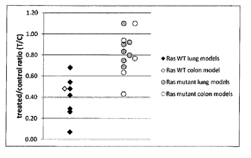
10064] In an evaluation of 514 known mutations in 41 distinct oncogenes and
tumor
suppressor genes in tumor samples from xenograft tumors and cell lines, there
were some
samples which were resistant to inhibition by a proteasome inhibitor.
Resistance to inhibition by
a proteasome inhibitor was correlated to the mutation status of KRAS gene.
Surprisingly, all of
the samples from resistant xenografts had a mutation in KRAS and nearly all of
the sensitive or
responsive samples had wild type KRAS. Accordingly, a patient with a solid
tumor whose
tumor cells comprise wild type KRAS can be a candidate for treatment with a
proteasome
- 20 -
CA 02855356 2014-05-09
WO 2013/071142 PCT/US2012/064496
inhibitor. In some embodiments, the solid tumor is non-small cell lung cancer,
colon cancer,
pancreatic cancer, breast cancer, ovarian cancer, melanoma, head and neck
carcinoma, prostate
cancer or renal cell carcinoma. In other embodiments, the solid tumor is non-
small cell lung
cancer, colon cancer, pancreatic cancer, breast cancer, ovarian cancer or
melanoma. In some
embodiments, the solid tumor is non-small cell lung cancer, colon cancer,
prostate cancer or
pancreatic cancer. In some embodiments, the solid tumor is prostate cancer,
pancreatic cancer,
non-small cell lung cancer or colon cancer. In some embodiments, the solid
tumor is prostate
cancer, non-small cell lung cancer or colon cancer. In some embodiments, the
solid tumor is
selected from the group consisting of lung cancer and colon cancer. In some
embodiments, the
lung cancer is non-small cell lung cancer or a metastatic form of colon
cancer. In other
embodiments, the solid tumor is non-small cell lung cancer or colon cancer. In
some
embodiments, the solid tumor is lung cancer. In some embodiments, the solid
tumor is non-
small cell lung cancer. In some embodiments, the solid tumor is colon cancer.
[0065] Compositions and methods are provided to determine the mutational
status, e.g., to
identify mutations in marker genes in solid tumor, e.g., non-hematological
tumor, e.g., non-small
cell lung cancer, colon cancer, pancreatic cancer, breast cancer, ovarian
cancer, melanoma, head
and neck carcinoma, prostate cancer or renal cell carcinoma, to predict
outcome, e.g., response to
treatment, time-to-progression or survival, upon treatment with a proteasome
inhibitor, e.g., a
peptidyl boronic acid or peptidyl epoxy ketone. In some embodiments,
compositions and
methods are provided to determine the mutational status of a solid tumor
selected from the group
consisting of non-small cell lung cancer, colon cancer and prostate cancer to
predict outcome of
treatment with a proteasome inhibitor, e.g., a peptidyl boronic acid.
[0066] Unless otherwise defined, all technical and scientific terms used
herein have the
meanings which are commonly understood by one of ordinary skill in the art to
which this
invention belongs. Generally, nomenclature utilized in connection with, and
techniques of cell
and tissue culture, molecular biology and protein and oligo- or polynucleotide
chemistry and
hybridization described herein are those known in the art. GenBank or GenPept
accession
numbers and useful nucleic acid and peptide sequences can be found at the
website maintained
by the National Center for Biotechnology Information, Bethesda, MD. The
content of all
database accession records (e.g., from Affymetrix HG133 annotation files,
Entrez, GenBank,
RefSeq, COSMIC) cited throughout this application (including the Tables) are
hereby
-21-
CA 02855356 2014-05-09
WO 2013/071142 PCT/US2012/064496
incorporated by reference. Standard techniques are used for recombinant DNA,
oligonucleotide
synthesis, protein purification, tissue culture and transformation and
transfection (e.g.,
electroporation, lipofection, etc). Enzymatic reactions, such as GTPase assay
for RAS activity or
assays, e.g., reporter assays, for RAS-activated signaling activity, are
performed according to
manufacturer's specifications or as commonly accomplished in the art or as
described herein.
Some methods for determining RAS localization and signaling are reviewed in
Prior and
Hancock (2011) Semin.Clin. Dev. Biol. Sep 8 epub; or found in Cuiffo and Ren
(2010) Blood
114:3598-3605 or reviewed in Lim et al. (1996) Eur. .1. Biochem. 242:171-185.
The foregoing
techniques and procedures generally are performed according to methods known
in the art, e.g.,
as described in various general and more specific references that are cited
and discussed
throughout the present specification. See e.g., Sambrook et al. (2000)
Molecular Cloning: A
Laboratory Manual (3rd ed., Cold Spring Harbor Laboratory Press, Cold Spring
Harbor, NY) or
Harlow, E. and Lane, D. (1988) Antibodies: A Laboratory Manual (Cold Spring
Harbor
Laboratory Press, Cold Spring Harbor, NY). The nomenclatures utilized in
connection with, and
the laboratory procedures and techniques of, analytical chemistry, synthetic
organic chemistry,
and medicinal and pharmaceutical chemistry described herein are known in the
art. Standard
techniques are used for chemical syntheses, chemical analyses, pharmaceutical
preparation,
formulation and delivery, and treatment of patients. Furthermore, unless
otherwise required by
context, singular terms shall include pluralities and plural terms shall
include the singular. In the
case of conflict, the present specification, including definitions, will
control.
[0067] The articles "a," "an" and "at least one" are used herein to refer
to one or to more than
one of the grammatical object of the article. By way of example, "an element"
means one or
more than one element, at least one element. In the case of conflict, the
present specification,
including definitions, will control.
[0068] As used herein, a "marker gene" or a "genotype marker gene" refers to a
gene which
can have a mutation, e.g., a genotype, such that its DNA, RNA and/or protein
has a
characteristic, e.g., size, sequence, composition, activity or amount(s) which
provide information
about prognosis or outcome (i.e., are "informative") upon treatment. Marker
genes, e.g.,
genotype marker genes, described herein as linked to outcome after proteasome
inhibitor, e.g.,
peptidyl boronic acid (e.g., bortezomib or ixazomib citrate) treatment are
examples of genes
within the chromosome locus markers described herein and are provided in Table
1. Sequences
-22 -
CA 02855356 2014-05-09
WO 2013/071142 PCT/US2012/064496
of mRNA, open reading frames and proteins corresponding to marker genes also
are listed in
Table 1. A marker gene, e.g., a genotype marker gene, listed in Table 1 can
have isoforms which
are either ubiquitous or have restricted expression. The DNA SEQ ID NOs in
Table 1 refer only
to the mRNA encoding the major or longest isoform and the protein SEQ ID NOs
represent at
least a precursor of such isoform and not necessarily the mature protein.
These sequences are
not intended to limit the marker gene identity to that isoform or precursor.
The additional
isoforms and mature proteins are readily retrievable and understandable to one
of skill in the art
by reviewing the information provided under the Entrez Gene (database
maintained by the
National Center for Biotechnology Information, Bethesda, MD) identified by the
ID number
listed in Table 1.
[0069] Table 1 Marker Gene Description
Marker Marker Gene Name Entrez Chromo- Start base End base SEQ ID NOs:
Gene ID Gene ID some pair pair
location
KRAS v-Ki-ras2 Kirsten 3845 12p 25358180 25403854 1, 2, 3
rat sarcoma viral
oncogene homolog
NRAS neuroblastoma RAS 4893 lp 115247085 115259515 4, 5, 6
viral (v-ras)
oncogene homolog
100701 As used herein, a "phenotype marker gene" refers to a marker gene in
which there is
no somatic DNA mutation, i.e., it has no genotype alteration, but its other
markers, e.g.,
transcript, e.g., RNA, and/or protein can have a characteristic, e.g., size,
sequence, composition,
activity or amount which provides information about prognosis or outcome
(i.e., is
"informative") upon treatment. This designation is not to be confused with the
characteristics,
such as composition, amount and activity, measured for a marker associated
with a genotype
marker gene but which are known in the art as phenotypic characteristics. A
phenotype marker
gene described herein as linked to outcome after proteasome inhibitor, e.g.,
peptidyl boronic acid
(e.g., bortezomib or ixazomib citrate) treatment includes GLUT4, SEQ ID NOs:7,
8, 9.
[0071] As used herein, "KRAS" refers to v-Ki-ras2 Kirsten rat sarcoma viral
oncogene
homolog, the gene associated with GenBank Accession No. NM_004985, SEQ ID NO:1
(open
reading frame is SEQ ID NO:2, nucleotides 182 to 748 of SEQ ID NO:!), encoding
GenPept
Accession No. NP 004976, SEQ ID NO:3, the predominant transcript variant of
KRAS gene on
- 23 -
CA 02855356 2014-05-09
WO 2013/071142 PCT/US2012/064496
chromosome 12. Other names for KRAS include KRAS2, and Noonan Syndrome 3
(NS3).
KRAS functions as an oncogene with GTPase activity and can be found on
chromosome 12.
KRAS interacts with the cell membrane and various effector proteins, such as
Akt and Cdc42,
which carry out its signaling function through the cytoskeleton and effects on
cell motility
(Fotiadou et al. (2007) MoL Cel. BioL 27:6742-6755).
100721 As used herein, "NRAS" refers to neuroblastoma RAS viral (v-ras)
oncogene
homolog, the gene associated with GenBank Accession No. NM_002524, SEQ ID NO:4
(open
reading frame is SEQ ID NO:5, nucleotides 255 to 824 of SEQ ID NO:4), encoding
GenPept
Accession No. NP 002515, SEQ ID NO:6). Other names for NRAS include Autoimmune
Lymphoproliferative Syndrome type IV (ALPS4), NRAS I, and Noonan Syndrome 6
(NS6).
NRAS functions as an oncogene with GTPase activity and can be found on
chromosome lp.
NRAS interacts with the cell membrane and various effector proteins, such as
Raf and RhoA,
which carry out its signaling function through the cytoskeleton and effects on
cell adhesion
(Fotiadou et al. (2007) Ma Gel. Biol. 27:6742-6755).
[0073] As used herein, "GLUT4" refers to glucose transporter-4, the gene
associated with
GenBank Accession No. NM 001042, SEQ ID NO:7 (open reading frame is SEQ ID
NO:8,
nucleotides 201 to 1730 of SEQ ID NO:7), encoding GenPept Accession No.
NP_001033, SEQ
ID NO:8). Another name for GLUT4 is solute carrier family 2 (facilitated
glucose transporter)
member 4 (SLC2A4). GLUT4 functions as a glucose transporter and can be found
on
chromosome 17p. GLUT4 cellular location can depend on the presence of insulin,
which
stimulates cells such as muscle and adipose tissue to move GLUT4 from
intracellular stores to
the cell surface to commence its function as a glucose transporter. Glucose
transporters,
including GLUT1, GLUT4 and GLUT9 can have higher than normal activity in tumor
cells to
allow higher levels of glucose metabolism than in normal cells (reviewed
Adekola et al. (2012)
24:650-654). GLUT1, GLUT3 and GLUT4 can be expressed in lung carcinoma (Ito et
al. (1999)
Histol. Histopathol. 14:895-904). KRAS mutant colorectal cancer cells showed
higher glucose
uptake and glycolysis and better growth and survival under nutrient stress
than wild type cells
(Yun et al. 2009 Science 325:1555). Those studies identified a correlation
between the
upregulation of GLUTI, glucose transporter 1, with mutant KRAS in colorectal
cancer cells, in
contrast with an earlier study (Noguchi et al. (2000) Cancer Lett. 154:137-
142).
-24 -
CA 02855356 2014-05-09
WO 2013/071142 PCT/US2012/064496
[0074] A "marker" as used herein, includes a material associated with a
marker gene which
has been identified as having a mutation in tumor cells of a patient and
furthermore that mutation
is characteristic of a patient whose outcome is favorable or unfavorable with
treatment e.g., by a
proteasome inhibitor, e.g., a peptidyl boronic acid or peptidyl epoxy ketone.
Examples of a
marker include a material, e.g., a chromosome locus, DNA for a gene, RNA for a
gene or protein
for a gene. For example, a marker includes a marker gene material, e.g., a
chromosome locus,
DNA, RNA or protein whose mutation or characteristic, e.g., size, sequence,
composition,
activity or amount is indicative of a patient with a poor response to
treatment; alternatively a
marker includes a marker gene material, e.g., a chromosome locus, DNA, RNA or
protein whose
mutation or characteristic, e.g., size, sequence, composition, activity or
amount is indicative of a
patient with a good response. In another example, a marker includes a marker
gene material,
e.g., a chromosome locus, DNA, RNA or protein whose mutation or
characteristic, e.g., size,
sequence, composition, activity or amount is indicative of a patient whose
disease has a short
time-to-progression (TTP) upon treatment; alternatively a marker includes a
marker gene
material, e.g., a chromosome locus, DNA, RNA or protein whose mutation or
characteristic, e.g.,
size, sequence, composition, activity or amount is indicative of a patient
whose disease has a
long TTP. In yet a further example, a marker includes a marker gene material,
e.g., a
chromosome locus, DNA, RNA or protein whose mutation or characteristic, e.g.,
size, sequence,
composition or amount is indicative of a patient whose disease has a short
term survival upon
treatment; alternatively a marker includes a marker gene material, e.g., a
chromosome locus,
DNA, RNA or protein whose mutation or characteristic, e.g., size, sequence,
composition or
amount is indicative of a patient whose disease has a long term survival. A
marker can include a
material associated with a marker gene which has not been identified as having
a mutation in
tumor cells of a patient, but whose characteristic, e.g., size, sequence,
composition, activity or
amount is indicative of response to proteasome inhibition treatment of a solid
tumor in a patient.
Thus, as used herein, a marker is intended to include each and every one of
these possibilities,
and further can include each single marker individually or independently as a
marker; or
alternatively can include one or more, or all of the characteristics
collectively when reference is
made to "markers" or "marker sets."
[0075] In some embodiments, the marker is selected from the group
consisting of nucleic acid
and protein. In some embodiments, the marker is nucleic acid. In some
embodiments, the
-25 -
CA 02855356 2014-05-09
WO 2013/071142 PCT/US2012/064496
nucleic acid marker is chromosomal or genomic DNA. In some embodiments, the
nucleic acid is
mRNA. In some embodiments, the nucleic acid is cDNA prepared from mRNA. In
some
embodiments, the marker is protein.
[0076] In some embodiments, the characteristic is selected from the group
consisting of size,
sequence, composition, activity and amount. In some embodiments, the
characteristic is
sequence. In some embodiments, the characteristic is amount. In some
embodiments, the
characteristic is composition. In some embodiments, the characteristic is
size. In some
embodiments, the characteristic is activity.
[0077] In some embodiments, the marker is chromosome DNA, or genomic DNA and
the
characteristic is sequence. In some embodiments, the marker is mRNA and the
characteristic is
sequence. In some embodiments, the marker is cDNA and the characteristic is
sequence. In
some embodiments, the marker is mRNA and the characteristic is amount. In some
embodiments, the marker is mRNA and the characteristic is size.
[0078] In some embodiments, the marker is protein and the characteristic is
amount. In some
embodiments, the marker is protein and the characteristic is activity. In some
embodiments, the
marker is protein and the characteristic is sequence.
[0079] A chromosome locus marker useful to measure for determination of
prognosis or
treatment or disease management strategy is selected from the group consisting
of chromosome
1p13.2 (NRAS), e.g., from base pair 115247085 to 115259515 and chromosome
12p12.1
(KRAS), e.g., from base pair 25358180 to 25403854. Chromosome locus and base
pair numbers
are based on the reference human genome Build 37.3 (current as of October 5,
2011) in the
NCBI Gene database. A marker DNA, marker RNA or marker protein can correspond
to base
pairs on a chromosome locus marker. For example, a marker DNA can include
genomic DNA
from a chromosome locus marker, marker RNA can include a polynucleotide
transcribed from a
locus marker, and a marker protein can include a polypeptide resulting from
expression at a
chromosome locus marker in a sample, e.g., comprising tumor cells.
[0080] A "marker nucleic acid" is a nucleic acid (e.g., genomic DNA, mRNA,
cDNA)
encoded by, associated with or corresponding to a marker gene of the
invention. Such marker
nucleic acids include DNA, e.g., sense and anti-sense strands of genomic DNA
(e.g., including
any introns occurring therein), comprising the entire or a partial sequence,
e.g., one or more of
the exons of the genomic DNA, up to and including the open reading frame of
any of the marker
-26 -
CA 02855356 2014-05-09
WO 2013/071142 PCT/US2012/064496
genes or the complement of such a sequence. The marker nucleic acids also
include RNA
comprising the entire or a partial sequence of any marker or the complement of
such a sequence,
wherein all thymidine residues are replaced with uridine residues, RNA
generated by
transcription of genomic DNA (i.e. prior to splicing), RNA generated by
splicing of RNA
transcribed from genomic DNA, and proteins generated by translation of spliced
RNA (i.e.
including proteins both before and after cleavage of normally cleaved regions
such as
transmembrane signal sequences). As used herein, a "marker nucleic acid" may
also include a
cDNA made by reverse transcription of an RNA generated by transcription of
genomic DNA
(including spliced RNA). A marker nucleic acid also includes sequences which
differ, due to
degeneracy of the genetic code, from the nucleotide sequence of nucleic acids
encoding a protein
which corresponds to a marker of the invention, and thus encode the same
protein or highly
similar protein, e.g., wild type protein or protein with polymorphism but wild
type function. As
used herein, the phrase "allelic variant" refers to a nucleotide sequence
which occurs at a given
locus or to a polypeptide encoded by the nucleotide sequence. Such naturally
occurring allelic
variations can typically result in 1-5% variance in the nucleotide sequence of
a given gene.
Alternative alleles can be identified by sequencing the gene of interest in a
number of different
individuals, e.g., in cells, e.g., germline cells, of individuals without
cancer. This can be readily
carried out by using hybridization probes to identify the same genetic locus
in a variety of
individuals. Detection of any and all such nucleotide variations and resulting
amino acid
polymorphisms or variations that are the result of naturally occurring allelic
variation and that do
not alter the functional activity of a wild type marker gene is intended to be
within the scope of
the wild type version of a marker described herein. A "marker protein" is a
protein encoded by
or corresponding to a marker, e.g., a nucleic acid, of the invention. The
terms "protein" and
"polypeptide' are used interchangeably. A protein of a marker specifically can
be referred to by
its name or amino acid sequence, but it is understood by those skilled in the
art, that mutations,
such as non-sense, missense, insertions or deletions can affect protein
structure, appearance,
cellular location and/or behavior in a variety of ways. Unless indicated
otherwise, such
differences are not distinguished herein, and a mutant marker described herein
is intended to
include any or all such varieties.
[0081] A typical tumor with a mutated RAS gene has a mutation in one RAS gene
or another,
not more than one. In the case of KRAS, an example of wild type nucleic acid
such as mRNA is
- 27 -
CA 02855356 2014-05-09
WO 2013/071142 PCT/US2012/064496
SEQ ID NO:1. Another example of wild type KRAS nucleic acid is SEQ ID NO:2. An
example
of wild type KRAS protein is SEQ ID NO:3. In some embodiments, a mutation can
be found in
at least one mutation site of a KRAS marker. In the present studies, mutations
were found in
codons 12, 13, 61 and 146 of KRAS (SEQ ID NO:2). In general embodiments, a
wild type RAS
gene, e.g., KRAS, is predictive of favorable outcome, e.g., responsiveness,
long time to
progression and/or long term survival, for solid, e.g., non-hematological
tumors, e.g., non-small
cell lung cancer or colon cancer, while a mutated KRAS gene is predictive of
unfavorable
outcome. In some embodiments, a mutation site comprising a mutation of codon
146 of KRAS
(SEQ ID NO:2) is not predictive of unfavorable outcome of treatment with a
proteasome
inhibitor. As used herein, a "mutation of codon 146" or "mutated codon 146"
refers to a rare
mutation in the open reading frame of KRAS, SEQ ID NO:2. A marker whose
characteristic is
informative of a mutated codon 146 can be a KRAS protein with an amino acid
sequence which
differs from SEQ ID NO:3 at amino acid residue 146. In some embodiments, a
protein marker
whose characteristic is informative of a favorable outcome in the methods has
a threonine instead
of an alanine at residue 146 of KRAS. A nucleic acid marker comprising a
fragment of at least
consecutive nucleotides of SEQ ID NO:1, or a complement thereof, wherein the
fragment
comprises bases 617 to 619, can have a characteristic informative of a
mutation of codon 146 if
at least one base selected from the group consisting of bases 617, 618 and 619
is mutated.
10082] Examples of mutant KRAS, whose occurrence in a solid tumor cancer
patient is
indicative of nonresponse or unfavorable outcome include marker nucleic acid
with at least one
change in at least one mutation site, such as at least one base of codon 12
(bases 34-36), codon
13 (bases 37-39) or codon 61 (bases 181-183) of SEQ ID NO:2, or the analogous
codons in SEQ
ID NO:1 (bases 215-217, bases 218-220 or bases 362-364, respectively of SEQ ID
NO:1), and
results in a change of at least one amino acid residue 12, 13 or 61 of SEQ ID
NO:3. In some
embodiments, an allelic variant of KRAS has a change in a codon that is not
codon 12, codon 13
or codon 61 of SEQ ID NO:2, or the analogous codons in SEQ ID NO:1, wherein
the resulting
encoded allelic variant protein has wild type RAS, e.g., GTPase, activity, and
thus is not
associated with unfavorable outcome. Alternatively, an allelic variant of KRAS
has a change in
a base of codon 12, codon 13, or codon 61 wherein the change, e.g., is due to
the degeneracy of
the genetic code and does not result in a change in the translated residue. In
such embodiments,
an allelic variant nucleic acid encodes a wild type amino acid residue, such
as glycine at position
-28-
CA 02855356 2014-05-09
WO 2013/071142 PCT/US2012/064496
12 of SEQ ID NO:3, glycine at position 13 of SEQ ID NO:3 and glutamine at
position 61 of SEQ
ID NO:3 and the encoded polypeptide has wild type RAS activity, e.g., GTPase
activity, and thus
is not associated with unfavorable outcome.
[0083] A nucleic acid marker comprising a fragment of at least 10
consecutive nucleotides of
SEQ ID NO:1, or a complement thereof, wherein the fragment comprises bases 215
to 217 can
have a characteristic informative of a mutation of codon 12 if at least one
base selected from the
group consisting of bases 215, 216 and 217 is mutated in a manner that can
result in a change of
the amino acid residue 12 of SEQ ID NO:3. In some embodiments, the nucleic
acid marker
comprises bases 34 to 36 of SEQ ID NO:2. In some embodiments, a mutation in
KRAS nucleic
acid can result in a KRAS protein which has an amino acid residue selected
from the group
consisting of alanine, cysteine, aspartate, phenylalanine, arginine, serine
and valine instead of
glycine at residue 12 of SEQ ID NO:3. A nucleic acid marker comprising a
fragment of at least
consecutive nucleotides of SEQ ID NO:1, or a complement thereof, wherein the
fragment
comprises bases 218 to 220, can have a characteristic informative of a
mutation of codon 13 if at
least one base selected from the group consisting of bases 218, 219 and 220 is
mutated in a
manner that can result in a change of the amino acid residue 13 of SEQ ID
NO:3. In some
embodiments, the nucleic acid marker comprises bases 37 to 39 of SEQ ID NO:2.
In some
embodiments, a mutation in KRAS nucleic acid can result in a KRAS protein
which has an
amino acid residue selected from the group consisting of aspartate, valine,
cysteine, serine,
alanine and arginine instead of glycine at residue 13 of SEQ ID NO:3. A
nucleic acid marker
comprising a fragment of at least 10 consecutive nucleotides of SEQ ID NO:1,
or a complement
thereof, wherein the fragment comprises bases selected from the group
consisting of bases 362 to
364, can have a characteristic informative of a mutation of codon 61 if at
least one base selected
from the group consisting of bases 362, 363 and 364 is mutated in a manner
that can result in a
change of the amino acid residue 61 of SEQ ID NO:3. In some embodiments, the
nucleic acid
marker comprises bases 181 to 183 of SEQ ID NO:2. In some embodiments, a
mutation in
KRAS nucleic acid can result in a KRAS protein which has an amino acid residue
selected from
the group consisting of glutamate, histidine, lysine, !mein; proline and
arginine instead of
glutamine at residue 61 of SEQ ID NO:3.
[0084] As used herein, an "informative" characteristic, e.g., size,
sequence, composition,
activity or amount of a marker refers to a characteristic, e.g., size,
sequence, composition,
- 29 -
CA 02855356 2014-05-09
WO 2013/071142 PCT/US2012/064496
activity or amount whose value or difference is correlated to prognosis or
outcome. The
informative characteristic, e.g., size, sequence, composition, activity or
amount of a marker can
be obtained by analyzing either nucleic acid, e.g., DNA or RNA, or protein
corresponding to the
marker gene, e.g., a genotype marker gene or phenotype marker gene. The
characteristic, e.g.,
size (e.g., length or molecular weight), sequence (e.g., nucleic acid sequence
or protein
sequence), composition (e.g., base or amino acid composition or peptide digest
or gene fragment
pattern) , activity (enzymatic activity or signaling activity) or amount
(e.g., copy number and/or
expression level) of a marker, e.g., a chromosome locus marker or a marker in
a sample from a
patient is "informative" if it is different than the wild type or allelic
variant of the substance
being analyzed. In some embodiments, a characteristic of a marker is
informative if it indicates
that the marker gene is wild type. In some embodiments where the amount of a
marker is being
measured, an amount is "informative" if it is greater than or less than a
reference amount by a
degree greater than the standard error of the assay employed to assess
expression. The
informative expression level of a marker can be determined upon statistical
correlation of the
measured expression level and the outcome, e.g., good response, poor response,
long time-to-
progression, short time-to-progression, short term survival or long term
survival. The result of
the statistical analysis can establish a threshold for selecting markers to
use in the methods
described herein. Alternatively, a marker, e.g., a chromosome locus marker, or
a marker gene
that has differential characteristic, e.g., size, sequence, composition,
activity or amounts will
have typical ranges of amounts that are predictive of outcome. An informative
characteristic,
e.g., size, sequence, composition, activity or amount is a characteristic,
e.g., size, sequence,
composition, activity or amount that falls within the range of characteristic,
e.g., size, sequence,
composition, activity or amounts determined for the outcome. Still farther, a
set of markers may
together be "informative" if the combination of their characteristics, e.g.,
sizes, sequences,
compositions, activities or amounts either meets or is above or below a pre-
determined score for
a marker, e.g., a chromosome locus marker, or a marker gene set as determined
by methods
provided herein. Gene translocation, transcript splice variation, deletion and
truncation are
examples of events which can change marker size, sequence or composition, in
addition to point
mutations which can change marker sequence or composition. Measurement of only
one
characteristic, e.g., marker, of a marker gene (i.e., DNA, RNA or protein) can
provide a
prognosis, i.e., indicate outcome. Measurement of only one characteristic,
e.g., marker, of a
- 30 -
CA 02855356 2014-05-09
WO 2013/071142 PCT/US2012/064496
marker gene (i.e., DNA, RNA or protein) can provide a prognosis, i.e., predict
or indicate
outcome. Measurement of more than one characteristic, e.g., marker, of a
marker gene can
provide a prognosis when the informative results of the two characteristics
are consistent with
each other, i.e., the biologies of the results are not contradictory. Examples
of consistent results
from measurement of multiple characteristics of a marker gene can be
identification of a
nonsense mutation, point mutation, insertion or deletion in a DNA or RNA and a
low amount or
altered molecular weight of encoded protein, or a mutation in a region which
encodes a binding
pocket or active site of a protein and altered activity of the encoded
protein. A different example
can occur when a protein is in a pathway with a feedback loop controlling its
synthesis based on
its activity level. In this example, a low amount or activity of protein can
be associated with a
high amount of its mutated mRNA as a tissue, due to the marker gene mutation,
thus is starved
for the protein activity and repeatedly signals the production of the protein.
[0085] By way of non-limiting illustration, in the present case, an example
of an informative
result upon measuring a characteristic, e.g, a sequence, of a KRAS marker,
e.g., DNA, mRNA,
or protein, would be a result identifying the mutational status of a KRAS
sequence, e.g., SEQ ID
NO:1, 2 or 3. In the present case, identifying a mutation in the KRAS sequence
in a sample
comprising tumor cells would be informative of an unfavorable outcome, while
identifying wild
type sequence would be informative of a favorable outcome of treatment of the
tumor with a
proteasome inhibitor. In one embodiment, identifying an A146T mutation in KRAS
would be
informative of a favorable outcome. In another example, measuring a
characteristic, e.g, activity
of a KRAS marker, e.g, protein, would provide an informative result of a
favorable outcome if
there is a high GTPase activity or a low signaling activity.
[0086] A "normal" characteristic, e.g., size, sequence, composition,
activity or amount of a
marker may refer to the characteristic, e.g., size, sequence, composition,
activity or amount in a
"reference sample." A reference sample can be a matched normal, e.g.,
germline, sample from
the same patient from whom the tumor, e.g., with a somatic mutation, is
derived. A reference
sample can be a sample from a healthy subject not having the marker-associated
disease or a
reference characteristic e.g., the average characteristic, e.g., size,
sequence, composition, activity
or amount of the wild type marker in several healthy subjects. A reference
sample characteristic,
e.g., size, sequence, composition, activity or amount may be comprised of a
characteristic, e.g.,
size, sequence, composition, activity or amount of one or more markers from a
reference
-31 -
CA 02855356 2014-05-09
WO 2013/071142 PCT/US2012/064496
database. Alternatively, a "normal" characteristic, e.g., size, sequence,
composition, activity or
level of expression of a marker is the characteristic, e.g., size, sequence,
composition, activity or
amount of the marker, e.g., marker gene in non-tumor cells in a similar
environment or response
situation from the same patient from whom the tumor is derived. The normal
amount of DNA
copy number is 2 or diploid, with the exception of X-linked genes in males,
where the normal
DNA copy number is I.
[0087] "Over-expression" and "under-expression" of a marker gene, refer to
expression of the
marker gene of a patient at a greater or lesser level (e.g. more than three-
halves-fold, at least two-
fold, at least three-fold, greater or lesser level etc.), respectively, than
normal level of expression
of the marker gene, e.g., as measured by mRNA or protein, in a test sample
that is greater than
the standard error of the assay employed to assess expression. A "significant"
expression level
may refer to a level which either meets or is above or below a pre-determined
score for a marker
gene set as determined by methods provided herein.
[0088] As used herein, "gene deletion" refers to an amount of DNA copy number
less than 2
and "amplification" refers to an amount of DNA copy number greater than 2. A
"diploid"
amount refers to a copy number equal to 2. The term "diploid or amplification"
can be
interpreted as "not deletion" of a gene copy. In a marker whose alternative
informative amount
is gene deletion, amplification generally would not be seen. Conversely, the
term "diploid or
deletion" can be interpreted as "not amplification" of copy number. In a
marker whose
alternative informative amount is amplification, gene deletion generally would
not be seen. For
the sake of clarity, sequence deletion can occur within a gene as a result of
marker gene mutation
and can result in absence of transcribed protein or a shortened mRNA or
protein. Such a deletion
may not affect copy number.
[0089] As used herein, a "favorable" outcome or prognosis refers to long
time-to-progression
(TTP, or progression-free survival), long term survival, and/or good response.
Conversely, an
"unfavorable" outcome or prognosis refers to short term survival, short time-
to-progression
(TTP, or progression-free survival) and/or poor response.
[0090] The terms "long term survival" and "short term survival" refer to
the length of time after
receiving a first dose of treatment that a cancer patient is predicted to
live. A "long term survivor" refers
to a patient expected have a slower rate of progression or later death from
the tumor than those patients
identified as short term survivors. "Enhanced survival" or "a slower rate of
death" are estimated life span
- 32 -
CA 02855356 2014-05-09
WO 2013/071142 PCT/US2012/064496
determinations based upon characteristic, e.g., size, sequence, composition,
activity or amount of
one or more of markers described herein, e.g., as compared to a reference
standard such that 70%, 80%,
90% or more of the population will be alive a sufficient time period after
receiving a first dose of
treatment. A "faster rate of death" or "shorter survival time" refer to
estimated life span determinations
based upon characteristic, e.g., size, sequence, composition, activity or
amount of one or more of
markers described herein, e.g., as compared to a reference standard such that
50%, 40%, 30%, 20%, 10%
or less of the population will not live a sufficient time period after
receiving a first dose of treatment. In
some embodiments, the sufficient time period is at least 6, 12, 18, 24 or 30
months measured from the
first day of receiving a cancer therapy.
[0091] A cancer is "responsive" to a therapeutic agent or there is a "good
response" to a
treatment if its rate of growth is inhibited as a result of contact with the
therapeutic agent,
compared to its growth in the absence of contact with the therapeutic agent.
Growth of a cancer
can be measured in a variety of ways, for instance, the characteristic, e.g.,
size of a tumor or the
expression of tumor markers appropriate for that tumor type may be measured.
International
Working Groups convene periodically to set, update and publish response
criteria for various
types of cancers. Such published reports can be followed to support the
identification of markers
of the subject tumors and their response to proteasome inhibitors. For
example, for solid tumors,
the Response Evaluation Criteria in Solid Tumors (RECIST) guidelines
(Eisenhauer et al. (2009)
E. J Canc. 45:228-247) can be used to support the identification of markers
associated with solid
tumors and response of solid tumors to a proteasome inhibitor. The response
definitions used to
support the identification of markers associated with rnyeloma and its
response to an proteasome
inhibitor, e.g., peptidyl boronic acid therapy, the Southwestern Oncology
Group (SWOG) criteria
as described in Blade et al. (1998) Br J Haematol. 102:1115-23 can be used.
These criteria
define the type of response measured in myeloma and also the characterization
of time to disease
progression which is another important measure of a tumor's sensitivity to a
therapeutic agent.
Other examples are criteria for Acute Myelogenous Leukemia (AML, Cheson et al.
(2003)
J.Clin. Oncol. 21:4642-4649), lymphomas, e.g., non-Hodgkin's and Hodgkin's
lymphoma
(Cheson et al. (2007) JClin. Oncol. 25:579-596). Criteria take into account
analysis methods
such as Positron Emission Tomography (PET), e.g., for identifying sites with
measurable altered
metabolic activity (e.g., at tumor sites) or to trace specific markers into
tumors in vivo,
immunohistochemistry, e.g., to identify tumor cells by detecting binding of
antibodies to specific
tumor markers, and flow cytometry, e.g., to characterize cell types by
differential markers and
- 33 -
CA 02855356 2014-05-09
WO 2013/071142 PCT/US2012/064496
fluorescent stains, in addition to traditional methods such as histology to
identify cell
composition (e.g., blast counts in a blood smear or a bone marrow biopsy,
presence and number
of mitotic figures) or tissue structure (e.g., disordered tissue architecture
or cell infiltration of
basement membrane). The quality of being responsive to a proteasome inhibitor,
e.g., a peptidyl
boronic acid therapy can be a variable one, with different cancers exhibiting
different levels of
"responsiveness" to a given therapeutic agent, under different conditions.
Still further, measures
of responsiveness can be assessed using additional criteria beyond growth size
of a tumor,
including patient quality of life, degree of metastases, etc. In addition,
clinical prognostic
markers and variables can be assessed (e.g., M protein in myeloma, PSA levels
in prostate
cancer) in applicable situations.
[0092] A cancer is "non-responsive" or has a "poor response" to a
therapeutic agent or there
is a poor response to a treatment if its rate of growth is not inhibited, or
inhibited to a very low
degree, as a result of contact with the therapeutic agent when compared to its
growth in the
absence of contact with the therapeutic agent. As stated above, growth of a
cancer can be
measured in a variety of ways, for instance, the size of a tumor or the
expression of tumor
markers appropriate for that tumor type may be measured. For example, the
response definitions
used to support the identification of markers associated with non- response of
tumors to
therapeutic agents, guidelines such as those described above can be used. The
quality of being
non-responsive to a therapeutic agent can be a highly variable one, with
different cancers
exhibiting different levels of "non-responsiveness" to a given therapeutic
agent, under different
conditions. Still further, measures of non-responsiveness can be assessed
using additional
criteria beyond growth size of a tumor, including patient quality of life,
degree of metastases, etc.
In addition, clinical prognostic markers and variables can be assessed (e.g.,
M protein in
myeloma, PSA levels in prostate cancer) in applicable situations.
[0093] As used herein, "long time-to-progression, "long TTP" and "short
time-to-
progression," "short TTP" refer to the amount of time until when the stable
disease brought by
treatment converts into an active or progressive disease. On occasion, a
treatment results in
stable disease (SD) which is neither a good nor a poor response, or MR, the
disease merely does
not get worse, e.g., does not become a progressive disease, for a period of
time. This period of
time can be at least 4-8 weeks, at least 3-6 months or more than 6 months.
-34 -
CA 02855356 2014-05-09
WO 2013/071142 PCT/US2012/064496
[0094] "Treatment" shall mean the use of a therapy to prevent or inhibit
further tumor growth,
as well as to cause shrinkage of a tumor, and to provide longer survival
times. Treatment is also
intended to include prevention of metastasis of tumor. A tumor is "inhibited"
or "treated" if at
least one symptom (as determined by responsiveness/non-responsiveness, time to
progression, or
indicators known in the art and described herein) of the cancer or tumor is
alleviated, terminated,
slowed, minimized, or prevented. Any amelioration of any symptom, physical or
otherwise, of a
tumor pursuant to treatment using a therapeutic regimen (e.g., proteasome
inhibitor, e.g., a
peptidyl boronic acid regimen) as further described herein, is within the
scope of the invention.
[0095] As used herein, the term "agent" is defined broadly as anything that
cancer cells,
including tumor cells, may be exposed to in a therapeutic protocol. In the
context of the present
invention, such agents include, but are not limited to, proteasome inhibitor,
e.g., a peptidyl
boronic acid agents, as well as chemotherapeutic agents as known in the art
and described in
further detail herein.
[0096] The term "probe" refers to any molecule, e.g., an isolated molecule,
which is capable
of selectively binding to a specifically intended target molecule, for example
a marker of the
invention. Probes can be either synthesized by one skilled in the art, or
derived from appropriate
biological preparations. For purposes of detection of the target molecule,
probes may be
specifically designed to be labeled, as described herein. Examples of
molecules that can be
utilized as probes include, but are not limited to, RNA, DNA, proteins,
antibodies, and organic
monomers.
[0097] "Complementary" refers to the broad concept of sequence
complementarity between
regions of two nucleic acid strands or between two regions of the same nucleic
acid strand. It is
known that an adenine residue of a first nucleic acid region is capable of
forming specific
hydrogen bonds ("base pairing") with a residue of a second nucleic acid region
which is
antiparallel to the first region if the residue is thymine or uracil.
Similarly, it is known that a
cytosine residue of a first nucleic acid strand is capable of base pairing
with a residue of a second
nucleic acid strand which is antiparallel to the first strand if the residue
is guanine. A first region
of a nucleic acid is complementary to a second region of the same or a
different nucleic acid if,
when the two regions are arranged in an antiparallel fashion, at least one
nucleotide residue of
the first region is capable of base pairing with a residue of the second
region. In some
embodiments, the first region comprises a first portion and the second region
comprises a second
-35-
CA 02855356 2014-05-09
WO 2013/071142 PCT/US2012/064496
portion, whereby, when the first and second portions are arranged in an
antiparallel fashion, at
least about 50%, at least about 75%, at least about 90%, or at least about 95%
or all of the
nucleotide residues of the first portion are capable of base pairing with
nucleotide residues in the
second portion.
[0098] "Homologous" as used herein, refers to nucleotide sequence
similarity between two
regions of the same nucleic acid strand or between regions of two different
nucleic acid strands.
When a nucleotide residue position in both regions is occupied by the same
nucleotide residue,
then the regions are homologous at that position. A first region is homologous
to a second
region if at least one nucleotide residue position of each region is occupied
by the same residue.
Homology between two regions is expressed in terms of the proportion of
nucleotide residue
positions of the two regions that are occupied by the same nucleotide residue
(i.e., by percent
identity). By way of example, a region having the nucleotide sequence 5'-
ATTGCC-3' and a
region having the nucleotide sequence 5'-TATGGC-3' share homology with 50%
identity. In
one embodiment, the first region comprises a first portion and the second
region comprises a
second portion, whereby, at least about 50%, at least about 75%, at least
about 90%, or at least
about 95% of the nucleotide residue positions of each of the portions are
occupied by the same
nucleotide residue. In some embodiments of 100% identity, all nucleotide
residue positions of
each of the portions are occupied by the same nucleotide residue.
[0099] Unless otherwise specified herewithin, the terms "antibody" and
"antibodies" broadly
encompass naturally-occurring forms of antibodies, e.g., polyclonal antibodies
(e.g., IgG, IgA,
IgM, IgE) and monoclonal and recombinant antibodies such as single-chain
antibodies, two-
chain and multi-chain proteins, chimeric, CDR-grafted, human and humanized
antibodies and
multi-specific antibodies, as well as fragments and derivatives of all of the
foregoing, which
fragments (e.g., dAbs, scFv, Fab, F(ab)'2, Fab') and derivatives have at least
an antigenic binding
site. Antibody derivatives may comprise a protein or chemical moiety
conjugated to an
antibody. The term "antibody" also includes synthetic and genetically
engineered variants.
1001001 A "kit" is any article of manufacture (e.g., a package or container)
comprising at least
one reagent, e.g. a probe, for specifically detecting a marker or marker set
of the invention. The
article of manufacture may be promoted, distributed, sold or offered for sale
as a unit for
performing, e.g., in vitro, the methods of the present invention, e.g., on a
sample having been
obtained from a patient. The reagents included in such a kit can comprise at
least one nucleic
- 36 -
CA 02855356 2014-05-09
WO 2013/071142 PCT/US2012/064496
acid probe and, optionally, one or more primers and/or antibodies for use in
analyzing one or
more markers described herein, e.g., detecting marker characteristics, e.g.,
size, sequence
composition or amount, e.g., expression. In addition, a kit of the present
invention can contain
instructions which describe a suitable detection assay. Such a kit can be
conveniently used, e.g.,
in a clinical or a contract testing setting, to generate information, e.g., on
expression levels,
characteristic, e.g., size, sequence, activity or composition of one or more
marker, to be recorded,
stored, transmitted or received to allow for diagnosis, evaluation or
treatment of patients
exhibiting symptoms of cancer, in particular patients exhibiting the possible
presence of a cancer
capable of treatment with proteasome inhibition therapy, including, e.g., non-
hematological
cancers e.g., non-small cell lung cancer, colon cancer, pancreatic cancer,
breast cancer, ovarian
cancer, melanoma, head and neck carcinoma, prostate cancer or renal cell
carcinoma.
[00101] The present methods and compositions are designed for use in
diagnostics and
therapeutics for a patient suffering from cancer. A cancer or tumor is treated
or diagnosed
according to the present methods. "Cancer" or "tumor" is intended to include
any neoplastic
growth in a patient, including an initial tumor and any metastases. The cancer
can be of the
hematological or solid tumor type. Hematological tumors include tumors of
hematological
origin, including, e.g., myelomas (e.g., multiple myeloma), leukemias (e.g.,
Waldenstrom's
syndrome, chronic lymphocytic leukemia, acute myelogenous leukemia, chronic
myelogenous
leukemia, other leukemias), lymphomas (e.g., B-cell lymphomas, non-Hodgkin's
lymphoma)
and myelodysplastic syndrome. Solid tumors can originate in organs, and
include cancers such
as in skin, lung, brain, breast, prostate, ovary, colon, kidney, pancreas,
liver, esophagus, stomach,
intestine, bladder, uterus, cervix, testis, adrenal gland, etc. The cancer can
comprise a cell in
which a marker gene has a mutation. As used herein, cancer cells, including
tumor cells, refer to
cells that divide at an abnormal (increased) rate or whose control of growth
or survival is
different than for cells in the same tissue where the cancer cell arises or
lives. Cancer cells
include, but are not limited to, cells in carcinomas, such as squamous cell
carcinoma, basal cell
carcinoma, sweat gland carcinoma, sebaceous gland carcinoma, adenocarcinoma,
papillary
carcinoma, papillary adenocarcinoma, cystadenocarcinoma, medullary carcinoma,
undifferentiated carcinoma, bronchogenic carcinoma, melanoma, renal cell
carcinoma,
hepatoma-liver cell carcinoma, bile duct carcinoma, cholangiocarcinoma,
papillary carcinoma,
transitional cell carcinoma, choriocarcinoma, semonoma, embryonal carcinoma,
mammary
- 37 -
CA 02855356 2014-05-09
WO 2013/071142 PCT/US2012/064496
carcinomas, gastrointestinal carcinoma, colonic carcinomas, bladder carcinoma,
prostate
carcinoma, and squamous cell carcinoma of the neck and head region; sarcomas,
such as ,
tibrosarcoma, myxosarcoma, liposarcoma, chondrosarcorna, osteogenic sarcoma,
chordosarcoma, angiosarcoma, endothelio sarcoma, lymphangiosarcoma,
synoviosarcoma and
mesotheliosarcoma; hematologic cancers, such as myelomas, leukemias (e.g.,
acute
myelogenous leukemia, chronic lymphocytic leukemia, granulocytic leukemia,
monocytic
leukemia, lymphocytic leukemia), and lymphomas (e.g., follicular lymphoma,
mantle cell
lymphoma, diffuse large Bcell lymphoma, malignant lymphoma, plasmocytoma,
reticulum cell
sarcoma, or Hodgkins disease); and tumors of the nervous system including
glioma, meningoma,
medulloblastoma, schwannoma or epidymoma.
[00102] As used herein, the term "noninvasive" refers to a procedure which
inflicts minimal
harm to a subject. In the case of clinical applications, a noninvasive
sampling procedure can be
performed quickly, e.g., in a walk-in setting, typically without anaesthesia
and/or without
surgical implements or suturing. Examples of noninvasive samples include
blood, serum, saliva,
urine, buccal swabs, throat cultures, stool samples and cervical smears.
Noninvasive diagnostic
analyses include x-rays, magnetic resonance imaging, positron emission
tomography, etc.
[00103] Described herein is the assessment of outcome for treatment of a tumor
through
measurement of the amount of pharmacogenomic markers, e.g, the mutation status
of a genotype
marker gene, e.g., RAS. Methods of the invention can be characterized as
comprising detecting,
in a sample of cells or nucleic acid from the patient, the presence or absence
of wild type or
mutant KRAS gene. The mutations can be: (i) a difference in the identity of at
least one
nucleotide or (ii) a difference in the number of nucleotides, which difference
can be a single
nucleotide or several nucleotides. The invention also provides methods for
detecting differences
in KRAS genes such as chromosomal rearrangements, e.g., chromosomal
dislocation. Also
described are assessing the outcome by noninvasive, convenient or low-cost
means, for example,
from blood samples. Typical methods to determine extent of cancer or outcome
of a solid, e.g.,
non-hematological tumor, e.g non-small cell lung cancer, colon cancer,
pancreatic cancer, breast
cancer, ovarian cancer, melanoma, head and neck carcinoma, prostate cancer or
renal cell
carcinoma can employ biopsy to collect tissue for genotype or phenotype, e.g.,
histological
analysis. The invention provides methods for determining, assessing, advising
or providing an
appropriate therapy regimen for treating a tumor or managing disease in a
patient. Monitoring a
-38-
CA 02855356 2014-05-09
WO 2013/071142 PCT/US2012/064496
treatment using the kits and methods disclosed herein can identify the
potential for unfavorable
outcome and allow their prevention, and thus a savings in morbidity, mortality
and treatment
costs through adjustment in the therapeutic regimen, cessation of therapy or
use of alternative
therapy.
[00104] The term "sample" is intended to include a sample, e.g, tissue, cells,
biological fluids
and isolates thereof, isolated from a subject, as well as tissues, cells and
fluids present within a
subject and can be obtained from a patient or a normal subject. In
hematological tumors of the
bone marrow, e.g., myeloma tumors, primary analysis of the tumor can be
performed on bone
marrow samples, e.g., samples which comprise myeloma tumor cells. However,
some tumor
cells, (e.g., clonotypic tumor cells, circulating endothelial cells), are a
percentage of the cell
population in whole blood. These cells also can be mobilized into the blood
during treatment of
the patient with granulocyte-colony stimulating factor (G-CSF) in preparation
for a bone marrow
transplant, a standard treatment for hematological tumors, e.g., leukemias,
lymphomas and
myelomas. Examples of circulating tumor cells in multiple myeloma have been
studied e.g., by
Pilarski et al. (2000) Blood 95:1056-65 and Rigolin et al. (2006) Blood
107:2531-5. Thus,
noninvasive samples, e.g., for in vitro measurement of markers to determine
outcome of
treatment, can include peripheral blood samples. Accordingly, cells within
peripheral blood can
be tested for marker amount. For patients with hematological tumors, a
control, reference
sample for normal characteristic, e.g., size, sequence, composition, activity
or amount can be
obtained from skin or a buccal swab of the patient. For solid tumors, a
typical sample
comprising tumor cells is a biopsy of the primary tumor or neighboring lymph
nodes. Solid
tumor samples obtained by less invasive means e.g., shed or scraped from the
tumor site include
a cervical smear (e.g., from a cervical cancer patient), tumor exudate, e.g.,
lymph fluid, cystic
fluid, nipple aspirate (e.g., from a breast cancer patient), ascites fluid,
pleural fluid, sputum (e.g.,
from lung cancer patient), gynecological fluids (e.g., from an ovarian cancer
patient), urine, stool
(e.g., for colon cancer). For solid tumors, a control, reference sample for
normal characteristic,
e.g., size, sequence, composition, activity or amount can be obtained from
blood of the patient.
[00105] In some embodiments, tumor cells are selected from the group
consisting of lung
cancer cells and colon cancer cells. In some embodiments, a sample comprising
solid tumor
cells comprises lung cancer cells. In some embodiments, a sample comprising
solid tumor cells
- 39 -
CA 02855356 2014-05-09
WO 2013/071142 PCT/US2012/064496
comprises non-small cell lung cancer cells. In some embodiments, a sample
comprising solid
tumor cells comprises colon cancer cells.
[00106] Blood collection containers can comprise an anti-coagulant, e.g.,
heparin or ethylene-
diaminetetraacetic acid (EDTA), sodium citrate or citrate solutions with
additives to preserve
blood integrity, such as dextrose or albumin or buffers, e.g., phosphate. If
the amount of marker
is being measured by measuring the level of its DNA in the sample, a DNA
stabilizer, e.g., an
agent that inhibits DNAse, can be added to the sample. If the amount of marker
is being
measured by measuring the level of its RNA in the sample, an RNA stabilizer,
e.g., an agent that
inhibits RNAse, can be added to the sample. If the amount of marker is being
measured by
measuring the level of its protein in the sample, a protein stabilizer, e.g.,
an agent that inhibits
proteases, can be added to the sample. An example of a blood collection
container is
PAXGENE tubes (PREANALYTIX, Valencia, CA), useful for RNA stabilization upon
blood
collection. Peripheral blood samples or tumor exudates can be modified, e.g.,
fractionated,
sorted or concentrated (e.g., to result in samples enriched with tumor or
depleted of tumor (e.g.,
for a reference sample)). Examples of modified samples include clonotypic
myeloma cells,
which can be collected by e.g., negative selection, e.g., separation of white
blood cells from red
blood cells (e.g., differential centrifugation through a dense sugar or
polymer solution (e.g.,
FICOLL solution (Amersham Biosciences division of GE healthcare, Piscataway,
NJ) or
HISTOPAQUE0-1077 solution, Sigma-Aldrich Biotechnology LP and Sigma-Aldrich
Co., St.
Louis, MO)) and/or positive selection by binding B cells to a selection agent
(e.g., a reagent
which binds to a tumor cell or myeloid progenitor marker, such as CD34, CD38,
CD138, or
CD133, for direct isolation (e.g., the application of a magnetic field to
solutions of cells
comprising magnetic beads (e.g., from Miltenyi Biotec, Auburn, CA) which bind
to the B cell
markers) or fluorescent-activated cell sorting). Non-myeloma samples, e.g.,
tumor exudates
from solid tumors, can be treated by similar methods as myeloma samples to
enrich for tumor
cells, e.g., using tumor cell selection markers known in the art.
[00107] Alternatively, a tumor cell line, e.g., HCT-116, A549, NCI-H1975, NCI-
H1650, HCC-
827, SW48, Calu-1, OCI-Ly3, OCI-Ly10 cell (Alizadeh et al. (2000) Nature
403:503-511), a
RPM! 6666 cell, a SUP-B15 cell, a KG-1 cell, a CCRF-SB cell, an 8ES cell, a
Kasumi-1 cell, a
Kasumi-3 cell, a BDCM cell, an HL-60 cell, a Mo-B cell, a JM1 cell, a GA-10
cell or a B-cell
lymphoma (e.g., BC-3) or a cell line or a collection of tumor cell lines (see
e.g., McDermott et al.
- 40 -
CA 02855356 2014-05-09
WO 2013/071142 PCT/US2012/064496
(2007) PNAS 104:19936-19941 or ONCOPANELTM anti-cancer tumor cell profiling
screen
(Ricerca Biosciences, Bothell, WA)) can be assayed. A skilled artisan readily
can select and
obtain the appropriate cells (e.g., from American Type Culture Collection
(ATCCO), Manassas,
VA) that are used in the present method. If the compositions or methods are
being used to
predict outcome of treatment in a patient or monitor the effectiveness of a
therapeutic protocol,
then a tissue or blood sample having been obtained from the patient being
treated is a useful
source of cells or marker gene or gene products for an assay.
[00108] The sample, e.g., tumor, e.g., biopsy or bone marrow, blood or
modified blood, (e.g.,
comprising tumor cells), tumor exudate and/or the reference, e.g., matched
control (e.g.,
germline), sample can be subjected to a variety of well-known post-collection
preparative and
storage techniques (e.g., nucleic acid and/or protein extraction, fixation,
storage, freezing,
ultrafiltration, concentration, evaporation, centrifugation, etc.) prior to
assessing the amount of
the marker in the sample.
[00109] In some embodiments, a mutation in a marker can be identified by
sequencing a
nucleic acid, e.g., a DNA, RNA, cDNA or a protein correlated with the marker
gene, e.g., a
genotype marker gene, e.g., KRAS. There are several sequencing methods known
in the art to
sequence nucleic acids. A nucleic acid primer can be designed to bind to a
region comprising a
potential mutation site or can be designed to complement the mutated sequence
rather than the
wild type sequence. Primer pairs can be designed to bracket a region
comprising a potential
mutation in a marker gene. A primer or primer pair can be used for sequencing
one or both
strands of DNA corresponding to the marker gene. A primer can be used in
conjunction with a
probe, e.g., a nucleic acid probe, e.g., a hybridization probe, to amplify a
region of interest prior
to sequencing to boost sequence amounts for detection of a mutation in a
marker gene.
Examples of regions which can be sequenced include an entire gene, transcripts
of the gene and a
fragment of the gene or the transcript, e.g., one or more of exons or
untranslated regions or a
portion of a marker comprising a mutation site. Examples of mutations to
target for primer
selection and sequence or composition analysis can be found in public
databases which collect
mutation information, such as COSMIC and dbGaP. Some mutations of a marker
gene such as
KRAS are listed in Example 1 and in Table 5 in the Examples as examples of
mutations that can
=be associated with resistance to proteasome inhibition, e.g., inhibition by a
peptidyl boronic acid,
e.g., bortezomib or ixazomib citrate.
- 41 -
CA 02855356 2014-05-09
WO 2013/071142 PCT/US2012/064496
100110] Sequencing methods are known to one skilled in the art. Examples of
methods include
the Sanger method, the SEQUENOMTm method and Next Generation Sequencing (NGS)
methods. The Sanger method, comprising using electrophoresis, e.g., capillary
electrophoresis to
separate primer-elongated labeled DNA fragments, can be automated for high-
throughput
applications. The primer extension sequencing can be performed after PCR
amplification of
regions of interest. Software can assist with sequence base calling and with
mutation
identification. SEQUENOMTm MASSARRAY sequencing analysis (San Diego, CA) is a
mass-spectrometry method which compares actual mass to expected mass of
particular fragments
of interest to identify mutations. NGS technology (also called "massively
parallel sequencing"
and "second generation sequencing") in general provides for much higher
throughput than
previous methods and uses a variety of approaches (reviewed in Zhang et al.
(2011) 1 Genet.
Genomics 38:95-109 and Shendure and Hanlee (2008) Nature Biotech. 26:1135-
1145). NGS
methods can identify low frequency mutations in a marker in a sample. Some NGS
methods
(see, e.g., GS-FLX Genome Sequencer (Roche Applied Science, Branford, CT),
Genome
analyzer (Illumina, Inc. San Diego, CA) SOLIDTM analyzer (Applied Biosystems,
Carlsbad,
CA), Polonator G.007 (Dover Systems, Salem, NH), HELISCOPETM (Helicos
Biosciences
Corp., Cambridge, MA)) use cyclic array sequencing, with or without clonal
amplification of
PCR products spatially separated in a flow cell and various schemes to detect
the labeled
modified nucleotide that is incorporated by the sequencing enzyme (e.g.,
polymerase or ligase).
In one NGS method, primer pairs can be used in PCR reactions to amplify
regions of interest.
Amplified regions can be ligated into a concatenated product. Clonal libraries
are generated in
the flow cell from the PCR or ligated products and further amplified ("bridge"
or "cluster" PCR)
for single-end sequencing as the polymerase adds a labeled, reversibly
terminated base that is
imaged in one of four channels, depending on the identity of the labeled base
and then removed
for the next cycle. Software can aid in the comparison to genomic sequences to
identify
mutations. Another NGS method is exome sequencing, which focuses on sequencing
exons of
all genes in the genome. As with other NGS methods, exons can be enriched by
capture methods
or amplification methods.
[00111] Composition of proteins and nucleic acids can be determined by many
ways known in
the art, such as by treating them in ways that cleave, degrade or digest them
and then analyzing
the components. Mass spectrometry, electrophoresis and chromatography can
separate and
- 42 -
CA 02855356 2014-05-09
WO 2013/071142 PCT/US2012/064496
define components for comparison. Mutations which cause deletions or
insertions can be
identified by size or charge differences in these methods. Protein digestion
or restriction enzyme
nucleic acid digestion can reveal different fragment patterns after some
mutations. Antibodies
that recognize particular mutant amino acids in their structural contexts can
identify and detect
these mutations in samples (see below).
[00112] In some embodiments, DNA, e.g., genomic DNA corresponding to the wild
type or
mutated marker can be analyzed both by in situ and by in vitro formats in a
biological sample
using methods known in the art. DNA can be directly isolated from the sample
or isolated after
isolating another cellular component, e.g., RNA or protein. Kits are available
for DNA isolation,
e.g., QIAAMP@ DNA Micro Kit (Qiagen, Valencia, CA). DNA also can be amplified
using
such kits.
1001131 In another embodiment, rnRNA corresponding to the marker can be
analyzed both by
in situ and by in vitro formats in a biological sample using methods known in
the art. An
example of a method for measuring expression level is included in the
Examples. Many
expression detection methods use isolated RNA. For in vitro methods, any RNA
isolation
technique that does not select against the isolation of mRNA can be utilized
for the purification
of RNA from tumor cells (see, e.g., Ausubel et al., ed., Current Protocols in
Molecular Biology,
John Wiley & Sons, New York 1987-1999). Additionally, large numbers of tissue
samples can
readily be processed using techniques well known to those of skill in the art,
such as, for
example, the single-step RNA isolation process of Chomczynski (1989, U.S.
Patent No.
4,843,155). RNA can be isolated using standard procedures (see e.g.,
Chomczynski and Sacchi
(1987) Anal. Biochem.162:156-159), solutions (e.g., trizol, TRI REAGENT il)
(Molecular
Research Center, Inc., Cincinnati, OH; see U.S. Patent No. 5,346,994) or kits
(e.g., a QIAGEN01)
Group RNEASY isolation kit (Valencia, CA) or LEUKOLOCKTM Total RNA Isolation
System, Ambion division of Applied Biosystems, Austin, TX).
1001141 Additional steps may be employed to remove DNA from RNA samples. Cell
lysis can
be accomplished with a nonionic detergent, followed by microcentrifugation to
remove the
nuclei and hence the bulk of the cellular DNA. DNA subsequently can be
isolated from the
nuclei for DNA analysis. In one embodiment, RNA is extracted from cells of the
various types
of interest using guanidinium thiocyanate lysis followed by CsC1
centrifugation to separate the
RNA from DNA (Chirgwin et al. (1979) Biochemistry 18:5294-99). Poly(A)+RNA is
selected
- 43 -
CA 02855356 2014-05-09
WO 2013/071142 PCT/US2012/064496
by selection with oligo-dT cellulose (see Sambrook et al. (1989) Molecular
Cloning--A
Laboratory Manual (2nd ed.), Cold Spring Harbor Laboratory, Cold Spring
Harbor, N.Y.).
Alternatively, separation of RNA from DNA can be accomplished by organic
extraction, for
example, with hot phenol or phenol/chloroform/isoamyl alcohol. If desired,
RNAse inhibitors
may be added to the lysis buffer. Likewise, for certain cell types, it may be
desirable to add a
protein denaturation/digestion step to the protocol. For many applications, it
is desirable to
enrich mRNA with respect to other cellular RNAs, such as transfer RNA (tRNA)
and ribosomal
RNA (rRNA). Most mRNAs contain a poly(A) tail at their 3' end. This allows
them to be
enriched by affinity chromatography, for example, using oligo(dT) or poly(U)
coupled to a solid
support, such as cellulose or SEPHADEX® medium (see Ausubel et al. (1994)
Current
Protocols In Molecular Biology, vol. 2, Current Protocols Publishing, New
York). Once bound,
poly(A)+mRNA is eluted from the affinity column using 2 mM EDTA/0.1% SDS.
[00115] A characteristic of a marker of the invention in a sample, e.g., after
obtaining a sample
(e.g., a bone marrow sample, a tumor biopsy or a reference sample) from a test
subject, can be
assessed by any of a wide variety of well known methods for detecting or
measuring the
characteristic, e.g., of a marker or plurality of markers, e.g., of a nucleic
acid (e.g., RNA, mRNA,
genomic DNA, or cDNA) and/or translated protein. Non-limiting examples of such
methods
include immunological methods for detection of secreted, cell-surface,
cytoplasmic, or nuclear
proteins, protein purification methods, protein function or activity assays,
nucleic acid
hybridization methods, optionally including "mismatch cleavage" steps (Myers,
et al. (1985)
Science 230:1242) to digest mismatched, i.e. mutant or variant, regions and
separation and
identification of the mutant or variant from the resulting digested fragments,
nucleic acid reverse
transcription methods, and nucleic acid amplification methods and analysis of
amplified
products. These methods include gene array/chip technology, RT-PCR, TAQMA1\101
gene
expression assays (Applied Biosystems, Foster City, CA), e.g., under GLP
approved laboratory
conditions, in situ hybridization, immunohistochemistry, immunoblotting, FISH
(flourescence in
situ hybridization), FACS analyses, northern blot, southern blot, INFINIUMO
DNA analysis
Bead Chips (Illumina, Inc., San Diego, CA), quantitative PCR, bacterial
artificial chromosome
arrays, single nucleotide polymorphism (SNP) arrays (Affymetrix, Santa Clara,
CA) or
cytogenetic analyses.
-44 -
CA 02855356 2014-05-09
WO 2013/071142 PCT/US2012/064496
[00116] Examples of techniques for detecting differences of at least one
nucleotide between
two nucleic acids include, but are not limited to, selective oligonucleotide
hybridization,
selective amplification, or selective primer extension. For example,
oligonucleotide probes can
be prepared in which the known polymorphic nucleotide is placed centrally
(allele- or mutant-
specific probes) and then hybridized to target DNA under conditions which
permit hybridization
only if a perfect match is found (Saiki et aL (1986) Nature 324:163); Saiki et
al (1989) Proc.
Nail Acad. Sci USA 86:6230; and Wallace et al. (1979) Nucl. Acids Res.
6:3543). Such allele
specific oligonucleotide hybridization techniques can be used for the
simultaneous detection of
several nucleotide changes in different polymorphic or mutated regions of
KRAS. For example,
oligonucleotides having nucleotide sequences of specific allelic variants or
mutants are attached
to a solid support, e.g., a hybridizing membrane and this support, e.g.,
membrane, is then
hybridized with labeled sample nucleic acid. Analysis of the hybridization
signal thus can reveal
the identity of the nucleotides of the sample nucleic acid.
[00117] The detection methods of the invention can thus be used to detect RNA,
inRNA,
protein, cDNA, or genomic DNA, for example, in a biological sample in vitro as
well as in vivo.
Furthermore, in vivo techniques for detection of a polypeptide or nucleic acid
corresponding to a
marker of the invention include introducing into a subject a labeled probe to
detect the
biomarker, e.g., a nucleic acid complementary to the transcript of a biomarker
or a labeled
antibody, Fe receptor or antigen directed against the polypeptide, e.g., wild
type or mutant
marker. For example, the antibody can be labeled with a radioactive isotope
whose presence and
location in a subject can be detected by standard imaging techniques. These
assays can be
conducted in a variety of ways. A skilled artisan can select from these or
other appropriate and
available methods based on the nature of the marker(s), tissue sample and
mutation in question.
Some methods are described in more detail in later sections. Different methods
or combinations
of methods could be appropriate in different cases or, for instance in
different types of tumors or
patient populations.
[00118] In vitro techniques for detection of a polypeptide corresponding to a
marker of the
invention include enzyme linked immunosorbent assays (ELISAs), Western blots,
protein array,
immunoprecipitations, immunohistochemistry and immunofluorescence. In such
examples,
expression of a marker is assessed using an antibody (e.g., an unlabeled, a
radio-labeled,
chromophore-labeled, fluorophore-labeled, or enzyme-labeled antibody), an
antibody derivative
-45 -
CA 02855356 2014-05-09
WO 2013/071142 PCT/US2012/064496
(e.g., an antibody conjugated with a substrate or with the protein or ligand
of a protein-ligand
pair (e.g., biotin-streptavidin)), or an antibody fragment (e.g., a single-
chain antibody, an isolated
antibody hypervariable domain, etc.) which binds specifically with a marker
protein or fragment
thereof, e.g., a protein or fragment comprising a region which can be mutated
or a portion
comprising a mutated sequence, such as a mutation site, or a mutated residue
in its structural
context, including a marker protein which has undergone all or a portion of
its normal post-
translational modification. An antibody can detect a protein with an amino
acid sequence
selected from the group consisting of SEQ ID NO:3, 6 and 9. Alternatively, an
antibody can
detect a mutated protein with a variant amino acid sequence selected from the
group consisting
of a mutant of SEQ ID NO:3 and 6. Residues listed as mutated in public
databases such as
COSMIC of dbGaP can be prepared in immunogenic compositions for generation of
antibodies
that will specifically recognize and bind to the mutant residues. Another
method can employ
pairs of antibodies, wherein one of the pair would bind a marker protein
upstream, i.e. N-
terminal to the region of expected mutation, e.g., nonsense mutation, point
mutation, insertion or
deletion and the other of the pair would bind the protein downstream. Wild
type protein would
bind both antibodies of the pair, but a protein with a nonsense mutation,
point mutation, insertion
or deletion mutation would bind only the N-terminal antibody of the pair. An
assay such as a
sandwich ELISA assay could detect a loss of quantity of the wild type protein
in the tumor
sample, e.g., in comparison to the reference sample, or a standard ELISA would
comparison of
the levels of binding of the antibodies to infer that a mutation is present in
a tumor sample.
[00119] Indirect methods for determining the amount or functionality of a
protein marker also
include measurement of the activity of the protein. For example, a sample, or
a protein isolated
from the sample or expressed from nucleic acid isolated, cloned or amplified
from the sample
can be assessed for marker protein activity. For a RAS oncogene, an activating
mutation can be
measured as reduced GTPase activity or altered binding to RasGAP or a cell
membrane.
[00120] In some embodiments, the method includes measuring the amount of
GLUT4. In
some embodiments, an assay to measure GLUT4 expression uses an antibody which
binds to
SEQ ID NO:9. In some embodiments, quantification of GLUT4 expression measures,
in a
sample comprising tumor cells, the amount of binding an antibody which binds
to SEQ ID NO:9.
In some embodiments, the amount of GLUT4 is quantified by immunohistochemistry
of a tumor
biopsy. In some embodiments, the amount of GLUT4 is quantified by
immunohistochemistry of
-46 -
CA 02855356 2014-05-09
WO 2013/071142 PCT/US2012/064496
a tumor exudate. In some embodiments, the amount of GLUT4 is determined by a
score of
antibody binding or staining intensity. In some embodiments, the amount of
GLUT4 is
determined by comparison of the antibody binding or staining in a tumor cell
with a non-tumor
cell in the sample comprising tumor cells.
[00121] In one embodiment, expression of a marker is assessed by preparing
mRNA/cDNA
(i.e., a transcribed polynucleotide) from cells in a patient sample, and by
hybridizing the
mRNA/cDNA with a reference polynucleotide which is a complement of a marker
nucleic acid,
or a fragment thereof. cDNA can, optionally, be amplified using any of a
variety of polymerase
chain reaction methods prior to hybridization with the reference
polynucleotide. Expression of
one or more markers likewise can be detected using quantitative PCR to assess
the level of
expression of the marker(s). An example of the use of measuring mRNA levels is
that an
inactivating mutation in a marker gene can result in an altered level of mRNA
in a cell. The
level can be upregulated due to feedback signaling protein production in view
of nonfunctional
or absent protein or downregulated due to instability of an altered mRNA
sequence.
Alternatively, any of the many known methods of detecting mutations or
variants (e.g. single
nucleotide polymorphisms, deletions, etc., discussed above) of a marker of the
invention may be
used to detect occurrence of a mutation in a marker gene in a patient.
[00122] An example of direct measurement is quantification of transcripts. As
used herein, the
level or amount of expression refers to the absolute amount of expression of
an mRNA encoded
by the marker or the absolute amount of expression of the protein encoded by
the marker. As an
alternative to making determinations based on the absolute expression amount
of selected
markers, determinations may be based on normalized expression amounts.
Expression amount
can be normalized by correcting the absolute expression level of a marker upon
comparing its
expression to the expression of a control marker that is not a marker, e.g.,
in a housekeeping role
that is constitutively expressed. Suitable markers for normalization also
include housekeeping
genes, such as the actin gene or beta-2 microglobulin. Reference markers for
data normalization
purposes include markers which are ubiquitously expressed and/or whose
expression is not
regulated by oncogenes. Constitutively expressed genes are known in the art
and can be
identified and selected according to the relevant tissue and/or situation of
the patient and the
analysis methods. Such normalization allows one to compare the expression
level in one sample,
to another sample, e.g,, between samples from different times or different
subjects. Further, the
- 47 -
CA 02855356 2014-05-09
WO 2013/071142 PCT/US2012/064496
expression level can be provided as a relative expression level. The baseline
of a genomic DNA
sample, e.g., diploid copy number, can be determined by measuring amounts in
cells from
subjects without a tumor or in non-tumor cells from the patient. To determine
a relative amount
of a marker or marker set, the amount of the marker or marker set is
determined for at least 1, or
2, 3,4, 5, or more samples, e.g., 7, 10, 15, 20 or 50 or more samples in order
to establish a
baseline, prior to the determination of the expression level for the sample in
question. To
establish a baseline measurement, the mean amount or level of each of the
markers or marker
sets assayed in the larger number of samples is determined and this is used as
a baseline
expression level for the biomarkers or biomarker sets in question. The amount
of the marker or
marker set determined for the test sample (e.g., absolute level of expression)
is then divided by
the baseline value obtained for that marker or marker set. This provides a
relative amount and
aids in identifying abnormal levels of marker protein activity.
[00123] Probes based on the sequence of a nucleic acid molecule of the
invention can be used
to detect transcripts or genomic sequences corresponding to one or more
markers of the
invention. The probe can comprise a label group attached thereto, e.g., a
radioisotope, a
fluorescent compound, an enzyme, or an enzyme co-factor. Such probes can be
used as part of a
diagnostic test kit for identifying cells or tissues which express the
protein, such as by measuring
levels of a nucleic acid molecule encoding the protein in a sample of cells
from a subject, e.g.,
detecting mRNA levels or determining whether a gene encoding the protein has
been mutated or
deleted.
[00124] In addition to the nucleotide sequences described in the database
records described
herein, it will be appreciated by those skilled in the art that DNA sequence
polymorphisms that
lead to changes in the amino acid sequence can exist within a population
(e.g., the human
population). Such genetic polymorphisms can exist among individuals within a
population due
to naturally occuring allelic variation. An allele is one of a group of genes
which occur
alternatively at a given genetic locus. In addition, it will be appreciated
that DNA
polymorphisms that affect RNA expression levels can also exist that may affect
the overall
expression level of that gene (e.g., by affecting regulation or degradation).
[00125] Primers or nucleic acid probes comprise a nucleotide sequence
complementary to a
specific a marker or a mutated region thereof and are of sufficient length to
selectively hybridize
with a marker gene or nucleic acid associated with a marker gene, e.g., they
can bind to the
-48-
CA 02855356 2014-05-09
WO 2013/071142 PCT/US2012/064496
nucleic acid with base sequence specificity and remain bound, e.g., after
washing. Primers and
probes can be used to aid in the isolation and sequencing of marker nucleic
acids. In one
embodiment, the primer or nucleic acid probe, e.g., a substantially purified
oligonucleotideõ an
isolated nucleic acid, comprises a region having a nucleotide sequence which
hybridizes, e.g.,
under stringent conditions to about 6, 8, 10, 12, or 15, 20, 25, 30, 40, 50,
60, 75, 100, 200, 350,
500 or more consecutive nucleotides of a marker gene or a region comprising a
mutation in a
marker gene or transcript therefrom or a complement thereof. In another
embodiment, the
primer or nucleic acid probe is capable of hybridizing to a marker nucleic
acid comprising a
nucleotide sequence of any sequence set forth in any of SEQ ID NOs:1, 2, 4, 5,
7, 8 or a
sequence on chromosome lp, e.g., from base pair 115247085 to 115259515 and
chromosome
12p, e.g., from base pair 25358180 to 25403854, or a complement of any of the
foregoing. For
example, a primer or nucleic acid probe comprising a nucleotide sequence of at
least about 15
consecutive nucleotides, at least about 20 consecutive nucleotides, at least
about 25 consecutive
nucleotides, at least about 35 consecutive nucleotides, at least about 50
consecutive nucleotides,
or having from about 15 to about 20 consecutive nucleotides set forth in any
of SEQ ID NOs: 1,
2,4, 5, 7, 8, or a sequence on chromosome lp from base pair 115247085 to
115259515 or
chromosome 12p, from base pair 25358180 to 25403854, or a complement of any of
the
foregoing are provided by the invention. Primers or nucleic acid probes having
a sequence of
more than about 25, 40 or 50 nucleotides are also within the scope of the
invention. In another
embodiment, a primer or nucleic acid probe can have a sequence at least 70%,
at least 75%, 80%
or 85%, or at least, 90%, 95% or 97% identical to the nucleotide sequence of
any sequence set
forth in any of SEQ ID NOs: 1, 2, 4, 5, 7, 8, or a sequence on chromosome lp
from base pair
115247085 to 115259515, chromosome 12p from base pair 25358180 to 25403854, or
a
complement of any of the foregoing. Nucleic acid analogs can be used as
binding sites for
hybridization. An example of a suitable nucleic acid analogue is peptide
nucleic acid (see, e.g.,
Egholm et al., Nature 363:566 568 (1993); U.S. Pat. No. 5,539,083).
100126] In some embodiments, a nucleic acid probe can be designed to bind to
the wild type
sequence, so the presence of a mutation in that region can cause a decrease,
e.g., measurable
decrease, in binding or hybridization by that probe. In another embodiment, a
nucleic acid probe
can be designed to bind to a mutant sequence, so the presence of a mutation in
that region can
cause an increase in binding or hybridization by that probe. In other
embodiments, a probe and
-49 -
CA 02855356 2014-05-09
WO 2013/071142 PCT/US2012/064496
primer set or a primer pair can be designed to bracket a region in a marker
that can have a
mutation so amplification based on that set or pair can result in nucleic
acids which can be
sequenced to identify the mutation.
[00127] Primers or nucleic acid probes can be selected using an algorithm that
takes into
account binding energies, base composition, sequence complexity, cross-
hybridization binding
energies, and secondary structure (see Friend et al., International Patent
Publication WO
01/05935, published Jan. 25, 2001; Hughes et aL, Nat. Biotech. 19:342-7
(2001). Useful primers
or nucleic acid probes of the invention bind sequences which are unique for
each transcript, e.g.,
target mutated regions and can be used in PCR for amplifying, detecting and
sequencing only
that particular nucleic acid, e.g., transcript or mutated transcript. Examples
of some mutations of
a marker gene, e.g., KRAS are found in Example 1 and in Table 5 in the
Examples. Other
mutations are described in reference articles cited herein and in public
databases described
herein. One of skill in the art can design primers and nucleic acid probes for
the markers
disclosed herein or related markers with similar characteristics, e.g.,
markers on the chromosome
loci, or mutations in different regions of the same marker gene described
herein, using the skill in
the art, e.g., adjusting the potential for primer or nucleic acid probe
binding to standard
sequences, mutants or allelic variants by manipulating degeneracy or GC
content in the primer or
nucleic acid probe. Computer programs that are well known in the art are
useful in the design of
primers with the required specificity and optimal amplification properties,
such as Oligo version
5.0 (National Biosciences, Plymouth, MN). While perfectly complementary
nucleic acid probes
and primers can be used for detecting the markers described herein and
mutants, polymorphisms
or alleles thereof, departures from complete complementarity are contemplated
where such
departures do not prevent the molecule from specifically hybridizing to the
target region. For
example, an oligonueleotide primer may have a non-complementary fragment at
its 5' end, with
the remainder of the primer being complementary to the target region.
Alternatively, non-
complementary nucleotides may be interspersed into the nucleic acid probe or
primer as long as
the resulting probe or primer is still capable of specifically hybridizing to
the target region.
[00128] An indication of treatment outcome can be assessed by studying the
amount of 1
marker, 2 markers, 3 markers or 4 markers, or more, e.g., 5, 6, 7, 8, 9, 10,
15, 20, or 25 markers,
portions comprising mutation sites or mutated portions thereof e.g., marker
genes which
participate in or interact with the RAS pathway e.g., genes which control the
cell cycle, e.g.,
- 50 -
CA 02855356 2014-05-09
WO 2013/071142 PCT/US2012/064496
which can be inactivated by somatic mutation in cancer, or which participate
in glucose
transport. Markers can be studied in combination with another measure of
treatment outcome,
e.g., biochemical markers (e.g., M protein in myeloma, kidney health marker
such as proteinuria,
serum levels of C-reactive protein or cytokeratin 19, cytokeratin fragment 21-
1 (CYFRA21-1)
for NSCLC) or serum levels of carbohydrate antigen 19-9 (CA 19-9) or metabolic
profiling for
pancreatic cancer) or histology markers (e.g., blast count, number of mitotic
figures per unit area,
depth measurement of invasion of melanoma tumors or head and neck, e.g.,
esophageal tumors).
[00129] Statistical methods can assist in the determination of treatment
outcome upon
measurement of the amount of markers, e.g., measurement of DNA, RNA or
protein. The
amount of one marker can be measured at multiple timepoints, e.g., before
treatment, during
treatment, after treatment with an agent, e.g., a proteasome inhibitor. To
determine the
progression of change in expression of a marker from a baseline, e.g., over
time, the expression
results can be analyzed by a repeated measures linear regression model
(Littell, Miliken, Stroup,
Wolfinger, Schabenberger (2006) SAS for Mixed Models, 2nd edition. SAS
Institute, Inc., Cary,
NC)):
Equation 1
¨ = Y,J0 + treatment, + day I, + (treatment * day), k + Euk
where Yijk is the log2 transformed expression (normalized to the housekeeping
genes) on the kth
day of the jth animal in the ith treatment, Ye is the defined baseline log2
transformed expression
(normalized to the housekeeping genes) of the jth animal in the ith treatment,
dayk is treated as a
categorical variable, and col, is the residual error term. A covariance matrix
(e.g., first-order
autoregressive, compound symmetry, spatial power law) can be specified to
model the repeated
measurements on each animal over time. Furthermore, each treatment time point
can be
compared back to the same time point in the vehicle group to test whether the
treatment value
was significantly different from vehicle.
[00130] A number of other methods can be used to analyze the data. For
instance, the relative
expression values could be analyzed instead of the cycle number. These values
could be
examined as either a fold change or as an absolute difference from baseline.
Additionally, a
repeated-measures analysis of variance (ANOVA) could be used if the variances
are equal across
all groups and time points. The observed change from baseline at the last (or
other) time point
could be analyzed using a paired t-test, a Fisher exact test (p-value = P(X=x)
from x=1 to the
-51 -
CA 02855356 2014-05-09
WO 2013/071142 PCT/US2012/064496
number of situations, e.g., wild type or mutations, tested that show
sensitivity, e.g., favorable
outcome, or nonresponse to proteasome inhibition) for testing significance of
data of small
sample sizes, or a Wilcoxon signed rank test if the data is not normally
distributed, to compare
whether a tumor patient was significantly different from a normal subject.
[00131] A difference in amount from one timepoint to the next or from the
tumor sample to the
normal sample can indicate prognosis of treatment outcome. A baseline level
can be determined
by measuring expression at 1, 2, 3, 4, or more times prior to treatment, e.g.,
at time zero, one
day, three days, one week and/or two weeks or more before treatment.
Alternatively, a baseline
level can be determined from a number of subjects, e.g., normal subjects or
patients with the
same health status or disorder, who do not undergo or have not yet undergone
the treatment, as
discussed above. Alternatively, one can use expression values deposited with
the Gene
Expression Omnibus (GEO) program at the National Center for Biotechnology
Information
(NCBI, Bethesda, MD). For example, datasets of myeloma mRNA expression amounts
sampled
prior to proteasome inhibition therapy include GEO Accession number GSE9782,
also analyzed
in Mulligan, etal. (2006) Blood 109:3177-88 and GSE6477, also analyzed by Chng
etal. (2007)
Cancer Res. 67:292-9. To test the effect of the treatment on the tumor, the
expression of the
marker can be measured at any time or multiple times after some treatment,
e.g., after 1 day, 2
days, 3 days, 5 days, 1 week, 2 weeks, 3 weeks, one 3 week treatment cycle, 4
weeks, one 4
week treatment cycle, 1 month, one 5 week treatment cycle, 2 months, 3 months,
5 cycles and/or
6 or more months of treatment. For example, the amount of a marker can be
measured once after
some treatment, or at multiple intervals, e.g., 1-week, 2-week, 4-week, one 3-
week, 4-week or 5-
week cycle, two cycles, 2-month, 3-month, five cycles or longer intervals
during treatment. A
treatment cycle for bortezomib can be found in the publications of treatment
with the agents, or
in the product inserts. A treatment cycle for ixazomib citrate (MLN9708) can
be found at the
clinical trials website maintained by the U.S. National Institutes of Health,
Bethesda, MD.
Conversely, to determine onset of progressive disease after stopping the
administration of a
therapeutic regimen, the amount of the marker can be measured at any time or
multiple times
after, e.g., 1 day, 2 days, 3 days, 5 days, 1 week, 2 weeks, 3 weeks, 4 weeks,
1 month, 2 months,
3 months and/or 6 or more months after the last treatment. The measurement of
a marker after
treatment can be compared to the same marker measurement at the end of
treatment. One of skill
in the art would determine the timepoint or timepoints to assess the amount of
the marker
- 52 -
CA 02855356 2014-05-09
WO 2013/071142 PCT/US2012/064496
depending on various factors, e.g., the pharmacokinetics of the treatment, the
treatment duration,
pharmacodynamics of the treatment, age of the patient, the nature of the
disorder or mechanism
of action of the treatment. A trend in the negative direction or a decrease in
the amount relative
to baseline or a pre-determined standard of expression of a marker of
sensitivity to proteasome
inhibition therapy indicates a decrease in response of the tumor to the
therapy, e.g., increase in
resistance. A trend toward a favorable outcome relative to the baseline or a
pre-determined
standard of expression of a marker of treatment outcome indicates usefulness
of the therapeutic
regimen or continued benefit of the therapy.
[00132] Any marker, e.g., marker gene or combination of marker, e.g., marker
genes of the
invention, or mutations thereof as well as any known markers in combination
with the markers,
e.g., marker genes of the invention, may be used in the compositions, kits,
and methods of the
present invention. In general, markers are selected for as great as possible
difference between
the characteristic, e.g., size, sequence, composition, activity or amount of
the marker in samples
comprising tumor cells and the characteristic, e.g., size, sequence,
composition, activity or
amount of the same marker in control cells. Although this difference can be as
small as the limit
of detection of the method for assessing the amount of the marker, in another
embodiment, the
difference can be at least greater than the standard error of the assessment
method. In the case of
RNA or protein amount, a difference can be at least 1.5-, 2-, 3-, 4-, 5-, 6-,
7-, 8-, 9-, 10-, 15-, 20-,
25-, 100-, 500-, 1000-fold or greater. "Low" RNA or protein amount can be that
expression
relative to the overall mean across tumor samples (e.g., solid tumor) is low.
In the case of
amount of DNA, e.g., copy number, the amount is 0, 1, 2, 3, 4, 5, 6, or more
copies. A deletion
causes the copy number to be 0 or 1; an amplification causes the copy number
to be greater than
2. The difference can be qualified by a confidence level, e.g., p < 0.05, p <
0.02, p <0.01 or
lower p-value.
[00133] Measurement of more than one marker, e.g., a set of 2, 3, 4, 5, 6, 7,
8, 9, 10, 12, 15,
20, or 25 or more markers, e.g., a set of markers comprising a KRAS marker,
can provide an
expression profile or a trend indicative of treatment outcome. In some
embodiments, the marker
set comprises no more than 2, 3, 4, 5, 6, 7, 8, 9, 10, 12, 15, 20, or 25
markers. In some
embodiments, the marker set includes a plurality of chromosome loci, a
plurality of marker
genes, or a plurality of markers of one or more marker genes (e.g., nucleic
acid and protein,
genomic DNA and mRNA, or various combinations of markers described herein).
Analysis of
- 53 -
CA 02855356 2014-05-09
WO 2013/071142 PCT/US2012/064496
treatment outcome through assessing the amount of markers in a set can be
accompanied by a
statistical method, e.g., a weighted voting analysis which accounts for
variables which can affect
the contribution of the amount of a marker in the set to the class or trend of
treatment outcome,
e.g., the signal-to-noise ratio of the measurement or hybridization efficiency
for each marker. A
marker set, e.g., a set of 2, 3, 4, 5, 6, 7, 8, 9, 10, 12, 15, 20, or 25 or
more markers, can comprise
a primer, probe or primers to analyze at least one marker DNA or RNA described
herein, e.g., a
marker on chromosome lp from base pair 115247085 to 115259515, chromosome 12p
from base
pair 25358180 to 25403854, NRAS, KRAS, GLUT4, or a complement of any of the
foregoing.
A marker set, e.g., a set of 2, 3,4, 5, 6, 7, 8, 9, 10, 12, 15, 20, or 25 or
more markers, can
comprise a primer, probe or primers to detect at least one or at least two or
more markers, or at
least 2, 3, 4, 5, 6, 7, 8, 9, 10, 12, 15, 20, or 25 or more mutations on the
markers e.g., of NRAS
and/or KRAS and/or GLUT4. In another embodiment, a marker set can comprise
wild type
KRAS nucleic acid or probes or primers comprising wild type versions of
mutated regions, or the
complement thereof or capable of aiding in the identification of the sequence
of mutated regions
of KRAS, e.g., codon 12, codon 13 or codon 61 of SEQ ID NO:2. In some
embodiments, a
marker set can comprise a probe or primer capable of aiding in the
identification of the sequence
of mutated codon 146 of KRAS. Selected marker sets can be assembled from the
markers
provided herein or selected from among markers using methods provided herein
and analogous
methods known in the art. A way to qualify a new marker for use in an assay of
the invention is
to correlate DNA copy number in a sample comprising tumor cells with
differences in expression
(e.g., fold-change from baseline) of a marker, e.g., a marker gene. A useful
way to judge the
relationship is to calculate the coefficient of determination r2, after
solving for r, the Pearson
product moment correlation coefficient and/or preparing a least squares plot,
using standard
statistical methods. A correlation can analyze DNA copy number versus the
level of expression
of marker, e.g., a marker gene. A gene product can be selected as a marker if
the result of the
correlation (r2, e.g., the linear slope of the data in this analysis), is at
least 0.1- 0.2, at least 0.3-
0.5, or at least 0.6-0.8 or more. Markers can vary with a positive correlation
to response, TTP or
survival (i.e., change expression levels in the same manner as copy number,
e.g., decrease when
copy number is decreased). Markers which vary with a negative correlation to
copy number
(i.e., change expression levels in the opposite manner as copy number levels,
e.g., increase when
copy number is decreased) provide inconsistent determination of outcome.
- 54 -
CA 02855356 2014-05-09
WO 2013/071142 PCT/US2012/064496
[00134] Mother way to qualify a new marker for use in the assay would be to
assay the
expression of large numbers of markers in a number of subjects before and
after treatment with a
test agent. The expression results allow identification of the markers which
show large changes
in a given direction after treatment relative to the pre-treatment samples.
One can build a
repeated-measures linear regression model to identify the genes that show
statistically significant
changes or differences. To then rank these significant genes, one can
calculate the area under the
change from e.g., baseline vs time curve. This can result in a list of genes
that would show the
largest statistically significant changes. Then several markers can be
combined together in a set
by using such methods as principle component analysis, clustering methods
(e.g., k-means,
hierarchical), multivariate analysis of variance (MANOVA), or linear
regression techniques. To
use such a gene (or group of genes) as a marker, genes which show 2-, 2.5-, 3-
, 3.5-, 4-, 4.5-, 5-,
7-,10- fold, or more differences of expression from baseline would be included
in the marker set.
An expression profile, e.g., a composite of the expression level differences
from baseline or
reference of the aggregate marker set would indicate at trend, e.g., if a
majority of markers show
a particular result, e.g., a significant difference from baseline or
reference, e.g., 60%, 70%, 80%,
90%, 95% or more markers; or more markers, e.g., 10% more, 20% more, 30% more,
40% more,
show a significant result in one direction than the other direction.
[00135] In embodiments when the compositions, kits, and methods of the
invention are used
for characterizing treatment outcome in a patient, the marker or set of
markers of the invention is
selected such that a significant result is obtained in at least about 20%, at
least about 40%, 60%,
or 80%, or in substantially all patients treated with the test agent. The
marker or set of markers
of the invention can be selected such that a positive predictive value (PPV)
of greater than about
10% is obtained for the general population and additional confidence in a
marker can be inferred
when the PPV is coupled with an assay specificity greater than 80%.
Therapeutic Agents
[00136] The markers and marker sets of the present invention can be used to
assess the
likelihood of favorable outcome (e.g., sensitivity to a therapeutic agent) in
patients, e.g., cancer
patients, e.g., patients having a solid tumor cancer (e.g., lung cancer, such
as non-small cell lung
cancer (NSCLC), or adenocarcinoma of the lung, colon cancer, pancreatic cancer
or prostate
cancer), based on values or changes in at least one characteristic, e.g.,
composition or amount of
a marker or markers of the invention. Using this prediction, cancer therapies
can be evaluated to
-55-
CA 02855356 2014-05-09
WO 2013/071142 PCT/US2012/064496
design a therapy regimen best suitable for a patient either predicted to have
a favorable outcome
or an unfavorable outcome.
[00137] In particular, the methods can be used to predict patient sensitivity
to proteasome
inhibitors as described in earlier sections. The agents tested in the present
methods can be a
single agent or a combination of agents. The methods of the invention include
combination of
proteasome inhibition therapy with other or additional agents, a "combination
agent", e.g.,
selected from the group consisting of chemotherapeutic agents. For example,
the present
methods can be used to determine whether a single chemotherapeutic agent, such
as a
proteasome inhibitor, such as a peptidyl boronic acid (e.g., MLN9708) or a
peptidyl epoxy
ketone can be used to treat a cancer or whether a one or more agents should be
used in
combination with the proteasome inhibitor (e.g., MLN9708). Useful combination
agents can
include agents that have different mechanisms of action, e.g., the use of an
anti-mitotic agent or
an alkylating agent in combination with a proteasome inhibitor. Proteasome
inhibitors are
described in an earlier section.
[00138] The methods of the invention include combination of proteasome
inhibition therapy
with glucocorticoid inhibition therapy. In certain applications of the
invention, the combination
agent used in combination with a proteasome inhibitor (e.g., MLN9708) is a
glucocorticoid agent
(e.g., dexamethasone, hydrocortisone, predisolone, prednisone, or
triamcinolone). Other
therapeutic agents for use in combination with proteasome inhibition therapy
include
chemotherapeutic agents. A "chemotherapeutic agent" is intended to include
chemical reagents
which inhibit the growth of proliferating cells or tissues wherein the growth
of such cells or
tissues is undesirable. Chemotherapeutic agents such as anti-metabolic agents,
e.g., Ara AC, 5-
FU and methotrexate, antimitotic agents, e.g., taxanes, such as paclitaxel and
docetaxel,
vinblastine and vincristine, alkylating agents, e.g., melphanlan, Carmustine
(BCNU) and
nitrogen mustard, Topoisomerase II inhibitors, e.g., VW-26, topotecan and
Bleomycin, strand-
breaking agents, e.g., doxorubicin and Mitoxantrone (DHAD), Topoisomerase I
inhibitors, e.g.,
topotecan and irinotecan, tyrosine kinase inhibitors, e.g, sorafenib or
erlotinib, angiogenesis
inhibitors/immunomodulatory agents, e.g., thalidomide, lenalidomide and
pomalidomide, cross-
linking agents, e.g., cisplatin, oxaliplatin and carboplatin (CBDCA),
radiation and ultraviolet
light and are well known in the art (see e.g., Gilman A.G., et al., The
Pharmacological Basis of
Therapeutics, 8th Ed., Sec 12:1202-1263 (1990)), and are typically used to
treat neoplastic
- 56 -
CA 02855356 2014-05-09
WO 2013/071142 PCT/US2012/064496
diseases. Examples of chemotherapeutic agents generally employed in
chemotherapy treatments
are listed below in Table 2.
100139] TABLE 2: Chemotherapeutic Agents
NONPROPRIETARY NAMES
CLASS TYPE OF AGENT (OTHER NAMES)
Nitrogen Mustards Mechlorethamine (HN2)
Cyclophosphamide
Ifosfamide
Melphalan (L-sarcolysin)
Chlorambucil
Alkylating Ethylenimines Hexamethylmelatnine
And Thiotepa
Methylmelamines
Alkyl Sulfonates Busulfan
Alkylating Nitrosoureas Carmustine (BCNU)
Lomustine (CCNU)
Semustine (methyl-CCNU)
Streptozocin (streptozotocin)
Triazenes Decarbazine (DTIC; dimethyltriazenoimi-
dazolecarboxamide)
Alkylating Allcylator cis-diamminedichloroplatinum II (CDDP)
Folic Acid Analogs Methotrexate (amethopterin)
leucovorin
pemetrexed
Pyrimidine Fluorouracil ('5-fluorouracil; 5-FU)
Antimetabolites Analogs Floxuridine (fluorode-oxyuridine; FUdR)
Cytarabine (cytosine arabinoside)
gemcitabine
Purine Analogs and Mercaptopuine (6-mercaptopurine; 6-11.1P)
Related Thioguanine (6-thioguanine; TG)
Inhibitors Pentostatin (2' - deoxycoformycin)
Microtubute-acting Vinblastin (VLB)
agents Vincristine
vinorelbine
TAXOL (paclitaxel)
Taxotere (docetaxel)
Etoposide
Topoisomerase Tcniposide
Inhibitors Camptothecin
Topotecan
9-amino-campotothecin CPT-11
Natural Dactinomycin (actinomycin D)
Products Adriamycin
Daunontbicin (daunomycin; rubindomycin)
Antibiotics Doxorubicin
Bleomycin
Plicamycin (mitIvamycin)
Mitomycin (mitomycin C)
Enzymes L-Asparaginase
Biological Response Interfon alfa
Modifiers Interleukin 2
- 57 -
CA 02855356 2014-05-09
WO 2013/071142 PCT/US2012/064496
Natural Platinum cis-diatnminedichloroplatinum II (CDDP)
Products Coordination Carboplatin
Complexes
Anthracendione Mitoxantrone
Substituted Urea Hydroxyurea
Miscellaneous Methyl Hydraxzine Procarbazine
Agents Derivative (N-methylhydrazine,(M1H)
Adrenocortical Mitotane (o,p'-DDD)
Suppressant Aminoglutethimide
Hydroxyprogesterone caproate
Medroxyprogesterone acetate
Progestins Megestrol acetate
Hormones and Estrogens Diethylstilbestrol
Antagonists Ethinyl estradiol
Antiestrogen Tamoxifen
Androgens Testosterone propionate
Fluoxyrnesterone
Antiandrogen Flutamide
Gonadotropin- Leuprolide
releasing
Hormone analog
[00140] The agents tested in the present methods can be a single agent or a
combination of
agents. For example, the present methods can be used to determine whether a
single
chemotherapeutic agent, such as a proteasome inhibitor, can be used to treat a
cancer or whether
a combination of two or more agents can be used in combination with a
proteasome inhibitor
(e.g., bortezomib or ixazomib citrate). Useful combination agents can include
agents that have
different mechanisms of action, e.g., the use of an anti-mitotic agent in
combination with an
alkylating agent and a proteasome inhibitor. In one example, a proteasome
inhibitor is
administered in combination with adriamycin. In another embodiment, a
proteasome inhibitor is
administered with taxotere.
[00141] In some embodiments, a proteasome inhibitor is administered in
combination with at
least one combination agent. In some embodiments, the combination agent is
selected from the
group consisting of irinotecan, CPT-11, cisplatin, carboplatin, docetaxel,
gemcitabine, etoposide,
pemetrexed and vinorelbine. In some embodiments, the combination agent is
selected from the
group consisting of 5-fluorouracil or a variant thereof, irinotecan,
oxaliplatin and leucovorin.
1001421 The agents disclosed herein may be administered by any route,
including
intradermally, subcutaneously, orally, intraarterially or intravenously. In
one embodiment,
administration will be by the intravenous route. Parenteral administration can
be provided in a
bolus or by infusion.
- 58 -
CA 02855356 2014-05-09
WO 2013/071142 PCT/US2012/064496
[00143] The concentration of a disclosed compound in a pharmaceutically
acceptable mixture
will vary depending on several factors, including the dosage of the compound
to be administered,
the pharmacokinetic characteristics of the compound(s) employed, and the route
of
administration. The agent may be administered in a single dose or in repeat
doses. Treatments
may be administered daily or more frequently depending upon a number of
factors, including the
overall health of a patient, and the formulation and route of administration
of the selected
compound(s).
Detection Methods
[00144] A general principle of prognostic assays involves preparing a sample
or reaction
mixture that may contain a marker, and a probe, under appropriate conditions
and for a time
sufficient to allow the marker and probe to interact and bind, thus forming a
complex that can be
removed and/or detected in the reaction mixture. These assays can be conducted
in a variety of
ways.
[00145] For example, one method to conduct such an assay would involve
anchoring the
marker or probe onto a solid phase support, also referred to as a substrate,
and detecting target
marker/probe complexes anchored on the solid phase at the end of the reaction.
In one
embodiment of such a method, a sample from a subject, which is to be assayed
for presence
and/or concentration of marker, can be anchored onto a carrier or solid phase
support. In another
embodiment, the reverse situation is possible, in which the probe can be
anchored to a solid
phase and a sample from a subject can be allowed to react as an unanchored
component of the
assay. One example of such some embodiments includes use of an array or chip
which contains a
predictive marker or marker set anchored for expression analysis of the
sample.
[00146] There are many established methods for anchoring assay components to a
solid phase.
These include, without limitation, marker or probe molecules which are
immobilized through
conjugation of biotin and streptavidin. Such biotinylated assay components can
be prepared
from biotin-NHS (N-hydroxy-succinimide) using techniques known in the art
(e.g., biotinylation
kit, Pierce Chemicals, Rockford, IL), and immobilized in the wells of
streptavidin-coated 96 well
plates (Pierce Chemical). In certain embodiments, the surfaces with
immobilized assay
components can be prepared in advance and stored.
[00147] Other suitable carriers or solid phase supports for such assays
include any material
capable of binding the class of molecule to which the marker or probe belongs.
Well-known
- 59 -
CA 02855356 2014-05-09
WO 2013/071142 PCT/US2012/064496
supports or carriers include, but are not limited to, glass, polystyrene,
nylon, polypropylene,
nylon, polyethylene, dextran, amylases, natural and modified celluloses,
polyacrylamides,
gabbros, and magnetite. One skilled in the art will know many other suitable
carriers for binding
antibody or antigen, and will be able to adapt such support for use with the
present invention.
For example, protein isolated from cells can be run on a polyacrylamide gel
electrophoresis and
immobilized onto a solid phase support such as nitrocellulose. The support can
then be washed
with suitable buffers followed by treatment with the detectably labeled
antibody. The solid
phase support can then be washed with the buffer a second time to remove
unbound antibody.
The amount of bound label on the solid support can then be detected by
conventional means.
[00148] In order to conduct assays with the above mentioned approaches, the
non-immobilized
component is added to the solid phase upon which the second component is
anchored. After the
reaction is complete, uncomplexed components may be removed (e.g., by washing)
under
conditions such that any complexes formed will remain immobilized upon the
solid phase. The
detection of marker/probe complexes anchored to the solid phase can be
accomplished in a
number of methods outlined herein.
[00149] In some embodiments, the probe, when it is the unanchored assay
component, can be
labeled for the purpose of detection and readout of the assay, either directly
or indirectly, with
detectable labels discussed herein and which are well-known to one skilled in
the art. The term
"labeled", with regard to the probe (e.g., nucleic acid or antibody), is
intended to encompass
direct labeling of the probe by coupling (i.e., physically linking) a
detectable substance to the
probe, as well as indirect labeling of the probe by reactivity with another
reagent that is directly
labeled. An example of indirect labeling includes detection of a primary
antibody using a
fluorescently labeled secondary antibody. It is also possible to directly
detect marker/probe
complex formation without further manipulation or labeling of either component
(marker or
probe), for example by utilizing the technique of fluorescence energy transfer
(PET, see, for
example, Lakowicz et al., U.S. Patent No. 5,631,169; Stavrianopoulos, et al.,
U.S. Patent No.
4,868,103). A fluorophore label on the first, 'donor' molecule is selected
such that, upon
excitation with incident light of appropriate wavelength, its emitted
fluorescent energy will be
absorbed by a fluorescent label on a second 'acceptor' molecule, which in turn
is able to
fluoresce due to the absorbed energy. Alternately, the 'donor' protein
molecule may simply
utilize the natural fluorescent energy of tryptophan residues. Labels are
chosen that emit
- 60 -
CA 02855356 2014-05-09
WO 2013/071142 PCT/US2012/064496
different wavelengths of light, such that the 'acceptor' molecule label may be
differentiated from
that of the 'donor'. Since the efficiency of energy transfer between the
labels is related to the
distance separating the molecules, spatial relationships between the molecules
can be assessed.
In a situation in which binding occurs between the molecules, the fluorescent
emission of the
'acceptor' molecule label in the assay should be maximal. An PET binding event
can be
conveniently measured through standard fluorometric detection means well known
in the art
(e.g., using a fluorimeter).
1001501 In another embodiment, determination of the ability of a probe to
recognize a marker
can be accomplished without labeling either assay component (probe or marker)
by utilizing a
technology such as real-time Biomolecular Interaction Analysis (B IA) (see,
e.g., Sjolander, S.
and Urbaniczky, C. (1991) Anal. Chem. 63:2338-2345 and Szabo et al. (1995)
Curr. Opin.
Struct. Biol. 5:699-705). As used herein, "BIA" or "surface plasmon resonance"
is a technology
for studying biospecific interactions in real time, without labeling any of
the interactants ( e.g.,
BIACORETm). Changes in the mass at the binding surface (indicative of a
binding event) result
in alterations of the refractive index of light near the surface (the optical
phenomenon of surface
plasmon resonance (SPR)), resulting in a detectable signal which can be used
as an indication of
real-time reactions between biological molecules.
[00151] Alternatively, in another embodiment, analogous diagnostic and
prognostic assays can
be conducted with marker and probe as solutes in a liquid phase. In such an
assay, the
complexed marker and probe are separated from uncomplexed components by any of
a number
of standard techniques, including but not limited to: differential
centrifugation, chromatography,
electrophoresis and immunoprecipitation. In differential centrifugation,
marker/probe complexes
may be separated from uncomplexed assay components through a series of
centrifugal steps, due
to the different sedimentation equilibria of complexes based on their
different sizes and densities
(see, for example, Rivas, G., and Minton, A.P. (1993) Trends Biochem Sci.
18:284-7). Standard
chromatographic techniques also can be utilized to separate complexed
molecules from
uncomplexed ones. For example, gel filtration chromatography separates
molecules based on
size, and through the utilization of an appropriate gel filtration resin in a
column format, for
example, the relatively larger complex may be separated from the relatively
smaller
uncomplexed components. Similarly, the relatively different charge properties
of the
marker/probe complex as compared to the uncomplexed components may be
exploited to
- 61 -
CA 02855356 2014-05-09
WO 2013/071142 PCT/US2012/064496
differentiate the complex from uncomplexed components, for example through the
utilization of
ion-exchange chromatography resins. Such resins and chromatographic techniques
are well
known to one skilled in the art (see, e.g., Heegaard, N.H. (1998) J. MoL
Recognit. 11:141-8;
Hage, D.S., and Tweed, S.A. (1997)1 Chromatogr. B. Biomed. Sci. AppL 699:499-
525). Gel
electrophoresis may also be employed to separate complexed assay components
from unbound
components (see, e.g., Ausubel et al., ed., Current Protocols in Molecular
Biology, John Wiley
& Sons, New York, 1987-1999). In this technique, protein or nucleic acid
complexes are
separated based on size or charge, for example. In some embodiments, non-
denaturing gel
matrix materials and conditions in the absence of reducing agent are used in
order to maintain the
binding interaction during the electrophoretic process. Appropriate conditions
to the particular
assay and components thereof will be well known to one skilled in the art.
[00152] The isolated mRNA can be used in hybridization or amplification assays
that include,
but are not limited to, Southern or Northern analyses, polymerase chain
reaction and
TAQMAN gene expression assays (Applied Biosystems, Foster City, CA) and probe
arrays.
One diagnostic method for the detection of mRNA levels involves contacting the
isolated mRNA
with a nucleic acid molecule (probe) that can hybridize to the mRNA encoded by
the gene being
detected. Nucleic acids comprising mutations of marker genes can be used as
probes or primers.
The nucleic acid probes or primers of the invention can be single stranded DNA
(e.g., an
oligonucleotide), double stranded DNA (e.g., double stranded oligonucleotide)
or RNA. Primers
of the invention refer to nucleic acids which hybridize to a nucleic acid
sequence which is
adjacent to the region of interest and is extended or which covers the region
of interest. A
nucleic acid probe can be, for example, a full-length cDNA, or a portion
thereof, such as an
oligonucleotide of at least 7, 15, 20, 25, 30, 50, 75, 100, 125, 150, 175,
200, 250 or 500 or more
consecutive nucleotides of the marker and sufficient to specifically hybridize
under stringent
conditions to a mRNA or genornic DNA encoding a marker of the present
invention. The exact
length of the nucleic acid probe will depend on many factors that are
routinely considered and
practiced by the skilled artisan. Nucleic acid probes of the invention may be
prepared by
chemical synthesis using any suitable methodology known in the art, may be
produced by
recombinant technology, or may be derived from a biological sample, for
example, by restriction
digestion. Other suitable probes for use in the diagnostic assays of the
invention are described
herein. The probe can comprise a label group attached thereto, e.g., a
radioisotope, a fluorescent
-62 -
CA 02855356 2014-05-09
WO 2013/071142 PCT/US2012/064496
compound, an enzyme, an enzyme co-factor, a hapten, a sequence tag, a protein
or an antibody.
The nucleic acids can be modified at the base moiety, at the sugar moiety, or
at the phosphate
backbone. An example of a nucleic acid label is incorporated using SUPERTM
Modified Base
Technology (Nanogen, Bothell, WA, see U.S. Patent No. 7,045,610). The level of
expression
can be measured as general nucleic acid levels, e.g., after measuring the
amplified DNA levels
(e.g. using a DNA intercalating dye, e.g., the SYBR green dye (Qiagen Inc.,
Valencia, CA) or as
specific nucleic acids, e.g., using a probe based design, with the probes
labeled. TAQMAN
assay formats can use the probe-based design to increase specificity and
signal-to-noise ratio.
1001531 Such primers or probes can be used as part of a diagnostic test kit
for identifying cells
or tissues which express the protein, such as by measuring amounts of a
nucleic acid molecule
transcribed in a sample of cells from a subject, e.g., detecting transcript,
mRNA levels or
determining whether a gene encoding the protein has been mutated or deleted.
Hybridization of
an RNA or a cDNA with the nucleic acid probe can indicate that the marker in
question is being
expressed. The invention further encompasses detecting nucleic acid molecules
that differ, due
to degeneracy of the genetic code, from the nucleotide sequence of nucleic
acids encoding a
marker protein (e.g., protein having the sequence of the SEQ ID NO:3, 6 or 9)
and thus encode
the same protein. It will be appreciated by those skilled in the art that DNA
sequence
polymorphisms that lead to changes in the amino acid sequence can exist within
a population
(e.g., the human population). Such genetic polymorphisms can exist among
individuals within a
population due to natural allelic variation. An allele is one of a group of
genes which occur
alternatively at a given genetic locus. Such natural allelic variations can
typically result in 1-5%
variance in the nucleotide sequence of a given gene. Alternative alleles can
be identified by
sequencing the gene of interest in a number of different individuals, e.g.,
normal samples from
individuals. This can be readily carried out by using hybridization probes to
identify the same
genetic locus in a variety of individuals. Detecting any and all such
nucleotide variations and
resulting amino acid polymorphisms or variations that are the result of
natural allelic variation
and that do not alter the functional activity are intended to be within the
scope of the invention.
In addition, it will be appreciated that DNA polymorphisms that affect RNA
expression levels
can also exist that may affect the overall expression level of that gene
(e.g., by affecting
regulation or degradation).
- 63 -
CA 02855356 2014-05-09
WO 2013/071142 PCT/US2012/064496
[001541 As used herein, the term "hybridizes" is intended to describe
conditions for
hybridization and washing under which nucleotide sequences that are
significantly identical or
homologous to each other remain hybridized to each other. In some embodiments,
the
conditions are such that sequences at least about 70%, at least about 80%, at
least about 85%,
90% or 95% identical to each other remain hybridized to each other for
subsequent amplification
and/or detection. Stringent conditions vary according to the length of the
involved nucleotide
sequence but are known to those skilled in the art and can be found or
determined based on
teachings in Current Protocols in Molecular Biology, Ausubel et al., eds.,
John Wiley & Sons,
Inc. (1995), sections 2, 4 and 6. Additional stringent conditions and formulas
for determining
such conditions can be found in Molecular Cloning: A Laboratory Manual,
Sambrook et al.,
Cold Spring Harbor Press, Cold Spring Harbor, NY (1989), chapters 7, 9 and 11.
A non-limiting
example of stringent hybridization conditions for hybrids that are at least 10
basepairs in length
includes hybridization in 4X sodium chloride/sodium citrate (SSC), at about 65-
70 C (or
hybridization in 4X SSC plus 50% formamide at about 42-50 C) followed by one
or more
washes in lx SSC, at about 65-70 C. A non-limiting example of highly stringent
hybridization
conditions for such hybrids includes hybridization in lx SSC, at about 65-70 C
(or hybridization
in lx SSC plus 50% formamide at about 42-50 C) followed by one or more washes
in 0.3X
SSC, at about 65-70 C. A non-limiting example of reduced stringency
hybridization conditions
for such hybrids includes hybridization in 4X SSC, at about 50-60 C (or
alternatively
hybridization in 6X SSC plus 50% formamide at about 40-45 C) followed by one
or more
washes in 2X SSC, at about 50-60 C. Ranges intermediate to the above-recited
values, e.g., at
65-70 C or at 42-50 C are also intended to be encompassed by the present
invention. Another
example of stringent hybridization conditions are hybridization in 6X sodium
chloride/sodium
citrate (SSC) at about 45 C, followed by one or more washes in 0.2X SSC, 0.1%
SDS at 50-
65 C. A further example of stringent hybridization buffer is hybridization in
1 M NaCl, 50 rnM
2-(N-morpholino)ethanesulfonic acid (MES) buffer (pH 6.5), 0.5% sodium
sarcosine and 30%
formamide. SSPE (1xSSPE is 0.15M NaC1, 10mM NaH2PO4, and 1.25mM EDTA, pH 7.4)
can
be substituted for SSC (1xSSC is 0.15M NaC1 and 15mM sodium citrate) in the
hybridization
and wash buffers; washes are performed for 15 minutes each after hybridization
is complete The
hybridization temperature for hybrids anticipated to be less than 50 base
pairs in length should be
5-10 C less than the melting temperature (Tm) of the hybrid, where Tm is
determined according
- 64 -
CA 02855356 2014-05-09
WO 2013/071142 PCT/US2012/064496
to the following equations. For hybrids less than 18 base pairs in length,
Tõ,( C) = 2(# of A + T
bases) + 4(# of G + C bases). For hybrids between 18 and 49 base pairs in
length, Tm( C) = 81.5
+ 16.6(1og1o[Na+i) + 0.41(%G+C) - (600/N), where N is the number of bases in
the hybrid, and
[Nal is the concentration of sodium ions in the hybridization buffer ([Na] for
1xSSC = 0.165
M). It will also be recognized by the skilled practitioner that additional
reagents may be added to
hybridization and/or wash buffers to decrease non-specific hybridization of
nucleic acid
molecules to membranes, for example, nitrocellulose or nylon membranes,
including but not
limited to blocking agents (e.g., B SA or salmon or herring sperm carrier
DNA), detergents (e.g.,
SDS), chelating agents (e.g., EDTA), Ficoll, polyvinylpyrrolidone (PVP) and
the like. When
using nylon membranes, in particular, an additional non-limiting example of
stringent
hybridization conditions is hybridization in 0.25-0.5M NaH2PO4, 7% SDS at
about 65 C,
followed by one or more washes at 0.02M NaH2PO4, 1% SDS at 65 C, see e.g.,
Church and
Gilbert (1984) Proc. Natl. Acad. Sci. USA 81:1991-1995, (or alternatively 0.2X
SSC, 1% SDS).
A primer or nucleic acid probe can be used alone in a detection method, or a
primer can be used
together with at least one other primer or nucleic acid probe in a detection
method. Primers can
also be used to amplify at least a portion of a nucleic acid. In some
embodiments, a portion of a
nucleic acid marker comprising a mutation site is amplified. Nucleic acid
probes of the
invention refer to nucleic acids which hybridize to the region of interest and
which are not
further extended. For example, a nucleic acid probe is a nucleic acid which
specifically
hybridizes to a mutant region of a biomarker, and which by hybridization or
absence of
hybridization to the DNA of a patient or the type of hybrid formed can be
indicative of the
presence or identity of the mutation of the biomarker or the amount of marker
activity.
[00155] In one format, the RNA is immobilized on a solid surface and contacted
with a probe,
for example by running the isolated RNA on an agarose gel and transferring the
RNA from the
gel to a membrane, such as nitrocellulose. In an alternative format, the
nucleic acid probe(s) are
immobilized on a solid surface and the RNA is contacted with the probe(s), for
example, in an
AFFYMETRIX gene chip array or a SNP chip (Santa Clara, CA) or customized
array using a
marker set comprising at least one marker indicative of treatment outcome. A
skilled artisan can
readily adapt known RNA and DNA detection methods for use in detecting the
amount of the
markers of the present invention. For example, the high density microarray or
branched DNA
assay can benefit from a higher concentration of tumor cell in the sample,
such as a sample
- 65 -
CA 02855356 2014-05-09
WO 2013/071142 PCT/US2012/064496
which had been modified to isolate tumor cells as described in earlier
sections. In a related
embodiment, a mixture of transcribed polynucleotides obtained from the sample
is contacted
with a substrate having fixed thereto a polynucleotide complementary to or
homologous with at
least a portion (e.g., at least 7, 10, 15, 20, 25, 30, 40, 50, 100, 500, or
more consecutive
nucleotide residues) of a marker nucleic acid. If polynucleotides
complementary to or
homologous with the marker are differentially detectable on the substrate
(e.g., detectable using
different chromophores or fluorophores, or fixed to different selected
positions), then the levels
of expression of a plurality of markers can be assessed simultaneously using a
single substrate
(e.g., a "gene chip" microarray of polynucleotides fixed at selected
positions). In some
embodiments when a method of assessing marker expression is used which
involves
hybridization of one nucleic acid with another, the hybridization can be
performed under
stringent hybridization conditions.
[00156] An alternative method for determining the amount of RNA corresponding
to a marker
of the present invention in a sample involves the process of nucleic acid
amplification, e.g., by
RT-PCR (the experimental embodiment set forth in Mullis, 1987, U.S. Patent No.
4,683,202),
ligase chain reaction (Barany, 1991, Proc. Natl. Acad. ScL USA, 88:189-193),
self sustained
sequence replication (Guatelli et al., 1990, Proc. Natl. Acad. ScL USA 87:1874-
1878),
transcriptional amplification system (Kwoh et al., 1989, Proc. Natl. Acad.
Sci. USA 86:1173-
1177), Q-Beta Replicase (Lizardi et al., 1988, Bio/Technology 6:1197), rolling
circle replication
(Lizardi et al.,U.S. Patent No. 5,854,033) or any other nucleic acid
amplification method,
followed by the detection of the amplified molecules using techniques well
known to those of
skill in the art. These detection schemes are especially useful for the
detection of nucleic acid
molecules if such molecules are present in very low numbers. As used herein,
amplification
primers are defined as being a pair of nucleic acid molecules that can anneal
to 5' or 3' regions of
a gene (plus and minus strands, respectively, or vice-versa) and contain a
short region in
between. In general, amplification primers are from about 10 to about 30
nucleotides in length
and flank a region from about 50 to about 200 nucleotides in length. Under
appropriate
conditions and with appropriate reagents, such primers permit the
amplification of a nucleic acid
molecule comprising the nucleotide sequence flanked by the primers.
[00157] For in situ methods, RNA does not need to be isolated from the cells
prior to
detection. In such methods, a cell or tissue sample is prepared/processed
using known
- 66 -
CA 02855356 2014-05-09
WO 2013/071142 PCT/US2012/064496
histological methods. The sample is then immobilized on a support, typically a
glass slide, and
then contacted with a probe that can hybridize to RNA that encodes the marker.
[00158] In another embodiment of the present invention, a polypeptide
corresponding to a
marker is detected. In some embodiments, an agent for detecting a polypeptide
of the invention
is an antibody capable of binding to a polypeptide corresponding to a marker
of the invention. In
related embodiments, the antibody has a detectable label. Antibodies can be
polyclonal, or
monoclonal. An intact antibody, or a fragment thereof (e.g., Fab or F(ab)2)
can be used.
[00159] A variety of formats can be employed to determine whether a sample
contains a
protein that binds to a given antibody. Examples of such formats include, but
are not limited to,
immunohistochemistry (IHC), enzyme immunoassay (EIA), radioimmunoassay (RIA),
Western
blot analysis and enzyme linked immunoabsorbant assay (ELISA). A skilled
artisan can readily
adapt known protein/antibody detection methods for use in determining whether
tumor cells
express a marker of the present invention. A skilled artisan also can readily
adapt such methods
for quantifying the amount of the protein in tumor cells or determining
whether tumor cells
express more, a normal amount, or less of a protein than non-tumor cells in
the sample. In some
embodiments, GLUT4 expression is measured in an immunohistochemistry assay.
1001601 Another method for determining the level of a polypeptide
corresponding to a marker
is mass spectrometry. For example, intact proteins or peptides, e.g., tryptic
peptides can be
analyzed from a sample, e.g., a blood sample, a lymph sample or other sample,
containing one or
more polypeptide markers. The method can further include treating the sample
to lower the
amounts of abundant proteins, e.g., serum albumin, to increase the sensitivity
of the method. For
example, liquid chromatography can be used to fractionate the sample so
portions of the sample
can be analyzed separately by mass spectrometry. The steps can be performed in
separate
systems or in a combined liquid chromatography/mass spectrometry system
(LC/MS, see for
example, Liao, et al. (2004) Arthritis Rheum. 50:3792-3803). The mass
spectrometry system
also can be in tandem (MS/MS) mode. The charge state distribution of the
protein or peptide
mixture can be acquired over one or multiple scans and analyzed by statistical
methods, e.g.
using the retention time and mass-to-charge ratio (m/z) in the LC/MS system,
to identify proteins
expressed at statistically significant levels differentially in samples from
patients responsive or
non-responsive to proteasome inhibition therapy. Examples of mass
spectrometers which can be
used are an ion trap system (ThermoFinnigan, San Jose, CA) or a quadrupole
time-of-flight mass
- 67 -
CA 02855356 2014-05-09
WO 2013/071142 PCT/US2012/064496
spectrometer (Applied Biosystems, Foster City, CA). The method can further
include the step of
peptide mass fingerprinting, e.g. in a matrix-assisted laser desorption
ionization with time-of-
flight (MALDI-TOF) mass spectrometry method. The method can further include
the step of
sequencing one or more of the tryptic peptides. Results of this method can be
used to identify
proteins from primary sequence databases, e.g., maintained by the National
Center for
Biotechnology Information, Bethesda, MD, or the Swiss Institute for
Bioinformatics, Geneva,
Switzerland, and based on mass spectrometry tryptic peptide m/z base peaks.
Electronic Apparatus Readable Arrays
[00161] Electronic apparatus, including readable arrays comprising at least
one predictive
marker of the present invention is also contemplated for use in conjunction
with the methods of
the invention. As used herein, "electronic apparatus readable media" refers to
any suitable
medium for storing, holding or containing data or information that can be read
and accessed
directly by an electronic apparatus. As used herein, the term "electronic
apparatus" is intended
to include any suitable computing or processing apparatus or other device
configured or adapted
for storing data or information. Examples of electronic apparatus suitable for
use with the
present invention and monitoring of the recorded information include stand-
alone computing
apparatus; networks, including a local area network (LAN), a wide area network
(WAN)
Internet, Intranet, and Extranet; electronic appliances such as personal
digital assistants (PDAs),
cellular phone, pager and the like; and local and distributed processing
systems. As used herein,
"recorded" refers to a process for storing or encoding information on the
electronic apparatus
readable medium. Those skilled in the art can readily adopt any of the
presently known methods
for recording information on known media to generate manufactures comprising
the markers of
the present invention.
[00162] For example, microanay systems are well known and used in the art for
assessment of
samples, whether by assessment gene expression (e.g., DNA detection, RNA
detection, protein
detection), or metabolite production, for example. Microarrays for use
according to the
invention include one or more probes of predictive marker(s) of the invention
characteristic of
response and/or non-response to a therapeutic regimen as described herein. In
one embodiment,
the microarray comprises one or more probes corresponding to one or more of
markers selected
from the group consisting of markers whose mutation status indicates response,
markers whose
- 68 -
CA 02855356 2014-05-09
WO 2013/071142 PCT/US2012/064496
mutation status indicates long time-to-progression, and markers whose mutation
status indicates
long term survivors among patients. A number of different microarray
configurations and
methods for their production are known to those of skill in the art and are
disclosed, for example,
in U.S. Pat. Nos: 5,242,974; 5,384,261; 5,405,783; 5,412,087; 5,424,186;
5,429,807; 5,436,327;
5,445,934; 5,556,752; 5,405,783; 5,412,087; 5,424,186; 5,429,807; 5,436,327;
5,472,672;
5,527,681; 5,529,756; 5,545,531; 5,554,501; 5,561,071; 5,571,639; 5,593,839;
5,624,711;
5,700,637; 5,744,305; 5,770,456; 5,770,722; 5,837,832; 5,856,101; 5,874,219;
5,885,837;
5,919,523; 5981185; 6,022,963; 6,077,674; 6,156,501; 6261776; 6346413;
6440677; 6451536;
6576424; 6610482; 5,143,854; 5,288,644; 5,324,633; 5,432,049; 5,470,710;
5,492,806;
5,503,980; 5,510,270; 5,525,464; 5,547,839; 5,580,732; 5,661,028; 5,848,659;
and 5,874,219;
Shena, etal. (1998), Tibtech 16:301; Duggan et al. (1999) Nat. Genet. 21:10;
Bowtell et al.
(1999) Nat. Genet. 21:25; Lipshutz etal. (1999) Nature Genet. 21:20-24, 1999;
Blanchard, et al.
(1996) Biosensors and Bioelectronics, 11:687-90; Maskos, et al., (1993)
Nucleic Acids Res.
21:4663-69; Hughes, et al. (2001) Nat. Biotechol. 19:342, 2001; each of which
are herein
incorporated by reference. A tissue microarray can be used for protein
identification (see Hans
et al. (2004)Blood 103:275-282). A phage-epitope microarray can be used to
identify one or
more proteins in a sample based on whether the protein or proteins induce auto-
antibodies in the
patient (Bradford et at. (2006) Urol. Oncol. 24:237-242).
1001631 A microarray thus comprises one or more probes corresponding to one or
more
markers identified herein, e.g., those indicative of treatment outcome, e.g.,
to identify wild type
marker genes, normal allelic variants and mutations of marker genes. The
microarray can
comprise probes corresponding to, for example, at least 2, at least 3, at
least 4, at least 5, at least
10, at least 15, at least 20, at least 25, at least 30, at least 35, at least
40, at least 45, at least 50, at
least 75, or at least 100, biomarkers and/or mutations thereof indicative of
treatment outcome.
The microarray can comprise probes corresponding to one or more biomarkers as
set forth
herein. Still further, the microarray may comprise complete marker sets as set
forth herein and
which may be selected and compiled according to the methods set forth herein.
The microarray
can be used to assay expression of one or more predictive markers or
predictive marker sets in
the array. In one example, the array can be used to assay more than one
predictive marker or
marker set expression in a sample to ascertain an expression profile of
markers in the array. In
this manner, up to about 44,000 markers can be simultaneously assayed for
expression. This
- 69 -
CA 02855356 2014-05-09
WO 2013/071142 PCT/US2012/064496
allows an expression profile to be developed showing a battery of markers
specifically expressed
in one or more samples. Still further, this allows an expression profile to be
developed to assess
treatment outcome.
[00164] The array is also useful for ascertaining differential expression
patterns of one or more
markers in normal and abnormal (e.g., sample, e.g., tumor) cells. This
provides a battery of
markers that could serve as a tool for ease of identification of treatment
outcome of patients.
Further, the array is useful for ascertaining expression of reference markers
for reference
expression levels. In another example, the array can be used to monitor the
time course of
expression of one or more markers in the array.
[00165] In addition to such qualitative determination, the invention allows
the quantification of
marker expression. Thus, predictive markers can be grouped on the basis of
marker sets or
outcome indications by the amount of the marker in the sample. This is useful,
for example, in
ascertaining the outcome of the sample by virtue of scoring the amounts
according to the
methods provided herein.
[00166] The array is also useful for ascertaining the effect of the expression
of a marker on the
expression of other predictive markers in the same cell or in different cells.
This provides, for
example, a selection of alternate molecular targets for therapeutic
intervention if patient is
predicted to have an unfavorable outcome.
Reagents and Kits
[00167] The invention also encompasses kits for detecting the presence of a
polypeptide or
nucleic acid corresponding to a marker of the invention in a sample (e.g. a
bone marrow sample,
tumor biopsy or a reference sample). Such kits can be used to assess treatment
outcome, e.g.,
determine if a subject can have a favorable outcome, e.g., after proteasome
inhibitor treatment.
For example, the kit can comprise a labeled compound or agent capable of
detecting a genomic
DNA segment, a polypeptide or a transcribed RNA corresponding to a marker of
the invention or
a mutation of a marker gene in a biological sample and means for determining
the amount of the
genomic DNA segment, the polypeptide or RNA in the sample. Suitable reagents
for binding
with a marker protein include antibodies, antibody derivatives, antibody
fragments, and the like.
Suitable reagents for binding with a marker nucleic acid (e.g., a genomic DNA,
an mRNA, a
spliced mRNA, a cDNA, or the like) include complementary nucleic acids. The
kit can also
contain a control or reference sample or a series of control or reference
samples which can be
- 70 -
CA 02855356 2014-05-09
WO 2013/071142 PCT/US2012/064496
assayed and compared to the test sample. For example, the kit may have a
positive control
sample, e.g., including one or more markers or mutations described herein, or
reference markers,
e.g. housekeeping markers to standardize the assay among samples or timepoints
or reference
genomes, e.g., form subjects without tumor e.g., to establish diploid copy
number baseline or
reference expression level of a marker. By way of example, the kit may
comprise fluids (e.g.,
buffer) suitable for annealing complementary nucleic acids or for binding an
antibody with a
protein with which it specifically binds and one or more sample compartments.
The kit of the
invention may optionally comprise additional components useful for performing
the methods of
the invention, e.g., a sample collection vessel, e.g., a tube, and optionally,
means for optimizing
the amount of marker detected, for example if there may be time or adverse
storage and handling
conditions between the time of sampling and the time of analysis. For example,
the kit can
contain means for increasing the number of tumor cells in the sample, as
described above, a
buffering agent, a preservative, a stabilizing agent or additional reagents
for preparation of
cellular material or probes for use in the methods provided; and detectable
label, alone or
conjugated to or incorporated within the provided probe(s). In one exemplary
embodiment, a kit
comprising a sample collection vessel can comprise e.g., a tube comprising
anti-coagulant and/or
stabilizer, e.g., an RNA stabilizer, as described above, or known to those
skilled in the art. The
kit can further comprise components necessary for detecting the detectable
label (e.g., an enzyme
or a substrate). For marker sets, the kit can comprise a marker set array or
chip for use in
detecting the biomarkers. Kits also can include instructions for interpreting
the results obtained
using the kit. The kit can contain reagents for detecting one or more
biomarkers, e.g., 2, 3, 4, 5,
or more biomarkers described herein.
[00168] In one embodiment, the kit comprises a probe to detect at least one
biomarker, e.g., a
marker indicative of treatment outcome (e.g., upon proteasome inhibitor
treatment). In an
exemplary embodiment, the kit comprises a nucleic acid probe to detect a
marker gene selected
from the group consisting of SEQ ID NO: 1, 2, 4, 5, 7, 8 or a sequence on
chromosome lp from
base pair 115247085 to 115259515, chromosome 12p from base pair 25358180 to
25403854, or
a complement of any of the foregoing or SEQ ID NO: 3, 6 and/or 9. In some
embodiments, the
kit comprises a probe to detect a marker selected from the group consisting of
NRAS and KRAS.
In some embodiments, a kit comprises probes to detect a marker set comprising
two or more
markers from the group consisting of NRAS and KRAS. In another embodiment, a
kit
- 71 -
CA 02855356 2014-05-09
WO 2013/071142 PCT/US2012/064496
comprises a probe to detect KRAS in non-hematological, e.g., solid tumor
cancer samples. In
related embodiments, the kit comprises a nucleic acid probe comprising or
derived from (e.g., a
fragment, mutant or variant (e.g., homologous or complementary) thereof) a
nucleic acid
sequence selected from the group consisting of SEQ 1D NOs: 1, 2, 4 and 5. A
kit can comprise
reagents for identifying the presence of a mutation in codon 12, codon 13
and/or codon 61 of
SEQ ID NO:2 or the analogous sequence in SEQ ID NO:l. In some embodiments, a
kit
comprises probes to detect a phenotypic marker gene, such as a glucose
transporter, e.g.,
GLUT4. In some embodiments a kit comprises reagents, e.g., probes, e.g., a
nucleic acid probe
or a protein probe, to detect at least two markers, such as at least one
marker corresponding to a
genotypic marker gene, such as a RAS marker gene, e.g., KRAS and at least one
marker
corresponding to a phenotypic marker gene, such as a glucose transporter,
e.g., GLUT4. In one
embodiment, a kit comprising at least two reagents for assessing whether a
tumor sample from a
patient is associated with a favorable outcome upon treatment with a
proteasome inhibitor
comprises at least one reagent to detect wild type or mutant KRAS and at least
one reagent
which allow measurement of the amount, e.g., normal, low or high expression,
of GLUT4. In
the foregoing embodiments, the at least two reagents are nucleic acid
reagents. Alternatively, at
least one of the at least two reagents, e.g., a reagent to detect KRAS
mutation, is a nucleic acid
reagent and at least one of the at least two reagents, e.g., a reagent to
measure expression of
GLUT4, is a protein reagent, e.g., an antibody which binds to GLUT4, e.g., SEQ
m NO:9. For
kits comprising nucleic acid probes, e.g., oligonucleotide-based kits, the kit
can comprise, for
example: one or more nucleic acid reagents such as an oligonucleotide (labeled
or non-labeled)
which hybridizes to a nucleic acid sequence corresponding to a marker of the
invention,
optionally fixed to a substrate; and can optionally further comprise labeled
oligonucleotides not
bound with a substrate, a primer, a pair of PCR primers, e.g., useful for
amplifying a nucleic acid
molecule corresponding to a marker of the invention, molecular beacon probes,
a marker set
comprising oligonucleotides which hybridize to at least two nucleic acid
sequences
corresponding to markers of the invention, and the like. The kit can contain
an RNA-stabilizing
agent.
[00169] Alternatively, a kit can comprise reagents for determining whether the
glycine at
residue 12, the glycine at residue 13 and/or the glutamine at residue 61 of
SEQ ID NO:3 is
present or is a different amino acid. For kits comprising protein probes,
e.g., antibody-based
- 72 -
CA 02855356 2014-05-09
WO 2013/071142 PCT/US2012/064496
kits, the kit can comprise, for example: (1) a first antibody (e.g., attached
to a solid support)
which binds to a polypeptide corresponding to a marker of the invention; and,
optionally, (2) a
second, different antibody which binds to either the polypeptide or the first
antibody and is
conjugated to a detectable label. The kit can contain a protein stabilizing
agent. The kit can
contain reagents to reduce the amount of non-specific binding of non-biomarker
material from
the sample to the probe. Examples of reagents include nonioinic detergents,
non-specific protein
containing solutions, such as those containing albumin or casein, or other
substances known to
those skilled in the art.
[00170] The invention also provides a kit containing an antibody of the
invention conjugated to
a detectable substance, and instructions for use. Still another aspect of the
invention is a
diagnostic composition comprising a probe of the invention and a
pharmaceutically acceptable
carrier. In one embodiment, the diagnostic composition contains an antibody of
the invention, a
detectable moiety, and a pharmaceutically acceptable carrier.
Antibodies
1001711 An isolated polypeptide corresponding to a predictive marker of the
invention, or a
fragment or mutant thereof, can be used as an immunogen to generate antibodies
using standard
techniques for polyclonal and monoclonal antibody preparation. For example, an
immunogen
typically is used to prepare antibodies by immunizing a suitable (i.e.,
immunocompetent) subject
such as a rabbit, goat, mouse, or other mammal or vertebrate. In still a
further aspect, the
invention provides monoclonal antibodies or antigen binding fragments thereof,
which
antibodies or fragments specifically bind to a polypeptide comprising an amino
acid sequence
selected from the group consisting of the amino acid sequences of the present
invention, an
amino acid sequence encoded by the cDNA of the present invention, a fragment
of at least 8, 10,
12, 15, 20 or 25 amino acid residues of an amino acid sequence of the present
invention, an
amino acid sequence which is at least 95%, 96%, 97%, 98% or 99% identical to
an amino acid
sequence of the present invention (wherein the percent identity is determined
using the ALIGN
program of the GCG software package with a PAM120 weight residue table, a gap
length
penalty of 12, and a gap penalty of 4) and an amino acid sequence which is
encoded by a nucleic
acid molecule which hybridizes to a nucleic acid molecule consisting of the
nucleic acid
molecules of the present invention, or a complement thereof, under conditions
of hybridization
of 6X SSC at 45 C and washing in 0.2 X SSC, 0.1% SDS at 65 C. A KRAS fragment
for use as
- 73 -
CA 02855356 2014-05-09
WO 2013/071142 PCT/US2012/064496
an immunogen can comprise amino acid 12, amino acid 13 or amino acid 61 of SEQ
ID NO:3.
The monoclonal antibodies can be human, humanized, chimeric and/or non-human
antibodies.
An appropriate immunogenic preparation can contain, for example, recombinantly-
expressed or
chemically-synthesized polypeptide. The preparation can further include an
adjuvant, such as
Freund's complete or incomplete adjuvant, or a similar immunostimulatory
agent.
[00172] Methods for making human antibodies are known in the art. One method
for making
human antibodies employs the use of transgenic animals, such as a transgenic
mouse. These
transgenic animals contain a substantial portion of the human antibody
producing genome
inserted into their own genome and the animal's own endogenous antibody
production is
rendered deficient in the production of antibodies. Methods for making such
transgenic animals
are known in the art. Such transgenic animals can be made using XENOMOUSE Tm
technology
or by using a "minilocus" approach. Methods for making XENOMICETm are
described in U.S.
Pat. Nos. 6,162,963, 6,150,584, 6,114,598 and 6,075,181, which are
incorporated herein by
reference. Methods for making transgenic animals using the "minilocus"
approach are described
in U.S. Pat. Nos. 5,545,807, 5,545,806 and 5,625,825; also see International
Publication No.
W093/12227, which are each incorporated herein by reference.
[00173] Antibodies include immunoglobulin molecules and immunologically active
portions
of immunoglobulin molecules, i.e., molecules that contain an antigen binding
site which
specifically binds an antigen, such as a polypeptide of the invention, e.g.,
an epitope of a
polypeptide of the invention. A molecule which specifically binds to a given
polypeptide of the
invention is a molecule which binds the polypeptide, but does not
substantially bind other
molecules in a sample, e.g., a biological sample, which naturally contains the
polypeptide. For
example, antigen-binding fragments, as well as full-length monomeric, dimeric
or trimeric
polypeptides derived from the above-described antibodies are themselves
useful. Useful
antibody homologs of this type include (i) a Fab fragment, a monovalent
fragment consisting of
the VL, VH, CL and CH1 domains; (ii) a F(ab')2 fragment, a bivalent fragment
comprising two
Fab fragments linked by a disulfide bridge at the hinge region; (iii) a Fd
fragment consisting of
the VH and CH1 domains; (iv) a Fv fragment consisting of the VL and VH domains
of a single
arm of an antibody, (v) a dAb fragment (Ward et al., Nature 341:544-546
(1989)), which
consists of a VH domain; (vii) a single domain functional heavy chain
antibody, which consists
of a VHH domain (known as a nanobody) see e.g., Cortez-Retamozo, etal., Cancer
Res. 64:
-74 -
CA 02855356 2014-05-09
WO 2013/071142 PCT/US2012/064496
2853-2857(2004), and references cited therein; and (vii) an isolated
complementarity
determining region (CDR), e.g., one or more isolated CDRs together with
sufficient framework
to provide an antigen binding fragment. Furthermore, although the two domains
of the Fv
fragment, VL and VH, are coded for by separate genes, they can be joined,
using recombinant
methods, by a synthetic linker that enables them to be made as a single
protein chain in which
the VL and VH regions pair to form monovalent molecules (known as single chain
Fv (scFv);
see e.g., Bird etal. Science 242:423-426 (1988); and Huston et al. Proc. Natl.
Acad. Sci. USA
85:5879-5883 (1988). Such single chain antibodies are also intended to be
encompassed within
the term "antigen-binding fragment" of an antibody. These antibody fragments
are obtained
using conventional techniques known to those with skill in the art, and the
fragments are
screened for utility in the same manner as are intact antibodies. Antibody
fragments, such as Fv,
F(abt)2 and Fab may be prepared by cleavage of the intact protein, e.g. by
protease or chemical
cleavage. The invention provides polyclonal and monoclonal antibodies.
Synthetic and
genetically engineered variants (See U.S. Pat. No. 6,331,415) of any of the
foregoing are also
contemplated by the present invention. Polyclonal and monoclonal antibodies
can be produced
by a variety of techniques, including conventional murine monoclonal antibody
methodology
e.g., the standard somatic cell hybridization technique of Kohler and
Milstein, Nature 256: 495
(1975) the human B cell hybridoma technique (see Kozbor et al., 1983, Immunol
Today 4:72),
the EBV-hybridoma technique (see Cole et al., pp. 77-96 In Monoclonal
Antibodies and Cancer
Therapy, Alan R. Liss, Inc., 1985) or trioma techniques. See generally,
Harlow, E. and Lane, D.
(1988) Antibodies: A Laboratoiy Manual, Cold Spring Harbor Laboratory Press,
Cold Spring
Harbor, NY; and Current Protocols in Immunology, Coligan et al. ed., John
Wiley & Sons, New
York, 1994. For diagnostic applications, the antibodies can be monoclonal
antibodies, e.g.,
generated in mouse, rat, or rabbit. Additionally, for use in in vivo
applications the antibodies of
the present invention can be human or humanized antibodies. Hybridoma cells
producing a
monoclonal antibody of the invention are detected by screening the hybridoma
culture
supernatants for antibodies that bind the polypeptide of interest, e.g., using
a standard ELISA
assay.
[00174] If desired, the antibody molecules can be harvested or isolated from
the subject (e.g.,
from the blood or serum of the subject) and further purified by well-known
techniques, such as
protein A chromatography to obtain the IgG fraction. Alternatively, antibodies
specific for a
- 75 -
CA 02855356 2014-05-09
WO 2013/071142 PCT/US2012/064496
protein or polypeptide of the invention can be selected or (e.g., partially
purified) or purified by,
e.g., affinity chromatography to obtain substantially purified and purified
antibody. By a
substantially purified antibody composition is meant, in this context, that
the antibody sample
contains at most only 30% (by dry weight) of contaminating antibodies directed
against epitopes
other than those of the desired protein or polypeptide of the invention, and
at most 20%, at most
10%, or at most 5% (by dry weight) of the sample is contaminating antibodies.
A purified
antibody composition means that at least 99% of the antibodies in the
composition are directed
against the desired protein or polypeptide of the invention.
[00175] An antibody directed against a polypeptide corresponding to a marker
of the invention
(e.g., a monoclonal antibody) can be used to detect the marker (e.g., in a
cellular sample) in order
to evaluate the level and pattern of expression of the marker. The antibodies
can also be used
diagnostically to monitor protein levels in tissues or body fluids (e.g. in a
blood sample) as part
of a clinical testing procedure, e.g., to, for example, determine the efficacy
of a given treatment
regimen. Detection can be facilitated by coupling the antibody to a detectable
substance.
Examples of detectable substances include various enzymes, prosthetic groups,
fluorescent
materials, luminescent materials, bioluminescent materials, and radioactive
materials. Examples
of suitable enzymes include horseradish peroxidase, alkaline phosphatase, P-
galactosidase, or
acetylcholinesterase; examples of suitable prosthetic group complexes include
streptavidin/biotin
and avidin/biotin; examples of suitable fluorescent materials include
umbelliferone, fluorescein,
fluorescein isothiocyanate, rhodamine, dichlorotriazinylamine fluorescein,
dansyl chloride or
phyeoerythrin; an example of a luminescent material includes luminol; examples
of
bioluminescent materials include luciferase, luciferin, and aequorin, and
examples of suitable
radioactive material include 1251, 1311, 35S or 31-1.
[00176] Accordingly, in one aspect, the invention provides substantially
purified antibodies or
fragments thereof, and non-human antibodies or fragments thereof, which
antibodies or
fragments specifically bind to a polypeptide comprising an amino acid sequence
encoded by a
marker identified herein. The substantially purified antibodies of the
invention, or fragments
thereof, can be human, non-human, chimeric and/or humanized antibodies.
[00177] In another aspect, the invention provides non-human antibodies or
fragments thereof,
which antibodies or fragments specifically bind to a polypeptide comprising an
amino acid
sequence which is encoded by a nucleic acid molecule of a predictive marker of
the invention.
- 76 -
CA 02855356 2014-05-09
WO 2013/071142 PCT/US2012/064496
Such non-human antibodies can be goat, mouse, sheep, horse, chicken, rabbit,
or rat antibodies.
Alternatively, the non-human antibodies of the invention can be chimeric
and/or humanized
antibodies. In addition, the non-human antibodies of the invention can be
polyclonal antibodies
or monoclonal antibodies.
[00178] The substantially purified antibodies or fragments thereof may
specifically bind to a
signal peptide, a secreted sequence, an extracellular domain, a transmembrane
or a cytoplasmic
domain or cytoplasmic loop of a polypeptide of the invention. The
substantially purified
antibodies or fragments thereof, the non-human antibodies or fragments
thereof, and/or the
monoclonal antibodies or fragments thereof, of the invention specifically bind
to a secreted
sequence or an extracellular domain of the amino acid sequences of the present
invention.
Sensitivity Assays
[00179] A sample of cancerous cells is obtained from a patient. An expression
level is
measured in the sample for a marker corresponding to at least one of the
markers described
herein. A marker set can be utilized comprising markers identified described
herein, and put
together in a marker set using the methods described herein. Such analysis is
used to obtain an
expression profile of the tumor in the patient. Evaluation of the expression
profile is then used to
determine whether the patient is expected to have a favorable outcome and
would benefit from
treatment, e.g., proteasome inhibition therapy (e.g., treatment with a
proteasome inhibitor (e.g.,
bortezomib or ixazomib citrate) alone, or in combination with additional
agents)), or an
alternative agent expected to have a similar effect on survival. Evaluation of
the expression
profile can also be used to determine whether a patient is expected to have an
unfavorable
outcome and would benefit from a cancer therapy other than proteasome
inhibition therapy or
would benefit from an altered proteasome inhibition therapy regimen.
Evaluation can include
use of one marker set prepared using any of the methods provided or other
similar scoring
methods known in the art (e.g., weighted voting, combination of threshold
features (CTF), Cox
proportional hazards analysis, principal components scoring, linear predictive
score, K-nearest
neighbor, etc), e.g., using expression values deposited with the Gene
Expression Omnibus
(GEO) program at the National Center for Biotechnology Information (NCBI,
Bethesda, MD).
Still further, evaluation can comprise use of more than one prepared marker
set. A proteasome
inhibition therapy will be identified as appropriate to treat the cancer when
the outcome of the
- 77 -
CA 02855356 2014-05-09
WO 2013/071142 PCT/US2012/064496
evaluation demonstrates a favorable outcome or a more aggressive therapy
regimen will be
identified for a patient with an expected unfavorable outcome.
[00180] In one aspect, the invention features a method of evaluating a
patient, e.g., a patient
with cancer, e.g., a solid tumor e.g. a non-hematological cancer (e.g non-
small cell lung cancer,
colon cancer, pancreatic cancer, breast cancer, ovarian cancer, melanoma, head
and neck
carcinoma, prostate cancer or renal cell carcinoma) for treatment outcome. The
method includes
providing an evaluation of the expression of the markers in a marker set of
markers in the
patient, wherein the marker set has the following properties: it includes a
plurality of genes, each
of which is differentially expressed as between patients with identified
outcome and non-
afflicted subjects and it contains a sufficient number of differentially
expressed markers such that
differential amount (e.g., as compared to a level in a non-afflicted reference
sample) of each of
the markers in the marker set in a subject is predictive of treatment outcome
with no more than
about 15%, about 10%, about 5%, about 2.5%, or about 1% false positives
(wherein false
positive means predicting that a patient as responsive or non-responsive when
the subject is not);
and providing a comparison of the amount of each of the markers in the set
from the patient with
a reference value, thereby evaluating the patient.
[00181] Examining the amount of one or more of the identified markers or
marker sets in a
tumor sample taken from a patient during the course of proteasome inhibition
therapy, it is also
possible to determine whether the therapeutic agent is continuing to work or
whether the cancer
has become non-responsive (refractory) to the treatment protocol. For example,
a patient
receiving a treatment of bortezomib or ixazomib citrate would have tumor cells
removed and
monitored for the expression of a marker or marker set. If the profile of the
amount of one or
more markers identified herein more typifies favorable outcome in the presence
of the agent,
e.g., the proteasome inhibitor, the treatment would continue. However, if the
profile of the
amount of one or more markers identified herein more typifies unfavorable
outcome in the
presence of the agent, then the cancer may have become resistant to therapy,
e.g., proteasome
inhibition therapy, and another treatment protocol should be initiated to
treat the patient. For
example, the cancer may comprise a mutation in a marker gene associated with
resistance to
proteasome inhibition.
[00182] Importantly, these determinations can be made on a patient-by-patient
basis or on an
agent-by-agent (or combinations of agents). Thus, one can determine whether or
not a particular
-78-
CA 02855356 2014-05-09
WO 2013/071142 PCT/US2012/064496
proteasome inhibition therapy is likely to benefit a particular patient or
group/class of patients, or
whether a particular treatment should be continued.
Use of Information
[00183] In one method, information, e.g., about the mutational status of a
patient's tumor, e.g.,
the patient's marker(s) characteristic, e.g., size, sequence, composition,
activity or amount (e.g.,
the result of evaluating a marker or marker set described herein), or about
whether a patient is
expected to have a favorable outcome, is provided (e.g., communicated, e.g.,
electronically
communicated) to a third party, e.g., a hospital, clinic, a government entity,
reimbursing party or
insurance company (e.g., a life insurance company). For example, choice of
medical procedure,
payment for a medical procedure, payment by a reimbursing party, or cost for a
service or
insurance can be function of the information. E.g., the third party receives
the information,
makes a determination based at least in part on the information, and
optionally communicates the
information or makes a choice of procedure, payment, level of payment,
coverage, etc. based on
the information. In the method, informative characteristic of a marker or a
marker set selected
from or derived from Table 1 and/or described herein is determined.
[00184] In one embodiment, a premium for insurance (e.g., life or medical) is
evaluated as a
function of information about one or more marker mutational status or
expression levels, e.g., a
marker or marker set, e.g., a level of expression associated with treatment
outcome (e.g., the
informative amount). For example, premiums can be increased (e.g., by a
certain percentage) if
the marker genes of a patient or a patient's marker set described herein have
different
characteristic, e.g., size, sequence, composition, activity or amount between
an insured candidate
(or a candidate seeking insurance coverage) and a reference value (e.g., a non-
afflicted person)
or a reference sample, e.g., matched control. Premiums can also be scaled
depending on the
result of evaluating a marker or marker set described herein. For example,
premiums can be
assessed to distribute risk, e.g., as a function of marker, e.g., the result
of evaluating a marker or
marker set described herein. In another example, premiums are assessed as a
function of
actuarial data that is obtained from patients that have known treatment
outcomes.
[00185] Information about marker characteristic, e.g., size, sequence,
composition, activity or
amount, e.g., the result of evaluating a marker or marker set described herein
(e.g., the
informative characteristic), can be used, e.g., in an underwriting process for
life insurance. The
information can be incorporated into a profile about a subject. Other
information in the profile
- 79 -
CA 02855356 2014-05-09
WO 2013/071142 PCT/US2012/064496
can include, for example, date of birth, gender, marital status, banking
information, credit
information, children, and so forth. An insurance policy can be recommended as
a function of
the information on marker characteristic, e.g., size, sequence, composition,
activity or amount,
e.g., the result of evaluating a marker or marker set described herein, along
with one or more
other items of information in the profile. An insurance premium or risk
assessment can also be
evaluated as function of the marker or marker set information. In one
implementation, points are
assigned on the basis of expected treatment outcome.
100186] In one embodiment, information about marker characteristic, e.g.,
size, sequence,
composition, activity or amount, e.g., the result of evaluating a marker or
marker set described
herein, is analyzed by a function that determines whether to authorize the
transfer of funds to pay
for a service or treatment provided to a subject (or make another decision
referred to herein). For
example, the results of analyzing a characteristic, e.g., size, sequence,
composition, activity or
amount of a marker or marker set described herein may indicate that a subject
is expected to
have a favorable outcome, suggesting that a treatment course is needed,
thereby triggering an
result that indicates or causes authorization to pay for a service or
treatment provided to a
subject. In one example, informative characteristic, e.g., size, sequence,
composition, activity or
amount of a marker or a marker set selected from or derived from Table 1
and/or described
herein is determined and payment is authorized if the informative amount
identifies a favorable
outcome. For example, an entity, e.g., a hospital, care giver, government
entity, or an insurance
company or other entity which pays for, or reimburses medical expenses, can
use the result of a
method described herein to determine whether a party, e.g., a party other than
the subject patient,
will pay for services (e.g., a particular therapy) or treatment provided to
the patient. For
example, a first entity, e.g., an insurance company, can use the outcome of a
method described
herein to determine whether to provide financial payment to, or on behalf of,
a patient, e.g.,
whether to reimburse a third party, e.g., a vendor of goods or services, a
hospital, physician, or
other care-giver, for a service or treatment provided to a patient. For
example, a first entity, e.g.,
an insurance company, can use the outcome of a method described herein to
determine whether
to continue, discontinue, enroll an individual in an insurance plan or
program, e.g., a health
insurance or life insurance plan or program.
[00187] In one aspect, the disclosure features a method of providing data. The
method
includes providing data described herein, e.g., generated by a method
described herein, to
- 80 -
CA 02855356 2014-05-09
WO 2013/071142 PCT/US2012/064496
provide a record, e.g., a record described herein, for determining if a
payment will be provided.
In some embodiments, the data is provided by computer, compact disc,
telephone, facsimile,
email, or letter. In some embodiments, the data is provided by a first party
to a second party. In
some embodiments, the first party is selected from the subject, a healthcare
provider, a treating
physician, a health maintenance organization (HMO), a hospital, a governmental
entity, or an
entity which sells or supplies the drug. In some embodiments, the second party
is a third party
payor, an insurance company, employer, employer sponsored health plan, HMO, or
governmental entity. In some embodiments, the first party is selected from the
subject, a
healthcare provider, a treating physician, an HMO, a hospital, an insurance
company, or an entity
which sells or supplies the drug and the second party is a governmental
entity. In some
embodiments, the first party is selected from the subject, a healthcare
provider, a treating
physician, an HMO, a hospital, an insurance company, or an entity which sells
or supplies the
drug and the second party is an insurance company.
[00188] In another aspect, the disclosure features a record (e.g., computer
readable record)
which includes a list and value of characteristic, e.g., size, sequence,
composition, activity or
amount for the marker or marker set for a patient. In some embodiments, the
record includes
more than one value for each marker.
Screening Assays
[00189] The invention provides methods (also referred to herein as "screening
assays") for
identifying proteasome inhibitors, i.e., candidate or test compounds or agents
(e.g., proteins,
peptides, peptidomimetics, peptoids, small molecules or other drugs) which
have a inhibitory
effect on, for example, KRAS mutant expression or RAS pathway activity, or
have a stimulatory
or inhibitory effect on, for example, the expression or activity of a KRAS
substrate or proteins in
the RAS pathway, or on the expression or activity of a glucose transporter,
e.g., GLUT4.
Compounds thus identified can be used to modulate the activity of target gene
products (e.g.,
KRAS mutant genes) in a therapeutic protocol, to elaborate the biological
function of the target
gene product, or to identify compounds that disrupt KRAS mutant interactions.
Compounds,
e.g., proteasome inhibitors, can be identified that cause the death, apoptosis
or senescence of
cells, e.g., cells from a solid tumor, e.g., non-hematological tumor, e.g.,
non-small cell lung
cancer, colon cancer, pancreatic cancer, breast cancer, ovarian cancer,
melanoma, head and neck
carcinoma, prostate cancer or renal cell carcinoma, or a cell line, e.g.,
cells grown from an
- 81 -
CA 02855356 2014-05-09
WO 2013/071142 PCT/US2012/064496
explant of a tumor from a nonresponsive patient, which have a mutant KRAS
gene, or an active
RAS pathway and/or increased glucose transport.
1001901 In other embodiments, the assay can identify compounds which modulate
one or more
activity of a KRAS mutant, e.g., the ability to bind a nucleotide, e.g., GTP
or GDP; the ability to
hydrolyze a nucleotide; the ability to bind RASGAP, the ability to bind a
phospholipid bilayer,
e.g, a cell membrane; the ability to control the cell cycle, the ability of
the cell to regulate protein
homeostasis; and/or the ability to support tumor cell survival. In some
embodiments, there can
be a comparison of the activity of the KRAS mutant in the presence of the test
agent with the
activity in the presence of a proteasome inhibitor, e.g., a peptidyl boronic
acid, e.g., bortezomib
or ixazomib citrate, to which the KRAS mutant or the cell comprising the KRAS
mutant has
resistance.
[00191] The test compounds of the present invention can be obtained using any
of the
numerous approaches in combinatorial library methods known in the art,
including: biological
libraries; peptoid libraries (libraries of molecules having the
functionalities of peptides, but with
a novel, non-peptide backbone which are resistant to enzymatic degradation but
which
nevertheless remain bioactive; see, e.g., Zuckermann et al. (1994) J Med.
Chem. 37:2678-85);
spatially addressable parallel solid phase or solution phase libraries;
synthetic library methods
requiring deconvolution; the 'one-bead one-compound' library method; and
synthetic library
methods using affinity chromatography selection. The biological library and
peptoid library
approaches are limited to peptide libraries, while the other four approaches
are applicable to
peptide, non-peptide oligomer or small molecule libraries of compounds (Lam
(1997) Anticancer
Drug Des.12:145). Additional compounds can be synthesized from the guidance
provided in the
publications disclosing proteasome inhibitors described in an earlier section.
[00192] Libraries of compounds can be presented in solution (e.g., Houghten
(1992)
Biotechniques 13:412-421), or on beads (Lam (1991) Nature 354:82-84), chips
(Fodor (1993)
Nature 364:555-556), bacteria (Ladner, USP 5,223,409), spores (Ladner USP
'409), plasmids
(Cull et al. (1992) Proc Nati Acad Sci USA 89:1865-1869) or on phage (Scott
and Smith (1990)
Science 249:386-390; Devlin (1990) Science 249:404-406; Cwirla etal. (1990)
Proc. Natl. Acad.
ScL 87:6378-6382; Felici (1991)1 MoL Biol. 222:301-310; Ladner supra.).
[00193] In one embodiment, an assay is a cell-based assay in which a cell
which expresses a
KRAS mutant protein or biologically active portion thereof is contacted with a
test compound,
- 82 -
CA 02855356 2014-05-09
WO 2013/071142 PCT/US2012/064496
and the ability of the test compound to modulate KRAS mutant activity or the
viability of the cell
is determined. In one embodiment, an in vitro cell-based assay is conducted on
cells grown
under nutrient-poor, e.g., low serum or low glucose, conditions. Determining
the ability of the
test compound to modulate KRAS mutant activity can be accomplished by
monitoring, for
example, the ability to bind a nucleotide, e.g., GTP or GDP; the ability to
hydrolyze a nucleotide;
the ability to bind RASGAP, the ability to bind a phospholipid bilayer, e.g, a
cell membrane, the
ability to control the cell cycle, the ability of the cell to regulate protein
homeostasis, and/or the
ability to support tumor cell survival. The effect of the test compound can be
compared to a
control cell not exposed to the test compound. In some embodiments, there can
be a comparison
of the activity of the KRAS mutant in the presence of the test agent with the
activity in the
presence of a proteasome inhibitor, e.g., a peptidyl boronic acid, e.g.,
bortezomib or ixazomib
citrate, to which the KRAS mutant or the cell comprising the KRAS mutant has
resistance. The
cell, for example, can be of mammalian origin, e.g., human. In other
embodiments, the assay can
determine the ability of the test compound to modulate a variant of an enzyme
structurally or
mechanistically similar to KRAS in a drug resistant cell line in vitro or in
vivo, e.g, in a
xenograft tumor model. The compound is identified as modulator of drug
resistance or a
proteasome inhibitor agent when the cell viability or cell growth is
decreased.
[00194] The present invention will now be illustrated by the following
Examples, which are
not intended to be limiting in any way.
EXAMPLES
Example 1
Xenografts
[00195] Xenografts with names beginning with PHTX were derived at Millennium
Pharmaceuticals, Inc. as follows: patient-derived tumors were obtained through
the Cooperative
Human Tissue Network and the National Disease Research Interchange. Within 24
hours of
surgery, tumors were implanted into 4 SCID-NOD mice. The tumors were serially
passaged 2-3
times in SCID-NOD mice to confirm growth, and material was banked in liquid
nitrogen in order
to re-derive tumors for future use. Tumors were further passaged into larger
numbers of NCr-
Nude and/or CB17 SCID mice for studies of MLN2238 as listed in Table 3. The
patient-derived
- 83 -
CA 02855356 2014-05-09
WO 2013/071142 PCT/US2012/064496
tumors were characterized histologically by H&E staining, and DNA from frozen
sections of the
passaged tumors was prepared for mutation analysis by Sequenom.
[00196] Table 3. Source information of primary human tumors
Primary
implantation
Tumor ID Tissue Type Diagnosis Donor Sex Donor Age _________
Donor Race Date
PHTX-09C Colon Adenocarcinoma Female 80 White
6/27/2006
PHTX-11C Colon Adenocarcinoma Male 49 White
7/13/2006
PHDC-17C Colon Adenocarcinoma Male 71 Unknown
7/27/2006
PHTX-21C Colon Adenocarcinoma Female 91 Caucasian
8/9/2006
PHTX-24C Colon Adenocarcinoma Female 89 White
8/22/2006
PHTX-
132Lu Lung Adenocarcinoma Female 56 Caucasian
5/29/2009
PHTX-
192Lu Lung Adenocarcinoma Female 67 Unknown
12/4/2009
1001971 Xenografts with names beginning with DCF are from Oncotest GmbH,
Freiburg,
Germany. Studies with these tumors were performed at neatest GmbH, with
MLN2238
provided by Millennium Pharmaceuticals, Inc.. SW48 and SW48-K-Ras G13D cell
lines were
obtained from Horizon Discovery Ltd., Cambridge UK, and experiments with
MLN2238 were
performed at Millennium Pharmaceuticals, Inc.. All other xenografts in these
studies were
derived from cell lines purchased from ATCC.
Analysis of Genomic Alterations
1001981 Genomic DNA was isolated from xenograft tumors and cell lines using
Qiagen
recommended protocols. Mutation status was determined by testing a panel of
cancer genes and
known tumor suppressors using the Sequenom (San Diego, CA) mass spectrometry
genotype
analysis system.
1001991 The panel of mutations evaluated consisted of the ONCOCARTATm version
1.0 (the
LXF cell models) and custom assays designed in collaboration with Sequenom and
Millennium
to expand the list of mutations surveyed (versions 2 and 3) to 514 known
mutations in 41
oncogenes and tumor suppressor genes. Table 4 lists genes with mutations
included in the panel
and the number in parentheses () indicates the approximate number of mutations
targeted in
assays of the genes. The cell lines described herein were tested for KRAS
mutations resulting in
the following amino acid changes: G12 to A, C, D, F, R, S or V; G13 to D, V,
C, S, A or R; L19
to F; Q22 to K; T58 to I; A59 to T or V; G60 to D; Q61 to E, H, K, L, P or R;
and A146 to T.
1002001 Table 4. Genes with mutant regions included in ONCOCARTATm panel.
- 84 -
CA 02855356 2014-05-09
WO 2013/071142 PCT/US2012/064496
ABL1 (16) EGFR (74) GNAQ (1) MEH1 (1) RB1 (11)
AKT1 (9) ERBB (2) HRAS (6) MYC (6) RET (20)
AKT2 (2) ERBB2 (7) JAK2 (1) NRAS (10) SOS1 (3)
APC (12) FBX4 (6) JAK3 (3) PDGFRA (27) SRC (1)
BRAF (44) FBXW7 (4) KIT (69) PIK3CA (39) STKI1 (11)
CDK (2) FGFR1 (2) KRAS (16) PTEN (12) TP53 (15)
CDKN2A (7) FGFR2 (2) MAP2K1 (5) PTPN11 (1) VHL (7)
CSF1R (4) FGFR3 (6) MAP21{2 (5)
C'TNNB1 (27) FLT3 (7) MET (11)
1002011 The custom assays were designed using TYPEPLEXO chemistry with single-
base
extension which determines the expected mass weight of the extend products to
ensure
separation between all potential peaks found within a multiplexed reaction. 15
ni of of amplified
and extended product is spotted on a 384 SpectroCHIP II using a Nanodispenser.
A 3-point
calibrant is added to every chip to ensure proper performance of the Sequenom
Maldi-tof
compact mass spectrometer. The SpectroCHIP Ills placed in the Sequenom MALDI-
TOF
compact mass spectrometer. The mass spectrometer is set to fire a maximum of 9
acquisitions for
each spot on the 384 well spectroCHIP. TypePLEX Gold kit SpectroCHIP II is
used following
manufacturers recommended protocols.
Analysis is performed using Sequenom analysis software, MassARRAY Typer
Analyzer v4
with a default mutation call filter of 10%. Default threshold for positive
mutant call is a minimal
10% mutant allele frequency, though signal as low as 3% may be detected. In
house evaluation
of sensitivity suggests an 8% threshold may be employed with low false
positive rates.
Xenograft tumor growth in immunocompromised mice
[00202] The investigational drug MLN9708 (Kupperman et al. (2010) Cancer Res.
70:1970-
1980) is an oral proteasome inhibitor currently being evaluated in Phase III
trials of multiple
myeloma and light chain amyloidosis and in Phase 1 and 2 trials in
hematological and solid
tumor malignancies. Upon exposure to aqueous solutions or plasma, MLN9708
immediately
hydrolyzes to MLN2238, the biologically active form. In many studies, MLN2238
is used as a
surrogate for MLN9708. In vitro, MLN2238 is potent across a broad range of
solid tumor cell
- 85 -
CA 02855356 2014-05-09
WO 2013/071142 PCT/US2012/064496
types evaluated with standard cell viability and colony formation assays
(Kupperman et al.
supra). However, not all cell lines which respond to MLN2238 in vitro are
responsive to
MLN2238 when grown in vivo as xenografts in immunocompromised mice. In order
to
understand the contributing factors that determine in vivo sensitivity to
MLN2238, a panel of 6
colon and 14 non-small cell lung (NSCLC) xenogTaft tumors were evaluated for
mutations in
cancer-related genes and for tumor pharmacokinetics and pharmacodynamic
markers.
[00203] Cell-line derived xenografts: freshly dissociated tumor cells grown
using standard cell
culture procedures were aseptically injected into the subcutaneous space in
the dorsal flank of
immunocompromised mice. The number of injected cells and the strain of mice
are listed in
Table 5.
[00204] Primary human tumor derived xenografts: tumors are grown by serial
passage in
immunocompromised mice, and no cell culture procedures were used. Freshly
excised tumor
fragments from xenograft-bearing mice were implanted via trochar into the
subcutaneous space
in the dorsal flank of recipient immunocompromised mice of the strain shown in
Table 5.
[00205] Tumor measurement: After inoculation, tumors were measured twice
weekly using a
vernier caliper. Tumor volumes were calculated using a standard formula (0.5 x
[length x
width2j). When the tumors reached a volume of approximately 150- 250 MM3, mice
were
randomized into treatment groups.
[002061 Treatment period: Drug-treated group received MLN2238 by intravenous
administration twice weekly for 3 weeks, at or below the maximum tolerated
dose identified in
tolerability studies conducted in the same strain of tumor bearing mice
(strain and dose listed in
table). The control group received vehicle (5% hydroxyprolyl beta-
cyclodextrin) by the same
route and schedule. Tumor size and body weight were measured twice a week. T/C
value
(average volume of treatment group/average volume of control group) was
calculated on the
days listed in the Table 6.
[00207] Table 5. Xenograft model details
Xenograft Type Ras status Dose # of cells
Matrigel Inoculation mouse
model tumor (mg/kg) inoculated/ route
strain
mouse
,
Balb/c
NCI-H1650 Lung WT 11 2 x 10⁶ yes (1:1) Flank (SC)
nu/nu
HCC827 Lung WT 13 6 X 10⁶ yes (1:1) Flank (SC) NCr-
Nude
NCI-H1975 Lung WT 13 1 x 10⁶ yes (1:1) Flank (SC) NCr-
Nude
- 86 -
CA 02855356 2014-05-09
WO 2013/071142 PCT/US2012/064496
CB-17
PHTX132Lu Lung WT 14 NA NA Trocar SCID
NMRI
LXFE409 Lung WT 13 NA NA Trocar nu/nu
NMRI
LXFA677 Lung WT 13 NA NA Trocar nu/nu
NMRI
LXFL1121 Lung WT 13 NA NA Trocar nu/nu
Balb/c
A549 Lung Kras G125 11 5 X 10⁶ yes (1:1) Flank (SC)
nu/nu
Calu6 Lung Kras Q61K 14 5 X 10⁶ No Flank (SC) NCr-Nude
Balb/c
NCI-H358 Lung Kras G12C 11 5 X 10⁶ yes (1:1) Flank (SC)
nu/nu
Kras
NCI-H460 Lung Q61H 14 2.5 X 10⁶ No Flank (SC) NCr-Nude
NMRI
LXFA1041 Lung Kras G12V 13 NA NA Trocar nu/nu
NMRI
LXFL1674 Lung Kras G12C 13 NA NA Trocar nu/nu
PHTX- CB-17
I92Lu Lung Kras G13D 14 NA NA Trocar , SCID
CB-17
PHTX21C Colon WT 11 NA NA Trocar SCID
PHTX24C Colon Kras A146T* 14 NA NA Trocar NCr-Nude
HCT116 Colon Kras G13D 14 2 X 10⁶ No Flank (SC) NCr-Nude
Kras CB-17
PHTX1IC Colon Q61H 13 NA NA Trocar SCID
PHTX17C Colon Kras G12V 14 NA NA Trocar NCr-Nude
CB-17
PHTX9C Colon Kras G12D 13 NA NA Trocar SCID
SW48 Colon WT 13 2 x 10⁶ yes (1:1) Flank (SC) NCr-
Nude
SW48-Kras
G13D Colon Kras G13D 13 2 x 10⁶ yes (1:1) Flank (SC) NCr-
Nude
SW48-Kras
G12V Colon Kras G12V 13 2 x 10⁶ yes (1:1) Flank (SC) NCr-
Nude
NMRI
LXFL1072 Lung Kras G12C 8 NA NA Trocar nu/nu
NMRI
1XFA1335 Lung Kras G12C 8 NA NA Trocar nu/nu
NMRI
LXFA1647 Lung WT 8 NA NA Trocar nu/nu
NMRI
LXFE397 Lung %UT 8 NA NA Trocar nu/nu
NMRI
LXFE937 Lung WT 8 NA NA Trocar nu/nu
NMRI
LXFE409 Lung WT 8 NA NA Trocar nu/nu
- 87 -
CA 02855356 2014-05-09
WO 2013/071142 PCT/US2012/064496
* a rare mutation
1002081 Table 6 summarizes the tumor growth results after treatment with
MLN2238. Table 6
section A lists the results with higher dose MLN2238 (11-14 mg/kg) and section
B lists the
results with lower dose MLN2238 (8 mg/kg). Figure 1 is a chart which organizes
the TIC values
on the day of analysis (listed in Table 6, section A) by the status of the
KRAS gene (wild type or
mutant). There is a significant association of KRAS status with sensitivity or
response to
treatment with the proteasome inhibitor. In this example, colorectal and non-
small cell lung
xenograft tumors with no mutations in KRAS (wild type) are more sensitive to
MLN2238 than
those with KRAS mutations. The average Tumor to Control ratio (TIC) of KRAS
wild type
xenografts was 0.4 and the average TIC of KRAS mutant xenografts was 0.82.
[00209] Table 6 : Xenograft tumor growth results after treatment with MLN2238
Xenograft model Day of analysis Av. Volume of Av. Volume of
T/C (ay. Volume
control tumors treated tumors of treated/ay.
(mm3 SEM) (mm3 SEM) Volume control)
A. Higher dose MLN2238
LXFE409 20 671.6 86.5 46.4 10 0.07
HCC827 22 1706.3 95 448.5 45.8 0.26
PHTX132Lu 21 1906.7 114.5 462.8 45.2 0.24
PHTX132Lu 21 1767.4 155 605.9 66.6 0.34
LXFA677 21 1338.4 186.4 586.2 82.1 0.42
PHTX24C 21 897.5 123.8 382.1 63.5 0.43
PHTX24C 22 589.8 125.9 255.3 36.3 0.43
PHTX21C 21 598.5 56.5 287.7 46.8 0.48
NC1-H1650 21 1218.4 91.6 630.1 50.4 0.52
NCI-H1650 21 1017.1 66.3 581.2! 55.1 0.57
PHTX9C 21 1517.3 159.3 950.3 88.4 0.63
PHTX9C 18 1258.8 241.1 809.9 95.8 0.64
NCI-H1975 19 2544.6 244.6 1230.4 227.7 0.48
LXFL1121 22 1375.1 154.1 940.8 127.2 ____ 0.68
Calu-6 18 1,156.5 163.5 799.6 182.7 0.69
Calu6 20 1796.9 238.5 1758.3 203.4 0.98
LXFL1674 20 2023.8 183.4 1408.0! 124.2 0.69
LXFA1041 21 810.0 108.6 609.3 50.7 0.75
PHTX11C 22 685 111 526.7 44.5 0.77
PHTX-192Lu 21 1335.4 139.7 1062.5 101.9 0.8
A549 21 1093.4 139.9 1021.3 97.7 0.9
NCI-H358 24 591.2 71.6 546.6 89.8 0.92
HCT116 22 1541.4 212.3 1455.2 1 92.1 0.94 _
NCI-H460 16 1162.2 196.5 1301.3 120.6 1.1
PHTX17C 21 747.6 66.1 848.5 132.1 1.1
- 88 -
CA 02855356 2014-05-09
WO 2013/071142 PCT/US2012/064496
SW48 21 2131 151.4 856 69.8 0.4
SW48 20 2429.1 263.1 1083.4
51.7 0.45
SW48-Kras G130 19 2141.3 126.2 2205.1
121.5 1.03
SW48-Kras G13D 20 2074.7 243.3 2232.3 274
1.08
SW48-Kras G12V 22 1547.3 144 1525.9 130.8 0.99
B. Lower Dose MLN2238
LXFE409 21 644.86 90 186.21 22.7 0.28
1XFE397 21 1758.55 170.6 1017.41
211.7 _ 0.57
LXFE937 26 623.48 93.7 370.96 38.4 0.59
LXFA1647 25 481.31 53.9 384.3 74.4 0.79
LXFA1335 21 499.07 60.3 419.42 45.7 0.84
LXFL1072 20 614.49 67.1 675.83 84.5 1.09
[00210] Figures 2A and 2B illustrate the antitumor activity of MLN2238 in
representative
tumor xenografts. Figure 2A illustrates the response to MLN2238 in a xenograft
with wild type
KRAS. Figure 2B illustrates the response to MLN2238 in a xenograft with mutant
KRAS. It
was determined that resistance of KRAS mutant tumors to MLN2238 was not due to
a lack of
drug exposure (as measured in a liquid chromatography-tandem mass spectrometry
(LC/MS/MS)-based method) or lack of tumor proteasome inhibition (as measured
by inhibition
of the p5 subunit of the 20S proteasome) in tumor samples.
[00211] The impacts of KRas mutation on MLN2238 response in vivo were further
assessed
using KRas-SW48 isogenic colon cell lines (Horizon Discovery Ltd), in which
KRas-G13D and
KRas-G12V mutations were introduced into SW48 cells (KRas WT) by rAAV gene
editing
technology to generate stable cell lines. Although in vitro sensitivity (as
measured by
CELLTITER-GLO Luminescent Cell Viability Assay (Promega) at 72 hrs of
treatment) to
MLN2238 was similar among the three cell lines (EC50 of 33 nM, wt, 19.5 nM,
G13D and 20
nM, G12V), in vivo studies showed a difference in sensitivity. Figures 3A-3C
illustrate the time
course of tumor growth in a xenograft model which has a wild type KRAS (Figure
3A) or in a
matched xenograft model in which KRAS was mutated at open reading frame codon
13 or 12
(Figures 3B and 3C, respectively) to substitute another amino acid for glycine
at each position.
The results of this matched model show that the wild type line is sensitive
and the mutant lines
are resistant to MLN2238 in the xenograft study. This suggests that activated
KRAS can confer
some level of resistance to the proteasome inhibitor.
- 89 -
CA 02855356 2014-05-09
WO 2013/071142 PCT/US2012/064496
Example 2. Glucose Transporter
[002121 hi general, tumor cells exhibit higher levels of glucose metabolism
than normal cells
(reviewed Adekola et al. (2012) 24:650-654). KRAS mutant colorectal cancer
cells showed
higher glucose uptake and glycolysis and better growth and survival under
nutrient stress than
wild type cells (Yun et al. 2009 Science 325:1555). Those studies identified
GLUTI, glucose
transporter 1, as upregulated in KRAS mutant colorectal cancer cells.
[00213] Most of the tumor cell lines are sensitive to MLN2238 in vitro under
standard culture
conditions using high glucose, making it difficult to distinguish in vitro
between cell lines which
are sensitive versus resistant. However, under in vivo conditions, less
glucose is available.
Glucose transporter expression was analyzed by western blot of tumor cell
lines listed in Table 5
not treated with MLN2238, but grown as xenografts in mice or grown in culture.
Proteins were
prepared from xenograft tumor samples or cell samples in MPER mammalian
protein extraction
reagent from Thermo Scientific (VA). Equal amounts of proteins were run in 4-
12% Bis-Tris
gel, transferred onto PVDF membrane and analyzed using reagents by LI-COR
(Lincoln, NE).
The primary antibody was rabbit polyclonal antibody to mammalian glucose
transporter 4
(Abeam, Cambridge, MA, cat #ab654). Analysis of GLUT4 levels in many cell
types (Figure
4A for cultured cells and Figure 4B for xenografts (one lane per animal))
shows that GLUT4
levels are higher in KRAS mutant cells and xenografts than in KRAS wild type
cells and
xenografts. These results suggest that the lower GLUT4 levels in KRAS-wild
type tumors may
play a role in their sensitivity to MLN2238. Conversely, the higher GLUT4
levels in KRAS
mutant tumors may allow them to survive the stress of proteasome inhibition by
MLN2238.
Example 3. Additional sequencing methods and General Procedures
[00214] SANGER Sequencing methodology. PCR amplifications are conducted using
optimized cycling conditions per gene-exon. Primer extension sequencing is
performed using
Applied Biosystems BigDye version 3.1. The reactions are then run on Applied
Biosystem's
3730x1 DNA Analyzer. Sequencing base calls are done using KBTM Basecaller
(Applied
Biosystems). Somatic Mutation calls are determined by Mutation Surveyor
(SoftGenetics) and
confirmed manually by aligning sequencing data with the corresponding
reference sequence
using Seqman (DNASTAR).
[00215] NEXT GENERATION SEQUENCING (NGS) methodology. Targeted NGS using
the Illumina platform (Illumina, Inc. San Diego, CA) is used to confirm and
identify low
- 90 -
CA 02855356 2014-05-09
WO 2013/071142 PCT/US2012/064496
frequency mutations in a marker. Primer pairs are designed to amplify coding
exons. PCR
products are quantified using a PicoGreen assay and combined in equal molar
ratios for each
sample. The purified products are end-repaired and concatenated by ligation.
The concatenated
products are used for Hi-Seq 2000 library preparation. The concatenated PCR
products are
sheared and used to make barcoded Hi-Seq 2000 libraries consisting of 12 bar-
coded samples per
multiplexed pool. The pooled Hi-Seq 2000 libraries undergo clonal
amplification by cluster
generation on eight lanes of a Hi-Seq 2000 flow cell and are sequenced using
lx100 single-end
sequencing on a Hi-Seq 2000. Matching of primary sequencing reads to the human
genome build
Hg18, as well as SNP analysis are performed using Illumina's CASAVA software
version 1.7.1.
[00216] Preparation of compounds and pharmaceutical compositions. The compound
of
formula (1.1), R1R)-1({[(2,4-dichlorobenzoyDamino]acetylf-amino)-3-
methylbutyllboronie acid,
is prepared by methods disclosed in Olhava and Danca, U.S. Patent No.
7,442,830, herein
incorporated by reference in its entirety. The compound of formula (III-A),
2,2'-{2-{(1R)-1-
(1[(2,5dichlorobenzoyDamino]acetyllamino)-3-methylbutyl]-5-oxo-1,3,2-
dioxaborolane-4,4-
diyll diacetic acid, is prepared by methods disclosed in Elliott et al., WO
09/154737, herein
incorporated by reference in its entirety. An oral capsule formulation of the
compound of
formula (III-A) is prepared by methods disclosed in Elliott et al., WO
09/154737, herein
incorporated by reference in its entirety. An IV formulation of the compound
of formula (III-A)
is prepared by methods disclosed in Elliott et al., WO 09/154737, herein
incorporated by
reference in its entirety. A lyophilized formulation of the compound of
formula (III-A) suitable
for reconstitution into an IV formulation is prepared by methods disclosed in
Elliott et al., WO
09/154737, herein incorporated by reference in its entirety.
[00217] Quantitative RT-PCR. cDNA synthesis and quantitative RT-PCR is
performed using
ABI Gene Expression Assays, reagents, and ABI PRISM 7900HT Sequence Detection
Systems (Applied Biosystems, Foster City, CA) using the following cycle
conditions: hold at
50 C for 2 minutes for AmpErase UNG activation, then 95.0 C for 10 minutes to
activate DNA
polymerase then run 40 two-part cycles of 95.0 C for 15 seconds and 60.0 C for
1 minute. The
dCt is calculated by using the average Ct of control genes B2M (Hs99999907_ml)
and RPLPO
(Hs99999902 m1). Relative mRNA expression quantification is derived using the
Comparative
Ct Method (Applied Biosystems). mRNA expression fold change values are
generated from a
normal sample and corresponding tumor sample.
- 91 -
CA 02855356 2014-05-09
WO 2013/071142
PCT/US2012/064496
Equivalents
[00218] Although embodiments of the invention have been described using
specific terms,
such description are for illustrative purposes only, and it is to be
understood that changes and
variations may be made without departing from the spirit or scope of the
invention. Those
skilled in the art will recognize, or be able to ascertain using no more than
routine
experimentation, many equivalents of the specific embodiments of the invention
described
herein. Such equivalents are intended to be encompassed by the following
claims.
- 92 -