Note: Descriptions are shown in the official language in which they were submitted.
CA 02855570 2014-05-09
WO 2013/090633 PCT/US2012/069584
COMPOSITION AND METHOD FOR THE DIAGNOSIS AND TREATMENT OF
IRON-RELATED DISORDERS
RELATED APPLICATION INFORMATION
This application claims the benefit of U.S. Provisional Patent Application No.
61/570,499, filed on December 14, 2011 and U.S. Provisional Patent Application
No. 61/578,122
filed on December 20, 2011 the contents of each of which are herein fully
incorporated by
reference.
TECHNICAL FIELD
[0001] The present invention relates to antibodies and methods of using the
antibodies to treat,
prevent, modulate, attenuate and diagnose iron-related disorders.
BACKGROUND
[0002] Iron homeostasis is critical for the normal function of the body.
Because iron is central to
hemoglobin production, deficient levels of iron result in iron-deficient
anemia. Iron overload
can also upset the balance of iron by inappropriately increasing intestinal
iron absorption. This
increase often results in the deposition of iron in the liver, pancreas,
heart, pituitary, and other
organs, leading to tissue damage and impairment of normal function of those
organs.
[0003] A variety of iron-related diseases can be attributed, at least in part,
to the mis-regulation
of iron and can be difficult to diagnose and treat. Such disorders include
liver disease,
hypogonadism, diabetes, cirrhosis, cardiomyopathy, iron-deficient anemia, and
anemia of
chronic disease ("ACD"), which is characterized by a maldistribution of iron
that is associated
with infection, malignancy and/or chronic inflammation. Because symptoms
related to iron-
related disorders are often vague and the resultant effects tend not to appear
immediately, current
procedures often fail to properly diagnose and treat an iron disorder. These
difficulties can cause
delays in administering the appropriate therapy.
[0004] Accordingly, there is a need for reliable methods of diagnosis and
treatment for iron-
related disorders. Current treatment options for iron-related disorders,
including anemia of
chronic disease, include the administration of erythropoetic agents, such as
epoetin alpha,
1
CA 02855570 2014-05-09
WO 2013/090633 PCT/US2012/069584
epoetin beta, and darbepoetin. Further treatments include oral or parental
iron therapy and/or
blood transfusions. Iron therapies however have limited efficacy and are
usually not
recommended for ACD subjects. In addition, blood transfusions have the ongoing
issue of
multiorgan failure and increased mortality in critical care patients.
Accordingly, there exists a
need for a new method of treatment for iron-related diseases that is highly
specific, well-
tolerated, and can serve as a useful therapy for those subjects that do not
respond to epoetin and
its related analogs in a sufficient manner.
SUMMARY
[0005] In one aspect, the present invention is directed to an isolated
monoclonal antibody,
antibody fragment, mixture, or derivative thereof, which binds to Repulsive
Guidance Molecule
c ("RGMc"). The monoclonal antibody may be expressed from a cell, such as a
hybridoma cell
line. The monoclonal antibody may be an immunoglobulin molecule, a disulfide
linked Fv, a
monoclonal antibody, an affinity matured antibody, a scFv, a chimeric
antibody, a single domain
antibody, a CDR-grafted antibody, a diabody, a humanized antibody, a human
antibody, a
multispecific antibody, a Fab, a dual specific antibody, a DVD, a Fab', a
bispecific antibody, a
or a Fv. The isolated monoclonal antibody or antibody fragment may be
humanized or
human. The monoclonal antibody or antibody fragment may contain a heavy chain
immunoglobulin constant domain such as a human IgM constant domain, a human
IgG4 constant
domain, a human IgG1 constant domain, a human IgE constant domain, a human
IgG2 constant
domain, a human IgG3 constant domain, or a human IgA constant domain.
[0006] In another aspect, the present invention is also directed to an
isolated antibody or
antibody fragment thereof which binds to Repulsive Guidance Molecule c
("RGMc"), wherein
said antibody comprises a domain or region selected from the group consisting
of: (a) a variable
heavy domain region comprising the amino acid sequence of SEQ ID NO:43, (b) a
variable light
domain region comprising the amino acid sequence of SEQ ID NO:47, (c) a
variable heavy
domain region comprising the amino acid sequence of SEQ ID NO:51, (d) a
variable light
domain region comprising the amino acid sequence of SEQ ID NO:55, (e) a
variable heavy
domain region comprising the amino acid sequence of SEQ ID NO:59, (f) a
variable light
domain region comprising the amino acid sequence of SEQ ID NO:63, (g) a
variable heavy
domain region comprising the amino acid sequence of SEQ ID NO:67, (h) a
variable light
2
CA 02855570 2014-05-09
WO 2013/090633 PCT/US2012/069584
domain region comprising the amino acid sequence of SEQ ID NO:71, (i) a
variable heavy
domain region comprising the amino acid sequence of SEQ ID NO:75, (j) a
variable light
domain region comprising the amino acid sequence of SEQ ID NO:79; (k) a
variable heavy
domain region comprising the amino acid sequence of SEQ ID NO:94; (1) a
variable light
domain region comprising the amino acid sequence SEQ ID NO:98; (m) a variable
heavy
domain region comprising the amino acid sequence of SEQ ID NO:43 and a
variable light
domain region comprising the amino acid sequence of SEQ ID NO:47, (n) a
variable heavy
domain region comprising the amino acid sequence of SEQ ID NO:51 and a
variable light
domain region comprising the amino acid sequence of SEQ ID NO:55, (o) a
variable heavy
domain region comprising the amino acid sequence of SEQ ID NO:59 and a
variable light
domain region comprising the amino acid sequence of SEQ ID NO:63, (p) a
variable heavy
domain region comprising the amino acid sequence of SEQ ID NO:67 and a
variable light
domain region comprising the amino acid sequence of SEQ ID NO:71, (q) a
variable heavy
domain region comprising the amino acid sequence of SEQ ID NO:75 and a
variable light
domain region comprising the amino acid sequence of SEQ ID NO:79, (r) a
variable heavy
domain region comprising the amino acid sequence of SEQ ID NO:94 and a
variable light
domain region comprising the amino acid sequence of SEQ ID NO:98, (s) a
variable heavy chain
comprising a complementarity determining region (CDR)1 comprising the amino
acid sequence
of SEQ ID NO:44, a CDR2 comprising the amino acid sequence of SEQ ID NO:45,
and a CDR3
comprising the amino acid sequence of SEQ ID NO:46, (t) a variable light chain
comprising a
CDR1 comprising the amino acid sequence of SEQ ID NO:48, a CDR2 comprising the
amino
acid sequence of SEQ ID NO:49, and a CDR3 comprising the amino acid sequence
of SEQ ID
NO:50, (u) a variable heavy chain comprising a CDR1 comprising the amino acid
sequence of
SEQ ID NO:52, a CDR2 comprising the amino acid sequence of SEQ ID NO:53, and a
CDR3
comprising the amino acid sequence of SEQ ID NO:54, (v) a variable light chain
comprising a
CDR1 comprising the amino acid sequence of SEQ ID NO:56, a CDR2 comprising the
amino
acid sequence of SEQ ID NO:57, and a CDR3 comprising the amino acid sequence
of SEQ ID
NO:58, (w) a variable heavy chain comprising a CDR1 comprising the amino acid
sequence of
SEQ ID NO:60, a CDR2 comprising the amino acid sequence of SEQ ID NO:61, and a
CDR3
comprising the amino acid sequence of SEQ ID NO:62, (x) a variable light chain
comprising a
CDR1 comprising the amino acid sequence of SEQ ID NO:64, a CDR2 comprising the
amino
3
CA 02855570 2014-05-09
WO 2013/090633 PCT/US2012/069584
acid sequence of SEQ ID NO:65, and a CDR3 comprising the amino acid sequence
of SEQ ID
NO:66, (y) a variable heavy chain comprising a CDR1 comprising the amino acid
sequence of
SEQ ID NO:68, a CDR2 comprising the amino acid sequence of SEQ ID NO:69, and a
CDR3
comprising the amino acid sequence of SEQ ID NO:70, (z) a variable light chain
comprising a
CDR1 comprising the amino acid sequence of SEQ ID NO:72, a CDR2 comprising the
amino
acid sequence of SEQ ID NO:73, and a CDR3 comprising the amino acid sequence
of SEQ ID
NO:74, (aa) a variable heavy chain comprising a CDR1 comprising the amino acid
sequence of
SEQ ID NO:76, a CDR2 comprising the amino acid sequence of SEQ ID NO:77, and a
CDR3
comprising the amino acid sequence of SEQ ID NO:78, (bb) a variable light
chain comprising a
CDR1 comprising the amino acid sequence of SEQ ID NO:80, a CDR2 comprising the
amino
acid sequence of SEQ ID NO:81, and a CDR3 comprising the amino acid sequence
of SEQ ID
NO:82, (dd) a variable heavy chain comprising a CDR1 comprising the amino acid
sequence of
SEQ ID NO:95, a CDR2 comprising the amino acid sequence of SEQ ID NO:96, and a
CDR3
comprising the amino acid sequence of SEQ ID NO:97, (ee) a variable light
chain comprising a
CDR1 comprising the amino acid sequence of SEQ ID NO:99, a CDR2 comprising the
amino
acid sequence of SEQ ID NO:100, and a CDR3 comprising the amino acid sequence
of SEQ ID
NO:101, (if) a variable heavy chain comprising CDR1 comprising the amino acid
sequence of
SEQ ID NO:44, a CDR2 comprising the amino acid sequence of SEQ ID NO:45, and a
CDR3
comprising the amino acid sequence of SEQ ID NO:46 and a variable light chain
comprising a
CDR1 comprising the amino acid sequence of SEQ ID NO:48, a CDR2 comprising the
amino
acid sequence of SEQ ID NO:49, and a CDR3 comprising the amino acid sequence
of SEQ ID
NO:50, (gg) a variable heavy chain comprising a CDR1 comprising the amino acid
sequence of
SEQ ID NO:52, a CDR2 comprising the amino acid sequence of SEQ ID NO:53, and a
CDR3
comprising the amino acid sequence of SEQ ID NO:54 and a variable light chain
comprising a
CDR1 comprising the amino acid sequence of SEQ ID NO:56, a CDR2 comprising the
amino
acid sequence of SEQ ID NO:57, and a CDR3 comprising the amino acid sequence
of SEQ ID
NO:58, (hh) a variable heavy chain comprising a CDR1 comprising the amino acid
sequence of
SEQ ID NO:60, a CDR2 comprising the amino acid sequence of SEQ ID NO:61, and a
CDR3
comprising the amino acid sequence of SEQ ID NO:62 and a variable light chain
comprising a
CDR1 comprising the amino acid sequence of SEQ ID NO:64, a CDR2 comprising the
amino
acid sequence of SEQ ID NO:65, and a CDR3 comprising the amino acid sequence
of SEQ ID
4
CA 02855570 2014-05-09
WO 2013/090633 PCT/US2012/069584
NO:66, (ii) a variable heavy chain comprising a CDR1 comprising the amino acid
sequence of
SEQ ID NO:68, a CDR2 comprising the amino acid sequence of SEQ ID NO:69, and a
CDR3
comprising the amino acid sequence of SEQ ID NO:70 and a variable light chain
comprising a
CDR1 comprising the amino acid sequence of SEQ ID NO:72, a CDR2 comprising the
amino
acid sequence of SEQ ID NO:73, and a CDR3 comprising the amino acid sequence
of SEQ ID
NO:74, (jj) a variable heavy chain comprising a CDR1 comprising the amino acid
sequence of
SEQ ID NO:76, a CDR2 comprising the amino acid sequence of SEQ ID NO:77, and a
CDR3
comprising the amino acid sequence of SEQ ID NO:78 and a variable light chain
comprising a
CDR1 comprising the amino acid sequence of SEQ ID NO:80, a CDR2 comprising the
amino
acid sequence of SEQ ID NO:81, and a CDR3 comprising the amino acid sequence
of SEQ ID
NO:82, and (kk) a variable heavy chain comprising a CDR 1 comprising the amino
acid
sequence of SEQ ID NO:95, a CDR2 comprising the amino acid sequence of SEQ ID
NO:96,
and a CDR3 comprising the amino acid sequence of SEQ ID NO:97, and a variable
light chain
comprising a CDR1 comprising the amino acid sequence of SEQ ID NO:99, a CDR2
comprising
the amino acid sequence of SEQ ID NO:100, and a CDR3 comprising the amino acid
sequence
of SEQ ID NO:101. The isolated antibody or antibody fragment described above
can be selected
from the group consisting of an immunoglobulin molecule, a disulfide linked
Fv, an affinity
matured antibody, a scFv, a chimeric antibody, a single domain antibody, a CDR-
grafted
antibody, a diabody, a monoclonal antibody, a humanized antibody, a human
antibody, a
multispecific antibody, a Fab, a dual specific antibody, a DVD, a Fab', a
bispecific antgibody, a
F(ab')2, and a Fv. Moreover, the isolated antibody or antibody fragment
described above can be
a monoclonal antibody, a humanized antibody or a human antibody. Specifically,
the isolated
antibody or antibody fragment described above can comprise a heavy chain
immunoglobulin
constant domain selected from the group consisting of a human IgM constant
domain, a human
IgG4 constant domain, a human IgG1 constant domain, a human IgE constant
domain, a human
IgG 2 constant domain, a human IgG3 constant domain, and a human IgA constant
domain. The
isolated antibody or antibody fragment described above comprises a variable
heavy domain
region comprising the amino acid sequence of SEQ ID NO:43, a variable heavy
domain region
comprising the amino acid sequence of SEQ ID NO:51, a variable heavy domain
region
comprising the amino acid sequence of SEQ ID NO:59, a variable heavy domain
region
comprising the amino acid sequence of SEQ ID NO:67, a variable heavy domain
region
CA 02855570 2014-05-09
WO 2013/090633 PCT/US2012/069584
comprising the amino acid sequence of SEQ ID NO:75, or a variable heavy domain
region
comprising the amino acid sequence of SEQ ID NO:94. Alternatively, the
isolated antibody or
antibody fragment described above comprises a a variable light domain region
comprising the
amino acid sequence of SEQ ID NO:47, a variable light domain region comprising
the amino
acid sequence of SEQ ID NO:55, a variable light domain region comprising the
amino acid
sequence of SEQ ID NO:63, a variable light domain region comprising the amino
acid sequence
of SEQ ID NO:71, a variable light domain region comprising the amino acid
sequence of SEQ
ID NO:79, or a variable light domain region comprising the amino acid sequence
of SEQ ID
NO:98. Alternatively, the the isolated antibody or antibody fragment described
above comprises
a variable heavy domain region comprising the amino acid sequence of SEQ ID
NO:43 and a
variable light domain region comprising the amino acid sequence of SEQ ID
NO:47.
Alternatively, the isolated antibody or antibody fragment described above
comprises a variable
heavy domain region comprising the amino acid sequence of SEQ ID NO:51 and a
variable light
domain region comprising the amino acid sequence of SEQ ID NO:55.
Alternatively, the isolated
antibody or antibody fragment described above comprises a variable heavy
domain region
comprising the amino acid sequence of SEQ ID NO:59 and a variable light domain
region
comprising the amino acid sequence of SEQ ID NO:63. Alternatively, the
isolated antibody or
antibody fragment described above comprises a variable heavy domain region
comprising the
amino acid sequence of SEQ ID NO:67 and a variable light domain region
comprising the amino
acid sequence of SEQ ID NO:71. Alternatively, the isolated antibody or
fragment described
above comprises a variable heavy domain region comprising the amino acid
sequence of SEQ ID
NO:75 and a variable light domain region comprising the amino acid sequence of
SEQ ID
NO:79. Alternatively, the isolated antibody or fragment described above
comprises a variable
heavy domain region comprising the amino acid sequence of SEQ ID NO:94 and a
vriable light
domain region comprising the amino acid sequence of SEQ ID NO:98.
Alternatively, the
isolated antibody or fragment described above comprises a variable heavy chain
comprising a
CDR1 comprising the amino acid sequence of SEQ ID NO:44, a CDR2 comprising the
amino
acid sequence of SEQ ID NO:45, and a CDR3 comprising the amino acid sequence
of SEQ ID
NO:46. Alternatively, the isolated antibody or fragment described above
comprises a variable
light chain comprising a CDR1 comprising the amino acid sequence of SEQ ID
NO:48, a CDR2
comprising the amino acid sequence of SEQ ID NO:49, and a CDR3 comprising the
amino acid
6
CA 02855570 2014-05-09
WO 2013/090633 PCT/US2012/069584
sequence of SEQ ID NO:50. Alternatively, the isolated antibody or fragment
described above
comprises a variable heavy chain comprising a CDR1 comprising the amino acid
sequence of
SEQ ID NO:52, a CDR2 comprising the amino acid sequence of SEQ ID NO:53, and a
CDR3
comprising the amino acid sequence of SEQ ID NO:54. Alternatively, the
isolated antibody or
fragment described above comprises a variable light chain comprising a CDR1
comprising the
amino acid sequence of SEQ ID NO:56, a CDR2 comprising the amino acid sequence
of SEQ ID
NO:57, and a CDR3 comprising the amino acid sequence of SEQ ID NO:58.
Alternatively, the
isolated antibody or fragment described above comprisesa variable heavy chain
comprising a
CDR1 comprising the amino acid sequence of SEQ ID NO:60, a CDR2 comprising the
amino
acid sequence of SEQ ID NO:61, and a CDR3 comprising the amino acid sequence
of SEQ ID
NO:62. Alternatively, the isolated antibody or fragment described above
comprises a variable
light chain comprising a CDR1 comprising the amino acid sequence of SEQ ID
NO:64, a CDR2
comprising the amino acid sequence of SEQ ID NO:65, and a CDR3 comprising the
amino acid
sequence of SEQ ID NO:66. Alternatively, the isolated antibody or fragment
described above
comprises a variable heavy chain comprising a CDR1 comprising the amino acid
sequence of
SEQ ID NO:68, a CDR2 comprising the amino acid sequence of SEQ ID NO:69, and a
CDR3
comprising the amino acid sequence of SEQ ID NO:70. Alternatively, the
isolated antibody or
fragment described above comprises a variable light chain comprising a CDR1
comprising the
amino acid sequence of SEQ ID NO:72, a CDR2 comprising the amino acid sequence
of SEQ ID
NO:73, and a CDR3 comprising the amino acid sequence of SEQ ID NO:74.
Alternatively, the
isolated antibody or fragment described above comprises a variable heavy chain
comprising a
CDR1 comprising the amino acid sequence of SEQ ID NO:76, a CDR2 comprising the
amino
acid sequence of SEQ ID NO:77, and a CDR3 comprising the amino acid sequence
of SEQ ID
NO:78. Alternatively, the isolated antibody or fragment described above
comprises a variable
light chain comprising a CDR1 comprising the amino acid sequence of SEQ ID
NO:80, a CDR2
comprising the amino acid sequence of SEQ ID NO:81, and a CDR3 comprising the
amino acid
sequence of SEQ ID NO:82. Alternatively, the isolated antibody or fragment
described above
comprises a variable heavy chain comprising a CDR1 comprising the amino acid
sequence of
SEQ ID NO:95, a CDR2 comprising the amino acid sequence of SEQ ID NO:96, and a
CDR3
comprising the amino acid sequence of SEQ ID NO:97. Alternatively, the
isolated antibody or
fragment described above comprises a variable light chain comprising a CDR1
comprising the
7
CA 02855570 2014-05-09
WO 2013/090633 PCT/US2012/069584
amino acid sequence of SEQ ID NO:99, a CDR2 comprising the amino acid sequence
of SEQ ID
NO:100, and a CDR3 comprising the amino acid sequence of SEQ ID NO:101.
Alternatively,
the isolated antibody or fragment described above comprises a variable heavy
chain comprising a
complementarity determining region (CDR)1 comprising the amino acid sequence
of SEQ ID
NO:44, a CDR2 comprising the amino acid sequence of SEQ ID NO:45, and a CDR3
comprising
the amino acid sequence of SEQ ID NO:46 and a variable light chain comprising
a CDR1
comprising the amino acid sequence of SEQ ID NO:48, a CDR2 comprising the
amino acid
sequence of SEQ ID NO:49, and a CDR3 comprising the amino acid sequence of SEQ
ID
NO:50. Alternatively, the isolated antibody or fragment described above
comprises a variable
heavy chain comprising a CDR1 comprising the amino acid sequence of SEQ ID
NO:52, a
CDR2 comprising the amino acid sequence of SEQ ID NO:53, and a CDR3 comprising
the
amino acid sequence of SEQ ID NO:54 and a variable light chain comprising a
CDR1
comprising the amino acid sequence of SEQ ID NO:56, a CDR2 comprising the
amino acid
sequence of SEQ ID NO:57, and a CDR3 comprising the amino acid sequence of SEQ
ID
NO:58. Alternatively, the isolated antibody or fragment described above
comprises a variable
heavy chain comprising a CDR1 comprising the amino acid sequence of SEQ ID
NO:60, a
CDR2 comprising the amino acid sequence of SEQ ID NO:61, and a CDR3 comprising
the
amino acid sequence of SEQ ID NO:62 and a variable light chain comprising a
CDR1
comprising the amino acid sequence of SEQ ID NO:64, a CDR2 comprising the
amino acid
sequence of SEQ ID NO:65, and a CDR3 comprising the amino acid sequence of SEQ
ID
NO:66. Alternatively, the isolated antibody or fragment described above
comprises a variable
heavy chain comprising a CDR1 comprising the amino acid sequence of SEQ ID
NO:68, a
CDR2 comprising the amino acid sequence of SEQ ID NO:69, and a CDR3 comprising
the
amino acid sequence of SEQ ID NO:70 and a variable light chain comprising a
CDR1
comprising the amino acid sequence of SEQ ID NO:72, a CDR2 comprising the
amino acid
sequence of SEQ ID NO:73, and a CDR3 comprising the amino acid sequence of SEQ
ID
NO:74. Alternatively, the isolated antibody or fragment described above
comprises a variable
heavy chain comprising a CDR1 comprising the amino acid sequence of SEQ ID
NO:76, a
CDR2 comprising the amino acid sequence of SEQ ID NO:77, and a CDR3 comprising
the
amino acid sequence of SEQ ID NO:78 and a variable light chain comprising a
CDR1
comprising the amino acid sequence of SEQ ID NO:80, a CDR2 comprising the
amino acid
8
CA 02855570 2014-05-09
WO 2013/090633 PCT/US2012/069584
sequence of SEQ ID NO:81, and a CDR3 comprising the amino acid sequence of SEQ
ID
NO:82. Alternatively, the isolated antibody or fragment described above
comprises a variable
heavy chain comprising a CDR1 comprising the amino acid sequence of SEQ ID
NO:95, a
CDR2 comprising the amino acid sequence of SEQ ID NO:96, and a CDR3 comprising
the
amino acid sequence of SEQ ID NO:97 and a variable light chain comprising a
CDR1
comprising the amino acid sequence of SEQ ID NO:99, a CDR2 comprising the
amino acid
sequence of SEQ ID NO:100, and a CDR3 comprising the amino acid sequence of
SEQ ID
NO:101.
[0007] The above described antibody or fragment thereof can comprise an agent
selected from
the group consisting of: an immunoadhesion molecule, an imaging agent, and a
therapeutic
agent. For example, the imaging agent can be selected from the group
consisting of a radiolabel,
an enzyme, a fluorescent label, a luminescent label, a bioluminescent label, a
magnetic label, and
biotin. More specifically, the radiolabel is selected from the group
consisting of 3H, 14C, 35S,
90Y, 99Tc, 111In, 1251, 1311, 177Lu, 166Ho, and 1535m.
[0008] In another aspect, the present invention also relates to an isolated
nucleic acid encoding
any one of SEQ ID NOs:43-82.
[0009] In another aspect, the present invention also relates to an isolated
nucleic acid encoding
the antibody or antibody fragment described above.
[0010] In another aspect, the present invention also relates to a a
pharmaceutical composition
comprising the antibody, antibody fragment, mixture or derivative thereof
described above.
[0011] In another aspect, the present invention also relates to a method of
treating a disease of
iron metabolism. The method comprises the steps of administering to a subject
in need thereof a
therapeutically or prophylactically effective amount of the antibody described
above, wherein a
disease of iron metabolism in the subject is treated therapeutically or
prophylactically. For
example, the disease of iron metabolism treated in the method can be selected
from the group
consisting of Anemia of Chronic Disease (ACD), iron-refractory iron-deficiency
anemia, anemia
of chronic kidney disease, resistance to erythropoiesis stimulating agents,
and f3-thalassemia.
[0012] In another aspect, the present invention also relates to a method for
determining whether
a subject has an iron-related disorder. The method comprises the steps of:
9
CA 02855570 2014-05-09
WO 2013/090633 PCT/US2012/069584
a. measuring the level of membrane-associated or soluble RGMc in a sample from
the
subject;
b. comparing the level of RGMc in the sample with the RGMc level of a normal
control
or calibrator, wherein an altered level of RGMc indicates that the subject has
an iron-related
disorder; and
c. diagnosing the subject as having an iron-related disorder. An altered level
of RGMc
as compared to the control may indicate that the subject has an iron-related
disorder. In the
above method, a decreased level of membrane-associated RGMc as compared to the
RGMc level
of a normal control, indicates that the subject has an iron-related disorder
related to iron
overload. In the above method, an increased level of membrane-associated RGMc
as compared
to the RGMc level of a normal control, indicates that the subject has an iron-
related disorder
related to iron deficiency. In the above method, a decreased level of soluble
RGMc as compared
to the RGMc level of a normal control, indicates that the subject has an iron-
related disorder
related to iron deficiency. In the above method, an increased level of soluble
RGMc as
compared to the RGMc level of a normal control, indicates that the subject has
an iron-related
disorder related to iron overload. In the above method, the subject has been
or may have been
previously diagnosed with a disorder selected from the group consisting of
cancer, acute
infection, chronic infection, autoimmune disease, liver disease, and chronic
kidney disease. In
the above method, the sample can be selected from the group consisting of a
blood sample and a
serum sample. In the above method, step a) is an immunoassay, such as an an
enzyme-linked
immunosorbent assay (ELISA).
[0013] Specifically, the ELISA is a sandwich ELISA. In the above method, the
level of
membrane-associated RGMc or soluble RGMc in a sample can be determined using
any of the
isolated antibodies described above.
[0014] In another aspect, the present invention also relates to a method of
determining the
presence, amount or concentration of RGMc or a fragment thereof in a test
sample. The method
comprises the steps of assaying the test sample for RGMc (or a fragment
thereof) by an
immunoassay employing at least one antibody and at least one detectable label
and comprising
comparing a signal generated by the detectable label as a direct or indirect
indication of the
presence, amount or concentration of RGMc in the test sample to a signal
generated as a direct or
CA 02855570 2014-05-09
WO 2013/090633 PCT/US2012/069584
indirect indication of the presence, amount or concentration of RGMc in a
control or calibrator,
wherein one of the at least one antibody is an isolated antibody, which
specifically binds to
RGMc or a fragment thereof, and wherein the antibody comprises a domain or
region selected
from the group consisting of: (a) a variable heavy domain region comprising
the amino acid
sequence of SEQ ID NO:43, (b) a variable light domain region comprising the
amino acid
sequence of SEQ ID NO:47, (c) a variable heavy domain region comprising the
amino acid
sequence of SEQ ID NO:51, (d) a variable light domain region comprising the
amino acid
sequence of SEQ ID NO:55, (e) a variable heavy domain region comprising the
amino acid
sequence of SEQ ID NO:59, (f) a variable light domain region comprising the
amino acid
sequence of SEQ ID NO:63, (g) a variable heavy domain region comprising the
amino acid
sequence of SEQ ID NO:67, (h) a variable light domain region comprising the
amino acid
sequence of SEQ ID NO:71, (i) a variable heavy domain region comprising the
amino acid
sequence of SEQ ID NO:75, (j) a variable light domain region comprising the
amino acid
sequence of SEQ ID NO:79; (k) a variable heavy domain region comprising the
amino acid
sequence of SEQ ID NO:94; (1) a variable light domain region comprising the
amino acid
sequence SEQ ID NO:98; (m) a variable heavy domain region comprising the amino
acid
sequence of SEQ ID NO:43 and a variable light domain region comprising the
amino acid
sequence of SEQ ID NO:47, (n) a variable heavy domain region comprising the
amino acid
sequence of SEQ ID NO:51 and a variable light domain region comprising the
amino acid
sequence of SEQ ID NO:55, (o) a variable heavy domain region comprising the
amino acid
sequence of SEQ ID NO:59 and a variable light domain region comprising the
amino acid
sequence of SEQ ID NO:63, (p) a variable heavy domain region comprising the
amino acid
sequence of SEQ ID NO:67 and a variable light domain region comprising the
amino acid
sequence of SEQ ID NO:71, (q) a variable heavy domain region comprising the
amino acid
sequence of SEQ ID NO:75 and a variable light domain region comprising the
amino acid
sequence of SEQ ID NO:79, (r) a variable heavy domain region comprising the
amino acid
sequence of SEQ ID NO:94 and a variable light domain region comprising the
amino acid
sequence of SEQ ID NO:98, (s) a variable heavy chain comprising a
complementarity
determining region (CDR)1 comprising the amino acid sequence of SEQ ID NO:44,
a CDR2
comprising the amino acid sequence of SEQ ID NO:45, and a CDR3 comprising the
amino acid
sequence of SEQ ID NO:46, (t) a variable light chain comprising a CDR1
comprising the amino
11
CA 02855570 2014-05-09
WO 2013/090633 PCT/US2012/069584
acid sequence of SEQ ID NO:48, a CDR2 comprising the amino acid sequence of
SEQ ID
NO:49, and a CDR3 comprising the amino acid sequence of SEQ ID NO:50, (u) a
variable heavy
chain comprising a CDR1 comprising the amino acid sequence of SEQ ID NO:52, a
CDR2
comprising the amino acid sequence of SEQ ID NO:53, and a CDR3 comprising the
amino acid
sequence of SEQ ID NO:54, (v) a variable light chain comprising a CDR1
comprising the amino
acid sequence of SEQ ID NO:56, a CDR2 comprising the amino acid sequence of
SEQ ID
NO:57, and a CDR3 comprising the amino acid sequence of SEQ ID NO:58, (w) a
variable
heavy chain comprising a CDR1 comprising the amino acid sequence of SEQ ID
NO:60, a
CDR2 comprising the amino acid sequence of SEQ ID NO:61, and a CDR3 comprising
the
amino acid sequence of SEQ ID NO:62, (x) a variable light chain comprising a
CDR1
comprising the amino acid sequence of SEQ ID NO:64, a CDR2 comprising the
amino acid
sequence of SEQ ID NO:65, and a CDR3 comprising the amino acid sequence of SEQ
ID
NO:66, (y) a variable heavy chain comprising a CDR1 comprising the amino acid
sequence of
SEQ ID NO:68, a CDR2 comprising the amino acid sequence of SEQ ID NO:69, and a
CDR3
comprising the amino acid sequence of SEQ ID NO:70, (z) a variable light chain
comprising a
CDR1 comprising the amino acid sequence of SEQ ID NO:72, a CDR2 comprising the
amino
acid sequence of SEQ ID NO:73, and a CDR3 comprising the amino acid sequence
of SEQ ID
NO:74, (aa) a variable heavy chain comprising a CDR1 comprising the amino acid
sequence of
SEQ ID NO:76, a CDR2 comprising the amino acid sequence of SEQ ID NO:77, and a
CDR3
comprising the amino acid sequence of SEQ ID NO:78, (bb) a variable light
chain comprising a
CDR1 comprising the amino acid sequence of SEQ ID NO:80, a CDR2 comprising the
amino
acid sequence of SEQ ID NO:81, and a CDR3 comprising the amino acid sequence
of SEQ ID
NO:82, (dd) a variable heavy chain comprising a CDR1 comprising the amino acid
sequence of
SEQ ID NO:95, a CDR2 comprising the amino acid sequence of SEQ ID NO:96, and a
CDR3
comprising the amino acid sequence of SEQ ID NO:97, (ee) a variable light
chain comprising a
CDR1 comprising the amino acid sequence of SEQ ID NO:99, a CDR2 comprising the
amino
acid sequence of SEQ ID NO:100, and a CDR3 comprising the amino acid sequence
of SEQ ID
NO:101, (if) a variable heavy chain comprising CDR1 comprising the amino acid
sequence of
SEQ ID NO:44, a CDR2 comprising the amino acid sequence of SEQ ID NO:45, and a
CDR3
comprising the amino acid sequence of SEQ ID NO:46 and a variable light chain
comprising a
CDR1 comprising the amino acid sequence of SEQ ID NO:48, a CDR2 comprising the
amino
12
CA 02855570 2014-05-09
WO 2013/090633 PCT/US2012/069584
acid sequence of SEQ ID NO:49, and a CDR3 comprising the amino acid sequence
of SEQ ID
NO:50, (gg) a variable heavy chain comprising a CDR1 comprising the amino acid
sequence of
SEQ ID NO:52, a CDR2 comprising the amino acid sequence of SEQ ID NO:53, and a
CDR3
comprising the amino acid sequence of SEQ ID NO:54 and a variable light chain
comprising a
CDR1 comprising the amino acid sequence of SEQ ID NO:56, a CDR2 comprising the
amino
acid sequence of SEQ ID NO:57, and a CDR3 comprising the amino acid sequence
of SEQ ID
NO:58, (hh) a variable heavy chain comprising a CDR1 comprising the amino acid
sequence of
SEQ ID NO:60, a CDR2 comprising the amino acid sequence of SEQ ID NO:61, and a
CDR3
comprising the amino acid sequence of SEQ ID NO:62 and a variable light chain
comprising a
CDR1 comprising the amino acid sequence of SEQ ID NO:64, a CDR2 comprising the
amino
acid sequence of SEQ ID NO:65, and a CDR3 comprising the amino acid sequence
of SEQ ID
NO:66, (ii) a variable heavy chain comprising a CDR1 comprising the amino acid
sequence of
SEQ ID NO:68, a CDR2 comprising the amino acid sequence of SEQ ID NO:69, and a
CDR3
comprising the amino acid sequence of SEQ ID NO:70 and a variable light chain
comprising a
CDR1 comprising the amino acid sequence of SEQ ID NO:72, a CDR2 comprising the
amino
acid sequence of SEQ ID NO:73, and a CDR3 comprising the amino acid sequence
of SEQ ID
NO:74, (jj) a variable heavy chain comprising a CDR1 comprising the amino acid
sequence of
SEQ ID NO:76, a CDR2 comprising the amino acid sequence of SEQ ID NO:77, and a
CDR3
comprising the amino acid sequence of SEQ ID NO:78 and a variable light chain
comprising a
CDR1 comprising the amino acid sequence of SEQ ID NO:80, a CDR2 comprising the
amino
acid sequence of SEQ ID NO:81, and a CDR3 comprising the amino acid sequence
of SEQ ID
NO:82, and (kk) a variable heavy chain comprising a CDR 1 comprising the amino
acid
sequence of SEQ ID NO:95, a CDR2 comprising the amino acid sequence of SEQ ID
NO:96,
and a CDR3 comprising the amino acid sequence of SEQ ID NO:97, and a variable
light chain
comprising a CDR1 comprising the amino acid sequence of SEQ ID NO:99, a CDR2
comprising
the amino acid sequence of SEQ ID NO:100, and a CDR3 comprising the amino acid
sequence
of SEQ ID NO:101, whereupon the presence, amount or concentration of RGMc or a
fragment
thereof in a test sample is determined. The above method, the presence, amount
or concentration
of RGMc or a fragment thereof in a test sample is used to determine or assess
whether a subject
has or is at risk of developing an iron-related disorder. In the above method,
the RGMc is
membrane-associated RGMc or soluble RGMc. In the above method, a decreased
level of
13
CA 02855570 2014-05-09
WO 2013/090633 PCT/US2012/069584
membrane-associated RGMc as compared to the RGMc level of a normal control
indicates that
the subject has an iron-related disorder related to iron overload. In the
above method, an
increased level of membrane-associated RGMc as compared to the RGMc level of a
normal
control indicates that the subject has an iron-related disorder related to
iron deficiency. In the
above method, a decreased level of soluble RGMc as compared to the RGMc level
of a normal
control indicates that the subject has an iron-related disorder related to
iron deficiency. In the
above method, an increased level of soluble RGMc as compared to the RGMc level
of a normal
control indicates that the subject has an iron-related disorder related to
iron overload. In the
above method, the iron-related disorder is selected from the group consisting
of cancer, acute
infection, chronic infection, autoimmune disease, liver disease, and chronic
kidney disease.
Additionally, the above method can further comprise the following steps:
a. contacting the test sample with at least one capture antibody, which binds
to an
epitope on RGMc (or a fragment thereof) so as to form a capture antibody/RGMc
(or a fragment
thereof) complex,
b. contacting the capture antibody/RGMc (or a fragment thereof) complex with
at least
one detection antibody, which comprises a detectable label and binds to an
epitope on RGMc (or
a fragment thereof) that is not bound by the capture antibody, to form a
capture antibody/RGMc
(or a fragment thereof)/detection antibody complex, and
c. determining the presence, amount or concentration of RGMc (or a fragment
thereof) in
the test sample based on the signal generated by the detectable label in the
capture
antibody/RGMc (or a fragment thereof)/detection antibody complex formed in
(b), whereupon
the presence, amount or concentration of RGMc (or a fragment thereof) in the
test sample is
determined.
[0015] Alternatively, the above method can further comprise the following
steps:
a. contacting the test sample with at least one capture antibody, which binds
to an
epitope on RGMc (or a fragment thereof) so as to form a capture antibody/RGMc
(or a fragment
thereof) complex, and simultaneously or sequentially, in either order,
contacting the test sample
with detectably labeled RGMc (or a fragment thereof), which can compete with
any RGMc (or a
fragment thereof) in the test sample for binding to the at least one capture
antibody, wherein any
RGMc (or a fragment thereof) present in the test sample and the detectably
labeled RGMc
14
CA 02855570 2014-05-09
WO 2013/090633 PCT/US2012/069584
compete with each other to form a capture antibody/RGMc (or a fragment
thereof) complex and
a capture antibody/detectably labeled RGMc (or a fragment thereof) complex,
respectively, and
b. determining the presence, amount or concentration of RGMc in the test
sample based
on the signal generated by the detectable label in the capture
antibody/detectably labeled RGMc
(or a fragment thereof) complex formed in (b), wherein the signal generated by
the detectable
label in the capture antibody/detectably labeled RGMc (or a fragment thereof)
complex is
inversely proportional to the amount or concentration of RGMc in the test
sample, whereupon
the presence, amount or concentration of RGMc in the test sample is
determined. The above
method can further comprise assaying the test sample for hepcidin.
[0016] In another aspect, the presen invention also relates to a method for
determining whether a
subject has an iron-related disorder. The method comprises the steps of:
a. measuring the level of membrane-associated or soluble RGMc in a sample from
the
subject;
b. measuring the level of hepcidin in the sample from the subject;
c. comparing the level of RGMc in the sample with the RGMc level of a normal
control
or calibrator;
d. comparing the level of hepcidin in the sample with the hepcidin level of a
normal
control or calibrator, wherein an altered level of each RGMc and hepcidin in
the sample as
compared to the control or calibrator indicates that the subject has an iron-
related disorder; and
e. diagnosing the subject as having an iron-related disorder. In the above
method, a
decreased level of membrane-associated RGMc as compared to the level of a
normal control
indicates that the subject has an iron-related disorder related to iron
overload. In the above
method, an increased level of membrane-associated RGMc as compared to the
level of a normal
control indicates that the subject has an iron-related disorder related to
iron deficiency. In the
above method, a decreased level of soluble RGMc as compared to the soluble
RGMc level in a
normal control indicates that the subject has an iron-related disorder related
to iron deficiency.
In the above method, an increased level of soluble RGMc as compared to the
level of soluble
RGMc in a normal control indicates that the subject has an iron-related
disorder related to iron
overload. In the above method, a decreased level of hepcidin as compared to
the level of
CA 02855570 2014-05-09
WO 2013/090633 PCT/US2012/069584
hepcidin in a normal control, indicates that the subject has an iron-related
disorder related to iron
overload. In the above method, an increased level of hepcidin as compared to
the level of
hepcidin in a normal control indicates that the subject has an iron-related
disorder related to iron
deficiency.
[0017] In the above method, the subject has been diagnosed with a disorder
selected from the
group consisting of cancer, acute infection, chronic infection, autoimmune
disease, liver disease,
and chronic kidney disease. In the above method, the level of membrane-
associated or soluble
RGMc and the level of hepcidin are determined sequentially. Alternatively, in
the above
method, the level of membrane-associated or soluble RGMc and the level of
hepcidin are
determined simulatenously. In the above method, the sample is selected from
the group
consisting of a blood sample and a serum sample. In the above method, a) is an
immunoassay,
such as an enzyme-linked immunosorbent assay (ELISA). For example, the ELISA
can be a
sandwich ELISA. In the above method, the membrane-associated or soluble RGMc
in a sample
is determined using any of the isolated antibodies described above.
[0018] In another aspect, the present invention also relates to a method for
determining whether
a subject has an iron-related disorder. The method comprises the steps of:
a. measuring the level of membrane-associated or soluble RGMc in a first
sample from
the subject;
b. measuring the level of hepcidin in a second sample from the subject;
c. comparing the level of RGMc in the first sample with the level of RGMc in a
normal
control or calibrator, and
d. comparing the level of hepcidin in the second sample with the level of
hepcidin in a
normal control or calibrator, wherein an altered level of each RGMc and
hepcidin indicates that
the subject has an iron-related disorder, whereupon it is determined whether a
subject has an
iron-related disorder.
[0019] In the above method, a decreased level of membrane-associated RGMc as
compared to
the level of membrane-associated RGMc in a normal control, indicates that the
subject has an
iron-related disorder related to iron overload. In the above method, an
increased level of
membrane-associated RGMc as compared to the level of membrane-associated RGMc
in a
16
CA 02855570 2014-05-09
WO 2013/090633 PCT/US2012/069584
normal control, indicates that the subject has an iron-related disorder
related to iron deficiency.
In the above method, a decreased level of soluble RGMc as compared to the
level of soluble
RGMc in a normal control indicates that the subject has an iron-related
disorder related to iron
deficiency. In the above method, an increased level of soluble RGMc as
compared to the level
of soluble RGMc in a normal control indicates that the subject has an iron-
related disorder
related to iron overload. In the above method, a decreased level of hepcidin
as compared to the
level of hepcidin in a normal control indicates that the subject has an iron-
related disorder related
to iron overload. In the above method, an increased level of hepcidin as
compared to the level of
hepcidin in a normal control indicates that the subject has an iron-related
disorder related to iron
deficiency.
[0020] In the above method, a subject has been diagnosed with a disorder
selected from the
group consisting of cancer, acute infection, chronic infection, autoimmune
disease, liver disease,
and chronic kidney disease. In the above method, the level of membrane-
associated or soluble
RGMc and the level of hepcidin in each of the first and second samples are
determined
sequentially. In the above method, the level of membrane-associated or soluble
RGMc and the
level of hepcidin in each of the first and second samples are determined
simulatenously.
[0021] In the above method, the sample is selected from the group consisting
of a blood sample
and a serum sample. In the above method, step a) is an enzyme-linked
immunosorbent assay
(ELISA). For example, the ELISA is a sandwich ELISA. In the above method, the
level
membrane-associated RGMc or soluble RGMc in a sample is determined using any
of the above
described isolated antibodies.
[0022] Any assay for RGMc (such as membrane-associated RGMc, soluble RGMc,
fragments
ofmembrane-asociated RGMc, fragments of soluble RGMc, variants of RGMc
(membrane-
associated or soluble RGMc) or any combinations thereof) and hepcidin can be
simultaneous or
sequential, in either order, using the same type of methodology or different
methodology and
using the same test sample or a different test sample obtained from the same
source, such as the
same patient. Alternatively, the method may also comprise using data obtained
from the assay of
a test sample obtained from the same source, such as the same patient, but
either assayed or
obtained and assayed for hepcidin at a different point in time.
17
CA 02855570 2014-05-09
WO 2013/090633 PCT/US2012/069584
[0023] In another aspect, the present invention also relates to a kit for
assaying a test sample for
RGMc (or a fragment thereof). The kit can comprise at least one component for
assaying the test
sample for RGMc (or a fragment thereof) and instructions for assaying the test
sample for RGMc
(or a fragment thereof), wherein the at least one component includes at least
one composition
comprising an isolated antibody that specifically binds to RGMc (or a fragment
thereof), wherein
the antibody comprises a domain or region selected from the group consisting
of: (a) a variable
heavy domain region comprising the amino acid sequence of SEQ ID NO:43, (b) a
variable light
domain region comprising the amino acid sequence of SEQ ID NO:47, (c) a
variable heavy
domain region comprising the amino acid sequence of SEQ ID NO:51, (d) a
variable light
domain region comprising the amino acid sequence of SEQ ID NO:55, (e) a
variable heavy
domain region comprising the amino acid sequence of SEQ ID NO:59, (f) a
variable light
domain region comprising the amino acid sequence of SEQ ID NO:63, (g) a
variable heavy
domain region comprising the amino acid sequence of SEQ ID NO:67, (h) a
variable light
domain region comprising the amino acid sequence of SEQ ID NO:71, (i) a
variable heavy
domain region comprising the amino acid sequence of SEQ ID NO:75, (j) a
variable light
domain region comprising the amino acid sequence of SEQ ID NO:79; (k) a
variable heavy
domain region comprising the amino acid sequence of SEQ ID NO:94; (1) a
variable light
domain region comprising the amino acid sequence SEQ ID NO:98; (m) a variable
heavy
domain region comprising the amino acid sequence of SEQ ID NO:43 and a
variable light
domain region comprising the amino acid sequence of SEQ ID NO:47, (n) a
variable heavy
domain region comprising the amino acid sequence of SEQ ID NO:51 and a
variable light
domain region comprising the amino acid sequence of SEQ ID NO:55, (o) a
variable heavy
domain region comprising the amino acid sequence of SEQ ID NO:59 and a
variable light
domain region comprising the amino acid sequence of SEQ ID NO:63, (p) a
variable heavy
domain region comprising the amino acid sequence of SEQ ID NO:67 and a
variable light
domain region comprising the amino acid sequence of SEQ ID NO:71, (q) a
variable heavy
domain region comprising the amino acid sequence of SEQ ID NO:75 and a
variable light
domain region comprising the amino acid sequence of SEQ ID NO:79, (r) a
variable heavy
domain region comprising the amino acid sequence of SEQ ID NO:94 and a
variable light
domain region comprising the amino acid sequence of SEQ ID NO:98, (s) a
variable heavy chain
comprising a complementarity determining region (CDR)1 comprising the amino
acid sequence
18
CA 02855570 2014-05-09
WO 2013/090633 PCT/US2012/069584
of SEQ ID NO:44, a CDR2 comprising the amino acid sequence of SEQ ID NO:45,
and a CDR3
comprising the amino acid sequence of SEQ ID NO:46, (t) a variable light chain
comprising a
CDR1 comprising the amino acid sequence of SEQ ID NO:48, a CDR2 comprising the
amino
acid sequence of SEQ ID NO:49, and a CDR3 comprising the amino acid sequence
of SEQ ID
NO:50, (u) a variable heavy chain comprising a CDR1 comprising the amino acid
sequence of
SEQ ID NO:52, a CDR2 comprising the amino acid sequence of SEQ ID NO:53, and a
CDR3
comprising the amino acid sequence of SEQ ID NO:54, (v) a variable light chain
comprising a
CDR1 comprising the amino acid sequence of SEQ ID NO:56, a CDR2 comprising the
amino
acid sequence of SEQ ID NO:57, and a CDR3 comprising the amino acid sequence
of SEQ ID
NO:58, (w) a variable heavy chain comprising a CDR1 comprising the amino acid
sequence of
SEQ ID NO:60, a CDR2 comprising the amino acid sequence of SEQ ID NO:61, and a
CDR3
comprising the amino acid sequence of SEQ ID NO:62, (x) a variable light chain
comprising a
CDR1 comprising the amino acid sequence of SEQ ID NO:64, a CDR2 comprising the
amino
acid sequence of SEQ ID NO:65, and a CDR3 comprising the amino acid sequence
of SEQ ID
NO:66, (y) a variable heavy chain comprising a CDR1 comprising the amino acid
sequence of
SEQ ID NO:68, a CDR2 comprising the amino acid sequence of SEQ ID NO:69, and a
CDR3
comprising the amino acid sequence of SEQ ID NO:70, (z) a variable light chain
comprising a
CDR1 comprising the amino acid sequence of SEQ ID NO:72, a CDR2 comprising the
amino
acid sequence of SEQ ID NO:73, and a CDR3 comprising the amino acid sequence
of SEQ ID
NO:74, (aa) a variable heavy chain comprising a CDR1 comprising the amino acid
sequence of
SEQ ID NO:76, a CDR2 comprising the amino acid sequence of SEQ ID NO:77, and a
CDR3
comprising the amino acid sequence of SEQ ID NO:78, (bb) a variable light
chain comprising a
CDR1 comprising the amino acid sequence of SEQ ID NO:80, a CDR2 comprising the
amino
acid sequence of SEQ ID NO:81, and a CDR3 comprising the amino acid sequence
of SEQ ID
NO:82, (dd) a variable heavy chain comprising a CDR1 comprising the amino acid
sequence of
SEQ ID NO:95, a CDR2 comprising the amino acid sequence of SEQ ID NO:96, and a
CDR3
comprising the amino acid sequence of SEQ ID NO:97, (ee) a variable light
chain comprising a
CDR1 comprising the amino acid sequence of SEQ ID NO:99, a CDR2 comprising the
amino
acid sequence of SEQ ID NO:100, and a CDR3 comprising the amino acid sequence
of SEQ ID
NO:101, (if) a variable heavy chain comprising CDR1 comprising the amino acid
sequence of
SEQ ID NO:44, a CDR2 comprising the amino acid sequence of SEQ ID NO:45, and a
CDR3
19
CA 02855570 2014-05-09
WO 2013/090633 PCT/US2012/069584
comprising the amino acid sequence of SEQ ID NO:46 and a variable light chain
comprising a
CDR1 comprising the amino acid sequence of SEQ ID NO:48, a CDR2 comprising the
amino
acid sequence of SEQ ID NO:49, and a CDR3 comprising the amino acid sequence
of SEQ ID
NO:50, (gg) a variable heavy chain comprising a CDR1 comprising the amino acid
sequence of
SEQ ID NO:52, a CDR2 comprising the amino acid sequence of SEQ ID NO:53, and a
CDR3
comprising the amino acid sequence of SEQ ID NO:54 and a variable light chain
comprising a
CDR1 comprising the amino acid sequence of SEQ ID NO:56, a CDR2 comprising the
amino
acid sequence of SEQ ID NO:57, and a CDR3 comprising the amino acid sequence
of SEQ ID
NO:58, (hh) a variable heavy chain comprising a CDR1 comprising the amino acid
sequence of
SEQ ID NO:60, a CDR2 comprising the amino acid sequence of SEQ ID NO:61, and a
CDR3
comprising the amino acid sequence of SEQ ID NO:62 and a variable light chain
comprising a
CDR1 comprising the amino acid sequence of SEQ ID NO:64, a CDR2 comprising the
amino
acid sequence of SEQ ID NO:65, and a CDR3 comprising the amino acid sequence
of SEQ ID
NO:66, (ii) a variable heavy chain comprising a CDR1 comprising the amino acid
sequence of
SEQ ID NO:68, a CDR2 comprising the amino acid sequence of SEQ ID NO:69, and a
CDR3
comprising the amino acid sequence of SEQ ID NO:70 and a variable light chain
comprising a
CDR1 comprising the amino acid sequence of SEQ ID NO:72, a CDR2 comprising the
amino
acid sequence of SEQ ID NO:73, and a CDR3 comprising the amino acid sequence
of SEQ ID
NO:74, (jj) a variable heavy chain comprising a CDR1 comprising the amino acid
sequence of
SEQ ID NO:76, a CDR2 comprising the amino acid sequence of SEQ ID NO:77, and a
CDR3
comprising the amino acid sequence of SEQ ID NO:78 and a variable light chain
comprising a
CDR1 comprising the amino acid sequence of SEQ ID NO:80, a CDR2 comprising the
amino
acid sequence of SEQ ID NO:81, and a CDR3 comprising the amino acid sequence
of SEQ ID
NO:82, and (kk) a variable heavy chain comprising a CDR 1 comprising the amino
acid
sequence of SEQ ID NO:95, a CDR2 comprising the amino acid sequence of SEQ ID
NO:96,
and a CDR3 comprising the amino acid sequence of SEQ ID NO:97, and a variable
light chain
comprising a CDR1 comprising the amino acid sequence of SEQ ID NO:99, a CDR2
comprising
the amino acid sequence of SEQ ID NO:100, and a CDR3 comprising the amino acid
sequence
of SEQ ID NO:101, wherein the antibody is optionally detectably labeled. In
the above kit, the
RGMc or a fragment thereof assayed in the test sample is used to determine or
assess whether a
subject has or is at risk of developing an iron-related disorder.
Additionally, the RGMc assayed
CA 02855570 2014-05-09
WO 2013/090633 PCT/US2012/069584
is RGMc is membrane-associated RGMc or soluble RGMc. The kit can further
comprise at least
one component for assaying a test sample for hepcidin and instructions for
assaying the test
sample for hepcidin.
BRIEF DESCRIPTION OF THE DRAWINGS
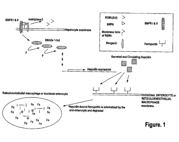
[0024] Figure 1 shows a simplified schematic of a signaling pathway related to
iron homeostasis.
[0025] Figure 2 shows a second simplified schematic of a signaling pathway
related to iron
homeostasis. Figure 3 is a histogram showing the results of a first set of
experiments described
in Example 16, demonstrating that h5F923.AM8 and 1A-2989 improved anemia in
ACD rates at
day 30 by increasing the haemoglobin level. As also shown in this figure,
dorsomorphin was
inactive.
[0026] Figure 4 is a histogram showing the results of a second series of
experiments described in
Example 16. Specifically, Figure 4A shows that the control antibody hIgG does
not change
significantly the low hemoglobin level of the anemic rats on days 41, 47 and
51. * p <0.05:
significance versus DO hemoglobin level. Figure 4B shows that humanized
monoclonal antibody
that was selective for RGM A does not change significantly the low haemoglobin
level of the
anemic rates on days 41, 47 and 51. * p <0.05, ** p < 0.01, significance
versus DO hemoglobin
level. Figure 4C shows that antibody h5F9.AM8 significantly increases the low
hemoglobin
level (D24) of the anemic rats on days 41,47 and 51. *** p <0.001,
significance versus Day 0
(DO) hemoglobin level.. D41: * p < 0.05 significance versus D24, D47/55: * p
<0.05
significance versus D24. Figure 4D shows that antibody h5F9.23, increases the
low hemoglobin
level (day 24 (D24) of the anemic rats on days 41, 47 and 51.* p <0.05; ** p
<0.001,
significance versus DO hemoglobin level at day 41: (faint star) p < 0.05
significance versus
days 24, 47 and 51: (faint star) p < 0.05 significance versus day.
DETAILED DESCRIPTION
[0027] RGMc is a glycosylphosphatidylinositol ("GPI") anchored membrane
protein expressed
in muscle, the retina and periportal hepatocytes. RGMc works in conjunction
with hepcidin via
signaling proteins to maintain iron homeostasis in the body. See, for example,
Severyn et al.,
21
CA 02855570 2014-05-09
WO 2013/090633 PCT/US2012/069584
Biochem. J., 422:393-403 (2009) and Pietrangelo, J. Hepatology, 54:173-181
(2011). As shown
in Figure 1, cell membrane RGMc binds to neogenin and facilitates signaling
through bone
morphogenetic proteins (BMPs), which trigger intracellular signaling through
downstream
effectors to promote hepcidin gene expression. See again, for example,
Pietrangelo, J.
Hepatology, 54:173-181 (2011). Soluble RGMc is released by cleavage of
membrane bound
RGMc by a transmembrane serine protease, matriptase-2 (TMPRSS6). The release
of soluble
RGMc is induced by decreasing extracellular concentrations of iron and,
conversely, inhibited by
increased extracellular concentrations of iron. See again, for example,
Severyn et al., Biochem.
J., 422:393-403 (2009) and Figure 1. The soluble form of RGMc sequesters
BMP6/BMP4/BMP2
and other BMPs from membrane-bound RGMc, thereby preventing the induction of
hepcidin
expression. See Figure 2.
[0028] Upon BMP binding to BMP receptors I and II, a membrane associated
complex is formed
with neogenin, BMP6 and RGMc. This complex, along with intracellular proteins,
called Smads
(Smads 1, 5 and 8), transduce extracellular signals thereby initiating a
signaling pathway that
governs hepcidin expression and, ultimately, systemic iron metabolism. See
again, for example,
Pietrangelo, J. Hepatology, 54:173-181(2011) and Figure 1. Hepcidin binds to
ferroportin, the
exclusive iron exporter of mammals. Upon hepcidin binding to ferroportin,
ferroportin is
internalized by macrophages and duodenal enterocytes where it is degraded,
thereby shutting
down the iron export pathway. See, for example, Hentz et al, Cell, 142:24-38
(2010) and Cheng
et al., Clin. Exp. Med., 11:33-42 (2011).
[0029] Both macrophages and duodenal enterocytes express ferroportin; at high
hepcidin levels,
the hepcidin-induced degradation of ferroportin shuts down the only available
iron export
pathway. See Figure 1. As a consequence, both macrophages and duodenal
enterocytes
accumulate large amounts of intracellular iron. Anemia of chronic disease
("ACD") is a
common consequence, as these cells are no longer able to release iron into the
blood. See again,
for example, Cheng et al., Clin. Exp. Med., 11:33-42 (2011).
[0030] RGMc-specific antibodies interrupt the normal expression of hepcidin,
which directly
regulates iron concentration in the plasma and the distribution of iron to a
variety of tissues. The
antibodies may prevent binding between BMPs and RGMc. The antibodies may
prevent binding
22
CA 02855570 2014-05-09
WO 2013/090633 PCT/US2012/069584
between BMPs and the N-terminus of RGMc. A consequence of this action is the
decreased, or
inhibited, expression of hepcidin. As hepcidin levels decrease, the
ferroportin-dependent export
of iron is increased because hepcidin is no longer available to bind
ferroportin and induce its
internalization and degradation. See Figure 2.
[0031] It has been surprisingly and unexpectedly discovered that antibodies,
which bind to
RGMc, may be used to regulate iron metabolism. Provided herein are RGMc-
specific antibodies
that interrupt the normal expression of hepcidin, which directly regulates
iron concentration in
plasma and the distribution of iron to a variety of tissues. Excess levels of
hepcidin causes iron-
restricted anemia. For example, pronounced increases in hepcidin levels have
been reported in
patients suffering from ACD and in patients suffering with acute inflammation
(Al). Slightly
increased hepcidin levels were observed in patients suffering from ACD and
iron-deficiency
anemia (ACD-IDA). Patients suffering only from iron deficiency anemia (IDA)
showed a trend
towards lower serum hepcidin levels. For example, serum hepcidin levels have
been shown to
be 177,58 [t.g/1 (+/- 119,84) in healthy controls, 434,83 gill (+/- 217) in
ACD patients, 410,08
lug/1 (+/-299,96) in Al patients, 238,32 [t.g/1 (+/-93,85) in ACD-IDA
patients, and slightly
decreased in IDA patients 110,79 [t.g/1 (+/-19,22). See, for example, Cheng et
al. Clin. Exp.
Med., 11:33-42 (2011). In contrast, hemochromatosis is characterized by low
serum hepcidin
levels. In addition, f3-thalaassaemia is a disease where hepcidin levels may
be low.
[0032] The disclosed RGMc-specific and non-specific antibodies disclosed
herein are useful in
the treatment of diseases of iron metabolism. In addition, the disclosed RGMc-
specific
antibodies disclosed herein can be used in various assays, such as diagnostic
assays for
determining whether a subject has an iron-related disorder.
1. Definitions
[0033] The terminology used herein is for the purpose of describing particular
embodiments only
and is not intended to be limiting. As used in the specification and the
appended claims, the
singular forms "a," "and" and "the" include plural references unless the
context clearly dictates
otherwise.
23
CA 02855570 2014-05-09
WO 2013/090633 PCT/US2012/069584
a. About
[0034] "About" as used herein may refer to approximately a +/- 10% variation
from the stated
value. It is to be understood that such a variation is always included in any
given value provided
herein, whether or not specific reference is made to it.
b. Affinity Matured Antibody
[0035] "Affinity Matured Antibody" is used herein to refer to an antibody with
one or more
alterations in one or more CDRs, which result in an improvement in the
affinity (i.e. KD, kd or ka)
of the antibody for a target antigen compared to a parent antibody, which does
not possess the
alteration(s). Exemplary affinity matured antibodies will have nanomolar or
even picomolar
affinities for the target antigen. A variety of procedures for producing
affinity matured
antibodies are known in the art, including the screening of a combinatory
antibody library that
has been prepared using bio-display. For example, Marks et al., BioTechnology,
10: 779-783
(1992) describes affinity maturation by VH and VL domain shuffling. Random
mutagenesis of
CDR and/or framework residues is described by Barbas et al., Proc. Nat. Acad.
Sci. USA, 91:
3809-3813 (1994); Schier et al., Gene, 169: 147-155 (1995); Yelton et al., J.
Immunol., 155:
1994-2004 (1995); Jackson et al., J. Immunol., 154(7): 3310-3319 (1995); and
Hawkins et al, J.
Mol. Biol., 226: 889-896 (1992). Selective mutation at selective mutagenesis
positions and at
contact or hypermutation positions with an activity-enhancing amino acid
residue is described in
U.S. Pat. No. 6,914,128 Bl.
c. Antibody and Antibodies
[0036] "Antibody" and "antibodies" as used herein refers to monoclonal
antibodies,
multispecific antibodies, human antibodies, humanized antibodies (fully or
partially humanized),
animal antibodies (such as, but not limited to, a bird (for example, a duck or
a goose), a shark, a
whale, and a mammal, including a non-primate (for example, a cow, a pig, a
camel, a llama, a
horse, a goat, a rabbit, a sheep, a hamster, a guinea pig, a cat, a dog, a
rat, a mouse, etc.) or a
non-human primate (for example, a monkey, a chimpanzee, etc.), recombinant
antibodies,
chimeric antibodies, single-chain Fvs ("scFv"), single chain antibodies,
single domain
antibodies, Fab fragments, F(ab') fragments, F(aN)2 fragments, disulfide-
linked Fvs ("sdFv"),
and anti-idiotypic ("anti-Id") antibodies, dual-domain antibodies, dual
variable domain (DVD) or
triple variable domain (TVD) antibodies (dual-variable domain immunoglobulins
and methods
for making them are described in Wu, C., et al., Nature Biotechnology,
25(11):1290-1297 (2007)
24
CA 02855570 2014-05-09
WO 2013/090633 PCT/US2012/069584
and PCT International Application WO 2001/058956, the contents of each of
which are herein
incorporated by reference), and functionally active epitope-binding fragments
of any of the
above. In particular, antibodies include immunoglobulin molecules and
immunologically active
fragments of immunoglobulin molecules, namely, molecules that contain an
analyte-binding site.
Immunoglobulin molecules can be of any type (for example, IgG, IgE, IgM, IgD,
IgA and IgY),
class (for example, IgGl, IgG2, IgG3, IgG4, IgAl and IgA2) or subclass. For
simplicity sake, an
antibody against an analyte is frequently referred to herein as being either
an "anti-analyte
antibody," or merely an "analyte antibody" (e.g., an anti-RGMc antibody or an
RGMc antibody).
d. Antibody Fragment
[0037] "Antibody fragment" as used herein refers to a portion of an intact
antibody comprising
the antigen-binding site or variable region. The portion does not include the
constant heavy
chain domains (i.e. CH2, CH3 or CH4, depending on the antibody isotype) of the
Fc region of
the intact antibody. Examples of antibody fragments include, but are not
limited to, Fab
fragments, Fab' fragments, Fab'-SH fragments, F(aN)2 fragments, Fd fragments,
Fv fragments,
diabodies, single-chain Fv (scFv) molecules, single-chain polypeptides
containing only one light
chain variable domain, single-chain polypeptides containing the three CDRs of
the light-chain
variable domain, single-chain polypeptides containing only one heavy chain
variable region, and
single-chain polypeptides containing the three CDRs of the heavy chain
variable region.
e. Binding Constants
[0038] "Binding Constants" are described herein. The term "association rate
constant," "kon" or
"ka" as used herein, refers to the value indicating the binding rate of an
antibody to its target
antigen or the rate of complex formation between an antibody and antigen as
shown by the
equation below:
Antibody (Ab) + Antigen (Ag) ¨> Ab-Ag.
[0039] The term "dissociation rate constant," "koff" or "kd" as used
interchangeably herein, refers
to the value indicating the dissociation rate of an antibody form its target
antigen or separation of
Ab-Ag complex over time into free antibody and antigen as shown by the
equation below:
Antibody (Ab) + Antigen (Ag) <¨ Ab-Ag.
CA 02855570 2014-05-09
WO 2013/090633 PCT/US2012/069584
[0040] Methods for determining association and dissociation rate constants are
well known in
the art. Using fluorescence-based techniques offers high sensitivity and the
ability to examine
samples in physiological buffers at equilibrium. Other experimental approaches
and instruments
such as a BIAcore (biomolecular interaction analysis) assay can be used
(e.g., instrument
available from BIAcore International AB, a GE Healthcare company, Uppsala,
Sweden).
Additionally, a KinExA (Kinetic Exclusion Assay) assay, available from
Sapidyne Instruments
(Boise, Idaho) can also be used.
[0041] The term "equilibrium dissociation constant" or "KD" as used
interchangeably, herein,
refers to the value obtained by dividing the dissociation rate (koff) by the
association rate (kon).
The association rate, the dissociation rate and the equilibrium dissociation
constant are used to
represent the binding affinity of an antibody to an antigen.
f. Binding Protein
[0042] "Binding Protein" is used herein to refer to a monomeric or multimeric
protein that binds
to and forms a complex with a binding partner, such as, for example, a
polypeptide, an antigen, a
chemical compound or other molecule, or a substrate of any kind. A binding
protein specifically
binds a binding partner. Binding proteins include antibodies, as well as
antigen-binding
fragments thereof and other various forms and derivatives thereof as are known
in the art and
described herein below, and other molecules comprising one or more antigen-
binding domains
that bind to an antigen molecule or a particular site (epitope) on the antigen
molecule.
Accordingly, a binding protein includes, but is not limited to, an antibody a
tetrameric
immunoglobulin, an IgG molecule, an IgGi molecule, a monoclonal antibody, a
chimeric
antibody, a CDR-grafted antibody, a humanized antibody, an affinity matured
antibody, and
fragments of any such antibodies that retain the ability to bind to an
antigen.
g. Bispecific Antibody
[0043] "Bispecific antibody" is used herein to refer to a full-length antibody
that is generated by
quadroma technology (see Milstein et al., Nature, 305(5934): 537-540 (1983)),
by chemical
conjugation of two different monoclonal antibodies (see, Staerz et al.,
Nature, 314(6012): 628-
631 (1985)), or by knob-into-hole or similar approaches, which introduce
mutations in the Fc
region (see Holliger et al., Proc. Natl. Acad. Sci. USA, 90(14): 6444-6448
(1993)), resulting in
multiple different immunoglobulin species of which only one is the functional
bispecific
26
CA 02855570 2014-05-09
WO 2013/090633 PCT/US2012/069584
antibody. A bispecific antibody binds one antigen (or epitope) on one of its
two binding arms
(one pair of HC/LC), and binds a different antigen (or epitope) on its second
arm (a different pair
of HC/LC). By this definition, a bispecific antibody has two distinct antigen-
binding arms (in
both specificity and CDR sequences), and is monovalent for each antigen to
which it binds to.
h. CDR
[0044] "CDR" is used herein to refer to the "complementarity determining
region" within an
antibody variable sequence. There are three CDRs in each of the variable
regions of the heavy
chain and the light chain, which are designated "CDR1", "CDR2", and "CDR3",
for each of the
variable regions. The term "CDR set" as used herein refers to a group of three
CDRs that occur
in a single variable region that binds the antigen. The exact boundaries of
these CDRs have been
defined differently according to different systems. The system described by
Kabat (Kabat et al.,
Sequences of Proteins of Immunological Interest (National Institutes of
Health, Bethesda, Md.
(1987) and (1991)) not only provides an unambiguous residue numbering system
applicable to
any variable region of an antibody, but also provides precise residue
boundaries defining the
three CDRs. These CDRs may be referred to as "Kabat CDRs". Chothia and
coworkers (Chothia
and Lesk, J. Mol. Biol., 196: 901-917 (1987); and Chothia et al., Nature, 342:
877-883 (1989))
found that certain sub-portions within Kabat CDRs adopt nearly identical
peptide backbone
conformations, despite having great diversity at the level of amino acid
sequence. These sub-
portions were designated as "Li", "L2", and "L3", or "Hl", "H2", and "H3",
where the "L" and
the "H" designate the light chain and the heavy chain regions, respectively.
These regions may be
referred to as "Chothia CDRs", which have boundaries that overlap with Kabat
CDRs. Other
boundaries defining CDRs overlapping with the Kabat CDRs have been described
by Padlan,
FASEB J., 9: 133-139 (1995), and MacCallum, J. Mol. Biol., 262(5): 732-745
(1996). Still other
CDR boundary definitions may not strictly follow one of the herein systems,
but will nonetheless
overlap with the Kabat CDRs, although they may be shortened or lengthened in
light of
prediction or experimental findings that particular residues or groups of
residues or even entire
CDRs do not significantly impact antigen binding. The methods used herein may
utilize CDRs
defined according to any of these systems, although certain embodiments use
Kabat- or Chothia-
defined CDRs.
27
CA 02855570 2014-05-09
WO 2013/090633 PCT/US2012/069584
i. Component or Components
[0045] "Component," "components," or "at least one component," refer generally
to a capture
antibody, a detection or conjugate a calibrator, a control, a sensitivity
panel, a container, a buffer,
a diluent, a salt, an enzyme, a co-factor for an enzyme, a detection reagent,
a pretreatment
reagent/solution, a substrate (e.g., as a solution), a stop solution, and the
like that can be included
in a kit for assay of a test sample, such as a patient urine, serum or plasma
sample, in accordance
with the methods described herein and other methods known in the art. Some
components can
be in solution or lyophilized for reconstitution for use in an assay.
j. Consensus or Consensus Sequence
[0046] "Consensus" or "Consensus Sequence" as used herein refers to a
synthetic nucleic acid
sequence, or corresponding polypeptide sequence, constructed based on analysis
of an alignment
of multiple subtypes of a particular antigen. The sequence may be used to
induce broad
immunity against multiple subtypes or sertypes of a particular antigen.
Synthetic antigens, such
as fusion proteins, may be manipulated to consensus sequences (or consensus
antigens).
k. Control
[0047] "Control" as used herein refers to a composition known to not contain
an analyte of
interest ("negative"), e.g., RGMc (such as membrane-associated RGMc, soluble
RGMc,
fragments of membrane-associated RGMc, fragments of soluble RGMc, variants of
RGMc
(membrane-associated or soluble RGMc) or any combinations thereof), or to
contain an analyte
of interest ("positive control"), e.g., RGMc (such as membrane-associated
RGMc, soluble
RGMc, fragments of membrane-associated RGMc, fragments of soluble RGMc,
variants of
RGMc (membrane-associated or soluble RGMc) or any combinations thereof). A
positive
control can comprise a known concentration of RGMc. "Control," "positive
control," and
"calibrator" may be used interchangeably herein to refer to a composition
comprising a known
concentration of RGMc. A "positive control" can be used to establish assay
performance
characteristics and is a useful indicator of the integrity of reagents (e.g.,
analytes). A "normal
control" may refer to a sample or a subject that is free from an iron-related
disease or disorder.
1. Derivative
[0048] "Derivative" of an antibody as used herein may refer to an antibody
having one or more
modifications to its amino acid sequence when compared to a genuine or parent
antibody and
28
CA 02855570 2014-05-09
WO 2013/090633 PCT/US2012/069584
exhibit a modified domain structure. The derivative may still be able to adopt
the typical domain
configuration found in native antibodies, as well as an amino acid sequence,
which is able to
bind to targets (antigens) with specificity. Typical examples of antibody
derivatives are
antibodies coupled to other polypeptides, rearranged antibody domains or
fragments of
antibodies. The derivative may also comprise at least one further compound,
e.g. a protein
domain, said protein domain being linked by covalent or non-covalent bonds.
The linkage can
be based on genetic fusion according to the methods known in the art. The
additional domain
present in the fusion protein comprising the antibody employed in accordance
with the invention
may preferably be linked by a flexible linker, advantageously a peptide
linker, wherein said
peptide linker comprises plural, hydrophilic, peptide-bonded amino acids of a
length sufficient to
span the distance between the C-terminal end of the further protein domain and
the N-terminal
end of the antibody or vice versa. The antibody may be linked to an effector
molecule having a
conformation suitable for biological activity or selective binding to a solid
support, a biologically
active substance (e.g. a cytokine or growth hormone), a chemical agent, a
peptide, a protein or a
drug, for example.
m. Dual-Specific Antibody
[0049] "Dual-specific antibody" is used herein to refer to a full-length
antibody that can bind
two two different antigens (or epitopes) in each of its two binding arms (a
pair of HC/LC) (see
PCT publication WO 02/02773). Accordingly a dual-specific binding protein has
two identical
antigen binding arms, with identical specificity and identical CDR sequences,
and is bivalent for
each antigen to which it binds.
n. Dual Variable Domain
[0050] "Dual variable domain" is used herein to refer to two or more antigen
binding sites on a
binding protein, which may be divalent (two antigen binding sites),
tetravalent (four antigen
binding sites), or multivalent binding proteins. DVDs may be monospecific,
i.e., capable of
binding one antigen (or one specific epitope), or multispecific, i.e., capable
of binding two or
more antigens (i.e., two or more epitopes of the same target antigen molecule
or two or more
epitopes of different target antigens). A preferred DVD binding protein
comprises two heavy
chain DVD polypeptides and two light chain DVD polypeptides and is referred to
as a "DVD
immunoglobulin" or "DVD-Ig". Such a DVD-Ig binding protein is thus tetrameric
and
reminiscent of an IgG molecule, but provides more antigen binding sites than
an IgG molecule.
29
CA 02855570 2014-05-09
WO 2013/090633 PCT/US2012/069584
Thus, each half of a tetrameric DVD-Ig molecule is reminiscent of one half of
an IgG molecule
and comprises a heavy chain DVD polypeptide and a light chain DVD polypeptide,
but unlike a
pair of heavy and light chains of an IgG molecule that provides a single
antigen binding domain,
a pair of heavy and light chains of a DVD-Ig provide two or more antigen
binding sites.
[0051] Each antigen binding site of a DVD-Ig binding protein may be derived
from a donor
("parental") monoclonal antibody and thus comprises a heavy chain variable
domain (VH) and a
light chain variable domain (VL) with a total of six CDRs involved in antigen
binding per
antigen binding site. Accordingly, a DVD-Ig binding protein that binds two
different epitopes
(i.e., two different epitopes of two different antigen molecules or two
different epitopes of the
same antigen molecule) comprises an antigen binding site derived from a first
parental
monoclonal antibody and an antigen binding site of a second parental
monoclonal antibody.
[0052] A description of the design, expression, and characterization of DVD-Ig
binding
molecules is provided in PCT Publication No. WO 2007/024715, U.S. Pat. No.
7,612,181, and
Wu et al., Nature Biotech., 25: 1290-1297 (2007). A preferred example of such
DVD-Ig
molecules comprises a heavy chain that comprises the structural formula VD1-
(X1)n-VD2-C-
(X2)n, wherein VD1 is a first heavy chain variable domain, VD2 is a second
heavy chain
variable domain, C is a heavy chain constant domain, X1 is a linker with the
proviso that it is not
CH1, X2 is an Fc region, and n is 0 or 1, but preferably 1; and a light chain
that comprises the
structural formula VD1-(X1)n-VD2-C-(X2)n, wherein VD1 is a first light chain
variable domain,
VD2 is a second light chain variable domain, C is a light chain constant
domain, X1 is a linker
with the proviso that it is not CH1, and X2 does not comprise an Fc region;
and n is 0 or 1, but
preferably 1. Such a DVD-Ig may comprise two such heavy chains and two such
light chains,
wherein each chain comprises variable domains linked in tandem without an
intervening constant
region between variable regions, wherein a heavy chain and a light chain
associate to form
tandem functional antigen binding sites, and a pair of heavy and light chains
may associate with
another pair of heavy and light chains to form a tetrameric binding protein
with four functional
antigen binding sites. In another example, a DVD-Ig molecule may comprise
heavy and light
chains that each comprise three variable domains (VD1, VD2, VD3) linked in
tandem without an
intervening constant region between variable domains, wherein a pair of heavy
and light chains
may associate to form three antigen binding sites, and wherein a pair of heavy
and light chains
CA 02855570 2014-05-09
WO 2013/090633 PCT/US2012/069584
may associate with another pair of heavy and light chains to form a tetrameric
binding protein
with six antigen binding sites.
[0053] In a preferred embodiment, a DVD-Ig binding protein according to the
invention not only
binds the same target molecules bound by its parental monoclonal antibodies,
but also possesses
one or more desirable properties of one or more of its parental monoclonal
antibodies.
Preferably, such an additional property is an antibody parameter of one or
more of the parental
monoclonal antibodies. Antibody parameters that may be contributed to a DVD-Ig
binding
protein from one or more of its parental monoclonal antibodies include, but
are not limited to,
antigen specificity, antigen affinity, potency, biological function, epitope
recognition, protein
stability, protein solubility, production efficiency, immunogenicity,
pharmacokinetics,
bioavailability, tissue cross reactivity, and orthologous antigen binding.
[0054] A DVD-Ig binding protein binds at least one epitope of a RGMc. Non-
limiting examples
of a DVD-Ig binding protein include a DVD-Ig binding protein that binds one or
more epitopes
of RGMc, a DVD-Ig binding protein that binds an epitope of a human RGMc and an
epitope of a
RGMc of another species (for example, mouse), and a DVD-Ig binding protein
that binds an
epitope of a human RGMc and an epitope of another target molecule (for
example, VEGFR2 or
VEGFR1).
o. Epitope or Epitopes
[0055] "Epitope," or "epitopes," or "epitopes of interest" refer to a site(s)
on any molecule that is
recognized and can bind to a complementary site(s) on its specific binding
partner. The
molecule and specific binding partner are part of a specific binding pair. For
example, an
epitope can be on a polypeptide, a protein, a hapten, a carbohydrate antigen
(such as, but not
limited to, glycolipids, glycoproteins or lipopolysaccharides), or a
polysaccharide. Its specific
binding partner can be, but is not limited to, an antibody.
p. Framework or Framework Sequence
[0056] "Framework" (FR) or "Framework sequence" as used herein may mean the
remaining
sequences of a variable region minus the CDRs. Because the exact definition of
a CDR
sequence can be determined by different systems (for example, see above), the
meaning of a
framework sequence is subject to correspondingly different interpretations.
The six CDRs (CDR-
Li, -L2, and -L3 of light chain and CDR-H1, -H2, and -H3 of heavy chain) also
divide the
31
CA 02855570 2014-05-09
WO 2013/090633 PCT/US2012/069584
framework regions on the light chain and the heavy chain into four sub-regions
(FR1, FR2, FR3,
and FR4) on each chain, in which CDR1 is positioned between FR1 and FR2, CDR2
between
FR2 and FR3, and CDR3 between FR3 and FR4. Without specifying the particular
sub-regions
as FR1, FR2, FR3, or FR4, a framework region, as referred by others,
represents the combined
FRs within the variable region of a single, naturally occurring immunoglobulin
chain. As used
herein, a FR represents one of the four sub-regions, and FRs represents two or
more of the four
sub-regions constituting a framework region.
[0057] Human heavy chain and light chain FR sequences are known in the art
that can be used as
heavy chain and light chain "acceptor" framework sequences (or simply,
"acceptor" sequences)
to humanize a non-human antibody using techniques known in the art. In one
embodiment,
human heavy chain and light chain acceptor sequences are selected from the
framework
sequences listed in publicly available databases such as V-base (hypertext
transfer
protocol://vbase.mrc-cpe.cam.ac.uk/) or in the international ImMunoGeneTics
(IMGTIO)
information system (hypertext transfer
protocol://imgt.cines.fr/texts/EVIGTrepertoire/LocusGenes/).
q. Functional Antigen Binding Site
[0058] "Functional antigen binding site" as used herein may mean a site on a
binding protein
(e.g. an antibody) that is capable of binding a target antigen. The antigen
binding affinity of the
antigen binding site may not be as strong as the parent binding protein, e.g.,
parent antibody,
from which the antigen binding site is derived, but the ability to bind
antigen must be measurable
using any one of a variety of methods known for evaluating protein, e.g.,
antibody, binding to an
antigen. Moreover, the antigen binding affinity of each of the antigen binding
sites of a
multivalent protein, e.g., multivalent antibody, herein need not be
quantitatively the same.
r. Humanized Antibody
[0059] "Humanized antibody" is used herein to describe an antibody that
comprises heavy and
light chain variable region sequences from a non-human species (e.g. a mouse)
but in wheihc at
least a portion of the VH and/or VL sequence has been altered to be more
"human-like," i.e.,
more similar to human germline variable sequences. A "humanized antibody" is
an antibody or a
variant, derivative, analog, or fragment thereof, which immunospecifically
binds to an antigen of
interest and which comprises a framework (FR) region having substantially the
amino acid
32
CA 02855570 2014-05-09
WO 2013/090633 PCT/US2012/069584
sequence of a human antibody and a complementary determining region (CDR)
having
substantially the amino acid sequence of a non-human antibody. As used herein,
the term
"substantially" in the context of a CDR refers to a CDR having an amino acid
sequence at least
80%, at least 85%, at least 90%, at least 95%, at least 98% or at least 99%
identical to the amino
acid sequence of a non-human antibody CDR. A humanized antibody comprises
substantially all
of at least one, and typically two, variable domains (Fab, Fab', F(aN)2, FabC,
Fv) in which all or
substantially all of the CDR regions correspond to those of a non-human
immunoglobulin (i.e.,
donor antibody) and all or substantially all of the framework regions are
those of a human
immunoglobulin consensus sequence. In an embodiment, a humanized antibody also
comprises
at least a portion of an immunoglobulin constant region (Fc), typically that
of a human
immunoglobulin. In some embodiments, a humanized antibody contains the light
chain as well
as at least the variable domain of a heavy chain. The antibody also may
include the CH1, hinge,
CH2, CH3, and CH4 regions of the heavy chain. In some embodiments, a humanized
antibody
only contains a humanized light chain. In some embodiments, a humanized
antibody only
contains a humanized heavy chain. In specific embodiments, a humanized
antibody only
contains a humanized variable domain of a light chain and/or humanized heavy
chain.
[0060] A humanized antibody can be selected from any class of immunoglobulins,
including
IgM, IgG, IgD, IgA, and IgE, and any isotype, including without limitation
IgGl, IgG2, IgG3,
and IgG4. A humanized antibody may comprise sequences from more than one class
or isotype,
and particular constant domains may be selected to optimize desired effector
functions using
techniques well-known in the art.
[0061] The framework regions and CDRs of a humanized antibody need not
correspond
precisely to the parental sequences, e.g., the donor antibody CDR or the
consensus framework
may be mutagenized by substitution, insertion, and/or deletion of at least one
amino acid residue
so that the CDR or framework residue at that site does not correspond to
either the donor
antibody or the consensus framework. In a preferred embodiment, such
mutations, however, will
not be extensive. Usually, at least 80%, preferably at least 85%, more
preferably at least 90%,
and most preferably at least 95% of the humanized antibody residues will
correspond to those of
the parental FR and CDR sequences. As used herein, the term "consensus
framework" refers to
the framework region in the consensus immunoglobulin sequence. As used herein,
the term
"consensus immunoglobulin sequence" refers to the sequence formed from the
most frequently
33
CA 02855570 2014-05-09
WO 2013/090633 PCT/US2012/069584
occurring amino acids (or nucleotides) in a family of related immunoglobulin
sequences (see,
e.g., Winnaker, From Genes to Clones (Verlagsgesellschaft, Weinheim, 1987)). A
"consensus
immunoglobulin sequence" may thus comprise a "consensus framework region(s)"
and/or a
"consensus CDR(s)". In a family of immunoglobulins, each position in the
consensus sequence
is occupied by the amino acid occurring most frequently at that position in
the family. If two
amino acids occur equally frequently, either can be included in the consensus
sequence.
s. Identical or Identity
[0062] "Identical" or "identity," as used herein in the context of two or more
polypeptide or
polynucleotide sequences, can mean that the sequences have a specified
percentage of residues
that are the same over a specified region. The percentage can be calculated by
optimally
aligning the two sequences, comparing the two sequences over the specified
region, determining
the number of positions at which the identical residue occurs in both
sequences to yield the
number of matched positions, dividing the number of matched positions by the
total number of
positions in the specified region, and multiplying the result by 100 to yield
the percentage of
sequence identity. In cases where the two sequences are of different lengths
or the alignment
produces one or more staggered ends and the specified region of comparison
includes only a
single sequence, the residues of the single sequence are included in the
denominator but not the
numerator of the calculation.
t. Isolated Polynucleotide
[0063] "Isolated polynucleotide" as used herein may mean a polynucleotide
(e.g. of genomic,
cDNA, or synthetic origin, or a combination thereof) that, by virtue of its
origin, the isolated
polynucleotide is not associated with all or a portion of a polynucleotide
with which the "isolated
polynucleotide" is found in nature; is operably linked to a polynucleotide
that it is not linked to
in nature; or does not occur in nature as part of a larger sequence.
u. Label and Detectable Label
[0064] "Label" and "detectable label" as used herein refer to a moiety
attached to an antibody or
an analyte to render the reaction between the antibody and the analyte
detectable, and the
antibody or analyte so labeled is referred to as "detectably labeled." A label
can produce a signal
that is detectable by visual or instrumental means. Various labels include
signal-producing
substances, such as chromogens, fluorescent compounds, chemiluminescent
compounds,
34
CA 02855570 2014-05-09
WO 2013/090633 PCT/US2012/069584
radioactive compounds, and the like. Representative examples of labels include
moieties that
produce light, e.g., acridinium compounds, and moieties that produce
fluorescence, e.g.,
fluorescein. Other labels are described herein. In this regard, the moiety,
itself, may not be
detectable but may become detectable upon reaction with yet another moiety.
Use of the term
"detectably labeled" is intended to encompass such labeling.
[0065] Any suitable detectable label as is known in the art can be used. For
example, the
detectable label can be a radioactive label (such as 3H, 1251, 35s, 14C,
32,,r,
and 33P), an enzymatic
label (such as horseradish peroxidase, alkaline peroxidase, glucose 6-
phosphate dehydrogenase,
and the like), a chemiluminescent label (such as acridinium esters,
thioesters, or sulfonamides;
luminol, isoluminol, phenanthridinium esters, and the like), a fluorescent
label (such as
fluorescein (e.g., 5-fluorescein, 6-carboxyfluorescein, 3'6-
carboxyfluorescein, 5(6)-
carboxyfluorescein, 6-hexachloro-fluorescein, 6-tetrachlorofluorescein,
fluorescein
isothiocyanate, and the like)), rhodamine, phycobiliproteins, R-phycoerythrin,
quantum dots
(e.g., zinc sulfide-capped cadmium selenide), a thermometric label, or an
immuno-polymerase
chain reaction label. An introduction to labels, labeling procedures and
detection of labels is
found in Polak and Van Noorden, Introduction to Immunocytochemistry, 2nd ed.,
Springer
Verlag, N.Y. (1997), and in Haugland, Handbook of Fluorescent Probes and
Research
Chemicals (1996), which is a combined handbook and catalogue published by
Molecular Probes,
Inc., Eugene, Oregon. A fluorescent label can be used in FPIA (see, e.g., U.S.
Patent Nos.
5,593,896, 5,573,904, 5,496,925, 5,359,093, and 5,352,803, which are hereby
incorporated by
reference in their entireties). An acridinium compound can be used as a
detectable label in a
homogeneous chemiluminescent assay (see, e.g., Adamczyk et al., Bioorg. Med.
Chem. Lett. 16:
1324-1328 (2006); Adamczyk et al., Bioorg. Med. Chem. Lett. 4: 2313-2317
(2004); Adamczyk
et al., Biorg. Med. Chem. Lett. 14: 3917-3921 (2004); and Adamczyk et al.,
Org. Lett. 5: 3779-
3782 (2003)).
[0066] In one aspect, the acridinium compound is an acridinium-9-carboxamide.
Methods for
preparing acridinium 9-carboxamides are described in Mattingly, J. Biolumin.
Chemilumin. 6:
107-114 (1991); Adamczyk et al., J. Org. Chem. 63: 5636-5639 (1998); Adamczyk
et al.,
Tetrahedron 55: 10899-10914 (1999); Adamczyk et al., Org. Lett. 1: 779-781
(1999); Adamczyk
et al., Bioconjugate Chem. 11: 714-724 (2000); Mattingly et al., In
Luminescence Biotechnology:
Instruments and Applications; Dyke, K. V. Ed.; CRC Press: Boca Raton, pp. 77-
105 (2002);
CA 02855570 2014-05-09
WO 2013/090633 PCT/US2012/069584
Adamczyk et al., Org. Lett. 5: 3779-3782 (2003); and U.S. Pat. Nos. 5,468,646,
5,543,524 and
5,783,699 (each of which is incorporated herein by reference in its entirety
for its teachings
regarding same).
[0067] Another example of an acridinium compound is an acridinium-9-
carboxylate aryl ester.
An example of an acridinium-9-carboxylate aryl ester of formula II is 10-
methy1-9-
(phenoxycarbonyl)acridinium fluorosulfonate (available from Cayman Chemical,
Ann Arbor,
MI). Methods for preparing acridinium 9-carboxylate aryl esters are described
in McCapra et al.,
Photochem. Photobiol. 4: 1111-21(1965); Razavi et al., Luminescence 15: 245-
249 (2000);
Razavi et al., Luminescence 15: 239-244 (2000); and U.S. Patent No. 5,241,070
(each of which
is incorporated herein by reference in its entirety for its teachings
regarding same). Such
acridinium-9-carboxylate aryl esters are efficient chemiluminescent indicators
for hydrogen
peroxide produced in the oxidation of an analyte by at least one oxidase in
terms of the intensity
of the signal and/or the rapidity of the signal. The course of the
chemiluminescent emission for
the acridinium-9-carboxylate aryl ester is completed rapidly, i.e., in under 1
second, while the
acridinium-9-carboxamide chemiluminescent emission extends over 2 seconds.
Acridinium-9-
carboxylate aryl ester, however, loses its chemiluminescent properties in the
presence of protein.
Therefore, its use requires the absence of protein during signal generation
and detection.
Methods for separating or removing proteins in the sample are well-known to
those skilled in the
art and include, but are not limited to, ultrafiltration, extraction,
precipitation, dialysis,
chromatography, and/or digestion (see, e.g., Wells, High Throughput
Bioanalytical Sample
Preparation. Methods and Automation Strategies, Elsevier (2003)). The amount
of protein
removed or separated from the test sample can be about 40%, about 45%, about
50%, about
55%, about 60%, about 65%, about 70%, about 75%, about 80%, about 85%, about
90%, or
about 95%. Further details regarding acridinium-9-carboxylate aryl ester and
its use are set forth
in U.S. Pat. App. No. 11/697,835, filed April 9, 2007. Acridinium-9-
carboxylate aryl esters can
be dissolved in any suitable solvent, such as degassed anhydrous N,N-
dimethylformamide
(DMF) or aqueous sodium cholate.
v. Linking Sequence and Linking Peptide Sequence
[0068] "Linking sequence" or "linking peptide sequence" refers to a natural or
artificial
polypeptide sequence that is connected to one or more polypeptide sequences of
interest (e.g.,
full-length, fragments, etc.). The term "connected" refers to the joining of
the linking sequence
36
CA 02855570 2014-05-09
WO 2013/090633 PCT/US2012/069584
to the polypeptide sequence of interest. Such polypeptide sequences are
preferably joined by one
or more peptide bonds. Linking sequences can have a length of from about 4 to
about 50 amino
acids. Preferably, the length of the linking sequence is from about 6 to about
30 amino acids.
Natural linking sequences can be modified by amino acid substitutions,
additions, or deletions to
create artificial linking sequences. Exemplary linking sequences include, but
are not limited to:
(i) Histidine (His) tags, such as a 6X His tag, which has an amino acid
sequence of HHHHHH
(SEQ ID NO:83), are useful as linking sequences to facilitate the isolation
and purification of
polypeptides and antibodies of interest; (ii) Enterokinase cleavage sites,
like His tags, are used
in the isolation and purification of proteins and antibodies of interest.
Often, enterokinase
cleavage sites are used together with His tags in the isolation and
purification of proteins and
antibodies of interest. Various enterokinase cleavage sites are known in the
art. Examples of
enterokinase cleavage sites include, but are not limited to, the amino acid
sequence of DDDDK
(SEQ ID NO:84) and derivatives thereof (e.g., ADDDDK (SEQ ID NO:85), etc.);
(iii)
Miscellaneous sequences can be used to link or connect the light and/or heavy
chain variable
regions of single chain variable region fragments. Examples of other linking
sequences can be
found in Bird et al., Science 242: 423-426 (1988); Huston et al., PNAS USA 85:
5879-5883
(1988); and McCafferty et al., Nature 348: 552-554 (1990). Linking sequences
also can be
modified for additional functions, such as attachment of drugs or attachment
to solid supports. In
the context of the present disclosure, the monoclonal antibody, for example,
can contain a
linking sequence, such as a His tag, an enterokinase cleavage site, or both.
w. Multivalent Binding Protein
[0069] "Multivalent binding protein" is used herein to refer to a binding
protein comprising two
or more antigen binding sites (also referred to herein as "antigen binding
domains"). A
multivalent binding protein is preferably engineered to have three or more
antigen binding sites,
and is generally not a naturally occurring antibody. The term "multispecific
binding protein"
refers to a binding protein that can bind two or more related or unrelated
targets, including a
binding protein capable of binding two or more different epitopes of the same
target molecule.
x. Predetermined Cutoff and Predetermined Level
[0070] "Predetermined cutoff" and "predetermined level" refer generally to an
assay cutoff value
that is used to assess diagnostic/prognostic/therapeutic efficacy results by
comparing the assay
results against the predetermined cutoff/level, where the predetermined
cutoff/level already has
37
CA 02855570 2014-05-09
WO 2013/090633 PCT/US2012/069584
been linked or associated with various clinical parameters (e.g., severity of
disease,
progression/nonprogression/improvement, etc.). The present disclosure provides
exemplary
predetermined levels. However, it is well-known that cutoff values may vary
depending on the
nature of the immunoassay (e.g., antibodies employed, etc.). It further is
well within the
ordinary skill of one in the art to adapt the disclosure herein for other
immunoassays to obtain
immunoassay-specific cutoff values for those other immunoassays based on this
disclosure.
Whereas the precise value of the predetermined cutoff/level may vary between
assays, the
correlations as described herein should be generally applicable.
y. Pretreatment Reagent
[0071] "Pretreatment reagent," e.g., lysis, precipitation and/or
solubilization reagent, as used in a
diagnostic assay as described herein is one that lyses any cells and/or
solubilizes any analyte that
is/are present in a test sample. Pretreatment is not necessary for all
samples, as described further
herein. Among other things, solubilizing the analyte (i.e., RGMc (such as
membrane-associated
RGMc, soluble RGMc, fragments of membrane-associated RGMc, fragments of
soluble RGMc,
variants of RGMc (membrane-associated or soluble RGMc) or any combinations
thereof))
entails release of the analyte from any endogenous binding proteins present in
the sample. A
pretreatment reagent may be homogeneous (not requiring a separation step) or
heterogeneous
(requiring a separation step). With use of a heterogeneous pretreatment
reagent there is removal
of any precipitated analyte binding proteins from the test sample prior to
proceeding to the next
step of the assay. The pretreatment reagent optionally can comprise: (a) one
or more solvents
and salt, (b) one or more solvents, salt and detergent, (c) detergent, (d)
detergent and salt, or (e)
any reagent or combination of reagents appropriate for cell lysis and/or
solubilization of analyte.
z. Quality Control Reagents
[0072] "Quality control reagents" in the context of immunoassays and kits
described herein,
include, but are not limited to, calibrators, controls, and sensitivity
panels. A "calibrator" or
"standard" typically is used (e.g., one or more, such as a plurality) in order
to establish
calibration (standard) curves for interpolation of the concentration of an
analyte, such as an
antibody or an analyte. Alternatively, a single calibrator, which is near a
predetermined
positive/negative cutoff, can be used. Multiple calibrators (i.e., more than
one calibrator or a
varying amount of calibrator(s)) can be used in conjunction so as to comprise
a "sensitivity
panel."
38
CA 02855570 2014-05-09
WO 2013/090633 PCT/US2012/069584
aa. Recombinant Antibody and Recombinant Antibodies
[0073] "Recombinant antibody" and "recombinant antibodies" refer to antibodies
prepared by
one or more steps, including cloning nucleic acid sequences encoding all or a
part of one or more
monoclonal antibodies into an appropriate expression vector by recombinant
techniques and
subsequently expressing the antibody in an appropriate host cell. The terms
include, but are not
limited to, recombinantly produced monoclonal antibodies, chimeric antibodies,
humanized
antibodies (fully or partially humanized), multi-specific or multi-valent
structures formed from
antibody fragments, bifunctional antibodies, heteroconjugate Abs, DVD-IgiOs,
and other
antibodies as described in (i) herein. (Dual-variable domain immunoglobulins
and methods for
making them are described in Wu, C., et al., Nature Biotechnology, 25:1290-
1297 (2007)). The
term "bifunctional antibody," as used herein, refers to an antibody that
comprises a first arm
having a specificity for one antigenic site and a second arm having a
specificity for a different
antigenic site, i.e., the bifunctional antibodies have a dual specificity.
bb. Sample, Test Sample, and Patient Sample
[0074] "Sample," "test sample," and "patient sample" may be used
interchangeably herein. The
sample, such as a sample of urine, serum, plasma, amniotic fluid,
cerebrospinal fluid, placental
cells or tissue, endothelial cells, leukocytes, or monocytes, can be used
directly as obtained from
a patient or can be pre-treated, such as by filtration, distillation,
extraction, concentration,
centrifugation, inactivation of interfering components, addition of reagents,
and the like, to
modify the character of the sample in some manner as discussed herein or
otherwise as is known
in the art.
cc. Series of Calibrating Compositions
[0075] "Series of calibrating compositions" refers to a plurality of
compositions comprising a
known concentration of Cys-RGMcC, wherein each of the compositions differs
from the other
compositions in the series by the concentration of Cys-CRGMc.
dd. Solid Phase
[0076] "Solid phase" refers to any material that is insoluble, or can be made
insoluble by a
subsequent reaction. The solid phase can be chosen for its intrinsic ability
to attract and
immobilize a capture agent. Alternatively, the solid phase can have affixed
thereto a linking
agent that has the ability to attract and immobilize the capture agent. The
linking agent can, for
39
CA 02855570 2014-05-09
WO 2013/090633 PCT/US2012/069584
example, include a charged substance that is oppositely charged with respect
to the capture agent
itself or to a charged substance conjugated to the capture agent. In general,
the linking agent can
be any binding partner (preferably specific) that is immobilized on (attached
to) the solid phase
and that has the ability to immobilize the capture agent through a binding
reaction. The linking
agent enables the indirect binding of the capture agent to a solid phase
material before the
performance of the assay or during the performance of the assay. The solid
phase can, for
example, be plastic, derivatized plastic, magnetic or non-magnetic metal,
glass or silicon,
including, for example, a test tube, microtiter well, sheet, bead,
microparticle, chip, and other
configurations known to those of ordinary skill in the art.
ee. Specific Binding
[0077] "Specific binding" or "specifically binding" as used herein may refer
to the interaction of
an antibody, a protein, or a peptide with a second chemical species, wherein
the interaction is
dependent upon the presence of a particular structure (e.g., an antigenic
determinant or epitope)
on the chemical species; for example, an antibody recognizes and binds to a
specific protein
structure rather than to proteins generally. If an antibody is specific for
epitope "A", the presence
of a molecule containing epitope A (or free, unlabeled A), in a reaction
containing labeled "A"
and the antibody, will reduce the amount of labeled A bound to the antibody.
ff. Specific Binding Partner
[0078] "Specific binding partner" is a member of a specific binding pair. A
specific binding pair
comprises two different molecules, which specifically bind to each other
through chemical or
physical means. Therefore, in addition to antigen and antibody specific
binding pairs of common
immunoassays, other specific binding pairs can include biotin and avidin (or
streptavidin),
carbohydrates and lectins, complementary nucleotide sequences, effector and
receptor molecules,
cofactors and enzymes, enzymes and enzyme inhibitors, and the like.
Furthermore, specific
binding pairs can include members that are analogs of the original specific
binding members, for
example, an analyte-analog. Immunoreactive specific binding members include
antigens,
antigen fragments, and antibodies, including monoclonal and polyclonal
antibodies as well as
complexes and fragments thereof, whether isolated or recombinantly produced.
CA 02855570 2014-05-09
WO 2013/090633 PCT/US2012/069584
gg. Stringent Conditions
[0079] "Stringent conditions" is used herein to describe hybridization to
filter-bound DNA in 6x
sodium chloride/sodium citrate (SSC) at about 45 C followed by one or more
washes in
0.2xSSC/0.1% SDS at about 50-65 C. The term "under highly stringent
conditions", refers to
hybridization to filter-bound nucleic acid in 6xSSC at about 45 C followed by
one or more
washes in 0.1xSSC/0.2% SDS at about 68 C., or under other stringent
hybridization conditions.
See, for example, Ausubel, F. M. et al., eds., 1989, Current Protocols in
Molecular Biology, Vol.
I, Green Publishing Associates, Inc. and John Wiley & Sons, Inc., New York at
pages 6.3.1-6.3.6
and 2.10.3.
hh. Treat, Treating or Treatment
[0080] "Treat", "treating" or "treatment" are each used interchangeably herein
to describe
reversing, alleviating, or inhibiting the progress of a disease, or one or
more symptoms of such
disease, to which such term applies. Depending on the condition of the
subject, the term also
refers to preventing a disease, and includes preventing the onset of a
disease, or preventing the
symptoms associated with a disease. A treatment may be either performed in an
acute or chronic
way. The term also refers to reducing the severity of a disease or symptoms
associated with such
disease prior to affliction with the disease. Such prevention or reduction of
the severity of a
disease prior to affliction refers to administration of a antibodies or
pharmaceutical composition
of the present invention to a subject that is not at the time of
administration afflicted with the
disease. "Preventing" also refers to preventing the recurrence of a disease or
of one or more
symptoms associated with such disease. "Treatment" and "therapeutically,"
refer to the act of
treating, as "treating" is defined above.
ii. Tracer
[0081] "Tracer" as used herein refers to an analyte or analyte fragment
conjugated to a label,
such as Cys-CRGMc conjugated to a fluorescein moiety, wherein the analyte
conjugated to the
label can effectively compete with the analyte for sites on an antibody
specific for the analyte.
jj. Variant
[0082] "Variant" is used herein to describe a peptide or polypeptide that
differs in amino acid
sequence by the insertion, deletion, or conservative substitution of amino
acids, but retain at least
one biological activity. Representative examples of "biological activity"
include the ability to be
41
CA 02855570 2014-05-09
WO 2013/090633 PCT/US2012/069584
bound by a specific antibody or to promote an immune response. Variant is also
used herein to
describe a protein with an amino acid sequence that is substantially identical
to a referenced
protein with an amino acid sequence that retains at least one biological
activity. A conservative
substitution of an amino acid, i.e., replacing an amino acid with a different
amino acid of similar
properties (e.g., hydrophilicity, degree and distribution of charged regions)
is recognized in the
art as typically involving a minor change. These minor changes can be
identified, in part, by
considering the hydropathic index of amino acids, as understood in the art.
Kyte et al., J. Mol.
Biol. 157:105-132 (1982). The hydropathic index of an amino acid is based on a
consideration
of its hydrophobicity and charge. It is known in the art that amino acids of
similar hydropathic
indexes can be substituted and still retain protein function. In one aspect,
amino acids having
hydropathic indexes of 2 are substituted. The hydrophilicity of amino acids
can also be used to
reveal substitutions that would result in proteins retaining biological
function. A consideration
of the hydrophilicity of amino acids in the context of a peptide permits
calculation of the greatest
local average hydrophilicity of that peptide, a useful measure that has been
reported to correlate
well with antigenicity and immunogenicity. U.S. Patent No. 4,554,101,
incorporated fully herein
by reference. Substitution of amino acids having similar hydrophilicity values
can result in
peptides retaining biological activity, for example immunogenicity, as is
understood in the art.
Substitutions may be performed with amino acids having hydrophilicity values
within 2 of each
other. Both the hyrophobicity index and the hydrophilicity value of amino
acids are influenced
by the particular side chain of that amino acid. Consistent with that
observation, amino acid
substitutions that are compatible with biological function are understood to
depend on the
relative similarity of the amino acids, and particularly the side chains of
those amino acids, as
revealed by the hydrophobicity, hydrophilicity, charge, size, and other
properties. "Variant" also
can be used to refer to an antigenically reactive fragment of an anti-RGMc
antibody that differs
from the corresponding fragment of anti-RGMc antibody in amino acid sequence
but is still
antigenically reactive and can compete with the corresponding fragment of anti-
RGMc antibody
for binding with RGMc. "Variant" also can be used to describe a polypeptide or
a fragment
thereof that has been differentially processed, such as by proteolysis,
phosphorylation, or other
post-translational modification, yet retains its antigen reactivity.
42
CA 02855570 2014-05-09
WO 2013/090633 PCT/US2012/069584
kk. Vector
[0083] "Vector" is used herein to describe a nucleic acid molecule that can
transport another
nucleic acid to which it has been linked. One type of vector is a "plasmid",
which refers to a
circular double-stranded DNA loop into which additional DNA segments may be
ligated.
Another type of vector is a viral vector, wherein additional DNA segments may
be ligated into
the viral genome. Certain vectors can replicate autonomously in a host cell
into which they are
introduced (e.g., bacterial vectors having a bacterial origin of replication
and episomal
mammalian vectors). Other vectors (e.g., non-episomal mammalian vectors) can
be integrated
into the genome of a host cell upon introduction into the host cell, and
thereby are replicated
along with the host genome. Moreover, certain vectors are capable of directing
the expression of
genes to which they are operatively linked. Such vectors are referred to
herein as "recombinant
expression vectors" (or simply, "expression vectors"). In general, expression
vectors of utility in
recombinant DNA techniques are often in the form of plasmids. "Plasmid" and
"vector" may be
used interchangeably as the plasmid is the most commonly used form of vector.
However, other
forms of expression vectors, such as viral vectors (e.g., replication
defective retroviruses,
adenoviruses and adeno-associated viruses), which serve equivalent functions,
can be used. In
this regard, RNA versions of vectors (including RNA viral vectors) may also
find use in the
context of the present disclosure.
[0084] For the recitation of numeric ranges herein, each intervening number
there between with
the same degree of precision is explicitly contemplated. For example, for the
range of 6-9, the
numbers 7 and 8 are contemplated in addition to 6 and 9, and for the range 6.0-
7.0, the number
6.0, 6.1, 6.2, 6.3, 6.4, 6.5, 6.6, 6.7, 6.8, 6.9, and 7.0 are explicitly
contemplated.
2. Anti-RGMc Antibodies
[0085] Among the antibodies provided herein are RGMc-specific antibodies that
interrupt the
normal expression of hepcidin, which directly regulates iron concentration in
plasma and the
distribution of iron to a variety of tissues.
a. RGMc
[0086] As discussed previously herein, RGMc is a glycosylphosphatidylinositol
("GPI")
anchored membrane protein expressed in muscle, the retina and periportal
hepatocytes. RGMc
works in conjunction with hepcidin via signaling proteins to maintain iron
homeostasis in the
43
CA 02855570 2014-05-09
WO 2013/090633 PCT/US2012/069584
body. Hepcidin is a small peptide (20 - 25 amino acids) that regulates
systemic iron metabolism
by binding to ferroportin, the exclusive iron exporter of mammals. Anemia of
chronic disease
("ACD") is a common consequence, as these cells are no longer able to release
iron into the
blood. Hepcidin induced degradation of ferroportin may occur at a level of
greater than 300 mg/1
of hepcidin, of greater than 325 mg/1 of hepcidin, of greater than 350 mg/1 of
hepcidin, of greater
than 375 mg/1 of hepcidin, of greater than 400 mg/1 of hepcidin, of greater
than 425 mg/1 of
hepcidin, of greater than 450 mg/1 of hepcidin, or of greater than 475 mg/1 of
hepcidin.
[0087] Human RGMc, a 426 amino acid protein with a predicted N-terminal signal
peptide of 31
amino acids and a C-terminal GPI-attachment signal of 45 amino acids, was
first proposed to be
involved in systemic iron metabolism when mutations in the human gene were
linked to a severe
iron overload disorder, juvenile hemochromatosis. Hepcidin expression is
controlled by
membrane-bound RGMc expressed on the surface of hepatocytes and by soluble
RGMc present
at a concentration of approximately 1 lug/m1 in human blood. Soluble RGMc is
produced by
cleavage of membrane-bound RGMc by matriptase-2 (TMPRSS6). The soluble form of
RGMc
binds to and sequesters BMP-6, thereby preventing the induction of hepcidin
expression. The
membrane form of RGMc has an opposite effect: it increases hepcidin
expression.
[0088] Human RGMc may have the following amino acid sequence:
MGEPGQSPSPRS SHGSPPTLS TLTLLLLLCGHAHS QCKILRCNAEYVS S TLS LRGGGS SGA
LRGGGGGGRGGGVGSGGLCRALRS YALCTRRTARTCRGDLAFHS AVHGIEDLMIQHNC
SRQGPTAPPPPRGPALPGAGSGLPAPDPCDYEGRFSRLHGRPPGFLHCASFGDPHVRSFH
HHFHTCRVQGAWPLLDNDFLFVQATS SPMALGANATATRKLTIIFKNMQECIDQKVYQ
AEVDNLPVAFEDGS INGGDRPGGS SLSIQTANPGNHVEIQAAYIGTTIIIRQTAGQLS FS IK
VAEDVAMAFS AEQDLQLCVGGCPPS QRLSRSERNRRGAITIDTARRLCKEGLPVEDAYF
HSCVFDVLISGDPNFTVAAQAALEDARAFLPDLEKLHLFPSDAGVPLS S ATLLAPLLS GL
FVLWLCIQ (SEQ ID NO:1). The human RGMc may be a fragment or a variant of SEQ
ID
NO:l.
[0089] The fragment of RGMc may be between about 5 and about 425 amino acids,
between
about 10 and about 400 amino acids, between about 50 and about 350 amino
acids, between
about 100 and about 300 amino acids, between about 150 and about 250 amino
acids, between
44
CA 02855570 2014-05-09
WO 2013/090633 PCT/US2012/069584
about 200 and about 300 amino acids, or between about 75 and about 150 amino
acids in length.
The fragment may comprise a contiguous number of amino acids from RGMc.
[0090] The fragment of RGMc may have the following amino acid sequence:
AHSQCKILRCNAEYVSSTLSLRGGGSSGALRGGGGGGRGGGVGSGGLCRALRSYALCT
RRTARTCRGDLAFHSAVHGIEDLMIQHNCSRQGPTAPPPPRGPALPGAGSGLPAPDPCD
YEGRFSRLHGRPPGFLHCASFGDPHVRSFHHHFHTCRVQGAWPLLDNDFLFVQATSSPM
ALGANATATRKLTIIFKNMQECIDQKVYQAEVDNLPVAFEDGSINGGDRPGGSSLSIQTA
NPGNHVEIQAAYIGTTIIIRQTAGQLSFSIKVAEDVAMAFSAEQDLQLCVGGCPPSQRLSR
SERNRRGAITIDTARRLCKEGLPVEDAYFHSCVFDVLISGDPNFTVAAQAALEDARAFLP
DL (SEQ ID NO:2). The RGMc fragment may be a variant of SEQ ID NO:2.
b. RGMc ¨ Recognizing Antibody
[0091] The antibody is an antibody that binds to human RGMc (such as membrane-
associated
RGMc, soluble RGMc, fragments of membrane-associated RGMc, fragments of
soluble RGMc,
variants of RGMc (membrane-associated or soluble RGMc) or any combinations
thereof). The
antibody may be a fragment of the anti-RGMc antibody or a variant or a
derivative thereof. The
antibody may be a polyclonal antibody or monoclonal antibody. The antibody may
be a
chimeric antibody, a single chain antibody, a humanized antibody, a fully
human antibody or an
antibody fragment, such as a Fab fragment, or a mixture thereof. The antibody
may be an
immunoglobulin molecule, a disulfide linked Fv, an affinity matured antibody,
a scFv, a
chimeric antibody, a single domain antibody, a CDR-grafted antibody, a
diabody, a humanized
antibody, a fully human antibody, a multispecific antibody, a Fab, a dual
specific antibody, a
DVD, a Fab', a bispecific antgibody, a F(ab')2, or a Fv. Antibody fragments or
derivatives may
comprise F(ab')2, Fv or scFv fragments. The antibody derivatives can be
produced by
peptidomimetics. Further, techniques described for the production of single
chain antibodies can
be adapted in accordance with methods known in the art to produce single chain
antibodies.
Also, transgenic animals may be used to express humanized or fully human
antibodies.
[0092] The antibody may recognize and specifically bind an epitope present on
a RGMc
polypeptide or a variant as described above (e.g., an epitope contained in SEQ
ID NO:1 or a
variant of SEQ ID NO:1). The epitope may be an epitope contained in SEQ ID
NO:2 or a
variant of SEQ ID NO:2.
CA 02855570 2014-05-09
WO 2013/090633 PCT/US2012/069584
[0093] The antibody is distinguishable from known anti-RGMc antibodies,
preferably by
possession of different biological function(s) from anti-RGMc antibodies known
in the art. For
example, in addition to recognizing and binding to membrane-bound RGMc, the
antibody
preferably has an additional biological activity, for example, the ability to
increase or decrease
hepcidin expression. Additionally, or alternatively, the antibody has the
ability to block RGMc-
neogenin interaction and/or RGMc-BMP-6 (bone morphogenetic protein 6)
interaction.
(1) Antibody Binding Characteristics
[0094] The antibody may immunospecifically bind to RGMc (membrane-associated
RGMc,
soluble RGMc or combinations thereof), a fragment thereof, or a variant
thereof and has a koff (or
kd) of at least about 1.0x10⁻³ s-1, of at least about 1.0x10⁻⁴ s-1, of at
least about 1.0x10⁻⁵ s-1, of at
least about 1.0x10⁻⁶ s-1 or has a ice (or kd) ranging from about 1.0x10⁻³ s-1
to about 1.0x10⁻⁶ s-1,
from about 1.0x10⁻³ s-1 to about 1.0x10⁻⁵ s-1 or from about 1.0x10⁻³ s-1 to
about 1.0x10⁻⁴ s-1. The
fragment may be SEQ ID NO:2.
[0095] The antibody may immunospecifically bind to RGMc (membrane-associated
RGMc,
soluble RGMc or a combination thereof), a fragment thereof, or a variant
thereof and has a kõ
(or ka) of at least about 2.4x10⁴ m-is-i,
of at least about 2.5x10⁴ M-1s-1, of at least about 3.3x10⁴
M-1s-1, of at least about 5.0x10⁴ m-is-i,
of at least about 1.25x10⁷M-1s-1 of at least about 1.35x10⁷
M-1s-1, of at least about 1.0x10⁸ M-1s-1, of at least about 1.0x10⁹M-1s-1, or
has a kõ (or ka)
ranging from about 5.0x10⁴ m-is-i
- to about 1.0x10⁸ M-1s-1, from about 3.3x10⁴ M-1s-1 to about
1.0x10⁹ M-1s-1, from about 2.5x10⁴ m-is-i
- to about 1.25x10⁷ M-1s-1, from about 2.4x10⁴ M-1s-1 to
about 1.35x10⁷M-1s-1. The fragment may be SEQ ID NO:2.
(2) Antibody Structure
(a) Variable Heavy and Light Chain Regions and Heavy and Light
Chain CDRs
[0096] The antibody may immunospecifically bind to RGMc, a fragment thereof,
or a variant
thereof and comprise a variable heavy chain and/or variable light chain shown
in Table 1. The
antibody may immunospecifically bind to RGMc, a fragment thereof, or a variant
thereof and
comprise one or more of the heavy chain or light chain CDR sequences also
shown in Table 1.
46
CA 02855570 2014-05-09
WO 2013/090633 PCT/US2012/069584
Table 1 List of Nucleic Acid and Amino Acid Sequences of VH and VL Regions of
Humanized Anti- RGMc Monoclonal Antibodies
PROTEIN SEQ SEQUENCE
REGION ID
NO.
1A-2258-107 3 GAAGTG CAGCTG GTGGAG TCTGGG GGAGGC TTAGTG
(VH) AAGCCT GGAGGG TCCCTG AAACTC TCCTGT GCAGCC
TCTGGA TTCACT TTCAGT GACTAT TTCATG TTTTGG
GTTCGC CAGACT CCGGAG AAGAGG CTGGAG TGGGTC
GCAACC ATTAGT GATGGC GGTAGT TACACC TACTAT
TCAGAC AGTGTG AAGGGG CGATTC ACCATC TCCAGA
GACAAT GCCAAG AACAAC CTGTTC CTGCAA ATGAGC
AGTCTG AAGTCT GAGGAC ACAGCC ATGTAT TACTGT
GCAAGA GACAAG TATGGT GACTAC GATGCT ATGGAC
TACTGG GGTCAA GGAACC TCAGTC ACTGTC TCCTCA
1A-2258-107 4 GGA TTCACT TTCAGT GACTAT TTCATG TTT
(VH) CDR-H1
1A-2258-107 5 ACC ATTAGT GATGGC GGTAGT TACACC TACTAT TCAGAC
(VH) CDR-H2 AGTGTG AAGGGG
1A-2258-107 6 GACAAG TATGGT GACTAC GATGCT ATGGAC TAC
(VH) CDR-H3
1A-2258-107 7 GACGTT GTGCTG ACCCAA TCTCCA GCTTCT TTGGCT
(VL) GTGTCT CTAGGG CAGAGG GCCACC ATCTCC TGCAAG
GCCAGC CAAAGT GTTGAT TATGAT AGTGAT AGTTAT
ATGAAC TGGTAC CAACAG AAACCA GGACAG CCACCC
AAACTC CTCATC TATGCT GCATCC AATCTA GAATCT
GGGATC CCAGCC AGGTTT AGTGGC GGTGGG TCTGGG
ACAGAC TTCACC CTCAAC ATCCAT CCTGTG GAGGAG
GAGGAT GCTGCA ACCTAT TACTGT CAGCAA AGTAAT
GAGGAT CCTCCG ACGTTC GGTGGA GGCACC AAGCTG
47
CA 02855570 2014-05-09
WO 2013/090633 PCT/US2012/069584
GAAATC AAACGG
1A-2258-107 8 AAG GCCAGC CAAAGT GTTGAT TATGAT AGTGAT AGTTAT
(VL) CDR-L1 ATGAAC
1A-2258-107 9 GCT GCATCC AATCTA GAATCT
(VL) CDR-L2
1A-2258-107 10 CAGCAA AGTAAT GAGGAT CCTCCG ACG
(VL) CDR-L3
1A-2151-163 11 GAGGTC CAGCTG CAACAG TCTGGA CCTGAG CTGCTG
(VH) AAGCCT GGGGCT TCAGTG AAGATA TCCTGC AAGGCT
TCTGGA TACTCA TTCACT GACAAC ACCATA CACTGG
GTGAAG CAGAGC CAAAGA AAGAGC CTTGAG TGGATT
GGAGGT ATTAGT CCTAGG TATGGT GATATT AGATAC
AACGTG CAGTTC AAGGAC AAGGCC ACATTG ACTGTA
GACAAG TCCTCA AGTACA GCCTAC ATGGAG CTCCGC
AGCCTG ACATCT GAGGAT TCTGCA GTCTAT TACTGT
ACAAGA TGGGAT GATGGT TACTAC GAGGAC TATGCT
ATGGAC TACTGG GGTCAA GGAACC TCAGTC ACCGTT
TCCTCA
1A-2151-163 12 GGA TACTCA TTCACT GACAAC ACCATA CAC
(VH) CDR-H1
1A-2151-163 13 GGT ATTAGT CCTAGG TATGGT GATATT AGATAC AACGTG
(VH) CDR-H2 CAGTTC AAGGAC
1A-2151-163 14 TGGGAT GATGGT TACTAC GAGGAC TATGCT ATGGAC TAC
(VH) CDR-H3
1A-2151-163 15 GACATC TTGTTG ACTCAG TCTCCA GCCATC CTGTCT
(VL) GTGAGT CCAGGA GAAAGA GTCAGT TTCTCC TGTAGG
GCCAGT CAGAGC ATTGGC ACAAGT TTACAT TGGTAT
CAGCAA AGAAGA AATGGT TCTCCA AGGCTT CTCATA
48
CA 02855570 2014-05-09
WO 2013/090633 PCT/US2012/069584
AAGTAT GCTTCT GAGTCT ATTTCA GGGATC CCGTCC
AGGTTC AGTGGC AGTGGA TCAGGG ACAGAT TTTACT
CTTAGC ATCAAC ACTGTG GAGTCT GAAGAT ATTGCA
GATTAT TACTGT CAACAA AGTAAT AGCTGG CCGTAC
ACGTTC GGAGGG GGGACC AAGCTG GAAATA AAACGG
1A-2151-163 16 AGG GCCAGT CAGAGC ATTGGC ACAAGT TTACAT
(VL) CDR-L1
1A-2151-163 17 TAT GCTTCT GAGTCT ATTTCA
(VL) CDR-L2
1A-2151-163 18 CAACAA AGTAAT AGCTGG CCGTAC ACG
(VL) CDR-L3
1A-2989-187 19 GAGGTT CAGCTG CAGCAG TCTGGG GCAGAG CTTGTG
(VH) AGGTCA GGGGCC TCAGTC AAGTTG TCCTGC ACAGCT
TCTGGC TTCAAC ATTAGA GACTTC TATATA CACTGG
GTGAAA CAGAGG CCTGAA CAGGGC CTGGAG TGGCTT
GGATGG ATTGAT CCTGAG AATGGT GATATT GAATAT
GCCCCG AAGTTC CAGGGC AAGGCC ACTATG ACTGCA
GACACA TCCTCC AACACA GCCTAC CTGCAA CTCAAC
AGCCTG ACATCT GAGGAC ACTGCC CTCTAT TACTGT
AATGGG AATGGT TACTAC CTTGAC TACTGG GGCCAA
GGCACC ACTCTC ACAGTC TCCTCA
1A-2989-187 20 GGC TTCAAC ATTAGA GACTTC TATATA CAC
(VH) CDR-H1
1A-2989-187 21 TGG ATTGAT CCTGAG AATGGT GATATT GAATAT GCCCCG
(VH) CDR-H2 AAGTTC CAGGGC
1A-2989-187 22 AATGGT TACTAC CTTGAC TAC
(VH) CDR-H3
1A-2989-187 23 GATGTT GTGATG ACCCAG ACTCCA CTCACT TTGTCG
49
CA 02855570 2014-05-09
WO 2013/090633
PCT/US2012/069584
(VL) GTTACC ATTGGA CAACCA GCCTCC ATCTCT TGCAAG
TCAGGT CAGAGC CTCTTA CATAGT GATGGA AAGACA
TATTTG AATTGG TTGTTA CAGAGG CCAGGC CAGTCT
CCAAAG CGCCTA ATCTAT CTGGTG TCTAAA CTGGAC
TCTGGA GTCCCT GACAGG TTCACT GGCAGT GGATCA
GGGACA GATTTC ACACTG AAAATC AGCAGA GTGGAG
GCTGAG GATTTG GGAGTT TATTAT TGCTGG CAAGGT
ACACAT TCTCCG TGGACG TTCGGT GGAGGC ACCAAG
CTGGAA ATCAAA CGG
1A-2989-187 24 AAG TCAGGT CAGAGC CTCTTA CATAGT GATGGA
(VL) CDR-L1 AAGACA TATTTG AAT
1A-2989-187 25 CTGGTG TCTAAA CTGGAC TCT
(VL) CDR-L2
1A-2989-187 26 TGG CAAGGT ACACAT TCTCCG TGGACG
(VL) CDR-L3
1A-2860-475 27 GATGTG CTTCTT CAGGAG TCAGGA CCTGAC CTGGTG
(VH) AAACCT TCTCAG TCACTT TCACTC ACCTGC ACTGTC
ACTGGC TACTCC ATCTCC AGTGGT TATAGT TGGCAC
TGGATC CGGCAG TTTCCA GGAAAC AAACTG GAATGG
ATGGCC TACATA CATTAT ACTGGT AATACT AATTAC
AATCCA TCTCTC AAAAGT CGAATC TCTATC ACTCGA
GACACA TCCAAG AACCAG TTCTTC CTGCAC TTGAAT
TCTGTG ACTACT GAGGAC ACAGCC ACATAT TATTGT
GCCCTT TTTGGT CTTAGC GGTTTC TGGGGC CAAGGG
ACTCTG GTCACT GTCTCT GCA
1A-2860-475 28 GGC TACTCC ATCTCC AGTGGT TATAGT TGGCAC
(VH) CDR-H1
1A-2860-475 29 TACATA CATTAT ACTGGT AATACT AATTAC AATCCA
(VH) CDR-H2 TCTCTC AAAAGT
CA 02855570 2014-05-09
WO 2013/090633 PCT/US2012/069584
1A-2860-475 30 TTTGGT CTTAGC GGTTTC
(VH) CDR-H3
1A-2860-475 31 GATGTT TTGATG ACCCAA ACTCCA CTCTCC CTGCCT
(VL) GTCAGT CTTGGA GATCAA GCCTCC ATCTCT TGCAGA
TCTAGT CAGACC ATTGTG CATAGT AATGGA AACACC
TATTTA GACTGG TACCTG CAGAAA CCAGGC CAGTCT
CCAAAG GTCCTG ATCTAC AAAGTT TCCAAC CGATTT
TCTGGG GTCCCA GACAGG TTCAGT GGCAGT GGATCA
GGGACA GATTTC ACACTC AAGATC AGTAGA GTGGAG
GCTGAG GATCTG GGAGTT TATTTT TGCTTA CAAGGT
TCACAT GTTCCG TGGACG TTCGGT GGAGGC ACCCAG
CTGGAG ATCAAG CGG
1A-2860-475 32 AGA TCTAGT CAGACC ATTGTG CATAGT AATGGA AACACC
(VL) CDR-L1 TATTTA GAC
1A-2860-475 33 AAAGTT TCCAAC CGATTT TCT
(VL) CDR-L2
1A-2860-475 34 TTA CAAGGT TCACAT GTTCCG TGGACG
(VL) CDR-L3
1B-3363-715 35 GATGTG CAGCTT CAGGAG TCAGGA CCTGAC CTGGTG
(VH) AAACCT TCTCAG TCACTT TCACTC ACCTGC ACTGTC
ACTGGC TACTCC ATCACC AGTGGT TATAGC TGGCAC
TGGATC CGGCAG TTTCCG GGAAAC AAACTG GAATGG
ATGGGC TACATC CACTAC AGTGGT AACACT GACTAC
AACCCA TCTCTC AAAAGT CGCATC TCTTTC ACTCGA
GACACA TCCAAG AACCAG TTCTTC CTGCAG TTGAAT
TCTGTG ACTACT GAGGAC ACAGCC ACATAT TTCTGT
GCAATA GGAACT GGGCCC TGGGGC CAAGGC ACCACT
CTCACA GTCTCC TCA
1B-3363-715 36 GGC TACTCC ATCACC AGTGGT TATAGC TGGCAC
51
CA 02855570 2014-05-09
WO 2013/090633 PCT/US2012/069584
(VH) CDR-H1
1B-3363-715 37 TACATC CACTAC AGTGGT AACACT GACTAC AACCCA
(VH) CDR-H2 TCTCTC AAAAGT
1B-3363-715 38 GGAACT GGGCCC
(VH) CDR-H3
1B-3363-715 39 GATGTT TTGATG ACCCAA ACTCCA CTCTCC CTGCCT
(VL) GTCAGT CTTGGA GATCAA GCCTCC ATCTCT TGCAGA
TCTAGT CAAAAT ATTGTA CATAGT AATGGA CACACC
TATTTA GAATGG TACCTG CAGAAA CCAGGC CAGTCT
CCAAAG CTCCTG ATCTAC AAAGTT TCCAAC CGATTT
TCTGGG GTCCCA GACAGG TTCAGT GGCAGT GGATCA
GGGACA GATTTC ACACTC AAGATC AGCAGA GTGGAG
GCTGAG GATCTG GGAGTT TATTAC TGCTTT CAAGGT
TCACAT GTTCCG TGGACG TTCGGT GGAGGC ACCAAG
CTGGAA ATCAAA CGG
1B-3363-715 40 AGA TCTAGT CAAAAT ATTGTA CATAGT AATGGA CACACC
(VL) CDR-L1 TATTTA GAA
1B-3363-715 41 AAAGTT TCCAAC CGATTT TCT
(VL) CDR-L2
1B-3363-715 42 TTT CAAGGT TCACAT GTTCCG TGGACG
(VL) CDR-L3
1A-2493-121 86 GATGTG CAGCTT CAGGCG TCAGGA CCTGAC CTGGTG
(VH) AAACCT TCTCAG TCACTT TCACTC ACCTGC ACTGTC
ACTGGC TACTCC ATCACC AGTGGT TATAGC TGGCAC
TGGATC CGGCAG TTTCCA GGAAAC AAACTG GAATGG
ATGGCC TACATA CACTAC ACTGGT GACTCT AACTAC
AACCCA TCTCTC AAAAGT CGAATC TCTATC ACTCGC
GACACA TCCAAG AACCAG TTCTTC CTGCAA TTGACT
TCTGTG ACTACT GAGGAC ACAGCC ACATAT TACTGT
52
CA 02855570 2014-05-09
WO 2013/090633 PCT/US2012/069584
GCCCTT TTTGGT CTTAGC GGTTAC TGGGGC CAAGGG
ACTCTG GTCACT GTCTCT GCA
1A-2493-121 87 GGC TACTCC ATCACC AGTGGT TATAGC TGGCAC
(VH) CDR-H1
1A-2493-121 88 TACATA CACTAC ACTGGT GACTCT AACTAC AACCCA
(VH) CDR-H2 TCTCTC AAAAGT
1A-2493-121 89 TTTGGT CTTAGC GGTTAC
(VH) CDR-H3
1A-2493-121 90 GATGTT TTGATG ACCCAA ACTCCA CTCTCC CTGCCT
(VL) GTCAGT CTTGGA GATCAA GCCTCC ATCTCT TGCAGA
TCTAGT CAGAAC ATTATA CATAGT AATGGA AACACC
TATTTG GACTGG TACCTG CAGAAA CCAGGC CAGTCT
CCAAAG GTCCTG ATCTAC AAAGTT TCCAAC CGATTT
TCTGGG GTCCCA GACAGG TTCAGT GGCAGT GGATCA
GGGACA GATTTC ATACTC AAGATC AGCAGA GTGGAG
GCTGAG GATCTG GGAGTT TATTAC TGCCTT CAAGGT
TCACAT GTTCCG TGGACG TTCGGT GGAGGC ACCAAG
CTGGAA ATCAAA CGG
1A-2493-121 91 AGA TCTAGT CAGAAC ATTATA CATAGT AATGGA AACACC
(VL) CDR-L1 TATTTG GAC
1A-2493-121 92 AAAGTT TCCAAC CGATTT TCT
(VL) CDR-L2
1A-2493-121 93 CTT CAAGGT TCACAT GTTCCG TGGACG
(VL) CDR-L3
AMINO ACID SEQUENCES
(1A-2258-107) 43 EVQLVESGGG LVKPGGS LKL SCAASGFTFS DYFMFWVRQT
mAb VH PEKRLEWVAT ISDGGSYTYY SDSVKGRFTI SRDNAKNNLF
Amino Acid LQMSSLKSED TAMYYCARDK YGDYDAMDYW
53
CA 02855570 2014-05-09
WO 2013/090633 PCT/US2012/069584
Sequence GQGTSVTVSS
1A-2258-107 44 GFTFS DYFMF
(VH) CDR-H1
1A-2258-107 45 T ISDGGSYTYY SDSVKG
(VH) CDR-H2
1A-2258-107 46 DK YGDYDAMDY
(VH) CDR-H3
1A-2258-107 47 DVVLTQSPAS LAVSLGQRAT ISCKASQSVD YDSDSYMNWY
(VL) QQKPGQPPKL LIYAASNLES GIPARFSGGG SGTDFTLNIH
PVEEEDAATY YCQQSNEDPP TFGGGTKLEI KR
1A-2258-107 48 KASQSVD YDSDSYMN
(VL) CDR-L1
1A-2258-107 49 AASNLES
(VL) CDR-L2
1A-2258-107 50 QQSNEDPP T
(VL) CDR-L3
1A-2151-163 51 EVQLQQSGPE LLKPGASVKI SCKASGYSFT DNTIHWVKQS
(VH) QRKSLEWIGG ISPRYGDIRY NVQFKDKATL TVDKSSSTAY
MELRSLTSED SAVYYCTRWD DGYYEDYAMD
YWGQGTSVTV SS
1A-2151-163 52 GYSFT DNTIH
(VH) CDR-H1
1A-2151-163 53 G ISPRYGDIRY NVQFKD
(VH) CDR-H2
1A-2151-163 54 WD DGYYEDYAMD Y
(VH) CDR-H2
1A-2151-163 55 DILLTQSPAI LSVSPGERVS FSCRASQSIG TSLHWYQQRR
54
CA 02855570 2014-05-09
WO 2013/090633 PCT/US2012/069584
(VL) NGSPRLLIKY ASESISGIPS RFSGSGSGTD FTLSINTVES
EDIADYYCQQ SNSWPYTFGG GTKLEIKR
1A-2151-163 56 RASQSIG TSLH
(VL) CDR-L1
1A-2151-163 57 Y ASESIS
(VL) CDR-L2
1A-2151-163 58 QQ SNSWPYT
(VL) CDR-L3
1A-2989-187 59 EVQLQQSGAE LVRSGASVKL SCTASGFNIR DFYIHWVKQR
(VH) PEQGLEWLGW IDPENGDIEY APKFQGKATM TADTSSNTAY
LQLNSLTSED TALYYCNGNG YYLDYWGQGT TLTVSS
1A-2989-187 60 GFNIR DFYIH
(VH) CDR-H1
1A-2989-187 61 W IDPENGDIEY APKFQG
(VH) CDR-H2
1A-2989-187 62 NG YYLDY
(VH) CDR-H3
1A-2989-187 63 DVVMTQTPLT LSVTIGQPAS ISCKSGQSLL HSDGKTYLNW
(VL) LLQRPGQSPK RLIYLVSKLD SGVPDRFTGS GSGTDFTLKI
SRVEAEDLGV YYCWQGTHSP WTFGGGTKLE IKR
1A-2989-187 64 KSGQSLL HSDGKTYLN
(VL) CDR-L1
1A-2989-187 65 LVSKLD S
(VL) CDR-L2
1A-2989-187 66 WQGTHSP WT
(VL) CDR-L3
1A-2860-475 67 DVLLQESGPD LVKPSQSLSL TCTVTGYSIS SGYSWHWIRQ
CA 02855570 2014-05-09
WO 2013/090633 PCT/US2012/069584
(VH) FPGNKLEWMA YIHYTGNTNY NPSLKSRISI TRDTSKNQFF
LHLNSVTTED TATYYCALFG LSGFWGQGTL VTVSA
1A-2860-475 68 GYSIS SGYSWH
(VH) CDR-H1
1A-2860-475 69 YIHYTGNTNY NPSLKS
(VH) CDR-H2
1A-2860-475 70 FG LSGF
(VH) CDR-H3
1A-2860-475 71 DVLMTQTPLS LPVSLGDQAS ISCRSSQTIV HSNGNTYLDW
(VL) YLQKPGQSPK VLIYKVSNRF SGVPDRFSGS GSGTDFTLKI
SRVEAEDLGV YFCLQGSHVPWTFGGGTQLE IKR
1A-2860-475 72 RSSQTIV HSNGNTYLD
(VL) CDR-L1
1A-2860-475 73 KVSNRF S
(VL) CDR-L2
1A-2860-475 74 LQGSHVPWT
(VL) CDR-L3
1B-3363-715 75 DVQLQESGPD LVKPSQSLSL TCTVTGYSIT SGYSWHWIRQ
(VH) FPGNKLEWMG YIHYSGNTDY NPSLKSRISF TRDTSKNQFF
LQLNSVTTED TATYFCAIGT GPWGQGTTLT VSS
1B-3363-715 76 GYSIT SGYSWH
(VH) CDR-H1
1B-3363-715 77 YIHYSGNTDY NPSLKS
(VH) CDR-H2
1B-3363-715 78 GT GP
(VH) CDR-H3
1B-3363-715 79 DVLMTQTPLS LPVSLGDQAS ISCRSSQNIV HSNGHTYLEW
56
CA 02855570 2014-05-09
WO 2013/090633 PCT/US2012/069584
(VL) YLQKPGQSPK LLIYKVSNRF SGVPDRFSGS GSGTDFTLKI
SRVEAEDLGV YYCFQGSHVP WTFGGGTKLE IKR
1B-3363-715 80 RSSQNIV HSNGHTYLE
(VL) CDR-L1
1B-3363-715 81 KVSNRF S
(VL) CDR-L2
1B-3363-715 82 FQGSHVP WT
(VL) CDR-L3
1A-2493-121 94 DVQLQASGPD LVKPSQSLSL TCTVTGYSIT SGYSWHWIRQ
(VH) FPGNKLEWMA YIHYTGDSNY NPSLKSRISI TRDTSKNQFF
LQLTSVTTED TATYYCALFG LSGYWGQGTL VTVSA
1A-2493-121 95 GYSIT SGYSWH
(VH) CDR-H1
1A-2493-121 96 YIHYTGDSNY NPSLKS
(VH) CDR-H2
1A-2493-121 97 FG LSGY
(VH) CDR-H3
1A-2493-121 98 DVLMTQTPLS LPVSLGDQAS ISCRSSQNII HSNGNTYLDW
(VL) YLQKPGQSPK VLIYKVSNRF SGVPDRFSGS GSGTDFILKI
SRVEAEDLGV YYCLQGSHVP WTFGGGTKLE IKR
1A-2493-121 99 RSSQNII HSNGNTYLD
(VL) CDR-L1
1A-2493-121 100 KVSNRF S
(VL) CDR-L2
1A-2493-121 101 LQGSHVP WT
(VL) CDR-L3
57
CA 02855570 2014-05-09
WO 2013/090633 PCT/US2012/069584
[0097] Furthermore, an isolated antibody that specifically binds to RGMc (or
fragment thereof)
of the present disclosure can have a region or domain selected from the group
consisting of: (a) a
variable heavy domain region comprising the amino acid sequence of SEQ ID
NO:43, (b) a
variable light domain region comprising the amino acid sequence of SEQ ID
NO:47, (c) a
variable heavy domain region comprising the amino acid sequence of SEQ ID
NO:51, (d) a
variable light domain region comprising the amino acid sequence of SEQ ID
NO:55, (e) a
variable heavy domain region comprising the amino acid sequence of SEQ ID
NO:59, (f) a
variable light domain region comprising the amino acid sequence of SEQ ID
NO:63, (g) a
variable heavy domain region comprising the amino acid sequence of SEQ ID
NO:67, (h) a
variable light domain region comprising the amino acid sequence of SEQ ID
NO:71, (i) a
variable heavy domain region comprising the amino acid sequence of SEQ ID
NO:75, (j) a
variable light domain region comprising the amino acid sequence of SEQ ID
NO:79; (k) a
variable heavy domain region comprising the amino acid sequence of SEQ ID
NO:94; (1) a
variable light domain region comprising the amino acid sequence SEQ ID NO:98;
(m) a variable
heavy domain region comprising the amino acid sequence of SEQ ID NO:43 and a
variable light
domain region comprising the amino acid sequence of SEQ ID NO:47, (n) a
variable heavy
domain region comprising the amino acid sequence of SEQ ID NO:51 and a
variable light
domain region comprising the amino acid sequence of SEQ ID NO:55, (o) a
variable heavy
domain region comprising the amino acid sequence of SEQ ID NO:59 and a
variable light
domain region comprising the amino acid sequence of SEQ ID NO:63, (p) a
variable heavy
domain region comprising the amino acid sequence of SEQ ID NO:67 and a
variable light
domain region comprising the amino acid sequence of SEQ ID NO:71, (q) a
variable heavy
domain region comprising the amino acid sequence of SEQ ID NO:75 and a
variable light
domain region comprising the amino acid sequence of SEQ ID NO:79, (r) a
variable heavy
domain region comprising the amino acid sequence of SEQ ID NO:94 and a
variable light
domain region comprising the amino acid sequence of SEQ ID NO:98, (s) a
variable heavy chain
comprising a complementarity determining region (CDR)1 comprising the amino
acid sequence
of SEQ ID NO:44, a CDR2 comprising the amino acid sequence of SEQ ID NO:45,
and a CDR3
comprising the amino acid sequence of SEQ ID NO:46, (t) a variable light chain
comprising a
CDR1 comprising the amino acid sequence of SEQ ID NO:48, a CDR2 comprising the
amino
acid sequence of SEQ ID NO:49, and a CDR3 comprising the amino acid sequence
of SEQ ID
58
CA 02855570 2014-05-09
WO 2013/090633 PCT/US2012/069584
NO:50, (u) a variable heavy chain comprising a CDR1 comprising the amino acid
sequence of
SEQ ID NO:52, a CDR2 comprising the amino acid sequence of SEQ ID NO:53, and a
CDR3
comprising the amino acid sequence of SEQ ID NO:54, (v) a variable light chain
comprising a
CDR1 comprising the amino acid sequence of SEQ ID NO:56, a CDR2 comprising the
amino
acid sequence of SEQ ID NO:57, and a CDR3 comprising the amino acid sequence
of SEQ ID
NO:58, (w) a variable heavy chain comprising a CDR1 comprising the amino acid
sequence of
SEQ ID NO:60, a CDR2 comprising the amino acid sequence of SEQ ID NO:61, and a
CDR3
comprising the amino acid sequence of SEQ ID NO:62, (x) a variable light chain
comprising a
CDR1 comprising the amino acid sequence of SEQ ID NO:64, a CDR2 comprising the
amino
acid sequence of SEQ ID NO:65, and a CDR3 comprising the amino acid sequence
of SEQ ID
NO:66, (y) a variable heavy chain comprising a CDR1 comprising the amino acid
sequence of
SEQ ID NO:68, a CDR2 comprising the amino acid sequence of SEQ ID NO:69, and a
CDR3
comprising the amino acid sequence of SEQ ID NO:70, (z) a variable light chain
comprising a
CDR1 comprising the amino acid sequence of SEQ ID NO:72, a CDR2 comprising the
amino
acid sequence of SEQ ID NO:73, and a CDR3 comprising the amino acid sequence
of SEQ ID
NO:74, (aa) a variable heavy chain comprising a CDR1 comprising the amino acid
sequence of
SEQ ID NO:76, a CDR2 comprising the amino acid sequence of SEQ ID NO:77, and a
CDR3
comprising the amino acid sequence of SEQ ID NO:78, (bb) a variable light
chain comprising a
CDR1 comprising the amino acid sequence of SEQ ID NO:80, a CDR2 comprising the
amino
acid sequence of SEQ ID NO:81, and a CDR3 comprising the amino acid sequence
of SEQ ID
NO:82, (dd) a variable heavy chain comprising a CDR1 comprising the amino acid
sequence of
SEQ ID NO:95, a CDR2 comprising the amino acid sequence of SEQ ID NO:96, and a
CDR3
comprising the amino acid sequence of SEQ ID NO:97, (ee) a variable light
chain comprising a
CDR1 comprising the amino acid sequence of SEQ ID NO:99, a CDR2 comprising the
amino
acid sequence of SEQ ID NO:100, and a CDR3 comprising the amino acid sequence
of SEQ ID
NO:101, (if) a variable heavy chain comprising CDR1 comprising the amino acid
sequence of
SEQ ID NO:44, a CDR2 comprising the amino acid sequence of SEQ ID NO:45, and a
CDR3
comprising the amino acid sequence of SEQ ID NO:46 and a variable light chain
comprising a
CDR1 comprising the amino acid sequence of SEQ ID NO:48, a CDR2 comprising the
amino
acid sequence of SEQ ID NO:49, and a CDR3 comprising the amino acid sequence
of SEQ ID
NO:50, (gg) a variable heavy chain comprising a CDR1 comprising the amino acid
sequence of
59
CA 02855570 2014-05-09
WO 2013/090633 PCT/US2012/069584
SEQ ID NO:52, a CDR2 comprising the amino acid sequence of SEQ ID NO:53, and a
CDR3
comprising the amino acid sequence of SEQ ID NO:54 and a variable light chain
comprising a
CDR1 comprising the amino acid sequence of SEQ ID NO:56, a CDR2 comprising the
amino
acid sequence of SEQ ID NO:57, and a CDR3 comprising the amino acid sequence
of SEQ ID
NO:58, (hh) a variable heavy chain comprising a CDR1 comprising the amino acid
sequence of
SEQ ID NO:60, a CDR2 comprising the amino acid sequence of SEQ ID NO:61, and a
CDR3
comprising the amino acid sequence of SEQ ID NO:62 and a variable light chain
comprising a
CDR1 comprising the amino acid sequence of SEQ ID NO:64, a CDR2 comprising the
amino
acid sequence of SEQ ID NO:65, and a CDR3 comprising the amino acid sequence
of SEQ ID
NO:66, (ii) a variable heavy chain comprising a CDR1 comprising the amino acid
sequence of
SEQ ID NO:68, a CDR2 comprising the amino acid sequence of SEQ ID NO:69, and a
CDR3
comprising the amino acid sequence of SEQ ID NO:70 and a variable light chain
comprising a
CDR1 comprising the amino acid sequence of SEQ ID NO:72, a CDR2 comprising the
amino
acid sequence of SEQ ID NO:73, and a CDR3 comprising the amino acid sequence
of SEQ ID
NO:74, (jj) a variable heavy chain comprising a CDR1 comprising the amino acid
sequence of
SEQ ID NO:76, a CDR2 comprising the amino acid sequence of SEQ ID NO:77, and a
CDR3
comprising the amino acid sequence of SEQ ID NO:78 and a variable light chain
comprising a
CDR1 comprising the amino acid sequence of SEQ ID NO:80, a CDR2 comprising the
amino
acid sequence of SEQ ID NO:81, and a CDR3 comprising the amino acid sequence
of SEQ ID
NO:82, and (kk) a variable heavy chain comprising a CDR 1 comprising the amino
acid
sequence of SEQ ID NO:95, a CDR2 comprising the amino acid sequence of SEQ ID
NO:96,
and a CDR3 comprising the amino acid sequence of SEQ ID NO:97, and a variable
light chain
comprising a CDR1 comprising the amino acid sequence of SEQ ID NO:99, a CDR2
comprising
the amino acid sequence of SEQ ID NO:100, and a CDR3 comprising the amino acid
sequence
of SEQ ID NO:101.
[0098] The antibody, a fragment thereof, a variant or a derivative thereof may
contain one or
more amino acid sequences that are equal to or have greater than about 95%,
about 90%, about
about 85%, about 80%, about 75%, about 70%, about 65%, about 60%, about 55%,
or about
50% identity to one or more of SEQ ID NOs:43-82 and 94-101. The antibody or
variant or
derivative thereof may be encoded by one or more nucleic acid sequences that
are equal to or
have greater than about 95%, about 90%, about 85%, about 80%, about 75%, about
70%, about
CA 02855570 2014-05-09
WO 2013/090633 PCT/US2012/069584
65%, about 60%, about 55%, or about 50% identity to one or more of SEQ ID
NOs:3-42 and 86-
93. Polypeptide identity and homology can be determined, for example, by the
algorithm
described in the report: Wilbur, W. J. and Lipman, D. J. Proc. Natl. Acad.
Sci. USA 80, 726-730
(1983). The herein described antibody, fragment thereof, variant thereof or a
derivative thereof
may be encoded by a nucleic acid that hybridizes under stringent conditions
with the
complement of one or more of SEQ ID NOs:3-42 and 86-93. The herein described
antibody,
fragment thereof, variant thereof, or derivative thereof may be encoded by a
nucleic acid that
hybridizes under highly stringent conditions with the complement of one or
more of SEQ ID
NOs:3-42 and 86-93.
c. Antibody Preparation/Production
[0099] Antibodies may be prepared by any of a variety of techniques which are
known to those
skilled in the art. In general, antibodies can be produced by cell culture
techniques, including the
generation of monoclonal antibodies via conventional techniques, or via
transfection of antibody
genes, heavy chains and/or light chains into suitable bacterial or mammalian
cell hosts, in order
to allow for the production of antibodies, wherein the antibodies may be
recombinant. The
various forms of the term "transfection" are intended to encompass a wide
variety of techniques
commonly used for the introduction of exogenous DNA into a prokaryotic or
eukaryotic host
cell, e.g., electroporation, calcium-phosphate precipitation, DEAE-dextran
transfection and the
like. Although it is possible to express the antibodies in either prokaryotic
or eukaryotic host
cells, expression of antibodies in eukaryotic cells is preferable, and most
preferable in
mammalian host cells, because such eukaryotic cells (and in particular
mammalian cells) are
more likely than prokaryotic cells to assemble and secrete a properly folded
and
immunologically active antibody.
[00100] Exemplary mammalian host cells for expressing the recombinant
antibodies
include Chinese Hamster Ovary (CHO cells) (including dihydrofolate reductase
deficient
(DHFR) CHO cells, described in Urlaub and Chasin, Proc. Natl. Acad. Sci. USA,
77: 4216-4220
(1980) used with a DHFR selectable marker, e.g., as described in Kaufman and
Sharp, J. Mol.
Biol., 159: 601-621 (1982), NSO myeloma cells, COS cells, and 5P2 cells. When
recombinant
expression vectors encoding antibody genes are introduced into mammalian host
cells, the
antibodies are produced by culturing the host cells for a period of time
sufficient to allow for
expression of the antibody in the host cells or, more preferably, secretion of
the antibody into the
61
CA 02855570 2014-05-09
WO 2013/090633 PCT/US2012/069584
culture medium in which the host cells are grown. Antibodies can be recovered
from the culture
medium using standard protein purification methods.
[00101] Host cells can also be used to produce functional antibody
fragments, such as Fab
fragments or scFv molecules. It will be understood that variations on the
above procedure are
within the scope of the present disclosure. For example, it may be desirable
to transfect a host
cell with DNA encoding functional fragments of either the light chain and/or
the heavy chain of
an antibody. Recombinant DNA technology may also be used to remove some, or
all, of the
DNA encoding either or both of the light and heavy chains that is not
necessary for binding to
the antigens of interest. The molecules expressed from such truncated DNA
molecules are also
encompassed by the antibodies herein. In addition, bifunctional antibodies may
be produced in
which one heavy chain and one light chain are from an antibody described
herein (i.e., binds
human RGMc) and the other heavy chain and light chain are specific for an
antigen other than
human RGMc, can be produced by crosslinking an antibody described herein to a
second
antibody by standard chemical crosslinking methods.
[00102] In a preferred system for recombinant expression of an antibody,
or antigen-
binding portion thereof, a recombinant expression vector encoding both of the
antibody heavy
chain and the antibody light chain is introduced into dhfr-CHO cells by
calcium phosphate-
mediated transfection. Within the recombinant expression vector, each of the
antibody heavy
chain and the antibody light chain genes is operatively linked to CMV
enhancer/AdMLP
promoter regulatory elements to drive high levels of transcription of the
genes. The recombinant
expression vector also carries a DHFR gene, which allows for selection of CHO
cells that have
been transfected with the vector using methotrexate selection/amplification.
The selected
transformant host cells are cultured to allow for expression of the antibody
heavy and light
chains and intact antibody is recovered from the culture medium. Standard
molecular biology
techniques are used to prepare the recombinant expression vector, transfect
the host cells, select
for transformants, culture the host cells and recover the antibody from the
culture medium. Still
further provided is a method of synthesizing a recombinant antibody by
culturing a host cell in a
suitable culture medium until a recombinant antibody is synthesized. The
method can further
comprise isolating the recombinant antibody from the culture medium.
62
CA 02855570 2014-05-09
WO 2013/090633 PCT/US2012/069584
[00103] Methods of preparing monoclonal antibodies may involve the
preparation of
immortal cell lines that can produce antibodies having the desired
specificity. Such cell lines
may be produced from spleen cells obtained from an immunized animal. The
animal may be
immunized with RGMc or a fragment and/or variant thereof. For example, any of
SEQ ID
NO:1, SEQ ID NO:2, a fragment of SEQ ID NO:1 or SEQ ID NO:2, or a variant of
SEQ ID
NO:1 or SEQ ID NO:2 may be used to immunize the animal. The peptide used to
immunize the
animal may comprise amino acids encoding human Fc, for example the fragment
crystallizable
region or tail region of human antibody. The spleen cells may then be
immortalized by, for
example, fusion with a myeloma cell fusion partner. A variety of fusion
techniques may be
employed. For example, the spleen cells and myeloma cells may be combined with
a nonionic
detergent for a few minutes and then plated at low density on a selective
medium that supports
the growth of hybrid cells, but not myeloma cells. One such technique uses
hypoxanthine,
aminopterin, thymidine (HAT) selection. After a sufficient time, usually about
1 to 2 weeks,
colonies of hybrids are observed. Single colonies are selected and their
culture supernatants are
tested for binding activity against the polypeptide. Hybridomas having high
reactivity and
specificity may be used.
[00104] Monoclonal antibodies may be isolated from the supernatants of
growing
hybridoma colonies. In addition, various techniques may be employed to enhance
the yield, such
as injection of the hybridoma cell line into the peritoneal cavity of a
suitable vertebrate host,
such as a mouse. Monoclonal antibodies may then be harvested from the ascites
fluid or the
blood. Contaminants may be removed from the antibodies by conventional
techniques, such as
chromatography, gel filtration, precipitation, and extraction. Affinity
chromatography is an
example of a method that can be used in a process to purify the antibodies.
[00105] The proteolytic enzyme papain preferentially cleaves IgG molecules
to yield
several fragments, two of which (the F(ab) fragments) each comprise a covalent
heterodimer that
includes an intact antigen-binding site. The enzyme pepsin is able to cleave
IgG molecules to
provide several fragments, including the F(ab')2 fragment, which comprises
both antigen-binding
sites.
[00106] The Fv fragment can be produced by preferential proteolytic
cleavage of an IgM,
and on rare occasions IgG or IgA immunoglobulin molecules. The Fv fragment may
be derived
63
CA 02855570 2014-05-09
WO 2013/090633 PCT/US2012/069584
using recombinant techniques. The Fv fragment includes a non-covalent VH::VL
heterodimer
including an antigen-binding site which retains much of the antigen
recognition and binding
capabiltites of the native antibody molecule.
[00107] The antibody, antibody fragment, or derivative may comprise a
heavy chain and a
light chain complementarity determining region ("CDR") set, respectively
interposed between a
heavy chain and a light chain framework ("FR") set, which provides support to
the CDRs and
define the spatial relationship of the CDRs relative to each other. The CDR
set may contain
three hypervariable regions of a heavy or light chain V region. Proceeding
from the N-terminus
of a heavy or light chain, these regions are denoted as "CDR1," "CDR2," and
"CDR3,"
respectively. An antigen-binding site, therefore, may include six CDRs,
comprising the CDR set
from each of a heavy and a light chain V region. A polypeptide comprising a
single CDR, (e.g.,
a CDR1, CDR2 or CDR3) may be referred to as a "molecular recognition unit."
Crystallographic analyses of antigen-antibody complexes have demonstrated that
the amino
residues of CDRs form extensive contact with bound antigen, wherein the most
extensive antigen
contact is with the heavy chain CDR3. Thus, the molecular recognition units
may be primarily
responsible for the specificity of an antigen-binding site. In general, the
CDR residues may be
directly and most substantially involved in influencing antigen binding.
[00108] A humanized antibody may be designed to minimize unwanted
immunological
response toward rodent anti-human antibodies, which limits the duration and
effectiveness of
therapeutic applications of those moieties in human recipients. The humanized
antibody may
have one or more amino acid residues introduced into it from a source that is
non-human. These
non-human residues are often referred to as "import" residues, which are
typeically taken from a
variable domain. Humanization may be performed by substituting hypervariable
region
sequences for the corresponding sequences of a human antibody. Accordingly,
such
"humanized" antibodies are chimeric antibodies wherein substantially less than
an intact human
variable domain has been substituted by the corresponding sequence from a non-
human species.
For example, see U.S. Patent No. 4,816,567, the contents of which are herein
incorporated by
reference. The humanized antibody may be a human antibody in which some
hypervariable
region residues, and possibly some FR residues are substituted by residues
from analogous sites
in rodent antibodies. Humanization or engineering of antibodies can be
performed using any
known method, such as but not limited to those described in U.S. Pat. Nos.
5,723,323, 5,976,862,
64
CA 02855570 2014-05-09
WO 2013/090633 PCT/US2012/069584
5,824,514, 5,817,483, 5,814,476, 5,763,192, 5,723,323, 5,766,886, 5,714,352,
6,204,023,
6,180,370, 5,693,762, 5,530,101, 5,585,089, 5,225,539; and 4,816,567.
[00109] The humanized antibody may retain high affinity for RGMc and other
favorable
biological properties. The humanized antibody may be prepared by a process of
analysis of the
parental sequences and various conceptual humanized products using three-
dimensional models
of the parental and humanized sequences. Three-dimensional immunoglobulin
models are
commonly available. Computer programs are available that illustrate and
display probable three-
dimensional conformational structures of selected candidate immunoglobulin
sequences.
Inspection of these displays permits analysis of the likely role of the
residues in the functioning
of the candidate immunoglobulin sequence, i.e., the analysis of residues that
influence the ability
of the candidate immunoglobuline to bind its antigen. In this way, FR residues
can be selected
and combined form the recipient and import sequences so that the desired
antibody
characteristics, such as increased affinity for RGMc, is achieved. In general,
the hypervariable
region residues may be directly and most substantially involved in influencing
antigen binding.
[00110] As an alternative to humanization, human antibodies (also referred
to herein as
"fully human antibodies") can be generated. For example, it is possible to
produce transgenic
animals (e.g. mice that, upon immunization, can produce a full repertoire of
human antibodies in
the absence of endogenous immunoglobulin production). For example, the
homozygous deletion
of the antibody heavy-chain joining region (JH) gene in chimeric and germ-line
mutant mice
results in complete inhibition of endogenous antibody production. Transfer of
the human
germline immunoglobulin gene array in such germline mutant mice will result in
the production
of human antibodies upon antigen challenge. The humanized or fully human
antibodies may be
prepared according to the methods described in U.S. Patent Nos. 5,770,429;
5,833,985;
5,837,243; 5,922,845; 6,017,517; 6,096,311; 6,111,166; 6,270,765; 6,303,755;
6,365,116;
6,410,690; 6,682,928; and 6,984,720, the contents of each of which are herein
incorporated by
reference.
[00111] Other suitable methods of producing or isolating antibodies of the
requisite
specificity can be used, including, but not limited to, methods that select
recombinant antibody
from a peptide or protein library (e.g., but not limited to, a bacteriophage,
ribosome,
oligonucleotide, RNA, cDNA, or the like, display library; e.g., as available
from various
CA 02855570 2014-05-09
WO 2013/090633 PCT/US2012/069584
commercial vendors such as Cambridge Antibody Technologies (Cambridgeshire,
UK),
MorphoSys (Martinsreid/Planegg, Del.), Biovation (Aberdeen, Scotland, UK), and
BioInvent
(Lund, Sweden), using methods known in the art. See U.S. Pat. Nos. 4,704,692;
5,723,323;
5,763,192; 5,814,476; 5,817,483; 5,824,514; 5,976,862. Alternative methods
rely upon
immunization of transgenic animals (e.g., SCID mice, Nguyen et al. (1977)
Microbiol. Immunol.
41:901-907 (1997); Sandhu et al. (1996) Crit. Rev. Biotechnol. 16:95-118; Eren
et al. (1998)
Immunol. 93:154-161 that can produce a repertoire of human antibodies, as
known in the art
and/or as described herein. Such techniques, include, but are not limited to,
ribosome display
(Hanes et al. (1997) Proc. Natl. Acad. Sci. USA, 94:4937-4942; and Hanes et
al. (1998) Proc.
Natl. Acad. Sci. USA, 95:14130-14135); single cell antibody producing
technologies (e.g.,
selected lymphocyte antibody method ("SLAM") (U.S. Pat. No. 5,627,052, Wen et
al. (1987) J.
Immunol. 17:887-892; and Babcook et al. (1996) Proc. Natl. Acad. Sci. USA
93:7843-7848); gel
microdroplet and flow cytometry (Powell et al. (1990) Biotechnol. 8:333-337;
One Cell Systems,
(Cambridge, Mass).; Gray et al. (1995) J. Imm. Meth. 182:155-163; and Kenny et
al. (1995)
Bio/Technol. 13:787-790); and B-cell selection (Steenbakkers et al. (1994)
Molec. Biol. Reports
19:125-134 (1994).
[00112] An affinity matured antibody may be produced by any one of a
number of
procedures that are known in the art. For example, Marks et al.,
BioTechnology, 10: 779-783
(1992) describes affinity maturation by VH and VL domain shuffling. Random
mutagenesis of
CDR and/or framework residues is described by Barbas et al., Proc. Nat. Acad.
Sci. USA, 91:
3809-3813 (1994); Schier et al., Gene, 169: 147-155 (1995); Yelton et al., J.
Immunol., 155:
1994-2004 (1995); Jackson et al., J. Immunol., 154(7): 3310-3319 (1995); and
Hawkins et al, J.
Mol. Biol., 226: 889-896 (1992). Selective mutation at selective mutagenesis
positions and at
contact or hypermutation positions with an activity-enhancing amino acid
residue is described in
U.S. Pat. No. 6,914,128 Bl.
[00113] Antibody variants can also be prepared by delivering a
polynucleotide encoding
an antibody to a suitable host so as to provide transgenic animals or mammals,
such as goats,
cows, horses, sheep, and the like, that produce such antibodies in their milk.
These methods are
known in the art and are described for example in U.S. Pat. Nos. 5,827,690;
5,849,992;
4,873,316; 5,849,992; 5,994,616; 5,565,362; and 5,304,489.
66
CA 02855570 2014-05-09
WO 2013/090633 PCT/US2012/069584
[00114] Antibody variants can also be prepared by delivering a
polynucleotide to provide
transgenic plants and cultured plant cells (e.g., but not limited to tobacco,
maize, and duckweed)
that produce such antibodies, specified portions or variants in the plant
parts or in cells cultured
therefrom. For example, Cramer et al. Curr. Top. Microbol. Immunol. 240:95-118
(1999) and
references cited therein, describe the production of transgenic tobacco leaves
expressing large
amounts of recombinant proteins, e.g., using an inducible promoter. Transgenic
maize has been
used to express mammalian proteins at commercial production levels, with
biological activities
equivalent to those produced in other recombinant systems or purified from
natural sources. See,
e.g., Hood et al., Adv. Exp. Med. Biol. 464:127-147 (1999) and references
cited therein.
Antibody variants have also been produced in large amounts from transgenic
plant seeds
including antibody fragments, such as single chain antibodies (scFv's),
including tobacco seeds
and potato tubers. See, e.g., Conrad et al. Plant Mol. Biol. 38:101-109 (1998)
and reference cited
therein. Thus, antibodies can also be produced using transgenic plants,
according to know
methods.
[00115] Antibody derivatives can be produced, for example, by adding
exogenous
sequences to modify immunogenicity or reduce, enhance or modify binding,
affinity, on-rate,
off-rate, avidity, specificity, half-life, or any other suitable
characteristic. Generally, part or all
of the non-human or human CDR sequences are maintained while the non-human
sequences of
the variable and constant regions are replaced with human or other amino
acids.
[00116] Small antibody fragments may be diabodies having two antigen-
binding sites,
wherein fragments comprise a heavy chain variable domain (VH) connected to a
light chain
variable domain (VL) in the same polypeptide chain (VH VL). See for example,
EP 404,097; WO
93/11161; and Hollinger et al., Proc. Natl. Acad. Sci. USA 90:6444-6448
(1993). By using a
linker that is too short to allow pairing between the two domains on the same
chain, the domains
are forced to pair with the complementary domains of another chain and create
two antigen-
binding sites. See also, U.S. Pat. No. 6,632,926 to Chen et al., which is
hereby incorporated by
reference in its entirety and discloses antibody variants that have one or
more amino acids
inserted into a hypervariable region of the parent antibody and a binding
affinity for a target
antigen which is at least about two-fold stronger than the binding affinity of
the parent antibody
for the antigen.
67
CA 02855570 2014-05-09
WO 2013/090633 PCT/US2012/069584
[00117] The antibody may be a linear antibody. The procedure for making a
linear
antibody is known in the art and described in Zapata et al. Protein Eng.
8(10):1057-1062 (1995).
Briefly, these antibodies comprise a pair of tandem Fd segments (VH-Cm-VH-Cm)
which form a
pair of antigen binding regions. Linear antibodies can be bispecific or
monospecific.
[00118] The antibodies may be recovered and purified from recombinant cell
cultures by
known methods including, but not limited to, protein A purification, ammonium
sulfate or
ethanol precipitation, acid extraction, anion or cation exchange
chromatography,
phosphocellulose chromatography, hydrophobic interaction chromatography,
affinity
chromatography, hydroxylapatite chromatography and lectin chromatography. High
performance
liquid chromatography ("HPLC") can also be used for purification.
[00119] It may be useful to detectably or therapeutically label the
antibody. Methods for
conjugating antibodies to these agents are known in the art. For the purpose
of illustration only,
antibodies can be labeled with a detectable moiety such as a radioactive atom,
a chromophore, a
fluorophore, or the like. Such labeled antibodies can be used for diagnostic
techniques, either in
vivo, or in an isolated test sample. Antibodies can also be conjugated, for
example, to a
pharmaceutical agent, such as chemotherapeutic drug or a toxin. They can be
linked to a
cytokine, to a ligand, or to another antibody. Suitable agents for coupling to
antibodies to achieve
an anti-tumor effect include cytokines, such as interleukin 2 (IL-2) and Tumor
Necrosis Factor
(TNF); photosensitizers, for use in photodynamic therapy, including aluminum
(III)
phthalocyanine tetrasulfonate, hematoporphyrin, and phthalocyanine;
radionuclides, such as
iodine-131 (131-,
i) yttrium-90 (90Y), bismuth-212 (212-b - i), bismuth-213 (213Bi), technetium-
99m
(99mTc), rhenium-186 (186Re,
) and rhenium-188 (88Re); antibiotics, such as doxorubicin,
adriamycin, daunorubicin, methotrexate, daunomycin, neocarzinostatin, and
carboplatin;
bacterial, plant, and other toxins, such as diphtheria toxin, Pseudomonas
exotoxin A,
staphylococcal enterotoxin A, abrin-A toxin, ricin A (deglycosylated ricin A
and native ricin A),
TGF-alpha toxin, cytotoxin from Chinese cobra (naja naja atra), and gelonin (a
plant toxin);
ribosome inactivating proteins from plants, bacteria and fungi, such as
restrictocin (a ribosome
inactivating protein produced by Aspergillus restrictus), saporin (a ribosome-
inactivating protein
from Saponaria officinalis), and RNase; tyrosine kinase inhibitors; 1y207702
(a difluorinated
purine nucleoside); liposomes containing anti-cystic agents (e.g., antisense
oligonucleotides,
68
CA 02855570 2014-05-09
WO 2013/090633 PCT/US2012/069584
plasmids that encode toxins, methotrexate, etc.); and other antibodies or
antibody fragments,
such as F(ab).
[00120] The antibodies can be sequenced and replicated by recombinant or
synthetic
means. They also can be further sequenced down to the linear sequence of
nucleotides that
encode them. Accordingly, also provided are polynucleotides, which encode an
antibody or a
fragment thereof as described herein, alone or in combination with a carrier,
as part of a vector or
contained in a host cell, such as when part of a vector. In view of the
foregoing, also provided is
an isolated nucleic acid comprising (i) a nucleotide sequence encoding an
amino acid sequence
comprising SEQ ID NO:43, SEQ ID NO:47, SEQ ID NOS: 43 and 47, SEQ ID NO:44,
SEQ ID
NO:45, SEQ ID NO:46, SEQ ID NOS:44-46, SEQ ID NO:48, SEQ ID NO:49, SEQ ID
NO:50,
SEQ ID NOS:48-50, or SEQ ID NOS:44-46 and 48-50, (ii) a nucleotide sequence
encoding an
amino acid sequence comprising SEQ ID NO:51, SEQ ID NO:55, SEQ ID NOS: 51 and
55, SEQ
ID NO:52, SEQ ID NO:53, SEQ ID NO:54, SEQ ID NOS:52-54, SEQ ID NO:56, SEQ ID
NO:57, SEQ ID NO:58, SEQ ID NOS:56-58, or SEQ ID NOS: 52-54 and 56-58, (iii) a
nucleotide sequence encoding an amino acid sequence comprising SEQ ID NO:59,
SEQ ID
NO:63, SEQ ID NOS: 59 and 63, SEQ ID NO:60, SEQ ID NO:61, SEQ ID NO:62, SEQ ID
NOS:60-62, SEQ ID NO:64, SEQ ID NO:65, SEQ ID NO:66, SEQ ID NOS:64-66, or SEQ
ID
NOS:60-62 and 64-66, (iv) a nucleotide sequence encoding an amino acid
sequence comprising
SEQ ID NO:67, SEQ ID NO:71, SEQ ID NOS: 67 and 71, SEQ ID NO:68, SEQ ID NO:69,
SEQ
ID NO:70, SEQ ID NOS:68-70, SEQ ID NO:72, SEQ ID NO:73, SEQ ID NO:74, SEQ ID
NOS:72-74, or SEQ ID NOS:68-70 and 72-74, (v) a nucleotide sequence encoding
an amino acid
sequence comprising SEQ ID NO:94, SEQ ID NO:98, SEQ ID NOS:94 and 98, SEQ ID
NO:95,
SEQ ID NO:96, SEQ ID NO:97, SEQ ID NOS:95-97, SEQ ID NO:99, SEQ ID NO:100, SEQ
ID
NO:101 or SEQ ID NOS:99-101, or (vi) a nucleotide sequence comprising SEQ ID
NO:75, SEQ
ID NO:79, SEQ ID NOS: 75 and 79, SEQ ID NO:76, SEQ ID NO:77, SEQ ID NO:78, SEQ
ID
NOS:76-78, SEQ ID NO:80, SEQ ID NO:81, SEQ ID NO:82, SEQ ID NOS:80-82, or SEQ
ID
NOS: 76-78 and 80-82, optionally as part of a vector, wherein the vector
optionally is contained
in a host cell. Further provided is an isolated nucleic acid comprising a
fragment or a variant of
any of the preceding nucleotide sequences, optionally as part of a vector,
wherein the vector
optionally is contained in a host cell. In view of the foregoing, also
provided is an isolated
nucleic acid comprising (i) a nucleotide sequence comprising SEQ ID NO:3, SEQ
ID NO:7, SEQ
69
CA 02855570 2014-05-09
WO 2013/090633 PCT/US2012/069584
ID NOS: 3 and 7, SEQ ID NO:4, SEQ ID NO:5, SEQ ID NO:6, SEQ ID NOS:4-6, SEQ ID
NO:8, SEQ ID NO:9, SEQ ID NO:10, SEQ ID NOS:8-10, or SEQ ID NOS:4-6 and 8-10,
(ii) a
nucleotide sequence comprising SEQ ID NO:11, SEQ ID NO:15, SEQ ID NOS: 11 and
15, SEQ
ID NO:12, SEQ ID NO:13, SEQ ID NO:14, SEQ ID NOS:12-14, SEQ ID NO:16, SEQ ID
NO:17, SEQ ID NO:18, SEQ ID NOS:16-18, or SEQ ID NOS: 12-14 and 16-18, (iii) a
nucleotide sequence comprising SEQ ID NO:19, SEQ ID NO:23, SEQ ID NOS: 19 and
23, SEQ
ID NO:20, SEQ ID NO:21, SEQ ID NO:22, SEQ ID NOS:20-22, SEQ ID NO:24, SEQ ID
NO:25, SEQ ID NO:26, SEQ ID NOS:24-26, or SEQ ID NOS:20-22 and 24-26, (iv) a
nucleotide
sequence comprising SEQ ID NO:27, SEQ ID NO:31, SEQ ID NOS: 27 and 31, SEQ ID
NO:28,
SEQ ID NO:29, SEQ ID NO:30, SEQ ID NOS:28-30, SEQ ID NO:32, SEQ ID NO:33, SEQ
ID
NO:34, SEQ ID NOS:32-34, or SEQ ID NOS:28-30 and 32-34, (v) a nucleotide
sequence
comprising SEQ ID NO:86, SEQ ID NO:90, SEQ ID NOS:86 and 90, SEQ ID NO:87, SEQ
ID
NO:88, SEQ ID NO:89, SEQ ID NOS:87-89, SEQ ID NO:91, SEQ ID NO:92, SEQ ID
NO:93,
SEQ ID NOS:91-93, or SEQ ID NOS:87-89 and SEQ ID NOS:91-93, or (vi) a
nucleotide
sequence comprising SEQ ID NO:35, SEQ ID NO:39, SEQ ID NOS: 35 and 39, SEQ ID
NO:36,
SEQ ID NO:37, SEQ ID NO:38, SEQ ID NOS:36-38, SEQ ID NO:40, SEQ ID NO:41, SEQ
ID
NO:42, SEQ ID NOS:40-42, or SEQ ID NOS: 36-38 and 40-42, optionally as part of
a vector,
wherein the vector optionally is contained in a host cell. Further provided is
an isolated nucleic
acid comprising a fragment or a variant of any of the preceding nucleotide
sequences, optionally
as part of a vector, wherein the vector optionally is contained in a host
cell.
[00121] Antibody production via the use of hybridoma technology, the
selected
lymphocyte antibody method (SLAM), transgenic animals, and recombinant
antibody libraries is
described in more detail below.
(1) Anti-RGMc Monoclonal Antibodies Using Hybridoma Technology
[00122] As described above, monoclonal antibodies can be prepared using a
wide variety
of techniques known in the art including the use of hybridoma, recombinant,
and phage display
technologies, or a combination thereof. For example, monoclonal antibodies can
be produced
using hybridoma techniques including those known in the art and taught, for
example, in Harlow
et al., Antibodies: A Laboratory Manual, second edition, (Cold Spring Harbor
Laboratory Press,
Cold Spring Harbor, 1988); Hammerling, et al., In Monoclonal Antibodies and T-
Cell
Hybridomas, (Elsevier, N.Y., 1981). It is also noted that the term "monoclonal
antibody" as used
CA 02855570 2014-05-09
WO 2013/090633 PCT/US2012/069584
herein is not limited to antibodies produced through hybridoma technology. The
term
"monoclonal antibody" refers to an antibody that is derived from a single
clone, including any
eukaryotic, prokaryotic, or phage clone, and not the method by which it is
produced.
[00123] In an embodiment, provided are methods of generating monoclonal
antibodies as
well as antibodies produced by the method comprising culturing a hybridoma
cell secreting an
antibody wherein, preferably, the hybridoma is generated by fusing splenocytes
isolated from an
animal, e.g., a rat or a mouse, immunized with RGMc with myeloma cells and
then screening the
hybridomas resulting from the fusion for hybridoma clones that secrete an
antibody that can bind
to RGMc (or a fragment or a variant thereof). Briefly, rats can be immunized
with an RGMc
antigen (see, Examples, below). In a preferred embodiment, the RGMc antigen is
administered
with an adjuvant to stimulate the immune response. Such adjuvants include
complete or
incomplete Freund's adjuvant, RIBI (muramyl dipeptides) or ISCOM
(immunostimulating
complexes). Such adjuvants may protect the polypeptide from rapid dispersal by
sequestering it
in a local deposit, or they may contain substances that stimulate the host to
secrete factors that
are chemotactic for macrophages and other components of the immune system.
Preferably, if a
polypeptide is being administered, the immunization schedule will involve two
or more
administrations of the polypeptide, spread out over several weeks; however, a
single
administration of the polypeptide may also be used.
[00124] After immunization of an animal with an RGMc antigen, antibodies
and/or
antibody-producing cells may be obtained from the animal. An anti-RGMc
antibody-containing
serum is obtained from the animal by bleeding or sacrificing the animal. The
serum may be used
as it is obtained from the animal, an immunoglobulin fraction may be obtained
from the serum,
or the anti-RGMc antibodies may be purified from the serum. Serum or
immunoglobulins
obtained in this manner are polyclonal, thus having a heterogeneous array of
properties.
[00125] Once an immune response is detected, e.g., antibodies specific for
the antigen
RGMc are detected in the rat serum, the rat spleen is harvested and
splenocytes isolated. The
splenocytes are then fused by well-known techniques to any suitable myeloma
cells, for example
cells from cell line 5P20 available from the American Type Culture Collection
(ATCC,
Manassas, Va., US). Hybridomas are selected and cloned by limited dilution.
The hybridoma
clones are then assayed by methods known in the art for cells that secrete
antibodies that can
71
CA 02855570 2014-05-09
WO 2013/090633 PCT/US2012/069584
bind RGMc (or a fragment or a variant thereof). Ascites fluid, which generally
contains high
levels of antibodies, can be generated by immunizing rats with positive
hybridoma clones.
[00126] In another embodiment, antibody-producing immortalized hybridomas
may be
prepared from the immunized animal. After immunization, the animal is
sacrificed and the
splenic B cells are fused to immortalized myeloma cells as is well-known in
the art. See, e.g.,
Harlow and Lane, supra. In a preferred embodiment, the myeloma cells do not
secrete
immunoglobulin polypeptides (a non-secretory cell line). After fusion and
antibiotic selection,
the hybridomas are screened using RGMc, or a fragment or a variant thereof, or
a cell expressing
RGMc (or a fragment or a variant thereof). In a preferred embodiment, the
initial screening is
performed using an enzyme-linked immunosorbent assay (ELISA) or a
radioimmunoassay
(RIA), preferably an ELISA. An example of ELISA screening is provided in PCT
Publication
No. WO 00/37504.
[00127] Anti-RGMc antibody-producing hybridomas are selected, cloned, and
further
screened for desirable characteristics, including robust hybridoma growth,
high antibody
production, and desirable antibody characteristics, as discussed further
below. Hybridomas may
be cultured and expanded in vivo in syngeneic animals, in animals that lack an
immune system,
e.g., nude mice, or in cell culture in vitro. Methods of selecting, cloning
and expanding
hybridomas are well-known to those of ordinary skill in the art.
[00128] In a preferred embodiment, hybridomas are rat hybridomas, as
described herein.
In another embodiment, hybridomas are produced in a non-human, non-rat species
such as mice,
sheep, pigs, goats, cattle, or horses. In yet another preferred embodiment,
the hybridomas are
human hybridomas, in which a human non-secretory myeloma is fused with a human
cell
expressing an anti-RGMc antibody.
[00129] Antibody fragments that recognize specific epitopes may be
generated by known
techniques. For example, Fab and F(abt)2 fragments may be produced by
proteolytic cleavage of
immunoglobulin molecules, using enzymes such as papain (to produce two
identical Fab
fragments) or pepsin (to produce a F(abt)2 fragment). A F(abt)2 fragment of an
IgG molecule
retains the two antigen-binding sites of the larger ("parent") IgG molecule,
including both light
chains (containing the variable light chain and constant light chain regions),
the CH1 domains of
72
CA 02855570 2014-05-09
WO 2013/090633 PCT/US2012/069584
the heavy chains, and a disulfide-forming hinge region of the parent IgG
molecule. Accordingly,
a F(abt)2 fragment can crosslink antigen molecules like the parent IgG
molecule.
(2) Anti-RGMc Monoclonal Antibodies Using SLAM.
[00130] Recombinant antibodies can be generated from single, isolated
lymphocytes using
a procedure referred to in the art as the selected lymphocyte antibody method
(SLAM), as
described in U.S. Pat. No. 5,627,052; PCT Publication No. WO 92/02551; and
Babcook et al.,
Proc. Natl. Acad. Sci. USA, 93: 7843-7848 (1996). In this method, single cells
secreting
antibodies of interest, e.g., lymphocytes derived from any one of the
immunized animals
described in Section I.A.1 (above), are screened using an antigen-specific
hemolytic plaque
assay, wherein the antigen RGMc, a subunit of RGMc, or a fragment thereof, is
coupled to sheep
red blood cells using a linker, such as biotin, and used to identify single
cells that secrete
antibodies with specificity for RGMc. Following identification of antibody-
secreting cells of
interest, heavy- and light-chain variable region cDNAs are rescued from the
cells by reverse
transcriptase-PCR (RT-PCR) and these variable regions can then be expressed,
in the context of
appropriate immunoglobulin constant regions (e.g., human constant regions), in
mammalian host
cells, such as COS or CHO cells. The host cells transfected with the amplified
immunoglobulin
sequences, derived from in vivo-selected lymphocytes, can then undergo further
analysis and
selection in vitro, for example, by panning the transfected cells to isolate
cells expressing
antibodies to RGMc. The amplified immunoglobulin sequences further can be
manipulated in
vitro, such as by in vitro affinity maturation method. See, for example, PCT
Publication No. WO
97/29131 and PCT Publication No. WO 00/56772.
(3) Anti-RGMc Monoclonal Antibodies Using Transgenic Animals.
[00131] Antibodies also can be produced by immunizing a non-human animal
comprising
some, or all, of the human immunoglobulin locus with a RGMc antigen. In an
embodiment, the
non-human animal is a XENOMOUSE transgenic mouse, an engineered mouse strain
that
comprises large fragments of the human immunoglobulin locus and is deficient
in mouse
antibody production. See, e.g., Green et al., Nature Genetics, 7: 13-21 (1994)
and U.S. Pat. Nos.
5,916,771; 5,939,598; 5,985,615; 5,998,209; 6,075,181; 6,091,001; 6,114,598;
and 6,130,364.
See also PCT Publication Nos. WO 91/10741; WO 94/02602; WO 96/34096; WO
96/33735;
WO 98/16654; WO 98/24893; WO 98/50433; WO 99/45031; WO 99/53049; WO 00/09560;
and
WO 00/37504. The XENOMOUSE transgenic mouse produces an adult-like human
repertoire
73
CA 02855570 2014-05-09
WO 2013/090633 PCT/US2012/069584
of fully human antibodies, and generates antigen-specific human monoclonal
antibodies. The
XENOMOUSE transgenic mouse contains approximately 80% of the human antibody
repertoire through introduction of megabase-sized, germline configuration YAC
fragments of the
human heavy chain loci and x light chain loci. See Mendez et al., Nature
Genetics, 15: 146-156
(1997), Green and Jakobovits, J. Exp. Med., 188: 483-495 (1998), the
disclosures of which are
hereby incorporated by reference.
(4) Anti-RGMc Monoclonal Antibodies Using Recombinant Antibody
Libraries.
[00132] In vitro methods also can be used to make the antibodies, wherein
an antibody
library is screened to identify an antibody having the desired RGMc-binding
specificity.
Methods for such screening of recombinant antibody libraries are well-known in
the art and
include methods described in, for example, U.S. Pat. No. 5,223,409 (Ladner et
al.); PCT
Publication No. WO 92/18619 (Kang et al.); PCT Publication No. WO 91/17271
(Dower et al.);
PCT Publication No. WO 92/20791 (Winter et al.); PCT Publication No. WO
92/15679
(Markland et al.); PCT Publication No. WO 93/01288 (Breitling et al.); PCT
Publication No.
WO 92/01047 (McCafferty et al.); PCT Publication No. WO 92/09690 (Garrard et
al.); Fuchs et
al., Bio/Technology, 9: 1369-1372 (1991); Hay et al., Hum. Antibod.
Hybridomas, 3: 81-85
(1992); Huse et al., Science, 246: 1275-1281 (1989); McCafferty et al.,
Nature, 348: 552-554
(1990); Griffiths et al., EMBO J., 12: 725-734 (1993); Hawkins et al., J. Mol.
Biol., 226: 889-
896 (1992); Clackson et al., Nature, 352: 624-628 (1991); Gram et al., Proc.
Natl. Acad. Sci.
USA, 89: 3576-3580 (1992); Garrard et al., Bio/Technology, 9: 1373-1377
(1991); Hoogenboom
et al., Nucl. Acids Res., 19: 4133-4137 (1991); Barbas et al., Proc. Natl.
Acad. Sci. USA, 88:
7978-7982 (1991); US Patent Application Publication No. 2003/0186374; and PCT
Publication
No. WO 97/29131, the contents of each of which are incorporated herein by
reference.
[00133] The recombinant antibody library may be from a subject immunized
with RGMc,
or a portion of RGMc. Alternatively, the recombinant antibody library may be
from a naive
subject, i.e., one who has not been immunized with RGMc, such as a human
antibody library
from a human subject who has not been immunized with human RGMc. Antibodies
are selected
by screening the recombinant antibody library with the peptide comprising
human RGMc to
select those antibodies that recognize RGMc. Methods for conducting such
screening and
selection are well-known in the art, such as described in the references in
the preceding
74
CA 02855570 2014-05-09
WO 2013/090633 PCT/US2012/069584
paragraph. To select antibodies having particular binding affinities for RGMc,
such as those that
dissociate from human RGMc with a particular Koff rate constant, the art-known
method of
surface plasmon resonance can be used to select antibodies having the desired
Koff rate constant.
To select antibodies having a particular neutralizing activity for hRGMc, such
as those with a
particular IC50, standard methods known in the art for assessing the
inhibition of RGMc activity
may be used.
[00134] In one aspect, the isolated antibody, or an antigen-binding
portion thereof, that
binds human RGMc. Preferably, the antibody is a neutralizing antibody. In
various
embodiments, the antibody is a recombinant antibody or a monoclonal antibody.
[00135] For example, antibodies can also be generated using various phage
display
methods known in the art. In phage display methods, functional antibody
domains are displayed
on the surface of phage particles which carry the polynucleotide sequences
encoding them. Such
phage can be utilized to display antigen-binding domains expressed from a
repertoire or
combinatorial antibody library (e.g., human or murine). Phage expressing an
antigen-binding
domain that binds the antigen of interest can be selected or identified with
antigen, e.g., using
labeled antigen or antigen bound or captured to a solid surface or bead. Phage
used in these
methods are typically filamentous phage including fd and M13 binding domains
expressed from
phage with Fab, Fv, or disulfide stabilized Fv antibody domains recombinantly
fused to either
the phage gene III or gene VIII protein. Examples of phage display methods
that can be used to
make the antibodies include those disclosed in Brinkmann et al., J. Immunol.
Methods, 182: 41-
50 (1995); Ames et al., J. Immunol. Methods, 184:177-186 (1995); Kettleborough
et al., Eur. J.
Immunol., 24: 952-958 (1994); Persic et al., Gene, 187: 9-18 (1997); Burton et
al., Advances in
Immunology, 57: 191-280 (1994); PCT Publication Nos. WO 92/01047; WO 90/02809;
WO
91/10737; WO 92/01047; WO 92/18619; WO 93/11236; WO 95/15982; WO 95/20401; and
U.S.
Pat. Nos. 5,698,426; 5,223,409; 5,403,484; 5,580,717; 5,427,908; 5,750,753;
5,821,047;
5,571,698; 5,427,908; 5,516,637; 5,780,225; 5,658,727; 5,733,743; and
5,969,108.
[00136] As described in the above references, after phage selection, the
antibody-coding
regions from the phage can be isolated and used to generate whole antibodies
including human
antibodies or any other desired antigen-binding fragment, and expressed in any
desired host,
including mammalian cells, insect cells, plant cells, yeast, and bacteria,
e.g., as described in
CA 02855570 2014-05-09
WO 2013/090633 PCT/US2012/069584
detail below. For example, techniques to recombinantly produce Fab, Fab', and
F(aN)2 fragments
can also be employed using methods known in the art such as those disclosed in
PCT publication
No. WO 92/22324; Mullinax et al., BioTechniques, 12(6): 864-869 (1992); Sawai
et al., Am. J.
Reprod. Immunol., 34: 26-34 (1995); and Better et al., Science, 240: 1041-1043
(1988).
Examples of techniques which can be used to produce single-chain Fvs and
antibodies include
those described in U.S. Pat. Nos. 4,946,778 and 5,258,498; Huston et al.,
Methods in
Enzymology, 203: 46-88 (1991); Shu et al., Proc. Natl. Acad. Sci. USA, 90:
7995-7999 (1993);
and Skerra et al., Science, 240: 1038-1041 (1988).
[00137] Alternative to screening of recombinant antibody libraries by
phage display, other
methodologies known in the art for screening large combinatorial libraries can
be applied to the
identification of antibodies. One type of alternative expression system is one
in which the
recombinant antibody library is expressed as RNA-protein fusions, as described
in PCT
Publication No. WO 98/31700 (Szostak and Roberts), and in Roberts and Szostak,
Proc. Natl.
Acad. Sci. USA, 94: 12297-12302 (1997). In this system, a covalent fusion is
created between an
mRNA and the peptide or protein that it encodes by in vitro translation of
synthetic mRNAs that
carry puromycin, a peptidyl acceptor antibiotic, at their 3' end. Thus, a
specific mRNA can be
enriched from a complex mixture of mRNAs (e.g., a combinatorial library) based
on the
properties of the encoded peptide or protein, e.g., antibody, or portion
thereof, such as binding of
the antibody, or portion thereof, to the dual specificity antigen. Nucleic
acid sequences encoding
antibodies, or portions thereof, recovered from screening of such libraries
can be expressed by
recombinant means as described above (e.g., in mammalian host cells) and,
moreover, can be
subjected to further affinity maturation by either additional rounds of
screening of mRNA-
peptide fusions in which mutations have been introduced into the originally
selected sequence(s),
or by other methods for affinity maturation in vitro of recombinant
antibodies, as described
above. A preferred example of this methodology is PROfusion display
technology.
[00138] In another approach the antibodies can also be generated using
yeast display
methods known in the art. In yeast display methods, genetic methods are used
to tether antibody
domains to the yeast cell wall and display them on the surface of yeast. In
particular, such yeast
can be utilized to display antigen-binding domains expressed from a repertoire
or combinatorial
antibody library (e.g., human or murine). Examples of yeast display methods
that can be used to
76
CA 02855570 2014-05-09
WO 2013/090633 PCT/US2012/069584
make the antibodies include those disclosed in U.S. Pat. No. 6,699,658
(Wittrup et al.), which is
incorporated herein by reference.
(5) Synthetic Production
[00139] Once sequenced, polypeptides, such as a monoclonal antibody (or a
fragment
thereof), which specifically binds to RGMc, can be synthesized using methods
known in the art,
such as, for example, exclusive solid phase synthesis, partial solid phase
synthesis, fragment
condensation, and classical solution synthesis. See, e.g., Merrifield, J. Am.
Chem. Soc. 85: 2149
(1963). On solid phase, the synthesis typically begins from the C-terminal end
of the peptide
using an alpha-amino protected resin. A suitable starting material can be
prepared, for instance,
by attaching the required alpha-amino acid to a chloromethylated resin, a
hydroxymethyl resin,
or a benzhydrylamine resin. One such chloromethylated resin is sold under the
tradename BIO-
BEADS SX-1 by Bio Rad Laboratories (Richmond, CA), and the preparation of the
hydroxymethyl resin is described by Bodonszky et al., Chem. Ind. (London) 38:
1597 (1966).
The benzhydrylamine (BHA) resin has been described by Pietta and Marshall,
Chem. Comm.
650 (1970) and is commercially available from Beckman Instruments, Inc. (Palo
Alto, CA) in the
hydrochloride form. Automated peptide synthesizers are commercially available,
as are services
that make peptides to order.
[00140] Thus, the polypeptides can be prepared by coupling an alpha-amino
protected
amino acid to the chloromethylated resin with the aid of, for example, cesium
bicarbonate
catalyst, according to the method described by Gisin, Hely. Chim. Acta. 56:
1467 (1973). After
the initial coupling, the alpha-amino protecting group is removed by a choice
of reagents
including trifluoroacetic acid (TFA) or hydrochloric acid (HC1) solutions in
organic solvents at
room temperature.
[00141] Suitable alpha-amino protecting groups include those known to be
useful in the
art of stepwise synthesis of peptides. Examples of alpha-amino protecting
groups are: acyl type
protecting groups (e.g., formyl, trifluoroacetyl, and acetyl), aromatic
urethane type protecting
groups (e.g., benzyloxycarbonyl (Cbz) and substituted Cbz), aliphatic urethane
protecting groups
(e.g., t-butyloxycarbonyl (Boc), isopropyloxycarbonyl, and
cyclohexyloxycarbonyl), and alkyl
type protecting groups (e.g., benzyl and triphenylmethyl). Boc and Fmoc are
preferred
protecting groups. The side chain protecting group remains intact during
coupling and is not
77
CA 02855570 2014-05-09
WO 2013/090633 PCT/US2012/069584
split off during the deprotection of the amino-terminus protecting group or
during coupling. The
side chain protecting group must be removable upon the completion of the
synthesis of the final
peptide and under reaction conditions that will not alter the target peptide.
[00142] After removal of the alpha-amino protecting group, the remaining
protected
amino acids are coupled stepwise in the desired order. An excess of each
protected amino acid is
generally used with an appropriate carboxyl group activator such as
dicyclohexylcarbodiimide
(DCC) in solution, for example, in methylene chloride and dimethyl formamide
(DMF) mixtures.
[00143] After the desired amino acid sequence has been completed, the
desired peptide is
decoupled from the resin support by treatment with a reagent, such as TFA or
hydrogen fluoride
(HF), which not only cleaves the peptide from the resin, but also cleaves all
remaining side chain
protecting groups. When the chloromethylated resin is used, HF treatment
results in the
formation of the free peptide acids. When the benzhydrylamine resin is used,
HF treatment
results directly in the free peptide amide. Alternatively, when the
chloromethylated resin is
employed, the side chain protected peptide can be decoupled by treatment of
the peptide resin
with ammonia to give the desired side chain protected amide or with an
alkylamine to give a side
chain protected alkylamide or dialkylamide. Side chain protection is then
removed in the usual
fashion by treatment with hydrogen fluoride to give the free amides,
alkylamides, or
dialkylamides.
[00144] These and other solid phase peptide synthesis procedures are well-
known in the
art. Such procedures are also described by Stewart and Young in Solid Phase
Peptide Syntheses
(2nd Ed., Pierce Chemical Company, 1984).
3. Pharmaceutical Composition
[00145] The antibodies can be incorporated into pharmaceutical
compositions suitable for
administration to a subject (such as a patient, which can be a human or non-
human). Typically,
the pharmaceutical composition comprises an antibody and a pharmaceutically
acceptable
carrier. As used herein, "pharmaceutically acceptable carrier" includes any
and all solvents,
dispersion media, coatings, antibacterial and antifungal agents, isotonic and
absorption delaying
agents, and the like that are physiologically compatible. Examples of
pharmaceutically
acceptable carriers include one or more of water, saline, phosphate-buffered
saline, dextrose,
glycerol, ethanol and the like, as well as combinations thereof. In many
cases, it will be
78
CA 02855570 2014-05-09
WO 2013/090633 PCT/US2012/069584
preferable to include isotonic agents, for example, sugars, polyalcohols such
as mannitol,
sorbitol, or sodium chloride in the composition. Pharmaceutically acceptable
carriers may further
comprise minor amounts of auxiliary substances such as wetting or emulsifying
agents,
preservatives or buffers, which enhance the shelf life or effectiveness of the
antibody.
[00146] In a further embodiment, the pharmaceutical composition comprises
at least one
additional therapeutic agent for treating, preventing, modulating or
attenuating a disorder as
disclosed herein. The additional therapeutic agent may be an erythropoietin or
other
erythropoiesis-stimulating agent (ESA). The additional therapeutic agent may
be one or more
other antibodies that activate the EPO receptor, such as a bispecific antibody
or a dual variable
antibody, and/or that bind to IL-6, BMP-2, BMP-4, and or BMP-6. Other
therapeutic agents,
such as hepcidin-lowering compounds may be used. Examples of hepcidin-lowering
compounds
include spiegelmere NOX-H94 and/or Dorsomorphin.
[00147] Various delivery systems are known and can be used to administer
one or more
antibodies or the combination of one or more antibodies and a prophylactic
agent or therapeutic
agent useful for treating or ameliorating a disorder or one or more symptoms
thereof, e.g.,
encapsulation in liposomes, microparticles, microcapsules, recombinant cells
that can express the
antibody or antibody fragment, receptor mediated endocytosis (see, e.g., Wu
and Wu, J. Biol.
Chem. 262:4429-4432 (1987)), construction of a nucleic acid as part of a
retroviral or other
vector, etc. Methods of administering a prophylactic or therapeutic agent
include, but are not
limited to, parenteral administration (e.g., intradermal, intramuscular,
intraperitoneal,
intravenous and subcutaneous), epidural administration, intratumoral
administration, and
mucosal adminsitration (e.g., intranasal and oral routes). In addition,
pulmonary administration
can be employed, e.g., by use of an inhaler or nebulizer, and formulation with
an aerosolizing
agent. See, e.g., U.S. Pat. Nos. 6,019,968, 5,985,320, 5,985,309, 5,934,272,
5,874,064,
5,855,913, 5,290,540, and 4,880,078; and PCT Publication Nos. WO 92/19244,
W097/32572,
W097/44013, W098/31346, and W099/66903, each of which is incorporated herein
by
reference in its entirety. In one embodiment, an antibody, combination
therapy, or a composition
is administered using Alkermes AIR pulmonary drug delivery technology
(Alkermes, Inc.,
Cambridge, Mass.). In a specific embodiment, prophylactic or therapeutic
agents are
administered intramuscularly, intravenously, intratumorally, orally,
intranasally, pulmonary, or
subcutaneously. The prophylactic or therapeutic agents may be administered by
any convenient
79
CA 02855570 2014-05-09
WO 2013/090633 PCT/US2012/069584
route, for example by infusion or bolus injection, by absorption through
epithelial or
mucocutaneous linings (e.g., oral mucosa, rectal and intestinal mucosa, etc.)
and may be
administered together with other biologically active agents. Administration
can be systemic or
local.
[00148] In a specific embodiment, it may be desirable to administer the
antibodies locally
to the area in need of treatment; this may be achieved by, for example, and
not by way of
limitation, local infusion, by injection, or by means of an implant, said
implant being of a porous
or non-porous material, including membranes and matrices, such as sialastic
membranes,
polymers, fibrous matrices (e.g., Tissuel ), or collagen matrices. In one
embodiment, an
effective amount of one or more antibodies is administered locally to the
affected area to a
subject to prevent, treat, manage, and/or ameliorate a disorder or a symptom
thereof. In another
embodiment, an effective amount of one or more antibodies is administered
locally to the
affected area in combination with an effective amount of one or more therapies
(e.g., one or
more prophylactic or therapeutic agents) other than an antibody of the
invention of a subject to
prevent, treat, manage, and/or ameliorate a disorder or one or more symptoms
thereof.
[00149] In another embodiment, the antobody can be delivered in a
controlled release or
sustained release system. In one embodiment, a pump may be used to achieve
controlled or
sustained release (see Langer, supra; Sefton, 1987, CRC Crit. Ref. Biomed.
Eng. 14:20;
Buchwald et al., 1980, Surgery 88:507; Saudek et al., 1989, N. Engl. J. Med.
321:574). In
another embodiment, polymeric materials can be used to achieve controlled or
sustained release
of the therapies (see e.g., Medical Applications of Controlled Release, Langer
and Wise (eds.),
CRC Pres., Boca Raton, Fla. (1974); Controlled Drug Bioavailability, Drug
Product Design and
Performance, Smolen and Ball (eds.), Wiley, New York (1984); Ranger and
Peppas, 1983, J.,
Macromol. Sci. Rev. Macromol. Chem. 23:61; see also Levy et al., 1985, Science
228:190;
During et al., 1989, Ann. Neurol. 25:351; Howard et al., 1989, J. Neurosurg. 7
1:105); U.S. Pat.
No. 5,679,377; U.S. Pat. No. 5,916,597; U. S. Pat. No. 5,912,015; U.S. Pat.
No. 5,989,463; U.S.
Pat. No. 5,128,326; PCT Publication No. W099/15154; and PCT Publication No.
W099/20253.
Examples of polymers used in sustained release formulations include, but are
not limited to,
poly(2-hydroxy ethyl methacrylate), poly(methyl methacrylate), poly(acrylic
acid),
poly(ethylene-co-vinyl acetate), poly(methacrylic acid), polyglycolides (PLG),
polyanhydrides,
poly(N-vinyl pyrrolidone), poly(vinyl alcohol), polyacrylamide, poly(ethylene
glycol),
CA 02855570 2014-05-09
WO 2013/090633 PCT/US2012/069584
polylactides (PLA), poly(lactide-co-glycolides) (PLGA), and polyorthoesters.
In a particular
embodiment, the polymer used in a sustained-release formulation is inert, free
of leachable
impurities, stable on storage, sterile, and biodegradable. In yet another
embodiment, a controlled
or sustained-release system can be placed in proximity of the prophylactic or
therapeutic target,
thus requiring only a fraction of the systemic dose (see, e.g., Goodson, in
Medical Applications
of Controlled Release, supra, vol. 2, pp. 115-138 (1984)).
[00150] Controlled-release systems are discussed in the review by Langer
(1990, Science
249:1527-1533). Any technique known to one of skill in the art can be used to
produce
sustained-release formulations comprising one or more antibodies. See, e.g.,
U. S. Pat. No.
4,526, 938, PCT publication W091/05548, PCT publication W096/20698, Ning et
al., 1996,
"Intratumoral Radioimmunotheraphy of a Human Colon Cancer Xenograft Using a
Sustained-
Release Gel," Radiotherapy &Oncology 39:179-189, Song et al., 1995, "Antibody
Mediated
Lung Targeting of Long-Circulating Emulsions," PDA Journal of Pharmaceutical
Science &
Technology 50:372-397, Cleek et al., 1997, "Biodegradable Polymeric Carriers
for a bFGF
Antibody for Cardiovascular Application," Pro. Int'l. Symp. Control. Rel.
Bioact. Mater. 24:853-
854, and Lam et al., 1997, "Microencapsulation of Recombinant Humanized
Monoclonal
Antibody for Local Delivery," Proc. Int'l. Symp. Control Rel. Bioact. Mater.
24:759- 760, each
of which is incorporated herein by reference in its entirety.
[00151] In a specific embodiment, where the composition is a nucleic acid
encoding an
antibody, the nucleic acid can be administered in vivo to promote expression
of its encoded
antibody, by constructing it as part of an appropriate nucleic acid expression
vector and
administering it so that it becomes intracellular, e.g., by use of a
retroviral vector (see U. S. Pat.
No. 4,980,286), or by direct injection, or by use of microparticle bombardment
(e.g., a gene gun;
Biolistic, Dupont), or coating with lipids or cell-surface receptors or
transfecting agents, or by
administering it in linkage to a homeobox-like peptide which is known to enter
the nucleus (see,
e.g., Joliot et al., 1991, Proc. Natl. Acad. Sci. USA 88:1864-1868).
Alternatively, a nucleic acid
can be introduced intracellularly and incorporated within host cell DNA for
expression by
homologous recombination.
[00152] A pharmaceutical composition is formulated to be compatible with
its intended
route of administration. Examples of routes of administration include, but are
not limited to,
81
CA 02855570 2014-05-09
WO 2013/090633 PCT/US2012/069584
parenteral, e.g., intravenous, intradermal, subcutaneous, oral, intranasal
(e.g., inhalation),
transdermal (e.g., topical), transmucosal, and rectal administration. In a
specific embodiment, the
composition is formulated in accordance with routine procedures as a
pharmaceutical
composition adapted for intravenous, subcutaneous, intramuscular, oral,
intranasal, or topical
administration to human beings. Typically, compositions for intravenous
administration are
solutions in sterile isotonic aqueous buffer. Where necessary, the composition
may also include a
solubilizing agent and a local anesthetic such as lignocaine to ease pain at
the site of the
injection.
[00153] If the compositions are to be administered topically, the
compositions can be
formulated in the form of an ointment, cream, transdermal patch, lotion, gel,
shampoo, spray,
aerosol, solution, emulsion, or other form well-known to one of skill in the
art. See, e.g.,
Remington's Pharmaceutical Sciences and Introduction to Pharmaceutical Dosage
Forms, 19th
ed., Mack Pub. Co., Easton, Pa. (1995). For non-sprayable topical dosage
forms, viscous to semi-
solid or solid forms comprising a carrier or one or more excipients compatible
with topical
application and having a dynamic viscosity greater than water are typically
employed. Suitable
formulations include, without limitation, solutions, suspensions, emulsions,
creams, ointments,
powders, liniments, salves, and the like, which are, if desired, sterilized or
mixed with auxiliary
agents (e.g., preservatives, stabilizers, wetting agents, buffers, or salts)
for influencing various
properties, such as, for example, osmotic pressure. Other suitable topical
dosage forms include
sprayable aerosol preparations wherein the active ingredient, for example in
combination with a
solid or liquid inert carrier, is packaged in a mixture with a pressurized
volatile (e.g., a gaseous
propellant, such as freon) or in a squeeze bottle. Moisturizers or humectants
can also be added to
pharmaceutical compositions and dosage forms if desired. Examples of such
additional
ingredients are well-known in the art.
[00154] If the method comprises intranasal administration of a
composition, the
composition can be formulated in an aerosol form, spray, mist or in the form
of drops. In
particular, prophylactic or therapeutic agents for use according to the
present invention can be
conveniently delivered in the form of an aerosol spray presentation from
pressurized packs or a
nebuliser, with the use of a suitable propellant (e.g.,
dichlorodifluoromethane,
trichlorofluoromethane, dichlorotetrafluoroethane, carbon dioxide or other
suitable gas). In the
case of a pressurized aerosol the dosage unit may be determined by providing a
valve to deliver a
82
CA 02855570 2014-05-09
WO 2013/090633 PCT/US2012/069584
metered amount. Capsules and cartridges (composed of, e.g., gelatin) for use
in an inhaler or
insufflator may be formulated containing a powder mix of the compound and a
suitable powder
base such as lactose or starch.
[00155] If the method comprises oral administration, compositions can be
formulated
orally in the form of tablets, capsules, cachets, gelcaps, solutions,
suspensions, and the like.
Tablets or capsules can be prepared by conventional means with
pharmaceutically acceptable
excipients such as binding agents (e.g., pregelatinised maize starch,
polyvinylpyrrolidone, or
hydroxypropyl methylcellulose); fillers (e.g., lactose, microcrystalline
cellulose, or calcium
hydrogen phosphate); lubricants (e.g., magnesium stearate, talc, or silica);
disintegrants (e.g.,
potato starch or sodium starch glycolate); or wetting agents (e.g., sodium
lauryl sulphate). The
tablets may be coated by methods well-known in the art. Liquid preparations
for oral
administration may take the form of, but not limited to, solutions, syrups or
suspensions, or they
may be presented as a dry product for constitution with water or other
suitable vehicle before
use. Such liquid preparations may be prepared by conventional means with
pharmaceutically
acceptable additives such as suspending agents (e.g., sorbitol syrup,
cellulose derivatives, or
hydrogenated edible fats); emulsifying agents (e.g., lecithin or acacia); non-
aqueous vehicles
(e.g., almond oil, oily esters, ethyl alcohol, or fractionated vegetable
oils); and preservatives
(e.g., methyl or propyl-p-hydroxybenzoates or sorbic acid). The preparations
may also contain
buffer salts, flavoring, coloring, and sweetening agents as appropriate.
Preparations for oral
administration may be suitably formulated for slow release, controlled
release, or sustained
release of a prophylactic or therapeutic agent(s).
[00156] The method may comprise pulmonary administration, e.g., by use of
an inhaler or
nebulizer, of a composition formulated with an aerosolizing agent. See, e.g.,
U.S. Pat. Nos.
6,019, 968, 5,985, 320, 5, 985,309, 5,934,272, 5,874,064, 5,855,913,
5,290,540, and 4,880,078;
and PCT Publication Nos. WO 92/19244, WO 97/32572, WO 97/44013, WO 98/31346,
and WO
99/66903, each of which is incorporated herein by reference their entireties.
In a specific
embodiment, an antibody, combination therapy, and/or composition is
administered using
Alkermes AIR pulmonary drug delivery technology (Alkermes, Inc., Cambridge,
Mass.).
[00157] The method may comprise administration of a composition formulated
for
parenteral administration by injection (e.g., by bolus injection or continuous
infusion).
83
CA 02855570 2014-05-09
WO 2013/090633 PCT/US2012/069584
Formulations for injection may be presented in unit dosage form (e.g., in
ampoules or in multi-
dose containers) with an added preservative. The compositions may take such
forms as
suspensions, solutions or emulsions in oily or aqueous vehicles, and may
contain formulatory
agents such as suspending, stabilizing and/or dispersing agents.
Alternatively, the active
ingredient may be in powder form for constitution with a suitable vehicle
(e.g., sterile pyrogen-
free water) before use. The methods may additionally comprise of
administration of
compositions formulated as depot preparations. Such long-acting formulations
may be
administered by implantation (e.g., subcutaneously or intramuscularly) or by
intramuscular
injection. Thus, for example, the compositions may be formulated with suitable
polymeric or
hydrophobic materials (e.g., as an emulsion in an acceptable oil) or ion
exchange resins, or as
sparingly soluble derivatives (e.g., as a sparingly soluble salt).
[00158] The methods encompass administration of compositions formulated as
neutral or
salt forms. Pharmaceutically acceptable salts include those formed with anions
such as those
derived from hydrochloric, phosphoric, acetic, oxalic, tartaric acids, etc.,
and those formed with
cations such as those derived from sodium, potassium, ammonium, calcium,
ferric hydroxides,
isopropylamine, triethylamine, 2-ethylamino ethanol, histidine, procaine, etc.
[00159] Generally, the ingredients of compositions are supplied either
separately or mixed
together in unit dosage form, for example, as a dry lyophilized powder or
water-free concentrate
in a hermetically sealed container such as an ampoule or sachette indicating
the quantity of
active agent. Where the mode of administration is infusion, the composition
can be dispensed
with an infusion bottle containing sterile pharmaceutical grade water or
saline. Where the mode
of administration is by injection, an ampoule of sterile water for injection
or saline can be
provided so that the ingredients may be mixed prior to administration.
[00160] In particular, one or more of the antibodies, or pharmaceutical
compositions, can
be packaged in a hermetically sealed container such as an ampoule or sachette
indicating the
quantity of the antibody. In one embodiment, one or more of the antibodies, or
pharmaceutical
compositions is/are supplied as a dry sterilized lyophilized powder or water-
free concentrate in a
hermetically sealed container and can be reconstituted (e.g., with water or
saline) to the
appropriate concentration for administration to a subject. In one embodiment,
one or more of the
antibodies or pharmaceutical compositions is supplied as a dry, sterile,
lyophilized powder in a
84
CA 02855570 2014-05-09
WO 2013/090633 PCT/US2012/069584
hermetically sealed container at a unit dosage of at least 5 mg, for example
at least 10 mg, at
least 15 mg, at least 25 mg, at least 35 mg, at least 45 mg, at least 50 mg,
at least 75 mg, or at
least 100 mg. The lyophilized antibodies or pharmaceutical compositions should
be stored at
between 2 C and 8 C in the original container and the antibodies or
pharmaceutical
compositions should be administered within 1 week, for example within 5 days,
within 72 hours,
within 48 hours, within 24 hours, within 12 hours, within 6 hours, within 5
hours, within 3 hours,
or within 1 hour after being reconstituted. In an alternative embodiment, one
or more of the
antibodies or pharmaceutical compositions is supplied in liquid form in a
hermetically sealed
container indicating the quantity and concentration of the antibody. In a
further embodiment, the
liquid form of the administered composition is supplied in a hermetically
sealed container at least
0.25 mg/ml, for example at least 0.5 mg/ml, at least 1 mg/ml, at least 2.5
mg/ml, at least 5
mg/ml, at least 8 mg/ml, at least 10 mg/ml, at least 15 mg/kg, at least 25
mg/ml, at least 50
mg/ml, at least 75 mg/ml or at least 100 mg/ml. The liquid form should be
stored at between 2 C
and 8 C in its original container.
[00161] The antibodies can be incorporated into a pharmaceutical
composition suitable for
parenteral administration. In one aspect, antibodies will be prepared as an
injectable solution
containing 0.1-250 mg/ml antibody. The injectable solution can be composed of
either a liquid or
lyophilized dosage form in a flint or amber vial, ampule or pre-filled
syringe. The buffer can be
L-histidine (1-50 mM), optimally 5-10 mM, at pH 5.0 to 7.0 (optimally pH 6.0).
Other suitable
buffers include but are not limited to, sodium succinate, sodium citrate,
sodium phosphate or
potassium phosphate. Sodium chloride can be used to modify the toxicity of the
solution at a
concentration of 0-300 mM (optimally 150 mM for a liquid dosage form).
Cryoprotectants can
be included for a lyophilized dosage form, principally 0-10% sucrose
(optimally 0.5-1.0%).
Other suitable cryoprotectants include trehalose and lactose. Bulking agents
can be included for a
lyophilized dosage form, principally 1-10% mannitol (optimally 2-4%).
Stabilizers can be used
in both liquid and lyophilized dosage forms, principally 1-50 mM L-Methionine
(optimally 5-10
mM). Other suitable bulking agents include glycine, arginine, can be included
as 0-0.05%
polysorbate-80 (optimally 0.005-0.01%). Additional surfactants include but are
not limited to
polysorbate 20 and BRIJ surfactants. The pharmaceutical composition comprising
the antibodies
prepared as an injectable solution for parenteral administration, can further
comprise an agent
useful as an adjuvant, such as those used to increase the absorption, or
dispersion of the
CA 02855570 2014-05-09
WO 2013/090633 PCT/US2012/069584
antibody. A particularly useful adjuvant is hyaluronidase, such as Hylenex
(recombinant
human hyaluronidase). Addition of hyaluronidase in the injectable solution
improves human
bioavailability following parenteral administration, particularly subcutaneous
administration. It
also allows for greater injection site volumes (i.e. greater than 1 ml) with
less pain and
discomfort, and minimum incidence of injection site reactions. (See
International Appin.
Publication No. WO 04/078140 and U.S. Patent Appin. Publication No. US
2006104968,
incorporated herein by reference.)
[00162] The compositions may be in a variety of forms. These include, for
example,
liquid, semi-solid and solid dosage forms, such as liquid solutions (e.g.,
injectable and infusible
solutions), dispersions or suspensions, tablets, pills, powders, liposomes and
suppositories. The
preferred form depends on the intended mode of administration and therapeutic
application.
Compositions can be in the form of injectable or infusible solutions, such as
compositions
similar to those used for passive immunization of humans with other
antibodies. In one
embodiment, the antibody is administered by intravenous infusion or injection.
In another
embodiment, the antibody is administered by intramuscular or subcutaneous
injection.
[00163] Therapeutic compositions typically must be sterile and stable
under the conditions
of manufacture and storage. The composition can be formulated as a solution,
microemulsion,
dispersion, liposome, or other ordered structure suitable to high drug
concentration. Sterile
injectable solutions can be prepared by incorporating the active compound
(i.e., a binding
protein, e.g. an antibody) in the required amount in an appropriate solvent
with one or a
combination of ingredients enumerated above, as required, followed by filtered
sterilization.
Generally, dispersions are prepared by incorporating the active compound into
a sterile vehicle
that contains a basic dispersion medium and the required other ingredients
from those
enumerated above. In the case of sterile, lyophilized powders for the
preparation of sterile
injectable solutions, methods of preparation comprise vacuum drying and spray
drying that
yields a powder of the active ingredient plus any additional desired
ingredient from a previously
sterile-filtered solution thereof. The proper fluidity of a solution can be
maintained, for example,
by the use of a coating such as lecithin, by the maintenance of the required
particle size in the
case of dispersion and by the use of surfactants. Prolonged absorption of
injectable compositions
can be brought about by including, in the composition, an agent that delays
absorption, for
example, monostearate salts and gelatin.
86
CA 02855570 2014-05-09
WO 2013/090633 PCT/US2012/069584
[00164] The antibodies can be administered by a variety of methods known
in the art. For
many therapeutic applications, the route/mode of administration may be
subcutaneous injection,
intravenous injection or infusion. As will be appreciated by the skilled
artisan, the route and/or
mode of administration will vary depending upon the desired results. In
certain embodiments, the
active compound may be prepared with a carrier that will protect the compound
against rapid
release, such as a controlled-release formulation, including implants,
transdermal patches, and
microencapsulated delivery systems. Biodegradable, biocompatible polymers can
be used, such
as ethylene vinyl acetate, polyanhydrides, polyglycolic acid, collagen,
polyorthoesters, and
polylactic acid. Many methods for the preparation of such formulations are
patented or generally
known to those skilled in the art. See, e.g., Sustained and Controlled Release
Drug Delivery
Systems, J.R. Robinson, ed., Marcel Dekker, Inc., New York, 1978.
[00165] In certain embodiments, an antibody may be orally administered,
for example,
with an inert diluent or an assimilable edible carrier. The antibody (and
other ingredients, if
desired) may also be enclosed in a hard or soft shell gelatin capsule,
compressed into tablets, or
incorporated directly into the subject's diet. For oral therapeutic
administration, the antibody may
be incorporated with excipients and used in the form of ingestible tablets,
buccal tablets, troches,
capsules, elixirs, suspensions, syrups, wafers, and the like. To administer an
antibody by other
than parenteral administration, it may be necessary to coat the antibody with,
or co-administer
the antibody with, a material to prevent its inactivation.
[00166] Supplementary active compounds can also be incorporated into the
compositions.
In certain embodiments, an antibody is coformulated with and/or coadministered
with one or
more additional therapeutic agents that are useful for treating disorders or
diseases described
herein. For example, an anti-RGMc globulomer antibody may be coformulated
and/or
coadministered with one or more additional antibodies that bind other targets
(e.g., antibodies
that bind other soluble antigens or that bind cell surface molecules).
Furthermore, one or more
antibodies may be used in combination with two or more of the foregoing
therapeutic agents.
Such combination therapies may advantageously utilize lower dosages of the
administered
therapeutic agents, thus avoiding possible toxicities or complications
associated with the various
monotherapies.
87
CA 02855570 2014-05-09
WO 2013/090633 PCT/US2012/069584
[00167] In certain embodiments, an antibody is linked to a half-life
extending vehicle
known in the art. Such vehicles include, but are not limited to, the Fc
domain, polyethylene
glycol, and dextran. Such vehicles are described, e.g., in U.S. Application
Serial No. 09/428,082
and published PCT Application No. WO 99/25044, which are hereby incorporated
by reference
for this purpose.
[00168] In a specific embodiment, nucleic acid sequences comprising
nucleotide
sequences encoding an antibody are administered to treat, prevent, manage, or
ameliorate a
disorder or one or more symptoms thereof by way of gene therapy. Gene therapy
refers to
therapy performed by the administration to a subject of an expressed or
expressible nucleic acid.
In this embodiment, the nucleic acid produces its encoded antibody that
mediates a prophylactic
or therapeutic effect.
[00169] Any of the methods for gene therapy available in the art can be
used according to
the present invention. For general reviews of the methods of gene therapy, see
Goldspiel et al.,
Clinical Pharmacy 12:488-505, (1993); Wu and Wu, Biotherapy 3:87-95, (1991);
Tolstoshev,
Ann. Rev. Pharmacol. Toxicol. 32:573-596, (1993); Mulligan, Science 260:926-
932 (1993); and
Morgan and Anderson, Ann. Rev. Biochem. 62:191-217 (1993); May, 1993, TIBTECH
11(5):155-215. Methods commonly known in the art of recombinant DNA technology
which can
be used are described in Ausubel et al. (eds.), Current Protocols in Molecular
Biology, John
Wiley & Sons, NY (1993); and Kriegler, Gene Transfer and Expression, A
Laboratory Manual,
Stockton Press, NY (1990). A detailed description of various methods of gene
therapy are
disclosed in US 20050042664 Al which is incorporated herein by reference.
[00170] It should further be understood that the combinations are those
combinations
useful for their intended purpose. The agents set forth above are for
illustrative purposes and not
intended to be limiting. The combinations can comprise an antibody and at
least one additional
agent selected from the lists below. The combination can also include more
than one additional
agent, e.g., two or three additional agents if the combination is such that
the formed composition
can perform its intended function.
[00171] The pharmaceutical compositions may include a "therapeutically
effective
amount" or a "prophylactically effective amount" of an antibody. A
"therapeutically effective
amount" refers to an amount effective, at dosages and for periods of time
necessary, to achieve
88
CA 02855570 2014-05-09
WO 2013/090633 PCT/US2012/069584
the desired therapeutic result. A therapeutically effective amount of the
antibody may be
determined by a person skilled in the art and may vary according to factors
such as the disease
state, age, sex, and weight of the individual, and the ability of the antibody
to elicit a desired
response in the individual. A therapeutically effective amount is also one in
which toxic or
detrimental effects, if any, of the antibody are outweighed by the
therapeutically beneficial
effects. A "prophylactically effective amount" refers to an amount effective,
at dosages and for
periods of time necessary, to achieve the desired prophylactic result.
Typically, since a
prophylactic dose is used in subjects prior to or at an earlier stage of
disease, the prophylactically
effective amount will be less than the therapeutically effective amount.
[00172] Dosage regimens may be adjusted to provide the optimum desired
response (e.g.,
a therapeutic or prophylactic response). For example, a single bolus may be
administered, several
divided doses may be administered over time or the dose may be proportionally
reduced or
increased as indicated by the exigencies of the therapeutic situation. It is
especially advantageous
to formulate parenteral compositions in dosage unit form for ease of
administration and
uniformity of dosage. Dosage unit form as used herein refers to physically
discrete units suited as
unitary dosages for the mammalian subjects to be treated; each unit containing
a predetermined
quantity of active compound calculated to produce the desired therapeutic
effect in association
with the required pharmaceutical carrier. The specification for the dosage
unit forms are dictated
by and directly dependent on (a) the unique characteristics of the active
compound and the
particular therapeutic or prophylactic effect to be achieved, and (b) the
limitations inherent in the
art of compounding such an active compound for the treatment of sensitivity in
individuals.
[00173] An exemplary, non-limiting range for a therapeutically or
prophylactically
effective amount of the antibody is a dose of between 0.1 and 200 mg/kg, for
example between
0.1 and 10 mg/kg. The therapeutically or prophylactically effective amount of
the antibody may
be between 1 and 200 mg/kg, 10 and 200 mg/kg, 20 and 200 mg/kg, 50 and 200
mg/kg, 75 and
200 mg/kg, 100 and 200 mg/kg, 150 and 200 mg/kg, 50 and 100 mg/kg, 5 and 10
mg/kg, or 1
and 10 mg/kg. It is to be noted that dosage values may vary with the type and
severity of the
condition to be alleviated. Further, the antibody dose may be determined by a
person skilled in
the art and may vary according to factors such as the disease state, age, sex,
and weight of the
individual, and the ability of the antibody to elicit a desired response in
the individual. The dose
is also one in which toxic or detrimental effects, if any, of the antibody are
outweighed by the
89
CA 02855570 2014-05-09
WO 2013/090633 PCT/US2012/069584
therapeutically beneficial effects. It is to be further understood that for
any particular subject,
specific dosage regimens should be adjusted over time according to the
individual need and the
professional judgment of the person administering or supervising the
administration of the
compositions, and that dosage ranges set forth herein are exemplary only and
are not intended to
limit the scope or practice of the claimed composition.
4. Method of Treating, Preventing, Modulating or Attenuating a Disease of Iron
Metabolism
[00174] In any subject, an assessment may be made as to whether the
subject has an iron
metabolism-related disorder using routine techniques known in the art (e.g.,
such an assessment
can include one or more blood tests to determine hemoglobin level, red blood
count, reticulocyte
count, serum ferritin, serum iron, saturated serum transferrin, serum
hepcidin, serum RGMc,
etc.). The assessment may be made as to whether the subject has an iron-
related disorder related
to iron deficiency or iron overload and, thus, may indicate an appropriate
course of therapy, such
as preventative therapy, maintenance therapy, or modulative therapy. As a
reference, a
haematologist may use the following reference numbers to indicate that the
patient has normal
levels of the corresponding parameter. See Table 2.
Table 2
Serum Iron in Micrograms per deciliter (Rows 1-4)
1. Men 65 to 176
2. Women 50 to 170
3. Newborn 100 to 250
4. Child 50 to 120
5. Total Iron Binding Capacity ("TIBC") 240 to 450
6. Transferrin Saturation 20% to 50%
[00175] Accordingly, provided herein is a method of treating, preventing,
modulating or
attenuating a disease of iron metabolism. The antibody may be administered to
a subject in need
thereof. The antibody may be administered to the subject in a therapeutically
effective amount,
wherein said amount can be readily determined by one skilled in the art. The
method of treating,
preventing, modulating or attenuating the disease may modulate up or down the
level of hepcidin
protein in a cell or tissue as compared to the level of hepcidin in a normal
control or a calibrator.
CA 02855570 2014-05-09
WO 2013/090633 PCT/US2012/069584
The method of treating, preventing, modulating or attenuating the disease may
attenuate the level
of hepcidin protein a cell or tissue as compared to a the level of hepcidin in
a normal control or a
calibrator.
a. Disease of Iron Metabolism
[00176] The disease or disorder of iron metabolism may be any disease or
disorder in
which iron homeostasis is perturbed in the subject. This homeostasis relies on
the proper
regulation of adequate plasma iron levels. Iron circulates in plasma bound to
transferrin, which
is a vehicle for iron delivery into cells. Plasma transferrin is normally
about 30% saturated with
iron. Accordingly, transferrin saturation must be maintained at appropriate
physiological levels
in response to a variety of signals from pathways involved in iron
consumption.
[00177] Hepcidin coordinates systemic iron fluxes and controls plasma iron
levels by
binding to ferroportin and inducing its degradation. Because ferroportin is
degraded,
macrophages and duodenal enterocytes are no longer able to release iron into
the blood and, as a
consequence, iron transfer to transferrin is reduced. Accordingly, inherited
and acquired
disorders that upset normal hepcidin production can cause iron deficiency
(high hepcidin levels)
or iron overload (hepcidin deficiency).
[00178] This perturbation may result in an iron deficiency, an iron
overload, or an iron
overload with anemia. This perturbation may also result in anemia of chronic
disease, wherein a
subject with the disease exhibits high levels of blood hepcidin. The subject
may have, or be at
risk of, a disease or disorder such as fatigue, joint pain, bone or joint
disease (osteoarthritis,
osteoporosis), rheumatoid arthritis, inflammatory bowel disease, shortness of
breath, irregular
heart beat, liver trouble, diabetes, infertility, impotence, depression, mood
or mental disorders,
poor cognitive skills or neurodegenerative diseases, ACD, iron-refractory iron-
deficiency
anemia, anemia of chronic kidney disease, resistance to erythropoiesis-
stimulating agents,
aplastic anemia, myelodysplastic syndromes, sideroblastic anemia, hypoplastic
anemias,
paroxysmal nocturnal hemoglobinuria, von Willebrand disease, hemophilia
hereditary
hemorrhagic telangiectasia, red cell enzymopathies: glucose-6 phosphate
dehydrogenase (G6PD)
or pyruvate kinase deficiency (PKD), atransferrinemia or hypotransferrinemia,
aceruloplasminemia or hypoceruloplasminia, CDAII: (congenital
dyserythropoietic anemia),
91
CA 02855570 2014-05-09
WO 2013/090633 PCT/US2012/069584
which is also called:HEMPAS (hereditary erythroblastic multi-nuclearity with
positive acidified
serum lysis test).
[00179] The suppression of hepcidin in the liver, along with the increased
expression of
ferroportin and reduced intracellular iron and oxidative stress within
peritoneal macrophages, are
associated with increased expression of the macrophage cholesterol efflux
proteins ABCA1 and
ABCG1, which are ABC transporters. See Saeed et al., Arterioscler. Thromb.
Vasc. Biol., 32
(February, 2012), Accepted on November 5,2011. The suppression of hepcidin
increases the
expression of ABCA1 and ABCG1, which can result in increased lipid efflux and
reduced foam
cell formation. Accordingly, the antibody may be administered to a subject in
need thereof. The
antibody may be administered to the subject in a therapeutically effective
amount, wherein the
formation of foam cells and atherosclerosis can be limited and/or
atherosclerosis can be treated,
prevented, modulated or attenuated. The antibody may reduce macrophage
intracellular iron
leading to enhanced ABC transporter expression and lipid efflux capacity.
[00180] The cyclic activity of hair follicles may be regulated by
signaling molecules
normally expressed in the dermal macro-environment. See, for example, U.S.
Patent Application
No. 2011/0293526, the content of which is hereby incorporated in its entirety.
For example,
expression of BMP may be negatively correlated with hair growth. The antibody
may be used to
stimulate resting hair follicles to be reactivated to grow again. The antibody
may disrupt,
directly or indirectly, the signaling of molecules normally expressed in the
dermal macro-
environment.
(1) Iron Deficiency
[00181] The disease of iron metabolism may be one in which there is too
little iron in the
body. For example, a subject may be diagnosed with an iron deficiency if serum
iron is found to
be below 60 lug/d1, below 55 lug/d1, below 50 lug/d1, below 45 lug/d1, or
below 40 lug/d1. A
subject may be diagnosed with an iron deficiency if his/her total iron binding
capacity ("TIBC")
is lower than 50%, 45%, 40%, 35%, 30%, 25%, 20%, 15%, or 10%. A subject may be
diagnosed with an iron deficiency if he/she has increased ferritin levels as
compared to a subject
that does not have an iron deficiency. A subject may be diagnosed with an iron
deficiency if
he/she has a hemoglobin level of lower than 15.5, 15, 14.5, 14, 13.5, 13,
12.5, 12, 11.5, 11, 10.5,
10, 9.5, 9, 8.5, 8, 7.5, 7, 6.5, or 6 g/dl. A transferrin saturation of less
than 25%, less than 20%,
92
CA 02855570 2014-05-09
WO 2013/090633 PCT/US2012/069584
less than 19%, 18%, 17%, 16%, 15%, 14%, 13%, 12%, 11%, 10%, 9%, 8%, or 7% may
be
indicative of iron deficiency. A subject may be diagnosed as having an iron
deficiency based on
one or more factors as set forth above.
[00182] Iron deficiency at critical times of growth and development can
result in
premature births, low birth weight babies, delayed growth and development, and
delayed normal
infant activity and movement; iron deficiency can result in poor memory or
poor cognitive skills
(mental function) resulting in poor performance in school, work, the military
or in recreation.
Lower IQs have been linked to iron deficiency occurring during critical
periods of growth.
[00183] Iron Deficiency Anemia ("IDA") is a condition where a subject has
inadequate
amounts of iron to meet body demands. IDA results from a decrease in the
amount of red cells in
the blood, which is related to the subject having too little iron. IDA may be
caused by a diet
insufficient in iron or from blood loss. IDA is the most common form of
anemia. About 20% of
women, 50% of pregnant women, and 3% of men are iron-deficient.
[00184] Iron refractory iron anemia ("IRIDA") afflicted subjects suffer
from microcytic
anemia and do not respond to oral therapy and are partially refractory to
parenteral iron, because
of inappropriately high hepcidin levels. IRIDA is caused by a mutation in the
matriptase-2 gene
(TMPRSS6), which encodes a serine protease that negatively regulates hepcidin
expression by
cleaving membrane-bound RGMc.
(2) Iron Overload
[00185] Examples of disorders associated with iron overload include:
chronic fatigue,
joint pain, abdominal pain, liver disease (cirrhosis, liver cancer), diabetes
mellitus, irregular
heart rhythm, heart attack, or heart failure, skin color changes (bronze,
ashen-gray green), loss of
period, loss of interest in sex, osteoarthritis, osteoporosis, hair loss,
enlarged liver or spleen,
impotence, infertility, hypogonadism, hypothyroidism, hypopituitarism,
depression, adrenal
function problems, early onset neurodegenerative disease, elevated blood
sugar, elevated liver
enzymes, and elevated iron (serum iron, serum ferritin). For example, a
subject may be
diagnosed with an iron overload if serum iron is found to be above 150 lug/d1,
above 155 lug/d1,
above 160 lug/d1, above 165 lug/d1, or above 170 lug/d1. A subject may be
diagnosed with an iron
deficiency if his/her total iron binding capacity ("TIBC") is greater than
50%, 55%, 60%, 65%,
70%, 75%, or 80%. A subject may be diagnosed with an iron deficiency if he/she
has increased
93
CA 02855570 2014-05-09
WO 2013/090633 PCT/US2012/069584
ferritin levels as compared to a subject that does not have an iron
deficiency. A subject may be
diagnosed with an iron deficiency if he/she has a hemoglobin level of greater
than 18.5, 18, 17.5,
17, 16.5, 16, 15.5, 15, 14.5, 14, 13.5, 13, 12.5 or 12 g/dl. A transferrin
saturation of greater than
35%, 40%, 41%, 42%, 43%, 44%, 45%, 46%, 47%, 48%, 49%, 50%, 55%, 60%, 65%, or
70%
may be indicative of iron overload. A subject may be diagnosed as having an
iron overload
based on one or more factors as set forth above.
[00186] Hemochromatosis (HH) is another disorder that results from
excessive amounts of
iron in the body (iron overload). Hereditary (genetic) hemochromatosis (HHC)
an inherited
disorder of abnormal iron metabolism. Individuals with HHC absorb too much
dietary iron. Once
absorbed, the body does not have an efficient way of excreting iron excesses.
Over time, these
excesses build to a condition of iron overload, which is a toxic to cells.
Glands and organs,
including the liver, heart, pituitary, thyroid, pancreas, synovium (joints)
and bone marrow
burdened with excess iron cannot function properly. Symptoms develop and
disease progresses.
[00187] There are several types of HHC. These include: Type I or Classic
(HHC); Type II
a, b or Juvenile (JHC); Type III or Transferrin Receptor Mutation; and Type IV
or Ferroportin
Mutation.
[00188] HHC is an autosomal recessive disease that may lead to iron
overload of the liver
and other organs. Four genes have been implicated in hemochromatosis: the HFE
(C282Y),
TfR2, hemojuvelin (HJV), and HAMP genes (hepcidin). The recessive forms of the
disease are a
result of inappropriately low hepcidin expression, whereby the disease
severity and the age of
onset may correlate with the degree of hepcidin expression.
[00189] A dominant form of hereditary hemochromatosis is caused by
missense mutations
in the cellular iron exporter, and hepcidin receptor, ferroportin. For
example, mutations tha
reduce ferroportin's membrane localization or its ability to export iron
result in macrophage iron
overload or retention, normal to low plasma iron levels, and in some cases
iron-restricted
erythropoiesis.
[00190] Hemochromatosis-related disorders, in which there is high plasma
iron and
hepatocyte iron accumulation may be caused by hepcidin-resistant ferroportin
mutations,
whereby hepcidin fails to bind ferroportin (C3265) or the internalization and
degradation of
ferroportin following hepcidin binding is impaired.
94
CA 02855570 2014-05-09
WO 2013/090633 PCT/US2012/069584
[00191] Hepcidin levels may also be inappropriately low in "iron-loading
anemia,"
whereby erythropoietic signals suppress hepcidin transcription even when
systemic iron is high.
f3-thalassemia intermedia is an example of such an anemia and is characterized
by transfusion-
independent iron overloads and low to absent hepcidin levels.
(3) Iron Overload with Anemia
[00192] Iron Overload with Anemia (IA), also called aceruloplasminemia, is
a recessive
disorder and may be characterized by anemia, iron overload, and
neurodegeneration. The
disorder is caused by mutations in the gene coding for the copper-containing
ferroxidase
ceruloplasmin. Patients suffering from aceruloplasminemia have lower serum
hepcidin levels
and a decreased ferroportin expression in the liver, due to the lack of a
stabilizing function of
mutant ceruloplasmin on ferroportin.
[00193] Iron overload is regarded as the main cause of mortality and
morbidity in anemias
with ineffective erythropoiesis (e.g. f3-thalassemias, and congenital
dyserythropoietic anemias).
In these disorders, high levels of erythropoietin stimulate an extensive but
ineffective
erythropoiesis. Severe iron overload resembling juvenile hemochromatosis can
develop in a
subject who rarely or never receives a blood ransfusion and indicate that
dietary iron is
hyperabsorbed under these conditions. Those patients not receiving
transfusions usually have
low hepcidin levels, despite high serum ferritin levels and high liver iron
overload. It may be
assumed that ineffective erythropoieses produce mediators like GDF-15, which
can suppress
liver hepcidin synthesis. See Ganz, T., Blood, 117:4425-33, 2011, Hepcidin and
Iron
Regulation: 10 Years Later.
[00194] IOA is often caused by circumstances whereby subjects have very
high body iron,
which may be due to whole blood transfusions or blood cell disorders that
cause chronic
hemolytic anemia (the premature turnover, or break down, of red blood cells).
This process may
cause body iron accumulations similar to those found in hemochromatosis
patients. Various
circumstances (including high dietary consumption) can cause iron surpluses to
build rapidly.
The levels of erythropoietin, hepcidin, and/or growth differentiation factor-
15 (GDF-15) may be
used to distinguish subjects that have IOA. In f3-thalassaemia, for example,
subjects have
dramatically elevated GDF-15 levels. A subject that has f3-thalassaemia may
have greater than
45,000 pg/ml of GDF-15, greater than 50,000 pg/ml of GDF-15, greater than
55,000 pg/ml of
CA 02855570 2014-05-09
WO 2013/090633 PCT/US2012/069584
GDF-15, greater than 60,000 pg/ml of GDF-15, greater than 65,000 pg/ml of GDF-
15, greater
than 66,000 pg/ml of GDF-15, or greater than 70,000 pg/ml of GDF-15. For
example, f3-
thalassaemia subjects may have mean levels of GDF-15 at or near 66,000 +/-
9,600 pg/ml as
compared to levels at or near 450 +/- 50 pg/ml in healthy subjects. The red
blood cells in
hemoglobin may be too few to sustain life and whole blood transfusions may be
needed for the
subject to survive. Examples of disorders associated with iron overload with
anemia include
sickle cell anemia, thalassemia, sideroblastic anemia, and enzyme deficiency.
b. Subject
[00195] The subject may be a mammal, which may be a human or a non-human.
The
subject may be a critical care patient, undergoing chemotherapy, recovering
from surgery, or
may be at risk for, or have, an infection, cancer, an autoimmunce disease or
disorder, chronic
organ disease and/or inflammation, and/or chronic rejection of an organ after
solid organ
transplantation. The infection may be acute or chronic. The infection may be
viral, bacterial,
parasitic, or fungal. The cancer may be any cancer, such as hematologic or a
solid tumor. The
autoimmune disease may be any autoimmune disease, such as rheumatoid
arthritis, systemic
lupus erythematosus and connective tissue diseases, vasculitis, sarcoidosis,
and inflammatory
bowel disease. The chronic organ disease may be chronic kidney disease, in
which the subject
may or may not be undergoing dialysis. The viral infection may be hepatitis B
or C infection, or
human immunodeficiency virus infection. Any of the diseases and disorders may
be an
underlying cause of ACD. The surgery may be perioperative or postoperative.
The surgery may
be oncologic surgery.
5. Method of Diagnosis
[00196] Provided herein is a method for determining whether a subject has
an iron-related
disorder. The level of membrane-associated RGMc or soluble RGMc may be
measured in a
sample from a subject and compared to a level of RGMc in a control sample or a
calibrator, such
as a series of calibrators. The control sample may be from a normal tissue or
a bodily fluid (such
as from whole blood, serum, plasma, etc). An altered level of RGMc as compared
to the control
may indicate that the subject has an iron-related disorder. For example, a
decreased level of
membrane-associated RGMc as compared to the level of membrane-associated RGMc
in a
normal control may indicate that the subject has an iron-related disorder
related to iron overload.
96
CA 02855570 2014-05-09
WO 2013/090633 PCT/US2012/069584
Alternatively, an increased level of membrane-associated RGMc, as compared to
the level of
membrane-associated RGMc in a normal control, may indicate that the subject
has an iron-
related disorder related to iron-deficiency. Further, an increased level of
soluble RGMc, or a
soluble fragment thereof, may indicate that the subject has an iron-related
disorder related to
iron-overload. A decreased level of soluble RGMc, or a soluble fragment
thereof, may indicate
that the subject has an iron-related disorder related to iron-deficiency. The
level of RGMc
(membrane-associated or soluble) may be measured using the herein described
antibodies.
[00197] The methods for determining whether a subject has an iron-related
disorder may
also involve measuring the level of hepcidin in addition to measuring the
level of membrane-
associated RGMc or soluble RGMc in one or more samples obtained from a
subject.
Specifically, in one aspect, the level of membrane-associated RGMc or soluble
RGMc in a
sample from a subject may be measured and compared to a level of RGMc in a
control sample or
a calibrator, such as a series of calibrators. The control sample for the
membrane-associated
RGMc or soluble RGMc may be from a normal tissue or a bodily fluid (such as
from whole
blood, serum, plasma, etc). The method may also involve measuring the level of
hepcidin in the
same sample and comparing the level of hepcidin in a control sample or a
calibrator, such as a
series of calibrators. The control sample for the hepcidin can be from normal
tissue or a bodily
fluid (such as from whole blood, serum, plasma, etc). An altered level of RGMc
as compared to
the level of RGMc in a control may indicate that the subject has an iron-
related disorder. For
example, a decreased level of membrane-associated RGMc as compared to the
membrane-
associated RGMc in a normal control may indicate that the subject has an iron-
related disorder
related to iron overload. Alternatively, an increased level of membrane-
associated RGMc, as
compared to the level of membrane-associated RGMc in a normal control, may
indicate that the
subject has an iron-related disorder related to iron-deficiency. Further, an
increased level of
soluble RGMc, or a soluble fragment thereof, may indicate that the subject has
an iron-related
disorder related to iron-overload. A decreased level of soluble RGMc, or a
soluble fragment
thereof, may indicate that the subject has an iron-related disorder related to
iron-deficiency. The
level of RGMc may be measured using the herein described antibodies. An
altered level of
hepcidin as compared to the level of hepcidin in a normal control may indicate
that the subject
has an iron-related disorder. For example, a decreased level of hepcidin as
compared to the level
of hepcidin in a normal control may indicate that the subject has an iron-
related disorder related
97
CA 02855570 2014-05-09
WO 2013/090633 PCT/US2012/069584
to iron overload. Alternatively, an increased level of hepcidin, as compared
to the level of
hepcidin in a normal control, may indicate that the subject has an iron-
related disorder related to
iron-deficiency. The order in which the level of membrane-associated RGMc or
soluble RGMc
and hepcidin are measured is not critical. They can be measured simultaneously
or sequentially
in any order. In addition, the level of membrane-associated RGMc or soluble
RGMc and
hepcidin can be measured in the same reaction vessel or in different reaction
vessel. In another
aspect, the level of membrane-associated RGMc or soluble RGMc and hepcidin do
not have be
determined in the same sample from a subject. For example, the level of
membrane-associated
RGMc or soluble RGMc can be determined in a first sample obtained in a
subject. The level of
hepcidin can be determined in a second sample obtained in a subject.
Alternatively, the level of
hepcidin can be determined in a first sample obtained from a subject and the
level of membrane-
associated RGMc or soluble RGMc can be determined in a second sample obtained
from the
subject. The first and second samples obtained from the patient can be
obtained at the same
time or at different periods of time from one another. The level of RGMc
(membrane-associated
or soluble) may be measured using the herein described antibodies.
[00198] The method may further comprise assaying a test sample for the
presence, amount
or concentration of hepcidin, wherein either (i) the test sample assayed for
hepcidin may be the
same test sample assayed for RGMc or (ii) the test sample assayed for hepcidin
is a different test
sample from the test sample assayed for RGMc but the source of the test sample
assayed for
hepcidin and the source of the test sample assayed for RGMc are the same. The
test sample is,
or the test samples are, assayed for RGMc and hepcidin simultaneously or
sequentially in either
order using the same type of methodology or different types of methodology as
described herein
and known in the art. Alternatively, the method can further comprising using
results of an assay
of a test sample for the presence, amount or concentration of hepcidin,
wherein either (i) the test
sample assayed for hepcidin is the same test sample assayed for RGMc or (ii)
the test sample
assayed for hepcidin is a different test sample from the test sample assayed
for RGMc but the
source of the test sample assayed for hepcidin and the source of the test
sample assayed for
RGMc are the same. In this regard, the assay of a test sample for hepcidin,
the results of which
are used in the context of the above method, can be performed at a different
point in time, either
before or after, from the time of assay of a test sample for RGMc, such as
hours (e.g., 12 hours),
a day, two days, three days, a week, two weeks, three weeks, or even a month,
provided that the
98
CA 02855570 2014-05-09
WO 2013/090633 PCT/US2012/069584
results are still deemed representative and reliable. It can be preferred that
the assay of a test
sample for hepcidin enables the determination of the presence, amount or
concentration of
hepcidin-25. A sample, such as a plasma sample, a serum sample, or a urine
sample, can be
assayed for hepcidin-25 using TOF-MS and an internal standard, for example.
[00199] The level of hepicidin may be measured using hepcidin antibodies
known in the
art, such as those available from ABCAM (Cambridge, MA) and BACHEM
(Torrance, CA),
such as the Hepcidin-25 antibodies. The level of hepcidin in a sample can be
determined using
the various formats described herein (such as an immunoassay).
[00200] The level of hepcidin and the level of RGMc determined by any of
the methods
described herein may be compared to indicate the presence of an iron-related
disorder. For
example, a ratio of membrane-associated RGMc to hepcidin in a test sample that
is different
from the ratio of membrane-associated RGMc to hepcidin in a normal control,
may indicate that
the subject from which the test sample is derived has an iron-related
disorder. For example, an
increased ratio of soluble RGMc to hepcidin in a test sample, as compared to
the ratio of the
soluble RGMc to hepcidin in a normal control, may indicate that the subject
from which the test
sample is derived has an iron-related disorder, such as iron overload. A
decreased ratio of
membrane-associated RGMc to hepcidin in a test sample, as compared to the
ratio of the soluble
RGMc to hepcidin in a normal control, may indicate that the subject from which
the test sample
is derived has an iron-related disorder, such as iron deficiency.
a. Sample
[00201] The sample may be any tissue sample from the subject. The sample
may
comprise protein from the subject.
[00202] Any cell type, tisse, or bodily fluid may be utilized to obtain a
sample. Such cell
types, tissues, and fluid may include sections of tissues such as biopsy and
autopsy samples,
frozen sections taken for histologic purposes, blood (such as whole blood),
plasma, serum
sputum, stool, tears, mucus, saliva, hair, and skin. Cell types and tissues
may also include lymph
fluid, ascetic fluid, gynecological fluid, urine, peritoneal fluid,
cerebrospinal fluid, a fluid
collected by vaginal rinsing, or a fluid collected by vaginal flushing. A
tissue or cell type may be
provided by removing a sample of cells from an animal, but can also be
accomplished by using
previously isolated cells (e.g., isolated by another person, at another time,
and/or for another
99
CA 02855570 2014-05-09
WO 2013/090633 PCT/US2012/069584
purpose). Archival tissues, such as those having treatment or outcome history,
may also be used.
Protein purification may not be necessary.
[00203] Methods well-known in the art for collecting, handling and
processing urine,
blood, serum and plasma, and other body fluids, are used in the practice of
the present disclosure,
for instance, when the antibodies provided herein are employed as
immunodiagnostic reagents,
and/or in an RGMc immunoassay kit. The test sample can comprise further
moieties in addition
to the RGMc analyte of interest, such as antibodies, antigens, haptens,
hormones, drugs,
enzymes, receptors, proteins, peptides, polypeptides, oligonucleotides or
polynucleotides. For
example, the sample can be a whole blood sample obtained from a subject. It
can be necessary
or desired that a test sample, particularly whole blood, be treated prior to
immunoassay as
described herein, e.g., with a pretreatment reagent. Even in cases where
pretreatment is not
necessary (e.g., most urine samples), pretreatment optionally can be done for
mere convenience
(e.g., as part of a regimen on a commercial platform). The sample may be used
directly as
obtained from the subject or following pretreatment to modify a characteristic
of the sample.
Pretreatment may include extraction, concentration, inactivation of
interfering components,
and/or the addition of reagents.
[00204] The pretreatment reagent can be any reagent appropriate for use
with the assay,
e.g., immunoassay, and kit described herein. The pretreatment optionally
comprises: (a) one or
more solvents (e.g., methanol and ethylene glycol) and salt, (b) one or more
solvents, salt and
detergent, (c) detergent, or (d) detergent and salt. Pretreatment reagents are
known in the art, and
such pretreatment can be employed, e.g., as used for assays on Abbott TDx,
AxSYM , and
ARCHITECT analyzers (Abbott Laboratories, Abbott Park, IL), as described in
the literature
(see, e.g., Yatscoff et al., Abbott TDx Monoclonal Antibody Assay Evaluated
for Measuring
Cyclosporine in Whole Blood, Clin. Chem. 36: 1969-1973 (1990), and Wallemacq
et al.,
Evaluation of the New AxSYM Cyclosporine Assay: Comparison with TDx Monoclonal
Whole
Blood and EMIT Cyclosporine Assays, Clin. Chem. 45: 432-435 (1999)), and/or as
commercially available. Additionally, pretreatment can be done as described in
Abbott's U.S.
Pat. No. 5,135,875, European Pat. Pub. No. 0 471 293, and U.S. Pat. App. Pub.
No.
2008/0020401 (incorporated by reference in its entirety for its teachings
regarding pretreatment).
The pretreatment reagent can be a heterogeneous agent or a homogeneous agent.
100
CA 02855570 2014-05-09
WO 2013/090633 PCT/US2012/069584
[00205] With use of a heterogeneous pretreatment reagent, the pretreatment
reagent
precipitates analyte binding protein (e.g., protein that can bind to RGMc
(membrane-associated
RGMc or soluble RGMc) or a fragment thereof) present in the sample. Such a
pretreatment step
comprises removing any analyte binding protein by separating from the
precipitated analyte
binding protein the supernatant of the mixture formed by addition of the
pretreatment agent to
sample. In such an assay, the supernatant of the mixture absent any binding
protein is used in the
assay, proceeding directly to the antibody capture step.
[00206] With use of a homogeneous pretreatment reagent there is no such
separation step.
The entire mixture of test sample and pretreatment reagent are contacted with
a labeled specific
binding partner for RGMc (membrane-associated RGMc, soluble RGMc, fragments of
membrane-associated RGMc, fragments of soluble RGMc, variants of RGMc
(membrane-
associated or soluble RGMc) or any combinations thereof), such as a labeled
anti-RGMc
monoclonal antibody (or an antigenically reactive fragment thereof). The
pretreatment reagent
employed for such an assay typically is diluted in the pretreated test sample
mixture, either
before or during capture by the first specific binding partner. Despite such
dilution, a certain
amount of the pretreatment reagent (for example, 5 M methanol and/or 0.6 M
ethylene glycol) is
still present (or remains) in the test sample mixture during capture.
b. RGMc Detection
[00207] The presence or amount of RGMc (membrane-associated RGMc, soluble
RGMc,
fragments of membrane-associated RGMc, fragments of soluble RGMc, variants of
RGMc
(membrane-associated or soluble RGMc) or any combinations thereof) present in
a body sample
may be readily determined using any suitable assay as is known in the art.
Examples include,
but are not limited to, immunoassay, such as sandwich immunoassay (e.g.,
monoclonal-
polyclonal sandwich immunoassays, including radioisotope detection
(radioimmunoassay (RIA))
and enzyme detection (enzyme immunoassay (ETA) or enzyme-linked immunosorbent
assay
(ELISA) (e.g., Quantikine ELISA assays, R&D Systems, Minneapolis, MN)),
competitive
inhibition immunoassay (e.g., forward and reverse), fluorescence polarization
immunoassay
(FPIA), enzyme multiplied immunoassay technique (EMIT), bioluminescence
resonance energy
transfer (BRET), and homogeneous chemiluminescent assay, etc. In a SELDI-based
immunoassay, a capture reagent that specifically binds RGMc (or a fragment
thereof) of interest
is attached to the surface of a mass spectrometry probe, such as a pre-
activated protein chip
101
CA 02855570 2014-05-09
WO 2013/090633 PCT/US2012/069584
array. The RGMc (membrane-associated RGMc, soluble RGMc, fragments of membrane-
associated RGMc, fragments of soluble RGMc, variants of RGMc (membrane-
associated or
soluble RGMc) or any combinations thereof) is then specifically captured on
the biochip, and the
captured RGMc (membrane-associated RGMc, soluble RGMc, fragments of membrane-
associated RGMc, fragments of soluble RGMc, variants of RGMc (membrane-
associated or
soluble RGMc) or any combinations thereof) is detected by mass spectrometry.
Alternatively,
the RGMc (membrane-associated RGMc, soluble RGMc, fragments of membrane-
associated
RGMc, fragments of soluble RGMc, variants of RGMc (membrane-associated or
soluble RGMc)
or any combinations thereof) can be eluted from the capture reagent and
detected by traditional
MALDI (matrix-assisted laser desorption/ionization) or by SELDI. A
chemiluminescent
microparticle immunoassay, in particular one employing the ARCHITECT
automated analyzer
(Abbott Laboratories, Abbott Park, IL), is an example of a preferred
immunoassay. Other
methods include, for example, mass spectrometry and immunohistochemistry (e.g.
with sections
from tissue biopsies) using the herein described antibodies (monoclonal,
polyclonal, chimeric,
humanized, human etc) or fragments thereof against RGMc. Anti-RGMc antibodies
and
fragments thereof can be produced as described above. Other methods of
detection include those
described in, for example, U.S. Patent Nos. 6,143,576; 6,113,855; 6,019,944;
5,985,579;
5,947,124; 5,939,272; 5,922,615; 5,885,527; 5,851,776; 5,824,799; 5,679,526;
5,525,524; and
5,480,792, each of which is hereby incorporated by reference in its entirety.
(1) Immunoassay
[00208] RGMc, and/or peptides or fragments thereof (such as membrane-
associated
RGMc, soluble RGMc, fragments of membrane-associated RGMc, fragments of
soluble RGMc,
variants of RGMc (membrane-associated or soluble RGMc) or any combinations
thereof), may
be analyzed using an immunoassay. The presence or amount of RGMc (such as
membrane-
associated RGMc, soluble RGMc, fragments of membrane-associated RGMc,
fragments of
soluble RGMc, variants of RGMc (membrane-associated or soluble RGMc) or any
combinations
thereof) can be determined using the herein-described antibodies and detecting
specific binding
to RGMc. For example, the antibody, or fragment thereof, may specifically bind
to a
polypeptide comprising SEQ ID NO:1, or a fragment thereof. The antibody, or
fragment thereof,
may specifically bind to a polypeptide comprising SEQ ID NO:2, or a fragment
thereof. If
desired, one or more of the antibodies described herein can be used in
combination with one or
102
CA 02855570 2014-05-09
WO 2013/090633 PCT/US2012/069584
more commercially available monoclonal/polyclonal antibodies. Such antibodies
are available
from companies such as R&D Systems, Inc. (Minneapolis, MN) and Enzo Life
Sciences
International, Inc. (Plymouth Meeting, PA).
[00209] Any immunoassay may be utilized. The immunoassay may be an enzyme-
linked
immunoassay (ELISA), radioimmunoassay (RIA), a competitive inhibition assay,
such as
forward or reverse competitive inhibition assays, a fluorescence polarization
assay, or a
competitive binding assay, for example. The ELISA may be a sandwich ELISA.
[00210] A heterogeneous format may be used. For example, after the test
sample is
obtained from a subject, a first mixture is prepared. The mixture contains the
test sample being
assessed for RGMc (such as membrane-associated RGMc, soluble RGMc, fragments
of
membrane-associated RGMc, fragments of soluble RGMc, variants of RGMc
(membrane-
associated or soluble RGMc) or any combinations thereof) and a first specific
binding partner,
wherein the first specific binding partner and any RGMc contained in the test
sample form a first
specific binding partner-RGMc complex. Preferably, the first specific binding
partner is an anti-
RGMc antibody or a fragment thereof. The order in which the test sample and
the first specific
binding partner are added to form the mixture is not critical. Preferably, the
first specific binding
partner is immobilized on a solid phase. The solid phase used in the
immunoassay (for the first
specific binding partner and, optionally, the second specific binding partner)
can be any solid
phase known in the art, such as, but not limited to, a magnetic particle, a
bead, a test tube, a
microtiter plate, a cuvette, a membrane, a scaffolding molecule, a film, a
filter paper, a disc and a
chip.
[00211] After the mixture containing the first specific binding partner-
RGMc complex is
formed, any unbound RGMc is removed from the complex using any technique known
in the art.
For example, the unbound RGMc can be removed by washing. Desirably, however,
the first
specific binding partner is present in excess of any RGMc present in the test
sample, such that all
RGMc that is present in the test sample is bound by the first specific binding
partner.
[00212] After any unbound RGMc is removed, a second specific binding
partner is added
to the mixture to form a first specific binding partner-RGMc-second specific
binding partner
complex. The second specific binding partner is preferably an anti-RGMc
antibody that binds to
an epitope on RGMc that differs from the epitope on RGMc bound by the first
specific binding
103
CA 02855570 2014-05-09
WO 2013/090633 PCT/US2012/069584
partner. Moreover, also preferably, the second specific binding partner is
labeled with or
contains a detectable label as described above.
[00213] The use of immobilized antibodies or fragments thereof may be
incorporated into
the immunoassay. The antibodies may be immobilized onto a variety of supports,
such as
magnetic or chromatographic matrix particles, the surface of an assay plate
(such as microtiter
wells), pieces of a solid substrate material, and the like. An assay strip can
be prepared by
coating the antibody or plurality of antibodies in an array on a solid
support. This strip can then
be dipped into the test biological sample and then processed quickly through
washes and
detection steps to generate a measurable signal, such as a colored spot.
(a) Sandwich ELISA
[00214] The Sandwich ELISA measures the amount of antigen between two
layers of
antibodies (i.e., a capture antibody (i.e., at least one capture antibody) and
a detection antibody
(i.e. at least one detection antibody). The capture antibody and the detection
antibody bind to
different epitopes on the antigen, e.g., RGMc. Desirably, binding of the
capture antibody to an
epitope does not interfere with binding of the detection antibody to an
epitope. Either
monoclonal or polyclonal antibodies may be used as the capture and detection
antibodies in the
sandwich ELISA.
[00215] Generally, at least two antibodies are employed to separate and
quantify RGMc
(such as membrane-associated RGMc, soluble RGMc, fragments of membrane-
associated
RGMc, fragments of soluble RGMc, variants of RGMc (membrane-associated or
soluble RGMc)
or any combinations thereof) in a test sample. More specifically, the at least
two antibodies bind
to certain epitopes of RGMc or an RGMc fragment forming an immune complex
which is
referred to as a "sandwich". One or more antibodies can be used to capture the
RGMc (such as
membrane-associated RGMc, soluble RGMc, fragments of membrane-associated RGMc,
fragments of soluble RGMc, variants of RGMc (membrane-associated or soluble
RGMc) or any
combinations thereof) in the test sample (these antibodies are frequently
referred to as a
"capture" antibody or "capture" antibodies) and one or more antibodies is used
to bind a
detectable (namely, quantifiable) label to the sandwich (these antibodies are
frequently referred
to as the "detection" antibody or "detection" antibodies). In a sandwich
assay, the binding of an
antibody to its epitope desirably is not diminished by the binding of any
other antibody in the
104
CA 02855570 2014-05-09
WO 2013/090633 PCT/US2012/069584
assay to its respective epitope. In other words, antibodies are selected so
that the one or more
first antibodies brought into contact with a test sample suspected of
containing RGMc (such as
membrane-associated RGMc, soluble RGMc, fragments of membrane-associated RGMc,
fragments of soluble RGMc, variants of RGMc (membrane-associated or soluble
RGMc) or any
combinations thereof) do not bind to all or part of an epitope recognized by
the second or
subsequent antibodies, thereby interfering with the ability of the one or more
second detection
antibodies to bind to the RGMc (such as membrane-associated RGMc, soluble
RGMc, fragments
of membrane-associated RGMc, fragments of soluble RGMc, variants of RGMc
(membrane-
associated or soluble RGMc) or any combinations thereof).
[00216] The antibodies may be used as a first antibody in said
immunoassay. Preferably,
the antibody immunospecifically binds to an epitope comprising at least three
contiguous (3)
amino acids of SEQ ID NO:2 with a KD of from 4.2x10⁻¹¹ M to 7.4x10⁻¹³ M. The
immunoassay
may comprise a second antibody that immunospecifically binds to an epitope
comprising at least
three contiguous (3) amino acids of SEQ ID NO:2, wherein the contiguous (3)
amino acids to
which the second antibody binds is different from the three (3) contiguous
amino acids to which
the first antibody binds.
[00217] In a preferred embodiment, a test sample suspected of containing
RGMc (such as
membrane-associated RGMc, soluble RGMc, fragments of membrane-associated RGMc,
fragments of soluble RGMc, variants of RGMc (membrane-associated or soluble
RGMc) or any
combinations thereof) can be contacted with at least one capture antibody (or
antibodies) and at
least one detection antibodies either simultaneously or sequentially. In the
sandwich assay
format, a test sample suspected of containing RGMc (membrane-associated RGMc,
soluble
RGMc, fragments of membrane-associated RGMc, fragments of soluble RGMc,
variants of
RGMc (membrane-associated or soluble RGMc) or any combinations thereof) is
first brought
into contact with the at least one capture antibody that specifically binds to
a particular epitope
under conditions which allow the formation of a antibody-RGMc complex. If more
than one
capture antibody is used, a multiple capture antibody-RGMc complex is formed.
In a sandwich
assay, the antibodies, preferably, the at least one capture antibody, are used
in molar excess
amounts of the maximum amount of RGMc or the RGMc fragment expected in the
test sample.
For example, from about 5 lug/mL to about 1 mg/mL of antibody per mL of
microparticle coating
buffer may be used.
105
CA 02855570 2014-05-09
WO 2013/090633 PCT/US2012/069584
[00218] Optionally, prior to contacting the test sample with the at least
one first capture
antibody, the at least one capture antibody can be bound to a solid support
which facilitates the
separation the antibody-RGMc complex from the test sample. Any solid support
known in the art
can be used, including but not limited to, solid supports made out of
polymeric materials in the
forms of wells, tubes or beads. The antibody (or antibodies) can be bound to
the solid support by
adsorption, by covalent bonding using a chemical coupling agent or by other
means known in the
art, provided that such binding does not interfere with the ability of the
antibody to bind RGMc
or RGMc fragment. Moreover, if necessary, the solid support can be derivatized
to allow
reactivity with various functional groups on the antibody. Such derivatization
requires the use of
certain coupling agents such as, but not limited to, maleic anhydride, N-
hydroxysuccinimide and
1-ethy1-3-(3-dimethylaminopropyl)carbodiimide.
[00219] After the test sample suspected of containing RGMc (such as
membrane-
associated RGMc, soluble RGMc, fragments of membrane-associated RGMc,
fragments of
soluble RGMc, variants of RGMc (membrane-associated or soluble RGMc) or any
combinations
thereof) is brought into contact with the at least one capture antibody, the
test sample is
incubated in order to allow for the formation of a capture antibody (or
capture antibodies)-RGMc
complex. The incubation can be carried out at a pH of from about 4.5 to about
10.0, at a
temperature of from about 2 C. to about 45 C., and for a period from at least
about one (1)
minute to about eighteen (18) hours, from about 2-6 minutes, or from about 3-4
minutes.
[00220] After formation of the capture antibody (antibodies)-RGMc complex,
the complex
is then contacted with at least one detection antibody (under conditions which
allow for the
formation of a capture antibody (antibodies)-RGMc-detection antibody
(antibodies) complex). If
the capture antibody-RGMc complex is contacted with more than one detection
antibody, then a
capture antibody (antibodies)-RGMc-detection antibody (antibodies) detection
complex is
formed. As with the capture antibody, when the at least one detection (and
subsequent) antibody
is brought into contact with the capture antibody-RGMc complex, a period of
incubation under
conditions similar to those described above is required for the formation of
the capture antibody
(antibodies)-RGMc-detection antibody (antibodies) complex. Preferably, at
least one detection
antibody contains a detectable label. The detectable label can be bound to the
at least one
detection antibody prior to, simultaneously with or after the formation of the
capture antibody
106
CA 02855570 2014-05-09
WO 2013/090633 PCT/US2012/069584
(antibodies)-RGMc-detection antibody (antibodies) complex. Any detectable
label known in the
art can be used as discussed herein and known in the art.
[00221] Chemiluminescent assays can be performed in accordance with the
methods
described in Adamczyk et al., Anal. Chim. Acta 579(1): 61-67 (2006). While any
suitable assay
format can be used, a microplate chemiluminometer (Mithras LB-940, Berthold
Technologies
U.S.A., LLC, Oak Ridge, TN) enables the assay of multiple samples of small
volumes rapidly.
The chemiluminometer can be equipped with multiple reagent injectors using 96-
well black
polystyrene microplates (Costar #3792). Each sample can be added into a
separate well,
followed by the simultaneous/sequential addition of other reagents as
determined by the type of
assay employed. Desirably, the formation of pseudobases in neutral or basic
solutions
employing an acridinium aryl ester is avoided, such as by acidification. The
chemiluminescent
response is then recorded well-by-well. In this regard, the time for recording
the
chemiluminescent response will depend, in part, on the delay between the
addition of the
reagents and the particular acridinium employed.
[00222] The order in which the test sample and the specific binding
partner(s) are added to
form the mixture for chemiluminescent assay is not critical. If the first
specific binding partner
is detectably labeled with an acridinium compound, detectably labeled first
specific binding
partner-RGMc complexes form. Alternatively, if a second specific binding
partner is used and
the second specific binding partner is detectably labeled with an acridinium
compound,
detectably labeled first specific binding partner-RGMc-second specific binding
partner
complexes form. Any unbound specific binding partner, whether labeled or
unlabeled, can be
removed from the mixture using any technique known in the art, such as
washing.
[00223] Hydrogen peroxide can be generated in situ in the mixture or
provided or supplied
to the mixture before, simultaneously with, or after the addition of an above-
described
acridinium compound. Hydrogen peroxide can be generated in situ in a number of
ways such as
would be apparent to one skilled in the art.
[00224] Alternatively, a source of hydrogen peroxide can be simply added
to the mixture.
For example, the source of the hydrogen peroxide can be one or more buffers or
other solutions
that are known to contain hydrogen peroxide. In this regard, a solution of
hydrogen peroxide can
simply be added.
107
CA 02855570 2014-05-09
WO 2013/090633 PCT/US2012/069584
[00225] Upon the simultaneous or subsequent addition of at least one basic
solution to the
sample, a detectable signal, namely, a chemiluminescent signal, indicative of
the presence of
RGMc or a fragment thereof is generated. The basic solution contains at least
one base and has a
pH greater than or equal to 10, preferably, greater than or equal to 12.
Examples of basic
solutions include, but are not limited to, sodium hydroxide, potassium
hydroxide, calcium
hydroxide, ammonium hydroxide, magnesium hydroxide, sodium carbonate, sodium
bicarbonate, calcium hydroxide, calcium carbonate, and calcium bicarbonate.
The amount of
basic solution added to the sample depends on the concentration of the basic
solution. Based on
the concentration of the basic solution used, one skilled in the art can
easily determine the
amount of basic solution to add to the sample.
[00226] The chemiluminescent signal that is generated can be detected
using routine
techniques known to those skilled in the art. Based on the intensity of the
signal generated, the
amount of RGMc (such as membrane-associated RGMc, soluble RGMc, fragments of
membrane-associated RGMc, fragments of soluble RGMc, variants of RGMc
(membrane-
associated or soluble RGMc) or any combinations thereof) in the sample can be
quantified.
Specifically, the amount of RGMc in the sample is proportional to the
intensity of the signal
generated. The amount of RGMc present can be quantified by comparing the
amount of light
generated to a standard curve for RGMc or by comparison to a reference
standard. The standard
curve can be generated using serial dilutions or solutions of known
concentrations of RGMc by
mass spectroscopy, gravimetric methods, and other techniques known in the art.
[00227] In a chemiluminescent microparticle assay employing the ARCHITECT
(or its
successor) analyzer, the conjugate diluent pH should be about 6.0 +/- 0.2, the
microparticle
coating buffer should be maintained at room temperature (i.e., at about 17 to
about 27 C), the
microparticle coating buffer pH should be about 6.5 +/- 0.2, and the
microparticle diluent pH
should be about 7.8 +/- 0.2. Solids preferably are less than about 0.2%, such
as less than about
0.15%, less than about 0.14%, less than about 0.13%, less than about 0.12%, or
less than about
0.11%, such as about 0.10%.
(b) Forward Competitive Inhibition
[00228] In a forward competitive format, an aliquot of labeled RGMc (such
as membrane-
associated RGMc peptide, soluble RGMc peptide, fragments of membrane-
associated RGMc
108
CA 02855570 2014-05-09
WO 2013/090633 PCT/US2012/069584
peptide, fragments of soluble RGMc, variants of RGMc (membrane-associated or
soluble
RGmc) or any combinations thereof thereof) of a known concentration is used to
compete with
RGMc in a test sample for binding to RGMc antibody (such as an antibody).
[00229] In a forward competition assay, an immobilized antibody (such as
an antibody)
can either be sequentially or simultaneously contacted with the test sample
and a labeled RGMc,
RGMc fragment or RGMc variant thereof. The RGMc peptide, RGMc fragment or RGMc
variant can be labeled with any detectable label, including those detectable
labels discussed
above in connection with the anti-RGMc antibodies. In this assay, the antibody
can be
immobilized on to a solid support. Alternatively, the antibody can be coupled
to an antibody,
such as an antispecies antibody, that has been immobilized on a solid support,
such as a
microparticle.
[00230] The labeled RGMc (such as membrane-associated RGMc peptide,
soluble RGMc
peptide, fragments of membrane-associated RGMc peptide, fragments of soluble
RGMc, variants
of RGMc (membrane-associated or soluble RGMc) or any combinations thereof
thereof), the test
sample and the antibody are incubated under conditions similar to those
described above in
connection with the sandwich assay format. Two different species of antibody-
RGMc complexes
may then be generated. Specifically, one of the antibody-RGMc complexes
generated contains a
detectable label while the other antibody-RGMc complex does not contain a
detectable label.
The antibody-RGMc complex can be, but does not have to be, separated from the
remainder of
the test sample prior to quantification of the detectable label. Regardless of
whether the
antibody-RGMc complex is separated from the remainder of the test sample, the
amount of
detectable label in the antibody-RGMc complex is then quantified. The
concentration of RGMc
(such as membrane-associated RGMc, soluble RGMc, fragments of soluble RGMc,
variants of
RGMc (membrane-associated or soluble RGMc) or any combinations thereof) in the
test sample
can then be determined by comparing the quantity of detectable label in the
antibody-RGMc
complex to a standard curve. The standard curve can be generated using serial
dilutions of
RGMc (such as membrane-associated RGMc, soluble RGMc, fragments of soluble
RGMc,
variants of RGMc (membrane-associated or soluble RGMc) or any combinations
thereof) of
known concentration, by mass spectroscopy, gravimetrically and by other
techniques known in
the art.
109
CA 02855570 2014-05-09
WO 2013/090633 PCT/US2012/069584
[00231] The antibody-RGMc complex can be separated from the test sample by
binding
the antibody to a solid support, such as the solid supports discussed above in
connection with the
sandwich assay format, and then removing the remainder of the test sample from
contact with the
solid support.
(c) Reverse Competition Assay
[00232] In a reverse competition assay, an immobilized RGMc (such as
membrane-
associated RGMc peptide, soluble RGMc peptide, fragments of membrane-
associated RGMc
peptide, fragments of soluble RGMc, variants of RGMc (membrane-associated or
soluble
RGmc) or any combinations thereof thereof) can either be sequentially or
simultaneously
contacted with a test sample and at least one labeled antibody. Preferably,
the antibody
specifically binds to an epitope comprising at least three (3) amino acids of
SEQ ID NO:2 or to
an epitope comprising amino acids 5-13, 5-12, 5-11, 5-10, 5-9, 5-8, 5-7, 6-13,
6-12, 6-11, 6-10,
6-9, 6-8, 7-13, 7-13, 7-11, 7-10, 7-9, 8-13, 8-12, 8-11, 8-10, 9-13, 9-12, 9-
11, 10-13, 10-12 or 11-
13 of RGMc (SEQ ID NO:1).
[00233] The RGMc (such as membrane-associated RGMc peptide, soluble RGMc
peptide,
fragments of membrane-associated RGMc peptide, fragments of soluble RGMc,
variants of
RGMc (membrane-associated or soluble RGMc) or any combinations thereof
thereof) can be
bound to a solid support, such as the solid supports discussed above in
connection with the
sandwich assay format. Preferably, the RGMc (membrane-associated or soluble)
peptide
fragment comprises amino acids 5-13, 5-12, 5-11, 5-10, 5-9, 5-8, 5-7, 6-13, 6-
12, 6-11, 6-10, 6-9,
6-8, 7-13, 7-13, 7-11, 7-10, 7-9, 8-13, 8-12, 8-11, 8-10, 9-13, 9-12, 9-11, 10-
13, 10-12 or 11-13
of RGMc (SEQ ID NO:1).
[00234] The immobilized RGMc (such as membrane-associated RGMc peptide,
soluble
RGMc peptide, fragments of membrane-associated RGMc peptide, fragments of
soluble RGMc,
variants of RGMc (membrane-associated or soluble RGMc) or any combinations
thereof
thereof), test sample and at least one labeled antibody are incubated under
conditions similar to
those described above in connection with the sandwich assay format. Two
different species
RGMc-antibody complexes are then generated. Specifically, one of the RGMc-
antibody
complexes generated is immobilized and contains a detectable label while the
other RGMc-
antibody complex is not immobilized and contains a detectable label. The non-
immobilized
110
CA 02855570 2014-05-09
WO 2013/090633 PCT/US2012/069584
RGMc-antibody complex and the remainder of the test sample are removed from
the presence of
the immobilized RGMc-antibody complex through techniques known in the art,
such as washing.
Once the non-immobilized RGMc antibody complex is removed, the amount of
detectable label
in the immobilized RGMc-antibody complex is then quantified. The concentration
of RGMc
((such as membrane-associated RGMc, soluble RGMc, fragments of membrane-
associated
RGMc, fragments of soluble RGMc, variants of RGMc (membrane-associated or
soluble RGMc)
or any combinations thereof) in the test sample can then be determined by
comparing the
quantity of detectable label in the RGMc-complex to a standard curve. The
standard curve can
be generated using serial dilutions of RGMc or RGMc fragment of known
concentration, by
mass spectroscopy, gravimetrically and by other techniques known in the art.
(d) Fluorescence Polarization
[00235] In a fluorescence polarization assay, an antibody or functionally
active fragment
thereof may be first contacted with an unlabeled test sample suspected of
containing RGMc
(such as membrane-associated RGMc peptide, soluble RGMc peptide, fragments of
membrane-
associated RGMc peptide, fragments of soluble RGMc, variants of RGMc (membrane-
associated
or soluble RGMc) or any combinations thereof) to form an unlabeled RGMc-
antibody complex.
The unlabeled RGMc-antibody complex is then contacted with a fluorescently
labeled RGMc
(such as membrane-associated RGMc peptide, soluble RGMc peptide, fragments of
membrane-
associated RGMc peptide, fragments of soluble RGMc, variants of RGMc (membrane-
associated
or soluble RGMc). The labeled RGMc (such as membrane-associated RGMc peptide,
soluble
RGMc peptide, fragments of membrane-associated RGMc peptide, fragments of
soluble RGMc,
variants of RGMc (membrane-associated or soluble RGMc) or any combinations
thereof)
competes with any unlabeled RGMc (such as membrane-associated RGMc peptide,
soluble
RGMc peptide, fragments of membrane-associated RGMc peptide, fragments of
soluble RGMc,
variants of RGMc (membrane-associated or soluble RGMc) or any combinations
thereof) in the
test sample for binding to the antibody or functionally active fragment
thereof. The amount of
labeled RGMc-antibody complex formed is determined and the amount of RGMc
(such as
membrane-associated RGMc peptide, soluble RGMc peptide, fragments of membrane-
associated
RGMc peptide, fragments of soluble RGMc, variants of RGMc (membrane-associated
or soluble
RGMc) or any combinations thereof) in the test sample determined via use of a
standard curve.
111
CA 02855570 2014-05-09
WO 2013/090633 PCT/US2012/069584
[00236] The antibody used in a fluorescence polarization assay
specifically binds to an
epitope comprising at least three (3) amino acids of SEQ ID NO:2 or to an
epitope comprising
amino acids 5-13, 5-12, 5-11, 5-10, 5-9, 5-8, 5-7, 6-13, 6-12, 6-11, 6-10, 6-
9, 6-8, 7-13, 7-13, 7-
11, 7-10, 7-9, 8-13, 8-12, 8-11, 8-10, 9-13, 9-12, 9-11, 10-13, 10-12 or 11-13
of RGMc (SEQ ID
NO:1).
[00237] The antibody, labeled RGMc (such as membrane-associated RGMc
peptide,
soluble RGMc peptide, fragments of membrane-associated RGMc peptide, fragments
of soluble
RGMc, variants of RGMc (membrane-associated or soluble RGMc) or any
combinations
thereof) and test sample and at least one labeled antibody may be incubated
under conditions
similar to those described above in connection with the sandwich immunoassay.
[00238] Alternatively, an antibody or functionally active fragment thereof
may be
simultaneously contacted with a fluorescently labeled RGMc (such as membrane-
associated
RGMc peptide, soluble RGMc peptide, fragments of membrane-associated RGMc
peptide,
fragments of soluble RGMc, variants of RGMc (membrane-associated or soluble
RGMc) or any
combinations thereof) and an unlabeled test sample suspected of containing
RGMc (such as
membrane-associated RGMc peptide, soluble RGMc peptide, fragments of membrane-
associated
RGMc peptide, fragments of soluble RGMc, variants of RGMc (membrane-associated
or soluble
RGMc) or any combinations thereof) thereof to form both labeled RGMc-antibody
complexes
and unlabeled RGMc-antibody complexes. The amount of labeled RGMc-antibody
complex
formed is determined and the amount of RGMc in the test sample determined via
use of a
standard curve. The antibody used in this immunoassay specifically may bind to
an epitope
having an amino acid sequence from SEQ ID NO:1 or 2 to an epitope having an
amino acid
sequence containing amino acids 5-13, 5-12, 5-11, 5-10, 5-9, 5-8, 5-7, 6-13, 6-
12, 6-11, 6-10, 6-
9, 6-8, 7-13, 7-13, 7-11, 7-10, 7-9, 8-13, 8-12, 8-11, 8-10, 9-13, 9-12, 9-11,
10-13, 10-12 or 11-
13 of RGMc (SEQ ID NO:1 or 2).
[00239] Alternatively, an antibody or functionally active fragment thereof
is first
contacted with a fluorescently labeled RGMc (such as membrane-associated RGMc
peptide,
soluble RGMc peptide, fragments of membrane-associated RGMc peptide, fragments
of soluble
RGMc, variants of RGMc (membrane-associated or soluble RGMc) or any
combinations
thereof) to form a labeled RGMc-antibody complex. The labeled RGMc-antibody
complex is
112
CA 02855570 2014-05-09
WO 2013/090633 PCT/US2012/069584
then contacted with an unlabeled test sample suspected of containing RGMc
(such as membrane-
associated RGMc peptide, soluble RGMc peptide, fragments of membrane-
associated RGMc
peptide, fragments of soluble RGMc, variants of RGMc (membrane-associated or
soluble
RGMc) or any combinations thereof). Any unlabeled RGMc in the test sample
competes with the
labeled RGMc (such as membrane-associated RGMc peptide, soluble RGMc peptide,
fragments
of membrane-associated RGMc peptide, fragments of soluble RGMc, variants of
RGMc
(membrane-associated or soluble RGMc) or any combinations thereof) for binding
to the
antibody or functionally active fragment thereof. The amount of labeled RGMc-
antibody
complex formed is determined the amount of RGMc in the test sample determined
via use of a
standard curve. The antibody used in this immunoassay specifically binds to an
epitope
comprising at least three (3) amino acids of amino acids 5-13 of RGMc (SEQ ID
NO:2) or to an
epitope comprising amino acids 13-20, 13-19, 13-18, 13-17, 13-16, 14-20, 14-
19, 14-18, 14-17,
14-16, 15-20, 15-19, 15-18, 16-20, 16-19, 17-24, 17-23, 17-22, 17-21, 17-20,
17-19, 18-24, 18-
23, 18-22, 18-21, 18-20, 19-24, 19-23, 19-22 or 19-21 of RGMc (SEQ ID NO:1 or
2).
(e) Mass Spectrometry
[00240] Mass spectrometry (MS) analysis may be used alone or in
combination with other
methods. Other methods include immunoassays and those described above to
detect specific
polynucleotides. The mass spectrometry method may be used to determine the
presence and/or
quantity of one or more biomarkers. MS analysis may comprise matrix-assisted
laser
desorption/ionization (MALDI) time-of-flight (TOF) MS analysis, such as, for
example, directed
¨spot MALDI-TOF or liquid chromatography MALDI-TOF analysis. In some
embodiments, the
MS analsyis comprises electrospray ionization (ESI) MS, such as liquid
chromatography (LC)
ESI-MS. Mass analysis can be accomplished using commercially available
spectrometers.
Methods for utilizing MS analysis, including MALDI-TOF MS and ESI-MS, to
detect the
presence and quantity of biomarker peptides in biological samples may be used.
See, for
example, U.S. Patent Nos. 6,925,389; 6,989,100; and 6,890,763 for guidance,
each of which is
incorporated herein by reference.
c. Control
[00241] It may be desirable to include a control sample or a calibrator,
such as a series of
calibrators. The control sample may be analyzed concurrently with the sample
from the subject
as described above. The results obtained from the subject sample can be
compared to the results
113
CA 02855570 2014-05-09
WO 2013/090633 PCT/US2012/069584
obtained from the control sample. Standard curves may be provided, with which
asay results for
the biological sample may be compared. Such standard curves present levels as
a function of
assay units, i.e. fluorescent signal intensity, if a fluorescent label is
used. Using samples taken
from multiple donors, standard curves can be provided for control levels of
the RGMc in normal
tissue, as well as for "at-risk" levels of the RGMc in tissue taken from
donors, who may have
one or more of the characteristics set forth above.
[00242] Thus, in view of the above, a method of determining the presence,
amount or
concentration of RGMc (such as membrane-associated RGMc peptide, soluble RGMc
peptide,
fragments of membrane-associated RGMc peptide, fragments of soluble RGMc,
variants of
RGMc (membrane-associated or soluble RGMc) or any combinations thereof) in a
test sample is
provided. The method comprises assaying the test sample for RGMc (such as
membrane-
associated RGMc peptide, soluble RGMc peptide, fragments of membrane-
associated RGMc
peptide, fragments of soluble RGMc, variants of RGMc (membrane-associated or
soluble
RGMc) or any combinations thereof) by an immunoassay, for example, employing
at least one
antibody and at least one detectable label and comprising comparing a signal
generated by the
detectable label as a direct or indirect indication of the presence, amount or
concentration of
RGMc in the test sample to a signal generated as a direct or indirect
indication of the presence,
amount or concentration of RGMc in a calibrator. The calibrator is optionally,
and is preferably,
part of a series of calibrators in which each of the calibrators differs from
the other calibrators in
the series by the concentration of RGMc. One of the at least one antibody is
an isolated
antibody, which specifically binds to RGMc (such as membrane-associated RGMc
peptide,
soluble RGMc peptide, fragments of membrane-associated RGMc peptide, fragments
of soluble
RGMc, variants of RGMc (membrane-associated or soluble RGMc) or any
combinations
thereof), wherein the antibody has a domain or region selected from (i) a
variable heavy domain
region comprising the amino acid sequence of SEQ ID NO:43 and a variable light
domain region
comprising the amino acid sequence of SEQ ID NO:47, (ii) a variable heavy
domain region
comprising the amino acid sequence of SEQ ID NO:51 and a variable light domain
region
comprising the amino acid sequence of SEQ ID NO:55, (iii) a variable heavy
domain region
comprising the amino acid sequence of SEQ ID NO:59 and a variable light domain
region
comprising the amino acid sequence of SEQ ID NO:63, (iv) a variable heavy
domain region
comprising the amino acid sequence of SEQ ID NO:67 and a variable light domain
region
114
CA 02855570 2014-05-09
WO 2013/090633 PCT/US2012/069584
comprising the amino acid sequence of SEQ ID NO:71, or (v) a variable heavy
domain region
comprising the amino acid sequence of SEQ ID NO:75 and a variable light domain
region
comprising the amino acid sequence of SEQ ID NO:79. Alternatively, one of the
at least one
antibody is an isolated antibody, which specifically binds to RGMc (such as
membrane-
associated RGMc peptide, soluble RGMc peptide, fragments of membrane-
associated RGMc
peptide, fragments of soluble RGMc, variants of RGMc (membrane-associated or
soluble
RGMc) or any combinations thereof), wherein the antibody has (i) a variable
heavy chain
comprising a complementarity determining region (CDR)1 comprising the amino
acid sequence
of SEQ ID NO:44, a CDR2 comprising the amino acid sequence of SEQ ID NO:45,
and a CDR3
comprising the amino acid sequence of SEQ ID NO:46 and a variable light chain
comprising a
CDR1 comprising the amino acid sequence of SEQ ID NO:48, a CDR2 comprising the
amino
acid sequence of SEQ ID NO:49, and a CDR3 comprising the amino acid sequence
of SEQ ID
NO:50, (ii) a variable heavy chain comprising a CDR1 comprising the amino acid
sequence of
SEQ ID NO:52, a CDR2 comprising the amino acid sequence of SEQ ID NO:53, and a
CDR3
comprising the amino acid sequence of SEQ ID NO:54 and a variable light chain
comprising a
CDR1 comprising the amino acid sequence of SEQ ID NO:56, a CDR2 comprising the
amino
acid sequence of SEQ ID NO:57, and a CDR3 comprising the amino acid sequence
of SEQ ID
NO:58, (iii) a variable heavy chain comprising a CDR1 comprising the amino
acid sequence of
SEQ ID NO:60, a CDR2 comprising the amino acid sequence of SEQ ID NO:61, and a
CDR3
comprising the amino acid sequence of SEQ ID NO:62 and a variable light chain
comprising a
CDR1 comprising the amino acid sequence of SEQ ID NO:64, a CDR2 comprising the
amino
acid sequence of SEQ ID NO:65, and a CDR3 comprising the amino acid sequence
of SEQ ID
NO:66, (iv) a variable heavy chain comprising a CDR1 comprising the amino acid
sequence of
SEQ ID NO:68, a CDR2 comprising the amino acid sequence of SEQ ID NO:69, and a
CDR3
comprising the amino acid sequence of SEQ ID NO:70 and a variable light chain
comprising a
CDR1 comprising the amino acid sequence of SEQ ID NO:72, a CDR2 comprising the
amino
acid sequence of SEQ ID NO:73, and a CDR3 comprising the amino acid sequence
of SEQ ID
NO:74, and (v) a variable heavy chain comprising a CDR1 comprising the amino
acid sequence
of SEQ ID NO:76, a CDR2 comprising the amino acid sequence of SEQ ID NO:77,
and a CDR3
comprising the amino acid sequence of SEQ ID NO:78 and a variable light chain
comprising a
CDR1 comprising the amino acid sequence of SEQ ID NO:80, a CDR2 comprising the
amino
115
CA 02855570 2014-05-09
WO 2013/090633 PCT/US2012/069584
acid sequence of SEQ ID NO:81, and a CDR3 comprising the amino acid sequence
of SEQ ID
NO:82.
[00243] The method can comprise (i) contacting the test sample with at
least one capture
antibody, which binds to an epitope on RGMc (such as membrane-associated RGMc
peptide,
soluble RGMc peptide, fragments of membrane-associated RGMc peptide, fragments
of soluble
RGMc, variants of RGMc (membrane-associated or soluble RGMc) or any
combinations
thereof), so as to form a capture antibody/ RGMc (such as membrane-associated
RGMc peptide,
soluble RGMc peptide, fragments of membrane-associated RGMc peptide, fragments
of soluble
RGMc, variants of RGMc (membrane-associated or soluble RGMc) or any
combinations
thereof) complex, (ii) contacting the capture antibody/RGMc (such as membrane-
associated
RGMc peptide, soluble RGMc peptide, fragments of membrane-associated RGMc
peptide,
fragments of soluble RGMc, variants of RGMc (membrane-associated or soluble
RGMc) or any
combinations thereof) complex with at least one detection antibody, which
comprises a
detectable label and binds to an epitope on RGMc (such as membrane-associated
RGMc peptide,
soluble RGMc peptide, fragments of membrane-associated RGMc peptide, fragments
of soluble
RGMc, variants of RGMc (membrane-associated or soluble RGMc) or any
combinations
thereof) that is not bound by the capture antibody, to form a capture
antibody/RGMc (such as
membrane-associated RGMc peptide, soluble RGMc peptide, fragments of membrane-
associated
RGMc peptide, fragments of soluble RGMc, variants of RGMc (membrane-associated
or soluble
RGMc) or any combinations thereof) /detection antibody complex, and (iii)
determining the
amount of RGMc (or a fragment thereof) in the test sample based on the signal
generated by the
detectable label in the capture antibody/RGMc (such as membrane-associated
RGMc peptide,
soluble RGMc peptide, fragments of membrane-associated RGMc peptide, fragments
of soluble
RGMc, variants of RGMc (membrane-associated or soluble RGMc) or any
combinations
thereof) /detection antibody complex formed in (ii).
[00244] Alternatively, the method can comprise (i) contacting the test
sample with at least
one capture antibody, which binds to an epitope on RGMc (such as membrane-
associated RGMc
peptide, soluble RGMc peptide, fragments of membrane-associated RGMc peptide,
fragments of
soluble RGMc, variants of RGMc (membrane-associated or soluble RGMc) or any
combinations
thereof) so as to form a capture antibody/RGMc (such as membrane-associated
RGMc peptide,
soluble RGMc peptide, fragments of membrane-associated RGMc peptide, fragments
of soluble
116
CA 02855570 2014-05-09
WO 2013/090633 PCT/US2012/069584
RGMc, variants of RGMc (membrane-associated or soluble RGMc) or any
combinations
thereof) complex, and simultaneously or sequentially, in either order,
contacting the test sample
with detectably labeled RGMc (such as membrane-associated RGMc peptide,
soluble RGMc
peptide, fragments of membrane-associated RGMc peptide, fragments of soluble
RGMc, variants
of RGMc (membrane-associated or soluble RGMc) or any combinations thereof),
which can
compete with any RGMc (such as membrane-associated RGMc peptide, soluble RGMc
peptide,
fragments of membrane-associated RGMc peptide, fragments of soluble RGMc,
variants of
RGMc (membrane-associated or soluble RGMc) or any combinations thereof) in the
test sample
for binding to the at least one capture antibody. Any RGMc (such as membrane-
associated
RGMc peptide, soluble RGMc peptide, fragments of membrane-associated RGMc
peptide,
fragments of soluble RGMc, variants of RGMc (membrane-associated or soluble
RGMc) or any
combinations thereof) present in the test sample and the detectably labeled
RGMc compete with
each other to form a capture antibody/RGMc (such as membrane-associated RGMc
peptide,
soluble RGMc peptide, fragments of membrane-associated RGMc peptide, fragments
of soluble
RGMc, variants of RGMc (membrane-associated or soluble RGMc) or any
combinations
thereof) complex and a capture antibody/detectably labeled RGMc (such as
membrane-
associated RGMc peptide, soluble RGMc peptide, fragments of membrane-
associated RGMc
peptide, fragments of soluble RGMc, variants of RGMc (membrane-associated or
soluble
RGMc) or any combinations thereof) complex, respectively. The method further
comprises (ii)
determining the presence, amount or concentration of RGMc in the test sample
based on the
signal generated by the detectable label in the capture antibody/detectably
labeled RGMc (such
as membrane-associated RGMc peptide, soluble RGMc peptide, fragments of
membrane-
associated RGMc peptide, fragments of soluble RGMc, variants of RGMc (membrane-
associated
or soluble RGMc) or any combinations thereof) complex formed in (ii). The
signal generated by
the detectable label in the capture antibody/detectably labeled RGMc (such as
membrane-
associated RGMc peptide, soluble RGMc peptide, fragments of membrane-
associated RGMc
peptide, fragments of soluble RGMc, variants of RGMc (membrane-associated or
soluble
RGMc) or any combinations thereof) complex is inversely proportional to the
amount or
concentration of RGMc in the test sample.
[00245] In one embodiment, a mouse anti-RGMc Ab can be attached directly
or indirectly,
e.g., via a sheep (or other species) anti-mouse Ab, to a solid support. Any
RGMc, which is
117
CA 02855570 2014-05-09
WO 2013/090633 PCT/US2012/069584
present in a sample and brought into contact with the solid support, is bound
by the mouse anti-
RGMc Ab. A biotin-labeled goat anti-RGMc Ab also binds to the RGMc.
Streptavidin, which is
linked to horseradish peroxidase (HRPO), binds to the biotin on the goat anti-
RGMc Ab. Upon
being contacted with o-phenylenediamine, the HRPO converts the o-
phenylenediamine to 2,3-
diaminophenazine, which is orange-brown in color and can be measured
spectrophotometrically
at 492 nm.
[00246] The method can further comprise diagnosing, prognosticating, or
assessing the
efficacy of a therapeutic/prophylactic treatment of a patient from whom the
test sample was
obtained. If the method further comprises assessing the efficacy of a
therapeutic/prophylactic
treatment of the patient from whom the test sample was obtained, the method
optionally further
comprises modifying the therapeutic/prophylactic treatment of the patient as
needed to improve
efficacy. The method can be adapted for use in an automated system or a semi-
automated
system.
[00247] Generally, a predetermined level can be employed as a benchmark
against which
to assess results obtained upon assaying a test sample for RGMc (such as
membrane-associated
RGMc peptide, soluble RGMc peptide, fragments of membrane-associated RGMc
peptide,
fragments of soluble RGMc, variants of RGMc (membrane-associated or soluble
RGMc) or any
combinations thereof). Generally, in making such a comparison, the
predetermined level is
obtained by running a particular assay a sufficient number of times and under
appropriate
conditions such that a linkage or association of analyte presence, amount or
concentration with a
particular stage or endpoint of a disease, disorder or condition (e.g., an
iron-related disorder,
such as one related to iron overload or iron deficiency as discussed herein
and/or known in the
art) or with particular indicia can be made. Typically, the predetermined
level is obtained with
assays of reference subjects (or populations of subjects). The RGMc measured
can include
fragments thereof, degradation products thereof, and/or enzymatic cleavage
products thereof.
[00248] In particular, with respect to a predetermined level as employed
for monitoring
disease progression and/or treatment, the amount or concentration of RGMc
(such as membrane-
associated RGMc peptide, soluble RGMc peptide, fragments of membrane-
associated RGMc
peptide, fragments of soluble RGMc, variants of RGMc (membrane-associated or
soluble
RGMc) or any combinations thereof) may be "unchanged," "favorable" (or
"favorably altered"),
118
CA 02855570 2014-05-09
WO 2013/090633 PCT/US2012/069584
or "unfavorable" (or "unfavorably altered"). "Elevated" or "increased" refers
to an amount or a
concentration in a test sample that is higher than a typical or normal level
or range (e.g.,
predetermined level), or is higher than another reference level or range
(e.g., earlier or baseline
sample). The term "lowered" or "reduced" refers to an amount or a
concentration in a test
sample that is lower than a typical or normal level or range (e.g.,
predetermined level), or is
lower than another reference level or range (e.g., earlier or baseline
sample). The term "altered"
refers to an amount or a concentration in a sample that is altered (increased
or decreased) over a
typical or normal level or range (e.g., predetermined level), or over another
reference level or
range (e.g., earlier or baseline sample).
[00249] The typical or normal level or range for RGMc is defined in
accordance with
standard practice. A so-called altered level or alteration can be considered
to have occurred
when there is any net change as compared to the typical or normal level or
range, or reference
level or range, that cannot be explained by experimental error or sample
variation. Thus, the
level measured in a particular sample will be compared with the level or range
of levels
determined in similar samples from a so-called normal subject. In this
context, a "normal
subject" is an individual with no detectable disease or disorder, and a
"normal" (sometimes
termed "control") patient or population is/are one(s) that exhibit(s) no
detectable disease or
disorder, respectively, for example. An "apparently normal subject" is one in
which RGMc has
not been or is being assessed. The level of an analyte is said to be
"elevated" when the analyte is
normally undetectable (e.g., the normal level is zero, or within a range of
from about 25 to about
75 percentiles of normal populations), but is detected in a test sample, as
well as when the
analyte is present in the test sample at a higher than normal level. Thus,
inter alia, the disclosure
provides a method of screening for a subject having, or at risk of having, an
iron-related disorder,
such as one related to iron overload or iron deficiency as discussed herein
and/or known in the
art.
[00250] Generally, a predetermined level can be employed as a benchmark
against which
to assess results obtained upon assaying a test sample for hepcidin.
Generally, in making such a
comparison, the predetermined level is obtained by running a particular assay
a sufficient
number of times and under appropriate conditions such that a linkage or
association of analyte
presence, amount or concentration with a particular stage or endpoint of a
disease, disorder or
condition (e.g., an iron-related disorder, such as one related to iron
overload or iron deficiency as
119
CA 02855570 2014-05-09
WO 2013/090633 PCT/US2012/069584
discussed herein and/or known in the art) or with particular indicia can be
made. Typically, the
predetermined level is obtained with assays of reference subjects (or
populations of subjects).
The hepcidin measured can include fragments thereof, degradation products
thereof, and/or
enzymatic cleavage products thereof.
[00251] In particular, with respect to a predetermined level as employed
for monitoring
disease progression and/or treatment, the amount or concentration of hepcidin
may be
"unchanged," "favorable" (or "favorably altered"), or "unfavorable" (or
"unfavorably altered").
"Elevated" or "increased" refers to an amount or a concentration in a test
sample that is higher
than a typical or normal level or range (e.g., predetermined level), or is
higher than another
reference level or range (e.g., earlier or baseline sample). The term
"lowered" or "reduced"
refers to an amount or a concentration in a test sample that is lower than a
typical or normal level
or range (e.g., predetermined level), or is lower than another reference level
or range (e.g., earlier
or baseline sample). The term "altered" refers to an amount or a concentration
in a sample that is
altered (increased or decreased) over a typical or normal level or range
(e.g., predetermined
level), or over another reference level or range (e.g., earlier or baseline
sample).
[00252] The typical or normal level or range for hepcidin is defined in
accordance with
standard practice. A so-called altered level or alteration can be considered
to have occurred
when there is any net change as compared to the typical or normal level or
range, or reference
level or range that cannot be explained by experimental error or sample
variation. Thus, the
level measured in a particular sample will be compared with the level or range
of levels
determined in similar samples from a so-called normal subject. In this
context, a "normal
subject" is an individual with no detectable disease or disorder, and a
"normal" (sometimes
termed "control") patient or population is/are one(s) that exhibit(s) no
detectable disease or
disorder, respectively, for example. An "apparently normal subject" is one in
which hepcidin
has not been or is being assessed. The level of an analyte is said to be
"elevated" when the
analyte is normally undetectable (e.g., the normal level is zero, or within a
range of from about
25 to about 75 percentiles of normal populations), but is detected in a test
sample, as well as
when the analyte is present in the test sample at a higher than normal level.
Thus, inter alia, the
disclosure provides a method of screening for a subject having, or at risk of
having, an iron-
related disorder, such as one related to iron overload or iron deficiency as
discussed herein
and/or known in the art.
120
CA 02855570 2014-05-09
WO 2013/090633 PCT/US2012/069584
[00253] The method of assay can also involve the assay of other markers
and the like as
discussed herein and known in the art. For example, the method of assay can
also involve the
assay of hepcidin (as described above), neogenin, growth differentiation
factor 15 (GDF-15),
neutrophil gelatinase-associated lipocalin (NGAL), interleukin 6 (IL-6),
and/or BMP-6, for
example.
[00254] The methods described herein also can be used to determine whether
or not a
subject has or is at risk of developing an iron-related disorder, such as
discussed herein and
known in the art. Specifically, such a method can comprise the steps of:
(a) determining the concentration or amount in a test sample from a subject of
RGMc
(such as membrane-associated RGMc peptide, soluble RGMc peptide, fragments of
membrane-
associated RGMc peptide, fragments of soluble RGMc, variants of RGMc (membrane-
associated
or soluble RGMc) or any combinations thereof using the methods described
herein, or methods
known in the art); and
(b) comparing the concentration or amount of RGMc (such as membrane-associated
RGMc peptide, soluble RGMc peptide, fragments of membrane-associated RGMc
peptide,
fragments of soluble RGMc, variants of RGMc (membrane-associated or soluble
RGMc) or any
combinations thereof) determined in step (a) with a predetermined level,
wherein, if the
concentration or amount of RGMc determined in step (a) is favorable with
respect to a
predetermined level, then the subject is determined not to have or be at risk
for an iron-related
disorder as discussed herein and known in in the art. However, if the
concentration or amount of
RGMc determined in step (a) is unfavorable with respect to the predetermined
level, then the
subject is determined to have or be at risk for an iron-related disorder as
discussed herein and
known in the art.
[00255] Additionally, provided herein is method of monitoring the
progression of disease
in a subject. Optimally, the method comprises the steps of:
(a) determining the concentration or amount in a test sample from a subject of
RGMc
(such as membrane-associated RGMc peptide, soluble RGMc peptide, fragments of
membrane-
associated RGMc peptide, fragments of soluble RGMc, variants of RGMc (membrane-
associated
or soluble RGMc) or any combinations thereof);
121
CA 02855570 2014-05-09
WO 2013/090633 PCT/US2012/069584
(b) determining the concentration or amount in a later test sample from the
subject of
RGMc; and
(c) comparing the concentration or amount of RGMc as determined in step (b)
with the
concentration or amount of RGMc determined in step (a), wherein if the
concentration or amount
determined in step (b) is unchanged or is unfavorable when compared to the
concentration or
amount of RGMc determined in step (a), then the disease in the subject is
determined to have
continued, progressed or worsened. By comparison, if the concentration or
amount of RGMc as
determined in step (b) is favorable when compared to the concentration or
amount of RGMc as
determined in step (a), then the disease in the subject is determined to have
discontinued,
regressed or improved.
[00256] Optionally, the method further comprises comparing the
concentration or amount
of RGMc as determined in step (b), for example, with a predetermined level.
Further, optionally
the method comprises treating the subject with one or more pharmaceutical
compositions for a
period of time if the comparison shows that the concentration or amount of
RGMc as determined
in step (b), for example, is unfavorably altered with respect to the
predetermined level.
[00257] Still further, the methods can be used to monitor treatment in a
subject receiving
treatment with one or more pharmaceutical compositions. Specifically, such
methods involve
providing a first test sample from a subject before the subject has been
administered one or more
pharmaceutical compositions. Next, the concentration or amount in a first test
sample from a
subject of RGMc is determined (e.g., using the methods described herein or as
known in the art).
After the concentration or amount of RGMc is determined, optionally the
concentration or
amount of RGMc is then compared with a predetermined level. If the
concentration or amount
of RGMc as determined in the first test sample is lower than the predetermined
level, then the
subject is not treated with one or more pharmaceutical compositions. However,
if the
concentration or amount of RGMc as determined in the first test sample is
higher than the
predetermined level, then the subject is treated with one or more
pharmaceutical compositions
for a period of time. The period of time that the subject is treated with the
one or more
pharmaceutical compositions can be determined by one skilled in the art (for
example, the period
of time can be from about seven (7) days to about two years, preferably from
about fourteen (14)
days to about one (1) year).
122
CA 02855570 2014-05-09
WO 2013/090633 PCT/US2012/069584
[00258] During the course of treatment with the one or more pharmaceutical
compositions, second and subsequent test samples are then obtained from the
subject. The
number of test samples and the time in which said test samples are obtained
from the subject are
not critical. For example, a second test sample could be obtained seven (7)
days after the subject
is first administered the one or more pharmaceutical compositions, a third
test sample could be
obtained two (2) weeks after the subject is first administered the one or more
pharmaceutical
compositions, a fourth test sample could be obtained three (3) weeks after the
subject is first
administered the one or more pharmaceutical compositions, a fifth test sample
could be obtained
four (4) weeks after the subject is first administered the one or more
pharmaceutical
compositions, etc.
[00259] After each second or subsequent test sample is obtained from the
subject, the
concentration or amount of RGMc (such as membrane-associated RGMc peptide,
soluble RGMc
peptide, fragments of membrane-associated RGMc peptide, fragments of soluble
RGMc, variants
of RGMc (membrane-associated or soluble RGMc) or any combinations thereof) is
determined
in the second or subsequent test sample is determined (e.g., using the methods
described herein
or as known in the art). The concentration or amount of RGMc as determined in
each of the
second and subsequent test samples is then compared with the concentration or
amount of RGMc
as determined in the first test sample (e.g., the test sample that was
originally optionally
compared to the predetermined level). If the concentration or amount of RGMc
as determined in
step (c) is favorable when compared to the concentration or amount of RGMc as
determined in
step (a), then the disease in the subject is determined to have discontinued,
regressed or
improved, and the subject should continue to be administered the one or
pharmaceutical
compositions of step (b). However, if the concentration or amount determined
in step (c) is
unchanged or is unfavorable when compared to the concentration or amount of
RGMc as
determined in step (a), then the disease in the subject is determined to have
continued,
progressed or worsened, and the subject should be treated with a higher
concentration of the one
or more pharmaceutical compositions administered to the subject in step (b) or
the subject should
be treated with one or more pharmaceutical compositions that are different
from the one or more
pharmaceutical compositions administered to the subject in step (b).
Specifically, the subject can
be treated with one or more pharmaceutical compositions that are different
from the one or more
123
CA 02855570 2014-05-09
WO 2013/090633 PCT/US2012/069584
pharmaceutical compositions that the subject had previously received to
decrease or lower said
subject's RGMc level.
[00260] Generally, for assays in which repeat testing may be done (e.g.,
monitoring
disease progression and/or response to treatment), a second or subsequent test
sample is obtained
at a period in time after the first test sample has been obtained from the
subject. Specifically, a
second test sample from the subject can be obtained minutes, hours, days,
weeks or years after
the first test sample has been obtained from the subject. For example, the
second test sample can
be obtained from the subject at a time period of about 1 minute, about 5
minutes, about 10
minutes, about 15 minutes, about 30 minutes, about 45 minutes, about 60
minutes, about 2 hours,
about 3 hours, about 4 hours, about 5 hours, about 6 hours, about 7 hours,
about 8 hours, about 9
hours, about 10 hours, about 11 hours, about 12 hours, about 13 hours, about
14 hours, about 15
hours, about 16 hours, about 17 hours, about 18 hours, about 19 hours, about
20 hours, about 21
hours, about 22 hours, about 23 hours, about 24 hours, about 2 days, about 3
days, about 4 days,
about 5 days, about 6 days, about 7 days, about 2 weeks, about 3 weeks, about
4 weeks, about 5
weeks, about 6 weeks, about 7 weeks, about 8 weeks, about 9 weeks, about 10
weeks, about 11
weeks, about 12 weeks, about 13 weeks, about 14 weeks, about 15 weeks, about
16 weeks, about
17 weeks, about 18 weeks, about 19 weeks, about 20 weeks, about 21 weeks,
about 22 weeks,
about 23 weeks, about 24 weeks, about 25 weeks, about 26 weeks, about 27
weeks, about 28
weeks, about 29 weeks, about 30 weeks, about 31 weeks, about 32 weeks, about
33 weeks, about
34 weeks, about 35 weeks, about 36 weeks, about 37 weeks, about 38 weeks,
about 39 weeks,
about 40 weeks, about 41 weeks, about 42 weeks, about 43 weeks, about 44
weeks, about 45
weeks, about 46 weeks, about 47 weeks, about 48 weeks, about 49 weeks, about
50 weeks, about
51 weeks , about 52 weeks, about 1.5 years, about 2 years, about 2.5 years,
about 3.0 years,
about 3.5 years, about 4.0 years, about 4.5 years, about 5.0 years, about 5.5.
years, about 6.0
years, about 6.5 years, about 7.0 years, about 7.5 years, about 8.0 years,
about 8.5 years, about
9.0 years, about 9.5 years or about 10.0 years after the first test sample
from the subject is
obtained. When used to monitor disease progression, the above assay can be
used to monitor the
progression of disease in subjects suffering from acute conditions. Acute
conditions, also known
as critical care conditions, refer to acute, life-threatening diseases or
other critical medical
conditions involving, for example, the cardiovascular system or excretory
system. Typically,
critical care conditions refer to those conditions requiring acute medical
intervention in a
124
CA 02855570 2014-05-09
WO 2013/090633 PCT/US2012/069584
hospital-based setting (including, but not limited to, the emergency room,
intensive care unit,
trauma center, or other emergent care setting) or administration by a
paramedic or other field-
based medical personnel. For critical care conditions, repeat monitoring is
generally done within
a shorter time frame, namely, minutes, hours or days (e.g., about 1 minute,
about 5 minutes,
about 10 minutes, about 15 minutes, about 30 minutes, about 45 minutes, about
60 minutes,
about 2 hours, about 3 hours, about 4 hours, 4about 5 hours, about 6 hours,
about 7 hours, about
8 hours, about 9 hours, about 10 hours, about 11 hours, about 12 hours, about
13 hours, about 14
hours, about 15 hours, about 16 hours, about 17 hours, about 18 hours, about
19 hours, about 20
hours, about 21 hours, about 22 hours, about 23 hours, about 24 hours, about 2
days, about 3
days, about 4 days, about 5 days, about 6 days or about 7 days), and the
initial assay likewise is
generally done within a shorter timeframe, e.g., about minutes, hours or days
of the onset of the
disease or condition.
[00261] The assays also can be used to monitor the progression of disease
in subjects
suffering from chronic or non-acute conditions. Non-critical care or, non-
acute conditions, refers
to conditions other than acute, life-threatening disease or other critical
medical conditions
involving, for example, the cardiovascular system and/or excretory system.
Typically, non-acute
conditions include those of longer-term or chronic duration. For non-acute
conditions, repeat
monitoring generally is done with a longer timeframe, e.g., hours, days,
weeks, months or years
(e.g., about 1 hour, about 2 hours, about 3 hours, about 4 hours, about 5
hours, about 6 hours,
about 7 hours, about 8 hours, about 9 hours, about 10 hours, about 11 hours,
about 12 hours,
about 13 hours, about 14 hours, about 15 hours, about 16 hours, about 17
hours, about 18 hours,
about 19 hours, about 20 hours, about 21 hours, about 22 hours, about 23
hours, about 24 hours,
about 2 days, about 3 days, about 4 days, about 5 days, about 6 days, about 7
days, about 2
weeks, about 3 weeks, about 4 weeks, about 5 weeks, about 6 weeks, about 7
weeks, about 8
weeks, about 9 weeks, about 10 weeks, about 11 weeks, about 12 weeks, about 13
weeks, about
14 weeks, about 15 weeks, about 16 weeks, about 17 weeks, about 18 weeks,
about 19 weeks,
about 20 weeks, about 21 weeks, about 22 weeks, about 23 weeks, about 24
weeks, about 25
weeks, about 26 weeks, about 27 weeks, about 28 weeks, about 29 weeks, about
30 weeks, about
31 weeks, about 32 weeks, about 33 weeks, about 34 weeks, about 35 weeks,
about 36 weeks,
about 37 weeks, about 38 weeks, about 39 weeks, about 40 weeks, about 41
weeks, about 42
weeks, about 43 weeks, about 44 weeks, about 45 weeks, about 46 weeks, about
47 weeks, about
125
CA 02855570 2014-05-09
WO 2013/090633 PCT/US2012/069584
48 weeks, about 49 weeks, about 50 weeks, about 51 weeks , about 52 weeks,
about 1.5 years,
about 2 years, about 2.5 years, about 3.0 years, about 3.5 years, about 4.0
years, about 4.5 years,
about 5.0 years, about 5.5. years, about 6.0 years, about 6.5 years, about 7.0
years, about 7.5
years, about 8.0 years, about 8.5 years, about 9.0 years, about 9.5 years or
about 10.0 years), and
the initial assay likewise generally is done within a longer time frame, e.g.,
about hours, days,
months or years of the onset of the disease or condition.
[00262] Furthermore, the above assays can be performed using a first test
sample obtained
from a subject where the first test sample is obtained from one source, such
as urine, serum or
plasma. Optionally the above assays can then be repeated using a second test
sample obtained
from the subject where the second test sample is obtained from another source.
For example, if
the first test sample was obtained from urine, the second test sample can be
obtained from serum
or plasma. The results obtained from the assays using the first test sample
and the second test
sample can be compared. The comparison can be used to assess the status of a
disease or
condition in the subject.
[00263] Moreover, the present disclosure also relates to methods of
determining whether a
subject predisposed to or suffering from a disease (e.g., an iron-related
disorder, such as iron
overload or iron deficiency, as discussed herein and known in the art) will
benefit from
treatment. In particular, the disclosure relates to RGMc companion diagnostic
methods and
products. Thus, the method of "monitoring the treatment of disease in a
subject" as described
herein further optimally also can encompass selecting or identifying
candidates for therapy, such
as therapy with erythropoietin (EPO).
[00264] Thus, in particular embodiments, the disclosure also provides a
method of
determining whether a subject having, or at risk for, an iron-related
disorder, such as iron
overload or iron deficiency, as discussed herein and known in the art) is a
candidate for therapy.
Generally, the subject is one who has experienced some symptom of the disease
or who has
actually been diagnosed as having, or being at risk for, such a disease,
and/or who demonstrates
an unfavorable concentration or amount of RGMc or a fragment thereof, as
described herein.
[00265] The method optionally comprises an assay as described herein,
where analyte is
assessed before and following treatment of a subject with one or more
pharmaceutical
compositions (e.g., particularly with a pharmaceutical related to a mechanism
of action involving
126
CA 02855570 2014-05-09
WO 2013/090633 PCT/US2012/069584
RGMc), or where analyte is assessed following such treatment and the
concentration or the
amount of analyte is compared against a predetermined level. An unfavorable
concentration of
amount of analyte observed following treatment confirms that the subject will
not benefit from
receiving further or continued treatment, whereas a favorable concentration or
amount of analyte
observed following treatment confirms that the subject will benefit from
receiving further or
continued treatment. This confirmation assists with management of clinical
studies, and
provision of improved patient care.
[00266] It goes without saying that, while certain embodiments herein are
advantageous
when employed to assess an iron-related disorder, such as iron deficiency or
iron overload, the
assays and kits also optionally can be employed to assess RGMc in other
diseases, disorders and
conditions as appropriate.
[00267] The method of assay also can be used to identify a compound that
ameliorates an
iron-related disorder, such as iron deficiency or iron overload. For example,
a cell that expresses
RGMc can be contacted with a candidate compound. The level of expression of
RGMc in the
cell contacted with the compound can be compared to that in a control cell
using the method of
assay described herein.
6. Kits
[00268] Provided herein is a kit, which may be used for treating a subject
suffering from
an iron-related disorder or diagnosing a subject as having an iron-related
disorder as described
previously herein.
[00269] Kits to be used for treating a patient will contain an antibody
specific for RGMc.
The kits preferably include instructions for treating a subject using the
antibodies described
herein. Instructions included in kits can be affixed to packaging material or
can be included as a
package insert. While the instructions are typically written or printed
materials they are not
limited to such. Any medium capable of storing such instructions and
communicating them to an
end user is contemplated by this disclosure. Such media include, but are not
limited to,
electronic storage media (e.g., magnetic discs, tapes, cartridges, chips),
optical media (e.g., CD
ROM), and the like. As used herein, the term "instructions" can include the
address of an
intern& site that provides the instructions.
127
CA 02855570 2014-05-09
WO 2013/090633 PCT/US2012/069584
[00270] Also provided is a kit for assaying a test sample for RGMc (such
as membrane-
associated RGMc peptide, soluble RGMc peptide, fragments of membrane-
associated RGMc
peptide, fragments of soluble RGMc, variants of RGMc (membrane-associated or
soluble
RGMc) or any combinations thereof). The kit comprises at least one component
for assaying the
test sample for RGMc and instructions for assaying the test sample for RGMc
(such as
membrane-associated RGMc peptide, soluble RGMc peptide, fragments of membrane-
associated
RGMc peptide, fragments of soluble RGMc, variants of RGMc (membrane-associated
or soluble
RGMc) or any combinations thereof). The at least one component includes at
least one
composition comprising an isolated antibody that specifically binds to RGMc
(such as
membrane-associated RGMc peptide, soluble RGMc peptide, fragments of membrane-
associated
RGMc peptide, fragments of soluble RGMc, variants of RGMc (membrane-associated
or soluble
RGMc) or any combinations thereof). The antibody has (i) a variable heavy
domain region
comprising the amino acid sequence of SEQ ID NO:43 and a variable light domain
region
comprising the amino acid sequence of SEQ ID NO:47, (ii) a variable heavy
domain region
comprising the amino acid sequence of SEQ ID NO:51 and a variable light domain
region
comprising the amino acid sequence of SEQ ID NO:55, (iii) a variable heavy
domain region
comprising the amino acid sequence of SEQ ID NO:59 and a variable light domain
region
comprising the amino acid sequence of SEQ ID NO:63, (iv) a variable heavy
domain region
comprising the amino acid sequence of SEQ ID NO:67 and a variable light domain
region
comprising the amino acid sequence of SEQ ID NO:71, (v) a variable heavy
domain region
comprising the amino acid sequence of SEQ ID NO:75 and a variable light domain
region
comprising the amino acid sequence of SEQ ID NO:79, (vi) a variable heavy
chain comprising a
complementarity determining region (CDR)1 comprising the amino acid sequence
of SEQ ID
NO:44, a CDR2 comprising the amino acid sequence of SEQ ID NO:45, and a CDR3
comprising
the amino acid sequence of SEQ ID NO:46 and a variable light chain comprising
a CDR1
comprising the amino acid sequence of SEQ ID NO:48, a CDR2 comprising the
amino acid
sequence of SEQ ID NO:49, and a CDR3 comprising the amino acid sequence of SEQ
ID
NO:50, (vii) a variable heavy chain comprising a CDR1 comprising the amino
acid sequence of
SEQ ID NO:52, a CDR2 comprising the amino acid sequence of SEQ ID NO:53, and a
CDR3
comprising the amino acid sequence of SEQ ID NO:54 and a variable light chain
comprising a
CDR1 comprising the amino acid sequence of SEQ ID NO:56, a CDR2 comprising the
amino
128
CA 02855570 2014-05-09
WO 2013/090633
PCT/US2012/069584
acid sequence of SEQ ID NO:57, and a CDR3 comprising the amino acid sequence
of SEQ ID
NO:58, (viii) a variable heavy chain comprising a CDR1 comprising the amino
acid sequence of
SEQ ID NO:60, a CDR2 comprising the amino acid sequence of SEQ ID NO:61, and a
CDR3
comprising the amino acid sequence of SEQ ID NO:62 and a variable light chain
comprising a
CDR1 comprising the amino acid sequence of SEQ ID NO:64, a CDR2 comprising the
amino
acid sequence of SEQ ID NO:65, and a CDR3 comprising the amino acid sequence
of SEQ ID
NO:66, (ix) a variable heavy chain comprising a CDR1 comprising the amino acid
sequence of
SEQ ID NO:68, a CDR2 comprising the amino acid sequence of SEQ ID NO:69, and a
CDR3
comprising the amino acid sequence of SEQ ID NO:70 and a variable light chain
comprising a
CDR1 comprising the amino acid sequence of SEQ ID NO:72, a CDR2 comprising the
amino
acid sequence of SEQ ID NO:73, and a CDR3 comprising the amino acid sequence
of SEQ ID
NO:74, or (x) a variable heavy chain comprising a CDR1 comprising the amino
acid sequence of
SEQ ID NO:76, a CDR2 comprising the amino acid sequence of SEQ ID NO:77, and a
CDR3
comprising the amino acid sequence of SEQ ID NO:78 and a variable light chain
comprising a
CDR1 comprising the amino acid sequence of SEQ ID NO:80, a CDR2 comprising the
amino
acid sequence of SEQ ID NO:81, and a CDR3 comprising the amino acid sequence
of SEQ ID
NO:82. The antibody is optionally detectably labeled.
[00271] For
example, the kit can comprise instructions for assaying the test sample for
RGMc (such as membrane-associated RGMc peptide, soluble RGMc peptide,
fragments of
membrane-associated RGMc peptide, fragments of soluble RGMc, variants of RGMc
(membrane-associated or soluble RGMc) or any combinations thereof) by
immunoassay, e.g.,
chemiluminescent microparticle immunoassay. The instructions can be in paper
form or
computer-readable form, such as a disk, CD, DVD, or the like. The antibody can
be a RGMc
capture antibody and/or a RGMc detection antibody. Alternatively or
additionally, the kit can
comprise a calibrator or control, e.g., purified, and optionally lyophilized,
RGMc (such as
membrane-associated RGMc peptide, soluble RGMc peptide, fragments of membrane-
associated
RGMc peptide, fragments of soluble RGMc, variants of RGMc (membrane-associated
or soluble
RGMc) or any combinations thereof), and/or at least one container (e.g., tube,
microtiter plates
or strips, which can be already coated with an anti-RGMc monoclonal antibody)
for conducting
the assay, and/or a buffer, such as an assay buffer or a wash buffer, either
one of which can be
provided as a concentrated solution, a substrate solution for the detectable
label (e.g., an
129
CA 02855570 2014-05-09
WO 2013/090633 PCT/US2012/069584
enzymatic label), or a stop solution. Preferably, the kit comprises all
components, i.e., reagents,
standards, buffers, diluents, etc., which are necessary to perform the assay.
The instructions also
can include instructions for generating a standard curve or a reference
standard for purposes of
quantifying RGMc.
[00272] Any antibodies, which are provided in the kit, such as recombinant
antibodies
specific for RGMc, can incorporate a detectable label, such as a fluorophore,
radioactive moiety,
enzyme, biotin/avidin label, chromophore, chemiluminescent label, or the like,
or the kit can
include reagents for labeling the antibodies or reagents for detecting the
antibodies (e.g.,
detection antibodies) and/or for labeling the analytes or reagents for
detecting the analyte. The
antibodies, calibrators and/or controls can be provided in separate containers
or pre-dispensed
into an appropriate assay format, for example, into microtiter plates.
[00273] Optionally, the kit includes quality control components (for
example, sensitivity
panels, calibrators, and positive controls). Preparation of quality control
reagents is well-known
in the art and is described on insert sheets for a variety of immunodiagnostic
products.
Sensitivity panel members optionally are used to establish assay performance
characteristics, and
further optionally are useful indicators of the integrity of the immunoassay
kit reagents, and the
standardization of assays.
[00274] The kit can also optionally include other reagents required to
conduct a diagnostic
assay or facilitate quality control evaluations, such as buffers, salts,
enzymes, enzyme co-factors,
substrates, detection reagents, and the like. Other components, such as
buffers and solutions for
the isolation and/or treatment of a test sample (e.g., pretreatment reagents),
also can be included
in the kit. The kit can additionally include one or more other controls. One
or more of the
components of the kit can be lyophilized, in which case the kit can further
comprise reagents
suitable for the reconstitution of the lyophilized components.
[00275] The various components of the kit optionally are provided in
suitable containers
as necessary, e.g., a microtiter plate. The kit can further include containers
for holding or storing
a sample (e.g., a container or cartridge for a urine, plasma, or serum
sample). Where
appropriate, the kit optionally also can contain reaction vessels, mixing
vessels, and other
components that facilitate the preparation of reagents or the test sample. The
kit can also include
130
CA 02855570 2014-05-09
WO 2013/090633 PCT/US2012/069584
one or more instrument for assisting with obtaining a test sample, such as a
syringe, pipette,
forceps, measured spoon, or the like.
[00276] If the detectable label is at least one acridinium compound, the
kit can comprise at
least one acridinium-9-carboxamide, at least one acridinium-9-carboxylate aryl
ester, or any
combination thereof. If the detectable label is at least one acridinium
compound, the kit also can
comprise a source of hydrogen peroxide, such as a buffer, solution, and/or at
least one basic
solution. If desired, the kit can contain a solid phase, such as a magnetic
particle, bead, test tube,
microtiter plate, cuvette, membrane, scaffolding molecule, film, filter paper,
disc or chip.
[00277] If desired, the kit can further comprise one or more components,
alone or in
further combination with instructions, for assaying the test sample for
another analyte, which can
be a biomarker, such as a biomarker of an iron-related disorder, such as iron
deficiency or iron
overload. Examples of other analytes include, but are not limited to,
hepcidin, neogenin, growth
differentiation factor 15 (GDF-15), CRP, ferritin, neutrophil gelatinase-
associated lipocalin
(NGAL), interleukin 6 (IL-6), and/or BMP-6, as well as other analytes and
biomarkers discussed
herein. It can be preferred that one or more components for assaying a test
sample for hepcidin
enable the determination of the presence, amount or concentration of hepcidin-
25. A sample,
such as a plasma or serum sample, or a urine sample, can be assayed for
hepcidin-25 using TOF-
MS and an internal standard.
a. Adaptation of Kit and Method
[00278] The kit (or components thereof), as well as the method of
determining the
concentration of RGMc in a test sample by an immunoassay as described herein,
can be adapted
for use in a variety of automated and semi-automated systems (including those
wherein the solid
phase comprises a microparticle), as described, e.g., in U.S. Patent Nos.
5,089,424 and
5,006,309, and as commercially marketed, e.g., by Abbott Laboratories (Abbott
Park, IL) as
ARCHITECT .
[00279] Some of the differences between an automated or semi-automated
system as
compared to a non-automated system (e.g., ELISA) include the substrate to
which the first
specific binding partner (e.g., analyte antibody or capture antibody) is
attached (which can
impact sandwich formation and analyte reactivity), and the length and timing
of the capture,
detection and/or any optional wash steps. Whereas a non-automated format such
as an ELISA
131
CA 02855570 2014-05-09
WO 2013/090633 PCT/US2012/069584
may require a relatively longer incubation time with sample and capture
reagent (e.g., about 2
hours), an automated or semi-automated format (e.g., ARCHITECT and any
successor
platform, Abbott Laboratories) may have a relatively shorter incubation time
(e.g.,
approximately 18 minutes for ARCHITECT ). Similarly, whereas a non-automated
format
such as an ELISA may incubate a detection antibody such as the conjugate
reagent for a
relatively longer incubation time (e.g., about 2 hours), an automated or semi-
automated format
(e.g., ARCHITECT and any successor platform) may have a relatively shorter
incubation time
(e.g., approximately 4 minutes for the ARCHITECT and any successor platform).
[00280] Other platforms available from Abbott Laboratories include, but
are not limited
to, AxSYM , IIVIx (see, e.g., U.S. Pat. No. 5,294,404, which is hereby
incorporated by
reference in its entirety), PRISM , EIA (bead), and QuantumTM II, as well as
other platforms.
Additionally, the assays, kits and kit components can be employed in other
formats, for example,
on electrochemical or other hand-held or point-of-care assay systems. The
present disclosure is,
for example, applicable to the commercial Abbott Point of Care (i-STAT ,
Abbott Laboratories)
electrochemical immunoassay system that performs sandwich immunoassays.
Immunosensors
and their methods of manufacture and operation in single-use test devices are
described, for
example in, U.S. Patent No. 5,063,081, U.S. Pat. App. Pub. No. 2003/0170881,
U.S. Pat. App.
Pub. No. 2004/0018577, U.S. Pat. App. Pub. No. 2005/0054078, and U.S. Pat.
App. Pub. No.
2006/0160164, which are incorporated in their entireties by reference for
their teachings
regarding same.
[00281] In particular, with regard to the adaptation of an assay to the I-
STAT system,
the following configuration is preferred. A microfabricated silicon chip is
manufactured with a
pair of gold amperometric working electrodes and a silver-silver chloride
reference electrode.
On one of the working electrodes, polystyrene beads (0.2 mm diameter) with
immobilized
capture antibody are adhered to a polymer coating of patterned polyvinyl
alcohol over the
electrode. This chip is assembled into an I-STAT cartridge with a fluidics
format suitable for
immunoassay. On a portion of the wall of the sample-holding chamber of the
cartridge there is a
layer comprising the detection antibody labeled with alkaline phosphatase (or
other label).
Within the fluid pouch of the cartridge is an aqueous reagent that includes p-
aminophenol
phosphate.
132
CA 02855570 2014-05-09
WO 2013/090633 PCT/US2012/069584
[00282] In operation, a sample suspected of containing RGMc is added to
the holding
chamber of the test cartridge and the cartridge is inserted into the I-STAT
reader. After the
second antibody (detection antibody) has dissolved into the sample, a pump
element within the
cartridge forces the sample into a conduit containing the chip. Here it is
oscillated to promote
formation of the sandwich between the first capture antibody, RGMc, and the
labeled second
detection antibody. In the penultimate step of the assay, fluid is forced out
of the pouch and into
the conduit to wash the sample off the chip and into a waste chamber. In the
final step of the
assay, the alkaline phosphatase label reacts with p-aminophenol phosphate to
cleave the
phosphate group and permit the liberated p-aminophenol to be electrochemically
oxidized at the
working electrode. Based on the measured current, the reader is able to
calculate the amount of
analyte RGMc in the sample by means of an embedded algorithm and factory-
determined
calibration curve.
[00283] It further goes without saying that the methods and kits as
described herein
necessarily encompass other reagents and methods for carrying out the
immunoassay. For
instance, encompassed are various buffers such as are known in the art and/or
which can be
readily prepared or optimized to be employed, e.g., for washing, as a
conjugate diluent, and/or as
a calibrator diluent. An exemplary conjugate diluent is ARCHITECT conjugate
diluent
employed in certain kits (Abbott Laboratories, Abbott Park, IL) and containing
2-(N-
morpholino)ethanesulfonic acid (MES), a salt, a protein blocker, an
antimicrobial agent, and a
detergent. An exemplary calibrator diluent is ARCHITECT human calibrator
diluent
employed in certain kits (Abbott Laboratories, Abbott Park, IL), which
comprises a buffer
containing MES, other salt, a protein blocker, and an antimicrobial agent.
Additionally, as
described in U.S. Patent Application No. 61/142,048 filed December 31, 2008,
improved signal
generation may be obtained, e.g., in an I-STAT cartridge format, using a
nucleic acid sequence
linked to the signal antibody as a signal amplifier.
[00284] The present invention has multiple aspects, illustrated by the
following non-
limiting examples. It will be readily apparent to those skilled in the art
that other suitable
modifications and adaptations of the methods described herein are obvious and
may be made
using suitable equivalents without departing from the scope of the present
disclosure or the
embodiments disclosed herein. Having now described the present disclosure in
detail, the same
will be more clearly understood by reference to the following examples, which
are included for
133
CA 02855570 2014-05-09
WO 2013/090633 PCT/US2012/069584
purposes of illustration only and are not intended to limit the scope of the
claimed invention.
The disclosures of all journal references, U.S. patents and publications
referred to herein are
hereby incorporated by reference in their entireties to the same extent as if
each individual
publication were specifically and individually indicated to be incorporated by
reference. The
terms and expressions, which have been employed, are used as terms of
description and not of
limitation. In this regard, where certain terms are defined under
"Definitions" and are otherwise
defined, described, or discussed elsewhere in the "Detailed Description," all
such definitions,
descriptions, and discussions are intended to be attributed to such terms.
There also is no
intention in the use of such terms and expressions of excluding any
equivalents of the features
shown and described or portions thereof. Furthermore, while subheadings, e.g.,
"Definitions,"
are used in the "Detailed Description," such use is solely for ease of
reference and is not intended
to limit any disclosure made in one section to that section only; rather, any
disclosure made
under one subheading is intended to constitute a disclosure under each and
every other
subheading.
EXAMPLES
Example 1
Animal Immunization
[00285] CAF la and RBF/DnJ mice (The Jackson Laboratory, Bar Harbor,
Maine) were
immunized three times over the course of eleven weeks with 25 lug RGMc
antigen. The antigen
consisted of amino acids 33-391 of RGMc followed by amino acids encoding human
Fc
(fragment crystallizable region or tail region of human antibody). For the
primary immunization,
the inoculum was prepared by diluting the RGMc antigen in sterile saline
(Abbott Laboratories)
and then mixing 1:1 (volume/volume, v/v) with Adjulite Complete Freund's
adjuvant (ACFA,
Pacific Immunology, Ramona, CA). Inoculum for the second and third
immunizations was
prepared in an identical manner to the primary immunization except that
Adjulite Incomplete
Freund's Adjuvant (Pacific Immunology) was used in place of ACFA. Two weeks
following the
final immunization a sera sample was taken from each mouse. Four days prior to
B cell harvest,
25 lug RGMc antigen diluted in sterile saline was injected into the body
cavity of one RBF/DnJ
134
CA 02855570 2014-05-09
WO 2013/090633
PCT/US2012/069584
mouse near the spleen. Three or four days prior to B cell harvest, one CAF la
mouse was
administered 10 [tg diluted RGMc antigen directly into the spleen and an
additional 10 [tg into
the body cavity near the spleen (one CAFla mouse was boosted 4 days prior to B
cell harvest
and a second 3 days prior).
Example 2
Animal Sera Screening ¨ Antibody Titration
[00286]
Maxisorp assay plates (NUNC, Naperville, IL) were coated with 100 pL/well
sheep anti-mouse IgG Fc fragment specific antibody (Jackson ImmunoResearch,
West Grove,
PA) diluted to 2 [t.g/mL in phosphate buffered saline, pH 7.2 (PBS, Abbott
Laboratories) and
held overnight at 15-30 C. The antibody solution was then removed and the
assay wells blocked
using blocking reagent (2% bovine serum albumin [BSA, Abbott Laboratories] and
0.5%
polysorbate-20 [Abbott Laboratories] diluted in PBS). The plates were
incubated for 30 minutes
at 15-30 C, the blocking solution removed and the plates washed 3x by
flushing with distilled
water (dH20, Abbott Laboratories) and aspirating. Next, 100 [t.L mouse sera
sequentially
diluted in blocking reagent was added to each assay well, incubated for 1 hour
at 15-30 C and
the plates washed as above. 100 [IL of an irrelevant antibody linked to human
Fc (anti-HCV
antibody with human Fc region, Abbott Laboratories) and diluted in blocking
reagent was then
added to each assay well, incubated for 30 minutes at 15-30 C and the plates
washed as above.
This reagent was used to bind any anti-human Fc antibodies present in the
mouse sera sample.
Next, 100 [IL/well of a 1 [t.g/mL solution of RGMc antigen (diluted in
blocking reagent) was
added, the plates incubated for 10 minutes at 15-30 C and then washed as
above. Next, 100 [t.L
of a 500 ng/mL solution of biotin labeled goat anti-RGMc antibody (R&D
Systems,
Minneapolis, MN) diluted in blocking reagent was added to each well, incubated
for 30 minutes
at 15-30 C and the plates washed as above. 100 [IL of a 200 ng/mL solution of
horseradish
peroxidase labeled streptavidin diluted in blocking reagent was then added to
each well,
incubated for 30 minutes at 15-30 C and the plates washed as above. Substrate
solution was
prepared by dissolving 1 tablet 0-Phenylenediamine-2HCL (OPD, Abbott
Laboratories) per 10
mL OPD diluent (Abbott Laboratories). Signal was generated by adding 100 [IL
of this substrate
solution to each assay well, incubating for 1-2 minutes and then quenching the
reaction with 100
pL/well 1N sulfuric acid (H2504, Abbott Laboratories). Signal was read at 492
nm.
135
CA 02855570 2014-05-09
WO 2013/090633 PCT/US2012/069584
[00287] To select animals for B cell fusion, the relative affinity of the
serum antibody
from each was determined using an antigen titration assay. The assay was
performed in a
manner identical to the antibody titration assay except that the serum was
used at one dilution
while the antigen was tested at varying concentrations. Results were plotted
using Kaleidagraph
software and the antigen concentration calculated that yielded 50% of maximal
signal (antigen50
or Ag50 value). The Ag50 value along with the antibody titer from each animal
was used to
select animals for B cell fusion.
Example 3
Splenocyte Fusion
[00288] On the day of fusion, CAF/1 mouse #793 and 794 as well as RBF/DnJ
mouse
#814 were euthanized and their spleens containing anti-RGMc splenocytes were
harvested and
placed into Petri dishes containing Hybridoma Serum Free Medium (HSFM)
(Invitrogen
Corporation, Grand Island NY). A cell fusion was performed as described by
Kohler and
Milstein (Nature 1975; 256:495-7). The splenocytes were perfused out of the
spleen using a
syringe containing HSFM and cell scraper, then counted using a hemocytometer.
RGMc fusion
#1 was divided into two parts. The splenocytes from the two CAF/1 mice were
pooled into
fusion lA and the splenocytes from the RBF/DnJ mouse were used for fusion 1B.
Approximately 2.5 x 10⁷ splenocytes from both mouse 793 and 794 were washed by
centrifugation into a cell pellet, resuspended in HSFM, and pooled together.
Approximately 5.0 x
107 splenocytes from mouse 814 were washed by centrifugation into a cell
pellet and
resuspended in HSFM. The splenocytes for fusion lA and 1B were mixed with an
equal number
of NS/0 myeloma cells and centrifuged into pellets. The fusion of each pellet
was accomplished
by exposing the splenocytes and NS/0 cells to 50% Polyethylene glycol (PEG)
(ATCC
Molecular Weight 1300-1600, Manassas VA) in HSFM. One mL of the PEG solution
was added
to the cell pellets over 30 seconds, followed by one minute of additional
incubation. The PEG
and cell pellets were diluted by slowly adding 30 mL of HSFM over
approximately 30 seconds.
The fused cells were then removed from suspension by centrifugation and
decanting the
supernatant. The cell pellet was resuspended into HSFM supplemented with 10%
FBS (Hyclone
Laboratories, Logan UT), HAT (Hypoxanthine - Aminopterin - Thymidine) (Sigma
Laboratories, St. Louis, MO), BM Condimed (Roche Applied Science,
Indianapolis, IN), HT
136
CA 02855570 2014-05-09
WO 2013/090633 PCT/US2012/069584
Supplement, Cholesterol, Penn/Strep, and L-Glutamine (Invitrogen Corporation)
in order to
select for fused hybridoma cells. The fused cells were seeded into T162
culture flasks at an
approximate density of 4.0 x 10⁵ cells/ml and cultured in bulk for
approximately 48 hours at 37
C with 5% CO2. Following 48 hours of HAT selection, the bulk culture was
centrifuged and the
pellet was resuspended into semi-solid tissue culture medium. The semi-solid
tissue culture
medium consisted of a 50% mixture of 2X IMDM (Invitrogen) with Clone Matrix
(Genetix Ltd)
supplemented with 10% FBS, HT Supplement, Penn/Strep, L-Glutamine, and a 5
ug/mL solution
of Clone Detect (Genetix Ltd., New Milton, UK). The semi-solid culture plates
were allowed to
incubate for 7-10 days before colony selection on the ClonepixFL (Genetix
Ltd.). A colony
grown in the semi-solid medium was considered a clone because the single cell
initiating it had
not been allowed to move and mix with other cells during growth, but all cell
lines will be
subcloned at a later date to ensure clonality. An immunoprecipitation reaction
occurs between
the antibody being produced by the colony and the goat anti-mouse IgG Fc-FITC
that fluoresces.
The brighter the fluorescence signal observed, the more antibody being
produced. Colonies were
analyzed for fluorescence on the ClonepixFL and the ones with the brightest
fluorescent signal
were selected for automated transfer to 96 well tissue culture plates
containing HSFM with 10%
FBS and L-Glutamine. The 96 well tissue culture plates were allowed to grow
for 3 to 5 days at
37 C prior to supernatant screening for antibody production.
Example 4
Screening and Selection
[00289] Cell supernatant samples were analyzed for anti-RGMc antibodies by
Chemiluminescence immunoassay (CIA). Sheep anti-mouse IgG Fc (Jackson
Immunoresearch,
West Grove, PA) was coated on 96 well micro-titer CIA plates (NUNC) at 5
ug/mL. After the
capture reagent had been coated on the solid phase, it was removed and any
open binding sites
on the plates were blocked using a Fish gel/PBS (2% v/v) block solution. The
wells were washed
with distilled water and cell supernatants were added to the blocked plates
and allowed to
incubate at room temperature for at least one hour. The anti-mouse IgG Fc
captures the anti-
RGMc mouse antibody from the supernatant. Following the incubation, the
supernatants were
washed off using distilled water. A mouse/human IgG chimeric antibody (Abbott
Laboratories)
diluted in fish gel block to 5 ug/ml was added to each well and incubated for
approximately 30
137
CA 02855570 2014-05-09
WO 2013/090633
PCT/US2012/069584
minutes at room temperature to block any captured mAbs that are specific for
human IgG Fc
portion of the immunogen. Following the incubation, the human IgG blocking
solution was
washed off using distilled water. RGMc-huFc antigen was then added to the
plates at 200 ng/mL
in fish gel block and incubated for 30 minutes at room temperature. Following
this incubation,
the antigen was washed from the plates using distilled water. Acridinium
labeled goat anti-
RGMc polyclonal antibody (R&D Systems, Minneapolis, MN) was diluted to 200
ng/ml in fish
gel block, then added to the plates and allowed to incubate for 30 minutes at
room temperature.
The plates were washed with distilled water then processed on the Microbeta
Jet instrument
(Perkin Elmer, Waltham, MA). The Microbeta Jet adds Architect Pre-Trigger and
Trigger
solutions (Abbott Laboratories). The plates are then read for flash
chemiluminescence in
luminescent counts per second (LCPS). Clones were considered positive if they
had an ETA
signal at least 3 times greater than background. The positive control (PC)
used in this assay was
spent medium from unfused splenocytes harvested from mouse # 814 that were
cultured for 7 to
days at 37 C.
Table 3
Sample LCPS
Background 114
PC 4641
1A-2151 4391
1A-2258 2642
1A-2860 6609
1A-2989 2496
1B-3363 5257
[00290]
Positive clones were expanded to 24 well plates in IIVIDM supplemented with
10% FBS, L-Glutamine, Cholesterol, and HT supplement. Following 5-14 days
growth, the 24
well cultures were evaluated by ETA against two different RGMc antigens. In
addition to the
RGMc-huFc antigen, carrier free RGMc (R&D Systems) was also tested to
determine if the
antibody response was directed towards the RGMc portion or human IgG Fc
portion of the
molecule.
138
CA 02855570 2014-05-09
WO 2013/090633 PCT/US2012/069584
[00291] Sheep anti-mouse IgG Fc was coated on 96 well micro-titer ETA
plates at 2
ug/mL. After the capture reagent had been coated on the solid phase, it was
removed and any
open binding sites on the plates were blocked using a BSA block solution (2%
BSA/0.05%
Tween 20 in PBS). The wells were washed with distilled water and cell
supernatants or control
reagents were added to the blocked plates and allowed to incubate at room
temperature for at
least one hour. In this assay, the positive control was purified Fery-1 mAb
(Alexis
Biochemicals, Sand Diego, CA) and the negative control a purified mAb from an
irrelevant
antigen system. Following the incubation, the supernatants were washed off
using distilled water.
[00292] Clones generating signal at least 3 times greater than background
were considered
positive and selected for further evaluation.
Table 4
Sample Dilution Background RGMc-CF RGMc-Fc
NC 8 [tg/m1 0.10 0.09 0.16
PC (Fery-1) 8 [tg/m1 0.05 0.73 0.39
1A-2151 1:25 0.09 1.58 1.69
1A-2258 1:5 0.05 0.55 0.62
1A-2860 1:10 0.08 0.74 1.03
1A-2989 1:10 0.06 1.40 1.04
1B-3363 Neat 0.07 0.46 1.05
[00293] The clones that were identified as positive at the 24 well stage
were expanded into
a T25 flask for cryopreservation, followed by generation of high-density cell
supernatant. These
supernatant samples were tested to confirm they were not reactive with the
human IgG Fc
portion of the antigen used to immunize the mice fused in this experiment.
[00294] A mouse:human chimeric antibody or RGMc-huFc was coated on the
solid phase
of micro-titer ETA plates at 1.0 ug/mL. After the antigen had been coated on
the solid phase, it
was removed and any open binding sites on the plates were blocked using BSA
block solution.
The wells were washed with distilled water. Cell supernatants and control
reagents were added to
the blocked plates and allowed to incubate at room temperature for at least
one hour. The
negative control was a purified mAb from a different antigen system. The
positive control for
139
CA 02855570 2014-05-09
WO 2013/090633 PCT/US2012/069584
human IgG was a purified anti-hu IgG mAb developed against another antigen
containing human
IgG Fc carrier protein. The RGMc positive control is Novus mab 1C12. Following
this
incubation, the solutions were removed from the plates and they were washed
with distilled
water. Goat anti-mouse IgG Fc-HRPO (Jackson Immunoresearch) diluted to
approximately 200
ng/mL in block solution was added to the plates and allowed to incubate for 30
minutes at room
temperature. The plates were washed with distilled water and o-
phenylenediamine substrate
(Abbott Laboratories) was used as the chromogen to generate signal. Plates
were read at 492 nm
and the results were analyzed.
Table 5
Sample Dilution Hu IgG RGMc-huFc
NC 2.0 [tg/m1 0.10 0.09
RGMc PC 2.0 [tg/m1 0.13 1.69
Hu IgG PC 0.08 [tg/m1 1.94 1.90
1A-2151 1:125 0.07 1.25
1A-2258 Neat 0.08 0.81
1A-2860 1:125 0.06 1.66
1A-2989 1:125 0.06 1.55
1B-3363 1:125 0.07 2.80
[00295] Clones were then tested for cross-reactivity with RGMa and RGMb by
ETA.
Carrier free RGMa, RGMb, or RGMc (R&D Systems) was coated on the solid phase
of micro-
titer ETA plates at 0.5 ug/mL. After the antigen had been coated on the solid
phase, it was
removed and any open binding sites on the plates were blocked using fish gel
block solution. The
wells were washed with distilled water following this incubation. Cell
supernatants and control
reagents were added to the blocked plates and allowed to incubate at room
temperature for at
least one hour. The negative control was a purified mouse mAb from a different
antigen system.
The positive control for RGMa, RGMb, & RGMc was a goat anti-RGMa, RGMb, or
RGMc
polyclonal antibody (R&D Systems). Following this incubation, the solutions
were removed
from the plates and they were washed with distilled water. Donkey anti-Goat
IgG-HRPO diluted
140
CA 02855570 2014-05-09
WO 2013/090633 PCT/US2012/069584
to approximately 200 ng/mL in block solution was added to the plates with the
positive control
test samples and goat anti-mouse IgG-HRPO (Jackson Immunoresearch) diluted to
approximately 200 ng/mL in block solution was added to the plates with the
fusion # 1 test
samples. All plates were allowed to incubate for 30 minutes at room
temperature. The plates
were washed with distilled water and o-phenylenediamine substrate was used as
the chromogen
to generate signal. Plates were read at 492 nm and the results were analyzed.
Table 6
Sample Dilution RGMa-CF RGMb-CF RGMc-CF
Goat anti-RGMa 0.4 [t.g/m1 1.75 0.23 0.13
Goat anti-RGMb 0.4 [t.g/m1 0.19 1.89 0.14
Goat anti-RGMc 2 [t.g/m1 0.12 0.15 0.85
Mouse NC 2 [t.g/m1 0.10 0.09 0.14
Ms 794 B-cell Neat 0.07 0.07 2.06
supernatent
1A-2151 1:25 0.16 0.08 1.27
1A-2258 Neat 0.06 0.07 0.41
1A-2860 1:125 0.08 0.06 1.34
1A-2989 1:125 0.07 0.07 1.58
1B-3363 1:125 0.09 0.09 2.53
[00296] The 1A-2151 cell line was subcloned by growing cells in semi-solid
tissue culture
medium and picking colonies for subculture with the ClonepixFL instrument
(Genetix Ltd.). The
cell suspension was diluted into a 2x concentration of IMDM supplemented with
10% FBS and
an equal volume of Clone Matrix methylcellulose medium (Genetix Ltd.), The
semi-solid cell
suspension is seeded into tissue culture plates and allowed to incubate for
approximately 7 days
at 37 C. At the time of cell plating, a 5 ug/mL solution of goat anti-mouse
IgG-FITC solution
(Clone Detect) is added to the semi-solid medium. Colonies are analyzed for
fluorescence on the
ClonepixFL and the ones with the most intense signal are selected for
automated transfer to 96
well tissue culture plates containing HSFM with 10% FBS. These plates were
incubated for 7-10
days and clone supernatants were tested for anti-RGMc titer as previously
described. Clone 1A-
2151 was selected for cell banking purposes. Liquid nitrogen freezers are used
for long-term
141
CA 02855570 2014-05-09
WO 2013/090633 PCT/US2012/069584
storage of the cell bank. The results shown in Table 6 show support the notion
that each of
antibody clones 1A-2151, 1A-2258, 1A2860, 1A-2989, and 1B-3363 bind to RGMc
and
demonstrate no significant cross-reactivity with RGMa or RGMb.
[00297] The 1A-2258 cell line was subcloned using ClonepixFL technology as
previously
described. Clone 1A-2258-107 was selected for cell banking.
[00298] The 1A-2860 cell line was subcloned twice. The first subcloning
was performed
by limiting dilution. Cells were serially diluted in IMDM containing 10% FBS
and seeded into
96 well tissue culture plates. These plates were incubated at 37 C until
confluent growth was
apparent. Clone supernatants were tested for anti-RGMc titer as previously
described. Clone 1A-
2860-172 was selected for additional evaluation. This cell line was subcloned
using semi-solid
medium and ClonepixFL technology as previously described. Clone line 1A-2860-
475 was
selected for cell banking.
[00299] The 1A-2989 cell line was subcloned using ClonepixFL technology as
previously
described. Clone 1A-2989-187 was selected for cell banking.
[00300] The 1B-3363 cell line was subcloned twice. The first subcloning
was performed
by limiting dilution. Cells were serially diluted in IMDM containing 10% FBS
and seeded into
96 well tissue culture plates. These plates were incubated at 37 C until
confluent growth was
apparent. Clone supernatants were tested for anti-RGMc titer as previously
described. Clone 1B-
3363-502 was selected for additional evaluation. This cell line was subcloned
using semi-solid
medium and ClonepixFL technology as previously described. Clone line 1B-3363-
715 was
selected for cell banking.
Example 5
Antibody Production and Purification
[00301] The cell lines were expanded in IMDM (Invitrogen Corporation)
supplemented
with L-glutamine and 10% Ultra Low IgG FBS (Invitrogen Corporation) and seeded
into roller
bottles at approximately 0.5 x1 0E5 cells/mL. The cultures were incubated at
37 C while rotating
at approximately 1 revolution per minute for 10-14 days, or until a terminal
end culture was
obtained. The terminal roller bottle supernatant was harvested and clarified
with a 0.45 micron
142
CA 02855570 2014-05-09
WO 2013/090633 PCT/US2012/069584
filter. The clarified supernatant was diluted with an equal volume of 1.5 M
glycine / 3 N NaC1
buffer at Ph 8.9 (Abbott Laboratories), then loaded onto a pre-equilibrated 5
ml Protein A
column using the AKTA automated purification system (Amersham/Pharmacia/GE).
The column
was then washed with approximately 5 column volumes of binding buffer and when
a stable
baseline is achieved, the mAb was eluted with a pH 3.0 citrate buffer (Abbott
Laboratories). The
mAb was then transferred to a desalting column for an exchange into PBS, and
then further
dialyzed in PBS using 10,000 molecular weight cut-off dialysis membrane
(Pierce Chemical,
Rockford, IL).
Example 6
Epitope Grouping Based on Sandwich Formation
[00302] The purified antibodies were epitope grouped based on their
ability to form a
binding pair with RGMc. The purified antibodies were biotin labeled for use as
a secondary
reagent of the binding pair. Sulfo-NHS-LC-Biotin (Pierce) was added to
purified antibody at a
20 molar excess and allowed to incubate for 30 minutes. Unbound biotin was
removed through
dialysis in PBS and the mAbs were tested by ETA to determine if they were
labeled. Purified
antibody from each cell line was coated on 96 well micro-titer ETA plates at
1.0 ug/mL. After the
capture reagent had been coated on the solid phase, it was removed and any
open binding sites
on the plates were blocked using BSA block solution. RGMc antigen was added to
the plates at
500 ng/mL in block solution and incubated for 15 minutes. Following this
incubation, the antigen
solution was removed and plates were washed with water. Biotin labeled
antibodies were added
to the wells at predetermined concentrations ranging from 90-5000 ng/mL and
incubated for 15
minutes. Following this incubation, the biotin labeled mAb solutions were
removed from the
plates and they were washed with distilled water. Streptavidin-HRPO (Jackson
Immunoresearch)
diluted to approximately 200 ng/mL in block solution was added to the plates
and allowed to
incubate for 30 minutes at room temperature. The plates were washed with
distilled water and o-
phenylenediamine substrate was used as the chromogen to generate signal.
Plates were read at
492 nm and the results were analyzed.
[00303] In Tables 7 and 8, the absorbance values from each antibody
pairing is
summarized. The greater the absorbance value observed, the stronger the
binding pair reactivity.
If an antibody pair does not form a sandwich, or forms a weak sandwich, it can
be assumed that
143
CA 02855570 2014-05-09
WO 2013/090633 PCT/US2012/069584
their epitopes are in close proximity and can be grouped together. If a strong
absorbance signal is
observed, it can be assumed that the antibodies bind to complimentary epitopes
at different sites
on the molecule.
[00304] An absorbance value less than 0.3 was assigned a "-" designation
for no sandwich
formation. Absorbance values between 0.3 and 0.7 were assigned a "-F1-"
designation for weak
sandwich formation. Absorbance values between 0.7 and 1.5 were assigned a "+"
designation for
good sandwich formation. Absorbance values greater than 1.5 were assigned a
"++" designation
for very strong sandwich formation.
[00305] Table 9 summarizes binding pair formation strength value assigned
for each
antibody combination. Each antibody was assigned an epitope grouping based on
the reactivity
pattern and its ability to form a sandwich with the other antibodies from
fusion # 1. There was
one additional epitope grouping including clone 1A-2258 which appears to lose
its ability to bind
RGMc antigen in solution phase or coated on the solid phase after
biotinylation. Biotin labeling
may have occurred in the binding pocket, which may have disrupted antibody
binding for this
reagent.
Table 7
Unlabeled Antibodies ¨>
Btn Conc (c/mL) 1A-2151 1A-2258 1A-2989
MAbs
1A-2151 2.00 0.20 0.31 2.29
1A-2258 5.00 3.17 0.09 1.76
1A-2989 2.00 3.94 0.15 0.39
1B-3363- 5.00 1.72 0.15 1.91
502
1A-2860- 1.00 0.57 0.09 0.76
177
Table 8
144
CA 02855570 2014-05-09
WO 2013/090633 PCT/US2012/069584
Unlabeled Antibodies ¨>
Btn Conc 1B-3363- 1A-2860- R&D Block
MAbs (c/mL) 502 177 Mab
1A-2151 2.00 0.55 0.52 0.33 0.06
1A-2258 5.00 0.79 0.66 0.30 0.06
1A-2989 2.00 1.21 0.99 0.34 0.06
1B- 5.00 0.20 0.14 0.29 0.04
3363-
502
1A- 1.00 0.10 0.15 0.58 0.04
2860-
177
Table 9
Unlabeled Antibodies ¨>
Btn 1A-2860- 1A-2151 1A-2258 1A-2989 1B-3363-
MAbs 177 502
1A-2151 +1- _ +1- ++ +1-
1A-2258 + ++ _ ++ +
1A-2989 + ++ - +1- +
1B-3363- _ ++ _ ++ _
502
1A- _ +1- +1- + _
2860-177
Example 7
RGMc-Specific Monoclonal Antibodies Block RGMc-Neogenin and/or BMP-6
Interaction
[00306] Table 10 shows that RGMc-specific monoclonal antibodies directed
to epitopes 1
and 3 are most efficient (++) in blocking the RGMc-neogenin and/or BMP-6
interaction. ("-")
corresponds to a very weak signal and to a AOD of 0.0-0.1; ("+/-") corresponds
to some effect
and to a AOD of 0.0-0.2; ("+") corresponds to a clear effect and to a AOD of
0.2-0.4; and ("++")
corresponds to a strong effect and to a AOD of >0.4.
145
CA 02855570 2014-05-09
WO 2013/090633 PCT/US2012/069584
Table 10
Clone # Isotype Epitope Compete Compete Compete Compete Compete
Group # with with with with with
RGMc RGMc RGMc RGMc RGM
for for for for FL-Fc
Binding Binding Binding Binding for
to BMP- to BMP- to BMP- to Neo- Binding
2 4 6 His to BMP-
6
1A-2151 IgGlk 1 + ++ + +
1A-2258 IgGlk 5* + +1- + +
1A-2860- IgGK 3 + + ++ ++
177
1A-2989 IgGlk 1 ++ +1- + +
1B-3363- IgG2bK 3 + + ++ ++
502
[00307]
Table 11 shows RGMc-binding specificity for the identified antibodies, wherein
("*") denotes loss of binding activity with biotinylation.
Table 11
Clone Isotype Dilution BSA RGMc- RGMc- RGMa RGMb IgG Epitope
CF
Fc Fc
Group
# #
1A- IgGlk 1:25 0.09 1.58 1.69 Neg. Neg. Neg. 1
2151
1A- IgGlk 1:5 0.05 0.55 0.62 Neg. Neg. Neg. 5*
2258
1A- IgGK 1:10 0.08 0.74 1.03 Neg. Neg. Neg. 3
2860-
177
1A- IgGlk 1:10 0.06 1.44 1.13
Neg. Neg. Neg. 1
2989
1B- IgG2bK 1:10 0.07 0.52 0.90
Neg. Neg. Neg. 3
3363-
502
146
CA 02855570 2014-05-09
WO 2013/090633 PCT/US2012/069584
Example 8
Antibody Sequencing
[00308] The anti-RGMc variable gene sequences were determined by the
following
procedure. mRNA was extracted from appropriate hybridoma cell cultures, using
commercially
available reagents (Oligotex direct mRNA kit, Qiagen) following the
manufacturer's
recommendations. IgG heavy chain cDNA and kappa light chain cDNA was generated
from the
extracted mRNA using commercially available murine Ig primers, MuIgGVH3'-2 and
MuIgkVL3'-1, respectively (Mouse Ig-Primer set, Novagen), following standard
protocols. The
variable heavy (VH) and variable light (VL) genes were then PCR amplified from
their
respective cDNA using pools or individual IgG and Igk-specific primers from
the same
commercially available murine Ig primer kits referenced above, using standard
methods.
Amplified VH and VL PCR products were either sequenced directly, or cloned
into a
commercially available vector (pCR2.1-TOPO cloning kit, QIAGEN, Valencia, CA),
per the
manufacturer's directions, and transformed into E. coli. Sequence analysis was
performed
(BigDye Terminator v3.1 cycle sequencing kit, Applied Biosystems, Foster City,
CA) on
multiple PCR products amplified directly from transformed E. coli colonies, or
plasmids isolated
from transformed colonies, to identify the VH and VL gene sequences.
Example 9
[00309] A 2-step Architect sandwich prototype assay reagent for RGMc
consists of anti-
RGMc 1A-2493-121 monoclonal antibody coated onto Polymer Labs Carboxy
microparticles
along with R&D System Goat polyclonal AF3720 CPSP conjugate. The Architect
assay is a
two-step assay format, wherein a 30 pi sample is mixed with 50 pi of line
diluent. This is then
incubated with a 50 pi of anti-RGMc 1A-2493-121 monoclonal antibody, coated
onto Polymer
Labs Carboxy microparticles, for 18 minutes. At the end of 18 minutes, unbound
sample is
washed away. 50 pi of R&D System Goat polyclonal AF3720 CPSP conjugate at 800
ng/mL
was added to the mixture, vortexed, and incubated for 4 minutes. After 4
minutes, all unbound
material is washed away and the amount of AF3720 CPSP bound to the hu-RGMc-hu-
Fc+anti-
RGMc 1A-2493-121 microparticle complex is determined by added
trigger/pretrigger reagent
that gives off light upon chemiluminescent reaction. The relative light units
are then measured.
147
CA 02855570 2014-05-09
WO 2013/090633 PCT/US2012/069584
Example 10
Stability of RGMc Monoclonal Antibody Microparticles
[00310] This example describes the stability of the 2-2314 PL
microparticle and the 2-
2272 CPSP conjugate reagents and the RGMc calibrators.
[00311] The 1A-2493-121 PL microparticle was prepared by using Polymer
Labs Carboxy
microparticles, 200 lug/mL RGMc in-house monoclonal antibody 1A-2493-121, pH =
6, 2-(N-
morpholino)ethanesulfonic acid (MES) coating buffer, and 200-500 lug/mL 1-(3-
Dimethylaminopropy1)-3-ethylcarbodiimide (EDAC). The 1A-2493-121 PL
microparticle
showed acceptable performance at 45 C for 5 days (less than about a 10% shift
in RLU from
day 0) and at 2-8 C in 100 mM Bis-Tris, 500 mM NaC1, 0.1% Triton X-405, 0.5%
BSA, and
Proclin 300 (pH = 7) microparticle diluent.
[00312] The R&D System Goat AF3720 CPSP conjugate is a CPSP active ester
conjugated to R&D System Goat polyclonal antibody AF3720 (IR 3 to 4) and
bulked in 180 mM
MES, 100 mM NaC1, 0.5% BSA, 1% Carnation Milk, 1% Saponin , and 0.1% Proclin
300 (pH =
6) conjugate buffer to 800 ng/mL.
[00313] The Architect RGMc calibrators are composed of Abbott GPRD
recombinant hu-
RGMC-hu-Fc antigen and RGMc calibrator diluent (35 mM phosphate buffer, 150 mM
NaC1,
1% BSA, 0.1% NaN3, pH = 6). The recombinant hu-RGMC-hu-Fc antigen was spiked
into the
calibrator matrix at 0, 0.05, 0.25, 1, 5, and 10 ug/mL.
Example 11
ARCHITECT Two-Step RGMc Assay Recovers Known Concentrations of RGMc
[00314] This example demonstrates the ability of the ARCHITECT two-step
RGMc
assay to recover known concentrations of Abbott GPRD recombinant hu-RGMC-hu-Fc
antigen
and R&D Systems recombinant human RGMc (Cat# 3720-RG) when spiked into a
plasma or
serum specimen as compared to a RGMc calibrator diluent (within +/-10% of each
other).
[00315] A recombinant hu-RGMC-hu-Fc antigen stock solution stored at -70
C was used
to spike negative RGMc plasma or serum samples stored at about 2-8 C as
compared to a
148
CA 02855570 2014-05-09
WO 2013/090633 PCT/US2012/069584
RGMc calibrator diluent (35 mM phosphate buffer, 150 mM NaC1, 1% bovine serum
albumin
(BSA), and 0.1% NaN3, pH = 6). Using the ARCHITECT two-step assay, the spiked
samples
were tested with the following RGMc microparticle/conjugate combinations: 1A-
2493-121
microparticle with R&D System Goat AF3720 conjugate, which showed the best
spiked
recovery within +/-10% (about -8.556% to about 5.373%), while all other RGMc
microparticle/conjugate pairs under-recovered by about 15-50%.
Example 12
ARCHITECT Two-Step RGMc Assay to Detect RGMc in Diluted Samples
[00316] This example describes the performance of the ARCHITECT RGMc
assay on
dilutions of samples across 0 to 2ug/mL using a two-step, 18-4 minute assay
pipetting format
with a sample volume of 30uL. Serum/plasma samples stored at 2-8 C were
tested using the
ARCHITECT two-step 1A-2493-121 microparticle with R&D System Goat AF3720
conjugate
combination. The 1A-2493-121 microparticle with R&D System Goat AF3720
conjugate
combination showed the dilution linearity with all serial dilutions, detecting
down to about 0.044
ug/mL RGMc at less than about +/-20% from target.
Example 13
Interference Testing of ARCHITECT RGMc Assay
[00317] This example describes the interference testing of the ARCHITECT
RGMc
assay using the 1A-2493-121 microparticle with R&D System Goat AF3720
conjugate
combination.
[00318] A low and high RGMc plasma/serum panel was used for interferent
spiking. The
high control was a urine pool prepared from normal human serum spiked with a
human
recombinant RGMc antigen to attain a 4ug/m1 spike and the low control was a
human sodium
citrate plasma. The assay goal was to attain a mean change in measured RGMc
concentration of
less than or equal to about 10% across the range of 0 to about lOug/mL when
comparing the
serum or plasma sample control (unspiked) to the same sample spiked with the
level of
interferent specified in Table 12.
149
CA 02855570 2014-05-09
WO 2013/090633 PCT/US2012/069584
[00319] The Architect 1A-2493-121 microparticle with R&D System Goat
AF3720
conjugate combination showed that all interferents tested below passed the
goal of less than or
equal to about 10% interference. This low interference is the result of the
low (30 jut) sample
volume with a 50uL line chase used in the ARCHITECT RGMc assay in which the
sample is
diluted online to allow for acceptable recovery.
Table 12
Interferent Matrix p g/mL % Difference
2 g/dL Human Serum (serum) 4.098 -4.16
Albumin
7 g/dL Human Serum (serum) 4.276
Albumin
13 g/dL Human (serum) 4.145 -3.06
Serum Albumin
Hemolys ate (H20) (serum) 4.189
Control
600 mg/dL (serum) 4.299 2.63
hemolysate
Triglyceride Sodium citrate plasma 1.452
(Glycerin) Control
Triglycerides
Bilirubin (0.1 N Sodium citrate plasma 1.628
NaOH) Control
Example 14
ARCHITECT RGMc Assay Across a Sample Distribution
[00320] This example demonstrates the performance of the ARCHITECT RGMc
assay
(1A-2493-121 microparticle with R&D System Goat AF3720 conjugate combination)
across the
sample distribution.
[00321] Normal plasma/serum samples (N=43, samples stored at -20 C) were
tested
using the ARCHITECT RGMc assay (1A-2493-121 microparticle with R&D System
Goat
150
CA 02855570 2014-05-09
WO 2013/090633 PCT/US2012/069584
AF3720 conjugate). The assay showed a normal range of about 0 to about
3.04ug/mL (mean
=0.23ug/mL, median = 0.022ug/mL) RGMc. Twenty normal urine specimens were also
tested
and RGMc was not present in urine.
[00322] It is known in the art that the normal range for RGMc in serum of
healthy
volunteers varies from 0.88 to 1.14 mg/L (mean: 1.01 0.06mg/L; n=48).
Example 15
Competitive Hepcidin Binding Assay
[00323] A competitive prototype assay reagent for hepcidin consisted of
biotinylated
rabbit anti-hepcidin polyclonal antibody coated on Dynal streptavidin magnetic
microparticles
along with N-terminal SPSP acridinium labeled Hepcidin peptide. The assay was
run on
Architect instrument, on a delayed on step format, wherein 30 [t.L sample is
mixed with 90 [t.L of
assay specific diluent, and 50 [t.L of biotinylated rabbit anti-hepcidin
polyclonal antibody coated
on Dynal streptavidin magnetic uparticles are incubated for 18 minutes and at
the end of 18
minutes 30 uL of tracer at 50 ng/mL was added to the mixture, and incubated
for 4 more
minutes, at the end of the run, the microparticles are washed and the amount
of acridinium
hepcidin tracer bound to the microparticles are determined adding
trigger/pretrigger reagent that
gives our light upon chemiluminescent reaction. The relative light units are
measured.
[00324] The hepcidin peptide was custom designed with a N-terminal lysine
carrying a
serine residue at its epsilon position, this serine was oxidized with sodium
periodate to form a
aldehyde selectively at the N-terminus. The Hepcidin aldehyde was chemo-
selectively reacted
with amino-oxy SPSP-acridinium reagent to form the hepcidin tracer that was
used in the
competitive chemiluminescent assay.
[00325] The anti-Hepcidin rabbit polyclonal antibody (Bachem Americas Inc.
Torrance
CA) was biotinylated using Biotin-PEO-4-NHS reagent (Thermo Scientific-Pierce
protein
research products, Rockford IL). Biotin-PEO-4-NHS solution at 10 mg/mL was
prepared in
unhydrous DMF, by dissolving 2 mg of Biotin-PEO-4-NHS in 0.2 mL of dry DMF
(Sigma, St
Louis, MO). To 1 mL of anti-Hepcidin rabbit polyclonal antibody at 1 mg/mL (at
6.6 [t.M) was
added 3.1 lug or 0.31 [t.L of 10 mg/mL solution of Biotin-PEO-4-NHS in DMF and
vortexed to
151
CA 02855570 2014-05-09
WO 2013/090633 PCT/US2012/069584
mix and the mixture was incubated at room temp overnight. The hydrolyzed
Biotin-PEO-4-NHS
and other small molecule by products were removed by desalting.
[00326] The Dynal streptavidin paramagnetic M270 particles (Life
Technologies-
Invitrogen, Grand Island, NY) were pelleted using a magnet and supernatant
remove by
siphoning, the microparticles were further washed with 0.1% Chaps/PBS,
biotinylated anti-
Hepcidin rabbit polyclonal antibody, was added to microparticle suspended at
1% solids in 0.1%
Chaps PBS. The microparticles suspension was rotated for 2 hours at room
temperature for
efficient antibody binding to microparticles and then washed 3 time with 0.1%
Chaps PBS. The
microparticles were finally resuspended at 0.1 % solids in 0.1% BSA, Tris
buffer, with Proclin
300 as an antimicrobial.
[00327] Hepcidin calibrators were prepared using hepcidin peptide ((Bachem
Americas
Inc. Torrance CA) in 35 mM phosphate, 150 mM sodium chloride, 0.1 % BSA, 0.01%
PLL and
0.1% Proclin 300 at pH 7.3. The calibrator levels were selected to be 0.0,
10.0, 30.0, 100.0,
500.0 and 1000.0 ng/mL.
Example 16
Use of RGM A/C and RGM C Selective Antibodies in the Treatment of Anemia of
Chronic
Disease (A CD) in Rats
[00328] In this example, a rat arthritis model as described by Theurl et
al. (Theurl et al.
Blood, 118: 4977-94 (2011)), the contents of which are herein incorporated by
reference, was
used. Female Lewis rats, aged 8-10 weeks, (obtained from the Charles River
Laboratories,
Germany, Sulzfeld), kept on a standard rodent diet (namely, 180 mg iron/kg,
C1000 from
Altromin, Lage, Germany) received an intraparental injection of peptidoglycan-
polysaccharide
fragments (PG-APS) (adapted from Theurl et al., supra). Rats had free access
to food and water
and were housed according to institutional and governmental guidelines with a
12 hour light-
dark cycle and a temperature of 20 C 1 C. Female Lewis rats were inoculated
on day 0 with a
single interparental. injection of Group A Streptococcal Peptidoglycan-
Polysaccharide (PG-
APS) (Lee Laboratories, Grayson, GA) suspended in 0.85% saline with a total
dose of 151..tg
rhamnose/g body weight. Three weeks after PG-APS administration, animals were
tested for the
development of anemia and randomized into groups with similar hemoglobulin
levels. Rats
152
CA 02855570 2014-05-09
WO 2013/090633 PCT/US2012/069584
which developed anemia (namely, exhibited greater than a 2 g/dL drop from
baseline range)
were designated as anemic ACD rats.
[00329] For long-term treatment experiments, ACD rats were injected at 21
days post PG-
APS administration with:
(a) one of two control antibodies, namely either a (i) humanized monoclonal
antibody
that was selective for RGM A or (ii) human IgG isotype (hIgG) control
antibody;
(b) 20 mg/kg intravenously for 28 days (n=10) of one of: (i) humanized
antibody 5F9.23
(h5F9.23; described in U.S. Patent Publication 2010/0028340, the contents of
which are herein
incorporated by reference); (ii) humanized antibody 5F923.AM8 (h5F923.AM8)
which is a
humanized affinity-matured monoclonal antibody that is selective for both RGM
A and RGM C
and is described in U.S. Application No. 61/570,715, filed on December 14,
2011, the contents
of which are herein incorporated by reference; (iii) mouse monoclonal antibody
1A-2989; and
(c) 2 mg/kg intraperitonealy, every second day, of dorsomorphin (n=8), a small
molecule
inhibitor of the BMP receptors I and II.
[00330] Throughout the treatment period, a total of 5001AL blood was
collected weekly by
puncture of the tail veins for complete blood counts (CBC) and serum iron
analysis. CBC
analysis was performed using Vet-ABC Animal blood counter (Scil Animal Care
Company,
Viernheim, Germany) Serum iron was determined using commercially available
colorimetric
assay (Cobas system; Roche Diagnostics Deutschland GmbH).
[00331] After 28 days of treatment (49 days after induction of ACD), all
rats were
euthanized and tissues were harvested for necropsy, histopathology, gene
expression and protein
analysis. As shown in Fig. 3, h5F923.AM8 and 1A-2989 improved anemia in ACD
rates at day
30 by increasing the haemoglobin level. Dorsomorphin was inactive.
[00332] In a second set of experiments, the hIgG control antibody, h5F9.23
and
h5F923.AM8 and were tested in anemic ACD rats (obtained from the Charles River
Laboratories, Germany, Sulzfeld) The rats were classified as anemic, when the
hemoglobin level
dropped by greater than 2 g/dL at day 24 as determined using hepicidin assay
techniques known
in the art such as those described in Kroot, J.J.C, et al., Clinical
Chemistry, 57:12:1650-1669
(2011) and Kroot, J.J.C., et al., American Journal of Hematology, 87:977-983
(2012). Starting
153
CA 02855570 2014-05-09
WO 2013/090633 PCT/US2012/069584
with 20 mg/kg antibody treatment intraveneously once weekly for 28 weeks,
hemoglobin levels
were analysed on days 41 and 47 and 51 As shown in Fig. 4A, the control
antibody hIgG does
not change significantly the low hemoglobin level of the anemic rats on days
41 47 and 51. Fig.
4B shows that a humanized monoclonal antibody that was selective for RGM A,
called anti-
RGMa 1, does not change significantly the low haemoglobin level of the anemic
rates on days
41, 47 and 51. Fig. 4C shows that antibody h5F9.AM8 significantly increases
the low
hemoglobin level (D24) of the anemic rats on days 41, 47 and 51. Fig. 4D shows
that antibody
h5F9.23, increases the low hemoglobin level (day 24 (D24) of the anemic rats
on days 41, 47 and
51.
154