Note: Descriptions are shown in the official language in which they were submitted.
CA 02855973 2014-07-04
Multiple-Color Monochromatic Light Absorption and Quantification of Light
Absorption in a Stained Sample
Technical Field
The present invention relates to laser scanning cytometry and, more
particularly, to
imaging systems and methods employing multiple-color laser absorption for
analysis of
tissue or cellular samples stained with chromatic, fluorescent or other dyes.
Background Art
Laser scanning cytometry ("LSC") is a technology where one or more laser beams
are
scanned across an analysis surface which typically contains cells or tissue.
Photomultiplier
tubes and photodiodes are used to detect fluorescent light emitted from the
samples as well as
modifications to the interrogating laser light. The outputs of the detectors
are digitized, and
synchronous movements of a computer controlled microscope stage allow
accumulation of
computer memory arrays of detector outputs that can be treated as images of
the areas of the
specimen scanned. The memory arrays differ from camera-based images in that
there is not a
one-to-one correspondence between the pixel areas of the image and the
physical area of the
slide; instead, a variable-sized evaluation area is centered about the pixel
location. The array
"images" are segmented by a number of methods to identify events of interest.
Quantitative
data is calculated for each event and multi-feature data is analyzed for each
of many
thousands of events in a typical analysis.
U. S. Patent Nos. 5,072,382 and 5,107,422 describe the general operation of
laser
scanning cytometers. U. S. Patent No. 6,002,788 describes details of laser
light scatter, light
loss and absorbance measurements.
Light scatter and absorption may also be measured by a LSC system using a
iihotodiode detector. In accordance with one such system, a blocker bar is
placed between a
laser beam and a detector. When a cell or other object interferes with the
laser beam, light
scattered by the object bypasses the blocker bar and strikes the detector,
producing an
increased signal. The resultant image has a dark background with bright areas
where cells or
1
CA 02855973 2014-07-04
other objects are present. This type of light scatter is analogous to the
light scatter used in
flow cytometry and is often used for the initial identification of cells.
A variation of light scatter measurement may be used to obtain bright field
images of
cells with a high degree of morphological detail. This is accomplished by
varying the
position of the blocker bar to allow a portion of the laser beam to impinge on
the detector at
all times. The signal produced by the portion of the laser which impinges on
the detector at
all times serves as a reference signal. As cells and other objects interact
with the laser beam,
structures within them scatter and/or absorb light and modulate the strength
of the reference
signal. (An example of such an LSC and system is described in U.S. Patent No.
6,002,788.)
Another variation of laser light measurement is the "light loss mode." In
accordance
with this variation, no blocker bar is employed. The laser beam continuously
impinges on the
detector and produces a high reference signal. When objects interact with the
beam signal
strength is diminished. Refractile objects, such as beads and spherical cells,
will refract light
away from the detector and chromatically stained objects, such as cells in a
tissue section,
will absorb the laser light. In both cases bright-field images are produced
with dark objects.
These images are often digitally inverted so that they can be analyzed in a
manner similar to
fluorescence-based analysis. (An example of such an LSC and system is
described in co-
pending U.S. Patent Application Publication No. US 2005/0190365, entitled
"Method and
Device for Interrogating Samples Using Laser Scanning Cytometry and Other
Techniques".)
Most laser scanning cytometers are equipped with multiple lasers to excite a
wide
variety of fluorescent dyes. Often this analysis is done in a multiplexed
fashion, where a scan
area is first scanned with one color laser and then the same scan area is
scanned with a
second color laser. The data from both scans are combined and images are
interchangeable.
(An example of a LSC system employing multiple lasers is described in U.S.
Patent No.
5,885,840.)
In accordance with multiple laser LSC systems, for each scan pass, laser
scatter or
absorption can be obtained. Chromatic dyes absorb light at different portions
of the
electromagnetic spectrum, with the combination of the interrogating
wavelengths and the
dyes' absorption spectral response giving the dyes their distinctive colors.
For each laser
2
CA 02855973 2014-07-04
used, there will be differential absorption of the beam by the different dyes
used to the
stain the sample. In a standard iCyte LSC system (manufactured by Compucyte
Corporation of Cambridge, Massachusetts), blue laser absorption can be
obtained along
with red laser absorption, as seen in Figs. lA and 1B.
As noted above, multi-color fluorescence technology has developed, largely in
the
area of flow cytometric analysis. Research-grade instruments are capable of
measuring
up to 12 colors of fluorescence on individual cells using a combination of
multiple
excitation lasers and a plurality of photomultiplier tubes coupled to discrete
bandwidth
filters. One problem encountered in performing multi-color fluorescence
analysis is
spectral overlap, where the fluorescence emission spectrum of a dye extends
into the
bandwidths measured by several detectors. Compensation techniques have been
developed that can correct for this spectral overlap by taking a proportion of
the signal
from an interfering dye's detector and subtracting it from the signal being
quantified.
In the biological arts, tissue analysis is often performed using sections of
tissues
that have been stained with chromatic dyes. Such techniques are often applied
in
connection with research pathology, drug discovery and validation, biomarker
discovery,
and drug safety procedures based on tissue analysis. Chromatic dyes are
traditionally
examined by techniques related to bright field microscopy, and methods of
evaluating
chromatically stained samples include (1) manual scoring (0, to +++),
depending on
various factors including the staining intensity and the number of cells
stained and (2)
automated image analysis techniques using images obtained by digital photo-
microscopy
of samples where the optical density measurements are used as the metric.
One of the inherent problems in undertaking quantitative analysis of tissue
sections is the fact that tissues are heterogeneous in nature, and they often
contain varying
levels of either endogenous or preparation-associated auto-fluorescence. This
auto-
fluorescence is known to interfere with fluorescence analysis. Correction for
auto-
fluorescence is a distinct process, different from spectral overlap
correction. Methods to
correct for the interference of auto-fluorescence associated with fluorescence
using
multiple wavelength laser excitation are known in the art. (See, for example,
Lee, M.,
Luther, E. (2004). "Using virtual channels to peiform compensation and correct
background autofluorescence in laser scanning cytometry." ISAC XXII
International
Congress. Cytometry Part A 59A(1): 27-73.
Methods have also been described to convert color camera RGB or HSL values to
dye equivalents. See, for example, U. S. Patent No. 6,819,787 issued to Stone
et al. and
3
CA 02855973 2014-07-04
Ruifrok et. al., Comparison of Quantification of Histochemical Staining by Hue-
Saturation-Intensity (HIS) Transformation and Color-Deconvolution. Applied
Immunohistochemistry and Molecular Morphology, vol. 11(1), pp. 85-91, March
2003.
However, these methods have the disadvantage that broad spectrum light is used
as the
light source, resulting in less control of the spectral characteristics of the
fluorochromes
being,evaluated.
Summary of the Invention
In accordance with one embodiment of the invention, an absorption detection
system includes a plurality of monochromatic light sources and a separator for
separating
the light from the plurality of monochromatic light sources into a plurality
of
wavelengths. Each of a plurality of detectors receives light of a single
wavelength to
measure absorption of light in a biological sample. The monochromatic light
sources
may produce light directed at the biological sample containing a dye such that
light
passes through the sample, and the separator may separate light that has
passed through
the sample.
In accordance with related embodiments, at least one of the monochromatic
light
sources may be a laser. Further, a beam of light from each of the plurality of
monochromatic light sources may be received by the sample such that the beams
are
coaxial. The separator may include a beam-splitting mirror for receiving light
from the
monochromatic light sources. Similarly, the separator may include a band-pass
filter for
receiving light from the beam-splitting mirror. Further, the separator may
include a
prism. In accordance with other related embodiments, at least one detector may
include a
photodiode and/or at least one detector may include a photomultiplier tube.
In accordance with yet another related embodiment, a beam of light from at
least
one monochromatic light source may be divided into two portions by the beam-
splitting
mirror. The two portions may be received by two separate detectors and/or the
two
separate detectors may have different signal acquisition characteristics. The
acquisition
characteristics may include absorption and low-angle light scatter.
In accordance with a further related embodiment, a signal from at least one
detector is filtered to match a wavelength of light produced by at least one
of the plurality
of monochromatic light sources. In accordance with yet another related
embodiment, the
system may include two polarizing filters that may be oriented perpendicular
to one
another and each of the polarizing filters may receive one of the two
portions. The two
4
CA 02855973 2014-07-04
detectors may measure orthogonal polarization states. In accordance with
another related
embodiment, the wavelengths of the monochromatic light sources may correspond
to the
wavelengths absorbed by the dye.
In accordance with another embodiment of the invention, a method for detecting
light absorption includes directing a plurality of monochromatic beams of
light to a
surface containing a biological sample and separating the light received at
the surface into
a plurality of wavelengths of light. Light of a single wavelength is detected
at each of a
plurality of detectors to measure absorption of light in the sample.
In accordance with related embodiments, directing a plurality of monochromatic
beams of light to the surface may include directing at least one laser beam to
the surface
and/or directing a plurality of monochromatic beams of light to the surface
may include
directing the beams to the surface such that the beams are coaxial when
received by the
surface. Separating the light received at the surface may include receiving
the light at a
mirror.
In accordance with another related embodiment, the method further includes
receiving light from the mirror at a plurality of band-pass filters. In
accordance with
other related embodiments, separating the light received at the surface may
include
receiving the light at a prism and/or detecting light of a single wavelength
may include
detecting light of a single wavelength at each of a plurality of photodiodes
and/or
photomultiplier tubes.
In accordance with further related embodiments, at least one monochromatic
beam of light may be separated into two portions and/or the two portions may
be received
by two separate detectors. The two separate detectors may have different
signal
acquisition characteristics. The different signal acquisition characteristics
may include
absorption and low-angle light scatter. Further, the two portions may be
received by two
polarizing filters, the polarizing filters may be oriented perpendicular to
each other and
the two detectors may measure orthogonal polarization states.
In accordance with yet another related embodiment, directing a plurality of
monochromatic beams of light to the surface may include directing N
monochromatic
beams of light to the surface and detecting light of a single wavelength at
each of a
plurality of detectors to measure absorption of light in the sample may
include detecting
light of a single wavelength at each of the plurality of detectors to measure
the absorption
of N dyes in the sample. Each of the N dyes may absorb a percentage of light
from each
of the N monochromatic beams of light and a one-to-one correspondence between
each
5
CA 02855973 2014-07-04
dye and any given monochromatic beam of light may be established. Establishing
a one-
to-one correspondence may include algebraically compensating for an overlap in
absorption due to any of the N dyes absorbing light at more than one
wavelength and
algebraically compensating for the overlap may include solving a system of N
simultaneous equations.
In accordance with another related embodiment, at least one of the N dyes may
comprise an off-color dye and algebraically compensating for an overlap in
absorption
due to any of the N dyes absorbing light at one wavelength may include
measuring
absorption at a first wavelength, measuring absorption at a second wavelength,
multiplying the measurement taken at the second wavelength by a ratio of the
measurement taken at the first wavelength to the measurement taken at the
second
wavelength to produce a compensation factor and subtracting the compensation
factor
from the measurement taken at the first wavelength. Detecting light of a
single
wavelength at each of a plurality of detectors may include detecting light of
a single
wavelength at up to N detectors and detecting light of a single wavelength at
up to N
detectors may include simultaneously detecting light of a single wavelength at
up to N
detectors.
In accordance with a further related embodiment, detecting light of a single
wavelength to measure absorption of dye in the sample may include detecting
fluorescence and/or auto-fluorescence emitted by the sample and the method may
further
include using a signal produced by the fluorescence and/or auto-fluorescence
to quantify
the absorption of dye in the sample.
In accordance with yet a further related embodiment, signals produced in
accordance with variations of intensity when the beams impinge upon a blank
surface
may be measured and a per-pixel correction lookup table may be created. Values
associated with the signals produced when the beams impinge upon the blank
surface
may be used to compensate for intensity variations produced when the beams
impinge
upon the sample Detecting signals produced in accordance with the variations
of
intensity may include creating a per-pixel correction lookup table containing
values
associated with the detected signals. Detecting signals produced in accordance
with
variations in the intensity of the beams of monochromatic light may also
include
detecting systemic, optically induced variations in the intensity.
In accordance with another embodiment of the invention, a method for
quantifying the light absorption in a biological sample (such as a
chromatically stained
6
CA 02855973 2015-09-04
sample) includes impinging a beam of light on the sample and measuring an
amount of light
loss due to interference of the beam by the sample to produce a first signal.
An amount of
fluorescence emitted by the sample is measured and a second signal is
produced. The second
signal is used to correct the first signal in order to quantify the amount of
light loss due to a
dye in the sample. In accordance with a related embodiment, measuring the
amount of
fluorescence emitted by the sample may include measuring the amount of auto-
fluorescence
emitted by the sample and/or measuring the amount of fluorescence emitted by
the sample
may include measuring the amount of green fluorescence emitted by the sample.
Impinging a
beam of light on the sample may include impinging at least one laser beam of
light on the
sample.
In accordance with a further embodiment of the invention, an apparatus for
quantifying light absorbance in a biological sample includes a light source
for producing a
beam of light to be impinged on the sample. A detector detects an amount of
light loss due to
interference to the beam by the sample and produces a first signal. A
photomultiplier detects
the amount of fluorescence emitted by the sample and produces a second signal.
Data
associated with the first and second signals is received at a processor and
the data associated
with the second signal is used to quantify the amount of light loss due to dye
in the sample.
Another illustrative embodiment includes a method for quantifying light
absorption in
a sample with a use of a system including a source of light, a first detector
and a second
detector. The method includes impinging a beam of light on the sample,
measuring an
amount of light loss due to interference of the beam by the sample with the
first detector and
producing a first signal. The method further includes measuring an amount of
fluorescence
emitted by the sample with the second detector and producing a second signal.
The method
further includes using the second signal to correct the first signal in order
to quantify the
amount of light loss due to a dye in the sample.
Another illustrative embodiment includes an apparatus for quantifying light
absorption in a biological sample. The apparatus includes a light source for
producing a
beam of light which is impinged on the sample. The apparatus further includes
a detector for
detecting an amount of light loss due to interference to the beam by the
sample and
producing a first signal, and a photomultiplier for detecting an amount of
fluorescence
7
CA 02855973 2014-07-04
emitted by the sample and producing a second signal. The apparatus further
includes a
processor for receiving data associated with the first signal and the second
signal and using
the data associated with the second signal to quantify the amount of light
loss due to dye in
the sample.
Brief Description of the Drawings
The foregoing features of illustrative embodiments will be more readily
understood
by reference to the following detailed description, taken with reference to
the accompanying
drawings, in which:
Figs. lA and 1B are illustrations showing images produced in accordance with a
prior
art LSC system, where the laser absorbance was measured in sequential scans
with different
excitation wavelengths;
Fig. 2 is a block diagram of a multiple-color monochromatic light absorption
detection system in accordance with one embodiment of the present invention,
where
simultaneous measurement of three colors of laser absorption is employed;
Fig. 3 is a block diagram of a multiple-color monochromatic light absorption
detection system in accordance with another embodiment of the invention;
Fig. 4 is an illustration of a chromaticity diagram produced in accordance
with the
systems of Figs. 2 and 3;
7A
CA 02855973 2014-07-04
Figs. 5A and 5B are illustrations of images produced in accordance with the
multiple-color monochromatic light absorption systems of Figs. 2 and 3, either
as
individual color channels or as a composite color image;
Fig. 6 is a block diagram of a two-channel monochromatic light multiple
absorption mode detection system in accordance with another embodiment of the
invention;
Figs. 7A and 7B are illustrations of images produced using the detection
system of
in Fig. 6;
Fig. 8 is an illustration of overlapping absorption spectra for chromatic dyes
employed in a multiple-color laser absorption detection system showing
monochromatic
light wavelengths employed in the analysis;
Figs. 9A ¨ 9E are illustrations of uncompensated scan images for the three
detectors employed and images showing the compensation for the spectral
overlap of
DAB chromogen into the green and red channels;
Fig. 10 is an illustration of the application of previously disclosed random
sampling elements to obtain quantitative data from the corrected scan images;
Figs. 11A ¨ 11E are illustrations of histograms of the random sampling
elements
of uncompensated data for the three detectors employed and compensated data
for the
overlap of DAB chromogen into the green and red channels.
Fig. 12 is an illustration of an image where cell nuclei are used to segment
events
in a compensated image which would not have been possible the embodiments of
the
invention;
Fig. 13 is a block diagram illustrating laser light loss associated with a
chromatic
particle;
Fig. 14 is a block diagram illustrating laser light loss associated with a
fluorescent
particle;
Fig 15 is a block diagram illustrating how measured green fluorescence may be
used to restore the baseline voltage level in accordance with an embodiment of
the
invention;
Fig. 16 is a block diagram illustrating a tissue section with both fluorescent
and
chromatic components that may contribute to light loss;
Fig. 17 is a block diagram illustrating how measurement of the green
fluorescence
may be used to correct the light loss signal of Fig. 16 to be specific for
chromatic light
loss in accordance with another embodiment of the invention;
8
CA 02855973 2014-07-04
Fig. 18 is an illustration showing the signal-to-noise ratios in a data set
representing an uncorrected light loss signal;
Fig. 19 is an illustration showing the increased signal-to-noise ratios in a
data set
representing a light loss signal corrected in accordance with an embodiment of
the
invention;
Fig. 20 is a flow chart illustrating a method for quantifying the light
absorption in
a biological sample;
Fig. 21 is a flow chart illustrating a method for correcting input signals
associated
with light absorption in a biological sample; and
Figs. 22A and 22B are illustrations of light absorption images produced before
and after per-pixel correction is applied, respectively.
Detailed Description of Specific Embodiments
Light absorption is the process by which colors are generated, and RGB (red,
green, blue) describes the possible colors available in a given system. Red,
green and
blue are the primary colors and from combinations of these colors, any other
color can be
generated. A system for three-color absorption gives the advantage of being
able to cover
more of the color map; for that reason, a three-color absorption system has
been designed
in accordance with embodiments of the present invention.
As discussed above, laser scanning technology is a quantitative technology
that
can reliably calculate the amount of staining of markers for fluorescent dyes.
The same
principles hold true for absorption measurements. After events are segmented
(or by
using other sampling methods), the amount of staining for each of the
constituents may
be quantified. A method is described herein for correcting the resultant
detector
measurement arrays (images) for variation in the laser illumination.
Measurements of absorbance and fluorescence, along with combinations of the
two, are useful analytical tools, with overlap in the areas where they might
be used. In
general, fluorescent dyes are thought to be capable of producing better
quantitative data
than chromatic dyes, but chromatic dyes are more easily visualized. Chromatic
dyes are
more commonly used than fluorescent dyes, in part for historical reasons, but
also
because they require less expensive equipment for readouts, are more
permanent, and are
more widely accepted. Much archival material is in the form of chromatically
dyed
sections and samples; there is a need to quantify the staining in
chromatically dyed
sections and samples.
9
CA 02855973 2014-07-04
For example, in the area of toxicologic pathology, large-scale studies are
often
done, and the results have very significant implications in the very expensive
process of
drug discovery. If something goes wrong in an experimental study, the results
need to be
investigated and reanalyzed. Often the material from the original studies is
in the form of
chromatic stained slides and thus absorbance analysis capabilities are
necessary. Thus,
there are applications where automated tissue analysis would be useful for
pathological
diagnosis.
In accordance with an embodiment of the present invention, a series of slides
may
be scanned automatically to detect events of importance that may be missed
during a
cursory examination by a pathologist. In this scenario, the slides are scanned
first by the
instrument, and then events of interest are automatically determined, based on
the
quantitative data. In the second stage of the analysis, the pathologist makes
the actual
determination. Here the instrument would bring pre-identified cells or objects
of interest
to the proper location on the viewing microscope so that the pathologist can
make the
determination.
As discussed in greater detail below, spectral overlap may also be a problem
encountered when performing multiple-color absorption analysis. However, as dn
the
case of fluorescence, the chromatic dyes are being "activated" by specific
wavelengths of
light. Their response, in this case absorption, is a function of the spectral
characteristics
of the dye and of the incident wavelength, but is a constant for a given set
of instrument
settings, and the ratio of the amount of dye detected in two detection zones
also remains
constant. From this ratio, it can be determined what percentage of the signal
produced at
one detector channel comes from the dye intended to be measured at another
detector
channel.
Fig. 2 is a block diagram of a multiple-color laser absorption detection
system in
accordance with one embodiment of the present invention. In accordance with
this
embodiment, simultaneous measurement of three colors of laser absorption is
employed.
Simultaneous measurement of three colors is realized by utilizing a lasers (or
other
monochromatic light sources) which are arranged such that a beams from three
different
colored lasers are received by a biological sample such that the beams are
coaxial with
one another.
In accordance with the embodiment of Fig. 2, a multiple-color laser absorption
detection system includes one or more laser beams 201 (in this case, three
different
colored beams arranged coaxially as described above) are guided by focusing
optics (such
CA 02855973 2014-07-04
as an objective lens 212) through a specimen on a microscope slide 202. The
beam
impinges on one or more mirrors (in this case three mirrors 203-205). Each
mirror 203-
205 redirects the beam to band pass filters 206-208, providing blue, green and
red laser
wavelengths, for example, at 440 nm, 532 nm, and 633 nm, respectively.
Together, the
three lasers give chromatic coverage enclosed in the triangle 401 of the
chromaticity chart
shown in Fig. 4. Each filtered beam is then incident upon a unique
photodetector. The
three detectors 209-211 allow simultaneous acquisition of spectrally distinct
data. The
three detectors 209-211 may each consist of a photodiode. Other detection
devices, such
as CCD devices, digital cameras or other apparatuses known in the art, may
also be
employed. In accordance with an embodiment of the invention, all three of the
lasers are.
simultaneously impinging upon the sample. Simultaneous scanning with all three
of the
lasers enables a single-pass detection of the three chromatic colors, saving
considerably
in analysis time.
Fig. 3 is a block diagram of a multiple-color laser (or other monochromatic
light)
absorption detection system in accordance with another embodiment of the
invention. In
accordance with this embodiment, a prism 301, rather than mirrors 203-205 and
band
pass filters 206-208, is used to spatially separate the wavelengths of light.
Detectors 302-
304 as described above are then positioned to detect each of the separate
wavelengths.
Fig. 5A is an illustration showing images produced in accordance with the
multiple-color
laser absorption systems of Figs. 2 and 3 for the individual detectors being
employed.
Fig. 5l3 shows a composite color image produced in accordance with the
multiple-color
laser absorption systems of Figs. 2 and 3.
Fig. 6 is a block diagram of a two-channel monochromatic light multiple
absorption mode detection system in accordance with another embodiment of the
invention. As shown in Fig. 6, a laser beam 601 is guided by focusing optics
(such as an
objective lens 606) through a specimen on a microscope slide 602. The beam
impinges
on a beam -splitting mirror 603, producing transmitted and reflected beams.
The two
beams are directed to unique photodetectors 604 and 605. Each photodetector
604 and
605 may be manipulated independently to produce a "light loss" (combination of
absorption, scatter, and refraction) signal or a "shaded relief' (forward
scatter) signal
respectively or some position intermediate to these two modes. In the "light
loss" mode,
the received signal is centered upon the photodetector, and the photodetector
captures all
of the laser beam directed towards the detector. In the "shaded relief' mode,
the received
signal impinges upon the edge of the photodetector, and the photodetector
captures a
11
CA 02855973 2014-07-04
portion of the laser beam directed towards the detector. The portion of the
laser beam
directed toward the detector may be controlled by computer software.
Independent
adjustment of the two photodiode modes allows for simultaneous collection of
sample
absorption signal (improved quantification) and the sample scattered signal
(improved
contrast and image quality). Figures 7A and 7B display images acquired using
the "light
loss" and "shaded relief' modes respectively.
The systems of the present invention may be computer-operated. For example,
software may determine, among other things, the number of scans. The software
associated with the present invention may provide the ability to do up to
three successive
scans with one or more lasers. This may be desirable in applications where a
user may
want to simultaneously quantify fluorescence markers along with the
absorption.
Because the interaction of the dyes with the lasers is constant, the composite
signal can be
compensated, adjusted or corrected. In accordance with an embodiment of the
invention,'
the software associated with the system may compensate for spectral overlap.
Spectral
overlap compensation is performed in a manner similar to that used in
fluorescence laser
scanning cytometry images. A general formula for correction of two dyes is:
(Dyel corrected) = (Dye 1 uncorrected) - (Dye 2 multiplied by a correction
factor)
wherein the correction factor is empirically determined for the combination of
instrument
settings.
As shown above with respect to Figs 7A and 7B, compensated images of the red
and green inverted scatter are generated, indicating transferability of
fluorescence-based
techniques to the absorption method. In practical terms, this allows use of
the LSC-based
techniques for cellular event segmentation to evaluate and analyze the
samples. To
facilitate analysis using techniques developed for fluorescence-based laser
scanning
cytometry analysis, the images are inverted in what is called a virtual
channel, so that the
background levels are black, and the absorption signals are white.
Fig. 8 is a graphical illustration of overlapping absorption spectra obtained
in
accordance with a multiple-color monochromatic light absorption detection
system. In
Fig. 8, two dyes, colored red and blue, have different but overlapping
absorbance spectra.
Both dyes absorb some amount of light from both lasers, thus the signal
produced at each
detector is a composite signal. The interaction of the dyes is substantially
constant, thus
to generate the contribution of only the red dye signal at wavelength 802, the
red channel
composite signal may be compensated by multiplying the signal produced by
absorption
of the blue dye by an empirically derived multiplication factor. This factor
will
12
CA 02855973 2014-07-04
correspond to the ratio of the absorbance of the blue dye at the wavelength
801 and the
red dye at wavelength 802. This gives an intermediate signal, which is
subtracted from
the composite detected signal, removing the blue dye component from the
composite
signal. The remaining signal is the compensated red signal, corresponding to
the red dye
signal that is present at the detection wavelength 802. The same process could
be applied
to the blue channel composite signal to generate the contribution of only the
blue dye at
wavelength 801. In practice, the process is applied at the level of the laser
scan images.
Figs 9A-9E are illustrations of images produced before and after compensating
for
the spectral overlap. Fig. 9A shows the blue absorbance of a tissue section.
The white
=areas are specific antibody staining. Figs. 9B and 9C show the green and red
absorbance,
respectively, of the same scan area wherein some of the antibody signal is
bleeding into
the images. Fig. 9D shows the green absorbance and Fig. 9E shows the red
absorbance
after the compensation is applied using the method described in relation to
Fig. 6. In
contrast, Fig. 10 shows the application of sampling elements according to a
previously
disclosed method to quantify the amount of laser absorbance for each of the
photodiode
detectors involved in the analysis.
Figs. 11A ¨ 11E show analytical data that was obtained without and with the
compensation applied. Fig. 11A shows the blue absorbance histogram, with the
specific
signal colored green, red and yellow. In the green (Fig. 11B) and red (Fig.
11C)
histograms, the spectral overlap is seen as the offset of the green and yellow
peaks from
the blue peak. In the green-compensated (Fig. 11D) and the red-compensated
(Fig. 11E)
histograms, all of the peaks are aligned. Fig. 12 shows a scan field of
corrected red laser
absorption wherein individual nuclei stained with hematoxylin are segmented
without
interference from overlapping dyes in the sample.
In accordance with embodiments of the invention, the absorption of N different
dyes may be quantified by utilizing N monochromatic light sources of different
wave
lengths. Each dye may absorb a percentage of light from at least one light
source. In this
manner, a one-to-one correspondence may be established between each dye and a
given
light source. Quantification may be achieved by algebraically compensating for
the
overlap in absorption produced when a given dye absorbs light at more than one
wavelength. Such algebraic compensation is performed by solving a system of N
simultaneous equations where N is the number of dyes for which absorption is
being
quantified.
13
CA 02855973 2014-07-04
Compensation factors for off-color dyes (dyes not optimal for the particular
laser
wavelength, but providing for enough absorption to interfere with the
measurement of
another dye that is optimal for that laser wavelength) is determined by
measuring the
absorbance of the off color dye at an first wavelength (which may correspond
to an
optimal wavelength) and measuring the absorbance of the off color dye at a
second
wavelength (which may correspond to a sub-optimal wavelength). The ratio of
the
absorbances is used as a multiplier that is applied to the signal (or
measurement) obtained
at the second wavelength during sample analysis. The result obtained from the
multiplication is subtracted from the measurement taken at the first
wavelength to
produce an accurate signal. Multiple (up to N) absorption measurements may be
made
simultaneously using up to N different detectors, one for each monochromatic
light
source.
As will be discussed in greater detail below, fluorescence emitted by a dyed
sample (for example, a chromatically dyed sample) may be used to correct for
the
absorption signal in order to accurately quantify the amount of light loss due
to the dye in
the sample. Auto-fluorescence emitted by a dyed sample may also be used to
correct for
the absorption signal in order to accurately quantify the amount of light loss
due to the
dye (in accordance with the above example, a chromatic dye) in the sample.
Further,
intensity variations (such as systemic, optically induced variations) of the
laser beams
along the scan axis, as measured at the multiple-color monochromatic light
absorption
detectors, may be compensated for by measuring the response from the beams
traveling
through a blank target, and creating a per-pixel correction lookup table.
Values from the
per-pixel correction lookup table may be applied to raw acquired pixel values
during
scanning to correct for the intensity variations. The corrected data is
applied to analysis
and images produced by the system.
In should be noted that the above compensation process can be repeated on
multiple channels in a sequential manner.
Corrections to the Images¨Auto-Fluorescence Correction
Tissue auto-fluorescence interferes with quantitative chromatic dye (or other
dye
or absorbing material) analysis and methods and apparatuses are provided
herein to
correct for such interference in laser scanning-based tissue analysis. These
methods and
apparatuses are applicable to other sample types, including cytological and
even non-
biological specimens. Further, the methods may be extended to correct for the
interference of chromatic (or other) dye quantification caused by fluorescent
dyes that
14
CA 02855973 2014-07-04
may be present within the sample. Note that although the method is illustrated
herein as
employing laser-based systems and a photomultiplier, it is also applicable to
camera-
based systems with either laser or other light sources that emit light in
various ranges of
the electromagnetic spectrum.
As will be explained in more detail below, the absorption of light (such as
monochromatic light produced by lasers or light-emitting diodes) by chromatic
dyes or
other absorbing materials may be quantified.
Fig. 13 is a block diagram illustrating laser light loss associated with a
chromatic
particle. The amount of chromatic dye expression in tissue sections or other
samples can
be quantified by measuring the light loss of an interrogating laser (or other
light) beam.
The measurement systems are typically calibrated by establishing a reference
signal for
the laser beam 1301 after it passes through a carrier platform with no sample
present.
The reference signal is set to a high level as shown at 1303. When a
chromatically
labeled entity 1305 is in the path of a laser beam 1307, laser light is
absorbed, and there is
a reduction in the amount of laser light that impinges on detector 1309
(typically a
photodiode). The signal change, shown at 1311, is referred to as light loss
and is used as
a metric to quantify the amount of chromatic label in the laser's path.
Fig. 14 is a block diagram illustrating laser light loss associated with a
fluorescent
.particle. Here, laser light loss is produced by a fluorescent or auto-
fluorescent particle
1405 in the path of a laser beam 1407. The amount of laser light that impinges
on the
detector 1309 is reduced and this reduction produces a voltage change from the
level
shown at 1303 to the level shown at 1411.
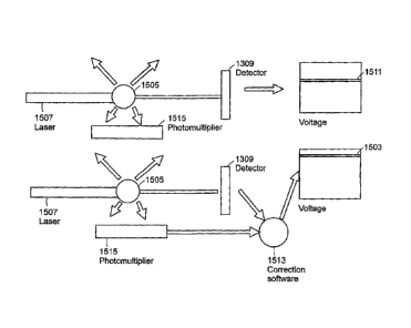
Fig 15 is a block diagram illustrating how measured green fluorescence may be
used to restore the baseline voltage level in accordance with an embodiment of
the
invention. In accordance with embodiments of the present invention, the amount
of green
fluorescence emitted by a particle 1505 in the pathway of the laser beam 1507
is
measured using a photomultiplier tube 1515. The amount of fluorescence emitted
is an
indicator of the amount of light that was lost from the laser beam 1507 due to
conversion
to fluorescence. Computer software or analog electronic circuitry 1513 (which
may
contain standard components such as operational amplifiers to modify the
voltage
signals) are used to apply a correction factor to the photodiode detector to
restore the
baseline 1511 to the baseline level 1503 in order to measure chromatic dye-
based light
loss.
Auto-Fluorescence Correction--Example Procedure
CA 02855973 2014-07-04
The analysis technique that follows is based on the following reasoning: 1)
green
auto-fluorescence is detected at the same time that blue light-loss signal is
obtained; 2)
for green auto-fluorescence to occur, there must have been conversion of the
exciting 488
nm laser light into green light; 3) the laser light that is converted to green
fluorescence is
lost to the blue scatter detector; and 4) this gives an artificially high
measurement of
specific blue-laser absorption. To correct for this artifact, the green
fluorescence signal
(or an adjusted signal based on it) may be subtracted from the inverted blue
light loss
signal. Subtracting the green fluorescence signal from the inverted signal is
mathematically equivalent to adding it to the non-inverted signal. Thus, in
effect, a
correction factor may be added to the inverted blue light loss signal to
compensate for the
amount of laser light that was lost to fluorescence.
To illustrate the method, tissue sections stained with antibodies to a
specific
antigen and developed with the chromatic dye diaminobenzidine (DAB) were
analyzed
on a laser scanning cytometer. The slides were segregated into groups that
either had no
staining (exhibiting only background levels of staining), or varying amounts
of specific
staining. Quantification of the amount of DAB staining was the goal of this
particular
experiment.
As shown in Fig. 16, a tissue section (or other cellular sample) 1601 may
include
both fluorescent and chromatic components (1603 and 1605 respectively).which
contribute to light loss. Thus, when analyzed by laser scanning cytometry,
these tissue
sections exhibited both light loss and green fluorescence. The fluorescence
was caused
by auto-fluorescence of the tissue. The light loss indicated by voltage 1611
can be caused
by either chromatic or fluorescent entities within the tissue. The light loss
caused by the
auto-fluorescent components was not of interest in this example as it
interferes with the
assay sensitivity.
As shown in Fig. 17, a photomultiplier tube 1715 was introduced into the
system
of Fig. 16 to measure the amount of green fluorescence. The photomultiplier
tube 1715
was used as an input for the computer correction algorithms 1713, and the
effect of the
auto-fluorescence on the light loss signal was effectively eliminated from the
analysis
system such that the signal 1711 indicates light loss due to absorbance of
light by the
stained sample.
The efficacy of the correction algorithm is shown in the graphs of
experimental
data shown in Figs. 18 and 19. The analysis results from five groups of slides
are shown
as uncorrected data in Fig. 18 and corrected data in Fig. 19. A red box 1801
and 1901 has=
16
CA 02855973 2014-07-04
been drawn around the background-level control group and a green box 1803 and
1903
has been drawn around the groups expressing specific chromatic dye staining.
As can be
seen from the graphs of Figs. 18 and 19, the ratio of the specific signal to
background
staining is greatly increased in the corrected group.
Fig. 20 is a flow diagram illustrating a method for quantifying the light
absorption
in a biological sample. In accordance with this embodiment, a beam of light is
impinged
2001 on the sample. An amount of light loss due to interference of the beam by
the
sample is measured and a first signal is produced 2002. An amount of
fluorescence
emitted by the sample is also measured and a second signal is produced 2003.
The
second signal is used 2004 to correct the first signal in order to quantify
the amount of
light loss due to a chromatic dye in the sample.
The example shown above corrects for auto-fluorescence, but similar strategies
can be used to correct for the effects of fluorescent dyes on light-loss
signals.
Additionally, the method described above may be applied to samples other than
tissue
sections. Further, the method may also be applied to camera-based systems.
Corrections to the input signals ¨ Per Pixel Correction.
Due to the nature of the scanning optics, the intensity of the laser beams
varies as
it scans across the specimen ma Y (or vertical) direction. Corrections for
this variation
for fluorescence measurements include empirically measuring the intensity of
calibration
particles at a plurality of positions that cover the entire scan field. In
accordance with
fluorescence-based analysis, the mean of the fluorescence intensity of the
particles is
calculated for each possible Y position and a correction factor is calculated
for each Y
position. These calculated values are and stored in the look-up table. In
subsequent
image acquisition, the detector values may be multiplied by the correction
factor to obtain
the background corrected data (see, for example, United States Patent No.
5,885,840).
For light scatter absorption measurements, the same principle is applied, but
instead of using calibration particles, a blank microscope slide is used. The
photodetectors are set to give a signal in the working range of the
instrument, usually near
the upper limits of absorbance detection, and laser scans are obtained. Fig.
21 is a flow
chart illustrating a method for correcting input signals associated with light
absorption in
a biological sample. In accordance with this embodiment, signals produced in
accordance with the variations of intensity are measured 2101 when the beams
impinge
upon a blank surface. Values for each pixel across the scan line are averaged
across the
group of laser scans and a per-pixel correction lookup table is produced 2102
and values
17
CA 02855973 2015-09-04
associated with the signals produced when the beams impinge upon the blank
surface are
used 2103 to compensate for intensity variations produced when the beams
impinge upon the
sample. In subsequent image acquisition, the detector values are multiplied by
the correction
factor to obtain the background corrected data. The corrected data is
available for viewing
and analysis in image displays with improved accuracy of the quantitative
data. Figs. 22A
and 22B are illustrations of light absorption images produced before and after
per-pixel
correction is applied, respectively.
It should be understood that various changes and modifications to the
preferred
embodiments described above will also be apparent to those skilled in the art.
Modifications
can be made without departing from the scope of the invention and without
diminishing its
attendant advantages. While specific embodiments have been described and
illustrated, such
embodiments should be viewed as illustrative only, and not as limiting the
invention as
defined by the accompanying claims.
18