Note: Descriptions are shown in the official language in which they were submitted.
CA 02856099 2016-12-06
- 1 -
PROCESS FOR OPTICAL COHERENCE TOMOGRAPHY AND APPARATUS
FOR OPTICAL COHERENCE TOMOGRAPHY
The present invention relates to a process for optical coherence tomography,
in
particular for generating sets of three-dimensional image data pertaining to
an
object to be examined. The invention further relates to an apparatus for
optical
coherence tomography.
For the purpose of creating a three-dimensional (3D for short) tomogram of an
object to be examined with the aid of optical coherence tomography (OCT for
short),
it is conventional to record a large number of OCT slice images oriented in
layers
with respect to one another within a volume of the object to be scanned and to
orient said slice images subsequently with respect to one another. A 3D
registration
of such a type can be generated by arranging the individual slice images with
respect to one another in each instance in the way in which they were arranged
originally at the time of the recording thereof in a coordinate system of the
recording
apparatus.
A problem of this approach, however, is that the eye move during the recording
of
the slice images representing the 3D tomogram. In the course of the subsequent
3D
registration of the individual slice images, imaging errors in the 3D tomogram
may
occur by reason of motion artefacts.
It is an object of embodiments of the invention to specify a process that
enables a
three-dimensional representation of an object that can be examined by means of
optical coherence tomography. Furthermore, an object of embodiments of the
invention is to specify an apparatus that operates in accordance with a
process of
such a type.
CA 02856099 2016-12-06
- 2
Certain exemplary embodiments can provide a process for optical coherence
tomography, comprising: recording a plurality of first OCT slice images, each
first
slice image representing a different slice of an object; ascertaining a
reference
figure that is representative of the three-dimensional contour of at least one
structural feature of the object in a given three-dimensional coordinate
system by
feature recognition of the at least one structural feature in the first slice
images;
recording a plurality of second OCT slice images, each second slice image
representing a different slice of the object, wherein a time period for
recording a
second slice image is longer than a time period for measuring a first slice
image;
automatically displacing by an apparatus at least a fraction of the second
slice
images in the coordinate system until each second slice image is in feature
overlap
with the reference, figure in order to reduce motion artefacts; and generating
a set
of three-dimensional OCT image data at least from the feature-overlapped
second
slice images.
Certain exemplary embodiments can provide an apparatus for optical coherence
tomography, comprising an OCT image-acquisition unit and a computer
arrangement that has been set up to: control the OCT image-acquisition unit in
such
a manner that the OCT image-acquisition unit records a plurality of first OCT
slice
images, each first slice image representing a different slices of an object;
ascertain
a reference figure that is representative of the three-dimensional contour of
at least
one structural feature of the object in a given three-dimensional coordinate
system
by feature recognition of the at least one structural feature in the first
slice images;
control the OCT image-acquisition unit in such a manner that the OCT image-
acquisition unit records a plurality of second OCT slice images, each second
slice
image representing a different slice of the object, wherein a time period for
recording a second slice image is longer than a time period for measuring a
first
slice image; displace at least a fraction of the second slice images in the
coordinate
system until each second slice image is in feature overlap with the reference
figure
in order to reduce motion artefacts; and generate a set of three-dimensional
OCT
image data at least from the feature-overlapped second slice images.
CA 02856099 2016-12-06
- 2a -
In other words: a plurality of first slice images are recorded, the first
slice images
representing various longitudinal or cross-sectional projections of at least
one
structural feature of the object. Then the structural feature is recognised,
for
example by image processing of each one of the first slice images. From this,
interpolation coordinates can be ascertained that represent the position of
the
structural feature in a coordinate system. A reference figure representing the
shape
of the structural feature can be adapted to the interpolation coordinates.
Subsequent thereto, a plurality of second slice images are recorded which also
represent various longitudinal or cross-sectional projections of the
structural feature.
In each of the second slice images the structural feature is recognised, and
corresponding positions of the structural feature in the coordinate system are
ascertained. In addition, puncture points can be ascertained that represent
the
position of points of intersection of each one of the second slice images with
the
reference figure. The positions of the structural feature can be compared with
the
puncture points. If for a second slice image not all the positions of the
structural
feature are congruent with the puncture points, the second slice image is
displaced,
tilted and/or rotated by coordinate transformation in the coordinate system
until such
time as the second slice image is oriented with respect to the reference
figure in
exactly fitting manner and the positions of the structural feature are
congruent with
the puncture points. For the purpose of creating a 3D tomogram of the object,
finally the first and/or second slice images can be assembled to form an
overall set
of image data.
The present invention consequently makes it possible that during a first (e.g.
comparatively short) period of time first slice images are recorded to begin
with
which may serve to determine the position, orientation and/or size of a
reference
figure of predetermined shape. The reference figure may subsequently be
utilised as a 3D registration support, in order to suitably orient with
respect to
one another the second slice images recorded during a second period of time
(e.g. longer in comparison with the first period of time). Motion artefacts in
the
3D tomogram of the object, caused by movement of the object,
CA 02856099 2014-05-16
WO 2013/097877 PCT/EP2011/006594
- 3 -
are thereby avoided. The 3D registration consequently contributes to the
creation of a 3D projection of the object with reduced errors.
The first and/or second slice images constitute, for example, so-called B-
scans.
These represent flat, two-dimensional (2D for short) OCT projections of the
object. A B-scan can be obtained on the basis of a plurality of line scans, so-
called A-scans. An A-scan constitutes a measured OCT interferogram and
represents a rectilinear, one-dimensional (1D for short) OCT projection over
an
axial distance of the object. A B-scan may be formed from several A-scans of
equal length situated in one plane and running parallel to one another. All
the
slice images and the set of image data may also be stored in a suitable
storage
medium.
The object to be examined may be, for example, an eye. The object may be
any other suitable physical entity that can be imaged, such as a workpiece
with
internal structural features.
The position and the orientation of the structural feature may reproduce, in
substitutional manner, the position and the orientation of the object in a
coordinate system. For this purpose the structural feature extends, for
example,
over an extensive region on or in the object. If the object is an eye, the
structural feature may be, for example, the outer and/or inner margin of the
iris
and/or the limbus of the eye. Alternatively the structural feature may be a
surface or internal structural face of the human lens and/or the cornea.
The reference figure may simulate the geometry of the structural feature
schematically. For this purpose the reference figure may represent a
simplified
model of the structural feature that substantially reproduces the position and
the
orientation of the structural feature in a coordinate system. Accordingly it
is
conceivable that the reference flgure represents a geometrical shape such as
an
ellipse, a circular disc, a circular disc with concentrically inscribed
circular hole, a
sphere, a spherical shell, a cylinder, a hollow cylinder with finite thickness
of the
circumferential surface, or the like. If the structural feature is, for
example, the
margin of the iris, the reference flgure may exhibit a circular shape and/or
elliptical shape. If the structural feature is, for example, both the inner
and
outer margins of the iris, the reference figure may represent a geometrical
CA 02856099 2014-05-16
WO 2013/097877 PCT/EP2011/006594
- 4 -
shape that comprises two circular shapes and/or elliptical shapes that have a
certain spatial positioning and orientation with respect to one another.
The term "displacing" refers to changing the position and/or the orientation
of
an entity in any suitable manner. For example, a coordinate transformation may
be applied to the position and/or the orientation to change the position and
the
orientation, respectively. A coordinate transformation may include at least
one
spatial translation parallel or antiparallel to the x-, y- and/or z-axes of a
coordinate system and/or at least one spatial rotation about an axis of
rotation
along the x-, y- and/or z-axes and/or a spatial rotation about an axis of
rotation
between the x-, y- and/or z-axes by a positive or negative angle in the
coordinate system. Accordingly, the term "displacing" may include an arbitrary
tilting in space. A coordinate transformation preserves some or all the
relative
spacings between individual constituents of the projection within the second
slice image. Merely the position and the orientation of the slice image as
such
are changed. The space coordinates of each image pixel of the second slice
image are affected, but not the colour value or tonal value of the image
pixel.
In certain embodiments, a first period of time expended overall for the
recording
of the plurality of first slice images may be shorter than a second period of
time
expended overall for the recording of the plurality of second slice images.
The
first period of time is determined, inter alia, from the number of first slice
images, from the number of interferograms recorded per slice image, and from
the recording-time of an individual one of these interferograms. The recording-
time of an individual interferogram is determined, inter alia, from the
exposure-
time, from a following period of reworking (for instance, for the sampling of
the
interferogram, for possible image-processing steps such as Fourier
transformations, image-recognition processes and such like) and from the time
needed for storage. Analogous remarks apply to the second period of time.
In certain embodiments, the number of first slice images may be smaller than
the number of second slice images. For example, the ratio of the number of
first
slice images to the number of second slice images amounts to 1:2, 1:5, 1:10 or
1:100.
Furthermore, the recording time for a first slice image may be shorter than
the
recording time for a second slice image. For example, the exposure time, the
CA 02856099 2014-05-16
WO 2013/097877 PCT/EP2011/006594
- 5 -
period of reworking and/or the storage time of the first slice images is/are
shorter than corresponding time intervals for the second slice images. In
particular, for each slice image a plurality of A-scans of the object are
recorded
that is smaller than the number of A-scans for the second slice images.
Accordingly, a first slice image may consist of 200 A-scans, and the frequency
of
recording A-scans may amount to 70 kHz. A second slice image consists, for
example, of 500 to 2000 A-scans, which are recorded at a recording-rate from
20 kHz to 70 kHz.
In certain embodiments, the recording-time for a first slice image may be
sufficiently short that motion artefacts during the recording of the first
slice
image, caused by typical movements of the object, are substantially avoided.
The first slice images may be recorded by means of B-scans that are
distributed
over the object in a regular pattern. For example, the first slice images can
be
recorded by means of B-scans that are distributed in a cross-grid pattern. For
example, first slice images oriented orthogonally with respect to one another
are
acquired, whereby in each instance two adjacent first slice images exhibit a
constant spacing from one another.
As an alternative, the first slice images may be oriented with respect to one
another in the shape of a star in such a manner that the first slice images
intersect one another in a straight line. The straight line may coincide with
an
axis of symmetry of the object and/or may run through points of the object
that
have been marked out. For example, the straight line is centred with the
pupillary centre of an eye and runs along the optical axis thereof or through
the
apex of the cornea.
The first slice images may be recorded in such a distribution pattern that
points
of intersection of the first slice images with the reference figure are
situated,
distributed substantially at equal spacings, along the reference figure after
the
reference figure has been adapted to the first slice images.
Additionally or alternatively, the first slice images may be recorded in such
a
distribution pattern that the number n of points of intersection at which the
reference figure intersects the surface normals of the first slice images at
an
angle within the range of more than 30 and less than 600, after the reference
CA 02856099 2014-05-16
WO 2013/097877 PCT/EP2011/006594
- 6 -
figure has been adapted to the first slice images, amounts to at least 2(N-2),
where N is the number of first slice images. In other words: of the N first
slice
images of, for example, an orthogonal cross pattern, after the reference
figure
has been adapted at least N-2 flrst slice images are intersected by the
reference
figure in such a manner that the reference figure includes with the respective
surface normals of the first slice images at the respective point of
intersection an
angle of more than 300 and less than 60 . In this case there are a total of at
least n=2(N-2) such points of intersection, whereby the n points of
intersection
differ from one another, i.e. amongst themselves are not situated on top of
one
another.
Additionally or alternatively, the first slice images may be recorded in such
a
distribution pattern that the number n of points of intersection at which the
reference figure intersects the first slice images, after the reference figure
has
been adapted to the first slice images, suffices for describing the geometry
of
the reference figure.
The second slice images may also be recorded by means of B-scans that are
distributed over the object in a certain pattern. The pattern may include, for
example, a cross-grid pattern. In this case the second slice images may have
been oriented orthogonally and/or parallel to one another. Additionally or
alternatively, the pattern may include two cross-grid patterns placed over one
another in angle-offset manner. For example, the angle amounts to about 450.
Additionally or alternatively, the pattern may include three cross-grid
patterns
placed over one another in angle-offset manner. For example, the angle
amounts to about 60 .
The pattern of the second slice images may be irregular. For instance, the
grid-
line density of a cross pattern in a central region of the reference figure is
lower
than in a region of the reference figure remote from the centre. For this
purpose a spacing of two adjacent second slice images oriented parallel to one
another that intersect the region of the reference figure remote from the
centre
may be smaller than a spacing of two adjacent second slice images oriented
parallel to one another that intersect the central region of the reference
figure.
If the second slice images also contain cross-sectional projections of the
cornea
of a human eye, on the basis of the irregular pattern the aspherical regions
of
the cornea can be represented with higher resolution than can regions of the
CA 02856099 2015-09-03
- 7 -
cornea in the vicinity of the apex of the cornea. The density of cross-
sectional
projections of the cornea is accordingly higher in a region representing the
aspherical region of the cornea. Positions of these cross-sectional
projections may
serve as interpolation nodes for the segmentation of structural layers in the
object
being examined, or for an adaptation of a predetermined surface shape to the
cornea by means of Zernike polynomials.
The process may additionally include the following step: by image processing
in a
first and/or second slice image an indication of motion artefacts that have
occurred
during the recording of the respective slice image is recognised. An
indication of
motion artefacts includes, for example, a discontinuity, a waviness, a
contraction
and/or an elongation within a profile in the slice image representing the
structural
feature and/or a low signal-to-noise ratio (SNR for short) of adjacent A-scans
of a
slice image. This step may take place 'online' before the next slice image is
acquired in accordance with the distribution pattern. If motion artefacts are
discernible within a first and/or second slice image, the acquisition of the
defective
slice image may be repeated until the slice image is present in flawless
manner.
But the acquisition of an individual first and/or second slice image may take
place
so quickly that the recording time required for the acquisition is short in
comparison
with a timescale that is typical of eye movements.
An apparatus for optical coherence tomography comprises an OCT image-
acquisition unit and a computer arrangement that has been set up to control
the
OCT image-acquisition unit in such a manner that the latter records a
plurality of
first OCT slice images, each first slice image representing a different slice
of an
object, to ascertain a reference figure that is representative of the three-
dimensional
contour of at least one structural feature of the object in a given three-
dimensional
coordinate system by feature recognition of the at least one structural
feature in the
first slice images, to control the OCT image-acquisition unit in such a manner
that
the OCT image-acquisition unit records a plurality of second OCT slice images,
each second slice image representing a different slice of the object, to
displace at
least a fraction of the second slice images in the coordinate system until
each
second slice image is in feature overlap with the reference figure in order to
reduce
motion artefacts, and to generate a set of three-dimensional OCT image data at
least from the feature-overlapped second slice images.
CA 02856099 2014-05-16
WO 2013/097877 PCT/EP2011/006594
- 8 -
The apparatus may have been set up to bring about a process, described above,
for optical coherence tomography.
To the extent that a process or individual steps of a process for optical
coherence tomography is/are described in this description, the process or
individual steps of the process can be executed by an appropriately configured
apparatus. Analogous remarks apply to the elucidation of the mode of operation
of an apparatus that executes process steps. To this extent, apparatus
features
and process features of this description are equivalent.
The invention will be elucidated further in the following on the basis of the
appended drawings, of which:
Fig. 1 shows, in schematic block representation, elements of
an
apparatus for optical coherence tomography according to
one embodiment,
Fig. 2 shows, in top view schematically, an example of a
distribution pattern in which the first slice images are
recorded, with a reference figure drawn in,
Fig. 3 shows, in a three-dimensional view schematically, the
distribution pattern from Fig. 2,
Figs. 4a and 4b show schematically examples of a first slice image,
Fig. 5 shows, in top view schematically, a further example of
a
distribution pattern in which the first slice images are
recorded,
Figs. 6a and 6b show, in top view schematically, examples of
distribution
patterns in which the second slice images are recorded,
Figs. 7a to 7c show schematically an example relating to the
displacing of
a second slice image parallel to an x-axis until the second
slice image is in feature overlap with a reference figure,
CA 02856099 2014-05-16
WO 2013/097877 PCT/EP2011/006594
- 9 -
Figs. 8a to 8c show schematically an example relating to the
displacing of
a second slice image antiparallel to a y-axis until the
second slice image is in feature overlap with a reference
figure,
Figs. 9a to 9c show schematically an example relating to the
displacing of
a second slice image parallel to a z-axis until the second
slice image is in feature overlap with a reference figure,
Figs. 10a to 10c show schematically an example relating to the rotation of
a
second slice image about an axis of rotation running
parallel to an x-axis until the second slice image is in
feature overlap with a reference figure, and
Figs. 11a to 11e show schematically an example relating to the displacing
of
a second slice image until the second slice image is in
feature overlap with a reference figure.
The apparatus for optical coherence tomography in Fig. 1 ¨ denoted generally
therein by 10 ¨ serves for creating 3D tomograms of an object shown in the
exemplary case as a human eye 12. The optical coherence tomography is
based, for example, on so-called time-domain (TD for short) OCT or on so-
called
frequency-domain (FD for short) OCT.
The apparatus 10 includes a light-source 14 for emitting coherent light. The
light-source 14 is designed, for example, for the purpose of FD OCT as a
tuneable light-source or emits a spectrum of coherent light that is broadband
within the frequency space. The light emitted from the light-source 14 is
directed onto a beam-splitter 16. The beam-splitter 16 is a constituent part
of a
Michelson interferometer 18 and splits up the incident optical output in
accordance with a predetermined splitting ratio, for example 50:50. One ray 20
runs within a reference arm; the other ray 22 runs within a specimen arm.
Instead of the free-space setup represented in Fig. 1 the Michelson
interferometer 18 may also have been realised partly or entirely with the aid
of
fibre-optic components.
CA 02856099 2014-05-16
WO 2013/097877 PCT/EP2011/006594
- 10 -
The light that has been branched off in the reference arm impinges onto a
mirror 24 which reflects the light back onto the beam-splitter 16 collinearly.
For
the purpose of TD OCT the mirror 24 may be displaceable along the direction of
propagation of the ray 20. The light that has been branched off in the
specimen
arm impinges onto the object 12 to be examined, which back-scatters or
reflects
back the light in the direction of the beam-splitter 16.
In Fig. 1 a three-dimensional Cartesian coordinate system of the apparatus 10
has been drawn in schematically which serves as coordinate system in the
following. In this connection the z-axis represents the direction of
propagation
of the light ray 22 in the region immediately upstream of the object 12.
Within the specimen arm further optical elements 26 and adjusting components
28 are provided, which have been set up to focus the light ray 22 coming in
from the beam-splitter 16 onto the object 12 and to adjust the focus position
(for example in the lateral directions x, y or in all three directions in
space x, y,
z). A computer arrangement 30 controls the adjusting components 28 for the
purpose of obtaining 1D, 2D and/or 3D tomograms.
The light back-scattered from the object 12 in the specimen arm is collinearly
superimposed at the beam-splitter 16 with the light reflected back from the
mirror 24 in the reference arm so as to form an interference beam 32. The
optical path lengths in the reference arm and specimen arm are substantially
equally long, so that the interference beam 32 displays an interference
between
the constituent rays 20, 22 from reference arm and specimen arm. A detector
34' registers the intensity of the interference beam 32 as a function of the
time,
the wavelength and/or the wave number. For this purpose the detector 34' may
take the form of a photodiode or spectrometer.
The signal registered by the detector 34' is transferred to the control
arrangement 30 which ascertains 2D OCT slice images therefrom. In this sense
the computer arrangement 30, the light-source 14, the detector 34' and the
Michelson interferometer 18, inclusive of the optical elements 26 and the
adjusting components 28, may be understood as an OCT image-acquisition unit
33 which is controlled by the computer arrangement 30.
CA 02856099 2014-05-16
WO 2013/097877 PCT/EP2011/006594
- 11 -
For the purpose of creating a 3D tomogram of the object 12 the computer
arrangement 30 controls the adjusting components 28 in accordance with such a
scan pattern that a 3D registration of the acquired slice images within a
scanned
volume of the object 12 with respect to one another can be undertaken. This
process will be described in detailed manner in the following with reference
to
Figs. 2 to 11.
First of all, a plurality of first slice images 34, each first slice image
representing
a different slice of the object 12, are recorded and are stored in a memory of
the
computer arrangement 30. The first slice images 34 represent OCT B-scans
which are obtained from a large number of OCT A-scans. A first slice image 34
consists of, for example, 200 A-scans. Furthermore, a short exposure-time is
chosen in which the individual A-scans are recorded. The rate of recording of
A-
scans amounts to, for example, 70 kHz.
As shown in Fig. 2, in the present exemplary case three horizontal and three
vertical first slice images 34 are acquired in an orthogonal cross pattern 35
similar to a chessboard. In this example the horizontal and vertical first
slice
images 34 are arranged parallel to one another, the spacing d of adjacent
first
slice images 34 being constant for all adjacent slice images 34. In Fig. 3 the
distribution pattern, shown in Fig. 2, of the first slice images 34 and a part
of the
eye 12 are illustrated again three-dimensionally.
As an alternative to the distribution pattern shown in Figs. 2 and 3, the
distribution pattern, shown in Fig. 5, of first slice images 34 is also
possible. In
this case the first slice images 34 have been oriented with respect to one
another in the shape of a star in such a manner that the first slice images 34
intersect one another in a straight line G. In the example shown in Fig. 5 the
straight line G is centred with the pupillary centre of the eye 12 and runs
along
the optical axis of the eye 12, that is to say, substantially parallel to the
z-axis.
A first slice image 34 is shown in Figs. 4a and 4b. The first slice images 34
represent cross-sectional projections of at least one structural feature 36 of
the
object 12. In Fig. 4a the structural feature 36 is, for example, the outer
margin
of the iris 38 in the eye 12. In Fig. 4b the structural feature 36 is, for
example,
the outer and inner margins of the iris 38. Besides the iris 38, in the first
slice
image 34 the cornea 40, for example, is also imaged.
CA 02856099 2014-05-16
WO 2013/097877 PCT/EP2011/006594
- 12 -
In the first slice images 34 which have been prepared the computer
arrangement 30 now recognises the structural feature 36 on the basis of an
image-recognition algorithm and determines the position(s) 42 thereof in the
coordinate system of the apparatus 10. In Figs. 2, 4a and 4b these positions
42
are labelled by means of small circles filled in with black.
As shown in Fig. 2, the computer arrangement 30 subsequently adapts a
reference figure 44 that is representative of the three-dimensional contour of
the
structural feature 36 to the positions 42 ascertained beforehand serving as
interpolation nodes. In Fig. 2 the reference figure 44 represents a circular
shape
in imitation of the margin of the iris 38, which is predetermined by a
midpoint M
(i.e. a 3D space coordinate) and a further parameter R defining the radius.
The
adapting or fitting of the reference figure 44 to the interpolation nodes 42
is
based on a mathematical optimisation method in order to determine (to
estimate) the unknown parameters M and R of the reference figure 44 for a
series of interpolation nodes 42.
The spacing d of the slice images 34 is chosen in such a way that the
interpolation nodes 42 have an almost equidistant spacing on the periphery of
the reference figure 44 after the reference figure 44 has been adapted to the
first slice images 34.
Additionally or alternatively, the first slice images 34 can be recorded in
such a
distribution pattern 35 that the number n of points of intersection at which
the
reference figure 44 intersects the first slice images 34, in each instance at
an
angle within the range of more than 30 and less than 60 in relation to the
surface normal of the respective slice image 34, amounts to at least 2(N-2),
where N is the number of first slice images 34, after the reference figure 44
has
been adapted to the first slice images 34. This is represented in exemplary
manner in Fig. 2: of the six flrst images 34 (i.e. N = 6) of the orthogonal
cross
pattern 35, after the reference figure 44 has been adapted four first slice
images
34 are intersected by the reference figure 44 in such a manner that the
reference figure 44 includes with the respective surface normals of the first
slice
images 34 at the respective point of intersection an angle of more than 300
and
less than 60 (i.e. n=2(N-2)=2(6-2)=8).
CA 02856099 2014-05-16
WO 2013/097877 PCT/EP2011/006594
- 13 -
Additionally or alternatively, the first slice images 34 can be recorded in
such a
distribution pattern 35 that the number n of points of intersection at which
the
reference figure 44 intersects the first slice images 34 suffices for
describing the
geometry of the reference figure 44 after the reference figure 44 has been
adapted to the first slice images 34. This is again represented in Fig. 2: the
six
first slice images 34 of the orthogonal cross pattern 35 are intersected by
the
reference figure 44 at twelve points of intersection after the reference
figure 44
has been adapted. Each of these twelve points of intersection is described in
three-dimensional space by three parameters (space coordinates x, y, z), so
that
a total of 36 parameters are available for fitting the reference figure 44.
For
example, the circle shown in Fig. 2 is described by a midpoint in three-
dimensional space (consisting of three parameters) and a radius (vector) in
three-dimensional space (likewise consisting of three parameters). For the
purpose of fitting the reference figure 44, at least six parameters are
accordingly
required, so that the 36 parameters of the twelve points of intersection
between
the reference figure 44 and the first slice images 34 are sufficient.
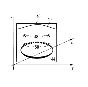
Subsequently a plurality of second slice images 46, each second slice image
representing a different slice of the object 12, are recorded and stored. The
second slice images 46 also represent OCT B-scans which are obtained from a
large number of OCT A-scans. The second slice images 46 consist, for example,
of 2000 A-scans per B-scan, the A-scans being recorded at a recording-rate
from, for example, 20 kHz to 70 kHz. In comparison with the first slice images
34, the second slice images 46 therefore offer higher statistics and image
quality. This permits higher-quality, in the sense of a signal-to-noise ratio,
second slice images 46 to be created.
As shown in Figs. 6a and 6b, the second slice images 46, in the course of the
acquisition thereof, are distributed over the object 12 in accordance with an
irregular pattern 45. In Figs. 6a and 6b, in addition the reference figure 44
ascertained beforehand has been drawn in. The irregular distribution pattern
includes a cross-grid pattern, the grid-line density of which in a central
region
47a of the reference figure 44 is lower than in a region 47b of the reference
figure 44 remote from the centre. In Figs. 6a and 6b the central region 47a
and
the region 47b remote from the centre are separated from one another in
exemplary manner by a dashed line. Consequently a spacing of two adjacent
second slice images 46a oriented parallel to one another in the central region
CA 02856099 2014-05-16
WO 2013/097877 PCT/EP2011/006594
- 14 -
47a of the reference figure 44 is larger than a spacing of two adjacent second
slice images 46b oriented parallel to one another in the region 47b of the
reference figure 44 remote from the centre.
In Fig. 6a the second slice images 46 are arranged in such a manner that they
are oriented orthogonally and/or parallel to one another. But alternatively
the
distribution pattern shown in Fig. 6b is also possible, in which the second
slice
images are arranged in such a manner that they are oriented orthogonally
and/or parallel to one another and/or intersect one another at an angle of
45 degrees. Accordingly, the irregular pattern includes two cross-grid
patterns
placed on top of one another in angle-offset manner.
In Figs. 7a to 11e schematic representations of second slice images 46 are
shown. The second slice images 46 likewise represent cross-sectional
projections of the structural feature 36. In the second slice images 46 the
cornea 40, for example, is also imaged. As in the case of the flrst slice
images
34, on the basis of image processing the computer arrangement 30 ascertains in
the second slice images 46 the structural feature 36 and determines the
position(s) 48 thereof in the coordinate system of the apparatus 10. In Figs.
7a
zo to lle these positions 48 are labelled by means of small black circles
with
inscribed black cross.
The reference figure 44 can also be seen in Figs. 7a to 11e, wherein the
margin
thereof is represented by a continuous line if the reference figure 44, viewed
from the observer, runs spatially in front of the second slice image 46, and
the
margin thereof is represented by a dashed line if the reference figure 44,
viewed
from the observer, runs spatially behind the second slice image 46.
For each second slice image 46 the computer arrangement 30 ascertains by
calculation the positions of puncture points 50 at which the reference figure
44
intersects the second slice image 46. The (original) position and orientation
of a
second slice image 46 which are required for this purpose are predetermined by
the distribution pattern 45. The puncture points 50 are labelled in Figs. 7a
to
11e, like the positions 48, by means of small black circles with inscribed
black
cross. The puncture points 50 are situated at the transition from the
continuous
margin to the dashed margin of the reference figure 44.
CA 02856099 2014-05-16
WO 2013/097877 PCT/EP2011/006594
- 15 -
If not all the positions 48 are in congruence with the puncture points 50, the
computer arrangement 30 displaces a second slice image 46 in the coordinate
system of the apparatus 10 until all the positions 48 in the second slice
image 46
are congruent with the puncture points 50. For this purpose the computer
arrangement 30 ascertains a suitable coordinate transformation for the second
slice image 46. In the course of the transformation all the relative spacings
between individual projection constituents 40, 48 within the second slice
image
46 are preserved. Merely the position and the orientation of the slice image
46
as such are changed.
In Figs. 7a to 11e exemplary displacements, rotations or coordinate
transformations are shown, on the basis of which second slice images 46 are
oriented with respect to a reference figure 44. Small arrows shown therein
illustrate the respective displacement or rotation.
Accordingly, Figs. 7a to 7c show a translation of a second slice image 46
parallel
to the x-axis. In Fig. 7a the computer arrangement 30 recognises that the
positions 48 are not congruent with the puncture points 50, since the spacing
between the two positions 48 is shorter than between the puncture points 50.
The reason for this is that the eye 12 has been displaced effectively
antiparallel
to the x-axis during the period of time between creation of the reference
figure
44 and the recording of the second slice image 46. With a view to
compensating the eye movement, the computer arrangement 30 carries out a
coordinate transformation for the second slice image 46, whereby the space
coordinates of each image pixel in the second slice image 46 are corrected in
such a manner that after the coordinate transformation the positions 48 are
congruent with the puncture points 50.
In this way the second slice images 46 are oriented, image by image, with
respect to the reference flgure 44 and are stored. This type of 3D
registration
enables the creation of 3D tomograms of the object 12 that are free from
motion
artefacts. In this manner, motion artefacts such as, for example, level
errors,
rotation errors orthogonal to the optical axis and/or lateral movements can be
compensated. Accordingly, the computer arrangement 30 generates from the
feature-overlapping second slice images 46 a set of three-dimensional OCT
image data which is then displayed on a display unit 52 of the apparatus 10 as
a
3D tomogram of the object 12 to be examined.
CA 02856099 2014-05-16
WO 2013/097877 PCT/EP2011/006594
- 16 -
In Figs. 8a to 8c a further coordinate transformation of a second slice image
46
is shown, in the course of which the second slice image 46 is displaced
antiparallel to the y-axis. In Figs. 9a to 9c a coordinate transformation for
a
second slice image 46 is again shown. In this case the displacement is
effected
parallel to the z-axis of the coordinate system of the apparatus 10.
In Figs. 10a to 10c a spatial rotation of a second slice image 46 is shown.
Although in Fig. 10a one position 48 is initially congruent with one puncture
point 50, the second position 48 does not tally with the second puncture point
50. The second slice image 46 is therefore rotated about an axis of rotation
running parallel to the x-axis, see Fig. 10b.
In Figs. 11a to 11e a somewhat more complex transformation of a second slice
image 46 is shown. In this example the reference figure 44 represents both the
inner and outer margins of the iris 38 of the eye 12. The reference figure
consists of two circular shapes arranged parallel to one another, the
midpoints of
which lie on a straight line perpendicular to the surfaces of the circular
shapes.
=
In Fig. lla it can be discerned that in the second slice image 46 four
positions
48 corresponding to a cross-section through the inner and outer margins of the
iris were recognised by the computer arrangement 30 but the reference figure
44 intersects the second slice image 46 merely at two puncture points 50. In
Fig. llb it can be seen how the second slice image 46 is therefore displaced
firstly antiparallel to the x-axis until four puncture points 50 with relative
spacings corresponding to the relative spacings of the positions 48 are
present,
see Fig. 11c. In Fig. 11d a spatial translation of the second slice image 46
parallel to the z-axis is subsequently effected until, as shown in Fig. 11e, a
total
overlap of features occurs.
The computer arrangement 30 has furthermore been set up to recognise, by
image processing in a first and/or second slice image 34, 46, an indication of
motion artefacts that have arisen during the recording of the respective slice
image 34, 46. If motion artefacts are recognisable within a slice image 34,
46,
the computer arrangement 30 controls the OCT image-acquisition unit 33 in
such a manner that the acquisition of the defective slice image 34, 46 15
CA 02856099 2014-05-16
WO 2013/097877 PCT/EP2011/006594
- 17 -
repeated. But the acquisition of a slice image 34, 46 is effected so quickly
that
the individual B-scan is free from motion artefacts.
Unless expressly stated otherwise, identical reference symbols in the Figures
stand for identical or identically-acting elements. In other respects, an
arbitrary
combination of the features elucidated in the Figures in connection with
individual embodiments is conceivable.