Note: Descriptions are shown in the official language in which they were submitted.
CA 02856117 2014-05-15
WO 2013/075140
PCT/US2012/065945
AUTO-RECOGNIZING THERAPEUTIC R/DNA CHIMERIC NANOPARTICLES (NP)
CROSS-REFERENCE TO RELATED APPLICATION
This application claims the benefit of U.S. Provisional Application Nos.
61/561,257, filed
November 17, 2011, and 61/698,113, filed September 7,2012, the contents of
which are
incorporated herein by reference in their entirety.
STATEMENT OF RIGHTS TO INVENTIONS MADE UNDER FEDERALLY
SPONSORED RESEARCH
This work was supported by the National Institutes of Health. The government
has
certain rights in the invention.
BACKGROUND OF THE INVENTION
In the past several years, there has been a tremendous increase of interest in
using RNA
interference (RNAi) for biomedical applications. RNAi is a posttranscriptional
sequence
specific process of gene silencing employing double-stranded RNAs (dsRNAs) and
a set of
specific proteins and enzymes. To briefly explain the mechanism, the RNaseIII-
like enzyme,
Dicer, processes a dsRNAs into shorter duplexes (21-23 bp). These duplexes,
referred to as
short interfering RNAs (siRNAs), are then loaded into a RNA-induced silencing
complex (RISC)
and one of the siRNA strands, called passenger or sense, is discarded. The
other strand, called
guide or antisense, is used by RISC to recognize the target mRNA for cleavage
and translation
prevention. RNAi has become a powerful technique for selective suppression of
particular
genes of interest in different species showing potential for use as cancer and
HIV therapeutics.
Synthetic siRNAs against particular genes of interest can be exogenously
introduced into cells
to activate RNAi. Moreover, introduction of synthetic asymmetric Dicer
substrates slightly
longer than siRNAs (25 bp) tremendously increases the potency of silencing.
This can be
explained by the involvement of Dicer in the process of loading the RISC
complex with
siRNAs. Despite the potential for siRNA, there is a need for novel approaches
that overcome
several challenges associated with the clinical delivery of RNAi.
As described herein, the present invention splits the functionality of Dicer
substrates
siRNA duplexes into two R/DNA hybrids, which upon simultaneous presence inside
the same
America 4443807.2
CA 02856117 2014-05-15
WO 2013/075140
PCT/US2012/065945
ATTORNEY DOCKET NO. 89711W0(47992)
November 19, 2012
diseased cell will recognize each other through toehold interaction and re-
associate releasing
active siRNAs. This novel approach will overcome several challenges associated
with the
clinical delivery of RNAi, such as intravascular degradation (will be reduced
for R/DNA
hybrids), tissue specificity (DNA chemistry is more parsimonious than RNA and
amenable to
chemical modifications with different features for targeting or delivery),
pharmacodynamics
(fluorescent tags can be activated upon R/DNA hybrid re-association assisting
in Forster
resonance energy transfer (FRET) imaging of delivery and response). Moreover,
all these
additional functionalities can be introduced through chemical modifications of
the DNA strands
in the R/DNA hybrids thus, not interfering with the processivity of the
released siRNAs.
Additionally, the number of these functionalities can be at least as large as
twice the number of
DNA strands entering into the composition of the duplex hybrids or more
complex hybrid
nanostructures.
SUMMARY OF THE INVENTION
The invention provides R/DNA chimeric polyfunctional nanoparticles which are
able to
reassemble to produce functionalized dsRNA.
In one aspect, the invention generally features an R/DNA chimeric
polyfunctional
nanoparticles (R/DNA NP) having at least two chimeric nanoparticles wherein
the first chimeric
nanoparticle having a first DNA oligonucleotide and a complementary first RNA
oligonucleotide, and the second chimeric nanoparticle having a second DNA
oligonucleotide and
a complementary second RNA oligonucleotide, where the first DNA
oligonucleotide has a 5'
toehold sequence and the second DNA oligonucleotide has a 3' toehold sequence.
In
embodiments, the first RNA is complementary to the second RNA and when
duplexed form an
siRNA.
In embodiments, the R/DNA NP contain another pair of chimeric nanoparticles
having
complementary 5' and 3' toehold sequences.
In embodiments, the first RNA oligonucleotide comprises at least two RNA
oligonucleotides. In related embodiments, the second RNA oligonucleotide
comprises at least
two RNA oligonucleotides corresponding to the at least two RNA
oligonucleotides that make up
the first RNA oligonucleotide. In embodiments, the duplexed form of the
complementary RNA
oligonucleotides is an siRNA.
America 4443807.2 2
CA 02856117 2014-05-15
WO 2013/075140
PCT/US2012/065945
ATTORNEY DOCKET NO. 89711W0(47992)
November 19, 2012
In embodiments, the first chimeric nanoparticle and the second chimeric
nanoparticle
each comprises at least one additional RNA that is complementary to each other
and to the DNA
oligonucleotides. In related embodiments, the duplexed form of the at least
one additional RNAs
is an siRNA.
In embodiments the 5' toehold comprises from 5 to 50 nucleotides. In another
embodiment the 5' toehold comprises from 12 to 30 nucleotides.
In embodiments the 3' toehold comprises from 5 to 50 nucleotides. In another
embodiment the 3' toehold comprises from 12 to 30 nucleotides.
In embodiments at least one oligonucleotide comprises at least one modified
nucleotide.
In some embodiments the modified nucleotide is selected from the group
consisting of locked
nucleic acids (LNAs), 2' Fluoro amidites, and 2'0Me RNA amidites.
In embodiments, the siRNA inhibits a target RNA. In general, the target RNA is
one
which produces a therapeutically beneficial result when inhibited. In
embodiments, the target
RNA comprises an RNA that encodes a protein involved in a disease process or a
portion therof.
In related embodiments, the target RNA comprises an RNA that encodes an
apoptosis
inhibitor protein or a portion thereof (e.g., Survivin, BCL-2, FLIP, STAT3,
and XIAP).
In related embodiments, the target RNA is a pathogenic RNA genome, an RNA
transcript
derived from the genome of the pathogenic agent, or a portion thereof. In some
embodiments,
the target RNA is a viral RNA genome or a portion thereof (e.g., an HIV
genome).
In any of the above aspects and embodiments, the first chimeric nanoparticle
can
comprise a first functional moiety.
In any of the above aspects and embodiments, the second chimeric nanoparticle
can
comprise a second functional moiety.
In related embodiments, the first and/or second functional moiety is a
recognition
domain. In embodiments, the recognition domain binds to a recognition target.
The recognition
target can be located on or in a target cell.
In embodiments, the target cell is a diseased cell (e.g., a neoplastic cell, a
cell infected
with a pathogen, or a cell having a genetic disorder).
In embodiments, the recognition domain specifically binds to a nucleic acid
molecule,
polypeptide, or fragment thereof.
America 4443807.2 3
CA 02856117 2014-05-15
WO 2013/075140
PCT/US2012/065945
ATTORNEY DOCKET NO. 89711W0(47992)
November 19, 2012
In embodiments, fluorescent tags, domains facilitating cellular uptake, split
functionality
domains, split lipase, and split GFP. In some embodiments, the functional
moieties can also be
RNA-fluorophore complexes that emit a signal upon association. See Paige, J.S.
et al., Science
333:642-646 (2011).
In embodiments, the recognition domain is an aptamer. In embodiments, the
aptamer
binds a cell membrane polypeptide or cell membrane structure. The cell
membrane polypeptide
or cell membrane structure can be a disease specific membrane protein or
structure (e.g., cancer
specific membrane protein or structure, a specific membrane protein or
structure associated with
infection by a pathogenic agent, and the like). In embodiments, the aptamer
binds a molecule in
the cell. For example, the aptamer can bind a nucleic acid molecule in the
target cell or a portion
thereof (e.g., DNA molecule, RNA molecule, or fragment thereof).
In some embodiments, the R/DNA NPs contain at least one of the sequences
described
herein (in the description and the figures).
In another aspect, the invention features methods for using the R/DNA NPs
described
herein.
In aspects, the invention features methods for inhibiting or reducing the
expression of a
target gene in a cell. In embodiments, the methods involve contacting the cell
with a
therapeutically effective amount of at least one of the R/DNA NPs described
herein. In
embodiments, the cell is in a subject.
In aspects, the invention features methods for killing a pathogen infected
cell. In
embodiments, the methods involve contacting the cell with a therapeutically
effective amount of
at least one of the R/DNA NPs described herein. In embodiments, the cell is in
a subject.
In aspects, the invention features methods for inhibiting replication of a
pathogen in a
cell. In embodiments, the methods involve contacting the cell with a
therapeutically effective
amount of at least one of the R/DNA NPs described herein. In embodiments, the
cell is in a
subject.
In aspects, the invention features methods for reducing pathogenic burden in a
subject. In
embodiments, the methods involve administering a therapeutically effective
amount of a
therapeutically effective amount of at least one of the R/DNA NPs described
herein. In
embodiments, the subject is at risk of developing a pathogenic infection. In
embodiments, the
subject is diagnosed with having a pathogenic infection.
America 4443807.2 4
CA 02856117 2014-05-15
WO 2013/075140
PCT/US2012/065945
ATTORNEY DOCKET NO. 89711W0(47992)
November 19, 2012
In aspects, the invention features methods for treating or preventing a
pathogenic
infection in a subject. In embodiments, the methods involve administering a
therapeutically
effective amount of a therapeutically effective amount of at least one of the
R/DNA NPs
described herein. In embodiments, the methods reduce the pathogenic burden,
thereby treating
or preventing the pathogenic infection. In embodiments, the methods induce
death in infected
cell, thereby treating or preventing the pathogenic infection.
In any of the above aspects and embodiments, the subject can be a mammal
(e.g.,
human).
In any of the above aspects and embodiments, the pathogen can be a virus,
bacteria,
fungus, or parasite. In some embodiments, the pathogen is a virus (e.g., HIV).
In any of the above aspects and embodiments, the methods can involve further
contacting
the cell with a therapeutically effective amount of a second therapeutic agent
or administering a
therapeutically effective amount of the second therapeutic agent to the
subject. The second
therapeutic agent can treat the pathogenic infection or the symptoms
associated with pathogenic
infection. For example, the second therapeutic agent can be an anti-viral
agent, an anti-bacterial
agent, an anti-fungal agent, or an anti-parasitic agent. Such agents are well
known in the art, and
it is within the purview of a physician to select and determine the
appropriate dosage of the
second therapeutic agent. See, e.g., Drug Information Handbook: A
Comprehensive Resource
for All Clinicians and Healthcare Professionals, 20th Ed., C. F. Lacy et al.
(eds.) (Lexi-Comp
2011); Johns Hopkins ABX Guide: Diagnosis & Treatment of Infectious Diseases,
2' Ed., J. G.
Bartlett et al. (eds.) (Jones & Bartlett Publishers 2010); and Mandell,
Douglas, and Bennett's
Principles and Practice of Infectious Diseases: Expert Consult Premium
Edition, 7th Ed., G. L.
Mandell (ed.) (Churchill Livingstone 2009); The Sanford Guide to Antimicrobial
Therapy 2012,
42' Ed., D. N. Gilbert et al. (eds.) (Antimicrobial Therapy 2012); Clinical
Infectious Disease
2013, 11th Ed., C. G. Weber (ed.) (Pacific Primary Care Software 2012), the
contents of which
are hereby incorporated by reference in their entirety.
In aspects, the invention features methods for killing a neoplastic cell. In
embodiments,
the methods involve contacting the cell with a therapeutically effective
amount of at least one of
the R/DNA NPs described herein. In embodiments, the cell is in a subject.
America 4443807.2 5
CA 02856117 2014-05-15
WO 2013/075140
PCT/US2012/065945
ATTORNEY DOCKET NO. 89711W0(47992)
November 19, 2012
In aspects, the invention features methods for treating a subject having a
neoplasia. In
embodiments, the methods involve administering a therapeutically effective
amount of a
therapeutically effective amount of at least one of the R/DNA NPs described
herein.
In embodiments, the neoplastic cell is a cancer cell which is present in a
solid tumor. In
embodiments, the cancer is selected from the group consisting of breast
cancer, prostate cancer,
melanoma, glioblastomas, colon cancer, ovarian cancer, and non-small cell lung
cancer.
In related embodiments, the methods involve contacting the cell with a
therapeutically
effective amount of a second therapeutic agent or administering a
therapeutically effective
amount of the second therapeutic agent to the subject. In some embodiments,
the second
therapeutic agent is an anti-cancer agent. Anti-cancer agents are well known
in the art, and any
such agent is suitable for use in the present invention. See, e.g., Anticancer
Drugs: Design,
Delivery and Pharmacology (Cancer Etiology, Diagnosis and Treatments) (eds.
Spencer, P. &
Holt, W.) (Nova Science Publishers, 2011); Clinical Guide to Antineoplastic
Therapy: A
Chemotherapy Handbook (ed. Gullatte) (Oncology Nursing Society, 2007);
Chemotherapy and
Biotherapy Guidelines and Recommendations for Practice (eds. Polovich, M. et
al.) (Oncology
Nursing Society, 2009); Physicians' Cancer Chemotherapy Drug Manual 2012 (eds.
Chu, E. &
DeVita, Jr., V.T.) (Jones & Bartlett Learning, 2011); DeVita, Hellman, and
Rosenberg's Cancer:
Principles and Practice of Oncology (eds. DeVita, Jr., V.T. et al.)
(Lippincott Williams &
Wilkins, 2011); and Clinical Radiation Oncology (eds. Gunderson, L.L. &
Tepper, I.E.)
(Saunders) (2011), the contents of which are hereby incorporated by references
in their entirety.
For example, nonlimiting examples of anti-cancer agents include Abiraterone
Acetate, Afatinib,
Aldesleukin, Alemtuzumab, Alitretinoin, Altretamine, Amifostine,
Aminoglutethimide
Anagrelide, Anastrozole, Arsenic Trioxide, Asparaginase, Azacitidine,
Azathioprine,
Bendamustine, Bevacizumab, Bexarotine, Bicalutamide, Bleomycin, Bortezomib,
Busulfan,
Capecitabine, Carboplatin, Carmustine, Cetuximab, Chlorambucil, Cisplatin,
Cladribine,
Crizotinib, Cyclophosphamide, Cytarabine, Dacarbazine, Dactinomycin,
Dasatinib,
Daunorubicin, Denileukin diftitox, Decitabine, Docetaxel, Dexamethasone,
Doxifluridine,
Doxorubicin, Epirubicin, Epoetin Alpha, Epothilone, Erlotinib, Estramustine,
Etinostat,
Etoposide, Everolimus, Exemestane, Filgrastim, Floxuridine, Fludarabine,
Fluorouracil,
Fluoxymesterone, Flutamide, folate linked alkaloids, Gefitinib, Gemcitabine,
Gemtuzumab
ozogamicin, GM-CT-01, Goserelin, Hexamethylmelamine, Hydroxyureas,
Ibritumomab,
America 4443807.2 6
CA 02856117 2014-05-15
WO 2013/075140
PCT/US2012/065945
ATTORNEY DOCKET NO. 89711W0(47992)
November 19, 2012
Idarubicin, Ifosfamide, Imatinib, Interferon alpha, Interferon beta,
Irinotecan, Ixabepilone,
Lapatinib, Leucovorin, Leuprolide, Lenalidomide, Letrozole, Lomustine,
Mechlorethamine,
Megestrol, Melphalan, Mercaptopurine, Methotrexate, Mitomycin, Mitoxantrone,
Nelarabine,
Nilotinib, Nilutamide, Octreotide, Ofatumumab, Oprelvekin, Oxaliplatin,
Paclitaxel,
Panitumumab, Pemetrexed, Pentostatin, polysaccharide galectin inhibitors,
Procarbazine,
Raloxifene, Retinoic acids, Rituximab, Romiplostim, Sargramostim, Sorafenib,
Streptozocin,
Sunitinib, Tamoxifen, Temsirolimus, Temozolamide, Teniposide, Thalidomide,
Thioguanine,
Thiotepa, Tioguanine, Topotecan, Toremifene, Tositumomab, Trametinib,
Trastuzumab,
Tretinoin, Valrubicin, VEGF inhibitors and traps, Vinblastine, Vincristine,
Vindesine,
Vinorelbine, Vintafolide (EC145), Vorinostat, or a salt thereof.
In any of the above aspects and embodiments, the pathogen can be any known
virus,
bacteria, fungus, or parasite known in the art. See, e.g., Clinical Infectious
Disease 2013, 11th
Ed., C. G. Weber (ed.) (Pacific Primary Care Software 2012).
Exemplary bacterial pathogens include, but are not limited to, Aerobacter,
Aeromonas,
Acinetobacter, Actinomyces israelli, Agrobacterium, Bacillus, Bacillus
antracis, Bacteroides,
Bartonella, Bordetella, Bortella, Borrelia, Brucella, Burkholderia,
Calymmatobacterium,
Campylobacter, Citrobacter, Clostridium, Clostridium perfringers, Clostridium
tetani,
Comyebacterium, corynebacterium diphtheriae, corynebacterium sp.,
Enterobacter,
Enterobacter aero genes, Enterococcus, Erysipelothrix rhusiopathiae,
Escherichia, Francisella,
Fusobacterium nucleatum, Gardnerella, Haemophilus, Hafnia, Helicobacter,
Klebsiella,
Klebsiella pneumoniae, Lactobacillus, Legionella, Leptospira, Listeria,
Morganella, Moraxella,
Mycobacterium, Neisseria, Pasteurella, Pasturella multocida, Proteus,
Providencia,
Pseudomonas, Rickettsia, Salmonella, Serratia, Shigella, Staphylococcus,
Stentorophomonas,
Streptococcus, Streptobacillus moniliformis, Treponema, Treponema pallidium,
Treponema
pertenue, Xanthomonas, Vibrio, and Yersinia.
Exemplary viruses include, but are not limited to, Retroviridae (e.g., human
immunodeficiency viruses, such as HIV-1 (also referred to as HDTV-III, LAVE or
HTLV-
III/LAV, or HIV-III; and other isolates, such as HIV-LP; Picomaviridae (e.g.,
polio viruses,
hepatitis A virus; enteroviruses, human Coxsackie viruses, rhinoviruses,
echoviruses);
Calciviridae (e.g., strains that cause gastroenteritis); Togaviridae (e.g.,
equine encephalitis
viruses, rubella viruses); Flaviridae (e.g., dengue viruses, encephalitis
viruses, yellow fever
America 4443807.2 7
CA 02856117 2014-05-15
WO 2013/075140
PCT/US2012/065945
ATTORNEY DOCKET NO. 89711W0(47992)
November 19, 2012
viruses); Coronoviridae (e.g., coronaviruses); Rhabdoviridae (e.g., vesicular
stomatitis viruses,
rabies viruses); Filoviridae (e.g., ebola viruses); Paramyxoviridae (e.g.,
parainfluenza viruses,
mumps virus, measles virus, respiratory syncytial virus); Orthomyxoviridae
(e.g. influenza
viruses); Bungaviridae (e.g., Hantaan viruses, bunga viruses, phleboviruses
and Nairo viruses);
Arena viridae (hemorrhagic fever viruses); Reoviridae (e.g., reoviruses,
orbiviurses and
rotaviruses); Birnaviridae; Hepadnaviridae (Hepatitis B virus); Parvovirida
(parvoviruses);
Papovaviridae (papilloma viruses, polyoma viruses); Adenoviridae (most
adenoviruses);
Herpesviridae (herpes simplex virus (HSV) 1 and 2, varicella zoster virus,
cytomegalovirus
(CMV), herpes virus; Poxviridae (variola viruses, vaccinia viruses, pox
viruses); and
Iridoviridae (e.g. African swine fever virus); and unclassified viruses (e.g.
the agent of delta
hepatitis (thought to be a defective satellite of hepatitis B virus), the
agents of non-A, non-B
hepatitis (class 1 = internally transmitted; class 2 = parenterally
transmitted (i.e., Hepatitis C);
Norwalk and related viruses, and astroviruses).
Examples of pathogenic fungi include, without limitation, Altemaria,
Aspergillus,
Basidiobolus, Bipolaris, Blastoschizomyces, Candida, Candida albi cans,
Candida krusei,
Candida glabrata (formerly called Torulopsis glabrata), Candida parapsilosis,
Candida
tropicalis, Candida pseudotropicalis, Candida guilliermondii, Candida
dubliniensis, and
Candida lusitaniae, Coccidioides, Cladophialophora, Cryptococcus,
Cunninghamella,
Curvularia, Exophiala, Fonsecaea, Histoplasma, Madurella, Malassezia,
Plastomyces,
Rhodotorula, Scedosporium, Scopulariopsis, Sporobolomyces, Tinea, and
Trichosporon.
Parasites can be classified based on whether they are intracellular or
extracellular. An
"intracellular parasite" as used herein is a parasite whose entire life cycle
is intracellular.
Examples of human intracellular parasites include Leishmania, Plasmodium,
Trypanosoma cruzi,
Toxoplasma gondii, Babesia, and Trichinella spiralis. An "extracellular
parasite" as used herein
is a parasite whose entire life cycle is extracellular. Extracellular
parasites capable of infecting
humans include Entamoeba histolytica, Giardia lamblia, Enterocytozoon
bieneusi, Naegleria
and Acanthamoeba as well as most helminths. Yet another class of parasites is
defined as being
mainly extracellular but with an obligate intracellular existence at a
critical stage in their life
cycles. Such parasites are referred to herein as "obligate intracellular
parasites". These parasites
may exist most of their lives or only a small portion of their lives in an
extracellular
environment, but they all have at lest one obligate intracellular stage in
their life cycles. This
America 4443807.2 8
CA 02856117 2014-05-15
WO 2013/075140
PCT/US2012/065945
ATTORNEY DOCKET NO. 89711W0(47992)
November 19, 2012
latter category of parasites includes Trypanosoma rhodesiense and Trypanosoma
gambiense,
Isospora, Cryptosporidium, Eimeria, Neospora, Sarcocystis, and Schistosoma. In
one aspect, the
invention relates to the prevention and treatment of infection resulting from
intracellular
parasites and obligate intracellular parasites which have at least in one
stage of their life cycle
that is intracellular. In some embodiments, the invention is directed to the
prevention of infection
from obligate intracellular parasites which are predominantly intracellular.
An exemplary and
non-limiting list of parasites for some aspects of the invention include
Plasmodium spp. such as
Plasmodium falciparum, Plasmodium malariae, Plasmodium ovate, and Plasmodium
vivax and
Toxoplasma gondii. Blood-borne and/or tissues parasites include Plasmodium
spp., Babesia
microti, Babesia divergens, Leishmania tropica, Leishmania spp., Leishmania
braziliensis,
Leishmania donovani, Trypanosoma gambiense and Trypanosoma rhodesiense
(African sleeping
sickness), Trypanosoma cruzi (Chagas' disease), and Toxoplasma gondii. Blood-
borne and/or
tissues parasites include Plasmodium, Babesia microti, Babesia divergens,
Leishmania tropica,
Leishmania, Leishmania braziliensis, Leishmania donovani, Trypanosoma
gambiense and
Trypanosoma rhodesiense (African sleeping sickness), Trypanosoma cruzi
(Chagas' disease),
and Toxoplasma gondii.
The invention also features compositions (including pharmaceutical
compositions)
containing at least one of the R/DNA NPs described herein. In embodiments, the
composition
also contains a pharmaceutically acceptable excipient, carrier, or diluent.
In embodiments, the compositions are used for one of at least one of the
methods
described herein.
In embodiments, the compositions further contain at least one additional
therapeutic
agent. In some embodiments, the second therapeutic agent treats or reduces the
symptoms
associated with infection by a pathogenic agent. In some embodiments, the
second therapeutic
agent is an anti-cancer agent.
The invention further features kits containing the R/DNA NPs and/or
compositions
described herein. In embodiments, the kits are used for at least one of the
methods described
herein. In related embodiments, the kits further contain instructions for
using the kits in at least
one of the methods described herein.
In some embodiments, the kits contain at least one additional therapeutic
agent. In
embodiments, the second therapeutic agent treats or reduces the symptoms
associated with
America 4443807.2 9
CA 02856117 2014-05-15
WO 2013/075140
PCT/US2012/065945
ATTORNEY DOCKET NO. 89711W0(47992)
November 19, 2012
infection by a pathogenic agent. In embodiments, the second therapeutic agent
is an anti-cancer
agent.
Computationally designed therapeutic R/DNA chimeric polyfunctional
nanoparticles
(R/DNA NP) are described which represent a means for triggering the RNAi
pathway as well as
other functionalities inside targeted or diseased cells. Therapeutic R/DNA NP
are at least a pair
of RNA/DNA duplexes where the two DNA and the two RNAs are complements of each
other.
The DNA molecules have toehold sequences that are complementary. The toehold
sequences in
each R/DNA NP causes the molecules to undergo re-association which results in
a DNA/DNA
duplex and an RNA/RNA duplex. The RNA/RNA duplex is designed to be an siRNA
which
inhibits a target RNA. The target RNA can be any transcript the silencing of
which would have
a therapeutic effect. Accordingly, the R/DNA NPs can be used in any situation
where siRNAs
are used or contemplated to be used. The R/DNA NP molecules can also have
moieties that bind
to various molecules. For example, the moieties can bind to cell surface
proteins that are only
present on cells of interest (e.g., diseased cells, neoplastic cells, or cells
infected with a virus,
e.g., HIV infected cells). Thus, the particles are targeted to and enter the
specific cells in which a
target gene is intended to be inhibited (e.g. disease cells including
neoplastic cells, infected cells,
etc.). Following re-association the siRNA becomes activated and results in the
inhibition of the
target gene resulting in a therapeutically desirable effect (e.g., death of a
neoplastic or infected
cell).
Additional objects and advantages of the invention will be set forth in part
in the
description which follows, and in part will be obvious from the description,
or may be learned by
practice of the invention. The objects and advantages of the invention will be
realized and
attained by means of the elements and combinations disclosed herein, including
those pointed
out in the appended claims. It is to be understood that both the foregoing
general description and
the following detailed description are exemplary and explanatory only and are
not restrictive of
the invention as claimed. The accompanying drawings, which are incorporated in
and constitute
a part of this specification, illustrate several embodiments of the invention
and, together with the
description, serve to explain the principles of the invention.
America 4443807.2 10
CA 02856117 2014-05-15
WO 2013/075140
PCT/US2012/065945
ATTORNEY DOCKET NO. 89711W0(47992)
November 19, 2012
Definitions
By "thereapeutic R/DNA chimeric polyfunctional nanoparticles" or "R/DNA NP" is
meant a pair of RNA/DNA hybrid molecules in which the DNA and RNA molecules
are
complementary. The DNA molecule of the first R/DNA NP has a 3' toehold
sequence and the
DNA molecule of the second R/DNA NP has a 5' toehold sequence. The 3' and 5'
toehold
sequences are complementary to each other. When the two R/DNA NPs are mixed
the toehold
sequences form a duplex which results in the re-association of the two R/DNA
NPs. The end
result of the re-association is a DNA/DNA duplex and an RNA/RNA duplex wherein
the
RNA/RNA duplex is designed to operate as an siRNA that inhibits a target RNA.
By "toehold" is meant single stranded stretches of nucleic acids. The R/DNA
NPs
contain complementary toeholds where the binding of the complementary toeholds
results in re-
association between the two R/DNA NPs. Toeholds described herein can be from 5
to 50
nucleotides in length and preferably from 12 to 30 nucleotides in length.
By "target RNA" or "target human RNA" is meant an RNA that encodes a
polypeptide
that has a functionality whose inhibition would be therapeutically beneficial.
By "agent" is meant any small molecule chemical compound, antibody, nucleic
acid
molecule, or polypeptide, or fragments thereof.
By "ameliorate" is meant decrease, suppress, attenuate, diminish, arrest, or
stabilize the
development or progression of a disease.
By "alteration" is meant a change (increase or decrease) in the expression
levels or
activity of a gene or polypeptide as detected by standard art known methods
such as those
described herein. As used herein, an alteration includes a 10% change in
expression levels,
preferably a 25% change, more preferably a 40% change, and most preferably a
50% or greater
change in expression levels. "
By "analog" is meant a molecule that is not identical, but has analogous
functional or
structural features. For example, a polypeptide analog retains the biological
activity of a
corresponding naturally-occurring polypeptide, while having certain
biochemical modifications
that enhance the analog's function relative to a naturally occurring
polypeptide. Such
biochemical modifications could increase the analog's protease resistance,
membrane
permeability, or half-life, without altering, for example, ligand binding. An
analog may include
an unnatural amino acid.
America 4443807.2 11
CA 02856117 2014-05-15
WO 2013/075140
PCT/US2012/065945
ATTORNEY DOCKET NO. 89711W0(47992)
November 19, 2012
In this disclosure, "comprises," "comprising," "containing" and "having" and
the like can
have the meaning ascribed to them in U.S. Patent law and can mean "includes,"
"including," and
the like; "consisting essentially of' or "consists essentially" likewise has
the meaning ascribed in
U.S. Patent law and the term is open-ended, allowing for the presence of more
than that which is
recited so long as basic or novel characteristics of that which is recited is
not changed by the
presence of more than that which is recited, but excludes prior art
embodiments.
"Detect" refers to identifying the presence, absence or amount of the analyte
to be
detected.
By "detectable label" is meant a composition that when linked to a molecule of
interest
renders the latter detectable, via spectroscopic, photochemical, biochemical,
immunochemical, or
chemical means. For example, useful labels include radioactive isotopes,
magnetic beads,
metallic beads, colloidal particles, fluorescent dyes, electron-dense
reagents, enzymes (for
example, as commonly used in an ELISA), biotin, digoxigenin, or haptens.
By "disease" is meant any condition or disorder that damages or interferes
with the
normal function of a cell, tissue, or organ.
By "effective amount" is meant the amount of a required to ameliorate the
symptoms of a
disease relative to an untreated patient. The effective amount of active
compound(s) used to
practice the present invention for therapeutic treatment of a disease varies
depending upon the
manner of administration, the age, body weight, and general health of the
subject. Ultimately,
the attending physician or veterinarian will decide the appropriate amount and
dosage regimen.
Such amount is referred to as an "effective" amount.
The invention provides a number of targets that are useful for the development
of highly
specific drugs to treat or a disorder characterized by the methods delineated
herein. In addition,
the methods of the invention provide a facile means to identify therapies that
are safe for use in
subjects. In addition, the methods of the invention provide a route for
analyzing virtually any
number of compounds for effects on a disease described herein with high-volume
throughput,
high sensitivity, and low complexity.
By "fragment" is meant a portion of a polypeptide or nucleic acid molecule.
This portion
contains, preferably, at least 10%, 20%, 30%, 40%, 50%, 60%, 70%, 80%, or 90%
of the entire
length of the reference nucleic acid molecule or polypeptide. A fragment may
contain 10, 20,
America 4443807.2 12
CA 02856117 2014-05-15
WO 2013/075140
PCT/US2012/065945
ATTORNEY DOCKET NO. 89711W0(47992)
November 19, 2012
30, 40, 50, 60, 70, 80, 90, or 100, 200, 300, 400, 500, 600, 700, 800, 900, or
1000 nucleotides or
amino acids.
"Hybridization" means hydrogen bonding, which may be Watson-Crick, Hoogsteen
or
reversed Hoogsteen hydrogen bonding, between complementary nucleobases. For
example,
adenine and thymine are complementary nucleobases that pair through the
formation of
hydrogen bonds.
By "inhibits neoplasia" is meant decreases the propensity of a cell to develop
into
neoplasia or slows, decreases, or stabilizes the growth or proliferation of a
neoplasia.
By "inhibitory nucleic acid" is meant a double-stranded RNA, siRNA, shRNA, or
antisense RNA, or a portion thereof, or a mimetic thereof, that when
administered to a
mammalian cell results in a decrease (e.g., by 10%, 25%, 50%, 75%, or even 90-
100%) in the
expression of a target gene. Typically, a nucleic acid inhibitor comprises at
least a portion of a
target nucleic acid molecule, or an ortholog thereof, or comprises at least a
portion of the
complementary strand of a target nucleic acid molecule. For example, an
inhibitory nucleic acid
molecule comprises at least a portion of any or all of the nucleic acids
delineated herein.
By "isolated polynucleotide" is meant a nucleic acid (e.g., a DNA) that is
free of the
genes which, in the naturally-occurring genome of the organism from which the
nucleic acid
molecule of the invention is derived, flank the gene. The term therefore
includes, for example, a
recombinant DNA that is incorporated into a vector; into an autonomously
replicating plasmid or
virus; or into the genomic DNA of a prokaryote or eukaryote; or that exists as
a separate
molecule (for example, a cDNA or a genomic or cDNA fragment produced by PCR or
restriction
endonuclease digestion) independent of other sequences. In addition, the term
includes an RNA
molecule that is transcribed from a DNA molecule, as well as a recombinant DNA
that is part of
a hybrid gene encoding additional polypeptide sequence.
By an "isolated polypeptide" is meant a polypeptide of the invention that has
been
separated from components that naturally accompany it. Typically, the
polypeptide is isolated
when it is at least 60%, by weight, free from the proteins and naturally-
occurring organic
molecules with which it is naturally associated. Preferably, the preparation
is at least 75%, more
preferably at least 90%, and most preferably at least 99%, by weight, a
polypeptide of the
invention. An isolated polypeptide of the invention may be obtained, for
example, by extraction
from a natural source, by expression of a recombinant nucleic acid encoding
such a polypeptide;
America 4443807.2 13
CA 02856117 2014-05-15
WO 2013/075140
PCT/US2012/065945
ATTORNEY DOCKET NO. 89711W0(47992)
November 19, 2012
or by chemically synthesizing the protein. Purity can be measured by any
appropriate method,
for example, column chromatography, polyacrylamide gel electrophoresis, or by
HPLC analysis.
By "marker" is meant any protein or polynucleotide having an alteration in
expression
level or activity that is associated with a disease or disorder.
By "neoplasia" is meant any disease that is caused by or results in
inappropriately high
levels of cell division, inappropriately low levels of apoptosis, or both. For
example, cancer is a
neoplasia. Examples of cancers include, without limitation, leukemias (e.g.,
acute leukemia,
acute lymphocytic leukemia, acute myelocytic leukemia, acute myeloblastic
leukemia, acute
promyelocytic leukemia, acute myelomonocytic leukemia, acute monocytic
leukemia, acute
erythroleukemia, chronic leukemia, chronic myelocytic leukemia, chronic
lymphocytic
leukemia), polycythemia vera, lymphoma (Hodgkin's disease, non-Hodgkin's
disease),
Waldenstrom's macroglobulinemia, heavy chain disease, and solid tumors such as
sarcomas and
carcinomas (e.g., fibrosarcoma, myxosarcoma, liposarcoma, chondrosarcoma,
osteogenic
sarcoma, chordoma, angiosarcoma, endotheliosarcoma, lymphangiosarcoma,
lymphangioendotheliosarcoma, synovioma, mesothelioma, Ewing's tumor,
leiomyosarcoma,
rhabdomyosarcoma, colon carcinoma, pancreatic cancer, breast cancer, ovarian
cancer, prostate
cancer, squamous cell carcinoma, basal cell carcinoma, adenocarcinoma, sweat
gland carcinoma,
sebaceous gland carcinoma, papillary carcinoma, papillary adenocarcinomas,
cystadenocarcinoma, medullary carcinoma, bronchogenic carcinoma, renal cell
carcinoma,
hepatoma, nile duct carcinoma, choriocarcinoma, seminoma, embryonal carcinoma,
Wilm's
tumor, cervical cancer, uterine cancer, testicular cancer, lung carcinoma,
small cell lung
carcinoma, bladder carcinoma, epithelial carcinoma, glioma, astrocytoma,
medulloblastoma,
craniopharyngioma, ependymoma, pinealoma, hemangioblastoma, acoustic neuroma,
oligodenroglioma, schwannoma, meningioma, melanoma, neuroblastoma, and
retinoblastoma).
Lymphoproliferative disorders are also considered to be proliferative
diseases.
By "neoplastic cell" is meant a cell that is a component of a neoplasia.
As used herein, "obtaining" as in "obtaining an agent" includes synthesizing,
purchasing,
or otherwise acquiring the agent.
"Primer set" means a set of oligonucleotides that may be used, for example,
for PCR. A
primer set would consist of at least 2, 4, 6, 8, 10, 12, 14, 16, 18, 20, 30,
40, 50, 60, 80, 100, 200,
250, 300, 400, 500, 600, or more primers.
America 4443807.2 14
CA 02856117 2014-05-15
WO 2013/075140
PCT/US2012/065945
ATTORNEY DOCKET NO. 89711W0(47992)
November 19, 2012
By "recognition domain" is meant a chemical structure that binds to a
recognition target.
By "recognition target" is meant a structure that is present on the surface of
a target cell
that is bound by a recognition domain.
By "reduces" is meant a negative alteration of at least 10%, 25%, 50%, 75%, or
100%.
By "reference" is meant a standard or control condition.
A "reference sequence" is a defined sequence used as a basis for sequence
comparison. A
reference sequence may be a subset of or the entirety of a specified sequence;
for example, a
segment of a full-length cDNA or gene sequence, or the complete cDNA or gene
sequence. For
polypeptides, the length of the reference polypeptide sequence will generally
be at least about 16
amino acids, preferably at least about 20 amino acids, more preferably at
least about 25 amino
acids, and even more preferably about 35 amino acids, about 50 amino acids, or
about 100 amino
acids. For nucleic acids, the length of the reference nucleic acid sequence
will generally be at
least about 50 nucleotides, preferably at least about 60 nucleotides, more
preferably at least about
75 nucleotides, and even more preferably about 100 nucleotides or about 300
nucleotides or any
integer thereabout or therebetween.
By "siRNA" is meant a double stranded RNA. Optimally, an siRNA is 18, 19, 20,
21,
22, 23 or 24 nucleotides in length and has a 2 base overhang at its 3' end.
These dsRNAs can be
introduced to an individual cell or to a whole animal; for example, they may
be introduced
systemically via the bloodstream. Such siRNAs are used to downregulate mRNA
levels or
promoter activity.
By "specifically binds" is meant a compound or antibody that recognizes and
binds a
polypeptide of the invention, but which does not substantially recognize and
bind other
molecules in a sample, for example, a biological sample, which naturally
includes a polypeptide
of the invention.
Nucleic acid molecules useful in the methods of the invention include any
nucleic acid
molecule that encodes a polypeptide, non-coding RNA, or a fragment thereof.
Such nucleic acid
molecules need not be 100% identical with an endogenous nucleic acid sequence,
but will
typically exhibit substantial identity. Polynucleotides having "substantial
identity" to an
endogenous sequence are typically capable of hybridizing with at least one
strand of a double-
stranded nucleic acid molecule. Nucleic acid molecules useful in the methods
of the invention
include any nucleic acid molecule that encodes a polypeptide of the invention
or a fragment
America 4443807.2 15
CA 02856117 2014-05-15
WO 2013/075140
PCT/US2012/065945
ATTORNEY DOCKET NO. 89711W0(47992)
November 19, 2012
thereof. Such nucleic acid molecules need not be 100% identical with an
endogenous nucleic
acid sequence, but will typically exhibit substantial identity.
Polynucleotides having "substantial
identity" to an endogenous sequence are typically capable of hybridizing with
at least one strand
of a double-stranded nucleic acid molecule. By "hybridize" is meant pair to
form a double-
stranded molecule between complementary polynucleotide sequences (e.g., a gene
described
herein), or portions thereof, under various conditions of stringency. (See,
e.g., Wahl, G. M. and
S. L. Berger (1987) Methods Enzymol. 152:399; Kimmel, A. R. (1987) Methods
Enzymol.
152:507).
For example, stringent salt concentration will ordinarily be less than about
750 mM NaC1
and 75 mM trisodium citrate, preferably less than about 500 mM NaC1 and 50 mM
trisodium
citrate, and more preferably less than about 250 mM NaC1 and 25 mM trisodium
citrate. Low
stringency hybridization can be obtained in the absence of organic solvent,
e.g., formamide,
while high stringency hybridization can be obtained in the presence of at
least about 35%
formamide, and more preferably at least about 50% formamide. Stringent
temperature conditions
will ordinarily include temperatures of at least about 30 C, more preferably
of at least about 37
C, and most preferably of at least about 42 C. Varying additional parameters,
such as
hybridization time, the concentration of detergent, e.g., sodium dodecyl
sulfate (SDS), and the
inclusion or exclusion of carrier DNA, are well known to those skilled in the
art. Various levels
of stringency are accomplished by combining these various conditions as
needed. In a preferred:
embodiment, hybridization will occur at 30 C in 750 mM NaC1, 75 mM trisodium
citrate, and
1% SDS. In a more preferred embodiment, hybridization will occur at 37 C in
500 mM NaC1,
50 mM trisodium citrate, 1% SDS, 35% formamide, and 100 µg/m1 denatured
salmon sperm
DNA (ssDNA). In a most preferred embodiment, hybridization will occur at 42 C
in 250 mM
NaC1, 25 mM trisodium citrate, 1% SDS, 50% formamide, and 200 lag/m1 ssDNA.
Useful
variations on these conditions will be readily apparent to those skilled in
the art.
For most applications, washing steps that follow hybridization will also vary
in
stringency. Wash stringency conditions can be defined by salt concentration
and by temperature.
As above, wash stringency can be increased by decreasing salt concentration or
by increasing
temperature. For example, stringent salt concentration for the wash steps will
preferably be less
than about 30 mM NaC1 and 3 mM trisodium citrate, and most preferably less
than about 15 mM
NaC1 and 1.5 mM trisodium citrate. Stringent temperature conditions for the
wash steps will
America 4443807.2 16
CA 02856117 2014-05-15
WO 2013/075140
PCT/US2012/065945
ATTORNEY DOCKET NO. 89711W0(47992)
November 19, 2012
ordinarily include a temperature of at least about 25 C, more preferably of
at least about 42 C,
and even more preferably of at least about 68 C. In a preferred embodiment,
wash steps will
occur at 25 C in 30 mM NaCl, 3 mM trisodium citrate, and 0.1% SDS. In a more
preferred
embodiment, wash steps will occur at 42 C in 15 mM NaCl, 1.5 mM trisodium
citrate, and 0.1%
SDS. In a more preferred embodiment, wash steps will occur at 68 C in 15 mM
NaC1, 1.5 mM
trisodium citrate, and 0.1% SDS. Additional variations on these conditions
will be readily
apparent to those skilled in the art. Hybridization techniques are well known
to those skilled in
the art and are described, for example, in Benton and Davis (Science 196:180,
1977); Grunstein
and Hogness (Proc. Natl. Acad. Sci., USA 72:3961, 1975); Ausubel et al.
(Current Protocols in
Molecular Biology, Wiley Interscience, New York, 2001); Berger and Kimmel
(Guide to
Molecular Cloning Techniques, 1987, Academic Press, New York); and Sambrook et
al.,
Molecular Cloning: A Laboratory Manual, Cold Spring Harbor Laboratory Press,
New York.
By "substantially identical" is meant a polypeptide or nucleic acid molecule
exhibiting at
least 50% identity to a reference amino acid sequence (for example, any one of
the amino acid
sequences described herein) or nucleic acid sequence (for example, any one of
the nucleic acid
sequences described herein). Preferably, such a sequence is at least 60%, more
preferably 80%
or 85%, and more preferably 90%, 95% or even 99% identical at the amino acid
level or nucleic
acid to the sequence used for comparison.
Sequence identity is typically measured using sequence analysis software (for
example,
Sequence Analysis Software Package of the Genetics Computer Group, University
of Wisconsin
Biotechnology Center, 1710 University Avenue, Madison, Wis. 53705, BLAST,
BESTFIT,
GAP, or PILEUP/PRETTYBOX programs). Such software matches identical or similar
sequences by assigning degrees of homology to various substitutions,
deletions, and/or other
modifications. Conservative substitutions typically include substitutions
within the following
groups: glycine, alanine; valine, isoleucine, leucine; aspartic acid, glutamic
acid, asparagine,
glutamine; serine, threonine; lysine, arginine; and phenylalanine, tyrosine.
In an exemplary
approach to determining the degree of identity, a BLAST program may be used,
with a
probability score between e-3 and e-100 indicating a closely related sequence.
By "subject" is meant a mammal, including, but not limited to, a human or non-
human
mammal, such as a bovine, equine, canine, ovine, or feline.
America 4443807.2 17
CA 02856117 2014-05-15
WO 2013/075140
PCT/US2012/065945
ATTORNEY DOCKET NO. 89711W0(47992)
November 19, 2012
Ranges provided herein are understood to be shorthand for all of the values
within the
range. For example, a range of 1 to 50 is understood to include any number,
combination of
numbers, or sub-range from the group consisting 1, 2, 3, 4, 5, 6, 7, 8, 9, 10,
11, 12, 13, 14, 15,
16, 17, 18, 19, 20, 21, 22, 23, 24, 25, 26, 27, 28, 29, 30, 31, 32, 33, 34,
35, 36, 37, 38, 39, 40, 41,
42, 43, 44, 45, 46, 47, 48, 49, or 50.
As used herein, the terms "treat," treating," "treatment," and the like refer
to reducing or
ameliorating a disorder and/or symptoms associated therewith. It will be
appreciated that,
although not precluded, treating a disorder or condition does not require that
the disorder,
condition or symptoms associated therewith be completely eliminated.
Unless specifically stated or obvious from context, as used herein, the term
"or" is
understood to be inclusive. Unless specifically stated or obvious from
context, as used herein,
the terms "a", "an", and "the" are understood to be singular or plural.
Unless specifically stated or obvious from context, as used herein, the term
"about" is
understood as within a range of normal tolerance in the art, for example
within 2 standard
deviations of the mean. About can be understood as within 10%, 9%, 8%, 7%, 6%,
5%, 4%, 3%,
2%, 1%, 0.5%, 0.1%, 0.05%, or 0.01% of the stated value. Unless otherwise
clear from context,
all numerical values provided herein are modified by the term about.
The recitation of a listing of chemical groups in any definition of a variable
herein
includes definitions of that variable as any single group or combination of
listed groups. The
recitation of an embodiment for a variable or aspect herein includes that
embodiment as any
single embodiment or in combination with any other embodiments or portions
thereof.
Any compositions or methods provided herein can be combined with one or more
of any
of the other compositions and methods provided herein.
BRIEF DESCRIPTION OF THE DRAWINGS
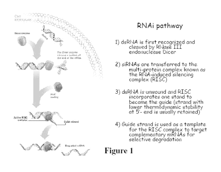
Figure 1 is a schematic showing the RNAi pathway.
Figure 2 shows the structural characteristics of the Dicer enzyme.
America 4443807.2 18
CA 02856117 2014-05-15
WO 2013/075140
PCT/US2012/065945
ATTORNEY DOCKET NO. 89711W0(47992)
November 19, 2012
Figure 3 is a schematic showing that Dicer can cleave double stranded RNA but
not DNA/RNA
hybrids.
Figure 4 is a schematic that illustrates the first step in the design of self-
recognizing hybrid
duplexes.
Figure 5 is a schematic that illustrates the second step in the design of self-
recognizing hybrid
duplexes ¨ the addition of toehold sequences.
Figure 6 is a schematic that illustrates the operation of the designed self-
recognizing hybrid
duplexes.
Figure 7 is a schematic that illustrates the operation of auto-activated
therapeutics.
Figure 8 is a schematic that illustrates how the presence of the complementary
toehold sequences
in each of the self-recognizing hybrids form toehold duplexes which result in
the formation of an
RNA duplex.
Figure 9 illustrates the use of rationale design to produce the self-
recognizing particles.
Figure 10 shows the in vitro formation of hybrid re-as sociationas measured by
FRET.
Figure 11 shows the affinity of hybrid re-association using the negative
control ¨ duplexes
without toeholds.
Figure 12 shows the affinity of hybrid re-association using duplexes with
toeholds.
Figure 13 are a set of graphs that show the kinetics of hybrid recombination.
Figure 14 shows the tracking of re-association of hybrids in vivo.
America 4443807.2 19
CA 02856117 2014-05-15
WO 2013/075140
PCT/US2012/065945
ATTORNEY DOCKET NO. 89711W0(47992)
November 19, 2012
Figure 15 shows the tracking of re-association of hybrids in living cells.
Figure 16 illustrates how re-association can be monitored in vitro through de-
quenching (FRET).
Figure 17 is an illustrative example of tracking re-association in vitro.
Figure 18 is an illustrative example of the kinetics of hybrid de-quenching.
Figure 19 shows the tracking of re-association of hybrids inside living cells
through de-
quenching.
Figure 20 shows the tracking of the re-association of hybrids inside living
cells through de-
quenching.
Figure 21 shows that the self-recognizing particles are able to silence target
gene expression at
levels comparable to pre-formed siRNA duplexes.
Figure 22 shows that same day co-transfection of hybrids results in levels of
target gene
silencing comparable to those seen with pre-formed siRNA duplexes.
Figure 23 is an example of the same day co-transfection of hybrids.
Figure 24 shows that hybrid co-transfection on two different days results in
target gene silencing.
Figure 25 shows that lipofectamine 2000 partially quenches fluorescence.
Figure 26 shows that lipofectamine 2000 prevents hybrid recombination.
Figure 27 shows that pre-formed lipofectamine 2000/hybrids exhibited no
recombination.
Figure 28 is a schematic showing the design of hybrids with variable toehold
lengths.
America 4443807.2 20
CA 02856117 2014-05-15
WO 2013/075140
PCT/US2012/065945
ATTORNEY DOCKET NO. 89711W0(47992)
November 19, 2012
Figure 29 shows the effects of variable toehold lengths on silencing in same
day transfections.
Figure 30 shows the effects of variable toehold lengths on silencing in
different day
transfections.
Figure 31 shows the design of improved hybrids that increase multi-level
delivery.
Figure 32 shows the activation of functionality by two auto-recognizing R/DNA
hybrids. (a)
illustration showing a general principle of functionality activation upon re-
association of two
non-functional units. (b) Schematic representation of auto-recognizing R/DNA
hybrid re-
association resulting in asymmetric Dicer substrate siRNA release. The color
code is kept the
same throughout the figure.
Figure 33 is an illustration showing the rational design of RNA/DNA hybrids
able to release the
functionality (asymmetric 25/27mer Dicer substrate) upon re-association.
Figure 34 shows the comparative analysis of R/DNA hybrids and RNA duplexes.
(a) Total
SYBR Gold staining native PAGE results for dicing experiments carried out for
R/DNA hybrid
and asymmetric 25/27mer Dicer substrate siRNA duplex respectively with
recombinant human
turbo dicer enzyme kit (Genlantis). (b) Total ethidium bromide staining
agarose gel and
quantification results representing the relative stabilities of R/DNA hybrid
and asymmetric
25/27mer Dicer substrate siRNA duplex respectively in 80% human blood serum.
Figure 35 shows fluorescent studies of auto-recognizing R/DNA hybrid re-
association in solution
at 37 C. (a) Schematic representations of control DNA duplexes fluorescently
labeled with
A1exa488 and A1exa546 unable to recombine (upper part) and fluorescently
labeled auto-
recognizing R/DNA hybrids programmed for re-association (lower part). (b)
Emission spectra of
control DNA duplexes showing no FRET and recombined auto-recognizing R/DNA
hybrids with
increased A1exa546 emission signal. (c) FRET time traces during re-association
of auto-
recognizing R/DNA hybrids labeled with A1exa488 and A1exa546. (d)
Fluorescently labeled
America 4443807.2 21
CA 02856117 2014-05-15
WO 2013/075140
PCT/US2012/065945
ATTORNEY DOCKET NO. 89711W0(47992)
November 19, 2012
R/DNA hybrids individually associated with L2K prior to mixing were followed
by fluorescent
time tracing. Please note that L2K forms complexes with hybrids thus,
preventing their re-
association and the emission signal of A1exa488 (in green) stays above
A1exa546 (in red)
comparing to (c). (e-f) Schematic representations and FRET time traces during
re-association of
auto-recognizing R/DNA hybrids labeled with A1exa488 and quencher IowaBlack FQ
(in green)
with schematic representation of corresponding hybrids programmed for
recombination. Please
note that as well as in (d), L2K forms complexes with quenched hybrids (in
blue) and prevents
their recombination.
Figure 36 shows fluorescent studies of auto-recognizing R/DNA hybrid re-
association in solution
after 3 hour incubation at 37 C. Emission spectra and schematic
representations of (upper panel)
control DNA duplexes fluorescently labeled with A1exa488 and A1exa546 unable
to recombine
and (lower panel) emissions of fluorescently labeled auto-recognizing R/DNA
hybrids
programmed for re-association. Please note an increase in A1exa546 emission
signals. For all
samples at different concentrations (as indicated in nM), the excitation was
at 460 nm.
Figures 37A-37E are schematics and graphs showing the kinetics of auto-
recognizing R/DNA
hybrid re-association at 37 C. (a) Schematic representation of re-association
and (b-f) FRET
time traces and their fittings during re-association of auto-recognizing R/DNA
hybrids labeled
with A1exa488 and A1exa546 which were mixed at different equimolar
concentrations specified
above for each case and incubated at 37 C. For all samples, excitation was set
at 460 nm and
emission was measured at 520 nm every 1 second for b-d and every 30 seconds
for e and f. (g)
Constants of auto-recognizing R/DNA hybrid re-association (derived from b-f
fittings) depend
on the concentrations of hybrids at lower concentrations (below ¨ 30 nM). (h)
Schematic
representation of truncated hybrid without toehold and corresponding FRET time
traces. Please
note that there are no significant changes in the fluorescence signals
suggesting that the toeholds
are essential for re-association.
Figure 38 shows that the Addition of lipofectamine2000 (L2K) quenches the
fluorescence of
recombined R/DNA hybrids in solution and protects duplexes from enzymatic
degradation. (a)
R/DNA hybrids labeled with A1exa488 and A1exa546 were mixed at 100 nM
concentrations and
America 4443807.2 22
CA 02856117 2014-05-15
WO 2013/075140
PCT/US2012/065945
ATTORNEY DOCKET NO. 89711W0(47992)
November 19, 2012
L2K was added after two hours of incubation at 37 C. Excitation was set at 460
nm and emission
was measured at 520 nm and 570 nm. Please note that the emission signal of
A1exa546 stays
above A1exa488. (b) Quenched DNA duplex labeled with A1exa488 and IowaBlack FQ
was
mixed at 300 nM concentrations with L2K and DNase was added after two minutes
of incubation
at 37 C (blue line). No degradation was observed after three hours of
incubation. As the control,
the same duplex without L2K was completely digested by DNase (red line). L2K
was then added
upon digestion to assess its' own quenching potential on the digested duplex
thereby providing a
reference for the signal expected if digestion was to take place with L2K
complexed duplexes.
Excitation was set at 460 nm and emission was measured at 520 nm.
Figure 39 shows fluorescent studies of auto-recognizing R/DNA hybrid re-
association in
solution after 3 hour incubation at 37 C. For all samples, excitation was set
at 460 nm and a 100
nM concentration was used. The emission spectra were collected for an R/DNA
hybrid labeled
with A1exa488 (green curve) or A1exa546 (red curve), a duplex containing
A1exa488 and the
quencher IowaBlack FQ (yellow curve) and for the mixture of a duplex
Alexa488/IowaBlack FQ
with its cognate R/DNA A1exa546 hybrid (black).
Figures 40A-40D shows the re-association and localization of auto-recognizing
R/DNA hybrids
in human breast cancer cells (MDA-MB-231) visualized by confocal fluorescence
microscopy.
(Fig. 40A) FRET experiments: cells were co-transfected with cognate hybrids
labeled with
A1exa488 and A1exa546 and images were taken on the next day. (Fig. 40B)
Dequenching
experiments: cells were co-transfected with cognate duplexes, having one
duplex labeled with
A1exa488 and IowaBlack FQ. Images were taken on the next day. (Fig. 40C-D)
Localization of
auto-recognizing R/DNA hybrids with commonly used markers for endosomal
compartments
(Fig. 40C) EEA1 and (Fig. 40D) Rab7.
Image numbers in a-d correspond to: differential interference contrast (DIC)
images (1),
A1exa488 emission (2), A1exa546 emission (3), bleed-through corrected FRET
image (4), 3D
chart representation of zoomed fragment indicated by a white box of bleed-
through corrected
FRET image with the yellow star indicating the correspondence (5), EAA1
antibody staining
(6), and Rab7 antibody staining (7). Images (1+2+3), (1+4), (1+2), (1+3+6),
(1+3+7) are
superpositions of two or three different images.
America 4443807.2 23
CA 02856117 2014-05-15
WO 2013/075140
PCT/US2012/065945
ATTORNEY DOCKET NO. 89711W0(47992)
November 19, 2012
Figure 41 shows the re-association of auto-recognizing R/DNA hybrids in human
breast cancer
cells (MDA-MB-231) visualized by confocal fluorescence microscopy (N=5). Cells
were co-
transfected with cognate hybrids labeled with A1exa488 and A1exa546 and images
were taken on
the next day. Image numbers correspond to: A1exa546 emission (1) A1exa488
emission (2), DIC
(3).
Figure 42 is a set of photographs showing the dequenching re-association
experiments (N=6) of
auto-recognizing R/DNA hybrids in human breast cancer cells (MDA-MB-231)
visualized by
confocal fluorescence microscopy: cells were co-transfected with cognate
hybrids, having one
hybrid labeled with A1exa488 and IowaBlack FQ. As the control (upper panel),
N=6, cells were
transfected with only quenched duplex. Images were taken on the next day.
Image numbers
correspond to: DIC (1), A1exa488 emission (2).
Figures 43A-43C GFP knockdown assays for human breast cancer cells (MDA-MB-
231/GFP)
which stably express enhanced GFP (eGFP). Three days after the transfection of
cells with auto-
recognizing R/DNA hybrids programmed to release siRNAs against eGFP (H_s and
H_ant),
(Fig. 43A) eGFP expression was observed by fluorescence microscopy and (Fig.
43B) eGFP
expression was statistically analyzed with flow cytometry experiments. As the
control, siRNA
duplexes against eGFP were used. Please note that the individual R/DNA hybrids
cause no
decrease in eGFP production (supporting Figure S8). (Fig. 43C) Dependency of
toehold lengths
in auto-recognizing R/DNA hybrids co-transfected on two different days show
their ability to
knockdown eGFP expression. R/DNA hybrids containing antisense (H_ant) were
transfected
one day prior to hybrids with sense (H_s). Three days after, eGFP expression
was analyzed.
Figure 44 is a set of photographs showing GFP knockdown assays for human
breast cancer cells
(MDA-MB-231/GFP) which stably express enhanced GFP (eGFP). Three days after
the
transfection of cells with different equimolar concentrations of auto-
recognizing R/DNA hybrids
programmed to release siRNA against eGFP (H_s and H_ant), eGFP expression was
observed by
fluorescence microscopy. As the control, siRNA duplex against eGFP was used.
America 4443807.2 24
CA 02856117 2014-05-15
WO 2013/075140
PCT/US2012/065945
ATTORNEY DOCKET NO. 89711W0(47992)
November 19, 2012
Figures 45A & 45B are photographs and a graph showing the result of GFP
knockdown
assays for human breast cancer cells (MDA-MB-231/GFP) which stably express
enhanced GFP
(eGFP). (a) Three days after the transfection of cells with single R/DNA
hybrids, eGFP
expression was observed by fluorescence microscopy and statistically analyzed
by flow
cytometry experiments. Please note that individual R/DNA hybrids cause no
decrease in eGFP
production. (b) As negative controls, unrelated to eGFP silencing, auto-
recognizing R/DNA
hybrids designed against HIV-1 were used. Three days after the transfection of
cells with
R/DNA hybrids designed to release siRNAs against HIV, no eGFP expression was
observed by
fluorescence microscopy and flow cytometry experiments. Auto-recognizing R/DNA
hybrids
and siRNA against eGFP were used as the positive control.
Figure 46 shows the use of classical size siRNAs (21 nts) with R/DNA hybrids
demonstrates
comparable silencing efficiency of GFP knockdown compared to the elongated
(25/27 mer)
hybrids. Statistical analysis of eGFP expression was performed with flow
cytometry.
R/DNA hybrids were co-transfected on the same day and three days after, eGFP
expression
was analyzed.
Figures 47A and 47B show the use of internally segmented interfering RNAs with
R/DNA
hybrids leads to a higher efficiency of GFP knockdown compared to the regular
hybrids. (a)
Schematic representation of auto-recognizing R/DNA hybrids having internally
segmented
interfering RNAs (H_segmented_s). (b) Statistical analysis of eGFP expression
with flow
cytometry. R/DNA hybrids were co-transfected on the same day and three days
after, eGFP
expression was analyzed.
Figure 48 shows the dependency of toehold lengths in auto-recognizing R/DNA
hybrids co-
transfected on the same day on their ability to knockdown eGFP expression. (a)
Schematic
representation of auto-recognizing R/DNA hybrids having different lengths of
unzipped
toeholds. (b) Statistical analysis of eGFP expression with flow cytometry.
R/DNA hybrids (H_s
and H_anO were co-transfected on the same day. Three days after, eGFP
expression was
analyzed.
America 4443807.2 25
CA 02856117 2014-05-15
WO 2013/075140
PCT/US2012/065945
ATTORNEY DOCKET NO. 89711W0(47992)
November 19, 2012
Figure 49 shows the result of in vivo studies of auto-recognizing R/DNA
hybrids in tumor
xenograft mouse model. (a) Pharmacokinetic profile of fluorescently labeled
R/DNA hybrids in
tumor-bearing mice three hours post tail-vein injection. A relatively high
level of hybrid
accumulation occurs in tumor tissue. In three hours image, fluorescent
maximums (in red)
correspond to the places of injection (1), tumor (2), and blood withdrawal
(3). (b) The amounts
of the fluorescent probe (R/DNA hybrids and Dicer substrate siRNAs labeled
with IRDye700)
in the mouse blood-stream were measured three hours post-injection. (c) Ex
vivo fluorescent
imaging and analysis of mouse kidneys and tumors indicate a relatively higher
tumor uptake for
the R/DNA hybrids compared to Dicer substrate siRNAs. (d) Ex vivo fluorescent
imaging of
tumors after five days post-injections in vivo demonstrate comparable levels
of eGFP silencing
caused by siRNA and auto-recognizing R/DNA hybrids.
Figure 50 is a set of photographs showing ex vivo fluorescent imaging of
tumors after five days
post-injections in vivo demonstrate comparable levels of eGFP silencing caused
by siRNA and
auto-recognizing R/DNA hybrids. Tumors were removed from mice, fixed overnight
at 4 C in
4% PFA and processed to paraffin. 5ium sections were mounted on slides
followed by
deparaffinization and rehydration through graded ethanol to dH20 then PBS.
Proteinase K
(DAKO) pretreatment was performed for 5 min at RT. Sections were blocked in
NGS and
incubated ON at 4 C with anti GFP antibody (abcam #ab6556, diluted 1:1000).
For fluorescent
labeling, sections were incubated with Goat a/Rabbit IgG Alexa 488
(Invitrogen/Molecular
Probes), counterstained with DAPI then cover-slipped with Prolong Gold a/Fade
reagent
(Invitrogen). Images were captured using Nikon's Eclipse 80i microscope, with
a QImaging
Retiga-2000R camera and Nikon's NIS-Elements AR Imaging Software. For DAB
labeling,
sections were incubated in Biotinylated Goat a/Rabbit IgG (Vector Labs) then
ABC Elite
reagent (Vector Labs), followed by DAB. A Hematoxylin counterstain was
performed and
slides were coverslipped with Permount. Whole-slide digital images were
captured using
Aperio's ScanScope digital slide scanner.
Figure 51 shows that HIV-1 expression and production is inhibited by siRNAs
and recombined
R/DNA hybrids. (a) HeLa cells were transfected with pNL4-3, with and without
siRNAs. Virus
supernatant was harvested and RT activity was measured; data are shown
normalized to virus
America 4443807.2 26
CA 02856117 2014-05-15
WO 2013/075140
PCT/US2012/065945
ATTORNEY DOCKET NO. 89711W0(47992)
November 19, 2012
controls (VC.1 and VC.2) without siRNAs. Error bars denote SD; N=4. (b) At 48
h
posttransfection, HeLa cells were metabolically labeled with [35S]MetCys for
4h. Cell lysates
were radioimunoprecipitated. Positions of envelope glycoprotein precursor,
gp160, and surface
glycoprotein gp120; Pr55Gag (Pr55), CA-SP1 (p25) and CA (p24) are indicated.
Quantification
of total cell-associated Gag: Pr55+p25+24. Total Gag in virus control (VC)
without siRNAs set
at 100. Error bars denote SD; N=3.
Figures 52A-52D show the transfection efficiencies measured by flow cytometry
(Fig. 51A) and
Rab5/7 (endosomal) co-localization (Fig. 51B) in HeLa cells with auto-
recognizing R/DNA
hybrids. Transfection efficiencies were statistically analyzed by flow
cytometry (Fig. 52A) and
co-localization was visualized by confocal fluorescence microscopy (Fig. 52B).
Cytotoxicity of
siRNAs (LCPS = luciferase counts per second) in HIV-1-expressing HeLa cells
(Fig. 52C) is
minimal between 5 and 20 nlVl. Cells are co-transfected with pNL4-3 and
p5iCHECKTm-1
(Promega), with and without siRNAs. At 48 h post transfection, cells were
lysed and Renilla
luciferase was measured. eGFP siRNA was used as a control. VC. Virus control.
LucC.
Luciferase control. Error bars denote SD; N=4. (Fig. 52D) Effect on RT
production. eGFP
siRNA was used as a negative control. eGFP siRNAs were co-transfected with
pNL4-3 plasmid
and RT levels were measured in the supernatant after 48 h. VC. Virus control.
Figure 53 shows the relative decrease in GSTP1 protein expression (with
corresponding
standard deviation (SD) and standard error (SE) calculated from three
repetitions) in A549 lung
adenocarcinoma cells after R/DNA treatment as shown by Western blot. In the
case of
H s+H ant (GSTP1), auto-recognizing R/DNA hybrids containing RNA sense strand
were
transfected first and after 24 hours of incubation at 37 C, a complement
R/DNA hybrid
containing RNA anti-sense strand of GSTP1 siRNA were transfected. Cells were
collected and
processed for immunoblotting using standard protocol 24 h later. Anti-GSTP1
antibody was
from Cell Signaling Technology, and 8-actin antibody from Abcam.
Figure 54 is a schematic representation of two cognate auto-recognizing
therapeutics R/DNA NP
before (upper panel) and after (lower panel) intracellular recombination.
America 4443807.2 27
CA 02856117 2014-05-15
WO 2013/075140
PCT/US2012/065945
ATTORNEY DOCKET NO. 89711W0(47992)
November 19, 2012
Figure 55 is a schematic showing recombination of auto-recognizing R/DNA
duplexes releasing
siRNA designed against GFP gene (upper panel) and FACS data demonstrating
silencing of GFP
expression inside the human cells triggered by re-association of the auto-
recognizing R/DNA
duplexes.
Figure 56 is a schematic showing an exemplary auto-recognizing RNA/DNA hybrids
releasing
multiple functionalities (FRET response, siRNA and RNA aptamer) during re-
association.
Figures 57A-57C show the release of multiple functionalities (FRET, siRNA, MG
aptamer)
upon re-association of auto-recognizing RNA/DNA hybrids. Figure 57A is a
schematic of
hybrid re-association and native PAGE demonstrating the release of siRNA and
MG aptamer
upon re-association. Figure 57B is the static and kinetics fluorescent
experiments showing
activation of MG aptamer during the release. MG by itself is non-fluorescent
(blue curve) and
the presence of either one of the hybrids does not activate its fluorescence
(green curve).
However, re-association of two cognate hybrids leads to the release of
individual MG aptamer
strands, their assembly and further MG uptake leading to the significant
increase of its
fluorescence (magenta curve). Figure 57C upper panel: Activation of FRET
during re-
association. Emission spectra of control DNA duplexes showing no FRET (blue
curve) and re-
associated auto-recognizing R/DNA hybrids with increased A1exa546 emission
signal (red
curve). Figure 57C lower panel: FRET time traces during re-association of auto-
recognizing
R/DNA hybrids labeled with A1exa488 and A1exa546.
Figure 58 shows the release of multiple functionalities (two siRNAs, MG
aptamer) upon re-
association of auto-recognizing RNA/DNA hybrids. MG aptamer was positioned in
three
different places (three sets of hybrids were tested) thus, the activation of
MG fluorescence in all
three cases proves the release of all functionalities (despite the position
with respect to the
ssDNA toeholds). Left panel: Schematic of hybrid re-association and total SYBR
Gold staining
native PAGE demonstrating the release of siRNAs and MG aptamer upon re-
association. Right
panel: Static and kinetics fluorescent experiments showing activation of MG
aptamer during the
release.
America 4443807.2 28
CA 02856117 2014-05-15
WO 2013/075140
PCT/US2012/065945
ATTORNEY DOCKET NO. 89711W0(47992)
November 19, 2012
Figure 59A-59D show the re-association, localization of auto-recognizing R/DNA
hybrids in
human breast cancer cells (MDA-MB-231) visualized by confocal fluorescence
microscopy and
further release of siRNAs tracked by GFP knockdown assays for human breast
cancer cells
(MDA-MB-231/GFP) which stably express enhanced GFP (eGFP). Figure 59A is a
schematic of
different size hybrids re-association compared in these experiments. FRET
experiments (Figure
59B): cells were co-transfected with cognate hybrids labeled with A1exa488 and
A1exa546 and
images were taken on the next day. Figure 59C is a gel showing total SYBR Gold
staining native
PAGE demonstrating the release of siRNAs. GFP knockdown assays (Figure 59D):
three days
after the transfection of cells with auto-recognizing R/DNA hybrids programmed
to release one
(hybrids (i)), two (hybrids (ii)), and three (hybrids (iii)) siRNAs against
eGFP. eGFP expression
was observed by fluorescence microscopy and statistically analyzed with flow
cytometry
experiments. As the control, siRNA duplexes against eGFP were used. Image
numbers in
Figure 59B correspond to: A1exa488 emission (1), A1exa546 emission (2), bleed-
through
corrected FRET image (3), differential interference contrast (DIC) image with
corrected FRET
overlap (4), 3D chart representation of zoomed fragment indicated by a white
box of bleed-
through corrected FRET image with the yellow star indicating the
correspondence (5).
DETAILED DESCRIPTION OF THE INVENTION
Using RNA interference (RNAi) as a therapeutic agent it is routinely possible
to knock
down the expression of target genes in diseased cells. One of the ways to
initiate the RNAi
machinery is through the direct exogenous introduction to the cells of small
interfering RNA
(siRNA) duplexes. The invention described herein provides for a strategy based
on therapeutic
RNA/DNA hybrids which can be generally used for triggering the RNAi pathway as
well as
other functionalities inside the diseased cells. Individually, each of the
hybrids is functionally
inactive and the therapeutic siRNA representation can only be activated by the
re-association
of at least two cognate hybrids simultaneously present in the same cell. The
invention features a
method for siRNA release where cognate hybrids are co-delivered to the cell
either on the same
or on two different days. The invention provides for nucleic acids based
"smart" nanoparticles
for biomedical applications.
The invention is based on a novel strategy to design and engineer programmable
auto-recognizing R/DNA hybrids capable of undergoing triggered release of
embedded
America 4443807.2 29
CA 02856117 2014-05-15
WO 2013/075140
PCT/US2012/065945
ATTORNEY DOCKET NO. 89711W0(47992)
November 19, 2012
functionalities upon their re-association inside cells. The R/DNA hybrids have
significantly
higher stabilities in blood serum compared to the siRNA and that the use of
the R/DNA hybrid
approach allows (i) introduction of additional functionalities without direct
interference of
siRNA processivity, (ii) activation of split functionalities (e.g., FRET) in
vitro and
intracellularly, (ii) tracking of the delivery and re-association of these
hybrids in real-time inside
cells, (iii) the triggered release of siRNAs in cell culture and in vivo, (iv)
pro-longed effect of
gene silencing compared to the siRNA. Additionally, this approach may permit
(i) higher
control over targeting specificity (e.g. if two hybrids are decorated with two
different tissue
specific recognition moieties), (ii) increasing the number of functionalities
by introducing a
branched hybrid structure, (iii) increasing the retention time in biological
fluids by fine-tuning
chemical stability through substituting the DNA strands with chemical analogs
(e.g. locked
nucleic acids (LNA), peptide nucleic acids (PNA), etc.), (iv) conditional
release of siRNAs or
other functionalities. Moreover, the thermal stabilities of toehold
interactions can be fine-tuned
by altering their lengths and compositions. The invention opens new routes for
further
developments in nucleic acids based nanoparticles for a broad array of
nanotechnological and biomedical applications.
The certain aspects the invention features compositions and methods that are
useful for
inhibiting gene expression in a subject. The invention features a thereapeutic
R/DNA chimeric
polyfunctional nanoparticles (R/DNA NP) which are a pair of RNA/DNA duplexes
where the
first DNA molecule has a 5' toehold sequence and the second DNA molecule has a
3' toehold
sequence wherein the 5' and 3' toehold sequences are complementary. When the
two R/DNA
NPs are mixed the toehold sequences form a duplex which results in the re-
association of the two
R/DNA NPs. The product of re-association is a DNA/DNA duplex and an RNA/RNA
duplex
where the RNA duplex is designed to function as an siRNA to target a specific
target RNA. The
R/DNA NPs also have moieties attached which specifically bind target
molecules. In one
embodiment the target molecules are cell surface proteins present on cells
that express a target
gene. Therefore, the R/DNA NPs allow for the targeting of specific target
genes in a specific
cells.
The invention may be used to target medically important genes or genes in
medically
important cells. For example, in one embodiment, R/DNA NPs could be used to
reduce viral
burden in a subject by targeting virally infected cells. In specific
embodiments the R/DNA NPs
America 4443807.2 30
CA 02856117 2014-05-15
WO 2013/075140
PCT/US2012/065945
ATTORNEY DOCKET NO. 89711W0(47992)
November 19, 2012
inhibit apoptosis inhibitors in virally infected cells thereby specifically
killing the virally infected
cells. In particular embodiments the virus is HIV. In other embodiments, R/DNA
NPs could be
used to target neoplastic cells. In additional embodiments, R/DNA NPs could
inhibit a target
gene in a neoplasia resulting in the death or apoptosis of the neoplasia. Thus
R/DNA NPs could
be used to treat any disease that is amenable to gene silencing approaches.
The invention also provides methods of treating HIV infected subjections by
administering an effective amount of R/DNA NPs that targets HIV infected cells
and results in
the destruction of the targeted apoptosis inhibitors resulting in the death of
the HIV infected cells
thereby eradicating HIV from the subject.
The invention further features methods of treating a subject having neoplasia
by
administering an effective amount of R/DNA NPs that targets neoplastic cells
and results in the
knock-down of target genes that results in the death or apoptosis of the
neoplastic cells.
The methods herein include administering to the subject (including a subject
identified as
in need of such treatment) an effective amount of a compound described herein,
or a composition
described herein to produce such effect. Identifying a subject in need of such
treatment can be in
the judgment of a subject or a health care professional and can be subjective
(e.g. opinion) or
objective (e.g. measurable by a test or diagnostic method).
As used herein, the terms "treat," treating," "treatment," and the like refer
to reducing or
ameliorating a disorder and/or symptoms associated therewith. It will be
appreciated that,
although not precluded, treating a disorder or condition does not require that
the disorder,
condition or symptoms associated therewith be completely eliminated.
As used herein, the terms "prevent," "preventing," "prevention," "prophylactic
treatment" and the like refer to reducing the probability of developing a
disorder or condition in a
subject, who does not have, but is at risk of or susceptible to developing a
disorder or condition.
The therapeutic methods of the invention (which include prophylactic
treatment) in
general comprise administration of a therapeutically effective amount of the
compounds herein,
such as a compound of the formulae herein to a subject (e.g., animal, human)
in need thereof,
including a mammal, particularly a human. Such treatment will be suitably
administered to
subjects, particularly humans, suffering from, having, susceptible to, or at
risk for a disease,
disorder, or symptom thereof. Determination of those subjects "at risk" can be
made by any
objective or subjective determination by a diagnostic test or opinion of a
subject or health care
America 4443807.2 31
CA 02856117 2014-05-15
WO 2013/075140
PCT/US2012/065945
ATTORNEY DOCKET NO. 89711W0(47992)
November 19, 2012
provider (e.g., genetic test, enzyme or protein marker, Marker (as defined
herein), family history,
and the like). The compounds herein may be also used in the treatment of any
other disorders in
which viruses, particularly HIV may be implicated.
In one embodiment, the invention provides a method of monitoring treatment
progress.
The method includes the step of determining a level of diagnostic marker for
virus infected cells,
particularly HIV infected cells (e.g., any target delineated herein modulated
by a compound
herein, a protein or indicator thereof, etc.) or diagnostic measurement (e.g.,
screen, assay) in a
subject suffering from or susceptible to a disorder or symptoms thereof
associated with viruses,
particularly HIV, in which the subject has been administered a therapeutic
amount of a
compound herein sufficient to treat the disease or symptoms thereof. The level
of Marker
determined in the method can be compared to known levels of Marker in either
healthy normal
controls or in other afflicted patients to establish the subject's disease
status. In preferred
embodiments, a second level of Marker in the subject is determined at a time
point later than the
determination of the first level, and the two levels are compared to monitor
the course of disease
or the efficacy of the therapy. In certain preferred embodiments, a pre-
treatment level of Marker
in the subject is determined prior to beginning treatment according to this
invention; this pre-
treatment level of Marker can then be compared to the level of Marker in the
subject after the
treatment commences, to determine the efficacy of the treatment.
Pharmaceutical Therapeutics
The present disclosure provides R/DNA NPs that decrease the expression or
activity of
target proteins in diseased cells. For example, in one non-limiting
embodiment, the disclosure
provides pharmaceutical compositions comprising a R/DNA NPs that inhibits the
expression or
activity of an apoptosis inhibitor in the diseased cell. In a further
embodiment, the diseased cell
is a neoplastic cell or a virally infected cell. However, the pharmaceutical
applications described
herewith are applicable to the treatment of any disease state that is or is
contemplated to be
amenable to gene silencing. For therapeutic uses, the compositions or agents
identified using the
methods disclosed herein may be administered systemically, for example,
formulated in a
pharmaceutically-acceptable carrier. Preferable routes of administration
include, for example,
subcutaneous, intravenous, interperitoneally, intramuscular, or intradermal
injections that
provide continuous, sustained levels of the drug in the patient. Treatment of
human patients or
America 4443807.2 32
CA 02856117 2014-05-15
WO 2013/075140
PCT/US2012/065945
ATTORNEY DOCKET NO. 89711W0(47992)
November 19, 2012
other animals will be carried out using a therapeutically effective amount of
R/DNA NPs in a
physiologically-acceptable carrier. Suitable carriers and their formulation
are described, for
example, in Remington's Pharmaceutical Sciences by E. W. Martin. The amount of
the
therapeutic agent to be administered varies depending upon the manner of
administration, the age
and body weight of the patient, and the clinical symptoms of disease
progression. Generally,
amounts will be in the range of those used for other agents used in the
treatment of the disease,
although in certain instances lower amounts will be needed because of the
increased specificity
of the compound. A compound is administered at a dosage that controls the
clinical or
physiological symptoms of cancer progression or metastasis as determined by a
diagnostic
method known to one skilled in the art, or using any that assay that measures
the transcriptional
activation of a gene associated with the disease.
Formulation of Pharmaceutical Compositions
The administration of a R/DNA NPs of the disclosure or analog thereof for the
treatment
of a disease may be by any suitable means that results in a concentration of
the R/DNA NPs that,
combined with other components, is effective in ameliorating, reducing,
eradicating, or
stabilizing the disease. In one embodiment, administration of the R/DNA NPs
reduces the
expression or activity of a target gene. In another embodiment, the R/DNA NPs
is administered
to a subject for the prevention or treatment of a disease associated with
disease.
Methods of administering such R/DNA NPs are known in the art. The disclosure
provides for the therapeutic administration of an agent by any means known in
the art. The
compound may be contained in any appropriate amount in any suitable carrier
substance, and is
generally present in an amount of 1-95% by weight of the total weight of the
composition. The
composition may be provided in a dosage form that is suitable for parenteral
(e.g.,
subcutaneously, intravenously, intramuscularly, or intraperitoneally)
administration route. The
pharmaceutical compositions may be formulated according to conventional
pharmaceutical
practice (see, e.g., Remington: The Science and Practice of Pharmacy (20th
ed.), ed. A. R.
Gennaro, Lippincott Williams & Wilkins, 2000 and Encyclopedia of
Pharmaceutical
Technology, eds. J. Swarbrick and J. C. Boylan, 1988-1999, Marcel Dekker, New
York).
Suitable formulations include forms for oral administration, depot
formulations, formulations for
delivery by a patch, and semi-solid dosage forms to be topically or trans-
dermally delivered.
America 4443807.2 33
CA 02856117 2014-05-15
WO 2013/075140
PCT/US2012/065945
ATTORNEY DOCKET NO. 89711W0(47992)
November 19, 2012
Pharmaceutical compositions according to the disclosure may be formulated to
release
the active compound substantially immediately upon administration or at any
predetermined time
or time period after administration. The latter types of compositions are
generally known as
controlled release formulations, which include (i) formulations that create a
substantially
constant concentration of the drug within the body over an extended period of
time; (ii)
formulations that after a predetermined lag time create a substantially
constant concentration of
the drug within the body over an extended period of time; (iii) formulations
that sustain action
during a predetermined time period by maintaining a relatively, constant,
effective level in the
body with concomitant minimization of undesirable side effects associated with
fluctuations in
the plasma level of the active substance (saw-tooth kinetic pattern); (iv)
formulations that
localize action by, e.g., spatial placement of a controlled release
composition adjacent to or in the
central nervous system or cerebrospinal fluid; (v) formulations that allow for
convenient dosing,
such that doses are administered, for example, once every one or two weeks;
and (vi)
formulations that target tumor cells by using carriers or chemical derivatives
to deliver the
therapeutic agent to a particular cell type whose function is perturbed in
cancer. For some
applications, controlled release formulations obviate the need for frequent
dosing during the day
to sustain the plasma level at a therapeutic level.
Any of a number of strategies can be pursued in order to obtain controlled
release in
which the rate of release outweighs the rate of metabolism of the compound in
question. In one
example, controlled release is obtained by appropriate selection of various
formulation
parameters and ingredients, including, e.g., various types of controlled
release compositions and
coatings. Thus, the therapeutic is formulated with appropriate excipients into
a pharmaceutical
composition that, upon administration, releases the therapeutic in a
controlled manner.
Examples include single or multiple unit tablet or capsule compositions, oil
solutions,
suspensions, emulsions, microcapsules, microspheres, molecular complexes,
nanoparticles,
patches, and liposomes.
Parenteral Compositions
The pharmaceutical composition may be administered parenterally by injection,
infusion
or implantation (subcutaneous, intravenous, intramuscular, intraperitoneal, or
the like) in dosage
forms, formulations, or via suitable delivery devices or implants containing
conventional, non-
America 4443807.2 34
CA 02856117 2014-05-15
WO 2013/075140
PCT/US2012/065945
ATTORNEY DOCKET NO. 89711W0(47992)
November 19, 2012
toxic pharmaceutically acceptable carriers and adjuvants. The formulation and
preparation of
such compositions are well known to those skilled in the art of pharmaceutical
formulation.
Formulations can be found in Remington: The Science and Practice of Pharmacy,
supra.
Compositions for parenteral use may be provided in unit dosage forms (e.g., in
single-dose
ampoules), or in vials containing several doses and in which a suitable
preservative may be
added (see below). The composition may be in the form of a solution, a
suspension, an
emulsion, an infusion device, or a delivery device for implantation, or it may
be presented as a
dry powder to be reconstituted with water or another suitable vehicle before
use. Apart from the
active therapeutic (s), the composition may include suitable parenterally
acceptable carriers
and/or excipients. The active therapeutic (s) may be incorporated into
microspheres,
microcapsules, nanoparticles, liposomes, or the like for controlled release.
Furthermore, the
composition may include suspending, solubilizing, stabilizing, pH-adjusting
agents, tonicity
adjusting agents, and/or dispersing, agents.
As indicated above, the pharmaceutical compositions according to the
disclosure may be
in the form suitable for sterile injection. To prepare such a composition, the
suitable active
therapeutic(s) are dissolved or suspended in a parenterally acceptable liquid
vehicle.
Controlled Release Parenteral Compositions
Controlled release parenteral compositions may be in the form of suspensions,
microspheres, microcapsules, magnetic microspheres, oil solutions, oil
suspensions, or
emulsions. Alternatively, the active drug may be incorporated in biocompatible
carriers,
liposomes, nanoparticles, implants, or infusion devices. Materials for use in
the preparation of
microspheres and/or microcapsules are, e.g., biodegradable/bioerodible
polymers such as
polygalactin, poly-(isobutyl cyanoacrylate), poly(2-hydroxyethyl-L-glutam-
nine) and,
poly(lactic acid). Biocompatible carriers that may be used when formulating a
controlled release
parenteral formulation are carbohydrates (e.g., dextrans), proteins (e.g.,
albumin), lipoproteins,
or antibodies. Materials for use in implants can be non-biodegradable (e.g.,
polydimethyl
siloxane) or biodegradable (e.g., poly(caprolactone), poly(lactic acid),
poly(glycolic acid) or
poly(ortho esters) or combinations thereof).
America 4443807.2 35
CA 02856117 2014-05-15
WO 2013/075140
PCT/US2012/065945
ATTORNEY DOCKET NO. 89711W0(47992)
November 19, 2012
Inhibitory Nucleic Acids
The R/DNA NPs molecules described herein operate by forming inhibitory nucleic
acid
molecules once in target cells. Such inhibitory nucleic acids include single
and double stranded
nucleic acid molecules (e.g., DNA, RNA, and analogs thereof) that bind a
nucleic acid molecule
that encodes target RNA (e.g., antisense oligonucleotide molecules, siRNA,
shRNA) as well as
nucleic acid molecules that bind directly to a target polypeptide to modulate
its biological
activity (e.g., aptamers).
Ribozymes
Catalytic RNA molecules or ribozymes that include an antisense target RNA
sequence of
the present disclosure can be used to inhibit expression of target RNAs in
vivo. The inclusion of
ribozyme sequences within antisense RNAs confers RNA-cleaving activity upon
them, thereby
increasing the activity of the constructs. The design and use of target RNA-
specific ribozymes is
described in Haseloff et al., Nature 334:585-591. 1988, and U.S. Patent
Application Publication
No. 2003/0003469 Al, each of which is incorporated by reference.
Accordingly, the disclosure also features a catalytic RNA molecule that
includes, in the
binding arm, an antisense RNA having between eight and nineteen consecutive
nucleobases. In
preferred embodiments of this disclosure, the catalytic nucleic acid molecule
is formed in a
hammerhead or hairpin motif. Examples of such hammerhead motifs are described
by Rossi et
al., Aids Research and Human Retroviruses, 8:183, 1992. Example of hairpin
motifs are
described by Hampel et al., "RNA Catalyst for Cleaving Specific RNA
Sequences," filed Sep.
20, 1989, which is a continuation-in-part of U.S. Ser. No. 07/247,100 filed
Sep. 20, 1988,
Hampel and Tritz, Biochemistry, 28:4929, 1989, and Hampel et al., Nucleic
Acids Research, 18:
299, 1990. These specific motifs are not limiting in the disclosure and those
skilled in the art
will recognize that all that is important in an enzymatic nucleic acid
molecule of this disclosure
is that it has a specific substrate binding site which is complementary to one
or more of the target
gene RNA regions, and that it have nucleotide sequences within or surrounding
that substrate
binding site which impart an RNA cleaving activity to the molecule.
Small hairpin RNAs consist of a stem-loop structure with optional 3' UU-
overhangs.
While there may be variation, stems can range from 21 to 31 bp (desirably 25
to 29 bp), and the
loops can range from 4 to 30 bp (desirably 4 to 23 bp). For expression of
shRNAs within cells,
plasmid vectors containing either the polymerase III H1-RNA or U6 promoter, a
cloning site for
America 4443807.2 36
CA 02856117 2014-05-15
WO 2013/075140
PCT/US2012/065945
ATTORNEY DOCKET NO. 89711W0(47992)
November 19, 2012
the stem-looped RNA insert, and a 4-5-thymidine transcription termination
signal can be
employed. The Polymerase III promoters generally have well-defined initiation
and stop sites
and their transcripts lack poly(A) tails. The termination signal for these
promoters is defined by
the polythymidine tract, and the transcript is typically cleaved after the
second uridine. Cleavage
at this position generates a 3' UU overhang in the expressed shRNA, which is
similar to the 3'
overhangs of synthetic siRNAs. Additional methods for expressing the shRNA in
mammalian
cells are described in the references cited above.
siRNA
Short twenty-one to twenty-five nucleotide double-stranded RNAs are effective
at down-
regulating gene expression (Zamore et al., Cell 101: 25-33; Elbashir et al.,
Nature 411: 494-498,
2001, hereby incorporated by reference). The therapeutic effectiveness of an
siRNA approach in
mammals was demonstrated in vivo by McCaffrey et al. (Nature 418: 38-39,2002).
Given the sequence of a target gene, siRNAs may be designed to inactivate that
gene. Such
siRNAs, for example, could be administered directly to an affected tissue, or
administered
systemically. The nucleic acid sequence of an Parl gene can be used to design
small interfering
RNAs (siRNAs). The 21 to 25 nucleotide siRNAs may be used, for example, as
therapeutics to
inhibit disease related genes.
The inhibitory nucleic acid molecules of the present disclosure may be
employed as
double-stranded RNAs for RNA interference (RNAi)-mediated knock-down of target
RNA
expression. In therapeutic embodiments, the target RNA is a disease related
gene. For example,
in a non-limiting embodiment, the target RNA is a gene that is involved in
cancer development
or progression. In another embodiment, target RNA expression is reduced in a
virus infected
cell. In another embodiment, the target RNA encodes apoptosis inhibitor
proteins and the cells
are infected with HIV. RNAi is a method for decreasing the cellular expression
of specific
proteins of interest (reviewed in Tuschl, ChemBioChem 2:239-245, 2001; Sharp,
Gene Dev
15:485-490, 2000; Hutvagner and Zamore, Curr Opin Genet Devel 12:225-232,
2002; and
Hannon, Nature 418:244-251, 2002). The introduction of siRNAs into cells
either by
transfection of dsRNAs or through expression of siRNAs using a plasmid-based
expression
system is increasingly being used to create loss-of-function phenotypes in
mammalian cells.
In one embodiment of the disclosure, a double-stranded RNA (dsRNA) molecule is
made
that includes between eight and nineteen consecutive nucleobases of a
nucleobase oligomer of
America 4443807.2 37
CA 02856117 2014-05-15
WO 2013/075140
PCT/US2012/065945
ATTORNEY DOCKET NO. 89711W0(47992)
November 19, 2012
the disclosure. The dsRNA can be two distinct strands of RNA that have
duplexed, or a single
RNA strand that has self-duplexed (small hairpin (sh)RNA). Typically, dsRNAs
are about 21 or
22 base pairs, but may be shorter or longer (up to about 29 nucleobases) if
desired. dsRNA can
be made using standard techniques (e.g., chemical synthesis or in vitro
transcription). Kits are
available, for example, from Ambion (Austin, Tex.) and Epicentre (Madison,
Wis.). Methods for
expressing dsRNA in mammalian cells are described in Brummelkamp et al.
Science 296:550-
553, 2002; Paddison et al. Gene Dev 16:948-958, 2002. Paul et al. Nat
Biotechnol 20:505-508,
2002; Sui et al. Proc Natl Acad Sci USA 99:5515-5520, 2002; Yu et al. Proc
Natl Acad Sci USA
99:6047-6052, 2002; Miyagishi et al. Nat Biotechnol 20:497-500, 2002; and Lee
et al. Nat
Biotechnol 20:500-505, 2002, each of which is hereby incorporated by
reference. In certain
embodiments, the sense strand of the double stranded siRNA is split into two
smaller
oligonucleotides, also referred to as three stranded siRNA.
Small hairpin RNAs consist of a stem-loop structure with optional 3' UU-
overhangs.
While there may be variation, stems can range from 21 to 31 bp (desirably 25
to 29 bp), and the
loops can range from 4 to 30 bp (desirably 4 to 23 bp). For expression of
shRNAs within cells,
plasmid vectors containing either the polymerase III Hl-RNA or U6 promoter, a
cloning site for
the stem-looped RNA insert, and a 4-5-thymidine transcription termination
signal can be
employed. The Polymerase III promoters generally have well-defined initiation
and stop sites
and their transcripts lack poly(A) tails. The termination signal for these
promoters is defined by
the polythymidine tract, and the transcript is typically cleaved after the
second uridine. Cleavage
at this position generates a 3' UU overhang in the expressed shRNA, which is
similar to the 3'
overhangs of synthetic siRNAs. Additional methods for expressing the shRNA in
mammalian
cells are described in the references cited above.
The invention encompasses stabilized R/DNA NPs having modifications that
protect
against 3' and 5' exonucleases as well as endonucleases. Such modifications
desirably maintain
target affinity while increasing stability in vivo. In various embodiments,
R/DNA NPs of the
invention include chemical substitutions at the ribose and/or phosphate and/or
base positions of a
given nucleobase sequence. For example, R/DNA NPs of the invention include
chemical
modifications at the 2' position of the ribose moiety, circularization of the
aptamer, 3' capping
and `spiegelmer' technology. R/DNA NPs having A and G nucleotides sequentially
replaced
with their 2'-OCH3 modified counterparts are particularly useful in the
methods of the invention.
America 4443807.2 38
CA 02856117 2014-05-15
WO 2013/075140
PCT/US2012/065945
ATTORNEY DOCKET NO. 89711W0(47992)
November 19, 2012
Such modifications are typically well tolerated in terms of retaining affinity
and specificity. In
various embodiments, R/DNA NPs include at least 10%, 25%, 50%, or 75% modified
nucleotides. In other embodiments, as many as 80-90% of the R/DNA NPs'
nucleotides contain
stabilizing substitutions. In other embodiments, 2'-0Me containing R/DNA NPs
are
synthesized. Such R/DNA NPs are desirable because they are inexpensive to
synthesize and
natural polymerases do not accept 2'-0Me nucleotide triphosphates as
substrates so that 2'-0Me
nucleotides cannot be recycled into host DNA. Using methods described herein,
R/DNA NPs
will be selected for increased in vivo stability. In one embodiment, R/DNA NPs
having 2'-F and
2'-OCH3 modifications are used to generate nuclease resistant aptamers. In
other embodiments,
the nucleic acids of the invention have one or more locked nucleic acids
(LNA). LNA refers to a
modified RNA nucleotide. The ribose of the LNA is modified with an extra
bridge connecting
the 2' oxygen and the 4' carbon which locks the ribose into the North or 3'-
endo conformation.
See e.g., Kaur, H. et al., Biochemistry, vol. 45, pages 7347-55; and Koshkin,
A.A., et al.,
Tetrahedron, vol. 54, pages 3607-3630. In other embodiments, one or more
nucleic acids of the
invention incorporate a morpolino structure where the nucleic acid bases are
bound to
morpholine rings instead of deoxyribose rings and are linked through
phosphorodiamidate
groups instead of phosphates. See eg., Summerton, J. and Weller, D., Antisense
& Nucleic Acid
Drug Development, vol. 7, pages 187-195. Yet other modifications, include (PS)-
phosphate
sulfur modifications wherein the phosphate backbone of the nucleic acid is
modified by the
substitution of one or more sulfur groups for oxygen groups in the phosphate
backbone. Other
modifications that stabilize nucleic acids are known in the art and are
described, for example, in
U.S. Patent Nos. 5,580,737; and in U.S. Patent Application Publication Nos.
20050037394,
20040253679, 20040197804, and 20040180360.
Delivery of Nucleotide-base Oligomers
Naked inhibitory nucleic acid molecules, or analogs thereof, are capable of
entering
mammalian cells and inhibiting expression of a gene of interest. Nonetheless,
it may be
desirable to utilize a formulation that aids in the delivery of
oligonucleotides or other nucleobase
oligomers to cells to deliver the claimed R/DNA NPs (see, e.g., U.S. Pat. Nos.
5,656,611,
5,753,613, 5,785,992, 6,120,798, 6,221,959, 6,346,613, and 6,353,055, each of
which is hereby
incorporated by reference).
America 4443807.2 39
CA 02856117 2014-05-15
WO 2013/075140
PCT/US2012/065945
ATTORNEY DOCKET NO. 89711W0(47992)
November 19, 2012
Dosage
Human dosage amounts can initially be determined by extrapolating from the
amount of
compound used in mice, as a skilled artisan recognizes it is routine in the
art to modify the
dosage for humans compared to animal models. In certain embodiments it is
envisioned that the
dosage may vary from between about 1 mg compound/Kg body weight to about 5000
mg
compound/Kg body weight; or from about 5 mg/Kg body weight to about 4000 mg/Kg
body
weight or from about 10 mg/Kg body weight to about 3000 mg/Kg body weight; or
from about
50 mg/Kg body weight to about 2000 mg/Kg body weight; or from about 100 mg/Kg
body
weight to about 1000 mg/Kg body weight; or from about 150 mg/Kg body weight to
about 500
mg/Kg body weight. In other embodiments this dose may be about 1, 5, 10, 25,
50, 75, 100, 150,
200, 250, 300, 350, 400, 450, 500, 550, 600, 650, 700, 750, 800, 850, 900,
950, 1000, 1050,
1100, 1150, 1200, 1250, 1300, 1350, 1400, 1450, 1500, 1600, 1700, 1800, 1900,
2000, 2500,
3000, 3500, 4000, 4500, 5000 mg/Kg body weight. In other embodiments, it is
envisaged that
higher does may be used, such doses may be in the range of about 5 mg
compound/Kg body to
about 20 mg compound/Kg body. In other embodiments the doses may be about 8,
10, 12, 14,
16 or 18 mg/Kg body weight. Of course, this dosage amount may be adjusted
upward or
downward, as is routinely done in such treatment protocols, depending on the
results of the
initial clinical trials and the needs of a particular patient.
Therapeutic Methods
The present disclosure provides methods of treating diseases, particularly
neoplasia and
viral infections by specifically inhibiting or reducing one or more target
genes in specific target
cells. The methods comprise administering a therapeutically effective amount
of R/DNA NPs to
a subject wherein the R/DNA NPs bind and enter a target cell. Once inside the
target cell, the
R/DNA NPs recombine through toehold sequences present in the DNA
oligonucleotides. The re-
association release functional RNA molecules. In one embodiment, the RNA
molecules are
complementary and form a duplex that is an siRNA. The release siRNA inhibits a
target gene
resulting in the treatment of the disease cell.. The method includes the step
of administering to
the subject a therapeutic amount or an amount of a compound herein sufficient
to treat the
disease or symptom thereof, under conditions such that the disease is treated.
America 4443807.2 40
CA 02856117 2014-05-15
WO 2013/075140
PCT/US2012/065945
ATTORNEY DOCKET NO. 89711W0(47992)
November 19, 2012
The methods herein include administering to the subject (including a subject
identified as
in need of such treatment) an effective amount of a compound described herein,
or a composition
described herein to produce such effect. Identifying a subject in need of such
treatment can be in
the judgment of a subject or a health care professional and can be subjective
(e.g. opinion) or
objective (e.g. measurable by a test or diagnostic method).
The therapeutic methods of the disclosure, which include prophylactic
treatment, in
general comprise administration of a therapeutically effective amount of the
agent herein, such as
a compound of the formulae herein to a subject (e.g., animal, human) in need
thereof, including a
mammal, particularly a human. Such treatment will be suitably administered to
subjects,
particularly humans, suffering from, having, susceptible to, or at risk for a
cancer progression or
metastasis or symptom thereof. Determination of those subjects "at risk" can
be made by any
objective or subjective determination by a diagnostic test or opinion of a
subject or health care
provider (e.g., genetic test, enzyme or protein marker, Marker (as defined
herein), family history,
and the like). The agent herein may be also used in the treatment of any other
disorders in which
transcriptional activity may be implicated.
In one embodiment, the disclosure provides a method of monitoring treatment
progress.
The method includes the step of determining a level of diagnostic marker
(Marker) (e.g., a
marker indicative of neoplasia or viral infection) or diagnostic measurement
(e.g., screen, assay)
in a subject suffering from or susceptible to a disease, in which the subject
has been administered
a therapeutic amount of a compound herein sufficient to treat the disease or
symptoms thereof.
The level of Marker determined in the method can be compared to known levels
of Marker in
either healthy normal controls or in other afflicted patients to establish the
subject's disease
status. In one embodiment, the Marker is indicative of neoplasia or viral
infection. In preferred
embodiments, a second level of Marker in the subject is determined at a time
point later than the
determination of the first level, and the two levels are compared to monitor
the course of disease
or the efficacy of the therapy. In certain preferred embodiments, a pre-
treatment level of Marker
in the subject is determined prior to beginning treatment according to this
disclosure; this pre-
treatment level of Marker can then be compared to the level of Marker in the
subject after the
treatment commences, to determine the efficacy of the treatment.
America 4443807.2 41
CA 02856117 2014-05-15
WO 2013/075140
PCT/US2012/065945
ATTORNEY DOCKET NO. 89711W0(47992)
November 19, 2012
Kits
The disclosure provides kits for the treatment or prevention of disease. In
one
embodiment, the kit includes a therapeutic or prophylactic composition
containing an effective
amount of an agent of the invention (e.g., R/DNA NPs) in unit dosage form. In
some
embodiments, the kit comprises a sterile container which contains a
therapeutic or prophylactic
compound; such containers can be boxes, ampoules, bottles, vials, tubes, bags,
pouches, blister-
packs, or other suitable container forms known in the art. Such containers can
be made of
plastic, glass, laminated paper, metal foil, or other materials suitable for
holding medicaments.
If desired an agent of the disclosure is provided together with instructions
for
administering it to a subject having or at risk of developing a disease. The
instructions will
generally include information about the use of the composition for the
treatment or prevention of
the disease (e.g., neoplasia or viral infection). In other embodiments, the
instructions include at
least one of the following: description of the compound; dosage schedule and
administration for
treatment or prevention of the disease or symptoms thereof; precautions;
warnings; indications;
counter-indications; overdosage information; adverse reactions; animal
pharmacology; clinical
studies; and/or references. The instructions may be printed directly on the
container (when
present), or as a label applied to the container, or as a separate sheet,
pamphlet, card, or folder
supplied in or with the container.
Combination Therapies
Compositions and methods of the disclosure may be used in combination with any
conventional therapy known in the art. In one embodiment, a composition of the
disclosure (e.g.,
a composition comprising a R/DNA NPs) having anti-HIV activity may be used in
combination
with any anti-viral known in the art. In other embodiments, a composition
comprising R/DNA
NPs having anti-neoplastic activity may be used as an adjuvant to surgery or
in combination with
one or more anti-neoplastic chemotherapeutic. In certain embodiments the one
or more
chemotherapeutics is selected from abiraterone acetate, altretamine,
anhydrovinblastine,
auristatin, bexarotene, bicalutamide, BMS184476, 2,3,4,5,6-pentafluoro-N-(3-
fluoro-4-
methoxyphenyl)benzene sulfonamide, bleomycin, N,N-dimethyl-L-valyl-L-valyl-N-
methyl-L-
valyl-L-proly- 1-Lproline-t-butylamide, cachectin, cemadotin, chlorambucil,
cyclophosphamide,
3',4'-didehydro-4'-deoxy-8'-norvin- caleukoblastine, docetaxol, doxetaxel,
cyclophosphamide,
America 4443807.2 42
CA 02856117 2014-05-15
WO 2013/075140
PCT/US2012/065945
ATTORNEY DOCKET NO. 89711W0(47992)
November 19, 2012
carboplatin, carmustine (BCNU),cisplatin, cryptophycin, cyclophosphamide,
cytarabine,
dacarbazine (DTIC), dactinomycin, daunorubicin, decitabine dolastatin,
doxorubicin
(adriamycin), etoposide, 5-fluorouracil, finasteride, flutamide, hydroxyurea
and
hydroxyureataxanes, ifosfamide, liarozole, lonidamine, lomustine (CCNU),
MDV3100,
mechlorethamine (nitrogen mustard), melphalan, mivobulin isethionate,
rhizoxin, sertenef,
streptozocin, mitomycin, methotrexate, taxanes, nilutamide, onapristone,
paclitaxel,
prednimustine, procarbazine, RPR109881, stramustine phosphate, tamoxifen,
tasonermin, taxol,
tretinoin, vinblastine, vincristine, vindesine sulfate, and vinflunine.
Recombinant Polypeptide Expression
The practice of the present invention employs, unless otherwise indicated,
conventional
techniques of molecular biology (including recombinant techniques),
microbiology, cell biology,
biochemistry and immunology, which are well within the purview of the skilled
artisan. Such
techniques are explained fully in the literature, such as, "Molecular Cloning:
A Laboratory
Manual", second edition (Sambrook, 1989); "Oligonucleotide Synthesis" (Gait,
1984); "Animal
Cell Culture" (Freshney, 1987); "Methods in Enzymology" "Handbook of
Experimental
Immunology" (Weir, 1996); "Gene Transfer Vectors for Mammalian Cells" (Miller
and Cabs,
1987); "Current Protocols in Molecular Biology" (Ausubel, 1987); "PCR: The
Polymerase
Chain Reaction", (Mullis, 1994); "Current Protocols in Immunology" (Coligan,
1991). These
techniques are applicable to the production of the polynucleotides and
polypeptides of the
invention, and, as such, may be considered in making and practicing the
invention. Particularly
useful techniques for particular embodiments will be discussed in the sections
that follow.
The following examples are put forth so as to provide those of ordinary skill
in the art
with a complete disclosure and description of how to make and use the assay,
screening, and
therapeutic methods of the invention, and are not intended to limit the scope
of what the
inventors regard as their invention.
America 4443807.2 43
CA 02856117 2014-05-15
WO 2013/075140
PCT/US2012/065945
ATTORNEY DOCKET NO. 89711W0(47992)
November 19, 2012
EXAMPLES
Example 1. Design of self-recognizing hybrid duplexes
The RNAi pathway and the involvement of the Dicer enzymes are shown in Figures
1-3.
The steps involved in the design of self-recognizing R/DNA hybrids are shown
in Figures 4-6.
The operation of self-recognizing R/DNA hybrids are shown in Figures 7-9. The
in vitro
formation of hybrid re-association and the affinities and kinetics of
formation are shown in
Figures 10-13. The formation of hybrids in vivo is shown in Figures 14 and 15.
The re-
association of R/DNA hybrids can be tracked, both in vitro and in vivo, by de-
quenching
(FRET) as shown in Figures 16-20. As shown in Figures 21-24 the R/DNA hybrids
are
able to silence target gene expression at levels comparable to pre-formed
siRNA duplexes.
Lipofectamine 2000 partially quenches fluorescence (Figure 25) and prevents
hybrid re-
association (Figures 26 & 27). As shown in Figures 28-30 the toehold lengths
can be
altered with various effects on gene silencing. As shown in Figure 31 R/DNA
hybrids can
be designed with chemically modified RNA, having different cell surface
recognition
domains, and moieties that improve endosomal escape.
Example 2. Rational design of R/DNA hybrids and nomenclature.
As a proof of principle, several pairs of R/DNA hybrids were designed which
upon re-
association release asymmetric Dicer substrates against enhanced green
fluorescent protein
(eGFP), HIV-1, or glutathione S-transferase P1 (GSTP1). The design rationale
of R/DNA
hybrids is the following (Figures 32 and 33): functional Dicer substrate
siRNAs are split between
two R/DNA hybrids preventing them from being diced and thus, making them non-
functional
(Figure 33, step 1). Additionally, it has been shown that substitution of one
or both siRNA
strands with DNA completely eradicates RNAi. Next, each of the hybrid DNA
strands is
decorated with a complementary toehold required for hybrid re-association
(Figure 33, step 2)
resulting in Dicer substrate siRNA release. All hybrids containing the sense
strand are referred
to as H_s and hybrids containing the antisense strand as H_ant. A partial set
of sequences used
are listed below.
RNA sequences
siRNA duplex:
America 4443807.2 44
CA 02856117 2014-05-15
WO 2013/075140
PCT/US2012/065945
ATTORNEY DOCKET NO. 89711W0(47992)
November 19, 2012
Asymmetric 25/27mer Dicer substrate siRNA duplex designed against eGFP
sense
5' - pACCCUGAAGUUCAUCUGCACCACCG
antisense
5' - CGGUGGUGCAGAUGAACUUCAGGGUCA
Nicked sense strand for siRNA:
Nicked sense strand for asymmetric 25/27mer Dicer substrate siRNA duplex
designed against
eGFP
1/2 Sense 1
5' - pACCCUGAAGUUC
1/2 sense 2
5' - AUCUGCACCACCG
5' sides of sense and 1/2 Sense_12nt strands are phosphorylated.
siRNA duplex designed against eGFP
sense
5' - pACCCUGAAGUUCAUCUGCACC
antisense
5' - UGCAGAUGAACUUCAGGGUCA
Asymmetric Dicer substrate siRNA duplexes designed against HIV-1
Protease (Pro). Positions 2332 to 2356, according to pNL4-3.
sense
5' - pGAGCAGAUGAUACAGUAUUAGAAGA
antisense
5' - UCUUCUAAUACUGUAUCAUCUGCUCCU
Envelope (Env). gp120, Positions 7642 to 7665, according to pNL4-3.
America 4443807.2 45
CA 02856117 2014-05-15
WO 2013/075140
PCT/US2012/065945
ATTORNEY DOCKET NO. 89711W0(47992)
November 19, 2012
sense
5' - GGACAAUUGGAGAAGUGAAUUAUAUU
antisense
5' - pUAUAAUUCACUUCUCCAAUUGUCC
Asymmetric Dicer substrate siRNA duplexes designed against GSTP1-1
sense
5'- pAAGGAUGACUAUGUGAAGGCACUGC
antsense
5'- GCAGUGCCUUCACAUAGUCAUCCUUGC
DNA sequences
Unzipped toeholds are underlined and their lengths (lit) are represented by
the numbers
next to the names
Hybrids designed against eGFP
DNA for sense_12
5' - GGAGACCGTGACCGGTGGTGCAGATGAACTTCAGGGTCA
DNA for antisense_12
5' ¨ TGACCCTGAAGTTCATCTGCACCACCGGTCACGGTCTCC
DNA for sense 20
5' - GGAGACCGTGACAGTGATTACGGTGGTGCAGATGAACTTCAGGGTCA
DNA for antisense_20
5' - TGACCCTGAAGTTCATCTGCACCACCGTAATCACTGTCACGGTCTCC
DNA for sense_30
5' -
GGAGACCGTGACAGTGATTAGATTACACTCCGGTGGTGCAGATGAACTTCAGGGT
CA DNA for antisense_30
America L438077 46
CA 02856117 2014-05-15
WO 2013/075140
PCT/US2012/065945
ATTORNEY DOCKET NO. 89711W0(47992)
November 19, 2012
5' ¨
TGACCCTGAAGTTCATCTGCACCACCGGAGTGTAATCTAATCACTGTCACGGTCT
CC
Hybrids designed against HIV-1
Protease (Pro)
DNA for sense_12
5' - AGUCUUCUAAUACUGUAUCAUCUGCUCCUGTCACGGTCTCC
DNA for antisense_12
5' ¨ GGAGACCGTGACGAGCAGAUGAUACAGUAUUAGAAGA
Envelope (Env)
DNA for sense_12
5' - AATATAATTCACTTCTCCAATTGTCCGTCACGGTCTCC
DNA for antisense_12
5' ¨ GGAGACCGTGACGGACAATTGGAGAAGTGAATTATATT
5' ¨ GGAGACCGTGACTGGAGGAAATGAACAAGTAGATAAAT
Hybrids designed against GSTP1-1
DNA for sense
5' - GCAGTGCCTTCACATAGTCATCCTTGCGTCACGGTCTCC
DNA for antisense
5' ¨ GGAGACCGTGACGCAAGGATGACTATGTGAAGGCACTGC
Fluorescently labeled RNA sequences
Sense strand of siRNA duplex designed against eGFP1
America 4143807.2 47
CA 02856117 2014-05-15
WO 2013/075140
PCT/US2012/065945
ATTORNEY DOCKET NO. 89711W0(47992)
November 19, 2012
RNA sense_IRDye700
5' -/5IRD700/ACCCUGAAGUUCAUCUGCACCACCG
Fluorescentbr labeled DNA sequences
DNA for sense_12_A1exa488
5' - GGAGACCGTGACCGGTGGTGCAGATGAACTTCAGGGTCAttRAI
exF488N/ DNA for antisense_12_A1exa546
5' -/5A1
exF546N/aaTGACCCTGAAGTTCATCTGCACCACCGGTCACGGTCTCC
Truncated DNA for antisense_12_A1exa546
5' - /5AI exF546N/aaTGACCCTGAAGTTCATCTGCACCACCG
DNA sense_Iowa Black FQ
5' -/5IAbFQ/ ACCCTGAAGTTCATCTGCACCACCG
A scheme of re-association for the hybrids is shown in Figure 34. The
complementary
single-stranded unzipped toeholds in R/DNA hybrids are designed using Mfold
(Zuker, M,
Nucleic Acids Res 31, 3406-3415 (2003)) to avoid any stable secondary
structures. In order to
exceed a melting temperature (TO of 37 C, the minimal length of the unzipped
toeholds with GC
content >60% should be at least 12 nucleotides (nts). The Tni for designed
single stranded
toeholds is estimated to be - 40 C using the Wallace rule (Wallace, R.B. et
al., Nucleic Acids Res
6, 3543-3557 (1979)). Toeholds of 20 nts and 30 nts were also tested. All 11_s
hybrids have
two base 3'-overhangs due to the asymmetry of siRNA duplexes (Figure 34). The
relative
thermodynamic stabilities for DNA, R/DNA and RNA duplexes can be ordered with
the
highest for RNA and the lowest for DNA duplexes respectively (Sugimoto, N. et
al.,
Biochemistry 34, 11211-11216 (1995)). Using nucleic acid package NUPACK
(Zadeh, J.N. et
al., J Comput Chem 32, 170-173 (2011)), free energies of dimerization for RNA
and DNA
duplexes were calculated at 37 C and equimolar (liuM) concentrations of
individual strands.
Currently, there are no publically available computational methods for R/DNA
hybrid free
energy calculations. Therefore, to estimate AGs of designed R/DNA hybrids, we
used the
following approximated equation:
America 4443807.2 48
CA 02856117 2014-05-15
WO 2013/075140
PCT/US2012/065945
ATTORNEY DOCKET NO. 89711W0(47992)
November 19, 2012
AG(R/DNA hybrid) - (AG(DNA "hybrid" duplex) + AG(RNA "hybrid" duplex))/2
(Eq. 1)
where AG(DNA "hybrid" duplex) is the free energy calculated for a DNA duplex
having
sequences identical to the corresponding R/DNA hybrid and AG(RNA "hybrid"
duplex) is the
free energy of an RNA duplex with sequences identical to the same hybrid.
Free energy of the initial state was calculated:
AGinitial AG(R/DNA hybrid 1) + AG(R/DNA hybrid 2)
(Eq. 2)
Free energy of the final state was calculated:
AGfinal = AG(final RNA duplex) + AG(final DNA duplex)
(Eq. 3)
The difference in free energies between final and initial states was
calculated
AAG AGfi
nal - AG ¨initial
(Eq.4)
Therefore, the driving force for re-association after toehold zipping is the
difference in
free energies (AAG - -19.5 kcal/mol, Eq. 4, above) between the initial (R/DNA
hybrids (25 and
27 bps) with AG - -85.4 kcal/mol, Eq. 2, above) and the final (siRNA (25 bp)
and DNA duplex
(39 bps) with AG - 104.9 kcal/mol, Eq. 3, above) states. Free energies of
dimerization for
RNA and DNA duplexes were calculated using NUPACK (Zadeh, J.N. et al., J
Comput Chem
32, 170-173 (2011)).
Example 3. Dicing assays and resistance to ribonuclease degradation in human
serum
Engineered R/DNA hybrids and siRNA duplexes were prepared and tested for their
ability to be processed by human recombinant Dicer as described previously
(Afonin, K.A.
et al., Nat Protoc 6, 2022-2034 (2011) & Grabow, W.W. et al., Nano Lett 11,
878-887 (2011)).
Native gel shift assays presented in Figure 34 confirmed previously published
observations
(Zhang, H. et al., The EMBO journal 21, 5875-5885 (2002)) that human enzyme
Dicer is
inactive against individual R/DNA hybrids but will cleave the RNA duplexes.
Thus,
preliminary dicing results support the idea that only recombined R/DNA hybrids
that
separately enter the cells will be processed by Dicer and further loaded to
RISC which in turn
will activate the RNAi.
It is known that in a biological context, naked siRNAs can be rapidly degraded
by
nucleases and therefore, to increase the retention time of the functional
siRNAs in the blood
stream, chemically modified dNMPs are often introduced (Guo, P., Nat
Nanotechnol 5, 833-842
America 4443807.2 49
CA 02856117 2014-05-15
WO 2013/075140
PCT/US2012/065945
ATTORNEY DOCKET NO. 89711W0(47992)
November 19, 2012
(2010)). However, RNA/DNA hybrids were reported to be well protected in the
blood serum
(Hoerter, J.A. et al., PloS one 6, e20359 (2011)). The relative stabilities
measured for auto-
recognizing R/DNA hybrids and their corresponding siRNA duplexes strongly
confirmed
previous observations Figure 34.
Example 4. Studies of R/DNA hybrid re-association in vitro using FRET.
To introduce the additional functionality of imaging the triggered response to
delivery and
re-association of R/DNA hybrids in cells and to study their interactions in
vitro, the 3'-side of
antisense and the 5'-side of sense binding DNAs were fluorescently tagged with
A1exa488 and
A1exa546 respectively (Figure 35A). These dyes are commonly used in Forster
resonance energy
transfer (FRET) studies (Berney, C. & Danuser, G., Biophysical journal 84,
3992-4010 (2003)).
When two fluorescently labeled R/DNA hybrids are mixed and incubated at 37 C,
their re-
association places A1exa488 within the Forster distance (Ro = 6.31 nm) of
A1exa546. As a
result, when excited at 460 nm, the emission of A1exa546 tremendously
increases and the signal
of A1exa488 drops compared to a control system of the pre-made fluorescently
labeled DNA
duplexes unable to recombine (Figure 35B and 36). Titration experiments for
different
concentrations were carried out to determine the lowest sensitivity
concentration (- 5 nM) of
cognate R/DNA duplexes at which FRET can be recorded (Figure 36).
The kinetics of re-association were also studied using the same FRET based
system.
In these experiments, R/DNA hybrids labeled with A1exa488 were incubated at 37
C for two
minutes followed by the addition of hybrids tagged with A1exa546. The process
of re-association
through FRET measurements was tracked every 30 seconds (Figure 35C). The
experiments
were repeated at several different concentrations of cognate R/DNA hybrids
(Figures 37A-37F).
The reaction of re-association for auto-recognizing R/DNA hybrids consists of
two steps
and can be represented by the following equation:
227D RDDR 4 RR 4- DD
where R and D stand for single-stranded RNA and DNA. The first step (L) is the
auto-
recognition of the R/DNA hybrids through the zipping of the toeholds leading
to the
America 4443807.2 50
CA 02856117 2014-05-15
WO 2013/075140
PCT/US2012/065945
ATTORNEY DOCKET NO. 89711W0(47992)
November 19, 2012
formation of a tetramer. The second step (k2) is the rehybridization which
yields RNA and
DNA duplexes.
Kinetics of recombination
The reaction taking place during the re-association process can be described
as follows:
?RD RD DR 4 R, + D ,
Or more simply
A B C
The decay of the different products can then be described as follows:
d [A]
d[E]
= [A] -Hicz [B]
at
d [C]
t
Provided the two kinetic constants are different, the integration of the
differential equations
yields the following rate equations:
[A] [Ale e-ke
:==
k
[B] = [A]D ______________ (e [B]
k, e-ktt
(C) = (1 __________________ ) [B]CI e-k'49 trio
Initially, since no re-association has taken place, [B]o = 0 and [00 = 0 and
the equation
simplifies as follows:
America 4443807.2 51
CA 02856117 2014-05-15
WO 2013/075140
PCT/US2012/065945
ATTORNEY DOCKET NO. 89711W0(47992)
November 19, 2012
- - k,
Since the first reaction between the two hybrids is of second order, ki is
proportional to [A]o, the
initial concentration.
For the case of very high initial concentrations, No, k2 <<ki and the
appearance of the product
producing FRET, C, can be modeled with the following exponential decay:
= [A]a - e-71/4t)
for which the rate constant k2 is not concentration dependent.
For the case of very low initial concentrations [A]o, k2>> kit and the
appearance of the product
producing FRET, C, can be modeled with the following exponential decay:
[c] = (i -
for which the rate constant ki depends on the initial hybrid concentration
[A]o.
The mathematical model predicts that for high initial concentrations of
hybrids the second step
is rate limiting and that this rate is concentration independent while for low
initial
concentrations of hybrids the first step becomes rate determining and that the
rate is
concentration dependent. According to that model, we could fit the data at the
different
concentrations with a single exponential decay (Figure 37) and show that at
concentrations
lower than -30 nM the limiting step of re-association is the zipping of the
toeholds, while at
higher concentrations, the rehybridization becomes the rate determining step.
To emphasize the importance of toehold interactions in the process of re-
association and
siRNA release, a hybrid without a toehold was tested for its ability to
recombine (Figure
37H). In this experiment, H_s hybrid labeled with A1exa488 was mixed with the
truncated hybrid H_ant_truncated labeled with Alexa546 and their interactions
were tracked
through FRET measurements as described above. The results indicate no
significant
interactions within 3 hours of incubation at 37 C, thus providing evidence
for the crucial
role of toehold zipping in the re-association process.
To mimic the transfection conditions in vitro, fluorescently labeled hybrids
were
separately pre-incubated with Lipofectamine2000 (L2K), a polycationic carrier
used in this
America 4443807.2 52
CA 02856117 2014-05-15
WO 2013/075140
PCT/US2012/065945
ATTORNEY DOCKET NO. 89711W0(47992)
November 19, 2012
work for all transfection experiments, and then the kinetics of re-association
were tracked
(Figure 35D). Results demonstrated no re-association between auto-recognizing
R/DNA
hybrids associated with the L2K in solution. Interestingly, the addition of
L2K causes a
-10-fold drop in the fluorescence signals for Alexa488 and 546 (Figure 38A).
Additionally,
L2K provides good protection (less than -4% degradation) for duplexes against
enzymatic
activity (Figure 38B).
As an alternative way to track the recombination, another FRET system based on
an
auto-recognizing duplex having A1exa488 quenched by the IowaBlack fluorescence
quencher
was used (Figure 39). In this case, re-association separates the quencher from
Alexa488
restoring its emission. Consistent with previous findings, experiments with
L2K showed
no recombination.
Example 5. R/DNA hybrid re-association in cells monitored using FRET
The ability of the auto-recognizing R/DNA hybrids to enter and recombine
within cells
was assessed through confocal microscopy. R/DNA hybrids labeled with A1exa488
and
A1exa546 were co-transfected in MDA MB 231 cells and imaged through confocal
microscopy the next day (Figures 40 and 41). The punctuated inhomogeneous
pattern
observed in Figure 40A is consistent with an endosomal location of the
fluorescent hybrids
Figure 40. The overlap of the A1exa488 and A1exa546 fluorescence indicates
that while a
portion of them are distributed in distinct endosomes, a significant amount of
co-localization
characterized by a yellow signal takes place. To further check whether FRET
occurs within
those endosomal compartments, A1exa546 sensitized emission was imaged. The
sample
was excited at 488 nm and the emission of A1exa546 was collected. The FRET
signal remaining
upon bleed through correction is presented in Figure 40A (1+4 and 5). Not only
do the R/DNA
hybrid co-localize in endosomic compartments, they also exhibit considerable
FRET. Since a
high concentration of hybrids is accumulated within endosomes, the FRET
observed could
still emanate from close proximity that did not lead to recombination. In
order to address this
point in a more conclusive manner, a different system was used. Instead of
introducing
fluorophores able to exhibit FRET upon re-association a quenched auto-
recognizing duplex
containing A1exa488 and IowaBlack fluorescence quencher were transfected. This
duplex
America 4443807.2 53
CA 02856117 2014-05-15
WO 2013/075140
PCT/US2012/065945
ATTORNEY DOCKET NO. 89711W0(47992)
November 19, 2012
by itself does not exhibit any fluorescence (Figures 40B, top row and 42).
However, if this
quenched duplex is co-transfected with an auto recognizing R/DNA hybrid, an
unquenching
of A1exa488 within the endosomic compartments was observed, additionally
evidencing the
occurrence of re-association between the two constructs within the cells.
Example 6. Release of siRNA upon re-association R/DNA hybrids in cells.
To assess the ability of auto-recognizing R/DNA hybrids to recombine in cells
and
release therapeutic moieties (siRNAs), experiments with human breast cancer
cells stably
expressing eGFP (MDA-MB231/eGFP) were =Tied out (Figures 44 and 45). First,
cells
were co-transfected with only one hybrid at a time (H_s or H_ant) and three
days after, the
level of eGFP expression was analyzed with fluorescence microscopy and flow
cytometry.
All experiments were repeated at least three times. The results demonstrated
no silencing in
eGFP production caused by the individual hybrids (Figures 43 and 45A).
However, when
cells were co-transfected with separately prepared complexes of L2K with
individual cognate
R/DNA hybrids (1-1_s/L2K and H_ant/L2K), the level of silencing measured three
days after
was comparable to the silencing resulting from the transfections with control
pre-formed
asymmetric Dicer substrate siRNAs (Figures 43 and 43B). Based on the kinetic
studies of re-
association for L2K bound R/DNA hybrids (Figures 35D & 35E), it can concluded
that the re-
association of co-transfected hybrids and siRNA release takes place not in the
media but in
cells.
As negative controls, unrelated to eGFP silencing, auto-recognizing R/DNA
hybrids
designed against HIV-1 were used (Figure 43B). Also, the release of classical-
size siRNAs (21
nts) upon R/DNA hybrids re-association demonstrated lower silencing efficiency
comparing to
asymmetric Dicer substrate siRNAs (Figure 46) which is in agreement with
published data
(Rose, S.D. et al., Nucleic acids research 33, 4140-4156 (2005)).
Furthermore, small internally segmented interfering RNAs reported to have a
higher
silencing potency (Bramsen, J.B. et al., Nucleic Acids Res 35, 5886-5897
(2007)), were tested
at the level of R/DNA hybrids (Figure 47). In these experiments, the sense
strand was segmented
into two shorter RNAs (1/2sense1 and 1/2sense2) that were used together with a
DNA sense
strand to make a segmented R/DNA hybrid (H_segmented_s). The co-transfection
results
America 4443807.2 54
CA 02856117 2014-05-15
WO 2013/075140
PCT/US2012/065945
ATTORNEY DOCKET NO. 89711W0(47992)
November 19, 2012
showed a higher silencing efficiency for hybrids with segmented siRNAs which
is in agreement
with published data (Bramsen, J.B. et al., Nucleic Acid.s Res 35, 5886-5897
(2007)).
Example 7. Effect of toehold length on siRNA release in cells.
The experiments in which the R/DNA hybrids were co-transfected on two
different
days revealed some silencing as well (Figure 43B, red line). This serves as
additional
evidence for the intracellular re-association and therapeutic release of
siRNA. The effect of
the lengths of the zipping toeholds in R/DNA hybrids on the relative
efficiencies of siRNA
release were then investigated. The relative silencing of eGFP was
statistically analyzed for
constructs having toeholds of 12, 20 and 30 nts, which were co-transfected on
the same
and on two different days. The results presented in Figure 48 show no
difference in
silencing efficiency for hybrids co-transfected the same day. However, when
the individual
hybrids were transfected with a one day interval the construct with a 30 nts
toehold showed
the highest efficiency (Figure 43) while weaker and comparable silencing was
observed for
the constructs with 12 and 20 nts toeholds.
Example 8. Delivery and re-association of R/DNA hybrid in vivo.
To assess the delivery of auto-recognizing R/DNA hybrids in vivo, bio-
distribution
experiments were carried out in athymic nude mice bearing xenograft tumors.
R/DNA hybrids
and Dicer substrate siRNAs fluorescently labeled with 1RDye700 were
systemically delivered to
the mice by tail-vein injections. Consequently, in vivo bio-distribution and
tumor uptake were
evaluated by fluorescence imaging after 10 min, 20 min, 30 min, 45 min, 1
hour, 2 hours, and 3
hours (Figure 49). The bio-distribution profile shows a relatively higher
uptake of the auto-
recognizing R/DNA hybrids within the tumor compared to other major organs
(spleen, liver,
kidney, intestines, and gallbladder) within the time course of 3 hours.
Fluorescent signals in the
spleen, lungs, heart and brain were not detected. At the three hour time
point, some organs,
tumor and blood samples were quantitatively analyzed ex vivo. As shown in
Figure 49B, the
relative concentration of R/DNA hybrids in mouse blood after three hours is
almost six times
higher than the concentration of siRNAs. The tumor/kidney ratios presented in
Figure 49C
indicate a significantly higher uptake (¨ 2.5-fold) for the R/DNA hybrids
compared to siRNAs
accumulated mostly in the kidneys. This can be attributed to the relative
chemical instability of
America 4443807.2 55
CA 02856117 2014-05-15
WO 2013/075140
PCT/US2012/065945
ATTORNEY DOCKET NO. 89711W0(47992)
November 19, 2012
siRNAs in vivo leading to its degradation and renal excretion. In addition, we
performed in vivo
gene silencing of eGFP expressing MDA-MB-231 xenografts. Auto-recognizing
R/DNA hybrids
and siRNAs against eGFP were administrated by intra-tumor injections into two
mice. The
extents of silencing were analyzed ex vivo by measuring the fluorescent
intensities of eGFP in
tumors (compared to the control animal) five days post-injection (Figure 49D
and Figure 50).
Both injections with the R/DNA hybrids and the siRNAs resulted in a comparable
decrease of
¨70% in eGFP fluorescent intensities. These results are in a good agreement
with our multiple in
vitro silencing experiments, confirming a successful silencing of target genes
by auto-
recognizing R/DNA hybrids.
Example 9. Auto-recognizing R/DNA hybrids against HIV- and cancer targets.
To demonstrate the generality of the approach and the feasibility of using
auto-
recognizing R/DNA hybrids as therapeutic moieties, several HIV-1 and cancer
genes were also
targeted.
In the case of HIV-1 target genes, siRNAs previously described (Lowe, J.T. et
al., Mol
Ther 20, 820-828 (2012)) were used to design auto-recognizing R/DNA hybrids
and
corresponding siRNA duplexes. Two main targets ¨ the protease-coding region
found in full-
length, genomic RNA that encodes the Gag and Gag-Pol polyprotein precursors
(Pro_siRNA),
and env mRNA that encodes the HIV-1 glycoproteins (Env_siRNA, located in
gp120) were
selected. Results presented in Figure 51 demonstrated dose-dependent viral
inhibition with
siRNA and R/DNA hybrids. HIV-1 production was inhibited by 85% with only 20 nM
of the
Pro_Hybrids. Env_siRNA, which also binds full-length mRNA encoding Gag and Gag-
Pol,
reduced virus production by 65% to 80% (Figure 51A). Levels of gp160 and gp120
were also
reduced inside the cell (Figure 51B). Inhibition of HIV-1 gp160 and gp120
reached as high as
76% and 82%, respectively (data not shown). The total amount of Gag
(Pr55+p24/p25) was
reduced on average by 72% with 20 nM of Pro Hybrids siRNA after recombination
inside the
cell. Env_Hybrids siRNA knocked down up to 75% of cellular Gag (Figure 51B).
We also tested
different approaches to study the toxicity effect of these siRNAs in our
system. Toxicity levels
were low, as demonstrated by expressing of a co-transfected vector encoding
Renilla luciferase
(supporting Figure 52). Cellular expression levels of glyceraldehyde 3-
phosphate dehydrogenase
(GAPDH) were also not significantly affected (data not show). These results
clearly demonstrate
America 4443807.2 56
CA 02856117 2014-05-15
WO 2013/075140
PCT/US2012/065945
ATTORNEY DOCKET NO. 89711W0(47992)
November 19, 2012
that auto-recognizing R/DNA hybrids can recombine inside the cell and inhibit
HIV-1 through
different targets.
As a cancer target gene, glutathione S-transferase P1 (GSTP1) was chosen.
GSTP1 is the
phase II detoxification enzyme that catalyzes conjugation of glutathione to a
variety of
electrophilic compounds, including anticancer agents. GSTP1 is believed to
play an important
protective role in tumor cell pathogenesis and survival, and the
overexpression of GSTP1 that is
frequently observed in cancer has been linked to chemoresistance (Townsend,
D.M. & Tew,
K.D., Oncogene 22, 7369-7375 (2003)). In addition, GSTP1 has been proposed to
inhibit the
mitogen-activated protein kinase (MAPK) pathway through direct interaction
with c-jun-NH2-
kinase 1 (JNK1), decreasing a cells' sensitivity to drug-induced apoptosis
(Townsend, D.M. &
Tew, K.D., Oncogene 22, 7369-7375 (2003)). Downregulation of GSTP1 expression
by RNA
interference could be used therapeutically to sensitize cancer cells to
chemotherapy.
It was shown that the endogenous GSTP1 protein expression could be effectively
down-
regulated with R/DNA hybrids individually co-transfected on two different
days, 24 hours apart.
Incubation of the A549 lung adenocarcinoma cells with R/DNA hybrids resulted
in significant
(-55%) decrease in GSTP1 protein production (supporting Figure 53).
Example 10. R/DNA chimeric polyfunctional nanoparticles (NP) bind and enter
HIV
infected cells and increase apoptosis in HIV infected cells.
Therapeutic R/DNA chimeric polyfunctional nanoparticles (NP) were
computationally
designed towards recognition, visualization, and functional cure of HIV
infection. Without
being bound to a particular theory, the release of multiple therapeutic siRNAs
can be triggered
by the presence of at least two R/DNA NP separately entering cells infected
with HIV. Two or
more cognate R/DNA NP are decorated with different recognition domains
targeting expressing
cell surface proteins characteristic of HIV infected cells (e.g. aptamers for
gp41 and/or gp120).
Therapeutic R/DNA NP are shown in Figure 54. Each of the cognate R/DNA NP is
independently therapeutically inactive and has several functionalities (cell
surface recognition
domains, fluorescent tags, domains facilitating cellular uptakes, etc.) as
well as several split
functionalities (split lipase, split green fluorescent protein (GFP), etc.)
covalently attached to the
DNA strands.
America 4443807.2 57
CA 02856117 2014-05-15
WO 2013/075140
PCT/US2012/065945
ATTORNEY DOCKET NO. 89711W0(47992)
November 19, 2012
In one embodiment, two or more cognate R/DNA NP are decorated with different
recognition domains targeting cell surface proteins characteristic of HIV
infected cells (e.g.
aptamers for gp41 and/or gp120). Computational predictions show that the
presence of both
cognate R/DNA NP in close proximity (e.g. in the endosome or cytoplasm)
promotes structural
re-association through engineered toehold recognition. This activates the
functionalities (lipase
for endosomal escape, GFP for visualization, etc.) and releases therapeutic
siRNAs. Using small
interfering RNAs (siRNAs) it is routinely possible to knock down target mRNA
expression.
Furthermore, it is possible to induce cell death (apoptosis) through the
combinatorial RNA
interference (co-RNAi) by simultaneously targeting several human apoptosis
inhibitor genes
with different siRNAs.
Importantly, in the R/DNA NP of the invention, the number of therapeutic
siRNAs is
fully programmable by modulating the number of branches depending on
particular tasks. The
human enzyme Dicer is inactive against individual R/DNA NP, but cleaves the
recombined
siRNAs and transfers one of the siRNA strands (the guide strand) to the RNA
induced silencing
complex (RISC), which in turns activates the co-RNAi. The guide strand is
designed to have an
antisense sequence to human apoptosis inhibitor genes (BCL-2, FLIP, STAT3,
XIAP, etc.).
Thus, the activation of co-RNAi results in apoptosis of an HIV-infected cell.
It is known that the pharmokinetics of regular siRNAs is very poor because of
their
chemical instabilities and small sizes (<10 nm) which promote kidney
clearance. The R/DNA
NP are chemically stable with an average size exceeding 10 nm which makes them
attractive
candidates for therapeutical applications. Such synthetic "smart" R/DNA NP
represent a key
building block for eradication therapies of HIV infected patients.
Example 11. Auto-recognizing R/DNA duplexes targeting GFP decreased GFP
expression
in cells.
Ab initio and studied self-recognizing R/DNA duplex system, targeting the
production of
GFP in human cells were engineered (Figure 55). The auto-recognizing R/DNA
duplexes
effectively found each other in cells and released siRNAs which knocked down
the synthesis of
GFP. Thus, the auto-recognizing R/DNA duplexes were not processed by Dicer
until
recombined. Moreover, even when auto-recognizing R/DNA duplexes were
individually
transfected on two different days, the siRNA release was still observed.
America 4443807.2 58
CA 02856117 2014-05-15
WO 2013/075140
PCT/US2012/065945
ATTORNEY DOCKET NO. 89711W0(47992)
November 19, 2012
Example 12. Therapeutic R/DNA NP having HIV cell surface recognition domains
and
siRNA sequences targeting apoptosis inhibitor genes increase apoptosis in HIV
infected
cells.
Novel and leading bioinformatics approaches for RNA secondary structure
prediction,
RNA 3D structure modeling and RNA sequence design have been developed for the
computational characterization of the designed particles. Generating three-
dimensional models
of therapeutic R/DNA NP subjected to molecular dynamics simulations provides
important
information regarding the kinetic behavior of synthetic R/DNA NP.
In silico designed library of therapeutic R/DNA NP are tested in vitro and in
HIV
infected cells. Newly designed sequences are tested in vitro for their
abilities to assemble into
the R/DNA NP of desirable compositions. Once assembled, cognate R/DNA NP are
tested in
pairs for their auto-recognition (binding affinities, kinetics of
recombination, etc.). The release
of siRNA duplexes from cognate R/DNA NP pairs is amenable to human Dicer
processivity and
is useful for therapy. Dicer activity is expected in cases of auto-recognized
recombination. If
required, proper chemical modifications on RNA structures is performed to
promote Dicer
processivity of the recombined siRNAs and further loading of the RISC complex.
To test the activity of R/DNA NP pairs, HIV infected human cells (H9 and/or
Jurkat
cells) are transfected with cognate R/DNA NP at different time points,
concentrations and
compositions. R/DNA NP having cell surface recognition domains targeting cell
surface HIV
markers bind to and enter the HIV infected cells The R/DNA NP contain siRNA
sequences
targeting apoptosis inhibitor genes increase apoptosis in the HIV infected
cells. Without being
bound to a particular theory, the presence of both cognate R/DNA NP in a close
proximity (e.g.
in endosome or cytoplasm) promotes structural re-association through
engineered toehold
recognition moieties. Apoptosis can be measured by a commercial kit, e.g., by
flow cytometry
using the BDTM MitoScreen (JC-1) flow cytometry kit. Non-infected cell lines
are used as a
control.
America 4443807.2 59
CA 02856117 2014-05-15
WO 2013/075140
PCT/US2012/065945
ATTORNEY DOCKET NO. 89711W0(47992)
November 19, 2012
Example 13. Auto-recognizing RNA/DNA hybrids releasing multiple
functionalities upon
re-association.
One can split multiple functionalities (FRET, aptamers, ribozymes, siRNAs,
proteins,
etc.) and introduce them simultaneously in auto-recognizing RNA/DNA hybrids.
These hybrids,
by themselves, are inactive, but the presence of at least two of them results
in re-association and
functionality release (Figure 56). Several hybrids containing aptamers,
various siRNAs (against
eGFP and HIV-1), and fluorescent pair of Foerster dyes were designed and
experimentally
tested. The sequences are shown below:
sense
3' - pACCCUGAAGUUCAUCUGCACC
antisense
5' - pUGCAGAUGAACUUCAGGGUCA
MG aptamer 1
5' -UAUGACAUGGUAACGAAUGACAGUAU
MG aptamer 2
5' -AUACUGUCCGACAUGUCAUA
Hybrids for siRNA and MG aptamer
Small letter sequences were added to DNA strands to compensate asymmetry of
MG aptamer and 2nts 3' overhangs of siRNAs
5' - GGAGACCGTGACGGTGCAGATGAACTTCAGGGTca
D_La
5' - TGACCCTGAAOTTCATCTGCAccGTCACGGTCTCC
mgl
5' - GGAGACCGTGACATACTGTCATTCGTTACCATGTCATAgcatg
D4_mg2
5' - catgcTATGACATGTCGGACAGTATGTCACGGTCTCC
MG aptamer and siRNA
D4smgl
5' -GaGACCGTGACGGTGCAGATGAACTTCAGGGTcaATACTGTCATTCGTTACCATGTCATAgcatg
D_4_a_mg2
3' -catgcTATGACATGTCGGACAGTATTGACCCTGAAGTTCATCTGCAccGTCACGGTCTCC
D 4 mgl s
57 7AL5-4 6-GGAGACCGTGACATACTGTCATTCGTTACCATGTCATAgcatgGGTGCAGATGAACTTCAGGGTca
Dj_mg2 a
5' -TGA-ECCTGAAGTTCATCTGCAcccatccTATGACATGTCGGACAGTATGTCACGGTCTCC-AL488
2 siRNAs
D 4 ss
_ _
5' -GGAGACCGTGACGGTGCAGATGAACTTCAGGGTcaGGTGCAGATGAACTTCAGGGTca
D 4 aa
_
5' -TGACCCTGAAGTTCATCTGCAccTGACCCTGAAGTTCATCTGCAccGTCACGGTCTCC
America 4443807.2 60
SUBSTITUTE SHEET (RULE 26)
CA 02856117 2014-05-15
WO 2013/075140
PCT/US2012/065945
ATTORNEY DOCKET NO. 89711W0(47992)
November19,2012
MG aptamer and 2 siRNAs
mg1
5' -
GGAGACCGTGACGGTGCAGATGAACTTCAGGGTcaGGTGCAGATGAACTTCAGGGTcaATACTGTCATTCGTTACCA
TGTCATAgcatg
D_4_a_a mg2
5' -
catgcTATGACATGTCGGACAGTATTGACCCTGAAGTTCATCTGCAccTGACCCTGAAGTTCATCTGCAccGTCACG
GTCTCC
D 4 s mgl s
_ _
5' -
GGAGACCGTGACGGTGCAGATGAACTTCAGGGTcaATACTGTCATTCGTTACCATGTCATAgcatgGGTGCAGATGA
ACTTCAGGGTca
D 4_a mg2_a
57- _
TGACCCTGAAGTTCATCTGCAcccatgcTATGACATGTCGGACAGTATTGACCCTGAAGTTCATCTGCAccGTCACG
GTCTCC
D 4 mgl s s
_ _
5' -
GGAGACCGTGACATACTGTCATTCGTTACCATGTCATAgcatgGGTGCAGATGAACTTCAGGGTcaGGTGCAGATGA
ACTTCAGGGTca
D 4 mg2_a _a
57 7
TGACCCTGAAGTTCATCTGCAccTGACCCTGAAGTTCATCTGCAcccatgcTATGACATGTCGGACAGTATGTCACG
GTCTCC
3 siRNAs
D_4_sss
5' -
GGAGACCGTGACGGTGCAGATGAACTTCAGGGTcaGGTGCAGATGAACTTCAGGGTcaGGTGCAGATGAACTTCAGG
GTca
D_4_aaa
5' -
TGACCCTGAAGTTCATCTGCAccTGACCCTGAAGTTCANCTGCAccTGACCCTGAAGTTCATCTGCAccGTCACGGT
CTCC
3 siRMAs against HIV-1 (1877, ldr, Gag)
Protease (Pro). Positions 2332 to 2356, according to pNL4-3.- 1877
sense
5' - pGAGCAGAUGAUACAGUAUUAGAAGA
antisense
5' - UCUUCUAAUACUGUAUCAUCUGCUCCU
Envelope (Env). gp120, Positions 7642 to 7665, accord to pNL4-3.- 7193
sense
5' - GGACAAUUGGAGAAGUGAAUUAUAUU
antisense
5' - pUADAAUUCACUUCUCCAAUUGUCC
R/T - 3722
sense
5' - pGAGGAAAUGAACAAGUAGAUAAATI
antisense
5' - AUUUAUCUACUUGUUCAUUUCCUCCA
America 4443807.2 61
SUBSTITUTE SHEET (RULE 26)
CA 02856117 2014-05-15
WO 2013/075140
PCT/US2012/065945
ATTORNEY DOCKET NO. 89711W0(47992)
November 19, 2012
D 4 s(18-71-37)
G-G-
ALACCGTGACATTTATCTACTTGTTCATTTCCTCCAAATATAATTCACTTCTCCAATTGTCCTCTTCTAATACTG
TATCATCTGCTCCT
D 4 a (18-71-37)
A-G-
G¨AGCAGATGATACAGTATTAGAAGAGGACAATTGGAGAAGTGAATTATATTTGGAGGAAATGAACAAGTAGATAA
ATGTCACGGTCTCC
The results show the successful release of all fimctionalities. A split
Malachite Green
aptamer as a fluorescent reporter which was embedded in hybrids at different
positions was used
(Figures 57 and 58). We chose the triphenylmethane dye, Malachite Green (MG),
as the
fluorescent reporter because in its unbound state in water solution it
exhibits extremely low
fluorescence quantum yield from the Si excited state because of efficient
internal conversion.
The emission of the dye increases substantially when the nonradiative
relaxation channels from
Si are shut down. Whereas the detailed underlying mechanisms of this
phenomenon are still
being debated, related studies show that "rigidifying" the dye by placing it
in a highly viscous
environment or in a binding cage increases its emission dramatically. For
instance, it was
reported recently that the emission of MG increases by several orders of
magnitude upon binding
to an RNA aptamer obtained by in vitro selection (SELEX).
The ability of the auto-recognizing R/DNA hybrids to enter and recombine
within cells
was assessed through confocal microscopy (Figure 59). R/DNA hybrids labeled
with Alexa488
and A1exa546 were co-transfected into MDA-MB-231 cells and imaged through
confocal
microscopy the next day. The punctuated inhomogeneous pattern observed in
Figure 59B is
consistent with an endosomal location of the fluorescent hybrids (Figures 59C
and 59D). The
overlap of the A1exa488 and A1exa546 fluorescence indicates that while a
portion of them are
distributed in distinct endosomes, a significant amount of co-localization
characterized by a
yellow signal takes place. To farther check whether FRET occurs within those
endosomal
compartments, A1exa546 sensitized emission was imaged. The sample was excited
at 488 nm
and the emission of Alexa546 was collected. The FRET signal remaining upon
bleed through
correction is presented in Figure 59B (4 and 5).
To assess the ability of auto-recognizing R/DNA hybrids to re-associate in
cells and
release functional moieties (asymmetric Dicer substrate siRNAs), experiments
with human
breast cancer cells stably expressing eGFP (MDA-MB-231/eGFP) were carried out
(Figure
59D). First, cells were co-transfected with only one hybrid at a time (H1 or
H2) and three days
America 4443807.2 62
SUBSTITUTE SHEET (RULE 26)
CA 02856117 2014-05-15
WO 2013/075140
PCT/US2012/065945
ATTORNEY DOCKET NO. 89711W0(47992)
November 19, 2012
after, the level of eGFP expression was analyzed with fluorescence microscopy
and flow
cytometry. All experiments were repeated at least three times. The results
demonstrated no
silencing in eGFP production caused by the individual hybrids. However, when
cells were co-
transfected with separately prepared complexes of L2K and individual cognate
R/DNA hybrids
(H1/L2K and H2/L2K), the level of silencing measured three days after was
comparable to the
silencing resulting from the transfections with control pre-formed asymmetric
Dicer substrate
siRNAs with the higher silencing efficiencies for the hybrids releasing three
siRNAs (Hybrids
(iii)).
The above examples were carried out using the following materials and methods.
RNA and DNA sequence design. Single-stranded DNA toehold sequences were
optimized
with the mFold program (Zuker, M, Nucleic Acids Res 31, 3406-3415 (2003)) to
minimize the
occurrence of alternative secondary structure folds. siRNA sequences for the
duplex were used
from previous studies (Afonin, K.A. et al., Nat Protoc 6, 2022-2034 (2011) and
Rose, S.D. et
al., Nucleic Acids Res 33, 4140-4156 (2005)). The full list of RNA and DNA
sequences used is
available (Supporting Information). RNAs, DNAs and fluorescently labeled DNAs
for hybrid
duplexes were purchased from Integrated DNA Technologies, Inc. In the case of
the
fluorescently labeled DNAs, additional linkers of two nucleotides (either TT
or AA) were
added for the fluorescent tags.
Hybrid R/DNA duplexes assemblies and native PAGE. There is a variety of duplex
formation approaches detailed elsewhere (Afonin, K.A. et al., Nat Protoc 6,
2022-2034
(2011)) and in this work; we used the fastest protocol. The oligo (RNAs and/
DNAs) units at
concentrations specified in the text were mixed in doubledeionized water and
incubated in a heat
block at 95C for two minutes. The block containing the samples was removed
from heat, and
placed directly on ice over a period of 10 minutes. Hybridization buffer (89
mM Tris, 80 mNI
Boric Acid (pH 8.3), 10 mM magnesium acetate) was added to the mixtures either
prior to
heating, or after the step at 95 C. Native PAGE experiments were performed as
described
(Afonin, K.A. et al., Nat Protoc 6, 2022-2034 (2011); Afonin, K.A. et al.,
Chembiochem 9,
1902-1905 (2008); Afonin, K.A. & Leontis, N.B. Journal of the American
Chemical Society
128, 16131-16137 (2006)) Typically, assembly experiments reported were
analyzed at 10 C on
America 4443807.2 63
CA 02856117 2014-05-15
WO 2013/075140
PCT/US2012/065945
ATTORNEY DOCKET NO. 89711W0(47992)
November 19, 2012
7% (29:1) native polyacrylamide gels in the presence of 89 mM Trisborate, pH
8.3, 2 mM
Mg(0Ac)2. A Hitachi FMBIO II Multi-View Imager was used to visualize SYBR Gold
stained
R/DNA hybrids.
Recombinant human Dicer assay. Hybrid R/DNA duplexes were prepared as
described
above to a final concentration of 3 M. For dicing experiments, samples were
incubated for 4
hours at 37 C with recombinant human turbo dicer enzyme kit (Genlantis),
containing an ultra-
active form of human recombinant dicer enzyme, according to the manufacturer's
suggested
protocol. Dicing reactions were quenched by adding dicer stop solution
(provided by the
manufacturer) prior to analysis on 2mM Mg(0Ac)2 native 7% PAGE (described
above).
Human serum degradation studies. Aliquots of freshly drawn human whole blood
serum
(blood was allowed to coagulate, then spun down and supernatant was collected)
were
immediately used for each new study. RNA duplexes and R/DNA hybrids at the
concentration
100 times above that used in our in vitro efficacy studies were kept on ice
prior to incubation
with 80% (v/v) human blood serum at 37 C for various time periods. Final RNA
concentration
was 2 M. Prior to immediate loading on 2% agarose gel, degradation time
courses were
quenched on dry ice. A Hitachi FMBIO II Multi-View Imager was used to
visualize ethidium
bromide stained RNA duplexes and R/DNA hybrids.
Fluorescence studies. To assess the re-association of R/DNA hybrids in vitro,
FRET
measurements were performed using a FluoroMax3 (Jobin-Yvon, Horiba). For all
the
experiments, the excitation wavelength was set at 460 nm and the excitation
and emission slit
widths were set at 2 nm. In a first set of experiments, complementary DNAs
were modified with
A1exa488 or A1exa546. To follow the kinetics of recombination, an A1exa488
R/DNA hybrid
containing sense RNA was first incubated for two minutes at 37 C and an
A1exa543 R/DNA
hybrid containing antisense RNA was then added in equimolar amounts specified
in text. Upon
excitation at 460 nm, the emissions at 520 nm and 570 nm were recorded
simultaneously
every 30 seconds to follow the process of re-association through FRET
measurements. This was
done with naked hybrids and hybrids pre-complexed with Lipofectamine 2000 in
the amounts
relevant for transfection conditions (see below). Static measurements were
also performed
upon 3 hours co-incubation of equimolar amounts of the two fluorescently
labeled hybrids. In a
second set of experiments a DNA duplex containing one strand modified with
A1exa488 and
another with IowaBlack FQ was used. The dequenching of A1exa488 upon addition
of the
America 4443807.2 64
CA 02856117 2014-05-15
WO 2013/075140
PCT/US2012/065945
ATTORNEY DOCKET NO. 89711W0(47992)
November 19, 2012
A1exa543 R/DNA hybrid or unlabeled R/DNA hybrid was followed as described
above for the
FRET experiments. The decrease of Alexa488 fluorescence was fitted in
Sigmaplot. A linear
regression was applied to fit the data to a single exponential decay equation
with 3 parameters as
follows: y = yo + Cle-kt
Stability assays with RQ1 DNase. To study the stability of pre-formed
duplex/L2K
complexes in presence of nucleases, DNA duplexes containing one strand
modified with
A1exa488 and another with IowaBlack FQ were pre-incubated with L2K and the
dequenching of
A1exa488 upon digestion with RQ1 RNase free DNase (Promega) was followed as
described
above for the FRET experiments. As the control, quenched DNA duplexes were
used alone. RQ1
RNase free DNase was used according to the manufacturer's protocol.
Transfection of human breast cancer cells with siRNA-containing RNA NPs. For
assaying
the delivery of functional R/DNA hybrids, human breast cancer cell line MDA-MB-
231 (with or
without eGFP) was grown in D-MEM media (Gibco BRL) supplemented with 10% FBS
and
penicillin-streptomycin in a 5% CO2 incubator. All transfections in this
project were performed
using Lipofectamine 2000 (L2K) purchased from Invitrogen. 10X or 50X solutions
of R/DNA
hybrids were pre-incubated at 30 C with L2K. Prior to each transfection, the
cell media was
swapped with OPTI-MEM and prepared 10X or 50X RNA NP/L2K complex was added to
the
final concentration of lx. The cells were incubated for 4 hours followed by
the media change
(D-MEM, 10%FCS, 1% pen-strep).
Microscopy. To assess the re-association of R/DNA hybrids in cells,
measurements were
performed using a LSM 710 confocal microscope (Carl Zeiss) with a 63x, 1.4 NA
magnification
lens. MDA-MB-231 cells were plated in glass bottom petri dishes (Ibidi,
Germany) and
subjected to transfection with R/DNA hybrids as described above. In a first
set of experiments
R/DNA hybrids individually modified A1exa488 and A1exa546 were co-transfected
into cells as
described above. On the next day, the samples were fixed by incubation in 4%
paraforinaldehyde
for 20 minutes at room temperature. Images of the cells were then taken to
assess the
appearance of FRET within the sample. For A1exa488 imaging, the 488 um line of
an Argon
laser was used as excitation and the emission was collected between 493 and
557 nm. For
A1exa546 imaging, a DPSS 561 laser was used for excitation and emission was
collected between
566 and 680 nm. In order to evaluate the sensitized emission through FRET,
images were taken
exciting the sample with the 488 nm line and collecting emission between 566
and 680 nm.
America 4443807.2 65
CA 02856117 2014-05-15
WO 2013/075140
PCT/US2012/065945
ATTORNEY DOCKET NO. 89711W0(47992)
November 19, 2012
Because of spectral overlap, the FRET signal is contaminated by donor emission
into the acceptor
channel and by the excitation of acceptor molecules by the donor excitation
wavelength. This
bleed through was assessed through measurements performed with samples
transfected with
individual dyes and mathematically removed from the images of FRET. In another
set of
experiments, a DNA duplex containing one strand modified with Alexa 488 and
another
modified with Iowa Black FQ was used. This duplex was either transfected alone
or
cotransfected with an R/DNA hybrid able to recombine with the duplex. Alexa
488
fluorescence was monitored as described above. All images were taken with a
pinhole
adjusted to 1 airy unit.
Endosomal co-localization studies. To confirm the endosomal location of
endocytosed
fluorescently labeled R/DNA hybrids in cells, co-staining experiments with
several endosomal
markers were performed. In one set of experiments, the cells were transfected
with Alexa 546
R/DNA hybrids. On the next day, the cells were fixed with 4% paraformaldehyde
for 20 minutes
at room temperature and handled at this temperature thereafter. Samples were
washed three times
with PBS and then permeabilized with 0.2% Triton X-100 for 20 minutes. Upon
washing three
times with PBS, samples were blocked for one hour with 1% BSA and then exposed
to primary
antibodies against the early endosome associated protein EEA1 (Cell signaling)
or against the
late endosome marker Rab7 (Cell signaling). Upon washing three times with PBS,
the samples
were stained with a secondary Alexa 488 antibody (Molecular Probes). In a
second set of
experiments, the cells were transfected with plasmids expressing GFP-Rab5 or
GFP-Rab7. Two
days upon transfection, the cells were re-transfected with Alexa 546 R/DNA
hybrids and imaged
the day after. In both sets of experiments, Alexa 488 and 546 fluorescence was
analyzed by
confocal microscopy as described above.
Flow cytometry experiments. For statistical analysis with flow cytometry
experiments, the
MDA-MB-231 231 (with or without eGFP) cells grown in 12-well plates (10x104
cells per well)
were lifted with cell dissociation buffer, washed twice with PBS and the level
of expression of
eGFP was determined by fluorescence-activated cell sorting (FACS) analysis on
a FACScalibur
flow cytometer (BD Bioscience). At least 20,000 events were collected and
analyzed using the
Cell quest software.
In vivo experiments. Animal studies were performed according to the Frederick
National
Laboratory for Cancer Research (Frederick, MD) Animal Care and Use Committee
guidelines.
America 4443807.2 66
CA 02856117 2014-05-15
WO 2013/075140
PCT/US2012/065945
ATTORNEY DOCKET NO. 89711W0(47992)
November 19, 2012
For all experiments, 106 MDA-MB-231 tumor cells were injected in the flank of
each athymic
nude mouse (Charles River Laboratories, Frederick, MD). Bio-distribution
experiments were
performed when the tumors reached 5mm in their longest diameter (about two
weeks after
MDA-MB-231 (no eGFP) injection). The mice were injected once in the tail vein
with 1001,11
(500 nM) of siRNA_IRDye700 or R/DNA_IRDye700 associated with bolaamphiphilic
cationic
carriers at 101Ag/m1 (described in Grinberg, S. etal., Langmuir 21, 7638-7645
(2005)). Control
mice were injected with 100 1 of PBS buffer. Fluorescence imaging (Maestro
GNIR-FLEX,
Cambridge Research & Instrumentation, Inc. Woburn, MA) was performed at
baseline (pre-
injection for determining auto-fluorescence), and 10 min, 20 min, 30 min, 45
min, 1 hr and 2 hrs
and 3 hrs post injection while the animal was anesthetized (1-2% isoflurane in
02 at 1 L/min
flow). The animal's internal temperature was maintained prior, during the scan
(heated imaging
table), and post imaging while the animal recovered from anesthesia. Image
analysis (image
library for auto-fluorescence and contrast agent) was performed according to
manufacturer's
protocol (Maestro software 2.10.0, CRi, Woburn, MA). Due to the IR wavelength
parameters of
the contrast agent, image acquisition utilized an excitation filter (590 15
nm), emission filter
(645 nm long pass) and a multispectral acquisition of 650-850 nm with 10 nm
steps. Regions of
interests were drawn around different organs and the total signal (counts/s)
recovered for the
different time points. The signal was then normalized by the weight of the
different organs. After
the 3 hrs post injection time-point, mice were euthanized (CO2 asphyxiation as
per ACUC
guideline) to measure pertinent organ (spleen, lung, brain, liver, kidney,
intestines, heart, tumor,
and bladder) weights and uptake implementing the in vivo imaging acquisition
parameters. For
silencing experiments MDA-MB-231 tumor cells expressing eGFP were used. 5 days
post tumor
cell injection, the mice were injected intra-tumorally with 100 Ill (500 nM)
of either siRNA or
co-injected with self-recognizing R/DNA hybrids 50 Ill (500 nM) each,
associated with
bolaamphiphilic cationic carriers at 10 ii.g/m1 (described elsewhere). Control
micewere injected
with 100 1 of PBS buffer. After five (120 hours) and ten days (240 hours),
mice were sacrificed.
Tumors were removed from mice, fixed overnight at 4 C in 4% PFA, then
transferred to 20%
sucrose overnight at 4 C. Excess sucrose was blotted from the tumor, and the
tumor was
embedded in OCT Compound (Tissue-Tek). 10ium cryosections were mounted on
slides and
stained with DAPI (Invitrogen) then coverslipped with Prolong Gold a/Fade
reagent (Invitrogen).
America 4443807.2 67
CA 02856117 2014-05-15
WO 2013/075140
PCT/US2012/065945
ATTORNEY DOCKET NO. 89711W0(47992)
November 19, 2012
Images were captured using Nikon's Eclipse 80i microscope, with a QImaging
Retiga-2000R
camera and Nikon's NIS-Elements AR Imaging Software.
HIV-1 inhibition by auto-recognizing R/DNA hybrids. To test the potential for
HIV-1
inhibition from hybrids after intracellular re-association, Hela cells were co-
transfected with the
WT HIV-1 molecular clone, pNL4-3, and the R/DNA hybrids. Two different targets
were
chosen: Protease, targeting the full-length, genomic mRNA; and gp120,
targeting both env and
full-length, genomic mRNAs. As knockdown controls, heteroduplex siRNAs were
used. siRNA
concentrations used in the assays were 5, 10 and 20 nM. Incubation of Hybrids
Protease_antisense and Protease_sense with pNL4-3 and Lipofectamine 2000
(Invitrogen) was
performed separately and then added to the cells (2 [tg of DNA and 2 pi of
Lipofectamine
2000/well). At 48 h posttransfection, the supernatants were harvested and the
reverse
transcriptase (RT) activity was measured in an in vitro reaction (Freed 1994).
Levels of RT
activity are directly proportional to levels of released virus. Cell lysates
were analyzed by
radioimmunoprecipitation assay according to the protocol described previously
(Waheed AA et
al, Methods Mol. Biol. 485, 163-84 (2009)). Briefly, 48 hours
posttransfection, cells were
starved in Met/Cys-free RPMI medium for 30 min and metabolically labeled with
[35S]Met/Cys -
Pro-mix (Amersham) for 4 h. Cells lysates were prepared and immunoprecipitated
with pooled
immunoglobulin from HIV-1-infected patients (HIV-Ig; NIH AIDS Research and
Reference
Reagent Program). Immunoprecipitated proteins were separated on 12% acrylamide
gels by
SDS-PAGE; gels were exposed to a phosphorimager plate (Kodak or Fuji) and
bands quantified
by Quantity One software (Bio-Rad). Total HIV-1 Gag protein was measured (55
kDa Gag
precursor + capsid p24/p25) and values were normalized with virus control (no
siRNA co-
transfected with pNL4-3).
Transfection experiments with anti-GSTP1 auto-recognizing R/DNA hybrids and
immunoblotting. A549 lung adenocarcinoma cells at 60% confluence were
transfected with 25
nmols of R/DNA hybrid 1 or 2 using HiPerfect transfection reagent (Qiagen,
Valencia, CA)
using manufacturer's protocol. 24-h after addition of the first hybrid, the
complement hybrid
was co-transfected using the same protocol. Cells were collected and processed
for
immunoblotting using standard protocol 24 h later. Anti-GSTP1 antibody was
from Cell
Signaling Technology, and 13-actin antibody from Abcam.
America 4443807.2 68
CA 02856117 2014-05-15
WO 2013/075140
PCT/US2012/065945
ATTORNEY DOCKET NO. 89711W0(47992)
November 19, 2012
Sensitized Emission Method
1) Two fluorescent probes were used, A1exa488 (G for green) and A1exa546 (R
for Red). The
upper case letters G and R will be used to abbreviate the probes themselves,
while lower case
letters refer to their corresponding excitation (488 nm for A1exa488 and 561
nm for
A1exa546) and emission wavelengths (between 493 and 557 nm for A1exa488 and
between
566 and 680 nm for A1exa546) as described below.
2) For each sample, 3 images were taken. The first 2 were taken simultaneously
using a 488
excitation (g) with two collecting emission channels, one between 493 and 557
nm for g (gg,
first image) and another between 566 and 680 nm for r (gr, second image
corresponding to
the sensitized emission plus bleed through). A third image was taken just
after using a 561
excitation (r) with a collecting emission between 566 and 680 nm for r (rr,
third image).
For samples containing the two probes, the signal, S at a given pixel in the
sensitized
emission image (gr), can be described by the following equation
SgrsamPle = F + BgrG x GggsamPle Bg-R x Rn-samPle
(Equation 1),
where the first letter of the subscript refers to the excitation wavelength
used and the
second one to the emission wavelength collected (example: gr stands for green
excitation
at 488 nm and red emission between 566 and 680 nm).The superscript sample
means that
this value is sample dependent. F is the FRET signal at the given pixel. The
subscripts
of G and R refer to A1exa488 and A1exa546 respectively. BgrG is the bleed
through of
A1exa488 into the A1exa546 emission channel when excited at 488 nm. Likewise
BgrR is
the bleed through of A1exa546 into the A1exa488 emission channel when excited
at 488
nm. Those bleed through correction factors are constants measured separately
as
described below. Ggg sample is the intensity from A1exa488 when excited with
488 and
emitted light from 493 to 557 nm is collected. Likewise, Rir sample is the
intensity from
alexa546 when excited with 561 nm and emitted light from 566 and 680 nm is
collected.
Ggg sample and RIT sample are sample dependent and correspond to the signal
from the first
image and third image respectively.
For the first image, equation 1 simplifies as follows SGG sample = GGG sample
For the third image, equation 1 simplifies as follows SRR sample = RRR sample
The two bleed through correction factors are calculated from different
samples containing only A1exa488 or only A1exa546.
America 4443807.2 69
CA 02856117 2014-05-15
WO 2013/075140
PCT/US2012/065945
ATTORNEY DOCKET NO. 89711W0(47992)
November 19, 2012
On samples containing only A1exa488, equation 1 simplifies as follows For
the first image, Sggsm*= GõsanPe
For the second image, Sgrck=BgrG x GõsamPle
sample satnpe
By taking the ratio of the two we obtain BgrG =S IS'
gr
gg On samples
containing only A1exa546, equation 1 simplifies as follows For the third
image,
sirsance= &sari*
For the second image, Sgrsam*=BgrR X Rirs"P
By taking the ratio of the two we obtain BgrR=SgrsamPle/VmPie
4) All images were collected under the same confocal microscope settings, and
background signal
was subtracted before performing the above calculations.
Other Embodiments
From the foregoing description, it will be apparent that variations and
modifications may
be made to the invention described herein to adopt it to various usages and
conditions. Such
embodiments are also within the scope of the following claims.
The recitation of a listing of elements in any definition of a variable herein
includes
definitions of that variable as any single element or combination (or
subcombination) of listed
elements. The recitation of an embodiment herein includes that embodiment as
any single
embodiment or in combination with any other embodiments or portions thereof.
All patents and publications mentioned in this specification are herein
incorporated by
reference to the same extent as if each independent patent and publication was
specifically and
individually indicated to be incorporated by reference.
America 4443807.2 70