Note: Descriptions are shown in the official language in which they were submitted.
CA 02856404 2014-05-20
WO 2013/078256 PCT/US2012/066156
MYOCARDIAL DRUG DELIVERY APPARATUS AND METHODS
CROSS-REFERENCE
[0001] This application claims the benefit of priority of Provisional
Application Nos.
61/629,599 and 61/629,609 both entitled "Myocardial Drug Delivery Apparatus
for
Treatment of Cardiac Rhythm Disorders" and both filed November 21, 2011; which
are fully
incorporated by reference herein for all purposes.
BACKGROUND OF THE INVENTION
[0002] Field of the Invention. Embodiments of the invention relate to drug
delivery
devices and methods of use thereof. More specifically, embodiments of the
invention relate
to a drug delivery apparatus for the delivery of solid form drugs and other
therapeutic agents.
Still more specifically embodiments of the invention relate to a drug delivery
apparatus for
the delivery of solid form drugs to the myocardium for the treatment of atrial
fibrillation.
[0003] The heart has four chambers, the right and left atria and the right and
left ventricles.
The atria serve as primer pumps to the ventricles which in turn pump blood to
the lungs (the
right ventricle) or the aorta and the remainder of the body (the left
ventricle). The heart is
essentially and electromechanical pump, which contracts and pumps blood by
means of a
wave of depolarization that spreads from the atria to the ventricles in a
timed fashion through
a series of conduction pathways. Cardiac arrhythmia is a condition afflicting
the heart and is
characterized by abnormal conduction patterns in cardiac tissue. These
abnormal conduction
patterns can in turn affect the pumping efficiency in one of more chambers of
the heart. It
can occur in either the atria, ventricles or both. Particular types of atrial
arrhythmia can cause
a condition known as atrial fibrillation (AF) in which the pumping efficiency
of the atria are
compromised. Instead of contracting in a coordinated fashion, the left or
right atrial flutter
with little or no pumping efficiency.
[0004] During an episode of AF, the normal electrical impulses that are
generated by the
sino-atrial node (the SA node), the natural pacemaker of the heart are
overwhelmed by
disorganized electrical impulses, known as ectopic foci that may originate in
the atria or
pulmonary veins, leading to conduction of irregular impulses to the atria and
the ventricles.
This can result in an irregular heartbeat, known as an arrhythmia which may
occur in
episodes lasting from minutes to weeks, or years. Left unchecked, AF often
progresses to
become a chronic condition.
-1-
CA 02856404 2014-05-20
WO 2013/078256 PCT/US2012/066156
[0005] Atrial fibrillation is often asymptomatic, and while not immediately
life-threatening,
may result in palpitations, fainting, chest pain (angina), or congestive heart
failure. Patients
with AF have a significantly increased risk of stroke and pulmonary embolism
due to the
tendency of blood to pool and form clots or emboli in the poorly contracting
atria which are
then sent to the lungs in the case of the right atria causing pulmonary
embolism, or the brain
causing stroke.
[0006] Atrial fibrillation may be treated with medications, implanted
ventricular
defibrillators or surgical procedures. The current medications used either
slow the heart rate
or revert the heart rhythm back to normal. However patients must remain on
medication for
life and many patients cannot be successfully treated with medication.
Implanted ventricular
defibrillators may be used to deliver a series of high voltage electric shocks
to convert AF to
a normal heart rhythm in a technique known as synchronized electrical
cardioversion.
However, these shocks are extremely painful and may cause the patient to pass
or literally be
knocked to the ground from the shock. Surgical and catheter-based therapies
may also be
used to ablate or destroy portions of the atria and pulmonary veins containing
the ectopic and
other foci responsible for the generation of arrhythmias causing AF; however,
these require
open heart surgery, cardiac catheterization or both and have met with limited
success. While
there are drugs for the treatment of AF they need to be delivered quickly
requiring IV or
other rapid form of administration which the patient is not capable of in a
healthy condition
let alone when stricken with an episode of AF. Thus, there is a need for
improved methods
for the treatment of atrial fibrillation.
[0007] The current trend in many medical treatments requires the delivery of a
drug to a
specific target site so as to avoid the toxicity to other tissue and more
precisely, as well
controlling the timing and amount of drug delivered to that site. In many
cases, this can
require an implantable drug pump. However, due to their size and power
requirements the
current available pumps do not lend themselves to all medical applications,
particularly for
delivery of medication to the brain and other tissues, where very precisely
controlled doses of
drug can be required. Also current devices can require frequent replenishment
of the drug
due to limited reservoir size and/or limited shelf life of the drug. Thus,
there is a need for
improved implantable drug delivery devices and associated methods for in vivo
drug
delivery.
SUMMARY OF THE INVENTION
[0008] Embodiments of the invention provide apparatus, systems methods and
formulations for delivering medication in solid form to various locations in
the body. A
-2-
CA 02856404 2014-05-20
WO 2013/078256 PCT/US2012/066156
preferred embodiment is directed to an apparatus for delivery of medication to
a delivery site
within the body of a patient. The apparatus comprises a drug storage device
configured to be
implanted within the patient's body, that is configured to store a plurality
of solid form
medication elements, each medication element comprising a drug. In many
preferred
embodiments the apparatus further comprises means for dissolving or suspending
the at least
one solid form medication element with a bodily fluid or mixture of bodily
fluids to form a
liquid drug solution or suspension. The terms dissolve and suspend are
hereinafter used
interchangeably, for the purposes of this application the term solution also
encompasses a
suspension. Likewise, the terms solution and suspension are used
interchangeably. In many
preferred embodiments the apparatus also further comprises a means for
delivering the drug
solution to the delivery site. In many preferred embodiments the delivery site
is solid tissue.
The solid tissue may comprise a surface of a heart.
[0009] In some embodiments, the means for dissolving or suspending the at
least one solid
form medication element with a body fluid comprises a flexible delivery member
having a
proximal and distal end, the proximal end coupled to the drug storage device.
The delivery
member also includes a lumen for the advancement of the medication element
through the
delivery member. Such means for dissolving or suspending the medication
element may
further comprise an advancement member configured to advance the medication
element
through the delivery member lumen and a capture member coupled to the distal
end of the
delivery member. The capture chamber includes a housing having an interior
volume for
receiving the medication element, the housing may also include at least on
porous section
allowing tissue fluids to enter and exit the chamber. The chamber is
configured to retain the
medication element received from the delivery member and dissolve or suspend
the
medication element in the tissue fluids within the interior volume to form a
drug solution.
Many preferred embodiments further comprise a means for delivering the drug
solution or
suspension to the delivery site. The means for delivering the drug solution or
suspension to
the delivery site comprise at least one porous section of the capture chamber
configured to
deliver the drug solution to the delivery site. The porous section allows the
drug solution to
pass from the capture chamber into the delivery site. To facilitate this
delivery, in many
preferred embodiments the at least one porous section includes a tissue
contacting porous
section configured to contact tissue so as to deliver the drug solution to the
delivery site. In
some embodiments the at least one porous section may further comprise a non-
tissue
contacting porous section. The tissue contacting section may have a first
porosity and the
non-tissue contacting section may have a second porosity.
-3-
CA 02856404 2014-05-20
WO 2013/078256 PCT/US2012/066156
[0010] Many embodiments provide an implanted apparatus for delivering solid
form
medication including one or more drugs to the heart for treatment of
conditions such as
various forms of arrhythmia (e.g., atrial fibrillation) or other cardiac
conduction disorder.
Particular embodiments provide an implanted apparatus for delivering solid
form medications
such as pellets or other solid form medication element to a myocardial
delivery site on the
surface of the heart to treat atrial fibrillation.
[0011] In one embodiment, the invention provides an apparatus for treatment of
cardiac
arrhythmia or other cardiac conduction disorder comprising a drug delivery
member coupled
to a drug storage device. The drug storage device is configured to both store
drug and
advance drug through the drug delivery member to a target tissue site in the
heart or other
location. In many embodiments, it includes a drug advancement means such as a
stylete
which is advanced by electrically driven rollers or other drive means. The
drug storage
device can be implanted subcutaneously in the pectoral area or other region on
the patient's
torso. Also, it may be incorporated into a pacemaker housing or into the
housing for another
device used to send electrical signals to the heart. Alternatively, it may be
have its own
housing which may be placed in the same or a different location as the
pacemaking device.
[0012] The drug delivery member will typically comprise a catheter having one
or more
lumens for delivery of the drug pellet to the myocardium. The catheter may
comprise any
number of biocompatible polymers known in the art. In many embodiments, the
catheter will
also include at least a first and second electrical lead for sensing
electrical activity of the
heart. The leads comprise any conductive metal and are desirably insulated for
most of their
length. They may be coiled around the perimeter of the catheter and/or placed
within a
lumen of the catheter separate from the drug delivery lumen. In preferred
embodiments, all
or a portion of the catheter may comprise a first (an inner) and second
(outer) tubular member
(also referred to as inner and outer catheters) concentrically arranged with
the leads
positioned between the two tubular members. Desirably, this configuration is
used for at
least the mid portion of the catheter, but it may be used for substantially
the entire length the
catheter. In a specific preferred embodiment, the leads are coiled around the
outer perimeter
of the first tubular member with the second tubular member then jacketing the
leads and the
inner member. The coiled leads may also be arranged to provide torsional
support to the
catheter so as maintain patentcy of the drug delivery lumen if the catheter is
put in a bent,
twisted or crimped position. In another embodiment for supporting the drug
delivery lumen
to maintain patentcy, the lumen can include a support coil, such as 0.005"
trifilar wire wound
around the inner surface of the lumen. The tubular members can comprise
silicone rubber (or
-4-
CA 02856404 2014-05-20
WO 2013/078256 PCT/US2012/066156
other biocompatible elastic material known in the art) so as to allow the
catheter to bend and
flex so as to have a distal section placed in various locations on the heart
wall and be
connected to the drug storage device.
[0013] The catheter has one end (the proximal end) coupled to the drug storage
device and
the other end (the distal end) coupled to a drug capture chamber described
below which may
be positioned adjacent a section of the myocardium. In some embodiments, the
apparatus
does not include the capture chamber and thus the distal end of the catheter
may be
positioned adjacent the myocardium. The catheter is configured to allow the
solid drug
pellets to be advanced from the drug storage device through the catheter lumen
and then be
directly ejected on or near the surface of the heart or be ejected into a
capture chamber that is
positioned in proximity to a wall (e.g., of the atria) of the heart (e.g., the
myocardial wall) In
many embodiments, the catheter is configured to place the pellet in proximity
to the
epicardial surface of the heart. However, placement at other locations
including the
endocardial surface within the left or right atrial or ventricular chambers is
also contemplated.
The drug pellet is configured to dissolve when so placed and deliver a
therapeutically
effective amount of drug for terminating and/or otherwise mitigating the
episode of atrial
fibrillation or other related condition. The drug pellet is transported
through an inner lumen
of the catheter or other like structure (e.g., a hypotube) by means of an
advanceable stylete or
advancement member that is advanced from the drug storage device by an
electric motor or
other advancement means. According to one or more embodiments, the stylete may
comprise
a metal wire or ribbon that is wound for example in a spool and then unwound
by drive
means such as electrically driven pinch rollers. The stylete will typically
have a ball tip that
is sized to push the drug pellet through drug delivery lumen and out the
septum; however
other shapes are also contemplated such as hot dog shape, or a concave shaped
tip having a
concavity sized to engage the diameter of the drug pellet. Also the stylette
tip may be
configured to sense contact with the drug pellet so as to be able to determine
that the pellet is
being advanced and that the pellet has been ejected. This can be accomplished
by
configuring the tip and/or the stylete to be capacitively coupled to the drug
pellet so as to
sense changes in capacitance when the tip makes and breaks contact with the
drug pellet.
[0014] In many embodiments, the distal tip of the catheter is coupled to a
capture chamber
which is configured to hold the drug pellet while allowing blood or other
floods to flow or
seep into the chamber and then flow or seep out. This allows the drug to be
dissolved by
blood (or other fluids) which flow or seeps into the chamber and then
delivered to the
myocardium as the blood or other fluid flows or seeps out. The capture chamber
will
-5-
CA 02856404 2014-05-20
WO 2013/078256 PCT/US2012/066156
typically comprise a non-porous section and a porous section. In some
embodiments
substantially all of the capture chamber can be porous. The non-porous
sections of the
housing can comprise various biocompatible polymers known in the art and its
blood
contacting surfaces may comprise one or more non-thrombogenic materials known
in the art
such as silicone, polyurethane and expanded polytetraflouroethylene (ePTFE)
and example of
which includes TEFLON. Also one or both of the non-porous and porous sections
can
include a drug eluting coating configured to elute a drug to reduce thrombus
formation and
platelet adhesion. Such coatings can include paclitaxel and other anti-
thrombogenic coatings
known in the art.
[0015] Typically, the porous section comprises a portion (e.g., the bottom
portion) of the
housing positioned in contact with or close proximity to a myocardial wall.
However, in
some embodiments all or multiple portions (e.g., the bottom and sides) of the
capture
chamber can be fabricated from porous materials. The porous section can be
fabricated from
any number of porous biomaterials such as various polymeric fiber materials
such as
polyethylene teraphalate (PET) or NYLON. In preferred embodiments, the chamber
can be
fabricated from porous DACRON, such as a DACRON mesh, which can be either
woven or
knitted. The size and porosity of the porous section can be selected to allow
blood (or other
tissue fluid) to seep in or out of the chamber at a selected rate to in turn
achieve a selected
rate of disintegration of the drug pellet. In some embodiments, the porous
sections of the
capture chamber can be fabricated from porous materials having varying
porosity. For
example, for embodiments where most of the chamber is porous, the top and
sides of the
chamber can be fabricated from a material having a first porosity, while the
portion in contact
with the myocardial wall (the bottom portion) can be fabricated from a
material having a
second porosity, typically higher than the first porosity so as to retain
blood or other fluid
having the dissolved drug within the chamber while allowing it to readily wick
out on the
surface in contact with the myocardial wall so as to bathe the contacted
myocardium with a
solution (blood or other bodily fluid) containing the drug. In use,
embodiments of the
chamber having such a directionally varying porosity serve to improve delivery
of drug to the
myocardial wall both in terms of amount and rate of delivery.
[0016] The capture chamber is desirably positioned adjacent or near the
myocardial wall so
as to retain a drug blood solution adjacent the wall and then transport drug
into the
myocardium by transdermal delivery, e.g., by diffusion into myocardial wall.
In many
embodiments, the capture chamber is fixed to the myocardial wall by means of a
helical wire
(coupled to the chamber) or other fixation device that is anchored into the
heart wall.
-6-
CA 02856404 2014-05-20
WO 2013/078256 PCT/US2012/066156
Embodiments of the invention contemplate a number of configurations for the
helical wire
having varying pitch and number of coils. In many embodiments, the helical
wire comprises
an insulated tip section of the one of the electrical leads (the tip section
also serves to as
electrode to make electrical contact with the myocardial tissue). Other
anchoring means are
also contemplated.
[0017] The distal tip of the catheter extends into the capture chamber and may
include an
elastic self-closing septum for preventing fluid intrusion into the inner
lumen. The septum
includes a slit which is configured open when the drug pellet is advanced
against the slit so as
to allow passage of the drug pellet through the septum and then close to
prevent blood or
other fluid ingress into the catheter lumen.
[0018] In many embodiments, the apparatus is coupled to a controller for
controlling one
more aspects of the medication delivery process including actuation and
control of the drive
source to deliver a medication pellet into the myocardium or other location.
The controller
can be programmed to include a delivery regimen wherein medication is
delivered at regular
intervals (e.g., once or twice a day, etc.) over an extended period. It can
also be configured to
receive a signal (e.g., wireless or otherwise) to initiate the delivery of
medication or to change
the delivery regimen (e.g., from once a day to twice a day). In this way, the
patient or a
medical care provider can control the delivery of medication in response to a
specific event
(e.g., an episode of arrhythmia ) or longer term changes in the patient's
condition or
diagnosis.
[0019] The controller can be coupled to or otherwise receive inputs from the
pacemaker or
a sensor. When the controller receives an input from the sensor indicative of
a condition such
as an episode of arrhythmia, it initiates the delivery of one or more
medication pellets to the
heart or other target tissue site so as to treat the medical condition. Both
the initial and
subsequent inputs from the sensor can be used to titrate the delivery of
medication pellets
over an extended period until the condition is dissipated or otherwise
treated. The controller
can also receive inputs from other sensors configured to measure the tissue
concentration of
the delivered drug. These inputs can also be used to titrate the delivery of
the medication to
achieve a selected concentration of drug (e.g., in plasma, tissue, etc). The
drug sensors can
be positioned on distal portions of the drug delivery device such as on the
catheter or the
outside of the porous chamber, or the as well as other sites in the body
(e.g., a vein or artery)
in order to develop a pharmacokinetic model of the distribution of the drug at
multiple sites in
the body. The apparatus can also include a sensor coupled to the controller
which indicates
when the medication pellets have been used up and/or exactly how many are
left. The
-7-
CA 02856404 2014-05-20
WO 2013/078256 PCT/US2012/066156
controller in turn can signal this data to an external communication device
such as a cell
phone, portable monitor or remote monitor (e.g., at the physician's office).
In this way, the
patient and/or medical care provider can take appropriate action before the
apparatus runs out
of medication.
[0020] The pellets or other solid form of medication are delivered to a
delivery site such as
the endocardial or epicardial surface of the heart where they are configured
to be broken
down, disintegrate and absorbed by body tissue fluids so as to produce a
desired
concentration of the drug at a target tissue site such as the myocardial wall.
In some
applications, the delivery site can be the same as the target site, for
example the heart. In
other applications, the target site can be different from the delivery site,
for example, the
delivery site can be intramuscular tissue in the chest and the target site can
be the heart or the
liver. The delivery site can be adjacent the target site, for example adipose
to deliver to
underlying muscle tissue, or it can be placed at a non-oppositional site, for
example,
intramuscular delivery to reach the site of the heart. In each case, the
medication pellet can
include a selected dose of drug and be configured to disintegrate and be
dissolved by body
tissue fluids so as to yield a therapeutically effective concentration of the
drug at the target
tissue site such as the endocardial or epicardial surface of the heart. In
many applications,
this involves the pellet being dissolved by body tissue fluids at the delivery
site (e.g.,
interstitial fluids bathing the epicardium or the blood bathing the
endocardium) where the
drug then diffuses into the myocardial wall. Accordingly, in these and other
applications, the
dose of the drug in the pellet can be titrated to achieve a selected
concentration of the drug (or
concentration range) for a selected period of time during and after
dissolution of the pellet.
[0021] In many embodiments, the pellet (including the drug dose) is configured
to
disintegrate and be dissolved by blood or tissue fluids which seep or
otherwise enter into the
porous chamber. In particular embodiments for treating various cardiac rhythm
disorders
such as arrhythmia, the pellet is configured to rapidly disintegrate and be
dissolved in blood
or other fluid within the porous chamber. This can be achieved through the use
of one or
more super disintegrants as well as disintegrating enhancing features (e.g.,
pores, cracks or
other intrusions) in or one the pellet. The particular selection of
disintegrants can be matched
to the fluid and flow conditions within the capture chamber. Faster
disintegrants can be used
in chambers where the flow rate into the chamber is slower and/or the
viscosity of the fluid is
higher (e.g., blood vs. interstitial fluids). It can also be achieved by
treating the pellet prior
or after delivery into the capture chamber with mechanical, electromagnetic,
acoustical or
-8-
CA 02856404 2014-05-20
WO 2013/078256 PCT/US2012/066156
other energy to weaken the pellet structure, create cracks and other
structural defects for the
ingress of fluids or initiate the breakup of the pellet into smaller pieces.
[0022] In various applications, embodiments of the invention can be used to
deliver solid
form drugs to provide treatment for a number of medical conditions including
coronary
arrhythmia's (both atrial and ventricular), coronary ischemia (e.g., from a
narrowed or
blocked artery including that resulting in a heart attack), cerebral ischemia,
stroke, anemia or
other like condition. The apparatus can be implanted at or near the target
tissue site (e.g., the
heart) or at remote delivery site (e.g., intramuscularly in the chest or thigh
[0023] In an exemplary embodiment of a method for using the invention, the
apparatus can
be implanted at or near a selected delivery site such as the heart. For
embodiments where the
device is used to deliver drug to the myocardial wall, the lead and porous
chamber can be
fixed to the endocardial or epicardial wall using the corkscrew fixation
element or other
fixation device. Implantation can be done using an open or minimally invasive
procedure, for
example, via percutaneous access through the vascular system. Prior to
implantation, the
drug reservoir can be loaded with a selected number of pellets to provide for
delivery of
pellets to the delivery site over an extended period of time, e.g., years.
Once implanted, the
pellets can be stored in the apparatus for an extended period of years (e.g.,
1, 2, 5 or longer)
without degradation or deleterious effect to the pellets (e.g., loss of drug
potentcy or
therapeutic effectiveness). The apparatus can deliver solid form medication to
the delivery
site at regular intervals (e.g., once a day, week, month, etc) or in response
to an input from a
sensor. In the latter case, the input can be indicative of a particular
medical condition or
event such as an episode of arrhythmia. Embodiments of the controller
described herein can
be used to determine when to initiate delivery based on the sensor input
and/or the time
intervals for delivery for embodiments employing delivery at regular
intervals. In either case,
the controller can send a signal to the drug storage device. There it
disintegrates/degrades
and is dissolved in local tissue fluids to treat a local target tissue site
(e.g., it dissolves in the
CSF to treat the brain), or it is subsequently absorbed into the blood stream
where it is carried
to a remote target tissue site (e.g., the liver, heart, etc) or both. Further
pellets can be
delivered based on input from a sensor providing physiologic data predictive
of the medical
condition (e.g., blood glucose) or another sensor that is configured to sense
the local and/or
plasma concentration of the drug. In some embodiments, pellet delivery can be
controlled by
sensing the state of disintegration of previously delivered pellets. For
example, another pellet
can be delivered when it has been determined that the previous pellet is in a
particular state of
disintegration (e.g., it has been completely or substantially disintegrated).
This can be
-9-
CA 02856404 2014-05-20
WO 2013/078256 PCT/US2012/066156
achieved by sending and receiving a signal from the pellet such as an optical,
ultrasound or
electrical signal. For example, for the use of optical signal reflectance
measurements can be
used to determine the state of disintegration. A particular disintegration
state can be
determined when the reflectance signal falls below a particular threshold.
Similar approaches
can be used for use of reflected ultrasound or impedance. The pellet can even
include various
echogenic, or optically opaque or other agents to enhance the reflected
ultrasonic, optical or
other signal. The pellet may also include various optical indicia having one
or more of a
pattern, size or shape configured to provide an indication of the state of
disintegration of the
pellet.
[0024] Further details of these and other embodiments and aspects of the
invention are
described more fully below, with reference to the attached drawing figures.
INCORPORATION BY REFERENCE
[0025] All publications, patents, and patent applications mentioned in this
specification are
herein incorporated by reference to the same extent as if each individual
publication, patent,
or patent application was specifically and individually indicated to be
incorporated by
reference.
BRIEF DESCRIPTION OF THE DRAWINGS
[0026] The novel features of the invention are set forth with particularity in
the appended
claims. A better understanding of the features and advantages of the present
invention will be
obtained by reference to the following detailed description that sets forth
illustrative
embodiments, in which the principles of the invention are utilized, and the
accompanying
drawings of which:
[0027] Fig. la is a lateral view showing an embodiment of a drug delivery
apparatus.
[0028] Fig. lb is a perspective view showing an embodiment of a drug delivery
apparatus
including a drug delivery catheter, electrical lead and capture chamber; the
figure also shows
connectors used for the delivery catheter and electrical lead.
[0029] Fig. lc is a top view showing the distal portion of the catheter and
the capture
chamber of the embodiment of Fig. lb.
[0030] Fig. 2 is a cross sectional view of the distal portion of the catheter,
the capture
chamber and a cork screw fixation device, it also illustrates ejection of a
drug pellet from the
catheter into the capture chamber.
-10-
CA 02856404 2014-05-20
WO 2013/078256 PCT/US2012/066156
[0031] Fig. 3A is a perspective view illustrating an embodiment of the capture
chamber
having a cork screw fixation device and electrode and connection of the
corkscrew to
electrical leads of the drug delivery apparatus.
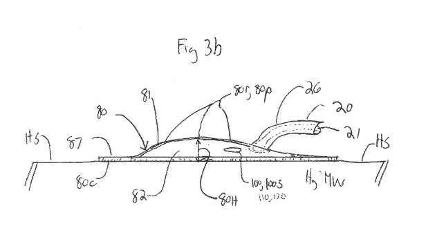
[0032] Fig. 3B is a lateral view illustrating an embodiment of the capture
chamber having a
curved contour for not interfering with heart wall motion and/or blood flow in
the heart.
[0033] Fig. 4 illustrates an embodiment of the corkscrew fixation device and
electrode.
[0034] Figs. 5A-5F illustrate components of an embodiment of the corkscrew
fixation
device and electrode.
[0035] Fig. 6a is a cross sectional side view showing the drug delivery lumen
of an
embodiment of the drug delivery catheter.
[0036] Fig. 6b is a perspective side view showing an embodiment of the
delivery catheter
having an atruamatic tip for delivery of a drug pellet to a delivery site
without use of a
capture chamber.
[0037] Fig. 7a shows an embodiment of the drive stylete having a ball tip.
[0038] Fig. 7b shows an alternative embodiment of the tip of the drive
stylete.
[0039] Fig. 8 is a perspective view illustrating an embodiment of drug
delivery member
including an inner and outer member.
DETAILED DESCRIPTION OF THE INVENTION
[0040] Embodiments of the invention provide apparatus, systems, methods and
formulations for delivering medications in solid form to various locations in
the body. Many
embodiments provide an implanted apparatus for delivering solid form
medication to the
heart for treatment of conditions such as various forms of arrhythmia (e.g.,
those resulting in
atrial fibrillation) or other cardiac conduction disorder. Particular
embodiments provide an
implanted apparatus for delivering solid form medication to a myocardial
delivery site on the
surface of the heart to treat atrial fibrillation.
[0041] Referring now to Figs. 1-8, in one or more embodiments, the invention
provides an
apparatus 10 for the treatment of cardiac arrhythmias comprising a drug
delivery member 20
coupled to a drug storage device 30. Delivery member 20 has a distal end 20de
positioned at
or near a delivery site DS on or near the heart H or other location. In
various embodiments
described herein, delivery member 20 can be coupled at its distal end 20ed to
a capture
chamber 80 positioned on the surface S of a delivery site DS such as
myocardial wall MW of
a patient's heart H so as to deliver drug to the heart for the treatment of
cardiac arrhythmias.
-11-
CA 02856404 2014-05-20
WO 2013/078256 PCT/US2012/066156
[0042] Delivery member 20 may be contained in an outer sheath 15 which also
contains
one or more electrical leads 50 (described herein) for sending and receiving
electrical signals
to and from the heart. Sheath 15 may be fabricated from various biocompatible
resilient
polymers known in the art (e.g., polyurethane, silicone, PEBAX, HDPE, etc.)
and has at least
one lumen 16 for delivery member 20 and lead 50. The drug storage device 30 is
configured
to both store a solid form medication 100 and advance the drug through the
drug delivery
member 20 to a delivery site DS on/in the heart or other location.
[0043] Solid form medication 100 also described herein as formulation 100,
medication
100 or medication element 100, will typically be formulated into pellets 100,
though other
solid formulations are also contemplated. For ease of discussion, solid form
medication 100
will now be referred to as medication pellets 100 and/or pellets 100, but it
will be appreciated
that other forms of solid medication 100 are equally applicable. Medication
100 typically
comprises one or more drugs or other therapeutic agents 110 for the treatment
of one or more
conditions. Medication 100 may also include one or more pharmaceutical
excipients 120
including for example, one or more of disintegrants, super-disintegrants,
binders, anti-
oxidants and other excipients known in the art. In case of various cardiac
applications,
including applications where the medication pellet 100 is configured to
dissolve within
capture chamber 80, the dis-integrant can be selected or otherwise adjusted to
allow for rapid
disintegration in bodily fluids such as one more of blood and/or interstitial
fluid which bathes
the epicardium and/or pericardium.
[0044] In various embodiments pellets 100 can comprise various drugs and other
therapeutics agents 110 for the treatment of cardiac arrhythmias and related
cardiac
conduction disorders. In particular embodiments, such drugs and other
therapeutic agents
can comprise cholinergic compounds such as atropine, scopalomine or
methylscopalomine;
sodium channel blockers such as, quinidine, procainamide, disopyramide or
lydocaine; and
cardiac glycoside such as digoxin or digitoxin. Further, as is described
elsewhere herein,
since these drugs are being delivered near to and/or directly to the surface
of the heart, the
dosage of drug to treat the arrhythmia can be titrated to produce the desired
therapeutic effect
while reducing or preventing adverse effects or reactions which may result
from larger doses
when the drug is delivered orally and/or parenterally (e.g., via IV). For
example, in the case
of sodium channel blockers for the treatment of arrhythmia (e.g., quinidine,
procainamide and
disopryamide) the dosage of drug can be titrated to prevent or reduce the
severity or
incidence of adverse reactions such as one or more of tachycardia, dry mouth,
urinary
retention or blurred vision. For example, in the case cardiac glycosides for
the treatment of
-12-
CA 02856404 2014-05-20
WO 2013/078256 PCT/US2012/066156
arrhythmia (e.g., Digoxin or Digitoxin) the dosage of drug can be titrated to
prevent or reduce
the severity or incidence of adverse reactions such as atrial tachycardias,
atrioventricular
block and various forms of digitalis toxicity. Such titrations can be done
using dose response
curve methods known in the art.
[0045] In particular emodiments, where atropine is used, the dose of this drug
(and/or its
analogues or derivitives) delivered by various embodiments of the can be in a
range from
about 1 to 500 micrograms, or 2 to 250 micrograms, or 5 to 100 micrograms, or
1 to 10
micrograms, or 1 to 20 microgram per dose. In embodiments where scopolamine is
used, the
dose of this drug (and/or its analogues or derivitives) delivered by one or
more embodiments
of the invention can be in a range from about 0.1 to 50 micrograms, or 0.2 to
25 micrograms,
or 0.50 to 10 micrograms, or 0.50 to 5 micrograms per dose. In embodiments
where
lidocaine is used the dose of this drug (and/or its analogues or derivitives)
delivered by one or
more embodiments of the invention can be in a range from about 10 to 1000
micrograms, or
20 to 500 micrograms, or 50 to 250 micrograms, or 1 to 10 micrograms, or 1 to
5 micrograms
per dose. Other dosage ranges are also contemplated. The dosage may be
titrated based on
one or more factors such as the patient's age, particular cardiac arrhythmia,
its severity and
other medications that the patient is recieving. In one or more embodiments of
the invention,
the aforementioned dosages are stored in apparatus 10 and delivered in solid
form to capture
chamber 80 where they are dissolved in tissue fluids (either blood or
interstitial fluids) and
delivered to the myocardial wall(either the endocardial or pericardial wall)
of the left or right
atria.
[0046] In various embodiments, pellets 100 can comprise a single or a
plurality of drugs
110. In particular embodiments, pellets 100 can include a combination of drugs
for treatment
of a single or multiple conditions, for example, a cocktail of drugs for the
treatment of
various cardiac conditions such as arrhythmia, angina, myocardial infarction,
stroke; a
cocktail of antiviral drugs such as protease inhibitors for treatment of HIV
AIDS and also
antibiotics for the treatment of adjunct bacterial infections.
[0047] The drug storage device 30 can be implanted subcutaneously in the
pectoral area or
other region on the patient's torso. In one or more embodiments, the drug
storage device 30
may be incorporated into a housing 36 of a pacemaker or other cardiac device
37 used to send
electrical signals to the heart. Alternatively, it may be have its own housing
38 which may be
placed in the same or a different location as pacemaker or other cardiac
device 37.
[0048] Drug pellet 100 or other solid form drug 100 can be stored in drug
storage device 30
in a variety of configurations. In preferred embodiments, pellet 100 are
contained in/on a belt
-13-
CA 02856404 2014-05-20
WO 2013/078256 PCT/US2012/066156
101 containing a plurality 100p of drug pellets 100 which may be stored in
individual
packing containers 102 attached to belt 100. Pellets 100 can be removed from
belt 101 and
advanced out of storage device 30 through use of a drug advancement means 40.
In many
embodiments, advancement means 40 corresponds to a stylete or other
advancement member
70 that is configured to advance pellet 100 from storage device 30 into
catheter 20 and
capture chamber 80. Stylett 70 can be advanced by a drive means 33 which may
correspond
to rollers 34 driven by an electric motor 35. In a particular embodiment,
stylete 70 is
advanced by two opposing rollers, 34 driven by an electric motor.
[0049] The drug delivery member 20 will typically comprise a catheter 20 or
other like
flexible member having one or more lumens 21 which have an internal diameter
21d sized for
delivery of a drug such as drug pellet 100 to the myocardium or other tissue
site. Catheter 20
may have other lumens 22 which may be configured for other purposes besides
drug delivery
such as placement of one or more electrical leads 50. The catheter 20 may
comprise any
number of biocompatible resilient polymers known in the art (e.g., silicone,
PeBax,
polyurethane, polyethylene (e.g., HDPE, LDPE), etc.) and may be formed using
various
extrusion methods also known in the art.
[0050] In many embodiments, catheter 20 has one end 20e (the proximal end
20ep) coupled
to the drug storage device 30 and the other end 20e' (the distal end 20ed also
referred to as
distal tip 20ed) coupled to a drug capture chamber 80 described below which is
positioned
adjacent a section of the myocardium. Proximal end 20ep of catheter 20 can
include or be
coupled to a connector 20c known in the medical device/catheter arts such as a
luer-lock,
snap fit or swaged connector for coupling to drug storage device 30. The
connector 20c has a
sufficient inner diameter to accommodate drug pellet 100 (as does catheter
lumen 21) and
also desirably provides a watertight seal for preventing tissue fluids from
getting into drug
storage device 30.
[0051] In some embodiments, apparatus 10 does not include a capture chamber 80
and in
these cases, catheter distal end 20ed may be configured to be positioned in or
near the heart
(and may include a fixation device described herein) so as to release drug
pellet 100 directly
to the myocardial wall MW. In such embodiments, catheter distal end 20ed is
desirably
configured to have an atraumatic tip 20at to minimize or prevent injury or
irritation to the
myocardial wall or other tissue. This can be achieved by configuring the tip
to have a
rounded and/or tapered shape (as shown the embodiment of Fig. 6b) as well as
through the
use of soft low durometer polymer materials known in the art such as hydrogels
and silicone.
In particular embodiments, an atraumatic tip 20at can be fabricated from
silicone materials
-14-
CA 02856404 2014-05-20
WO 2013/078256 PCT/US2012/066156
having a durometer of between 1-20 Shore A, more preferably between 1-10 Shore
A and
still more preferably between 1-5 Shore A.
[0052] Catheter 20 is configured to allow solid drug pellets 100 (or other
shape/form of
solid form medication 100) to be advanced from the drug storage device 30
through the
catheter lumen 21 and then be directly ejected on or near the surface of the
heart or be ejected
into a capture chamber that is positioned in proximity to a myocardial wall
(e.g., of the atria).
In many embodiments, catheter 20 is configured to place the pellet in
proximity to the
epicardial surface of the heart. However, placement at other locations
including the
endocardial surface within the left or right atrial or ventricular chambers is
also contemplated.
The drug pellet 100 is configured to dissolve (in blood and/or other tissue
fluids) when so
placed and deliver a therapeutically effective amount of drug for terminating
and/or otherwise
mitigating the episode of atrial fibrillation or other related condition.
[0053] In preferred embodiments, all or a portion of catheter 20 may comprise
a first (an
inner) and second (outer) tubular member 23 and 24 (also referred to as inner
and outer
catheters 23 and 24) concentrically arranged with one or more electrical leads
50 such as
electrical leads 51 and 52 (described below) positioned between the two
tubular members.
Desirably, this configuration is used for at least the mid portion 20m of the
catheter, but it
may be used for substantially the entire length of catheter 20. In a specific
preferred
embodiment, leads 51 and 52 are coiled around the outer perimeter 23p of the
inner member
23 with the outer member 24 then jacketing the leads and the inner member 23.
Coiled leads
51 and 52 may also be arranged to provide torsional support to the catheter 20
so as maintain
patentcy of the drug delivery lumen 21 if the catheter is put into a bent,
twisted or other
contorted position. In other embodiments for supporting the drug delivery
lumen to maintain
patentcy, lumen 21 can include a support coil 25, such as a 0.005" trifilar
wire wound around
the inner surface 21i of the lumen 21 as is shown in the embodiments of Figs.
2 and 6. In
such embodiments support coil 25 may have a lubricous coating, for example, a
TEFLON
coating, to facilitate passage of drug pellet 100 through the lumen 21. In an
alternative
embodiment support coil 25 can be positioned around the exterior of lumen 21
and thus
within catheter wall 20w. Such embodiments can be produced using co-extrusion
methods
known in the catheter and polymer processing arts.
[0054] Inner and outer members 23 and 24 can comprise an elastomeric material
such as
silicone (or other elastic material known in the art) so as to allow the
catheter 20 to bend and
flex so as to be positioned in different locations in the body. In particular
embodiments,
catheter is 20 is sufficiently flexible so as to be able to position the
distal portion 26 of the
-15-
CA 02856404 2014-05-20
WO 2013/078256 PCT/US2012/066156
catheter in various locations on or near the heart wall (e.g., on the right or
left atria) while
allowing the proximal portion 27 of the catheter to be connected to drug
storage device 30.
[0055] As described above in one or more embodiments, apparatus 10 includes
one or more
electrical leads 50 for performing one or more of the following functions: i)
sensing electrical
activity of the heart; ii) pacing the heart; iii) sending an electrical signal
to the heart to
depolarize a selected area of the myocardium (e.g., an area containing a foci
of aberrant
electrical activitity); and iv) sending an electrical signal to the heart to
defibrillate one or
more chambers of the heart. Leads 50 can placed within sheath 15, catheter 20
or both.
They may comprise various insulated conductive wires (also described as
cables) known in
the art which are configured for use in pacemakers and other cardiac
stimulation devices
known in the art such as ICD (implantable cardiac defibrillators). At their
proximal end 50p,
they will typically include an electrical connector 50c, such as an IS-1
connector for
connection for example to a cardiac stimulation device as is shown in the
embodiment of Fig
lb. Lead 50 can also be configured to contain multiple leads 50. Accordingly,
in one or
more embodiments, lead 50 may also be of a coaxial design as is know in the
art so to include
a first and second lead 51 and 52 as is described below.
[0056] In many embodiments, apparatus 10 will also include at least a first
and second
electrical lead 51 and 52 for sensing electrical activity of the heart. The
leads 51 and 52 may
comprise any conductive metal and are desirably insulated for most of their
length. They
may be coiled around the perimeter 20p of catheter 20 and/or placed within a
lumen 22 of the
catheter separate from the drug delivery lumen 21. They may also configured as
a first and
second lead 50 and 51 placed within a coaxial cable 50.
[0057] Embodiments of the invention contemplate a variety of means for
advancing drug
pellet 100 through the lumen 21 or other lumen of drug delivery catheter 20.
Such means
may include, for example, mechanical, pneumatic, hydraulic or magnetic means.
In preferred
embodiments, drug pellet 100 is transported through drug delivery lumen 21 of
catheter 20 or
other like structure (e.g., a hypotube) by means of an advanceable member 70
such as a
stylete 70 that is advanced from the drug storage device 30 by an electric
motor or other
advancement means. Stylete 70 can have a length 701 in the range of about 40-
50 cm with
longer and shorter lengths contemplated. According to one or more embodiments,
stylete 70
may comprise a metal wire or ribbon that is wound, for example, in a spool 72
and then
unwound by drive means 33 such as electrically driven pinch rollers 34. In a
preferred
embodiment the advanceable member has a wound state when not advanced and an
unwound
state when advanced. The metal wire or ribbon may comprise various flexible
metals known
-16-
CA 02856404 2014-05-20
WO 2013/078256 PCT/US2012/066156
in the art including super elastic metals such as NITNOL so as to readily
unwind when
spooled and then rewind. It may also have a preformed shape and/or be spring
loaded. Other
advanceable members 70 known in the catheter/guide wires arts are also
contemplated.
[0058] Stylete 70 will typically have a ball tip 71 that is sized to push drug
pellet 100
through drug delivery lumen and out the septum; however other shapes for tip
71 are also
contemplated such as hot dog shape, or a concave shaped tip 71c having a
concavity sized to
engage the diameter of the drug pellet. In one or more embodiments, stylete
tip 71 may be
configured to sense contact with the drug pellet 100 (or other form of solid
drug 100) so as to
able be to determine that the pellet is being advanced and/or that the pellet
has been ejected.
This can be accomplished by configuring the tip and/or the stylete to be
capacitively coupled
to the drug pellet so as to sense changes in capacitance when tip 71 makes and
breaks contact
with the drug pellet.
[0059] In many embodiments, the distal tip 20ed of catheter 20 is coupled to a
capture
chamber 80 which is configured to hold the drug pellet 100 while allowing
blood or other
floods to flow or seep into the chamber and then flow or seep out. This allows
the drug pellet
to be dissolved by blood (or other fluids) which flow or seeps into the
chamber and then
delivered to the myocardium as the blood or other fluid flows or seeps out.
Capture chamber
80 includes a housing 81 having an interior volume 82 in which drug pellet 100
is contained
while it dissolves. The housing 80 will typically include an opening 83 for
insertion of
catheter 20 so as to form a joint 84 with the catheter as is shown in the
embodiment of Fig. 2.
The housing will also typically include a second opening 85 for placement of
porous section
87 discussed below.
[0060] Joint 84 can comprise any number of those known in the art such as a
weld,
ultrasonic weld, adhesive joint, crimp, snap fit and the like. In particular
embodiments joint
84 may comprise a pivotal type joint so as to allow the capture chamber 80 to
move freely
with beating of the heart while imparting reduced force and motion to catheter
20. In use,
such embodiments improve the reliability and mechanical life of joint 84,
chamber 80 and
catheter 20 by reducing the stress imparted on one or more of these
components. Such
embodiments also reduce the likelihood of hemolysis caused by movement of
catheter 20 by
minimizing the motion of catheter within the chambers of the beating heart.
[0061] Capture chamber 80 and housing 81 will typically comprise at a non-
porous section
86 and a porous section 87 both of which may comprise multiple sections 86 and
87. In some
embodiments, substantially all of the capture chamber 80/housing 81 can be
porous. The
non-porous section(s) 86 of the housing 81 can comprise various biocompatible
polymers
-17-
CA 02856404 2014-05-20
WO 2013/078256 PCT/US2012/066156
known in the art. The blood contacting surfaces of the housing 81(including
one or both of
the porous and non-porous sections) may comprise one or more non-thrombogenic
materials
known in the art such as silicone, polyurethane and expanded
polytetraflouroethylene
(ePTFE, and example of which includes TEFLON). Also, one or both of the non-
porous and
porous sections 86 and 87 can include a drug eluting coating 80d configured to
elute a drug to
reduce thrombus formation and platelet adhesion. Such coatings can include
paclitaxel and
other anti-thrombogenic coatings known in the art. The coating can also be
selected so as not
interfere with the bioactivity of medication 100 and/or to produce a
synergetic effect
[0062] Typically, the porous section 87 comprises a portion (e.g., the bottom
portion or
side) of the housing 81 that is configured to be positioned in contact with or
close proximity
to the myocardial wall or other portion of the heart. However, in some
embodiments, all or
multiple portions (e.g., sides) of the capture chamber 80/housing 81 can be
fabricated from
porous materials. The porous section 87 can be fabricated from any number of
porous
biomaterials such as various polymeric fiber materials such as polyethylene
teraphalate or
NYLON. In preferred embodiments, the chamber housing can be fabricated from
porous
DACRON, such as a DACRON mesh, which can be either woven or knitted. The size
and
porosity of porous section 87 can be selected to allow blood (or other tissue
fluid) to seep in
or out of the chamber at a selected rate to in turn achieve a selected rate of
disintegration of
the drug pellet. In some embodiments, porous sections 87 can be fabricated
from porous
materials having varying porosity. For example, for embodiments where most of
chamber 80
is porous, the top 80t and sides 80s of the chamber 80 can be fabricated from
a first material
having a first porosity, while the portion in contact with the myocardial wall
80c (e.g., also
referred to as tissue contacting portion or surface 80c) can be fabricated
from a second
material having a second porosity, which is typically higher than the first
porosity so as to
retain blood or other fluid having the dissolved drug within the chamber 80
while allowing it
to readily wick out portion 80c in contact with the myocardial wall so as to
bathe the
contacted myocardium with a solution (blood or other bodily fluid) containing
the drug,
herein referred to as a drug solution. In use, embodiments of chamber 80
having such a
directionally varying porosity serve to improve delivery of drug to the
myocardial wall both
in terms of amount and rate of delivery. For purposes of reference, tissue
contacting portion
80c may also be referred to as a bottom portion 80b of chamber 80.
[0063] In many embodiments, the capture chamber 80 is positioned adjacent or
near the
myocardial wall so as to retain a drug-blood solution adjacent the wall and
then transport
drug into the myocardium by transdermal delivery, e.g., by diffusion into
myocardial wall. In
-18-
CA 02856404 2014-05-20
WO 2013/078256 PCT/US2012/066156
many embodiments, capture chamber 80 is fixed to the myocardial wall by means
of a helical
wire 91 (coupled to the chamber) or other fixation device 90 (also referred to
as anchoring
means 90) that is anchored into the heart wall. Embodiments of the invention
contemplate a
number of configurations for helical wire 91 having varying pitch and number
of coils so as
to achieve a desired level of anchoring force (up Sibs of force or more)
within myocardial
wall to retain the capture chamber against the myocardial wall even during
vigorous beating
of the heart. Other anchoring means are also contemplated.
[0064] In addition to functioning as fixation device, according to one or more
embodiments, helical wire 91 can also be configured as an electrode 92 to
sense electrical
activity of the heart and to conduct electrical signals to the heart for
purposes of depolarizing
sections of the generating aberrant electrical activity and/or to defibrillate
the atria or
ventricles of the heart. To achieve this purpose, i) wire 91 is fabricated
from a conductive
metal core 90c having insulation 91i, ii) a distal portion 93 of wire 91 is un-
insulated, and iii)
wire 91 is electrically coupled to a lead 51 or 52, for example, by crimp tube
or other crimp
joint 53 as is shown the embodiment of Fig. 3b. In order to have a two
electrodes for sending
and/or receiving electrical signals to and from the heart, in many
embodiments, wire fixation
device 91 can include another conductive wire 94 coiled over wire 91 as is
shown in the
embodiments of Figs 1B, 2-3, 5D and 5F. Wire 94 is configured function as a
second
electrode 95 and can be electrically coupled to lead 51 or 52, for example, by
a crimp tube or
other crimp joint 53 as shown n the embodiment of Fig. 3A and 5E. In one or
more
embodiments, electrodes 91 and 94 may be configured as bipolar electrodes 96
for sending
and/or receiving signal to and from the heart.
[0065] In many embodiments, the tissue contacting surface 80c of chamber 80 is
sufficiently flexible to bend and flexible with beating of the heart so as to
not impede heart
wall motion whether it be the atrial or ventricular wall and whether surface
80c is positioned
on the epicardial or endocardial surface of the heart. This flexibility can be
achieved through
the use of various flexible porous polymer materials for surface 80c, for
example a flexible
DACRON mesh. Likewise, chamber 80 can be configured not to impede heart wall
motion
as well. This can be achieved by the selection of the flexibility, mass and
contour 80r of
chamber 80. For example, various flexible polymer materials can be used for
housing 81
such as silicone, polyurethane, or other elastomeric polymer known in the art.
Also, the
contour 80r of chamber 80 is desirably configured to minimize any impediment
to blood flow
in the heart whether it be in the atria or ventricles (left or right in either
case) and/or to not
cause any appreciable turbulence in blood flow within the heart. This can be
achieved be
-19-
CA 02856404 2014-05-20
WO 2013/078256 PCT/US2012/066156
configuring chamber 80 to have a smooth/stream lined curved contour 80r and
low profile
80p such that housing 81 does not rise appreciably above the surface of the
heart HS, e.g., by
an amount 80H no more than about 1 cm, more preferably, no more than about
5mm, still
more preferably no more than about 2.5 mm with even smaller amounts
contemplated. One
example of such a curved contour is shown in the embodiment of Fig. 3b. In
such
embodiments, pellet 100 can be have an elongated thinner shape 100s so as to
be able to be
placed within the interior 82 of a chamber 80 having such a thinner shape.
[0066] As discussed above, the distal tip 20ed of catheter 20 extends into the
capture
chamber 80 for ejecting pellet 100 into the chamber. In many embodiments,
distal tip 20ed
includes an elastic self-closing septum 45 for preventing fluid intrusion into
drug delivery
lumen 21 or other lumen 22 of catheter 20. Septum 45 may be resealable, Septum
45 includes
a slit 46 which is configured open when drug pellet 100 is advanced against
the slit so as to
allow passage of the drug pellet through the septum and then close to prevent
blood or other
fluid ingress into the catheter lumen.
[0067] In many embodiments, the apparatus 10 is coupled to a controller (not
shown) for
controlling one more aspects of the medication delivery process including
actuation and
control of the drive source to deliver a medication pellet into the myocardium
or other
location. The controller can be programmed to include a delivery regimen
wherein
medication is delivered at regular intervals (e.g., once or twice a day, etc.)
over an extended
period. It can also be configured to receive a signal (e.g., wireless or
otherwise) to initiate the
delivery of medication or to change the delivery regimen (e.g., from once a
day to twice a
day). In this way, the patient or a medical care provider can control the
delivery of
medication in response to a specific event (e.g., an episode of arrhythmia )
or longer term
changes in the patient's condition or diagnosis.
[0068] The controller can be coupled to or otherwise receive inputs from the
pacemaker or
a sensor. When the controller receives an input from the sensor indicative of
a condition such
as an episode of arrhythmia, it initiates the delivery of one or more
medication pellets to the
heart or other target tissue site so as to treat the medical condition. Both
the initial and
subsequent inputs from the sensor can be used to titrate the delivery of
medication pellets
over an extended period until the condition is dissipated or otherwise
treated. The controller
can also receive inputs from other sensors configured to measure the tissue
concentration of
the delivered drug. These inputs can also be used to titrate the delivery of
the medication to
achieve a selected concentration of drug (e.g., in plasma, tissue, etc.). The
drug sensors can
be positioned on distal portions of the drug delivery device such as on the
catheter or the
-20-
CA 02856404 2014-05-20
WO 2013/078256 PCT/US2012/066156
outside of the porous chamber, or the as well as other sites in the body
(e.g., a vein or artery)
in order to develop a pharmacokinetic model of the distribution of the drug at
multiple sites in
the body. The apparatus can also include a sensor coupled to the controller
which indicates
when the medication pellets have been used up and/or exactly how many are
left. The
controller in turn can signal this data to an external communication device
such as a cell
phone, portable monitor or remote monitor (e.g., at the physician's office).
In this way, the
patient and/or medical care provider can take appropriate action before the
apparatus runs out
of medication.
[0069] The pellets or other solid form 100 of the medication are delivered to
a delivery site
such as the endocardial or epicardial surface of the heart where they are
configured to be
broken down, disintegrate and absorbed by body tissue fluids so as to produce
a desired
concentration of the drug at a target tissue site. In some applications, the
delivery site can be
the same as the target site, for example the heart. In other applications, the
target site can be
different from the delivery site, for example, the delivery site can be
intramuscular tissue in
the chest and the target site can be the heart or the liver. The delivery site
can be adjacent the
target site, for example adipose to deliver to underlying muscle tissue, or it
can be placed at a
non-oppositional site, for example, intramuscular delivery to reach the site
of the heart. In
each case, the medication pellet 100 can include a selected dose 100d of drug
and be
configured to disintegrate and be dissolved by body tissue fluids so as to
yield a
therapeutically effective concentration of the drug at the target tissue site
such as the
endocardial or epicardial surface of the heart. In many applications, this
involves the pellet
being dissolved by body tissue fluids at the delivery site (e.g., interstitial
fluids bathing the
pericardium or the blood bathing the endocardial or other portion of the
myocardial wall)
where the drug then diffuses into the myocardial wall. Accordingly, in these
and other
applications, the dose of the drug in the pellet can be titrated to achieve a
selected
concentration of the drug (or concentration range) for a selected period
during and after
dissolution of the pellet. Further, the dose of drug can be titrated to
produce a desired
therapeutic effect on the heart and/or cardiovascular system (e.g., treatment
of arrhythmia,
angina, myocardial infarction, congestive heart failure) while minimizing
adverse reactions
which may occur for larger doses of the drug when orally delivered. For
example, in the case
of sodium channel blockers for the treatment of arrhythmia (e.g., quinidine,
procainamide and
disopryamide), the dosage of drug can be titrated to prevent or reduce the
severity or
incidence of adverse reactions such as one or more of tachycardia, dry mouth,
urinary
retention, blurred vision and headache. In the case of cardiac glycosides for
the treatment of
-21-
CA 02856404 2014-05-20
WO 2013/078256 PCT/US2012/066156
arrhythmia (e.g., Digoxin or Digitoxin), the dosage of drug can be titrated to
prevent or
reduce the severity or incidence of adverse reactions such as atrial
tachycardias,
atrioventricular block and various forms of digitalis toxicity. Such
titrations can be done
using various dose response curve methods known in the art.
[0070] In some embodiments, the pellet 100 (including the drug dose) is
configured to
disintegrate and be dissolved by blood or tissue fluids which seep into the
porous chamber.
In particular embodiments for treating various cardiac rhythm disorders such
as arrhythmia,
the pellet is configured to rapidly disintegrate and be dissolved in blood or
other fluid within
the porous chamber. This can be achieved through the use of one or more super
dis-
integrants as well as disintegrating enhancing features (e.g., pores, cracks
or other intrusions)
in or one the pellet. It can also be achieved by treating the pellet prior or
after delivery with
mechanical, electromagnetic, acoustical or other energy to weaken the pellet
structure, create
cracks and other structural defects for the ingress of fluids or initiate the
breakup of the pellet
into smaller pieces.
[0071] In various applications, embodiments of the invention can be used to
deliver solid
form drugs to provide treatment for a number of medical conditions including
coronary
arrhythmia's (both atrial and ventricular including fibrillation), coronary
ischemia (e.g., from
a narrowed or blocked artery including that resulting in a heart attack),
cerebral ischemia,
stroke, anemia or other like condition. The apparatus can be implanted at or
near the target
tissue site (e.g., the heart) or at remote delivery site (e.g.,
intramuscularly in the chest or
thigh.
[0072] In exemplary embodiments of methods for using the invention to treat a
heart
condition, for example cardiac arrhythmia, the apparatus can be implanted in
the patient's
chest to deliver drug to a delivery site DS within or near the heart. In
specific embodiments,
the drug storage device may be placed in the pectorial region while the distal
end of the
delivery member can be positioned on or near a surface of the heart, either
the epicardial or
endocardial surface. For embodiments where the apparatus is used to deliver
drug to the
myocardial wall, the lead and porous chamber can be fixed to the endocardial
or epicardial
wall using the corkscrew fixation element or other fixation device.
Implantation can be done
using an open or minimally invasive surgical procedure, for example, via
percutaneous access
through the vascular system. Prior to implantation, the drug reservoir can be
loaded with a
selected number of pellets to provide for delivery of pellets to the delivery
site over an
extended period of time, e.g., years. Once implanted, the pellets can be
stored in the
apparatus for an extended period of years (e.g., 1, 2, 5 or longer) without
degradation or
-22-
CA 02856404 2014-05-20
WO 2013/078256 PCT/US2012/066156
deleterious effect to the pellets (e.g., loss of drug potentcy or therapeutic
effectiveness). The
apparatus can deliver solid form medication to the delivery site at regular
intervals (e.g., once
a day, week, month, etc.) or in response to an input from a sensor. In the
latter case, the input
can be indicative of a particular medical condition or event such as an
episode of arrhythmia.
Embodiments of the controller described herein can be used to determine when
to initiate
delivery based on the sensor input and/or the time intervals for delivery for
embodiments
employing delivery at regular intervals. In either case, the controller can
send a signal to the
drug storage device. There it disintegrates/degrades and is dissolved in local
tissue fluids to
treat a local target tissue site (e.g., it dissolves in the interstitial
fluids bathing pericardium to
treat the heart or the CSF fluid to treat the brain), or it is subsequently
absorbed into the blood
stream where it is carried to a remote target tissue site (e.g., the liver,
heart, etc.) or both.
Further, pellets can be delivered based on input from a sensor providing
physiologic data
predictive of the medical condition (e.g., blood glucose) or another sensor
that is configured
to sense the local and/or plasma concentration of the drug. In some
embodiments, pellet
delivery can be controlled by sensing the state of disintegration of
previously delivered
pellets. For example, another pellet can be delivered when it has been
determined that the
previous pellet is in a particular state of disintegration (e.g., it has been
completely or
substantially disintegrated). This can be achieved by sending and receiving a
signal from the
pellet such as an optical, ultrasound or electrical signal. For example, for
the use of optical
signal reflectance measurements can be used to determine the state of
disintegration. A
particular disintegration state can be determined when the reflectance signal
falls below a
particular threshold. Similar approaches can be used for use of reflected
ultrasound or
impedance. The pellet can even include various echogenic, or optically opaque
or other
agents to enhance the reflected ultrasonic, optical or other signal. The
pellet may also include
various optical indicia having one or more of a pattern, size or shape
configured to provide an
indication of the state of disintegration of the pellet.
[0073] Conclusion
[0074] The foregoing description of various embodiments of the invention has
been
presented for purposes of illustration and description. It is not intended to
limit the invention
to the precise forms disclosed. Many modifications, variations and refinements
will be
apparent to practitioners skilled in the art. For example, embodiments of the
apparatus can be
sized and otherwise adapted for various pediatric and neonatal applications.
[0075] Elements, characteristics, or acts from one embodiment can be readily
recombined
or substituted with one or more elements, characteristics or acts from other
embodiments to
-23-
CA 02856404 2014-05-20
WO 2013/078256 PCT/US2012/066156
form numerous additional embodiments within the scope of the invention.
Moreover,
elements that are shown or described as being combined with other elements,
can, in various
embodiments, exist as stand-alone elements. Hence, the scope of the present
invention is not
limited to the specifics of the described embodiments, but is instead limited
solely by the
appended claims.
[0076] While preferred embodiments of the present invention have been shown
and
described herein, it will be obvious to those skilled in the art that such
embodiments are
provided by way of example only. Numerous variations, changes, and
substitutions will now
occur to those skilled in the art without departing from the invention. It
should be understood
that various alternatives to the embodiments of the invention described herein
may be
employed in practicing the invention. It is intended that the following claims
define the
scope of the invention and that methods and structures within the scope of
these claims and
their equivalents be covered thereby.
-24-