Note: Descriptions are shown in the official language in which they were submitted.
81779941
Tracking a Guidewire
TECHNICAL FIELD
The disclosure relates to tracking a guidewire.
BACKGROUND
Central venous access is an invasive procedure. Central venous access involves
placing a long catheter that extends into the deep veins of the chest or
abdomen. Central
venous access provides a way to infuse agents that are caustic to the smaller
veins of the arm.
As a result, central venous access is used for chemotherapy, total parenteral
nutrition, and
numerous other agents. Larger diameter catheters are used for applications
that require high
flow rates such as hemodialysis, plasmapheresis, and volume resuscitation.
SUMMARY
In one aspect, in general, a method comprising: providing, by a computer
system, an
AC current signal to a transmitter, the AC current signal for causing the
transmitter to transmit
an electromagnetic signal; receiving, at the computer system, data from a
first electromagnetic
sensor disposed in a metallic tube at a tip of a guidewire and connected to
the computer
system via a connector, the metallic tube configured to preserve flexibility
during use in a
patient, the first sensor for receiving the electromagnetic signal transmitted
by the transmitter,
wherein the first sensor is sealed using an encapsulant and the metallic tube
is coated with a
hydrophobic substance for protection during use in the patient; receiving, at
the computer
system, data from at least two electromagnetic sensors each mounted at a
respective pad
affixed to the patient, the at least two sensors for receiving the
electromagnetic signal
transmitted by the transmitter, wherein the pads are affixed to at least two
anatomic landmarks
selected from the group consisting of the patient's xiphoid, the patient's
sternal notch, and the
patient's acromioclavicular joints; determining, at the computer system, based
on the received
data, a location of the tip of the guidewire inserted in the patient relative
to locations of the at
least two sensors mounted at their respective pads; and causing, by the
computer system, an
indication of the determined location of the tip of the guidewire to be
displayed in an overlay
1
CA 2856519 2019-03-11
81779941
upon a reference image, the overlay representing at least part of the
guidewire, wherein the
reference image includes representations of the at least two sensors mounted
at their
respective pads, wherein the connector is configured to allow the guidewire to
be
disconnected from the computer system after the guidewire has been positioned
in the patient.
Implementations of this aspect can include one or more of the following
features. The
overlay includes an x-ray image. The overlay includes an ultrasound image. The
guidewire is
inserted into a vein of the patient. Determining the location of a tip of a
guidewire includes
measuring three-dimensional coordinates of the guidewire. The method includes
generating
an x-ray image after the location of the tip of the guidewire has been
determined. The tip of
the guidewire includes an electromagnetic transmitter. The electromagnetic
sensor is placed
external to the patient.
In another aspect, in general, a method includes receiving, at a computer
system, data
from an electromagnetic sensor, determining, at the computer system, based on
the received
data, a location of a tip of a guidewire inserted in a patient, and providing,
by the computer
.. system, an indication to a user interface that the tip of the guidewire has
been positioned at a
predetermined location.
Implementations of this aspect can include one or more of the following
features. The
method includes determining, at the computer system, if a tip of a catheter
has been positioned
at the determined location of the tip of the guidewire, and providing, by the
computer system
to a user interface, an indication that the tip of the catheter has been
positioned at the
determined location of the tip of the guidewire. The predetermined location
corresponds to a
location of a target device. The target device is internal to the patient. The
indication that the
tip of the catheter has been positioned at the determined location comprises
at least one of
visual and audible confirmation.
In another aspect, in general, a system, comprising a transmitter configured
to receive
an AC current signal, the AC current signal for causing the transmitter to
transmit an
electromagnetic signal; a guidewire comprising a metallic tube at a tip of the
guidewire, the
metallic tube configured to preserve flexibility during use in a patient; a
first sensor disposed
2
CA 2856519 2019-03-11
81779941
in the metallic tube of the guidewire, the first sensor for receiving the
electromagnetic signal
transmitted by the transmitter, wherein the first sensor is sealed using an
encapsulant and the
metallic tube is coated with a hydrophobic substance for protection during use
in the patient;
at least two sensors each mounted at a respective pad affixed to the patient,
the at least two
sensors for receiving the electromagnetic signal transmitted by the
transmitter, wherein the
pads are affixed to at least two anatomic landmarks selected from the group
consisting of the
patient's xiphoid, the patient's sternal notch, and the patient's
acromioclavicular joints; a
computer system in communication with the sensors, the computer system
configured to
determine a location of the tip of the guidewire inserted in the patient
relative to locations of
the at least two sensors mounted at their respective pads; and a display
system in
communication with the computer system, the display system configured to
display an
indication of the determined location of the tip of the guidewire in an
overlay upon a reference
image, the overlay representing at least part of the guidewire, wherein the
reference image
includes representations of the at least two sensors mounted at their
respective pads, wherein
the first sensor is connected to the computer system via a connector, and the
connector is
configured to allow the guidewire to be disconnected from the computer system
after the
guidewire has been positioned in the patient.
Implementations of this aspect can include one or more of the following
features. The
image includes an ultrasound image. The image includes an x-ray image. The
computer
system comprises an integrator for measuring rising edge and steady state of
the
electromagnetic signals. The transmitter comprises a multi-axis transmitter.
The sensor
comprises a one-axis coil. The transmitter provides pulsed DC current signals
to each
transmitter axis. The sensor comprises a 5 degrees-of-freedom sensor. The
sensor comprises
a pad that can be affixed to a patient.
In another aspect, in general, a non-transitory computer readable storage
device having
recorded thereon statements and instructions that, when executed, cause a
computer system to:
3
CA 2856519 2019-03-11
81779941
provide an AC current signal to a transmitter, the AC current signal for
causing the transmitter
to transmit an electromagnetic signal; receive data from a first
electromagnetic sensor
disposed in a metallic tube at a tip of a guidewire and connected to the
computer system via a
connector, the metallic tube configured to preserve flexibility during use in
a patient, the first
sensor for receiving the electromagnetic signal transmitted by the
transmitter, wherein the first
sensor is sealed using an encapsulant and the metallic tube is coated with a
hydrophobic
substance for protection during use in the patient; receive data from at least
two
electromagnetic sensors each mounted at a respective pad affixed to the
patient, the at least
two sensors for receiving the electromagnetic signal transmitted by the
transmitter, wherein
the pads are affixed to at least two anatomic landmarks selected from the
group consisting of
the patient's xiphoid, the patient's sternal notch, and the patient's
acromioclavicular joints;
determine, based on the received data, a location of the tip of the guidewire
inserted in the
patient relative to locations of the at least two sensors mounted at their
respective pads; and
cause an indication of the determined location of the tip of the guidewire to
be displayed in an
overlay upon a reference image, the overlay representing at least part of the
guidewire,
wherein the reference image includes representations of the at least two
sensors mounted at
their respective pads, wherein the connector is configured to allow the
guidewire to be
disconnected from the computer system after the guidewire has been positioned
in the patient.
Implementations of this aspect can include one or more of the following
features. The
image includes an ultrasound image. The image includes an x-ray image.
These and other aspects and features and various combinations of them may be
expressed as methods, apparatus, systems, means for performing functions,
program products,
and in other ways.
Other features and advantages will be apparent from the description and the
claims.
DESCRIPTION OF DRAWINGS
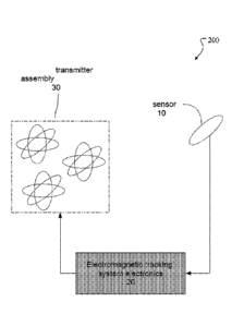
Figure 1 shows a central venous catheter.
Figure 2 is a block diagram of components of a guidewire tracking system.
3a
CA 2856519 2019-03-11
81779941
Figure 3 shows an electromagnetic sensor.
Figure 4 shows a flowchart.
Figure 5 shows anatomic landmarks.
Figure 6 shows a flowchart.
Figure 7 is a block diagram of a computer system.
Like reference symbols in the various drawings indicate like elements.
DETAILD DESCRIPTION
A guidewire tracking system (GTS) that uses electromagnetic signals can allow
a
surgeon to visualize catheter placement continuously through a virtual image
overlay (e.g.,
over an ultrasound image) while minimizing x-ray exposure to both the surgeon
and the
patient (e.g., a pediatric patient).
=
A guidevvire is a device that is inserted into a patient undergoing a
catheterization
procedure and used to position a catheter. Central catheters, e.g., the
central catheter shown in
figure 1, can be placed in the operating room under general anesthesia using
fluoroscopic
guidance, which results in multiple x-ray images being used. Radiation may
have negative
side effects. The system described here can minimize or eliminate the use of
radiation.
3b
CA 2856519 2019-03-11
CA 02856519 2014-05-21
WO 2013/078348
PCT/US2012/066304
-The system can also be adaptable for catheter placement in other settings,
e.g.,
outside of the operating room, where catheters are inserted without the use of
fluoroscopy. In this venue, catheter and guidewire manipulations are often
done
blindly. The lack of real-time feedback causes a variety of problems, which
can lead to
unsuccessful placement. For example, .malpositioned catheters can lead to
repeat
procedures that in turn. may increase the risk of infection, the potential for
vascular
injury, and the need for additional x-ray imaging for confirmation of
placement.
Another procedure that can benefit from the system described herein is
placement of a longer term intravenous line placed into a central vein in
children. This
procedure is used to give medicines, blood transfusions, fluids or nutrients.
Blood tests
may also be drawn through the catheter. The catheter is designed for long-time
use so
that many painful needle sticks can be avoided.
Imaging guidance can improve the success rate of catheter insertion by
facilitating needle placement in the vein and catheter advancement to the
target site.
Ultrasound imaging is typically used to help guide the needle during initial
access to
the vein. The introduction of small, light, and cheap ultrasound units have
facilitated
compliance with this recommendation. However, ultrasound is not suitable for
viewing
the final placement of the catheter. For this purpose, fluoroscopy is used as
described
below.
Referring to figure 1, catheter placement is generally within a certain
anatomical area, typically the superior vena cava above the right atrium 1, to
avoid
complications. Inserting a catheter too far increases the risks of cardiac
arrhythmia and
atrial perforation whereas not inserting the catheter far enough increases the
risks of
venous thrombosis and inadequate flow rates for dialysis and plasmapheresis.
Fluoroscopy is sometimes used during catheter insertion and the resulting
feedback can increases the likelihood that the catheter tip will be positioned
appropriately. An initial fluoroscopic image can be used to give an overall
view and
starting point, but subsequent fluoroscopy images can be avoided by real-time
tracking
of the guidewire tip using an electromagnetic sensor, and only one other final
confirming fluoroscopy image may be required at the completion of the
procedure,
minimizing x-ray dose. In another example, neither an initial nor a final
confirming
fluoroscopy image is required. In other words, the operator can perform a
procedure by
4
CA 02856519 2014-05-21
WO 2013/078348
PCT/US2012/066304
relying only on the feedback from the electromagnetic sensor and the
ultrasound.
Guidewire tracking can be improved by using electromagnetic tracking
technology.
This technology is based on the generation of known electromagnetic field
structures
and couplings. Systems can be designed to measure 3 degrees-of-freedom (DOF),
5DOF and/or 6D0F. 3DOF typically corresponds to the 3 cardinal position
coordinates,
51)OF to the 3 position and 2 orientation measurements (without roll) and
6130F to the
3 position and 3 orientation (azimuth, elevation and roll) measurements. All
systems
utilize a source of electromagnetic fields. These can be AC, pulsed DC,
permanent
magnets, moving magnets, among others. There are also techniques for measuring
the
electromagnetic fields. This can be done with fluxgates, cored and non-cored
coils that
have induced voltages across them, Hall effect devices, magneto-resistors of
all forms
(e.g., plain, giant, colossal and tunneling), field dependent oscillators,
squids,
magnetometers, among others. These systems can operate in either direction,
i.e., the
tracked object can be generating or sensing a magnetic field, and the tracking
system
sensing or generating the magnetic field.
Referring to figure 2, in some implementations, a 51)OF pulsed DC tracking
system 200 is employed for midewire tracking. The electromagnetic tracking
system
electronics 20 consists of a computer component, a transmitter excitation
component
and a receiving component. Under computer command and control, a multi axis
transmitter assembly 30 has each of its axes energized by DC drive electronics
to
transmit symmetrical, sequentially excited, nonoverlapping square DC-based
waveforms. These are received through the air or tissue by one or more sensors
10 that
conveys these signals to signal processing electronics within the
electromagnetic
tracking system electronics 20. The computer in the electromagnetic tracking
system
electronics 20 contains an integrator for measuring rising edge and steady
state of each
axes' sequential waveform so that an integrated result may be measured at the
end of
the steady state period. It further controls the transmitter DC drive
electronics to
operate the transmitter and receives signals from the signal processing
electronics for
the signal integration process, the end result being calculation of the
sensor's position
and orientation in three-dimensional space with significantly reduced eddy
current
distortion while providing improved compensation for sensor drift with respect
to the
Earth's stationary magnetic field and power-line induced noise.
5
CA 02856519 2014-05-21
WO 2013/078348
PCT/US2012/066304
Specifically, the transmitter DC drive electronics provides pulsed DC current
signals of known amplitude to each transmitter axis. The computer sets the
current
amplitude for each transmitting element. The transmitter is configured to work
near the
patient undergoing the procedure. The one or more sensors 10 measures the
position
and orientation of the guidewire tip. The system is sufficiently versatile
enough to
accommodate other transmitter configurations and form factors depending on the
medical procedure and the amount of conductive and ferrous metal in the nearby
environment. In each case, the system computer is pre-programmed to
accommodate
the required configuration.
The one or more sensors 10 can each be a one-axis coil. The sensor is
typically
mounted in the distal tip of the guidewire that is guided or localized to an
internal target
within the patient or localized within the anatomy. The sensor detects pulsed
DC
magnetic fields generated by the transmitter and its outputs are conveyed to
the signal
processing electronics 30. The electronics control conditions and convert
sensor signals
into a digital form suitable for further processing by the computer and
computation of
position and orientation measurements.
Referring to figure 3, a disposable 0.3 mm. diameter 5DOF electromagnetic
sensor 10 is placed near the end of a metallic braided wire tube 40 of roughly
50 cm in
length. The metallic braided wire tube can preserve the flexibility during
insertion and
manipulation and has an approximately 0.85 mm outer diameter and inner
diameter
large enough to accommodate the sensor and sensor cables. The sensor 10 is
sealed
using an encapsulant, for example epoxy or some other medically acceptable
material,
to achieve applied part regulatory certification and make it impervious to
blood or other
bodily fluids. The metallic tube with sensor can be coated with FIFE
(polytetrafluoroethylene) 50 to decrease and further protect the instrument.
The overall
outer diameter of the guidewire with coating will be 0.9 mm (0.035"), which
allows a
standard Broviac or Hickman catheter to be inserted over the guidewire.
A 20 mm long flexible Nitinol tip 60 with a 0.9 mm outer diameter can be
positioned at the front of the guidewire to help minimize vessel trauma. The
electrical
wires of the electromagnetic sensor can be passed through the braided wire
tube. At the
far end from the sensor, a small connector can be included. This connector can
be
designed to be easily decoupled from the (ITS connector 70. The connector can
have
6
CA 02856519 2014-05-21
WO 2013/078348
PCT/US2012/066304
insulated, concentric leads attached to the two sensor leads at the distal
portion of the
guidewire. This can mate with spring contacts contained within a cylindrical
housing.
This connector can allow, after positioning the guidewire in the patient's
blood vessels,
decoupling from the GTS to introduce the catheter along the guidewire.
The GTS can provide visual information regarding the relative position and
orientation of the guidewire. A. flowchart 400 of the workflow is shown in
Figure 4. In
block 100, the computer interface can require the operator to enter the
planned
procedure and indications for catheter placement. The interface can also
prompt for
compliance with standardized steps including informed consent, "time out",
site
marking, and hand hygiene.
In block 110, the patient can be positioned on the table in the usual fashion.
The
GTS transmitter 30 (figure 1) can be placed near the patient and positioned to
cover the
workspace from the mid-neck to the diaphragm. Electromagnetically trackable
pads can
be fixed to external anatomic landmarks. These pads can consist of a single
5DOF
sensor encapsulated onto a self-sticking pad. It is also possible to use 6DOF
sensors.
These landmarks can be used in system registration and to track patient
movement. The
anatomic landmarks can be the xiphoid 502, sternal notch 504, and both
acromioclavicular joints 506, 508 as shown in Figure 5, although others could
be used
depending on the procedure. This can allow referencing the guidewire position
relative
to these landmarks. Referencing is implemented to neutralize patient movement
and
respiration that might otherwise compromise accurate guidance of the guidewire
to it
anatomical destination.
Registration is accomplished by a number of techniques. Registration
algorithms, based on touching multiple fiducial points in image space
(reference frame
#1) and patient space (reference frame # 2), can be used for solving the
registration
problem. Some techniques for solving the registration problem involve
directing the
physician to place the tip of the instrument on fiducials, e.g., anatomical
landmarks or
markers affixed to the patient. In some examples, the trackable pads are
placed on the
anatomic landmarks before taking an x-ray, thereby capturing the locations of
the pads
in the x-ray. These data are then used in an algorithm, resident in the
imaging software,
to perform appropriate coordinate transformations and align image space to
patient
space, thus mapping the corresponding fiducials from one reference frame to
another. A
7
CA 02856519 2014-05-21
WO 2013/078348
PCT/US2012/066304
properly constructed registration algorithm accounts for shifts, rotations and
scaling of
points form one frame to another. The algorithm provides for a tight
registration
between frames with minimal errors between scanned images and targets. l'rom
this
point on, the patient's anatomy is correlated to the image data. The imaging
software
can now display the position of the instrument's tip in the patient to its
corresponding
position in the image and vice versa. In many procedures, instruments are
tracked on
interactive displays, adjacent to the operational field or even displayed on a
head-
mounted display. Such displays allow the physician to see anatomy through a
stereoscopic "window." In this way, as an instrument's distal tip is moved
toward an
internal target, the physician can see a high-resolution, full-color
stereoscopic rendering
of the patient's anatomy and the trajectory to an internal target.
Block 120 indicates an operating procedure of prepping the vascular access
site
and ultrasound probe. In block 130, the operator can gain venous access using
real-time
ultrasound guidance. Guidewire tracking can start as the guidewire tip
approaches the
insertion site. The guidewire can then be inserted through a needle into a
vein and the
position of the guidewire can then be provided by the electromagnetic tracking
system.
Guidewire position and orientation can be displayed on a virtual image overlay
using
the original x-ray image. The user can then advance the guidewire in block 140
toward
the target via guidance provided by the software and image display. In this
example, the
target location is the superior vena cava. When the tracked guidewire reaches
the
predetermined target, the system can provide visual and audible confirmation.
In block
150, the catheter is then placed. The depth of the guidewire insertion before
disconnecting the sensor cable can be noted. This measurement can be used to
cut the
catheter to the proper length. The catheter can then be placed over the
guidewire.
Finally, block 160 includes the steps of catheter securement, flushing, and
radiograph
and chart documentation.
In a second implementation, an x-ray is used at the start and end of the
procedure to verify correct guidewire/catheter placement. In block 110, the
patient can
be positioned on the table in the usual fashion. Electromagnetically trackable
pads can
be fixed to external anatomic landmarks. These pads can consist of a single
5DOF
sensor encapsulated onto a self-sticking pad along with a fiducial that can be
visible in
the x-ray image. It is also possible to use 600F sensors. The anatomic
landmarks can
8
CA 02856519 2014-05-21
WO 2013/078348
PCT/US2012/066304
be the xiphoid, sternal notch, and both acromioclavicular joints as shown in
Figure 5,
although others could be used depending on the procedure. These landmarks can
be
used in system registration and to track patient movement. This can allow
referencing
the guidewire position relative to these landmarks. Referencing is implemented
to
neutralize patient movement and respiration that might otherwise compromise
accurate
guidance of the guidewire to it anatomical destination.
A portable x-ray unit can be brought into place and a single pre-procedure x-
ray
can be obtained. This x-ray may later be used to visualize the position of the
tracked
guidewire as described in block 150. The x-ray unit can be pulled back and the
GTS
transmitter 30 (figure 1) can then be placed near the patient and positioned
to cover the
workspace from the mid-neck to the diaphragm. Block 120 indicates standard
operating
procedure of prepping the vascular access site and ultrasound probe.
Registration is
accomplished as noted in the first implementation.
In block 130, the operator can gain venous access using real-time ultrasound
guidance. Guidewire tracking can start as the guidewire tip approaches the
insertion
site. The guidewire can then be inserted through a needle into a vein and the
position of
the guidewire can then be provided by the electromagnetic tracking system.
Guidewire
position and orientation can be displayed on a virtual image overlay using the
original
x-ray image. The user can then advance the guidewire in block 140 toward the
target
via guidance provided by the software and image display. In this example, the
target
location is the superior vena cava. When the tracked guidewire reaches the
predetermined target, the system can provide visual and audible confirmation.
In block
150, the catheter is then placed. The depth of the guidewire insertion before
disconnecting the sensor cable can be noted. This measurement can be used to
cut the
catheter to the proper length. The catheter can then be placed over the
guidewire.
Finally, block 160 includes the steps of catheter securement, flushing, and
radiograph
and chart documentation. A confirming x-ray can also be taken to validate the
system
performance and confirm final catheter placement.
Figure 6 shows a flowchart 600 of example operations of a guidewire tracking
system. In step 602, data is received from an electromagnetic sensor. The
sensor can be
placed external to a patient undergoing a procedure. In some examples, the
data is
received from an electromagnetic transmitter disposed on the tip of a
guidewire. In step
9
CA 02856519 2014-05-21
WO 2013/078348
PCT/US2012/066304
604, a location of a tip of a guidewire inserted in a patient is determined
based on the
received data. For example, a computer system can make the determination based
on
signals received from the sensor. In some examples, the guidewire is inserted
into a
vein of the patient. In some examples, three-dimensional coordinates of the
guidewire
are measured to determine the location of the tip. In some implementations, an
x-ray
image is generated after the location of the tip of the guidewire has been
determined. In
step 606, an indication of the determined location of the tip of the guidewire
is caused
to be displayed in an overlay upon an image, e.g., an ultrasound image,
representing at
least part of the guidewire. The indication could be visual, audible, or other
type of
signaling for confirmation, individually or in combination. In some examples,
the
ultrasound image is displayed in an overlay upon an x-ray image of the
patient. In some
examples, the overlay image is an x-ray image. In some examples, the system
also
indicates when a catheter, e.g., the tip of the catheter, has been positioned
at a
predetermined location, e.g., at the location of the tip of the guidewire.
Further, in some examples, a computer system provides an indication to a user
interface that the tip of the guidewire has been positioned at a predetermined
location.
The predetermined location could correspond to a location of a target device
(e.g.,
placed inside a patient).
Figure 7 is a block diagram of an example computer system 700. For example,
the guidewire tracking system can provide visual information regarding the
relative
position and orientation of the guidewire with the aid of a computer system
700. The
computer system 700 includes a processor 710, a memory 720, a storage device
730,
and an inputioutput device 740. Each of the components 710, 720, 730, and 740
can be
interconnected, for example, using a system bus 750. The processor 710 is
capable of
processing instructions for execution within the system 700. In some
implementations,
the processor 710 is a single-threaded processor. In some implementations, the
processor 710 is a multi-threaded processor. In some implementations, the
processor
710 is a quantum computer. The processor 710 is capable of processing
instructions
stored in the memory 720 or on the storage device 730.
The memory 720 stores information within the system 700. In some
implementations, the memory 720 is a computer-readable medium. In some
CA 02856519 2014-05-21
WO 2013/078348
PCT/US2012/066304
implementations, the memory 720 is a volatile memory unit. In some
implementations,
the memory 720 is a non-volatile memory unit.
The storage device 730 is capable of providing mass storage for the system
700.
In some implementations, the storage device 730 is a computer-readable medium.
In
various different implementations, the storage device 730 can include, for
example, a
hard disk device, an optical disk device, a solid-date drive, a flash drive,
magnetic tape,
or some other large capacity storage device. The input/output device 740
provides
input/output operations for the system 700. In some implementations, the
input/output
device 740 can include one or more of a network interface devices, e.g., an
Ethernet
card, a serial communication device, e.g., an RS-232 port, and/or a wireless
interface
device, e.g., an 802.11 card, a 3G wireless modem, a 4G wireless modem, or
another
kind of interface. A network interface device allows the system 700 to
communicate,
for example, transmit and receive data over a network (e.g., the network 108
shown in
figure 1). In some implementations, the input/output device can include driver
devices
configured to receive input data and send output data to other input/output
devices, e.g.,
keyboard, printer and display devices 760. In some implementations, mobile
computing
devices, mobile communication devices, and other devices can be used. For
example,
the GTS can use a computer interface to allow the operator to enter the
planned
procedure and indications for the catheter placement. The computer interface
could be
an example of an input/output device 760. The GIS can also display visual
information
regarding the relative position and orientation of the guidewire on an
input/output
device 760. A server can be realized by instructions that upon execution cause
one or
more processing devices to carry out the processes and functions described
above. Such
instructions can comprise, for example, interpreted instructions such as
script
instructions, or executable code, or other instructions stored in a computer
readable
medium. A server can be distributively implemented over a network, such as a
server
farm, or a set of widely distributed servers or can be implemented in a single
virtual
device that includes multiple distributed devices that operate in coordination
with one
another. For example, one of the devices can control the other devices, or the
devices
may operate under a set of coordinated rules or protocols, or the devices may
be
coordinated in another fashion. The coordinated operation of the multiple
distributed
devices presents the appearance of operating as a single device.
11
CA 02856519 2014-05-21
WO 2013/078348
PCT/US2012/066304
Although an example processing system has been described, implementations of
the subject matter and the functional operations described above can be
implemented in
other types of digital electronic circuitry, or in computer software,
firmware, or
hardware, including the structures disclosed in this specification and their
structural
equivalents, or in combinations of one or more of them. Implementations of the
subject
matter described in this specification can be implemented as one or more
computer
program products, i.e., one or more modules of computer program instructions
encoded
on a tangible program carrier, for example a computer-readable medium, for
execution
by, or to control the operation of, a processing system. The computer readable
medium
can be a machine readable storage device, a machine readable storage
substrate, a
memory device, a composition of matter effecting a machine readable propagated
signal, or a combination of one or more of them.
The term "system" may encompass all apparatus, devices, and machines for
processing data, including by way of example a programmable processor, a
computer,
or multiple processors or computers. A processing system can include, in
addition to
hardware, code that creates an execution environment for the computer program
in
question, e.g., code that constitutes processor firmware, a protocol stack, a
database
management system, an operating system, or a combination of one or more of
them.
A computer program (also known as a program, software, software application,
script, executable logic, or code) can be written in any form of programming
language,
including compiled or interpreted languages, or declarative or procedural
languages,
and it can be deployed in any form, including as a standalone program or as a
module,
component, subroutine, or other unit suitable for use in a computing
environment. A
computer program does not necessarily correspond to a file in a file system. A
program
can be stored in a portion of a file that holds other programs or data (e.g.,
one or more
scripts stored in a markup language document), in a single file dedicated to
the program
in question, or in multiple coordinated files (e.g., files that store one or
more modules,
sub programs, or portions of code). A computer program can be deployed to be
executed on one computer or on multiple computers that are located at one site
or
distributed across multiple sites and interconnected by a communication
network.
Computer readable media suitable for storing computer program instructions
and data include all forms of non-volatile or volatile memory, media and
memory
12
CA 02856519 2014-05-21
WO 2013/078348
PCT/US2012/066304
devices, including by way of example semiconductor memory devices, e.g.,
EPROM,
EEPROM, and flash memory devices; magnetic disks, e.g., internal hard disks or
removable disks or magnetic tapes; magneto optical disks; and CD-ROM and DVD-
ROM disks. The processor and the memory can be supplemented by, or
incorporated in,
special purpose logic circuitry. Sometimes a server is a general purpose
computer, and
sometimes it is a custom-tailored special purpose electronic device, and
sometimes it is
a combination of these things.
Implementations can include a back end component, e.g., a data server, or a
middleware component, e.g., an application server, or a front end component,
e.g., a
client computer having a graphical user interface or a Web browser through
which a
user can interact with an implementation of the subject matter described is
this
specification, or any combination of one or more such back end, middleware, or
front
end components. The components of the system can be interconnected by any form
or
medium of digital data communication, e.g., a communication network. Examples
of
communication networks include a local area network ("LAN") and a wide area
network ("WAN"), e.g., the Internet.
Certain features that are described that are described above in the context of
separate implementations can also be implemented in combination in a single
implementation. Conversely, features that are described in the context of a
single
implementation can be implemented in multiple implementations separately or in
any
sub-combinations.
=The order in which operations are performed as described above can be
altered.
In certain circumstances, multitasking and parallel processing may be
advantageous.
The separation of system components in the implementations described above
should
not be understood as requiring such separation.
Other implementations not specifically described herein are also within the
scope of the following claims.
13