Note: Descriptions are shown in the official language in which they were submitted.
CA 02856989 2016-05-31
. =
CA2856989
TISSUE EXTRACTION DEVICES AND METHODS
FIELD
[0001] The present disclosure relates systems and methods for the cutting
and extraction of uterine
fibroid tissue, polyps and other abnormal uterine tissue.
BACKGROUND
[0002] Uterine fibroids are non-cancerous tumors that develop in the wall
of uterus. Such fibroids
occur in a large percentage of the female population, with some studies
indicating that up to 40 percent
of all women have fibroids. Uterine fibroids can grow over time to be several
centimeters in diameter
and symptoms can include menorrhagia, reproductive dysfunction, pelvic
pressure and pain.
[0003] One current treatment of fibroids is hysteroscopic resection or
myomectomy which involves
transcervical access to the uterus with a hysteroscope together with insertion
of a cutting instrument
through a working channel in the hysteroscope. The cutting instrument may be a
mechanical tissue
cutter or an electrosurgical resection device such as a cutting loop.
Mechanical cutting devices are
disclosed in U.S. Pat. No. 7,226,459; 6,032,673 and 5,730,752 and U.S.
Published Patent Appl.
2009/0270898. An electrosurgical cutting device is disclosed in U.S. Pat. No.
5,906,615.
[0004] While hysteroscopic resection can be effective in removing uterine
fibroids, many
commercially available instrument are too large in diameter and thus require
anesthesia in an operating
room environment. Conventional resectoscopes require cervical dilation to
about 9 mm. What is needed
is a system that can effectively cut and remove fibroid tissue through a small
diameter hysteroscope.
SUMMARY
[0005] The present disclosure provides methods for resecting and removing
target tissue from a
patient's body, such as fibroids from a uterus. The tissue is cut, captured in
a probe, catheter, or other
tissue-removal device, and expelled from the capture device by vaporizing a
fluid, typically a liquid,
adjacent to the captured tissue in order to propel the tissue from the device,
typically through an
extraction or other lumen present in a body or shaft of the device. Exemplary
embodiments, the tissue
removal device comprise a reciprocating blade, tubular cutter, or the like,
where the blade may be
advanced past a cutting window on the device in order to sever a tissue strip
and capture the strip within
an interior volume or receptacle on the device. The liquid or other expandable
fluid is also present in
the device, and energy is applied to the fluid in order to cause rapid
expansion, e.g. vaporization, in
order to propel the severed tissue strip through the extraction lumen. In this
way, the dimensions of the
extraction lumen can be reduced, particularly in the distal regions of the
device where size is of critical
importance.
1
CA 02856989 2016-05-31
CA2856989
[0006] In a first method, tissue is extracted from an interior of the
patient's body by capturing a
tissue volume in a distal portion of an interior passageway of an elongated
probe. A fluid located distal
to the captured tissue volume is expanded which proximally propels the tissue
volume from the device.
The fluid typically comprises a liquid, and the expansion typically comprises
a liquid-to-vapor phase
transition. In other cases, the fluid might be a gas where the expansion
results from very rapid heating.
In preferred embodiments, the phase transition is achieved by applying
electrical energy in an amount
sufficient to vaporize the liquid, typically applying RF current between first
and second polarity
electrodes, where at least one of the electrodes is disposed on a distal side
of the captured tissue volume.
[0007] The liquid or other fluid may be provided to a working end of the probe
in various ways.
Often, the liquid or other fluid is provided from a fluid-filled space in the
patient's body, for example
from a distension fluid filled in the cavity to be treated, such as the
uterus. Alternatively, the liquid or
other fluid may be provided from a remote source through a passageway in the
probe. The liquid
volume to be vaporized is typically in the range from 0.004 mL to 0.080 mL.
[0008] The tissue may be captured in a variety of ways. For example, the
tissue may be resected
with a blade number or alternatively with an RF electrode. In either case, the
resected tissue may then
be captured or sequestered within an interior passageway within the blade
itself and/or within another
portion of the probe. In addition to the propulsion force caused by the
vaporizing fluid, the present
invention might also rely on applying a negative pressure to a proximal end of
the anterior passageway
to assist in drawing the tissue in a proximal direction from the extraction
lumen.
[0009] In a further method, tissue is removed from the interior of a
patient's body by engaging a
tubular cutter against the targeted tissue. An RF electrode arrangement on the
cutter is energized to
electrosurgically cut the tissue, and the same or a different RF electrode is
used to vaporize a liquid to
apply a positive fluid pressure to a distal surface of the cut tissue.
Usually, the same RF electrode
arrangement is used to both electrosurgically cut the tissue and to vaporize
the liquid. In such instances,
the cutter carrying the RF electrode is usually first advanced to
electrosurgically cut the tissue and
thereafter advanced into the liquid to vaporize the liquid. The liquid is
usually present in a chamber or
other space having an active electrode at a distal end thereof, and the RF
electrode arrangement on the
cutter comprises a return electrode. In this way, with the smaller active
electrode on the distal side of
the tissue, the energy which vaporizes the liquid will be concentrated in the
chamber on the distal side of
the tissue, thus causing rapid vaporization of the liquid and propulsion of
the tissue through the
extraction lumen.
[0010] In a third method, tissue is cut and extracted from the interior of
a patient's body by
reciprocating a cutting member within a tubular cutter body to sever a tissue
strip. The severed tissue
strip is captured in an extraction lumen of the tubular cutter body, and a
phase transition is caused in a
2
CA 02856989 2017-02-14
CA2856989
fluid distal to the tissue strip to thereby apply a proximally directed
expelling or propulsion force to the
tissue strip. The phase transition may be caused by applying energy from any
one of a variety of energy
sources, including an ultrasound transducer, a high-intensity focused
ultrasound (HIFU) energy source,
a laser energy source, a light or optical energy source, a microwave energy
source, a resistive heat
source, or the like. Typically, the cutter will carry the energy source, and
the energy source is also used
to effect cutting of the tissue. In this way the cutter can also carry the
energy source into the fluid after
the tissue has been cut, and the cutting and vaporization steps can be
performed sequentially as the
cutter first moves through the tissue and then into the liquid or other fluid
to be vaporized.
100111 In a still further method according to the present invention, tissue
is cut and extracted by first
cutting the tissue with a reciprocating cutting member over an extending
stroke and a retracting stroke
within a sleeve. The extending stroke cuts and captures tissue which has been
drawn through a tissue-
receiving window in the sleeve. Vaporization of a liquid distal to the
captured tissue is caused by the
cutting member while the cutting member is in a transition range between
extension and retraction. The
tissue is typically captured in the tissue extraction lumen formed at least
partially in the cutter member.
The cutter member typically carries a cutting electrode, and a second
electrode is typically disposed at a
distal end of the sleeve. Thus, RF current may be delivered to the cutting
electrode and the second
electrode in order to both effect cutting of the tissue over the extending
stroke of the cutter and to also
effect vaporization of the fluid while the cutter is in the transition range.
[0012] Embodiments of the claimed invention pertain to a system for use in
treatment of tissue in a
targeted site in a space in a patient's body, comprising: an RF probe for
insertion in the space, the RF
probe including an outer sleeve having a window and an inner sleeve movably
positioned in the outer
sleeve; an electrode disposed at a distal end of the inner sleeve and
configured to move across the
window; means for controlling the RF probe in the space; means for applying RF
current to tissue from
the electrode to perform an electrosurgical resection procedure at the site;
and means for applying RF
current to tissue from the electrode to perform a coagulation procedure at the
site.
[0012a] Embodiments of the claimed invention also pertain to a system for
treating tissue in a
targeted site in a space in a patient's body, comprising: a tissue resecting
device including: an
outer sleeve having a lumen; and an inner sleeve movably positioned in the
lumen of the outer
sleeve, the inner sleeve including a tissue extraction lumen, wherein the
inner sleeve comprises
a first polarity electrode and the outer sleeve comprises a second polarity
electrode; and a
controller including a radio frequency generator for applying RF current
between the first and
second polarity electrodes, wherein the controller is configured to apply RF
current to tissue
from the first polarity electrode to perform an electrosurgical resection
procedure at the site
3
= CA 2856989
while the first polarity electrode is moving relative to the outer sleeve, and
wherein the
controller is configured to apply RF current to tissue from the first polarity
electrode to perform
a coagulation procedure at the site when the first polarity electrode is not
moving relative to the
outer sleeve.
10012b1 Embodiments of the claimed invention also pertain to a system for
resecting tissue,
comprising: a tissue resection device including an outer sleeve having a side
window and an
inner sleeve movably positioned in the outer sleeve, wherein a distal end of
the inner sleeve
comprises a first polarity electrode with a distal electrode edge configured
to move across the
side window, and the outer sleeve comprises a second polarity electrode; a
fluid management
system for circulating a fluid through a space in a patient's body with a
first flow into the space
and a second flow out of the space to thereby occupy or distend the space; and
a controller for
applying RF current to the first polarity electrode to resect tissue extending
into the side
window while the first polarity electrode is moving relative to the outer
sleeve and coagulate
tissue when the first polarity electrode is not moving relative to the outer
sleeve.
10012c1 Embodiments of the claimed invention also pertain to a system for
treating tissue in a
targeted site in a space in a patient's body, comprising: an RF probe for
insertion in the space,
the RF probe including an outer sleeve and an inner sleeve movably positioned
in the outer
sleeve; an electrode disposed at a distal end of the inner sleeve; a fluid
management system for
circulating a fluid through the space with a first flow into the space and a
second flow out of the
space to thereby occupy or distend the space; a controller for modulating an
operating
parameter of the RF probe in response to fluid pressure in the space; wherein
the RF probe is
configured to resect tissue extending through a side window of the outer
sleeve.
[0012d] Embodiments of the claimed invention also pertain to a system for
resecting tissue,
comprising: an RF probe including an outer tubular sleeve and an inner tubular
sleeve movably
positioned in the outer tubular sleeve, the inner tubular sleeve including an
RF resecting
component movable relative to the outer tubular sleeve; means for actuating
the RF resecting
component relative to the outer tubular sleeve of the RF probe to perform a
resection procedure
in the space; means for stopping movement of the RF resecting component
relative to the outer
tubular sleeve of the RF probe to perform a coagulation procedure with the RF
resecting
component; and means for circulating a fluid through a space in a patient's
body with a first
flow into the space and a second flow out of the space to thereby occupy or
distend the space.
3a
CA 2856989 2019-01-10
= CA 2856989
[0012e] Embodiments of the claimed invention also pertain to a system for
resecting tissue,
comprising: a tissue resecting probe including: an outer sleeve having a side
window
configured to receive tissue therein, the outer sleeve comprising a first
polarity electrode; and
an inner sleeve disposed within the outer sleeve, the inner sleeve comprising
a second polarity
electrode; wherein the inner sleeve is movable relative to the outer sleeve
between a window-
open position and a window-closed position; and a controller including an RF
generator
operatively connected to the tissue resecting probe; wherein in a first RF
mode, the controller is
configured to supply RF current between the first polarity electrode and the
second polarity
electrode to resect tissue extending into the side window.
BRIEF DESCRIPTION OF DRAWINGS
[0013] FIG. 1 is a plan view of an assembly including a hysteroscope and a
tissue-cutting device
corresponding to the invention that is inserted through a working channel of
the hysteroscope.
100141 FIG. 2 is a schematic perspective view of a fluid management system
used for distending the
uterus and for assisting in electrosurgical tissue cutting and extraction.
[0015] FIG. 3 is a cross-sectional view of the shaft of the hysteroscope of
FIG. 1 showing various
channels therein.
3b
CA 2856989 2019-01-10
CA 02856989 2014-05-26
WO 2013/067417 PCT/US2012/063406
[0016] FIG. 4 is a schematic side view of the working end of the
electrosurgical tissue-cutting
device of FIG. 1 showing an outer sleeve and a reciprocating inner sleeve and
an electrode
arrangement.
[0017] FIG. 5 is a schematic perspective view of the working end of the inner
sleeve of FIG. 4
showing its electrode edge.
[0018] FIG. 6A is a schematic cut-away view of a portion of outer sleeve,
inner RF cutting
sleeve and a tissue-receiving window of the outer sleeve.
[0019] FIG. 6B is a schematic view of a distal end portion another embodiment
of inner RF
cutting sleeve.
[0020] FIG. 7A is a cross sectional view of the inner RF cutting sleeve of
FIG. 6B taken along
line 7A-7A of FIG. 6B.
[0021] FIG. 7B is another cross sectional view of the inner RF cutting sleeve
of FIG. 6B taken
along line 7B-7B of FIG. 6B.
[0022] FIG. 8 is a schematic view of a distal end portion of another
embodiment of inner RF
cutting sleeve.
[0023] FIG. 9A is a cross sectional view of the RF cutting sleeve of FIG. 8
taken along line
9A-9A of FIG. 8.
[0024] FIG. 9B is a cross sectional view of the RF cutting sleeve of FIG. 8
taken along line
9B-9B of FIG. 8.
10025] FIG. 10A is a perspective view of the working end of the tissue-cutting
device of FIG.
1 with the reciprocating RF cutting sleeve in a non-extended position.
[0026] FIG. 10B is a perspective view of the tissue-cutting device of FIG. 1
with the
reciprocating RF cutting sleeve in a partially extended position.
[0027] FIG. 10C is a perspective view of the tissue-cutting device of FIG. 1
with the
reciprocating RF cutting sleeve in a fully extended position across the tissue-
receiving window.
[0028] FIG. 11A is a sectional view of the working end of the tissue-cutting
device of FIG.
10A with the reciprocating RF cutting sleeve in a non-extended position.
[0029] FIG. 11B is a sectional view of the working end of FIG. 10B with the
reciprocating RF
cutting sleeve in a partially extended position.
[0030] FIG. 11C is a sectional view of the working end of FIG. 10C with the
reciprocating RF
cutting sleeve in a fully extended position.
[0031] FIG. 12A is an enlarged sectional view of the working end of tissue-
cutting device of
FIG. 11B with the reciprocating RF cutting sleeve in a partially extended
position showing the
RF field in a first RF mode and plasma cutting of tissue.
4
CA 02856989 2014-05-26
WO 2013/067417 PCT/US2012/063406
[0032] FIG. 12B is an enlarged sectional view of the working end of FIG. 11C
with the
reciprocating RF cutting sleeve almost fully extended and showing the RF
fields switching to a
second RF mode from a first RF mode shown in FIG. 12.
10033] FIG. 12C is an enlarged sectional view of the working end of FIG. 11C
with the
reciprocating RF cutting sleeve again almost fully extended and showing the
explosive
vaporization of a captured liquid volume to expel cut tissue in the proximal
direction.
[0034] FIG. 13 is an enlarged perspective view of a portion of the working end
of FIG. 12C
showing an interior chamber and a fluted projecting element.
[0035] FIG. 14 is a sectional view of the working end of FIG. 12C showing an
interior
chamber and a variation of a projecting element.
10036] FIG. 15 is a sectional view of the working end of FIG. 12C showing an
interior
chamber and a variation of a projecting element configured to explosively
vaporize the captured
liquid volume.
[0037] FIG. 16A is a perspective view of an alternative working end with a
rotational cutter in
a window open position.
10038] FIG. 16B is a perspective view of the working end of FIG. 16A with the
rotating
cutting element in a second position.
[0039] FIG. 16C is a view of the working end of FIGS. 16A-16B with the
rotating cutting
element in a third position.
[0040] FIG. 17 is an exploded view of the outer sleeve of the working end of
FIGS. 16A-16C
showing the mating components comprising a ceramic body and a metal tube.
10041] FIG. 18 is a view of the inner sleeve of the working end of FIGS. 16A-
16C de-mated
from the outer sleeve.
[0042] FIG. 19 is an exploded view of the inner sleeve of FIG. 18 showing the
mating
components comprising a ceramic body and a metal tube.
[0043] FIG. 20A is a cross sectional view of the working end of FIGS. 16A-16C
with the
rotating inner sleeve in a first position cutting tissue in a first RF mode.
[0044] FIG. 20B is a cross sectional view of the working end of FIG. 20A with
the rotating
inner sleeve in a second window-closed position with a second RF mode
vaporizing saline
captured in the interior extraction channel.
[0045] FIG. 21 is a longitudinal sectional view corresponding to the view of
FIG. 20B with the
rotating inner sleeve in a window-closed position and with the second RF mode
vaporizing
saline captured in the interior extraction channel to expel tissue proximally.
[0046] FIG. 22 is a view of an alternative embodiment of a metal tube
component of an inner
sleeve.
CA 02856989 2014-05-26
WO 2013/067417
PCT/US2012/063406
[0047] FIG. 23 is a view of an alternative embodiment of a metal tube
component of an inner
sleeve.
[0048] FIG. 24 is a perspective view of an alternative probe that is
configured to stop the inner
rotating sleeve in a particular position.
[0049] FIG. 25 is a schematic view of another fluid management system
corresponding to the
invention.
10050] FIG. 26 is a diagram showing various pump and filter components of the
fluid
management system of FIG. 25.
[0051] FIG. 27 is a sectional view of an RF probe that is configured to stop
the inner rotating
sleeve in a particular position to coagulate tissue.
DETAILED DESCRIPTION OF THE INVENTION
[0052] FIG. 1 illustrates an assembly that comprises an endoscope 50 used for
hysteroscopy
together with a tissue-extraction device 100 extending through a working
channel 102 of the
endoscope. The endoscope or hysteroscope 50 has a handle 104 coupled to an
elongated shaft
105 having a diameter of 5 mm to 7 mm. The working channel 102 therein may be
round, D-
shaped or any other suitable shape. The endoscope shaft 105 is further
configured with an optics
channel 106 and one or more fluid inflow/outflow channels 108a, 108b (FIG. 3)
that
communicate with valve-connectors 110a, 110b configured for coupling to a
fluid inflow source
120 thereto, or optionally a negative pressure source 125 (FIGS. 1-2). The
fluid inflow source
120 is a component of a fluid management system 126 as is known in the art
(FIG. 2) which
comprises a fluid container 128 and pump mechanism 130 which pumps fluid
through the
hysteroscope 50 into the uterine cavity. As can be seen in FIG. 2, the fluid
management system
126 further includes the negative pressure source 125 (which can comprise an
operating room
wall suction source) coupled to the tissue-cutting device 100. The handle 104
of the endoscope
includes the angled extension portion 132 with optics to which a videoscopic
camera 135 can be
operatively coupled. A light source 136 also is coupled to light coupling 138
on the handle of
the hysteroscope 50. The working channel 102 of the hysteroscope is configured
for insertion
and manipulation of the tissue-cutting and extracting device 100, for example
to treat and
remove fibroid tissue. In one embodiment, the hysteroscope shaft 105 has an
axial length of 21
cm, and can comprise a 00 scope, or 150 to 30' scope.
[0053] Still
referring to FIG. 1, the tissue-cutting device 100 has a highly elongated
shaft
assembly 140 configured to extend through the working channel 102 in the
hysteroscope. A
handle 142 of the tissue-cutting device 100 is adapted for manipulating the
electrosurgical
working end 145 of the device. In use, the handle 142 can be manipulated both
rotationally and
axially, for example, to orient the working end 145 to cut targeted fibroid
tissue. The tissue-
cutting device 100 has subsystems coupled to its handle 142 to enable
electrosurgical cutting of
6
CA 02856989 2014-05-26
WO 2013/067417 PCT/US2012/063406
targeted tissue. A radio frequency generator or RF source 150 and controller
155 are coupled to at
least one RF electrode carried by the working end 145 as will be described in
detail below. In
one embodiment shown in FIG. 1, an electrical cable 156 and negative pressure
source 125 are
operatively coupled to a connector 158 in handle 142. The electrical cable
couples the RF source
150 to the electrosurgical working end 145. The negative pressure source 125
communicates
with a tissue-extraction channel 160 in the shaft assembly 140 of the tissue
extraction device 100
(FIG. 4).
[0054] FIG. 1 further illustrates a seal housing 162 that carries a
flexible seal 164 carried by
the hysteroscope handle 104 for sealing the shaft 140 of the tissue-cutting
device 100 in the
working channel 102 to prevent distending fluid from escaping from a uterine
cavity.
[0055] In one embodiment as shown in FIG. 1, the handle 142 of tissue-cutting
device 100
includes a motor drive 165 for reciprocating or otherwise moving a cutting
component of the
electrosurgical working end 145 as will be described below. The handle 142
optionally includes
one or more actuator buttons 166 for actuating the device. In another
embodiment, a footswitch
can be used to operate the device. In one embodiment, the system includes a
switch or control
mechanism to provide a plurality of reciprocation speeds, for example 1 Hz, 2
Hz, 3 Hz, 4 Hz
and up to 8 Hz. Further, the system can include a mechanism for moving and
locking the
reciprocating cutting sleeve in a non-extended position and in an extended
position. Further, the
system can include a mechanism for actuating a single reciprocating stroke.
[0056] Referring to FIGS. 1 and 4, an electrosurgical tissue-cutting device
has an elongate
shaft assembly 140 extending about longitudinal axis 168 comprising an
exterior or first outer
sleeve 170 with passageway or lumen 172 therein that accommodates a second or
inner sleeve
175 that can reciprocate (and optionally rotate or oscillate) in lumen 172 to
cut tissue as is known
in that art of such tubular cutters. In one embodiment, the tissue-receiving
window 176 in the
outer sleeve 170 has an axial length ranging between 10 mm and 30 mm and
extends in a radial
angle about outer sleeve 170 from about 45 to 210' relative to axis 168 of
the sleeve. The outer
and inner sleeves 170 and 175 can comprise a thin-wall stainless steel
material and function as
opposing polarity electrodes as will be described in detail below. FIGS. 6A-8
illustrate
insulative layers carried by the outer and inner sleeves 170 and 175 to
limits, control and/or
prevent unwanted electrical current flows between certain portions go the
sleeve. In one
embodiment, a stainless steel outer sleeve 170 has an O.D. of 0.143" with an
I.D. of 0.133" and
with an inner insulative layer (described below) the sleeve has a nominal I.D.
of 0.125". In this
embodiment, the stainless steel inner sleeve 175 has an O.D. of 0.120" with an
I.D. of 0.112".
The inner sleeve 175 with an outer insulative layer has a nominal O.D. of
about 0.123" to 0.124"
to reciprocate in lumen 172. In other embodiments, outer and or inner sleeves
can be fabricated
of metal, plastic, ceramic of a combination thereof. The cross-section of the
sleeves can be
round, oval or any other suitable shape.
7
CA 02856989 2014-05-26
WO 2013/067417 PCT/US2012/063406
[0057] As can be seen in FIG. 4, the distal end 177 of inner sleeve 175
comprises a first
polarity electrode with distal cutting electrode edge 180 about which plasma
can be generated.
The electrode edge 180 also can be described as an active electrode during
tissue cutting since
the electrode edge 180 then has a substantially smaller surface area than the
opposing polarity or
return electrode. In one embodiment in FIG. 4, the exposed surfaces of outer
sleeve 170
comprises the second polarity electrode 185, which thus can be described as
the return electrode
since during use such an electrode surface has a substantially larger surface
area compared to the
functionally exposed surface area of the active electrode edge 180.
[0058] In one aspect of the invention, the inner sleeve or cutting sleeve 175
has an interior
tissue extraction lumen 160 with first and second interior diameters that are
adapted to
electrosurgically cut tissue volumes rapidly¨and thereafter consistently
extract the cut tissue
strips through the highly elongated lumen 160 without clogging. Now referring
to FIGS. 5 and
6A, it can be seen that the inner sleeve 175 has a first diameter portion 190A
that extends from
the handle 142 (FIG. 1) to a distal region 192 of the sleeve 175 wherein the
tissue extraction
lumen transitions to a smaller second diameter lumen 190B with a reduced
diameter indicated at
B which is defmed by the electrode sleeve element 195 that provides cutting
electrode edge 180.
The axial length C of the reduced cross-section lumen 190B can range from
about 2 mm to 20
mm. In one embodiment, the first diameter A is 0.112" and the second reduced
diameter B is
0.100". As shown in FIG. 5, the inner sleeve 175 can be an electrically
conductive stainless steel
and the reduced diameter electrode portion also can comprise a stainless steel
electrode sleeve
element 195 that is welded in place by weld 196 (FIG. 6A). In another
alternative embodiment,
the electrode and reduced diameter electrode sleeve element 195 comprises a
tungsten tube that
can be press fit into the distal end 198 of inner sleeve 175. FIGS. 5 and 6A
further illustrates the
interfacing insulation layers 202 and 204 carried by the first and second
sleeves 170, 175,
respectively. In FIG. 6A, the outer sleeve 170 is lined with a thin-wall
insulative material 200,
such as PFA, or another material described below. Similarly, the inner sleeve
175 has an exterior
insulative layer 202. These coating materials can be lubricious as well as
electrically insulative
to reduce friction during reciprocation of the inner sleeve 175.
[0059] The insulative layers 200 and 202 described above can comprise a
lubricious,
hydrophobic or hydrophilic polymeric material. For example, the material can
comprise a bio-
compatible material such as PFA, TEFLON , polytetrafluroethylene (PTFE), FEP
(Fluorinated
ethylenepropylene), polyethylene, polyamide, ECTFE (Ethylenechlorotrifluoro-
ethylene), ETFE,
PVDF, polyvinyl chloride or silicone.
[0060] Now turning to FIG. 6B, another variation of inner sleeve 175 is
illustrated in a
schematic view together with a tissue volume being resected with the plasma
electrode edge 180.
In this embodiment, as in other embodiments in this disclosure, the RF source
operates at
selected operational parameters to create a plasma around the electrode edge
180 of electrode
sleeve 195 as is known in the art. Thus, the plasma generated at electrode
edge 180 can cut and
8
CA 02856989 2014-05-26
WO 2013/067417 PCT/US2012/063406
ablate a path P in the tissue 220, and is suited for cutting fibroid tissue
and other abnormal
uterine tissue. In FIG. 6B, the distal portion of the cutting sleeve 175
includes a ceramic collar
222 which is adjacent the distal edge 180 of the electrode sleeve 195. The
ceramic 222 collar
functions to confine plasma formation about the distal electrode edge 180 and
functions further
to prevent plasma from contacting and damaging the polymer insulative layer
202 on the cutting
sleeve 175 during operation. In one aspect of the invention, the path P cut in
the tissue 220 with
the plasma at electrode edge 180 provides a path P having an ablated width
indicated at W,
wherein such path width W is substantially wide due to tissue vaporization.
This removal and
vaporization of tissue in path P is substantially different than the effect of
cutting similar tissue
with a sharp blade edge, as in various prior art devices. A sharp blade edge
can divide tissue
(without cauterization) but applies mechanical force to the tissue and may
prevent a large cross
section slug of tissue from being cut. In contrast, the plasma at the
electrode edge 180 can
vaporize a path P in tissue without applying any substantial force on the
tissue to thus cut larger
cross sections or slugs strips of tissue. Further, the plasma cutting effect
reduces the cross
section of tissue strip 225 received in the tissue-extraction lumen 190B. FIG.
6B depicts a tissue
strip to 225 entering lumen 190B which has such a smaller cross-section than
the lumen due to
the vaporization of tissue. Further, the cross section of tissue 225 as it
enters the larger cross-
section lumen 190A results in even greater free space 196 around the tissue
strip 225. Thus, the
resection of tissue with the plasma electrode edge 180, together with the
lumen transition from
the smaller cross-section (190B) to the larger cross-section (190A) of the
tissue-extraction lumen
1 60 can significantly reduce or eliminate the potential for successive
resected tissue strips 225 to
clog the lumen. Prior art resection devices with such small diameter tissue-
extraction lumen
typically have problems with tissue clogging.
[0061] In another aspect of the invention, the negative pressure source 225
coupled to the
proximal end of tissue-extraction lumen 160 (see FIGS. 1 and 4) also assists
in aspirating and
moving tissue strips 225 in the proximal direction to a collection reservoir
(not shown) outside
the handle 142 of the device.
[0062] FIGS. 7A-7B illustrate the change in lumen diameter of cutting sleeve
175 of FIG. 6B.
FIG. 8 illustrates the distal end of a variation of cutting sleeve 175' which
is configured with an
electrode cutting element 195' that is partially tubular in contrast to the
previously described
tubular electrode element 195 (FIGS. 5 and 6A). FIGS. 9A-9B again illustrate
the change in
cross-section of the tissue- extraction lumen between reduced cross-section
region 190B'and the
increased cross-section region 190A' of the cutting sleeve 175' of FIG. 8.
Thus, the
functionality remains the same whether the cutting electrode element 195' is
tubular or partly
tubular. In FIG. 8A, the ceramic collar 222' is shown, in one variation, as
extending only
partially around sleeve 175 to cooperate with the radial angle of cutting
electrode element 195'.
Further, the variation of FIG. 8 illustrates that the ceramic collar 222' has
a larger outside
diameter than insulative layer 202. Thus, friction may be reduced since the
short axial length of
9
CA 02856989 2014-05-26
WO 2013/067417 PCT/US2012/063406
the ceramic collar 222' interfaces and slides against the interfacing
insulative layer 200 about the
inner surface of lumen 172 of outer sleeve 170.
[0063] In general, one aspect of the invention comprises a tissue cutting and
extracting device
(FIGS. 10A-11C) that includes first and second concentric sleeves having an
axis and wherein
the second (inner) sleeve 175 has an axially-extending tissue-extraction lumen
therein, and
wherein the second sleeve 175 is moveable between axially non-extended and
extended positions
relative to a tissue-receiving window 176 in first sleeve 170 to resect
tissue, and wherein the
tissue extraction lumen 160 has first and second cross-sections. The second
sleeve 175 has a
distal end configured as a plasma electrode edge 180 to resect tissue disposed
in tissue-receiving
window 176 of the first sleeve 170. Further, the distal end of the second
sleeve, and more
particularly, the electrode edge 180 is configured for plasma ablation of a
substantially wide path
in the tissue. In general, the tissue-extraction device is configured with a
tissue extraction lumen
160 having a distal end portion with a reduced cross-section that is smaller
than a cross-section
of medial and proximal portions of the lumen 160.
[0064] In one aspect of the invention, referring to FIGS. 7A-7B and 9A-9B, the
tissue-
extraction lumen 160 has a reduced cross-sectional area in lumen region 190A
proximate the
plasma cutting tip or electrode edge 180 wherein said reduced cross section is
less that 95%,
90%, 85% or 80% than the cross sectional area of medial and proximal portions
190B of the
tissue-extraction lumen, and wherein the axial length of the tissue-extraction
lumen is at least 10
cm, 20 cm, 30 cm or 40 cm. In one embodiment of tissue-cutting device 100 for
hysteroscopic
fibroid cutting and extraction (FIG. 1), the shaft assembly 140 of the tissue-
cutting device is 35
cm in length.
[0065] FIGS. 10A-10C illustrate the working end 145 of the tissue-cutting
device 100 with the
reciprocating cutting sleeve or inner sleeve 175 in three different axial
positions relative to the
tissue receiving window 176 in outer sleeve 170. In FIG. 10 A, the cutting
sleeve 175 is shown
in a retracted or non-extended position in which the sleeve 175 is at it
proximal limit of motion
and is prepared to advance distally to an extended position to thereby
electrosurgically cut tissue
positioned in and/or suctioned into in window 176. FIG. 10B shows the cutting
sleeve 175
moved and advanced distally to a partially advanced or medial position
relative to tissue cutting
window 176. FIG. 10C illustrates the cutting sleeve 175 fully advanced and
extended to the
distal limit of its motion wherein the plasma cutting electrode 180 has
extended past the distal
end 226 of tissue-receiving window 176 at which moment the resected tissue
strip 225 in excised
from tissue volume 220 and captured in reduced cross-sectional lumen region
190A.
[0066] Now referring to FIGS. 10A-10C, FIGS. 11A-11C and FIGS. 12A-12C,
another aspect
of the invention comprises "tissue displacement" mechanisms provided by
multiple elements and
processes to "displace" and move tissue strips 225 (FIG. 12A) in the proximal
direction in lumen
160 of cutting sleeve 175 to thus ensure that tissue does not clog the lumen
of the inner sleeve
175. As can seen in FIG. 10A and the enlarged views of FIGS. 11A-11C, one
tissue
CA 02856989 2014-05-26
WO 2013/067417 PCT/US2012/063406
displacement mechanism comprises a projecting element 230 that extends
proximally from distal
tip 232 which is fixedly attached to outer sleeve 170. The projecting element
230 extends
proximally along central axis 168 in a distal chamber 240 defined by outer
sleeve 170 and distal
tip 232. In one embodiment depicted in FIG. 11A, the shaft-like projecting
element 230, in a
first functional aspect, comprises a mechanical pusher that functions to push
a captured tissue
strip 225 proximally from the small cross-section lumen 190B of cutting sleeve
175 (FIG. 12A)
as the cutting sleeve 175 moves to its fully advanced or extended position.
[0067] In a second functional aspect, the chamber 240 in the distal end of
sleeve 170 is
configured to capture a volume of saline distending fluid 244 (FIG. 12A) from
the working
space, and wherein the existing RF electrodes of the working end 145 are
further configured to
explosively vaporize the captured fluid 244 to generate proximally-directed
forces on tissue
strips 225 resected and disposed in lumen 160 of the cutting sleeve 175 (FIGS.
12B and 12C).
Both of these functional elements and processes (tissue displacement
mechanisms) can apply a
substantial mechanical force on the captured tissue strips 225 by means of the
explosive
vaporization of liquid in chamber 240 and can function to move tissue strips
225 in the proximal
direction in the tissue-extraction lumen 160. It has been found that using the
combination of
multiple functional elements and processes can virtually eliminate the
potential for tissue
clogging the tissue extraction lumen 160.
[0068] More particularly, FIGS. 12A-12C illustrate the functional aspects of
the tissue
displacement mechanisms and the subsequent explosive vaporization of fluid
captured in
chamber 240. In FIG. 12A, the reciprocating cutting sleeve 175 is shown in a
medial position
advancing distally wherein plasma at the cutting electrode edge 180 is cutting
a tissue strip 225
that is disposed within lumen 160 of the cutting sleeve 175. In FIG. 12A-12C,
it can be seen that
the system operates in first and second electrosurgical modes corresponding to
the reciprocation
and axial range of motion of cutting sleeve 175 relative to the tissue-
receiving window 176. As
used herein, the term "electrosurgical mode" refers to which electrode of the
two opposing
polarity electrodes functions as an "active electrode" and which electrode
functions as a "return
electrode". The terms "active electrode" and "return electrode" are used in
accordance with
convention in the art¨wherein an active electrode has a smaller surface area
than the return
electrode which thus focuses RF energy density about such an active electrode.
In the working
end 145 of FIGS. 10A-11C, the cutting electrode element 195 and its cutting
electrode edge 180
must comprise the active electrode to focus energy about the electrode to
generate the plasma for
tissue cutting. Such a high-intensity, energetic plasma at the electrode edge
180 is needed
throughout stroke X indicated in FIG. 12A-12B to cut tissue. The first mode
occurs over an
axial length of travel of inner cutting sleeve 175 as it crosses the tissue-
receiving window 176, at
which time the entire exterior surface of outer sleeve 170 comprises the
return electrode
indicated at 185. The electrical fields EF of the first RF mode are indicated
generally in FIG.
12A.
11
CA 02856989 2014-05-26
WO 2013/067417 PCT/US2012/063406
10069] FIG. 12 B illustrates the moment in time at which the distal
advancement or extension
of inner cutting sleeve 175 entirely crosses the tissue-receiving window 176
(FIG. 12A). At this
time, the electrode sleeve 195 and its electrode edge 180 are confined within
the mostly
insulated-wall chamber 240 defined by the outer sleeve 170 and distal tip 232.
At this moment,
the system is configured to switch to the second RF mode in which the electric
fields EF switch
from those described previously in the first RF mode. As can be seen in FIG.
12B, in this second
mode, the limited interior surface area 250 (FIG. 12C) of distal tip 232 that
interfaces chamber
240 functions as an active electrode and the distal end portion of cutting
sleeve 175 exposed to
chamber 240 acts as a return electrode. In this mode, very high energy
densities occur about
surface 250 and such a contained electric field EF can explosively and
instantly vaporize the
fluid 244 captured in chamber 240. The expansion of water vapor can be
dramatic and can thus
apply tremendous mechanical forces and fluid pressure on the tissue strip 225
to move the tissue
strip in the proximal direction in the tissue extraction lumen 160. FIG. 12C
illustrates such
explosive or expansive vaporization of the distention fluid 244 captured in
chamber 240 and
further shows the tissue strip 225 being expelled in the proximal direction
the lumen 160 of inner
cutting sleeve 175.
[0070] FIG. 14 shows the relative surface areas of the active and return
electrodes at the
extended range of motion of the cutting sleeve 175, again illustrating that
the surface area of the
non-insulated distal end surface 250 is small compared to surface 255 of
electrode sleeve which
comprises the return electrode.
[0071] Still referring to FIGS. 12A-12C, it has been found that a single power
setting on the
RF source 150 and controller 155 can be configured both (i) to create plasma
at the electrode
cutting edge 180 of electrode sleeve 195 to cut tissue in the first mode, and
(ii) to explosively
vaporize the captured distention fluid 244 in the second mode. Further, it has
been found that the
system can function with RF mode-switching automatically at suitable
reciprocation rates
ranging from 0.5 cycles per second to 8 or 10 cycles per second. In bench
testing, it has been
found that the tissue-cutting device described above can cut and extract
tissue at the rate of from
4 grams/min to 8 grams/min without any potential for tissue strips 225
clogging the tissue-
extraction lumen 160. In these embodiments, the negative pressure source 125
also is coupled to
the tissue-extraction lumen 160 to assist in applying forces for tissue
extraction.
[0072] Of particular interest, the fluid-capture chamber 240 defined by sleeve
170 and distal
tip 232 can be designed to have a selected volume, exposed electrode surface
area, length and
geometry to optimize the application of expelling forces to resected tissue
strips 225. In one
embodiment, the diameter of the chamber is 3.175 mm and the length is 5.0 mm
which taking
into account the projecting element 230, provided a captured fluid volume of
approximately
0.040 mL. In other variations, the captured fluid volume can range from 0.004
ml. to 0.080 ml..
[0073] In one example, a chamber 240 with a captured liquid volume of 0.040
ml, together
with 100% conversion efficiency in and instantaneous vaporization would
require 103 Joules to
12
CA 02856989 2014-05-26
WO 2013/067417 PCT/US2012/063406
heat the liquid from room temperature to water vapor. In operation, since a
Joule is a W*s, and
the system reciprocate at 3 Hz, the power required would be on the order of
311 W for full,
instantaneous conversion to water vapor. A corresponding theoretical expansion
of 1700x would
occur in the phase transition, which would results in up to 25,000 psi
instantaneously (14.7psi x
1700), although due to losses in efficiency and non-instantaneous expansion,
the actual pressures
would be much less. In any event, the pressures are substantial and can apply
significant
expelling forces to the captured tissue strips 225.
[0074] Referring to FIG. 12A, the interior chamber 240 can have an axial
length from about
0.5 mm to 10 mm to capture a liquid volume ranging from about 0.004 mL 0.01
mL. It can be
understood in FIG. 12A, that the interior wall of chamber 240 has an insulator
layer 200 which
thus limits the electrode surface area 250 exposed to chamber 240. In one
embodiment, the
distal tip 232 is stainless steel and is welded to outer sleeve 170. The post
element 248 is welded
to tip 232 or machined as a feature thereof. The projecting element 230 in
this embodiment is a
non-conductive ceramic.
[0075] FIG. 13 shows the cross-section of the ceramic projecting element 230
which may be
fluted, and which in one embodiment has three flute elements 260 and three
corresponding axial
grooves 262 in its surface. Any number of flutes, channels or the like is
possible, for example
from two to about 20. The fluted design increases the available cross-
sectional area at the
proximal end of the projecting element 230 to push the tissue strip 225, while
at the same time
the three grooves 262 permit the proximally-directed jetting of water vapor to
impact the tissue
exposed to the grooves 262. In one embodiment, the axial length D (FIG. 12A)
of the projecting
element 230 is configured to push tissue entirely out of the reduced cross-
sectional region 190B
of the electrode sleeve element 195. In another embodiment, the volume of the
chamber 240 is
configured to capture liquid that when explosively vaporized provided a gas
(water vapor)
volume sufficient to expand into and occupy at least the volume defined by a
10% of the total
length of extraction channel 160 in the device, usually at least 20% of the
extraction channel
160, often at least 40% of the extraction channel 160, sometimes at least 60%
of the extraction
channel 160, other times at least 80% of the extraction channel 160, and
sometimes at least
100% of the extraction channel 160.
[0076] As can be understood from FIGS. 12A to 12C, the distending fluid 244 in
the working
space replenishes the captured fluid in chamber 240 as the cutting sleeve 175
moves in the
proximal direction or towards its non-extended position. Thus, when the
cutting sleeve 175
again moves in the distal direction to cut tissue, the interior chamber 240 is
filled with fluid 244
which is then again contained and is then available for explosive vaporization
as described above
when the cutting sleeve 175 closes the tissue-receiving window 176. In another
embodiment, a
one-way valve can be provided in the distal tip 232 to draw fluid directly
into interior chamber
240 without the need for fluid to migrate through window 176.
13
CA 02856989 2014-05-26
WO 2013/067417 PCT/US2012/063406
[0077] FIG. 15 illustrates another variation in which the active electrode
surface area 250' in
the second mode comprises a projecting element 230 with conductive regions and
non-
conductive regions 260 which can have the effect of distributing the focused
RF energy delivery
over a plurality of discrete regions each in contact with the captured fluid
244. This
configuration can more efficiently vaporize the captured fluid volume in
chamber 240. In one
embodiment, the conductive regions 250' can comprise metal discs or washers on
post 248. In
other variation (not shown) the conductive regions 250' can comprise holes,
ports or pores in a
ceramic material 260 fixed over an electrically conductive post 248.
[0078] In another embodiment, the RF source 150 and controller 155 can be
programmed to
modulate energy delivery parameters during stroke X and stroke Y in FIGS. 12A-
12C to provide
the optimal energy (i) for plasma cutting with electrode edge 180, and (ii)
for explosively
vaporizing the captured fluid in chamber 240.
[0079] FIGS. 16A-16C illustrate another embodiment RF cutting probe 700 with
working end
702 comprising a tubular cutter adapted for electrosurgical cutting and
extracting targeted tissue
from the interior of a patient's body. However, in this embodiment, the inner
cutting sleeve is
configured to rotate instead of reciprocate as in the previously-described
embodiments.
[0080] Referring to FIG. 16A, the outer sleeve 705 comprises a metal tubular
member 708 that
extends from a handle (not shown) to a working end 702 that again carries a
distal dielectric
body 710 defining a window 712 therein. The inner second sleeve or cutting
sleeve 715
comprises a metal tubular member 718 that carries a distal dielectric body 720
with a windowed
side 724 that is adapted to cooperate with window 712 in the outer sleeve 705.
[0081] FIGS. 16B-16C show the working end 702 of probe 700 with the rotating
cutting sleeve
715 and RF electrode edge 725 in two different rotational positions with
respect to outer sleeve
705 and window 712. In FIG. 16B, the inner sleeve 715 is rotated approximately
90 relative to
the outer sleeve 705. In FIG. 16C, the inner sleeve 715 is rotated 180 to a
position relative to
inner sleeve 715 to effectively close the window 712 defined by the outer
sleeve 705. It can
easily be understood how rotation of electrode edge 725 thus can cut tissue
during rotation and
capture the tissue in the window-closed position within the tissue-receiving
lumen 730 of the
probe.
[0082] In this embodiment of FIGS. 16A-16C, the RF electrode edge 725 of the
inner sleeve
715 comprises a first polarity electrode. The exterior surface 732 of the
outer sleeve 705
comprises a second polarity electrode as described in previous embodiments. As
can be
understood from FIGS. 16A-16C, it is critical that the first and second
polarity electrode surfaces
(725 and 732) are spaced apart by a predetermined dimension throughout the
rotation of inner
sleeve 715 relative to outer sleeve 705. In one aspect the invention, the
distal ends of the inner
and outer sleeves comprise ceramic bodies 710 and 720 with an interface 740
therebetween. In
other words, the ceramic bodies 710 and 720 rotate about interface 740 and the
bodies provide
exact electrode spacing ES between the first and second polarity electrodes
725 and 732.
14
CA 02856989 2014-05-26
WO 2013/067417 PCT/US2012/063406
[0083] Now referring to FIG. 17, it can be seen how the outer sleeve 705
comprises as an
assembly between the tubular metal sleeve 708 and the dielectric body 710,
which in this
variation can be a ceramic such as zirconium. In FIG. 17, it can be seen that
the ceramic body
710 has a thin wall 742 which can range in thickness from about 0.003" and
0.010" wherein the
ceramic extends 360 around window 712. Ceramic body 710 can thus be slidably
inserted into
and bonded to bore 728 in metal sleeve 708.
[0084] Now turning to FIG. 18, the distal end of inner sleeve 715 is shown de-
mated from the
outer sleeve assembly 705 (see FIG. 16A). The tubular metal sleeve 718 of FIG.
18 is fabricated
to allow insertion of the ceramic body 720 which supports the electrode edge
725 and provides a
rotational bearing surface about the interface 740 (see FIG. 16A). FIG. 19
shows an exploded
view of the inner sleeve assembly of FIG. 18. In FIG. 19, it can be seen that
ceramic body 720
has a hemispherical cross-sectional shape and includes an elongated slots 744
for receiving and
supporting an electrode edge 725. FIG. 19 further shows metal sleeve 718
without ceramic body
720 wherein the electrode edge 725 is cut from a rounded end sleeve 718. It
can be understood
that the slot 744 can receive ceramic body 720 and thus the electrode edge 725
extends in a loop
and under rotation will have a leading edge 745 and a trailing edge 745'
depending on the
direction of rotation. As used herein, the term 'leading edge' refers to the
electrode edge 725
extending around the distal end of the sleeve 715 to its centerline on its
rotational axis.
[0085] In one aspect of the invention, the tissue cutting probe 700 comprises
an outer sleeve
705 and an inner sleeve 715 that is rotatable to provide window-open and
window-closed
positions and wherein the distal ends of the first and second sleeves 705, 715
include ceramic
bodies 710, 720 that provide surfaces on either side of a rotational interface
740. Further, the
first and second sleeves provide ceramic bodies 710, 720 that contact one
another on either side
of the rotational interface 740 and thus provide a predetermined electrode
spacing ES (FIG.
16A). In one variation, the wall thickness of the ceramic body 710 is from
0.003" to 0.004".
Likewise, the wall thickness of ceramic body 720 can be from 0.003" to 0.004".
Thus, the radial
dimension between the first and second polarity electrodes at a minimum in
this variation is
0.006". In another variation in which the inner sleeve 715 carries an outer
polymeric dielectric
layer which can be 0.001" in thickness to thus provide an electrode spacing
dimension ES of
0.004". In other variations having a larger diameter, the dimension between
the first and second
polarity electrodes can range up to 0.030". In general, the scope of the
invention includes
providing a rotational tubular cutter with bi-polar electrodes spaced apart
between 0.004" inches
and 0.030" inches wherein the cutting sleeve 715 rotates about an interface
740 having dielectric
materials on either side thereof.
[0086] In the embodiment shown in FIGS 16A-16C, the length of the window can
range from
about 5 mm to 30 mm. The diameter of the probe working end can range from
about 3 mm to 6
mm or more. The rotational speed of the inner sleeve can range from 100 rpm to
5,000 rpm. In
CA 02856989 2014-05-26
WO 2013/067417 PCT/US2012/063406
one embodiment, a rotation ranging from about 200 rpm to 500 rpm cut tissue
efficiently and
allowed for effective tissue extraction as described below.
[0087] In another aspect of the invention, referring to FIGS. 17, 20A and 20B,
it can be seen
that an opening 748 is provided in ceramic body 710 which provides exposure
through the
ceramic body 701 to metal sleeve 708 which comprises the first polarity
electrode when
assembled. Thus, the metal sleeve provides an interior electrode surface 750
that is exposed to
interior chamber 730. It can be understood that in this variation, the working
end 702 can
function in two RF modes as described in the previous reciprocating probe
embodiments (see
FIGS. 12A-12C). In the first RF mode, the exterior surface 732 of outer sleeve
705 functions as
a first polarity electrode in the interval when the inner sleeve 715 and its
second polarity
electrode edge 725 rotates from the window-open position of FIG. 16A toward
the window-
closed position of FIG. 16B. FIG. 20A depicts this interval of rotation,
wherein it can be seen
that the first RF mode operates for approximately 180 of rotation of the
inner cutting sleeve 715.
In this position depicted in FIG. 20A, the leading edge 745 and trailing edge
745' of electrode
edge 725 are exposed to the open window 712 and electric fields EF extend to
the first polarity
electrode surface 732 about the exterior of the probe and plasma is formed at
leading electrode
edge 745 to cut tissue.
10088] The second RF mode is shown in FIG. 20B, wherein the inner sleeve 715
rotates to the
window-closed position and the probe switches instantly to such a second RF
mode since the
electrode edge 725 is exposed only to the tissue-receiving lumen 730. It can
be understood that
the second RF mode operates only when the window 712 is closed as in FIGS. 16C
and 20B
which causes the instant explosive vaporization of captured saline in the
lumen 730. In FIG.
20B, it can be seen that the electrode edge 725 is exposed only to the
interior of lumen 730 and
electric fields EF extend between the leading and trailing electrode edges
(745 and 745') to the
exposed electrode surface 750 to thus cause the explosive vaporization of
captured saline. The
vaporization occurs instantly within limited degrees of rotation of the inner
sleeve, e.g., 5 to 20
of rotation, upon closing the window 712 to thereby expel the resected tissue
in the proximal
direction as described previously. It has been found that saline captured in
the interior channel
730 can be distal to the resected tissue or adjacent to the resected tissue in
the lumen and the
fluid expansion in the liquid-to-vapor transition will instantly expel the
resected tissue outwardly
or proximally in lumen 730.
[0089] FIG. 21 is a longitudinal sectional view of the working end 702
corresponding to FIG.
20B wherein the electrical fields EF are confined within the interior lumen
730 to thus cause the
explosive vaporization of captured saline. Thus, the second RF mode and the
vaporization of
captured saline 754 as depicted in FIG. 20B will expel the resected tissue 755
proximally within
the tissue extraction channel 730 that extends proximally through the probe to
a collection
reservoir as described in previous embodiments. In general, a method of the
invention includes
capturing a tissue volume in a closed distal portion of an interior passageway
of an elongate
16
CA 02856989 2014-05-26
WO 2013/067417 PCT/US2012/063406
probe and causing a phase transition in a fluid proximate to the captured
tissue volume to expand
the fluid to apply a proximally directed expelling force to the tissue volume.
The time interval
for providing a closed window to capture the tissue and for causing the
explosive vaporization
can range from about 0.01 second to 2 seconds. A negative pressure source also
can be coupled
to the proximal end of the extraction lumen as described previously.
[0090] Now turning to FIG. 22, another variation of inner sleeve 715' is
shown. In this
embodiment, the leading edge 745 and the trailing edge 745' of electrode edge
725 are provided
with different electrical characteristics. In one variation, the leading edge
745 is a highly
conductive material suited for plasma ignition as described previously. In
this same variation
shown in FIG. 22, the trailing edge 745' comprises a different material which
is less suited for
plasma formation, or entirely not suited for plasma formation. In one example,
the trailing edge
745' comprises a resistive material (e.g., a resistive surface coating)
wherein RF current ignites
plasma about the leading edge 745 but only resistively heats the trailing 745'
edge to thus
provide enhanced coagulation functionality. Thus, the leading edge 745 cuts
and the trailing
edge 745' is adapted to coagulate the just-cut tissue. In another variation,
the trailing edge 745'
can be configured with a capacitive coating which again can be used for
enhancing tissue
coagulation. In yet another embodiment, the trailing edge 745' can comprise a
positive
temperature coefficient of resistance (PTCR) material for coagulation
functionality and further
for preventing tissue sticking. In another variation, the trailing edge 745'
can have a dielectric
coating that prevents heating altogether so that the leading edge 745 cut
tissues and the trailing
edge 745' has no electrosurgical functionality.
[0091] FIG. 23 illustrates another embodiment of inner sleeve component 718'
in which the
electrode edge 725 has a leading edge 745 with edge features for causing a
variable plasma
effect. In this embodiment, the projecting edges 760 of the leading edge 745
electrode will
create higher energy density plasma than the scalloped or recessed portions
762 which can result
to more efficient tissue cutting. In another embodiment, the electrode surface
area of the leading
edge 745 and trailing edge 745' can differ, again for optimizing the leading
edge 745 for plasma
cutting and the trailing edge 745' for coagulation. In another embodiment, the
trailing edge 745'
can be configured for volumetric removal of tissue by plasma abrasion of the
just-cut surface
since it wiped across the tissue surface. It has been found that a substantial
amount of tissue (by
weight) can be removed by this method wherein the tissue is disintegrated and
vaporized. In
general, the leading edge 745 and trailing edge 745' can be dissimilar with
each edge optimized
for a different effect on tissue.
[0092] FIG. 24 illustrates another aspect of the invention that can be adapted
for selective
cutting or coagulating of targeted tissue. In this variation, a rotation
control mechanism is
provided to which can move the inner sleeve 715 to provide the leading
electrode edge 745 in an
exposed position and further lock the leading edge 745 in such an exposed
position. In this
locked (non-rotating) position, the physician can activate the RF source and
controller to ignite
17
CA 02856989 2014-05-26
WO 2013/067417 PCT/US2012/063406
plasma along the exposed leading edge 745 and thereafter the physician can use
the working end
as a plasma knife to cut tissue. In another variation, the physician can
activate the RF source and
controller to provide different RF parameters configured to coagulate tissue
rather that to cut
tissue. In one embodiment, a hand switch or foot switch can upon actuation
move and lock the
inner sleeve in the position shown in FIG. 24 and thereafter actuate the RF
source to deliver
energy to tissue.
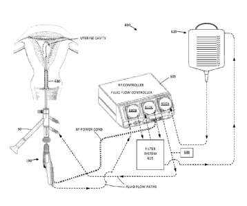
10093] FIGS. 25 and 26 are schematic illustrations and block diagrams of one
embodiment of
fluid management system 600 corresponding to the invention that is configured
for hysteroscopic
use with the probes as described above. As can be seen in FIG. 21, the
hysteroscope 50 and
tissue cutting probe 100 can deliver a cavity-distending fluid to the uterine
cavity as described
previously. In one embodiment, the fluid management system 600 includes a
controller 605 that
carries first, second and third peristaltic pumps 610A, 610B and 610C. The
peristaltic pump can
control pressures throughout the system and provide predetermined flow rates
into the uterine
cavity and outward from the uterine cavity. A predetermined flow rate and/or
pressure can be
used to distend the uterine cavity to thereby allow the physician to view
tissue targeted for
treatment. Of particular interest, the system 600 as shown in FIGS. 25 and 26
eliminates the
need to weigh fluid volumes to determine fluid deficit (and potential
intravasation) which is
found in prior art systems. The fluid management system 600 in FIG. 25
comprises fluid-
in/fluid-out system in which a volume of fluid is recirculated from a fluid
source 620 into the
uterine cavity and then outward from the uterine cavity into a filtering and
sterilization
subsystem 625. After the fluid is filtered and sterilized, it is returned to
the fluid source 620
which is typically a gravity-feed saline bag as illustrated in FIGS. 25 and
26.
10094] In using the fluid management system 600 of FIG. 25, the physician only
needs to
monitor the change in volume of fluid in the saline source or bag 620 to
determine the fluid
deficit. A cervical seal 630 is provided to prevent any substantial saline
leakage outward from
the uterine cavity around the hysteroscope 50. Similarly, the hysteroscope has
a seal 630 in its
working channel to prevent leakage around the shaft of the tissue cutting
probe 100.
[0095] In one embodiment as shown in FIG. 25, the plurality of peristaltic
pumps 610A-610C
are utilized to provide saline inflows into the uterine cavity as well
comprising a negative
pressure source (pump 610B) to withdraw saline and resected tissue from the
uterine cavity.
[0096] As can be seen in FIG. 26, a first peristaltic pump 610A is configured
as a saline inflow
pump and is positioned below the saline bag or source 620. In one variation, a
section of
polyurethane tubing is engaged by the peristaltic pump 610A which can consist
of 3/8" OD; 1/4"
ID tubing. Other flexible inflow tubing 635 not engaged by peristaltic pump
610A can consist of
1/4" OD 1/8" ID tubing of PVC. A pressure sensor 638 is provided downstream
from the
peristaltic pump 610A used for saline infusion. The pressure sensor 638 is
coupled to the
controller 605 and can pressure feedback signals can be used to modulate fluid
inflows into the
uterine cavity.
18
CA 02856989 2014-05-26
WO 2013/067417 PCT/US2012/063406
[0097] Still referring to FIG. 26, it can bee seen that pump 610B is
configured as a negative
pressure pump mechanism to extract fluid and resected tissue through flexible
tubing 640, for
example a 3/8" OD; 1/4" ID tubing rating as a vacuum tubing. In FIG. 22, the
second peristaltic
pump 610B or vacuum pump is provided to withdraw fluid from the probe 100 as
well as drive
the fluid into a first tissue collection filter 650. In one embodiment, the
tissue collection filter
650 is a coarse filter that can have any suitable form factor and can contain
melt spun
polypropylene fibers that provide a lu filtering pore size. In one example,
the filter 650 can be a
McMaster Can product having item number 5165K21 which has a diameter of about
2.5" and a
length of about 9.75". As can be seen in FIG. 26, resected tissue 652 is
collected in the bottom
of the filter assembly 650 for later collection for biopsy purposes.
[0098] The third peristaltic pump 610C comprises a high-pressure pump and is
downstream
from the coarse filter 650. The high-pressure pump 610C is adapted to drive
the coarsely filtered
fluid through a molecular filter 660 which is capable of removing all cells,
blood constituents
and the like in the fluid flow. In one embodiment, the molecular filter 650 is
a Nephros DSU
filter available from Nephros, Inc., 41 Grand Ave., River Edge, NJ 07661. As
can be further
seen in FIG. 22, downstream from the molecular filter 660 is a return flow
tubing 662 that
returns the cleansed and sterilized fluid to the saline source or bag 620.
[0099] Of particular interest, molecular filter 660 is configured to allow re-
infusion of a
distending fluid into the patient. In effect, the molecular filter 660 is
capable of cold sterilization
of the saline or other fluid before it returned to the saline source or bag
620.
[00100] The pressure sensor 638 can be used to measure in-line pressures and
can be used to
modulate the pressure inside the uterus via the controller. In one variation,
the pressure sensor is
air pressure sensor (converted from the water pressure through a balloon
within a pulse
dampener) to measure and control the pressure inside the uterus. In another
embodiment, the
probe 100 or hysteroscope 50 can carry a pressure sensor for measuring uterine
cavity pressure
and can be operatively connected to the controller 605.
[00101] In an other aspect of the invention, referring again to FIG. 25, a
method of use for
cutting tissue from a targeted site in a space or potential space in a
patient's body comprise
utilizing the controller to modulate RF parameters in response to rates of
fluid flow into an out of
the space in the patient's body. This aspect of the invention is enabled by
the fact that a single
controller is provided (i) to control the RF cutting probe and (ii) to control
the saline fluid
inflows and outflow. More in particular, a method of the invention for cutting
tissue in a body
space comprises circulating a fluid though the space with a first flow into
the space and a second
flow out of the space to thereby occupy or distend the space; actuating an RF
probe to perform a
cutting procedure at the site; and modulating an operating parameter of the RF
probe in response
to a rate of the first or second flow.
[00102] Additional aspects of the method of cutting tissue include accessing
the space with an
endoscope, providing a first flow of conductive fluid into the space with the
pump mechanism
19
CA 02856989 2014-05-26
WO 2013/067417 PCT/US2012/063406
comprising a peristaltic pump. A second flow of fluid is provide to move fluid
out of the space
which is again assisted by another peristaltic pump. The non-compliant aspects
of the peristaltic
pumps are important for controlling distending pressure in the body cavity,
for example a uterine
cavity. Further, the fact that the second flow through the probe varies
depending on whether
the cutting window is opened or closed and whether tissue contact is
substantial or insubstantial
make it important to have the capability to adjust an RF parameter in response
to inflows and
outflow, or a derivative parameter such as intra-cavity pressure.
1001031 Thus, in the method described above, the operating parameter of the RF
probe can
comprise RF power applied through the probe to tissue. In another embodiment,
the operating
parameter of the RF probe comprises the movement of an RF cutting component,
which may be
rotational speed of the RF cutting component, the speed of reciprocation of
the RF cutting
component, or axial-rotational oscillation of the RF cutting component. In
another embodiment,
the operating parameter of the RF probe can comprise movement of a non-RF
component of the
working end such as a moveable outer or inner sleeve (or partial sleeve) or
other element for
cleaning tissue from the electrode surface. In another embodiment, the
operating parameter of
the RF probe can comprise a duty cycle or pulse rate of the applied RF energy
or duty cycle of
the movement of the RF cutting component.
1001041 In another aspect of the invention, an operating parameter of the RF
probe comprises a
position of an RF cutting component relative to a tissue-receiving window.
Referring to FIG. 27,
a method corresponding to the invention comprises rotating the inner sleeve
and stopping the
sleeve in the maximum window-open position with the RF delivery off, applying
suction to the
central channel to suction tissue into the window, and then applying RF power
to the bi-polar
electrodes to coagulate tissue stabilized and captured in and about the
window.
[00105] The methods described above are applicable to any space or potential
space in a
patient's body and are particularly suited for fibroid removal from a uterine
cavity or removing
tissue from within a joint.
1001061 In a fibroid treatment, the system utilizes a flow rate for saline
inflows into the uterine
cavity that ranges between about 100 ml/min and 1,600 mFmin. In one
embodiment, the fluid
management system is configured to maintain a selected distending pressure it
the uterine cavity
by modulating only inflow rates provided by a first peristaltic pump
controlled by the controller,
with a constant outflow rate provided by a second peristaltic pump, with
controller algorithms
responsive to RF probe parameters including: (i) the degree of window-open or
window-closed
positions which affects outflow volume; (ii) whether RF power is ON or OFF,
and (iii) the
degree of tissue contact or engagement with the window which can be measured
by impedance
or capacitive signals from the bi-polar RF electrodes or other dedicated
electrodes.
[00107] In another method of the invention for cutting tissue from a targeted
site in a uterine
cavity, the measured or calculated pressure in the cavity can be used to
modulate an operating
parameter. In general, a method comprises circulating a fluid though the space
with a fluid
CA 02856989 2014-05-26
WO 2013/067417 PCT/US2012/063406
inflow into the space and a fluid outflow from the space to thereby occupy or
distend the space,
actuating an RF probe to perform a cutting procedure at the site and
modulating an operating
parameter of an RF probe and/or the fluid management system in response to
fluid pressure in
the space. The operating parameter of the RF probe can be at least one of the
following: applied
RF power, RF pulse rate, RF duty cycle, rotational and/or axial movement of an
RF electrode
component of the probe, rotational and/or axial movement of a non-RF component
of the probe,
fluid inflow into the space, or fluid outflow from the space.
1001081 Another method of treating tissue in a targeted site in a space in a
patient's body,
comprising the steps of positioning the working end of an RF probe in the
space, applying RF
current to tissue from a moving RF electrode to perform a cutting procedure at
the site and
applying RF current to tissue from a non-moving RF electrode to perform a
coagulation
procedure at the site. The system and method include providing and utilizing a
controller to
selectively move or terminate movement of the RF electrode cutting component.
Thus, the
probe suctions tissue into the window of the working end to permit the moving
RF electrode to
cut tissue and optionally suctions tissue into the window of the working end
to permit the non-
moving RF electrode to coagulate tissue.
1001091 It should be appreciated that while an RF source is suitable for
causing explosive
vaporization of the captured fluid volume to expel or extract tissue as
described above, any other
energy source can be used and falls within the scope of the invention, such as
an ultrasound
tranducer, HIFU, a laser or light energy source, a microwave or a resistive
heat source.
1001101 In another embodiment, the probe can be configured with a lumen in
communication
with a remote liquid source to deliver fluid to the interior chamber 240.
1001111 Although particular embodiments of the present invention have been
described above
in detail, it will be understood that this description is merely for purposes
of illustration and the
above description of the invention is not exhaustive. Specific features of the
invention are
shown in some drawings and not in others, and this is for convenience only and
any feature may
be combined with another in accordance with the invention. A number of
variations and
alternatives will be apparent to one having ordinary skills in the art. Such
alternatives and
variations are intended to be included within the scope of the claims.
Particular features that are
presented in dependent claims can be combined and fall within the scope of the
invention. The
invention also encompasses embodiments as if dependent claims were
alternatively written in a
multiple dependent claim format with reference to other independent claims.
21