Note: Descriptions are shown in the official language in which they were submitted.
81780137
MICRCNEEDLE DEVICE HAVING A PEPTIDE THERAPEUTIC AGENT AND AN AMINO ACID,
METHODS OF MAKING AND USING THE SAME
CROSS-REFERENCE To RELATED APPLICATION
This application claims priority to U.S. Application Nos. 61/565,227 and
61/565,247, both filed
November 30, 2011.
BACKGROUND
Transdermal delivery of a therapeutic agent such as a drug through the skin to
the local tissue or
systemic circulatory system without piercing the skin, such as with a
transdermal patch, has been used
successfully with a limited number of therapeutic molecules. The main bairier
to transport of molecules
through the skin is the stratum corneum (the outermost layer of the skin).
Devices including arrays of relatively small structures, sometimes referred to
as microneedles or
micro-pins, have been disclosed for use in connection with the delivery of
therapeutic agents and other
substances through the skin and other surfaces. The devices are typically
pressed against the skin in an
effort to pierce the stratum corneum such that the therapeutic agents and
other substances can pass
through that layer and into the tissues below.
Microneedle devices having a fluid reservoir and conduits through which a
therapeutic substance
may be delivered to the skin have been proposed, but there remain a number of
difficulties with such
systems, such as expense and the ability to make very fine channels that can
reliably be used for fluid
flow.
Microneedle devices having a dried coating on the surface of a microneedle
array have also been
developed. The devices are generally simpler than microneedle devices having
fluid reservoirs and may
directly introduce a therapeutic substance into the skin without the need for
providing reliable control of
fluid flow through very fine channels in the microneedle device.
SUMMARY
A challenging task in the development of peptide therapeutic agents,
particularly polypeptides
and proteins, is addressing physical (e.g., adsorption, aggregation,
denaturation, or precipitation) and
chemical (e.g, hydrolysis, oxidation, acylation, or deamidation) instability,
which may cause loss of
biological activity. It has now been found that the addition of an amino acid
typically enhances the
stability of a peptide therapeutic agent coated on a transdermal delivery
device having an array of skin-
piercing microneedles.
In one aspect, the present disclosure provides a medical device including an
array of
microneedles a coating on or within at least a portion of the microneedles.
The coating includes a peptide
therapeutic agent and an amino acid, wherein the peptide therapeutic agent and
the amino acid either both
- 1 -
CA 2857501 2019-05-09
CA 02857501 2014-05-29
WO 2013/082418
PCT/US2012/067278
have a net positive charge or both have a net negative charge. The coating can
be substantially free of
sorbitol.
In another aspect, a method of making such a medical device is disclosed. The
method typically
includes:
providing an aqueous composition comprising a peptide therapeutic agent, an
amino acid, and a
buffer, wherein the peptide therapeutic agent and the amino acid either both
have a net positive charge or
both have a net negative charge in the aqueous composition;
contacting the microneedles with the composition; and
volatilizing a portion of the aqueous composition to provide a coating on a
least a portion of the
microneedles, wherein the coating comprises at least the peptide therapeutic
agent and the amino acid.
In another aspect, the present disclosure provides a medical device including
an array of
microneedles a coating on or within at least a portion of the microneedles.
The coating includes a peptide
therapeutic agent and histidine. The molar ratio of the histidine to the
peptide therapeutic agent can be
less than 2:1.
In another aspect, a method of making such a medical device is disclosed. The
method typically
includes:
providing an aqueous composition comprising a peptide therapeutic agent,
histidine, and a buffer,
wherein the molar ratio of the histidine to the peptide therapeutic agent is
less than 2:1;
contacting the microneedles with the composition; and
volatilizing a portion of the aqueous composition to provide a coating on a
least a portion of the
microneedles, wherein the coating comprises at least the peptide therapeutic
agent and the histidine.
In another aspect, the present disclosure provides a method of stabilizing a
peptide therapeutic
agent on or within an array of microneedles, the method comprising
incorporating an amino acid into the
array of microneedles. In some embodiments, the peptide therapeutic agent and
the amino acid either
both have a net positive charge or both have a net negative charge. In some
embodiments, the amino acid
is histidine. In some embodiments, the molar ratio of the amino acid (e.g.,
histidine) to the peptide
therapeutic agent is less than 2:1.
In this application, terms such as "a", "an" and "the" are not intended to
refer to only a singular
entity, but include the general class of which a specific example may be used
for illustration. The terms
"a", "an", and "the" are used interchangeably with the term "at least one".
The phrases "at least one of'
and "comprises at least one of' followed by a list refers to any one of the
items in the list and any
combination of two or more items in the list. All numerical ranges are
inclusive of their endpoints and
non-integral values between the endpoints unless otherwise stated.
As used in this specification and the appended claims, the term "or" is
generally employed in its
sense including "and/or" unless the content clearly dictates otherwise. For
example, "a coating on or
within at least a portion of the microneedles" means the coating is on and/or
within at least a portion of
the microneedles.
- 2 -
CA 02857501 2014-05-29
WO 2013/082418
PCT/US2012/067278
Unless otherwise indicated, all numbers expressing feature sizes, amounts, and
physical
properties used in the specification and claims are to be understood as being
modified in all instances by
the term "about." Accordingly, unless indicated to the contrary, the numerical
parameters set forth in the
foregoing specification and attached claims are approximations that can vary
depending upon the desired
properties sought to be obtained by those skilled in the art utilizing the
teachings disclosed herein.
The term "peptide" as used herein refers to peptides, polypeptides, and
proteins. The terms
"peptide", "polypcptide", and "protein" are interchangeable in the context of
the present disclosure.
These terms refer to a molecule having at least two amino acids linked through
peptide bonds. The terms
include oligopeptides, protein fragments, analogs, derivatives (e.g.,
glycosylated derivatives and
pegylated derivatives), and fusion proteins.
All scientific and technical terms used herein have meanings commonly used in
the art unless
otherwise specified. The definitions provided herein are to facilitate
understanding of certain terms used
frequently herein and are not meant to limit the scope of the present
disclosure.
In embodiments where weight percent is based upon total weight of solids,
solids are those
ingredients which are not volatile. For example, the total weight of solids
does not include a volatilizable
carrier (e.g., water or a volatile co-solvent).
The above summary of the present invention is not intended to describe each
disclosed
embodiment or every implementation of the present invention. The description
that follows more
particularly exemplifies illustrative embodiments. In the application,
guidance is provided through lists
of examples, which examples can be used in various combinations. In each
instance, the recited list
serves only as a representative group and should not be interpreted as an
exclusive list.
BRIEF DESCRIPTION OF THE DRAWINGS
The disclosure may be more completely understood in consideration of the
following detailed
description of various embodiments of the disclosure in connection with the
accompanying drawings, in
which:
FIG. 1 is a schematic cross-sectional view of an uncoated microneedle array;
FIG. 2 is a schematic perspective view of a microneedle device in the form of
a patch; and
FIG. 3 is a schematic cross-sectional view of a coated microneedle array.
The figures are not necessarily to scale. Like numbers used in the figures
refer to like
components. However, it will be understood that the use of a number to refer
to a component in a given
figure is not intended to limit the component in another figure labeled with
the same number.
DETAILED DESCRIPTION OF ILLUSTRATIVE EMBODIMENTS
In the following description, reference is made to the accompanying drawings
that form a part
hereof, and in which are shown by way of illustration several specific
embodiments. It is to be
understood that other embodiments are contemplated and may be made without
departing from the scope
- 3 -
CA 02857501 2014-05-29
WO 2013/082418
PCT/US2012/067278
or spirit of the present disclosure. 'The following detailed description,
therefore, is not to be taken in a
limiting sense.
With the development of recombinant DNA technology, a number of peptide
therapeutic agents
have become available for therapeutic use. These agents include octreotide,
leuprolide, parathyroid
hormone, luteinizing hormone releasing hormone, insulin, vascular endothelial
arowth factor, and many
others. One challenging task in the development of peptide therapeutic agents
is addressing physical
(e.g., adsorption, aggregation, denaturation, or precipitation) and chemical
(e.g., hydrolysis, oxidation,
acylation, or deamidation) instability, which may cause loss of biological
activity.
Oxidation is a major chemical degradation pathway of peptide therapeutic
agents. The side
chains of His, Met, Cys, Trp and 'Tyr residues in proteins are potential sites
for oxidation. Some proteins
are very sensitive to light during manufacturing and storage, which can also
result in modification of the
molecules. Both oxygen content and light exposure may cause oxidation and
affect oxidation rate and
promote aggregation or other degradation pathways. Oxidation can alter a
protein's physiochemical
characteristics and led to aggregation or fragmentation, which can negatively
impact potency and
immunogenicity. Acylation is another pathway of instability of peptide
therapeutic agents. Nucleophilic
primary amines, such as at the N-terminus or a lysine side chain, can react
with carboxylate groups to
form acylated peptide adducts. Peptide acylation may cause loss of activity, a
change of receptor
specificity, or immunogenicity. Protein aggregation is an example of physical
instability, and aggregate
formation can lead to loss of biological activity, loss of solubility, and
increased immunogenicity.
These and other degradation pathways can result in loss of activity. It is
therefore desirable to
provide compositions for formulating and delivering peptide therapeutic agents
having enhanced
chemical and physical stability and exhibiting maximal shelf lives. It has now
been found that amino
acids are useful for stabilizing peptide therapeutic agents coated on and/or
within a medical device having
a plurality of skin-piercing microneedles. Without wishing to be bound by
theory, it is believed that the
addition of the amino acid substantially reduces the oxidation, photo-
oxidation, acylation, and
aggregation of peptide formulations.
A number of peptide therapeutic agents may usefully be incorporated into the
medical devices
according to and/or made according to the present disclosure. Exemplary
peptide therapeutic agents
include parathryroid hormone, calcitonin, lysozyme, insulin. glatiramer
acetate, goserelin acetate,
somatostatin , octreotide, leuprolide, vasopressin, thymosin alpha-1, atrial
natriuretic peptide (ANP),
endorphin, growth factors (e.g., vascular endothelial growth factor (VEGE),
fibroblast growth factor
(FGF), erythropoietin(EPO), bone morphoeenetic proteins (BMPs), and epidermal
growth factor(EFG),
granulocyte colony-stimulating factor (G-CSF), granulocyte macrophage colony
stimulating factor (GM-
CSE), insulin-like growth factor (ICE), platelet-derived growth factor
releasing factor), growth hormone
release hormone (GHRH), interferons (e.g., interferon alpha, interferon beta,
and interferon gamma),
antimicrobial peptides, dornase alfa, tissue plasminogen activator, uroldnase,
AMP clearance inhibitors,
luteinizing hormone releasing hormone (LHRH), Melanocyte Stimulating Hormones
(alpha & Beta
- 4 -
CA 02857501 2014-05-29
WO 2013/082418
PCT/US2012/067278
MSH), pituitary hormones (hGH), Adrenocorticotropic hormone (ACTH)õ human
chorionic
gonadotropin, streptokinase , interleukins, menotropins (urofollitropin (FSH)
and LH)), protein C,
protein Sõ angiotensin, anaiogenin, Endothel ins, pentigetide, Brain
natriuretic peptide (BNP),
neuropeptide Y,Islet Amyloid Polypeptide (IAPP), Vasoactive intestinal peptide
(VIP), hirudin,
glucaaon, insulin, insulinotropin analogs and derivatives of any of the
foregoing peptide therapeutic
agents, fusion proteins, and peptide vaccines. Peptide vaccines include those
with an antigen in the form
of a peptide as defined above. Exemplary peptide vaccines include therapeutic
cancer vaccine, anthrax
vaccine, flu vaccine, Lyme disease vaccine, rabies vaccine, measles vaccine,
mumps vaccine, chicken pox
vaccine, small pox vaccine, hepatitis vaccine, hepatitis A vaccine, hepatitis
B vaccine, hepatitis C
vaccine, pertussis vaccine, rubella vaccine, diphtheria vaccine, encephalitis
vaccine, Japanese encephalitis
vaccine, respiratory syncytial virus vaccine, yellow fever vaccine, polio
vaccine, herpes vaccine, human
papilloma virus vaccine, rotavirus vaccine. pneumococcal vaccine, meningitis
vaccine, whooping cough
vaccine, tetanus vaccine, typhoid fever vaccine, cholera vaccine, tuberculosis
vaccine, severe acute
respiratory syndrome (SARS) vaccine, HSV-1 vaccine, HSV-2 vaccine, HIV vaccine
and combinations
thereof. In some embodiments, the peptide vaccine includes at least one of
influenza vaccine, polio
vaccine, hepatitis A vaccine, and cancer vaccine.
A number of amino acids may usefully be incorporated into the medical devices
according to
and/or made according to the present disclosure. Useful amino acids include
naturally occurring amino
acids and synthetic amino acids that are capable of having a net positive
charge or a net negative charge.
Typically, useful amino acids are capable of having a net positive charge or a
net negative charge in a pH
range from 3 to 11. The amino acids may have either L- or D- Fischer
configuration. In some
embodiments, the amino acid is histidine, arginine, lysine, aspartic acid, or
glutamic acid. In some
embodiments, the amino acid is histidine, lysine, or arginine. Such amino
acids can be useful, in some
embodiments, with peptide therapeutic agents having a net positive charge. In
some embodiments, the
amino acid is aspartic acid or glutamic acid. Such amino acids can be useful,
in some embodiments, with
peptide therapeutic agents having a net negative charge. In some embodiments,
the amino acid is
histidine. In the coatings disclosed herein or in a coating formulation, which
may be an aqueous
composition, the amino acid may be in salt form.
In many embodiments, the peptide therapeutic agent and the amino acid either
both have a net
positive charge or both have a net negative charge. In some embodiments, the
peptide therapeutic agent
and the amino acid each have a net positive charge. For example, the peptide
therapeutic agent may be
parathyroid hormone, lysozyme, insulin, or salmon calcitonin, and the amino
acid may be histidine,
arginine, or lysine. In other embodiments, the peptide therapeutic agent and
the amino acid each have a
net negative charge. For example, the peptide therapeutic agent may be
insulin, and the amino acid may
be aspartic acid or glutamic acid. In embodiments where the peptide
therapeutic agent and the amino acid
each have a net positive charge and in embodiments where the peptide
therapeutic agent and the amino
acid each have a net negative charge, it may be said that the peptide
therapeutic agent and the amino acid
- 5 -
CA 02857501 2014-05-29
WO 2013/082418
PCT/US2012/067278
have the same charge or matched charges. That is, the charge on the peptide
therapeutic agent and the
amino acid might be considered the same or matched if the charge is in the
same direction, regardless of
the magnitude of the charge. An amino acid that has a net negative charge or a
net positive charge may
not be said to be a zwitterion (that is, neutral).
The net charges of the peptide therapeutic agent and the amino acid are
typically established in a
formulation that is used to coat the array of microneedles in the methods
described hereinbelow.
'Typically, the peptide therapeutic agent and the amino acid are dissolved or
dispersed in a solvent at an
established pH. For example, the pH may be in a range from 3 to 11, 4 to 10, 6
to 8, or 5.5 to 8.5. In
some embodiments wherein the amino acid is histidine, the pH may be in a range
from 3 to 6, 3 to 5, 3 to
4, or 4 to 5. The formulation can be an aqueous composition that is buffered
at a particular formulation
pH. When a portion of the aqueous composition is volatized after coating the
array of microneedles with
the composition, the peptide therapeutic agent and the amino acid maintain
their net charges and are
incorporated into the coating.
Amino acids having a net charge may be said to be substantially in charged
form since there is
usually equilibrium between charged and neutral species. In some embodiments,
the ratio of the charged
form of the amino acid to the neutral form that may be present can be at least
10:1. In some
embodiments, the ratio of the charged form of the amino acid to the neutral
form is at least 100:1, at least
1000:1, or at least 10,000:1. Such ratios can be determined in solution, for
example, using the difference
between the formulation pH and the pKa of the amino acid side chain. Amino
acids useful for practicing
the present disclosure are considered to have a net positive charge if their
isoelectric points are higher
than the formulation pH. Amino acids useful for practicing the present
disclosure are considered to have
a net negative charge if their isoelectric points are lower than the
formulation pH.
For peptide therapeutic agents, when the isoelectric point is lower than the
pH of the formulation,
it typically is described as having a net negative charge. When the
isoelectric point of the peptide
therapeutic agent is higher than the pH of the formulation, it typically is
described as having a net positive
charge.
In some embodiments, it may be useful to define the net charges of the amino
acid and the
peptide therapeutic agent at physiological pH (e.g., in a range from 7 to
7.4). In these embodiments, if an
isoelectric point of a peptide therapeutic agent is less than about 7, it
typically is described as having a net
negative charge. If an isoelectric point of a peptide therapeutic agent is
greater than about 7, it typically is
described as having a net positive charge.
In contrast to U. S. Pat. App. Pub. No. 2006/0188555 (Cormier et al.), which
suggests that
therapeutic peptide agents should be formulated with particular counterions to
limit fibril formation in a
formulation, it has now been found that formulations and coatings including
peptide therapeutic agent and
amino acids that do not have opposite net charges have useful physical and
chemical stability. In
embodiments where the peptide therapeutic agent and the amino acid either both
have a net positive
- 6 -
CA 02857501 2014-05-29
WO 2013/082418
PCT/US2012/067278
charge or both have a net negative charge, the amino acids cannot be
considered to be counterions for the
peptide therapeutic agents.
In some embodiments, the amino acid is histidine. Histidine may be useful in
coatings containing
a peptide therapeutic agent with either a net positive charge or a net
negative charge. In coatings
disclosed herein, histidine may stabilize a wide variety of peptide
therapeutic agents (e.g., both net
negatively charged and net positively charged) to at 40 C and 96% relative
humidity to a greater extent
that several other amino acids.
In the medical device according to and/or made according to the present
disclosure, the amino
acid may be present in a variety of useful amounts relative to the peptide
therapeutic agent. In some
embodiments, including any one of the above embodiments, the molar ratio of
the amino acid to the
peptide therapeutic agent is in a range from 0.25:1 to 51:1. In some
embodiments, the molar ratio of the
amino acid to the peptide therapeutic agent is in a range from 0.5:1 to 25:1
or 0.5:1 to 20:1.
Advantageously, in many embodiments, a large excess of the amino acid is not
required. In these
embodiments, the molar ratio of the amino acid to the peptide therapeutic
agent may be less than 2:1, less
than 1.5:1, or less than 1:1. For example, the molar ratio of the amino acid
to the peptide therapeutic
agent may be in a range from 0.5:1 to 2:1, 0.5:1 to 1.5:1, or 0.5:1 to 1:1. In
embodiments where molar
ratio of the amino acid to the peptide therapeutic agent may be less than 2:1,
less than 1.5:1, or less than
1:1, the amino acids would generally not be considered to be present in a
sufficient amount to neutralize
the peptide therapeutic agents.
In some embodiments, including any one of the above embodiments, particularly
when the
coating is on an external surface of the microneedles or on an interior
surface of hollow microneedles, the
amino acid is present in an amount of at least 0.1 weight percent based upon
total weight of solids in the
coating, in some embodiments at least 0.5 weight percent, in some embodiments
at least 1 weight percent,
and in some embodiments at least 2 weight percent. In some embodiments, the
amino acid is present in
an amount of up to 25 weight percent, in some embodiments, up to 15 weight
percent, in some
embodiments up to 10, 9, or 8 weight percent, based upon the total weight of
solids in the coating. In
some embodiments, the amino acid is present in an amount of 0.1 weight percent
to 20 weight percent,
0.1 weight percent to 10 weight percent, or 1 weight percent to 8 weight
percent, based upon total weight
of solids in the coating.
In some embodiments, including any one of the above embodiments, particularly
when the
coating is on an external surface of the microneedles or on an interior
surface of hollow microneedles, the
peptide therapeutic agent is present in an amount of at least 10 weight
percent based upon total weight of
solids in the coating, in some embodiments at least 20 weight percent, in
sonic embodiments at least 50
weight percent, and in some embodiments at least 60 weight percent. In some
embodiments, the peptide
therapeutic agent is present in an amount of up to 99.9 weight percent, in
some embodiments, up to 99.5
weight percent, in sonic embodiments up to 99, 95, or 92 weight percent, based
on the total weight of
solids in the coating. In some embodiments, the peptide therapeutic agent is
present in an amount of 10
- 7 -
CA 02857501 2014-05-29
WO 2013/082418
PCT/US2012/067278
weight percent to 99.9 weight percent, 50 weight percent to 99.5 weight
percent, or 50 weight percent to
95 weight percent, based upon total weight of solids in the coating.
The coatings disclosed herein may also contain at least one excipient. An
excipient can function
to maintain the active nature of the peptide therapeutic agent, to facilitate
the performance of a coating
formulation when depositing a coating on the microneedles, to resist
disruption of the coating or the
microneedle structure itself when penetrating the stratum corneum or other
tissue, or a combination
thereof. Exemplary excipients include, for example, buffers, carbohydrates,
polymers, surfactants, non-
volatile non-aqueous solvents, acids, bases, antioxidants, saccharin (e.g.,
saccharin sodium dehydrate),
lipids (e.g., dipalmitoylphosphatidylcholine (DPPC)), and inorganic salts
(e.g., sodium chloride and
potassium chloride).
The amount of the at least one excipient in the coating and therefore in the
coating formulation
used for depositing the coating can vary depending on at least one of the
identity of the components in the
coating formulation, the amount of peptide therapeutic agent and amino acid
desired on the microneedle
array, the type of microneedle array being coated, the shape and location of
the coating on the
microneedle, or other considerations.
In some embodiments, the method of making a medical device including
microneedles according
to the present disclosure includes providing an aqueous composition comprising
a peptide therapeutic
agent, an amino acid, and a buffer. Similarly, aqueous compositions disclosed
herein useful for coating
an array of microncedles typically include the peptide therapeutic agent, the
amino acid, and a buffer. A
buffer generally functions to stabilize the pH of a coating formulation used
for depositing the coating on
the microneedles. The particular buffer to be utilized can depend at least in
part on the particular peptide
therapeutic agent and amino acid that are included in the coating. The pH of
the formulation can, for
example, help to maintain the solubility of the peptide therapeutic agent and
amino acid in the
composition. As described above, the pH also generally controls the charges on
the amino acid and the
peptide therapeutic agent.
A variety of buffers can be useful in the aqueous compositions useful for
practicing the present
disclosure. Exemplary buffers include histidine, phosphate buffers, acetate
buffers, citrate buffers,
glycine buffers, ammonium acetate buffers, succinate buffers, pyrophosphate
buffers, 'Iris acetate (TA)
buffers, and Tris buffers. Buffered saline solutions can also be utilized as
buffers. Exemplary buffered
saline solutions include phosphate buffered saline (PBS), Tris buffered saline
(TBS), saline-sodium
acetate buffer (SSA), and saline-sodium citrate buffer (SSC). In some
embodiments, phosphate buffered
saline is used for the aqueous composition. A wide variety of pH values may be
useful depending, for
example, on the peptide therapeutic agent and the amino acid. In some
embodiments, the pH may be in a
range from 3 to 11, 4 to 10, 6 to 8, or 5.5 to 8.5. In some embodiments, for
example, wherein the amino
acid is histidine, the pH may be in a range from 3 to 6, 3 to 5, or 4 to 5.
It should be understood that in aqueous compositions disclosed herein that
include a peptide
therapeutic agent, an amino acid, and a buffer, a single amino acid cannot
serve as both the amino acid
- 8 -
CA 02857501 2014-05-29
WO 2013/082418
PCT/US2012/067278
component and the buffer. For example, in aqueous compositions including a
peptide therapeutic agent,
an amino acid, and a buffer in which histidine is used as a buffer, another
amino acid is present as the
amino acid component. Furthermore, it should be understood that the amino acid
in the aqueous
compositions useful for practicing the present disclosure does not necessarily
buffer the aqueous
composition.
In some embodiments, the coating comprising the peptide therapeutic agent and
the amino acid or
the aqueous composition disclosed herein further comprises a carbohydrate. The
carbohydrate may be
useful, for example, for stabilizing the aqueous composition containing a
peptide therapeutic agent useful
for coating the microneedles in the method disclosed herein. The carbohydrate
can be a saccharide,
including mono-, di-, and polysaccharides, and may include, for example, non-
reducing sugars such as
raffinose, stachyose, sucrose, and trehalose; and reducing sugars such as
monosaccharides and
disaccharides. Exemplary monosacharides include apiose, arabinose, digitoxose,
fucose, fructose,
galactose, glucose, gulose, hamamelose, idose, lyxose, mannosc, ribose,
tagatosc, sorbitol, xylitol, and
xylose. Exemplary disaccharides include sucrose, trehalose, cellobiose,
gentiobiose, lactose, lactulose,
maltose, melibiose, primeverose, rutinose, scillabiose, sophorose, turanose,
and vicianose. In
embodiments, sucrose, trehalose, fructose, maltose, or combinations thereof
can be utilized. All optical
isomers of exemplified sugars (D, L, and racemic mixtures) are also included
herein. Useful
polysaccharides include starches such as hyclroxyethyl starch, pregelatinized
corn starch, pentastarch,
dextrin, dextran or dextran sulfate, gamma-cyclodextrin, alpha-clyclodextrin,
beta-clyclodextrin,
glucosyl-alpha-cylcodextrin, maltosyl-alpha-cyclodextrin, glucosyl-beta-
cyclodextrin, maltosyl-beta-
cyclodextrin, 2-hydroxy-beta-cyclodextrin, 2-hydroxypropyl-beta-cyclodextrin,
2-hydroxypropyl-gamma-
cyclodextrin, hydroxyethyl-beta-cyclodextrin, methyl-beta-cyclodextrin,
sultobutylether-alpha-
cyclodextrin, sulfobutylether-beta-cyclodextrin, and sulfobutylether-gamma-
cyclodextrin. In
embodiments, hydroxyethyl starch, dextrin, dextran, gamma-clyclodextrin, beta-
cyclodextrin, or
combinations thereof can be utilized. In embodiments, dextrans having an
average molecular mass of
35,000 to 76,000 can be utilized. In some embodiments, the at least one
carbohydrate is a cellulose.
Suitable celluloses include hydroxyethyl cellulose (HEC), methyl cellulose
(MC), microcrystalline
cellulose, hydroxypropyl methyl cellulose (HPMC), hydroxyethylmethyl cellulose
(HEMC),
hydroxypropyl cellulose (HPC), and mixtures thereof.
Advantageously, typically, the aqueous composition and the coating comprising
the peptide
therapeutic agent and the amino acid are stable even in the absence of a
carbohydrate. In some of these
embodiments, the aqueous composition and the coating are substantially free of
a carbohydrate. For
example, the aqueous composition and the coating can be substantially free of
a saccharides, including
mono-, di-, and polysaccharides. For example, the aqueous composition and the
coating may be free of
any of the mono-, di-, and polysaccharides listed above. In some embodiments
of the medical device
according to the present disclosure is substantially free of sorbitol. In some
embodiments of the method
according the present disclosure, the aqueous composition is substantially
free of sorbitol. "Substantially
- 9 -
CA 02857501 2014-05-29
WO 2013/082418
PCT/US2012/067278
free", referrin2 to any carbohydrate listed above or sorbitol, means that a
carbohydrate or sorbitol could
be present but in an amount less than necessary to stabilize the aqueous
composition or the coating.
"Substantially free" of a specific carbohydrate or sorbitol includes being
free of the carbohydrate or
sorbitol and further includes wherein the molar amount of the carbohydrate or
sorbitol is less than the
molar amount of peptide therapeutic agent. In some embodiments, the term
"substantially free" of a
specific carbohydrate or sorbitol means that the amount of the carbohydrate or
sorbitol is less than 4, 3, 2,
or 1 percent by mole or by weight, based on the total amount of solids in the
composition. In some
embodiments, the aqueous composition and/or the coating comprising the peptide
therapeutic agent and
the amino acid are free of carbohydrates. In some embodiments, the aqueous
composition and/or the
coating comprising the peptide therapeutic agent and the amino acid are free
of sorbitol.
In some embodiments, the aqueous composition and the coating disclosed herein
include at least
one surfactant. The at least one surfactant can be amphoteric, cationic,
anionic, or nonionic. Exemplary
suitable surfactants include lecithin, polysorbates ( e.g., polysorbatc 20,
polysorbate 40, and polysorbatc
80), glycerol, sodium lauroamphoacetate, sodium dodecyl sulfate,
cetylpyridinium chloride (CPC),
dodecyltrimethyl anunonium chloride (DoTAC), sodium desoxycholate,
benzalkonium chloride. sorbitan
laurate, and alkoxylated alcohols (e.g., laureth-4). Advantageously, in some
embodiments, surfactants are
not necessary in the coatings and the aqueous compositions disclosed herein.
In some of these
embodiments, the aqueous composition and the coating are substantially free of
surfactant. "Substantially
free of surfactant" refers to being free of surfactant or having up to 1, 0.5,
0.1, or 0.01 percent by weight
surfactant, based on the total solids in the composition or coating.
Non-volatile, non-aqueous solvents may be useful in the aqueous compositions
disclosed herein
and may be present in the resulting coatings. Exemplary suitable non-volatile,
non-aqueous solvents
include propylene glycol, dimethylsulfoxide, glycerin, 1-methyl-2-
pyrrolidinone, and N,N-
dimethylformamide.
The aqueous compositions and the coatings disclosed herein may include at
least one antioxidant.
Exemplary suitable antioxidants include sodium citrate, citric acid, ascorbic
acid, methionine, sodium
ascorbate, and combinations thereof. Advantageously, in some embodiments, such
antioxidants are not
necessary in the coatings and the aqueous compositions disclosed herein. In
some of these embodiments,
the aqueous composition and the coating can have up to 1, 0.5, 0.1, or 0.01
percent by weight of any of
these antioxidants, based on the total solids in the composition or coating.
In some embodiments, the coating or the aqueous composition disclosed herein
includes at least
one polymer. Exemplary useful polymers include polyvinyl pyrrolidone (PVP),
polyethylene glycol
(PEG), polyvinyl alcohol (PVA), and polyethylene glycol sorbitan isostearate.
In some embodiments,
PVPs having a number average molecular weight of 5,000 to 1.5 million may be
useful. In some
embodiments, polyethylene glycols having a number average molecular weight of
300 to 8,000 may be
useful.
- 10-
CA 02857501 2014-05-29
WO 2013/082418
PCT/US2012/067278
In some embodiments, the coating or the aqueous composition disclosed herein
may include a
polypeptide other than the peptide therapeutic agent. The amino acids making
up the polypeptide may be
the same or at least some may he different from each other. Exemplary useful
polyamino acids (the same
amino acids) can include polyhistidine, polyaspartic acid, and polylysine.
In some embodiments of the coatings and aqueous compositions, counterions for
the peptide
therapeutic agent and/or the amino acid are present. Exemplary weak acids
useful for providing
counterions for postively charged peptide therapeutic agents or amino acids
include acetic acid, propionic
acid, pentanoic acid, citric acid, succinic acid, glycolic acid, gluconic
acid, glucuronic acid, lactic acid,
malic acid, pyruvic acid, tartaric acid, tartronic acid, fumaric acid, malonic
acid, butyric acid, crotonic
acid, digylcolic acid, and glutaric acid. Exemplary strong acids useful for
providing counterions for
postively charged peptide therapeutic agents or amino acids include
hydrochloric acid, hydrobromic acid,
nitric acid, sulfonic acid, sulfuric acid, maleic acid, phosphoric acid,
benzene sulfonic acid, and methane
sulfonic acid. Exemplary weak bases useful for providing counterions for
negative charged peptide
therapeutic agents or amino acids include ammonia, morpholine,
monoethanolamine, diethanolamine,
triethanolamine, tromethamine, methylglucamine, and glucosamine. Exemplary
strong bases useful for
providing counterions for negative charged peptide therapeutic agents or amino
acids include sodium
hydroxide, potassium hydroxide, calcium hydroxide, and magnesium hydroxide.
The method of making a medical device comprising microneedles according to the
present
disclosure includes providing an aqueous composition comprising a peptide
therapeutic agent, an amino
acid, and, in some embodiments, a buffer. Accordingly, the present disclosure
also provides an aqueous
composition suitable for coating an array of microneedles. The aqueous
composition includes a peptide
therapeutic agent, an amino acid, and optionally a buffer. The amounts of
these ingredients in the
composition are chosen in order to achieve the above described amounts of the
solid, non-volatile
ingredients in the resulting coating deposited on the microneedles. The
aqueous composition may further
include any of the excipients described above. The coating is deposited on the
microneedles by
contacting the microneedles with the composition.
In addition to water, which serves as a volatilizable carrier, the aqueous
composition can also
include at least one co-solvent (which may also be a volatilizable carrier).
Exemplary useful co-solvents,
which may be volatilizable carriers, include ethanol, isopropanol, methanol,
propanol, and butanol. C1_4
ethers, C14 ketones, and C14 esters, for example, may also be useful. Useful
volatile co-solvents are
typically those having a boiling point up to 120 C, in some embodiments up to
100 C. Non-volatile co-
solvents may also be included as described above. Generally, the solvent in
the coating formulation is
selected such that it may dissolve or disperse the peptide therapeutic agent,
the amino acid, and any
cxcipients that may be present. The aqueous compositions can have an overall
solids content from 5% to
80% by weight, from 10% to 70% by weight, or from 50% to 70% by weight, based
on the total weight of
the aqueous composition.
-11 -
CA 02857501 2014-05-29
WO 2013/082418
PCT/US2012/067278
Aqueous compositions useful for depositing the coating on the microneedles can
be designed to
have a desired viscosity, surface tension, and/or contact angle of the aqueous
composition on the material
comprising the microneedles.
The viscosity of the aqueous composition can be an important factor for
providing a desired
amount of uniform coatings on the microneedles. The desired viscosity of the
aqueous composition can
depend at least in part on at least one of the geometry of the microneedles,
the particular coating method
being utilized, and the desired number of coating steps, among other factors.
In some embodiments, the
aqueous composition has a shear viscosity in a range from 500 to 30,000
centipoise (cps) (in some
embodiments, in a range from 500 to 10,000 cps or 500 to 8,000 cps) when
measured at a shear rate of
100s at a temperature of 25 C. The shear viscosity is a measurement of the
resistance of a fluid to being
deformed by shear stress. Various instruments can be used for viscosity
testing, including rheometers, for
example rheometers from TA Instruments (New Castle, DE).
The surface tension of the aqueous composition can be an important factor for
providing a desired
amount of material on the microneedles without excessive spreading along the
needle or onto the
microneedle substrate. The desired surface tension of the aqueous composition
can depend at least in part
on at least one of the geometry of the microneedles, the particular coating
method being utilized, and the
desired number of coating steps, among other factors. In some embodiments, the
aqueous composition
has a surface tension (measured at ambient, or room temperature conditions) up
to 60 dynes/cm, in some
embodiments, up to 55 dynes/cm. In some embodiments, the aqueous composition
has a surface tension
in a range from 40 dynes/cm to 55 dynes/cm. The surface tension can be
determined using the pendant
drop method. In a pendant drop method of measuring surface tension, a drop of
liquid is suspended from
the end of a tube by surface tension. The force due to surface tension is
proportional to the length of the
boundary between the liquid and the tube. Various instruments that encompass
optical systems for
measuring the relevant parameters of the drop and software packages for
calculating the surface tension
based on the measured parameters can be utilized herein. An exemplary
instrument includes the Drop
Shape Analysis System (Model DSA 100S) available from Kriiss (Hamburg,
Germany).
The contact angle of the aqueous composition on the material comprising the
microneedles (also
referred to as the "microneedle material") can be an important factor for
providing a desired amount of
material on the microneedles without excessive spreading along the needle or
onto the microneedle
substrate. The desired contact angle of the aqueous composition on the
microneedle material can depend
at least in part on at least one of the composition of the microneedles, the
geometry of the microneedles,
the particular coating method being utilized, and the desired number of
coating steps, among other
factors. In some embodiments, the aqueous composition has a contact angle
(measured at ambient, or
room temperature conditions) with the microneedle material of at least 50
degrees, at least 55 degrees, or
at least 65 degrees. The contact angle of the aqueous composition on the
microneedle material can be
measured using various methods, for example, using the sessile drop method.
Generally, a goniometer
(or an instrument that employs a goniometer) can be utilized to measure
contact angles; an example of
- 12-
81780137
such an instrument is the Drop Shape Analysis System (Model DSA 100S)
available from Krtiss
(Hamburg, Germany). In embodiments, the contact angle can be measured within 5
seconds of the
transfer of the aqueous composition onto the substrate (microneedle material).
The contact angle of the
aqueous composition with respect to the microneedle material is measured on a
horizontal substrate made
of the microneedle material.
The microneedle material can be (or include) silicon or a metal such as
stainless steel, titanium, or
nickel titanium alloy. The microneedle material can also be (or include) a
medical grade polymeric
material. In some embodiments, including any one of the above embodiments, the
microneedle material
can be a medical grade polymeric material. Exemplary types of medical grade
polymeric materials
include polycarbonate and liquid crystalline polymer (referred to herein as
"LCP").
Generally, an "array" refers to medical devices described herein that include
more than one (in
embodiments, a plurality) structure capable of piercing the stratum corneum to
facilitate the transdermal
delivery of the peptide therapeutic agent and amino acid to the skin. The
terms "microstructure" and
"microneedle" refer to the structures associated with an array that are
capable of piercing the stratum
corneum to facilitate the transdermal delivery of the peptide therapeutic
agent and amino acid to the skin.
By way of example, microstructures can include needle or needle-like
structures as well as other
structures capable of piercing the stratum corneum. The term "microneedle
array" or "array of
microneedles" therefore can refer to a plurality of structures that are
capable of piercing the stratum
corneum to facilitate the transdermal delivery of the peptide therapeutic
agent and amino acid to the skin.
.Microneedle arrays useful for practicing the present disclosure can have a
variety of
configurations, such as those described in the following patents and patent
applications.
One embodiment for the microneedle arrays includes the structures disclosed in
U. S. Patent Application Publication No. 2005/0261631 (Clarke et al.), which
describes
microneedles having a truncated tapered shape and a controlled aspect ratio. A
further
embodiment for the microneedle arrays includes the structures disclosed in
U.S. Patent No. 6,881,203
(Delmore et al.), which describes tapered microneedles with at least one
channel formed on the outside
surface. Another embodiment for the microneedle arrays includes the structures
disclosed in Int. App.
Pub. Nos. W02011/014514 (Gonzalez et al.) and W02010/059065 (Burton et al.),
which both describe
hollow microneedles. For hollow microneedles, either the concave surface, the
convex surface, or both
may include the coating disclosed herein. A coating on the concave surface may
be considered "within"
the microneedles. In some embodiments, the microneedles are solid microneedles
(that is, the
microneedles are solid throughout).
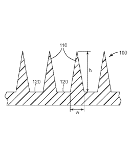
Generally, a microneedle array includes a plurality of microneedles. FIG. 1
shows a portion of a
microneedle array 100 that includes four microneedles 110 (of which two are
referenced in FIG. 1)
positioned on a microneedle substrate 120. Each microneedle 110 has a height
h, which is the length
from the tip of the microneedle 110 to the mieroneedle base at substrate 120.
Either the height of a single
microneedle or the average height of all microneedles on the microneedle array
can be referred to as the
- 13 -
CA 2857501 2019-05-09
CA 02857501 2014-05-29
WO 2013/082418
PCT/US2012/067278
height of the microneedle, h. In some embodiments, including any one of the
embodiments described
herein, each of the plurality of microneedles (or the average of all of the
plurality of microneedles) has a
height of about 100 to 1200 micrometers (gm), in some embodiments about 200 to
1000 gm, or about 200
to 750 gm. In some embodiments, including any one of the embodiments described
herein, the array of
microneedles contains 200 to 1500 microneedles per cm2 of the array of
microneedles.
A single microneedle or the plurality of microneedles in a microneedle array
can also be
characterized by their aspect ratio. "[he aspect ratio of a microneedle is the
ratio of the height of the
microneedle, h, to the width (at the base of the microneedle), w (as seen in
FIG. 1). The aspect ratio can
be presented as h:vv. In some embodiments, including any one of the
embodiments described herein, each
of the plurality of microneedles (or the average of all of the plurality of
microneedles) has (have) an
aspect ratio in the range of 2:1 to 5:1. In some of these embodiments, each of
the plurality of
microneedles (or the average of all of the plurality of microneedles) has
(have) an aspect ratio of at least
3:1.
A microneedle or the plurality of microneedles in a microneedle array useful
for practicing the
present disclosure can have a variety of shapes. In some embodiments,
including any one of the
embodiments described herein, each of the plurality of microneedles can have a
square pyramidal shape
or the shape of a hypodermic needle. In some of these embodiments, the shape
is square pyramidal.
In some embodiments, including any one of the embodiments described herein,
the medical
device according to the present disclosure may be in the form of a patch. One
example of such an
embodiment is shown in more detail in FIG. 2. FIG. 2 illustrates a medical
device comprising a patch 20
in the form of a combination of a microneedle array 22, pressure sensitive
adhesive 24 and backing 26.
Such a patch 20 or another device including multiple microneedle arrays or
multiple patches 20 can be
referred to as a delivery device. The microneedle array 22 is illustrated with
microneedles 10 protruding
from a microneedle substrate 14. The microneedles 10 may be arranged in any
desired pattern or
distributed over the microneedle substrate 14 randomly. As shown, the
microneedles 10 are arranged in
uniformly spaced rows. In some embodiments, including any one of the
embodiments described herein,
microneedle arrays can have a surface area on the non-structured surface of
more than about 0.1 cm2 and
less than about 20 cm2. In some of these embodiments, the microneedle array
area is at least about 0.5
cm2 and up to about 5 cm-. In one embodiment (not shown), a portion of the
substrate 14 of the patch 20
is not provided with microneedles (that is, it is non-structured). In some of
these embodiments, the non-
structured surface has an area of more than about 1 percent and less than
about 75 percent of the total area
of the device surface that faces a skin surface of a patient. In another of
these embodiments, the non-
structured surface has an area of more than about 0.10 square inch (0.65 cm2)
to less than about 1 square
inch (6.5 cm2). In another embodiment (shown in FIG. 2), the microneedles are
disposed over
substantially the entire surface area of the array 22, such that there is
essentially no non-structured area.
In the method of making a medical device described herein, contacting the
microneedles with the
aqueous composition can be carried out by dip coating the microneedles. Such
methods are described, for
- 14 -
81780137
example, in U.S.- Pat, App. Pub!. No. 2008/0051699 (Choi etal.),
particularly with reference
to FIGS. 10A, 1013, and 10C therein.
When dip coating, wasting peptide therapeutic agent and amino acid is avoided
by contacting
only a portion of the microneedle height with the aqueous composition and
avoiding contact with the
microneedle substrate. FIG. 3 illustrates, in cross-section, a portion of a
microneedle array 200 that
includes four microneedles 210 (of which two are referenced in FIG. 3)
positioned on a microneedle
substrate 220. Coating 250 is disposed on mieroneedles 210 at a distance 260
from the tip of the
microneedles. This is accomplished by contacting not more than a portion of
the microneedle height with
the aqueous composition. Accordingly, in some embodiments, including any one
of the method
.. embodiments described herein that includes contacting the microneedles with
the aqueous composition,
the microneedles each have a tip and a base, the tip extending a distance (h)
from the base, and contacting
is carried out by contacting the tips of the microneedles and a portion of the
microneedles extending not
more than 90 percent of the distance (0.91i) from the tips to the bases with
the composition, in sonic,
embodiments not more than 70 percent of the distance (0.7h), or not more than
50 percent of the distance
(0.5h). It is to be understood that the distance can apply to a single
mi.croneedle or to an average of the
microneedles in an array. In some embodiments, including any one of the
embodiments described herein
which includes a coating disposed on the microneedles, at least 50% of the
microneedles have the coating
present on the microncedles near the tip and extending not more than 90
percent of the distance toward
the base, preferably not more than 70 percent of the distance, more preferably
not more than 50 percent of
the distance.
In some embodiments, when the microneedles are contacted with the aqueous
composition, the
microneedles are facing downward into the aqueous composition. In some of
these embodiments, after
the microneedles are contacted with the aqueous composition, contacting is
terminated and the
microneedles are positioned facing upward before and/or during volatilizing a
portion of the aqueous
composition. In this position, a portion of the aqueous composition remaining
on the microneedles may
flow toward the base, leaving the tips of the microneedles exposed or with
only as small amount of
coating on the tips. The degree to which flow occurs can depend upon factors
such as the viscosity,
contact angle, and surface tension as described above.
After removing the microneedles from the aqueous composition, some of the
coating formulation
remains on the microneedles, the amount depending upon the aqueous composition
properties and surface
properties of the microneedle material as described above. At least a portion
of the water is removed
from the aqueous composition adhering to the microneedles, leaving the coating
disposed on the
microneedles. One or more additional contacting steps may be used. The shape
of the coating, average
coating thickness, and amount of the surface of the microneedle covered by the
coating depends upon the
factors discussed above as well as the number of times the contacting step is
repeated.
FIG. 3 illustrates one embodiment with the coating disposed on the
microneedles, wherein the
tips of the microneedles are essentially exposed (no coating or a relatively
small amount of coating) a
- 15 -
CA 2857501 2019-05-09
CA 02857501 2014-05-29
WO 2013/082418
PCT/US2012/067278
distance 270 from the tip. In some embodiments, including any one of the
embodiments described herein
which includes a coating disposed on the microneedles, the tips of the
microneedles are exposed or only
as small amount of coating is on the tips. In some of these embodiments
distance 270 is at least 1 percent
(0.1h), 3 percent (0.03h) or 6 percent (0.06h) of the distance from the tip to
the base. In some of these
embodiments, distance 270 is at most 10 percent (0.1h) of the distance from
the tip to the base.
In some embodiments, including any one of the embodiments described herein
which includes a
coating disposed on or within the microneedles, the coating is present on the
microneedles in an average
amount of 0.01 to 2 micrograms per microneedle. Coating weight can be
determined by weighing the
microneedle array before and after the coating is disposed on the microneedles
and dividing the difference
by the number of microneedles in the array. This measurement can be made once
the coated microneedle
array has come to a constant weight, indicating that the water and any other
volatilizable carrier has been
sufficiently removed, before taking the weight after coating. Alternatively,
the total amount of a solid
component in the coating on all the microneedles of the entire array can be
determined analytically and
then the total weight of solids calculated based upon the know weight of all
solid components used in the
aqueous composition.
Volatilizing the water and any other carrier can be performed using various
means including for
example, drying at ambient conditions; drying at conditions other than ambient
conditions (such as
temperatures other than room temperature or a humidity other than an average
humidity); drying for
various times; drying with heat, lyophilization, freeze drying; other similar
techniques; or combinations
thereof.
Once a portion of the aqueous composition (which may be a portion or all of
the water or non-
aqueous solvent) in the aqueous composition has evaporated (either after a
single contacting step or
multiple contacting steps), the aqueous composition on the microneedle array
can be referred to as the
"coating" as described above. A coating as described herein can generally be
referred to as a dried
coating or a solid coating.
In some embodiments, a medical device according to the present disclosure can
include an array
of dissolvable microneedles. The dissolvable microneedles may contain the
peptide therapeutic agent and
the amino acid in the various amounts described above for coatings disposed on
the microneedles.
Dissolvable microneedles further include a dissolvable matrix material. The
dissolvable matrix material
may be any solid material which dissolves sufficiently in the tissue
underlying the stratum corneum to
allow the peptide therapeutic agent and amino acid to be released into the
tissue. In some embodiments,
the dissolvable matrix material is selected from the group consisting of
hyaluronic acid,
carboxymethylcellulose, hydroxpropylmethylcellulose, methylcellulose,
polyvinyl alcohol, polyvinyl
pyrrolidone, sucrose, glucose, dextran, trehalose, maltodextrin, and a
combination thereof.
Dissolvable microneedle arrays may be fabricated by casting and drying a
solution containing
volatilizable carrier and dissolvable matrix material (preferably water
soluble) in a mold containing the
microstructured cavities. The internal shape of the microstructured cavities
corresponds to the external
- 16-
CA 02857501 2014-05-29
WO 2013/082418
PCT/US2012/067278
shape of the dissolvable microncedles. The mold can be comprised of materials
such as
polydimethylsiloxane (PDMS) or other plastics that do not permanently bind to
or that have low adhesion
to materials used to make the dissolvable microneedles.
The peptide therapeutic agent and amino acid component can be incorporated
into dissolvable
microneedles by first loading a solution of these components with a
volatilizable carrier (preferably also
including the dissolvable matrix material) into the mold containing
microstructured cavities. After at
least partially drying (volatilizing at least a portion of the volatilizable
carrier), the mold is filled with a
solution of dissolvable matrix material (without the peptide therapeutic agent
and amino acid), followed
by drying. Alternatively, in a one-step process, the peptide therapeutic agent
and the amino acid can be
combined with the dissolvable matrix material in a solution with the
volatilizable carrier and the mold
filled with this solution, followed by drying. The volatilizable carriers can
include water or any of the
volatile non-aqueous solvents (e.g., ethanol) described above. Drying can be
carried out using any of the
techniques described above.
In embodiments including dissolvable microneedles, the coating comprising the
peptide
therapeutic agent and the amino acid may be considered to be within a least a
portion of the microneedles.
Application of the microneedle device may be carried out by contacting the
tissue of a subject
with the microneedles and applying hand pressure to force the microneedles
into the tissue. Alternatively,
an application device may be used which applies the pressure, forcing the
microneedles into the tissue.
This can provide a more even distribution of pressure and force the
microneedles into the tissue at an
optimum velocity so that essentially all of the microneedles can release the
peptide therapeutic agent into
the tissue. In some embodiments, contacting the tissue with a microneedle
device is carried out at a
microneedle velocity of 5 to 10 meters/second. The "subject" can include
humans, sheep, horses, cattle,
pigs, dogs, cats, rats, mice, or other mammals.
Some Embodiments of the Disclosure
1. A medical device comprising:
an array of microneedles; and
a coating on or within at least a portion of the microneedles, wherein the
coating comprises:
a peptide therapeutic agent; and
an amino acid,
wherein the peptide therapeutic agent and the amino acid either both have a
net positive charge or both
have a net negative charge, and wherein the coating is substantially free of
sorbitol.
2. The medical device of embodiment 1, wherein the molar ratio of the amino
acid to the peptide
therapeutic agent is less than 2:1.
- 17 -
CA 02857501 2014-05-29
WO 2013/082418
PCT/US2012/067278
3. The medical device of embodiment 1, wherein the molar ratio of the amino
acid to the peptide
therapeutic agent is in a range from 0.5:1 to 55:1.
4. 'The medical device of any one of embodiments 1 to 3, wherein the amino
acid is histidine,
arginine, lysine, aspartic acid, or glutamic acid.
5. The medical device of embodiment 4, wherein the amino acid is histidine.
6. The medical device of any one of embodiments 1 to 5, wherein the peptide
therapeutic agent and
the amino acid each have a net positive charge.
7. The medical device of any one of embodiments 1 to 4, wherein the peptide
therapeutic agent and
the amino acid each have a net negative charge.
8. The medical device of any one of embodiments 1 to 7, wherein the amino
acid is present in the
coating in a range from 0.1 to 15 percent by weight, based on the total weight
of the coating.
9. The medical device of any one of embodiments 1 to 8, wherein the array
of microneedles
comprises a dissolvable matrix material.
10. The medical device of any one of embodiments 1 to 8, wherein at least
some of the microneedles
are hollow.
11. A medical device comprising:
an array of microneedles; and
a coating on or within at least a portion of the microneedles, wherein the
coating comprises:
a peptide therapeutic agent: and
histidine,
wherein the molar ratio of the histidine to the peptide therapeutic agent is
less than 2:1.
12. The medical device of embodiment 11, wherein the molar ratio of the
histidine to the peptide
therapeutic agent is less than 1.5:1.
13. The medical device of embodiment 11 or 12, wherein the peptide
therapeutic agent has a net
positive charge.
-18-
CA 02857501 2014-05-29
WO 2013/082418
PCT/US2012/067278
14. The medical device of embodiment 11 or 12, wherein the peptide
therapeutic agent has a net
negative charge.
15. The medical device of any one of embodiments 11 to 14, wherein
histidine is present in the
coating in a range from 0.1 to 15 percent by weight, based on the total weight
of the coating.
16. The medical device of any one of embodiments 11 to 15, wherein the
histidine stabilizes the
peptide therapeutic agent in the coating.
17. The medical device of any preceding embodiment, wherein the peptide
therapeutic agent is
parathryroid hormone, calcitonin, lysozyme, insulin, glatiramer acetate,
goserelin acetate, octreotide,
leuprolide, vasopressin, atrial natriuretic peptide(ANP), vascular endothelial
growth factor (VEGF),
fibroblast growth factor (FGE), erythropoietin(EPO), bone morphogenetic
proteins(BMPs), epidermal
growth factor(EFG), granulocyte clony-stimulating factor (G-CSF), granulocyte
macrophage colony
stimulating factor (GM-CSF), interferon alpha, interferon beta, interferon
gamma, antimicrobial peptides,
dornasc alfa, tissue plasminogen activator, a fusion protein, or a vaccine.
18. The medical device of any preceding embodiment, wherein the coating is
present on the
microneedles in an average amount of 0.01 to 2 micrograms per microneedle.
19. A method of making a medical device comprising microneedles, the method
comprising:
providing an aqueous composition comprising a peptide therapeutic agent, an
amino acid, and a
buffer, wherein the peptide therapeutic agent and the amino acid either both
have a net positive charge or
both have a net negative charge in the aqueous composition;
contacting the microneedles with the aqueous composition; and
volatilizing a portion of the aqueous composition to provide a coating on at
least a portion of the
microneedles, wherein the coating comprises at least the peptide therapeutic
agent and the amino acid.
20. The method of embodiment 19, wherein the buffer comprises phosphate,
acetate, citrate, or
tris(hydroxymethyl)aminomethane.
21. A method of stabilizing a peptide therapeutic agent in a coating on an
array of microneedles, the
method comprising incorporating an amino acid into the coating, wherein the
peptide therapeutic agent
and the amino acid either both have a net positive charge or both have a net
negative charge.
22. The method of any one of embodiments 19 to 21, wherein the molar ratio
of the amino acid to the
peptide therapeutic agent is less than 2:1.
- 19-
CA 02857501 2014-05-29
WO 2013/082418
PCT/US2012/067278
23. The method of any one of embodiments 19 to 21, wherein the molar
ratio of the amino acid to the
peptide therapeutic agent is in a range from 0.5:1 to 55:1.
24. The method of any one of embodiments 19 to 23, wherein the amino acid
is histidine, arginine,
lysine, aspartic acid, or glutamic acid.
25. The method of embodiment 24, wherein the amino acid is histidine.
26. The method of any one of embodiments 19 to 25, wherein the peptide
therapeutic agent and the
amino acid each have a net positive charge.
27. The method of any one of embodiments 19 to 25, wherein the peptide
therapeutic agent and the
amino acid each have a net negative charge.
28. The method of any one of embodiments 19 to 27, wherein the amino acid
is present in the coating
in a range from 0.1 to 15 percent by weight, based on the total weight of the
coating.
29. The method of any one of embodiments 19 to 28, wherein the aqueous
composition has a pH in a
range from 3 to 11.
30. The method of any one of embodiments 19 to 39, wherein the aqueous
composition or the coating
is substantially free of sorbitol.
31. A method of making a medical device comprising microneedles, the method
comprising:
providing an aqueous composition comprising a peptide therapeutic agent,
histidine, and a buffer,
wherein the molar ratio of the histidine to the peptide therapeutic agent in
the aqueous composition is less
than 2:1;
contacting the microneedles with the aqueous composition; and
volatilizing a portion of the aqueous composition to provide a coating on at
least a portion of the
microneedles, wherein the coating comprises at least the peptide therapeutic
agent and the histidine.
32. The method of embodiment 31, wherein the aqueous composition has a pH
in a range from 3 to
11.
33. The method of embodiment 31 or 32, wherein the molar ratio of histidine
to the peptide
therapeutic agent is less than 1.5:1.
- 20 -
CA 02857501 2014-05-29
WO 2013/082418
PCT/US2012/067278
34. The method of embodiment 31, 32, or 33, wherein the peptide
therapeutic agent has a net positive
charge.
35. The method of embodiment 31, 32, or 33, wherein the peptide therapeutic
agent has a net
negative charge.
36. The method of any one of embodiments 31 to 35, wherein histidine is
present in the coating in a
range from 0.1 to15 percent by weight, based on the total weight of the
coating.
37. The method of any one of embodiments 31 to 36, wherein buffer comprises
phosphate, acetate,
citrate, or tris(hydroxymethyBaininomethane.
38. The method of any one of embodiments 19 to 37, wherein the wherein the
peptide therapeutic
agent is parathryroid hormone, calcitonin, lysozyme, insulin, glatiramer
acetate, goserelin acetate,
octrcotide, leuprolide, vasopressin, atrial natriuretic peptide(ANP), vascular
endothelial growth factor
(VEGF), fibroblast growth factor (FGF), erythropoietin(EPO), bone
morphogenetic proteins(BMPs),
epidermal growth factor(EFG), granulocyte clony-stimulating factor (G-CSF),
granulocyte macrophage
colony stimulating factor (GM-CSF), interferon alpha, interferon beta,
interferon gamma, antimicrobial
peptides, dornase alfa, tissue plasminogen activator, a fusion protein, or a
vaccine.
39. The method of any one of embodiments 19 to 38, wherein the coating is
present on the
microneedles in an average amount of 0.01 to 2 micrograms per microneedle.
40. A method of making a medical device comprising microneedles according
to any one of
embodiments 1 to 10, the method comprising:
providing an aqueous composition comprising the peptide therapeutic agent and
the amino acid;
contacting the microneedles with the aqueous composition; and
volatilizing a portion of the aqueous composition to provide a coating on at
least a portion of the
microneedles, wherein the coating comprises at least the peptide therapeutic
agent and the amino acid.
41. A method of making a medical device comprising microneedles
according to any one of
embodiments 11 to 16, the method comprising:
providing an aqueous composition comprising the peptide therapeutic agent and
the histidine;
contacting the microneedles with the aqueous composition; and
volatilizing a portion of the aqueous composition to provide a coating on at
least a portion of the
microneedles, wherein the coating comprises at least the peptide therapeutic
agent and the histidine.
-21 -
CA 02857501 2014-05-29
WO 2013/082418
PCT/US2012/067278
The following examples are provided to more particularly illustrate various
embodiments of the
present invention, but the particular materials and amounts thereof recited in
these examples, as well as other
conditions and details are in no way intended to limit this invention.
EXAMPLES
Materials
The microneedle arrays were injection molded using VECTRA MT 1300
thermoplastic liquid
crystal polymer (LCP) (Ticona Engineering Polymers, Florence, KY). The
microneedle arrays featured
four-sided pyramidal shaped microneedles having heights of about 500 microns
and an aspect ratio of
approximately 3:1. The microneedles were arranged in an octagon shaped pattern
of about 316
microneedles with equal spacing between individual microneedles of about 550
microns (as measured
from tip to tip).
PTH, parathyroid hormone (1-34) (human), was obtained as the acetate salt from
Bachem,
Torrence, CA. Salmon calcitonin and lysozme were obtained from Calbiochem,
LaJolla, CA. Insulin
was obtained from Sigma-Aldrich, St. Louis, MO.
L-Histidine monohydrochloride (His-HC1) was obtained from J.T. Baker,
Phillipsburg, NJ.
L-Arginine hydrochloride (Arg-HC1) was obtained from Spectrum Chemical, New
Brunswick,
NJ.
Phosphate buffered saline (1X PBS) was obtained from Amresco LLC, Solon, OH.
The protein or polypeptide content in the formulation coated on the
microneedle arrays was
determined using an Agilent 1100 HPLC system (Agilent Technologies,
Wilmington, DE) equipped with
a binary pump, well-plated thermostated autosampler, thermostated column
compartment, and a diode
array UV detector. A Zorbax 300SB-C8 column (Agilent Technologies, Wilmington,
DE) having a 5 Inn
particle size and 2.1x150 mm inner diameter was used for the separations. The
column was maintained at
50 C. The mobile phase consisted of two eluents. Eluent A was 0.1% TFA
(trifluoroacetic acid) in water
and eluent B was 0.1% TFA in acetonitrile (Spectrum Chemical, New Brunswick,
NJ). A linear gradient
from 80/20 to 50/50 (A/B) was applied over 30 minutes. The flow rate was 0.4
mL/minute and the UV
detection wavelength was set at 214 nm. The total run time was 34 minutes and
the sample injection
volume was 15 pi__
The pH of the formulations was determined using pH color indicating strips
(available from EMD
Chemicals, Gibbstown, NJ under the trade designation "COLORpHAST").
- 22 -
CA 02857501 2014-05-29
WO 2013/082418
PCT/US2012/067278
Example 1
A sample formulation containing 10 mg/mL of PTH and 12 mg/mL of L-histidine
monohydrochloride (His-HC1) in phosphate buffered saline (PBS) was prepared.
The pH of the sample
formulation was 5.5-6Ø A control formulation was also prepared containing 10
mg/mL of PTH in PBS.
The control formulation did not have His-HC1 in the formulation. The pH of the
control formulation was
6Ø Microneedle arrays were coated with either the sample or the control
formulation. The coating was
applied by dropwise addition of 50 Ift of the formulation to the portion of
the array defined by the
octagon shaped pattern of microncedles. The flood coated arrays (both sample
and control formulation
coated) were dried in an oven at 35 C for about 15 hours.
After drying, the coated arrays were placed in a storage chamber that was
maintained at 40 C and
96 % relative humidity (RH). The PTH content of the coated arrays was
determined following storage in
the chamber for 1, 3, 7, and 14 days. At the designated time point, an array
was taken from the chamber
and washed with PBS (2mL) to remove the coating from the array. An aliquot of
the resulting wash
solution was analyzed for PTH content using the HPLC method described above.
Reference standards
were prepared by measuring the initial PTH content of freshly coated arrays
(both sample and control
formulation coated). The arrays used as reference standards were stored in a
refrigerator at about 4 C
and analyzed within one day of being prepared. At each time point, the
percentage of undegraded PTH
remaining in the sample or control coatings was determined by measuring the
peak area for PTH in the
selected coating and dividing it by the peak area measured for PTH in the
corresponding reference
standard. The results are reported in rf able 1.
Table 1.
Percent of Initial PTH Content in the Coating
Amino Acid
Initial Day 1 Day 3 Day 7 Day 14
None (control) 100 1.3 0 0 0
L-Histidine 100 85.2 60.3 23.1 11.0
Example 2
The same procedure as described in Example 1 was used with the exception that
the sample
formulation contained 12 mg/mL of L-arginine hydrochloride, instead of L-
histidine monohydrochloride.
The pII of the sample formulation was 6Ø The results are reported in Table
2.
Table 2.
Percent of Initial PTH Content in the Coating
Amino Acid
Initial Day 1 Day 3 Day 7 Day 14
None (control) 100 1.3 0 0 0
L-Arginine 100 3.5 0.8 0.4 0
-23 -
CA 02857501 2014-05-29
WO 2013/082418
PCT/US2012/067278
Example 3
A sample formulation containing 5 mg/mL of salmon calcitonin and 12 mg/mL of L-
histidine
monohydrochloride (His-HCI) in PBS was prepared. The pH of the sample
formulation was 5.5. A
control formulation was also prepared containing 5 mg/mL of salmon calcitonin
in PBS. The control
formulation did not have His-HC1 in the formulation. The pH of the control
formulation was 6.0-6.5.
Microneedle arrays were coated with either the sample or the control
formulation. The coating was
applied by dropwise addition of 501.IL of the formulation to the portion of
the array defined by the
octagon shaped pattern of microneedles. The flood coated arrays (both sample
and control formulation
coated) were dried in an oven at 35 C for about 15 hours.
After drying, the coated arrays were placed in a storage chamber that was
maintained at 40 C and
96 % relative humidity (RH). "[he salmon calcitonin content of the coated
arrays was determined
following storage in the chamber for 1, 3, 7, and 14 days. At the designated
time point, an array was
taken from the chamber and washed with PBS (1mI) to remove the coating from
the array. An aliquot of
the resulting wash solution was analyzed for salmon calcitonin content using
the HPLC method described
above. Reference standards were prepared by measuring the initial salmon
calcitonin content of freshly
coated arrays (both sample and control formulation coated). The arrays used as
reference standards were
stored in a refrigerator at about 4 C and analyzed within one day of being
prepared. "[he percentage of
undegraded salmon calcitonin remaining in the coating at each time point was
determined by measuring
the peak area for salmon calcitonin in the selected coating and dividing it by
the peak area measured for
salmon calcitonin in the corresponding reference standard. 'I he results are
reported in Table 3.
Table 3.
Percent of Initial Salmon Calcitonin Content in the Coating
Amino Acid
Initial Day 1 Day 3 Day 7 Day 14
None (control) 100 21.5 0.9 0.1 0
L-Histidine 100 30.3 31.7 7.5 2.7
Example 4
The same procedure as described in Example 3 was used with the exception that
the sample
formulation contained 12 mg/mL of L-arginine hydrochloride, instead of L-
histidine monohydrochloride.
The pII of the sample formulation was 6.0-6.5. The results are reported in
Table 4.
Table 4.
Percent of Initial Salmon Calcitonin Content in the Coating
Amino Acid
Initial Day 1 Day 3 Day 7 Day 14
None (control) 100 21.5 0.9 0.1 0
L-Arginine 100 5.8 0.3 0 0
- 24 -
CA 02857501 2014-05-29
WO 2013/082418
PCT/US2012/067278
Example 5
A sample formulation containing 5 mg/mL of insulin and 12 mg/mL of L-histidine
monohydrochloride (His-HC1) in 0.1 N acetic acid was prepared. The pH of the
sample formulation was
3.0-3.5. A control formulation was also prepared containing 5 mg/mL of insulin
in 0.1 N acetic acid. "[he
control formulation did not have His-HC1 in the formulation. The pH of the
control formulation was 3.0-
3.5. Microneedle arrays were coated with either the sample or the control
formulation. The coating was
applied by dropwise addition of 501.IL of the formulation to the portion of
the array defined by the
octagon shaped pattern of microneedles. The flood coated arrays (both sample
and control formulation
coated) were dried in an oven at 35 C for about 15 hours.
After drying, the coated arrays were placed in a storage chamber that was
maintained at 40 C and
96 % relative humidity (RH). The insulin content of the coated arrays was
determined following storage
in the chamber for 1, 3, 7, and 14 days. At the designated time point, an
array was taken from the
chamber and washed with 0.1 N acetic acid (1 nil) to remove the coating from
the array. The resulting
wash solution was analyzed for insulin content using the HPLC method described
above. Reference
standards were prepared by measuring the insulin content of freshly coated
arrays (both sample and
control formulation coated). The arrays used as reference standards were
stored in a refrigerator at about 4
C and analyzed within one day of being prepared. The percentage of undegraded
insulin remaining in
the coating at each time point was determined by measuring the peak area for
insulin in the selected
coating and dividing it by the peak area measured for insulin in the
corresponding reference standard.
The results are reported in fable 5.
Table 5.
Percent of Initial Insulin Content in the Coating
Amino Acid
Initial Day 1 Day 3 Day 7 Day 14
None (control) 100 7.8 0 0 0
L-Histidine 100 95.3 93.5 79.2 34.9
Example 6
The same procedure as described in Example 5 was used with the exception that
the sample
formulation contained 12 mg/mL of L-arginine hydrochloride, instead of L-
histidine monohydrochloride.
The pII of the sample formulation was 3.0-3.5. The results are reported in
Table 6.
Table 6.
Percent of Initial Insulin Content in the Coating
Amino Acid
Initial Day 1 Day 3 Day 7 Day 14
None (control) 100 7.8 0 0 0
L-Arginine 100 75.4 45.9 27.1 6.1
- 25 -
CA 02857501 2014-05-29
WO 2013/082418
PCT/US2012/067278
Example 7
A sample formulation containing 10 mg/mL of lysozyme and 12 mg/mL of L-
histidine
monohydrochloride (His-HC1) in PBS was prepared. The pH of the sample
formulation was 5.5-6Ø A
control formulation was also prepared containing 10 mg/mL of lysozyme in PBS.
The control
formulation did not have His-HC1 in the formulation. The pH of the control
formulation was 6.5.
Microneedle arrays were coated with either the sample or the control
formulation. The coating was
applied by dropwise addition of 50 Ift of the formulation to the portion of
the array defined by the
octagon shaped pattern of microneedles. The flood coated arrays (both sample
and control formulation
coated) were dried in an oven at 35 C for about 15 hours.
After drying, the coated arrays were placed in a storage chamber that was
maintained at 40 C and
96 % relative humidity (RM. The lysozyme content of the coated arrays was
determined following
storage in the chamber for 1, 3, 7, and 14 days. At the designated time point,
an array was taken from the
chamber and washed with PBS (2 mL) to remove the coating from the array. An
aliquot of the resulting
wash solution was analyzed for lysozyme content using the HPLC method
described above. Reference
standards were prepared by measuring the lysozyme content of freshly coated
arrays (both sample and
control formulation coated). The arrays used as reference standards were
stored in a refrigerator at about 4
C and analyzed within one day of being prepared. The percentage of undegraded
lysozyme remaining in
the coating at each time point was determined by measuring the peak area for
lysozyme in the selected
coating and dividing it by the peak area measured for lysozyme in the
corresponding reference standard.
The results are reported in fable 7.
Table 7.
Percent of Initial Lysozyme Content in the Coating
Amino Acid
Initial Day 1 Day 3 Day 7 Day 14
None (control) 100 26.9 4.4 1.9 1.9
L-Histidine 100 100.0 80.6 60.1 53.3
Example 8
The same procedure as described in Example 7 was used with the exception that
the sample
formulation contained 12 mg/mL of L-arginine hydrochloride, instead of L-
histidine monohydrochloride.
The pII of the sample formulation was 6.5. The results are reported in Table
8.
Table 8.
Percent of Initial Lysozyme Content in the Coating
Amino Acid
Initial Day 1 Day 3 Day 7 Day 14
None (control) 100 26.9 4.4 1.9 1.9
L-Arginine 100 52.6 33.1 22.4 23.0
- 26 -
CA 02857501 2014-05-29
WO 2013/082418 PCT/US2012/067278
'1'his disclosure may take on various modifications and alterations without
departing from its
spirit and scope. Accordingly, this disclosure is not limited to the above-
described embodiments but is to
be controlled by the limitations set forth in the following claims and any
equivalents thereof.
-27 -