Note: Descriptions are shown in the official language in which they were submitted.
CA 02857539 2014-05-29
BACTERIOPHAGE GENE 3 PROTEIN COMPOSITIONS AND USE AS AMYLOID
BINDING AGENTS
[001) The invention relates to pharmaceutical compositions comprising the
filamentous bacteriophage g3p protein, amyloid-binding fragments of g3p, and
amyloid-binding mutants and variants of g3p, and to the use of such
compositions as
a therapeutic to decrease amyloid load associated with diseases, such as
systemic
and peripheral amyloid diseases, neurodegenerative diseases including
neurodegenerative tauopathies, and transmissible spongiform encephalopathies
(prion-associated diseases). Also encompassed is the use of those compositions
to
prevent the accumulation of amyloid load associated with these diseases, and
the
use of those compositions as diagnostics to detect amyloid and thus, diagnose
such
diseases.
[002] Filamentous bacteriophage M13, and related filamentous phage, have
shown utility in animal models of protein misfolding disease, and therefore
represent
a potential therapeutic class for protein misfolding diseases. See United
States
patent publication US 2011/0142803.
In particular, it has been discovered that filamentous bacteriophage have the
ability
to mediate clearance of amyloid that have already formed in the brain. See,
e.g.,
W02006083795 and W02010060073.
[003] Amyloid forming diseases are characterized by neuronal degeneration
and the presence of misfolded, aggregated proteins in the brain. These
misfolded
and aggregated proteins vary in different diseases, but in most cases, they
have a
cross-beta-pleated sheet structure that binds Congo Red dye and shows apple
green birefringence. Removal of amyloid is expected to reduce, slow the
1
CA 02857539 2014-05-29
WO 2013/082114 PCl/US2012/066793
progression of, or even to reverse the symptoms associated with a variety of
diseases characterized by amyloid.
[004] Potential therapeutic approaches to prevent and/or reverse the
pathology and/or symptoms associated with amyloid forming diseases include,
for
example, inhibiting amyloid formation, promoting amyloid clearance, and
inhibiting
amyloid aggregation. See, for example, Aguzzi & 0-Connor, Nature Review Drug
Discovery (2010) 9:237-48. Removing and/or preventing the formation of toxic
oligomers may also be beneficial in the treatment and prevention of amyloid
forming
diseases. Id.
[005] Neurodegenerative diseases known to be associated with misfolded
and/or aggregated proteins include Alzheimer's disease, Parkinson's disease,
prion
diseases, neurodegenerative tauopathies, amyotrophic lateral sclerosis (ALS),
spinocerebellar ataxia (SCA1), (SCA3), (SCA6), (SCA7), Huntington disease,
entatorubral-pallidoluysian atrophy, spinal and bulbar muscular atrophy,
hereditary
cerebral amyloid angiopathy, familial amyloidosis, frontotemporal lobar
degeneration
(FTLD) including frontotemporal lobe dementia, British/Danish dementia, and
familial
encephalopathy. Other diseases involve misfolded and/or aggregated proteins in
the
periphery¨the so called peripheral amyloidoses. See, for example, Chiti &
Dobson,
Annu Rev Biochem (2006) 75:333-66; and Josephs et al., Acta Neuropathol (2011)
122:137-153. There is a great need to prevent and/or reduce amyloid aggregate
formation (i.e., misfolded and/or aggregated proteins) to treat or reduce the
symptoms or severity of these diseases.
[006] Recently, the National Institute on Aging and the Alzheimer's
Association published criteria for diagnosing "all-cause" and Alzheimer's
Disease
dementia. See, McKhann et al., Alzheimer's & Dementia, (2011) 7(3):263-9.
Based
2
CA 02857539 2014-05-29
WO 2013/082114 PCl/US2012/066793
on this guidance, "all cause" dementia is diagnosed when behavioral or
cognitive
symptoms satisfy five tests, which include, for example, the interference with
the
ability to function at work or at usual activities, and a decline from
previous levels of
functioning and performing. The tests involve a combination of history-taking
and
objective cognitive assessment. As described herein, the discovery that
filamentous
bacteriophage g3p protein, amyloid-binding fragments of g3p, and amyloid-
binding
mutants and variants of g3p bind amyloid provides complementary methods to
diagnose any disease or dementia resulting from the formation of amyloid,
including
"all cause" and Alzheimer's dementia.
(007] Filamentous bacteriophage are a group of structurally related viruses
that infect bacterial cells, and contain a circular single-stranded DNA
genome. They
do not kill their host during productive infection. Rasched and Oberer,
Microbiol Rev
(1986) 50:401-427. Examples of filamentous bacteriophage include phage of the
Ff
family (e.g., M13, ft and fd). The nucleotide sequence of fd has been known
since
1978. Beck et al., Nucleic Acids Research (1978) 5(12):4495-4503. The full
sequence of M13 was published in 1980. van Wezenbeek et al., Gene (1980)
11:129-148. Phage fl was sequenced by 1982. Hill and Petersen, J. Virol.
(1982)
44(1):32-46. The fl genome comprises 6407 nucleotides, one less than phage fd.
It
differs from the fd sequence by 186 nucleotides (including one nucleotide
deletion),
leading to 12 amino acid differences between the proteins of phages fl and fd.
The
fl sequence differs from that of M13 by 52 nucleotides, resulting in 5 amino
acid
differences between the corresponding proteins. Id. The DNA sequences of M13
and fd vary at 192(3%) nucleotides, yet only 12 of these differences result in
a
change in the corresponding amino acid sequence (6.25%). van Wezenbeek et al.,
Gene (1980) 11:129-148.
3
CA 02857539 2014-05-29
WO 2013/082114 PCT/US2012/066793
[008] The structure of filamentous phage is well established and is
reviewed, for example, in Marvin, Curr. Opin. in Struct. Biol. (1998) 8:150-
158;
Rasched and Oberer, Microbiological Reviews (1986) 50(4):401-427. Filamentous
phage have a "coat" that comprises thousands of copies of a major capsid
protein
encoded by gene 8 (g8p, p8 or pVIII). It is the assembled g8p-DNA complex that
forms the characteristic filamentous shape of the phage. Minor coat proteins,
i.e.,
those that are present in only a few (3-5) copies, are located at the ends of
the
filament. One of these tip proteins, g3p (also known as p3 or pill), is
necessary for
bacterial host binding and initiates infection.
[009] M13 phage has a mature g3p of 406 amino acids. GenBank Ref Seq
NP....510891.1 provides a reference sequence that includes the 18 residue
amino-
terminal signal sequence. Variants that have amino acid differences as
compared to
published sequences are common. Filamentous phage of the l-family have g3p
that
differs from Ff family members, but even between families g3p is still highly
conserved. Stassen at al., J Mol Evol (1992) 34:141-52.
[010] A crystal structure is available for g3p. Lubkowski et al., Structure
(1998) 7(6) 711-722. The protein comprises 3 folded domains separated by
flexible
glycine-rich linker sequences. There are two amino-terminal domains, N1 and N2
comprising 262 amino acids, that interact to form an N1-N2 complex. The
carboxy-
terminal (CT, also called N3) domain is 146 amino acids and it serves to
anchor g3p
in the phage particle by hydrophobic interactions with g8p. Marvin, Current
Opin. in
Structural Biology (1998) 8:150-158. A publically available ribbon structure
prepared
using the N1-N2 domain fusion protein 2g3p of Holliger, J Mol. Biol. (1999)
288(4):649-57 is presented in Fig. 1.
4
CA 02857539 2014-05-29
WO 2013/082114 PCT/US2012/066793
[011] Unlike most proteins, unfolding of the N1 and N2 domains from the
latent "locked" form is required for g3p to acquire its native biological
activity. Eckert
& Schmid, J. Mol. Biol. (2007) 373:452-461. In the initial step in infection,
N2 binds
the bacterial F-pilus via residues on the outer rim of N2. Deng & Perham,
2002.
This initial binding by N2 "unlocks" g3p by "opening" the N1-N2 complex,
permitting
N1 to then bind the co-receptor To1A. In an N1-N2 fragment of g3p, the thermal
transition for the initial unlocking step in which N2 unfolds occurs at a
melting
temperature (IM) of 48.1 C. Part of the process involves an isomerization at
the
Gln212-Pro213 peptide bond. Pro213 converting is trans in the unlocked state.
N1
remains stably folded until the second step, which occurs at a TM of 60.2 C.
Reviewed in Eckert & Schmid, 2007.
[012] Mutations in the N1-N2 fragment have been used to study the stability
and infectivity of various mutants. Eckert & Schmid, 2007. One variant,
designated
"3A" impaired pilus binding and decreased the stability of the N2 domain. For
this
mutation, the TM is decreased to 42.6 C. 3A carries the following mutations:
W181A, F190A, and F194A. Another mutant in N2, G153D, destabilized N2,
decreasing TM to 44.4 C. A Q129H mutant stabilized N2, increasing the TM to
51.4 C. The IY variant contains the mutations T1011 and 0209Y in the hinge and
increases the stability of the N1-N2 fragment (TM = 56.5 C). IHY contains the
mutations T1011, Q129H, and D209Y (Tm = 60.1 C). IIHY contains the mutations
T131, T1011, Q129H, and D209Y (IM = 61.8 C). Both the Q129Y and T131 mutations
are stabilizing, and adding these mutations further increases the melting
temperature, TM. Phage infectivity varied inversely with the strength of the
domain
interactions within g3p. Eckert & Schmid, 2007. Deletion of the N2 domain
(phage
CA 02857539 2014-05-29
WO 2013/082114 PCl/US2012/066793
fd(L1N2)) increased the infectivity by removing the blocking effect of the N2
domain
on NI-binding of TolA. Id.
[013] The invention is based in part on the discovery that g3p also mediates
binding of the filamentous phage to amyloid in a manner analogous to the
process
by which phage infect bacteria. U.S. Patent No. 7,867,487 postulated that the
mechanism underlying the therapeutic efficacy of phage in disaggregating
amyloid
reported in that patent was that the phage's long, thin, structure, might
enable it to
organize along the amyloid fibers. In addition, it was proposed that the high
alpha
helical content present in g8p, the major coat protein, might interfere with
the beta
sheet structure of amyloid. That mechanism is consistent with the patent's
report
that nanomolar amounts of phage can disaggregate micromolar quantities of 11-
amyloid, which would suggest a high copy component of the phage, i.e., g8p, is
mediating the effect. It is also consistent with the report in US20110182948
that
tobacco mosaic virus, which has a similar structure to filamentous phage, can
cause
disaggregation. Thus this earlier work suggested that either the intact
structure (a
long filament with many alpha helices) was important for therapeutic effect or
that, if
a particular coat protein was important, it was a protein that was highly
represented
on the phage coat, such as gap. None of this earlier work provided any
suggestion
that an isolated component of bacteriophage, as opposed to intact phage, could
bind
to amyloid and/or cause its disaggregation. Moreover, there was never any
suggestion that a minor coat protein of filamentous bacteriophage played a
role in its
ability to bind to and disaggregate amyloid.
[014] However, this disclosure provides evidence of an alternative (although
not necessarily mutually exclusive) mechanism of action. The inventor has
found
that phage g3p directly binds amyloid fibers and that phage-mediated
disaggregation
6
CA 02857539 2014-05-29
WO 2013/082114 PCT/US2012/066793
is dependent upon this initial binding step. The inventor's recognition that
g3p is
responsible for filamentous phage-mediated amyloid binding provides a
mechanism
for bacteriophage therapeutic efficacy, as well as provides a basis for new
classes of
therapeutics and diagnostics.
[015] Additional objects and advantages of the invention will be set forth in
part in the description which follows, and in part will be obvious from the
description,
or may be learned by practice of the invention. The objects and advantages of
the
invention will be realized and attained by means of the elements and
combinations
particularly pointed out in the appended claims.
[016] It is to be understood that both the foregoing general description and
the following detailed description are exemplary and explanatory only and are
not
restrictive of the invention, as claimed.
BRIEF DESCRIPTION OF THE DRAWINGS
[017] Fig. I presents a ribbon structure of the N1 and N2 domains of g3p,
and the hinge.
[018] Figs. 2A-2C present alignments of g3p's from different sources. Fig.
2A is an alignment of g3p from phage M13 (SEQ ID NO: 1), Fd (SEQ ID NO:2), and
Fl (SEQ ID NO: 3), including a consensus sequence (SEQ ID NO: 4). Fig 28
shows an alignment of g3p from phage 12-2 (SEQ ID NO: 5) and Ike (SEQ ID NO:
6),
along with a consensus sequence between 12-2 and Ike (SEQ ID NO: 7). Fig. 2C
presents the amino acid sequence of g3p from phage If (SEQ ID NO: 8).
[019] Fig. 3A presents a surface plasmon resonance (SPR) study of phage
binding. Binding to Ap fibrils was compared to binding to Ali monomers using
1014
phage/mL flowed across the biosensor chip. Fig. 3B shows the Ka, Kd, and K0
calculated from the SPR data shown in Fig. 3A.
7
CA 02857539 2014-05-29
WO 2013/082114 PCT/US2012/066793
[020] Figs. 4A and 48 present binding studies. Fig. 4A shows a direct
binding assay for two phage doses (1011/ml. and 1012/mL) with increasing molar
amounts of fAii42. Fig. 48 is a binding competition study and provides an
alternate
way to determine the KD for M13 binding. Construct 1 was used.
[021] Fig. 6 shows binding competition results using heat denatured (boxes
- 90 C for 10 minutes) versus native conformation (circles) M13 (Construct 1)
in the
amyloid fiber binding competition assay.
[022] Fig. 6 shows a Thioflavin T (ThT) fluorescence assay using fA1342
incubated in the presence or absence of 2 concentrations of M13 phage
(Construct
1).
[023] Figs. 7A and 78 show the effect of varying individual assay
parameters in the ThT disaggregation assay. Fig. 7A presents disaggregation
percentages in the presence of two salt concentrations (0.15 M and 1.5 M).
Fig. 78
, presents percentages of fAr3 remaining at two temperatures (4 C and 37 C).
Construct 1 was used.
[024] Figs. 8A and 88 represent M13-amyloid binding assays using fAii42.
In Fig. 8A, M13 binding is reported using incubation temperatures from 18 C to
58 C
for 3 hours. Fig. 88 shows binding kinetics for incubations at 37 C vs. 50 C.
[025] Figs. 9A-9C show the effect of proteolytic removal of g3p on phage-
arnyloid interactions. The protease Arg C was used to clip 93p from M13 phage
(M13/193p). Fig. 9A presents the results of an AO binding competition study
using
M1 343p phage compared to native (treated identically to the ArgC-treated
phage
but without protease treatment) phage. Fig. 98 shows the effect of Arg C
treatment
on infectivity of the M13Ag3p phage compared to native phage. Fig. 9C compares
ArgC treated phage to native phage in the disaggregation assay.
8
CA 02857539 2014-05-29
WO 2013/082114 PCT/US2012/066793
[026] Figs. 10A and 10B present the results of a binding competition assay
using a N1-N2 fragment of g3p, herein referred to as recombinant soluble N1N2
(rs-
g3p(N1N2): "Construct 3"), M13/1g3p (Arg C treated), and M13 as competitors of
labeled M13 binding to fA1342. Fig. 10B shows a repeat of the competition
assay.
[027] Fig. 11 presents competition data for phage fd, IIHY, AAA, and M13.
Phages fd, AAA, and 11HY were pre-activated at 50 C for 1.5 hours, then
activated
and non-activated Fd, AAA, &I1HY were compared for their ability to compete
with
labeled M13 for binding to Ap during a 45 minute incubation at 37 C.
[028] Fig. 12A shows a schematic of rs-g3p(N1N2) (Construct 3). Fig. 12B
presents an ion exchange profile for rs-g3p(N1N2). Fig. 12C shows the results
of a
gel filtration assay using Sephacryl S-300 and rs-g3p(N1N2). Fig. 12D shows a
Western Blot of rs-g3p(N1N2) together with g3p and g8p controls. M13 phage are
run in lanes 1 and 2 as a positive control, and detected with a polyclonal
anti-M13
antibody, which detects both g8p and g3p. Purified rs-g3p is run in lanes 3
and 4,
and detected with the same polyclonal anti-M13 antibody.
[029] Fig. 13 presents SPR data using rs-g3p(N1N2) (Construct 3). rs-
g3p(N1N2) potently binds fA342 with a KD of about 160 nM, but does not bind
monomers.
[030] Fig. 14 presents a ThI fluorescence assay used to measure the
amyloid present in a given sample. 10 pM of Ap42 monomers was incubated in the
presence or absence of 5 concentrations of rs-g3p(N1N2) (Construct 3) at 37 C
for 3
days. The amount of fibers formed at the end of 3 days was measured by
quantitating the bound ThT fluorescence. TheiC59 is approximately 20 nM
indicating
that rs-g3p(N1N2) potently inhibits formation of Ap42 fibers. The figure also
indicates that binding is dose-dependent.
9
CA 02857539 2014-05-29
WO 2013/082114 PCl/US2012/066793
[0311 Fig. 15A shows the transmission electron micrography (TEM) results
of incubating fAp42 in the presence or absence of rs-g3p(N1N2) (Construct 3).
Fig.
15B shows the results of a ThT fluorescence assay using Ap42 and 2pM rs-
g3p(N1N2) (Construct 3) incubated at 37 C for 7 days. rs-g3p(N1N2) blocks the
formation of fA042.
[032] Fig. 16 demonstrates that rs-g3p(N1N2) (Construct 3) potently inhibits
the formation of a-synuclein fibers. 25pM of a-synuclein was assembled by
agitating
at 300 rpm for 4 days at 37 C (see, Bar 1). The second bar on the graph
represents
alpha-synuclein monomers plus 1 x 10¹³ pentameric M13 phage shaking at 37 C
for
3 days. The results shown in bar 2 indicate that pentameric M13 blocks
assembly of
a-synuclein fibers. The third bar on the graph represents alpha-synuclein
monomers
4- 83 nM rsg3p monomers. The results shown in bar 3 indicate that monomers are
less effective at inhibiting a-synuclein fiber formation than pentameric M13.
Bar 4 is
a negative control showing alpha synuclein monomers at time zero. In bar 5,
g3p
monomers without a-synuclein fibers is shown to determine whether g3p binds to
pTAA and sequesters the dye from binding to the fibers. The results shown in
bar 5
indicate that g3p does not bind to pTAA.
[0331 Fig. 17 presents competition binding data for rs-g3p(N1N2)
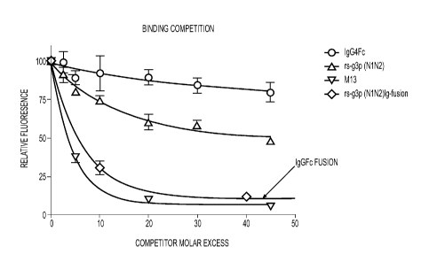
(Construct 3), M13 (Construct 2), rs-g3p(N1N2)-hIgG4-Fc fusion protein
(Construct
4), and an IgG4-Fc negative control.
10341 Fig. 18 presents competition binding data comparing M13 (Construct
2; squares), rs-g3p(N1N2) (Construct 3; triangles), rs-g3p(N1N2)-hIgG4-Fc
fusion
protein (Construct 4; upside down triangles), and a recombinant IgG4-Fc
negative
control (diamonds).
CA 02857539 2014-05-29
WO 2013/082114 PCT/US2012/066793
[035] Fig. 19 shows a filter trap assay comparing five concentrations of
Afi42 fibers plus or minus two concentrations of M13 (Construct 2), 800 nM rs-
g3p(N1N2) (Construct 3), and three concentrations of rs-g3p(N1N2)-higG4-Fc
fusion
protein (Construct 4).
[036] Fig. 20 presents competition binding data for rs-g3p(N1N2)
(Construct 3; "monomer") and streptavidin conjugated rs-g3p(N1N2)
("SA[g3pN1N2]n=2-4": "SA-g3p"; "tetramer"). rs-g3p(N1N2) and SA-g3p were
compared for their ability to compete with labeled M13 for binding to AO
during a
three hour incubation at 37 C.
[037] Fig. 21 shows a filter trap assay comparing five concentrations of
fA[342 plus or minus two concentrations of rs-g3p(N1N2) (Construct 3;
"monomer")
and two concentrations of SA-g3p ("tetrarner").
[038] Figs. 22A and 22B show TEMs of fA1342 at times zero (Fig. 22A) and
three days after incubation with SA-g3p (Fig. 22B).
[039] Fig. 23 shows the amino acid sequence of one rs-g3p(N1N2)-higG4-
Fc construct 'Construct 4" (SEQ ID NO:9). The Ni N2 region of "Construct 4" is
derived from the N1N2 region of "Construct 1" (SEQ ID NO:10).
[040] Fig. 24 shows the amino acid sequence of another rs-g3p(N1N2)-
higG4-Fc construct "Construct 5" (SEQ ID NO:11). The N1N2 region of "Construct
5" is derived from the N1N2 region of "Construct 2" (SEQ ID NO:12).
[041] Fig. 25 shows the amino acid sequence of one rs-g3p(N1N2)-hIgG1-
Fe construct "Construct 6" (SEQ ID NO:13). The N1N2 region of "Construct 6" is
derived from the Ni N2 region of "Construct 2".
[042] Fig. 26 shows the amino acid sequence alignment of N2 from: fd
(SEQ ID NO:14), f1 (SEQ ID NO:15), M13 (SEQ ID NO:16), Ike (SEQ ID NO:17), 12-
11
= CA 02857539 2014-05-29
2 (SEQ ID NO:18), and If1 (SEQ ID NO:19). An asterisk "*" indicates positions
which
have a single, fully conserved residue. A colon ":" indicates conservation
between
groups of strongly similar properties that score greater than 0.5 in the
Gonnet PAM
250 matrix. A period "." indicates conservation between groups of weakly
similar
properties that score equal to or less than 0.5 in the Gonnet PAM 250 matrix.
[043] Fig. 27A shows a schematic of Construct 3. Fig. 27B shows the DNA
sequence of the g3p portion of Construct 3 (SEQ ID NO:23). Fig. 27C shows the
amino acid sequence of the g3p portion of Construct 3 (SEQ ID NO:24).
[044] Fig. 28 shows the results of an experiment testing two rs-g3p(N1N2)-
IgG fusion proteins for their ability to reduce amyloid 13 in a transgenic
mouse model
of Alzheimer's Disease. rs-g3p(N1N2)-hIgG4-Fc (Construct 5) and rs-g3p(N1N2)-
hIgG1-Fc (Construct 6) both significantly reduced the level of amyloid 13 in
the
hippocampus of Alzheimer's Diseased mice.
[045] Fig. 29 shows the results of an experiment testing two rs-g3p(N1N2)-
IgG fusion proteins for their ability to reduce amyloid 13 in a transgenic
mouse model
of Alzheimer's Disease. rs-g3p(N1N2)-hIgG4-Fc (Construct 5) and rs-g3p(N1N2)-
hIgG1-Fc (Construct 6) were both able to significantly reduce the level of
amyloid 13
in the cerebral cortex of Alzheimer's Disease mice.
[046] Fig. 30 shows assembly inhibition of A1342 with rs-g3p(N1N2)-hIgGl-
Fc (Construct 6). Fig. 30A shows a "native" agarose gel made without SDS. The
samples were run in TEA buffer without SDS and not boiled. The results
indicate
that Construct 6 is capable of inhibiting the assembly of fAp42. Fig. 30B
presents a
ThT fluorescence assay used to measure the amyloid present in a given sample.
10
pM of A342 monomers were incubated in the presence or absence of 2
concentrations of rs-g3p(N1N2)-hIgG1-Fc (Construct 6) at 37 C for 1 day. The
12
CA 02857539 2014-05-29
WO 2013/082114 PCT/US2012/066793
amount of fibers formed at the end of day 1 was measured by quantitating the
bound
ThT fluorescence.
rs-g3p(N1N2)-hIgGl-Fc (Construct 6) potently inhibits formation of A[142
fibers. The
figure also indicates that inhibition of fiber formation with Construct 6 is
dose-
dependent.
[047] Fig. 31 presents representative circular dichroism data showing that
A1342 assembly is inhibited by rs-g3p(N1N2) (Construct 3). Circular dichrosism
measures the a-helix and 13-sheet content of the A13 fibers to be assessed.
Fig. 31A
shows the ellipticity versus wavelength for A1342 at T= 0, T=24 hours, and
T=48
hours. Fig. 31B shows ellipticity versus wavelength for A1342 plus Construct 3
at T=
0, T=24 hours, and 1=48 hours. Fig. 31C shows a representative ThT assay where
the amount of fibers formed between 24 and 48 hours was measured by
quantitating
the bound ThT fluorescence. Construct 3 potently inhibits formation of A[342
fibers.
Fig. 31D shows ellipticity versus wavelength for Construct 3 at T= 0, 1=24
hours,
and 1=48 hours. Taken together, these data confirm the ability of Construct 3
to
inhibit A1342 assembly.
[048] Fig. 32 presents representative data showing that M13 (Construct 2)
and rs-93p(N1N2)-higG1-Fc (Construct 6) block oligomer-induced toxicity of N2a
cells. See, e.g., Stine et al. (2003) J. Biol. Chem. 278(13): 11612-11622 and
Stine et
al. (2011) Erik D. Roberson (ed.) Alzheimer's Disease and Frontotemporal
Dementia, Methods in Molecular Biology, vol. 670: 13-32. N2a cells were
differentiated by serum starvation for 48 hours prior to treatment. A1342
oligomers
(2uM) were pre-incubated with Construct 2 and Construct 6 at 37 C for 3 hrs
before
addition to N2a cells. Time zero ("TO") complexes were not pre-incubated.
After 24
hours of incubation, adenylate kinase ("AK") release was monitored. AK release
into
13
CA 02857539 2014-05-29
WO 2013/082114 PCl/US2012/066793
the medium indicates cell death/lysis. A1342 oligomers were made as described
by
Stine et.al., 2011. The results indicate that M13 and rs-g3p(N1N2)-higGl-Fc
are
potent inhibitors of toxic oligomers.
[049] Fig. 33 shows a filter trap assay comparing six concentrations of
442 fibers plus or minus 1 x 10¹²/mIM13 (Construct 2); 80 nm and 800 nM rs-
g3p(N1N2)-higG4-Fc construct (Construct 5); and 80 nm and 800 nM of rs-
g3p(N1N2)-higGl-Fc (Construct 6). A[342 fibers were incubated with Constructs
2,
5, and 6 at 37 C for 3 days, followed by filter retardation. The filter was
probed by
mAb 6E10 (1:15000), which recognizes A1142 fibers trapped on the filter. 800nM
of
Construct 5 or Construct 6 equals 5 x 10¹⁴/mIConstruct 2 by molecular
molarity.
The results indicate that Constructs 2, 5, and 6 potently disaggregate (3-
amyloid
fibers.
[050] Figs. 34A and 34B present representative assays used to measure
the amount of M13 (Construct 2) bound to fA1342 after 3 hours of preincubation
with
ftau. 5 pM of Afi42 monomers bound to Construct 2 was incubated in the
presence
or absence of 4 concentrations of ftau at 37 C for 3 hours. Since fAbeta:M13-
Alexa488 pellets but ftau:M13-Alexa488 does not pellet, measuring the loss of
fluorescence from the pelleted material indicates that ftau competed the
fAbeta
binding. Here, the amount of M13-fA13 formed at the end of 3 hours was
measured
by quantitating the Alexa488 fluorescence in the pelleted binding competition
reaction. The results indicate that ftau is able to compete with M13-Alexa488
(Construct 2) for fA[342 binding.
[051] Fig. 35 shows the results of one representative SPR assay testing the
ability of rs-g3p(N1N2)-hIgG4-Fc (Construct 4) to bind to ftau. The results
indicate
that Construct 4 potently binds ftau.
14
CA 02857539 2014-05-29
WO 2013/082114 PCT/US2012/066793
[052] Fig. 36 shows the ability of rs-g3p(N1N2)-higGl-Fc (Construct 6) to
disaggregate ftau. Tau fibers were prepared by diluting 40 uM of the
microtubule
binding repeat region ("MTBR") of tau into 50 mM superoxide dismutase ("Sod").
Various concentrations of Construct 6 and the prepared ftau were incubated in
acetate buffer at pH7.0, 37 C for 72 his. ThT fluorescence was recorded in the
presence of 5 fold excess ThT. Fig. 36A presents the results of a
representative
ThT assay showing the ability of Construct 6 to disaggregate ftau. Fig. 36B
shows
another representative experiment confirming the ability of Construct 6 to
disaggregate tau. Figs. 36A and 368 also show that disaggregation of ftau by
Construct 6 is dose dependent.
[053] Fig. 37 presents representative experiments showing the inhibition of
AP aggregation by rs-g3p(N1N2)-higG1-Fc (Construct 6) and rs-g3p(N1N2)
(Construct 3) over time. A[342 was dissolved in DMSO and diluted into PBS
containing NaN3. A1342 was aggregated at 37 C plus or minus various
concentrations of Construct 3 and Construct 6. Aggregation of A1342 was
measured
by ThT fluorescence. Fig. 37A shows an SDS PAGE of the samples. Fig. 378
shows the results from one representative experiment. Fig. 37C shows the
results
from another representative experiment. Fig. 37D summarizes the results.
[054] Fig. 38A and Fig. 3813 present the results of experiments showing
the ability of rs-g3p(N1N2)-higGI-Fc (Construct 6) to block the conversion of
PrP to
PrP-Sc. Construct 6 and IgG cell lysates were subjected to ultra-
centrifugation to
separate soluble (supernatant) and insoluble (pellet) PrP species. PrP species
were
visualized biochemically with an anti-PrP monoclonal antibody (6D11). In the
presence of IgG, there is a partitioning of PrP in both soluble and insoluble
fractions.
In the presence of Construct 6, there is limited insoluble PrP. Data
represents n=4.
CA 02857539 2014-05-29
WO 2013/082114 PCT/US2012/066793
[055] Fig. 39A and Fig. 398 present the results of experiments showing the
ability of rs-g3p(N1N2)-higGl-Fc (Construct 6) to reduce the accumulation and
aggregation of PrPsc in a cell culture model of prion disease. Fig. 39A shows
biochemically resolved undigested and PK-digested N2a221.sc cell lysates
following
treatment with Construct 6 and IgG. A significant reduction in PrPsc levels is
clearly
observed in cells treated with increasing concentrations of Construct 6. An
approximately 50% reduction in PrPsc levels is achieved with treatment of
-0.08ug/m1Construct 6. Treatment with bug/m1 Construct 6 reduces PrPsc levels
to
5.725%, p<0.0001. No marked changes in Pri3sc levels were observed in N2A22Lse
cells treated with lug/ml murine IgG. For Fig. 39B, the X-ray films were
subsequently digitized and initially normalized to the effect in IgG treated
N2a22Lsc
cells from the same passage which was considered to be 100%. The densitometry
data from PK-digested blots was then analyzed relative to the equivalently
blotted
undigested lysates and expressed as a percent change PrPse/PrPc. Data
represents
n=4.
DESCRIPTION OF EMBODIMENTS
[056] The invention is based, in part, on the inventor's recognition of the
role of the gene 3 protein ("g3p," also known as "p3" or "pill") in mediating
amyloid
binding and disaggregation of amyloid aggregates. The invention is also based
on
the inventors' identification of a minimal sequence of g3p required for
binding to
amyloid.
[057] Thus, in certain embodiments, the invention provides molecules, in
particular polypeptides, that comprise minimal consensus amyloid binding
sequences derived from g3p. In one aspect of these embodiments, the molecules
are soluble. In another aspect of these embodiments, the molecules
disaggregale
16
CA 02857539 2014-05-29
WO 2013/082114 PCl/US2012/066793
and/or prevent the aggregation of amyloid (e.g., amyloid plaque). In another
aspect
of these embodiments, the molecules are fusion proteins. In a more specific
aspect
of these embodiments, the molecules are fusion proteins additionally
comprising an
amino acid sequence of an immunoglobulin chain. In an even more specific
aspect
of these embodiments, the molecules are fusion proteins additionally
comprising an
amino acid sequence of an immunoglobulin G (e.g., IgG) or immunoglobulin M
(e.g.,
IgM) chain. In still another aspect of these embodiments, the molecule
comprises
the N2 domain of g3p. In a more specific aspect of these embodiments, the
molecule comprises the N1-N2 domain of g3p. In still another aspect of these
embodiments, the molecule comprises a full length g3p. In yet another aspect,
the
molecule is a polypeptide that is a fragment, mutant, or variant of any of the
foregoing.
[058] In other aspects, the invention provides molecules that bind to TolA,
such as TolA inhibitor molecules, in particular polypeptides, that comprise
minimal
consensus amyloid binding sequences. The TolA binding molecules and/or TolA
inhibitors of the present invention bind to, depolymerize, prevent the
aggregation of,
and disaggregate amyloid. The TolA binding molecules and/or TolA inhibitor
molecules include fusion proteins. In certain embodiments the TolA binding
molecule and/or TolA inhibitor molecule is a colicin or amyloid binding
fragment of a
colicin. In certain embodiments the colicin is a Group A colicin. See, e.g.,
Cascales
et al., Microbiol. Mal. Biol. Rev. (2007) 71(1): 158-229. The TolA binding
molecules
and TolA inhibitor molecules of the invention are useful therapeutics to
decrease
amyloid load associated with diseases, such as systemic and peripheral amyloid
diseases, neurodegenerative diseases including neurodegenerative tauopathies,
and
transmissible spongiform encephalopathies (prion-associated diseases). Also
17
CA 02857539 2014-05-29
WO 2013/082114 PCT/US2012/066793
encompassed is the use of those compositions to prevent the accumulation of
amyloid load associated with these diseases, and the use of those compositions
as
diagnostics to detect amyloid and thus, diagnose such diseases.
[059] In another embodiment, the invention provides filamentous
bacteriophage that have been modified to overexpress g3p as compared to wild
type
phage, to express an amyloid binding fragment of g3p, an amyloid binding
mutant or
variant form of g3p, or an amyloid binding fusion protein comprising g3p.
[060] The invention provides compositions of matter and/or pharmaceutical
compositions of any of the foregoing molecules or bacteriophage, as well as
their
use to bind to, disaggregate, and prevent aggregation of amyloid, and to their
use to
detect amyloid deposits and diagnose diseases and disorders characterized by
amyloid.
Definitions
[061] The term "g3p" when used alone or in terms such as "93p-derived"
refers to any wild type or recombinant filamentous phage g3p protein
(including
fragments, variants, and mutants of g3p). The term should not be construed as
limited to any particular filamentous bacteriophage g3p. By way of example,
the
term "g3p" includes SEQ ID NO: 1 and the related proteins shown in Fig. 2.
[062] The term "filamentous bacteriophage" includes both wild type
filamentous bacteriophage, and recombinant filamentous bacteriophage. In the
present application, "filamentous bacteriophage" may also be referred to as
"bacteriophage," "phage," or "M13."
[063] The term "wild-type filamentous bacteriophage" as used herein refers
to filamentous phage found in nature, filamentous phage that have been
indicated as
"wild-type" in any nucleotide or amino acid sequence database, filamentous
18
CA 02857539 2014-05-29
WO 2013/082114 PCT/US2012/066793
bacteriophage that are commercially available and characterized as "wild-
type," and
filamentous bacteriophage that have acquired non-recombinant mutations
relative to
any of the foregoing through passaging.
[064] The term "domain" means a region of a polypeptide (including
proteins) having some distinctive physical feature or role including for
example an
independently folded structure composed of one section of a polypeptide chain.
A
domain may contain the sequence of the distinctive physical feature of the
polypeptide or it may contain a fragment of the physical feature which retains
its
binding characteristics (i.e., it can bind to a second domain). A domain may
be
associated with another domain. In other words, a first domain may naturally
bind to
a second domain. For example, the g3p N2 domain binds F-pili and the g3p Ni
domain binds TolA.
[065] The terms "amyloid," "amyloid fibrils," and "amyloid fibers," as used
herein are generic terms for a tertiary structure that is formed by
aggregation of any
of several different proteins and that consists of an ordered arrangement of p
sheets
stacked perpendicular to a fiber axis. Sunde et al., J. Mol. Biol. (1997)
273:729-39.
One exemplary amyloid is the aggregate of amyloid- fi formed in Alzheimer's
disease,
which is composed of beta-amyloid peptide "13A," which are 39-43 amino acid
internal fragments cleaved from the human amyloid precursor protein (hAPP).
There
are short forms, such as AI340, and long forms, such as the more fibrillogenic
A[i
isoform, A342. Other exemplary amyloid proteins include misfolded a-synuclein
(associated with Parkinson's disease), huntingtin (associated with
Huntington's
disease), tau (associated with Alzheimer's Disease), and the abnormal
conformation
of the prion protein, PrPsc. Additional examples are provided throughout the
description and are known to those of skill in the art (see, e.g., Aguzzi
(2010), and
19
CA 02857539 2014-05-29
WO 2013/082114 PCT/US2012/066793
Eichner and Radford, Mol. Cell (2011) 43:8-18). Thus, unless a protein or
peptide is
specified, use of the terms "amyloid," "amyloid fibrils," or "amyloid fibers"
should not
be construed as limited to any particular protein or disease.
[066] The term "beta amyloid peptide" is synonymous with "0-amyloid
peptide," "OAP," " 0A," and "Ai.i." All of these terms refer to an amyloid
forming
peptide derived from the human amyloid precursor protein (hAPP).
[067] A phage, protein, fusion protein, fusion protein domain, or a mutant,
fragment, or variant of the foregoing that "binds amyloid fibrils" or that is
"amyloid-
binding" is one that is positive in an amyloid binding assay. Amyloid binding
can be
detected in vitro using a direct binding assay such as surface plasmon
resonance
(SPR), in which case it will generally bind amyloid with a Kd of at least 10'8
M, i
M, 10-1 M, or 10-11 M. Alternatively, amyloid binding can be detected using
the
fA[142 binding assay described in the examples. Amyloid-binding fragments,
variants, and mutants of g3p may also be identified by their co-localization
to amyloid
when injected into a transgenic mouse model of any a protein misfolding
disease.
[068] Any of the products or compositions of the invention described as
"disaggregating" or "mediating disaggregation" reduce aggregates that have
already
formed. Disaggregation can be measured by the filter trap assay. Wanker et
at.,
Methods Enzymol (1999) 309:375-86. The filter trap assay is described herein
and
can be used both to detect aggregates and to monitor disaggregation mediated
by
compositions of the invention. Disaggregation is detected as decreased
retention of
amyloid on the filter, as shown by a decrease in staining, in the presence of
increasing concentrations of the disaggregating agent.
[069] As used herein, a composition that "reduces amyloid" does one or
more of the following: inhibits amyloid formation, causes amyloid
disaggregation,
CA 02857539 2014-05-29
WO 2013/082114 PCT/US2012/066793
promotes amyloid clearance, inhibits amyloid aggregation, blocks and/or
prevents
the formation of toxic amyloid oligomers, and/or promotes the clearance of
toxic
amyloid oligomers.
[070] Any of the products or compositions of the invention described as
"protecting neurons from amyloid damage" prevent the accumulation of new
amyloid
and/or prevent the formation of toxic amyloid oligomers. Products or
compositions of
the invention described as "protecting neurons from amyloid damage" may be
taken
prophylactically. Whether or not a product or composition protects neurons
from
amyloid damage may be measured by the neuronal cell culture cytotoxicity assay
described herein.
[071] As used herein, "PrP protein," "PrP," and "prion," refer to polypeptides
that are capable under appropriate conditions, of inducing the formation of
aggregates responsible for protein misfolding diseases. For example, normal
cellular prion protein (PrPc) is converted under such conditions into the
corresponding scrapie isoform (PrP) which is responsible for diseases such as,
but
not limited to, bovine spongiform encephalopathy (BSE), or mad cow disease,
feline
spongiform encephalopathy of cats, kuru, Creutzfeldt-Jakob Disease (CJD),
Gerstmann-Straussler-Scheinker disease (GSS), and fatal familial insomnia
(FFI).
[072] The term "variant" as used herein in conjunction with a bacteriophage,
protein, polypeptide or amino acid sequence (e.g., a g3p variant or a variant
of an
amyloid binding fragment of g3p), refers to a corresponding substance that
contains
at least one amino acid difference (substitution, insertion or deletion) as
compared to
the reference substance. In certain embodiments a "variant" has high amino
acid
sequence homology and/or conservative amino acid substitutions, deletions
and/or
insertions as compared to the reference sequence. In some embodiments, a
variant
21
CA 02857539 2014-05-29
WO 2013/082114 PCT/US2012/066793
has no more than 75, 50, 40, 30, 25, 20, 15, 12, 10, 9, 8, 7. 6, 5, 4, 3, 2, 1
amino
acid differences as compared to the reference sequence. A "conservative
substitution" refers to the replacement of a first amino acid by a second
amino acid
that does not substantially alter the chemical, physical and/or functional
properties of
the g3p protein or amyloid binding fragment of g3p (e.g., the g3p protein or
amyloid
binding fragment retains the same charge, structure, polarity,
hydrophobicity/hydrophilicity, and/or preserves functions such as the ability
to
recognize, bind to, and/or reduce amyloid). Such conservative amino acid
modifications are based on the relative similarity of the amino acid side-
chain
substituents, for example, their hydrophobicity, hydrophilicity, charge, size,
and the
like. Exemplary conservative substitutions which take various of the foregoing
characteristics into consideration are well known to those of skill in the art
and
include: arginine and lysine; glutamate and aspartate; serine and threonine;
glutamine and asparagine; and valine, leucine, and isoleucine.
[073] The term "mutant" (e.g., "mutant g3p" or "mutant amyloid binding
fragment") refers to a protein that is mutated at one or more amino acids in
order to
modulate its therapeutic or diagnostic efficacy. In certain embodiments, a
mutant
contains a substitution, deletion and/or insertion at an amino that is known
to interact
with amyloid. In other embodiments, a mutant contains a substitution, deletion
and/or insertion at an amino that is a conserved amino acid present in a wild-
type
g3p or amyloid binding fragment thereof. In some embodiments, a mutant has no
more than 75, 50, 40, 30, 25, 20, 15, 12, 10, 9, 8, 7, 6, 5, 4, 3, 2, 1 amino
acid
differences as compared to the reference sequence. In some embodiments, the
amino acid substitutions are conservative substitutions. The terms "variant"
and
22
CA 02857539 2014-05-29
WO 2013/082114 PCT/US2012/066793
"mutant" are used interchangeably herein except that a "variant" is typically
non-
recombinant in nature, whereas a "mutant" is typically recombinant.
[074] The term "high stringency," as used herein, includes conditions readily
determined by the skilled artisan based on, for example, the length of the
DNA.
Generally, such conditions are defined in Sambrook et al. Molecular Cloning: A
Laboratory Manual, 2 ed. Vol. 1, pp. 1.101-104, Cold Spring Harbor Laboratory
Press (1989), and include use of a prewashing solution for the nitrocellulose
filters
5X SSC, 0.5% SDS, 1.0 mM EDTA (PH 8.0), hybridization conditions of 50%
formamide, 6X SSC at 42 C (or other similar hybridization solution, such as
Stark's
solution, in 50% formamide at 42 C), and with washing at approximately 68 C,
0.2X
SSC, 0.1% SDS. The skilled artisan will recognize that the temperature and
wash
solution salt concentration can be adjusted as necessary according to factors
such
as the length of the probe.
[075] The term "moderate stringency," as used herein, includes conditions
that can be readily determined by those having ordinary skill in the art based
on, for
example, the length of the DNA. The basic conditions are set forth by Sambrook
et
al. Molecular Cloning: A Laboraloty Manual, 2d ed. Vol. 1, pp. 1.101-104, Cold
Spring Harbor Laboratory Press (1989), and include use of a prewashing
solution for
the nitrocellulose filters 5X SSC, 0.5% SDS, 1.0 mM EDTA (pH 8.0),
hybridization
conditions of 50% formamide, 6X SSC at 42 C (or other similar hybridization
solution, such as Stark's solution, in 50% formamide at 42 C), and washing
conditions of 60 C, 0.5X SSC, 0.1% SDS.
[076] The term "high sequence homology" means at least 70%, at least
75%, at least 80%, at least 85%, at least 90%, at least 91%, at least 92%, at
least
93%, at least 94%, at least 95%, at least 96%, at least 97%, at least 98%, or
at least
23
CA 02857539 2014-05-29
99% amino add sequence homology with the reference sequence as measured
using known computer programs, such as the Bestfit program.
[077] A "fusion protein" is a non-naturally occurring protein comprising at
least two polypeptide domains.
[078] A "g3p fusion protein" comprises a g3p protein linked to a second
domain.
[079] An "N1-N2 fusion protein" (also termed "N1N2 fusion protein")
comprises the N1 and N2 domains (or mutants, fragments, or variants of
either), but
not the N3/CT domain, of a g3p protein linked to a second domain. A NI N2
fusion
protein may or may not comprise the hinge region.
[080] An "N2 fusion protein" comprises the N2 domain (or mutants,
fragments, or variants of N2), but neither the N1 nor N3/CT domains, of a g3p
protein linked to a second domain. A N2 fusion protein may or may not comprise
the
hinge region.
[081] As used herein, "Construct 1" is derived from wild type M13 (see,
Genbank file: NC_003287.2, version GI:56718463. In Construct 1, as compared to
wild type M13, Ser378(AGC) is changed to Gly(GGC), and 11e87 (AU) is changed
to
Asn(AAC)). Construct 1 comprises the nucleic acids of SEQ ID NO: 10.
[082] "Construct 2" is a wild type M13 isolate (GenBank JX412914.1).
Construct 2 comprises the nucleic acids of SEQ ID NO:12.
[083] "Construct 3" is a recombinant soluble g3p fragment comprising the
Ni and N2 domains of g3p (rs-g3p(N1N2)) comprising the amino acids of SEQ ID
NO:20.
24
CA 02857539 2014-05-29
WO 2013/082114 PCT/US2012/066793
[084] "Construct 4" is recombinant soluble g3p fragment IgG4 Fc fusion
protein (rs-g3p(N1N2)-hIgG4-Fc) comprising the amino acids of SEQ ID NO:9. The
N1N2 region of "Construct 4" is derived from the N1N2 region of "Construct 1."
[085] "Construct 5" is a recombinant soluble g3p fragment IgG4 Fc fusion
protein (rs-g3p(N1N2)-hIgG4-Fc) comprising the amino acids of SEQ ID NO:11.
The
N1N2 region of "Construct 5" is derived from the N1N2 region of "Construct 2."
[086] "Construct 6" is recombinant soluble g3p fragment IgG1 Fc fusion
protein (rs-g3p(N1N2)-hIgGl-Fc) comprising the amino acids of SEQ ID NO:13.
The
N1N2 region of "Construct 6" is derived from the N1N2 region of "Construct 2."
Sources of q3p
[087] Filamentous bacteriophage are a group of related viruses that infect
gram negative bacteria, such as, e.g.. E. coll. See, e.g., Rasched and Oberer,
Microbiology Reviews (1986) Dec:401-427. Examples of filamentous bacteriophage
include, but are not limited to, phage of the Ff family (i.e., at least M13,
fi, and fd)
and phage of the I-family (i.e., at least 122, Ike, If1).
[088] All naturally occurring filamentous bacteriophage contain g3p as a
minor coat protein present in 3 to 5 copies per phage. Thus, in one aspect of
the
invention, an isolated g3p is obtained from any naturally occurring
filamentous
bacteriophage. Recombinant forms of g3p can also be produced. Recombinant g3p
may correspond to a wild type g3p from any naturally occurring filamentous
bacteriophage. Thus, recombinant, isolated g3p is also encompassed by the
invention.
[089] One example of g3p, from phage M13, is presented in SEQ ID NO: 1.
Unless otherwise clearly specified, any g3p mutations described are in
reference to
SEQ ID NO: 1 shown below in clean and annotated form:
CA 02857539 2019-05-29
WO 2013/082114 PCT/US2012/066793
AETVESCLAK rilTEN9ETNV WKODETLDRY ANYEGCLWNA TGVVVCTGDE TQCYGTWVPI
=
61 GLAIPENEGG GSEGGGSEGG GSEGGGTKPP EY011P1PGY TYINPLDGiY PPGTEQNPAN
12 PNPSL:S.SQP LNTFMFQNNR FRNRQGALTV YTGTVTQGTD PVKTYYQYTE 1,15AMYDAY
igi WNGKIRDCAF HS(>.NEDPFV C:EYQGQ3SDL PQPPVNAGGG SGGG'3GGG GGG3EGGGSE
241 GGGSEGGGSG GGSGSGDFDY EKMANANKGA MTENADENAL QSDAKGKLiX; VATDYGAAID
301 GFIGDVSGLA NGNGATGDFA GSNSQMAQW3 DGDNSPLMNN PRQYLPSLPQ SVECRPIVES
361 AGNPYEFSID CD1(INLFY<GV FAPLLYVATF MYVh'STI!'ANT LRNKES
SEQ ID NO:),
AETVESCLAK PliTENSFTNV WKDDKTLDRY ANYEGCLWNA TGV9Vc1GDE TQC7GTWVP1
61 GLAlt,E,NLCC QEEWSEW GSEGGGTKPP EYGDTPIPGY TYINPLDGTY PPGTEQNPAN
121 PNPSLEESQP LNTEMFONNR FRNRQGALTV YTGTVTQGTD PVKTYYQYTP VSSKAMYDAY
181 WNGKFRDCAF HSGFNEDPFV CEYQCOSDL PUPWAGGG SGGGSGGGSE GGGSEGGGSE
241 GOCSEGOGSG GGSGSGIrq...0ANTtNW6OWKAKKLTP.:00
301 .P.A.O.A6sTfiiiNA,3.N.iWAW*P4.4.0:000.4W.LKOMMPVW$
56 I =..................... ..........................
Table 1 - Key for annotated SEQ ID NO: 1 .............
Region Residues Residue when signal Key
................. peptide is present
N1 1-67 19-85 underline
GI 68-86 86-104 highlight __
N2 87-217 105-235 underline and bold
G2 218-256 I 236-274 _______ italic
-4
N3 257-406 i 275-424 highlight and underline
.;õ
SEQ ID NO: 1 is GenBank NP-510891.1 with the 18 amino acid signal peptide
removed, thus the amino acid numbering is for the mature g3p. The signal
peptide is
generally included in any expression construct, and immature g3p that includes
the
signal peptide is included within the scope of the various embodiments of the
invention unless context makes clear that it is expressly excluded. SEQ ID NO:
1 is
provided as a reference sequence only. It is in no way intended to limit the
invention.
[090] Sequences of g3p from multiple sources are known. Exemplary g3p
amino acid sequences for bacteriophage of the Ff family include those
sequences
26
CA 02857539 2014-05-29
WO 2013/082114 PCT/US2012/066793
found in UniProt accession numbers P69169 (phage fl), P03661 (phage fd), and
P69168 (phage m13). Exemplary g3p amino acid sequences for bacteriophage of
the I-family include P15415 (phage 122), P03663 (phage Ike), and 080297 (phage
If1). Alignments of several g3p sequences are presented in Fig. 2.
[091] G3p useful in this invention also includes fragments, mutants, and/or
variants of g3p. Mutants or variants may be described with reference to a full
length
g3p or with reference to a fragment of g3p. Any full length or fragment g3p,
including
mutants and/or variants thereof that retain the ability to bind to amyloid,
regardless of
their ability to disaggregate amyloid is within the scope of the present
invention. Any
protein "comprising" such g3p is also encompassed by the present invention.
Likewise, proteins "including," "consisting of," "consisting essentially of,"
or "having"
such g3p are also encompassed.
Amyloid-Binding and Amvloid Disaggregating Fragments of q3p
[092] As mentioned. g3p has two amino-terminal domains, Ni and N2, that
interact to form an N1-N2 complex, and one carboxy-terminal domain, N3 (also
called "CT"). In Ff phage, the N1 domain comprises residues 1-67 and the N2
domain comprises residues 87-217 of mature g3p. Residues 87-123 form the hinge
that allows opening and closing between Ni and N2. Sometimes the hinge is
considered part of N2, whereas in other instances it is treated as a separate
element. N1 and N2 are also linked by flexible glycine-rich linker sequence.
Within
N1, there are two disulphide bridges between Cys7 and Cys36 and between Cys46
and Cys53. There is a single disulphide bridge in N2 between Cys188 and
Cys201.
The N3/CT domain comprises residues 257 to 406. Hollinger, 1999; Marvin, 1998.
In the carboxy terminal domain there is a disulphide bridge between Cys354 and
Cys371. Marvin, 1998. There are no interdomain disulphide bridges in g3p.
27
CA 02857539 2014-05-29
WO 2013/082114 PCT/US2012/066793
[093] Non-limiting examples of amyloid binding fragments of g3p include
the N2 domain either with the hinge (e.g., at least residues 87-217 of SEQ ID
NO: 1)
or without the hinge (e.g., at least residues 124-217 of SEQ ID NO: 1); and
the N1-
N2 domains (e.g., at least residues 1-67 and 87-217 of SEQ ID NO: 1), either
with or
without the intervening linker sequence (e.g., with or without residues 68-86
of SEQ
ID NO: 1), and either with or without the hinge. In any of the foregoing
examples, the
N2 or N1N2 fragments may be the N2 or N1N2 found in a wild-type filamentous
bacteriophage or a recombinant N2 or N1N2. In any of the foregoing examples,
the
N2 or N1N2 fragments may mutants or variants of the wild-type filamentous
bacteriophage sequence.
[094] Useful amyloid binding fragments of g3p include any fragment of g3p,
including N2 and N1N2 fragments that retain the ability to bind to amyloid,
regardless
of the fragment's ability to disaggregate amyloid. Any protein "comprising"
such
amyloid binding fragment (or mutant or variant thereof) is encompassed by the
present invention. Likewise, proteins "including," "consisting of,"
"consisting
essentially of," or "having" the g3p fragment or variant are also encompassed.
N2 and N2 polypeptide mutants and variants
[095] A primary structure alignment of N2 from: fd, fl, M13, Ike, 12-2, and
Ifl is shown as Fig. 26. The amino acids of Id are shown in SEQ ID NO:14; fl
in
SEQ ID NO:15; M13 in SEQ ID NO:16; Ike in SEQ ID NO:17; 12-2 in SEQ ID NO:18;
and 1f1 in SEQ ID NO:19. Using this figure and alignment as guidance, one
embodiment of the invention encompasses a N2 polypeptide, N2 polypeptide
mutants, and N2 polypeptide variants, comprising the amino acids of SEQ ID NO:
14, 15, 16, 17, 18, or 19, including any amyloid binding fragments thereof.
28
CA 02857539 2014-05-29
WO 2013/082114 PCT/US2012/066793
[096] In other embodiments, the N2 polypeptide is a N2 polypeptide mutant
or variant that retains its ability to bind to amyloid and has an amino acid
sequence
that comprises no more than 75, 50, 40, 30, 25, 20, 15, 12, 10, 9, 8, 7, 6, 5,
4, 3, 2, 1
amino acid differences when aligned with the amino acid sequence of any one of
SEQ ID NO: 14, 15, 16, 17, 18, or 19. In Fig. 26, an asterisk "*" indicates
positions
which have a single, fully conserved residue. A colon ":" indicates
conservation
between groups of strongly similar properties that score greater than 0.5 in
the
Gonnet PAM 250 matrix. A period "." indicates conservation between groups of
weakly similar properties that score equal to or less than 0.5 in the Gonnet
PAM 250
matrix. In some aspects of these embodiments, the N2 polypeptide mutant or
variant does not comprise an amino acid difference at any position indicated
with an
in Fig. 26. In more specific aspects, the N2 polypeptide mutant or variant
does
not comprise an amino acid difference at any position indicated with an "*"
and
comprises the same amino acid as at least one of SEQ ID NO: 14, 15, 16, 17,
18, or
19 at each position indicated with a ":" in Fig. 26. In even more specific
aspects, the
N2 polypeptide mutant or variant does not comprise an amino acid difference at
any
position indicated with an "*" and comprises the same amino acid as at least
one of
SEQ ID NO: 14, 15, 16, 17, 18, or 19 at each position indicated with a ":" and
each
position indicated with a "." in Fig. 26.
[097] In other embodiments, a N2 polypeptide variant is described by
specifying a percent amino acid similarity to SEQ ID NO: 14, 15, 16, 17, 18,
or 19
again with the caveat that the N2 polypeptide variant binds amyloid. In these
embodiments, the N2 polypeptide shares at least 70%, at least 80%, at least
85%, at
least 86%, at least 87%, at least 88%, at least 89%, at least 90%, at least
91%, at
least 92%, at least 93%, at least 94%, at least 95%, at least 96%, at least
97%, at
29
CA 02857539 2014-05-29
WO 2013/082114 PCT/US2012/066793
least 98%, or at least 99% identity over the amino acids of reference
sequences
shown in SEQ ID NO: 14, 15, 16, 17, 18, or 19.
[098] In other embodiments, a N2 polypeptide is described by specifying a
percent amino acid similarity to the N2 region of SEQ ID NO: 1, again with the
caveat that the N2 polypeptide variant binds amyloid. In these embodiments,
the N2
polypeptide variant shares at least 70%, at least 80%, at least 85%, at least
86%, at
least 87%, at least 88%, at least 89%, at least 90%, at least 91%, at least
92%, at
least 93%, at least 94%, at least 95%, at least 96%, at least 97%, at least
98%, or at
least 99% identity over N2 region of SEQ ID NO: 1.
[099] In still other embodiments, a N2 polypeptide is described by
secondary or tertiary structure. It is known that fd-N2 and If1-N2 domains use
homologous parts of their surfaces to bind to the same site on the F-pilus in
E. coll.
Lorenz et al., J Mol Biol. 405:989-1003 (2011) at, for example, 990. The amino
acid
residues and secondary and tertiary structure that mediate N2 binding to F-
pilus also
mediate N2 binding to amyloid. Thus, amino acid residues and secondary and
tertiary structures that are critical for N2 to F-pilus binding are also
critical for N2-
amyloid binding. N2 polypeptide variants comprising the amino acids required
to
maintain the secondary and tertiary structure in the region of N2-F-pilus
binding are
within the scope of the present invention.
N1N2 and N1N2 polypeptide mutants and variants
[0100] A primary structure alignment of fd, fl, and M13 is shown as Fig. 2A,
and Ike, 12-2, and Ifl as Fig. 28. Using this alignment as guidance, one
embodiment of the invention encompasses a Ni N2 polypeptide, polypeptide
mutant,
or polypeptide variant comprising the amino acids that are conserved between
fd, f1,
and M13 or as between 12-2, Ide, and If1, as identified with reference to the
CA 02857539 2014-05-29
WO 2013/082114 PCT/US2012/066793
sequences of Fig. 2. In other embodiments, the N1N2 polypeptide is a N1N2
polypeptide mutant or variant, that retains its ability to bind to amyloid and
has an
amino acid sequence that comprises no more than 75, 50, 40, 30, 25, 20, 15,
12, 10,
9, 8, 7, 6, 5, 4, 3, 2, 1 amino acid differences when aligned with the amino
acid
sequence of any one of SEQ ID NO: 1, 2, 3, 5 or 6.
[0101] In other embodiments, a N1N2 polypeptide, mutant, or variant is
described by specifying a percent amino acid similarity to the N1N2 region of
SEQ ID
NO: 1, again with the caveat that the N1N2 polypeptide variant binds amyloid.
In
these embodiments, the N1N2 polypeptide variant shares at least 70%, at least
80%,
at least 85%, at least 86%, at least 87%, at least 88%, at least 89%, at least
90%, at
least 91%, at least 92%, at least 93%, at least 94%, at least 95%, at least
96%, at
least 97%, at least 98%, or at least 99% identity over N1 and N2 regions of
SEQ ID
NO: I.
Fusion Proteins
[0102] In one aspect, the invention relates to fusion proteins. The fusion
protein comprises g3p, an amyloid-binding fragment of g3p, a TolA binding
molecule,
or a TolA inhibitor. Fusion proteins comprising mutant or variant g3p or g3p
fragments are fully encompassed. The fusion protein is linked, fused,
conjugated,
coupled, or associated with/to at least one additional protein or protein
domain with
which it is not normally associated. In one embodiment, the fusion protein is
a g3p
fusion protein that comprises a g3p protein linked to a second domain. In
another
embodiment, the fusion protein is an amyloid-binding fragment of a g3p protein
linked to a second domain. In another embodiment, the fusion protein is an
N1N2
fusion protein, which comprises the Ni and N2 domains, but not the CT domain,
of a
g3p protein. In still another embodiment, the fusion protein is an N2 fusion
protein,
31
CA 02857539 2014-05-29
WO 2013/082114 PCT/US2012/066793
which comprises the N2 domain, but neither the N1 nor CT domains, of a g3p
protein. As noted, some aspects of the invention relate to mutated or variated
g3p
protein or amyloid-binding fragments thereof, and to mutated or variated Ni N2
or N2
domains that bind amyloid fibers. Thus, fusion proteins comprising these
mutated or
variated forms are also part of the invention.
[0103] The g3p or amyloid binding fragment and the fusion partner
polypeptide may be part of a continuous amino acid sequence with the fusion
partner
polypeptide linked directly or through a short peptide linker to either the N
terminus
or the C terminus of the g3p or amyloid binding fragment polypeptide. In such
cases, the g3p or amyloid binding fragment thereof and the fusion partner
polypeptide may be translated as a single polypeptide from a coding sequence
that
encodes both the g3p or amyloid binding fragment thereof and the fusion
partner
polypeptide.
[0104] In some embodiments, the fusion protein comprises an
immunoglobulin constant region as the second domain. Fusion proteins comprised
of immunoglobulin constant regions linked to a protein of interest, or
fragment
thereof, have been described (see, e.g., U.S. Patent Nos. 5,480,981 and
5,808,029;
Gascoigne et al. 1987, Proc. Natl. Acad. Sci. USA 84:2936; Capon et al. 1989.
Nature 337:525; Traunecker et at. 1989, Nature 339:68; Zeftmeissl et al. 1990,
DNA
Cell Biol. USA 9:347; Byrn et at. 1990, Nature 344:667; Watson et at. 1990, J.
Cell.
Biol. 110:2221; Watson et at. 1991, Nature 349:164; Aruffo et at. 1990, Cell
61:1303;
Linsley et at. 1991, J. Exp. Med. 173:721; Linsley et at. 1991, J. Exp. Med.
174:561;
Stamenkovic et at., 1991, Cell 66:1133; Ashkenazi et al. 1991, Proc. Natl.
Acad. Sc!.
USA 88:10535; Lesslauer et at. 1991, Eur. J. Immunol. 27:2883; Peppel et at.
1991,
J. Exp. Med. 174:1483; Bennett et at. 1991, J. Biol. Chem. 266:23060;
Kurschner et
32
CA 02857539 2014-05-29
WO 2013/082114 PCT/US2012/066793
al. 1992, J. Biol. Chem. 267:9354; Chalupny et al. 1992, Proc. Natl. Acad.
Sci. USA
89:10360; Ridgway and Gorman, 1991, J. Cell. Biol. 115, Abstract No. 1448;
Zheng
et al. 1995, J. lmmun. 154:5590). These molecules usually possess both the
biological activity associated with the linked molecule of interest as well as
the
effector function, or some other desired characteristic associated with the
immunoglobulin constant region (e.g., biological stability, cellular
secretion).
[0105] In some embodiments, the fusion protein comprises an Fc fragment of
an immunoglobulin constant region. Fc expression cassettes may be purchased
commercially. The Fc fragment can be comprised of the CH2 and CH3 domains of
an immunoglobulin and the hinge region of the immunoglobulin. The Fc fragment
can be the Fc fragment of an IgGl, an IgG2, an IgG3 or an IgG4. In one
specific
embodiment, the portion of an immunoglobulin constant region is an Fc fragment
of
an IgGl. In another embodiment, the portion of an immunoglobulin constant
region
is an Fc fragment of an IgG4. In still another embodiment, the portion of an
immunoglobulin constant region is an Fc fragment of an IgM.
[01061 Thus, in one embodiment, a recombinant soluble g3p or amyloid-
binding fragment is fused to an immunoglobulin Fc domain using standard
molecular
biology techniques. The recombinant soluble g3p or amyloid-binding fragment
may
be mutated or variated. For example, an amyloid-binding fragment of g3p, such
as
the NI N2 domain or the N2 domain, can be cloned into an IgGFc fusion
expression
vector. Exemplary IgGFc fusion vectors include, for example, one of the pFUSE-
Fc
vectors available from InvivoGen. In some embodiments, the resulting bivalent
(e.g.,
g3p(N1N2)-IgGFc or g3p(N2)-IgGFc fusion protein will have higher avidity for
amyloid binding than the recombinant soluble g3p since it is now bivalent.
33
CA 02857539 2014-05-29
WO 2013/082114 PCT/US2012/066793
[0107] In other embodiments, the fusion protein comprises a non-Fc protein
linked to a g3p or amyloid binding fragment of g3p.
[0108] In other embodiments, the fusion protein comprises at least two g3p
polypeptides or amyloid-binding fragments thereof. In other embodiments, the
fusion
protein comprises three or more g3p polypeptides, or amyloid-binding fragments
thereof. In other embodiments, the fusion protein comprises five g3p
polypeptides,
or amyloid-binding fragments thereof. Such dimeric and multimeric fusion
proteins
provide higher avidity interactions since they include more than one g3p, or
amyloid-
binding fragments thereof.
[0109] In other embodiments, the fusion protein comprises albumin. See for
example, U.S. Patent No. 6,686,179 to Fleer.
[0110] In all instances, the g3p or amyloid binding fragment of g3p in the
fusion protein encompasses mutants and variants thereof.
[0111] In general, the fusion proteins bind to amyloid at least as effectively
as the corresponding unlinked g3p or g3p fragment thereof. When applicable,
the
fusion proteins are at least as effective in mediating disaggregation of
amyloid,
promoting amyloid clearance, inhibiting amyloid aggregation, and/or removing
or
preventing the formation of toxic oligomers as the corresponding unlinked g3p
or
fragment thereof. In some embodiments, the fusion protein binds amyloid and is
at
least as effective in mediating disaggregation of amyloid, promoting amyloid
clearance, inhibiting amyloid aggregation, and/or removing or preventing the
formation of toxic oligomers as is a recombinant, soluble g3p comprising SEQ
ID
NO: 1. In still other embodiments, the fusion protein binds amyloid and is at
least as
effective in mediating disaggregation of amyloid, promoting amyloid clearance,
inhibiting amyloid aggregation, and/or removing or preventing the formation of
toxic
34
CA 02857539 2014-05-29
WO 2013/082114 PCT/US2012/066793
oligomers as phage M13. In yet other embodiments, the fusion protein binds
amyloid and is more effective in mediating disaggregation of amyloid,
promoting
amyloid clearance, inhibiting amyloid aggregation, and/or removing or
preventing the
formation of toxic oligomers than phage M13. In some embodiments, the fusion
protein binds amyloid and is at least as effective in reducing amyloid in a
protein
misfolding disease as phage M13. In still other embodiments, the fusion
protein
binds amyloid and is more effective in reducing amyloid in a protein
misfolding
disease as phage M13. In still other embodiments, the fusion protein binds
amyloid
and is at least or more effective in preventing amyloid formation as phage
M13.
[0112] Fusion proteins can be synthesized using techniques well known in
the art. For example, the fusion proteins of the invention can be synthesized
recombinantly in cells (see, e.g., Sambrook et at. 1989, Molecular Cloning A
Laboratory Manual, Cold Spring Harbor Laboratory, N.Y. and Ausubel et at.
1989,
Current Protocols in Molecular Biology, Greene Publishing Associates and Wiley
Interscience, N.Y.). Alternatively, the fusion proteins of the invention can
be
synthesized using known synthetic methods such as solid phase synthesis.
Synthetic techniques are well known in the art (see, e.g., Merrifield, 1973,
Chemical
Polypeptides, (Katsoyannis and Panayotis eds.) pp. 335-61; Merrifield 1963, J.
Am.
Chem. Soc. 85:2149; Davis et al. 1985, Biochem. Intl. 10:394; Finn et at.
1976, The
Proteins (3d ed.) 2:105; Erikson et at. 1976, The Proteins (3d ed.) 2:257;
U.S. Patent
No. 3,941,763. Alternatively, the final construct may share essentially the
same
function as a recombinantly produced fusion protein, but simply be produced
using
non-recombinant techniques, such as ligation chemistry. Components of the
fusion
proteins may be prepared using the same general methodology described for g3p
expression and g3p mutations.
CA 02857539 2014-05-29
WO 2013/082114 PCT/US2012/066793
[0113] In some embodiment, the g3p or amyloid binding fragment (or mutant
or variant form thereof) may be fused to a marker sequence, such as a peptide
that
facilitates purification of the fused polypeptide (either alone or in addition
to fusion to
another protein or incorporation of a carrier molecule). The marker amino acid
sequence may be a hexa-histidine peptide such as the tag provided in a pQE
vector
(Qiagen, Mississauga, Ontario, Canada), among others, many of which are
commercially available. As described in Gentz et al., Proc. Natl. Acad. Sc!.
(1989)
86:821-824, for instance, hexa-histidine provides for convenient purification
of the
fusion protein. Another peptide tag useful for purification, the hemagglutinin
(HA)
tag, corresponds to an epitope derived from the influenza HA protein. (Wilson
et al.,
(1984) Cell 37:767).
Phage overexpressing g3p
[0114] In another aspect, the invention relates to bacteriophage modified to
increase the number of copies of g3p expressed by the phage to more than the 3-
5
copies typically found in wild type filamentous bacteriophage. In one
embodiment,
phage that express increased numbers of g3p may be selected from naturally
occurring variants. In another embodiment, recombinant techniques are used to
increase the copy number of g3p.
[0115] In some embodiments, a wild type sequence encoding g3p or amyloid
binding fragments of g3p (including mutants or variants thereof) can be used
to
replace one of the genes encoding another bacteriophage coat protein.
Depending
upon the bacteriophage gene replaced, the number of g3p can be increased to 6,
7,
8, 9, 10, 15, 20, 25, 30, 35, 40, 50, 75, 100, 500, 1000, or even nearly 3000
copies
(for example, if the gene 3 coding sequence were used to replace the gene 8
coding
sequence or were fused to the end of the gene 8 coding sequence).
36
CA 02857539 2014-05-29
WO 2013/082114 PCl/US2012/066793
[0116] To produce phage expressing additional copies of g3p, a 93p coding
sequence (or mutant or variant form thereof) is cloned as described elsewhere
in the
description. The g3p coding sequence (or mutant or variant form thereof) may
then
be used to replace another phage gene and expressed, if necessary in
conjunction
with helper phage.
[0117] Alternatively, in some embodiments the g3p coding sequence (or
mutant or variant form thereof) is fused in frame to the coding sequence of
another
phage gene. either with or without an intervening "spacer" sequence. Methods
of
preparing phage proteins to which another protein or peptide is "fused" are
well
known in the phage display art, and g3p or an amyloid binding fragment thereof
can
be "displayed" in the same manner as, for example, antigens or antibody
chains.
E.g. Scott & Smith, Science (1990) 249:386-90: Devlin et al., Science (1990)
249:404-06. When expression of only a fragment of g3p is desired, the coding
sequence for the fragment (or mutant or variant form thereof) may be linked to
the
other gene so that one of the natural Ser/Gly linker sequences present in g3p
serves
as the linker. In some embodiments, only coding sequence for N2 or N1N2
domains
(or mutant or variant form thereof) is fused in frame to the other gene.
Mutant G3P and Amyloid Binding Fragments
[0118] In another aspect, the invention relates to mutant 93p proteins and
mutant amyloid-binding fragments thereof. Fusion proteins and phage comprising
the mutant g3p proteins and amyloid-binding fragments are also part of the
invention. Mutant g3p and mutant amyloid-binding fragments thereof may be
produced, or selected, for properties that contribute to the therapeutic
efficacy of the
pharmaceutical compositions described in this application. For example, g3p or
amyloid-binding fragments thereof may be recombinantly mutated or otherwise
37
CA 02857539 2014-05-29
WO 2013/082114 PCT/US2012/066793
selected to posses one or more of the following properties relative to g3p of
M13:
increased affinity for amyloid binding, a reduced hinge TM, increased avidity
(avidity
being distinguished from affinity in that avidity is used to describe the sum
of all
available amyloid binding where a g3p comprises more than one amyloid binding
site), increased ability to disaggregate amyloid aggregates, or increased
ability to
prevent aggregation of amyloid fibrils. Alternatively, or in addition, the
mutant 93p or
mutant amyloid fragments thereof may incorporate other useful properties
described
elsewhere in the description.
[0119] Mutant 93p proteins can be produced by mutagenesis of phage, or by
recombinant techniques, such as PCR-based site directed mutagenesis or random
mutagenesis.
[0120] In some embodiments, mutants with higher affinity are produced by
mutagenizing M13 and then selecting phage on an amyloid affinity column
coupled
with stringent washing conditions. Successive rounds of binding, washing,
elution,
and then expansion of selected phage enriches for those phage with high
affinity
binding to amyloid. Once increased affinity of the phage population is
achieved
using amyloid panning, individual clones with high affinity are selected and
analyzed.
In this way, phage mutants may be selected for high affinity binding following
random
mutagenesis.
[0121] G3p, or any amyloid binding fragments thereof, (e.g., N1 N2 domains
or N2 domains) may also be mutagenized using recombinant techniques. For
example, a vector as described herein carrying g3p or an amyloid binding
fragment
thereof (e.g., N1 N2 or N2) may be mutated using PCR-based mutagenesis
strategies. The encoded, mutated protein is then expressed and amyloid binding
and affinity of the mutants assessed as described.
38
CA 02857539 2014-05-29
WO 2013/082114 PCT/US2012/066793
[0122] Mutant amyloid binding fragments of g3p may also be derived from
mutant g3p. For example, by mutating g3p and/or selecting for a mutated g3p
with
desirable properties and then obtaining the desired amyloid binding fragment
therefrom, e.g., by proteolysis and subsequent purification.
[0123] Screening of phage bearing mutant g3p for increased affinity of
amyloid binding, changes in temperature-sensitivity of binding, etc., may be
used to
identify phage for further characterization of the g3p of that phage.
Screening for
properties such as temperature sensitivity of binding can utilize an amyloid
affinity
column with one or more of the binding, washing, or elution steps conducted in
a
temperature dependent fashion.
[0124] In some embodiments, the mutant g3p or g3p amyloid-binding
fragment binds amyloid with an affinity that is at least 3, 5, 10, 20, 30, 40,
50, 100,
200, 300, 400, 500 or even 1000 higher than binding of the corresponding
unmutated g3p or g3p fragment from M13. In other embodiments, the mutant g3p
or
g3p amyloid-binding fragment retains amyloid-binding that is at least 70, 75,
80, 85,
90, 91, 92, 93, 94, 95, 96, 97, 98, or 99% as strong as binding of the
corresponding
unmutated g3p or amyloid-binding g3p fragment from M13. In some embodiments a
mutant g3p or amyloid binding fragment that displays lower amyloid-binding
affinity
than the corresponding unmutated form also possesses another desirable
biological
(e.g., greater ability to disaggregate amyloid; greater ability to prevent
aggregation of
amyloid fibrils) or pharmaceutical (e.g., greater metabolic stability,
favorable
pharmacokinetic profile, greater solubility) property that is improved as
compared to
the corresponding unmutated form. Amyloid binding may be assessed by surface
plasmon resonance or in a competitive ELISA. as described in the Examples.
39
CA 02857539 2014-05-29
WO 2013/082114 PCT/US2012/066793
[0125] In some embodiments, variants and/or mutants of g3p may be
identified by screening DNA libraries using hybridization to M13 g3p to select
related
DNAs that hybridize to M13 g3p under either high stringency or moderate
stringency
conditions.
[0126] In some embodiments, a mutated g3p is a recombinantly produced
g3p or amyloid-binding fragment thereof that differs from mature M13 g3p
protein
(SEQ ID NO: 1) by at least one amino acid residue but still binds amyloid. In
some
embodiments, individual point mutations are specified by providing the amino
acid of
the M13 g3p at a particular residue of the mature protein and the replacement
amino
acid at that residue. For example, "F194A" means the phenylalanine at position
194
of the mature M13 sequence has been changed to an alanine. In other
embodiments, a mutated g3p is described by specifying a percent amino acid
similarity to SEQ ID NO: 1, again with the caveat that the mutated g3p binds
amyloid
fibrils. In these embodiments, the mutated g3p shares at least 70%, at least
80%, at
least 85%, at least 86%, at least 87%, at least 88%, at least 89%, at least
90%, at
least 91%, at least 92%, at least 93%, at least 94%, at least 95%, at least
96%, at
least 97%, at least 98%, or at least 99% identity over the full length of SEQ
ID NO: 1.
In those embodiments involving a mutated amyloid binding fragment of g3p, the
mutated amyloid-binding fragment shares at least 70%, at least 80%, at least
85%,
at least 86%, at least 87%, at least 88%, at least 89%, at least 90%, at least
91%, at
least 92%, at least 93%, at least 949/a, at least 95%, at least 96%, at least
97%, at
least 98%, or at least 99% identity over the full length of the corresponding
fragment
of SEQ ID NO: 1.
[0127] As a practical matter, whether any particular polypeptide is at least
70%, 80%, 85%, 86%, 87%, 88%, 89%, 90%, 91%, 92%, 93%, 94%, 95%, 96%,
CA 02857539 2014-05-29
WO 2013/082114 PCT/US2012/066793
97%, 98%, or 99% identical to SEQ ID NO: 1 can be determined conventionally
using known computer programs, such the Bestfit program. When using Bestfit or
other sequence alignment program to determine whether a particular sequence
is,
for instance, 95% identical to a reference sequence according to the present
invention, the parameters are set, of course, that the percentage of identity
is
calculated over the full length of the portion of the reference amino acid
sequence
that is homologous to the query sequence.
[0128] In some embodiments of the various aspects, mutant g3p and
amyloid binding fragments thereof include no mutations at an amino acid
residue
that is conserved among g3p of the Ff family, the I-family, or both the Ff and
!-
families. In other embodiments, the mutant g3p and amyloid binding fragments
thereof include at most mutations at 1, 2, 3, 4, 5, 6, 7, 8, 9, or 10 amino
acid residues
that are conserved among g3p of the Ff family, the I-family, or both the Ff
and !-
families. In still other embodiments, the mutant g3p and amyloid binding
fragments
thereof include at most mutations at 0, 1, 2, 3, 4, 5, 6, 7, 8, 9, or 10 amino
acid
residues that are not conserved among g3p of the Ff family, the I-family, or
both the
Ff and I-families. In still another embodiment, the mutant g3p and amyloid
binding
fragments thereof include at most mutations at 0, 1, 2, 3, 4, 5, 6, 7, 8, 9,
or 10 amino
acid residues that are not conserved between one or more of 122, Ike, and Ifl.
In yet
other embodiments, the mutant g3p and amyloid binding fragments thereof
include
at most mutations at 1, 2, 3, 4, 5, 6, 7, 8, 9, or 10 amino acid residues that
are not
conserved among g3p of the Ff family, the I-family, or both the Ff and I-
families. In
some embodiments, the at most 1, 2, 3, 4, 5, 6, 7, 8, 9, or 10 mutations are
located
within the Ni domain. In some embodiments, the at most 1, 2, 3, 4, 5, 6, 7, 8,
9, or
mutations are located within the N2 domain. In some embodiments, the at most
41
CA 02857539 2014-05-29
WO 2013/082114 PCT/US2012/066793
1,2, 3, 4, 5, 6, 7, 8, 9, or 10 mutations are located within the N2 domain and
are not
within the hinge region.
[0129] Site directed mutagenesis may target residues known to be important
for stability of g3p, Ni N2, or the N2 domain. For example, alanine
replacement
mutations at D94 and T95; E115, N122; L125; E126 and E127; E127 and E128;
Q129; 0145;1154 and T156; 0157; 1159 and D160; K163 and 1164: Y166; and
El 96 and 0197 have been previously shown to not significantly affect phage
binding
to F-pili, Deng & Perham, 2002. Accordingly, these positions are tolerant of
mutation
and a mutation at one or more of these positions may either enhance or have a
neutral effect on the amyloid-binding ability in the g3p and g3p amyloid-
binding
fragments of the invention. Thus, in some embodiments, the invention includes
a
g3p or g3p amyloid-binding fragment that is mutated at one or more of D94,
195,
E115, N122, L125, E126, E127, E128, Q129, Q145, T154,1156, 0157,1159, 0160,
K163, T164, Y166, E196. or D197 (relative to SEQ ID NO: 1). In some
embodiments, the mutation at one or more of 094, T95, E115, N122, L125, E126,
E127, E128, Q129, Q145, 1154, 1156, Q157, T159, D160, K163, T164, Y166, E196,
or D197 is not exclusively a mutation to alanine.
[01301 Alanine replacement mutations at F194; F190 and H191; K184, R186,
and 0187, R142 and R144 have been previously shown to decrease binding to F-
pili, Deng & Perham, 2002. Thus, in some embodiments, a mutation is chosen
from
a mutation that does not include one or more of the following residues: R142,
R144,
W181, K184, R186, 0187, F190, H191, or F194 (numbering relative to SEQ ID NO:
1). However, replacement of R142, R144, W181, K184, R186, 0187, F190, H191,
or F194 with a non-alanine residue may increase amyloid binding. Thus, in one
embodiment, the mutation is a non-alanine mutation at one or more of R142,
R144,
42
CA 02857539 2014-05-29
WO 2013/082114 PCT/US2012/066793
W181, K184, R186, D187, F190, H191, or F194. In one embodiment, the mutation
is a non-alanine mutation at F194. In another embodiment, the mutation is a
non-
alanine mutation at F190 and H191. In another embodiment, the mutation is a
non-
alanine mutation at K184, R186, and D187. In another embodiment, the mutation
is
a non-alanine mutation at W181. In another embodiment, the mutation is a non-
alanine mutation at R142 and R144. In certain embodiments, the mutation is not
exclusively one, some, or all of: T131, T1011, Q129H, G153D, W181A, F190A,
F194A, and D209Y.
[0131] In some embodiments, the mutation is at one or more residues
located on the surface of the N2 domain, which is the portion of g3p that
binds F-pili.
In one embodiment, the mutation is at one or more residues located on the
outer rim
of the N2 domain. In other embodiments, the mutation is at one or more
residues
located on the surface of the Ni domain, which is the portion of g3p that
binds ToIA.
In one embodiment, the mutation is at one or more residues located on the
outer rim
of the Ni domain. In another embodiment, the mutation is at one or more
solvent
accessible residues on g3p. In yet another embodiment, the mutation(s) shifts
the
cis/trans equilibrium at Pro213 to greater than 50, 60, 70, 80, 90, or 95%
trans.
Thus, in some embodiments, the g3p is a mutated g3p with a cis/trans
equilibrium at
Pro213 that is at least 50, at least 60, at least 70, at least 80, at least
90, or at least
95% trans.
[0132] In some embodiments, the g3p mutant or amyloid binding fragment
thereof does not include mutations at structurally conserved residues.
Examples of
structurally conserved residues include residues that, despite potential
sequence
insertions, are involved in providing domain structure in both Ff and I-family
members.
43
CA 02857539 2014-05-29
WO 2013/082114 PCT/US2012/066793
[0133] In some embodiments, any mutation made preserves amyloid
binding. In other embodiments, the mutation does not replace a proline
residue.
[0134] In some embodiments, any mutation made preserves amyloid binding
and does not replace a cysteine residue. In some embodiments, the mutation
preserves all, at least one, at least two, at least three or all four of the
disulphide
bridges found within g3p. Thus, in one embodiment, any mutation preserves the
two
disulphide bridges in N1 between Cys7 and Cys36 and between Cys46 and Cys53.
In another embodiment, any mutation preserves either, but not both, of the
disulphide bridges in N1 between Cys7 and Cys36 and between Cys46 and Cys53.
In one embodiment, the disulphide bridge between Cys188 and Cys201 is
preserved. In some embodiments, each of the disulphide bridges Cys7 and Cys36,
Cys46 and Cys53, and Cys188 and Cys201 are preserved. In one embodiment, the
mutations preserve the disulphide bridge between Cys354 and Cys371. In some
embodiments, the mutations preserve the disulphide bridges between Cys7 and
Cys36, Cys46 and Cys53, Cys188 and Cys201, and Cys354 and Cys371.
[0135] In some embodiments, any mutation made preserves amyloid binding
and decreases the melting temperature (TM) of NI N2. TM may be measured using
any of the methods described in the Examples. Mutants that decrease the TM of
NI N2 are expected to exhibit better binding to Ap, inhibit Ap assembly to a
greater
extent, and to be at least as effective in a disaggregation assay as g3p of
M13.
Accordingly, such mutants, as well as fusion proteins and phage comprising
these
mutants are expected to be at least as efficacious therapeutically as the
corresponding sequences in M13, fusion proteins thereof, and intact M13,
respectively, in treating one or more protein misfoiding diseases.
44
CA 02857539 2014-05-29
WO 2013/082114 PCT/US2012/066793
[0136] Mutants may also be designed to include a targeting sequence. Such
targeting sequences may be inserted into the flexible linker regions between
NI N2,
or between N2 and another domain in an N2 fusion protein. Targeting nuclear
localization sequences (NLS) might be beneficial in Huntington's disease.
Targeting
the endosome may be beneficial in Parkinson's Disease's.
[0137] In addition to targeting specific regions in the cell, targeting
sequences may be used to target different kinds of amyloid. Nucleating
sequences
may increase affinity and direct the mutant protein to a particular amyloid.
Other
mutants may be prepared that include peptide sequences that are so hydrophobic
that they precipitate on their own. For example, multiple AVVAI sequences can
be
added to g3p and or amyloid binding fragments thereof (e.g., N2 and N1N2)
and/or
their fusion proteins to generate chimeric proteins that have enhanced,
multiple
binding sequences. Some examples of peptides that bind amyloid and may be
incorporated into the mutant or chimeric proteins comprising g3p, N2, and N1N2
and/or their fusion proteins are the peptide inhibitors based on the GxFxGxF
(SEQ
ID NO: 21) framework described in Sato, Biochemistry (2006) 45:5503-16 and the
KLVFF (SEQ ID NO: 22) peptide described in Tjernberg et al., J. Biol. Chem.
(1996)
271:8545-48. Other targeting moieties are known and may also be used in the
present invention. See, e.g., Sciarretta et al., Methods in Enzymology (2006)
413:273-312.
Amyloid-Binding Display Vehicles and Carriers
[0138] In another aspect of the invention g3p and amyloid-binding fragments
thereof (including mutants and variants of any of the foregoing), including
but not
limited to N1N2 domains and N2 domains, as well as molecules, polypeptides and
fusion proteins that comprise them may be combined with other organic or even
CA 02857539 2014-05-29
WO 2013/082114 PCl/US2012/066793
inorganic carriers that provide molecular scaffolds that preserve amyloid
binding but
provide additional features.
[0139] In some embodiments, the g3p or amyloid binding fragment thereof,
or 93p fusion protein, and the carrier are covalently linked through non-
recombinant
means, such as, for example, a chemical linkage other than a peptide bond. Any
suitable chemical crosslinker may be used. Any known methods of covalently
linking
polypeptides to other molecules (for example, carriers) may also be used. In
some
embodiments, the 93p or amyloid binding fragment thereof, or g3p fusion
protein,
and the carrier may be fused through a linker that is comprised of at least
one amino
acid or chemical moiety.
[0140] In some embodiments, the g3p or amyloid binding fragment thereof,
or 93p fusion protein, and the carrier are noncovalently linked. In some such
embodiments, they may be linked, for example, using binding pairs. Exemplary
binding pairs include, but are not limited to, biotin and avidin or
streptavidin, an
antibody and its antigen, etc.
[0141] Examples of carriers include, but are not limited to, viral particles
(including phage, see below) in which a g3p protein or amyloid binding
fragment
thereof not native to the virus is incorporated as part of the viral
structure; polymers,
whether natural, synthetic, or mixed; polymer-coated structures, such as beads
(including surface derivatized beads); polyamino acids, nucleic acids; and
liposomes.
The carrier may be linked either directly or indirectly to the g3p or amyloid-
binding
fragment. Depending upon the carrier, intermediate linkages may be used to
provide
appropriate spacing between the carrier and the amyloid-binding domain.
[0142] A polyaminoacid may be a carrier protein. Such polyaminoacids may
be chosen from serum album (such as HSA), an additional antibody or portion
46
CA 02857539 2014-05-29
WO 2013/082114 PCT/US2012/066793
thereof, for example the Fc region, fetuin A, fetuin B, leucine zipper nuclear
factor
erythroid derivative-2 (NFE2), neuroretinal leucine zipper, tetranectin, or
other
polyaminoacids, for example, lysines. The location of attachment of the
polyaminoacid may be at the N terminus or C terminus, or other places in
between,
and also may be connected by a chemical linker moiety to the g3p or amyloid
binding fragment thereof.
[0143] In some embodiments, carriers include molecules with oligomerization
domains. Oligomerization offers functional advantages when one of the
functions of
a protein or fragment thereof is binding, including multivalency, increased
binding
strength, and the combined function of different domains. These features are
seen
in natural proteins and may also be introduced by protein engineering.
Accordingly,
the invention also provides 93p and amyloid binding fragments (including
mutants
and variants thereof) such as the N1N2 domain and N2 domain, comprising an
oligomerization domain, for example, a dimerization domain. Suitable
oligomerization domains include coiled-coil domains, including alpha-helical
coiled-
coil domains; collagen domains; collagen-like domains, and dimeric
immunoglobulin
domains. Suitable coiled-coil polypeptide fusion partners of the invention
include
tetranectin coiled-coil domain, the coiled-coil domain of cartilage oligomeric
matrix
protein; angiopoietin coiled-coil domains; and leucine zipper domains. When
collagen or collagen-like oligomerization domains are used, they may comprise,
for
example, those found in collagens, mannose binding lectin, lung surfactant
proteins
A and D, adiponectin, ficolin, conglutinin, macrophage scavenger receptor, and
ernilin. While some of these domains may be incorporated as fusion proteins,
in
many embodiments they are non-recombinantly linked to the g3p, N1N2 domain, N2
domain or other amyloid-binding fragments, for example, through covalent
bonding.
47
CA 02857539 2014-05-29
WO 2013/082114 PCT/US2012/066793
[01441 In addition, the invention provides g3p or amyloid-binding fragments
thereof, or g3p fusion proteins, linked to a polymer. Polymers employed in the
invention will be pharmaceutically acceptable for the preparation of a
therapeutic
product or composition.
[01451 Polymers are typically attached to a g3p or amyloid binding fragment
thereof with consideration of effects on functional or antigenic domains of
the
polypeptide. In general, chemical derivatization may be performed under any
suitable condition used to react a protein with an activated polymer molecule.
Activating groups which can be used to link the polymer to the active moieties
include sulfone, maleimide, sulfhydryl, thiol, triflate, tresylate, azidirine,
oxirane, and
5-pyridyl.
[0148/ Suitable, clinically acceptable, water soluble polymers include, but
are
not limited to, polyethylene glycol (PEG), polyethylene glycol
propionaldehyde,
copolymers of ethylene glycol/propylene glycol, monomethoxy-polyethylene
glycol,
carboxymethylcellulose, dextran, polyvinyl alcohol (PVA), polyvinyl
pyrrolidone, poly-
1 ,3-dioxolane, poly-1,3,6-trioxane, ethylene/maleic anhydride copolymer, poly
(8-
amino acids) (either homopolymers or random copolymers), poly(n-vinyl
pyrrolidone)
polyethylene glycol, polypropylene glycol homopolymers (PPG) and other
polyakylene oxides, polypropylene oxide/ethylene oxide copolymers,
polyoxyethylated polyols (POG) (e.g., glycerol) and other polyoxyethylated
polyols,
polyoxyethylated sorbitol, or polyoxyethylated glucose, colonic acids or other
carbohydrate polymers, Ficoll, or dextran and mixtures thereof.
[0147] PEG moieties of the invention may be branched or linear chain
polymers. In an embodiment, the present invention contemplates a chemically
derivatized polypeptide which includes mono- or poly- (e.g., 2-4) PEG
moieties.
48
CA 02857539 2014-05-29
WO 2013/082114 PCl/US2012/066793
Pegylation may be carried out by any of the pegylation reactions known in the
art.
Methods for preparing a pegylated protein product are generally known in the
art.
Optimal reaction conditions will be determined on a case by case basis,
depending
on known parameters and the desired result.
[0148] There are a number of PEG attachment methods available to those
skilled in the art, for example, EP 0 401 384; Malik et al., Exp. Hematol.,
(1992)
20:1028-1035; Francis, Focus on Growth Factors, 3:4-10 (1992); EP 0 154 316;
EP
0 401 384; WO 92/16221; WO 95/34326; and the other publications cited herein
that
relate to pegylation.
[0149] When a g3p, N1N2 domain, N2 domain or other amyloid-binding
fragment (as well as mutants and variants thereof and compounds, polypeptides
and
fusion proteins comprising any of the foregoing) is PEGylated, the PEG may be
attached by either chemically derivatizing the g3p, Ni N2 domain, N2 domain or
other amyloid-binding fragment. In other embodiments, an amino acid residue
suitable for modification by a PEG molecule may be recombinantly introduced
into
the g3p, N1N2 domain, N2 domain or other amyloid-binding fragment.
[0150] Pegylation may be performed via an acylation reaction or an
alkylation reaction with a reactive polyethylene glycol molecule. Thus,
protein
products of the present invention include pegylated proteins wherein the PEG
groups
are attached via acyl or alkyl groups. Such products may be mono-pegylated or
poly-pegylated (for example, those containing 2-6 or 2-5 PEG groups). An
example
of a suitable activated PEG ester is PEG esterified to N-hydroxysuccinimide
(NHS).
[0151] Pegylation by alkylation generally involves reacting a terminal
aldehyde derivative of PEG with a polypeptide in the presence of a reducing
agent.
For the reductive alkylation reaction, the polymer(s) selected should have a
single
49
CA 02857539 2014-05-29
WO 2013/082114 PCT/US2012/066793
reactive aldehyde group. An exemplary reactive PEG aldehyde is polyethylene
glycol propionaldehyde, which is water stable, or mono Cl-C10 alkoxy or
aryloxy
derivatives thereof, see for example, U.S. Patent No. 5,252,714.
[0152] In some embodiments, g3p, Ni N2 domain, N2 domain or other
amyloid-binding fragment is expressed as part of a phage and the g3p, N1N2
domain, N2 domain or other amyloid-binding fragment is prepared by isolating
it from
the phage particles. In general, however, recombinant techniques are used to
prepare the g3p, amyloid binding fragment of g3p (including mutants and
variants
thereof). In general, the resulting protein is isolated prior to combining
with a carrier.
[0153] In some embodiments, the display vehicle is a phage. In these
embodiments, a gene encoding a g3p protein, N1N2 domain, N2 domain, and other
amyloid-binding fragment (including mutants and variants of all the foregoing)
is
incorporated into a bacteriophage genome and expressed as part of the phage.
For
example, in one embodiment, a mutant g3p protein with higher amyloid-binding
affinity than the g3p of M13 phage is used to replace the wild type g3p of M13
phage. The resulting phage thus also has improved binding relative to wild
type
M13. However, any of the g3p or amyloid binding fragments described may be
incorporated into a phage. In these embodiments, the wild type gene 3 may be
replaced entirely by a 93p of the invention. Alternatively, as discussed for
phage
with increased copy number of g3p, the recombinant molecule may be fused to a
gene encoding a phage coat protein (including wild type g3p) and displayed on
the
phage in a manner analogous to antigen and antibody chains in phage display
libraries. Any filamentous bacteriophage may be modified to express a g3p of
the
invention, including, but not limited to M13, fd, fl, 122, Ike, or O. In some
embodiments, a helper phage may be used in conjunction with the modified
phage.
CA 02857539 2014-05-29
WO 2013/082114 PCl/US2012/066793
Recombinant Techniques
[0154] In general a DNA encoding a g3p protein or amyloid binding fragment
thereof (as well as mutants and variants thereof and compounds, polypeptides
and
fusion proteins comprising any of the foregoing) is prepared using
conventional
recombinant DNA techniques, such as cloning of the g3p gene, direct DNA
synthesis, or by isolating the corresponding DNA from a library using, for
example,
the M13 sequence as a probe. (See, e.g., Sambrook et al. 1989, Molecular
Cloning
A Laboratory Manual, Cold Spring Harbor Laboratory, N.Y. and Ausubel et al.
1989,
Current Protocols in Molecular Biology, Greene Publishing Associates and Wiley
Interscience, N.Y.).
[0155] For recombinant production, a nucleic acid sequence encoding a g3p
or amyloid binding fragment thereof is inserted into an appropriate expression
vector
which contains the necessary elements for the transcription and translation of
the
inserted coding sequence, or in the case of an RNA viral vector, the necessary
elements for replication and translation. The encoding nucleic acid is
inserted into
the vector in proper reading frame.
[0156] Accordingly, the invention provides vectors comprising
polynucleotides that encode g3p or an amyloid binding fragment thereof
(including
mutants and variants thereof). Vectors comprising polynucleotides that encode
a
g3p or g3p-fusion molecule are also provided. Such vectors include, but are
not
limited to, DNA vectors, phage vectors, viral vectors, retroviral vectors,
etc.
[0157] In some embodiments, a vector is selected that is optimized for
expression of polypeptides in CHO or CHO-derived cells. Exemplary such vectors
are described, e.g., in Running Deer et al., Biotechnol. Prog. (2004) 20:880-
889.
51
CA 02857539 2014-05-29
WO 2013/082114 PCl/US2012/066793
[01581 In some embodiments, a vector is chosen for in vivo expression of
g3p, amyloid binding fragment thereof and/or g3p fusion molecules in animals,
including humans. In some such embodiments, expression of the polypeptide is
under the control of a promoter that functions in a tissue-specific manner.
[0159] Expression vectors are transfected or co-transfected into a suitable
target cell, which will express the polypeptides. Nonlimiting exemplary
transfection
methods are described, e.g., in Sambrook et al., Molecular Cloning, A
Laboratory
Manual, 316 ed. Cold Spring Harbor Laboratory Press (2001). Nucleic acids may
be
transiently or stably transfected in the desired host cells, according to
methods
known in the art. A variety of host-expression vector systems may be utilized
to
express the proteins described herein including either prokaryotic or
eukaryotic
cells. These include, but are not limited to, microorganisms such as bacteria
(e.g.,
E. coh) transformed with recombinant bacteriophage DNA or plasmid DNA
expression vectors containing an appropriate coding sequence; yeast or
filamentous
fungi transformed with recombinant yeast or fungi expression vectors
containing an
appropriate coding sequence; insect cell systems infected with recombinant
virus
expression vectors (e.g., baculovirus) containing an appropriate coding
sequence;
plant cell systems infected with recombinant virus expression vectors (e.g.,
cauliflower mosaic virus or tobacco mosaic virus) or transformed with
recombinant
plasmid expression vectors (e.g., Ti plasmid) containing an appropriate coding
sequence; or animal cell systems, including mammalian cells (e.g., CHO, Cos,
HeLa
cells). The proteins may also be produced recombinantly in duckweed. See,
e.g.,
U.S. Patent 8,022,270.
[0160) Vectors used in transformation will usually contain a selectable marker
used to identify transformants. In bacterial systems, this can include an
antibiotic
52
CA 02857539 2014-05-29
WO 2013/082114 PCT/US2012/066793
resistance gene such as ampicillin or kanamycin. Selectable markers for use in
cultured mammalian cells include genes that confer resistance to drugs, such
as
neomycin, hygromycin, and methotrexate. The selectable marker may be an
amplifiable selectable marker. One amplifiable selectable marker is the DHFR
gene.
Another amplifiable marker is the DHFRr cDNA (Simonsen and Levinson, Proc.
Natl.
Acad. Sc!. (USA), (1983) 80:2495). Selectable markers are reviewed by Thilly
(Mammalian Cell Technology, Butterworth Publishers, Stoneham, MA) and the
choice of selectable markers is well within the level of ordinary skill in the
art.
[0161] The expression elements of the expression systems vary in their
strength and specificities. Depending on the host/vector system utilized, any
of a
number of suitable transcription and translation elements, including
constitutive and
inducible promoters, may be used in the expression vector. For example, when
cloning in bacterial systems, inducible promoters such as pL of bacteriophage
A,
plac, ptrp, ptac (ptrp-lac hybrid promoter) and the like may be used; when
cloning in
insect cell systems, promoters such as the baculovirus polyhedron promoter may
be
used; when cloning in plant cell systems, promoters derived from the genome of
plant cells (e.g., heat shock promoters; the promoter for the small subunit of
RUBISCO; the promoter for the chlorophyll a/b binding protein) or from plant
viruses
(e.g., the 35S RNA promoter of CaMV; the coat protein promoter of TMV) may be
used; when cloning in mammalian cell systems, promoters derived from the
genome
of mammalian cells (e.g., metallothionein promoter) or from mammalian viruses
(e.g., the adenovirus late promoter; the vaccinia virus 7.5 K promoter) may be
used;
when generating cell lines that contain multiple copies of expression product,
SV40-,
BPV- and EBV-based vectors may be used with an appropriate selectable marker.
53
CA 02857539 2014-05-29
WO 2013/082114 PCT/US2012/066793
[0162] In cases where plant expression vectors are used, the expression of
sequences encoding linear or non-cyclized forms of the expression product of
the
invention may be driven by any of a number of promoters. For example, viral
promoters such as the 35S RNA and 19S RNA promoters of CaMV (Brisson et al.,
Nature (1984) 310:511-514), or the coat protein promoter of TMV (Takamatsu et
al.,
EMBO J. (1987) 6:307-311) may be used; alternatively, plant promoters such as
the
small subunit of RUBISCO (Coruzzi at al., EMBO J. (1984) 3:1671-1680; Broglie
et
al., Science (1984) 224:838-843) or heat shock promoters, e.g., soybean
hsp17.5-E
or hsp17.3-B (Gurley et al., Mol. Cell. Biol. (1986) 6:559-565) may be used.
These
constructs can be introduced into plant cells using Ti plasmids, Ri plasmids,
plant
virus vectors, direct DNA transformation, nnicroinjection, electroporation,
etc. For
reviews of such techniques see, e.g., Weissbach & Weissbach 1988, Methods for
Plant Molecular Biology, Academic Press, NY, Section VIII, pp. 421-463; and
Grierson & Corey 1988, Plant Molecular Biology, 2d Ed., Blackie, London, Ch. 7-
9.
[0163] In one insect expression system that may be used to produce proteins
of the invention, Autographa califomica nuclear polyhidrosis virus (AcNPV) is
used
as a vector to express the foreign genes. The virus grows in Spodoptera
frugiperda
cells. A coding sequence may be cloned into non-essential regions (for
example, the
polyhedron gene) of the virus and placed under control of an AcNPV promoter
(for
example, the polyhedron promoter). Successful insertion of a coding sequence
will
result in inactivation of the polyhedron gene and production of non-occluded
recombinant virus (i.e. virus lacking the proteinaceous coat coded for by the
polyhedron gene). These recombinant viruses are then used to infect Spodoptera
frugiperda cells in which the inserted gene is expressed. (see, e.g., Smith et
al., J.
Virol. (1983) 46:584; U.S. Patent No. 4,215,051). Further examples of this
54
CA 02857539 2014-05-29
WO 2013/082114 PCT/US2012/066793
expression system may be found in Ausubel et al, eds. 1989, Current Protocols
in
Molecular Biology, Vol. 2, Greene Publish. Assoc. & Wiley Interscience.
[0164] In mammalian host cells, a number of viral based expression systems
may be utilized. In cases where an adenovirus is used as an expression vector,
a
coding sequence may be ligated to an adenovirus transcription/translation
control
complex, e.g., the late promoter and tripartite leader sequence. This fusion
gene
may then be inserted in the adenovirus genome by in vitro or in vivo
recombination.
Insertion in a non-essential region of the viral genome (e.g., region El or
E3) will
result in a recombinant virus that is viable and capable of expressing peptide
in
infected hosts (see, e.g., Logan & Shenk, Proc. Natl. Acad. Sal. (USA) (1984)
81:3655). Alternatively, the vaccinia 7.5 K promoter may be used (see, e.g.,
Mackett
et al, Proc. Natl. Acad. Sal. (USA) (1982) 79:7415; Mackett at al., J. Virol.
(1984)
49:857; Panicali et al., Proc. Natl. Acad. Sal. (USA) (1982) 79:4927). Other
viral
expression systems include adeno-associated virus and lentiviruses.
[0165] Host cells containing the DNA constructs are grown in an appropriate
growth medium. As used herein, the term "appropriate growth medium" means a
medium containing nutrients required for the growth of cells. The
recombinantly
produced protein of the invention can be isolated from the culture media using
techniques conventional in the art.
In Vitro Assays
[0166] In some embodiments, disaggregation of amyloid may be monitored
using the Thioflavin T Fluorescence (ThT) assay.
[0167] In some embodiments, disaggregation is tested by monitoring
detergent solubilization in the presence or absence of a composition of the
invention.
For example, aggregated a-synuclein can be treated with a composition of the
CA 02857539 2014-05-29
WO 2013/082114 PCT/US2012/066793
invention. A composition that disaggregates the aggregated a-synuclein will
cause
the a-synuclein fibers to solubilize faster in detergents such as SDS,
compared to
untreated fibers. This conversion of the amyloid fibers into soluble forms can
be
monitored by incorporating a proportion of labeled (e.g., with Cy5) a-
synuclein
monomers during aggregation.
[0168] In some embodiments, preventing the formation of toxic amyloid
oligomers is tested by a neuronal cell culture cytotoxicity assay. In this
assay,
differentiated N2a neuroblastoma cells or equivalents are coincubated with
A1342
oligomers. The oligomers bind membranes and cause membrane perturbation and
the leaking of cytosolic enzymes into the media. Prolonged incubation with
high
concentrations of oligomers will kill cells. When oligomers are pre-treated
with
phage or g3p prior to incubating with cells, the oligomers are at least less
toxic and
sometimes nontoxic. This neutralizing effect may be quantitated by measuring
the
release of adenylate kinase, one exemplary cytosolic enzyme released by the
neuronal cells after membrane perturbation.
[0169] In some embodiments, a composition of the invention inhibits
conversion of soluble prion protein into proteinase K resistant conformer in
the
protein misfolding cyclic amplification (PMCA) assay. Wang et al., Science,
(2010)
327:1132-35. In this assay, recombinant PrP is mixed with the lipid POPG and
RNA
in either the presence or absence of a composition of the invention. The
material is
then subjected to multiple (e.g., 48) cycles of a 30 second sonication
followed by
29.5 minute incubation. A fraction of the reaction mixture is then used to
seed
another substrate tube and the cycle repeated. Each round is tested for the
presence of proteinase K resistant material, which is indicative of the
infectious form
of PrP. Reduction in proteinase K resistant material in the presence of a
56
CA 02857539 2014-05-29
WO 2013/082114 PCT/US2012/066793
composition of the invention indicates that the composition inhibits formation
of the
PK resistant conformer.
[0170] As noted above, amyloid forms of certain prion proteins, such as
yeast prion protein NM, can also be detected in the filter trap assay.
Accordingly,
depending upon the prior protein, in some embodiments the ability of a
composition
of the invention to disaggregate prion protein aggregates may be tested in the
filter
trap assay.
In Vivo Functional Assays
[0171] In addition to activities such as increased binding affinity for
amyloid
or decrease in TM, that can be demonstrated in in vitro assays, compositions
of the
invention may also reduce amyloid in one of several in vivo assays. One method
for
determining amyloid reduction in vivo uses positron emission tomography (PET)
with
the imaging agent florbetapir (F18-AV-45, Eli Lilly) before and after
treatment to
compare the number and/or distribution of p-amyloid. Of course, as additional
biomarkers are identified, they may also be used to measure reduction of
amyloid.
[0172] Another method of determining whether a composition reduces
amyloid in vivo uses the hAPP mouse model. Rockenstein, J Neurosci Res. (2001)
66(4):573-82. These mice develop high levels of p-amyloid at an early age (3-4
months). The ability of a composition to reduce amyloid can be determined by
injecting mice with a composition of the invention then comparing levels of
amyloid in
those mice compared to non-injected controls. It is also possible to inject a
composition into only one hemisphere of an hAPP mouse, allowing comparison of
amyloid levels between injected and non-injected hemispheres in the same
mouse.
[0173] In another example, compositions of the invention are tested in the
transgenic mouse model for Alzheimer's disease (TgAD) described in
57
CA 02857539 2014-05-29
WO 2013/082114 PCT/US2012/066793
US2011/0142803, Hsiao et al, Science (1996) 274:99-102, or Duyckaerts et at.,
Acta Neuropathol (2008) 115:5-38. Briefly, wild-type, as well as transgenic
mice, are
challenged. To assess the potential of a composition of the invention to act
as
disaggregating agent, a composition is injected intracranially to transgenic
mice
(Taconic, APPSWE(2576), 10 month-old). For example, for compositions
comprising phage, 2.5 pi the filamentous phage solution (1014 phages/ml) are
injected over 10 minutes (Bregma -2.8 mm, lateral 2.5 mm, ventral 2.5 mm) to
one
hemisphere, while to the contra-lateral side, phosphate-bufferd-saline (PBS)
is
applied as a control. Treated mice are then sacrificed at different time
points and
brains post-fixed overnight in 4% paraformaldehyde, and cut using a microtome.
Thioflavin-S (ThS) staining is performed to evaluate amyloid load. Sections
are
stained with Mayers hematoxylin to quench nuclear autofluorescence and after
washing ThS solution (1%) is applied for 3 minutes. Differentiation is done
using 1%
acetic acid for 20 min, and after washes the slides are dried and mounted with
anti
fade mounting medium. Amyloid load is calculated using LEICA Qwin program.
Alternatively, amyloid load can be assessed with an anti-amyloid antibody.
[0174] Biodistribution of radioactive (e.g., 1125) or fluorescently labeled
compositions, or unlabelled compositions, including filamentous phage, can
also be
measured to show that a composition binds amyloid in vivo. For example, when
the
composition comprises phage, the filamentous phage may be radioactively or
fluorescently labeled. BALB/c mice are divided into groups. Each mouse then
receives intranasally 100 pl of phage (1.25x10¹² phage) over an hour. The
first
group of mice is sacrificed an hour after administration of intra-cardial
perfusion
using 4% paraformaldehyde. The second group is sacrificed 3 hours post-
treatment,
and the last group, after 24 hours. After perfusion, brains as well as
peripheral
58
CA 02857539 2014-05-29
WO 2013/082114 PCl/US2012/066793
organs are removed and the label is measured. Alternatively, unlabelled
compositions or phage can be assessed for binding using similar methods but co-
staining brain sections with a stain that recognizes amyloid and a stain that
recognizes the composition or phage.
[0175] Intranasal administration of filamentous phage is also used to fully
evaluate compositions comprising phage, such as phage comprising a mutant g3p
or
amyloid-binding fragment of g3p, or phage with an increased number of g3p
relative
to wild type phage, as provided by the invention. For example, phage are
administered intranasally to SWE/APP2576 transgenic mice (Taconic, 10 month-
old), a mouse model of Alzheimer's Disease. Twenty microliters of phage
solution
(5x10¹²/m1) are applied every two weeks, for 4 to 12 months and cognitive
functions
are evaluated. After the treatment period, a novel object recognition test is
carried
out to study the influence of phage treatment on memory improvement. On the
first
day, mice are exposed to two new objects for 20 minutes. On the following day,
one
object is replaced, and the curiosity of the mice to explore the novel item is
tested. A
recognition index is calculated for each mouse by dividing the time it spent
near the
new object by the total time spent near both objects. Thus, values above 0.5
are
indicative for recognizing the old item and spending more time around the new
object
for its investigation.
[0176] Other transgenic models of protein misfolding disease may also be
used to demonstrate that a composition of the invention reduces amyloid. Non
limiting examples include the "0 line" a-synuclein mice (a model of
Parkinson's
Disease, Masliah et at., Science (2000) 287:1265-1269); Tg2576 mice (a model
of
Alzheimer's Disease, Hsiao et at., Science (1996) 274:99-102 and Duyckaerts et
al,
Acta Neuropathol (2008) 115:5-38 at 9); various Jax Mice for Parkinson's
Disease
59
CA 02857539 2014-05-29
WO 2013/082114 PCl/US2012/066793
Research (Jackson Laboratories, Bar Harbor, ME); and mouse and rat models
available from JSW Lifescience, including those for Parkinson's Disease,
Alzheimer's Disease, Huntington's Disease.
[0177] Phage in which g3p has been rendered inactive are expected to be
inactive in these assays, whereas wild type phage co-localize to amyloid,
reduce
amyloid load, prevent amyloid formation, and/or remove toxic oligomers and
result in
improvement in cognitive function. Phage comprising a g3p or an amyloid-
binding
fragment thereof, as provided by the invention, can thus be tested for in vivo
activity
relative to these negative and positive controls.
Pharmaceutical Compositions
[0178] In another aspect, the invention provides pharmaceutically acceptable
compositions comprising any of the above-described agents of the invention
(i.e., (a)
g3p, amyloid binding fragments of g3p, or mutants or variants thereof; (b)
compounds, polypeptides and fusion proteins comprising g3p, amyloid binding
fragments of g3p, or mutants or variants thereof; (c) filamentous
bacteriophage
bearing an increased number of copies of g3p as compared to wild-type phage;
(d)
amyloid binding display vehicles bearing g3p, amyloid binding fragments of
g3p,
mutants or variants thereof, or compounds, polypeptides and fusion proteins
comprising g3p, amyloid binding fragments of g3p, or mutants or variants
thereof; or
(e) modified filamentous phage bearing variants of g3p, amyloid binding
fragments of
g3p (not as part of a displayed g3p protein), mutants or variants of such
binding
fragments, or fusion proteins or other heterologous polypeptides that comprise
g3p,
amyloid binding fragments of g3p, or mutants or variants thereof).
[0179] A "pharmaceutical composition" refers to a therapeutically effective
amount of a composition as described herein with a physiologically suitable
carrier
CA 02857539 2014-05-29
WO 2013/082114 PCl/US2012/066793
and/or excipient. A pharmaceutical composition does not cause significant
irritation
to an organism. The phrases "physiologically acceptable carrier" and
"pharmaceutically acceptable carrier" which may be used interchangeably refer
to a
carrier or a diluent that does not cause significant irritation to an organism
and does
not abrogate the biological activity and properties of the administered
composition.
[01801 The term "excipient" refers to an inert substance added to a
pharmaceutical composition to further facilitate administration of an active
ingredient.
Examples, without limitation, include, for example, saline, calcium carbonate,
calcium phosphate, various sugars and types of starch, cellulose derivatives,
gelatin,
vegetable oils, polyethylene glycols, and surfactants, including, for example,
polysorbate 20.
[01811 Pharmaceutical compositions for use in accordance with the present
invention may be formulated in a conventional manner using one or more
physiologically acceptable carriers comprising excipients and auxiliaries,
which
facilitate processing of the active ingredients into compositions which can be
used
pharmaceutically. Proper formulation is dependent upon the route of
administration
chosen and upon the nature of the composition delivered (e.g., protein versus
phage).
[0182] Suitable routes of administration for the pharmaceutical compositions
of the invention may, for example, include transmucosal, especially transnasal
delivery; parenteral delivery, including intramuscular, subcutaneous,
intramedullary,
intrathecal, intraventricular, intravenous, intraperitoneal, intranasal, or
intraocular
injections; oral; or rectal delivery.
[0183] In some embodiments, a pharmaceutical composition is administered
in a local rather than systemic manner, for example, via injection of the
61
CA 02857539 2014-05-29
WO 2013/082114 PCT/US2012/066793
pharmaceutical composition directly into the brain of a patient. In some
embodiments, the injection technique is any technique that avoids the blood-
brain
barrier, for example, by direct intramedullary, intrathecal, or
intraventricular injection.
[0184] For injection, the active ingredients of the invention may be
formulated in aqueous solutions, preferably in physiologically compatible
buffers
such as Hank's solution, Ringer's solution, or physiological salt buffer. For
transmucosal administration, penetrants appropriate to the barrier to be
permeated
are used in the formulation. Such penetrants are generally known in the art.
[0185] In some embodiments, a pharmaceutical composition of the invention
is administered via intranasal administration. Intranasal delivery has been
reported
to enable the direct entry of viruses and macromolecules into the
cerebrospinal fluid
(CSF) or CNS. Mathison et al, 1998; Chou et al, 1997; Draghia et al, 1995.
[0186] For administration by the intranasal route, compositions are
conveniently delivered in the form of an aerosol spray from a pressurized pack
or a
nebulizer with the use of a suitable propellant, e.g.,
dichlorodifluoromethane,
trichlorofluoromethane, dichloro-tetrafluoroethane or carbon dioxide. In the
case of a
pressurized aerosol, the dosage unit may be determined by providing a valve to
deliver a metered amount. Capsules and cartridges of, e.g., gelatin for use in
a
dispenser may be formulated containing a powder mix of the compound and a
suitable powder base such as lactose or starch.
[0187] The various proteins described herein as components of
pharmaceutical compositions may also be delivered to the brain using olfactory
receptor neurons as a point of delivery. For example, an adenovirus vector
comprising a gene encoding any of those proteins may be delivered via
olfactory
receptor neurons. Draghia et al, 1995.
62
CA 02857539 2014-05-29
WO 2013/082114 PCl/US2012/066793
[0188] The compositions described herein may be formulated for parenteral
administration, e.g., by bolus injection or continuous infusion.
Pharmaceutical
compositions for parenteral administration include aqueous solutions of the
composition in water-soluble form. Additionally, suspensions of the active
ingredients may be prepared as oily or water based injection suspensions.
Suitable
lipophilic solvents or vehicles include fatty oils such as sesame oil, or
synthetic fatty
acids esters such as ethyl oleate, triglycerides or liposomes. Aqueous
injection
suspensions may contain substances, which increase the viscosity of the
suspension, such as sodium carboxymethyl cellulose, sorbitol or dextran.
Optionally,
the suspension may also contain suitable stabilizers or agents (e.g.,
surfactants such
as polysorbate (Tween 20)) which increase the solubility of the active
ingredients to
allow for the preparation of highly concentrated solutions. A protein based
agent
such as, for example, albumin may be used to prevent adsorption of M13 to the
delivery surface (i.e., IV bag, catheter, needle, etc.).
[0189] For oral administration, the compositions can be formulated readily by
combining the active compounds with pharmaceutically acceptable carriers well
known in the art.
[0190] Formulations may be presented in unit dosage form, e.g., in vials,
ampoules or in multidose containers with optionally, an added preservative.
The
compositions may be suspensions, solutions or emulsions in oily or aqueous
vehicles, and may contain formulatory agents such as suspending, stabilizing
and/or
dispersing agents. Single dosage forms may be in a liquid or a solid form.
Single
dosage forms may be administered directly to a patient without modification or
may
be diluted or reconstituted prior to administration. In certain embodiments, a
single
dosage form may be administered in bolus form, e.g., single injection, single
oral
63
WO 2013/082114 PCT/US2012/066793
dose, including an oral dose that comprises multiple tablets, capsule, pills,
etc. In
alternate embodiments, a single dosage form may be administered over a period
of
time, such as by infusion, or via an implanted pump, such as an ICV pump. In
the
latter embodiment, the single dosage form may be an infusion bag or pump
reservoir
pre-filled with the indicated number of filamentous bacteriophage.
Alternatively, the
infusion bag or pump reservoir may be prepared just prior to administration to
a
patient by mixing a single dose of the filamentous bacteriophage with the
infusion
bag or pump reservoir solution.
[0191] Another aspect of the invention includes methods for preparing a
pharmaceutical composition of the invention. Techniques for formulation of
drugs
may be found, for example, in "Remington's Pharmaceutical Sciences," Mack
Publishing Co., Easton, Pa,, latest edition.
[0192] Pharmaceutical compositions suitable for use in the context of the
present invention include compositions wherein the active ingredients are
contained
in an amount effective to achieve the intended purpose.
[0193] Determination of a therapeutically or diagnostically effective amount
is
well within the capability of those skilled in the art, especially in light of
the detailed
disclosure provided herein.
[0194] Dosage amount and interval may be adjusted individually to provide
brain levels of the phage display vehicle which are sufficient to treat or
diagnose a
particular brain disease, disorder, or condition (minimal effective
concentration,
MEC). The MEC will vary for each preparation, but can be estimated from in
vitro
data. Dosages necessary to achieve the MEC will depend on individual
characteristics.
64
CA 2857539 2019-05-07
CA 02857539 2014-05-29
WO 2013/082114 PCl/US2012/066793
[0195] Dosage intervals can also be determined using the MEG value.
Preparations should be administered using a regimen, which maintains brain
levels
above the MEC for 10-90% of the time, preferable between 30-90% and most
preferably 50-90%.
[0196] Depending on the severity and responsiveness of the condition to be
treated, dosing can be of a single or a plurality of administrations, with
course of
treatment lasting from several days to several weeks or until cure is effected
or
diminution of the disease state is achieved.
[0197] The amount of a composition to be administered will, of course, be
dependent on the subject being treated or diagnosed, the severity of the
affliction,
the judgment of the prescribing physician, etc.
[0198] Compositions of the present invention may, if desired, be presented in
a pack or dispenser device, such as an FDA approved kit, which may contain one
or
more unit dosage forms containing the active ingredient. The pack may, for
example, comprise metal or plastic foil, such as a blister pack. The pack or
dispenser device may be accompanied by instructions for administration. The
pack
or dispenser may also be accommodated by a notice associated with the
container
in a form prescribed by a governmental agency regulating the manufacture, use
or
sale of pharmaceuticals, which notice is reflective of approval by the agency
of the
form of the compositions or human or veterinary administration. Such notice,
for
example, may be of labeling approved by the U.S. Food and Drug Administration
for
prescription drugs or of an approved product insert. Compositions comprising a
preparation of the invention formulated in a compatible pharmaceutical carrier
may
also be prepared, placed in an appropriate container, and labeled for
treatment of an
indicated condition, as if further detailed above.
WO 2013/082114 PCT/US2012/066793
[0199] It is to be understood that both the foregoing and following
description
are exemplary and explanatory only and are not restrictive of the invention,
as
claimed.
Therapeutic Uses
[0200] As noted, filamentous bacteriophage M13, and related filamentous
phage, have shown utility in animal models of protein misfolding disease. See
United States patent publication US 2011/0142803,
In particular, it has been discovered that filamentous bacteriophage
have the ability to disaggregate amyloid that have already formed in the
brain.
Removal of amyloid is expected to reduce, slow the progression of, or even to
reverse the symptoms associated with a variety of diseases characterized by
misfolded and/or aggregated proteins in the brain, See, e.g., W02006083795 and
W02010060073,
[0201] Further, M13 has been shown to disaggregate at least four different
amyloid fibers: amyloid-8 1-42 fibers (fA1342), a-synuclein fibers (fusyn),
yeast prion
NM fibers (fNM), and tau fibers (ftau).
[0202] Accordingly, another aspect of the invention relates to the use of any
of the compositions of the invention, such as those comprising g3p, N1N2
domain,
N2 domain or other amyloid-binding fragments (including mutants or variants of
all of
the foregoing), or g3p fusion proteins, display vehicles, or phage comprising
any of
the above, in the treatment of protein misfolding diseases, including, but not
limited
to, those diseases involving any of fA642, fasyn, fNM, or ftau.
[0203] In the context of treatments, the terms "patient", "subject" and
"recipient" are used interchangeably and include humans as well as other
mammals.
In some embodiments, a patient is a human who is positive for a biomarker
66
CA 2857539 20 1 9 -05-0 7
CA 02857539 2014-05-29
WO 2013/082114 PCT/US2012/066793
associated with a protein misfolding disease. In one embodiment, the patient
exhibits ii-amyloid deposits as detected by PET imaging with florbetapir.
[0204] The term "treating" is intended to mean reducing, slowing, or
reversing the progression of a disease in a patient exhibiting one or more
clinical
symptoms of a disease. "Treating" is also intended to mean reducing, slowing,
or
reversing the symptoms of a disease in a patient exhibiting one more more
clinical
symptoms of a disease. In one embodiment, the patient exhibits p-amyloid
deposits
as detected by PET imaging with florbetapir and the number of p-amyloid
deposits is
reduced by the treatment. In one embodiment, the patient exhibits ii-amyloid
deposits as detected by the g3p compositions of the present invention and the
number of 13-amyloid deposits are reduced or maintained by the treatment. In
another embodiment, the patient exhibits any type of amyloid deposits as
detected
by PET imaging and the cognitive function of the patient is improved by the
treatment. Improvement in cognitive function may be assayed by the methods and
tests of McKhann et al., Alzheimer's & Dementia (2011) May;7(3):263-9.
[0205] "Prophylaxis" is distinct from treating and refers to administration of
a
composition to an individual before the onset of any clinical symptoms.
Prophylaxis
using any of the g3p and/or TolA compositions of the present invention is
encompassed. Prophylaxis may be implicated in individuals who are known to be
at
increased risk for a disease, or whom are certain to develop a disease, solely
on the
basis of one or more genetic markers. Many genetic markers have been
identified
for the various protein misfolding diseases. For examples, individuals with
one or
more of the Swedish mutation, the Indiana mutation, or the London mutation in
human amyloid precursor protein (hAPP) are at increased risk for developing
early-
onset Alzheimer's Disease and so are candidates for prophylaxis. Likewise,
67
CA 02857539 2014-05-29
WO 2013/082114 PCT/US2012/066793
individuals with the trinucleotide CAG repeat in the huntingtin gene,
particularly those
with 36 or more repeats, will eventually develop Huntington's Disease and so
are
candidates for prophylaxis.
[0206] In some embodiments, a protein or fragment is used directly as a
therapeutic. In these embodiments, a g3p, Ni N2 domain, N2 domain, or other
amyloid-binding fragments (including mutants or variants of any of the
foregoing) is
directly incorporated into a pharmaceutical composition or formulation. In
other
embodiments, a g3p, N1N2 domain, N2 domain, or other amyloid-binding fragment
(including mutants or variants of any of the foregoing) is part of a fusion
protein or
display vehicle, such as a phage, and in these embodiments it is the fusion
protein or
display vehicle that is incorporated into a pharmaceutical composition or
formulation
of the invention. In other embodiments, the composition comprises a phage
comprising g3p or amyloid binding fragment thereof that is more efficacious
than g3p
of wild type M13 phage in reducing or maintaining levels of amyloid. In some
embodiments, the phage that is more efficacious in reducing or maintaining
levels of
amyloid than M13 expresses more than 5 copies of a g3p.
[0207] The term "protein misfolding" refers to diseases characterized by
formation of amyloid protein by an aggregating protein (amyloid forming
peptide),
such as, but not limited to, 13-amyloid, serum amyloid A, cystatin C, IgG
kappa light
chain, or a prion protein. Diseases known to be associated with misfolded
and/or
aggregated amyloid protein include Alzheimer's disease, which includes early
onset
Alzheimer's disease, late onset Alzheimer's disease, and presymptomatic
Alzheirner's disease, Parkinson's disease, SAA amyloidosis, cystatin C,
hereditary
Icelandic syndrome, senility, multiple myeloma, prion diseases including but
not
limited to kuru, Creutzfeldt-Jakob disease (CJD), Gerstmann-Straussler-
Scheinker
68
CA 02857539 2014-05-29
WO 2013/082114 PCT/US2012/066793
disease (GSS), fatal familial insomnia (FFI), scrapie, and bovine spongiform
encephalitis (BSE); amyotrophic lateral sclerosis (ALS), spinocerebellar
ataxia
(SCA1), (SCA3), (SCA6), (SCA7), Huntington disease, entatorubral-
pallidoluysian
atrophy, spinal and bulbar muscular atrophy, hereditary cerebral amyloid
angiopathy,
familial amyloidosis, frontotemporal lobe dementia, British/Danish dementia,
and
familial encephalopathy. The g3p compositions of the invention may be used to
treat
"protein misfolding" diseases.
[0208] Many of these misfolded and/or aggregated amyloid protein diseases
occur in the central nervous system (CNS). Some examples of diseases occurring
in
the CNS are Parkinson's Disease; Alzheimer's Disease; frontotemporal dementia
(FTD) including those patients having the following clinical syndromes:
behavioral
variant FTD (bvFTD), progressive non-fluent aphasia (PNFA) and semantic
dementia (SD); frontotemporal lobar degenerations (FTLDs); and Huntington's
Disease. The g3p compositions of the invention may be used to treat diseases
characterized by misfolded and/or aggregated amyloid protein that occur in the
central nervous system (CNS).
[0209] Misfolding and/or aggregation of proteins may also occur outside the
CNS. Amyloidosis A (AA) (for which the precursor protein is serum acute phase
apolipoprotein, SAA) and multiple myeloma (precursor proteins immunoglobulin
light
and/or heavy chain) are two widely known protein misfolding and/or aggregated
protein diseases that occur outside the CNS. Other examples include disease
involving amyloid formed by 32-microglobulin, transthyretin (Familial
Amyloidotic
Polyneuropathy [FAP], Familial Amyloidotic Cardiomyopathy [FAC], and Senile
Systemic Amyloidosis [SSA]), (apo)serum AA, apolipoproteins Al, All, and AIV,
gelsolin (Finnish form of Familial Amyloidotic Polyneuropathy), lysozyme,
firbrinogen,
69
CA 02857539 2014-05-29
WO 2013/082114 PCT/US2012/066793
cystatin C (Cerebral Amyloid Angiopathy, Hereditary Cerebral Hemorrhage with
Amyloidosis, Icelandic Type), (pro)calcitonin, islet amyloid polypeptide (IAPP
amyloidosis), atrial natriuretic factor, prolactin, insulin, lactadherin,
kerato-epithelin,
lactoferrin, odontogenic ameloblast-associated protein, and semenogelin I. The
g3p
compositions of the invention may be used to treat diseases involving
misfolding
and/or aggregation of proteins that occur outside the CNS.
[0210] Neurodegenerative diseases may also involve tau lesions. (Reviewed
in Lee et al. (2001) Annu. Rev. Neurosci. 24:1121-159). Tau proteins are
microtubule-associated proteins expressed in axons of both central and
peripheral
nervous system neurons. Neurodegenerative tauopathies (sometimes referred to
as
tauopathies) are encompassed. Examples of tauopathies include Alzheimer's
Disease, Amyotrophic lateral sclerosis/parkinsonism-dementia complex,
Argyrophilic
grain dementia, Corticobasal degeneration, Creutzfeldt-Jakob disease, Dementia
pugilistica, diffuse neurofibrillary tangles with calcification, Down's
syndrome,
Frontotemporal dementias including frontotemporal dementia with parkinsonism
linked to chromosome 17, Gerstmann-Straussler-Scheinker disease, Hallervorden-
Spatz disease, Myotonic dystrophy, Niemann-Pick disease type C, Non-Guamanian
motor neuron disease with neurofibrillary tangles, Pick's disease,
Postencephalitic
parkinsonism, Priori protein cerebral amyloid angiopathy, Progressive
subcortical
gliosis, Progressive supranuclear palsy, Subacute sclerosing panencephalitis,
and
Tangle only dementia. Some of these diseases may also include deposits of
fibrillar
amyloid 13 peptides. For example, Alzheimer's disease exhibits both amyloid
deposits and tau lesions. Similarly, priori-mediated diseases such as
Creutzfeldt-
Jakob disease, prion protein cerebral amyloid angiopathy, and Gerstmann-
Straussler-Scheinker syndrome may have also have tau lesions. Thus an
indication
CA 02857539 2014-05-29
WO 2013/082114 PCl/US2012/066793
that a disease is a "tauopathy" should not be interpreted as excluding the
disease
from other neurodegenerative disease classifications or groupings, which are
provided merely as a convenience. The g3p compositions of the invention may be
used to treat neurodegenerative diseases as well as diseases involving tau
lesions.
102111 In one embodiment, a pharmaceutical composition or formulation is
for use in a method of reducing amyloid in a patient exhibiting symptoms
related to
the presence of amyloid or that is positive for a biomarker associated with a
protein
misfolding disease, such as florbetapir (AV-45, Eli Lilly), comprising
administering to
the patient an effective amount of a pharmaceutical composition or formulation
as
described herein. In one embodiment, the route of administration is selected
from
intrathecal injection, direct intraventricular injection, intraparenchymal
injection, or
intranasal delivery.
[0212] In one embodiment, a pharmaceutical composition or formulation is
for use in a method of maintaining the level of amyloid in a patient
exhibiting
symptoms related to the presence of amyloid or that is positive for a
biomarker
associated with a protein misfolding disease, such as florbetapir (AV-45, Eli
Lilly),
comprising administering to the patient an effective amount of a
pharmaceutical
composition or formulation as described herein. In one embodiment, the route
of
administration is selected from intrathecal injection, direct intraventricular
injection,
intraparenchymal injection, or intranasal delivery.
[0213] In one embodiment, a pharmaceutical composition or formulation is
for use in a method of disaggregating amyloid in a patient comprising
administering
to a patient having amyloid an effective amount of a pharmaceutical
composition or
formulation as described herein. In one embodiment, the route of
administration is
71
CA 02857539 2014-05-29
WO 2013/082114 PCl/US2012/066793
selected from intrathecal injection, direct intraventricular injection,
intraparenchymal
injection, or intranasal delivery.
[0214] In one embodiment, a pharmaceutical composition or formulation is
for use in a method of causing the disaggregation of 13-amyloid deposits in
the brain,
comprising injecting directly into the brain of a patient in need thereof an
effective
amount of pharmaceutical composition as described herein, thereby causing a
reduction in fl-amyloid deposits in the brain.
[0215] In one embodiment, a pharmaceutical composition or formulation is
for use in a method of reducing amyloid formation in the brain. Reducing
amyloid
formation in the brain may prevent, treat or reduce the symptoms or severity
of a
protein-misfolding or neurodegenerative disease. In one embodiment, the route
of
administration is selected from intrathecal injection, direct intraventricular
injection,
intraparenchymal injection, or intranasal delivery.
[0216] In one embodiment, a pharmaceutical composition or formulation of
the invention is for use in a method for promoting amyloid clearance in the
brain.
Promoting amyloid clearance may prevent, treat or reduce the symptoms or
severity
of a protein-misfolding or neurodegenerative disease. In one embodiment, the
route
of administration is selected from intrathecal injection, direct
intraventricular injection,
intraparenchymal injection, or intranasal delivery.
[0217] In one embodiment, a pharmaceutical composition or formulation of
the invention is for use in a method for inhibiting amyloid aggregation in the
brain.
Inhibiting amyloid aggregation in the brain may prevent, treat or reduce the
symptoms or severity of a protein-misfolding or neurodegenerative disease. In
one
embodiment, the route of administration is selected from intrathecal
injection, direct
intraventricular injection, intraparenchymal injection, or intranasal
delivery.
72
CA 02857539 2014-05-29
WO 2013/082114 PCT/US2012/066793
[0218] In one embodiment, a pharmaceutical composition or formulation of
the invention is for use in a method for clearing toxic amyloid oligomers in
the brain.
Clearing toxic amyloid oligomers in the brain may prevent, treat or reduce the
symptoms or severity of a protein-misfolding or neurodegenerative disease. In
one
embodiment, the route of administration is selected from intrathecal
injection, direct
intraventricular injection, intraparenchymal injection, or intranasal
delivery.
[0219] In one embodiment, a pharmaceutical composition or formulation of
the invention is for use in a method for preventing the formation of toxic
amyloid
oligomers in the brain. Preventing the formation of toxic oligomers in the
brain may
prevent, treat or reduce the symptoms or severity of a protein-misfolding or
neurodegenerative disease. In one embodiment, the route of administration is
selected from intrathecal injection, direct intraventricular injection,
intraparenchymal
injection, or intranasal delivery.
[0220] In one embodiment, a pharmaceutical composition or formulation of
the invention is for use in a method for protecting neurons from amyloid
damage.
Protecting neurons from amyloid damage may prevent, treat or reduce the
symptoms or severity of a protein-misfolding or neurodegenerative disease. In
one
embodiment, the route of administration is selected from intrathecal
injection, direct
intraventricular injection, intraparenchymal injection, or intranasal
delivery. In one
embodiment, a pharmaceutical composition or formulation of the invention for
use in
protecting neurons from amyloid damage is given prophylactically.
[0221] In some embodiments, the patient is positive for a biomarker
associated with a protein misfolding and/or aggregation disease. In one
embodiment, the biomarker is florbetapir (AV45, Eli Lilly).
73
CA 02857539 2014-05-29
WO 2013/082114 PCT/US2012/066793
[0222] In some embodiments, the patient is exhibiting symptoms of a
neurodegenerative disease that is associated with the presence of amyloid. In
various embodiments, the amyloid is any of fki42, fasyn, fNM, or ftau.
[0223] In certain embodiments, the neurodegenerative disease is
Parkinson's disease, Alzheimer's disease, or Huntington's disease. In one
embodiment, the neurodegenerative disease is Alzheimer's disease. In one
embodiment, the neurodegenerative disease is Alzheimer's disease and the
patient
exhibits p-amyloid as detected by the imaging agent florbetapir (AV-45, Eli
Lilly).
[0224] In some embodiments, the patient is exhibiting symptoms of a prion-
mediated disease.
[0225] In certain embodiments, the prion-mediated disease is chosen from
Creutzfeldt-Jakob disease, kuru, fatal familial insomnia, or Gerstmann-
StrAussler-
Scheinker syndrome.
[0226] In some embodiments, the patient is exhibiting symptoms of a
neurodegenerative tauopathy other than Alzheimer's disease. In certain
embodiments, the disease to be treated is selected from Argyrophilic grain
dementia,
Corticobasal degeneration, Dementia pugilistica, diffuse neurofibrillary
tangles with
calcification, Down's syndrome, Frontotemporal dementias including
frontotemporal
dementia with parkinsonism linked to chromosome 17, Hallervorden-Spatz
disease,
Myotonic dystrophy, Niemann-Pick disease type C, Non-Guamanian motor neuron
disease with neurofibrillary tangles, Pick's disease, Postencephalitic
parkinsonism,
Progressive subcortical Oasis, Progressive supranuclear palsy, Subacute
sclerosing
panencephalitis, and Tangle only dementia.
74
CA 02857539 2014-05-29
WO 2013/082114 PCT/US2012/066793
Diagnostics
[0227] In another aspect of the invention, the g3p and TolA compositions
described herein, including g3p fusion proteins, are used in diagnostic
applications
associated with the various diseases described herein. For example, binding of
a
composition of the invention when used as an imaging agent either in vivo or
in vitro
may be part of a diagnosis of one of the protein misfolding diseases
described.
[0228] Diagnostic agents, otherwise referred to herein as diagnostic
compositions, are encompassed, and may comprise any of the above-described
agents of the invention (i.e., (a) g3p, amyloid binding fragments of 93p, or
mutants or
variants thereof; (b) compounds, polypeptides and fusion proteins comprising
g3p,
amyloid binding fragments of g3p, or mutants or variants thereof; (c)
filamentous
bacteriophage bearing an increased number of copies of g3p as compared to wild-
type phage; (d) amyloid binding display vehicles bearing 93p, amyloid binding
fragments of g3p, mutants or variants thereof, or compounds, polypeptides and
fusion proteins comprising g3p, amyloid binding fragments of g3p, or mutants
or
variants thereof; or (e) modified filamentous phage bearing variants of g3p,
amyloid
binding fragments of g3p (not as part of a displayed g3p protein), mutants or
variants
of such binding fragments, or fusion proteins or other heterologous
polypeptides that
comprise g3p, amyloid binding fragments of g3p, or mutants or variants
thereof).
The diagnostic agent may further comprise a detectable label, or may be be
otherwise detected in vivo.
[0229] In some embodiments, a composition of the invention, such as one
comprising a soluble g3p or an amyloid binding fragment (including mutants and
variants thereof), or a g3p fusion protein, is used as an amyloid imaging
agent. The
imaging agent can detect amyloid and diagnose diseases associated with
amyloid.
CA 02857539 2014-05-29
WO 2013/082114 PCT/US2012/066793
Because the compositions of the invention bind amyloid irrespective of the
type of
fiber, they are advantageous in that they can image any amyloid aggregate (An,
tau,
a-synuclein, etc.)¨all with a single imaging agent. At present, there are no
acceptable imaging agents/methods for tau or alpha synuclein aggregates in the
CNS. And while imaging agents for i3-amyloid exist, there is still a need for
additional agents that may provide improved correlation between cognitive
function
and imaging results and/or that better predict which patients will deteriorate
versus
remain stable. For a review, see Resnick & Sojkova, Alzheimer's Res Ther.
(2011)
3(1):3.
[0230] The diagnostic compositions of the invention may be used as imaging
agents in combination with an imaging agent that is specific forli-amyloid
such as,
for example, F18-AV-45, Eli Lilly. Since there are currently no known imaging
agents for non-ii-amyloid aggregates, the use of a diagnostic composition of
the
invention together with a 13-amyloid-specific imaging agent will result in the
detection
of non-3-amyloid aggregates based on differential detection. Thus, in one
embodiment, a diagnostic composition of the invention is used as an imaging
agent
in combination with a p-amyloid imaging agent to detect non-3-amyloid
aggregates.
[0231] In another embodiment, a diagnostic composition of the invention is
used as an imaging agent to detect p-amyloid in the CNS, including the brain.
[0232] A diagnostic composition of the invention generally requires that the
amyloid-binding component be attached to one or more detectable labels when it
is
used as an imaging agent. Various labels can be attached to the amyloid
binding
component of the diagnostic composition using standard techniques for labeling
proteins. Examples of labels include fluorescent labels and radiolabels. There
are a
wide variety of radiolabels that can be used, but in general the label is
often selected
76
CA 02857539 2014-05-29
WO 2013/082114 PCT/US2012/066793
from radiolabels including, but not limited to, 18F1 11C, and 1231. These and
other
radioisotopes can be attached to the protein using well known chemistry. In
one
embodiment, the label is detected using positron emission tomography (PET).
However, any other suitable technique for detection of radioisotopes may also
be
used to detect the radiotracer.
[0233] Diagnostic compositions of the invention may be administered using
the same routes described for therapeutic compositions. In one embodiment,
intrathecal administration is used as the route for administering the
diagnostic
composition. In another embodiment, intravenous administration is used as the
route for administering the diagnostic composition.
Examples
[0234] Although the demonstrated therapeutic efficacy of filamentous phage
as binding and anti-aggregation agents is not contingent upon any particular
mechanism of action, understanding the mechanism permits the design of phage
with greater therapeutic efficacy. In addition, it serves as a basis for
preparing
additional anti-aggregation agents.
[0235] As noted previously, M13 has been shown to bind to and
disaggregate at least four different amyloid fibers: amyloid-p 1-42 fibers
(fAp42), a-
synuclein fibers (fasyn), yeast prion NM fibers (fNM), and tau fibers (ftau).
The four
proteins that make up these amyloid fibers have unrelated primary amino acid
sequence, but all four are misfolded into the canonical amyloid fold. Eichner
&
Radford, 2011. The ability of M13 to bind to and mediate disaggregation of
each of
these indicates that M13 recognizes a structural motif, such as cross-beta
sheet
conformation or a conformational feature such as hydrophobic groves, both of
which
are defining characteristics of all amyloid fibers.
77
CA 02857539 2014-05-29
WO 2013/082114 PCT/US2012/066793
[0236] But amyloid disaggregation is not a general property of all phage. For
example, the structurally distinct icosahedral phage T7 does not mediate
disaggregation of fA1342, even when T7 is incubated with fA1.142 for 3 days at
37 C.
Bacteriophage T7 did not show any dissociation activity even at concentrations
at
which M13 dissociates over 70% of the co-incubated amyloid fibers. In
contrast, the
bacteriophage fd, which carries a negatively charged amino acid in its g8p
compared
to M13 (and therefore displays 2800 more negative charges/phage than M13 given
the copy number of g8p), bound and disaggregated fA1342 similar to M13. These
initial studies, along with the finding that amyloid disaggregation could also
be
mediated by tobacco mosaic virus (TMV) E. coil pili, and the tail tubes of T4,
all of
which also have a helical cylinder shape and repeating units (see US
2011/0182948), suggested that it may be the shape of the phage that is
critical for its
amyloid fiber-disassociation activity.
[0237] However, the following examples describe an alternate (although not
mutually exclusive) mechanism for the reported binding and anti-aggregation
property of filamentous phage. Based on these examples and the mechanism of
action they support, modified phage with improved binding to amyloid are
provided
along with new amyloid-binding agents.
Example 1: M13 phage preferentially binds Al) fibrils
[0238] Binding of M13 to Al) fibrils versus Al) monomers was determined by
surface plasmon resonance (SPR).
[0239] M13 phage preferentially binds Al) fibrils; it does not bind Al)
monomers. Surface plasmon resonance studies using 1014 phage/mt.. flowed
across
a biosensor chip with immobilized fA13 are reported in Fig. 3. Fig. 3 shows
that the
K0 of M13 binding is about 4 nM, which is comparable to binding by a
monoclonal
78
CA 02857539 2014-05-29
WO 2013/082114 PCT/US2012/066793
antibody. This high affinity interaction indicates that a specific binding
process is
occurring between phage and the amyloid fiber.
Example 2: Binding of M13 to Ab fibrils is dose dependant
[0240] M13 binding to fA1342 is also dose dependent. In Fig. 4A, the binding
of two phage doses with increasing molar amounts of fA1342 was compared. In
this
M13-Amyloid fiber binding assay, M13-Alexa488 was mixed with A13 (fA8) for 2-3
hours to allow complexes to form, then the complex sedimented by
centrifugation at
7500 rpm for 10 minutes. The fluorescence in the pellet was proportional to
the M13
bound to the amyloid. This assay provides both a quantitative measure of
binding of
phage to fA13 and provides a system for assessing the ability of other agents
to
compete with phage for binding. Fig. 4B shows that the KD for M13 binding
competition is similar to that observed for binding using surface plasmon
resonance.
Example 3: Binding of M13 to Ap fibrils requires native conformation
[0241] When M13 phage is heated at 90*C for 10 minutes, its ability to
compete for binding is essentially abrogated. Fig. 5 shows binding competition
results using heat treated (boxes) versus native conformation (circles) M13 in
the
amyloid fiber competition binding assay.
Example 4: Temperature correlates with M13-amyloid interactions
[0242] M13 potently disaggregates amyloid fibers. Fig. 6 shows a Thioflavin
T (ThT) fluorescence assay using fap. In the presence of M13, fA842
disaggregates.
[0243] Fig. 7A shows that changing the salt concentration in the ThT
fluorescence 10 fold (from 0.15 to 1.5 M) results in only a 2-3 fold
difference in the
percentage of fA13 that is disaggregated. This indicates that hydrophobic
interactions
are responsible for most of the disaggregation observed.
79
CA 02857539 2014-05-29
WO 2013/082114 PCT/US2012/066793
[0244] In contrast to the relatively minor effect of salt concentration, Fig.
7B
shows that changing the temperature from 4 C to 37 C results in an 8-10 fold
difference in disaggregation.
[0245] These results indicate that M13 disaggregation is dependent on a
protein that is more active at a higher temperature and that is relatively
insensitive to
the effect of salt in the assay, implying a hydrophobic interaction. Phage g3p
fits this
description. Its N1 and N2 domains are linked by a flexible glycine-rich
linker that
"opens" up following binding of N2 to the bacterial F-pilus. N1 is then
available for
binding a bacterial co-receptor as part of the infection process. Increasing
the
temperature in the disaggregation assay is expected to "open" up the N2 and Ni
domains of g3p.
[0246] While inactivating M13 at high temperature (90 C, 10 minutes, see
Fig. 5) abrogates binding, increasing the incubation temperature in the M13-
amyloid
binding assay has a positive effect on binding. Fig. 8A shows that increasing
the
temperature from 18 C to 58 C results in progressively better binding up to
about the
hinge unfolding TM of about 50 C, at which point binding begins to decrease.
This
optimal binding temperature is consistent with the temperature of the N1-N2
unfolding (the so-called melting temperature, or Thi) in g3p, which is 48.1 C.
Increasing the incubation temperature to 50 C vs 37 C also results in more
rapid
binding of M13 to fA1.342. Fig. 88.
Example 5: g3p is required for N113-13-amyloid interaction
[0247] To directly test whether g3p is required for M13-0-amyloid interaction,
g3p was removed from phage by proteolytic treatment with ArgC (M13Ag3p) and
the
M13Ag3p phage compared to refolded phage for Ap binding. Treatment with ArgC,
a Bacillus protease, selectively removes the g3p subunits from phage. The
results
CA 02857539 2014-05-29
WO 2013/082114 PCl/US2012/066793
are presented in Fig. 9A. Refolded M13 still competes with wildtype M13 in the
competition binding assay, albeit at a decreased level. However, even 15 fold
of
M13/g3p competed poorly, if at all with wildtype M13. This inability to
compete with
wild type M13 is consistent with a loss of infectivity in the M1 3Lig3p phage.
Fig. 9B.
Arge treatment also caused a loss of disaggregation activity. Fig. 9C.
[0248] If g3p is mediating binding in a manner analogous to its role in
infection, then the Ni and N2 domains that are important for infection should
also
compete with M13 for binding. To test this, recombinant soluble N1N2 ("rs-
g3p(N1N2)"; "Construct 3") was prepared and tested in the competition assay.
As
shown in Fig. 10A and 10B, M13 competes with the labeled M13 for binding to
fAI42, but M131g3p does not. In contrast, rs-g3p(N1N2) was able to compete
with
M13, indicating that the N1 and N2 domains of g3p are sufficient for 8-amyloid
binding. Similar results were obtained in a repeat of the competition assay.
Fig.
108.
Example 6: g3p hinge unfolding mutations modulate amyloid binding
[0249] Mutations that affect the ability of the hinge between the N1 and N2
domains of g3p to open up should also affect the ability of phage bearing
those
mutations to compete with wildtype M13 for binding to A. Eckert & Schmid,
2007,
described several variant phage that were used to test this hypothesis.
Variant
"AAA" (also known as "3A") impairs pilus binding and decreases the stability
of the
N2 domain. AAA carries the following mutations in g3p: W181A, F190A, and
F194A. 11HY contains the mutations T131, 11011, Q129H, and D209Y, which
stabilize the N2 domain and increase TM.
[0250] Binding competition was assessed for phage fd, which has the same
amino acid sequence as M13 g3p in the Ni and N2 domains (Fig. 2); 11HY, which
81
CA 02857539 2014-05-29
WO 2013/082114 PCl/US2012/066793
has a higher hinge Tm than M13, and AAA. Phages fd, AAA, and IIHY were pre-
activated at 50 C for 1.5 hours, then activated and non-activated Fd, AAA, &
IIHY
were compared for their ability to compete with labeled M13. Fig. 11 presents
the
results. Wild type fd was a better competitor when activated by heating. In
contrast,
heating had little effect on IIHY, which has a higher hinge Tm. AAA, which has
decreased N2 domain stability relative to M13, was a better competitor with or
without heat pretreatment.
[0251] These data support the conclusion that the interaction of M13 with 13-
amyloid is via a mechanism similar to that by which M13 infects bacteria.
First, they
indicate that hydrophobic interactions are important for the M13-p-amyloid
interaction. Second, the temperature dependence of M13 binding and
disaggregation activities reflect the N1-N2 hinge unfolding Tm. Third,
selective
proteolysis of 93p abrogates Ml 3-13-amyloid interactions.
Example 7: A 93p fragment selectively & potently binds amyloid, but not
monomers
[02521 To assess whether a g3p fragment retains the ability to bind to
amyloid, a g3p fragment comprising N1 and N2 was prepared and assessed for its
ability to bind AD fibrils versus Ap monomers by surface plasmon resonance
(SPR).
The results indicate that rs-g3p(N1N2) preferentially binds AD fibrils; it
does not bind
AD monomers. Surface plasmon resonance studies using 4pM rs-g3p(N1N2) are
reported in Fig. 13, which also shows the KD of rs-g3p(N1N2) binding to be
about
160 nM. This high affinity interaction indicates that a specific binding
process is
occurring between rs-93p(N1N2) and the amyloid fiber.
82
CA 02857539 2014-05-29
WO 2013/082114 PCl/US2012/066793
[0253] Additional constructs were assessed by SPR. The table below
summarizes the results,
Analytes ka kd (1/s) K0
I (1/M=s)
Construct 1 2.6e3 1-9.2e-6 3.59 nM
M13
Construct 3 1.5e3 2.4e-4 0.15 uM
rs-G3P(N1N2), 25 C
Construct 3 4.1e3 2e-4 0.05 uM
rs-G3P(N1N2), preheated at 37 C
-Construct 4 1.75e4 1.28e-4 7.32 nM
rs-g3p (N1N2)-higG4Fc fusion
protein. 25 C
Construct 5 1.52e4 1.66e-4 110.9 nM
rs-g3p (N1N2)-higG4Fc fusion
protein, 25 C
Construct 6 1.71e4 1.58e-4 9.2 nM
N1N2-IgG1Fc fusion protein, 25 C
Example 8: A g3p fragment potently disaggregates 442 fibers
[0254] To test whether a g3p fragment can disaggregate amyloid fibers, rs-
g3p(N1N2) was tested in a ThT fluorescence assay for its ability to degrade
preformed fA1342 fibrils. The results indicate that rs-g3p(N1N2) potently
disaggregates fAp42. Fig. 14A shows the results of this experiment, that rs-
g3p(N1N2) disaggregates fAp42 in a dose dependent fashion. Fig. 14B shows the
IC50 to be approximately 20 nM.
[0255] In a separate experiment, Ap42 was incubated with or without rs-
g3p(N1N2) at a concentration of 2 IJM for seven days at 37 C and the integrity
of the
A142 fibers was assessed by transmission electron micrograpy. Fig. 15A shows
the
results of this experiment, that rs-g3p(N1N2) disaggregates A1342 fibers. Fig.
16B
reports the results of a ThT assay on these same samples. Rs-g3p(N1N2)
degraded
preformed A342 fibers in this ThT assay.
83
CA 02857539 2014-05-29
WO 2013/082114 PCl/US2012/066793
Example 9: rs-g3p(N1N2) blocks a-synuclein and Afl assembly and rs-
g3p(N1N2)-higGl-Fc blocks assembly and inhibits aggregation of A8
[0256] To determine whether g3p can block a-synuclein fiber assembly, and
also to determine whether the valency (i.e., the number of copies of g3p)
plays a
role, an assay testing the ability of pentameric g3p (5 copies of g3p) and
monomeric
93p (one copy of g3p) to block a-synuclein activity was conducted. The results
show
that g3p blocks a-synuclein fiber assembly, and that pentameric g3p is more
efficient
than monomeric g3p at this activity. See Fig. 16.
[0257] The ability of rs-g3p(N1N2) (Construct 3) and rs-g3p(N1N2)-higGl-Fc
(Construct 6) to inhibit assembly of A842 was also assessed. As shown in Fig.
30
and Fig. 31, Construct 3 and Construct 6 are capable of inhibiting the
assembly of
fA1342 in a dose-dependent fashion. As shown in Fig. 37, Construct 3 and
Construct
6 are capable of inhibiting fA842 aggregation.
Example 10: rs-g3p(N1N2)-Ig fusion protein binds to and disaggregates iso
[0258] To assess whether g3p valency plays a role in the potency of g3p
binding to amyloid, an Ig fusion protein that is bivalent for rs-g3p(N1N2)
(ars-
g3p(N1N2)-Ig fusion") was made and compared with pentavalent M13 for its
ability to
bind to Ap fibers. As shown in Fig. 17, rs-g3p(N1N2)-Ig fusion binds to Al3
with
similar affinity as M13, and more potently than rs-g3p(N1N2) alone, indicating
that
the valency of g3p may be important. Similar results were obtained in a repeat
of the
competition assay. Fig. 18. In Fig. 18, the squares represent Construct 2
(M13);
the triangles represent Construct 3 (rs-g3p(N1N2)); the upside down triangles
represent Construct 4 (rs-g3p(N1N2)-Ig fusion); and the diamonds represent a r-
IgG4 Fc negative control.
84
CA 02857539 2014-05-29
WO 2013/082114 PCT/US2012/066793
[0259] To assess whether or not valency also plays a role in disaggregation,
bivalent rs-g3p(N1 N2)-19 fusion ("Construct 4") was compared to pentavalent
M13 in
a filter trap assay. Fig. 19. The results indicate that both bivalent rs-
g3p(N1N2)-Ig
fusion and pentavalent M13 potently disaggregate 13-amyloid fibers. Also
indicated is
that valency may be important for potency of disaggregation, as indicated by
the
ability of 1.7nM pentavalent M13 to reduce aggregates at a level similar to 40
nM rs-
g3p(N1N2)-Ig fusion. Fig. 19.
[0260] In a similar assay, 1 x 10¹²/m1 M13 (Construct 2); 80 nm and 800 nM
rs-g3p(N1N2)-higG4-Fc (Construct 5); and 80 nm and 800 nM of rs-g3p(N1N2)-
hIgGl-Fc (Construct 6) were assayed for their ability to disaggregate A1342
fibers in a
filter trap assay. Constructs 2. 5, and 6 potently disaggregate (3-amyloid
fibers. Fig.
33.
Example 11: Tetrameric streptavidin[biotin-g3p(N1N2)] protein binds to and
disaggregates fAp
[0261] To futher assess the role of valency on g3p's ability to bind and
disaggregate amyloid, a tetrameric streptavidin conjugated g3p(N1N2) was
prepared
by combining rs-g3p(N1N2) with Biotin-Lys-NTA in the presence of NiSO4 Excess
ligand was removed using a MWCO 3KDa membrane. Streptavidin was added, and
excess rs-g3p(N1N2)-Biotin was removed using a MWCO 100 KDa membrane. The
resulting g3p construct, streptavidin[biotin-g3p(N1N2)], has four rs-g3p(N1N2)
moieties. Streptavidin-[biotin-g3p(N1N2)] was compared to rs-g3p(N1N2)
("Construct 3") in a binding assay. Fig. 20. Tetrameric streptavidinibiotin-
93p(N1N2)] bound to fAi3 more potently than monomeric rs-g3p(N1N2), providing
a
further indication that valency is important for potency of binding. Fig. 20.
However,
even monomeric rs-g3p(N1N2) bound to fAi3 in therapeutically acceptable
levels.
CA 02857539 2014-05-29
WO 2013/082114 PCl/US2012/066793
[0262] To assess whether or not valency also plays a role in disaggregation,
monomeric rs-g3p(N1N2) was compared to tetrameric streptavidin-[biotin-
g3p(N1N2)] in a filter trap assay. Fig. 21. The results indicate that both
monomeric
rs-g3p(N1N2) and tetrameric streptavidin-[biotin-g3p(N1N2)] potently
disaggregate
fA13 fibers. Also indicated is that valency may be important for potency of
disaggregation, as indicated by the superior ability of 360 nM tetrameric
streptavidin-
[biotin-g3p(N1N2)] to abrogate up to 200 ng fA13 aggregates, as compared to
the
reduced disaggregation of AP by 2.5pM monomeric rs-g3p(N1N2). Fig. 21, row 2
compared to row 4, for example.
[0263] Disaggregation of Ar3 by streptavidin-[biotin-g3p(N1N2)] was also
assessed by TEM. Streptavidin-[biotin-g3p(N1N2)] completely disaggregated
fA1342
after a three day incubation. Fig. 22.
Example 12: N1N2-Ig fusion protein significantly reduces Ap in a murine
model of Alzheimers Disease
[0264] Using a well known mouse model for studying Alzheimer's Disease
(Hsiao et al., Science (1996) 274:99-102; Duyckaerts et at., Acta Neuropathol
(2008)
115:5-38), male Tg2576 mice were aged to greater than 500 days, injected (2
pl./injection) bilaterally into the hippocampus with two different
preparations of N1N2-
1g fusions (Construct 5 at 7.8 pg/injection and Construct 6 at
8.2pg/injection) or
saline as a negative control, and sacrificed on day 7. Brain tissue was
harvested,
sectioned, and stained for plaque load quantification using an anti-amyloid
beta
monoclonal antibody (82E1; cat. # MBS490005-IJ10323 from MyBioSource). As
shown in Fig. 28, both N1 N2-19 fusion proteins significantly reduced the
plaque load
measured in the hippocampus compared to saline-treated mice. As shown in Fig.
86
CA 02857539 2014-05-29
WO 2013/082114 PCT/US2012/066793
29, both N1N2-19 fusion proteins significantly reduced the plaque load
measured in
the cerebral cortex compared to saline-treated mice.
Example 13: N1N24g fusion protein blocks AO oligomer induced cytotoxicity
[0265] A13 oligomers cause the release of certain toxic enzymes in neuronal
cells. The enzyme can be assayed to determine whether a compound can inhibit
the
A13 oligomer induced cytotoxicity. Fig. 32 presents representative data
showing that
M13 (Construct 2) and rs-g3p(N1N2)-hIgG1-Fc (Construct 6) block oligomer-
induced
toxicity to N2a cells. g3p fragments are therefore potent inhibitors of A13
oligomer
induced cytotoxicity.
Example 14: N1N2-Ig fusion protein binds and disaggregates tau
[0266] To assess whether a g3p fragment binds to tau, a g3p fragment-Ig
fusion protein comprising N1 and N2 was prepared and assessed for its ability
to
bind ftau by surface plasmon resonance (SPR). Fig. 35 shows the results of one
representative SPR assay showing that rs-g3p(N1N2)-hIgG4-Fc (Construct 4)
potently binds ftau.
[0267] To test whether a g3p fragment can disaggregate tau, a g3p
fragment-Ig fusion protein comprising Ni and N2 was tested in a ThT
fluorescence
assay for its ability to degrade preformed ftau. The results indicate that an
N1N2-Ig
fusion protein potently disaggregates ftau. See, Fig. 36A and Fig. 36B.
Example 15: N1N24g fusion protein inhibits PrPs accumulation, aggregation
and PrPsc formation in a cell culture model of prion disease (N2a221sc).
[0268] Prion diseases are characterized by the conversion of normal cellular
prion protein (PrPc) to the protease-resistant pathological form PrPse. PrPsc
is
distinguished from PrPc on the basis of protease resistance: protease partly
degrades PrPsc to form a protease-resistant C-terminal core fragment (PrPres),
87
CA 02857539 2014-05-29
WO 2013/082114 PCl/US2012/066793
which has an unglycosylated form with a molecular weight of 19-21 kDa.
Inhibition,
reversal, and reduction of PrPsc constitutes a viable therapeutic approach to
treatment of several degenerative diseases.
[0269] To determine whether a g3p fragment-1g fusion protein comprising N1
and N2 (Construct 6) interferes with the formation of pathological prion
conformers
(PrP) in in vitro models of prion disease, and to verify disaggregation or
change in
solubility of PrP in N2a22Lsc cells in the presence or absence of Construct 6,
cells
were cultured for 24h in the absence or presence of 1 ug/mIConstruct 6 or IgG
and
harvested in lysis buffer. 100 pg of total protein was ultracentrifuged at 4 C
for 90
min at 55,000 rpm in a ILA 100.1 rotor in a Beckman Optima TL ultracentrifuge.
25p1 samples of solubilized pellets and supernatants were subjected to SDS-
PAGE
and downstream analysis with anti-PrP antibody 6D11 mAb. Increased detergent
insolubility precedes acquisition of proteinase K (PK) resistance by PrPsc or
PrP
mutants, therefore the ability of Construct 6 to alter PrP solubility was
assessed.
Construct 6-treated cells exhibited significantly reduced amounts of
aggregated/insoluble PrP compared to IgG treated N2a22Lsc cells. See Fig. 38A
and Fig. 38B.
[0270] For Figs. 38A and 38B, N2a22Lsc cells were be generated as
described previously (Pankiewicz et al., Eur. J. Neurosci. (2006) 23:2635-
2647.
Briefly, brains of terminally sick CD-1 mice infected with mouse-adapted 221_
prion
strain were homogenized by sonication (10% weight/volume) in cold phosphate-
buffered saline and 5% dextrose in sterile conditions. For infection, the
brain
homogenate was further diluted to 2% in Opti-MEM and added to subconfluent six-
well plates (Corning, Acton, MA, USA), 1 mt.. per 10-cm2 well. After 5 h, 1 mL
of
regular MEM was added and the cells were incubated in the presence of
infectious
88
CA 02857539 2014-05-29
WO 2013/082114 PCT/US2012/066793
brain homogenate for an additional 12 h. The cells are washed and standard MEM
growth media is added. Cells were grown until confluent and then split into
1:2
dilutions and transferred to 25-cm2 flasks (Corning). Cells grown in one of
the flasks
were split 1:2 every 4 days to give rise to subsequent passages, whereas cells
grown in the other flask were harvested and homogenized to monitor the level
of
PrPsc. Based on prior studies, the presence of inoculum derived PrPsc is only
detected in the first and second passages, so passage 4 (P4) cells were
utilized for
all subsequent studies. Cells were lysed in a homogenizing buffer composed of
(50
mM Tris-HCI, pH 7.4, 150 mM NaCI, 1 mM ethylene glycol tetraacetic acid
(EGTA), 1
mM Na3VO4, 1 mM NaF, 2.5 mM Na4P207, 1 mM 8-glycerophosphate, 1% NP-40,
0.25% sodium deoxycholate, 0.5 mM phenylmethylsulfonylfluoride (PMSF), 1 mM
leupeptin, 1 mM pepstatin A, 1 mM) or without PMSF for PK digestion) for 5 min
at 4
C and insoluble materials were removed by centrifugation at 10,000g for 10 min
at
4 C. For cellular fractionation, 100 pg of protein was spun at 55,000 rpm for
90 min,
after which the pellet was reconstituted in the starting volume. 20% of both
pellet
and supernatant were resolved and characterized biochemically.
[02711 To address whether Construct 6 dose-dependently alters the
propagation of PrPsc, by disaggregation or changing its physicochemical
properties,
N2a22Lsc cells were cultured for 24h in the absence or presence of increasing
concentrations of Construct 6 or IgG and harvested in lysis buffer. Aliquots
of lysed
cells with and without PK treatment were subjected to SOS-PAGE and downstream
analysis with anti-PrP antibody 6D11 and 6H4 mAb. PrP immunoreactivity in
biochemically resolved PK digested and undigested lysate from Control IgG and
Construct 6-treated cells was assessed. Treatments included: N2a221..sc
10pg/ml,
89
CA 02857539 2014-05-29
WO 2013/082114 PCT/US2012/066793
3pg/ml, lug/mt. 0.333pg/ml, 0.111pg/ml, 0.037pg/m1, 0.012pg/ml, and 0.004pg/m1
Construct 6, or N2a22Lsc 1pg/ml mIgG.
[02721 The results indicate a significant dose-dependent decrease in PrPsc in
the presence of Construct 6, with 50% less PrPsc generated in the presence of
0.08ug/m1Construct 6 compared to lug/m1 IgG. See Fig. 39A and Fig. 39B.
Repeated experiments confirmed these results.
[0273] To assess proteinase K (PK)-resistant conformer of PrP, aliquots of
lysed cells, were treated with PK (lpg/pg) 1:50 dilution at 37 C for 30 min,
according
to previous methods (Perrier et at., J. Neurochem (2004) 84:454-463,
Pankiewicz et
al., 2006). After incubation, digestion is stopped by the addition of PMSF to
4 mm.
[0274] Protein concentrations were determined using the BCA protein assay
kit (Pierce). Samples were diluted in sample buffer (250 mM Tris-HCI, pH 6.8,
10%
SDS, 5 mM13-mercaptoethanol, 50% glycerol, 0.02% coomassie blue G250) and
boiled for 5 min. Processed samples were resolved by SDS-PAGE under reducing
conditions.
[02751 Anti-PrP monoclonal antibody 6D11 (See Sadowski et al., Neurobiot
Dis. (2009) 34(2): 267-278) and 6H4 (See Cordes et at., J Immunol Methods
(2008)
337:106-120) as well as anti-actin were used to characterize the samples. The
antigen-antibody complexes were detected using horseradish peroxidase-
conjugated
anti-mouse IgG (GE Healthcare UK Limited, Buckinghamshire, UK) and visualized
using the ECL system (GE Healthcare UK Limited) following the manufacturer's
instructions. Quantification of protein bands was performed by densitometric
analysis of the films (Image J, NIH).
CA 02857539 2014-05-29
WO 2013/082114
PCT/US2012/066793
[0276] Taken together, the results shown in Figs. 38A and 388 and Figs.
39A and 398 demonstrate the ability of a g3p fragment Ig fusion protein to
directly
inhibit PrPsc formation in vitro.
91