Note: Descriptions are shown in the official language in which they were submitted.
CA 02857597 2014-05-30
WO 2013/080050
PCT/1B2012/002933
METHODS AND COMPOSITIONS FOR DETERMINING RESPONSIVENESS TO
TREATMENT WITH A TNF-alpha INHIBITOR
RELATED APPLICATIONS
This application claims priority to U.S. Provisional Patent Application No.
61/565168, filed on November 30, 2011, and U.S. Provisional Patent Application
No.
61/648815, filed on May 18, 2012. The entire contents of the priority
applications are
incorporated by reference herein in their entirety.
BACKGROUND OF THE INVENTION
Treatment of inflammatory bowel disease (IBD) often depends on the form (e.g.,
Crohn's disease (CD) or ulcerative colitis), as well as on the extent and
severity of the
disease. Generally, depending on the level of severity, IBD may ultimately
require systemic
immunosuppression to control the symptoms, such as prednisone, azathioprine,
methotrexate,
6-mercaptopurine or systemic tumor necrosis factor a (TNFa) inhibitors. Often,
steroids are
used to control disease flares. Topical therapy of IBD is generally limited to
mild to
moderate distal ulcerative colitis and can consist of mesalamine suppositories
or enemas or
hydrocortisone foam or enemas.
In recent years, treatment with systemic anti-TNFa antibodies has become a
cornerstone for the therapy of CD and ulcerative colitis. This therapy binds
and neutralizes an
important mediator of inflammation, TNFa. Anti-TNFa antibodies are
administered
systemically, either intravenously or subcutaneously, and exert their effect
via a systemic
activity. The functional relevance of TNFa in CD is highlighted by the
clinical efficacy of
neutralizing anti-TNFa antibodies such as adalimumab, certolizumab pegol and
infliximab
(Colombel et al. N Engl J Med 362, 1383-1395 (2010); Evans and Lee, Expert
Opin Biol
Ther 12, 363-370 (2012); and Hanauer et al. 130, 323-333 (2006)). Anti-TNFa
antibody
therapy has been approved for treatment of patients with moderate to severe
CD.
In spite of the clinical efficacy of anti-TNFa treatment, however, about 50%
of
patients do not respond to adalimumab treatment, as determined by a lacking
100 point
reduction of the clinical activity score (CDAI) within 4 weeks after
initiation of therapy
(Hanauer et al. (2006) ibid). These patients demonstrate little or no
improvement of clinical
symptoms upon anti-TNFa therapy but are potentially exposed to undesired side
effects of
such treatment such as infections, allergic reactions, skin disorders and
lupus-like
1
CA 02857597 2014-05-30
WO 2013/080050
PCT/1B2012/002933
autoimmunity (Colombel et al. Inflamm Bowel Dis 15, 1308-1319 (2009)). Thus,
improved
methods of treatment are needed.
SUMMARY OF INVENTION
The instant invention provides unexpected results which solve both the
problems of
predicting which patients will be responsive to anti-TNFa therapy for treating
an
inflammatory bowel disease and providing improved methods of treatment.
Applicants
demonstrate that application of a labeled anti-TNFa antibody to cells of the
intestinal mucosa
in vivo or ex vivo can be used to determine the level of expression of
membranous TNFa on
the cells, and that the determined level of expression can be used to predict
the subject's
response to treatment with a TNFa inhibitor. As described herein, it has been
determined that
a high level of expression of mTNFa in the intestinal mucosa correlates with
response to
treatment with a TNFa inhibitor, and that a low level of expression of mTNFa
in the
intestinal mucosa correlates with non-response to treatment with a TNFa
inhibitor. The
methods described herein also relate to topical or intraluminal administration
of therapeutic
antibodies, including anti-TNFa antibodies, to a subject having an
inflammatory bowel
disease. Such local delivery provides an effective and safe method of
treatment, while
reducing systemic exposure. Accordingly, the present invention provides
methods for
determining the responsiveness of a subject having inflammatory bowel disease
(IBD) to
treatment with a TNFa inhibitor, as well as methods of localized treatment.
In one embodiment, the invention provides methods include determining the
level of
expression of TNFa in the cells of the intestinal mucosa of the subject having
IBD, and
comparing the level of expression of TNFa in the cells of the intestinal
mucosa of the subject
to a control level of expression of TNFa from a non-responder, wherein a
higher level of
expression of TNFa in the cells of the intestinal mucosa of the subject as
compared to the
control level of expression of TNFa indicates that the subject will be
responsive to treatment
with the TNFa inhibitor, thereby predicting the responsiveness of the subject
having IBD to
treatment with the TNFa inhibitor.
In another aspect, the invention provides a method for determining whether a
TNFa
inhibitor will be effective for the treatment of a subject having inflammatory
bowel disease
(IBD). The method includes determining the level of expression of TNFa in the
cells of the
intestinal mucosa of the subject having IBD, wherein a higher level of
expression of TNFa in
the cells of the intestinal mucosa of the subject as compared to a control
level of expression
of TNFa for a nonresponder indicates that the TNFa inhibitor will be effective
for the
2
CA 02857597 2014-05-30
WO 2013/080050
PCT/1B2012/002933
treatment of the subject having IBD, thereby determining whether a TNFa
inhibitor will be
effective for the treatment of the subject having IBD.
In yet another aspect, the invention provides a method for treating a subject
having
inflammatory bowel disease (IBD). The method includes determining the level of
expression
of TNFa in the cells of the intestinal mucosa of the subject having IBD, and
administering a
TNFa inhibitor to the subject having IBD, provided that the level of
expression of TNFa in
the cells of the intestinal mucosa of the subject having IBD is higher than a
control level of
expression of TNFa for a nonresponder, thereby treating the subject having
IBD.
In a further aspect, the invention methods of the invention are achieved by
determining the level of expression of TNFa in the cells of the intestinal
mucosa of the
subject having IBD comprises topically applying a detectably labeled TNFa
inhibitor to the
cells of the intestinal mucosa of the subject having IBD. In one embodiment,
the detectably
labeled TNFa inhibitor is topically applied to the cells of the intestinal
mucosa of the subject
having IBD during colonoscopy.
In another aspect, the invention provides a method for treating a subject
having
inflammatory bowel disease (IBD). The method includes selecting a subject
having IBD and
having a level of expression of TNFa in the intestinal mucosa which is higher
than a control
level of expression of TNFa from a nonresponder, and topically administering a
TNFa
inhibitor to the intestinal mucosa of the subject having IBD, thereby treating
the subject
having IBD. In one embodiment, the TNFa inhibitor is administered using a
spraying
catheter.
Methods for determining responsiveness according to the invention may be
achieved
using in vivo or ex vivo assays.
In one embodiment of the invention, the level of expression of TNFa is
determined
using an in vivo assay. In one embodiment, the level of expression of TNFa is
determined in
vivo by confocal laser endomicroscopy. In one embodiment, a subject will be
responsive to
treatment of IBD with a TNFa inhibitor if the subject has twenty or more TNFa
positive cells
in an image obtained using endomicroscopy (e.g., a confocal laser
endomicroscopy) that is
about 475 p m x 475 p m. In another, embodiment a subject will be responsive
to treatment of
IBD with a TNFa inhibitor if the subject has ten or more TNFa positive cells
in an image
obtained using endomicroscopy (e.g., a confocal laser endomicroscopy) that is
about 240 p m
x 240 p m. In one embodiment, a subject will be responsive to treatment of IBD
with a TNFa
inhibitor if the subject has an increase of 180% in the number of TNFa
positive cells an in
vivo image in comparison to a non-responder control. Increases over 180%,
e.g., 190%,
3
CA 02857597 2014-05-30
WO 2013/080050
PCT/1B2012/002933
200%, 210%, 220%, 230%, 240%, etc. are also included in the methods of the
invention,
where, for example, a subject have a 230% increase in the image relative to a
non-responder
control would be determined to be responsive to treatment of IBD with a TNFa
inhibitor.
In another embodiment of the invention, the level of expression of TNFa is
determined using an ex vivo assay. For example, the level of expression of
TNFa in the
sample is determined by a technique selected from the group consisting of
immunohistochemistry, immunocytochemistry, flow cytometry, ELISA and mass
spectrometry. In another embodiment, the level of expression of TNFa in the
sample is
determined at the nucleic acid level, e.g., using either quantitative
polymerase chain reaction
or expression array analysis. In one embodiment, a subject will be responsive
to treatment of
IBD with a TNFa inhibitor if the subject has an increase of 170% in the level
of expression of
TNFa using an ex vivo assay in comparison to a non-responder control.
Increases over 170%,
e.g., 180%, 190%, 200%, 210%, 220%, 230%, 240%, etc. are also included in the
methods of
the invention, where, for example, a subject have a 185% increase in the level
of expression
of TNFa in comparison to a non-responder control, would be determined to be
responsive to
treatment of IBD with a TNFa inhibitor.
In another aspect, the invention provides a kit for determining if a TNFa
inhibitor will
be effective for the treatment of a subject having inflammatory bowel disease
(IBD). The kit
involves a means for determining the level of expression of TNFa in the cells
of the intestinal
mucosa of the subject having IBD, and instructions for recommended treatment
for the
subject based on the level of expression of TNFa in the cells of the
intestinal mucosa of the
subject having IBD, wherein a higher level of expression of TNFa in the cells
of the intestinal
mucosa of the subject as compared to a control level of expression of TNFa
from a
nonresponder indicates that the TNFa inhibitor will be effective for the
treatment of the
subject having IBD.
In one embodiment, the kit of the invention includes a pharmaceutical
composition
comprising the TNFa inhibitor. In another embodiment, the kit means for
determining the
level of expression of TNFa in the cells of the intestinal mucosa of the
subject having IBD
comprises a detectably labeled anti-TNFa antibody, or antigen-binding portion
thereof, In
one embodiment, the detectably labeled anti-TNFa antibody, or antigen-binding
portion
thereof, is labeled with fluorescein isothiocyanate (FITC). In one embodiment,
the means
for determining the level of expression of TNFa in the cells is a means for
determining the
level of membrane TNFa in the cells of the intestinal mucosa.
In one aspect of the invention, the IBD is Crohn's disease or ulcerative
colitis.
4
CA 02857597 2014-05-30
WO 2013/080050
PCT/1B2012/002933
In another aspect of the invention, the level of expression of membrane TNFa
(mTNFa) in the cells of the intestinal mucosa of the subject having IBD is
determined.
In one embodiment, the method of the invention determines or predicts clinical
responsiveness in the subject.
In one embodiment, the methods and compositions of the invention include a
TNFa
inhibitor which is an anti-TNFa antibody, or antigen-binding portion thereof.
In one
embodiment, the anti-TNFa antibody, or antigen-binding portion thereof, is
selected from the
group consisting of a human antibody, a chimeric antibody, and a humanized
antibody. In
another embodiment, the chimeric anti-TNFa antibody, or antigen-binding
portion thereof, is
infliximab. In yet another embodiment, the human anti-TNFa antibody, or
antigen-binding
portion thereof, is adalimumab or golimumab. In one embodiment, the human anti-
TNFa
antibody, or antigen-binding portion thereof, is an isolated human antibody
that dissociates
from human TNFa with a Kd of 1 x 10⁻⁸ M or less and a /coif rate constant of 1
x 10-3 s-1 or
less, both determined by surface plasmon resonance, and neutralizes human TNFa
cytotoxicity in a standard in vitro L929 assay with an IC50 of 1 x 10⁻⁷ M or
less. In one
embodiment, the human anti-TNFa antibody, or antigen-binding portion thereof,
is an
isolated human antibody with the following characteristics: dissociates from
human TNFa
with a /coif rate constant of 1 x 10⁻³ 51 or less, as determined by surface
plasmon resonance;
has a light chain CDR3 domain comprising the amino acid sequence of SEQ ID NO:
3, or
modified from SEQ ID NO: 3 by a single alanine substitution at position 1, 4,
5, 7 or 8 or by
one to five conservative amino acid substitutions at positions 1, 3, 4, 6, 7,
8 and/or 9; and has
a heavy chain CDR3 domain comprising the amino acid sequence of SEQ ID NO: 4,
or
modified from SEQ ID NO: 4 by a single alanine substitution at position 2, 3,
4, 5, 6, 8, 9, 10
or 11 or by one to five conservative amino acid substitutions at positions 2,
3, 4, 5, 6, 8, 9, 10,
11 and/or 12. In another embodiment, the human anti-TNFa antibody, or antigen-
binding
portion thereof, is an isolated human antibody with a light chain variable
region (LCVR)
comprising the amino acid sequence of SEQ ID NO: 1 and a heavy chain variable
region
(HCVR) comprising the amino acid sequence of SEQ ID NO: 2.
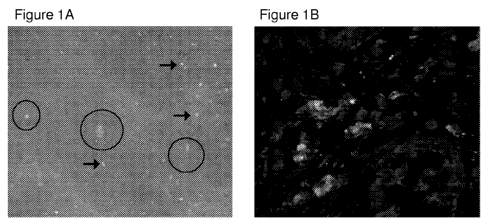
BRIEF DESCRIPTION OF THE DRAWINGS
Figure 1 describes ex vivo molecular imaging of mTNFa in surgical gut
specimens
from CD patients using fluorescent adalimumab. Figure 1A depicts ex vivo
molecular
imaging of mTNFa in surgically resected gut specimens from CD patients which
were
CA 02857597 2014-05-30
WO 2013/080050
PCT/1B2012/002933
incubated with fluorescent adalimumab to mimic topical application during
endoscopy.
Specific signals for mTNFa are indicated by arrows and single crypts with
crypt lumina are
within the circles. One representative experiment out of 5 is shown. Figure 1B
depicts
confocal microscopy of gut cryosection with mTNFa expressing immune cells
(arrows) from
the same patients upon immunohistochemical staining with fluorescent
adalimumab. One
representative experiment out of five is shown.
Figure 2 provides in vivo and ex vivo molecular imaging of mTNFa positive
mucosal
immune cells in the gut of CD patients. Figure 2A depicts in vivo specific
signals for mTNFa
positive mucosal cells (arrows) upon topical administration of fluorescent
adalimumab to the
inflamed gut of a CD patient. One representative image from 25 CD patients is
shown
(x1000 magnification). Figure 2B is an image showing molecular imaging of
single mTNFa
positive cells (arrows) in mucosa below crypts in CD patients (obtained by
digital
postprocessing of confocal in vivo images). Figure 2C provides a high
magnification image
of a single mTNFa positive cell in the lamina propria of a CD patient upon
topical
administration of fluorescent adalimumab in vivo (x1000). Figure 2C revealed
the
membranous fluorescence pattern of the mTNFa positive cell. Membranous cell
staining of
mTNFa in mucosal immune cells was comparable to the images obtained by
molecular
imaging in vivo. Quantitative analysis of ex vivo staining demonstrated that
patients with
clinical response to adalimumab therapy after 12 weeks had a significantly
higher number of
mTNFa expressing cells (mean number of 24 mTNFa expressing cells/high power
field) than
patients without clinical response (mean number of 13 mTNFa expressing
cells/high power
field). These results were statistically significant (Mean values s.e.m.;
*p=0.02) (Figure
2D).
Figure 3 provides clinical findings upon adalimumab treatment and in vivo
molecular
imaging of mTNFa-positive mucosal immune cells in CD. Figure 3A depicts in
vivo
molecular imaging of low (left panel) and high (right panel) numbers of mTNFa
expressing
immune cells in the inflamed intestinal mucosa of CD patients. Images
represent one quarter
of full scale confocal endomicroscopic images (475 p m x 475 pm). Figure 3B
shows the
mean mTNFa-positive cells in relation to whether or not a patient responded to
adalimumab
therapy. Data represent mean values s.e.m.; *p= 0.00003. Figure 3C depicts
the mean
histological inflammatory score of sections from mucosal biopsies from the
area where
molecular imaging in vivo was performed. Inflammation in these histological
sections were
blinded and graded by a pathologist with values ranging from 0 (no
inflammation) to 3 (high
inflammation). Data represent mean values s.e.m.; n.s. not significant.
Figure 3D
6
CA 02857597 2014-05-30
WO 2013/080050
PCT/1B2012/002933
graphically depicts the clinical response (defined as a reduction of the CDAI
score by > 100
points) after 12 weeks of adalimumab treatment. Response rates are shown for
all CD
patients in the study (n=25) as well as for the patients with low (n=13) and
high (n=12)
mTNFa expression. Patients in the high mTNFa group showed a markedly higher
response
rate as compared to the group with low numbers of mTNFa positive cells.
Figure 4 Figure 4A provides a SDS gel electrophoresis of fluorescein labelled
adalimumab (left panel is UV light exposure and right panel is Coomassie
staining). (H)
represents adalimumab and (HF1) represents fluorescein isothiocynate-
adalimumab. Figure
4B provides a hypothetical model of fluorescent adalimumab based on the
analysis provided
in Figure 4A.
Figure 5 describes clinical findings upon adalimumab therapy. Figure 5A
graphically
depicts the clinical outcome analysis showing that CD patients with a higher
number of
mTNFa positive intestinal cells had a statistically significant reduction of
their CDAI levels
after 4 and 12 weeks of adalimumab treatment in comparison to the baseline
CDAI before
initiation of adalimumab therapy. Patients were subsequently followed over a
period of 52
weeks. In the follow up of the patients with high mTNFa expression it was
shown that this
group has a sustained significant reduction of the CDAI score even one year
after the
initiation of the adalimumab treatment. In contrast, patients with low numbers
of mTNFa
positive cells did not show any significant reduction in CDAI scores. Data
represent mean
values s.e.m.; *p= 0.04; **p= 0.02. ***p= 0.006. Figure 5B graphically
depicts results
showing that patients with high numbers of mTNFa expressing cells had a
statistically
significant reduction of their corticosteroid use after 4 and 12 weeks of
adalimumab treatment
in comparison to patients with low numbers of mTNFa expressing cells. Data
represent mean
values s.e.m.; *p= 0.04.
DETAILED DESCRIPTION OF THE INVENTION
The instant invention provides solves both the problems of determining which
patients will be responsive to an anti-TNFa therapy, and alsoproviding
improved methods of
treatment.
In order that the present invention may be more readily understood, certain
terms are
first defined.
7
CA 02857597 2014-05-30
WO 2013/080050
PCT/1B2012/002933
I. Definitions
As used herein, the term "inflammatory bowel disease" or "IBD", used
interchangeably herein, refers to inflammatory conditions of the large and
small intestine.
Examples of an inflammatory bowel disease include, but are not limited to,
Crohn's disease
(also referred to herein as "CD") and ulcerative colitis.
As used herein, the term "intestinal mucosa" refers to the lining of the
intestines. The
mucosa is the innermost layer of the gastrointestinal tract and surrounds the
lumen, or open
space, within the tube. In one embodiment, the intestinal mucosa includes the
lining of the
small intestine and the large intestine (which includes the cecum, colon,
rectum and anal
canal). In one embodiment, the intestinal mucosa includes the lining of the
esophagus,
stomach, small intestine and the large intestine.
As used herein, the term "expression", refers to detecting transcription of
the gene
encoding tumor necrosis factor alpha (TNFa) or to detecting translation of
TNFa protein. To
detect expression of TNFa refers to the act of actively determining whether
TNFa is
expressed or not. To quantitate expression refers to the act of determining
the level of TNFa,
e.g., number of mTNFa positive cells. Detecting and/or quantitating expression
can include
determining whether TNFa expression is upregulated as compared to a control
level,
downregulated as compared to a control level, or substantially unchanged as
compared to a
control level. Therefore, the step of quantitating and/or detecting expression
does not require
that expression of TNFa actually is upregulated or downregulated, but rather,
can also
include detecting no expression of TNFa or detecting that the expression of
TNFa has not
changed or is not different (i.e., detecting no significant expression of TNFa
or no significant
change in expression of TNFa as compared to a control). In one embodiment,
expression
refers to detecting TNFa protein as it is found in the membrane of the cell
(i.e., detecting
mTNFa).
The term "level" or "amount" as used herein refers to the measurable quantity
of
TNFa. The amount may be either (a) an absolute amount as measured in an
appropriate unit,
e.g., number of cells, fluorescence intensity, molecules, moles or weight per
unit volume or
cell or (b) a relative amount. The level of expression of TNFa can be
considered "high",
"low", "increased" or "decreased" relative to a control level of expression or
relative to the
level of expression of TNFa in a "responder", relative to either the level of
expression of
TNFa in a "non-responder", or, in another embodiment, the level of expression
of a subject
who does not have an IBD. In one embodiment, the "level of expression" refers
to the level
8
CA 02857597 2014-05-30
WO 2013/080050
PCT/1B2012/002933
of expression of mTNFa (e.g., the number of cells expressing mTNFa on their
cell surface) in
a sample from a subject or observed in the patient in vivo.
The term "control level" refers to an accepted or pre-determined level of TNFa
which
is used to compare the TNFa level derived from a sample of a patient or
observed in the
patient in vivo. In one embodiment, the control level is based on a subject(s)
having IBD
who responded to treatment with a TNFa inhibitor. In another embodiment, the
control level
indicates the TNFa level of an unaffected, i.e., non-disease, state of a
subject who does not
have IBD. In another embodiment, the control level indicates a subject or
subjects having
IBD who did not respond to treatment with a TNFa inhibitor, and, therefore,
represents the
disease state of a non-responder to anti-TNFa therapy. When compared to the
control level
of TNFa, deviation from the control level generally indicates either that the
subject will be
responsive to treatment of an IBD with a TNFa inhibitor or will not be
responsive.
Alternatively, when compared to the control level, equivalence to the control
level generally
indicates confirmation of responsiveness or lack thereof.
As used herein, "responder" includes, but is not limited to, a subject with
IBD who
has improved clinical disease status following treatment with a TNFa inhibitor
(e.g.,
reduction in CDAI score or reduction in use of corticosteroids). In one
embodiment, a
responder is a subject having IBD who achieves a reduction of 100 points or
more in their
Crohn's Disease Activity Index (CDAI) score following treatment with a TNFa
inhibitor. In
one embodiment, a responder is a subject having IBD who achieves a reduction
of 100 points
or more in their Crohn's Disease Activity Index (CDAI) score in a specific
time frame
following treatment with a TNFa inhibitor. As used herein, "non-responder"
includes, but is
not limited to, a subject with IBD who has no, or limited improvement in their
clinical
disease status following treatment with a TNFa inhibitor (e.g., lack of
reduction in CDAI
score, lack of reduction in use of corticosteroids). In one embodiment, a non-
responder is a
subject having IBD who fails to achieve a reduction of 100 points or more in
their Crohn's
Disease Activity Index (CDAI) score following treatment with a TNFa inhibitor.
In one
embodiment, a non-responder is a subject having IBD who fails to achieve a
reduction of 100
points or more in their Crohn's Disease Activity Index (CDAI) score in a
specific time frame
following treatment with a TNFa inhibitor.
The term "sample" as used herein refers to a collection or image of similar
cells or
tissue obtained from a subject. The source of the tissue or cell sample may be
solid tissue as
from a fresh, frozen and/or preserved organ or tissue sample or biopsy or
aspirate. In a
9
CA 02857597 2014-05-30
WO 2013/080050
PCT/1B2012/002933
preferred embodiment, the sample is obtained from the intestinal mucosa of a
subject. In one
embodiment, the term "sample" includes an image of the intestinal mucosa from
a subject.
The term "subject" or "patient," as used herein interchangeably, refers to
either a
human or non-human animal. In one embodiment, the subject is a human.
As used herein, "TNFa" (or "TNFa") is intended to refer to a human cytokine
that
exists as a 17kD secreted form and a 26 kD membrane associated form
(abbreviated here as
"mTNFa"), the biologically active form of which is composed of a trimer of
noncovalently
bound 17 kD molecules. The structure of TNFa is described further in, for
example Pennica
et al. (1984) Nature 312: 724-729; Davis et al. (1987) Biochemistry 26:1322-
1326; and Jones
et al. (1989) Nature 338:225-228. The term TNFa is intended to include
recombinant human
TNFa (rhTNFa), which can be prepared by standard recombinant expression
methods or
purchased commercially (R & D Systems, Catalog No. 210-TA, Minneapolis, MN).
As used herein "mTNFa" (or "mTNFa") refers to membrane TNFa.
As used herein, "TNFa inhibitor" includes agents which inhibit TNFa. Examples
of
TNFa inhibitors include etancercept (ENBREL , Immunex), infliximab (REMICADE ,
Janssen / Johnson and Johnson), adalimumab (HUMIRA , also referred to as D2E7,
Abbott
Laboratories), golimumab (SIMPONI , Janssen / Johnson and Johnson), CDP 571
(Celltech), and certolizumab pegol (CIMZIA kor CDP 870 (Celltech) and other
compounds
which inhibit TNFa activity, such that when administered to a subject
suffering from or at
risk of suffering from a disorder in which TNFa activity is detrimental, the
disorder is treated.
The term also includes each of the anti-TNFa human antibodies and antibody
portions
described herein as well as those described in U.S. Patent Nos. 6,090,382,
6,258,562,
6,509,015, and 7,223,394, each of which is incorporated by reference in its
entirety.
As used herein, "detectably labeled TNFa inhibitor" refers to a TNFa inhibitor
which
is linked (e.g., covalently) to a molecule and can be used to determine the
presence of the
TNFa inhibitor. The detectably labeled TNFa inhibitor may be detected by the
methods
including, but not limited to, fluorescent, colormetric, spectrophotometric,
optic, luminescent,
radioactive, or X means.
The term "antibody", as used herein, broadly refers to any immunoglobulin (Ig)
molecule comprised of four polypeptide chains, two heavy (H) chains and two
light (L)
chains, or any functional fragment, mutant, variant, or derivation thereof,
which retains the
essential epitope binding features of an Ig molecule. Such mutant, variant, or
derivative
antibody formats are known in the art. Nonlimiting embodiments are discussed
below.
CA 02857597 2014-05-30
WO 2013/080050
PCT/1B2012/002933
In a full-length antibody, each heavy chain is comprised of a heavy chain
variable
region (abbreviated herein as HCVR or VH) and a heavy chain constant region.
The heavy
chain constant region is comprised of three domains, CH1, CH2 and CH3. Each
light chain
is comprised of a light chain variable region (abbreviated herein as LCVR or
VL) and a light
chain constant region. The light chain constant region is comprised of one
domain, CL. The
VH and VL regions can be further subdivided into regions of hypervariability,
termed
complementarity determining regions (CDR), interspersed with regions that are
more
conserved, termed framework regions (FR). Each VH and VL is composed of three
CDRs
and four FRs, arranged from amino-terminus to carboxy-terminus in the
following order:
FR1, CDR1, FR2, CDR2, FR3, CDR3, FR4. Immunoglobulin molecules can be of any
type
(e.g., IgG, IgE, IgM, IgD, IgA and IgY), class (e.g., IgG 1, IgG2, IgG 3,
IgG4, IgAl and
IgA2) or subclass.
The term "antigen-binding portion" of an antibody (or simply "antibody
portion" or
"antibody fragment"), as used herein, refers to one or more fragments of an
antibody that
retain the ability to specifically bind to an antigen (e.g., hIL-13). It has
been shown that the
antigen-binding function of an antibody can be performed by fragments of a
full-length
antibody. Such antibody embodiments may also be bispecific, dual specific, or
multi-specific
formats; specifically binding to two or more different antigens. Examples of
binding
fragments encompassed within the term "antigen-binding portion" of an antibody
include (i) a
Fab fragment, a monovalent fragment consisting of the VL, VH, CL and CH1
domains; (ii) a
F(ab')2 fragment, a bivalent fragment comprising two Fab fragments linked by a
disulfide
bridge at the hinge region; (iii) a Fd fragment consisting of the VH and CH1
domains; (iv) a
Fv fragment consisting of the VL and VH domains of a single arm of an
antibody, (v) a dAb
fragment (Ward et al., (1989) Nature 341:544-546, Winter et al., PCT
publication WO
90/05144 Al herein incorporated by reference), which comprises a single
variable domain;
and (vi) an isolated complementarity determining region (CDR). Furthermore,
although the
two domains of the Fv fragment, VL and VH, are coded for by separate genes,
they can be
joined, using recombinant methods, by a synthetic linker that enables them to
be made as a
single protein chain in which the VL and VH regions pair to form monovalent
molecules
(known as single chain Fv (scFv); see e.g., Bird et al. (1988) Science 242:423-
426; and
Huston et al. (1988) Proc. Natl. Acad. Sci. USA 85:5879-5883). Such single
chain antibodies
are also intended to be encompassed within the term "antigen-binding portion"
of an
antibody. Other forms of single chain antibodies, such as diabodies are also
encompassed.
11
CA 02857597 2014-05-30
WO 2013/080050
PCT/1B2012/002933
Diabodies are bivalent, bispecific antibodies in which VH and VL domains are
expressed on
a single polypeptide chain, but using a linker that is too short to allow for
pairing between the
two domains on the same chain, thereby forcing the domains to pair with
complementary
domains of another chain and creating two antigen binding sites (see e.g.,
Holtiger et al.
(1993) Proc. Natl. Acad. Sci. USA 90:6444-6448; Poljak et al. (1994) Structure
2:1121-
1123). Such antibody binding portions are known in the art (Kontermann and
Dubel eds.,
Antibody Engineering (2001) Springer-Verlag. New York. 790 pp. (ISBN 3-540-
41354-5).
An "isolated antibody", as used herein, is intended to refer to an antibody
that is
substantially free of other antibodies having different antigenic
specificities (e.g., an isolated
antibody that specifically binds hTNFa is substantially free of antibodies
that specifically
bind antigens other than hTNFa). An isolated antibody that specifically binds
hTNFa may,
however, have cross-reactivity to other antigens, such as TNFa molecules from
other species
(discussed in further detail below). Moreover, an isolated antibody may be
substantially free
of other cellular material and/or chemicals.
A "neutralizing antibody", as used herein (or an "antibody that neutralized
hTNFa
activity"), is intended to refer to an antibody whose binding to hTNFa results
in inhibition of
the biological activity of hTNFa. This inhibition of the biological activity
of hTNFa can be
assessed by measuring one or more indicators of hTNFa biological activity,
such as hTNFa-
induced cytotoxicity (either in vitro or in vivo), hTNFa-induced cellular
activation and
hTNFa binding to hTNFa receptors. These indicators of hTNFa biological
activity can be
assessed by one or more of several standard in vitro or in vivo assays known
in the art (see
U.S. Patent No. 6,090,382). In one embodiment, the ability of an antibody to
neutralize
hTNFa activity is assessed by inhibition of hTNFa-induced cytotoxicity of L929
cells. As
an additional or alternative parameter of hTNFa activity, the ability of an
antibody to inhibit
hTNFa-induced expression of ELAM-1 on HUVEC, as a measure of hTNFa-induced
cellular activation, can be assessed.
The term "surface plasmon resonance", as used herein, refers to an optical
phenomenon that allows for the analysis of real-time biospecific interactions
by detection of
alterations in protein concentrations within a biosensor matrix, for example
using the
BIAcore system (Pharmacia Biosensor AB, Uppsala, Sweden and Piscataway, NJ).
For
further descriptions, see Example 1 of U.S. Patent 6,258,562 and Jonsson et
al. (1993) Ann.
12
CA 02857597 2014-05-30
WO 2013/080050
PCT/1B2012/002933
Biol. Clin. 51:19; JOnsson et al. (1991) Biotechniques 11:620-627; Johnsson et
al. (1995) J.
MoL Recognit. 8:125; and Johnnson et al. (1991) Anal.Biochem.198:268.
The term "Koff", as used herein, is intended to refer to the off rate constant
for
dissociation of an antibody from the antibody/antigen complex.
The term "Kd", as used herein, is intended to refer to the dissociation
constant of a
particular antibody-antigen interaction.
The term "IC50" as used herein, is intended to refer to the concentration of
the
inhibitor required to inhibit the biological endpoint of interest, e.g.,
neutralize cytotoxicity
activity.
The term "nucleic acid molecule", as used herein, is intended to include DNA
molecules and RNA molecules. A nucleic acid molecule may be single-stranded or
double-
stranded, but preferably is double-stranded DNA.
The term "isolated nucleic acid molecule", as used herein in reference to
nucleic acids
encoding antibodies or antibody portions (e.g., VH, VL, CDR3) that bind hTNFa,
is intended
to refer to a nucleic acid molecule in which the nucleotide sequences encoding
the antibody
or antibody portion are free of other nucleotide sequences encoding antibodies
or antibody
portions that bind antigens other than hTNFa, which other sequences may
naturally flank the
nucleic acid in human genomic DNA. Thus, for example, an isolated nucleic acid
of the
invention encoding a VH region of an anti-hTNFa antibody contains no other
sequences
encoding other VH regions that bind antigens other than hTNFa.
The term "dose," as used herein, refers to an amount of TNFa inhibitor which
is
administered to a subject.
The term "multiple-variable dose" includes different doses of a TNFa inhibitor
which
are administered to a subject for therapeutic treatment. "Multiple-variable
dose regimen" or
"multiple-variable dose therapy" describe a treatment schedule which is based
on
administering different amounts of TNFa inhibitor at various time points
throughout the
course of treatment. Multiple-variable dose regimens are described in US
Patent Application
Publication No. 20060009385, which is incorporated by reference herein in its
entirety.
The term "dosing", as used herein, refers to the administration of a substance
(e.g., an
anti-TNFa antibody) to achieve a therapeutic objective (e. g. , the treatment
of IBD).
13
CA 02857597 2014-05-30
WO 2013/080050
PCT/1B2012/002933
The terms "biweekly dosing regimen", "biweekly dosing", and "biweekly
administration", as used herein, refer to the time course of administering a
substance (e.g., an
anti-TNFa antibody) to a subject to achieve a therapeutic objective. The
biweekly dosing
regimen is not intended to include a weekly dosing regimen. Preferably, the
substance is
administered every 9-19 days, more preferably, every 11-17 days, even more
preferably,
every 13-15 days, and most preferably, every 14 days. Biweekly dosing is
further described
in US Patent Application Publication No. 20030235585, which is incorporated by
reference
herein in its entirety.
The term "combination" as in the phrase "a first agent in combination with a
second
agent" includes co-administration of a first agent and a second agent, which
for example may
be dissolved or intermixed in the same pharmaceutically acceptable carrier, or
administration
of a first agent, followed by the second agent, or administration of the
second agent, followed
by the first agent. The present invention, therefore, includes methods of
combination
therapeutic treatment and combination pharmaceutical compositions.
The term "concomitant" as in the phrase "concomitant therapeutic treatment"
includes
administering an agent in the presence of a second agent. A concomitant
therapeutic
treatment method includes methods in which the first, second, third, or
additional agents are
co-administered. A concomitant therapeutic treatment method also includes
methods in
which the first or additional agents are administered in the presence of a
second or additional
agents, wherein the second or additional agents, for example, may have been
previously
administered. A concomitant therapeutic treatment method may be executed step-
wise by
different actors. For example, one actor may administer to a subject a first
agent and a
second actor may to administer to the subject a second agent, and the
administering steps may
be executed at the same time, or nearly the same time, or at distant times, so
long as the first
agent (and additional agents) are after administration in the presence of the
second agent (and
additional agents). The actor and the subject may be the same entity (e.g.,
human).
The term "combination therapy", as used herein, refers to the administration
of two or
more therapeutic substances, e.g., an anti-TNFa antibody and another drug. The
other
drug(s) may be administered concomitant with, prior to, or following the
administration of an
anti-TNFa antibody.
The term "kit" as used herein refers to a packaged product comprising
components
with which to determine the responsiveness of a subject to treatment of IBD
with a TNFa
14
CA 02857597 2014-05-30
WO 2013/080050
PCT/1B2012/002933
inhibitor, e.g., a means for detecting m TNFa in the intestinal mucosa of a
subject. In one
embodiment, the kit further provides components for administering aTNFa
antibody of the
invention for treatment of IBD. The kit preferably comprises a box or
container that holds
the components of the kit. The box or container is affixed with a label or a
Food and Drug
Administration approved protocol. The box or container holds components of the
invention
which are preferably contained within plastic, polyethylene, polypropylene,
ethylene, or
propylene vessels. The vessels can be capped-tubes or bottles. The kit can
also include
instructions for administering the TNFa antibody of the invention.
II. Methods of the Invention
An unmet need in the treatment of IBD is to establish predictive biomarkers
for
therapeutic responders in order to avoid exposure of non-responders to anti-
TNFa therapy,
thus decreasing morbidity in patients with a low likelihood of response and
enhancing safety
and cost effective use of this treatment. Although patients with elevated CRP-
levels in the
blood have demonstrated higher response rates to anti-TNFa treatment (Vermeire
et al.
Inflamm Bowel Dis 10, 661-665 (2004)), there is a need for additional specific
biomarkers
that allow the prediction of response to anti-TNFa therapy for inflammatory
bowel diseases.
Thus, the prediction of clinical responsiveness to anti-TNFa antibodies is a
key clinical
problem and approaches aiming at a better prediction of responsiveness will
have positive
effects on the therapeutic use of these substances. The instant invention
provides unexpected
results which solve the problem of how to predict which IBD patients will be
responsive to
anti-TNFa therapy. The instant invention also provides safe ways of delivering
anti-TNFa
antibodies to a subject having IBD though topical delivery, thus providing
improved methods
of treatment. In one embodiment, the anti- TNFa antibody is topically
administered to a
subject having IBD, e.g., Crohn's disease, where the subject was selected as
being a
responder to TNFa inhibitor therapy.
Methods for Determining Responsiveness to Treatment
The invention provides methods for predicting or determining the
responsiveness of a
subject having IBD to treatment with a TNFa inhibitor. Thus, the invention
provides
methods for determining whether a TNFa inhibitor will be effective for the
treatment of a
subject having IBD. In one embodiment, these methods comprise determining the
level of
expression of TNFa in the cells of the intestinal mucosa of a subject having
IBD and
CA 02857597 2014-05-30
WO 2013/080050
PCT/1B2012/002933
comparing the level of expression of TNFa in the cells of the intestinal
mucosa of the subject
to a control level of expression of TNFa.
The control level of TNFa that may be used to determine responsiveness of a
subject
may be the level of TNFa, e.g., mTNFa, in the intestinal mucosa of a responder
or a non-
responder. A higher level of expression of TNFa in the cells of the intestinal
mucosa of the
subject as compared to a control level of expression of TNFa of a non-
responder indicates
that the subject will be responsive to treatment with a TNFa inhibitor. In
contrast, an
equivalent or lower level of TNFa in the cells of the intestinal mucosa of the
subject as
compared to the control level of expression of TNFa which is that of a non-
responder
indicates that the subject will not be responsive to treatment with a TNFa
inhibitor. In
another alternative, the control level of expression of TNFa may be the level
of expression of
TNFa in the intestinal mucosa of a responder. In such a case, if the subject's
level of TNFa
is greater or equivalent to the control level, then the subject having IBD
will be responsive to
treatment with a TNFa inhibitor. If the subject's level of TNFa is less than
the control level,
however, where the control is from a responder, then that is indicative of the
fact that the
subject having IBD will not be responsive to treatment with a TNFa inhibitor.
In one
embodiment, levels of TNFa are determined by the number of mTNFa positive
cells in a
sample from the subject.
In one embodiment, the invention provides a method for determining the
responsiveness of a subject having inflammatory bowel disease (IBD) to
treatment with a
TNFa inhibitor, the method comprising determining the level of expression of
TNFa in the
cells of the intestinal mucosa of the subject having IBD; and comparing the
level of
expression of TNFa in the cells of the intestinal mucosa of the subject to a
control level of
expression of TNFa from a non-responder, wherein a higher level of expression
of TNFa in
the cells of the intestinal mucosa of the subject as compared to the control
level of expression
of TNFa indicates that the subject will be responsive to treatment with the
TNFa inhibitor,
thereby predicting the responsiveness of the subject having IBD to treatment
with the TNFa
inhibitor.
In an alternative, the invention provides a method of determining whether a
TNFa
inhibitor will be effective for the treatment of a subject having inflammatory
bowel disease
(IBD), the method comprising determining the level of expression of TNFa in
the cells of the
intestinal mucosa of the subject having IBD, wherein a higher level of
expression of TNFa in
the cells of the intestinal mucosa of the subject as compared to a control
level of expression
of TNFa for a nonresponder indicates that the TNFa inhibitor will be effective
for the
16
CA 02857597 2014-05-30
WO 2013/080050
PCT/1B2012/002933
treatment of the subject having IBD, thereby determining whether a TNFa
inhibitor will be
effective for the treatment of the subject having IBD.
In one embodiment, the level of expression may be determined by assessing the
level
of expression of TNFa in cells which do not appear to be involved with disease
and by
comparing the foregoing lower level of TNFa with the level of expression of
TNFa in cells in
an area with disease involvement. For example, when endoscopy or another
medical
procedure reveals the presence of IBD involvement in one portion of an organ,
the lower
level of expression of TNFa may be assessed using the non-affected portion of
the organ, and
this lower level of expression may be compared with the level of expression of
TNFa in an
affected portion (e.g., inflamed mucosa) of the organ.
The level of expression of TNFa may be assessed in a variety of ways. In one
embodiment of the invention, the level of expression of membrane TNFa (mTNFa)
in the
cells of the intestinal mucosa of the subject having IBD is determined by
counting the
number of mTNFa positive cells in a sample from the subject. This assessment
may be
performed in vivo, e.g., using endomicroscopy, or ex vivo, e.g., using
histology analysis of
intestinal mucosa biopsy sample(s) from a subject.
An anti-TNFa antibody used in the detection methods of the invention may be
labelled with a detectable agent suitable for either in vivo or ex vivo
analysis. Useful
detectable agents with which an antibody or antibody portion of the invention
may be
derivatized include fluorescent compounds for either in vivo or ex vivo
analysis. Exemplary
fluorescent detectable agents include fluorescein, fluorescein isothiocyanate,
rhodamine, 5-
dimethylamine- 1-napthalenesulfonyl chloride, phycoerythrin and the like. An
antibody may
also be derivatized with detectable enzymes, such as alkaline phosphatase,
horseradish
peroxidase, glucose oxidase and the like for ex vivo analysis. When an
antibody is
derivatized with a detectable enzyme, it is detected by adding additional
reagents that the
enzyme uses to produce a detectable reaction product. For example, when the
detectable
agent horseradish peroxidase is present, the addition of hydrogen peroxide and
diaminobenzidine leads to a colored reaction product, which is detectable. An
antibody may
also be derivatized with biotin, and detected through indirect measurement of
avidin or
streptavidin binding.
In one embodiment of the invention, the level of expression of TNFa in the
intestinal
mucosa of a subject having IBD is determined using an in vivo assay. In vivo
imaging may
be used to determine whether a subject having IBD will be responsive to
treatment with a
TNFa inhibitor, e.g., an anti-TNFa antibody. Such imaging may be performed
during a
17
CA 02857597 2014-05-30
WO 2013/080050
PCT/1B2012/002933
colonoscopy on the subject, e.g., a colonoscopy to determine the severity of
the IBD. During
the procedure, an anti-TNFa antibody may be delivered locally to the
intestinal mucosa to
determine TNFa expression. For example, a spray catheter may be used in
conjunction with
an endoscope (e.g., Glo-Tip Spray Catheter; Cook Medical) to topically deliver
a TNFa
inhibitor, e.g., an anti-TNFa antibody to the subject for analysis.
Preferably, the antibody is
detectably labeled, e.g., FITC-adalimumab. Following topical administration of
the antibody,
in vivo molecular imaging may be performed to determine the level of mTNFa
expression in
the mucosa of the subject. In one embodiment, levels of TNFa are determined
according to
the number of TNFa positive cells counted in a given image.
In order to determine the expression level of TNFa in the intestinal mucosa, a
detectably labeled anti-TNFa antibody, or antigen-binding portion thereof, may
be
administered to the subject, for example, by using a spraying catheter. The
labeled antibody,
or antigen-binding portion thereof, may be delivered to the intestinal tract
of the subject
during a colonoscopy. In one embodiment, the anti-TNFa antibody, or antigen-
binding
portion thereof, is delivered to a mucosal site within the large intestine
having inflammation.
Following delivery, imaging may be performed according to standard methods
known in the
art. In one embodiment, imaging of the intestinal mucosa of the subject is
performed using
confocal laser endomicroscopy.
In one embodiment the level of expression of TNFa is determined by topically
applying a detectably labeled TNFa inhibitor to the cells of the intestinal
mucosa of a subject
having IBD. In yet another embodiment, the detectably labeled TNFa inhibitor
is labeled
with fluorescein isothiocyanate.
Endoscopy has witnessed a rapid evolution of endoscopic techniques for
improved
detection of inflammatory and neoplastic lesions in recent years (Neumann et
al.
Gastroenterology 139, 388-392, 392 e381-382 (2010); Kendall et al. The Journal
of
pathology 200, 602-609 (2003); Evans et al. Gastrointestinal endoscopy 65, 50-
56 (2007);
Lovat et al. Gut 55, 1078-1083 (2006); Herrero et al. Gastroenterology Clinics
of North
America 39, 747-758 (2010); Qiu et al. Nat Med 16, 603-606, 601p following 606
(2010);
and Waldner et al. Nat Protoc 6, 1471-1481 (2011)). In addition to filter
techniques such as
narrow band imaging, optical coherence tomography, Raman spectroscopy, elastic
scattering
spectroscopy and multispectral imaging have been introduced. Furthermore,
confocal laser
endomicroscopy has recently been shown to augment detection of local
inflammation and
neoplasia in the gastrointestinal tract by providing optical biopsies and in
vivo imaging during
ongoing endoscopy (Kiesslich et al. Gastroenterology 132, 874-882 (2007) and
Kiesslich et
18
CA 02857597 2014-05-30
WO 2013/080050
PCT/1B2012/002933
al. Gut (2011)). For instance, endomicroscopy has been used in esophageal
squamous cell
carcinoma, Barrett's esophagus, colonic polyps, collagenous colitis and CD. In
addition,
endomicroscopy permitted the identification of neoplastic lesions during
colonoscopy in
patients by using a labelled heptapeptide derived from a phage library (Hsiung
et al. Nat Med
14, 454-458 (2008)).
Thus, in vivo methods described herein may be accomplished using
endomicroscopy,
including confocal laser endomicroscopy. Examples of confocal laser
endomicroscopes that
may be used include the Pentax Endomicroscopy System (Pentax) and the
Cellvizio high
resolution confocal microscope (Mauna Kea Technologies).
In one embodiment, 20 or more TNFa positive cells in an in vivo image that is
at least
475p m x 475pm indicates that the subject will be responsive to treatment with
an anti-TNFa
antibody, or antigen-binding portion thereof. Alternatively, less than 20 TNFa
positive cells
in an in vivo image that is at least 475p m x 475p m indicates that the
subject will not be
responsive to treatment with an anti-TNFa antibody, or antigen-binding portion
thereof.
Optical sections of 475pm x 475p m can be obtained using a high resolution
confocal
microscope, such as, but not limited to, the Pentax endomicroscopic system
(Pentax).
In another embodiment, 10 or more TNFa positive cells in an in vivo image that
is at
least 240p m x 240p m indicates that the subject will be responsive to
treatment with an anti-
TNFa antibody, or antigen-binding portion thereof. Alternatively, less than 10
TNFa
positive cells in an in vivo image that is at least 240pm x 240p m indicates
that the subject
will not be responsive to treatment with an anti-TNFa antibody, or antigen-
binding portion
thereof. Optical sections of 240 m x 240 m can be obtained using a high
resolution
confocal microscope, such as, but not limited to, the Cellvizio high
resolution confocal
microscope (Mauna Kea Technologies).
In one embodiment, at least a 180% increase (or at least a 185%, 190%, 195%,
200%,
205%, 210%, 215%, 220%, or 225%) in the level of expression of TNFa, e.g., the
number of
TNFa positive cells, in an in vivo image relative to the same size image from
a non-responder
control indicates that the subject will be responsive to treatment with an
anti- TNFa antibody,
or antigen-binding portion thereof. Alternatively, an equivalent or increased
level of
expression of TNFa, e.g., number of TNFa positive cells, in an in vivo image
relative to the
same size image from a responder control indicates that the subject will be
responsive to
treatment with an anti-TNFa antibody, or antigen-binding portion thereof. In
one
embodiment, an increase of 230%, 235%, 240%, 245%, 250%, 255%, 260%, 265%,
270%,
19
CA 02857597 2014-05-30
WO 2013/080050
PCT/1B2012/002933
275%, 280%, 285%, 290%, 295%, or 300% in the level of expression of TNFa,
e.g., number
of TNFa positive cells, in an in vivo image relative to the same size image
from a non-
responder control indicates that the subject will be responsive to treatment
with an anti-
TNFa antibody, or antigen-binding portion thereof. The level of expression of
TNFa, e.g.,
the number of TNFa positive cells may also be determined ex vivo using
standard histology
techniques, as described below.
The invention also provides methods of predicting the responsiveness of a
subject
having IBD to treatment with a TNFa inhibitor where the level of expression of
TNFa is
determined ex vivo. In ex vivo methods, the level of expression of TNFa in a
sample of cells
from the intestinal mucosa of a subject with IBD may be compared with sample
of cells from
a control (responder or non-responder). A lower level of expression of TNFa in
the subject's
sample, relative to a responder sample, is an indication that the subject will
not respond to
treatment with a TNFa inhibitor. A higher level of expression of TNFa in the
subject's
sample, relative to the non-responder sample, is an indication that that
subject will respond to
treatment with a TNFa inhibitor. Such a sample may be obtained by taking a
biopsy from the
mucosa of the intestinal tract of a subject having IBD.
Samples useful in the methods of invention for determining the level of TNFa
expression include any tissue, cell, biopsy, or surgically resected sample
from a subject
having IBD that may express TNFa. Body samples for ex vivo analysis may be
obtained from
a subject using a variety of techniques know in the art including, for
example, during a
surgical procedure or by use of a biopsy or by scraping or swabbing an area.
The samples
may, for example, be obtained during a colonoscopy. In particular embodiments,
the body
sample comprises intestinal tissue samples. In one embodiment, the tissue
sample is a small
intestine tissue sample or a large intestine tissue sample.
In one embodiment, the level of expression of TNFa is detected on a protein
level
using, for example, antibodies that specifically bind TNFa. The level of TNFa
expression
may be determined by topically applying an anti-TNFa antibody, or antigen-
binding portion
thereof, to the intestinal mucosa of a subject having IBD, obtaining a sample
from a biopsy of
the intestinal mucosa on which the anti-TNFa antibody, or antigen-binding
portion thereof,
was applied, and assaying the sample for levels of expression of TNFa. The
anti- TNFa
antibody, or antigen-binding portion thereof, may be labelled with a
detectable agent, e.g.,
FITC. Alternatively, the anti- TNFa antibody, or antigen-binding portion
thereof, may not be
labelled and may be assayed according to methods known in the art. In another
embodiment,
the sample is obtained via a biopsy from the intestinal mucosa of a subject
having IBD,
CA 02857597 2014-05-30
WO 2013/080050
PCT/1B2012/002933
whereupon an anti-TNFa antibody, or antigen-binding portion thereof, is
applied ex vivo to
the sample for analysis of the expression level of TNFa.
In one embodiment, 15 or more (e.g., 16 or more, 17 or more, 18 or more, 19 or
more,
or 20 or more) TNFa positive cells in an image obtained from ex vivo analysis
of an intestinal
mucosa sample from a subject having IBD (for example, an image that is
magnified by a SP-
confocal microscope with a 63x/1.3NA objective (Leica Microsystems)) indicates
that the
subject will be responsive to treatment with an anti-TNFa antibody, or antigen-
binding
portion thereof. Alternatively, less than 15 (e.g., 14 or less, 13 or less,
etc.) TNFa positive
cells in an in vivo image (for example an image that is at least magnified by
a SP-5 confocal
microscope with a 63x/1.3NA objective (Leica Microsystems)) indicates that the
subject will
not be responsive to treatment with an anti-TNFa antibody, or antigen-binding
portion
thereof.
In one embodiment, a 170% increase in the level of TNFa expression, e.g.,
number of
TNFa positive cells, in an image obtained from an ex vivo source, e.g., a
histological section
of the intestinal mucosa of a subject, relative to a control, e.g., an image
obtained from an ex
vivo source of a non-responder, indicates that the subject will be responsive.
In one
embodiment, an increase of 180% in the level of TNFa expression, e.g., the
number of TNFa
positive cells of a sample from a subject relative to a sample from a non-
responder indicates
that the subject will be responsive to treatment with a TNFa inhibitor.
Increases of 185%,
190%, 195%, 200%, 205%, and so forth also indicate a likelihood of
responsiveness in a
subject. Alternatively, a 170% decrease in the levels of TNFa expression,
e.g., number of
TNFa positive cells, in an image obtained from an ex vivo source, e.g., a
histological section
of the intestinal mucosa of a subject, relative to a control, e.g., an image
obtained from an ex
vivo source of a responder, indicates that the subject will be not be
responsive to TNFa
therapy for treatment of IBD. Decreases of 185%, 190%, 195%, 200%, 205%, and
so forth
also indicate a likelihood of responsiveness in a subject.
Tissue samples suitable for ex vivo detecting and quantifying the level of
expression
of TNFa may be fresh, frozen, or fixed according to methods known to one of
skill in the art.
Suitable tissue samples are preferably sectioned and placed on a microscope
slide for further
analyses. Alternatively, solid samples, i.e., tissue samples, may be analyzed.
In one embodiment, a freshly obtained biopsy sample is frozen using, for
example,
liquid nitrogen or difluorodichloromethane. The frozen sample is mounted for
sectioning
using, for example, OCT, and serially sectioned in a cryostat. The serial
sections are
21
CA 02857597 2014-05-30
WO 2013/080050
PCT/1B2012/002933
collected on a glass microscope slide. For immunohistochemical staining the
slides may be
coated with, for example, chrome-alum, gelatine or poly-L-lysine to ensure
that the sections
stick to the slides. In another embodiment, samples are fixed and embedded
prior to
sectioning. For example, a tissue sample may be fixed in, for example,
formalin, serially
dehydrated and embedded in, for example, paraffin.
Once the sample is obtained any method known in the art to be suitable for
detecting
and quantitating the level of expression of TNFa may be used (either at the
nucleic acid or,
preferably, at the protein level). Such methods are well known in the art and
include but are
not limited to western blots, northern blots, southern blots,
immunohistochemistry,
immunocytochemistry, ELISA, e.g., amplified ELISA, immunoprecipitation,
immunofluorescence, flow cytometry, immunocytochemistry, mass
spectrometrometric
analyses, e.g., MALDI-TOF and SELDI-TOF, nucleic acid hybridization
techniques, nucleic
acid reverse transcription methods, and nucleic acid amplification methods.
Samples for ex vivo analysis may need to be modified in order to make the TNFa
protein accessible to antibody binding. In a particular aspect of the
immunocytochemistry or
immunohistochemistry methods, slides may be transferred to a pretreatment
buffer and
optionally heated to increase antigen accessibility. Heating of the sample in
the pretreatment
buffer rapidly disrupts the lipid bi-layer of the cells and makes the antigens
(may be the case
in fresh specimens, but not typically what occurs in fixed specimens) (i.e.,
the TNFa) more
accessible for antibody binding. The pretreatment buffer may comprise a pH-
specific salt
solution, a polymer, a detergent, or a nonionic or anionic surfactant such as,
for example, an
ethyloxylated anionic or nonionic surfactant, an alkanoate or an alkoxylate or
even blends of
these surfactants or even the use of a bile salt. The pretreatment buffer may,
for example, be
a solution of 0.1% to 1% of deoxycholic acid, sodium salt, or a solution of
sodium laureth-
13-carboxylate (e.g., Sandopan LS) or and ethoxylated anionic complex. In some
embodiments, the pretreatment buffer may also be used as a slide storage
buffer. Any
method for making TNFa protein more accessible for antibody binding may be
used in the
practice of the invention, including the antigen retrieval methods known in
the art. See, for
example, Bibbo, et al. (2002) Acta. Cytol. 46:25-29; Saqi, et al. (2003)
Diagn. Cytopathol.
27:365-370; Bibbo, et al. (2003) Anal. Quant. Cytol. Histol. 25:8-11, the
entire contents of
each of which are incorporated herein by reference.
Following pretreatment to increase TNFa protein accessibility, samples may be
blocked using an appropriate blocking agent, e.g., a peroxidase blocking
reagent such as
hydrogen peroxide. In some embodiments, the samples may be blocked using a
protein
22
CA 02857597 2014-05-30
WO 2013/080050
PCT/1B2012/002933
blocking reagent to prevent non-specific binding of the antibody. The protein
blocking
reagent may comprise, for example, purified casein. An antibody, particularly
a monoclonal
antibody that specifically binds to TNFa is then incubated with the sample.
In one embodiment the level of expression of TNFa is determined by topically
applying a detectably labeled TNFa inhibitor, e.g., an anti- TNFa antibody, to
the cells of the
intestinal mucosa of a subject having IBD. In yet another embodiment, the
detectably labeled
TNFa inhibitor is labeled with fluorescein isothiocyanate. Alternatively, the
detectably
labeled TNFa inhibitor, e.g., an anti- TNFa antibody, may be applied directly
to a sample
obtained from the subject, e.g., a tissue biopsy.
Techniques for ex vivo antibody detection are well known in the art. Antibody
binding
to TNFa may be detected through the use of chemical reagents that generate a
detectable
signal that corresponds to the level of antibody binding and, accordingly, to
the level of
TNFa protein expression. In one of the immunohistochemistry or
immunocytochemistry
methods of the invention, antibody binding is detected through the use of a
secondary
antibody that is conjugated to a labeled polymer. Examples of labeled polymers
include but
are not limited to polymer-enzyme conjugates. The enzymes in these complexes
are typically
used to catalyze the deposition of a chromogen at the antigen-antibody binding
site, thereby
resulting in cell staining that corresponds to expression level of the
biomarker of interest.
Enzymes of particular interest include, but are not limited to, horseradish
peroxidase (HRP)
and alkaline phosphatase (AP).
In one particular immunohistochemistry or immunocytochemistry method of the
invention, antibody binding to the TNFa proteins is detected through the use
of an HRP-
labeled polymer that is conjugated to a secondary antibody. Antibody binding
can also be
detected through the use of a species-specific probe reagent, which binds to
monoclonal or
polyclonal antibodies, and a polymer conjugated to HRP, which binds to the
species specific
probe reagent. Slides are stained for antibody binding using any chromagen,
e.g., the
chromagen 3,3-diaminobenzidine (DAB), and then counterstained with hematoxylin
and,
optionally, a bluing agent such as ammonium hydroxide or TBS/Tween-20. Other
suitable
chromagens include, for example, 3-amino-9-ethylcarbazole (AEC). In some
aspects of the
invention, slides are reviewed microscopically by a cytotechnologist and/or a
pathologist to
assess cell staining, e.g., fluorescent staining (i.e., TNFa expression).
Alternatively, samples
may be reviewed via automated microscopy or by personnel with the assistance
of computer
software that facilitates the identification of positive staining cells.
23
CA 02857597 2014-05-30
WO 2013/080050
PCT/1B2012/002933
Detection of antibody binding can be facilitated by coupling the anti- TNFa
antibodies to a detectable substance. Examples of detectable substances
include various
enzymes, prosthetic groups, fluorescent materials, luminescent materials,
bioluminescent
materials, and radioactive materials. Examples of suitable enzymes include
horseradish
peroxidase, alkaline phosphatase, 13-galactosidase, or acetylcholinesterase;
examples of
suitable prosthetic group complexes include streptavidin/biotin and
avidin/biotin; examples
of suitable fluorescent materials include umbelliferone, fluorescein,
fluorescein
isothiocyanate, rhodamine, dichlorotriazinylamine fluorescein, dansyl chloride
or
phycoerythrin; an example of a luminescent material includes luminol; examples
of
bioluminescent materials include luciferase, luciferin, and aequorin; and
examples of suitable
radioactive material include 1251, 1311, 35s,
u or 3H.
In one embodiment of the invention frozen samples are prepared as described
above
and subsequently stained with antibodies against TNFa diluted to an
appropriate
concentration using, for example, Tris-buffered saline (TBS). Primary
antibodies can be
detected by incubating the slides in biotinylated anti-immunoglobulin. This
signal can
optionally be amplified and visualized using diaminobenzidine precipitation of
the antigen.
Furthermore, slides can be optionally counterstained with, for example,
hematoxylin, to
visualize the cells.
In another embodiment, fixed and embedded samples are stained with antibodies
against TNFa and counterstained as described above for frozen sections. In
addition, samples
may be optionally treated with agents to amplify the signal in order to
visualize antibody
staining. For example, a peroxidase-catalyzed deposition of biotinyl-tyramide,
which in turn
is reacted with peroxidase-conjugated streptavidin (Catalyzed Signal
Amplification (CSA)
System, DAKO, Carpinteria, CA) may be used.
One of skill in the art will recognize that the concentration of a particular
antibody
used to practice the methods of the invention will vary depending on such
factors as time for
binding, level of specificity of the antibody for TNFa, and method of sample
preparation.
Moreover, when multiple antibodies are used, the required concentration may be
affected by
the order in which the antibodies are applied to the sample, e.g.,
simultaneously as a cocktail
or sequentially as individual antibody reagents. Furthermore, the detection
chemistry used to
visualize antibody binding to TNFa must also be optimized to produce the
desired signal to
noise ratio.
24
CA 02857597 2014-05-30
WO 2013/080050
PCT/1B2012/002933
In one embodiment of the invention, proteomic methods, e.g., mass
spectrometry, are
used for detecting and quantitating the TNFa protein. For example, matrix-
associated laser
desorption/ionization time-of-flight mass spectrometry (MALDI-TOF MS) or
surface-
enhanced laser desorption/ionization time-of-flight mass spectrometry (SELDI-
TOF MS)
which involves the application of a biological sample, such as serum, to a
protein-binding
chip (Wright, G.L., Jr., et al. (2002) Expert Rev Mol Diagn 2:549; Li, J., et
al. (2002) Clin
Chem 48:1296; Laronga, C., et al. (2003) Dis Markers 19:229; Petricoin, E.F.,
et al. (2002)
359:572; Adam, B.L., et al. (2002) Cancer Res 62:3609; Tolson, J., et al.
(2004) Lab Invest
84:845; Xiao, Z., et al. (2001) Cancer Res 61:6029) can be used to detect and
quantitate the
TNFa proteins. Mass spectrometric methods are described in, for example, U.S.
Patent Nos.
5,622,824, 5,605,798 and 5,547,835, the entire contents of each of which are
incorporated
herein by reference.
In other embodiments, the level of expression of TNFa is detected at the
nucleic acid
level. Nucleic acid-based techniques for assessing expression are well known
in the art and
include, for example, determining the level of TNFa mRNA in a body sample.
Many
expression detection methods use isolated RNA. Any RNA isolation technique
that does not
select against the isolation of mRNA can be utilized for the purification of
RNA from cells
that express TNFa (see, e.g., Ausubel et al., ed., (1987-1999) Current
Protocols in Molecular
Biology (John Wiley & Sons, New York). Additionally, large numbers of tissue
samples can
readily be processed using techniques well known to those of skill in the art,
such as, for
example, the single-step RNA isolation process of Chomczynski (1989, U.S. Pat.
No.
4,843,155). In one embodiment, nucleic acids are analysed by either
quantitative polymerase
chain reaction or expression array analysis.
The term "probe" refers to any molecule that is capable of selectively binding
to
TNFa, for example, TNFa nucleotide transcript or TNFa protein. Probes can be
synthesized
by one of skill in the art, or derived from appropriate biological
preparations. Probes may be
specifically designed to be labeled. Examples of molecules that can be
utilized as probes
include, but are not limited to, RNA, DNA, proteins, antibodies, and organic
molecules.
Isolated mRNA can be used in hybridization or amplification assays that
include, but
are not limited to, Southern or Northern analyses, polymerase chain reaction
analyses and
probe arrays. One method for the detection of mRNA levels involves contacting
the isolated
mRNA with a nucleic acid molecule (probe) that can hybridize to the TNFa mRNA.
The
nucleic acid probe can be, for example, a full-length cDNA, or a portion
thereof, such as an
CA 02857597 2014-05-30
WO 2013/080050
PCT/1B2012/002933
oligonucleotide of at least 7, 15, 30, 50, 100, 250 or 500 nucleotides in
length and sufficient
to specifically hybridize under stringent conditions to TNFa mRNA or TNFa
genomic DNA.
In one embodiment, the mRNA is immobilized on a solid surface and contacted
with
a probe, for example by running the isolated mRNA on an agarose gel and
transferring the
mRNA from the gel to a membrane, such as nitrocellulose. In an alternative
embodiment, the
probe(s) are immobilized on a solid surface and the mRNA is contacted with the
probe(s), for
example, in an Affymetrix gene chip array. A skilled artisan can readily adapt
known mRNA
detection methods for use in detecting the level of TNFa mRNA.
An alternative method for determining the level of pTNFa mRNA in a sample
involves the process of nucleic acid amplification, e.g., by RT-PCR (the
experimental
embodiment set forth in Mullis, 1987, U.S. Pat. No. 4,683,202), ligase chain
reaction (Barany
(1991) Proc. Natl. Acad. Sci. USA 88:189-193), self sustained sequence
replication (Guatelli
et al. (1990) Proc. Natl. Acad. Sci. USA 87:1874-1878), transcriptional
amplification system
(Kwoh et al. (1989) Proc. Natl. Acad. Sci. USA 86:1173-1177), Q-Beta Replicase
(Lizardi et
al. (1988) Bio/Technology 6:1197), rolling circle replication (Lizardi et al.,
U.S. Pat. No.
5,854,033) or any other nucleic acid amplification method, followed by the
detection of the
amplified molecules using techniques well known to those of skill in the art.
These detection
schemes are especially useful for the detection of nucleic acid molecules if
such molecules
are present in very low numbers. In particular aspects of the invention, TNFa
expression is
assessed by quantitative fluorogenic RT-PCR (i.e., the TaqMan System). Such
methods
typically utilize pairs of oligonucleotide primers that are specific for TNFa.
Methods for
designing oligonucleotide primers specific for a known sequence are well known
in the art.
The expression levels of TNFa mRNA may be monitored using a membrane blot
(such as used in hybridization analysis such as Northern, Southern, dot, and
the like), or
microwells, sample tubes, gels, beads or fibers (or any solid support
comprising bound
nucleic acids). See U.S. Pat. Nos. 5,770,722, 5,874,219, 5,744,305, 5,677,195
and 5,445,934,
which are incorporated herein by reference. The detection of TNFa expression
may also
comprise using nucleic acid probes in solution.
In one embodiment of the invention, microarrays are used to detect TNFa
expression.
Microarrays are particularly well suited for this purpose because of the
reproducibility
between different experiments. DNA microarrays provide one method for the
simultaneous
measurement of the expression levels of large numbers of genes. Each array
consists of a
reproducible pattern of capture probes attached to a solid support. Labeled
RNA or DNA is
26
CA 02857597 2014-05-30
WO 2013/080050
PCT/1B2012/002933
hybridized to complementary probes on the array and then detected by laser
scanning.
Hybridization intensities for each probe on the array are determined and
converted to a
quantitative value representing relative gene expression levels. See, U.S.
Pat. Nos. 6,040,138,
5,800,992 and 6,020,135, 6,033,860, and 6,344,316, which are incorporated
herein by
reference. High-density oligonucleotide arrays are particularly useful for
determining the
gene expression profile for a large number of RNA's in a sample.
Methods of Treatment
The invention also provides methods for treating a subject having IBD.
Examples of
inflammatory bowel diseases that may be treated by the methods described
herein include,
but are not limited to, Crohn's disease and ulcerative colitis.
Crohn's disease (CD) represents one of the major entities of inflammatory
bowel
diseases, and is characterized by a chronic relapsing inflammation of the
intestinal mucosa
(Strober et al. J Clin Invest 117, 514-521 (2007) and Danese, S. New therapies
for
inflammatory bowel disease: from the bench to the bedside. Gut 61, 918-932
(2012)).
Patients with this incurable disease suffer from chronic diarrhea, rectal
bleeding, abdominal
cramping and fistula formation and many patients require surgical intervention
over time. It
is general consensus that inappropriate activation of the mucosal immune
system leading to
augmented cytokine production contributes to disease pathogenesis (Neurath et
al. Immunity
31, 357-361 (2009) and Atreya, et al. Nat Med 6, 583-588 (2000)). In this
context, the pro-
inflammatory cytokine tumor necrosis factor-a (TNFa) plays a pivotal role in
CD
immunopathogenesis. It is synthesised as a transmembrane protein (mTNFa) from
which the
soluble form (sTNFa) is released. While sTNFa preferentially binds to TNF
receptor 1 on
target cells, mTNFa mainly binds to TNF receptor 2. Intracellular TNFa
signalling is
mediated by members of the TNFR-associated family of regulatory proteins that
lead to
activation of the transcription factor NF-kappaB to induce pro-inflammatory
immune
responses in CD (Atreya et al. Gastroenterology 141, 2026-2038 (2011) and ten
Hove et al.
Gut 50, 206-211 (2002)).
Ulcerative colitis may also be treated by the methods disclosed herein.
Ulcerative
colitis is a type of inflammatory bowel disease (IBD) that affects the lining
of the large
intestine (colon) and rectum.
In one embodiment, a TNFa inhibitor, e.g., an anti- TNFa antibody, or antigen
binding portion thereof, is administered topically to the intestinal mucosa of
a subject having
27
CA 02857597 2014-05-30
WO 2013/080050
PCT/1B2012/002933
IBD for treatment. Topical administration may occur, for example, during a
colonoscopy or
during surgery.
The invention further provides a method of first determining the level of
expression of
TNFa in the cells of the intestinal mucosa of a subject having IBD and
subsequently topically
administering a TNFa inhibitor, e.g., an anti- TNFa antibody, or antigen
binding portion
thereof, to the subject having IBD for treatment, provided that the level of
expression of
TNFa in the cells of the intestinal mucosa of the subject having IBD is higher
than a non-
responder control level of expression of TNFa (or equal to or greater than a
responder control
level of expression). In one embodiment, the invention describes a method
including
selecting a subject having IBD and having a level of expression of TNFa in the
intestinal
mucosa which is higher than a non-responder control level of expression of
TNFa (or
equivalent to or higher than a responder level) and topically administering a
TNFa inhibitor,
e.g., an anti- TNFa antibody, or antigen binding portion thereof, to the
intestinal mucosa of
the subject having IBD.
Topical delivery of the TNFa inhibitor, e.g., anti-TNFa antibody, or antigen-
binding
portion thereof, to the intestinal mucosa may be achieved using methods known
in the art.
Topical delivery may be for diagnostic purposes, i.e., to determine if the
subject will be
responsive to an anti-TNFa antibody, or antigen-binding portion thereof, (as
described above)
or for therapeutic purposes, or both. Topical administration may occur, for
example, during
colonoscopy or during surgery.
In one embodiment, an anti-TNFa antibody, or antigen-binding portion thereof,
may
be administered to the intestinal mucosa of a subject having IBD using a
spraying catheter.
Compositions for use in the methods and compositions of the invention may be
in a
variety of forms suitable for topical delivery to the intestinal mucosa. These
include, for
example, liquid, semi-solid and solid dosage forms, such as liquid solutions,
dispersions or
suspensions, tablets, pills, powders, liposomes and suppositories.
In certain embodiments, a TNFa inhibitor, e.g., anti-TNFa antibody, or antigen-
binding portion thereof, may be orally administered, for example, with an
inert diluent or an
assimilable edible carrier. The compound (and other ingredients, if desired)
may also be
enclosed in a hard or soft shell gelatin capsule, compressed into tablets, or
incorporated
directly into the subject's diet. For oral therapeutic administration, the
compounds may be
incorporated with excipients and used in the form of ingestible tablets,
buccal tablets, troches,
capsules, elixirs, suspensions, syrups, wafers, and the like. To administer a
compound of the
28
CA 02857597 2014-05-30
WO 2013/080050
PCT/1B2012/002933
invention by other than parenteral administration, it may be necessary to coat
the compound
with, or co-administer the compound with, a material to prevent its
inactivation.
The pharmaceutical composition used in the methods of the invention may
include a
"therapeutically effective amount" or a "prophylactically effective amount" of
an antibody or
antibody portion of the invention. A "therapeutically effective amount" refers
to an amount
effective, at dosages and for periods of time necessary, to achieve the
desired therapeutic
result. A therapeutically effective amount of the antibody, antibody portion,
or other TNFa
inhibitor may vary according to factors such as the disease state, age, sex,
and weight of the
individual, and the ability of the antibody, antibody portion, other TNFa
inhibitor to elicit a
desired response in the individual. A therapeutically effective amount is also
one in which
any toxic or detrimental effects of the antibody, antibody portion, or other
TNFa inhibitor are
outweighed by the therapeutically beneficial effects. A "prophylactically
effective amount"
refers to an amount effective, at dosages and for periods of time necessary,
to achieve the
desired prophylactic result. Typically, since a prophylactic dose is used in
subjects prior to or
at an earlier stage of disease, the prophylactically effective amount will be
less than the
therapeutically effective amount.
In one embodiment, the subject having IBD who is identified as a responder to
TNFa
inhibitor therapy according to the methods described herein, is treated with a
human anti-
TNFa antibody, or antigen-binding portion thereof, according to a multiple
variable dose
regimen. Multiple-variable dose regimens are described in US Publication No.
20060009385, which is incorporated by reference herein in its entirety. In one
embodiment,
a subject identified as a responder is subcutaneously administered a loading
or induction dose
(s) followed by subsequent treatment or maintenance doses. In one embodiment,
the subject
is subcutaneously administered a first dose of 160 mg, a second dose of 80 mg,
and a dose of
40 mg. In a further embodiment a dose of 80 mg is administered subcutaneously
followed by
a dose of 40 mg for treatment of IBD in a subject identified as a responder.
III. TNFa Inhibitors for Use in Invention
The invention provides a method for determining the whether a subject will
respond
to treatment with a TNFa inhibitor, and, in some embodiments, topical delivery
of the TNFa
inhibitor in said subject.
In one embodiment, the TNFa inhibitor used in the methods and compositions of
the
invention is an anti-TNFa antibody, or antigen-binding portion thereof, such
as, but not
29
CA 02857597 2014-05-30
WO 2013/080050
PCT/1B2012/002933
limited to, a human antibody, a chimeric antibody, and a humanized antibody.
An example
of a chimeric antibody that may be used is infliximab.
In one embodiment, the invention features uses and composition for predicting
or
determining the responsiveness of a subject having an IBD to treatment with a
TNFa inhibitor, wherein the TNFa antibody is an isolated human antibody, or
antigen-
binding portion thereof, that binds to human TNFa with high affinity and a low
off rate, and
also has a high neutralizing capacity. Examples of such antibodies include
adalimumab or
golimumab. Preferably, the human antibodies used in the invention are
recombinant,
neutralizing human anti-hTNFa antibodies. The most preferred recombinant,
neutralizing
antibody of the invention is referred to herein as adalimumab, also referred
to as HUMIRA
or D2E7(the amino acid sequence of the adalimumab VL region is shown in SEQ ID
NO: 1;
the amino acid sequence of the adalimumab VH region is shown in SEQ ID NO: 2;
the
nucleic acid sequence of the VL and VH domains are described in SEQ ID NOs: 36
and 37,
respectively). The properties (and sequences) of D2E7 (adalimumab / HUMIRA )
have been
described in Salfeld et al., U.S. Patent Nos. 6,090,382, 6,258,562, and
6,509,015, which are
each incorporated by reference herein.
In one embodiment, the TNFa inhibitor for use in the invention is a fully
human
TNFa antibody which is a biosimilar to adalimumab. In one embodiment, the TNFa
inhibitor
is highly similar to adalimumab, and may, for example, include minor
differences in
clinically inactive components. In one embodiment, the TNFa inhibitor is
interchangeable
with adalimumab, and is, for example, able to produce the same clinical result
as adalimumab
in any given patient.
In one embodiment, the method of the invention includes determining the
responsiveness of a subject to treatment of IBD with adalimumab and antibody
portions,
adalimumab -related antibodies and antibody portions, or other human
antibodies and
antibody portions with equivalent properties to adalimumab, such as high
affinity binding to
hTNFa with low dissociation kinetics and high neutralizing capacity, for the
treatment of an
IBD, e.g., Crohn's disease. In one embodiment, the invention provides
treatment with an
isolated human antibody, or an antigen-binding portion thereof, that
dissociates from human
TNFa with a Kd of 1 x 10⁻⁸ M or less and a /coif rate constant of 1 x 10⁻³ s-1
or less, both
determined by surface plasmon resonance, and neutralizes human TNFa
cytotoxicity in a
standard in vitro L929 assay with an IC50 of 1 x 10⁻⁷ M or less. More
preferably, the isolated
human antibody, or antigen-binding portion thereof, dissociates from human
TNFa with a /coif
CA 02857597 2014-05-30
WO 2013/080050
PCT/1B2012/002933
of 5 x 10⁻⁴ s-1 or less, or even more preferably, with a /coif of 1 x 10⁻⁴ s-1
or less. More
preferably, the isolated human antibody, or antigen-binding portion thereof,
neutralizes
human TNFa cytotoxicity in a standard in vitro L929 assay with an IC50 of 1 x
10-8 M or less,
even more preferably with an IC50 of 1 x 10⁻⁹ M or less and still more
preferably with an IC50
of 1 x 10⁻¹⁰ M or less. In a preferred embodiment, the antibody is an isolated
human
recombinant antibody, or an antigen-binding portion thereof.
It is well known in the art that antibody heavy and light chain CDR3 domains
play an
important role in the binding specificity/affinity of an antibody for an
antigen. Accordingly,
in another aspect, the invention pertains to treating an IBD, e.g., Crohn's
disease, by
administering human antibodies that have slow dissociation kinetics for
association with
hTNFa and that have light and heavy chain CDR3 domains that structurally are
identical to
or related to those of adalimumab. Position 9 of the adalimumab VL CDR3 can be
occupied
by Ala or Thr without substantially affecting the /cop Accordingly, a
consensus motif for the
adalimumab VL CDR3 comprises the amino acid sequence: Q-R-Y-N-R-A-P-Y-(T/A)
(SEQ
ID NO: 3). Additionally, position 12 of the adalimumab VH CDR3 can be occupied
by Tyr
or Asn, without substantially affecting the /cop Accordingly, a consensus
motif for the
adalimumab VH CDR3 comprises the amino acid sequence: V-S-Y-L-S-T-A-S-S-L-D-
(Y/N)
(SEQ ID NO: 4). Moreover, as demonstrated in Example 2 of U.S. Patent No.
6,090,382, the
CDR3 domain of the adalimumab heavy and light chains is amenable to
substitution with a
single alanine residue (at position 1, 4, 5, 7 or 8 within the VL CDR3 or at
position 2, 3, 4, 5,
6, 8, 9, 10 or 11 within the VH CDR3) without substantially affecting the /cop
Still further,
the skilled artisan will appreciate that, given the amenability of the
adalimumab VL and VH
CDR3 domains to substitutions by alanine, substitution of other amino acids
within the
CDR3 domains may be possible while still retaining the low off rate constant
of the antibody,
in particular substitutions with conservative amino acids. Preferably, no more
than one to
five conservative amino acid substitutions are made within the adalimumab VL
and/or VH
CDR3 domains. More preferably, no more than one to three conservative amino
acid
substitutions are made within the adalimumab VL and/or VH CDR3 domains.
Additionally,
conservative amino acid substitutions should not be made at amino acid
positions critical for
binding to hTNFa. Positions 2 and 5 of the adalimumab VL CDR3 and positions 1
and 7 of
the adalimumab VH CDR3 are critical for interaction with hTNFa and thus,
conservative
amino acid substitutions preferably are not made at these positions (although
an alanine
31
CA 02857597 2014-05-30
WO 2013/080050
PCT/1B2012/002933
substitution at position 5 of the adalimumab VL CDR3 is acceptable, as
described above)
(see U.S. Patent No. 6,090,382).
Accordingly, in another embodiment, the antibody or antigen-binding portion
thereof
preferably contains the following characteristics:
a) dissociates from human TNFa with a /coif rate constant of 1 x 10⁻³ s-1 or
less, as
determined by surface plasmon resonance;
b) has a light chain CDR3 domain comprising the amino acid sequence of SEQ ID
NO: 3, or modified from SEQ ID NO: 3 by a single alanine substitution at
position 1, 4, 5, 7
or 8 or by one to five conservative amino acid substitutions at positions 1,
3, 4, 6, 7, 8 and/or
9;
c) has a heavy chain CDR3 domain comprising the amino acid sequence of SEQ ID
NO: 4, or modified from SEQ ID NO: 4 by a single alanine substitution at
position 2, 3, 4, 5,
6, 8, 9, 10 or 11 or by one to five conservative amino acid substitutions at
positions 2, 3, 4, 5,
6, 8, 9, 10, 11 and/or 12.
More preferably, the antibody, or antigen-binding portion thereof, dissociates
from
human TNFa with a /coif of 5 x 10⁻⁴ s-1 or less. Even more preferably, the
antibody, or
antigen-binding portion thereof, dissociates from human TNFa with a /coif of 1
x 10-4 s-1 or
less.
In yet another embodiment, the antibody or antigen-binding portion thereof
preferably
contains a light chain variable region (LCVR) having a CDR3 domain comprising
the amino
acid sequence of SEQ ID NO: 3, or modified from SEQ ID NO: 3 by a single
alanine
substitution at position 1, 4, 5, 7 or 8, and with a heavy chain variable
region (HCVR) having
a CDR3 domain comprising the amino acid sequence of SEQ ID NO: 4, or modified
from
SEQ ID NO: 4 by a single alanine substitution at position 2, 3, 4, 5, 6, 8, 9,
10 or 11. In one
embodiment, the LCVR further has a CDR2 domain comprising the amino acid
sequence of
SEQ ID NO: 5 (i.e., the adalimumab VL CDR2) and the HCVR further has a CDR2
domain
comprising the amino acid sequence of SEQ ID NO: 6 (i.e., the adalimumab VH
CDR2). In
one embodiment, the LCVR further has CDR1 domain comprising the amino acid
sequence
of SEQ ID NO: 7 (i.e., the adalimumab VL CDR1) and the HCVR has a CDR1 domain
comprising the amino acid sequence of SEQ ID NO: 8 (i.e., the adalimumab VH
CDR1).
The framework regions for VL preferably are from the VKI human germline
family, more
preferably from the A20 human germline Vk gene and most preferably from the
adalimumab
32
CA 02857597 2014-05-30
WO 2013/080050
PCT/1B2012/002933
VL framework sequences shown in Figures 1A and 1B of U.S. Patent No.
6,090,382. The
framework regions for VH preferably are from the VH3 human germline family,
more
preferably from the DP-31 human germline VH gene and most preferably from the
adalimumab VH framework sequences shown in Figures 2A and 2B of U.S. Patent
No.
6,090,382.
Accordingly, in another embodiment, the antibody or antigen-binding portion
thereof
preferably contains a light chain variable region (LCVR) comprising the amino
acid sequence
of SEQ ID NO: 1 (i.e., the adalimumab VL) and a heavy chain variable region
(HCVR)
comprising the amino acid sequence of SEQ ID NO: 2 (i.e., the adalimumab VH).
In certain
embodiments, the antibody comprises a heavy chain constant region, such as an
IgG1 , IgG2,
IgG3, IgG4, IgA, IgE, IgM or IgD constant region. Preferably, the heavy chain
constant
region is an IgG1 heavy chain constant region or an IgG4 heavy chain constant
region.
Furthermore, the antibody can comprise a light chain constant region, either a
kappa light
chain constant region or a lambda light chain constant region. Preferably, the
antibody
comprises a kappa light chain constant region. Alternatively, the antibody
portion can be, for
example, a Fab fragment or a single chain Fv fragment.
In still other embodiments, the invention includes uses of an isolated human
antibody,
or an antigen-binding portions thereof, containing adalimumab -related VL and
VH CDR3
domains. For example, antibodies, or antigen-binding portions thereof, with a
light chain
variable region (LCVR) having a CDR3 domain comprising an amino acid sequence
selected
from the group consisting of SEQ ID NO: 3, SEQ ID NO: 11, SEQ ID NO: 12, SEQ
ID NO:
13, SEQ ID NO: 14, SEQ ID NO: 15, SEQ ID NO: 16, SEQ ID NO: 17, SEQ ID NO: 18,
SEQ ID NO: 19, SEQ ID NO: 20, SEQ ID NO: 21, SEQ ID NO: 22, SEQ ID NO: 23, SEQ
ID NO: 24, SEQ ID NO: 25 and SEQ ID NO: 26 or with a heavy chain variable
region
(HCVR) having a CDR3 domain comprising an amino acid sequence selected from
the group
consisting of SEQ ID NO: 4, SEQ ID NO: 27, SEQ ID NO: 28, SEQ ID NO: 29, SEQ
ID
NO: 30, SEQ ID NO: 31, SEQ ID NO: 32, SEQ ID NO: 33, SEQ ID NO: 34 and SEQ ID
NO: 35.
In another embodiment, the antibody or antigen-binding portion thereof,
contains a
light chain variable region (LCVR) comprising the amino acid sequence of SEQ
ID NO: 9
and a heavy chain variable region (HCVR) comprising the amino acid sequence of
SEQ ID
NO: 10.
33
CA 02857597 2014-05-30
WO 2013/080050
PCT/1B2012/002933
The TNFa antibody used in the methods and compositions of the invention may be
modified for improved treatment of an IBD, e.g., Crohn's disease. In some
embodiments, the
TNFa antibody or antigen binding fragments thereof, is chemically modified to
provide a
desired effect. For example, pegylation of antibodies and antibody fragments
of the
invention may be carried out by any of the pegylation reactions known in the
art, as
described, for example, in the following references: Focus on Growth Factors
3:4-10 (1992);
EP 0 154 316; and EP 0 401 384 (each of which is incorporated by reference
herein in its
entirety). Preferably, the pegylation is carried out via an acylation reaction
or an alkylation
reaction with a reactive polyethylene glycol molecule (or an analogous
reactive water-soluble
polymer). A preferred water-soluble polymer for pegylation of the antibodies
and antibody
fragments of the invention is polyethylene glycol (PEG). As used herein,
"polyethylene
glycol" is meant to encompass any of the forms of PEG that have been used to
derivatize
other proteins, such as mono (CI-CIO) alkoxy- or aryloxy-polyethylene glycol.
Methods for preparing pegylated antibodies and antibody fragments of the
invention
will generally comprise the steps of (a) reacting the antibody or antibody
fragment with
polyethylene glycol, such as a reactive ester or aldehyde derivative of PEG,
under conditions
whereby the antibody or antibody fragment becomes attached to one or more PEG
groups,
and (b) obtaining the reaction products. It will be apparent to one of
ordinary skill in the art
to select the optimal reaction conditions or the acylation reactions based on
known
parameters and the desired result.
Pegylated antibodies and antibody fragments may generally be used to treat IBD
by
administration of the TNFa antibodies and antibody fragments described herein.
Generally
the pegylated antibodies and antibody fragments have increased half-life, as
compared to the
nonpegylated antibodies and antibody fragments. The pegylated antibodies and
antibody
fragments may be employed alone, together, or in combination with other
pharmaceutical
compositions.
In yet another embodiment of the invention, TNFa antibodies or fragments
thereof
can be altered wherein the constant region of the antibody is modified to
reduce at least one
constant region-mediated biological effector function relative to an
unmodified antibody. To
modify an antibody of the invention such that it exhibits reduced binding to
the Fc receptor,
the immunoglobulin constant region segment of the antibody can be mutated at
particular
regions necessary for Fc receptor (FcR) interactions (see e.g., Canfield, S.M.
and S.L.
Morrison (1991) J. Exp. Med. 173:1483-1491; and Lund, J. et al. (1991) J. of
Immunol.
34
CA 02857597 2014-05-30
WO 2013/080050
PCT/1B2012/002933
147:2657-2662). Reduction in FcR binding ability of the antibody may also
reduce other
effector functions which rely on FcR interactions, such as opsonization and
phagocytosis and
antigen-dependent cellular cytotoxicity.
An antibody or antibody portion used in the methods of the invention can be
derivatized or linked to another functional molecule (e.g., another peptide or
protein).
Accordingly, the antibodies and antibody portions of the invention are
intended to include
derivatized and otherwise modified forms of the human anti-hTNFa antibodies
described
herein, including immunoadhesion molecules. For example, an antibody or
antibody portion
of the invention can be functionally linked (by chemical coupling, genetic
fusion,
noncovalent association or otherwise) to one or more other molecular entities,
such as another
antibody (e.g., a bispecific antibody or a diabody), a detectable agent, a
cytotoxic agent, a
pharmaceutical agent, and/or a protein or peptide that can mediate associate
of the antibody
or antibody portion with another molecule (such as a streptavidin core region
or a
polyhistidine tag).
One type of derivatized antibody is produced by cross-linking two or more
antibodies
(of the same type or of different types, e.g., to create bispecific
antibodies). Suitable cross-
linkers include those that are heterobifunctional, having two distinctly
reactive groups
separated by an appropriate spacer (e.g., m-maleimidobenzoyl-N-
hydroxysuccinimide ester)
or homobifunctional (e.g., disuccinimidyl suberate). Such linkers are
available from Pierce
Chemical Company, Rockford, IL.
An antibody, or antibody portion, used in the methods and compositions of the
invention, can be prepared by recombinant expression of immunoglobulin light
and heavy
chain genes in a host cell. To express an antibody recombinantly, a host cell
is transfected
with one or more recombinant expression vectors carrying DNA fragments
encoding the
immunoglobulin light and heavy chains of the antibody such that the light and
heavy chains
are expressed in the host cell and, preferably, secreted into the medium in
which the host cells
are cultured, from which medium the antibodies can be recovered. Standard
recombinant
DNA methodologies are used to obtain antibody heavy and light chain genes,
incorporate
these genes into recombinant expression vectors and introduce the vectors into
host cells,
such as those described in Sambrook, Fritsch and Maniatis (eds), Molecular
Cloning; A
Laboratory Manual, Second Edition, Cold Spring Harbor, N.Y., (1989), Ausubel,
F.M. et al.
(eds.) Current Protocols in Molecular Biology, Greene Publishing Associates,
(1989) and in
U.S. Patent No. 4,816,397 by Boss et al.
CA 02857597 2014-05-30
WO 2013/080050
PCT/1B2012/002933
To express an anti-TNFa antibody, such as adalimumab (D2E7) or an adalimumab
(D2E7)-related antibody (e.g., an antibody have at least 95% identity in
sequence to the
amino acid sequence set forth in SEQ ID NOs: 1 and/or 2), DNA fragments
encoding the
light and heavy chain variable regions are first obtained. These DNAs can be
obtained by
amplification and modification of germline light and heavy chain variable
sequences using
the polymerase chain reaction (PCR). Germline DNA sequences for human heavy
and light
chain variable region genes are known in the art (see e.g., the "Vbase" human
germline
sequence database; see also Kabat, E.A., et al. (1991) Sequences of Proteins
of
Immunological Interest, Fifth Edition, U.S. Department of Health and Human
Services, NIH
Publication No. 91-3242; Tomlinson, I.M., et al. (1992) "The Repertoire of
Human Germline
VH Sequences Reveals about Fifty Groups of VH Segments with Different
Hypervariable
Loops" J. Mol. Biol. 227:776-798; and Cox, J.P.L. et al. (1994) "A Directory
of Human
Germ-line V78 Segments Reveals a Strong Bias in their Usage" Eur. J. Immunol.
24:827-836;
the contents of each of which are expressly incorporated herein by reference).
To obtain a
DNA fragment encoding the heavy chain variable region of adalimumab, a member
of the
VH3 family of human germline VH genes is amplified by standard PCR. Most
preferably,
the DP-31 VH germline sequence is amplified. To obtain a DNA fragment encoding
the light
chain variable region of adalimumab, or an adalimumab-related antibody, a
member of the
VKI family of human germline VL genes is amplified by standard PCR. Most
preferably, the
A20 VL germline sequence is amplified. PCR primers suitable for use in
amplifying the DP-
31 germline VH and A20 germline VL sequences can be designed based on the
nucleotide
sequences disclosed in the references cited supra, using standard methods.
Once the germline VH and VL fragments are obtained, these sequences can be
mutated to encode the adalimumab, or an adalimumab-related amino acid
sequences
disclosed herein. The amino acid sequences encoded by the germline VH and VL
DNA
sequences are first compared to the adalimumab, or an adalimumab-related VH
and VL
amino acid sequences to identify amino acid residues in the adalimumab, or an
adalimumab-
related sequence that differ from germline. Then, the appropriate nucleotides
of the germline
DNA sequences are mutated such that the mutated germline sequence encodes the
adalimumab, or an adalimumab-related amino acid sequence, using the genetic
code to
determine which nucleotide changes should be made. Mutagenesis of the germline
sequences
is carried out by standard methods, such as PCR-mediated mutagenesis (in which
the mutated
36
CA 02857597 2014-05-30
WO 2013/080050
PCT/1B2012/002933
nucleotides are incorporated into the PCR primers such that the PCR product
contains the
mutations) or site-directed mutagenesis.
Moreover, it should be noted that if the "germline" sequences obtained by PCR
amplification encode amino acid differences in the framework regions from the
true germline
configuration (i.e., differences in the amplified sequence as compared to the
true germline
sequence, for example as a result of somatic mutation), it may be desirable to
change these
amino acid differences back to the true germline sequences (i.e.,
"backmutation" of
framework residues to the germline configuration).
Once DNA fragments encoding the anti-TNFa antibody, e.g., adalimumab, VH and
VL segments are obtained (by amplification and mutagenesis of germline VH and
VL genes,
as described above), these DNA fragments can be further manipulated by
standard
recombinant DNA techniques, for example to convert the variable region genes
to full-length
antibody chain genes, to Fab fragment genes or to a scFv gene. In these
manipulations, a VL-
or VH-encoding DNA fragment is operatively linked to another DNA fragment
encoding
another protein, such as an antibody constant region or a flexible linker. The
term
"operatively linked", as used in this context, is intended to mean that the
two DNA fragments
are joined such that the amino acid sequences encoded by the two DNA fragments
remain in-
frame.
The isolated DNA encoding the VH region can be converted to a full-length
heavy
chain gene by operatively linking the VH-encoding DNA to another DNA molecule
encoding
heavy chain constant regions (CH1, CH2 and CH3). The sequences of human heavy
chain
constant region genes are known in the art (see e.g., Kabat, E.A., et al.
(1991) Sequences of
Proteins of Immunological Interest, Fifth Edition, U.S. Department of Health
and Human
Services, NIH Publication No. 91-3242) and DNA fragments encompassing these
regions can
be obtained by standard PCR amplification. The heavy chain constant region can
be an IgG 1,
IgG2, IgG3, IgG4, IgA, IgE, IgM or IgD constant region, but most preferably is
an IgG1 or
IgG4 constant region. For a Fab fragment heavy chain gene, the VH-encoding DNA
can be
operatively linked to another DNA molecule encoding only the heavy chain CH1
constant
region.
The isolated DNA encoding the VL region can be converted to a full-length
light
chain gene (as well as a Fab light chain gene) by operatively linking the VL-
encoding DNA
to another DNA molecule encoding the light chain constant region, CL. The
sequences of
human light chain constant region genes are known in the art (see e.g., Kabat,
E.A., et al.
37
CA 02857597 2014-05-30
WO 2013/080050
PCT/1B2012/002933
(1991) Sequences of Proteins of Immunological Interest, Fifth Edition, U.S.
Department of
Health and Human Services, NIH Publication No. 91-3242) and DNA fragments
encompassing these regions can be obtained by standard PCR amplification. The
light chain
constant region can be a kappa or lambda constant region, but most preferably
is a kappa
constant region.
To create a scFv gene, the VH- and VL-encoding DNA fragments are operatively
linked to another fragment encoding a flexible linker, e.g., encoding the
amino acid sequence
(G1y4-Ser)3 (SEQ ID NO: 38) such that the VH and VL sequences can be expressed
as a
contiguous single-chain protein, with the VL and VH regions joined by the
flexible linker
(see e.g., Bird et al. (1988) Science 242:423-426; Huston et al. (1988) Proc.
Natl. Acad. Sci.
USA 85:5879-5883; McCafferty et al., Nature (1990) 348:552-554).
To express the antibodies, or antibody portions used in the invention, DNAs
encoding
partial or full-length light and heavy chains, obtained as described above,
are inserted into
expression vectors such that the genes are operatively linked to
transcriptional and
translational control sequences. In this context, the term "operatively
linked" is intended to
mean that an antibody gene is ligated into a vector such that transcriptional
and translational
control sequences within the vector serve their intended function of
regulating the
transcription and translation of the antibody gene. The expression vector and
expression
control sequences are chosen to be compatible with the expression host cell
used. The
antibody light chain gene and the antibody heavy chain gene can be inserted
into separate
vector or, more typically, both genes are inserted into the same expression
vector. The
antibody genes are inserted into the expression vector by standard methods
(e.g., ligation of
complementary restriction sites on the antibody gene fragment and vector, or
blunt end
ligation if no restriction sites are present). Prior to insertion of the
adalimumab, or an
adalimumab-related light or heavy chain sequences, the expression vector may
already carry
antibody constant region sequences. For example, one approach to converting
the
adalimumab, or an adalimumab-related VH and VL sequences to full-length
antibody genes
is to insert them into expression vectors already encoding heavy chain
constant and light
chain constant regions, respectively, such that the VH segment is operatively
linked to the
CH segment(s) within the vector and the VL segment is operatively linked to
the CL segment
within the vector. Additionally or alternatively, the recombinant expression
vector can
encode a signal peptide that facilitates secretion of the antibody chain from
a host cell. The
antibody chain gene can be cloned into the vector such that the signal peptide
is linked in-
38
CA 02857597 2014-05-30
WO 2013/080050
PCT/1B2012/002933
frame to the amino terminus of the antibody chain gene. The signal peptide can
be an
immunoglobulin signal peptide or a heterologous signal peptide (i.e., a signal
peptide from a
non-immunoglobulin protein).
In addition to the antibody chain genes, the recombinant expression vectors of
the
invention carry regulatory sequences that control the expression of the
antibody chain genes
in a host cell. The term "regulatory sequence" is intended to include
promoters, enhancers
and other expression control elements (e.g., polyadenylation signals) that
control the
transcription or translation of the antibody chain genes. Such regulatory
sequences are
described, for example, in Goeddel; Gene Expression Technology: Methods in
Enzymology
185, Academic Press, San Diego, CA (1990). It will be appreciated by those
skilled in the art
that the design of the expression vector, including the selection of
regulatory sequences may
depend on such factors as the choice of the host cell to be transformed, the
level of expression
of protein desired, etc. Preferred regulatory sequences for mammalian host
cell expression
include viral elements that direct high levels of protein expression in
mammalian cells, such
as promoters and/or enhancers derived from cytomegalovirus (CMV) (such as the
CMV
promoter/enhancer), Simian Virus 40 (5V40) (such as the 5V40
promoter/enhancer),
adenovirus, (e.g., the adenovirus major late promoter (AdMLP)) and polyoma.
For further
description of viral regulatory elements, and sequences thereof, see e.g.,
U.S. Patent No.
5,168,062 by Stinski, U.S. Patent No. 4,510,245 by Bell et al. and U.S. Patent
No. 4,968,615
by Schaffner et al.
In addition to the antibody chain genes and regulatory sequences, the
recombinant
expression vectors used in the invention may carry additional sequences, such
as sequences
that regulate replication of the vector in host cells (e.g., origins of
replication) and selectable
marker genes. The selectable marker gene facilitates selection of host cells
into which the
vector has been introduced (see e.g., U.S. Patents Nos. 4,399,216, 4,634,665
and 5,179,017,
all by Axel et al.). For example, typically the selectable marker gene confers
resistance to
drugs, such as G418, hygromycin or methotrexate, on a host cell into which the
vector has
been introduced. Preferred selectable marker genes include the dihydrofolate
reductase
(DHFR) gene (for use in dhfr- host cells with methotrexate
selection/amplification) and the
neo gene (for G418 selection).
For expression of the light and heavy chains, the expression vector(s)
encoding the
heavy and light chains is transfected into a host cell by standard techniques.
The various
forms of the term "transfection" are intended to encompass a wide variety of
techniques
39
CA 02857597 2014-05-30
WO 2013/080050
PCT/1B2012/002933
commonly used for the introduction of exogenous DNA into a prokaryotic or
eukaryotic host
cell, e.g., electroporation, calcium-phosphate precipitation, DEAE-dextran
transfection and
the like. Although it is theoretically possible to express the antibodies of
the invention in
either prokaryotic or eukaryotic host cells, expression of antibodies in
eukaryotic cells, and
most preferably mammalian host cells, is the most preferred because such
eukaryotic cells,
and in particular mammalian cells, are more likely than prokaryotic cells to
assemble and
secrete a properly folded and immunologically active antibody. Prokaryotic
expression of
antibody genes has been reported to be ineffective for production of high
yields of active
antibody (Boss, M.A. and Wood, C. R. (1985) Immunology Today 6:12-13).
Preferred mammalian host cells for expressing the recombinant antibodies of
the
invention include Chinese Hamster Ovary (CHO cells) (including dhfr- CHO
cells, described
in Urlaub and Chasin, (1980) Proc. Natl. Acad. Sci. USA 77:4216-4220, used
with a DHFR
selectable marker, e.g., as described in R.J. Kaufman and P.A. Sharp (1982)
Mol. Biol.
159:601-621), NSO myeloma cells, COS cells and SP2 cells. When recombinant
expression
vectors encoding antibody genes are introduced into mammalian host cells, the
antibodies are
produced by culturing the host cells for a period of time sufficient to allow
for expression of
the antibody in the host cells or, more preferably, secretion of the antibody
into the culture
medium in which the host cells are grown. Antibodies can be recovered from the
culture
medium using standard protein purification methods.
Host cells can also be used to produce portions of intact antibodies, such as
Fab
fragments or scFv molecules. It is understood that variations on the above
procedure are
within the scope of the present invention. For example, it may be desirable to
transfect a host
cell with DNA encoding either the light chain or the heavy chain (but not
both) of an
antibody of this invention. Recombinant DNA technology may also be used to
remove some
or all of the DNA encoding either or both of the light and heavy chains that
is not necessary
for binding to hTNFa. The molecules expressed from such truncated DNA
molecules are
also encompassed by the antibodies of the invention. In addition, bifunctional
antibodies
may be produced in which one heavy and one light chain are an antibody of the
invention and
the other heavy and light chain are specific for an antigen other than hTNFa
by crosslinking
an antibody of the invention to a second antibody by standard chemical
crosslinking methods.
In a preferred system for recombinant expression of an antibody, or antigen-
binding
portion thereof, of the invention, a recombinant expression vector encoding
both the antibody
heavy chain and the antibody light chain is introduced into dhfr-CHO cells by
calcium
CA 02857597 2014-05-30
WO 2013/080050
PCT/1B2012/002933
phosphate-mediated transfection. Within the recombinant expression vector, the
antibody
heavy and light chain genes are each operatively linked to CMV enhancer/AdMLP
promoter
regulatory elements to drive high levels of transcription of the genes. The
recombinant
expression vector also carries a DHFR gene, which allows for selection of CHO
cells that
have been transfected with the vector using methotrexate
selection/amplification. The
selected transformant host cells are culture to allow for expression of the
antibody heavy and
light chains and intact antibody is recovered from the culture medium.
Standard molecular
biology techniques are used to prepare the recombinant expression vector,
transfect the host
cells, select for transformants, culture the host cells and recover the
antibody from the culture
medium.
In view of the foregoing, nucleic acid, vector and host cell compositions that
can be
used for recombinant expression of the antibodies and antibody portions used
in the invention
include nucleic acids, and vectors comprising said nucleic acids, comprising
the human
TNFa antibody adalimumab (D2E7). The nucleotide sequence encoding the
adalimumab
light chain variable region is shown in SEQ ID NO: 36. The CDR1 domain of the
LCVR
encompasses nucleotides 70-102, the CDR2 domain encompasses nucleotides 148-
168 and
the CDR3 domain encompasses nucleotides 265-291. The nucleotide sequence
encoding the
D2E7 heavy chain variable region is shown in SEQ ID NO: 37. The CDR1 domain of
the
HCVR encompasses nucleotides 91-105, the CDR2 domain encompasses nucleotides
148-
198 and the CDR3 domain encompasses nucleotides 295-330. It will be
appreciated by the
skilled artisan that nucleotide sequences encoding adalimumab-related
antibodies, or portions
thereof (e.g., a CDR domain, such as a CDR3 domain), can be derived from the
nucleotide
sequences encoding the adalimumab LCVR and HCVR using the genetic code and
standard
molecular biology techniques.
Recombinant human antibodies of the invention in addition to adalimumab or an
antigen binding portion thereof, or adalimumab-related antibodies disclosed
herein can be
isolated by screening of a recombinant combinatorial antibody library,
preferably a scFv
phage display library, prepared using human VL and VH cDNAs prepared from mRNA
derived from human lymphocytes. Methodologies for preparing and screening such
libraries
are known in the art. In addition to commercially available kits for
generating phage display
libraries (e.g., the Pharmacia Recombinant Phage Antibody System, catalog no.
27-9400-01;
and the Stratagene SurfZAPTM phage display kit, catalog no. 240612), examples
of methods
and reagents particularly amenable for use in generating and screening
antibody display
41
CA 02857597 2014-05-30
WO 2013/080050
PCT/1B2012/002933
libraries can be found in, for example, Ladner et al. U.S. Patent No.
5,223,409; Kang et al.
PCT Publication No. WO 92/18619; Dower et al. PCT Publication No. WO 91/17271;
Winter et al. PCT Publication No. WO 92/20791; Markland et al. PCT Publication
No. WO
92/15679; Breitling et al. PCT Publication No. WO 93/01288; McCafferty et al.
PCT
Publication No. WO 92/01047; Garrard et al. PCT Publication No. WO 92/09690;
Fuchs et
al. (1991) Bio/Technology 9:1370-1372; Hay et al. (1992) Hum Antibod
Hybridomas 3:81-
65; Huse et al. (1989) Science 246:1275-1281; McCafferty et al., Nature (1990)
348:552-
554; Griffiths et al. (1993) EMBO J 12:725-734; Hawkins et al. (1992) J Mol
Biol 226:889-
896; Clackson et al. (1991) Nature 352:624-628; Gram et al. (1992) PNAS
89:3576-3580;
Garrard et al. (1991) Bio/Technology 9:1373-1377; Hoogenboom et al. (1991) Nuc
Acid Res
19:4133-4137; and Barbas et al. (1991) PNAS 88:7978-7982.
In a preferred embodiment, to isolate human antibodies with high affinity and
a low
off rate constant for hTNFa, a murine anti-hTNFa antibody having high affinity
and a low
off rate constant for hTNFa (e.g., MAK 195, the hybridoma for which has
deposit number
ECACC 87 050801) is first used to select human heavy and light chain sequences
having
similar binding activity toward hTNFa, using the epitope imprinting methods
described in
Hoogenboom et al., PCT Publication No. WO 93/06213. The antibody libraries
used in this
method are preferably scFv libraries prepared and screened as described in
McCafferty et al.,
PCT Publication No. WO 92/01047, McCafferty et al., Nature (1990) 348:552-554;
and
Griffiths et al., (1993) EMBO J 12:725-734. The scFv antibody libraries
preferably are
screened using recombinant human TNFa as the antigen.
Once initial human VL and VH segments are selected, "mix and match"
experiments,
in which different pairs of the initially selected VL and VH segments are
screened for hTNFa
binding, are performed to select preferred VL/VH pair combinations.
Additionally, to further
improve the affinity and/or lower the off rate constant for hTNFa binding, the
VL and VH
segments of the preferred VL/VH pair(s) can be randomly mutated, preferably
within the
CDR3 region of VH and/or VL, in a process analogous to the in vivo somatic
mutation
process responsible for affinity maturation of antibodies during a natural
immune response.
This in vitro affinity maturation can be accomplished by amplifying VH and VL
regions
using PCR primers complimentary to the VH CDR3 or VL CDR3, respectively, which
primers have been "spiked" with a random mixture of the four nucleotide bases
at certain
positions such that the resultant PCR products encode VH and VL segments into
which
random mutations have been introduced into the VH and/or VL CDR3 regions.
These
42
CA 02857597 2014-05-30
WO 2013/080050
PCT/1B2012/002933
randomly mutated VH and VL segments can be rescreened for binding to hTNFa and
sequences that exhibit high affinity and a low off rate for hTNFa binding can
be selected.
Following screening and isolation of an anti-hTNFa antibody of the invention
from a
recombinant immunoglobulin display library, nucleic acid encoding the selected
antibody can
be recovered from the display package (e.g., from the phage genome) and
subcloned into
other expression vectors by standard recombinant DNA techniques. If desired,
the nucleic
acid can be further manipulated to create other antibody forms of the
invention (e.g., linked to
nucleic acid encoding additional immunoglobulin domains, such as additional
constant
regions). To express a recombinant human antibody isolated by screening of a
combinatorial
library, the DNA encoding the antibody is cloned into a recombinant expression
vector and
introduced into a mammalian host cells, as described in further detail in
above.
Methods of isolating human neutralizing antibodies with high affinity and a
low off
rate constant for hTNFa are described in U.S. Patent Nos. 6,090,382,
6,258,562, and
6,509,015, each of which is incorporated by reference herein.
Antibodies, antibody-portions, and other TNFa inhibitors for use in the
methods of
the invention, can be incorporated into pharmaceutical compositions suitable
for
administration to a subject. Typically, the pharmaceutical composition
comprises an
antibody, antibody portion, or other TNFa inhibitor, and a pharmaceutically
acceptable
carrier. As used herein, "pharmaceutically acceptable carrier" includes any
and all solvents,
dispersion media, coatings, antibacterial and antifungal agents, isotonic and
absorption
delaying agents, and the like that are physiologically compatible. Examples of
pharmaceutically acceptable carriers include one or more of water, saline,
phosphate buffered
saline, dextrose, glycerol, ethanol and the like, as well as combinations
thereof. In many
cases, it is preferable to include isotonic agents, for example, sugars,
polyalcohols such as
mannitol, sorbitol, or sodium chloride in the composition. Pharmaceutically
acceptable
carriers may further comprise minor amounts of auxiliary substances such as
wetting or
emulsifying agents, preservatives or buffers, which enhance the shelf life or
effectiveness of
the antibody, antibody portion, or other TNFa inhibitor.
Therapeutic compositions typically must be sterile and stable under the
conditions of
manufacture and storage. The composition can be formulated as a solution,
microemulsion,
dispersion, liposome, or other ordered structure suitable to high drug
concentration. Sterile
injectable solutions can be prepared by incorporating the active compound
(i.e., antibody,
43
CA 02857597 2014-05-30
WO 2013/080050
PCT/1B2012/002933
antibody portion, or other TNFa inhibitor) in the required amount in an
appropriate solvent
with one or a combination of ingredients enumerated above, as required,
followed by filtered
sterilization. Generally, dispersions are prepared by incorporating the active
compound into a
sterile vehicle that contains a basic dispersion medium and the required other
ingredients
from those enumerated above. In the case of sterile powders for the
preparation of sterile
injectable solutions, the preferred methods of preparation are vacuum drying
and freeze-
drying that yields a powder of the active ingredient plus any additional
desired ingredient
from a previously sterile-filtered solution thereof. The proper fluidity of a
solution can be
maintained, for example, by the use of a coating such as lecithin, by the
maintenance of the
required particle size in the case of dispersion and by the use of
surfactants. Prolonged
absorption of injectable compositions can be brought about by including in the
composition
an agent that delays absorption, for example, monostearate salts and gelatin.
In one embodiment, the invention includes pharmaceutical compositions
comprising
an effective TNFa inhibitor and a pharmaceutically acceptable carrier, wherein
the effective
TNFa inhibitor may be used to treat IBD.
IV. Kits of Invention
The invention also provides kits for assessing a subject's responsiveness to a
TNFa
inhibitor for the treatment of an IBD, e.g., Crohn's disease, a well as kits
for
treating a subject having an IBD, e.g., Crohn's disease. These kits include
means (e.g.,
labelled anti-TNFa antibody) for determining the mTNFa expression (or presence
or
absence) in the intestinal mucosa of a subject and instructions for use of the
kit.
One aspect of the invention includes a kit for determining if a TNFa
inhibitor, e.g., a
human anti-TNFa antibody, or antigen-binding portion thereof, will be
effective for the
treatment of a subject having inflammatory bowel disease (IBD), e.g., Crohn's
disease or
ulcerative colitis. To determine if the TNFa inhibitor will be effecting, the
kit may include a
means for determining the level of expression of TNFa in the cells of the
intestinal mucosa of
the subject having IBD, and instructions for recommended treatment for the
subject based on
the level of expression of TNFa in the cells of the intestinal mucosa of the
subject having
IBD. Instructions for recommended treatment will depend on the level of TNFa
in the
intestinal mucosa of the subject. For example, a higher level of expression of
TNFa in the
cells of the intestinal mucosa of the subject as compared to a control level
of expression of
44
CA 02857597 2014-05-30
WO 2013/080050
PCT/1B2012/002933
TNFa from a nonresponder indicates that the TNFa inhibitor will be effective
for the
treatment of the subject having IBD, whereas an equivalent or lower level
would indicate that
the subject will not be responsive. Alternatively, a lower level of expression
of TNFa in the
cells of the intestinal mucosa of the subject as compared to a control level
of expression of
TNFa from a responder indicates that the TNFa inhibitor will not be effective
for the
treatment of the subject having IBD, whereas an equivalent or higher level of
TNFa would
indicate that the subject will be responsive to said treatment. In one
embodiment, the TNFa
level which is determined is mTNFa.
In one embodiment, the means for determining the level of expression of TNFa
in the
cells of the intestinal mucosa of the subject having IBD comprises a
detectably labeled anti-
TNFa antibody, or antigen-binding portion thereof. The anti-TNFa antibody, or
antigen-
binding portion thereof, may be labeled, for example, with fluorescein
isothiocyanate (FITC).
For example, the detectably labeled anti-TNFa antibody may be detectably
labeled (e.g.,
FITC) adalimumab, or an antigen-binding portion thereof
In addition to the means of determining the level of TNFa, it is contemplated
that, in
one embodiment, kit further comprised a pharmaceutical composition comprising
a TNFa
inhibitor for treatment of the subject having IBD. Examples of TNFa are
provided above,
and include, but are not limited to, anti-TNFa antibodies.
Thus, kits of the invention can be used to determine if a subject with IBD,
e.g.,
Crohn's disease, will be effectively responsive to a TNFa inhibitor. These
kits may comprise
a carrier means being compartmentalized to receive in close confinement one or
more
container means such as vials, tubes, and the like, each of the container
means comprising
one of the separate elements to be used in the method. For example, one of the
container
means may comprise a probe that is or can be detectably labeled. Such probe
may be an
antibody or polynucleotide specific for a protein or a biomarker (mTNFa) gene
or message,
respectively. Where the kit utilizes nucleic acid hybridization to detect the
target nucleic acid,
the kit may also have containers containing nucleotide(s) for amplification of
the target
nucleic acid sequence and/or a container comprising a reporter-means, such as
a biotin-
binding protein, e.g., avidin or streptavidin, bound to a reporter molecule,
such as an
enzymatic, florescent, or radioisotope label.
Such a kit will typically comprise the container described above and one or
more
other containers comprising materials desirable from a commercial and user
standpoint,
including buffers, diluents, filters, needles, syringes, and package inserts
with instructions for
use. A label may be present on the container to indicate that the composition
is used for a
CA 02857597 2014-05-30
WO 2013/080050
PCT/1B2012/002933
specific application, and may also indicate directions for either in vivo or
in vitro use, such as
those described above.
Another aspect is a kit comprising a container, a label on the container, and
a
composition contained within the container, wherein the composition includes a
primary
antibody (e.g., adalimumab) that binds to a protein (i.e., mTNFa), and the
label on the
container indicates that the composition can be used to evaluate the presence
of such proteins
in a sample, and wherein the kit includes instructions for using the antibody
for evaluating the
presence of mTNFa in a particular sample type. The kit can further comprise a
set of
instructions and materials for preparing a sample and applying antibody to the
sample. The
kit may include both a primary and secondary antibody, wherein the secondary
antibody is
conjugated to a label, e.g., an enzymatic label.
Other optional components of the kit include one or more buffers (e.g., block
buffer,
wash buffer, substrate buffer, etc.), other reagents such as substrate (e.g.,
chromogen) that is
chemically altered by an enzymatic label, epitope retrieval solution, control
samples (positive
and/or negative controls), control slide(s), etc. Kits can also include
instructions for
interpreting the results obtained using the kit.
In further specific embodiments, for antibody-based kits, the kit can
comprise, for
example: (1) a first antibody (e.g., attached to a solid support) that binds
to a biomarker
protein (e.g., mTFNa); and, optionally, (2) a second, different antibody that
binds to either the
protein or the first antibody and is conjugated to a detectable label.
For oligonucleotide-based kits, the kit can comprise, for example: (1) an
oligonucleotide, e.g., a detectably labeled oligonucleotide, which hybridizes
to a nucleic acid
sequence encoding a biomarker protein or (2) a pair of primers useful for
amplifying a
biomarker nucleic acid molecule. The kit can also comprise, e.g., a buffering
agent, a
preservative, or a protein-stabilizing agent. The kit can further comprise
components
necessary for detecting the detectable label (e.g., an enzyme or a substrate).
The kit can also
contain a control sample or a series of control samples that can be assayed
and compared to
the test sample. Each component of the kit can be enclosed within an
individual container,
and all of the various containers can be included within a single package,
along with
instructions for interpreting the results of the assays performed using the
kit.
Generally, such information aids patients and physicians in using the enclosed
pharmaceutical compositions and dosage forms effectively and safely. For
example, the
following information regarding the antagonist may be supplied in the insert:
pharmacokinetics, pharmacodynamics, clinical studies, efficacy parameters,
indications and
46
CA 02857597 2014-05-30
WO 2013/080050
PCT/1B2012/002933
usage, contraindications, warnings, precautions, adverse reactions,
overdosage, proper dosage
and administration, how supplied, proper storage conditions, references, and
patent
information.
In a specific embodiment of the invention, an article of manufacture is
provided
comprising, packaged together, a pharmaceutical composition comprising a TNFa
inhibitor
and a pharmaceutically acceptable carrier and a label stating that the
inhibitor or
pharmaceutical composition is indicated for treating patients with an IBD,
e.g., Crohn's
disease, from which a sample has been obtained showing the increased presence
of mTNFa
within the intestinal mucosa.
The kits of the invention may optionally comprise additional components useful
for
performing the methods of the invention. By way of example, the kits may
comprise means
for obtaining a biological sample from a subject, a control sample, e.g., a
sample from a
subject, one or more sample compartments, an instructional material which
describes
performance of a method of the invention and specific controls/standards.
The instructions can be, for example, printed instructions for performing the
assay for
evaluating the results.
The means for isolating a biological sample from a subject can comprise one or
more
reagents that can be used to obtain tissue, e.g., intestinal mucosa, from a
subject.
Preferably, the kit is designed for use with a human subject.
The contents of all references, patents and published patent applications
cited
throughout this application are incorporated herein by reference
This invention is further illustrated by the following example, which should
not be
construed as limiting.
EXAMPLES
Anti-TNFa antibodies have proven clinical efficacy in the treatment of
inflammatory
bowel disease, such as Crohn's disease (CD), but only a subgroup of patients
often responds
to this therapy. A method to predict the therapeutic response is much needed.
Current data
indicate that anti-TNFa agents mediate their effects via membrane TNFa (mTNFa)
in CD.
The following examples describe a study that investigated mucosal mTNFa
expression and
whether it could be used as a predictor of a subject's response to an anti-
TNFa therapy.
47
CA 02857597 2014-05-30
WO 2013/080050
PCT/1B2012/002933
The study below describes the prediction of anti-TNFa antibody responses in CD
by
endoscopic molecular imaging in vivo and ex vivo, and includes results from a
clinical phase
1 study. The study shows that in vivo and ex vivo molecular imaging using
fluorescent anti-
TNFa antibodies predicts response to biological therapy in patients having CD.
EXAMPLE 1: PREDICTION OF RESPONSIVENESS TO ANTI-TNFa ANTIBODY
FOR TREATMENT OF INFLAMMATORY BOWEL DISEASE (IBD)
Biological therapy with antibodies against TNFa has revolutionised treatment
of
inflammatory bowel diseases, such as Crohn's disease (CD). Sometimes, however,
only a
subgroup of patients responds to anti- TNFa therapy. As anti-TNFa antibodies
suppress
immune responses in CD by binding to membrane TNFa (mTNFa) expressing effector
cells,
the following study examines whether in vivo and ex vivo detection of such
cells might be
used for prediction of therapeutic efficacy. In order to test the predictive
nature of mTNFa, a
GMP (Good Manufacturing Practice)-conform, fluorescent anti-TNFa antibody was
developed for in vivo molecular imaging. Topical administration of the anti-
TNFa antibody
in 25 CD patients led to detection of mTNFa positive immune cells in the gut
during
confocal laser endoscopy. Patients with high amounts of mTNFa positive cells
showed
significantly higher response rates at week 12 (92%) upon subsequent anti-TNFa
therapy as
compared to patients with low amounts of mTNFa positive cells (15%). This
clinical
response in the former patients was sustained over a follow-up period of one
year. These
data indicate for the first time that molecular imaging with fluorescent
antibodies can predict
therapeutic responses to biological treatment and open new avenues for
personalized
medicine by using fluorescent antibodies in CD and other autoimmune and
chronic
inflammatory diseases.
Materials and Methods
One of the goals of the following clinical phase 1 study was to visualize
mucosal
mTNFa expression in humans using confocal laser endomicroscopy (CLE) with
topical
application of fluorescin isothiocyanate-labeled adalimumab. This novel in
vivo diagnostic
modality was used to predict clinical response to subsequent adalimumab
therapy in CD
patients. Prospectively, 15 CD patients with an indication for anti-TNFa
treatment were
included in this study. Fluorescin isothiocyanate-labeled adalimumab was
topically applied
via a spray catheter onto the inflamed mucosa of CD patients during
colonoscopy prior to
48
CA 02857597 2014-05-30
WO 2013/080050
PCT/1B2012/002933
anti-TNFa therapy. Fluorescein expression on a cellular level, indicating
intestinal mTNFa
positive cells, was identified and quantified via CLE. CD patients were then
treated with
adalimumab and changes to the CDAI score were correlated to the amount of
mTNFa
positive cells in the mucosa. Response to treatment was defined as a decrease
in the CDAI
over 100 points from baseline after 12 weeks.
Details of the materials and methods used in the study are provided in more
detail
below:
Labelling of adalimumab with fluorescein isothiocyanate
The labeled antibody was manufactured in the GMP unit of the Department of
Pharmacy at the Erlangen University Hospital according to GMP-requirements.
Fluorescein
isothiocyanate was covalently conjugated to the fully human IgG1 monoclonal
anti-TNFa
antibody adalimumab (Abbott Laboratories) using specific labeling reagents
(Thermo Fisher
Scientific). The concentration of adalimumab was adjusted to 2 mg / ml with 50
mM borate
buffer (pH 8.5). The diluted protein was added to a vial containing
fluorescein isothiocyanate
(96 nmol). The sample was mixed and kept for 1 hour at room temperature
protected from
light. Dye Removal Columns were used to remove free fluorescein
isothiocyanate, as
described by the manufacturer (Thermo Scientific). The absorbance of the
solution
containing the fluorescein isothiocyanate labeled IgG was determined at 280
and 495 nm.
The binding molar ratio (F/P ratio) of fluorescein isothiocyanate to IgG was
calculated as
follows: F/P molar ratio = 2.97 A495 / (A280 - 0,32 A495). The study product
contained 1.07
ng/ 1 labeled adalimumab.
SDS -PAGE electrophoresis of fluorescent adalimumab.
Labeled adalimumab and purified human IgG (Innovative Research) were analyzed
by the SDS-PAGE Phast System (Amersham Biosciences) or SDS-PAGE Laemmli
system.
A sample containing 1 p g of fluorescent adalimumab was boiled for 5 minutes
in sample
buffer containing mercaptoethanol. The whole sample was applied to a 12%
acrylamide
separating gel (Bio-Rad Mini-PROTEAN Tetra cell). The gel was exposed to UV
light for
the detection of fluorescence (Bio-Rad Molecular Imager XR+ System) and to
exclude the
presence of unbound fluorescein isothiocyanate in the fluorescent adalimumab
working
solution. Thereafter, the gel was stained by Coomassie.
49
CA 02857597 2014-05-30
WO 2013/080050
PCT/1B2012/002933
Patient population
The molecular imaging studies in vivo were performed as a prospective,
monocentric,
open-label, one-arm clinical study. The trial was registered at
clinicaltrials.gov (Study
NCT01275508).
Twenty-five patients with histologically and clinically confirmed CD who had
active
disease as defined by the CDAI score of? 150 points were prospectively
included in this
study. Furthermore, all patients were required to have the clinical indication
for an anti-
TNFa therapy due to their clinical course of the disease (e.g. steroid and
immunosuppressive
refractory disease) (Hueber et al. (2010) Sci Translational Med 2:52-72).
Concurrent
therapies for CD, including 5-aminosalicylates, prednisone (<30 mg/day),
azathioprine and
antibiotics were permitted at stable dosages. Female patients with
childbearing potential
were required to use a highly effective form of birth control (failure rate of
<1% per year).
Patients were excluded if they had impaired blood clotting, underwent
extensive
bowel resection (> 100 cm), had a short bowel syndrome, were receiving total
parenteral
nutrition, were pregnant or breast feeding or had received enema therapy
within one month
prior to inclusion in the study. Patients with an anti-TNFa therapy within the
last 12 months
were also excluded. Furthermore, participation in any other clinical trial or
administration of
any investigational drug within the last four months prior to the screening
visit was not
allowed. Other contraindications included moderate to severe heart failure,
active
tuberculosis or acute infections.
Ex vivo molecular imaging
A hand held rigid confocal probe (FIVE1, Optiscan) was used for ex vivo
studies
(Foersch et al. Gut 59, 1046-1055 (2010)). The blue laser light incorporated
an excitation
spectrum of 488/505-585 nm, obtaining optical sections of 475 x 475 nm. The
lateral
resolution was 0.7 nm and the optical slice thickness was 7 nm. The depth of
this device
could be adjusted until 250 nm.
Ex vivo molecular imaging was performed using ex vivo surgical gut samples of
CD
patients who underwent surgery (n=5). Tissue samples were repeatedly rinsed
with
phosphate buffered saline (PBS). The samples were then incubated with the
study product
(20 ug labeled adalimumab/ 500 n1 PBS) for 1-10 minutes. After washing the
tissue with
PBS to remove unbound antibody, specimens were scanned by confocal laser
endomicroscopy (CLE).
CA 02857597 2014-05-30
WO 2013/080050
PCT/1B2012/002933
Part of the stained intestinal tissue was immediately frozen and cut into
histological
sections for further immunohistochemical analysis. These slides were counter-
stained with
an anti-fade medium containing DAPI (Vector Laboratories) and imaged using a
SP-5
confocal microscope with a 63x/1.3NA objective (Leica Microsystems).
Histological evaluation
Paraffin embedded sections of formalin-fixed biopsies from the imaged mucosal
areas
were stained by H&E and analyzed blindly by pathologists at the Klinikum of
Bayreuth and
the University of Erlangen (n=25). Histological scoring for the severity of
acute
inflammation was based on the infiltration rate of neutrophilic granulocytes
in the diseased
tissue. The histological score ranged between 0 (no acute inflammation) and 3
(massive
acute inflammation).
Immunohistochemistry and Confocal Microscopy
Immunohistochemistry was performed on paraffin-embedded sections of the
intestinal
biopsies taken during the endoscopic examination. After fixation with 4% PFA
and
conventional staining procedure, slides were incubated overnight with labelled
adalimumab.
Further staining was performed with fluorescein isothiocyanate-labeled
immunoglobulin (Ig)
G1 (BD PharMingen). Sections were counterstained with mounting medium (Vector
Laboratories) and analyzed with an immunofluorescence (Olympus) or a confocal
microscope (Leica Microsystems). Cells in 3 high-power fields were counted per
slide in all
patients.
Study design
Twenty-five patients with active CD and indication for anti-TNFa treatment
were
included in this study. Patients were assessed at weeks -1 (Visit 1), 0 (Visit
2), 1 (Visit 3), 5
(Visit 4) and 13 (Visit 5) with additional telephone interviews at days 1, 14
and 21. The
screening of the patients was performed during Visit 1. At Visit 2, the
molecular imaging in
vivo was performed. At Visit 3, subcutaneous adalimumab therapy was initiated
(160 mg at
visit 3 and 80 mg two weeks thereafter). This was followed by application of
adalimumab 40
mg every other week. The CDAI score was assessed during the visits 1, 3, 4 and
5. A
response was defined as a reduction of the CDAI score >100 points at visit 5
as compared to
visit 3. Crohn's disease patients with a high mTNFa expression were followed
up for 52
51
CA 02857597 2014-05-30
WO 2013/080050 PCT/1B2012/002933
weeks after the induction of the adalimumab therapy. The CDAI score was
assessed in these
patients during the visit. Adverse events were recorded throughout the study.
Normal CRP
levels were defined as CRP values <0.5 mg/l. The demographics of the patient
population is
described in Table 1.
Table 1: Baseline demographics and clinical characteristics of the CD patients
enrolled in the
molecular imaging study.
Characteristic Low mTNFa High mTNFa
(13 of 25 patients) (12 of 25 patients)
Female patients, n (%) 4 (30.8) 5 (41.7)
Age (y), mean (SD) 39.1 (13.1) 44.2 (16.3)
Body wt (kg), mean (SD) 71.9 (11.5) 75.3 (10.6)
Disease duration (y), mean (SD) 13.1 (5.0) 9.5 (10.6)
Involved intestinal area, n (%)
Colonic 9 (69,2) 6 (50,0)
Ileal 9 (69,2) 8 (66,7)
Gastroduodenal 1 (7,7) 3 (25,0)
Enterocutaneous or perianal fistula, 3 (23,1) 0
n (%)
Median CRP (mg/1)
Baseline (SD) 18.1 (22.3) 8.8 (16.8)
1 Month (SD) 19.1 (23.6) 8.2(17.5)
3 Months (SD) 19.7 (35.6) 3.0 (3.90)
Previous TNF antagonist exposure, 1 (7.7) 1 (8.3)
n (%)
52
CA 02857597 2014-05-30
WO 2013/080050
PCT/1B2012/002933
Concomitant medication, n (%)
5-Aminosalicylates 2 (15.4) 5 (41.7)
Corticosteroids 7 (53.9) 6 (50.0)
Azathioprine 4 (30.8) 5 (41.7)
Antibiotics 1 (7.7) 0
Current smoker, n (%) 0 (0.0) 4 (33.3)
In vivo molecular imaging
In vivo molecular imaging was performed in CD patients during routine
colonoscopy
prior to the initiation of adalimumab therapy. All patients were routinely
prepared for
colonoscopy using Moviprep (Norgine) for adequate bowel cleansing. Endoscopic
examination of the ileum and/or the colon was performed using a conventional
white light
video endoscope in which a confocal fluorescence microscope is integrated into
the distal tip
(Pentax Endomicroscopy System) (Neumann et al. Gastroenterology 139, 388-392,
392
e381-382 (2010)). The endoscope-integrated confocal microscope (iCLE)
collected images
at a scan rate of 1 frame per second yielding a resolution of 1024 x 1024
pixels (1 megapixel)
with a dynamically adjustable depth of scanning ranging from 0 to 250 p m.
This system used
an incident 488 nm wavelength laser and enabled the detection of fluorescence
between 205
and 585 nm wavelengths. The lateral and axial resolution was 0.7 nm, enabling
a confocal
image view of 475 x 475 nm. The laser power could be adjusted between 0 and
1000 W.
Conventional white light endoscopy was performed to select suitable intestinal
areas
for subsequent endomicroscopic examination. Mucosal sites with the heaviest
inflammation
were selected for the endomicroscopic procedure. Sites with ulcers and active
bleeding were
excluded from this study due to the risk of imaging artifacts. The mucosal
site of interest was
washed with water, which was applied through a spray catheter, to remove
excess mucus.
Before application of labeled adalimumab, the mucosa was endomicroscopically
inspected to
exclude unspecific background signals due to autofluorescence of the tissue.
Next, 20 lag of
labeled antibody was topically administered in a 4 ml watery dilution to the
surface of the
mucosa via a standard spraying catheter (Olympus). After an incubation time of
one minute,
excess antibody was removed by gently rinsing the mucosa with water.
Afterwards, imaging
was performed using the endomicroscopic confocal fluorescence imaging system
which uses
a laser light with a wavelength of 488 nm that was emitted via the confocal
optics to excite
53
CA 02857597 2014-05-30
WO 2013/080050
PCT/1B2012/002933
the fluorescent dye. The labelled moiety of the adalimumab antibody, which was
bound to
mTNFa positive lamina propria cells, reflected the emitted laser light by
confocal laser
endomicroscopy with a wavelength of 518 nm. The reflected light waves
therefore enabled
detection of fluorescent adalimumab binding on a cellular level, indicating
mTNFa positive
cells in the lamina propria. Digital images of the area were stored for
documentation and
later analysis. We collected fluorescence images in vivo at 1 frame per second
up to depths
of 50 nm beneath the mucosal surface at a resolution of 1 megapixel. This
staining procedure
was done in the same mucosal area four times altogether. Each CD patient
topically received
80 ng adalimumab, 183 ng fluorescein and 32 ng isothiocyanate altogether. Mean
imaging
time was approximately 15 minutes per patient. The signal-to-background ratio
(SBR) (ratio
between the mean pixel value of mTNFa positive cells and the pixel values in a
homogenous
block of pixels in the tissue) and the signal-to-noise ratio (SNR) (ratio
between the mean
pixel values of mTNFa positive cells against average signal in the imaging
field outside the
specimen or instrument noise) were mathematically calculated in 50
representative confocal
images.
Afterwards biopsies were taken with a standard endoscopic forceps instrument
from
the imaged mucosal area and submitted for histopathological evaluation. In
addition, samples
for ex vivo staining with labelled adalimumab were taken adjacent to the
imaging areas, as
specified below. At the end of the examination, the endoscope was re-advanced
to the
inspected mucosal area for a macroscopic assessment regarding signs of local
intolerance to
the study product.
Statistical analysis
Tests for significance of differences were made by Student t tests using Excel
(Microsoft Corp, Redmond, WA). Differences with a P value of < 0.05 were
considered
significant.
Results
Development of a GMP conform, fluorescent anti-TNFa antibody for molecular
imaging
The following study determined whether molecular imaging of the expression of
mTNFa in the gut immune cells could be used to predict a clinical response to
anti-TNFa
therapy. In order to permit visualization of the binding of the anti-TNFa
inhibitor to mTNFa
through confocal laser endomicroscopy, adalimumab was labelled with
fluorescein
54
CA 02857597 2014-05-30
WO 2013/080050
PCT/1B2012/002933
isothiocyanate under GMP conditions for in vivo use. Adalimumab was labeled
with
fluorescein isothiocyanate under GMP conditions for in vivo use in order to
permit
visualization through confocal laser endomicroscopy. On average one adalimumab
molecule
was labelled with 2.1 fluorescein molecules at 25 degrees C. Subsequently
labelled
antibodies were analysed by gel electrophoresis and Coomassie staining.
Detailed analysis
demonstrated that there was no free unbound fluorescein isothiocynate after
the labelling
procedure (as shown in Figure 4A).
Figure 4 provides an analysis of fluorescent adalimumab. Figure 4A provides an
SDS
PAGE gel showing electrophoresis of fluorescein isothiocyanate-adalimumab,
which was
labeled with a 14-fold excess of fluorescein isothiocyanate (HF1) and
adalimumab (H) and
fluorescein isothiocyanate-adalimumab after removing the excess fluorescent
dye with a dye
removal column according to the study protocol (HF2). The left panel of Figure
4A depicts
the fluorescence when the gel was exposed to UV light. The right panel of
Figure 4A shows
the gel after Coomassie staining. All lanes contained 1 p g of the protein.
(b) Hypothetical model of fluorescent adalimumab based on the above analysis.
Ex vivo molecular imaging of mTNFa-positive mucosal cells with fluorescent
adalimumab in
intestinal tissue of Crohn's disease patients
To test the specificity of the labeled antibody for mTNFa binding, surgical
specimens
of CD patients were shielded from light and incubated with fluorescent
adalimumab for 10
minutes at room temperature. After washing with PBS confocal imaging was
performed
using the FIVE1 probe. Ex vivo confocal imaging revealed a specific
fluorescence signal that
allowed identification of mTNFa expressing mucosal cells in the inflamed
tissue of CD
patients (Figure 1A). Subsequent analysis of sections from these specimens by
bench top
fluorescence microscopy confirmed the fluorescence signal of mTNFa-positive
lamina
propria mononuclear cells within the intestinal tissue after nuclear
counterstaining (Figure
1B).
In vivo and Ex vivo molecular imaging of mTNFa positive immune cells in the
gut of CD
patients
The labeled antibody was then used for in vivo and ex vivo molecular imaging
of the
mucosa in patients with CD. As labeled adalimumab had not been used in human
subjects
before, approval by the federal authorities was obtained. Subsequently,
endoscopic
examination with the fluorescent antibody was performed in 25 patients with
active CD
CA 02857597 2014-05-30
WO 2013/080050
PCT/1B2012/002933
(CDAI>150 points) prior to adalimumab therapy. Accordingly, labeled adalimumab
was
topically applied via a spray catheter onto the most inflamed region of the
bowel during
colonoscopy prior to clinical anti-TNFa therapy. Fluorescence of intestinal
mTNFa positive
cells was detected and quantified via confocal laser endomicroscopy.
In vivo imaging of inflamed areas of the intestinal mucosa of CD patients
showed a
specific fluorescence signal of mTNFa positive cells after topical application
of labeled
adalimumab (see Figure 2A which provides a representative image). These
specific
fluorescence signals were markedly greater than background autofluorescence.
The mean
signal to background ratio in CD patients was 9.74+/-2.4 (s.d.), whereas the
signal to noise
ratio was 10.96+/-1.9 (s.d.). Contrast enhanced imaging showed that positive
cells were
localized outside of the crypt in the lamina propira (see Figure 2B). Detailed
inspection by
high magnification revealed a membranous fluorescence pattern of mTNFa
positive cells
upon topical administration of fluorescent adalimumab in vivo (Figure 2C) that
was
comparable to confocal microscopic images of mTNFa expressing cells from
biopsies in the
same patients. Biopsy cryosections were made and analyzed by
immunohistochemistry upon
staining with adalimumab and counterstaining with DAPI. Administration of
labeled
adalimumab was well tolerated in all patients and no adverse events were
noted.
Although the inflamed mucosal areas in CD examined during the molecular
imaging
procedure with fluorescent adalimumab had similar macroscopic signs of
inflammation
during conventional endoscopy, there were nevertheless marked inter-individual
differences
regarding the number of mucosal mTNFa positive cells (see Figures 3A and 3B).
High-
resolution endoscopic images of the inflamed mucosa (sigmoid colon) of CD
patients with
low or high numbers of mTNFa positive immune cells were examined. Marked
mucosal
inflammation with edema, swelling of the mucosa and hyperemia was visible. In
spite of
similar levels of mucosal inflammation, molecular in vivo imaging with
fluorescent
adalimumab revealed low (Figure 3A, left panel) and high (Figure 3A, right
panel) numbers
of mTNFa-expressing immune cells in the above patients.
Following in vivo molecular imaging, patients received adalimumab therapy over
a
period of 12 weeks followed by assessment of clinical responses to therapy.
Response to
adalimumab therapy was defined as a drop of more than 100 points in the CDAI
score 12
weeks after in vivo molecular imaging and initiation of therapy.
Quantification of the median
of mTNFa positive cells obtained by in vivo molecular imaging in patients with
or without
response to adalimumab therapy is shown in Figure 3B. There were a
significantly lower
56
CA 02857597 2014-05-30
WO 2013/080050
PCT/1B2012/002933
number of mTNFa+ cells in patients without response to subsequent adalimumab
therapy as
compared to patients with response to anti-TNF therapy (mean values s.e.m.;
*p= 0.00003).
In spite of similar histological scores of inflammation (Figure 3C)
quantitative
analysis of the in vivo images (based on analysis of 8 confocal images
measuring 475 p m x
475 p m per patient) revealed one group of CD patients with high numbers of
mTNFa
positive mucosal immune cells (>20 cells/ confocal image), while the other
group had low
amounts of mTNFa expressing cells (<20 cells/ confocal image) in the lamina
propria (Figure
3A, 3B).
Clinical outcome analysis
Following in vivo imaging with labeled adalimumab during confocal laser
endoscopy,
all 25 CD patients with active disease were treated with adalimumab and the
clinical response
to anti-TNFa treatment was evaluated. The study outline can be described as
follows:
Patients with active CD and indication for anti-TNFa treatment were screened
at week -1
(day -7). Molecular in vivo imaging was performed at day 0. The baseline CDAI
score was
assessed at day 7, when adalimumab treatment was also initiated with a 160 mg
dose of the
adalimumab antibody given subcutaneously. Treatment was continued with 80 mg
adalimumab s.c. at day 21, and 40 mg adalimumab were given every other week
until day 91.
The CDAI score was assessed at days 35 and 91. Additional telephone interviews
were
conducted at days 1, 14 and 21 to record potential adverse advents.
The clinical analysis showed that 52% (13) of the CD patients had a clinical
response
(as defined as a decrease of the CDAI score > 100 points) after 12 weeks of
adalimumab
treatment. The mean number of in vivo detected mTNFa positive cells per
patient was then
correlated to the clinical outcome of adalimumab therapy. It was shown that
the mean
number of mTNFa positive cells/confocal image was 11+1 in CD patients without
subsequent clinical response to adalimumab treatment, while a mean number of
30+1.7
mTNFa expressing cells per confocal image was detected in patients with
clinical response
(see Figure 3B).
To confirm these in vivo molecular imaging results, histological gut sections
from the
mucosal area adjacent to the site where molecular imaging was performed were
stained ex
vivo with labeled adalimumab. CD patients with low and high numbers of mTNFa
expressing mucosal immune cells could be differentiated by ex vivo staining.
Quantitative
analysis of ex vivo staining demonstrated that patients with clinical response
to adalimumab
therapy after 12 weeks had a significantly higher number of mTNFa expressing
cells (mean
57
CA 02857597 2014-05-30
WO 2013/080050
PCT/1B2012/002933
number of 24 mTNFa expressing cells/high power field) than patients without
clinical
response (mean number of 13 mTNFa expressing cells/high power field). Figure
2D depicts
quantitative analysis of ex vivo staining demonstrating that CD patients with
clinical response
to adalimumab therapy after 12 weeks had a significantly higher number of
mTNFa
expressing immune cells than patients lacking clinical response to adalimumab
therapy. Ex
vivo images were magnified by a SP-5 confocal microscope with a 63x/1.3NA
objective
(Leica Microsystems
Due to the statistical difference regarding the in vivo mTNFa expression
between
patients with and without clinical benefit in adalimumab therapy, the
sensitivity and
specificity for the prediction of clinical response to adalimumab treatment
based on a
discriminative factor of 20 mTNFa-positive cells/confocal image was assessed.
Accordingly,
CD patients were stratified into high mTNFa (>20 cells/confocal image) and low
mTNFa
(<20 cells/confocal image) groups based on the mean number of mTNFa expressing
cells per
confocal high power field (475 p.m x 475 m). These groups demonstrated
neither a
significant difference in inflammatory activity in the colon (Figure 3C) nor
in systemic CRP
levels (see Table 1). However, it was found that CD patients with high numbers
of mTNF
expressing cells per confocal image (high mTNFa: >20 cells/ confocal image)
demonstrate a
markedly higher probability of clinical response to subsequent adalimumab
therapy than
patients with low numbers of mTNFa positive cells (low mTNFa: <20 cells/
confocal image)
(92% versus 15%; Figure 3D). The sensitivity, specificity and accuracy for the
prediction of
therapeutic responses were 92%, 85% and 88%, respectively. Positive and
negative predictive
values were 85% and 92% (Table 2).
Table 2
Characteristic Prediction of
clinical response (%)
Sensitivity 92
Specificity 85
Accuracy 88
Negative predictive value 92
Positive predictive value 85
58
CA 02857597 2014-05-30
WO 2013/080050
PCT/1B2012/002933
Table 2 describes the sensitivity, specificity and accuracy for the prediction
of clinical
response to adalimumab treatment based on a discriminative threshold of >20
mTNFa
positive cells (mean in confocal laser endomicroscopic images). Positive and
negative
predictive values regarding clinical response based on >20 mTNFa positive
cells per confocal
image.
Further analysis 4 and 12 weeks after adalimumab therapy revealed that
patients in
the high mTNFa group exhibited a statistically significant reduction of their
CDAI level,
while patients in the low mTNFa group had no significant reduction in their
CDAI score. The
mean CDAI score ( s.e.m.) in the former group was 253 29 prior to adalimumab
treatment,
while mean values of 117 34 and 93 29 were noted at 4 and 12 weeks after
therapy,
respectively. In contrast, patients in the latter group showed no significant
reduction in CDAI
scores after therapy: there was a mean CDAI score of 295 31 prior to
adalimumab treatment,
which changed to 238 35 after 4 weeks and to 249 52 after 12 weeks of
adalimumab
treatment.
In addition, it was shown that patients with high mTNFa expression had a
significant
reduction of the mean corticosteroid use in the course of adalimumab therapy.
Mean ( s.e.m.)
values changed significantly from 7.1 2.7 mg/d before therapy to 2.0 1.7 mg/d
and 1.25 1.2
mg/d after 4 and 12 weeks of adalimumab treatment (p= 0.04), respectively. In
contrast,
patients with low amounts of mTNFa expressing cells showed no significant
changes:
9.2 2.8 mg/d before treatment and 9.6 2.8 mg/d and 8.75 2.8 mg/d after 4 and
12 weeks of
adalimumab treatment (Figures 5A and 5B), respectively.
Following the 12 week therapy, all responders in the high mTNFa group received
extended therapy with adalimumab over a period of 12 months, while non-
responders in the
low mTNFa group were switched to other therapeutic regimens. The clinical
follow-up of the
responders in the Crohn's disease group with high mTNFa expression over one
year after the
induction of the adalimumab therapy demonstrated a sustained highly
significant reduction of
their CDAI level with a mean value of 68 20 at week 52 (see Figure 5A). In
contrast to the
low mTNFa group where 4 patients had to undergo surgery (3 patients due to
stenosis, 1
patient due to conglomerate tumor), none of the patients in the high mTNFa
group had to
undergo surgery within 12 months of adalimumab therapy underlining the
different responses
of the two groups to clinical anti-TNFa therapy.
59
CA 02857597 2014-05-30
WO 2013/080050
PCT/1B2012/002933
Discussion
The above results demonstrate that fluorescent anti-TNFa antibodies and
confocal
laser endomicroscopy can be used for in vivo molecular imaging of mucosal
immune cells in
CD patients during a colonoscopy. Quantitative assessment of the number of
immune cells
expressing mTNFa was achieved and can be used to predict clinical response to
subsequent
treatment with the anti-TNFa antibody adalimumab in CD. This is the first
report on the use
of GMP conform, fluorescent antibodies for in vivo imaging in humans. These
findings
suggest that fluorescent antibodies have a high potential for in vivo imaging
in humans with
broad applications in clinical medicine.
Confocal laser endomicroscopy has recently emerged as a novel technique for
performing real time in vivo imaging of the mucosa at cellular and subcellular
levels
(Neumann et al. Gastroenterology 139, 388-392, 392 e381-382 (2010) and
Kiesslich et al.
Nat Clin Pract Oncol 4, 480-490 (2007)). Additional studies revealed that this
technique
may be utilized for in molecular imaging procedures (Hsiung et al. Nat Med 14,
454-458
(2008)). However, in vivo molecular imaging using endomicroscopy in humans was
restricted to labelled peptides with relatively low binding affinity to target
structures. Here,
the above study took advantage of an anti-TNFa monoclonal antibody that
exhibits a high
affinity to human mTNFa, and used this antibody upon specific fluorescence
labelling under
GMP criteria for in vivo imaging during colonoscopy in CD. Imaging was
performed upon
topical administration of fluorescent antibody to the most inflamed part of
the gut mucosa in
active CD to identify mTNFa expressing cells, as it was suggested that this
area would
adequately reflect the highest inflammatory burden for subsequent adalimumab
therapy. As
the barrier function of intestinal epithelial cells is markedly impaired in
active CD (Salim and
Soderholm Inflamm Bowel Dis 17, 362-381 (2011)) and mTNFa is expressed on the
outer
membrane of mucosal immune cells (Atreya (2011) ibid.), topical administration
offered the
advantage of rapid access to the region of interest and was an ideal approach
for delivery of
the molecular probe in CD.
Minimal concentrations of antibody (3 orders of magnitude lower than systemic
adalimumab therapy in CD) were sufficient for successful topical visualization
of mTNFa
positive cells in intestinal biopsies by using ex vivo confocal imaging using
a hand held
probe, which also minimized the potential risk of allergic reactions to
fluorescent
adalimumab. This concept led to the approval of topical administration of the
fluorescent
antibody for diagnostic clinical use in this in vivo molecular imaging study
by the Paul-
CA 02857597 2014-05-30
WO 2013/080050
PCT/1B2012/002933
Ehrlich Institute as regulatory authority for antibody use in Germany. The
application of this
diagnostic procedure was easily implementable into clinical practice, as
colonoscopies are
routinely performed in CD patients before anti-TNFa therapy is initiated to
exclude possible
superinfections and to estimate the extent and severity of mucosal
inflammation (Wilkins et
al. American family physician 84, 1365-1375 (2011) and Neurath and Travis Gut
(2012)).
Local administration to the intestinal mucosa was safe and no adverse events
were
noted indicating that topical application of unlabeled adalimumab by spray
catheter during
colonoscopy is an acceptable method of administration of TNFa inhibitor to a
patient for the
treatment of CD. Topical administration of fluorescent adalimumab allowed
molecular in
vivo imaging of mTNFa positive cells with high signal to noise and signal to
background
ratios. Similarly, recent reports on molecular imaging using topically
delivered fluorescent
lectins or labelled heptapeptides showed high signal to noise and signal to
background ratios
suggesting that local administration of fluorescent agents may result in
substantially better
values as compared to results obtained after systemic administration of
antibody-based agents
(Hsiung et al. (2008) ibid.; Bird-Lieberman et al. 18, 315-321 (2012);
Medarova et al. Cancer
Res 69, 1182-1189 (2009); and Kobayashi, et al. Clinical cancer research: an
official
journal of the American Association for Cancer Research 10, 7712-7720 (2004)).
Thus,
topical administration of fluorescent adalimumab to the intestinal mucosa
enabled rapid
visualization of mTNFa expression on a cellular level. These findings are
likely related to
the known high binding affinity of adalimumab to mTNFa, and the observation
that
fluorescent antibodies rapidly reached the mucosa where they bound to mTNFa
positive
immune cells. These cells have been previously characterized as lamina propria
CD14+
macrophages and CD4+ T cells and are known to play a fundamental role in CD
pathogenesis
(Monteleone, G., et al. Current opinion in pharmacology 11, 640-645 (2011);
Kamada, N., et
al. J Clin Invest 118, 2269-2280 (2008); and Kamada, N., et al. J Immunol 183,
1724-1731
(2009)).
Several studies indicated that clinically effective anti-TNFa antibodies work
by
inducing T cell apoptosis via binding to mTNFa expressing target cells in CD
(Van den
Brande et al. Gut 56, 509-517 (2007); Van den Brande et al. Gastroenterology
124, 1774-
1785 (2003); and Mitoma et al. Gastroenterology 128, 376-392 (2005)). It was
therefore of
interest to correlate the results from in vivo and ex vivo molecular mTNFa
imaging with
clinical data from subsequent anti-TNFa therapy using a TNFa inhibitor, i.e.,
adalimumab.
In spite of the presence of similarly active mucosal inflammation in all
patients, it was found
that patients with high numbers of mTNFa positive immune cells show
significantly higher
61
CA 02857597 2014-05-30
WO 2013/080050
PCT/1B2012/002933
response rates to adalimumab therapy as compared to patients with low numbers
of mTNFa
positive cells. This finding was associated with a significantly lower
corticosteroid use in the
former as compared to the latter patients. Similarly, histological assessment
of mucosal
mTNFa expression in intestinal biopsies of CD patients also showed a
significant correlation
with the subsequent response to adalimumab therapy. While this correlation was
weaker than
the correlation found in the in vivo molecular imaging study, it was possibly
due to antigen
alterations during tissue processing and staining. Nonetheless the ex vivo
results support the
same conclusion and can be used for predictive methods as well. The presence
of similar
levels of mucosal inflammation in all patients suggested that the difference
in mTNFa-
expressing cells in vivo between both groups was not the result of varying
immune cell
infiltration levels but might rather be explained by a divergence of mTNFa
expressing cells
between the patients. These findings suggest that the low clinical response
rate in CD
patients with low numbers of mTNFa positive immune cells is at least partially
due to the
absence of anti-TNFa target cells in the inflamed gut, and is thus consistent
with the idea of
mechanistic failure of adalimumab therapy in mTNFa-independent CD
inflammation.
Molecular imaging with monoclonal antibodies in humans is currently restricted
mainly due to the limitation of using fluorescently labeled antibodies in
vivo. In the present
study, fluorescent monoclonal antibodies were used for the first time for
molecular imaging
in CD patients in vivo to establish a biomarker to differentiate between
unlikely and likely
responders to a disease-specific therapy, i.e., an anti-TNFa inhbitor. Thus,
fluorescent
antibodies appear to have significant potential to serve as biomarkers for
decisions on
subsequent therapy with biological agents. For instance, in the field of
gastrointestinal
disorders, endomicroscopic imaging with fluorescent antibodies would be
suitable for other
autoimmune and chronic inflammatory diseases such as ulcerative colitis, where
anti-TNFa
agents have been successfully used in subgroups of patients. Moreover, in
gastrointestinal
tumours, labeled antibodies against EGFR or VEGF could be used for novel
diagnostic
approaches aiming at predicting subsequent therapeutic responses in cancer
patients. This
concept is supported by recent studies on molecular imaging in colorectal
cancer identifying
VEGF expressing cells using anti-VEGF antibodies and endomicroscopy in
xenograft models
and tumor samples ex vivo (Foersch et al. (2010) Gut 59:1046-1055). Thus, this
approach
might be particularly attractive for colorectal cancer, as anti-EGFR and VEGF
antibodies
have been shown to induce clinical responses in subgroups of patients and are
used in clinical
routine for therapy of this disease. Finally, given the recent success of
neutralizing
monoclonal anti-cytokine antibodies (e.g. anti-TNFa, anti-IL-6R, anti-IL-17A
antibodies) in
62
CA 02857597 2014-05-30
WO 2013/080050
PCT/1B2012/002933
subgroups of patients with autoimmune and chronic inflammatory diseases such
as
rheumatoid arthritis and psoriasis, in vivo molecular imaging with labeled
antibodies could
also be used for prediction of responders to therapy in these diseases upon
topical
administration of labeled antibodies (e.g. epidermal or intraarticular
administration). Thus,
molecular imaging with fluorescent antibodies emerges as an approach for
identifying
responders to therapy in patients with chronic inflammatory and autoimmune
disorders, as
well as in cancer. The above data shows for the first time that molecular
imaging with
fluorescent antibodies can predict therapeutic responses to biological
treatment. This
approach might open new avenues for personalized medicine in CD and other
inflammatory
disorders.
Conclusion
As anti-TNFa antibodies appear to induce immunosuppression in CD by binding to
mTNFa on target cells in the mucosal immune system (Atreya, et al. (2011)
ibid., and ten
Hove et al. (2002) ibid.), the above study investigated whether the
identification of such
mTNFa expressing cells in the mucosa could be used to identify patients who
would respond
to subsequent anti-TNFa therapy. Accordingly, a GMP conform, fluorescent anti-
TNFa
antibody (fluorescent adalimumab) was synthesized and was topically applied to
the
intestinal mucosa in vivo during colonoscopy in CD patients prior to
adalimumab therapy.
The finding from this study suggests high safety and tolerability of topically
applied FITC-
adalimumab in patients with CD. Endomicroscopy allowed the detection of
fluorescent
mTNFa expressing mucosal immune cells in CD. The in vivo and ex vivo imaging
results
showed that CD patients with high numbers of mTNFa expressing target cells
respond
significantly better to subsequent anti-TNFa therapy with adalimumab as
compared to
patients with low numbers of mTNFa positive mucosal target cells. These
results
demonstrate that in vivo and ex vivo molecular imaging with fluorescent anti-
TNFa
antibodies can serve as a predictive biomarker for the therapeutic response to
adalimumab
therapy and therefore opens new avenues for individualized therapy.
EXAMPLE 2: TOPICAL ADMINISTRATION OF ANTI-TNFa INHIBITOR FOR
TREATMENT OF AN INFLAMMATORY BOWEL DISEASE
The study in Example 1 supports the assertion that it is safe to topically
deliver an
anti-TNFa antibody, i.e., adalimumab, to the intestinal mucosa of patients
having IBD, e.g.,
Crohn's disease. Thus, an anti-TNFa antibody (e.g., adalimumab), or antigen-
binding
63
CA 02857597 2014-05-30
WO 2013/080050
PCT/1B2012/002933
portion thereof, may be delivered topically to the intestinal mucosa of a
patient having an
inflammatory bowel disease, such as Crohn's, for treatment. Adalimumab is
administered to
a subject having Crohn's disease or ulcerative colitis via a spray catheter to
deliver the
antibody to the intestinal mucosa. In this manner, adalimumab is delivered to
the patient via
local administration to the intestinal mucosa for treatment rather than
through systemic
administration. Efficacy for the treatment of Crohn's disease in the patient
is then
determined according to a decrease in the CDAI. Subsequent treatments are also
performed
using a spray catheter which provides for topical administration to the
intestinal mucosa.
EQUIVALENTS
Those skilled in the art will recognize, or be able to ascertain using no more
than
routine experimentation, many equivalents to the specific embodiments of the
invention
described herein. Such equivalents are intended to be encompassed by the
following claims.
The contents of all references, patents and published patent applications
cited throughout this
application are incorporated herein by reference.
64