Note: Descriptions are shown in the official language in which they were submitted.
METHOD FOR REDUCING THE BLOOD PRIMING VOLUME AND MEMBRANE
SURFACE AREA IN MICROFLUIDIC LUNG ASSIST DEVICES
CROSS-REFERENCE TO RELATED PATENT APPLICATIONS
[0001] This application claims priority from Provisional U.S. Patent
Application 61/567,104,
filed December 5, 2011.
BACKGROUND OF THE DISCLOSURE
[0002] Blood oxygenation systems are used for short term respiratory support,
such as during
coronary artery bypass graft surgeries or for acute respiratory distress
syndrome patients. In
current systems, blood is oxygenated by pumping oxygen through an inner,
hollow fiber
pumping blood though a larger, outer fiber that encapsulates the inner fiber.
The walls of the
inner fiber are permeable to oxygen and allow for the oxygenation of blood
near the inner
fiber. Current oxygenation systems maintain a laminar blood flow, only
allowing the
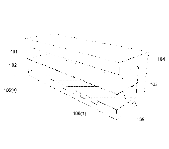
oxygenation of red blood cells within a close proximity of the permeable
membrane.
SUMMARY OF THE DISCLOSURE
[0003] According to one aspect of the disclosure, a microfluidic oxygenation
device includes
a first polymer layer defining a first oxygen flow channel. The device also
includes a second
polymer layer defining a first blood flow channel. The first blood flow
channel overlaps the
first oxygen flow channel, and the two channels are separated by a permeable
membrane that
allows communication between the channels at overlapping portions.
Additionally, first blood
flow channel further includes at least one passive mixing element along at
least one wall. The
passive mixing element is configured to redistribute a fluid flowing through
the first blood
flow channel within the channel.
[0004] In some implementations, the passive mixing element is one of a
straight ridge, an
angled ridge, a chevron canal, a dome, a cone, a pit or a post. In some
implementations, a first
fluid, such as oxygen, flows through the first oxygen flow channel and a
second fluid, such as
blood, flows through the first blood flow channel.
[0005] In some implementations, the height or depth of the passive mixing
element is less
than about 30% of the height of the first blood flow channel, and the passive
mixing elements
-1-
CA 2858080 2019-04-17
CA 02858080 2014-06-03
WO 2013/086011 PCMJS2012/067971
are incorporated into the floor of the first blood flow channel. In other
implementations, the
height of the first blood flow channel is between about 10 and 100 microns and
the
membrane thickness is between about 10 and about 50 microns. In yet other
implementations,
the length of the first oxygen flow channel and the first blood flow channel
is between about
1 mm and 50 mm and the width is between about 100 microns and 200 microns.
[0006] In other implementations, the membrane is permeable to oxygen and
carbon dioxide.
In yet other implementations, the walls of the first blood flow channel are
coated with an
anticoagulant. In yet other implementations, the device includes a second
blood flow channel
separated from the first oxygen flow channel by a second permeable membrane.
[0007] According to another aspect of the disclosure, a method for oxygenating
deoxygenate
blood includes providing a microfluidic device comprising a first polymer
layer defining a
first oxygen flow channel and a second polymer layer defining a first blood
flow channel.
The first blood flow channel also includes at least one passive mixing
element. A membrane
separates the first oxygen flow channel and the first blood flow channel and
allows
communication between the first oxygen flow channel and the first blood flow
channel. The
method also includes introducing partially deoxygenated blood into a proximal
end of the
microfluidic device, and flowing the partially deoxygenated blood through the
device.
Additionally, the method includes flowing oxygen through the first oxygen flow
channel.
Finally, oxygenated blood is received at a distal end of the microfluidic
device.
[0008] In some implementations, the method also includes collecting partially
deoxygenated
blood from a patient, flowing the partially deoxygenated blood through the
first blood flow
channel to reoxygenate the blood, and returning the reoxygenated blood to the
patient. In
other implementations, the method further includes removing carbon dioxide
from the
partially deoxygenated blood as the partially deoxygenated blood flows through
the first
blood flow channel.
[0009] In yet other implementations, the method also includes flowing oxygen
through the
first oxygen flow channel from a first direction, and flowing blood through
the first blood
flow channel in a second direction opposite to the first direction. In some
implementations,
the blood is flowed through the first blood flow channel at 4-5 L/min and
oxygen is
transferred to the blood at a rate of about 150-200 mL/min.
-2-
CA 02858080 2014-06-03
WO 2013/086011 PCMJS2012/067971
BRIEF DESCRIPTION OF THE DRAWINGS
[0010] The skilled artisan will understand that the figures, described herein,
are for
illustration purposes only. It is to be understood that in some instances
various aspects of the
described implementations may be shown exaggerated or enlarged to facilitate
an
understanding of the described implementations. In the drawings, like
reference characters
generally refer to like features, functionally similar and/or structurally
similar elements
throughout the various drawings. The drawings are not necessarily to scale,
emphasis instead
being placed upon illustrating the principles of the teachings. The drawings
are not intended
to limit the scope of the present teachings in any way. The system and method
may be better
understood from the following illustrative description with reference to the
following
drawings in which:
[0011] Figure IA is an isometric view of a device for oxygenating blood,
according to one
illustrative implementation of the present disclosure;
[0012] Figure 1B is a cut-away view of a device for oxygenating blood,
according to one
illustrative implementation of the present disclosure;
[0013] Figure 1C is an end view of a device of oxygenating blood as depicted
in Figure 1,
according to one illustrative implementation of the present disclosure;
[0014] Figure 2 is a cross-sectional view illustrating the flow patterns of
blood in a blood
oxygenation device without passive mixing elements, according to one
illustrative
implementation of the present disclosure;
[0015] Figure 3 is a cross-sectional view illustrating the flow patterns of
blood in a blood
oxygenation device with passive mixing elements as depicted in Figures 1A-1C,
according to
one illustrative implementation of the present disclosure;
[0016] FIGS. 4A¨J are top and isometric view of exemplary passive mixing
elements of a
blood oxygenation device as depicted in Figure 1, in accordance with an
illustrative
implementation of the present disclosure; and
[0017] Figure 5 is a flow chart of a method for oxygenating deoxygenated blood
with a blood
oxygenation device as depicted in Figure 1, in accordance with an illustrative
implementation
of the present disclosure.
-3-
CA 02858080 2014-06-03
WO 2013/086011 PCMJS2012/067971
DETAILED DESCRIPTION
[0018] The various concepts introduced above and discussed in greater detail
below may be
implemented in any of numerous ways, as the described concepts are not limited
to any
particular manner of implementation. Examples of specific implementations and
applications
are provided primarily for illustrative purposes.
[0019] The present system described herein generally relates to a system and
method for
oxygenating blood. Accordingly, in various implementations, the disclosure
relates to
oxygenating blood by passively mixing the blood as it flows through the blood
oxygenation
device. In certain implementations, the device includes a plurality of passive
elements on one
wall of the device to mix the flowing blood.
[0020] Figures lA and 1B show an isometric view of a blood oxygenation device
100 and a
cutaway view thereof Described in greater detail below, but briefly, the
device 100 includes
a first flow channel 101 separated from a second flow channel 102 by a gas
permeable
membrane 103. The floor 105 of the second flow channel 102 includes a passive
mixing
element 106. The flow channels 101 and 102 are fabricated within a polymer
substrate 104.
[0021] As illustrated in Figures lA and 1B and discussed above, device 100
includes a first
flow channel 101 and second flow channel 102 fabricated within a polymer
substrate 104. In
some implementations, the polymer substrate 104 is a thermoplastic, such as
polystyrene or
polyimide, biodegradable polyesters, such as polycaprolactone (PCL), or soft
elastomers such
as polyglycerol sebacate (PGS). In other implementations, the substrate 104 is
polydimethylsiloxane (PDMS), poly(N-isopropylacrylamide). In yet other
implementations,
the substrate 104 includes non-polymer materials such as, but not limited to,
ceramics;
metals; glasses; nanotubes or nanowires formed from, for example, carbon or
zinc oxide; or
other non-polymer materials.
[0022] In some implementations, the device 100 and the passive mixing elements
106 are
fabricated in the substrate 104 using, for example, photolithographic
techniques, injection
molding, direct micromachining, deep RIE etching, hot embossing, or any
combinations
thereof
[0023] The first flow channel 101 communicates with the second flow channel
102 via the
membrane 103. In some implementations, the membrane 103 is permeable or semi-
permeable
-4-
CA 02858080 2014-06-03
WO 2013/086011 PCMJS2012/067971
to ions, molecules, cells or any combination thereof. For example, the
membrane 103 may
allow for oxygen to pass from the first flow channel 101 to the second flow
channel 102 and
carbon dioxide to pass from the second flow channel 102 to the first flow
channel 101.
However, in some implementations, the membrane 103 is not permeable to red
blood cells. In
some implementations, the membrane 103 is fabricated from a semi-porous or
porous
material, such as polyethersulfone or PDMS. In other implementations, the
membrane 103 is
created by electrospinning a polymer to create a flexible, porous polymer
mesh.
[0024] The first flow channel 101 and the second flow channel 102 of device
100 run
substantially parallel to one another, and, as described above, are separated
by the membrane
103 at overlapping portions. In some implementations, the first flow channel
101 includes
three smooth walls, with the fourth wall being the membrane 103. In other
implementations,
the device 100 includes additional flow channels to the left, right, and/or
above the first flow
channel 101. In some of these implementations the first flow channel 101 is
also separated
from these additional flow channels by a permeable membrane 103. In other
implementations, the first flow channel is configured for the flow of a gas.
For example,
oxygen may be flowed through the first flow channel 101. In other
implementations, the first
flow channel 101 is configured to flow a liquid. For example, the flow first
flow channel may
be configured to flow blood.
[0025] The second flow channel 102 includes at least one passive mixing
element 106 along
at least one wall of the channel. In the implementation of device 100, the
floor 105 includes
passive mixing elements 106(1)-106(n). In other implementations, any wall of
the first or
second flow channel can include a passive mixing element 106. In some
implementations,
the floor, or other wall(s) that include a passive mixing element 106, is
replaceable, such that
different configurations of passive mixing elements can be used for different
fluids. In yet
other implementations the device 100, or components thereof, is disposable.
[0026] As described below, in some implementations, the passive mixing
elements include a
plurality of ridges, channels, protrusions, or any combination thereof. In
some
implementations the passive mixing elements 106(1)-106(n) span the entire
length of a flow
channel. In other implementations, the mixing elements 106 cover only a sub-
portion of the
total length of a flow channel 102. In yet other implementations, the passive
mixing elements
106(1)-106(n) are grouped together. For example, the fluid flow channel 102
may contain a
first type of passive mixing element 106 along a first portion of the flow
channel 102 and
-5-
then a second type of passive mixing element 106 along a second portion of the
flow channel
102.
100271 Figure 1C illustrates an end view of device 100. In some
implementations, the device
100 is fabricated as a top component 104(b) and a bottom component 104(a) that
are
fabricated separately and assembled to form device 100. In some
implementations, the
components 104(a), 104(b), and the membrane 103 are attached to one another
with an
adhesive. For example, the components of device 100 can be bound together with
a chemical
adhesive, plasma bonding, and/or by clamping the components together. In other
implementations, the device 100 is fabricated as a single, continuous unit.
For example,
device 100 can be created by injection molding. In yet other implementations,
the top portion
104(b) and the bottom portion 104(a) are formed by injection molding. In some
implementations, after the device 100 is fabricated the channels are coated
with an
anticoagulant. In other implementations, the anticoagulant is embedded in the
polymer
substrate 104.
100281 In some implementations the height or depth of a passive mixing element
106 is
between about 5% and about 10%, between about 10% and about 20%, or between
about
20% and 30% of the total height of the flow channel 102. In some
implementations, each
passive mixing element in a channel is the same height or depth. While in
other
implementations, the height or depth of the passive mixing elements changes
along the length
of the flow channel 102.
100291 In some implementations, the width, height, and length of the first
flow channel 101
and second flow channel 102 are the same. In other implementations, one or all
of the
dimensions between different flow channels is different. In some
implementations, the height
of the flow channels is between about 10 microns and 25 microns, between about
25 microns
and 50 microns, or between about 50 microns and 100 microns. In some
implementations the
thickness of the membrane 103 is between about 10 microns and 25 microns,
between about
25 microns and 50 microns, or between about 50 microns and 100 microns. In
some
implementations, the length of the flow channels is between about 1 mm and 10
mm,
between about 10 mm and 50 mm, or between about 50 mm and 100 mm and the width
is
between about 100 microns and 200 microns, between 200 microns and 500
microns, or
between about 500 microns and 1 cm.
-6-
CA 2858080 2019-04-17
CA 02858080 2014-06-03
WO 2013/086011 PCMJS2012/067971
[0030] Figure 2 illustrates how fluid may flow through a blood oxygenation
device without
passive mixing elements similar. Figure 2 illustrates a blood oxygenation
device 201 without
passive mixing elements along the floor 105(b). In this example, oxygen flows
through the
first flow channel 201 as deoxygenated blood (white circles) flows through the
second flow
channel 202. The blood in the second flow channel 201 flows in a laminar
pattern 203. The
blood cells become oxygenated (gray circles) as they flow substantially close
to the
membrane 204. In some implementations, because oxygen diffusion can only occur
at
distances substantially close to the membrane 204, the portion of blood
flowing along the
floor of the second flow channel 202 may never become oxygenated.
[0031] In contrast, Figure 3 illustrates how blood flows through a blood
oxygenation device
300 similar to the blood oxygenation device 100. The floor 105 of device 300
includes a
number of passive flow elements 106. These passive flow elements 106 create
non-laminar
flow 301 in the fluid of channel 102. In some implementations this creates
chaotic flow in
channel 102. For example, in some implementations, the passive mixing elements
106 drive
fluid from the bottom of the fluid flow channel 102 towards the membrane 103.
In some
implementations, the passive mixing elements 106 create a rotational flow
within in the flow
channel. For example, the passive mixing elements 106 may create a rifling
effect that causes
the fluid to swirl as it flows down the flow channel 102. In some
implementations, the device
100 induces mixing within a fluid without inducing mechanical trauma to the
components of
the fluid. For example, the passive mixing elements 106 of device 100 may
drive blood
towards the membrane 103 without causing the red blood cells to hemorrhage or
clot.
[0032] As illustrated in Figure 3, the device 300, with the passive mixing
elements 106, is
able to fully oxygenate the blood over a shorter span of the device's length
when compared to
device 200 that does not include a passive mixing element 106. This allows for
a shorter
channel, and therefore allows the device to be primed with less blood than a
channel without
such passive mixing elements.
[0033] Figures 4A¨J show a top and isometric view of possible, non-limiting
examples of
passive mixing elements 106. Figures 4A and 4B illustrate an alternating
herringbone pattern.
The design consists of a herringbone pattern wherein the center of the
herringbone pattern
shifts from one side of the flow channel to the other through the length of
the flow channel
102. In some implementations, the alternating herringbone pattern causes the
fluid in the flow
channel to enter the flow channel and begin rotating in a first direction.
Then, when the
-7-
CA 02858080 2014-06-03
WO 2013/086011 PCMJS2012/067971
flowing fluid encounters a shifted herringbone pattern, the fluid is forced to
rotate in a
direction opposite to the first rotational direction. For example, the fluid
may enter the
channel 102 flowing in a laminar fashion, and then alternatingly switch
between clockwise
and counter clockwise rotations as the fluid encounters consecutive, offset
herringbone
patterns. In some implementations, the number of chevrons per herringbone
pattern and/or
the number of groupings is configured to create a specific level of mixing
over a given length
of the device 100. In some implementations, the herringbone pattern does not
alternate, but is
constant along the duration of the flow channel. In some of these
implementations the center
of the herringbone patterns is in the center of the channel, while in other
implementations the
center of the pattern is off-center with respect to the channel.
[0034] As illustrated in Figures 4C and D, in some implementations, the mixing
of a fluid is
created with slanted ridges. Similar to the herringbone patter described
above, in some
implementations the slanted ridge pattern also creates a swirling rotation of
the fluid that
drives fluid from the bottom of the fluid flow channel towards the permeable
membrane 103.
In some implementations, the angle of the slanted ridge and the herringbone
patter is between
about 35 and about 55 degrees. In some implementations, the spacing between
the ridges is
between about 50 microns and about 100 microns, between about 100 microns and
150
microns, or between about 150 and about 200 microns. In some implementations,
the spacing
of the ridges is 27c/(the width of the channel), such that the diameter of the
induced rotation is
less than the width or depth of the channel. In some implementations, the
ridges of the above
implementations are rounded.
[0035] In yet other implementations, the passive mixing elements 106 are
designed to create
vortices and other high and low pressure areas which drive the fluid towards
the membrane
103. For example, Figures 4E and 4F, like the illustrative implementation of
Figure 3, include
a plurality of ridges. In some implementations, the ridges are spaced between
about 20
microns and about 50 microns, between about 50 microns and 100 microns or
between about
100 microns and 500 microns.
[0036] In some implementations, the passive mixing elements can be, but are
not limited to,
posts, mounds, ramps, pits, cones or any combination thereof. Figures 4G-4J
illustrate a
possible post and mound implementation. In the implementation illustrated in
Figures 4G and
4H, each row of posts is off set from the previous row. This causes the fluid
to mix laterally
in addition to driving fluid upwards. In other implementations, as illustrated
in Figure 41 and
-8-
CA 02858080 2014-06-03
WO 2013/086011 PCMJS2012/067971
4J, the passive mixing elements are aligned with the passive mixing elements
in the previous
row.
[0037] Figure 5 is a flow chart of a method 500 for oxygenating blood with a
microfluidic
device. First, a microfluidic device is provided (step 501). Then, partially
deoxygenated
blood is introduced into a proximal end of the microfluidic device (step 502).
The partially
deoxygenated blood is flowed through a first channel of the microfluidic
device (step 503),
and oxygen is flowed through a second channel of the microfluidic device (step
504). Finally,
oxygenated blood is collected from a distal end of the microfluidic device
(step 505).
[0038] As set forth above, and referring to Figure 1, the method 500 for
oxygenating partially
deoxygenated blood begins with providing a microfluidic device (step 501). In
some
implementations, the microfluidic device is similar to device 100 described
above. In other
implementations, the microfluidic device includes a plurality of oxygen
channels and/or a
plurality of blood flow channels. In yet other implementations, the
microfluidic device is a
array of devices similar to device 100. In some implementations, the device is
configured to
allow for about 500-1000 mL/min, about 1-4 L/min, or about 4-5 L/min of blood
flow. In
some implementations, the device is configured to transfer oxygen into the
blood at a rate of
about 160 to about 200 mL/min.
[0039] Next, the method 500 of oxygenating blood continues with the
introduction of
partially deoxygenated blood into a proximal end of the microfluidic device
(step 502). In
some implementations, the blood is directly collected form a patient and
introduced into the
device. For example, the device may be part of a heart-lung bypass system that
oxygenates
blood during surgery. In other implementations, the blood is collected,
stored, and then
oxygenated at a later time. For example, the blood may be collected during a
blood drive and
then oxygenated prior to being transfused into a patient. In some
implementation, the blood is
actively pumped through the device by an external pump, and in other
implementations the
blood is pumped through the device by the patient's heart.
[0040] The method 500 continues by flowing the partially deoxygenated blood
through a first
blood flow channel (step 503). As described above, the device includes at
least one passive
mixing element that inducing mixing within the channel as the blood travels
the length of the
device. In some implementations, the blood is thinned with a blood thinning
agent such as the
-9-
drug CoumadinTM or Heparin. In some implementations, the walls of the blood
flow channels
are coated with an anticoagulant.
100411 Responsive to flowing blood through the first blood flow channel, the
method 500
continues by flowing oxygen through a first oxygen flow channel (step 504).
Referring to
Figure I, the blood flow channel and oxygen flow channel are separated by a
permeable
membrane. Oxygen diffuses through the membrane, oxygenating the blood as it
flows down
the length of the channel. In some implementations, the blood is continually
mixed within the
channel by the passive mixing elements similar to those described above. In
some
implementations, the continual mixing allows a given volume of deoxygenated
blood to be
oxygenated more efficiently by continually exposing different red blood cells
to the region
near the membrane where oxygen diffusion can occur. In other implementations,
the
membrane is also porous to carbon dioxide, and the carbon dioxide initially
within the
deoxygenated blood diffuses into the oxygen flow channel. In yet other
implementations, the
oxygen and blood are flowed through the microfluidic device starting at
different ends. For
example, the blood may enter the device at a proximal end and the oxygen may
enter the
device at a distal end of the device.
100421 The method 500 continues, with the collection of the oxygenated blood
at a distal end
of the microfluidic channel. In some implementations, the oxygenated blood is
transfused
directly back into the patient from which it was collected. In other
implementations, the blood
is collected and stored for later transfusion or experimentation.
- I 0-
CA 2858080 2019-04-17