Note: Descriptions are shown in the official language in which they were submitted.
CA 02858270 2014-06-05
High Resolution Light Microscope
The present invention refers to an apparatus for the optical analysis of a
sample, also referred to
as microscope, which is configured for a high resolution optical analysis
process for the detection
of fluorescent molecules. The apparatus and the process applied by using the
apparatus are
arranged such that the excitation light generated by the excitation light
source is focused on a
sample and that the light emitted by the sample is detected. The apparatus and
the process are
characterized by the fact that the excitation light is synchronized with the
detection. The
excitation light source may consist of a laser device.
State of the art
US2001/045523 describes the stimulated emission depletion microscopy (STED)
which involves
a sample being irradiated and sampled via a microscope objective by two
parallel light paths,
while light emitted by the sample exits through the same optical path and is
detected. One of the
light beams is coupled into the optical path by means of a dichroic mirror and
has an excitation
wavelength which is specific to the sample's fluorescent molecules, while the
second light beam,
which is also coupled into the common optical path by means of a dichroic
mirror, has a
wavelength that is specific to the de-excitation of the sample's fluorescent
molecules from their
excitation state, wherein the second light beam irradiates only a ring-shaped
part of the focal area.
Subsequently, fluorescent molecules which are in the focus of both light beams
will be de-excited
by the second light beam despite the excitation by the first light beam while
the excited
fluorescent molecules located in the ring-shaped second light beam can emit so
that only their
emission is collected by the detector. For a particularly high resolution, the
second light beam is
irradiated in the shape of an interference pattern e.g. by means of a phase
plate positioned therein,
the interference pattern having a directional null in the measurement range
but has otherwise
reached saturation of the excited fluorescent molecules with excitation light.
1
' CA 02858270 2014-06-05
This process is disadvantageous in that the device requires an extremely high
precision of optical
path alignment for the excitation wavelength and for the de-excitation
wavelength and a time-
consuming deflection of excitation light over the sample, e.g. by means of a
scanner.
US 2009/0242798 Al describes the photo-activated localization microscopy
wherein fluorescent
molecules are individually detected in a sample by first irradiating
fluorescent molecules using a
switching wavelength light so that the fluorescent molecules are excited for
emission when being
irradiated with an excitation wavelength light. Repeated irradiation using
switching wavelength
light each causes a statistically distributed selection of fluorescent
molecules being excited for
emission by the following irradiation with excitation wavelength light. For
evaluation purposes,
the subsequently detected emission maxima are determined with high spatial
resolution and are
superimposed.
This process is disadvantageous in that it is limited to those fluorescent
molecules which are to be
brought to an excitable state using a switching wavelength radiation, with
time duration suitable
for the analysis, and that the sequential irradiation and locating of the
individual molecules is
time consuming.
Object of the Invention
The object of the present invention is to provide an alternative apparatus and
an alternative
process for the optical detection of fluorescent molecules in a sample with
high resolution,
preferably in the provision of an apparatus which allows a reduced
configuration effort, in
particular for focusing, and which allows for a process that can be performed
with any molecule
that is fluorescent when irradiated with an excitation wavelength, and which
preferably spatially
resolves a multitude of molecules. Furthermore, the process shall not be
limited e.g. to
fluorescent molecules that are switchable by a switching wavelength.
2
CA 02858270 2014-06-05
General Description of the Invention
The invention achieves this object by the features of the claims, in
particular with an apparatus
for optical analysis of a sample containing at least one fluorescent molecule,
using an excitation
light source which optionally consists of a laser that is set up to generate
light of an excitation
wavelength, wherein the apparatus has a beam path which is directed onto the
sample by an
optical element, also referred to as an objective. The apparatus is equipped
with a detector
adapted for detecting the radiation emitted by the fluorescent molecule, and
is positioned in
abeam path in which the light emitted from the sample is guided. The optical
path in which the
emitted light from the sample is guided is preferably directed through the
same objective through
which the beam path of the excitation light generated by the excitation light
source is guided. In a
section for excitation light adjacent to the objective, the beam path
preferably runs collinear to
the beam path of emitted light, e.g. in a section in which the beam path runs
between the
objective and a mirror, in particular a partially-transparent mirror, which
orients the beam path
for excitation light from the excitation light source to the objective, and/or
a mirror which orients
the optical path for emitted light emanating from the objective to the
detector or to a mirror
oriented to the detector.
The apparatus is characterized by a polarization device which is configured to
modulate the
polarization of the excitation light, or another feature of the excitation
light, with a modulation
signal, wherein the modulation signal has or consists of at least one
frequency, in particular one
predetermined frequency or several predetermined superimposed frequencies, or
the modulation
signal consists of a sequence of signals which has no repetition or no
periodic repetition.
Preferably the polarization is a linear polarization and the modulation is a
rotation. The
polarization device, which is configured according to the invention to
modulate the polarization
of the excitation light generated by the excitation light source, in
particular with the modulation
signal, to modulate e.g. into a frequency, may be referred to as a modulating
device for the
polarization or as a polarization modulator, wherein the polarization is
modulated with the
modulation signal, in particular into at least one frequency, and in
embodiments may be referred
to as a polarization rotation device, which in particular is controlled by the
modulation signal, for
3
CA 02858270 2014-06-05
example is frequency-controlled. Such a polarization modulator which is
controlled by the
modulation signal, preferably frequency-controlled, can be a k/2 plate, a X/4
plate linearly
movable in an angle, especially at 90 to its polarization direction, or it
can as polarizing element
have or consist of a circular polarization filter linearly movable at an
angle, in particular
perpendicular to its polarization direction. A preferred polarization
modulator is polarizing
element that is rotatably driven and controlled with the modulation signal,
which is arranged in
the beam path between the excitation light source and the objective. The
rotatable polarizing
element may be a linear polarization filter, especially in case of an
excitation light source which
is configured to generate non-polarized or circularly polarized excitation
light, or a phase shift
plate, preferably a k/2 or a 2\14 plate, in case of an excitation light source
which is configured to
generate a linearly polarized excitation light, or a combination of inversely
rotated mirrors, e.g.
inversely rotated against one another by 45 , so that its rotation is
controlled by the modulation
signal and determines the polarization modulation of the excitation light.
Alternatively, the
polarization modulator can be an acoustic-optical modulator or an electro-
optical modulator, in
particular a Pockels cell, which is static or rotatable, controlled via the
modulation signal, in
particular with at least one frequency.
For the purpose of the invention, a modulation signal is a sequence of
signals, in particular with a
fixed respectively predetermined frequency or with at least two superimposed
fixed respectively
predetermined frequencies, or a series of signals which optionally have no
repetition or no
periodic repetition during the duration of the analytical process, or which
preferably occur
periodically. In general, a signal can be a sinusoidal signal, a rectangular
signal and/or a saw-
tooth signal.
A modulation signal that consists of a sequence of signals, which has no
repetition or no periodic
repetition, can e.g. be a signal controlling a modulation of the polarization
which passes each
polarization direction at least once or exactly once, wherein the polarization
is preferably
modulated from an initial orientation to an identical orientation at the end
of the modulation,
wherein e.g. the modulation rotates a linear polarization exactly once by 180
or by 360 .
4
CA 02858270 2014-06-05
Furthermore the invention refers to a process for the optical analysis of a
sample which has the
steps that can be executed with the apparatus, respectively the steps for
which the apparatus is
configured, in particular a process for optical analysis of a sample by means
of the apparatus.
By means of the modulation of the polarization of the excitation light by at
least one modulation
signal, which can be a time constant or a time varying frequency, the
apparatus generates the
excitation of a portion of the fluorescent molecules of the sample depending
on the modulation
signal. Because a fluorescent molecule of the sample is mainly excited for
emission by the
excitation light only if the polarization vector of the excitation light is
oriented in parallel to the
transition dipole moment vector of the fluorescent molecule. Due to the
different orientation of
the transition dipole moment vectors of the sample's individual fluorescent
molecules, the
irradiation of excitation light with a frequency-modulated polarization causes
the excitation of the
suitably oriented fluorescent molecules in dependence on the modulation
signal. Therefore, by
means of the apparatus according to the present invention in the detection
process the sample's
fluorescent molecules are excited according to the different orientation of
their transition dipole
moment vectors in dependence on the modulation signal of the polarization of
the excitation
light, or depending on the modulation signal used to excite the polarization
modulator,
respectively, and emit in a distance of time, respectively with phase shift,
in dependence on the
modulation signal or depending on the rotation of the polarization of the
excitation light
controlled by the modulation signal.
For the synchronization of the detection with the modulation of the
polarization of the excitation
light, the detector of the apparatus is preferably controlled in dependence on
the modulation
signal that controls the polarization unit, respectively in dependence on the
frequency of the
polarization unit, and in particular is configured for detection or isolation
of the emitted light with
or without phase shift, optionally with or without amplitude displacement, in
at least one
frequency that is equal to the modulation signal. The detection of the
emission of fluorescent
molecules in at least the one frequency corresponding to the modulation signal
controlling the
polarization modulator results in the isolation of the emission excited at the
modulation signal. A
preferred device for the control and/or analysis of the modulation signal
controlling the
5
CA 02858270 2014-06-05
polarization device, in particular the frequency of the polarization device
and/or of a detector
controlled in dependence on the modulation signal, is a so-called demodulator,
e.g. a lock-in
amplifier, or another device configured for the analysis and determination of
the Fourier
transformation, in particular for the simultaneous analysis and determination
of the Fourier
For the purpose of the invention, such a modulation signal consisting of a
sequence of signals that
has no repetition or no periodic repetition is optionally comprised by the
term of a frequency
The detector is preferably coupled with a signal analysis device that is
configured to exclusively
collect or isolate the detected signals which are detected at least at a
frequency that is identical to
In embodiments in which the modulation signal consists of a frequency, the
signal analysis
device is configured to exclusively collect and isolate the detected signals
which are detected at
the frequency of the polarization device.
6
' CA 02858270 2014-06-05
In addition to the first detector described above, the apparatus optionally
has at least one second
detector onto which the light emitted by the probe is directed e.g. by means
of a partially
transparent mirror, and which is preferably coupled with a signal analysis
device that is set up to
exclusively collect or isolate those detected signals that are detected with a
constant phase shift to
the modulation signal which is used to control the polarization modulator, or
to the modulation
signal used by the polarization modulator to modulate the polarization of the
excitation light. A
partially transparent mirror generally is e.g. a polarization beam splitter.
In the process, such a
detector collects different components of the light emitted by the fluorescent
molecules,
preferably the polarization which is random according to the different
orientation of the transition
dipole moment vectors of the fluorescent molecules and which therefore results
in a scattered
detection of fluorescent molecules. In embodiments having a first and at least
one second detector
that are each coupled to a signal analysis device, from the different phase
shift, modulation
amplitude and the medium intensity of the light emitted by the fluorescent
molecules which the
two signal analysis devices collect and isolate, a higher spatial resolution
of individual molecules
is achieved.
Preferably the detector and/or the second detector are an area detector, e.g.
a CCD camera, which
offers the benefit of simultaneously collecting a large number of molecules.
Optionally the
detectors can be area sections of an area detector. Also in embodiments in
which the detectors are
area detectors or sections of an area detector, the apparatus can have a
device for deflecting the
excitation light across the sample, such as e.g. a scanner. The device for
deflecting the excitation
light can be located at any position in the beam path of the apparatus, in
particular between an
objective, which directs the light onto the sample, and a controlled
polarization device.
The excitation light source preferably consists of a laser. The polarization
modulator has a
polarization device controlled by a modulation signal, or it consists of a
polarization device
controlled by the modulation signal, in particular a frequency-controlled
polarization device.
Preferably, a polarization device is used which linearly polarizes the
excitation light. The
polarization device controlled by the modulation signal is positioned in the
beam path between
7
CA 02858270 2014-06-05
the excitation light source and the objective, and it can be a linear
polarizer rotating with the
modulation signal, in particular a linear polarizer rotating with a frequency.
Optionally the excitation light source can be pulsed; in particular it can
consist of 1 pulsed laser.
With general preference, the modulation signal into which the polarization
modulator modulates
the polarization, and/or which is used to control the polarization modulator,
is predetermined.
Optionally the light source can be configured to generate excitation light of
one or of at least two
light frequencies the sum of which is equal to the frequency of the excitation
light that is specific
for the fluorescent molecule, in particular when using the apparatus as a
microscope for the so-
called two photon excitation or multiple photon excitation. In embodiments in
which the light
source is configured to generate the excitation light with light frequencies
the sum of which is
equal to the frequency of the excitation light that is specific for the
fluorescent molecules,
multiple photon excitation is implemented, which has the benefit of a smaller
angle range in
which the fluorescent molecules are suitably oriented so that a higher
resolution results.
Optionally the apparatus, in addition to the aforementioned first excitation
light source can have
one second, or more light sources which can be additional excitation light
sources. The beam path
of the additional light sources is preferably directed via a dichroic beam
splitter into the same
objective into which the beam path of the first excitation light source is
directed. In the optical
path of the additional light source, a polarization modulator controlled by
the modulation signal,
especially a frequency-controlled polarization modulator, is arranged which
may be formed
identically to the first polarization modulator located in the beam path of
the first excitation light
source, and which may preferably be set up to modulate the polarization with
or without phase
shift, optionally using the same modulation signal or in the same frequency as
the polarization of
the excitation light of the first light source.
Alternatively the second light source can be configured to generate light of a
de-excitation
wavelength. This light of a de-excitation wavelength generally effects the
transition of the status
8
CA 02858270 2014-06-05
of the fluorescent molecules produced by the excitation light to a status of
lower energy from
which no emission is possible and which therefore can be called a light for
quenching of the
emission produced by the excitation light and it can e.g. have a wavelength
which de-excites only
a certain type of molecule, or several or even all types of molecules.
Special preference is on an apparatus where the first and the second light
source are linearly
polarized, with the polarization of the de-excitation light beam having an
angle of >0 to <1800,
preferably 60 to 120 , in particular 90 to the polarization of the
excitation light beam, e.g. by
means of a polarization modulator located in their beam path. In this
embodiment, only those
fluorescent molecules are not de-excited whose transition dipole moment
vectors exactly match
the polarization of the excitation light beam so that the angle range of the
suitably oriented
fluorescent molecules is drastically reduced so that as a consequence
individual molecules have a
much better resolution and can be better distinguished.
In this embodiment, the polarization modulator is preferably positioned in the
common beam
path of the linearly polarized excitation light source and de-excitation light
source, and is
configured to simultaneously modulate with the modulation signal the
polarization direction of
the excitation light and of the de-excitation light at constant angles between
the two polarization
devices.
The polarization of the excitation light is preferably oriented in an angle of
90 to the polarization
of the de-excitation light so that the polarization directions of the light
from the excitation light
source and from the de-excitation light source are at 90 to each other. In
this embodiment the
polarization modulator can have a polarization rotation element consisting
e.g. of a X/2-plate,
which is suitable for the wavelengths of both light sources. In a preferred
embodiment the
apparatus has a first light source and a second light source which are
configured to produce
parallel linearly polarized light which is deflected into a common beam path
by means of a
dichroic mirror that is directed onto a polarization modulator, e.g. a
frequency-controlled,
rotating X/2 plate wherein subsequently the optical beam is directed onto an
optical device which
is configured to split off a wavelength range as partial beam, to rotate its
polarization e.g. by 30-
9
CA 02858270 2014-06-05
150'; more preferable by 60-1200, in particular by 90 , and to direct this
partial beam in parallel
with the non-split off partial beam onto the optical element that focuses the
optical path on the
sample. Accordingly, the polarization device of the de-excitation light is
preferably controlled by
the same modulation signal as the polarization device of the excitation light.
Additionally or optionally, de-excitation light can be directed into the
objective which in
particular is a microscopic objective, that illuminates only parts of the
focal area. Optionally and
in addition to the modulation of the polarization, de-excitation light in the
form of an interference
pattern is directed on the sample which has a directional null at the
measurement position, and
outside this position has an intensity which achieves a saturation of the
fluorescent dye at the de-
excitation of the excited status generated by the excitation light. For a
particularly high resolution
this can be achieved by an additional second light beam of a de-excitation
wavelength e.g. by
means of a phase plate arranged therein which produces an interference pattern
with a directional
null and which in the other focal area has sufficient intensity for the
saturation of the excited
fluorescent molecules with de-excitation light. The directional null of the de-
excitation light at
this point allows for the emission by the fluorescent molecules. The phase
plate is preferably
configured to radiate ring-shaped de-excitation light on the sample around a
central directional
null area which particularly is in the focus.
Alternatively or additionally the apparatus in the beam path of the excitation
light source can
have a first dividing mirror which deflects a part of the excitation light, in
particular by 45 , and a
second dividing mirror to which the partial beam separated from the first
partially transparent
dividing mirror is directed into the optical path to which the non-deflected
partial beam is
directed, wherein another polarization modulator, a phase shift element,
and/or a polarization
rotation element is arranged in the optical path of the deflected partial
beam. After the first
dividing mirror, the beam path of the deflected partial beam is directed to
the second dividing
mirror, e.g. by means of a first and a second deflection mirror, wherein e.g.
another polarization
modulator, a phase shift element, and/or a polarization rotation element may
be arranged between
the deflection mirrors or between one deflection mirror and one of the
dividing mirrors.
10
CA 02858270 2014-06-05
In a further embodiment the apparatus has a deflection device in the beam path
in front of the
objective which is controlled with special preference to direct the beam path
into the objective
according to a predefined pattern, in particular to guide the beam path over
the sample in a
predefined pattern in order to consecutively scan segments of the sample with
the beam. Such an
optional deflection element, which in particular is a controllably deflectable
mirror, which directs
the beam path pointed at the objective to the objective and which preferably
also guides the light
emitted by the fluorescent molecules into a beam path with identical
orientation, the apparatus
preferably has an analysis unit that is configured to arrange the detection
signals collected during
the controlled guidance of the beam path across the sample according to the
controlled guidance.
This way the apparatus is set up for scanning a sample. This embodiment is
preferred for
embodiments in which the detectors are no area detectors or segments of an
area detector.
Optionally the apparatus can have an analysis unit that is configured to
display in a common
presentation the emission maxima of the emission signals detected at least at
one frequency
which depends on the modulation signal.
In general the process is described by the configuration of the apparatus. The
process for optical
analysis of a sample containing at least one fluorescent molecule comprises
the irradiation of
light on the sample, the light having an excitation wavelength that is
specific for the fluorescent
molecule for excitation of emission and the detection of the radiation emitted
by the fluorescent
molecule, wherein preferably the beam path, in which the emitted radiation is
guided, is directed
through the same objective through which the beam path of the excitation
wavelength light is
guided, as described with reference to the apparatus. The polarization or
another property of the
excitation light is modulated with a modulation signal which can have or
consist of one
frequency, several superimposed frequencies. As described with reference to
the apparatus, the
modulation signal can also consist of a sequence of signals that has no
repetition, e.g. of one
portion of 180 to 360 of a period of a trigonometric function. The
polarization of the excitation
light can be modulated by means of a polarization device that is controlled by
a modulation
signal.
11
CA 02858270 2014-06-05
Optionally light of a de-excitation wavelength and/or light of a switching
wavelength can be
irradiated on the sample. With preference, the light of a de-excitation
wavelength is polarized in
an angle of >0 to <180 , in particular from 300 to 120 , preferably 90 with
reference to the
polarization of the excitation wavelength. With preference, the polarization
of the light of the de-
excitation wavelength and/or the light of the switching wavelength is
modulated with the same
modulation signal as the light of the excitation wavelength.
The detection of emission is synchronized with the modulation of the
polarization of the
excitation light, e.g. the detection of emission is controlled by the
modulation signal so that the
emission can be isolated which is detected with or without phase shift to the
modulation signal.
This control of the detection of emission by the modulation signal can be
called demodulation, or,
in case of a modulation signal that has no repetition, as unfolding. A
modulation signal that has
no repetition may e.g. comprise or consist of exactly one period of a
trigonometric function,
while the signal analysis is done by unfolding this modulation signal.
Furthermore the invention refers to a microscopic representation which is
obtainable by the
process according to the invention and which is generated in particular by
using the apparatus.
The microscopic representations obtainable by the process according to the
invention are
characterized by showing a substantially higher resolution, especially at the
same magnification
and numeric aperture of the objective.
Detailed Description of the Invention
The invention is now described in more details with reference to the figures
that schematically
show in
- Figure 1 an embodiment,
- Figure 2 a section of a preferred embodiment,
- Figure 3, A to F, the modulation and demodulation in an embodiment
of the process with
excitation light only,
12
CA 02858270 2014-06-05
- Figure 4, A to F, the modulation and demodulation in an embodiment of the
process with
excitation light and de-excitation light,
- Figure 5 A a conventional fluorescence microscopic picture and in Figure
5 B a
microscopic picture generated using the invention and in Figure 5 C
superimposed
intensity profiles of the same section through Figure 5 A and 5 B and
- Figure 6 A a conventional fluorescence microscopic picture and in
Figure 6 B another
microscopic picture generated using the invention and in Figure 6 C
superimposed
intensity profiles of the same section through Figure 6 A and 6 B.
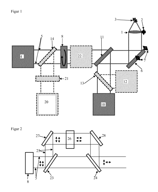
Using the focusing device 2, objective 1 can be focused on sample 3 which
contains fluorescent
molecules. An excitation light source 4 produces excitation light 5 the beam
path of which is
directed into the objective 1, shown here by means of a mirror 6 that is
deflecting and controlled
by means of the control device 7, e.g. for controlled guidance of excitation
light 5 over sample 3.
The polarization modulator 8 is configured to modulate the polarization of the
excitation light 5,
which preferably is a linear polarization direction, with at least one
frequency which represents
the modulation signal. As indicated by the arrow, the polarization modulator 8
can be a X/2 plate
rotating perpendicularly to the beam path of excitation light 5.
A first detector 10 is arranged in the beam path formed by objective 1,
emitted by sample 3, by a
first dichroic mirror 11 deflecting from the confocal section of the beam path
emitted light exiting
from objective 1. An optional second detector 12 can be directed to the
emitted light by one part
of the emitted light being deflected from a second partially transparent
mirror 13 to the second
detector 12. The second partially transparent mirror preferably is a
polarization beam splitter 13.
As shown in the figure, the second partially transparent mirror or
polarization beam splitter 13
can be arranged in the section of the optical path that is situated between
the first dichroic mirror
11 and the first detector 10, or in another section of the optical path.
First detector 10 and second detector 12 are each coupled with an analysis
unit which preferably
filters only signals which are modulated with a frequency equal to the
modulation signal that is
13
CA 02858270 2014-06-05
used to control the polarization modulator 8. The analysis unit can e.g. be a
demodulator, in
particular a lock-in amplifier. Here, first detector 10 and second detector 12
can each be coupled
with an analysis unit which only filters out signals in the frequency of the
modulation signal of
the polarization, each with different phase shift, in particular in an
embodiment in which beam
splitter 13 is a polarization beam splitter.
The apparatus optionally has s second light source 20, the beam path of which
is deflected into
the beam path of excitation light 5, e.g. by means of a second dichroic mirror
14 that is arranged
in the beam path of excitation light 5 and to which the second light source 20
is directed. The
second light source 20 can emit a second excitation light, in particular light
of a de-excitation
wavelength, or light of a switching wavelength, by means of an optical element
21 which may
comprise or consist of a second polarization modulator, a phase shift element,
and/or a
polarization rotation element. With general preference, the second light
source 20 is configured to
generate light of a de-excitation wavelength so that its polarization is
modulated with the
modulation signal together with the excitation light 5 by means of the
polarization modulator 8,
especially with an angle of preferably 90 being configured between the
polarization direction of
the excitation light 5 and the polarization device of the light of the second
light source 20.
The optional optical device 22, as shown schematically, is used e.g. to rotate
the polarization of a
partial beam of the light produced by the first light source 4 and/or by the
second light source 20.
With preference, the polarization of light having a de-excitation wavelength,
generated by a
second light source 20, is rotated. The optical device 22 can be configured
for a rotation of the
polarization of the partial beam by e.g. >0 to <180 , in particular by 30 to
150 , preferably by
60 to 120 , more preferably by 90 .
Figure 2 shows the arrangement of a first dichroic mirror 23 and of a second,
distanced dichroic
mirror 24 in the optical path of excitation light 5 which according to the
invention is pointed with
a frequency in a polarization plane by the polarization modulator 8. The
partial beam 25,
deflected by the first dichroic mirror 23, is directed to the second dichroic
mirror 24, where in the
optical path between the first and the second dichroic mirror 23, 24 at least
one optical element
14
CA 02858270 2014-06-05
26 is positioned which preferably is a phase shift element that rotates the
polarization of the
deflected partial beam by 90 , another polarization modulator and/or a
polarization rotation
element. In particular in this embodiment, the optical paths of excitation and
de-excitation light
can be guided in a common optical path. In this embodiment, the polarization
modulator 8 can
consist of 1 polarization modulator 8 in order to generate the identical
modulation frequency for
excitation and de-excitation light with a fixed phase angle, in particular of
90 . The polarization
is shown schematically in Figure 2 by means of the double arrows.
The first deflection mirror 27 and the second deflection mirror 28 are shown
as examples for
optical elements that are positioned in the partial beam 25 in order to
deflect the partial beam 25
from the first dichroic mirror 23 to the second dichroic mirror 24.
In the examples shown in Figures 3 and 4, the excitation light is modulated
with a fixed
frequency. One period of the signal is an example for a modulation consisting
of a signal
sequence without any repetition or without periodic repetition. Here, Figures
C show the
polarization of the excitation light at three points in time t1, t2, t3 and
Figures D show the
orientation of the dipole moment vectors r1, r2, r3 of three exemplary
fluorescent molecules. At
parallelism of the polarization of the excitation light to the direction of
one of the dipole moment
vectors ri, r2, r3, a signal as shown in Figures E and F is produced for the
points in time t1, t2, t3.
Due to the time shift, the fluorescent molecules, which have been individually
excited for
emission, are detected individually and are thus spatially separated from each
other, leading to an
improved resolution of the microscopic representation.
Figure 3 A to F shows the optical analysis for the embodiment of the process
in which only
excitation light of a first light source 4 is irradiated with frequency
modulation on a sample
provided with fluorescent molecules and emitted light is detected. Figure 3A
schematically shows
the modulated signal as a function of the angle between the linear
polarization of the excitation
light and the transition dipole moment of a molecule which is also shown
linearly in Figure 3 B.
Figure 3 C shows the stacked polarization vector of the excitation light which
is set up by the
modulation at the points in time t1, t2, t3. Figure 3 D shows the stacked
dipole moment vectors ri
CA 02858270 2014-06-05
(parallel to the polarization of the excitation light at 0 at the time ti),
r2 (parallel to the
polarization of the excitation light at approx. 45 at the time t2), r3
(parallel to the polarization of
the excitation light at approx. 135 at the time t3) for one of the three
exemplary fluorescent
molecules. In Figure 3 D, the fluorescent molecules are coupled to a filament-
like sample. The
dipole moment vectors for each of the exemplarily shown fluorescent molecules
shown in Figure
3 D are parallel to those in Figure 3 C.
Figure 3 E for each of the three fluorescent molecules shows in a box the
spatial emission signal
that can be detected during the polarization of the irradiated light of a
certain orientation. In the
first box, the polarization is parallel to ri while in the boxes below the
polarization of the radiated
light is oriented in parallel to r2 and r3, respectively. It becomes clear
that the modulation of the
polarization of the irradiated light leads to an emission only by those
fluorescent molecules the
dipole moment vector of which is parallel to the polarization. Figure 3 F
shows variation over
time of the polarization of the light detected for one of the fluorescent
molecules each. This
presentation of the polarization modulation of the intensity of individual
fluorescent molecules
makes clear that light is emitted without or with phase shift to the
modulation of the polarization
of excitation light, and that the demodulation or unfolding, respectively, of
the emitted light
signal leads to a spatial localization of the fluorescent molecules.
Figures 4 A to F show the optical analysis for the embodiment of the process
in which in addition
to the polarization-modulated excitation light, light of light source 20, the
polarization of which is
shifted by 90 to the polarization of the excitation light, is radiated on a
sample provided with
fluorescent molecules and emitted light is detected. Figure 4 A to F shows
data which correspond
to Figure 3 A to F.
Corresponding to Figure 3 A, Figure 4 A shows the modulated signal as function
of the angle
between the linear polarization of the excitation light and the transition
dipole moment of a
molecule which is also shown linearly in Figure 4 B. As the light of the
second light source has a
frequency or a wavelength, respectively, that suppresses or de-excites the
emission of fluorescent
molecules, this light is also referred to as de-excitation light. In general
the irradiation of de-
16
' CA 02858270 2014-06-05
excitation light with a polarization shifted by 900 leads to a limitation of
the angle range of the
suitably oriented fluorescent molecules. Figure 4 B shows that the modulation
becomes sharper
due to the polarization-shifted de-excitation light. The representations in
Figure 4 D show the
dipole moment vectors r1, r2, r3 which correspond to the polarization plane at
the points in time ti,
t2, t3 of the modulation in Figure 3 C. Furthermore, Figure 4 C also
additionally shows the
polarization vectors of the de-excitation light, at time ti the polarization
vector at 90 , at time t2
the polarization vector at 135 , and at time t3 the polarization vector at 225
. According to the
limitation of the angle range of the suitable oriented fluorescent molecules
of Fig. 4 A, the angle
ranges of the fluorescent molecules are limited or narrower. Figure 4 E shows
that the irradiation
of the de-excitation light with a polarization shifted to the polarization of
the excitation light, or
the limitation of the polarization of the excitation light, reduces or
prevents the excitation or
emission of those fluorescent molecules whose dipole moment vector is not
parallel to the
polarization of the excitation light while those fluorescent molecules are
stimulated for emission
whose dipole moment vector lies in parallel to the polarization of the
excitation light. Figure 4 E
also shows that the demodulated or unfolded detected emission allows for a
spatially better
resolved presentation without or with phase shift of the polarization, while
Figure 4 F shows that
the detected emission allows for temporally better resolved representation
without or with phase
shift of the polarization and therefore a spatially better resolved
representation.
Figures 3 and 4 also show that the modulation of the polarization of
excitation light and the
demodulation of detected emission allows for a suppression of unspecific
emission and for a
spatially resolved detection of individual fluorescent molecules.
Figure 5 A shows a conventional fluorescence-microscopic representation and
Figure 5 B shows
a microscopic presentation produced by means of the invention, Figure 5 C
shows the intensity
profiles along the lines marked in A and B. The signal collection for Figure 5
B was taken using
the process according to the invention, using only excitation light as shown
schematically in
Figure 3. A 488 nm CW laser with a linearly polarized beam has been used as
excitation light
source for the representations in Figure 5 B. For the expansion, the beam
passed through a
telescope system and a constantly rotating Al2 plate (achromatic, 400-800 nm,
Thorlab) as
polarization modulator in order to modulate the polarization plane of the
linearly polarized light
17
CA 02858270 2014-06-05
by rotation. The camera used as detector was synchronized with the control
signal of the
polarization modulator. The beam was then guided to a microscope objective (NA
= 1.35, oil
immersion, UPlanSApo, 60x, Olympus) that was mounted on an inverted microscope
(IX 71,
Olympus). The emitted light passed a dichroic mirror (beam splitter z 488 RDC,
AHF) and an
emission filter (ET Bandpass 525/50). A lens system was used for further
magnification and for
focusing on the detector (iXonEM+897, reverse light, Andor Technology). The
same optics was
used for the representation in Figure 5 A, however for a conventional
fluorescence-microscopic
image.
Details of cells are shown in which microtubuli filaments are labelled using
the dye Alexa488.
The modulation was done by linear rotation of the polarization of excitation
light with a period of
167 ms and the demodulation and localization were done using a temporal Cos2
function and a
spatial Gauss function. The excitation wavelength was at 488 nm. The
comparison of the
representation produced according to this invention as per Figure 5 B with a
conventionally
produced representation as per Figure 5 A demonstrates the improved resolution
of the
microscopic presentation that is achieved using the process according to the
invention.
The intensity profiles of Figure 5 C have been taken along the white line in
Figure 5 A and 5 B,
showing the intensity of Figure 5 A as upper curve and that of Figure 5 B as
lower curve. This
representation illustrates that the resolution of the microscopic
representation achieved using the
process according to the invention is far better than the resolution of a
conventionally produced
image.
An appropriate apparatus was used for the representation of Figure 6 B,
however with a 568 nm
laser as the excitation light source. The excitation beam was combined with
the 715 nm beam of
a Ti:sapphire laser (CW) using a dichroic mirror. The beam of 568 nm was
polarized
perpendicularly to the beam of 715 nm, with two rotating 214 plates
(achromatic 214 plates, 400-
700 nm, Newport) being used as polarization modulator for modulation of the
polarization by
rotation. The microscope objective was an NA = 1.3, 100x objective (Fluar,
Zeiss).
18
'
CA 02858270 2014-06-05
Figure 6 A shows a conventional fluorescence-microscopic representation and
Figure 6 B shows
a microscopic representation produced using the process according to the
invention, Figure 6 C
shows the intensity profiles along the lines marked in A and B. The signal
collection for Figure 6
B was made using the process according to the invention, as shown
schematically in Figure 4,
The intensity profiles of Figure 6 C have each been taken along the white line
in Figure 6 A and 6
B, respectively, showing the intensity of Figure 6 A as upper curve and the
intensity of Figure 6
B as lower curve. This representation illustrates that the resolution of the
microscopic
presentation achieved using the process according to the invention is far
better than the resolution
19