Note: Descriptions are shown in the official language in which they were submitted.
CA 02859249 2014-06-13
WO 2013/087656 PCT/EP2012/075137
APPARATUS FOR TESTING SAMPLES USING RAMAN RADIATION
This invention relates to an apparatus for testing of samples, in particular
samples of pharmaceutical products, using Raman radiation, and to a testing
method
using this apparatus.
When exciting photons are directed at a target object, photons are scattered
from atoms or molecules of the target object. Most of the scattering events
are
elastic such that a scattered photon has the same energy or frequency as the
exciting photon. This elastic scattering process is called Rayleigh
scattering.
Raman radiation results from inelastic scattering of light. When
monochromatic excitation radiation is directed to a target material, low-
energy
modes, such as vibration and rotation of molecules cause small deviations in
the
wavelength of the monochromatic radiation. The Raman effect is based on such
inelastic scattering of photons where a scattered photon has either higher or
usually
lower energy (Stokes scattering) than the excitation photon as a result of
simultaneous change in the vibrational, rotational or electronic quantum state
of the
molecule or atom. Most of the applications of the Raman effect are involved
with
the vibrational transitions of molecules. This scattered radiation ("Raman
radiation")
can be detected by suitable instruments, and as each such deviation is
characteristic
to each molecule in the material, molecules in the material can thereby be
identified.
Spectroscopic techniques and apparatus, for example US-A-2010/0309463
and WO-A-97/22872 relating respectively to a cylindrical cell for scattered
light
spectroscopy, and an annular scanning trace Raman spectroscopy system are
known
for use in analysis of samples. WO-A-2007/060467 relates to a scanning system
using
laser Raman for detection of substances in samples, primarily intended for
security
scanning for example drugs and explosives.
Compared to other vibrational spectroscopic techniques, a measurement of
Raman radiation combines the easy sampling characteristic of a measurement in
near infrared (NIR) with the high spectral information content characteristic
of
measurement in mid infrared (MIR). However, issues restricting the use of
Raman
spectroscopy are higher cost, excitation of fluorescence, and low sensitivity
requiring
1
CA 02859249 2014-06-13
WO 2013/087656
PCT/EP2012/075137
long integration times being a consequence of the low probability of Raman
scattering compared to Rayleigh scattering. The resulting low intensity of
Raman
emission especially limits the applicability of Raman spectroscopy for process
applications where a short integration time is needed.
The required integration time of the Raman system may be defined by two
factors: a signal-to-noise ratio (based on the response of the measured target
analyte); and signal noise. In many cases, the signal noise is dominated by
photon
shot noise. If the photon noise is dominated either by Raman photon shot noise
or
fluorescence photon shot noise, the instrument needs to collect a certain
number of
photons to reach a certain signal-to-noise ratio. Thus, for two similar
measurement
systems of Raman radiation having different rates in their collected photons
(1/(s'pixel)) or (pixel of the CCD) or (1/(s'1/cm)) (1/cm being the unit of
wavenumber), the measurement system having the higher rate produces the
required signal-to-noise ratio faster than the slower one, and consequently
also
provides the desired prediction accuracy more quickly. The fast online
measurement
system based on Raman radiation may be optimised to provide the maximal rate
of
detected photons (1/(s pixel)).
The main components of Raman spectroscopy systems such as lasers, charge-
coupled-devices etc. are known and are commercially/practically available. The
optics of a Raman system may be divided into four main parts; the excitation,
sampling, pickup and spectrograph optics. For simplicity it may be assumed
that the
pickup is designed to have the same etendue (solid angle area) as a
spectrograph.
A Raman system to be applied for online use may be optimised with respect
to two properties. First, since excitation lasers are expensive components and
the
power levels of suitable, commercially available lasers are limited, an
optimal system
may to be maximised for the efficiency of generated Raman scattering with
available
excitation power. Secondly, since the price of the spectrograph system
increases
with its throughput and since the sizes of the available charge-coupled-
devices set a
limit for the size of the optical etendue of the pickup that can be utilised
in at the
spectrograph, the instrument geometry may be optimised to provide maximal
2
CA 02859249 2014-06-13
WO 2013/087656
PCT/EP2012/075137
spectral radiance (photons/(s'mm2-str) of the scattered Raman radiation
entering
the pickup optics of a receiver.
In transmission Raman spectroscopy, by placing the excitation and pickup on
different sides of the sample, typically on the opposite sides of the sample,
a
predicted Raman spectrum correlates well with a spatially averaged value of a
true
concentration of a sample over the path between excitation and pickup sides. A
measurement in this way is suitable for analysis of powders, tablets or other
diffuse
and turbid samples.
Raman radiation can be very difficult to measure since its intensity with
respect to the excitation radiation is very low, and since it arrives at the
detector
almost simultaneously with the excitation radiation. Additionally, the
excitation
radiation can cause fluorescent radiation simultaneously with the Raman
radiation.
Various kinds of spectrometers have been used to measure Raman radiation.
Owing to the low intensity, an important feature of the measurement is
collecting as
much radiation energy as possible from a sample for the measurement while on
the
other hand efficiently filtering out the excitation radiation.
In transmission Raman spectroscopy, a bandpass filter may be used in the
proximity of the sample on the front side with respect to the incoming
direction of
excitation radiation, which is typically a collimated laser beam. The laser
beam
propagates through the filter but the Raman radiation which scatters back is
reflected back to the sample from the bandpass filter, which increases the
strength
of the Raman radiation on the detecting side. However, both the Raman
radiation
and the laser beam are typically scattered over a wide solid angle on the
detecting
side, and only a fraction of the scattered light can be collected to a
detector.
Additionally, a bandpass filter can be difficult to use near the sample,
because the
shape of the filter needs to match the shape of the sample, and the filter may
receive optically disadvantageous scratches owing to contact with samples. A
filter
for general use could be placed further from the sample, but this could result
in a
loss of optical gain.
Various systems for enhancing the collection of Raman radiation are known.
US-A-4,645,340 discloses the use of an optically reflecting spherical surface
for
3
CA 02859249 2014-06-13
WO 2013/087656 PCT/EP2012/075137
efficient collection of Raman scattered light. A sample is placed in the
center of an
internally reflective spherical surface. A beam of excitation radiation is
directed to
the sample through an aperture in the sphere. At a right angle from the
direction of
the excitation beam, the sphere has another aperture through which the
scattered
light can propagate to a detector. The inner surface of the sphere reflects
light
directed radially outwards from the sample back to the sample. Hence, light is
in
principle repeatedly bounced back and forth between the inner surface of the
sphere and the sample until it passes through the exit aperture to the
detector.
However, there are problems with this known solution. The repeated
reflections between the sample and the inner surface of the reflecting
spherical
surface heavily bias the Raman radiation to the outer surface of the sample,
and the
interior of the sample remains unmeasured. Hence, a need exists for a better
apparatus and method of measurement of Raman radiation.
An object of the present invention is to provide an improved solution to the
problems of detecting and measuring Raman radiation, in particular to
increasing the
number of detected photons from a sample.
According to the present invention, there is provided an apparatus for
measuring Raman radiation scattered from a target sample exposed to excitation
radiation, characterized in that the apparatus comprises:
a wall structure optically non-transparent to excitation radiation and having
an optically transparent aperture therein, configured to have a sample located
within or adjacent to said aperture during measurement of Raman radiation, the
wall
structure being located between a transmitter of excitation radiation and a
receiver
for Raman radiation;
and an optically reflecting surface facing the wall structure, the reflective
surface being configured to reflect optical radiation scattered from the
sample back
to the sample for enhancing Raman radiation at the receiver.
The excitation radiation may for example be optical radiation, i.e. light, and
the source of the excitation radiation may be a laser. Optical radiation may
be
defined to occupy a band from about 50nm to about 500 m. Lasers are commonly
used as a source of excitation radiation in Raman spectroscopy. The
transmitter is
4
CA 02859249 2014-06-13
WO 2013/087656 PCT/EP2012/075137
preferably a laser. Fluoresence from the sample can be a cause of
interference.
Fluoresence decreases with longer wavelength excitation radiation, but too
long a
wavelength can result in decreased quantum efficiency. A laser emitting
excitation
radiation of 785nm was found to be suitable, but 830nm was better. A laser
wavelength range 700 ¨ 900nm therefore appears to be preferable.
In the apparatus of this invention the wall structure may be made of any
convenient material that is non-transparent to the excitation radiation. If
laser
excitation radiation is used the wall structure should be of a material that
is not likely
to be damaged by such intense radiation. For example metal may be a suitable
material for the wall structure. The wall structure is preferably specularly
reflective
(i.e. mirror-like). Alternatively and less effectively the wall structure may
be diffuse
reflective. The wall structure may be non-reflecting. Reflection from the wall
structure may become important since a part of the radiation hitting the
reflecting
surface(s) will be reflected back toward the wall structure, which may then
reflect it
back toward the reflecting surface(s).
The optically transparent aperture in the wall structure may for example be a
simple opening in the wall structure, of a suitable size and shape to
accommodate a
sample. For example such an opening may have approximately the same cross-
section as a sample being a pharmaceutical tablet. Preferably the edges of the
aperture through the wall structure are made reflective, especially specularly
reflective. The area of the aperture should be maximized to maximize the
energy
throughput through a sample in the aperture, but should be small enough as to
minimize leakage of radiation scattered from the surface of the sample into
any void
space between the sample and the wall structure. For example the aperture may
incorporate a collar overlapping the edge of the sample to prevent such
leakage.
In a preferred embodiment the wall structure has a thickness "t", greater
than the thickness of the sample, between its opposed surfaces on the
transmitter
and receiver sides, and the aperture passes completely through the wall
structure
from one surface to the other. In such a construction the thickness "t" is
such that
when the sample is in place in the aperture the surface of the sample is below
one or
both of the opposite surfaces of the wall structure. In such a construction
the
5
CA 02859249 2014-06-13
WO 2013/087656 PCT/EP2012/075137
aperture is in effect a tunnel between the two opposite surfaces of the wall
structure. The sides of the aperture, i.e. the side wall of such a tunnel are
preferably
also reflective, preferably specularly reflective. Such a construction
facilitates
adaptation of the system to samples of different thicknesses.
In the apparatus of the invention there may be a reflecting surface only on
one side of the wall structure. Preferably there is a reflecting surface on
both sides of
the wall structure.
The reflecting surface can have various profiles, for example planar, but in a
preferred embodiment the reflecting surface is concave. It is well known that
concave curved reflecting surfaces have a focal length, and a focal point
being a
point at which the concave reflecting surface will concentrate radiation
reflected
from it.
In this embodiment such a concave surface may have a predefined radius of
curvature, and this radius may be at least approximately the same as the
distance
between the aperture in the wall structure and the concave surface on the side
of
the wall structure that faces the convex surface.
When facing each other with the wall structure between them two such
concave curved reflecting surfaces may have a common focal point. The
optically
transparent aperture is preferably configured to be as close as possible to
this
common focal point of both concave surfaces. For example the common focal
point
may be within the aperture.
An embodiment of a concave surface is a spherically curved surface, for
example a hemisphere, which term includes a surface as close to a hemisphere
as in
practice is feasible. In practice ca. 93-98% of an ideal hemisphere appears to
be
possible. For example a reflecting surface may be configured to cover a major
part of
a hemisphere on a side of the wall structure, preferably with the aperture
located as
close as practical to the spherical centre of the hemisphere.
A hemispherical reflective surface has the effect that radiation emitted from
an origin at a point at the spherical centre is reflected back from the
reflective
surface to the origin. Excitation radiation reflected from the surface of a
sample at
such a centre is consequently reflected back to the sample at that centre. A
6
CA 02859249 2014-06-13
WO 2013/087656
PCT/EP2012/075137
hemispherical reflecting surface also has the effect of maximizing the solid
angle
over which emitted radiation can be collected, consequently maximizing gain.
With a
hemisphere the theoretical maximum solid angle is 90 , but because of
practical
limitations of equipment 85 appears to be the maximum easily achievable in
practice. It is found that the more accurately a sample can be located at such
a
centre, the more loss of radiation can be minimized.
In the embodiment mentioned above in which the wall structure has a
thickness "t" greater than the thickness of the target sample between its
opposed
surfaces on the transmitter and receiver sides and the aperture passes
completely
through the wall structure from one surface to the other, and the thickness
"t" is
such that the surface of the sample when in place in the aperture is below one
or
both of the opposite surfaces of the wall structure, preferably at least one
reflective
surface, preferably both reflective surfaces if there is a reflective surface
on both
sides of the wall structure, are arranged such that the focal point of the
reflective
surface is at the level of the surface of the wall structure. In such an
embodiment if
one or both reflective surface is a hemispherical reflective surface then
preferably
the spherical centre of the one or both hemispherical surface(s) is at the
level of the
surface of the wall structure. This can have the effects of maximizing the
power
guided to the sample in the aperture, and of reducing the sensitivity of the
gain of
the instrument to sample thickness.
Suitably in such an embodiment comprising two hemispherical reflecting
surfaces, the wall structure may comprise a wall structure across the
equatorial
diameter of such a hemisphere such that with a hemispherical reflecting
surface on
each side of the wall structure, the assembly of reflecting surfaces and wall
structure
are in the form of a wall across a diameter of a substantially spherical
reflecting
surface.
In an embodiment of the apparatus of the invention the reflecting surface
comprises a first concave surface and second concave surface; the first
concave
surface and the second concave surface being configured to face each other
with an
optical axis between them, and to have a distance of their combined focal
lengths
along the optical axes between them; the wall structure being between the
concave
7
CA 02859249 2014-06-13
WO 2013/087656 PCT/EP2012/075137
surfaces and configured to optically isolate the concave surfaces from each
other;
the first concave surface comprising an input aperture for transmission of
excitation
radiation to the sample; and the second concave surface comprising an output
aperture for the Raman radiation produced by the interaction between the
sample
and the excitation radiation, the second concave surface being configured to
reflect
back to the sample optical radiation passed through or reflected from the
sample
without hitting the output aperture.
In another embodiment of the apparatus of the invention the reflecting
surface comprises a first hemispherical concave surface and a second
hemispherical
concave surface; the first concave surface and the second concave surface
being
configured to face each other with an optical axis between them, and to have a
distance of at least their combined hemispherical radii along the optical axes
between them; the wall structure being between the concave surfaces and
configured to optically isolate the concave surfaces from each other; the
first
concave surface comprising an input aperture for transmission of excitation
radiation
to the sample; and the second concave surface comprising an output aperture
for
the Raman radiation produced by the interaction between the sample and the
excitation radiation, the second concave surface being configured to reflect
back to
the sample optical radiation passed through or reflected from the sample
without
hitting the output aperture.
Various other types of reflecting surface or surfaces may be used.
For example a reflecting surface may comprise at least one retroreflector.
For example a concave curved reflecting surface may comprise a paraboloid
at least on one side of the wall structure, for example two paraboloids one on
each
side of the wall structure.
For example a reflecting surface may comprise a plurality of plane surfaces,
each of which being at least approximately parallel with a tangential plane of
a
corresponding continuous reflecting surface. Such a surface may comprise a
polyhedral profile of the concave reflecting surface, comprising a plurality
of plane
surfaces each of which is at least approximately parallel with a tangential
plane of a
corresponding continuous concave surface. This construction of reflecting
surface
8
CA 02859249 2014-06-13
WO 2013/087656 PCT/EP2012/075137
may not have a good gain if only a few plane surfaces are used. The optical
collecting
effect increases with an increasing number of such plane surfaces in the
concave
polyhedral profile. When the number of plane surfaces is so large that the
size of the
plane surface is about the same as or smaller than the sample or the aperture
in the
wall structure, increasing the number of plane surfaces may not lead to
further
improvement in gain, and the result may not essentially differ from that of a
continuously curved surface.
In another embodiment a concave reflecting surface may be a paraboloid,
and for example two such paraboloids may be arranged one on each side of the
wall
structure.
Preferably in the apparatus of the invention the, or one or both reflecting
surface(s) are configured to provide specular (mirror-like) reflection, as
specularly
reflective surfaces are found to minimize radiation losses. Alternatively the,
or if
there are two, one or both reflecting surface(s) may be configured to reflect
diffusely. In diffuse reflection, an incident beam of light is back-scattered
from an
object at a wide solid angle.
Reflective surfaces may be provided by a layer of reflective metal. Such a
layer is preferably provided on the side of the reflective surface which faces
the
sample. Such metal layers can be vulnerable to damage, so alternately the
reflective
surface may be provided by a transparent material, e.g. glass, having such a
reflective metal on the side facing away from the sample.
The optical axis along the direction of the excitation radiation from the
transmitter to the aperture in the wall structure, and the optical axis from
the
aperture in the wall structure to the receiver are preferably at an angle to
each other
of (a) the absolute value of the sine function of which is larger than zero
but is
smaller than one.
If the angle (a) is too small the excitation radiation may disadvantageously
be
aligned too directly with the receiver. If the angle (a) is too large, e.g. 45
there is a
possibility of collection of stray photons being collected from the from the
wall
structure rather than from the sample leading to reduced accuracy and lower
signal
levels. An angle (a) in the range 10-20 appears to be suitable.
9
CA 02859249 2014-06-13
WO 2013/087656 PCT/EP2012/075137
The apparatus of this invention facilitates the high speed measurement, e.g.
screening of samples using Raman spectroscopy. In particular the apparatus is
suited
for the measurement, e.g. screening and testing of multiple products produced
in a
production line for the presence and/or quantity of specific substances in the
products. Such substances may be desired ingredients of such products, or
undesired
ingredients of the products. An example of such products is pharmaceutical
products
such as tablets and pills, in which a desired ingredient is an active
pharmaceutical
substance, and an undesired ingredient is an impurity. The apparatus of the
invention can therefore facilitate the high speed screening and testing of
such
pharmaceutical products.
In a further aspect the present invention provides a system for testing target
samples on the basis of Raman radiation scattered from a target sample,
comprising
an apparatus as described herein provided with a mechanism configured to move
a
target sample into or adjacent to the aperture of said apparatus for a time
period
long enough to enable the apparatus to measure Raman radiation scattered from
the target sample, and thereafter to move the target sample away from the
aperture.
The mechanism may be configured to pick separate samples one by one and
to move them one by one for measurement in or adjacent to the aperture of the
wall structure. Alternatively the mechanism may be configured to deliver a
continuous stream of samples, e.g. as provided by a conveyor, for measurement
in
or adjacent to the aperture of the wall structure. A mechanism, suitably the
same
mechanism as delivers the samples for measurement, may also remove the samples
from the aperture after the sample has been measured.
Suitable mechanisms will be apparent to those skilled in the art, for example
pick-and-place robots, conveyors etc.
In a further aspect the present invention provides a method of measuring
samples on the basis of Raman radiation comprising directing excitation
radiation at
a sample located within or adjacent to an optically transparent aperture in a
wall
structure optically non-transparent to the excitation radiation, the wall
structure
CA 02859249 2014-06-13
WO 2013/087656 PCT/EP2012/075137
being located between a transmitter of excitation radiation and a receiver for
Raman
radiation;
and reflecting scattered radiation from the sample back to the sample from a
reflecting surface facing the wall structure for enhancing Raman radiation at
the
receiver.
Suitable and preferred apparatus for this method are as described herein.
Suitable and preferred embodiments of the method of this aspect of the
invention are analogous to the suitable and preferred features of the
apparatus
described herein.
For example radiation scattered from the sample may be reflected back to
the sample by a reflecting surface on only one side of the wall structure, or
alternately radiation scattered from the sample may be reflected back to the
sample
by a reflecting surface on both sides of the wall structure.
For example radiation scattered from the sample may be reflected back to
the sample by a concave reflecting surface.
For example the method may comprise directing an excitation beam from an
input aperture in a first concave reflecting surface to a sample in an
optically
transparent aperture in a wall structure whereby the radiation scattered from
the
sample is reflected from the sample back to the sample by the first concave
surface;
allowing optical radiation to pass through the optically transparent aperture
together with the sample; reflecting radiation passed through or reflected
from the
sample without hitting the output aperture back to the sample by a second
concave
surface; outputting the Raman radiation formed on the interaction between the
sample and the excitation beam on its way through the sample to a detector via
an
output aperture in the second concave surface.
In this method the direction of the output radiation may be in a direction
along an optical axis from the aperture in the wall structure to the output
aperture
which differs by an angle (a), the absolute value of the sine function of
which is
larger than zero but is smaller than one, from the optical axis from the input
aperture to the aperture in the wall structure.
11
CA 02859249 2014-06-13
WO 2013/087656 PCT/EP2012/075137
In the method each of such concave surfaces may have a predefined radius of
curvature and the radius being at least approximately the same as the distance
between the estimated position of the sample in the aperture in the wall
structure
and the concave surface on the side of the wall structure facing the
reflecting
surface.
In the method the reflecting surface may comprises at least one
retroreflector, or the reflecting surfaces may comprise two paraboloids at
least one
on side of the wall structure.
In the method the, or one or both of the reflecting surfaces may reflector
provide specular reflection.
In the method both reflecting surfaces may cover a major part of a
hemisphere on each side of the wall structure.
In the method a reflecting surface may comprise a plurality of plane surfaces,
each of the plane surfaces being at least approximately parallel with a
tangential
plane of a corresponding continuous reflecting surface.
The method may comprise moving separate samples one by one to the
aperture of the wall structure for the measurement, then moving each sample
away
from the aperture after the measurement. In a preferred embodiment the method
may comprise measuring a continuous stream of samples.
The apparatus and method of this invention is particularly suited for
measuring a pharmaceutical sample, for example a pharmaceutical sample which
has a solid surface, such as tablets and pills. The dimensions of such tablet
will of
course be a determining factor in the dimensions and other features of the
apparatus. For example a larger sample size will require larger reflective
surfaces,
and with increasing sample thickness the Raman signal may become fainter and
may
need a longer integration time.
The present apparatus and method can also be applied for measuring a liquid
sample, for example by causing the liquid sample to flow through a tube which
is
transparent to the excitation and scattered radiation in the vicinity of the
aperture in
the wall structure.
12
CA 02859249 2014-06-13
WO 2013/087656
PCT/EP2012/075137
In the measurement described herein optical radiation propagates through
the target sample, e.g. a pharmaceutical sample at least mainly via a
diffusion
process, which means that optical radiation is strongly scattered in the
sample. The
target sample may be still or move when measured. The state of the inner
material
of the target sample may be solid or liquid, including a gel-like state. A
pharmaceutical sample may be a tape, tablet, pill or a capsule (provided the
capsule
shell is transparent to the excitation and scattered radiation).
Pharmaceutical
samples are usually a mixture of several substances pressed into a tablet or
enclosed
inside a capsule. For example the sample may be a pharmaceutical product from
a
production line and the method of the invention may be a measurement to assess
quality, e.g. the purity or the presence of impurities, upon the basis of
which
assessment the sample may be either further processed or disposed of. The
method
of this invention appears to be suitable for all kinds of samples, but usually
pharmaceutical samples are white, near white, turbid or diffuse in colour.
The apparatus and method of this invention provide several advantages. The
Raman scattering efficiency based on Raman scattered photons/excitation
photons
is increased. Raman radiation is collected effectively. The intensity of the
Raman
scattered signal observed by the receiver is increased, for example by a
factor of two
or much more.
The invention will now be described in greater detail by means of examples
of preferred embodiments and with reference to the attached drawings, in which
some embodiments are shown. The present invention may be embodied in many
different forms and should not be construed as limited to the embodiments set
forth
herein. Although the specification may refer to "an", "one", or "some"
embodiment(s) in several locations, this does not necessarily mean that each
such
reference is to the same embodiment(s), or that the feature only applies to a
single
embodiment. Single features of different embodiments may also be combined to
provide other embodiments.
Figure 1 shows interaction between matter and optical radiation;
Figure 2A shows a measuring device with reflecting surfaces on both sides of
the wall structure;
13
CA 02859249 2014-06-13
WO 2013/087656 PCT/EP2012/075137
Figure 2B shows a measuring device with reflecting surfaces on the
transmitter side of the wall structure;
Figure 2C shows a measuring device with reflecting surfaces on the receiver
side of the wall structure;
Figure 3A shows a retroreflector;
Figure 3B shows retroreflectors on each side of the wall structure;
Figure 3C shows a retroreflector on each side of the wall structure;
Figure 4 shows two hemispherical reflecting surfaces facing each other;
Figure 5 shows a reflecting surface with a plurality of plane mirrors;
Figure 6 shows paraboloid surfaces as a reflecting surface;
Figure 7 shows an optical element associated with a transmitter;
Figure 8 shows an optical element associated with a receiver;
Figure 9 shows a system feeding samples to the measurement;
Figure 10 shows a flow chart of the process of this invention; and
Figure 11 shows a further construction of reflecting surfaces and wall
structure.
Referring to Fig. 1, this shows interaction between optical radiation and
matter in a simplified form. Optical radiation may be defined to occupy a band
from
about 50nm to about 500 m. In absorption of a photon, total molecular energy
jumps from a base energy level 104 to an excited energy level 106. When the
total
molecular energy returns from the excited level 106 to the base level 104,
often a
photon is emitted. Because of the vibrational and/or rotational modes of
molecules,
for example, a base energy level 104 may actually have several sub-levels 108,
110,
112 and in Raman scattering the total molecular energy may return to a sub-
level
108, 110, 112 different from the base level 104 it jumped from. When
absorption
100 has energy higher than that of emission 102, the emitted Raman radiation
is
based on Stokes scattering, and when the energies of absorption 100 and
emission
102 are vice versa, the emitted Raman radiation is based on anti-Stokes
scattering.
When a spectrum of a sample is measured, wavelengths of emitted Raman
radiation
102 provide means for identifying a desired molecule in the sample.
Additionally, the
proportion of the desired molecule in the sample may also be determined. In a
14
CA 02859249 2014-06-13
WO 2013/087656 PCT/EP2012/075137
pharmaceutical sample for example the quantity of at least one desirable
ingredient
may for example be determined.
Figure 2A illustrates a principle of a measuring device for transmission Raman
spectroscopy. In the illustrated embodiment, an optically non-transparent wall
structure 208 comprises an optically transparent aperture 210, e.g. a simple
opening
through the wall structure 208 in which can be placed a sample 212 for
measurement. A transmitter 220 of excitation radiation and a receiver 222 of
the
optical radiation from the sample 212 are on different sides of the wall
structure
208. The transmitter 220 may for example be a laser. The receiver 222 may for
example be a spectrometer. The laser may be a spectrally narrow semi-conductor
laser, and the spectrometer may have a semi-conductor detector such as a CCD
(Charge Coupled Device) for detecting a spectrum of the radiation received
from the
sample 212.
A reflecting surface 250 faces the wall structure 208. In Figure 2A a
reflecting
surface 250 is on both sides of the wall structure 208. The reflecting surface
250 may
have a predetermined position with respect to the wall structure 208, with the
distance between the position of the sample 212 and the reflecting surface 250
being predetermined. The distance between the surface of the sample 212, where
the position of the surface of the sample 212 may be estimated, and the
reflecting
surface 250 may be predetermined. The reflecting surface 250 reflects optical
radiation scattered from the sample 212 back to the sample 212 for increasing
Raman radiation at the receiver 222. The inclination angle of the reflecting
surface
250 relative to the wall structure 208 may also be predetermined. The
inclination
angle may depend on the distance of the reflecting surface 250 from the wall
structure 208 and on the distance of the wall structure 208 from the
transmitter 220
and/or the receiver 222.
The transmitter 220 is shown positioned behind the reflecting surface 250,
with an aperture 214 in the surface 250 through which the beam of excitation
radiation 224 may pass. In a similar manner the receiver 222 is shown
positioned
behind the reflecting surface 250 with an aperture 216 in surface 250 through
which
the beam of excitation radiation 226 may pass. However, the transmitter 220
and/or
CA 02859249 2014-06-13
WO 2013/087656 PCT/EP2012/075137
the receiver 222 may also be at least partly located in apertures 214, 216 of
the
respective reflecting surfaces 250. The transmitter 220 and/or the receiver
222 may
also be at least partly in the space between the wall structure 208 and the
respective
reflecting surface 250.
Figure 2B shows a measurement configuration where a first reflecting surface
252 is only on the same side of the wall structure 208 as the transmitter 220.
The first reflecting surface 252 may comprise an input aperture 214. The
input aperture 214 may be an opening in the first reflecting surface 252 to
direct an
excitation beam 224 from the transmitter 220 to the sample 212. Alternatively
or
additionally, the input aperture 214 may be for an optical element for
directing the
excitation beam 220 to the sample 212. Such an optical element may be an
optical
fibre of a pig-tailed transmitter (term of the art referring to an optical
source having
plural optical leads leading from it) 220. The first reflecting surface 252
reflects the
optical radiation 225 reflected from the sample 212 back to the sample 212.
The
reflections cause the excitation radiation and the Raman radiation to diffuse
through
the sample 212. No radiation can pass through the wall structure 208 to the
other
side and hence the wall structure 208 maximally lowers the strength of
excitation
radiation, particularly the part which is scattered, in detection.
Additionally, since
the wall structure 208 and the aperture 210 cause all optical radiation which
is to be
detected to pass through the sample 212, the effect of the interior of the
sample 212
on the excitation radiation is enhanced in the measurement of the Raman
radiation.
Figure 2C shows a measurement configuration where a second reflecting
surface 254 of the reflecting surface 250 is only on the same side of the wall
structure 208 as the receiver 222.
The second reflecting surface 254 in Fig. 2C may comprise an output aperture
216. The output aperture 216 may be an opening in the second reflecting
surface
254 for letting the optical radiation 226 from the sample 212 propagate to the
receiver 222. Alternatively or additionally, the output aperture 216 may be
for an
optical element directing or leading optical radiation to a slit of a
spectrometer of
the receiver 222. Such an optical element may be an optical fibre of a pig-
tailed
receiver 222, for example a fibre directing the optical radiation 226 to the
receiver
16
CA 02859249 2014-06-13
WO 2013/087656 PCT/EP2012/075137
222. The optical radiation 226 from the sample 212 comprises the Raman
radiation
formed on the interaction between the sample 212 and the excitation beam 224
scattered by the sample 212. The second reflecting surface 254 reflects the
optical
radiation 226 which has passed through or reflected from the sample 212 and
which
has not found a way to the receiver 222 back to the sample 212. After a
plurality of
reflections, a large part of the optical radiation is consequently fed to the
output
aperture 216 and consequently to detector 222.
When the reflectivity R of the sample 212 is high, for example 90% or higher,
most of the excitation radiation is reflected from the sample 212. The first
reflecting
surface 252 returns the reflected excitation radiation back to the sample 212.
Since a
series of reflections takes place, the net gain G of reflections may be
expressed as a
geometrical series:
G = 1/(1 ¨ q)
where q is R (S2/7c)'rs, S2 is a solid angle of the first reflecting surface
252 observed
from the position of the sample 212 in the aperture 210 in the wall structure
208, it
is a constant about 3.1415926, and rs is the reflectivity of the first
reflecting surface
252. If it is assumed that q is 0.75, the gain is 4, which means that the
first reflecting
surface 252 can apply a four times larger amount of optical radiation to the
surface
of the sample 212 than in the case without the first reflecting surface 252.
Such a
gain makes the effect of the interior of the sample 212 observable in the
measurement.
On the other side of the wall structure 208 and the sample 212 a similar
series of reflections involving reflecting surface 254 takes place. Since
optical
radiation can only exit the volume between the second reflecting surface 254
and
the wall structure 208 through aperture 216 to the receiver 222 the second
reflecting surface 254 substantially increases the optical radiation including
Raman
radiation directed to the receiver 222.
Together in combination as shown in Fig. 2A the first and the second
reflecting surfaces 252, 254 on opposite sides of the wall structure 208
increase the
strength of the Raman radiation that can be detected by the receiver 222.
17
CA 02859249 2014-06-13
WO 2013/087656 PCT/EP2012/075137
In an embodiment, either or both of the reflecting surfaces 252, 254 may
reflect diffusely. In diffuse reflection, an incident beam of light is back-
scattered
from an object at a wide solid angle. In an embodiment, the wall structure 208
may
also reflect diffusely. When the reflecting surfaces 252, 254 provide a
diffuse
reflection the reflectance of the wall structure 208 may become important
since a
part of the optical radiation hitting the diffuse reflecting surfaces 252, 254
is directed
towards the wall structure 208. In order to collect the optical radiation from
the wall
structure 208, it may be diffusely reflected back towards either the first
reflecting
surface 252 or the second reflecting surface 254, depending on which side of
the
wall structure 208 the reflecting surface 252, 254 is located.
In an embodiment based on diffuse reflecting first and the second reflecting
surfaces 252, 254 the basic idea is the same as with an embodiment based on
specular reflecting surfaces. The first reflecting surface 252 returns the
optical power
reflected from the sample 212 back to the sample 212 until it penetrates the
surface
of the sample 212 or is absorbed. However, diffuse reflections from the first
reflecting surface 252 make a part of the optical radiation take more
(randomly or
nearly randomly directed) reflections than in the embodiment based on specular
reflections before the penetration. On the other side of the wall structure
208,
diffuse reflections from the second reflecting surface 254 make a part of the
optical
radiation take more reflections than in the embodiment based on specular
reflections before finding its way to the receiver 222.
Figure 3A shows a cross section of a retroreflector 300. A beam 302 of optical
radiation directed to the retroreflector 300 is reflected back parallel to its
incoming
direction 304 irrespective of the angle of incidence beam 302. A
retroreflector may
be formed by three reflecting planes at 90 angles to each other, i.e. a so-
called
"cube corner". The reflecting surface 250 may comprise at least one such
retroreflector 300.
Figure 3B presents an embodiment comprising two reflecting surfaces 250
located on opposite sides of a sample 212 in a wall structure (not shown)
utilizing a
plurality of retroreflectors 300.
18
CA 02859249 2014-06-13
WO 2013/087656 PCT/EP2012/075137
Figure 3C presents an embodiment where the reflecting surface comprises a
retroreflector 300. The retroreflector 300 may comprise a lens 306 for
focusing the
reflected optical radiation on the sample 212 onto the transmitter 220 side of
the
wall structure (not shown). In a corresponding manner, the retroreflector 300
may
comprise a lens 306 for focusing the reflected optical radiation onto the
sample 212
on the receiver 222 side of the wall structure (not shown). The transmitter
220
and/or the receiver 222 may utilize the lens 306 (as shown in Figure 3C) or
the
apertures 214, 216 may penetrate the respective lenses 306 (not shown in
Figure
3C).
Figure 4 shows two concave reflecting surfaces 400, 402 for measuring
Raman radiation, being a first concave reflecting surface 400 and a second
concave
reflecting surface 402 which face each other. The surfaces 400, 402 may for
example
be surfaces of glass or metal. The first concave surface 400 has a predefined
focal
length. The second concave surface 402 also has a predefined focal length
which
may be the same as or different from that of the first concave surface 400.
The
curvatures of concave surfaces 400, 402 are close to spherical, and are
segments of
spheres. The curvatures of surfaces 400, 402 may alternatively be slightly
parabolic
or ellipsoid. The concave surfaces 400, 402 have centres of curvature at a
common
point. The distance between the concave surfaces 400, 402 along a first
optical axis
404 and a second optical axis 406 is at least approximately the same as the
combined focal lengths of the concave surfaces 400, 402. The first optical
axis 404
may be considered as a straight line between an input aperture 214 and the
aperture
210 of the wall structure 208. The second optical axis 404 may be considered
as a
straight line between the aperture 210 of the wall structure 208 and an output
aperture 416.
The sine function of an angle a between the first optical axis 404 along the
excitation beam 224 to the aperture 210 in the wall 208 and the second optical
axis
406 from the aperture 210 in the wall 208 towards the receiver 222 has an
absolute
value larger than zero but smaller than one. Such a value for the angle a
indicates
that the optical axes 404, 406 on the different sides of the wall structure
208 are not
in a straight line. The purpose of the angle a, whose absolute value can be
19
CA 02859249 2014-06-13
WO 2013/087656 PCT/EP2012/075137
considered to actually be between zero and 7c/2, is to reduce the strength of
the
excitation radiation received by the receiver 222.
A wall structure 208 is located between the concave surfaces 400, 402. The
purpose of the wall structure 208 is to optically isolate the concave surfaces
400, 402
from each other. The wall structure 208 comprises an optically transparent
aperture
210 which is located at least approximately at a common focal point of both
concave
surfaces 400, 402. The aperture 210 provides a place for the sample 212.
In an embodiment, at least one of the concave surfaces 400, 402 may provide
a specular reflection. In specular reflection, an incident beam of optical
radiation is
reflected as a beam of optical radiation in a direction defined by the law of
reflection. Either of both of the specular reflecting concave surfaces 400,
402 may
image the surface of the sample 212 back to the surface of the sample 212.
When
the concave surfaces 400, 402 provide a specular reflection, the optical
reflectivity of
the wall structure 208 is usually irrelevant to the collecting power of the
Raman
measurement. However, the wall structure 208 may also be made of a material
having a high reflectivity. The wall structure 208 may be diffusely or
specular
reflective. Alternatively, the wall structure 208 may have a low reflectivity.
In an embodiment, at least one of the concave surfaces 400, 402 may reflect
diffusely.
The concave surfaces 400, 402 may be metal mirrors, for example comprising
silver, though other reflective metals may also be used. Specular reflection
may be
achieved by making the surface finish of the concave surfaces 400, 402 highly
polished. Diffuse reflection may be achieved by making the concave surface
400, 402
suitably rough.
Each of the concave surfaces 400, 402 may cover a whole hemisphere or a
major part of a hemisphere, with the wall structure 208 occupying an
equatorial
diameter of the sphere formed by the two hemispherical surfaces 400, 402. In
an
embodiment, each of the concave surfaces 400, 402 has a predefined radius of
curvature and the radius is at least approximately the same as the distance
between
the aperture 210 in the wall structure 208 and the concave surface 400, 402 on
that
CA 02859249 2014-06-13
WO 2013/087656 PCT/EP2012/075137
side of the wall structure 208. The centre of radius for both hemispheres may
be
approximately at the surface of the sample 212.
Figure 5 presents a cross-section of a polyhedral profile of one of the
concave
reflecting surfaces 250. The surface 250 comprises a plurality of plane
surfaces 500,
each of which is at least approximately parallel with a tangential plane of a
corresponding continuous concave surface. The number of plane surfaces 500 is
so
large that the size of the plane surface 500 is about the same as or smaller
than the
sample 212 or the aperture 210 in the wall structure 208.
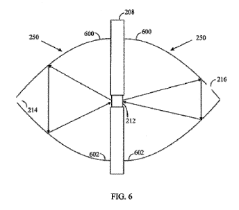
Figure 6 presents an embodiment where the reflecting surface 250 comprises
two reflecting paraboloids 600, 602 on both sides of the wall structure 208,
although
only one of such paraboloids 600, 602 may be located only on either side of
the wall
structure 208. The first paraboloid 600 enables an optical beam passing
through
aperture 214 and scattered from the sample 212 to be reflected to the second
paraboloid 602 which then is able to reflect the optical beam back to the
sample
212.
Figure 7 presents an optical element 700 of the excitation side. To focus
excitation radiation on the sample 212 a component like the optical element
700
may be needed. A source 702 may be an emitting part of the transmitter 220
shown
in previous drawings or an optical fibre coupled to the transmitter 220. The
source
702 may be placed in the input aperture 214 of the first reflecting surface
252. The
excitation radiation 224 is directed in a solid angle from the source 702 onto
the
sample 212. The optical element 700 may comprise lenses 704 and 706. The
excitation radiation 224 may be collected with the lens 704 which may
collimate the
excitation radiation. The collimated excitation radiation 224 may be converged
or
focused on the sample 212 by the second lens 706.
An optical element 700 may also be on the other side of the first reflecting
surface 252 than that in Figure 7. Additionally, a notch filter may be
provided to have
a narrow band for the excitation beam 224.
Figure 8 shows an optical element 800 of the pickup side. The optical
radiation 226 from the sample 212 may be collected in a solid angle
characteristic to
an optical element 800. A lens 802 inside the optical element 800 may
collimate the
21
CA 02859249 2014-06-13
WO 2013/087656 PCT/EP2012/075137
optical radiation 226. The collimated optical radiation 226 may be converged
or
focused onto the output aperture 216 by a second lens 804 of the optical
element
800. Alternatively, the collimated optical radiation 226 may be converged or
focused
onto a slit of a spectrometer (not shown) or an optical fibre 806 leading to
the
spectrometer by a second lens 804. The spectrometer or an optical fibre 806
fully or
partly represents the receiver 222. The optical element 800 may also be on the
other
side of the second reflecting surface 252 than that in Figure 8. Additionally,
a
Rayleigh line rejection filter may be placed between the lenses 802, 804 to
separate
the Raman radiation from the rest of the optical radiation.
A fraction of the optical radiation 226 hitting the optical element 800 may be
reflected back to the reflecting surface 252. But since the reflecting surface
252
reflects a large proportion of the optical radiation 226 back to the optical
element
800, the efficiency of collecting this part of the optical radiation is high.
A
corresponding effect takes place on the transmitter's side where the optical
radiation 224 is guided efficiently into the sample 212.
In every embodiment, the received optical radiation 226 may be filtered in
several manners known by a person skilled in the art in the receiver 222 for
effectively detecting the Raman radiation. The filtering may for example
include
temporal and band pass filtering.
Figure 9 presents a block diagram of an automated machine performing
Raman measurements of samples. A flow of samples such as tablets is fed to a
mechanism 900 which moves the tablets to a measuring unit 902 comprising the
reflective surface(s) as described above and positions the samples in the
aperture
210 of the wall structure 208. The mechanism 900 may be a part of the wall
structure 208 or the wall structure 208 may be a part of the mechanism 900.
Alternatively the mechanism 900 may be structurally independent of the wall
structure 208.
The tablets may be fed to the mechanism 900 by a conveyor or the like. The
mechanism 900 may pick each tablet one by one and move them one by one to the
measuring device 902. The mechanism 900 may have a picking and holding unit
which holds the tablet in the aperture 210 of the wall structure 208 during
the
22
CA 02859249 2014-06-13
WO 2013/087656 PCT/EP2012/075137
measurement. The tablet may move or its movement may be stopped during the
measurement. After the measurement, the mechanism 900 may shift the measured
tablet back to a feeding system (not shown) for further processing. The
mechanism
900 may move the tablets at constant speed and an optical pulse from a
transmitter
(220 not shown in Fig. 9) such as a laser may be directed at the tablet sample
212
during the time the tablet 212 is in the aperture 210 of the wall structure
208.
Although it is possible to stop each tablet 212 for the measurement, inertia
of the
mechanism 900 would slow down the rate of measurement because of repeated
acceleration and deceleration for each tablet 212. Continuous movement may be
used to average the variation of the spatial concentration in the tablet 212.
The
transmission and the reception of the optical radiation may also follow the
moving
sample 212 during each measurement and then return to a start position for the
next measurement. Using this apparatus, it may be possible to make
measurements
quickly which may enable measurements of all samples of a batch, irrespective
of
the number of samples in the batch.
If a liquid sample 212 is measured, liquid may be caused to flow in an
optically transparent pipe through the aperture 210 in the wall structure 208.
Then
the feeding mechanism 900 may comprise a pump and the pipe.
Since Raman radiation is effectively collected using the reflecting surface
250,
measurements may be performed very quickly. One measurement may take less
than one second, even as little as 0.1 second. Because the measurement is so
rapid
all tablets and capsules produced in a mass production process may be
measured.
This is an advantage since previously it has been possible only to make a
statistical
analysis by taking a representative number of tablets from a batch, measuring
the
tablets, and determining the whole batch as acceptable or not acceptable on
basis of
the representative measurement.
In the case of a liquid sample, a large volume of liquid may be measured
quickly since the speed of flow in the pipe may be high without loss of
accuracy in
the measurement. This is due to the fact that the liquid cannot flow a long
distance
between two successive measurements.
23
CA 02859249 2014-06-13
WO 2013/087656 PCT/EP2012/075137
In an experiment, an increase by factor of 26x in Raman photons was
obtained when a tablet, 5 mm thick, was measured with concave, specular
reflecting
surfaces 252, 254 on both receiver's and transmitter's side of a wall
structure 208
compared to a measurement without the reflecting surfaces 252, 254. This
increase
in Raman photons in detection enables the achievement of the same signal-to-
noise
ratio of the measured spectrum much faster than without the reflecting
surfaces.
Hence, it appears to be possible to make transmission Raman measurements
quicker
than earlier. The increased speed in measurement may enable 100 % inspection
of a
continuous product. Alternatively, the increased speed in measurement may
enable
higher accuracy (if integration time of the measurement is kept unchanged) and
hence provide fast enough response to allow a use of a closed loop control in
the
production process.
Figure 10 presents a flow chart of the method of the invention. In step 1000,
optical excitation radiation is directed to a sample 212 in an optically
transparent
aperture 210 of a wall structure 208 separating optically a transmitter 220 of
the
excitation radiation and a receiver 222 during a transmission Raman
measurement.
In step 1002, optical radiation scattering from the sample 212 is reflected
back to the
sample 212 by a reflecting surface 250 facing the wall structure 208 for
increasing
Raman radiation at the receiver 222.
Usually almost or totally white, turbid or diffuse samples are used. On the
excitation side, backreflected excitation light (typically 90%) and
backreflected
Raman signal (both totally diffuse) can be very effectively returned back to
the same
area which it left by means of the reflecting surface 250. Since the sample is
usually
at least rather diffuse, the propagation of Raman scattered light on the
detection
side is diffuse by its nature. This means that at the surface of the sample,
i.e. in a
layer thickness about 1/scattering constant, the density of Raman scattered
photons
is much less than somewhat deeper in the sample. This is because the surface
of the
sample 212 has nothing to reflect or scatter back the photons but instead the
photons disappear in the hemispherical space where the pickup probe catches
some
of them for detection. In this way, the Raman photon density is diluted at the
surface
of the tablet. However, if a reflecting surface 254 is placed at the detection
side, the
24
CA 02859249 2014-06-13
WO 2013/087656 PCT/EP2012/075137
photons hitting the reflecting surface are returned back to sample 212 and
this
effect eliminates the dilution process described above. As a result, the
intensity of
the Raman radiation observed by the pickup probe increases with factor often
much
larger than 2. The intensity may be increased in a similar manner also on the
excitation side which leads to increase in the observed Raman radiation.
Figure 11 shows an embodiment of the apparatus in which the wall structure
208 has a thickness "t" between its opposed surfaces 208A and 208B on the
transmitter 220 and receiver 224 sides. Parts corresponding to earlier Figures
are
numbered correspondingly. There are two opposite-facing hemispherical
reflecting
surfaces 250 arranged such that the spherical centre of each hemisphere is at
the
level of a surface 208A and 208B of the wall structure 208 and the wall
structure 208
is across the diameter of the spherical shape formed by the two hemispheres
250.
The aperture 210 passes completely through the wall structure 208 from one
surface
209A to the other 208B. The thickness "t" is such that the surface of the
sample 212
is below one or (as shown) both of the opposite surfaces 208A, 208B of the
wall
structure 208. The aperture 210 is in effect a tunnel between the two opposite
surfaces 208A, 208B of the wall structure 208. The walls 251 of such a tunnel
are also
specularly reflective. Such a construction facilitates adaptation of the
system to
samples 212 of different thicknesses in the direction of the thickness of the
wall
structure 208. Both reflective surfaces 200 are hemispherical reflective
surfaces,
with their spherical centre at the level of the surfaces 208A, 208B of the
wall
structure 208.
Although the invention is above described with reference to the examples
according to the attached drawings, the invention is not limited thereto. It
will be
obvious to a person skilled in the art that, as technology advances, the
inventive
concept can be implemented in various ways. The invention and its embodiments
are not limited to the examples described above but may vary within the scope
of
the claims.