Note: Descriptions are shown in the official language in which they were submitted.
CA 02859548 2014-06-16
WO 2013/119321 PCT/US2012/070150
FASTENERS FOR AFFIXING SHEET-LIKE MATERIALS TO BONE OR TISSUE
CROSS REFERENCE TO RELATED APPLICATIONS
[0001] This application claims priority to U.S. Provisional Application No.
61/577,626 filed
on December 19, 2011, the disclosure of which is incorporated by reference
herein. The present
disclosure is related to the following commonly assigned co-pending
applications, the
disclosures of which are incorporated herein by reference: U.S. Provisional
Application No.
61/577,621 filed on December 19, 2011, Attorney Docket No. 10322-711.100
entitled,
"APPARATUS AND METHOD FOR FORMING PILOT HOLES IN BONE AND
DELIVERING FASTENERS THEREIN FOR RETAINING AN IMPLANT" ; U.S. Provisional
Application No. 61/577,632 filed on December 19, 2011, Attorney Docket No.
10322-713.100
entitled, "FAS l'ENERS AND FASTENER DELIVERY DEVICES FOR AFFIXING SHEET-
LIKE MATERIALS TO BONE OR TISSUE" and U.S. Provisional Application No.
61/577,635
filed on December 19, 2011, Attorney Docket No. 10322-714.100 entitled, "FAS
l'ENERS AND
FASTENER DELIVERY DEVICES FOR AFFIXING SHEET-LIKE MATERIALS TO BONE
OR TISSUE."
INCORPORATION BY REFERENCE
[0002] All publications and patent applications mentioned in this
specification are herein
incorporated by reference to the same extent as if each individual publication
or patent
application was specifically and individually indicated to be incorporated by
reference.
FIELD
[0003] The present invention relates generally to orthopedic medicine and
surgery. More
particularly, the present invention relates to methods and apparatus for
delivery and fixation of
sheet-like materials, such as for treating tendons or like tissue of
articulating joints such as
tendons in the rotator cuff of the shoulder.
BACKGROUND
[0004] The glenohumeral joint of the shoulder is found where the head of
the humerus mates
with a shallow depression in the scapula. This shallow depression is known as
the glenoid fossa.
Six muscles extend between the humerus and scapula and actuate the
glenohumeral joint. These
six muscles include the deltoid, the teres major, and the four rotator cuff
muscles. The rotator
cuff muscles are a complex of muscles. The muscles of the rotator cuff include
the
supraspinatus, the infraspinatus, the subscapularis, and the teres minor. The
centering and
- 1 -
CA 02859548 2014-06-16
WO 2013/119321 PCT/US2012/070150
stabilizing roles played by the rotator cuff muscles are critical to the
proper function of the
shoulder. The rotator cuff muscles provide a wide variety of moments to rotate
the humerus and
to oppose unwanted components of the deltoid and pectoral muscle forces.
[0005] The muscles of the rotator cuff arise from the scapula. The
distal tendons of the
rotator cuff muscles splay out and interdigitate to form a common continuous
insertion on the
humerus. The supraspinatus muscle arises from the supraspinatus fossa of the
posterior scapula,
passes beneath the acromion and the acromioclavicular joint, and attaches to
the superior aspect
of the greater tuberosity. The mechanics of the rotator cuff muscles are
complex. The rotator
cuff muscles rotate the humerus with respect to the scapula, compress the
humeral head into the
glenoid fossa providing a critical stabilizing mechanism to the shoulder
(known as concavity
compression), and provide muscular balance. The supraspinatus and deltoid
muscles are equally
responsible for producing torque about the shoulder joint in the functional
planes of motion.
[0006] The rotator cuff muscles are critical elements of this shoulder
muscle balance
equation. The human shoulder has no fixed axis. In a specified position,
activation of a muscle
creates a unique set of rotational moments. For example, the anterior deltoid
can exert moments
in forward elevation, internal rotation, and cross-body movement. If forward
elevation is to occur
without rotation, the cross-body and internal rotation moments of this muscle
must be
neutralized by other muscles, such as the posterior deltoid and infraspinatus.
The timing and
magnitude of these balancing muscle effects must be precisely coordinated to
avoid unwanted
directions of humeral motion. Thus the simplified view of muscles as isolated
motors, or as
members of force couples must give way to an understanding that all shoulder
muscles function
together in a precisely coordinated way--opposing muscles canceling out
undesired elements
leaving only the net torque necessary to produce the desired action. Injury to
any of these soft
tissues can greatly inhibit ranges and types of motion of the arm.
[0007] With its complexity, range of motion and extensive use, a common
soft tissue injury
is damage to the rotator cuff or rotator cuff tendons. Damage to the rotator
cuff is a potentially
serious medical condition that may occur during hyperextension, from an acute
traumatic tear or
from overuse of the joint. With its critical role in abduction, rotational
strength and torque
production, the most common injury associated with the rotator cuff region is
a strain or tear
involving the supraspinatus tendon. A tear at the insertion site of the tendon
with the humerus,
may result in the detachment of the tendon from the bone. This detachment may
be partial or
full, depending upon the severity of the injury or damage. Additionally, the
strain or tear can
occur within the tendon itself. Injuries to the supraspinatus tendon and
current modalities for
treatment are defined by the type and degree of tear. The first type of tear
is a full thickness tear,
which as the term indicates is a tear that extends through the thickness of
the supraspinatus
- 2 -
CA 02859548 2014-06-16
WO 2013/119321 PCT/US2012/070150
tendon regardless of whether it is completely torn laterally. The second type
of tear is a partial
thickness tear which is further classified based on how much of the thickness
is torn, whether it
is greater or less than about 50% of the thickness.
[0008] The accepted treatment for a full thickness tear or a partial
thickness tear greater than
50% includes reconnecting the torn tendon via sutures. For the partial
thickness tears greater
than 50%, the tear is completed to a full thickness tear by cutting the tendon
prior to
reconnection. In contrast to the treatment of a full thickness tear or a
partial thickness tear of
greater than 50%, the current standard treatment for a partial thickness tear
less than 50% usually
involves physical cessation from use of the tendon, i.e., rest. Specific
exercises can also be
prescribed to strengthen and loosen the shoulder area. In many instances, the
shoulder does not
heal and the partial thickness tear can be the source of chronic pain and
stiffness. Further, the
pain and stiffness may cause restricted use of the limb which tends to result
in further
degeneration or atrophy in the shoulder. Surgical intervention may be required
for a partial
thickness tear of less than 50%, however, current treatment interventions do
not include repair of
the tendon, and rather the surgical procedure is directed to arthroscopic
removal of bone to
relieve points of impingement or create a larger tunnel between the tendon and
bone that is
believed to be causing tendon damage. As part of the treatment, degenerated
tendon may also be
removed using a debridement procedure in which tendon material is ablated.
Again, the tendon
partial thickness tear is not repaired. Several authors have reported
satisfactory early post
operative results from these procedures, but over time recurrent symptoms have
been noted. In
the event of recurrent symptoms, many times a patient will "live with the
pain". This may result
in less use of the arm and shoulder which causes further degeneration of the
tendon and may lead
to more extensive damage. A tendon repair would then need to be done in a
later procedure if
the prescribed treatment for the partial tear was unsuccessful in relieving
pain and stiffness or
over time the tear propagated through injury or degeneration to a full
thickness tear or a partial
thickness tear greater than 50% with attendant pain and debilitation. A
subsequent later
procedure would include the more drastic procedure of completing the tear to
full thickness and
suturing the ends of the tendon back together. This procedure requires
extensive rehabilitation,
has relatively high failure rates and subjects the patient who first presented
and was treated with
a partial thickness tear less than 50% to a second surgical procedure.
[0009] As described above, adequate treatments do not currently exist
for repairing a partial
thickness tear of less than 50% in the supraspinatus tendon. Current
procedures attempt to
alleviate impingement or make room for movement of the tendon to prevent
further damage and
relieve discomfort but do not repair or strengthen the tendon. Use of the
still damaged tendon
can lead to further damage or injury. Prior damage may result in degeneration
that requires a
- 3 -
CA 02859548 2014-06-16
WO 2013/119321 PCT/US2012/070150
second more drastic procedure to repair the tendon. Further, if the prior
procedure was only
partially successful in relieving pain and discomfort, a response may be to
use the shoulder less
which leads to degeneration and increased likelihood of further injury along
with the need for
more drastic surgery. Further, it would be beneficial to be able to treat
partial thickness tears
greater than 50% without cutting the untorn portion of the tendon to complete
the tear before
suturing back together. There is a large need for surgical techniques and
systems to treat partial
thickness tears and prevent future tendon damage by strengthening or repairing
the native tendon
having the partial thickness tear.
SUMMARY OF THE DISCLOSURE
[00010] The present disclosure is generally directed to a fastener or staple
that can be used to
attach an implant to bone or other tissue. The staple or fastener can be
included in a kit or
system that also can include a staple delivery device and a pilot hole forming
trocar assembly.
The trocar assembly is used to create pilot holes and retain instrument
position within those pilot
holes for staple insertion. The staple delivery device can carry the staple
into the pilot holes and
release the staple in engagement with bone to retain the implant in position.
[00011] The staple for insertion and retention in bone can include a bridge
portion having
arms extending from proximate each end thereof, at least a portion of each arm
including tissue
retention members, each tissue retention member having at least two barbed
projections
extending laterally therefrom. Each arm can have a cross sectional area of
reduced strength
proximate each projection relative to other portions of the tissue retention
member such that a
portion of the tissue retention member flexes laterally proximate each
projection in response to a
pullout force applied to the bridge. The tissue retention members can include
a trunk of greater
cross sectional area than a non-trunk portion of the arms.
[00012] The fastener or staple can also include, in alternative embodiments, a
first arm having
a proximal end and a distal end, a second arm having a proximal end and a
distal end, and a
bridge connecting the first arm and second arm, wherein each of the first and
second arms
include a trunk portion extending over at least a portion of the length
thereof. Each trunk can
have a lateral extent larger than a lateral extent of the bridge or non-trunk
arm portion adjacent
thereto such that the staple includes a first change in lateral stiffness
disposed proximate the
bridge or non-trunk arm portion abutment with the first trunk and a second
change in lateral
stiffness disposed proximate the bridge or non-trunk arm portion abutment with
the second
trunk. The lateral extent of each trunk in at least one direction can be at
least about three times
the lateral extent of at least a portion of the bridge or non-trunk portion of
the arm.
- 4 -
CA 02859548 2014-06-16
WO 2013/119321 PCT/US2012/070150
[00013] Each trunk can further include a first projection and a second
projection, the first
projection including a first proximal surface extending away from the trunk in
a first direction,
the first direction being such that the first proximal surface will engage the
tissue or bone when
the trunk is inserted therein so that a first moment is applied to the trunk
in response to a pullout
force on the bridge. Likewise, the second projection can include a second
proximal surface
extending away from the trunk in a second direction, the second direction
being such that the
second proximal surface will engage the tissue or bone when the trunk is
inserted therein so that
a second moment is applied to the trunk in response to a pullout force on the
bridge. Each of the
trunks can further include a localized area of weakness proximate the second
projection thereon.
For example, a second area of reduced strength can include a slit in the cross
section of the tissue
retention member or trunk adjacent at least one of the projections therefrom.
Further, reduced
strength can be created where the trunk meets the non-trunk portion of the arm
adjacent thereto
or the bridge.
[00014] In some embodiments, the change in lateral stiffness and the localized
area of
weakness allow flexing of each arm portion in response to the first and second
moment,
respectively.
[00015] The projections can be arranged to extend in first and second
directions to achieve
increased pullout strength. The first direction can extend proximally and
laterally away from
each trunk while the second direction can extend proximally and laterally away
from each trunk
and a lateral component of the second direction is generally opposite a
lateral component of the
first direction. The forces on the projections create moments about the more
flexible portions of
the staple where the direction of the first moment is generally opposite the
direction of the
second moment on each arm.
[00016] In some embodiments, the fastener first trunk and the second trunk
each define a
cavity, each cavity being spaced laterally from the respective non-trunk
portion or bridge
adjacent thereto. Each cavity defined by the first and the second trunk is
sized to receive a first
stake and a second stake, respectively, of a fastener delivery device. Each
cavity defined by the
first and the second trunk can extend from the proximal end to the distal end
of the trunk.
BRIEF DESCRIPTION OF THE DRAWINGS
[00017] Figure 1 is a perspective view illustrating an exemplary tissue
fastener or staple in
accordance with the present disclosure;
[00018] Figure 2 is a an alternative perspective view of the tissue fastener
or staple of Figure
1 illustrating other features in accordance with the present disclosure;
- 5 -
CA 02859548 2014-06-16
WO 2013/119321 PCT/US2012/070150
[00019] Figure 3 is a top plan view of the tissue fastener or staple of Figure
1 illustrating the
laterally extending legs having lumens for receiving the stakes of a delivery
device for
positioning the staple in desired tissue;
[00020] Figure 4 is a front plan view of the tissue fastener or staple of
Figure 1 illustrating in
phantom flexing of the barbs and legs of the staple in response to grasping of
tissue in one
embodiment of the disclosure;
[00021] Figure 5 is a stylized anterior view of a shoulder including a humerus
and a scapula;
[00022] Figure 6 is a stylized of a shoulder depicting the head of the humerus
shown mating
with the glenoid fossa of the scapula at a glenohumeral joint and a sheet-like
material is affixed
to the tendon;
[00023] Figure 7 is a stylized perspective view showing a portion of the body
of a human
patient divided into quadrants by planes for descriptive purposes herein;
[00024] Figure 8 is a stylized perspective view illustrating an exemplary
procedure for
arthroscopic treatment of a shoulder of a patient in accordance with one
embodiment of the
disclosure;
[00025] Figure 9 is a stylized perspective view of a shoulder including a
supraspinatus having
a distal tendon with a sheet-like material affixed thereto;
[00026] Figure 10A is a simplified perspective view of a tissue fastener or
staple delivery
device in accordance with the present disclosure;
[00027] Figure 10B is a simplified perspective view of a trocar assembly,
including a trocar
disposed within a guide sheath assembly for creating pilot holes and retaining
the sheath within
the formed pilot holes for delivery of a tissue fastener or staple by a device
such as that depicted
in Figure 10A;
[00028] Figure 11A is a perspective view of the sheath assembly of Figure 10B
with the trocar
removed;
[00029] Figure 11B is a perspective view of the trocar of Figure 10B as
removed from the
sheath assembly;
[00030] Figure 11C is a perspective view of one pilot hole position retention
member which is
positioned in a distal portion of the sheath assembly in one embodiment of the
present
disclosure;
[00031] Figure 12 is a perspective view depicting the sheath and staple pusher
assemblies of a
staple delivery device in one embodiment of the disclosure;
[00032] Figure 13 is a simplified exploded view of the tissue fastener or
staple delivery device
of Figure 10A depicting additional features thereof;
[00033] Figure 14 depicts further features of the staple pusher assembly of
Figure 13;
- 6 -
CA 02859548 2014-06-16
WO 2013/119321
PCT/US2012/070150
[00034] Figures 15A and 15B illustrate the features of the distal portion of
the staple pusher
assembly of Figure 13 with a staple mounted thereon in accordance with one
embodiment of the
disclosure;
[00035] Figure 16A and 16B further illustrate the staple pusher assembly in
one embodiment
of the disclosure;
[00036] Figure 17 is a more detailed perspective view of the distal portion of
the staple pusher
assembly illustrating stakes that mate with the staple in one embodiment of
the disclosure;
[00037] Figure 18A is simplified perspective view of a shoulder having an
implant affixed to
the tendon and depicting the first step in a method of delivering fasteners to
affix the implant to
bone of the humeral head in accordance with one method of the disclosure;
[00038] Figure 18B is a simplified plan view of the distal portion of the
trocar assembly as
position to create pilot holes for affixing the implant to bone in a further
step of a method of the
disclosure;
[00039] Figure 18C depicts the trocar assembly of Figure 18B as inserted into
the bone to
form pilot holes in accordance with a method of the disclosure;
[00040] Figure 18D depicts the trocar assembly with the trocar portion removed
and the
remaining sheath assembly retaining its position in the pilot holes formed;
[00041] Figure 18E depicts insertion of a fastener or staple into the formed
pilots holes
through the sheath assembly in accordance with a method of the disclosure;
and,
[00042] Figure 18F illustrates a fastener or staple as inserted in accordance
with a method of
the disclosure.
DETAILED DESCRIPTION
[00043]
The following detailed description should be read with reference to the
drawings in
which similar elements in different drawings are numbered the same. The
drawings, which are
not necessarily to scale, depict illustrative embodiments and are not intended
to limit the scope
of the invention.
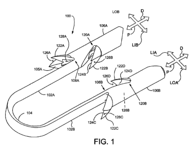
[00044] Figure 1 is a perspective view illustrating an exemplary staple 100 in
accordance with
the present detailed description. With reference to Figure 1, it will be
appreciated that staple 100
may assume various orientations without deviating from the spirit and scope of
this detailed
description. Although the various parts of this exemplary embodiment are
depicted in relative
proportion to other parts of the staple 100, other configurations in size and
orientation of the
various parts are possible. A number of reference directions are illustrated
using arrows in Figure
1 to assist in understanding the details of the staple 100. The illustrated
directions include: a
proximal direction P, a distal direction D, a first laterally outward
direction LOA, a second
- 7
CA 02859548 2014-06-16
WO 2013/119321 PCT/US2012/070150
laterally outward direction LOB, a first laterally inward direction LIA, and a
second laterally
inward direction LIB.
[00045] Staple 100 comprises a first arm 102A, a second arm 102B, and a bridge
104
extending from, abutting or adjacent to the proximal end of first arm 102A to
the proximal end
of second arm 102B. The first arm 102A includes a first trunk 106A extending
for at a least a
portion of the length of the first arm 102A. As depicted in Figure 1, a
proximal portion of the
first arm 102A abuts the proximal end of the first trunk 106A. = The first arm
102A, in this
embodiment includes the trunk portion 106A and a non-trunk portion 105A. The
length of first
trunk 106A relative to the overall length of the first arm 102A can vary in
different
embodiments. The first trunk 106A can extend for the entire length of the
first arm 102A such
that the bridge abuts with or is adjacent to the trunk 106A. Similarly, the
second arm 102B
includes a second trunk 106B extending for at least a portion of the length of
the second arm
102B. A proximal portion of the second arm 102B abuts the proximal end of the
second trunk
106B. The second arm 102B, in this embodiment includes the trunk portion 106B
and a non-
trunk portion 105B. The length of second trunk 106B relative to the overall
length of the second
arm 102B can vary in different embodiments. The second trunk 106B can extend
for the entire
length of the second arm 102B such that the bridge abuts with or is adjacent
to the trunk 106B.
In Figure 1, first trunk 106A and second trunk 106B are shown extending
distally from a
proximal portion of first arm 102A and second arm 102B, respectively.
[00046] In the embodiment of Figure 1, first trunk 106A has a lateral extent,
or cross sectional
area, that is larger than a lateral extent of the non-trunk portion 105A of
first arm 102A and
bridge 104. The staple 100 includes a first change in lateral stiffness 108A
disposed where the
distal end of non-trunk portion 105A of first arm 102A abuts first trunk 106A.
As depicted, the
change in stiffness is abrupt, but can be gradual in alternative embodiments.
In an embodiment
where the first trunk 106A extends for the full length of the first arm 102A,
the change in
stiffness occurs where the first trunk 106A abuts the bridge 104. With
reference to Figure 1, it
will be appreciated that first trunk 106A is mounted eccentrically to first
arm 102A and second
trunk 106B is mounted eccentrically to second arm 102B. As with first trunk
106A, second
trunk 106B has a lateral extent, or cross sectional area that is larger than a
lateral extent of
second arm 102B or bridge 104. The staple 100 includes a second change in
lateral stiffness
108B where the distal end of second arm 102B abuts second trunk 106A in the
embodiment of
Figure 1. If the second trunk 106B extends for the entire length of second arm
102B, the change
in stiffiless occurs at the abutment with the bridge 104.
[00047] Each of the first trunk 106A and second trunk 106B can include at
least a first
projection 122A, 122C and a second projection 122B, 122D, the first projection
122A, 122C on
- 8 -
CA 02859548 2014-06-16
WO 2013/119321 PCT/US2012/070150
each trunk 106A, 106B includes a first proximal surface 124A, 124C extending
away from the
trunk in a first direction, the first direction being such that the first
proximal surface 124A, 124C
will engage the tissue or bone after the trunk is inserted therein and a
pullout force is applied to
the bridge 104. This force creates a first moment centered on the area of
reduced lateral extent
adjacent the trunk, tending to rotate the trunk thereabout, further providing
a greater holding
force in response to the pullout force as the trunk presses against the tissue
or bone. The second
projection 122B, 122D includes a second proximal surface 124B, 124D extending
away from the
trunk in a second direction, different from the first direction, the second
direction being such that
the second proximal surfaces 124B, 124D will engage the tissue or bone after
the trunk is
inserted therein and a pullout force is applied to the bridge 104. A slit or
area of reduced cross
section in the trunk adjacent the second projections provide an area of
weakness so that a second
moment is applied to the trunk in response to a pullout force on the bridge
104. This moment
causes rotation of the trunk about the area of weakness and increases the
holding force with
increased pullout force.
[00048] As specifically illustrated in the embodiment of staple or fastener
100 in Figure 1,
first trunk 106A includes a first projection 122A disposed at an outer side of
trunk 106A and a
second projection 122B disposed at an inner side of the trunk. First
projection 122A includes a
first proximal surface 124A extending away from first trunk 106A in a first
direction. With
reference to Figure 1, it will be appreciated that the first direction has an
outward lateral
component and a proximal component so that first proximal surface 124A extends
outwardly and
proximally away from first trunk 106A. The first direction is selected such
that first proximal
surface 124A will engage tissue or bone proximate the outer side of first
trunk 106A after being
inserted therein so that a first moment is applied to the trunk in response to
a pullout force on
bridge 104. The moment centers on the arm portion of lesser cross section
adjacent the first
projection.
[00049] In the embodiment of Figure 1, first trunk 106A includes a first
localized area of
weakness 120A disposed proximate second projection 122B. Second projection
122B includes a
second proximal surface 124B extending away from first trunk 106A in a second
direction. The
second direction is selected such that second proximal surface 124A will
engage tissue or bone
proximate the inner side of first trunk 106A when inserted therein so that a
second moment is
applied to the trunk in response to a pullout force on bridge 104. The moment
centers around the
area of weakness 120A. The second moment has a direction that is generally
opposite a
direction of the first moment. It will be appreciated that the second
direction has an inward
lateral component and a proximal component so that second proximal surface
124B extends
inwardly and proximally away from first trunk 106A.
- 9 -
CA 02859548 2014-06-16
WO 2013/119321 PCT/US2012/070150
[00050] Second trunk 106B includes a third projection 122C disposed at an
outer side of
second trunk 106B and a fourth projection 122D disposed at an inner side of
the trunk. In the
embodiment of Figure 1, third projection 122C includes a third proximal
surface 124C extending
away from second trunk 106B in a third direction. With reference to Figure 1,
it will be
appreciated that the third direction has an outward lateral component and a
proximal component
so that third proximal surface 124C extends outwardly and proximally away from
second trunk
106B. The third direction is selected such that third proximal surface 124C
will engage tissue or
bone proximate the outer side of second trunk 106B when inserted therein so
that a third moment
is applied to the trunk in response to a pullout force on bridge 104.
[00051] In the embodiment of Figure 1, second trunk 106B includes a second
localized area of
weakness 120B disposed proximate fourth projection 122D. Fourth projection
122D includes a
fourth proximal surface 124D extending away from second trunk 106B in a fourth
direction. In
the embodiment of Figure 1, the fourth direction is selected such that second
proximal surface
124A will engage tissue or bone proximate the inner side of second trunk 106B
when inserted
therein so that a fourth moment is applied to the trunk in response to a
pullout force on bridge
104. The fourth moment has a direction that is generally opposite a direction
of the third
moment. It will be appreciated that the fourth direction has an inward lateral
component and a
proximal component so that fourth proximal surface 124D extends inwardly and
proximally
away from second trunk 106B.
[00052] As depicted in Figure 1, the staple 100 includes proximal projections
that extend
away from or outward from the bridge 104, while the distal projections extend
inward or toward
the center of the bridge 104. This creates generally opposing forces in
response to tension on the
bridge which, in combination with areas of weakness or reduced lateral extent,
substantially
increases the holding force of the staple in bone as the different portions of
the trunks tend to
rotate in opposite directions and apply force to an opposing wall in the hole
in bone in which the
staple is positioned. It is however, understood that other configurations of
the projections are
possible. In some embodiments, at least two projections are included and they
extend in
different directions to cause different force responses as tension is applied
to the bridge. It is
believed this provides adequate holding force in bone, which can include
differing thicknesses of
hard and soft tissue along with porous areas.
[00053] In some useful embodiments, each projection of staple 100 may be
clefted to form a
plurality of points for greater retention in tissue. In the exemplary
embodiment of Figure 1, first
projection 122A of first trunk 106A defines a first notch 126A that divides
first projection 122A
into a first sub-projection and a second sub-projection. Second projection
122B of second trunk
106B defines a second notch 126B. In the exemplary embodiment of Figure 1,
second notch
-10-
CA 02859548 2014-06-16
WO 2013/119321 PCT/US2012/070150
126B divides second projection 122B into a first sub-projection and a second
sub-projection.
Third projection 122C of second trunk 106B defines a third notch 126C that
divides third
projection 122C into a first sub-projection and a second sub-projection.
Fourth projection
122D of second trunk 106B defines a fourth notch 126D that divides fourth
projection 122D into
a first sub-projection and a second sub-projection.
[00054] With continued reference to Figure 1 and further reference to Figures
2 and 3, first
trunk 106A defines a first cavity 128A and second trunk 106B defines a second
cavity 128B. In
the exemplary embodiment of Figures 1, 2 and 3, first cavity 128A extends into
first trunk 106A
and second cavity 128B extends into second trunk 106B. The cavity is sized to
cooperate with a
staple delivery device for holding and inserting the staple into tissue or
bone, as later described
in detail herein. In summary, the staple delivery device includes
longitudinally extending stakes
that fit within the cavities 128A, 128B to hold the staple 100 and push it
into position in the
tissue as the stake abuts a portion of its respective trunk. In some
embodiments the cavity may
extend through a portion of the length of each trunk, as best depicted in
Figure 2 which indicates
the distal end of the staple 100 is closed. Alternatively, first cavity 128A
and second cavity
128B may extend through the entire length of each trunk 106A, 106B or other
portions of staple
100 in some embodiments. As illustrated by the top view of the staple 100 in
Figure 3, first
cavity 128A and second cavity 128B each have a generally rectangular or square
cross-sectional
shape to cooperate with a similarly shaped cross section on a staple delivery
device. However,
that first cavity 128A and second cavity 128B may have various cross-sectional
shapes to
cooperate with alternative staple delivery device designs without deviating
from the spirit and
scope of the present detailed description.
[00055] Figure 4 is an alternative perspective view of the embodiment in
Figure 1 illustrating
an exemplary staple 100 in accordance with the present detailed description.
In particular, Figure
4 illustrates in phantom the flexing and bending of the trunks 106A and 106B
after implant in
response to tension applied to the bridge, as by tissue or an implant affixed
at an implant site.
Staple 100 comprises a first arm 102A, a second arm 102B, and a bridge 104
extending from the
proximal end of first arm 102A to the proximal end of second arm 102B. The
distal end of first
arm non-trunk portion 105A abuts the proximal end of first trunk 106A.
Similarly, the distal end
of second arm non-trunk portion 105B abuts the proximal end of a second trunk
106B. In Figure
4, first trunk 106A and second trunk 106B are shown extending distally from
first arm 102A and
second arm 102B, respectively.
[00056] In the embodiment of Figure 4, first trunk 106A has a lateral extent
that is larger than
the lateral extent of the non-trunk portion 105A of first arm 102A. This
combination creates a
relatively abrupt change in lateral stiffness 108A disposed where the distal
end of the non-trunk
-11-
CA 02859548 2014-06-16
WO 2013/119321 PCT/US2012/070150
portion 108A of first arm 102A abuts first trunk 106A. With reference to
Figure 4, first trunk
106A is mounted eccentrically to first arm 102A and second trunk 106B is
mounted eccentrically
to second arm 102B, however, other mountings or abutments can be used, such as
a non-trunk
portion having walls that surround the cavity and include a lumen therethrough
to access the
cavity with a staple delivery stake. A change in lateral stiffness would still
be accomplished
=
where the lateral extend changed. Further, a change in lateral stiffness could
be accomplished by
using a different material for the non-trunk portion relative to the trunk
portion. Second trunk
106B in combination with the non-trunk portion 105B of second arm 102B
provides the same
change in lateral stiffness 108B.
[00057] As earlier described the configuration of the four projections 122A,
122B, 122C and
122D, contact the tissue or bone and provide a holding force upon
implantation. Each projection
is positioned to provide a force moment in a desired direction to the trunk in
response to the
pullout force on the bridge 104.
[00058] In the embodiment of Figure 4, first trunk 106A and second trunk 106B
include first
and second localized areas of weakness 120A, 120B disposed proximate second
projections
122B, 122D. This area of weakness is formed by a slit formed proximal of the
projection.
However, the area of weakness could be formed by other means, such as a change
in material,
pinching or perforations.
[00059] The combination of projections, areas of weakness and changes in
lateral extent
provide desired flexing, bending and rotating of the trunk in response to pull
out forces once
implanted in a bone, such as in a pilot hole formed in the bone. Together
these components act
as tissue retention members. An exemplary deflected shape is shown with dashed
lines in Figure
4. Staple 100 may be urged to assume the deflected shape shown in Figure 4,
for example, by
applying a pullout force on the bridge 104 of the staple 100. Alternatively,
distally directed
forces can be applied on staple 100 using, for example, the staple delivery
system shown later
and described herein. In some applications, the staple delivery tool may be
used to urge first
projection 122A and third projection 122C into orientations which lock staple
100 into a target
tissue. For example, first projection 122A and third projection 122C may be
rotated so that these
projections engage the target tissue. When this is the case, tension extending
through bridge 104
of staple 100 may keep first projection 122A and third projection 122C in the
rotated position.
Also when this is the case, the projections may inhibit staple pullout.
Further, rotation of any
projection causes a rotational force and within limits defined by the hole in
the bone some
rotation to an adjacent portion of the trunk which contacts or engages the
wall of the hole in the
bone. Increased pullout force results in increasing holding force with this
design.
- 12 -
CA 02859548 2014-06-16
WO 2013/119321 PCT/US2012/070150
[00060] Next referring to Figure 5, an exemplary use or application of the
staples of the
present disclosure is described. Figure 5 is a stylized anterior view of a
patient 20. For purposes
of illustration, a shoulder 22 of patient 20 is shown in cross-section in
Figure 5. Shoulder 22
includes a humerus 14 and a scapula 12. In Figure 5, a head 24 of humerus 14
can be seen
mating with a glenoid fossa of scapula 12 at a glenohumeral joint. With
reference to Figure 5, it
will be appreciated that the glenoid fossa comprises a shallow depression in
scapula 12. The
movement of humerus 14 relative to scapula 12 is controlled by a number of
muscles including:
the deltoid, the supraspinatus, the infraspinatus, the subscapularis, and the
teres minor. For
purposes of illustration, only the supraspinatus 26 is shown in Figure 5.
[00061] With reference to Figure 5, a distal tendon 28 of the supraspinatus 26
meets humerus
14 at an insertion point. Scapula 12 of shoulder 22 includes an acromium 32.
In Figure 5, a
subacromial bursa 34 is shown extending between acromium 32 of scapula 12 and
head 24 of
humerus 14. Subacromial bursa 34 is shown overlaying supraspinatus 26 as well
as
supraspinatus tendon 28 and a portion of humerus 14. Subacromial bursa 34 is
one of the
hundreds of bursae found the human body. Each bursa comprises a fluid filled
sac. The
presence of these bursae in the body reduces friction between bodily tissues.
[00062] The exemplary staples or fasteners described herein may be used to
affix tendon
repair implants to various target tissues. The shoulder depicted in Figure 5
is one example where
a tendon repair implant may be affixed to one or more bones associated with an
articulating joint,
such as the glenohumeral joint. Additionally, the tendon repair implant may be
affixed to one or
more tendons to be treated. The tendons to be treated may be torn, partially
torn, have internal
micro-tears, be untorn, and/or be thinned due to age, injury or overuse.
Applicants believe that
the methods and apparatus of the present application and related devices may
provide very
beneficial therapeutic effect on a patient experiencing joint pain believed to
be caused by partial
thickness tears and/or internal microtears. By applying a tendon-repair
implant early before a
full tear or other injury develops, the implant may cause the tendon to
thicken and/or at least
partially repair itself, thereby avoiding more extensive joint damage, pain,
and the need for more
extensive joint repair surgery.
[00063] Figure 6 is a stylized anterior view of a shoulder 22 including a
humerus 14 and a
scapula 12. In Figure 6, a head 24 of humerus 14 is shown mating with a
glenoid fossa of
scapula 12 at a glenohumeral joint. A supraspinatus 26 is also shown in Figure
6. This muscle,
along with others, controls the movement of humerus 14 relative to scapula 12.
A distal tendon
28 of supraspinatus 26 meets humerus 14 at an insertion point 30.
[00064] As depicted in Figure 6, distal tendon 28 includes a first damaged
portion 36. A
number of loose tendon fibers 40 in first damaged portion 36 are visible in
Figure 6. First
- 13 -
CA 02859548 2014-06-16
WO 2013/119321 PCT/US2012/070150
damaged portion 36 includes a first tear 42 extending partially through distal
tendon 28. First
tear 42 may therefore be referred to as a partial thickness tear. With
reference to Figure 6, first
tear 42 begins on the side of distal tendon 28 facing the subacromial bursa
(shown in the
previous Figure) and ends midway through distal tendon 28. Accordingly, first
tear 42 may be
referred to as a bursal side tear.
[00065] With reference to Figure 6, distal tendon 28 includes a second damaged
portion 38
located near insertion point 30. As illustrated, second damaged portion 38 of
distal tendon 28
has become frayed and a number of loose tendon fibers 40 are visible. Second
damaged portion
38 of distal tendon 28 includes second tear 44. Second tear 44 begins on the
side of distal tendon
28 facing the center of the humeral head 24. Accordingly, second damaged
portion 38 may be
referred to as an articular side tear.
[00066] Figure 6 illustrates a sheet-like implant 50 has been placed over the
bursal side of
distal tendon 28. The sheet-like implant 50 is affixed to distal tendon 28 by
a plurality of tendon
staples 51. Sheet-like implant 50 is affixed to humerus 14 by a plurality of
bone staples 100 in
accordance with designs of staples disclosed herein. Sheet-like implant 50
extends over insertion
point 30, first tear 42 and second tear 44. Some useful methods in accordance
with this detailed
description may include placing a tendon repair implant on the bursal side of
a tendon regardless
of whether the tears being treated are on the bursal side, articular side or
within the tendon. In
some cases the exact location and nature of the tears being treated may be
unknown. A tendon
repair implant may be applied to the bursal side of a tendon to treat shoulder
pain that is most
likely caused by one or more partial thickness tears in the tendon.
[00067] Figure 7 is a stylized perspective view showing a portion of the body
82 of a human
patient 20. Body 82 includes a shoulder 22. In the exemplary embodiment of
Figure 7, a
plurality of cannulas are positioned to access a treatment site within
shoulder 22. In some cases,
shoulder 22 may be inflated by pumping a continuous flow of saline through
shoulder 22 to
create a cavity proximate the treatment site. The cannulas shown in Figure 7
include a first
cannula 80A, a second cannula 80B and a third cannula 80C.
[00068] In Figure 7, a sagital plane SP and a frontal plane FP are shown
intersecting body 82.
Sagital plane SP and frontal plane FP intersect one another at a medial axis
MA of body 82.
With reference to Figure 7, sagital plane SP bisects body 82 into a right side
84 and a left side
86. Also with reference to Figure 7, frontal plane FP divides body 82 into an
anterior portion 92
and a posterior portion 88. Sagital plane SP and a frontal plane FP are
generally perpendicular to
one another. These planes and portions are used to describe the procedures
used in exemplary
embodiments.
-14-
'
CA 02859548 2014-06-16
WO 2013/119321 PCT/US2012/070150
[00069] First cannula 80A is accessing a treatment site within shoulder 22
using a lateral
approach in which first cannula 80A pierces the outer surface of right side 84
of body 82. The
term lateral approach could also be used to describe situations in which an
instrument pierces the
outer surface of left side 86 of body 82. Second cannula 80B is accessing a
treatment site within
shoulder 22 using a posterior approach in which second cannula 80B pierces the
outer surface of
posterior portion 88 of body 82. Third cannula 80C is accessing a treatment
site within shoulder
22 using an anterior approach in which third cannula 80C pierces the outer
surface of anterior
portion 92 of body 82.
[00070] Figure 8 is a stylized perspective view illustrating an exemplary
procedure for
treating a shoulder 22 of a patient 20. The procedure illustrated in Figure 8
may include, for
example, fixing tendon repair implants to one or more tendons of shoulder 22.
The tendons
treated may be torn, partially torn, have internal micro-tears, be untorn,
and/or be thinned due to
age, injury or overuse.
[00071] Shoulder 22 of Figure 8 has been inflated to create a cavity therein.
A fluid supply 52
is pumping a continuous flow of saline into the cavity. This flow of saline
exits the cavity via a
fluid drain 54. A camera 56 provides images from inside the cavity. The images
provided by
camera 56 may be viewed on a display 58.
[00072] Camera 56 may be used to visually inspect the tendons of shoulder 22
for damage. A
tendon repair implant in accordance with this disclosure may be affixed to a
bursal surface of the
tendon regardless of whether there are visible signs of tendon damage.
Applicants believe that
the methods and apparatus of the present application and related devices may
provide very
beneficial therapeutic effect on a patient experiencing joint pain believed to
be caused by internal
microtears, but having no clear signs of tendon tears. By applying a tendon
repair implant early
before a full tear or other injury develops, the implant may cause the tendon
to thicken and/or at
least partially repair itself, thereby avoiding more extensive joint damage,
pain, and the need for
more extensive joint repair surgery.
[00073] An implant delivery system 60 can be seen extending from shoulder 22
in Figure 8.
Implant delivery system 60 is extending through a first cannula 80A. In
certain embodiments,
first cannula 80A can access a treatment site within shoulder 22 using a
lateral approach in which
first cannula 80A pierces the outer surface of a right side of the patient's
body. In some cases a
physician may choose not to use a cannula in conjunction with implant delivery
system 60.
When that is the case, the implant delivery system may be advanced through
tissue. Implant
delivery system 60 comprises a sheath that is affixed to a handle. The sheath
defines a lumen
and a distal opening fluidly communicating with the lumen. In the embodiment
of Figure 8, the
- 15 -
CA 02859548 2014-06-16
WO 2013/119321
PCT/US2012/070150
distal opening of the sheath has been placed in fluid communication with the
cavity created in
shoulder 22.
[00074] A tendon repair implant is at least partially disposed in the lumen
defined by the
sheath of implant delivery system 60. Implant delivery system 60 can be used
to place the
tendon repair implant inside shoulder 22. In some embodiments, the tendon
repair implant is
folded into a compact configuration when inside the lumen of the sheath. When
this is the case,
implant delivery system 60 may be used to unfold the tendon repair implant
into an expanded
shape. Additionally, implant delivery system 60 can be used to hold the tendon
repair implant
against the tendon.
[00075] The tendon repair implant may be affixed to the tendon while it is
held against the
tendon by implant delivery system 60. Various attachment elements may be used
to fix the
tendon-repair implant to the tendon. Examples of attachment elements that may
be suitable in
some applications include sutures, tissue anchors, bone anchors, and staples.
In the exemplary
embodiment of Figure 8, the shaft of a fixation tool 70 is shown extending
into shoulder 22. In
one exemplary embodiment, fixation tool 70 is capable of fixing the tendon
repair implant to the
tendon and bone with one or more staples of the present disclosure while the
tendon repair
implant may held against the tendon by implant delivery system 60.
[00076] Figure 9 is a stylized perspective view of a shoulder 22 including a
supraspinatus 26
having a distal tendon 28. With reference to Figure 9, a tendon repair implant
50 has been
affixed to a surface of distal tendon 28. Tendon repair implant 50 may
comprise, for example,
various sheet-like structures without deviating from the spirit and scope of
the present detailed
description. In some useful embodiments, the sheet-like structure may comprise
a plurality of
fibers. The fibers may be interlinked with one another. When this is the case,
the sheet-like
structure may comprise a plurality of apertures comprising the interstitial
spaces between fibers.
Various processes may be used to interlink the fibers with one another.
Examples of processes
that may be suitable in some applications including weaving, knitting, and
braiding. In some
embodiments, the sheet-like structure may comprise a laminate including
multiple layers of film
with each layer of film defining a plurality of micro-machined or formed
holes. The sheet-like
structure of the tendon repair implant may also comprise a reconstituted
collagen material having
a porous structure. Additionally, the sheet-like structure of the tendon
repair implant may also
comprise a plurality of electro-spun nanofiber filaments forming a composite
sheet.
Additionally, the sheet-like structure may comprise a synthetic sponge
material that defines a
plurality of pores. The sheet-like structure may also comprise a reticulated
foam material.
Reticulated foam materials that may be suitable in some applications are
available from
Biomerix Corporation of Fremont, California which identifies these materials
using the
-16-
CA 02859548 2014-06-16
WO 2013/119321
PCT/US2012/070150
trademark BIOMATERIAL TM. The sheet-like structure may be circular, oval,
oblong, square,
rectangular, or other shape configured to suit the target anatomy.
[00077] Various attachment elements may be used to fix tendon repair implant
50 to distal
tendon 28 without deviating from the spirit and scope of this detailed
description. Examples of
attachment elements that may be suitable in some applications include sutures,
tissue anchors,
bone anchors, and staples. In the embodiment of Figure 9, sheet-like implant
50 is affixed to
distal tendon 28 by a plurality of tendon staples 51. Sheet-like implant 50 is
affixed to humerus
14 by a plurality of bone staples 100 as described with respect to the
exemplary embodiment of
Figure 1 and detailed throughout this disclosure.
[00078] In some exemplary methods, a plurality of staples may be applied using
a fixation
tool. After the staples are applied, the fixation tool may be withdrawn from
the body of the
patient. Distal tendon 28 meets humerus 14 at an insertion point 30. With
reference to Figure 9,
it will be appreciated that sheet-like implant 50 extends over insertion point
30. Tendon repair
implant may be applied to distal tendon 28, for example, using the procedure
illustrated in the
previous figures. In various embodiments, staples may straddle the perimeter
edge of the sheet-
like implant (as shown in Figure 9), may be applied adjacent to the perimeter,
and/or be applied
to a central region of the implant. In some embodiments, the staples may be
used to attach the
implant to soft tissue and/or to bone.
[00079] Staples or fasteners 100, as exemplified in Figure 1 and described and
illustrated
herein can be used to attach tissue and implants to bone. In at least some
embodiments, the
staple is generally flexible and includes areas of relative lateral weakness
on the trunks and can
further include an increase in flexibility at the transition from the trunk to
the non-trunk portion
of the arm or the transition from the trunk to the bridge. As described above,
these areas of
increased flexibility provide improved staple retention as these portions
allow flexing and
bending in response to increasing pullout forces. With this flexibility, the
fasteners cannot be
pounded or driven into bone or other tissue as a conventional hard staple
would be driven into
paper, wood, tissue or bone. Therefore, for application of the staple of the
present disclosure to
affixing tissue or implants to bone, the staple is generally included in a kit
that also includes a
staple delivery device 200 and a pilot hole forming trocar assembly 300, as
schematically
illustrated in Figures 10A and 10B, respectively.
[00080] In general, the staple delivery device 200 can include a handle
assembly 201 and a
barrel assembly 205. The handle assembly 201 includes a trigger 203 that is
operatively coupled
to mechanisms in the barrel assembly 205 to deploy a staple of the present
disclosure in bone.
The staple delivery device 200 can be used in conjunction with the pilot hole
forming trocar
assembly 300 of Figure 10B.
-17-
CA 02859548 2014-06-16
WO 2013/119321 PCT/US2012/070150
[00081] The pilot hole forming trocar assembly 300, illustrated generally in
Figure 10B
includes a trocar 302 and a position retention sleeve 304. The trocar 302 is
releasably coupled to
the position retention sleeve 304 and slides in keyed arrangement within the
sleeve 304 when
uncoupled. The trocar 302 includes a distal portion having a retractable blade
306 and a pair of
pilot hole forming spikes 308 extending distally from the trocar shaft. The
retractable blade 306
is useful in inserting the assembly through an incision. The retractable blade
306 can be
retracted in this embodiment by activating release button 315 which causes a
spring (not shown)
to pull the retractable blade 306 into the shaft of the trocar within the
position retention sleeve
304. In this the position, the pilot hole forming spikes remain extended from
the shaft. In some
embodiments the retractable blade 306 can be omitted if the pilot hole forming
trocar assembly is
to be inserted into an incision that already has a cannula extending
therethrough to provide an
instrument path.
[00082] Referring to Figures 11A-11C, details of the elements of one
embodiment of a pilot
hole forming trocar assembly 300 are illustrated. The pilot hole forming
trocar assembly is used
to created pilot holes in a bone for subsequent placement of a staple or
fastener, such as staple
100 of Figure 1. Further, the pilot hole forming trocar assembly includes a
means for retaining
instrument position with respect to the pilot holes when the trocar is removed
so that a staple
delivery device 200 can be inserted and the staple be in alignment with the
already formed pilot
holes. This prevents the time and difficulty associated with finding the pilot
holes with the
staple, which in fact may not be possible for many practitioners.
[00083] As previously stated, a pilot hole forming trocar assembly 300 can
include a trocar
302 and a position retention sleeve 304. One embodiment of a position
retention sleeve 304 is
illustrated in Figure 11A. The position retention sleeve 304 includes a shaft
311 having a lumen
310 extending therethrough. The lumen 310 is sized to receive the trocar 302
when used to form
pilot holes. The lumen 310 is also sized to receive a staple delivery device
200 when used to
position a staple in a pilot hole formed in bone. The lumen is shaped or keyed
to cooperate with
either of these instruments or other instruments so that relative rotational
position of the trocar
302 or staple delivery device 200 is fixed when slidably positioned in the
position retention
sleeve. An opening or window 313 may be included near the distal end of the
position retention
sleeve to allow viewing of devices inserted therein.
[00084] Position retention members 314 extend distally from the shaft 311. As
detailed in
Figure 11C, the position retention members can be included on an insert 312
that is affixed
proximate the distal end of the shaft 311. Alternatively, the position
retention members can be
integral to the shaft 311. The position retention members are sized and
designed to extend into
pilot holes as they are formed by the trocar 302 described below. When the
trocar 302 is
-18-
CA 02859548 2014-06-16
WO 2013/119321 PCT/US2012/070150
removed, the position retention members 314, along with the sleeve 311 remain
in position to
provide a guide for the staple delivery device 200 to be inserted into proper
position and position
a staple 100 in the pilot holes. As depicted, the position retention members
314 can include
longitudinally extending semi-cylindrical projections. In the disclosed
embodiment, the pilot
hole forming spikes 308 of the trocar 302 slide within the partial lumens of
the position retention
members 314. This design can provide support for the spikes as they are
pounded into bone and
can also allow the position retention members to readily slide into pilot
holes formed by the
spikes 308.
[00085] A more detailed depiction of one alternative embodiment of a trocar
302 is included
in Figure 11B. The trocar includes a shaft 320 having at its proximal end a
knob 324 that can be
used to pound or push the trocar 302 into bone. The trocar can further include
a collar 322
which can be used to releasable engage the position retention sleeve 304 when
the two are mated
for forming pilot holes. A spring 323 can be included which causes or aids the
retraction of the
trocar when it is released from the position retention sleeve.
[00086] As previously disclosed, the distal end of the trocar 302 includes two
pilot hole
forming spikes 308 extending from shaft 320. A retractable blade 306 is
positioned between the
spikes 308. In use, the blade 306 is retracted prior to the spikes 308 being
used to form pilot
holes in bone.
[00087] Now referring to Figure 12, the two main components of one embodiment
of the
barrel assembly 205 are illustrated. The barrel assembly includes an outer
sleeve 250 having a
lumen 251 extending therethrough. The outer sleeve 250 is secured to the
handle assembly 201
in fixed relationship when the staple delivery device 200 is assembled. A
staple delivery
assembly 252 is slidably disposed in the lumen 251 and includes a proximal end
254 extending
beyond the proximal end of the sleeve 250. The proximal end 254 of the staple
delivery
assembly 252 operatively interacts with trigger assembly 203 when the barrel
205 is mounted on
the handle assembly 201. In the embodiment of Figure 12, the outer surface of
the sleeve 250 is
shaped so as to be rotationally keyed and sized for desired fitting within the
position retention
sleeve 304. The sleeve 250 includes a flat surface 257 keyed to fit within a
flat surface on the
interior of the position retention sleeve 304.
[00088] The operation of some embodiments of the staple delivery device 200 is
further
understood with reference to Figure 13. Figure 13 is an exploded view showing
the staple
delivery device 200 that may be used in conjunction with a staple 100 and the
above described
pilot hole forming trocar 300. The handle assembly 201 and barrel assembly 205
are shown with
the barrel assembly including both the sleeve 250 and staple delivery assembly
252 included.
Staple delivery assembly 252 includes a fork 232, a shaft 240, and two staple
setting rods 234.
-19-
CA 02859548 2014-06-16
WO 2013/119321
PCT/US2012/070150
Staple setting rods 234 include a first staple setting rod 234A and a second
staple setting rod
234B. Both staple setting rods 234 are affixed to a rod coupler 236 of staple
delivery assembly
252 in the embodiment of Figure 13. When the barrel 205 is in an assembled
state, first staple
setting rod 234A and second staple setting rod 234B can extend through two
grooves defined by
shaft 240. Each groove is dimensioned so that a staple setting rod can be
partially disposed
therein while the sleeve 250 surrounds the staple setting rods 234 and shaft
240.
[00089] When staple delivery device 200 is in an assembled state, staple 100
may be carried
by a first stake 238A and a second stake 238B of fork 232. As previously
described with respect
to Figure 1, staple 100 can include a first arm 102A, a second arm 102B, and a
bridge 104
extending from the proximal end of first arm 102A to the proximal end of
second arm 102B.
The distal end of the non-trunk portion of first arm 102A abuts the proximal
end of a first trunk
106A. Similarly, the distal end of the non-trunk portion of second arm 102B
abuts the proximal
end of a second trunk 106B.
[00090] Now referring to Figures 14-17, details of some exemplary embodiments
and features
of the staple delivery assembly 252 and the mounting and delivery of a staple
100 are illustrated.
Various aspects of these elements may be included in embodiments of the
overall staple delivery
device 200 of this disclosure.
[00091] The components of a staple delivery assembly 252 are illustrated in
Figure 14. First
stake 238A and second stake 238B of fork 232 can be seen extending distally
away from a distal
end of shaft 240 in figure 14. The distal direction is indicated with an arrow
D. In the
embodiment of Figure 14, first stake 238A includes a distal portion 244A and a
proximal portion
246A. Second stake 238B includes a distal portion 244B and a proximal portion
246B. In some
useful embodiments, each distal portion 244 is dimensioned to extend into a
cavity defined by a
staple, such as cavity 128A, 128B of staple 100 in Figure 1. When this is the
case, the staple
may be supported by each distal portion 244 that extends into a passage
defined by the staple. In
this way, fork 232 may be used to carry a staple. Staple 100 is illustrated
proximate the distal end
of shaft 240 to show the staple features relative to the staple delivery
assembly 252 prior to
mounting the staple thereon. Staple setting rods 234 are illustrated as
attached to rod coupler
236 and it can be seen how these rods can slidably engage the channels running
longitudinally on
shaft 240. Spring 242 is also depicted.
[00092] In Figures 15A and 15B, the staple setting rods 234, fork 232 and
staple 100 are
shown as initially assembled in one embodiment, prior to adding shaft 240. In
particular, Figure
15B depicts fork 232 slidably disposed in channels 233. It further shows the
way in which staple
settings rods are disposed within cavities in the staple and the distal ends
of the staple setting
rods 234 extend to abut a proximal surface of the staple, in this embodiment
the proximal surface
- 20 -
CA 02859548 2014-06-16
WO 2013/119321
PCT/US2012/070150
is the proximal end of the trunk. In some useful methods, staple setting rods
234 are moved
distally to apply pushing forces to one or more proximal surfaces of staple
100. These pushing
forces may be used, for example, to urge first projection 122A and third
projection 122C into
orientations that lock staple 100 into a target tissue. For example, first
projection 122A and third
projection 122C may be rotated so that these projections engage the target
tissue. When this is
the case, tension extending through bridge 104 of staple 100 may keep first
projection 122A and
third projection 122C in the rotated position. Also when this is the case, the
projections may
inhibit staple pullout.
[00093] In Figures 16A and 16B, the initial assembly of Figure 15A is shown
with the shaft
240 in position, along with the staple setting rods affixed to the rod coupler
236 and the spring
positioned between the rod coupler 236 and the proximal end of the shaft 240.
The spring 242 of
staple delivery assembly 252 may be compressed as staple setting rods 234 are
moved distally to
urge first projection 122A and third projection 122C into orientations that
lock staple 100 into a
target tissue. After staple 100 has been set, spring 242 may urge staple
setting rods 234
proximally toward a starting position. When staple delivery assembly 252 is in
an assembled
state, a distal end of spring 242 is seated against a proximal end of shaft
240 and a proximal end
of spring 242 is seated against the distal end of rod coupler 236. Spring 242
may deflect as
staple setting rods 234 are moved proximally and distally relative to shaft
140. Distal and
proximal directions are indicated with arrows labeled D and P.
[00094] Figure 17 is a perspective view further illustrating fork 232 shown
more generally in
the previous figures. Fork 232 includes a first stake 238A and a second stake
238B. First stake
238A includes a distal portion 244A and a proximal portion 246A. Second stake
238B includes
a distal portion 244B and a proximal portion 246B. The proximal portion 246 of
each stake 238
has generally dovetail-shaped lateral cross-section. In some useful
embodiments, each proximal
portion 246 is dimensioned to be received in a dovetail-shaped slot defined by
a staple setting
rod 234. When this is the case, the staple setting rod and the fork are
coupled to each other with
a single degree of freedom for relative movement such that the staple setting
rod can slide in
distal and proximal directions relative to the fork, as previously described.
[00095] As depicted in the prior drawings, the manner in which a staple 100, a
first staple
setting rod 234A and a second staple setting rod 234B engage fork 232 allows
placement of the
staple with active engagement and retention in the tissue or bone. Each staple
setting rod 234 is
disposed in sliding engagement with fork 232. A distal end of each staple
setting rod 234 is
disposed near a staple 100 that is carried by fork 232.
[00096] Staple 100 is designed to cooperatively engage the fork and staple
setting rods when
mounted thereon for placement in bone. As previously described, the staple 100
can include a
- 21 -
CA 02859548 2014-06-16
WO 2013/119321 PCT/US2012/070150
first arm 102A, a second arm 102B, and a bridge 104 extending from the
proximal end of first
arm 102A to the proximal end of second arm 102B. At least the distal portion
of first arm 102A
is a trunk that abuts a non-trunk portion of first arm 102A or the bridge 104.
The same is true of
second arm 102B. First trunk 106A and second trunk 106B define a first cavity
128A and a
second cavity 128B, respectively.
[00097] Fork 132 includes a first stake 238A and a second stake 238B. A distal
portion 244A
of first stake 238A of fork 232 can be seen extending into first cavity 128A
defined by first trunk
106A of staple 100. A distal portion 244B of second stake 238B of fork 232
extends into second
cavity 128B defined by second trunk 106B of staple 100.
[00098] The proximal portion of each stake 238 has a generally dovetail-shaped
lateral cross-
section. Proximal portion 246A of first stake 238A is slidingly received in a
dovetail-shaped slot
defined by first staple setting rod 234A. Similarly, proximal portion 246B of
second stake 238B
is slidingly received in a dovetail-shaped slot defined by second staple
setting rod 234B.
Accordingly, each staple setting rod is coupled to fork 232 with a single
degree of freedom for
relative movement such that the staple setting rod can slide in distal and
proximal directions
relative to the fork.
[00099] The staple setting rods 234 may be moved so that the distal end of
each staple setting
rod abuts a proximal surface of staple 100. Each staple setting rod may apply
pushing forces to
one or more proximal surfaces of staple 100. Forces applied by the staple
setting rods may be
used to urge first projection 122A and third projection 122C into orientations
that lock staple 100
into a target tissue. For example, first projection 122A and third projection
122C may be rotated
so that these projections engage the target tissue. When this is the case,
tension extending
through bridge 104 of staple 100 may keep first projection 122A and third
projection 122C in the
rotated position in which the projections inhibit staple pullout.
[000100] As assembled, the distal end of the staple delivery assembly 252 is
enclosed by the
end of the sheath 250. Initial movement of the trigger causes the stable
delivery assembly to
extend beyond the distal end of the sheath 150 which inserts the staple 100
into pilot holes in the
bone. Continue movement of the trigger then forces the staple setting rods
distally to set the
staples in engagement with the bone.
[000101] The process of forming pilot holes and delivery staples of the
present disclosure to
bone is described with respect to Figures 18A-18F which depict the various
steps in affixing an
implant 50 to bone with staples or fasteners of the present disclosure. Figure
18A schematically
depicts a shoulder 22 of a patient 20 having an implant 50 positioned over a
supraspinitus tendon
28. The implant is partially affixed to the tendon 28 with fasteners 51 and
extends laterally to
and over the insertion point of the tendon to the humeral head 24. As
depicted, the implant 50 is
- 22 -
CA 02859548 2014-06-16
WO 2013/119321
PCT/US2012/070150
not yet affixed to the humeral head 24. A distal portion of a pilot hole
forming trocar assembly
300, in particular the position retention sleeve 304, is disposed over a
desired location near the
lateral edge of the implant 50 where it overlies the humeral head 24. It is
noted the Figure 18A
is a depiction with all overlying tissue removed from the shoulder 22 to
clearly show the location
of the entire implant 50 on the supraspinitus tendon 28. This view is not
possible during actual
arthroscopic procedures in which the fasteners and instruments of the present
disclosure can be
used, however the depiction provides a clear understanding of the placement of
an implant and
the use of fasteners disclosed herein. In actual use the surgeon will have a
side view from a
viewing scope (not shown) of a small space created by inflating the area with
fluid and clearing
necessary obstructions from the implant area.
[000102] Figure 18B is a schematic illustration of a cross-sectional side view
of the partially
affixed implant of Figure 18A showing the small portion of the implant 50 that
is not yet affixed
to the humeral head 24. As can be seen in the illustration, the humeral head
24 is shown in
cross-section which illustrates the composite nature of bone structure. In
general, bone includes
hard outer portion or cortical layer 375 and a porous softer inner portion or
cancellous bone 376.
The pilot hole forming trocar assembly 300 is positioned with the spikes 308
over a selected
position on the implant 50. As previously discussed, the trocar 302 is
positioned within the
lumen of the position retention sleeve 304 with spikes 308 extending distally.
The spikes 308
can be used to manipulate and position the implant as needed. Once in
position, the spikes 308
can be driven into the bone.
[000103] Referring to Figure 18C, the illustration of Figure 18B is re-
illustrated with the
pilot hole forming trocar 300 spikes pounded or otherwise driven into the
humeral head 24,
penetrating the cortical layer 375 into the cancellous portion 376. As
illustrated, position
retention members 314 also penetrate the bone with the spikes 308. In Figure
18D, it is
illustrated that the trocar 302 and its distal spikes 308 are now removed
leaving formed pilot
holes 309 with the position retention sleeve 304 remaining in position with
position retention
member 314 extending into pilot holes 309. The position retention member 304
lumen provides
a guide to the pilot holes 309 for a staple delivery device 200. In Figure
18E, a staple 100 is
shown extending into the pilot holes 309 as mounted on the distal end of a
staple delivery device
200 that has been inserted into the lumen of position retention member 304. In
this position the
staple can be delivered and retained in the tissue or bone as previously
described in the various
embodiments disclosed herein. Figure 18F depicts a staple 100 as delivered
into bone with
bridge 304 holding the implant in position on the bone and arms of the staple
retaining position
in the in the bone, such as within the cancellous portion 376.
- 23 -
CA 02859548 2014-06-16
WO 2013/119321
PCT/US2012/070150
[000104] While exemplary embodiments of the present invention have been shown
and
described, modifications may be made, and it is therefore intended in the
appended claims and
subsequently filed claims to cover all such changes and modifications which
fall within the true
spirit and scope of the invention.
- 24 -