Note: Descriptions are shown in the official language in which they were submitted.
CA 02859755 2016-11-18
WO 24)13/093809 PC171132012/057491
ENGINEERED ANTIBODY CONSTANT REGIONS FOR SITE-SPECIFIC CONJUGATION AND
METHODS AND USES THEREFOR
REFERENCE TO SEQUENCE LISTING
This application is being filed electronically via EFS-Web and includes an
electronically
submitted sequence listing in .txt format. The .txt file contains a sequence
listing entitled
¶PC071868ASequence_Listing.txt" created on December 15, 2012, and having a
size of 303 KB.
The sequence listing contained in this .txt file is part of the specification.
FIELD OF THE INVENTION
The present invention relates to antibodies, and fragments thereof, wherein at
least one
constant region is engineered to introduce an amino acid for site-specific
conjugation. The invention
further relates to methods and uses or the engineered antibodides and
fragments for, among other
things, production of antibody-drug conjugate therapeutics.
BACKGROUND OF THE INVENTION
More than 1.2 million Americans develop cancer each year. Cancer is the second
leading
cause of death in the United States with one in two men and one in three women
diagnosed with
cancer at some time during their lifetime.
Although many chemotherapeutic agents have been developed, they often
demonstrate
unacceptable toxicity and or lack of specificity for cancer cells over non-
cancer tissues. To avoid the
non-specific cytotoxic effects of chemotherapeutic agents, targeted antibody
therapy has
revolutionized cancer treatment, with several monoclonal antibodies (mAbs)
demonstrating clinical
potential. Because antibodies against tumor-specific antigens often lack
therapeutic activities, they
have been conjugated to cytotoxic agents in order to combine the effectiveness
of chemotherapy with
the targeting of antibodies. In principle, selective delivery of cytotoxic
agents to specific tumor issues
by antibody binding should reduce the systemic toxicity of traditional small-
molecule
chemotherapeutics.
Antibodies have been conjugated to a variety of cytotoxic drugs, including
small molecules
that alkylate DNA (e.g., duocarmycin and calicheamicin), disrupt microtubules
maytansinoids
and auristatins) or bind DNA (e.g., anthracyclins). One such antibody-drug
conjugate (ADC)
comprising a humanized anti-0D33 antibody conjugated to calicheamicin -
MylotargThi (gemtuzumab
ozogamicin, Wyeth) - was approved in 2000 for acute myeloid leukemia. More
recently. the US Food
and Drug Administration approved Adcetrisi" (brentuximab vedotin; Seattle
Genetics), an ADC
comprising a chimeric antibody to CD30 conjugated to the auristatin monomethyl
auristatin E (MMAE;
also referred to as N-methylvaline-valine-ciolaisoleuine-dolaproine-
norephedrine) for treatment of
Hodgkin's lymphoma and anaplastic large cell lymphoma.
CA 02859755 2014-06-18
WO 2013/093809 PCT/IB2012/057491
2
Although ADCs hold promise for cancer therapy, cytotoxic drugs are generally
conjugated to
the antibodides via lysine side chains or by reducing interchain disulfide
bonds present in the
antibodies to provide activated cysteine sulfhydryl groups. This non-specific
conjugation approach,
however, has numerous drawbacks. Not only is it capable of affecting protein
folding by disrupting
cystine bonds, non-specific conjugation creates a heterogeneous mixture of
antibodies having a
diverse mix of antibody-to-drug ratios (ADR) and also having a complex mixture
of antibodies
conjugated at a variety of positions. So, even if it was somehow possible to
purify sufficient
antibodies having a desired antibody:drug ratio, the fraction would still
comprise a complex mix of
antibodies conjugated at various positions. Each species could potentially
have distinct therapeutic
properties, and batch-to-batch consistency would be difficult to control, all
of which present significant
hurdles to success of using ADC for cancer therapy.
To attempt to avoid the drawbacks of non-specific conjugation, a number of
approaches have
been proposed to provide site-specific conjugation of drug to antibody.
However, previous studies
attempting to provide reactive conjugation sites in antibodies have shown that
biotin or other small
non-toxic molecules conjugated to engineered cysteines at other positions of
human IgG1 did not
appear to affect antibody binding to certain antigens. See, e.g., WO
2011/005481 (biotin-maleimide
conjugation); WO 2010/141902 (conjugating cysteine variants with maleimide
dyes); and WO
2006/034488 (biotin-maleimide conjugation was performed and all examples
describing conjugation to
monomethyl auristatin E (MMAE; N-methylvaline-valine-dolaisoleucine-dolaproine-
norephedrine) and
monomethyl auristatin F (MMAF; also referred to as "N-methylvaline-valine-
dolaisoleuine-dolaproine-
phenylalanine") were prophetic only). However, conjugation of a small non-
toxic molecule such as
biotin as was typically used in those studies is unlikely to mimic the impact
on the biological properties
an antibody molecule comprising a linker and cytotoxic molecule. Because a
successful ADC
platform antibody must successfully bind to a target antigen in order to
deliver a toxic payload to a
target cell without significant binding to non-target cells, it is crucial
that the engineered mutant
antibodies of the invention retain specific binding ability whilst conjugated
to a toxic payload. It is also
crucial that the ADC be able to deliver a toxic payload to a target cell, be
internalized thereby, and
then release the payload once inside the appropriate compartment within the
cell. Each of these
necessary characteristics for a successful ADC was not demonstrated by prior
studies.
Despite the successes of currently available anti-cancer treatments, complete
responses to
these treatments or prolonged survival are infrequently observed, and the
patient population refractory
to these treatments is still large. Thus, there is an unmet need for the
development of new
therapeutic modalities, particularly those capable of augmenting or
potentiating the anti-tumor activity
of anti-neoplastic agents while reducing the cytotoxic side effects of current
chemotherapeutics, and
the present invention meets this need.
CA 02859755 2014-06-18
WO 2013/093809 PCT/IB2012/057491
3
SUMMARY OF THE INVENTION
Alternate embodiments of the invention are described below including novel
engineered
antibody constant domains, antibodies incorporating them, novel antibody-drug
conjugates comprising
engineered antibody fragments and methods and uses relating thereto.
The invention includes an engineered antibody constant domain polypeptide, or
a portion
thereof, wherein the engineered constant domain comprises at least one amino
acid substitution to
introduce a cysteine residue useful for conjugation, wherein the constant
domain polypeptide is:
(a) an engineered human IgG heavy chain constant domain (Cy) polypeptide, or
portion
thereof, comprising at least one amino acid substitution selected from the
group consisting of at
K246, D249, D265, S267, D270, N276, Y278, E283, R292, E293, E294, Y300, V302,
V303, L314,
N315, E318, K320, 1332, E333, K334, 1336, E345, Q347, S354, R355, M358, K360,
Q362, K370,
Y373, D376, A378, E380, E382, Q386, E388, N390, K392, T393, D401, F404, T411,
D413, K414,
R416, Q418, Q419, N421, M428, A431, L432, T437, Q438, K439, L443, and S444,
according to the
EU index of Kabat;
(b) an engineered human lambda light chain constant domain (CA) polypeptide,
or portion
thereof, comprising at least one amino acid substitution selected from the
group consisting of K110,
A111, L125, K1490, V155, G158, T161, Q185, S188, H189, S191, T197, V205, E206,
K207, T208
and A210, according to the numbering of Kabat;
(c) an engineered human kappa light chain constant domain (CO polypeptide, or
portion
thereof, comprising at least one amino acid substitution selected from the
group consisting of A111,
K183, and N210, according to the numbering of Kabat;
(d) an engineered Cy polypeptide, or portion thereof, comprising at least one
amino acid
sequence selected from the group consisting of an amino acid sequence of SEQ
ID NOs:97-100, 102,
104, 107-127, and 129-163;
(e) an engineered OK polypeptide, or portion thereof, comprising at least one
amino acid
sequence selected from the group consisting of an amino acid sequence of SEQ
ID NOs:90, 92, 95,
164, 166, and 169; and
(f) an engineered CA polypeptide, or portion thereof, comprising at least one
amino acid
sequence selected from the group consisting of an amino acid sequence of SEQ
ID NOs:172-186.
In one aspect, the engineered Cy polypeptide further comprises at least one
mutation
selected from the group consisting of a mutation at amino acid position 284,
287, A327, N384, L398,
and V422, according to the EU index of Kabat.
In another aspect, the engineered Cy polypeptide comprises one or more of the
following
pairs of amino acid substitutions: a) E380 and L443; b) L398 and L443; c) V422
and L443; d) E380
and L398; e) L398 and V422; f) E380 and V422; g) K392 and L443; h) F404 and
L443; and i) K392
and F404.
In yet another aspect, the engineered Cy polypeptide comprises an amino acid
sequence
selected from the group consisting of (a) the amino acid sequence of SEQ ID
NO:99 and the amino
CA 02859755 2014-06-18
WO 2013/093809 PCT/IB2012/057491
4
acid sequence of SEQ ID NO:107; (b) the amino acid sequence of SEQ ID NO:103
and the amino
acid sequence of SEQ ID NO:107; (c) the amino acid sequence of SEQ ID NO:105
and the amino
acid sequence of SEQ ID NO:107; (d) the amino acid sequence of SEQ ID NO:99
and the amino
acid sequence of SEQ ID NO:103; (e) the amino acid sequence of SEQ ID NO:103
and the amino
acid sequence of SEQ ID NO:105; (f) the amino acid sequence of SEQ ID NO:99
and the amino acid
sequence of SEQ ID NO:105; (g) the amino acid sequence of SEQ ID NO:102 and
the amino acid
sequence of SEQ ID NO:107; (h) the amino acid sequence of SEQ ID NO:104 and
the amino acid
sequence of SEQ ID NO:107; and (i) the amino acid sequence of SEQ ID NO:102
and the amino acid
sequence of SEQ ID NO:104.
In another aspect, the engineered Cy polypeptide is selected from an IgG1,
IgG2, IgG3, or an
IgG4 subclass.
In yet another aspect, the engineered antibody constant domain polypeptide, or
a portion
thereof, is conjugated to one or more of a cytotoxic agent, cytostatic agent,
chemotherapeutic agent,
toxin, radionuclide, DNA, RNA, siRNA, microRNA, peptide nucleic acid, non-
natural amino acid,
peptide, enzyme, fluorescent tag, and biotin, wherein the conjugation is at
the substituted cysteine.
In a further aspect, the cytotoxic agent is conjugated to the polypeptide via
a linker.
In an even further aspect, the linker is selected from the group consisting of
mc
(maleimidocaproyl), val-cit (valine-citrulline), mc-val-cit (maleimidocaproyl-
valine-citrulline), mc-val-cit-
PABC (maleimidocaproyl-valine-citrulline-p-aminobenzylcarbamate), Mal-PEG2C2
(maleimido-
[CH2CH20]2CH2C1-12C(=0)), Mal-PEG3C2 (maleimido-[CH2CH20]3CH2CH2C(=0)), and
Mal-PEG6C2
(maleimido-[CH2CH2q6C1-12CH2C(=0)).
In another aspect, the cytotoxic agent is selected from the group consisting
of an auristatin, a
maytansinoid and a calicheamicin.
In one aspect, the linker and the cytotoxic agent are selected from the group
consisting of
maleimidocaproyl-monomethyl auristatin D (mcMMAD), maleimidocaproy1-0101
(mc0101),
maleimidocaproy1-3377 (mc3377), maleimidocaproy1-8261 (mc8261), valine-
citrulline-monomethyl
auristatin D (voMMAD), valine-citrulline-0101 (vc0101), valine-citrulline-3377
(vc3377), valine-
citrulline-8261 (vc8261), mcValCitPABCMMAD (maleimidocaproyl-valine-citrulline-
monomethyl
auristatin D), mcValCit0101
(maleimidocaproyl-valine-citrulline-0101), mcValCit3377
(maleimidocaproyl-valine-citrulline-3377), mcValCit8261 (maleimidocaproyl-
valine-citrulline-8261),
Mal-PEG2C2-MMAD, Mal-PEG3C2-MMAD, Mal-PEG6C2-MMAD, Mal-PEG2C2-0101, Mal-PEG3C2-
0101, Mal-PEG6C2-0101, Mal-PEG2C2-3377, Mal-PEG3C2-3377, and Mal-PEG6C2-3377,
Mal-
PEG2C2-8261, Mal-PEG3C2-8261, and Mal-PEG6C2-8261.
In another aspect, the invention includes an antibody, or antigen-binding
portion thereof,
comprising an engineered Cy polypeptide, or portion thereof, comprising at
least one amino acid
substitution selected from the group consisting of at K246, D249, D265, S267,
D270, N276, Y278,
E283, R292, E293, E294, Y300, V302, V303, L314, N315, E318, K320, 1332, E333,
K334, 1336,
E345, Q347, S354, R355, M358, K360, Q362, K370, Y373, D376, A378, E380, E382,
Q386, E388,
CA 02859755 2014-06-18
WO 2013/093809 PCT/IB2012/057491
N390, K392, T393, D401, F404, T411, D413, K414, R416, Q418, Q419, N421, M428,
A431, L432,
T437, Q438, K439, L443, and S444, according to the EU index of Kabat.
In another aspect, the antibody, or antigen-binding portion thereof, further
comprises an
engineered human lambda light chain constant domain (CA) polypeptide, or
portion thereof,
comprising at least one amino acid substitution selected from the group
consisting of K110, A111,
L125, K1490, V155, G158, T161, Q185, S188, H189, S191, T197, V205, E206, K207,
T208 and
A210, according to the numbering of Kabat.
In yet another aspect, the antibody, or antigen-binding portion thereof,
further comprises an
engineered human kappa light chain constant domain (CO polypeptide, or portion
thereof, comprising
at least one amino acid substitution selected from the group consisting of
A111, K183, and N210,
according to the numbering of Kabat.
In one aspect, the antibody, or antigen-binding portion thereof, comprises an
engineered Cy
polypeptide, or portion thereof, comprising at least one amino acid
substitution selected from the
group consisting of at K246, D249, D265, S267, D270, N276, Y278, E283, R292,
E293, E294, Y300,
V302, V303, L314, N315, E318, K320, 1332, E333, K334, 1336, E345, Q347, S354,
R355, M358,
K360, Q362, K370, Y373, D376, A378, E380, E382, Q386, E388, N390, K392, T393,
D401, F404,
T411, D413, K414, R416, Q418, Q419, N421, M428, A431, L432, T437, Q438, K439,
L443, and
S444, according to the EU index of Kabat, where the antibody further comprises
a CA polypeptide, or
portion thereof, comprising at least one amino acid substitution selected from
the group consisting of
K110, A111, L125, K1490, V155, G158, T161, Q185, S188, H189, S191, T197, V205,
E206, K207,
T208 and A210, according to the numbering of Kabat, and further comprises a OK
polypeptide, or
portion thereof, comprising at least one amino acid substitution selected from
the group consisting of
A111, K183, and N210, according to the numbering of Kabat.
In one aspect, the invention includes an antibody, or antigen-binding portion
thereof,
comprising an engineered CA polypeptide, or portion thereof, comprising at
least one amino acid
substitution selected from the group consisting of K110, A111, L125, K1490,
V155, G158, T161,
Q185, S188, H189, S191, T197, V205, E206, K207, T208 and A210, according to
the numbering of
Kabat.
In one aspect, the invention includes an an antibody, or antigen-binding
portion thereof,
comprising an engineered OK polypeptide, or portion thereof, comprising at
least one amino acid
substitution selected from the group consisting of A111, K183, and N210,
according to the numbering
of Kabat.
The invention includes an antibody, or antigen-binding portion thereof,
comprising an
engineered constant domain, or portion thereof, wherein the engineered
constant domain comprises
at least one amino acid substitution to introduce a cysteine residue useful
for conjugation, and
wherein the constant domain polypeptide is at least one of:
(a) an engineered human IgG heavy chain constant domain (Cy) polypeptide, or
portion
thereof, comprising at least one amino acid substitution selected from the
group consisting of at
CA 02859755 2014-06-18
WO 2013/093809 PCT/IB2012/057491
6
K246, D249, D265, S267, D270, N276, Y278, E283, R292, E293, E294, Y300, V302,
V303, L314,
N315, E318, K320, 1332, E333, K334, 1336, E345, Q347, S354, R355, M358, K360,
Q362, K370,
Y373, D376, A378, E380, E382, Q386, E388, N390, K392, T393, D401, F404, T411,
D413, K414,
R416, Q418, Q419, N421, M428, A431, L432, T437, Q438, K439, L443, and S444,
according to the
EU index of Kabat;
(b) an engineered human lambda light chain constant domain (CA) polypeptide,
or portion
thereof, comprising at least one amino acid substitution selected from the
group consisting of K110,
A111, L125, K1490, V155, G158, T161, Q185, S188, H189, S191, T197, V205, E206,
K207, T208
and A210, according to the numbering of Kabat;
(c) an engineered human kappa light chain constant domain (CO polypeptide, or
portion
thereof, comprising at least one amino acid substitution selected from the
group consisting of A111,
K183, and N210, according to the numbering of Kabat;
(d) an engineered Cy polypeptide, or portion thereof, comprising at least one
amino acid
sequence selected from the group consisting of an amino acid sequence of SEQ
ID NOs:97-100, 102,
104, 107-127, and 129-163;
(e) an engineered OK polypeptide, or portion thereof, comprising at least one
amino acid
sequence selected from the group consisting of an amino acid sequence of SEQ
ID NOs:90, 92, 95,
164, 166, and 169; and
(f) an engineered CA polypeptide, or portion thereof, comprising at least one
amino acid
sequence selected from the group consisting of an amino acid sequence of SEQ
ID NOs:172-186.
In one aspect, the antibody, or antigen-binding portion thereof, comprises an
engineered
heavy chain constant domain (Cy) polypeptide, or portion thereof, comprising
at least one amino acid
substitution selected from the group consisting of at K246, D249, D265, S267,
D270, N276, Y278,
E283, R292, E293, E294, Y300, V302, V303, L314, N315, E318, K320, 1332, E333,
K334, 1336,
E345, Q347, S354, R355, M358, K360, Q362, K370, Y373, D376, A378, E380, E382,
Q386, E388,
N390, K392, T393, D401, F404, T411, D413, K414, R416, Q418, Q419, N421, M428,
A431, L432,
T437, Q438, K439, L443, and S444, according to the EU index of Kabat; and
further comprising a
light chain comprising an engineered constant domain selected from the group
consisting of:
(a) an engineered human lambda light chain constant domain (CA) polypeptide,
or portion
thereof, comprising at least one amino acid substitution selected from the
group consisting of K110,
A111, L125, K1490, V155, G158, T161, Q185, S188, H189, S191, T197, V205, E206,
K207, T208
and A210, according to the numbering of Kabat; and
(b) an engineered human kappa light chain constant domain (CO polypeptide, or
portion
thereof, comprising at least one amino acid substitution selected from the
group consisting of A111,
K183, and N210, according to the numbering of Kabat.
In another aspect, the antibody, or antigen-binding portion thereof, further
comprises at least
one of:
CA 02859755 2014-06-18
WO 2013/093809 PCT/IB2012/057491
7
(a) an engineered OK polypeptide, or portion thereof, comprising an amino acid
substitution at
A1110 according to the numbering of Kabat, and an engineered Cy polypeptide,
or portion thereof,
comprising an amino acid substitution at Q3470 according to the Eu numbering
of Kabat;
(b) an engineered OK polypeptide, or portion thereof, comprising an amino acid
substitution at
A1110 according to the numbering of Kabat, and an engineered Cy polypeptide,
or portion thereof,
comprising an amino acid substitution at E3880 according to the Eu numbering
of Kabat;
(c) an engineered OK polypeptide, or portion thereof, comprising an amino acid
substitution at
A1110 according to the numbering of Kabat, and an engineered Cy polypeptide,
or portion thereof,
comprising an amino acid substitution at K3920 according to the Eu numbering
of Kabat;
(d) an engineered OK polypeptide, or portion thereof, comprising an amino acid
substitution at
A1110 according to the numbering of Kabat, and an engineered Cy polypeptide,
or portion thereof,
comprising an amino acid substitution at L4430 according to the Eu numbering
of Kabat;
(e) an engineered OK polypeptide, or portion thereof, comprising an amino acid
substitution at
K1830 according to the numbering of Kabat, and an engineered Cy polypeptide,
or portion thereof,
comprising an amino acid substitution at L4430 according to the Eu numbering
of Kabat; or
(f) an engineered OK polypeptide, or portion thereof, comprising an amino acid
substitution at
K2070 according to the numbering of Kabat, and an engineered Cy polypeptide,
or portion thereof,
comprising an amino acid substitution at L4430 according to the Eu numbering
of Kabat.
In one aspect, the invention includes an Fc fusion protein comprising an
engineered Cy
polypeptide, or portion thereof, comprising at least one amino acid
substitution selected from the
group consisting of at K246, D249, D265, S267, D270, N276, Y278, E283, R292,
E293, E294, Y300,
V302, V303, L314, N315, E318, K320, 1332, E333, K334, 1336, E345, Q347, S354,
R355, M358,
K360, Q362, K370, Y373, D376, A378, E380, E382, Q386, E388, N390, K392, T393,
D401, F404,
T411, D413, K414, R416, Q418, Q419, N421, M428, A431, L432, T437, Q438, K439,
L443, and
S444, according to the EU index of Kabat.
In one aspect, the invention includes a pharmaceutical composition comprising
an antibody,
or antigen-binding portion thereof, and a pharmaceutically acceptable carrier,
wherein the antibody, or
antigen-binding portion thereof, comprises an engineered constant domain
comprising at least one
amino acid substitution to introduce a cysteine residue useful for
conjugation, and wherein the
constant domain polypeptide is:
(a) an engineered human IgG heavy chain constant domain (Cy) polypeptide, or
portion
thereof, comprising at least one amino acid substitution selected from the
group consisting of at
K246, D249, D265, S267, D270, N276, Y278, E283, R292, E293, E294, Y300, V302,
V303, L314,
N315, E318, K320, 1332, E333, K334, 1336, E345, Q347, S354, R355, M358, K360,
Q362, K370,
Y373, D376, A378, E380, E382, Q386, E388, N390, K392, T393, D401, F404, T411,
D413, K414,
R416, Q418, Q419, N421, M428, A431, L432, T437, Q438, K439, L443, and S444,
according to the
EU index of Kabat;
CA 02859755 2014-06-18
WO 2013/093809 PCT/IB2012/057491
8
(b) an engineered human lambda light chain constant domain (CA) polypeptide,
or portion
thereof, comprising at least one amino acid substitution selected from the
group consisting of K110,
A111, L125, K1490, V155, G158, T161, Q185, S188, H189, S191, T197, V205, E206,
K207, T208
and A210, according to the numbering of Kabat;
(c) an engineered human kappa light chain constant domain (CO polypeptide, or
portion
thereof, comprising at least one amino acid substitution selected from the
group consisting of A111,
K183, and N210, according to the numbering of Kabat;
(d) an engineered Cy polypeptide, or portion thereof, comprising at least one
amino acid
sequence selected from the group consisting of an amino acid sequence of SEQ
ID NOs:97-100, 102,
104, 107-127, and 129-163;
(e) an engineered OK polypeptide, or portion thereof, comprising at least one
amino acid
sequence selected from the group consisting of an amino acid sequence of SEQ
ID NOs:90, 92, 95,
164, 166, and 169; and
(f) an engineered CA polypeptide, or portion thereof, comprising at least one
amino acid
sequence selected from the group consisting of an amino acid sequence of SEQ
ID NOs:172-186.
In one aspect, the invention includes a pharmaceutical composition comprising
an antibody,
or antigen-binding portion thereof, and a pharmaceutically acceptable carrier,
wherein the antibody, or
antigen-binding portion thereof, comprises an engineered heavy chain constant
domain (Cy)
polypeptide, or portion thereof, comprising at least one amino acid
substitution selected from the
group consisting of at K246, D249, D265, S267, D270, N276, Y278, E283, R292,
E293, E294, Y300,
V302, V303, L314, N315, E318, K320, 1332, E333, K334, 1336, E345, Q347, S354,
R355, M358,
K360, Q362, K370, Y373, D376, A378, E380, E382, Q386, E388, N390, K392, T393,
D401, F404,
T411, D413, K414, R416, Q418, Q419, N421, M428, A431, L432, T437, Q438, K439,
L443, and
S444, according to the EU index of Kabat;
and further comprises a light chain comprising an engineered constant domain
selected from
the group consisting of:
(a) an engineered human lambda light chain constant domain (CA) polypeptide,
or portion
thereof, comprising at least one amino acid substitution selected from the
group consisting of K110,
A111, L125, K1490, V155, G158, T161, Q185, S188, H189, S191, T197, V205, E206,
K207, T208
and A210, according to the numbering of Kabat; and
(b) an engineered human kappa light chain constant domain (CO polypeptide, or
portion
thereof, comprising at least one amino acid substitution selected from the
group consisting of A111,
K183, and N210, according to the numbering of Kabat.
The invention includes a method of treating cancer, autoimmune, inflammatory,
or infectious
diseases or disorders in a subject in need thereof. The method comprises
administering to the
subject a therapeutically effective amount of an antibody, or antigen-binding
portion thereof, or an Fc
fusion protein, wherein the antibody, or antigen-binding portion thereof, or
the Fc fusion protein,
comprises an engineered constant domain polypeptide, or a portion thereof,
comprising at least one
CA 02859755 2014-06-18
WO 2013/093809 PCT/IB2012/057491
9
amino acid substitution to introduce a cysteine residue useful for
conjugation, wherein the engineered
constant domain polypeptide is:
(a) an engineered human IgG heavy chain constant domain (Cy) polypeptide, or
portion
thereof, comprising at least one amino acid substitution selected from the
group consisting of at
K246, D249, D265, S267, D270, N276, Y278, E283, R292, E293, E294, Y300, V302,
V303, L314,
N315, E318, K320, 1332, E333, K334, 1336, E345, Q347, S354, R355, M358, K360,
Q362, K370,
Y373, D376, A378, E380, E382, Q386, E388, N390, K392, T393, D401, F404, T411,
D413, K414,
R416, Q418, Q419, N421, M428, A431, L432, T437, Q438, K439, L443, and S444,
according to the
EU index of Kabat;
(b) an engineered human lambda light chain constant domain (CA) polypeptide,
or portion
thereof, comprising at least one amino acid substitution selected from the
group consisting of K110,
A111, L125, K1490, V155, G158, T161, Q185, S188, H189, S191, T197, V205, E206,
K207, T208
and A210, according to the numbering of Kabat;
(c) an engineered human kappa light chain constant domain (CO polypeptide, or
portion
thereof, comprising at least one amino acid substitution selected from the
group consisting of A111,
K183, and N210, according to the numbering of Kabat;
(d) an engineered Cy polypeptide, or portion thereof, comprising at least one
amino acid
sequence selected from the group consisting of an amino acid sequence of SEQ
ID NOs:97-100, 102,
104, 107-127, and 129-163;
(e) an engineered OK polypeptide, or portion thereof, comprising at least one
amino acid
sequence selected from the group consisting of an amino acid sequence of SEQ
ID NOs:90, 92, 95,
164, 166, and 169; and
(f) an engineered CA polypeptide, or portion thereof, comprising at least one
amino acid
sequence selected from the group consisting of an amino acid sequence of SEQ
ID NOs:172-186.
In one aspect, the engineered constant domain polypeptide is a Cy polypeptide
further
comprising at least one mutation selected from the group consisting of a
mutation at amino acid
position 284, 287, 327, 359, 361, 383, 384, 398, and 422, according to the EU
index of Kabat.
In yet another aspect, the antibody, or antigen-binding portion thereof,
comprises an
engineered human IgG heavy chain constant domain (Cy) polypeptide, or portion
thereof, comprising
at least one amino acid substitution selected from the group consisting of at
K246, D249, D265,
S267, D270, N276, Y278, E283, R292, E293, E294, Y300, V302, V303, L314, N315,
E318, K320,
1332, E333, K334, 1336, E345, Q347, S354, R355, M358, K360, Q362, K370, Y373,
D376, A378,
E380, E382, Q386, E388, N390, K392, T393, D401, F404, T411, D413, K414, R416,
Q418, Q419,
N421, M428, A431, L432, T437, Q438, K439, L443, and S444, according to the EU
index of Kabat,
and further comprises at least one light chain constant domain selected from
the group consisting of
an engineered OK polypeptide, or portion thereof, comprising at least one
amino acid substitution
selected from the group consisting A111C, K1830, and N2100, according to the
numbering of Kabat,
and an engineered CA polypeptide, or portion thereof, comprising at least one
amino acid substitution
CA 02859755 2014-06-18
WO 2013/093809 PCT/IB2012/057491
selected from the group consisting of K1100, L1250, K1490, V1550, G1580,
T161C, Q1850,
S1880, H1890, S191C, T1970, V2050, E2060, and K2070, T2080, and A2100,
according to the
numbering of Kabat.
In yet another aspect, the engineered constant domain polypeptide, or portion
thereof, is
conjugated to one or more of a cytotoxic agent, cytostatic agent,
chemotherapeutic agent, toxin,
radionuclide, DNA, RNA, siRNA, microRNA, peptide nucleic acid, non-natural
amino acid, peptide,
enzyme, fluorescent tag, and biotin, and wherein the conjugation is at the
substituted amino acid.
In yet a further aspect, the antibody comprises an engineered constant domain
polypeptide,
or portion thereof, and further comprises a linker and a cytotoxic angent,
wherein the linker and the
cytotoxic agent are selected from the group consisting of maleimidocaproyl-
monomethyl auristatin D
(mcMMAD), maleimidocaproy1-0101 (mc0101), maleimidocaproy1-3377 (mc3377),
maleimidocaproyl-
8261 (mc8261), valine-citrulline-monomethyl auristatin D (voMMAD), valine-
citrulline-0101 (vc0101),
valine-citrulline-3377 (vc3377), valine-citrulline-8261
(vc8261), mcValCitPABCMMAD
(maleimidocaproyl-valine-citrulline-monomethyl auristatin D),
mcValCit0101 (maleimidocaproyl-
valine-citrull ine-0101), mcValCit3377
(maleimidocaproyl-valine-citrulline-3377), mcValCit8261
(maleimidocaproyl-valine-citrulline-8261), Mal-PEG2C2-MMAD, Mal-PEG3C2-MMAD,
Mal-PEG6C2-
MMAD, Mal-PEG2C2-0101, Mal-PEG3C2-0101, Mal-PEG6C2-0101, Mal-PEG2C2-3377, Mal-
PEG3C2-3377, and Mal-PEG6C2-3377, Mal-PEG2C2-8261, Mal-PEG3C2-8261, and Mal-
PEG6C2-
8261.
The invention includes a nucleic acid encoding an engineered constant domain
polypeptide of
or a portion thereof, wherein the engineered constant domain comprises at
least one amino acid
substitution to introduce a cysteine residue useful for conjugation, and
wherein the constant domain
polypeptide is:
(a) an engineered human IgG heavy chain constant domain (Cy) polypeptide, or
portion
thereof, comprising at least one amino acid substitution selected from the
group consisting of at
K246, D249, D265, S267, D270, N276, Y278, E283, R292, E293, E294, Y300, V302,
V303, L314,
N315, E318, K320, 1332, E333, K334, 1336, E345, Q347, S354, R355, M358, K360,
Q362, K370,
Y373, D376, A378, E380, E382, Q386, E388, N390, K392, T393, D401, F404, T411,
D413, K414,
R416, Q418, Q419, N421, M428, A431, L432, T437, Q438, K439, L443, and S444,
according to the
EU index of Kabat;
(b) an engineered human lambda light chain constant domain (CA) polypeptide,
or portion
thereof, comprising at least one amino acid substitution selected from the
group consisting of K110,
A111, L125, K1490, V155, G158, T161, Q185, S188, H189, S191, T197, V205, E206,
K207, T208
and A210, according to the numbering of Kabat;
(c) an engineered human kappa light chain constant domain (CO polypeptide, or
portion
thereof, comprising at least one amino acid substitution selected from the
group consisting of A111,
K183, and N210, according to the numbering of Kabat;
CA 02859755 2014-06-18
WO 2013/093809 PCT/IB2012/057491
11
(d) an engineered Cy polypeptide, or portion thereof, comprising at least one
amino acid
sequence selected from the group consisting of an amino acid sequence of SEQ
ID NOs:97-100, 102,
104, 107-127, and 129-163;
(e) an engineered OK polypeptide, or portion thereof, comprising at least one
amino acid
sequence selected from the group consisting of an amino acid sequence of SEQ
ID NOs:90, 92, 95,
164, 166, and 169; and
(f) an engineered CA polypeptide, or portion thereof, comprising at least one
amino acid
sequence selected from the group consisting of an amino acid sequence of SEQ
ID NOs:172-186.
The invention includes a nucleic acid encoding an engineered Fc polypeptide
wherein the
engineered Fc polypeptide comprises an engineered Cy polypeptide, or portion
thereof, comprising at
least one amino acid substitution selected from the group consisting of at
K246, D249, D265, S267,
D270, N276, Y278, E283, R292, E293, E294, Y300, V302, V303, L314, N315, E318,
K320, 1332,
E333, K334, 1336, E345, Q347, S354, R355, M358, K360, Q362, K370, Y373, D376,
A378, E380,
E382, Q386, E388, N390, K392, T393, D401, F404, T411, D413, K414, R416, Q418,
Q419, N421,
M428, A431, L432, T437, Q438, K439, L443, and S444, according to the EU index
of Kabat;
In one aspect, the invention includes a host cell comprising the nucleic acid
encoding the
engineered Fc polypeptide.
The invention includes a nucleic acid encoding an engineered OK polypeptide,
or portion
thereof, comprising at least one amino acid substitution selected from the
group consisting of A111,
K183, and N210, according to the numbering of Kabat;
In one aspect, the invention includes a host cell comprising the nucleic acid
encoding the
engineered OK polypeptide, or portion thereof,
The invention includes a nucleic acid encoding the engineered CA polypeptide,
or portion
thereof, comprising at least one amino acid substitution selected from the
group consisting of K110,
A111, L125, K1490, V155, G158, T161, Q185, S188, H189, S191, T197, V205, E206,
K207, T208
and A210, according to the numbering of Kabat;
The invention includes a nucleic acid encoding an antibody, or antigen-binding
portion
thereof, wherein the antibody comprises at least one engineered antibody
constant domain
polypeptide, or a portion thereof, wherein the engineered constant domain
polypeptide comprises at
least one amino acid substitution to introduce a cysteine residue useful for
conjugation, and wherein
the engineered constant domain polypeptide is selected from the group
consisting of:
(a) an engineered human IgG heavy chain constant domain (Cy) polypeptide, or
portion
thereof, comprising at least one amino acid substitution selected from the
group consisting of at
K246, D249, D265, S267, D270, N276, Y278, E283, R292, E293, E294, Y300, V302,
V303, L314,
N315, E318, K320, 1332, E333, K334, 1336, E345, Q347, S354, R355, M358, K360,
Q362, K370,
Y373, D376, A378, E380, E382, Q386, E388, N390, K392, T393, D401, F404, T411,
D413, K414,
R416, Q418, Q419, N421, M428, A431, L432, T437, Q438, K439, L443, and S444,
according to the
EU index of Kabat;
CA 02859755 2014-06-18
WO 2013/093809 PCT/IB2012/057491
12
(b) an engineered human lambda light chain constant domain (CA) polypeptide,
or portion
thereof, comprising at least one amino acid substitution selected from the
group consisting of K110,
A111, L125, K1490, V155, G158, T161, Q185, S188, H189, S191, T197, V205, E206,
K207, T208
and A210, according to the numbering of Kabat;
(c) an engineered human kappa light chain constant domain (Ck) polypeptide, or
portion
thereof, comprising at least one amino acid substitution selected from the
group consisting of A111,
K183, and N210, according to the numbering of Kabat;
(d) an engineered Cy polypeptide, or portion thereof, comprising at least one
amino acid
sequence selected from the group consisting of an amino acid sequence of SEQ
ID NOs:97-100, 102,
104, 107-127, and 129-163;
(e) an engineered CK polypeptide, or portion thereof, comprising at least one
amino acid
sequence selected from the group consisting of an amino acid sequence of SEQ
ID NOs:90, 92, 95,
164, 166, and 169; and
(f) an engineered CA polypeptide, or portion thereof, comprising at least one
amino acid
sequence selected from the group consisting of an amino acid sequence of SEQ
ID NOs:172-186.
In one aspect, the invention comprises a host cell comprising the nucleic
acid.
The invention includes a method of producing an engineered antibody, or
antigen-binding
portion thereof, comprising incubating the host cell under suitable conditions
for expressing the
antibody, or antigen-binding portion thereof, and isolating the antibody or
antigen-binding portion.
BRIEF DESCRIPTION OF THE DRAWINGS
The foregoing summary, as well as the following detailed description of the
invention, will be
better understood when read in conjunction with the appended drawings. For the
purpose of
illustrating the invention there are shown in the drawings embodiment(s) which
are presently
preferred. It should be understood, however, that the invention is not limited
to the precise
arrangements and instrumentalities shown.
In the drawings:
Figure 1 shows the results of a competition binding ELISA assay demonstrating
that binding
to a target antigen (i.e., 5T4) is not affected in antibodies comprising an
engineered Fc domain
comprising a cysteine substitution. Competition binding to human truncated
recombinant 5T4 protein
(5T4-tm-_myc_his) lacking the transmembrane and intracellular domains of 5T4
(and further
comprising Myc and histidine tags) was equivalent among antibodies comprising
a single cysteine
mutation in the Fc domain compared with the parental anti-5T4 antibody
comprising a wild type IgG1
Fc domain conjugated to biotin (bio anti-5T4 Ab [1.3 nM]). The substitutions
are indicated as follows:
5T4-T359C; 5T4-K392C; 5T4-L398C; 5T4-F404C; 5T4-V422C; 5T4-5440C.
Figure 2, comprising Figures 2A-2B, shows the analytical SEC traces for two
engineered
cysteine variants conjugated to voMMAD. Figure 2A shows the SEC tracing for
5T4-L398C-mcMMAD
CA 02859755 2014-06-18
WO 2013/093809 PCT/IB2012/057491
13
(maleimidocaproyl(monomethylauristatin D) (conjugated using method SEC-A).
Figure 2B shows the
SEC tracing for 5T4-V422C-voMMAD
(maleimidocaproyl-valine-citrulline-para-
aminobenzyloxycarbonyl (monomethylauristatin D)) (conjugated using method SEC-
B).
Figure 3, comprising Figures 3A-3B, shows the MS tracing and loading
calculations for two
exemplary ADCs. Figure 3A shows the MS tracing and loading calculation for 5T4-
E380C-mcMMAD.
Figure 3B shows the MS tracing and loading calculation for 5T4-L398C-voMMAD.
Figure 4 is a diagram depicting the fragments generated by treatment of an
intact antibody
with FabRICATOR followed by reduction of disulfide bonds by dithiothreitol
(DTT). The cysteine
residues are indicated by small black boxes and the interchain S-S bonds are
indiated by lines.
Figure 5, comprising panels A and B, depicts a MS tracing of the FabRICATOR
fragments
generated by digestion of an unconjugated cysteine variant antibody (5T4-
L443C) in Figure 5A
compared with the same antibody conjugated with mcMMAD (5T4-L443-McMMAD) in
Figure 5B. The
fragments generated are heavy chain C-terminus (HC(C)), heavy chain N-terminus
(HC(N)), light
chain (LC), heavy chain C-terminus conjugated with one mcMMAD (HC(C)+1), and a
small amount of
heavy chain N-terminus conjugated with one mcMMAD (HC(N)+1), which was
detected at 26505.1 on
the tracing shown in Figure 5B.
Figure 6, comprising panels A-D, shows tracings resulting from reverse phase
HPLC analysis
under reducing conditions demonstrating that the light chain (LC) remains
largely unmodified while the
heavy chain (HC) is modified. Figure 6A shows reverse phase HPLC traces under
reducing
conditions for unmodified wild type anti-5T4 antibody. Figure 6B shows reverse
phase HPLC traces
under reducing conditions for 5T4-E380C-mcMMAD. Figure 6C shows reverse phase
HPLC traces
under reducing conditions for 5T4-L443C-mcMMAD.
Figure 7, comprising panels A-D, shows the tracings obtained using hydrophobic
interaction
chromatography (HIC) for variant 5T4-L443C both uncojugated and conjugated
with voMMAD, and for
variant 5T4-E380C conjugated with voMMAD or mcMMAD. Figure 7A shows the
tracing for HIC
results for unconjugated 5T4-L443C. Figure 7B shows the tracing for HIC
results for 5T4-L443C
conjugated with voMMAD, and shows that the loading values determined using MS
(2.00) and HIC
(2.07) are consistent. The peaks comprising antibody loaded with one (+1), two
(+2) and four (+4)
voMMAD are indicated. Figure 7C shows the tracing for HIC results for 5T4-
E380C conjugated with
voMMAD, and shows that the loading values determined using MS (1.80) and HIC
(1.74) are
consistent. The peaks comprising antibody loaded with none (+0), one (+1), two
(+2), three (+3), and
four (+4) voMMAD are indicated. Figure 7D shows the tracing for HIC results
for 5T4-E380C
conjugated with mcMMAD, and shows that the loading values determined using MS
(1.78) and HIC
(1.81) are consistent. The peaks comprising antibody loaded with none (+0),
one (+1), two (+2), and
four (+4) voMMAD are indicated.
Figure 8, comprising panels A-J, shows the tracings produced by conjugations
using Method
"A" compared with Method "B" for various cysteine variant antibodies. Figures
8A, 8C, 8E, 8G, and 8J
show results for conjugations using "Method A" for antibodies 5T4-E380C-mcMMAD
(Figure 8A); 5T4-
CA 02859755 2014-06-18
WO 2013/093809 PCT/IB2012/057491
14
L3980-mcMMAD (Figure 80); 5T4-L4430-mcMMAD (Figure 8E); and 5T4-K3920-mcMMAD
(Figure
8G). Figures 8B, 8D, 8F, 8H, show results for conjugations using "Method B"
for antibodies: 5T4-
E3800-mcMMAD (Figure 8B); 5T4-L3980-mcMMAD (Figure 8D); 5T4-L4430-mcMMAD
(Figure 8F);
and 5T4-K3920-mcMMAD (Figure 8H).
Figure 9, comprising panels A-H, shows the tracings produced by conjugations
using Method
"A" compared with Method "B" for various cysteine variant antibodies. Figures
9A, 90, 9E, and 9G
show results for conjugations using "Method A" for antibodies 5T4-E3800+L3980-
mcMMAD (Figure
9A); 5T4-E3980+L4430-mcMMAD (Figure 90); 5T4-E3800+L4430-mcMMAD (Figure 9E);
and 5T4-
E3800+V4220-mcMMAD (Figure 9G). Figures 9B, 9D, 9F, and 9H, show results for
conjugations
using "Method B" for antibodies: 5T4-E3800+L3980-mcMMAD (Figure 9B); 5T4-
E3980+L4430-
mcMMAD (Figure 9D); 5T4-E3800+L4430-mcMMAD (Figure 9F); and 5T4-E3800+V4220-
mcMMAD
(Figure 9H).
Figure 10, comprising panels A-E, depict the structures of the following
linker-payload
combinations: mcMMAD (Figure 10A); mcMMAE (Figure 10B); mcMMAF (Figure 100);
mcValCitPABC-MMAD, also referred to herein as "voMMAD" (Figure 10D); Mal-
PEG6C2-MMAD
(Figure 10E) and Mal-PEG3C2-MMAD (Figure 10F).
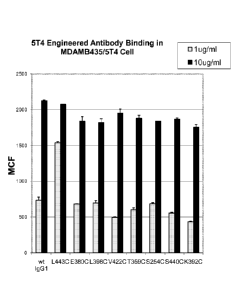
Figure 11 is a graph showing the binding of unconjugated cysteine mutant anti-
5T4 antibodies
to MDAMB435 cells expressing 5T4 antigen (MDAMB435/5T4) expressed as mean
calculated
fluorescence, compared with binding of parental anti-5T4 antibody comprising a
wild type IgG1 Fc
domain. The results demonstrate that the cysteine variant antibodides L4430,
E3800, L3980,
V4220, T3590, S2540, S4400 and K3920, at both 1 pg (gray bars) and 10 pg/ml
(black bars),
demonstrate binding to MDAMB435/5T4 cells comparable to the wild type parental
antibody (indicated
as "wt IgG1").
Figure 12, comprising panels A and B, is a graph showing the binding of
cysteine mutant
antibodies conjugated with mcMMAD compared to binding by parental antibody
comprising wild type
IgG1 Fc domain. Figure 12A is a graph showing binding of cysteine variant
antibodies conjugated to
mcMMAD to cells expressing 5T4 antigen (MDAMB435/5T4 cells) compared with wild
type parental
anti-5T4 antibody. Binding of antibodies 5T4-E3800-mcMMAD, 5T4-L3980-mcMMAD,
5T4-L4430-
mcMMAD, and 5T4-V4220-mcMMAD antibodies was compared with binding by parental
antibody
5T4 (wt IgG1). Figure 12B is a graph showing the lack of binding of cysteine
variant antibodies
conjugated to mcMMAD compared with similar lack of binding of wild type
parental antibody in Raji
cells which do not express the target antigen 5T4.
Figure 13 is a graph showing the internalization of cysteine variant
antibodies conjugated with
mcMMAD compared with the internalization of wild type antibody conjugated with
mcMMAD (5T4-
IgG1-mcMMAD) and wild type antibody which was not conjugated (wt IgG1). The
data show that
cysteine mutant antibody drug conjugates 5T4-E3800-mcMMAD, 5T4-L3980-mcMMAD,
and 5T4-
L4430-mcMMAD, were internalized by MDAMB435/5T4 cells substantially the same
as wild type
CA 02859755 2014-06-18
WO 2013/093809 PCT/IB2012/057491
parental antibody drug conjugate 5T4-IgG1-mcMMAD and wild type parental
antibody 5T4 not
conjugated (wt IgG1).
Figure 14, comprising panels A and B, show that engineered cysteine variant
antibodies do
not exhibit altered Fc effector activity compared with wild type parental
antibody. Figure 14A shows a
graph showing that cysteine variants 5-T4-E3800, 5T4-L3980, 5T4-V4220, and 5T4-
L4430
demonstrate the same ADCC activity as wild type parental antibody (5T4) in
cells expressing 5T4
(MDA435/5T4). Figure 14B shows a graph showing that cysteine variants 5T4-
E3800, 5T4-L3980,
5T4-V4220, and 5T4-L4430 demonstrate the same ADCC activity (none) compared
with wild type
parental antibody (5T4) in cells that do not express 5T4 antigen (MDA435/Neo).
Figure 15, comprising panels A-UUU, shows the following sequences: amino acid
sequence
of wild type human IgG1 heavy chain constant domain comprising the Fc region,
where the Fc region
begins at amino acid residue 236 (glycine, G) (Fig. 15A), an exemplary nucleic
acid sequence
encoding human wild type IgG1 constant domain comprising the Fc region (Fig.
15 B), amino acid
sequence of human IgG2 constant domain (Fig. 15 C), amino acid sequence of
human wild type IgG3
constant domain (Fig. 15 D), amino acid sequence of human wild type IgG4
constant domain (Fig. 15
E), and the amino acid sequences of engineered Fc polypeptides comprising a
substitution of a
cysteine at the following positions (all according to the EU numbering system
of Kabat) : K246 (Fig. 15
F), D249 (Fig. 15 G), 254 (Fig. 15 H), D265 (Fig. 15 1), S267 (Fig. 15 J),
D270 (Fig. 15 K), N276 (Fig.
15 L), Y278 (Fig. 15 M), E283 (Fig. 15 N), 284 (Fig. 15 0), 287 (Fig. 15 P),
R292 (Fig. 15 Q), E293
(Fig. 15 R), E294 (Fig. 15 S), Y300 (Fig. 15 T), V302 (Fig. 15 U), V303 (Fig.
15 V), L314 (Fig. 15 W),
N315 (Fig. 15 X), E318 (Fig. 15 Y), K320 (Fig. 15 Z), 327 (Fig. 15 AA), 1332
(Fig. 15 BB), E333 (Fig.
15 CC), K334 (Fig. 15 DD), 1336 (Fig. 15 EE), E345 (Fig. 15 FF), Q347 (Fig. 15
GG), S354 (Fig. 15
HH), R355 (Fig. 15 II), M358 (Fig. 15 JJ), T359 KK), K360 (Fig. 15 LL), N361
(Fig. 15 MM), Q362 (Fig.
15 NN), K370 (Fig. 15 00), Y373 (Fig. 15 PP), D376 (Fig. 15 QQ), A378 (Fig. 15
RR), E380 (Fig. 15
SS), E382 (Fig. 15 TT), S383 (Fig. 15 UU), 384 (Fig. 15 VV), Q386 (Fig. 15
WW), E388 (Fig. 15 XX),
N390 (Fig. 15 YY), K392 (Fig. 15 ZZ), T393 (Fig. 15 AAA), 398 (Fig. 15-BBB),
D401 (Fig. 15 CCC),
F404 (Fig. 15 DDD), T411 (Fig. 15 EEE), D413 (Fig. 15 FFF), K414 (Fig. 15
GGG), R416 (Fig. 15
HHH), Q418 (Fig. 15 111), Q419 (Fig. 15 JJJ), N421 (Fig. 15 KKK), 422 (Fig. 15
LLL), M428 (Fig. 15
MMM), A431 (Fig. 15 NNN), L432 (Fig. 15 000), T437 (Fig. 15 PPP), Q438 (Fig.
15 QQQ), K439
(Fig. 15 RRR), 440 (Fig. 15 SSS), L443 (Fig. 15 TTT), and S444 (Fig. 15 UUU).
Figure 16, comprising panels A-1, show the amino acid sequences of the
following IgG1
engineered Fc regions comprising two mutations as follows: E380C-L443C (Fig.
16 A); L398C-L443C
(Fig. 16B); V422C-L443C (Fig. 16C); E380C-L398C D); L398C-V422C (Fig. 16E);
E380C-V422C (Fig.
16F); L392C-L443C (Fig. 16G); L404C-L443C (Fig. 16H); L392C-L404C (Fig. 16G).
Figure 17, comprising panels A-F, shows the amino acid sequences of the full
length heavy
and light chains of various antibodies. Figure 17A shows the amino acid
sequence of the heavy chain
anti-5T4 antibody where the variable domain (VH) is capitalized and the three
(3) CDRs are
underlined and where the sequence of the human IgG1 constant region is shown
in lower case letters.
CA 02859755 2014-06-18
WO 2013/093809 PCT/IB2012/057491
16
Figure 17B shows the amino acid sequence of the light chain of the anti-5T4
antibody where the
variable domain (VL) is capitalized and the three (3) CDRs are underlined and
where the sequence of
the human Kappa constant region is shown in lower case letters. Figure 170
shows the amino acid
sequence of the heavy chain anti-Her2 antibody where the variable domain (VH)
is capitalized and the
three (3) CDRs are underlined and where the sequence of the human IgG1
constant region is shown
in lower case letters. Figure 17D shows the amino acid sequence of the light
chain of the anti-Her2
antibody where the variable domain (VL) is capitalized and the three (3) CDRs
are underlined and
where the sequence of the human Kappa constant region is shown in lower case
letters. Figure 17E
shows the amino acid sequence of the heavy chain anti-VEGFR2 (vascular
endothelial growth factor
receptor 2) antibody where the variable domain (VH) is capitalized and the
three (3) CDRs are
underlined and where the sequence of the human IgG1 constant region is shown
in lower case letters.
Figure 17F shows the amino acid sequence of the light chain of the anti-VEGFR2
antibody where the
variable domain (VL) is capitalized and the three (3) CDRs are underlined and
where the sequence of
the human Kappa constant region is shown in lower case letters.
Figure 18, comprising panels A-D, shows the amino acid sequences of the wild
type human
kappa constant region (Fig. 18A) and the amino acid sequence of the engineered
OK regions
comprising the following mutations: A1110 (Fig. 18B); K1830 (Fig. 180); and
N2100 (Fig. 18D).
Figure 19, comprising panels A and B, shows the amino acid sequence alignments
of human
IgG1, IgG2, IgG3 and IgG4 showing the equivalent positions among the four IgG
subclasses. Figure
19A shows the amino acid sequence alignment of the Fc domains of human wild
type IgG1 (hIgG1),
IgG2 (hIgG2), IgG3 (hIgG3) and IgG4 (hIgG4). Figure 19B shows the amino acid
sequence alignment
of the constant domain (comprising 0H1, hinge, 0H2 and 0H3 regions) of human
wild type IgG1
(human_gamma1), IgG2 (human_gamma2), IgG3 (human_gamma3) and IgG4
(human_gamma4).
Figure 20, comprising panels A and B, Figure 18, shows the nucleic acid
sequence encoding
wild type human lambda constant region (Fig. 20A), the amino acid sequence of
wild type human
lambda constant region (Fig. 20B) and the amino acid sequences of the
engineered CA regions
comprising the following mutations: K1100 (Fig. 200); A1110 (Fig. 20D); L1250
(Fig. 20E); K1490
(Fig. 20F); V1550 (Fig. 20G); G1580 (Fig. 20H); T1610 (Fig. 201); Q1850 (Fig.
20J); S1880 (Fig.
20K); H1890 (Fig. 20L); S1910 (Fig. 20M); T1970 (Fig. 20N); V2050 (Fig. 200);
E2060 (Fig. 20P);
K2070 (Fig. 20Q); T2080 (Fig. 20R); and A2100 (Fig. 20S).
Figure 21, comprising panels A and B, show graphs demonstrating the PK
parameters of
various engineered cysteine antibodies conjugated vi a MalPeg602 linker to a
proprietary auristatin
payload (Aur). Figure 21A is a graph illustrating the plasma concentration
over time of site-specific
conjugated ADCs where an anti-Her2 antibody was conjugated, at the specific
site(s) indicated, via a
MalPeg602 linker to Aur. The engineered conjugation sites were: Q3470; N4210;
kappa K1830;
K3880; L4430; L3980-FL4430; and K3920-FL4430. Figure 21B is a graph
illustrating the total anti-
Her2 ADC plasma concentration for various site-specific conjugate ADCs. Anti-
Her2 antibody was
conjugated, via a MalPeg602 linker, to a proprietary auristatin payload "Aur"
(also referred to herein
CA 02859755 2016-11-18
WO 2013/093809 PCT/1132012/057491
17
as "8261"). The specific engineered conjugation sites were as follows: 03470;
N4210; kappa
K1830; K3880; L443C; L3980+L443C; and K3920+L443C.
Figure 22, comprising panels AC, demonstrates the tumor reducing efficacy of
anti-Her2 site-
specific conjugated ADCs, where the site specific conjugation site is L4430,
and using different linker
and payload combinations. Figure 22A depicts a graph illustrating the tumor
size in an N87 mouse
model of gastric carcinoma where anti-Her2-L443C was conjugated to MMAD via a
MalPeg6C2 linker
and administered at 1 mg/kg, 3 mg/kg and 10 mg/kg compared with a negative
control (Vehicle).
Figure 22B depicts a graph illustrating the tumor size in an N87 mouse model
of gastric carcinoma
where anti-Her2-L4430 was conjugated to Aur (also referred to as "8261", a
novel auristatin-based
cytotoxic compound) via a MalPeg6C2 linker (abbreviated herein as "MP6") and
administered at 1
mg/kg, 3 mg/kg and 10 mg/kg compared with a negative control (Vehicle). Figure
220 depicts a
graph illustrating the tumor size in an N87 mouse model of gastric carcinoma
where anti-Her2-L4430
was conjugated to a proprietary payload (referred to as "0101") via a vc
linker and administered at 1
mg/kg, 3 mg/kg and 10 mg/kg compared with a negative control (Vehicle).
Figure 23 depicts a graph demonstrating the efficary of site-specific
conjugated anti-Her2
ADCs in the DYT2 Her2+ carcinoma model. The anti-Her2 ADCs were conjugated at
various
engineered cysteines (K392C+L4430, Q3470, kappa K1830; K388C; N4210, kappa
K2070;
1_398C+L443C: L443C; and their efficacy was compared with vehicle only and the
anti-Her2 antibody
conventionally conjugated with DM1 (Her2-DM1) and Aur (Her2-Aur).
DETAILED DESCRIPTION OF THE INVENTION
Unless otherwise defined herein, scientific and technical terms used in
connection with the
present invention shall have the meanings that are commonly understood by
those of ordinary skill in
the art. Further, unless otherwise required by context, singular terms shall
include pluralities and
plural terms shall include the singular. Generally, nomenclatures used in
connection with, and
techniques of, cell and tissue culture, molecular biology, immunology,
microbiology, genetics and
protein and nucleic acid chemistry and hybridization described herein are
those well known and
commonly used in the art.
The methods and techniques of the present invention are generally performed
according to
methods well known in the art and as described in various general and more
specific references that
are cited and discussed throughout the present specification unless otherwise
indicated. Such
references include, e.g., Sambrook and Russell, Molecular Cloning: A
Laboratory Manual, 3-d. ed.,
Cold Spring Harbor Laboratory Press, Cold Spring Harbor, NY (2001), Ausubel et
al., Current
Protocols in Molecular Biology, John Wiley & Sons, NY (2002), Harlow and Lane
Using Antibodies: A
Laboratory Manual, Cold Spring Harbor Laboratory Press, Gold Spring Harbor, NY
(1998), and
Coligan et al., Short Protocols in Protein Science, John Wiley & Sons, NY
(2003),
Enzymatic reactions and purification techniques are performed
according to manufacturer's specifications, as commonly accomplished in the
art or as described
CA 02859755 2016-11-18
WO 2013/093809 PCT/1B2012/057491
18
herein. The nomenclatures used in connection with, and the laboratory
procedures and techniques of,
analytical chemistry, biochemistry, immunology, molecular biology, synthetic
organic chemistry, and
medicinal and pharmaceutical chemistry described herein are those well known
and commonly used
in the art. Standard techniques are used for chemical syntheses, chemical
analyses, pharmaceutical
preparation, formulation, and delivery, and treatment of patients.
Throughout this specification and claims, the word "comprise,' or variations
such as
"comprises" or 'comprising," will be understood to imply the inclusion of a
stated integer or group of
integers but not the exclusion of any other integer or group of integers.
As used herein, each of the following terms has the meaning associated with it
in this section.
The articles "a" and "an" are used herein to refer to one or to more than one
(La, to at least
one) of the grammatical object of the article. By way of example, "an element
means one element or
more than one element.
Notwithstanding that the numerical ranges and parameters setting forth the
broad scope of
the invention are approximations, the numerical values set forth in the
specific examples are reported
as precisely as possible, Any numerical value, however, inherently contains
certain errors necessarily
resulting from the standard deviation found in their respective testing
measurements, Moreover, all
ranges disclosed herein are to be understood to encompass any and all
subranges subsumed therein.
For example, a stated range of "1 to 10" should be considered to include any
and all subranges
between (and inclusive of) the minimum value of 1 and the maximum value of 10;
that is, all
subranges beginning with a minimum value of 1 or more, e.g. 1 to 6.1, and
ending with a maximum
value of 10 or less, e.g,, 5.5 to 10.
Reference to "about" a value or parameter herein includes (and describes)
embodiments that
are directed to that value or parameter per se. For example, description
referring to "about X"
includes a description of "X." Numeric values are inclusive of numbers
defining the range.
Where aspects or embodiments of the invention are described in terms of a
Markush group or
other grouping of alternatives, the present invention encompasses not only the
entire group listed as a
whole, but each member of the group individually and all possible subgroups of
the main group, and
also the main group absent one or more of the group members. The present
invention also envisages
the explicit exclusion of one or more of any of the group members in the
claimed invention.
As used herein, the twenty conventional amino acids and their abbreviations
follow
conventional usage. See Immunology--A Synthesis (2nd Edition, E. S. Golub and
D. R. Gren, Eds.,
Sinauer Associates, Sunderland, Mass. (1991)),
As used herein, amino acids are represented by the full name thereof, by the
three letter code
corresponding thereto, or by the one-letter code corresponding thereto, as
indicated in the following
table:
CA 02859755 2014-06-18
WO 2013/093809 PCT/IB2012/057491
19
Full Name Three-Letter Code One-Letter Code
Aspartic Acid Asp
Glutamic Acid Glu
Lysine Lys
Arginine Arg
Histidine His
Tyrosine Tyr
Cysteine Cys
Asparagine Asn
Glutamine Gin
Serine Ser
Threonine Thr
Glycine Gly
Alanine Ala A
Valine Val V
Leucine Leu
Isoleucine Ile
Methionine Met
Proline Pro
Phenylalanine Phe
Tryptophan Trp
A "conservative amino acid substitution" is one in which an amino acid residue
is substituted
by another amino acid residue having a side chain R group with similar
chemical properties (e.g.,
charge or hydrophobicity). In general, a conservative amino acid substitution
will not substantially
change the functional properties of a protein. In cases where two or more
amino acid sequences differ
from each other by conservative substitutions, the percent sequence identity
or degree of similarity
may be adjusted upwards to correct for the conservative nature of the
substitution. Means for making
this adjustment are well-known to those of skill in the art. See, e.g.,
Pearson, Methods Mol. Biol.
243:307-31 (1994).
Examples of groups of amino acids that have side chains with similar chemical
properties
include 1) aliphatic side chains: glycine, alanine, valine, leucine, and
isoleucine; 2) aliphatic-hydroxyl
side chains: serine and threonine; 3) amide-containing side chains: asparagine
and glutamine; 4)
aromatic side chains: phenylalanine, tyrosine, and tryptophan; 5) basic side
chains: lysine, arginine,
and histidine; 6) acidic side chains: aspartic acid and glutamic acid; and 7)
sulfur-containing side
chains: cysteine and methionine. Preferred conservative amino acids
substitution groups are: valine-
leucine-isoleucine, phenylalanine-tyrosine, lysine-arginine, alanine-valine,
glutamate-aspartate, and
asparagine-glutamine.
CA 02859755 2016-11-18
WO 2013/093809 PCT/1B2012/057491
Alternatively, a conservative replacement is any change having a positive
value in the
PAM250 log-likelihood matrix disclosed in Gonnet et al., Science 256:1443-45
(1992),
A "moderately conservative" replacement is any change having a
nonnegative value in the PAM250 log-likelihood matrix.
Preferred amino acid substitutions are those which: (1) reduce susceptibility
to proteolysis, (2)
reduce susceptibility to oxidation, (3) alter binding affinity for forming
protein complexes, and (4)
confer or modify other physicochemical or functional properties of such
analogs. Analogs comprising
substitutions, deletions, and/or insertions can include various muteins of a
sequence other than the
specified peptide sequence. For example, single or multiple amino acid
substitutions (preferably
conservative amino acid substitutions) may be made in the specified sequence
(preferably in the
portion of the poiypeptide outside the domain(s) forming intermolecular
contacts, e.g.. outside of the
CDRs). A conservative amino acid substitution should not substantially change
the structural
characteristics of the parent sequence (e.g., a replacement amino acid should
not tend to break a
helix that occurs in the parent sequence, or disrupt other types of secondary
structure that
characterizes the parent sequence). Examples of art-recognized polypeptide
secondary and tertiary
structures are described in Proteins, Structures and Molecular Principles
(Creighton, Ed., W. H.
Freeman and Company, New York (1984)); Introduction to Protein Structure (C.
Branden and J.
Tooze, eds., Garland Publishing, New York, N.Y. (1991)); and Thornton et al.,
Nature 354:105 (1991).
Sequence similarity for polypeptides is typically measured using sequence
analysis software.
Protein analysis software matches similar sequences using measures of
similarity assigned to various
substitutions, deletions and other modifications, including conservative amino
acid substitutions. For
instance, Genetics Computer Group (GCG available from Genetics Computer Group,
Inc.), also
referred to as the Wisconsin Package, is an integrated software package of
over 130 programs for
accessing, analyzing and manipulating nucleotide and protein sequences. GCG
contains programs
such as "Gap" and "Bestfit" which can be used with default parameters to
determine sequence
similarity, homology and/or sequence identity between closely related
polypeptides, such as
homologous polypeptides from different species of organisms or between a wild
type protein and a
rautein thereof. See, e.g., GCG version 6.1, version 7.0, version 9.1, and
version 10Ø
Polypeptide sequences also can be compared using FASTA, a program in GCG,
using
default or recommended parameters. FASTA (e.g., FASTA2 and FASTA3) provides
alignments and
percent sequence identity of the regions of the best overlap between the query
and search sequences
(Pearson, Methods Enzymol. 183:63-98 (1990); Pearson, Methods Mol. Biol.
132:185-219 (2000)).
Another preferred algorithm when comparing a sequence of the invention to a
database containing a
large number of sequences from different organisms is the computer program
BLAST. especially
blastp or tblastn, using default parameters. See, e.g., Altschul et al., J.
Mol. Biol. 215:403-410 (1990);
Altschul et al., Nucleic Acids Res. 25:3389-402 (1997),
CA 02859755 2014-06-18
WO 2013/093809 PCT/IB2012/057491
21
Conventional notation is used herein to portray polypeptide sequences: the
left-hand end of a
polypeptide sequence is the amino-terminus; the right-hand end of a
polypeptide sequence is the
carboxyl-terminus.
As used herein, the term "upstream" refers to a residue that is N-terminal to
a second residue
where the molecule is a protein, or 5' to a second residue where the molecule
is a nucleic acid. Also
as used herein, the term "downstream" refers to a residue that is C-terminal
to a second residue
where the molecule is a protein, or 3' to a second residue where the molecule
is a nucleic acid.
A "nucleic acid" is a polynucleotide such as deoxyribonucleic acid (DNA) or
ribonucleic acid
(RNA). The term is used to include single-stranded nucleic acids, double-
stranded nucleic acids, and
RNA and DNA made from nucleotide or nucleoside analogues.
The term "vector" refers to a nucleic acid molecule that may be used to
transport a second
nucleic acid molecule into a cell. In one embodiment, the vector allows for
replication of DNA
sequences inserted into the vector. The vector may comprise a promoter to
enhance expression of
the nucleic acid molecule in at least some host cells.
Vectors may replicate autonomously
(extrachromasomal) or may be integrated into a host cell chromosome. In one
embodiment, the
vector may comprise an expression vector capable of producing a protein
derived from at least part of
a nucleic acid sequence inserted into the vector.
As is known in the art, conditions for hybridizing nucleic acid sequences to
each other can be
described as ranging from low to high stringency. Generally, highly stringent
hybridization conditions
refer to washing hybrids in low salt buffer at high temperatures.
Hybridization may be to filter bound
DNA using hybridization solutions standard in the art such as 0.5M NaHPO4, 7%
sodium dodecyl
sulfate (SDS), at 65 C, and washing in 0.25 M NaHPO4, 3.5% SDS followed by
washing 0.1 x
SSC/0.1% SDS at a temperature ranging from room temperature to 68 C depending
on the length of
the probe (see e.g. Ausubel, F.M. et al., Short Protocols in Molecular
Biology, 4th Ed., Chapter 2, John
Wiley & Sons, N.Y). For example, a high stringency wash comprises washing in
6x SSC/0.05%
sodium pyrophosphate at 37 C for a 14 base oligonucleotide probe, or at 48 C
for a 17 base
oligonucleotide probe, or at 55 C for a 20 base oligonucleotide probe, or at
60 C for a 25 base
oligonucleotide probe, or at 65 C for a nucleotide probe about 250 nucleotides
in length. Nucleic acid
probes may be labeled with radionucleotides by end-labeling with, for example,
[y-32P]ATP, or
incorporation of radiolabeled nucleotides such as [a-32P]dCTP by random primer
labeling.
Alternatively, probes may be labeled by incorporation of biotinylated or
fluorescein labeled
nucleotides, and the probe detected using Streptavidin or anti-fluorescein
antibodies.
The term "fusion protein" refers to a protein or polypeptide that has an amino
acid sequence
derived from two or more proteins. The fusion protein may also include linking
regions of amino acids
between amino acid portions derived from separate proteins.
The term "host cell" as used herein refers to a cell that is grown in culture
according to the
present invention to produce a protein or polypeptide of interest. In certain
embodiments, the host cell
is a mammalian cell.
CA 02859755 2014-06-18
WO 2013/093809 PCT/IB2012/057491
22
By the term "hybridoma" as the term is used herein, is meant to encompass a
cell or progeny
of a cell resulting from fusion of an immortalized cell and an antibody-
producing cell. The resulting
hybridoma is an immortalized cell that produces antibodies. The individual
cells used to create the
hybridoma can be from any mammalian source, including, but not limited to,
rat, pig, rabbit, sheep,
goat, and human. The term also encompasses trioma cell lines, which result
when progeny of
heterohybrid myeloma fusions, which are the product of a fusion between human
cells and a murine
myeloma cell line, are subsequently fused with a plasma cell. Furthermore, the
term is meant to
include any immortalized hybrid cell line that produces antibodies such as,
for example, quadromas
(See, e.g., Milstein et al., 1983, Nature 537:3053).
The term "polypeptide" as used herein refers a sequential chain of amino acids
linked
together via peptide bonds. The term is used to refer to an amino acid chain
of any length, but one of
ordinary skill in the art will understand that the term is not limited to
lengthy chains and can refer to a
minimal chain comprising two amino acids linked together via a peptide bond.
As is known to those
skilled in the art, polypeptides may be processed and/or modified. For
example, a polypeptide may be
glycosylated. A polypeptide to be expressed according to the present invention
can be a therapeutic
polypeptide. A therapeutic polypeptide is a polypeptide that has a biological
effect on a region in the
body on which it acts or on a region of the body on which it remotely acts via
intermediates. Examples
of therapeutic polypeptides are discussed in more detail below.
"Protein," as the term is used herein, refers to one or more polypeptides that
function as a
discrete unit. If a single polypeptide is the discrete functioning unit and
does not require permanent or
temporary physical association with other polypeptides in order to form the
discrete functioning unit,
the terms "polypeptide" and "protein" may be used interchangeably. If the
discrete functional unit is
comprised of multiple polypeptides that physically associate with one another,
the term "protein" as
used herein refers to the multiple polypeptides that are physically coupled
and function together as
the discrete unit. A protein to be expressed according to the present
invention can be a protein
therapeutic. A protein therapeutic is a protein that has a biological effect
on a region in the body on
which it acts or on a region of the body on which it remotely acts via
intermediates. Examples of
protein therapeutics are discussed in more detail below.
By the term "fragment" as used herein refers to a polypeptide and is defined
as any discrete
portion of a given polypeptide that is unique to or characteristic of that
polypeptide. The term as used
herein also refers to any discrete portion of a given polypeptide that retains
at least a fraction of the
activity of the full-length polypeptide. In certain embodiments, the fraction
of activity retained is at least
10% of the activity of the full-length polypeptide. In certain embodiments,
the fraction of activity
retained is at least 20%, 30%, 40%, 50%, 60%, 70%, 80% or 90% of the activity
of the full-length
polypeptide. In certain embodiments, the fraction of activity retained is at
least 95%, 96%, 97%, 98%
or 99% of the activity of the full-length polypeptide. In certain embodiments,
the fraction of activity
retained is 100% or more of the activity of the full-length polypeptide.
Alternatively or additionally, the
term as used herein also refers to any portion of a given polypeptide that
includes at least an
CA 02859755 2016-11-18
WO 2013/093809 PCT/182012/057491
23
established sequence element found in the full-length polypeptide. In some
embodiments, the
sequence element spans at least about 4-5, 10, 15, 20, 25, 30, 35, 40, 45, 50
or more amino acids of
the full-length polypeptide.
"Recombinantly expressed polypeptide" and "recombinant polypeptide" as used
herein refer
to a polypeptide expressed from a host cell that has been manipulated to
express that polypeptide. In
certain embodiments, the host cell is a mammalian cell. In certain
embodiments, this manipulation
may comprise one or more genetic modifications. For example, the host cells
may be genetically
modified by the introduction of one or more heterologous genes encoding the
polypeptide to be
expressed. The heterologous recombinantly expressed polypeptide can be
identical or similar to
polypeptides that are normally expressed in the host cell. The heterologous
recombinantly expressed
polypeptide can also be foreign to the host cell, e.g. heterologous to
polypeptides normally expressed
in the host cell. In certain embodiments, the heterologous recombinantly
expressed polypeptide is
chimeric. For example, portions of a polypeptide may contain amino acid
sequences that are identical
or similar to polypeptides normally expressed in the host cell, while other
portions contain amino acid
sequences that are foreign to the host cell. Additionally or alternatively, a
polypeptide may contain
amino acid sequences from two or more different polypeptides that are both
normally expressed in the
host cell. Furthermore, a polypeptide may contain amino acid sequences from
two or more
polypeptides that are both foreign to the host cell. In some embodiments, the
host cell is genetically
modified by the activation or upregulation of one or more endogenous genes.
An intact "antibody" comprises at least two heavy (H) chains and two light (L)
chains inter-
connected by disulfide bonds. See generally, Fundamental Immunology, Ch. 7
(Paul, W., ed.. 2nd ed.
Raven Press, N.Y. (1989)). Each heavy
chain is comprised of a heavy chain variable region (HCVR or VH) and a heavy
chain constant region
(CH). The heavy chain constant region is comprised of three domains, CH1, CH2
and CH3. Each light
chain is comprised of a light chain variable region (LCVR or VL) and a light
chain constant region. The
light chain constant region is comprised of one domain, CL. In humans, there
are two types of light
chains, kappa (k) and lambda (A), such that the constant regions of these two
types of light chains are
designated as C, and CA., respectively. The VH and VL regions can be further
subdivided into regions
of hypervariability, termed complementarity determining regions (CDR),
interspersed with regions that
are more conserved, termed framework regions (FR). Each VH and VL is composed
of three CDRs
and four FRs, arranged from amino-terminus to carboxyl-terminus in the
following order: FR1, CDR1,
FR2, CDR2, FR3, CDR3, FR4. The assignment of amino acids to each domain is in
accordance with
the definitions of Kabat, Sequences of Proteins of Immunological Interest
(National Institutes of
Health, Bethesda, MD (1987 and 1991)), or Chothia & Lesk, J. Mot Biol. 196:901-
917 (1987); Chothia
et al., Nature 342:878-883 (1989).
The constant regions of the antibodies may mediate the binding of the
immunoglobulin to host
tissues or factors, including various cells of the immune system (e.g.,
effector cells) and the first
component (Gig) of the classical complement system. Within light and heavy
chains, the variable and
CA 02859755 2014-06-18
WO 2013/093809 PCT/IB2012/057491
24
constant regions are joined by a "J" region of about 12 or more amino acids,
with the heavy chain also
including a "D" region of about 10 more amino acids. See generally,
Fundamental Immunology Ch. 7
(Paul, W., ed., 2nd ed. Raven Press, N.Y. (1989)).
The term "antigen-binding portion" of an antibody (or simply "antibody
portion"), as used
herein, refers to one or more fragments of an antibody that retain the ability
to specifically bind to an
antigen (e.g., a tumor-associated antigen, TAA). It has been shown that the
antigen-binding function
of an antibody can be performed by fragments of a full-length antibody.
Examples of binding
fragments encompassed within the term "antigen-binding portion" of an antibody
include (i) a Fab
fragment, a monovalent fragment consisting of the VL, VH, CI_ and CH1 domains;
(ii) a F(ab')2 fragment,
a bivalent fragment comprising two Fab fragments linked by a disulfide bridge
at the hinge region; (iii)
a Fd fragment consisting of the VH and CH1 domains; (iv) a Fv fragment
consisting of the VL and VH
domains of a single arm of an antibody, (v) a dAb fragment (Ward et al.,
(1989) Nature 341:544-546),
which consists of a VH domain; and (vi) an isolated complementarity
determining region (CDR),
disulfide-linked Fvs (dsFv), and anti-idiotypic (anti-Id) antibodies and
intrabodies. Furthermore,
although the two domains of the Fv fragment, VL and VH, are coded for by
separate genes, they can
be joined, using recombinant methods, by a synthetic linker that enables them
to be made as a single
protein chain in which the VL and VH regions pair to form monovalent molecules
(known as single
chain Fv (scFv)); see e.g., Bird et al. Science 242:423-426 (1988) and Huston
et al. Proc. Natl. Acad.
Sci. USA 85:5879-5883 (1988)). Such single chain antibodies are also intended
to be encompassed
within the term "antigen-binding portion" of an antibody. Other forms of
single chain antibodies, such
as diabodies are also encompassed. Diabodies are bivalent, bispecific
antibodies in which VH and VL
domains are expressed on a single polypeptide chain, but using a linker that
is too short to allow for
pairing between the two domains on the same chain, thereby forcing the domains
to pair with
complementary domains of another chain and creating two antigen binding sites
(see e.g., Holliger et
al. Proc. Natl. Acad. Sci. USA 90:6444-6448 (1993); Poljak et al., 1994,
Structure 2:1121-1123).
Antibodies may be derived from any mammal, including, but not limited to,
humans, monkeys, pigs,
horses, rabbits, dogs, cats, mice, etc., or other animals such as birds (e.g.
chickens), fish (e.g.,
sharks) and camelids (e.g., llamas).
The terms "IgG Fc region", "Fc region", "Fc domain" and "Fc", as
interchangeably used herein
refer to the portion of an IgG molecule that correlates to a crystallizable
fragment obtained by papain
digestion of an IgG molecule. As used herein, the terms relate to the constant
region of an antibody
excluding the first constant region immunoglobulin domain and further relates
to portions of that
region. Thus, Fc refers to the last two constant region immunoglobulin domains
of IgA, IgD, and IgG,
and the last three constant region immunoglobulin domains of IgE and IgM, and
the flexible hinge N-
terminal to these domains, or portions thereof. For IgA and IgM, Fc may
include the J chain. For IgG,
Fc comprises immunoglobulin domains 0y2 and 0y3 (C gamma 2 and C gamma 3) and
the hinge
between Cy1 (C gamma 1) and 0y2 (C gamma 2). Although the boundaries of the Fc
region may
vary, the human IgG heavy chain Fc region is usually defined to comprise
residues 0226 or P230 to
CA 02859755 2014-06-18
WO 2013/093809 PCT/IB2012/057491
its carboxyl-terminus, wherein the numbering is according to the EU index of
Edelman et al., 1969,
Proc. Natl. Acad. Sci. USA 63(1):78-85 as described in Kabat et al., 1991.
Typically, the Fc domain
comprises from about amino acid residue 236 to about 447 of the human IgG1
constant domain.
Exemplary human wild type IgG1, IgG2, IgG3 and IgG4 Fc domain amino acid
sequences are shown
in Figure 19B. Fc polypeptide may refer to this region in isolation, or this
region in the context of an
antibody, or an antigen-binding portion thereof, or Fc fusion protein.
Particularly preferred are
engineered Fc polypeptides, which are non-naturally occurring variants of an
Fc comprising at least
one amino acid substitution introducing a site-specific conjugation site.
The heavy chain constant domain comprises the Fc region and further comprises
the CH1
domain and hinge as well as the CH2 and CH3 (and, optionally, CH4 of IgA and
IgE) domains of the
IgG heavy chain. An exemplary human wild type IgG1 constant domain amino acid
sequence is set
forth in SEQ ID NO:1 and is shown in Figure 15A.
By "engineered Fc polypeptide", "engineered Fc region" and "engineered Fc" as
the terms are
interchangeably used herein, is meant an Fc polypeptide, or portion thereof,
comprising at least one
mutation, e.g., an amino acid substitution, introducing a site for
conjugation. Preferably, the mutation
introduces a cysteine in place of the naturally-occurring amino acid residue
at that position, where the
mutation creates a reactive site (e.g., a reactive sulfhydryl group) for
conjugation of a moiety to the Fc.
An "engineered Fc variant" refers to an engineered Fc polypeptide further
comprising at least
one additional modification, such as, but not limited to, an amino acid
mutation, a post-translational
modification (e.g., altered glycosylation), among others, in addition to the
mutation creating a
conjugation site.
"Hinge region" as used herein, is generally defined as stretching from Glu216
to Pro230 of
human IgG1 (Burton, 1985, Molec. Immunol. 22: 161-206), and refers to the
portion of an IgG
molecule comprising the C-terminal portion of the CH1 domain and the N-
terminal portion of the CH2
domain. Exemplary hinge regions for human IgG1, IgG2, IgG2 and IgG4 and mouse
IgG1 and IgG2A
are provided in US Patent No. 6,165,476, at the Table shown at column 4, line
54 to column 5, line
15, and also illustrated, for example, in Janeway et al., 1999, Immunology:
The Immune System in
Health and Disease, 4th ed. (Elsevier Science Ltd.); Bloom et al., 1997,
Protein Science 6:407-415;
Humphreys et al., 1997, J. Immunol. Methods 209:193-202. Hinge regions of
other IgG isotypes may
be aligned with the IgG 1 sequence by placing the first and last cysteine
residues forming inter-heavy
chain S--S bonds in the same positions. An exemplary alignment of the constant
domains of human
IgG1, IgG2, IgG3, and IgG4 showing the alignment of the hinge region of each
subclass is shown in
Figure 19B. The "lower hinge region" of an Fc region is normally defined as
the stretch of residues
immediately C-terminal to the hinge region, i.e. residues 233 to 239 of the Fc
region
The term "IgG hinge-Fc region" or "hinge-Fc fragment" as used herein refers to
a hinge region
(approximately residues 216-230) and an Fc region (residues 231-447) C-
terminal thereto.
CA 02859755 2014-06-18
WO 2013/093809 PCT/IB2012/057491
26
An "engineered Kappa light chain" as the term is used herein, refers to a
Kappa light chain, or
a portion thereof, comprising an engineered Kappa light chain constant region
(CO comprising at least
one amino acid substitution to introduce a reactive group useful for
conjugation at that site.
"Engineered kappa constant region", "engineered OK polypeptide", "engineered
OK," and
"engineered OK region" as used interchangeably herein, mean the constant
region of a kappa light
chain, or a portion thereof, comprising at least one amino acid mutation to
introduce an amino acid
comprising a reactive group useful for conjugation compared with a wild type
kappa constant region
that is not so modified. An exemplary human wild type kappa constant region
amino acid sequence is
as shown in Figure 18A and set forth in SEQ ID NO:89.
"Engineered lambda constant region", "engineered CA polypeptide", "engineered
CA," and
"engineered CA region" as used interchangeably herein, mean the constant
region of a lambda light
chain, or a portion thereof, comprising at least one amino acid mutation to
introduce an amino acid
comprising a reactive group useful for conjugation compared with a wild type
kappa constant region
that is not so modified.
An "engineered antibody," as the term is used herein, means an antibody, or
antigen binding
portion thereof, comprising at least one engineered constant region, e.g., an
engineered Fc region, an
engineered OK region and/or an engineered CA region.
By the term "engineered antibody antigen-binding portion," or "engineered
antibody portion,"
as used herein, is meant an antigen-binding fragment of an antibody, e.g., a
Fab, a F(ab')2, and the
like, comprising at least one engineered constant region.
Still further, an antibody or antigen-binding portion thereof may be part of
larger
immunoadhesion molecules, formed by covalent or noncovalent association of the
antibody or
antibody portion with one or more other proteins or peptides. Examples of such
immunoadhesion
molecules include use of the streptavidin core region to make a tetrameric
scFv molecule (Kipriyanov
et al. Human Antibodies and Hybridomas 6:93-101 (1995)) and use of a cysteine
residue, a marker
peptide and a 0-terminal polyhistidine tag to make bivalent and biotinylated
scFv molecules
(Kipriyanov et al. Mol. Immunol. 31:1047-1058 (1994)). Other examples include
where one or more
CDRs from an antibody are incorporated into a molecule either covalently or
noncovalently to make it
an immunoadhesin that specifically binds to an antigen of interest, such as a
tumor antigen. In such
embodiments, the CDR(s) may be incorporated as part of a larger polypeptide
chain, may be
covalently linked to another polypeptide chain, or may be incorporated
noncovalently. Antibody
portions, such as Fab and F(ab')2 fragments, can be prepared from whole
antibodies using
conventional techniques, such as papain or pepsin digestion, respectively, of
whole antibodies.
Moreover, antibodies, antibody portions and immunoadhesion molecules can be
obtained using
standard recombinant DNA techniques, as described herein.
Where an "antibody" is referred to herein with respect to the present
invention, it should be
understood that an antigen-binding portion thereof may also be used. An
antigen-binding portion
competes with the intact antibody for specific binding. See generally,
Fundamental Immunology, Ch.
CA 02859755 2016-11-18
WO 20131093809 PCT/1B20 1 2/0.5749 1
27
7 (Paul, W., ed., 2nd ed., Raven Press, N.Y. (1989)).
Antigen-binding portions may be produced by recombinant DNA techniques or by
enzymatic or chemical cleavage of intact antibodies. In some embodiments,
antigen-binding portions
include Fab, Fab', F(ab')2. Fd, Fv, dAb, and complementarity determining
region (CDR) fragments,
single-chain antibodies (scFv), chimeric antibodies, diabodies, single chain
antibodies such as those
derived from camelids or shark immunoglobulin novel antigen receptors
(IgNARs), and polypeptides
that contain at least a portion of an antibody that is sufficient to confer
specific antigen binding to the
polypeptide. In embodiments having one or more binding sites, the binding
sites may be identical to
one another or may be different.
The terms "monoclonal antibody" or "monoclonal antibody composition" as used
herein refer
to a preparation of antibody molecules of single molecular composition. A
monoclonal antibody
composition displays a single binding specificity and affinity for a
particular epitope
The terms "human antibody", or "fully human antibody", as used herein, are
intended to
include antibodies having variable regions in which both the framework and CDR
regions are derived
from human germline immunoglobulin sequences. Furthermore, if the antibody
contains a constant
region, the constant region also is derived from human germline immunoglobulin
sequences. The
human antibodies of the disclosure or antigen binding portions thereof may
include amino acid
residues not encoded by human germline immunoglobulin sequences (e.g.,
mutations introduced by
random or site-specific mutagenesis in vitro or by somatic mutation in vivo).
However, the term
"human antibody", as used herein, is not intended to include antibodies in
which CDR sequences
derived from the germline of another mammalian species, such as a mouse, have
been grafted onto
human framework sequences.
The terms "human monoclonal antibody" or "fully human monoclonal antibody"
refer to
antibodies displaying a single binding specificity which have variable regions
in which both the
framework and CDR regions are derived from human germline immunoglobulin
sequences. In one
embodiment, the human monoclonal antibodies are produced by a hybridoma which
includes a B cell
obtained from a transgenic nonhuman animal, e.g., a transgenic mouse, having a
genome comprising
a human heavy chain transgene and a light chain transgene, where the B cell is
fused to an
immortalized cell.
The term "recombinant human antibody", as used herein, includes all human
antibodies that
are prepared, expressed, created or isolated by recombinant means, such as (a)
antibodies isolated
from an animal (e.g., a mouse) that is transgenic or transchromosomal for
human immunoglobulin
genes or a hybridorna prepared therefrom (described further below), (b)
antibodies isolated from a
host cell transformed to express the human antibody, e.g,, from a
transfectoma, (c) antibodies
isolated from a recombinant, combinatorial human antibody library, and (d)
antibodies prepared,
expressed, created or isolated by any other means that involve splicing of
human immunoglobulin
gene sequences to other DNA sequences. Such recombinant human antibodies have
variable
regions in which the framework and CDR regions are derived from human germline
immunoglobulin
CA 02859755 2014-06-18
WO 2013/093809 PCT/IB2012/057491
28
sequences. In certain embodiments, however, such recombinant human antibodies
can be subjected
to in vitro mutagenesis (or, when an animal transgenic for human Ig sequences
is used, in vivo
somatic mutagenesis) and thus the amino acid sequences of the VH and VL
regions of the
recombinant antibodies are sequences that, while derived from and related to
human germline VH and
VL sequences, may not naturally exist within the human antibody germline
repertoire in vivo.
As used herein, "isotype" or "class" refers to the antibody class (e.g., IgM
or IgG) that is
encoded by the heavy chain constant region genes. The constant domains of
antibodies are not
involved in binding to antigen, but exhibit various effector functions.
Depending on the amino acid
sequence of the heavy chain constant region, a given human antibody or
immunoglobulin can be
assigned to one of five major classes of immunoglobulins: IgA, IgD, IgE, IgG,
and IgM. The structures
and three-dimensional configurations of different classes of immunoglobulins
are well-known. Of the
various human immunoglobulin classes, only human IgG1, IgG2, IgG3, IgG4, and
IgM are known to
activate complement. Human IgG1 and IgG3 are known to mediate ADCC in humans.
As used herein, "subclass" refers to the further specification within an
isotype of the heavy
chain constant region gene, such as, for example, the IgG1, IgG2, IgG3, or
IgG4 subclasses within
the IgG isotype.
The phrases "an antibody recognizing an antigen" and "an antibody specific for
an antigen"
are used interchangeably herein with the term "an antibody which binds
specifically to an antigen."
The term "antibody dependent cellular cytotoxicity" or "ADCC" refers to a cell-
mediated
reaction in which non-specific cytotoxic cells (e.g. NK cells, neutrophils,
macrophages, etc.) recognize
antibody bound on a target cell and subsequently cause lysis of the target
cell. Such cytotoxic cells
that mediate ADCC generally express Fc receptors (FcR). The primary cells for
mediating ADCC (NK
cells) express Fc7RIII, whereas monocytes express FOR!, FcyRll, Fc7RIII,
and/or Fc7RIV. FcR
expression on hematopoietic cells is summarized in Ravetch and Kinet, Annu.
Rev. Immunol. 9:457-
92 (1991). To assess ADCC activity of a molecule, an in vitro ADCC assay, such
as that described in
U.S. Pat. Nos. 5,500,362 or 5,821,337 may be performed. Useful effector cells
for such assays
include peripheral blood mononuclear cells (PBMC) and Natural Killer (NK)
cells. Alternatively, or
additionally, ADCC activity of the molecules of interest may be assessed in
vivo, e.g., in an animal
model such as that disclosed in Clynes et al., 1998, Proc. Natl. Acad. Sci.
USA 95:652-656.
The terms "Fc receptor" or "FcR" are used to describe a receptor that binds to
the Fc region
of an antibody where the Fc region comprises a hinge region and the CH2 and
CH3 domains of the
heavy chain. For example, the FcR can be a native sequence human FcR. The FcR
can be one that
binds an IgG antibody (a gamma receptor) and includes receptors of the Fc7RI,
FcyRll, Fc7RIII, and
Fc7RIV subclasses, including allelic variants and alternatively spliced forms
of these receptors. Fc7RII
receptors include Fc7RIIA (an "activating receptor') and Fc7RIIB (an
"inhibiting receptor'), which have
similar amino acid sequences that differ primarily in the cytoplasmic domains
thereof. Activating
receptor Fc7RIIA contains an immunoreceptor tyrosine-based activation motif
(ITAM) in its
cytoplasmic domain. Inhibiting receptor Fc7RIIB contains an immunoreceptor
tyrosine-based
CA 02859755 2014-06-18
WO 2013/093809 PCT/IB2012/057491
29
inhibition motif (ITIM) in its cytoplasmic domain (see, Daeron, 1997, Annu.
Rev. Immunol. 15:203-
234). FcRs are reviewed in Ravetch and Kinet, 1991, Annu. Rev. Immunol. 9:457-
92; Capel et al.,
1994, Immunomethods 4:25-34; de Haas et al., 1995, J. Lab. Olin. Med. 126:330-
341; and
Nimmerjahn et al., 2005, Immunity 23:2-4. Other FcRs, including those to be
identified in the future,
are encompassed by the term "FcR" herein. The term also includes the neonatal
receptor, FcRn,
which is responsible for the transfer of maternal IgGs to the fetus (Guyer et
al., 1976, Immunol.
117:587) and Kim et al., 1994, J. Immunol. 24:249). The primary FcR binding
site on immunoglobulin
Fc fragments resides in the hinge region between the CH1 and CH2. This hinge
region interacts with
the FcR1-3 on various leukocytes and trigger these cells to attack the target.
(Wines et al., 2000, J.
Immunol. 164:5313-5318). The hinge region encompasses, but is not limited to,
the sequences
described in U.S. Patent No. 6,165,476.
The term "capable of inducing antibody dependent cellular cytotoxicity (ADCC)"
refers to the
ability of an agent, such as an antibody, to demonstrate ADCC as measured by
assay(s) known to
those of skill in the art. Such activity is typically characterized by the
binding of the Fc region with
various FcRs. Without being limited by any particular mechanism, those of
skill in the art will
recognize that the ability of an antibody to demonstrate ADCC can be, for
example, by virtue of it
subclass (such as IgG1 or IgG3), by mutations introduced into the Fc region,
or by virtue of
modifications to the carbohydrate patterns in the Fc region of the antibody.
Such modifications are
described, for example, in U.S. Patent Publication No. 2007/0092521.
The term "human antibody derivatives" refers to any modified form of the human
antibody,
e.g., a conjugate of the antibody and another agent or antibody.
The term "humanized antibody" is intended to refer to antibodies in which CDR
sequences
derived from the germline of another mammalian species, such as a mouse, have
been grafted onto
human framework sequences. Additional framework region modifications may be
made within the
human framework sequences.
By the phrase "specifically binds," as used herein, is meant a compound, e.g.,
a protein, a
nucleic acid, an antibody, and the like, which recognizes and binds a specific
molecule, but does not
substantially recognize or bind other molecules in a sample. For instance, an
antibody or a peptide
inhibitor which recognizes and binds a cognate ligand (e.g., an anti-IgE
antibody that binds with its
cognate antigen, IgE) in a sample, but does not substantially recognize or
bind other molecules in the
sample. Thus, under designated assay conditions, the specified binding moiety
(e.g., an antibody or
an antigen-binding portion thereof) binds preferentially to a particular
target molecule, e.g., IgE, and
does not bind in a significant amount to other components present in a test
sample. A variety of assay
formats may be used to select an antibody that specifically binds a molecule
of interest. For example,
solid-phase ELISA immunoassay, immunoprecipitation, BlAcore, FAGS, and Western
blot analysis
are among many assays that may be used to identify an antibody that
specifically reacts with IgE.
Typically, a specific or selective reaction will be at least twice background
signal or noise and more
typically more than 10 times background, even more specifically, an antibody
is said to "specifically
CA 02859755 2014-06-18
WO 2013/093809 PCT/IB2012/057491
bind" an antigen when the equilibrium dissociation constant (KD) is < 1 pM,
preferably < 100 nM and
most preferably < 10 nM.
The term "binding affinity" is herein used as a measure of the strength of a
non-covalent
interaction between two molecules, e.g., and antibody, or fragment thereof,
and an antigen. The term
"binding affinity" is used to describe monovalent interactions (intrinsic
activity).
Binding affinity between two molecules, e.g. an antibody, or fragment thereof,
and an antigen,
through a monovalent interaction may be quantified by determination of the
dissociation constant (KD).
In turn, KD can be determined by measurement of the kinetics of complex
formation and dissociation,
e.g. by the SPR method (Biacore). The rate constants corresponding to the
association and the
dissociation of a monovalent complex are referred to as the association rate
constants ka (or kon) and
dissociation rate constant kd (or Ka), respectively. KD is related to ka and
kd through the equation KD =
kd / ka.
Following the above definition binding affinities associated with different
molecular
interactions, e.g. comparison of the binding affinity of different antibodies
for a given antigen, may be
compared by comparison of the KD values for the individual antibody/antigen
complexes.
Similarly, the specificity of an interaction may be assessed by determination
and comparison
of the KD value for the interaction of interest, e.g. a specific interaction
between an antibody and an
antigen, with the KD value of an interaction not of interest.
The term "Icon", as used herein, is intended to refer to the on-rate, or
association rate of a
particular antibody-antigen or receptor-ligand interaction, whereas the term
"koff," as used herein, is
intended to refer to the off-rate, or dissociation rate of a particular
antibody-antigen/receptor-ligand
interaction. The term "KD", as used herein, is intended to refer to the
dissociation constant, which is
obtained from the ratio of koff to Kw, (i.e,. koff/kon) and is expressed as a
molar concentration (M). KD
values for antibodies or other binding partners can be determined using
methods well established in
the art. One method for determining the KD is by using surface plasmon
resonance, typically using a
biosensor system such as a Biacore system.
The term "chimeric antibody" as used herein means an antibody that comprises
regions from
two or more different antibodies. In certain embodiments a "chimeric antibody"
comprises variable
region sequences derived from one species and constant region sequences
derived from another
species, such as an antibody in which the variable region sequences are
derived from a mouse
antibody and the constant region sequences are derived from a human antibody.
In one embodiment,
one or more of the CDRs are derived from a mouse anti-human tumor antigen
antibody. In another
embodiment, all of the CDRs are derived from a mouse anti-human tumor antigen
antibody. In
another embodiment, the CDRs from more than one mouse anti-human tumor antigen
antibodies are
combined in a chimeric human antibody. For instance, a chimeric antibody may
comprise a CDR1
from the light chain of a first mouse anti-human tumor antigen antibody, a
CDR2 from the light chain
of a second mouse anti-human tumor antigen antibody and a CDR3 and CDR3 from
the light chain of
a third mouse anti-human tumor antigen antibody, and the CDRs from the heavy
chain may be
CA 02859755 2014-06-18
WO 2013/093809 PCT/IB2012/057491
31
derived from one or more other anti-human tumor antigen antibodies. Further,
the framework regions
may be derived from one of the same mouse anti-human tumor antigen antibodies
or from one or
more different mice.
Moreover, as discussed previously herein, chimeric antibody includes an
antibody comprising
a portion derived from the germline sequences of more than one species.
"Glycoform" refers to a complex oligosaccharide structure comprising linkages
of various
carbohydrate units. Such structures are described in, e.g., Essentials of
Glycobiology Varki et al.,
eds., Cold Spring Harbor Laboratory Press, Cold Spring Harbor, NY (1999),
which also provides a
review of standard glycobiology nomenclature. Such glycoforms include, but are
not limited to, G2,
G1, GO, G-1, and G-2 (see, e.g., International Patent Publication No. WO
99/22764).
"Glycosylation pattern" is defined as the pattern of carbohydrate units that
are covalently
attached to a protein (e.g., the glycoform) as well as to the site(s) to which
the glycoform(s) are
covalently attached to the peptide backbone of a protein, more specifically to
an immunoglobulin
protein.
It is likely that antibodies expressed by different cell lines or in
transgenic animals will have
different glycoforms and/or glycosylation patterns compared with each other.
However, all antibodies
encoded by the nucleic acid molecules provided herein, or comprising the amino
acid sequences
provided herein are part of the instant invention, regardless of the
glycosylation of the antibodies.
"Antibody-drug conjugate" as used herein, refer to an antibody, or a portion
of an antibody,
covalently linked to a cytotoxic or cytostatic drug/agent where the drug/agent
is also referred to herein
as a "payload." The antibody and the drug may be directly linked or they may
be linked via a moiety
referred to as a "linker."
By the term "effective amount", or "therapeutically effective amount," as used
herein, is meant
an amount that when administered to a mammal, preferably a human, mediates a
detectable
therapeutic response compared to the response detected in the absence of the
compound. A
therapeutic response, such as, but not limited to, inhibition of and/or
decreased tumor growth, tumor
size, metastasis, and the like, can be readily assessed by a plethora of art-
recognized methods,
including, e.g., such methods as disclosed herein.
The skilled artisan would understand that the effective amount of the compound
or
composition administered herein varies and can be readily determined based on
a number of factors
such as the disease or condition being treated, the stage of the disease, the
age and health and
physical condition of the mammal being treated, the severity of the disease,
the particular compound
being administered, the level of expression/availability of the target of the
antibody-drug-conjugate,
and the like.
By the term "compete", as used herein with regard to an antibody, is meant
that a first
antibody, or an antigen-binding portion thereof, competes for binding with a
second antibody, or an
antigen-binding portion thereof, where binding of the first antibody with its
cognate epitope is
detectably decreased in the presence of the second antibody compared to the
binding of the first
CA 02859755 2014-06-18
WO 2013/093809 PCT/IB2012/057491
32
antibody in the absence of the second antibody. The alternative, where the
binding of the second
antibody to its epitope is also detectably decreased in the presence of the
first antibody, can, but need
not be the case. That is, a first antibody can inhibit the binding of a second
antibody to its epitope
without that second antibody inhibiting the binding of the first antibody to
its respective epitope.
However, where each antibody detectably inhibits the binding of the other
antibody with its cognate
epitope or ligand, whether to the same, greater, or lesser extent, the
antibodies are said to "cross-
compete" with each other for binding of their respective epitope(s). For
instance, cross-competing
antibodies can bind to the epitope, or potion of the epitope, to which the
antibodies used in the
invention bind. Use of both competing and cross-competing antibodies is
encompassed by the
present invention. Regardless of the mechanism by which such competition or
cross-competition
occurs (e.g., steric hindrance, conformational change, or binding to a common
epitope, or portion
thereof, and the like), the skilled artisan would appreciate, based upon the
teachings provided herein,
that such competing and/or cross-competing antibodies are encompassed and can
be useful for the
methods disclosed herein.
The term "epitope" includes any protein determinant capable of specific
binding to an
immunoglobulin or T-cell receptor. Epitopic determinants usually consist of
chemically active surface
groupings of molecules such as amino acids or sugar side chains and usually
have specific three
dimensional structural characteristics, as well as specific charge
characteristics. Conformational and
nonconformational epitopes are distinguished in that the binding to the former
but not the latter is lost
in the presence of denaturing solvents.
"Instructional material," as that term is used herein, includes a publication,
a recording, a
diagram, or any other medium of expression which can be used to communicate
the usefulness of the
compound, combination, and/or composition of the invention in the kit for
affecting, alleviating or
treating the various diseases or disorders recited herein. Optionally, or
alternately, the instructional
material can describe one or more methods of alleviating the diseases or
disorders in a cell, a tissue,
or a mammal, including as disclosed elsewhere herein.
The instructional material of the kit may, for example, be affixed to a
container that contains
the compound and/or composition of the invention or be shipped together with a
container which
contains the compound and/or composition. Alternatively, the instructional
material may be shipped
separately from the container with the intention that the recipient uses the
instructional material and
the compound cooperatively.
Except when noted, the terms "patient" or "subject" are used interchangeably
and refer to
mammals such as human patients and non-human primates, as well as veterinary
subjects such as
rabbits, rats, and mice, and other animals. Preferably, patient refers to a
human.
Conventional notation is used herein to portray polypeptide sequences: the
left-hand end of a
polypeptide sequence is the amino-terminus; the right-hand end of a
polypeptide sequence is the
carboxyl-terminus.
CA 02859755 2014-06-18
WO 2013/093809 PCT/IB2012/057491
33
By the phrase "specifically binds," as used herein, is meant a compound, e.g.,
a protein, a
nucleic acid, an antibody, and the like, which recognizes and binds a specific
molecule, but does not
substantially recognize or bind other molecules in a sample. For instance, an
antibody or a peptide
receptor which recognizes and binds a cognate ligand or binding partner (e.g.,
an anti-human tumor
antigen antibody that binds a tumor antigen) in a sample, but does not
substantially recognize or bind
other molecules in the sample. Thus, under designated assay conditions, the
specified binding moiety
(e.g., an antibody or an antigen-binding portion thereof or a receptor or a
ligand binding portion
thereof) binds preferentially to a particular target molecule and does not
bind in a significant amount to
other components present in a test sample. A variety of assay formats may be
used to select an
antibody or peptide that specifically binds a molecule of interest. For
example, solid-phase ELISA
immunoassay, immunoprecipitation, BlAcoreTm (GE Healthcare, Piscataway, NJ),
fluorescence-
activated cell sorting (FAGS), OctetTM (ForteBio, Inc., Menlo Park, CA) and
Western blot analysis are
among many assays that may be used to identify an antibody that specifically
reacts with an antigen
or a receptor, or ligand binding portion thereof, that specifically binds with
a cognate ligand or binding
partner. Typically, a specific or selective reaction will be at least twice
background signal or noise and
more typically more than 10 times background, even more specifically, an
antibody is said to
"specifically bind" an antigen when the equilibrium dissociation constant (KD)
is < 1 pM, preferably
100 nM and most preferably < 10 nM.
By "ADCC" or "antibody dependent cell-mediated cytotoxicity" as used herein is
meant the
cell-mediated reaction wherein nonspecific cytotoxic cells that express Fc
gamma Rs recognize
bound antibody on a target cell and subsequently cause lysis of the target
cell. ADCC activity of a
molecule of interest can be assessed using an in vitro ADCC assay, such as
that described in U.S.
Patent No. 5,500,362, or 5,821,337. Useful effector cells for such assays
include peripheral blood
mononuclear cells (PBMCs) and natural killer (NK) cells. Alternatively, or
additionally, ADCC activity
of the molecule of interest may be assessed in vivo, e.g., in an animal model
such as that disclosed in
Clynes et al., 1998, Proc. Natl. Acad. Sci. USA 95:652-656.
By "ADCP" or "antibody dependent cell-mediated phagocytosis" as used herein is
meant the
cell-mediated reaction wherein nonspecific cytotoxic cells that express Fc
gamma Rs recognize
bound antibody on a target cell and subsequently cause phagocytosis of the
target cell.
"CDC" or "complement dependent cytotoxicity" refer to the lysing of a target
cell in the
presence of complement. The complement activation pathway is initiated by the
binding of the first
component of the complement system (C1q) to a molecule (e.g., an antibody)
complexed with a
cognate antigen. To assess complement activation, a CDC assay, such as, but
not limited to, an
assay described in Gazzano-Santoro et al., 1996, J. Immnol. Methods 202:163,
may be performed.
The "CH2 domain" of a human IgG Fc region (also referred to as "Cy2" domain)
usually
extends from about amino acid 231 to about amino acid 340. The CH2 domain is
unique in that it is
not closely paired with another domain. Rather, two N-linked branched
carbohydrate chains are
interposed between the two CH2 domains of an intact native IgG molecule. It
has been speculated
CA 02859755 2014-06-18
WO 2013/093809 PCT/IB2012/057491
34
that the carbohydrate may provide a substitute for the domain-domain pairing
and help stabilize the
CH2 domain. Burton, Molec. Immunol. 22: 161-206 (1985).
The "CH3 domain" comprises the stretch of residues C-terminal to a CH2 domain
in an Fc
region (i.e. from about amino acid residue 341 to about amino acid residue 447
of an IgG).
The term "effector function," as the term is used herein, refers to the
biological activities
attributable to or mediated by the Fc region of an antibody. Exemplary
"effector functions" include, but
are not limited to, C1q binding; complement dependent cytotoxicity (CDC); Fc
receptor binding;
antibody-dependent cell-mediated cytotoxicity (ADCC); antibody dependent cell-
mediated
phagocytosis (ADCP); down regulation of cell surface receptors (e.g., B cell
receptor; BCR), etc. See,
e.g., U.S. Patent No. 6,737,056. Such effector functions generally require the
Fc region to be
combined with a binding domain (e.g., an antibody variable domain) and can be
assessed using
various assays as herein disclosed, for example, as well as those assays known
in the art, for
evaluating such antibody effector functions.
By "Fc fusion" or "Fc fusion protein" as used herein is meant a protein
wherein one or more
polypeptides is operably linked to an Fc region or a derivative thereof. Fc
fusion is herein meant to be
synonymous with the terms "immunoadhesin", "Ig fusion", "Ig chimera", and
"receptor globulin"
(sometimes with dashes) as used in the prior art (Chamow et al., 1996, Trends
Biotechnol. 14:52-60;
Ashkenazi et al., 1997, Curr. Opin. Immunol. 9:195-200). An Fc fusion combines
the Fc region of an
immunoglobulin with a fusion partner, which in general can be any protein or
small molecule. The role
of the non-Fc part of an Fc fusion, i.e. the fusion partner, is to mediate
target binding, and thus it is
functionally analogous to the variable regions of an antibody. Virtually any
protein or small molecule
may be linked to an Fc polypeptide to generate an Fc fusion. Protein fusion
partners may include, but
are not limited to, the target-binding region of a receptor, an adhesion
molecule, a ligand, an enzyme,
a cytokine, a chemokine, or some other protein or protein domain. Small
molecule fusion partners
may include any therapeutic agent that directs the Fc fusion to a therapeutic
target. Such targets may
be any molecule, preferably an extracellular receptor, that is implicated in
disease.
By "IgG" as used herein is meant a polypeptide belonging to the class of
antibodies that are
substantially encoded by a recognized immunoglobulin gamma gene. In humans
this class comprises
IgG1, IgG2, IgG3, and IgG4. In mice this class comprises IgG1, IgG2a, IgG2b,
IgG3. By
"immunoglobulin (Ig)" herein is meant a protein consisting of one or more
polypeptides substantially
encoded by immunoglobulin genes. Immunoglobulins include but are not limited
to antibodies.
Immunoglobulins may have a number of structural forms, including but not
limited to full length
antibodies, antibody fragments, and individual immunoglobulin domains. By
"immunoglobulin (Ig)
domain" herein is meant a region of an immunoglobulin that exists as a
distinct structural entity as
ascertained by one skilled in the art of protein structure. Ig domains
typically have a characteristic
folding topology. The known Ig domains in the IgG class of antibodies are the
variable heavy chain
domain (VH), the heavy chain constant domains - Cy1, Cy2, Cy3 ¨ together
comprising the Cy domain
which includes the hinge region between Cy1 and Cy2 , the variable domain of
the light chain (VA
CA 02859755 2014-06-18
WO 2013/093809 PCT/IB2012/057491
and and the constant domain of the light chain (CL), which in humans comprises
either the kappa (CO
or lambda (CA) light chain constant domain Typically, an "Fc polypeptide," as
the term is used herein,
comprises a Cy2 and a Cy3 domain and can include at least a portion of the
hinge domain, but does
not usually include the entire Cyl domain.
By "parent polypeptide" or "precursor polypeptide" (including Fc parent or
precursors) as used
herein is meant a polypeptide that is subsequently modified to generate a
variant or mutant. Said
parent polypeptide may be a naturally occurring polypeptide, or a variant or
engineered version of a
naturally occurring polypeptide. Parent polypeptide may refer to the
polypeptide itself, compositions
that comprise the parent polypeptide, to the amino acid sequence of the
polypeptide, or to the nucleic
acid sequence that encodes it. Accordingly, by "parent Fc polypeptide" as used
herein is meant an
unmodified Fc polypeptide that is modified to generate a variant, and by
"parent antibody" as used
herein is meant an unmodified antibody that is modified to generate a variant
antibody.
As used herein, the terms "wild-type amino acid," "wild-type IgG," "wild-type
antibody," or
"wild-type mAb," refer to a sequence of amino or nucleic acids that occurs
naturally within a certain
population (e.g., human, mouse, rats, cell, etc.).
As outlined above, certain positions of the Fc molecule can be altered. By
"position" as used
herein is meant a location in the sequence of a protein. Positions may be
numbered sequentially, or
according to an established format, for example the EU index as in Kabat. For
example, position 297
is a position in the human antibody IgG1 . Corresponding positions are
determined as outlined above,
generally through alignment with other parent sequences.
By "residue" as used herein is meant a position in a protein and its
associated amino acid
identity. For example, Asparagine 297 (also referred to as Asn297, also
referred to as N297) is a
residue in the human antibody IgG1 .
By "target antigen" as used herein is meant the molecule that is bound
specifically by the
variable region of a given antibody. A target antigen may be a protein,
carbohydrate, lipid, or other
chemical compound.
By "target cell" as used herein is meant a cell that expresses a target
antigen.
By "variable region" as used herein is meant the region of an immunoglobulin
that comprises
one or more Ig domains substantially encoded by any of the V kappa, V.Iamda.,
and/or VH genes that
make up the kappa, lambda, and heavy chain immunoglobulin genetic loci
respectively.
By "variant polypeptide" as used herein is meant a polypeptide sequence that
differs from that
of a parent polypeptide sequence by virtue of at least one amino acid
modification. Variant
polypeptide may refer to the polypeptide itself, a composition comprising the
polypeptide, or the amino
sequence that encodes it. Preferably, the variant polypeptide has at least one
amino acid modification
compared to the parent polypeptide, e.g., from about one to about ten amino
acid modifications, and
preferably from about one to about five amino acid modifications compared to
the parent. The variant
polypeptide sequence herein will preferably possess at least about 80 percent
homology with a parent
polypeptide sequence, and most preferably at least about 90 percent homology,
more preferably at
CA 02859755 2014-06-18
WO 2013/093809 PCT/IB2012/057491
36
least about 95 percent homology, even more preferably, at least about 97%
homology, more
preferably, at least about 98% homology, and yet more preferably, at least
about 99% homology with
a parent polypeptide sequence. Accordingly, by "Fc variant" as used herein is
meant an Fc sequence
that differs from that of a parent Fc sequence by virtue of at least one amino
acid modification. An Fc
variant may only encompass an Fc region, or may exist in the context of an
antibody, Fc fusion, or
other polypeptide that is substantially encoded by Fc. Fc variant may refer to
the Fc polypeptide itself,
compositions comprising the Fc variant polypeptide, the amino acid sequence of
the Fc polypeptide,
or the nucleic acid sequence that encodes it. In a preferred embodiment, the
variant proteins of the
invention comprise an Fc variant, as described herein, and as such, may
comprise an antibody (and
the corresponding derivatives) with the Fc variant, or an Fc fusion protein
that comprises the Fc
variant. In addition, in some cases, the Fc is a variant as compared to a wild-
type Fc, or to a "parent"
variant.
For all heavy chain constant region amino acid positions discussed in the
present invention,
numbering is according to the Eu index first described in Edelman et al.,
1969, Proc. Natl. Acad. Sci.
USA 63(1):78-85, describing the amino acid sequence of myeloma protein Eu,
which is the first
human IgG1 sequenced. The Eu index of Edelman et al. is also set forth in
Kabat et al., 1991,
Sequences of Proteins of Immunological Interest, 5th Ed., United States Public
Health Service,
National Institutes of Health, Bethesda. Thus, the "EU index as set forth in
Kabat" or "EU index of
Kabat" refers to the residue numbering system based on the human IgG1 Eu
antibody of Edelman et
al. as set forth in Kabat 1991.
The numbering system used for the light chain constant region amino acid
sequence is that
set forth in Kabat 1991.
As used herein, "substantially pure" means an object species is the
predominant species
present (i.e., on a molar basis it is more abundant than any other individual
species in the
composition), and preferably a substantially purified fraction is a
composition wherein the object
species (e.g., a glycoprotein, including an antibody or receptor) comprises at
least about 50 percent
(on a molar basis) of all macromolecular species present. Generally, a
substantially pure composition
will comprise more than about 80 percent of all macromolecular species present
in the composition,
more preferably more than about 85%, 90%, 95%, and 99%. Most preferably, the
object species is
purified to essential homogeneity (contaminant species cannot be detected in
the composition by
conventional detection methods) wherein the composition consists essentially
of a single
macromolecular species.
As used herein, to "treat" means reducing the frequency with which symptoms of
a disease
(i.e., tumor growth and/or metastasis, or other effect mediated by the numbers
and/or activity of
immune cells, and the like) are experienced by a patient. The term includes
the administration of the
compounds or agents of the present invention to prevent or delay the onset of
the symptoms,
complications, or biochemical indicia of a disease, alleviating the symptoms
or arresting or inhibiting
further development of the disease, condition, or disorder. Treatment may be
prophylactic (to prevent
CA 02859755 2014-06-18
WO 2013/093809 PCT/IB2012/057491
37
or delay the onset of the disease, or to prevent the manifestation of clinical
or subclinical symptoms
thereof) or therapeutic suppression or alleviation of symptoms after the
manifestation of the disease.
"Combination therapy" embraces the administration of an antibody-drug
conjugate, and
another therapeutic agent as part of a specific treatment regimen optionally
including a maintenance
phase, intended to provide a beneficial effect from the co-action of these
therapeutic agents. The
beneficial effect of the combination includes, but is not limited to,
pharmacokinetic or
pharmacodynamic co-action resulting from the combination of therapeutic
agents. Administration of
these therapeutic agents in combination typically is carried out over a
defined time period (usually
minutes, hours, days or weeks depending upon the combination selected).
"Combination therapy"
generally is not intended to encompass the administration of two or more of
these therapeutic agents
as part of separate monotherapy regimens that incidentally and arbitrarily
result in the combinations of
the present invention.
"Combination therapy" embraces administration of these therapeutic agents in a
sequential
manner, that is, wherein each therapeutic agent is administered at a different
time, as well as
administration of these therapeutic agents, or at least two of the therapeutic
agents, in a substantially
simultaneous manner. Sequential or substantially simultaneous administration
of each therapeutic
agent can be effected by any appropriate route including, but not limited to,
oral routes, intravenous
routes, intramuscular, subcutaneous routes, and direct absorption through
mucous membrane
tissues. The therapeutic agents can be administered by the same route or by
different routes. For
example, a first therapeutic agent (e.g., a chemotherapeutic agent) can be
administered orally, and a
second agent (e.g., an ADC) can be administered intravenously. Further, a
first therapeutic agent of
the combination selected may be administered by intravenous injection while
the other therapeutic
agents of the combination may be administered orally. Alternatively, for
example, both the therapeutic
agents may be administered by intravenous or subcutaneous injection.
In the present specification the term "sequential" means, unless otherwise
specified,
characterized by a regular sequence or order, e.g., if a dosage regimen
includes the administration of
an ADC and a chemotherapeutic agent, a sequential dosage regimen could include
administration of
the ADC before, simultaneously, substantially simultaneously, or after
administration of the
chemotherapeutic agent, but both agents will be administered in a regular
sequence or order. The
term "separate" means, unless otherwise specified, to keep apart one from the
other. The term
"simultaneously" means, unless otherwise specified, happening or done at the
same time, i.e., the
compounds of the invention are administered at the same time. The
term "substantially
simultaneously" means that the compounds are administered within minutes of
each other (e.g., within
minutes of each other) and intends to embrace joint administration as well as
consecutive
administration, but if the administration is consecutive it is separated in
time for only a short period
(e.g., the time it would take a medical practitioner to administer two
compounds separately). As used
herein, concurrent administration and substantially simultaneous
administration are used
CA 02859755 2014-06-18
WO 2013/093809 PCT/IB2012/057491
38
interchangeably. Sequential administration refers to temporally separated
administration of the ADC
and the chemotherapeutic agent.
"Combination therapy" also can embrace the administration of the therapeutic
agents as
described above in further combination with other biologically active
ingredients (such as, but not
limited to, a second and different antineoplastic agent, a dendritic cell
vaccine or other tumor vaccine)
and non-drug therapies (such as, but not limited to, surgery or radiation
treatment). Where the
combination therapy further comprises radiation treatment, the radiation
treatment may be conducted
at any suitable time so long as a beneficial effect from the co-action of the
combination of the
therapeutic agents and radiation treatment is achieved. For example, in
appropriate cases, the
beneficial effect is still achieved when the radiation treatment is temporally
removed from the
administration of the therapeutic agents, perhaps by days or even weeks.
As used herein, "pharmaceutically acceptable carrier" includes any and all
solvents,
dispersion media, coatings, antibacterial and antifungal agents, isotonic and
absorption delaying
agents, and the like that are physiologically compatible. Examples of
pharmaceutically acceptable
carriers include one or more of water, saline, phosphate buffered saline,
dextrose, glycerol, ethanol
and the like, as well as combinations thereof. In many cases, it will be
preferable to include isotonic
agents, for example, sugars, polyalcohols such as mannitol, sorbitol, or
sodium chloride in the
composition. Pharmaceutically acceptable substances such as wetting or minor
amounts of auxiliary
substances such as wetting or emulsifying agents, preservatives or buffers,
which enhance the shelf
life or effectiveness of the protein or portion thereof, may also be included
in the composition.
I. Engineered antibodies and antibody fragments
The disclosure is based on the finding that certain residues presumably
present on the
surface of the 0H2 or 0H3 domain of the heavy chain of antibodies, or on the
constant domain of the
light chain, or otherwise accessible, are suitable for the substitution of the
naturally-occurring wild type
amino acid with, for example, cysteine, and are therefore useful to engineer a
site capable of
conjugation to various agents.
Other amino acids besides cysteine, including natural and/or non-natural amino
acids, may be
used in the substitution to allow, among other things, for conjugation of
various agents. Such other
amino acids include lysine (described in Benhar et al., (1994) Bioconjug.
Chem. 55:321-326), tyrosine
(described in Byers and Baldwin, (1988) Immunol. 65:329-335), histidine
(described in Weibel et al.
(1999) Nature Biotechnol. 17:897-901), selenocysteine, selenomethionine,
and/or non-natural amino
acids. Thus, where one or more cysteine substitutions are described herein,
one of ordinary skill in the
art may optionally employ one or more of these natural and/or non-natural
amino acids instead of
cysteine. One of ordinary skill in the art may also use any combination of
amino acids in the
substitution, such as substituting with cysteine and lysine to produce a
variant antibody with cysteines
substituted at some positions and lysines, tyrosines, histidines,
selenocysteines, selenomethionines,
and/or non-natural amino acids at others in any combination thereof.
CA 02859755 2016-11-18
WO 2013/093809 PCT/1B2012/957491
39
Amino acid modifications can be made by any method known in the ad and many
such
methods are well known and routine for the skilled artisan. For example, but
not by way of limitation,
amino acid substitutions, deletions and insertions may be accomplished using
any well-known PCR-
based technique. Amino acid substitutions may be made by site-directed
mutagenesis (see, for
example, Zoller and Smith, 1982, Noel. Acids Res. 10:6487-6500; Kunkel, 1985,
Proc, Natl. Acad. Sci
USA 82:488).
In some embodiments, the engineered Fc polypeptide of the disclosure may be
used to
prepare an antibody, or antigen binding fragment thereof, such that the
antibody or fragment thereof
thereby comprises an engineered Fc region which can be used to conjugate, at
the engineered
residue (i.e., the amino acid substituted compared to wild type unmodified
Fc), a wide variety of
moieties.
In some embodiments, the engineered kappa light chain constant polypeptide of
the
disclosure may be used to prepare an antibody, or antigen binding fragment
thereof, such that the
antibody or fragment thereof thereby comprises an engineered CL region
comprising an amino acid
mutation, or portion thereof, which can be used to conjugate, at the
engineered amino acid residue, a
wide variety of moieties.
Engineered antibody constant regions
A. Engineered heavy chain constant region
The invention encompasses an engineered Cy, polypeptide, including, but not
limited to, an
Fc polypeptide, where 1, 2, 3, 4, 5, 6, 7, 8. 9, 10, 11, 12, 13, 14, 15, 16,
17, 18, 19, 20, or more amino
acids chosen from positions: 246, 249, 265, 267, 270, 276, 278, 283, 292, 293,
294, 300, 302, 303,
314, 315, 318, 320, 327, 332, 333, 334, 336, 345, 347, 354, 355, 358, 360,
362, 370, 373, 376, 378,
380, 382, 386, 388, 390, 392, 393, 401, 404, 411, 413, 414, 416, 418, 419,
421, 428, 431, 432, 437,
438, 439, 443, arid 444 of the antibody heavy chain wherein the numbering
system of the constant
region is that of the EU index as set forth in Kabat et al. (1991, NIH
Publication 91- 3242, National
Technical Information Service, Springfield, VA, hereinafter "Kober) of a
parent, native, or wild type
antibody, are substituted with another amino acid (including natural and non-
natural/synthetic amino
acids).
It should be noted that a single substitution in an Fc polypeptide, for
example of a cysteine
residue, normally results in the display of two corresponding residues in the
resultant IgG antibody
due to the homodimeric nature of IgG antibody molecules. Thus, the resultant
engineered IgG
antibodies of the invention may display at least 1, 2, 3, 4, 5, 6, 7, 8, 9,
10, 11, 12, 13, 14. 15,16, 17,
18. 19, 20, 21, 22, 23, 24, 25, 26, 27, 28, 29, 30, 31, 32, 33, 34, 35, 36,
37, 38, 39, 40, or more
reactive groups for the purpose of conjugation to a drug or compound. In an
embodiment, one or
more of the substitutions is with a cysteine residue, and the resulting
engineered antibodies may
display at least 1, 2, 3, 4, 5, 6, 7, 8, 9, 10, 11, 12, 13, 14, 15, 16,17, 18,
19, 20, 21, 22, 23, 24, 25, 26,
CA 02859755 2014-06-18
WO 2013/093809 PCT/IB2012/057491
27, 28, 29, 30, 31, 32, 33, 34, 35, 36, 37, 38, 39, 40, or more thiol groups
for the purpose of
conjugation to a drug or compound.
In another embodiment, the engineered antibody comprises one engineered Fc
polypeptide
comprising different substituted positions from a second engineered Fc region.
That is, because of
the dimeric nature of IgG antibodies and because a variety of art-recognized
methods for preparing
heterodimeric antibodies comprising, inter alia, two or more Fc regions that
differ from each other, the
present invention encompasses an antibody comprising at least one engineered
Fc region comprising
an amino acid substitution that is not present in the other Fc region, which
may or may not also be
engineered. Methods for making heterodimeric antibodies comprising Fc regions
comprising different
mutations are well-known in the art and include, but are not limited to, the
methods discussed in U.S.
Patent No. 7,183,076 to Arathoon et al.
In some embodiments, an engineered antibody comprises a first engineered Fc
polypeptide
comprising at least one substitution at positions selected from 246, 249, 265,
267, 270, 276, 278,
283, 292, 293, 294, 300, 302, 303, 314, 315, 318, 320, 332, 333, 334, 336,
345, 347, 354, 355, 358,
360, 362, 370, 373, 376, 378, 380, 382, 386, 388, 390, 392, 393, 401, 404,
411, 413, 414, 416, 418,
419, 421, 428, 431, 432, 437, 438, 439, 443, and 444, and further comprises a
second Fc region that
is not engineered, e.g., it comprises the amino acid sequence of wild type
IgG1.
In some embodiments, an engineered antibody comprises a first engineered Fc
polypeptide
comprising at least one substitution at positions selected from 246, 249, 265,
267, 270, 276, 278,
283, 292, 293, 294, 300, 302, 303, 314, 315, 318, 320, 332, 333, 334, 336,
345, 347, 354, 355, 358,
360, 362, 370, 373, 376, 378, 380, 382, 386, 388, 390, 392, 393, 401, 404,
411, 413, 414, 416, 418,
419, 421, 428, 431, 432, 437, 438, 439, 443, and 444, and further comprises a
second engineered Fc
polypeptide that comprises at least one substitution at positions selected
from 246, 249, 265, 267,
270, 276, 278, 283, 292, 293, 294, 300, 302, 303, 314, 315, 318, 320, 332,
333, 334, 336, 345, 347,
354, 355, 358, 360, 362, 370, 373, 376, 378, 380, 382, 386, 388, 390, 392,
393, 401, 404, 411, 413,
414, 416, 418, 419, 421, 428, 431, 432, 437, 438, 439, 443, and 444, wherein
the substitution present
in the first engineered Fc polypeptide is not a substitution present in the
second engineered Fc
polypeptide.
In some embodiments, the engineered Fc polypeptide of the disclosure comprises
at least
one substitution at positions selected from: 246, 249, 265, 267, 270, 276,
278, 283, 292, 293, 294,
300, 302, 303, 314, 315, 318, 320, 332, 333, 334, 336, 345, 347, 354, 355,
358, 360, 362, 370, 373,
376, 378, 380, 382, 386, 388, 390, 392, 393, 401, 404, 411, 413, 414, 416,
418, 419, 421, 428, 431,
432, 437, 438, 439, 443, and 444 of the heavy chain of an antibody, wherein
the numbering system of
the constant region is that of the EU index as set forth in Kabat et al.
(supra).
In some embodiments, the engineered Fc polypeptide of the disclosure comprises
at least
two substitutions at positions selected from: 246, 249, 265, 267, 270, 276,
278, 283, 292, 293, 294,
300, 302, 303, 314, 315, 318, 320, 332, 333, 334, 336, 345, 347, 354, 355,
358, 360, 362, 370, 373,
376, 378, 380, 382, 386, 388, 390, 392, 393, 401, 404, 411, 413, 414, 416,
418, 419, 421, 428, 431,
CA 02859755 2014-06-18
WO 2013/093809 PCT/IB2012/057491
41
432, 437, 438, 439, 443, and 444 of the heavy chain of an antibody, wherein
the numbering system of
the constant region is that of the EU index as set forth in Kabat et al.
(supra).
In other embodiments, the engineered Fc polypeptide of the disclosure
comprises at least two
substitutions selected from the positions 246, 249, 254, 265, 267, 270, 276,
278, 283, 284, 287, 292,
293, 294, 300, 302, 303, 314, 315, 318, 320, 327, 332, 333, 334, 336, 345,
347, 354, 355, 358, 359,
360, 361, 362, 370, 373, 376, 378, 380, 382, 383, 384, 386, 388, 390, 392,
393, 398, 401, 404, 411,
413, 414, 416, 418, 419, 421, 422, 428, 431, 432, 437, 438, 439, 440, 443, and
444, of the heavy
chain of an antibody, wherein at least one substitution is selected from the
positions 246, 249, 265,
267, 270, 276, 278, 283, 292, 293, 294, 300, 302, 303, 314, 315, 318, 320,
332, 333, 334, 336, 345,
347, 354, 355, 358, 360, 362, 370, 373, 376, 378, 380, 382, 386, 388, 390,
392, 393, 401, 404, 411,
413, 414, 416, 418, 419, 421, 428, 431, 432, 437, 438, 439, 443, and 444 of
the heavy chain of an
antibody, and wherein the numbering system of the constant region is that of
the EU index as set forth
in Kabat et al. (supra).
In other embodiments, the engineered Fc polypeptide of the disclosure
comprises at least
three substitutions selected from the positions 246, 249, 254, 265, 267, 270,
276, 278, 283, 284, 287,
292, 293, 294, 300, 302, 303, 314, 315, 318, 320, 327, 332, 333, 334, 336,
345, 347, 354, 355, 358,
359, 360, 361, 362, 370, 373, 376, 378, 380, 382, 383, 384, 386, 388, 390,
392, 393, 398, 401, 404,
411, 413, 414, 416, 418, 419, 421, 422, 428, 431, 432, 437, 438, 439, 440,
443, and 444, of the
heavy chain of an antibody, wherein at least one substitution is selected from
the positions 246, 249,
265, 267, 270, 276, 278, 283, 292, 293, 294, 300, 302, 303, 314, 315, 318,
320, 332, 333, 334, 336,
345, 347, 354, 355, 358, 360, 362, 370, 373, 376, 378, 380, 382, 386, 388,
390, 392, 393, 401, 404,
411, 413, 414, 416, 418, 419, 421, 428, 431, 432, 437, 438, 439, 443, and 444
of the heavy chain of
an antibody, and wherein the numbering system of the constant region is that
of the EU index as set
forth in Kabat et al. (supra).
In other embodiments, the engineered Fc polypeptide of the disclosure
comprises at least
four substitutions selected from the positions 246, 249, 254, 265, 267, 270,
276, 278, 283, 284, 287,
292, 293, 294, 300, 302, 303, 314, 315, 318, 320, 327, 332, 333, 334, 336,
345, 347, 354, 355, 358,
359, 360, 361, 362, 370, 373, 376, 378, 380, 382, 383, 384, 386, 388, 390,
392, 393, 398, 401, 404,
411, 413, 414, 416, 418, 419, 421, 422, 428, 431, 432, 437, 438, 439, 440,
443, and 444, of the
heavy chain of an antibody, wherein at least one substitution is selected from
the positions 246, 249,
265, 267, 270, 276, 278, 283, 292, 293, 294, 300, 302, 303, 314, 315, 318,
320, 332, 333, 334, 336,
345, 347, 354, 355, 358, 360, 362, 370, 373, 376, 378, 380, 382, 386, 388,
390, 392, 393, 401, 404,
411, 413, 414, 416, 418, 419, 421, 428, 431, 432, 437, 438, 439, 443, and 444
of the heavy chain of
an antibody, and wherein the numbering system of the constant region is that
of the EU index as set
forth in Kabat et al. (supra).
In other embodiments, the engineered Fc polypeptide of the disclosure
comprises at least five
substitutions selected from the positions 246, 249, 254, 265, 267, 270, 276,
278, 283, 284, 287, 292,
293, 294, 300, 302, 303, 314, 315, 318, 320, 327, 332, 333, 334, 336, 345,
347, 354, 355, 358, 359,
CA 02859755 2014-06-18
WO 2013/093809 PCT/IB2012/057491
42
360, 361, 362, 370, 373, 376, 378, 380, 382, 383, 384, 386, 388, 390, 392,
393, 398, 401, 404, 411,
413, 414, 416, 418, 419, 421, 422, 428, 431, 432, 437, 438, 439, 440, 443, and
444, of the heavy
chain of an antibody, wherein at least one substitution is selected from the
positions 246, 249, 265,
267, 270, 276, 278, 283, 292, 293, 294, 300, 302, 303, 314, 315, 318, 320,
332, 333, 334, 336, 345,
347, 354, 355, 358, 360, 362, 370, 373, 376, 378, 380, 382, 386, 388, 390,
392, 393, 401, 404, 411,
413, 414, 416, 418, 419, 421, 428, 431, 432, 437, 438, 439, 443, and 444 of
the heavy chain of an
antibody, and wherein the numbering system of the constant region is that of
the EU index as set forth
in Kabat et al. (supra).
In other embodiments, the engineered Fc polypeptide of the disclosure
comprises at least six
substitutions selected from the positions 246, 249, 254, 265, 267, 270, 276,
278, 283, 284, 287, 292,
293, 294, 300, 302, 303, 314, 315, 318, 320, 327, 332, 333, 334, 336, 345,
347, 354, 355, 358, 359,
360, 361, 362, 370, 373, 376, 378, 380, 382, 383, 384, 386, 388, 390, 392,
393, 398, 401, 404, 411,
413, 414, 416, 418, 419, 421, 422, 428, 431, 432, 437, 438, 439, 440, 443, and
444, of the heavy
chain of an antibody, wherein at least one substitution is selected from the
positions 246, 249, 265,
267, 270, 276, 278, 283, 292, 293, 294, 300, 302, 303, 314, 315, 318, 320,
332, 333, 334, 336, 345,
347, 354, 355, 358, 360, 362, 370, 373, 376, 378, 380, 382, 386, 388, 390,
392, 393, 401, 404, 411,
413, 414, 416, 418, 419, 421, 428, 431, 432, 437, 438, 439, 443, and 444 of
the heavy chain of an
antibody, and wherein the numbering system of the constant region is that of
the EU index as set forth
in Kabat et al. (supra).
In other embodiments, the engineered Fc polypeptide of the disclosure
comprises at least
seven substitutions selected from the positions 246, 249, 254, 265, 267, 270,
276, 278, 283, 284, 287,
292, 293, 294, 300, 302, 303, 314, 315, 318, 320, 327, 332, 333, 334, 336,
345, 347, 354, 355, 358,
359, 360, 361, 362, 370, 373, 376, 378, 380, 382, 383, 384, 386, 388, 390,
392, 393, 398, 401, 404,
411, 413, 414, 416, 418, 419, 421, 422, 428, 431, 432, 437, 438, 439, 440,
443, and 444, of the
heavy chain of an antibody, wherein at least one substitution is selected from
the positions 246, 249,
265, 267, 270, 276, 278, 283, 292, 293, 294, 300, 302, 303, 314, 315, 318,
320, 332, 333, 334, 336,
345, 347, 354, 355, 358, 360, 362, 370, 373, 376, 378, 380, 382, 386, 388,
390, 392, 393, 401, 404,
411, 413, 414, 416, 418, 419, 421, 428, 431, 432, 437, 438, 439, 443, and 444
of the heavy chain of
an antibody, and wherein the numbering system of the constant region is that
of the EU index as set
forth in Kabat et al. (supra).
In other embodiments, the engineered Fc polypeptide of the disclosure
comprises at least
eight substitutions selected from the positions 246, 249, 254, 265, 267, 270,
276, 278, 283, 284, 287,
292, 293, 294, 300, 302, 303, 314, 315, 318, 320, 327, 332, 333, 334, 336,
345, 347, 354, 355, 358,
359, 360, 361, 362, 370, 373, 376, 378, 380, 382, 383, 384, 386, 388, 390,
392, 393, 398, 401, 404,
411, 413, 414, 416, 418, 419, 421, 422, 428, 431, 432, 437, 438, 439, 440,
443, and 444, of the
heavy chain of an antibody, wherein at least one substitution is selected from
the positions 246, 249,
265, 267, 270, 276, 278, 283, 292, 293, 294, 300, 302, 303, 314, 315, 318,
320, 332, 333, 334, 336,
345, 347, 354, 355, 358, 360, 362, 370, 373, 376, 378, 380, 382, 386, 388,
390, 392, 393, 401, 404,
CA 02859755 2014-06-18
WO 2013/093809 PCT/IB2012/057491
43
411, 413, 414, 416, 418, 419, 421, 428, 431, 432, 437, 438, 439, 443, and 444
of the heavy chain of
an antibody, and wherein the numbering system of the constant region is that
of the EU index as set
forth in Kabat et al. (supra).
In other embodiments, the engineered Fc polypeptide of the disclosure
comprises at least
nine substitutions selected from the positions 246, 249, 254, 265, 267, 270,
276, 278, 283, 284, 287,
292, 293, 294, 300, 302, 303, 314, 315, 318, 320, 327, 332, 333, 334, 336,
345, 347, 354, 355, 358,
359, 360, 361, 362, 370, 373, 376, 378, 380, 382, 383, 384, 386, 388, 390,
392, 393, 398, 401, 404,
411, 413, 414, 416, 418, 419, 421, 422, 428, 431, 432, 437, 438, 439, 440,
443, and 444, of the
heavy chain of an antibody, wherein at least one substitution is selected from
the positions 246, 249,
265, 267, 270, 276, 278, 283, 292, 293, 294, 300, 302, 303, 314, 315, 318,
320, 332, 333, 334, 336,
345, 347, 354, 355, 358, 360, 362, 370, 373, 376, 378, 380, 382, 386, 388,
390, 392, 393, 401, 404,
411, 413, 414, 416, 418, 419, 421, 428, 431, 432, 437, 438, 439, 443, and 444
of the heavy chain of
an antibody, and wherein the numbering system of the constant region is that
of the EU index as set
forth in Kabat et al. (supra).
In other embodiments, the engineered Fc polypeptide of the disclosure
comprises at least ten
substitutions selected from the positions 246, 249, 254, 265, 267, 270, 276,
278, 283, 284, 287, 292,
293, 294, 300, 302, 303, 314, 315, 318, 320, 327, 332, 333, 334, 336, 345,
347, 354, 355, 358, 359,
360, 361, 362, 370, 373, 376, 378, 380, 382, 383, 384, 386, 388, 390, 392,
393, 398, 401, 404, 411,
413, 414, 416, 418, 419, 421, 422, 428, 431, 432, 437, 438, 439, 440, 443, and
444, of the heavy
chain of an antibody, wherein at least one substitution is selected from the
positions 246, 249, 265,
267, 270, 276, 278, 283, 292, 293, 294, 300, 302, 303, 314, 315, 318, 320,
332, 333, 334, 336, 345,
347, 354, 355, 358, 360, 362, 370, 373, 376, 378, 380, 382, 386, 388, 390,
392, 393, 401, 404, 411,
413, 414, 416, 418, 419, 421, 428, 431, 432, 437, 438, 439, 443, and 444 of
the heavy chain of an
antibody, and wherein the numbering system of the constant region is that of
the EU index as set forth
in Kabat et al. (supra).
In other embodiments, the engineered Fc polypeptide of the disclosure comprise
substitutions
at each of the positions 254, 359, 361, 380, 383, 384, 392, 398, 404, 422,
442, and 443 of the heavy
chain of an antibody wherein the numbering system of the constant region is
that of the EU index as
set forth in Kabat.
In other embodiments, the engineered Fc polypeptide of the disclosure
comprises at least one
amino acid sequence selected from the amino acid sequence of SEQ ID NOs:97-
100, 102, 104, 107-
127, 129-163.
In other embodiments, the engineered Fc polypeptide of the disclosure
comprises at least two
amino acid sequences selected from the amino acid sequence of SEQ ID NOs:97-
100, 102, 104, 107-
127, and 129-163.
In some embodiments, the engineered Fc polypeptide of the disclosure comprises
at least
one pair of amino acid sequences selected from: (a) the amino acid sequence of
SEQ ID NO:99 and
the amino acid sequence of SEQ ID NO:107; (b) the amino acid sequence of SEQ
ID NO:103 and the
CA 02859755 2014-06-18
WO 2013/093809 PCT/IB2012/057491
44
amino acid sequence of SEQ ID NO:107; (c) the amino acid sequence of SEQ ID
NO:105 and the
amino acid sequence of SEQ ID NO:107; (d) the amino acid sequence of SEQ ID
NO:99 and the
amino acid sequence of SEQ ID NO:103; (e) the amino acid sequence of SEQ ID
NO:103 and the
amino acid sequence of SEQ ID NO:105; (f) the amino acid sequence of SEQ ID
NO:99 and the
amino acid sequence of SEQ ID NO:105; (g) the amino acid sequence of SEQ ID
NO:102 and the
amino acid sequence of SEQ ID NO:107; (h) the amino acid sequence of SEQ ID
NO:104 and the
amino acid sequence of SEQ ID NO:107; and (i) the amino acid sequence of SEQ
ID NO:102 and the
amino acid sequence of SEQ ID NO:104.
B. Engineered light chain constant region (OK or CA) polypeptide
In other embodiments, the engineered OK polypeptide of the disclosure
comprises at least
one amino acid sequence selected from the group consisting of the sequence of
SEQ ID NOs:90-95.
In some embodiments, the engineered OK polypeptide of the disclosure comprises
at least
one amino acid sequence selected from the group consisting of the sequence of
SEQ ID NOs:164-
169.
One skilled in the art would appreciate once armed with the teachings provided
herein that
due to the dimeric nature of many antibodies (e.g., IgGs comprise two light
chains and two heavy
chains each heavy chain comprising an Fc region), an antibody of the invention
may comprise at least
one engineered Fc region and may comprise two engineered Fc regions, where
each engineered Fc
region may comprise the same or different mutations. More preferably, both
engineered Fc regions
comprise the same mutations thus providing at least one site-specific
conjugation site per each Fc
region.
One skilled in the art would appreciate once armed with the teachings provided
herein that
due to the dimeric nature of many antibodies (e.g., IgGs comprise two light
chains and two heavy
chains each heavy chain comprising an Fc region), an antibody of the invention
may comprise at least
one engineered light chain constant polypeptide (e.g., OK or CA) and may
comprise two engineered
light chain constant polypeptides, where each engineered light chain constant
polypeptide may
comprise the same or different mutations. More preferably, both engineered
light chain constant
polypeptides comprise the same mutations thus providing at least one site-
specific conjugation site
per each light chain constant region.
In other embodiments, due to the dimeric nature of many antibodies (e.g., IgGs
comprise two
light chains and two heavy chains each heavy chain comprising an Fc
polypeptide), an antibody of the
invention may comprise at least one engineered Fc polypeptide and may further
comprise at least one
engineered light chain constant polypeptide thereby providing at least two
site-specific conjugation
sites ¨ one in the Fc polypeptide and another in the CL polypeptide.
In another embodiment, an antibody of the invention may comprise two
engineered Fc
polypeptides, where each engineered Fc may comprise the same or different
mutations and the
antibody further comprises at least one engineered light chain constant region
(OK or CA) polypeptide
CA 02859755 2014-06-18
WO 2013/093809 PCT/IB2012/057491
comprising at least one mutation. In other embodiments, the antibody comprises
two engineered Fc
polypeptides comprising at least one mutation, and further comprises two
engineered light chain
constant (OK or CA) polypeptides each comprising at least one mutation thereby
providing at least four
site-specific conjugation sites ¨ one per heavy chain and one per light chain.
More preferably, both
engineered Fc polypeptides comprise the same mutation relative to each other
and both light chain
constant region (OK or CA) polypeptides comprise the same mutation relative to
each other.
In another embodiment, an antibody of the invention may comprise two
engineered Fc
polypeptides, where each engineered Fc may comprise the same or different
mutations and the
antibody further comprises at least one engineered light chain constant region
(OK or CA) polypeptide
comprising at least one mutation. In other embodiments, the antibody comprises
two engineered Fc
polypeptides comprising at least one mutation, and further comprises two
engineered light chain
constant (two OK, two CA or one OK and one CA) polypeptides each comprising at
least one mutation,
wherein the mutation may be the same or different between the two light chain
constant domains)
thereby providing at least four site-specific conjugation sites ¨ one per
heavy chain and one per light
chain.
That is, the invention encompasses a bispecific antibody comprising two
different heavy
chains and two different light chains such that the antibody binds, e.g., two
different antigens or
different epitopes of the same antigen, and wherein the heavy chains comprise
at least one
engineered Fc and/or one engineered light chain constant domain. In
one aspect, the antibody
comprises two different heavy chains each comprising the same or different
engineered cysteine
mutations and one lambda light chain and one kappa light chain wherein each
light chain may
comprise at least one engineered cysteine mutation.
In some embodiments, an engineered Fab may comprise at least one mutation in
the light
chain constant region (OK or CA) to provide at least one site-specific
conjugation site thereby providing
a Fab comprising at least one site-specific conjugation site.
In other embodiments, the invention encompasses an engineered F(ab')2 wherein
at least one
light chain constant region (OK or CA) comprises at least one mutation thereby
providing a Fab
comprising at least one site-specific conjugation site. In some embodiments,
the engineered F(ab')2
of the disclosure comprises at least one mutation in each light chain constant
region (OK or CA)
thereby providing an engineered F(ab')2 comprising at least two site-specific
conjugation sites.
In some embodiments, the antibody comprises one engineered Fc polypeptide
comprising at
least one mutation and two engineered light chain OK polypeptides each
comprising at least one
mutation.
In other embodiments, an antibody of the invention may comprise two engineered
Fc
polypeptides and two engineered (OK or CA) polypeptides where each Fc and each
CL comprises at
least one mutation and where the Fc region mutation is the same in each Fc
polypeptide and the
mutation in one CL (OK or CA) polypeptide is different from the mutation in
the other CL (OK or CA)
polypeptide.
CA 02859755 2014-06-18
WO 2013/093809 PCT/IB2012/057491
46
In other embodiments, where the antibody comprises at least two engineered Fc
polypeptides
and two engineered CL (OK or CA) polypeptides each of the mutations in the two
Fc regions may be
the same, each of the mutations in the CL may the same, or each Fc region
and/or each CL (OK or CA)
comprises a different mutation, or any permutation thereof.
In other embodiments, the engineered Fc polypeptide of the disclosure may be
used to
prepare an Fc fusion protein such that the Fc fusion protein comprises an
engineered Fc polypeptide
which can be used to conjugate a wide plethora of moieties to the Fc
polypeptide.
One skilled in the art would appreciate that due to the tendency of Fc
polypeptides to
dimerize, the invention encompasses dimeric Fc fusion proteins comprising at
least two engineered
Fc polypeptides, where each engineered Fc polypeptide may comprise at least
one mutation
providing a site specific for conjugation.
In one embodiment, the engineered Fc polypeptide comprises one of the
following pairs of
substitutions at positions: a) 380 and 443; b) 398 and 443; c) 422 and 443; d)
380 and 398; e) 398
and 442; f) 380 and 422; g) 392 and 443; h) 404 and 443; and i) 392 and 404.
In another embodiment, the engineered Fc polypeptide comprises at least one of
the following
pairs of substitutions at positions: a) 380 and 443; b) 398 and 443; c) 422
and 443; d) 380 and 398; e)
398 and 442; f) 380 and 422; g) 392 and 443; h) 404 and 443; and i) 392 and
404.
The substitutions described above correspond to the positions in SEQ ID NO: 1
(wild type
human IgG1 Fc region), and it is intended that the number with reference to
the Eu index numbering
system of Edelman et al., 1969, as described in Kabat 1991 throughout the
disclosure may be used
interchangeably with the sequential position numbering of the substitutions in
reference with SEQ ID
NO: 1 to describe the compositions of the disclosure.
In other embodiments, the engineered Fc polypeptide of the disclosure
comprises a
substitution of at least one naturally occurring amino acid chosen from: K246,
D249, D265, S267,
D270, N276, Y278, E283, R292, E293, E294, Y300, V302, V303, L314, N315, E318,
K320, 1332,
E333, K334, 1336, E345, Q347, S354, R355, M358, K360, Q362, K370, Y373, D376,
A378, E380,
E382, Q386, E388, N390, K392, T393, D401, F404, T411, D413, K414, R416, Q418,
Q419, N421,
M428, A431, L432, T437, Q438, K439, L443, and S444 where the numbering is
based on the heavy
chain of an antibody using the numbering system of the EU index as set forth
in Kabat.
In some embodiments, the engineered Fc polypeptide of the disclosure does not
comprise a
substitution at a position or positions selected from: 239, 254, 284, 287,
327, 361, 383, 384, 398, 422
and 440 of the heavy chain of an antibody wherein the numbering system of the
constant region is
that of the EU index as set forth in Kabat.
In one embodiment, the engineered Fc polypeptide of the disclosure includes an
IgG1 having
a naturally occurring amino acid substituted (for example, with a cysteine) at
a position chosen from:
246, 249, 265, 267, 270, 276, 278, 283, 292, 293, 294, 300, 302, 303, 314,
315, 318, 320, 332, 333,
334, 336, 345, 347, 354, 355, 358, 360, 362, 370, 373, 376, 378, 380, 382,
386, 388, 390, 392, 393,
401, 404, 411, 413, 414, 416, 418, 419, 421, 428, 431, 432, 437, 438, 439,
443, and 444 of the heavy
CA 02859755 2014-06-18
WO 2013/093809 PCT/IB2012/057491
47
chain of an antibody wherein the numbering system of the constant region is
that of the EU index as
set forth in Kabat. In other embodiments, the engineered Fc polypeptide of the
disclosure is derived
from an IgG1, IgG2, IgG3 or an IgG4 format. In yet other embodiments, the
engineered Fc
polypeptide of the disclosure is derived from non-IgG formats such as IgA1,
IgA2 IgM, IgD, or IgE. In
other embodiments, Fc polypeptide of the disclosure comprise engineering of
surface residues of the
CH2 and/or CH3 region of an IgG1 molecule or equivalents thereof by
substitution of a naturally-
occurring residue for cysteine and/or other amino acids.
The invention encompasses an engineered antibody light chain constant region
(CL) wherein
the light chain is a kappa light chain or a lambda light chain, or a portion
thereof, where 1, 2, 3, 4, 5,
6, 7, 8, 9, 10, 11, 12, 13, 14, 15, 16, 17, 18, 19, 20, or more amino acids
chosen from positions 111,
149, 183, 188, 207, and 210 of the antibody light chain, wherein the numbering
system of the light
chain constant region is that of the Kabat numbering system as set forth in
Kabat et al. (1991, NIH
Publication 91- 3242, National Technical Information Service, Springfield, VA,
hereinafter "Kabat"), of
a parent, native, or wild type antibody, are substituted with another amino
acid (including natural and
non-natural/synthetic amino acids).
In some embodiments, the light chain constant region is a lambda constant
region (CA). In
another embodiment, the light chain constant region is a kappa constant region
(CK).
It should be noted that a single substitution in a light chain constant
region, for example of a
cysteine residue, normally results in the display of two corresponding
residues in the resultant IgG
antibody due to the homodimeric nature of IgG antibody molecules. Thus, the
resultant engineered
IgG antibodies of the invention may display at least 1, 2, 3, 4, 5, 6, 7, 8,
9, 10, 11, 12, 13, 14, 15,16,
17, 18, 19, 20, 21, 22, 23, 24, 25, 26, 27, 28, 29, 30, 31, 32, 33, 34, 35,
36, 37, 38, 39, 40, or more
reactive groups for the purpose of conjugation to a drug or compound. In an
embodiment, one or
more of the substitutions is with a cysteine residue, and the resulting
engineered antibodies may
display at least 1, 2, 3, 4, 5, 6, 7, 8, 9, 10, 11, 12, 13, 14, 15, 16,17, 18,
19, 20, 21, 22, 23, 24, 25, 26,
27, 28, 29, 30, 31, 32, 33, 34, 35, 36, 37, 38, 39, 40, or more thiol groups
for the purpose of
conjugation to a drug or compound.
In one embodiment, the invention provides an engineered antibody comprising
one
engineered polypeptide comprising a kappa light chain constant domain (CK)
comprising the same
substituted position from a second engineered kappa light chain constant
domain. That is, because of
the dimeric bivalent nature of IgG antibodies, the antibody may comprise two
CK polypeptides that are
the same or differ from each other, and the present invention encompasses an
antibody comprising at
least one engineered CK polypeptide comprising an amino acid substitution that
is present in the other
CK polypeptide.
In another embodiment, the engineered antibody comprises one engineered
polypeptide
comprising a kappa light chain constant domain (CK) comprising different
substituted positions from a
second engineered kappa light chain constant domain. That is, because of the
dimeric nature of IgG
antibodies and because a variety of art-recognized methods for preparing
heterodimeric antibodies
CA 02859755 2014-06-18
WO 2013/093809 PCT/IB2012/057491
48
comprising, inter alia, two or more OK polypeptides that differ from each
other, the present invention
encompasses an antibody comprising at least one engineered OK polypeptide
comprising an amino
acid substitution that is not present in the other OK polypeptide, which may
or may not also be
engineered. Methods for making heterodimeric antibodies comprising CI_ regions
comprising different
mutations are well-known in the art and include, but are not limited to, the
methods discussed in U.S.
Patent No. 7,183,076 to Arathoon et al.
In some embodiments, an engineered antibody of the invention comprises a first
engineered
OK polypeptide comprising at least one substitution at positions selected from
111, 149, 183, 188,
207, and 210, and further comprises a second OK polypeptide that is not
engineered, e.g., it
comprises the amino acid sequence of wild type OK where an exemplary human
wild type OK
polypeptide amino acid sequence is shown in Figure 18A and is provided in SEQ
ID NO:89.
In other embodiments, an engineered Fab of the disclosure comprises an
engineered OK
polypeptide comprising at least one substitution at positions selected from
111, 183, and 210.
In some embodiments, an engineered antibody comprises a first engineered OK
polypeptide
comprising at least one substitution at positions selected from 111, 183, and
210, and further
comprises a second engineered OK polypeptide that comprises at least one
substitution at positions
selected from 111, 183, and 210, wherein the substitution present in the first
engineered OK
polypeptide is not a substitution present in the second engineered OK
polypeptide.
In some embodiments, an engineered F(ab')2 of the disclosure comprises a first
engineered
OK polypeptide comprising at least one substitution at positions selected from
111, 149, 183, 188,
207, and 210, and further comprises a second engineered OK polypeptide that
comprises at least one
substitution at positions selected from 111, 183, and 210, wherein the
substitution present in the first
engineered OK polypeptide is not a substitution present in the second
engineered OK polypeptide.
In other embodiments, an engineered F(ab')2 of the disclosure comprises a
first engineered
OK polypeptide comprising at least one substitution at positions selected from
111, 149, 183, 188,
207, and 210, and further comprises a second engineered OK polypeptide that
comprises at least one
substitution at positions selected from 111, 183, and 210, wherein at least
one substitution present in
the first engineered OK polypeptide is the same substitution present in the
second engineered OK
polypeptide.
In some embodiments, the engineered OK polypeptide of the disclosure comprises
at least
one substitution at positions selected from: 111, 183, and 210 of the light
chain of an antibody,
wherein the numbering system of the constant region is that of the Kabat index
as set forth in Kabat et
al. (supra).
In some embodiments, the engineered OK polypeptide of the disclosure comprises
at least
two substitutions at positions selected from: 111, 183, and 210 of the light
chain of an antibody,
wherein the numbering system of the constant region is that of the Kabat index
as set forth in Kabat et
al. (supra).
CA 02859755 2014-06-18
WO 2013/093809 PCT/IB2012/057491
49
In other embodiments, the engineered OK polypeptide of the disclosure
comprises all three
substitutions selected from the positions 111, 183, and 210, of the light
chain constant region of an
antibody, and wherein the numbering system of the constant region is that of
the Kabat numbering
index as set forth in Kabat et al. (supra).
The substitutions described above correspond to the positions in SEQ ID NO:89
(wild type
human kappa light chain constant region), and it is intended that the number
with reference to Kabat
throughout the disclosure may be used interchangeably with the sequential
position numbering of the
substitutions in reference with SEQ ID NO:89 to describe the compositions of
the disclosure.
In other embodiments, the engineered OK polypeptide of the disclosure
comprises a
substitution of at least one naturally occurring amino acid chosen from: A111,
K183, and N210 where
the numbering is based on the light chain of an antibody using the numbering
system of the Kabat
numbering index as set forth in Kabat.
In other embodiments, the engineered light chain constant domain is a lambda
light chain
constant domain (CA).
In one embodiment, the engineered antibody comprises one engineered
polypeptide
comprising a lambda light chain constant domain (CA) comprising the same or
different substituted
positions from a second engineered lambda light chain constant domain. That
is, because of the
dimeric nature of IgG antibodies and because a variety of art-recognized
methods for preparing
heterodimeric antibodies comprising, inter alia, two or more CA polypeptides
that are the same or that
differ from each other, the present invention encompasses an antibody
comprising at least one
engineered CA polypeptide comprising an amino acid substitution that is not
present in the other CA
polypeptide, which may or may not also be engineered. Methods for making
heterodimeric antibodies
comprising CL regions comprising different mutations are well-known in the art
and include, but are
not limited to, the methods discussed in U.S. Patent No. 7,183,076, to
Arathoon et al.
In another aspect, the engineered antibody comprises at least one engineered
polypeptide
comprising a lambda light chain constant domain (CA), wherein the antibody
comprises two CA
domains each comprising the same mutation or mutations.
In some embodiments, an engineered antibody of the invention comprises a first
engineered
CA polypeptide comprising at least one substitution at positions selected from
K1100, L1250, K1490,
V1550, G1580, T161C, Q1850, S1880, H1890, S191C, T1970, V2050, E2060, K2070,
T208 and
A210 and further comprises a second CA polypeptide that is not engineered,
e.g., it comprises the
amino acid sequence of wild type CA where an exemplary human wild type CA
polypeptide amino acid
sequence is shown in Figure 20A and is provided in SEQ ID NO:170.
In other embodiments, an engineered Fab of the disclosure comprises an
engineered CA
polypeptide comprising at least one substitution at positions selected from
K110, L125, K149, V155,
G158, T161, Q185, S188, H189, S191, T197, V205, E206, K2070, T208 and A210.
In some embodiments, an engineered antibody comprises a first engineered CA
polypeptide
comprising at least one substitution at positions selected from 110, 125, 149,
155, 158, 161, 185, 188,
CA 02859755 2014-06-18
WO 2013/093809 PCT/IB2012/057491
189, 191, 197, 205, 206, K2070, T208 and A210 and further comprises a second
engineered CA
polypeptide that comprises at least one substitution at positions selected
from 110, 125, 149, 155,
158, 161, 185, 188, 189, 191, 197, 205, 206, K2070, T208 and A210, wherein the
substitution present
in the first engineered CA polypeptide is not a substitution present in the
second engineered CA
polypeptide.
In some embodiments, an engineered F(ab')2 of the disclosure comprises a first
engineered
CA polypeptide comprising at least one substitution at positions selected from
110, 125, 149, 155,
158, 161, 185, 188, 189, 191, 197, 205, 206, 207, 208 and 210 and further
comprises a second
engineered CK polypeptide that comprises at least one substitution at
positions selected from 110,
125, 149, 155, 158, 161, 185, 188, 189, 191, 197, 205, 206, 207, 208 and 210
wherein the
substitution present in the first engineered CA polypeptide is not a
substitution present in the second
engineered CA polypeptide.
In other embodiments, an engineered F(ab')2 of the disclosure comprises a
first engineered
CA polypeptide comprising at least one substitution at positions selected from
110, 125, 149, 155,
158, 161, 185, 188, 189, 191, 197, 205, 206, 207, 208 and 210, and further
comprises a second
engineered CA polypeptide that comprises at least one substitution at
positions selected from 110,
125, 149, 155, 158, 161, 185, 188, 189, 191, 197, 205, 206, 207, 208 and 210,
wherein at least one
substitution present in the first engineered CA polypeptide is the same
substitution present in the
second engineered CA polypeptide.
In some embodiments, the engineered CA polypeptide of the disclosure comprises
at least
one substitution at positions selected from: 110, 125, 149, 155, 158, 161,
185, 188, 189, 191, 197,
205, 206, 207, 208 and 210, of the light chain of an antibody, wherein the
numbering system of the
constant region is that of the Kabat index as set forth in Kabat et al.
(supra).
In some embodiments, the engineered CA polypeptide of the disclosure comprises
at least
two substitutions at positions selected from: 110, 125, 149, 155, 158, 161,
185, 188, 189, 191, 197,
205, 206, 200, 208 andA210, of the light chain of an antibody, wherein the
numbering system of the
constant region is that of the Kabat index as set forth in Kabat et al.
(supra).
In other embodiments, the engineered CA polypeptide of the disclosure
comprises at least two
substitutions selected from the positions 110, 125, 149, 155, 158, 161, 185,
188, 189, 191, 197, 205,
206, 207, 208 and 210, of the light chain constant region of an antibody, and
wherein the numbering
system of the constant region is that of the Kabat numbering index as set
forth in Kabat et al. (supra).
In other embodiments, the engineered CA polypeptide of the disclosure
comprises at least
three substitutions selected from the positions 110, 125, 149, 155, 158, 161,
185, 188, 189, 191, 197,
205, 206, 207, 208 and 210, of the light chain constant region of an antibody,
and wherein the
numbering system of the constant region is that of the Kabat numbering index
as set forth in Kabat et
al. (supra).
In other embodiments, the engineered CA polypeptide of the disclosure
comprises at least
four substitutions selected from the positions 110, 125, 149, 155, 158, 161,
185, 188, 189, 191, 197,
CA 02859755 2014-06-18
WO 2013/093809 PCT/IB2012/057491
51
205, 206, 2070, 208 and 210, of the light chain constant region of an
antibody, wherein at least one
substitution is selected from the positions 110, 125, 149, 155, 158, 161, 185,
188, 189, 191, 197, 205,
206, 207, 208 and 210 of the light chain constant domain of an antibody, and
wherein the numbering
system of the constant region is that of the Kabat numbering index as set
forth in Kabat et al. (supra).
In other embodiments, the engineered CA polypeptide of the disclosure
comprises at least five
substitutions selected from the positions 110, 125, 149, 155, 158, 161, 185,
188, 189, 191, 197, 205,
206, 207, 208 and 210, of the light chain constant region of an antibody, and
wherein the numbering
system of the constant region is that of the Kabat numbering index as set
forth in Kabat et al. (supra).
In other embodiments, the engineered CA polypeptide of the disclosure
comprises at least six
substitutions selected from the positions 110, 125, 149, 155, 158, 161, 185,
188, 189, 191, 197, 205,
206, 207, 208 and 210, of the light chain of an antibody, and wherein the
numbering system of the
constant region is that of the Kabat numbering index as set forth in Kabat et
al. (supra).
In other embodiments, the engineered CA polypeptide of the disclosure
comprises at least
seven substitutions selected from the positions 110, 125, 149, 155, 158, 161,
185, 188, 189, 191, 197,
205, 206, 207, 208 and 210, of the light chain of an antibody, and wherein the
numbering system of
the constant region is that of the Kabat numbering index as set forth in Kabat
et al. (supra).
In other embodiments, the engineered CA polypeptide of the disclosure
comprises at least
eight substitutions selected from the positions 110, 125, 149, 155, 158, 161,
185, 188, 189, 191, 197,
205, 206, 207, 208 and 210, of the light chain of an antibody, and wherein the
numbering system of
the constant region is that of the Kabat numbering index as set forth in Kabat
et al. (supra).
In other embodiments, the engineered CA polypeptide of the disclosure
comprises at least
nine substitutions selected from the positions 110, 125, 149, 155, 158, 161,
185, 188, 189, 191, 197,
205, 206, 2070, 208 and 210, of the light chain of an antibody, and wherein
the numbering system of
the constant region is that of the Kabat numbering index as set forth in Kabat
et al. (supra).
In other embodiments, the engineered CA polypeptide of the disclosure
comprises at least ten
substitutions selected from the positions 110, 125, 149, 155, 158, 161, 185,
188, 189, 191, 197, 205,
206, 207, 208 and 210, of the light chain of an antibody, and wherein the
numbering system of the
constant region is that of the Kabat numbering index as set forth in Kabat et
al. (supra).
In other embodiments, the engineered CA polypeptide of the disclosure
comprises
substitutions at each of the positions 110, 125, 149, 155, 158, 161, 185, 188,
189, 191, 197, 205, 206,
207, 208 and 210, of the light chain of an antibody wherein the numbering
system of the constant
region is that of the Kabat numbering index as set forth in Kabat.
The substitutions described above correspond to the positions in SEQ ID NO:170
(wild type
human lambda light chain constant region), and it is intended that the number
with reference to Kabat
throughout the disclosure may be used interchangeably with the sequential
position numbering of the
substitutions in reference with SEQ ID NO:170 to describe the compositions of
the disclosure.
In other embodiments, the engineered CA polypeptide of the disclosure
comprises a
substitution of at least one naturally occurring amino acid chosen from: 110,
125, 149, 155, 158, 161,
CA 02859755 2014-06-18
WO 2013/093809 PCT/IB2012/057491
52
185, 188, 189, 191, 197, 205, 206, 207, 208 and 210 where the numbering is
based on the light chain
of an antibody using the numbering system of the Kabat numbering index as set
forth in Kabat.
C. Engineered antibody, or antigen-binding portion thereof, comprising and
engineered
constant domain (heavy and/or light)
One skilled in the art would appreciate once armed with the teachings provided
herein that
due to the dimeric nature of many antibodies (e.g., IgGs comprise two light
chains each comprising a
constant region (CL) and two heavy chains each heavy chain comprising an Fc
region), an antibody of
the invention may comprise at least one engineered constant region (e.g., a
heavy chain constant
region, an IgG Cy region, a CK region, and/or a CA region) and may comprise
two engineered CL
regions, where each engineered CL region may comprise the same or different
mutations. More
preferably, both engineered CL (CK or CA) regions comprise the same mutations
thus providing at
least one site-specific conjugation site per each CL (CK or CA) region.
In another embodiment, the engineered antibody of the invention comprises at
least one
engineered Fc polypeptide and may comprise two engineered Fc polypeptides,
where each
engineered Fc polypeptide may comprise the same or different mutations. More
preferably, both
engineered Fc polypeptides comprise the same mutations thus providing at least
one site-specific
conjugation site per each Fc polypeptide. The antibody may further comprise at
least one engineered
CK polypeptide and may comprise two engineered CK polypeptides wherein each
engineered CK
polypeptide may comprise the same or different mutations. More preferably,
both engineered Fc
polypeptides comprise the same mutations and both engineered CK polypeptides
comprise the same
mutations thus providing at least one site-specific conjugation site per each
Fc polypeptide and at
least one site-specific conjugation site per CK polypeptide thereby providing
an antibody comprising at
least four potential conjugation sites.
In some embodiments, the engineered antibody of the invention comprises at
least one
engineered Fc polypeptide and may comprise two engineered Fc polypeptides,
where each
engineered Fc polypeptide may comprise the same or different mutations. More
preferably, both
engineered Fc polypeptides comprise the same mutations thus providing at least
one site-specific
conjugation site per each Fc polypeptide. The antibody may further comprise at
least one engineered
CA polypeptide and may comprise two engineered CA polypeptides wherein each
engineered CA
polypeptide may comprise the same or different mutations. More preferably,
both engineered Fc
polypeptides comprise the same mutations and both engineered CA polypeptides
comprise the same
mutations thus providing at least one site-specific conjugation site per each
Fc polypeptide and at
least one site-specific conjugation site per CA polypeptide thereby providing
an antibody comprising at
least four potential conjugation sites.
In one embodiment, the engineered antibody of the invention comprises at least
one
engineered Fc polypeptide and may comprise two engineered Fc polypeptides,
where each
CA 02859755 2014-06-18
WO 2013/093809 PCT/IB2012/057491
53
engineered Fc polypeptide may comprise the same or different mutations. More
preferably, both
engineered Fc polypeptides comprise the same mutations thus providing at least
one site-specific
conjugation site per each Fc polypeptide. The antibody may further comprise at
least one engineered
CA polypeptide and may comprise two engineered CA polypeptides wherein each
engineered CA
polypeptide may comprise the same or different mutations. More preferably,
both engineered Fc
polypeptides comprise the same mutations and both engineered CA polypeptides
comprise the same
mutations thus providing at least one site-specific conjugation site per each
Fc polypeptide and at
least one site-specific conjugation site per CA polypeptide thereby providing
an antibody comprising at
least four potential conjugation sites.
In other embodiments, the engineered antibody of the invention comprises at
least one
engineered Fc polypeptide comprising at least one substitution selected from
the positions 246, 249,
254, 265, 267, 270, 276, 278, 283, 284, 287, 292, 293, 294, 300, 302, 303,
314, 315, 318, 320, 332,
333, 334, 336, 345, 347, 354, 355, 358, 360, 362, 370, 373, 376, 378, 380,
382, 386, 388, 390, 392,
393, 401, 404, 411, 413, 414, 416, 418, 419, 421, 428, 431, 432, 437, 438,
439, 443, and 444, of the
heavy chain using the numbering system of the EU index as set forth in Kabat,
and comprises at least
one engineered OK polypeptide comprising at least one substitution selected
from the positions 111,
149, 183, 188, 207, and 210, of the light chain of an antibody, and wherein
the numbering system of
the light chain constant polypeptide is that of the Kabat numbering index as
set forth in Kabat et al.
(supra). That is, where the engineered antibody comprises at least one
engineered heavy chain
constant domain (Cy) comprising at least one amino acid substitution selected
from a substitution at
position 246, 249, 254, 265, 267, 270, 276, 278, 283, 284, 287, 292, 293, 294,
300, 302, 303, 314,
315, 318, 320, 332, 333, 334, 336, 345, 347, 354, 355, 358, 360, 362, 370,
373, 376, 378, 380, 382,
386, 388, 390, 392, 393, 401, 404, 411, 413, 414, 416, 418, 419, 421, 428,
431, 432, 437, 438, 439,
443, and 444, according to the Eu numbering of Kabat, and/or where the
antibody comprises at least
one engineered CA domain comprising at least one amino acid substitution
selected from the group
consisting of K1100, L1250, K1490, V1550, G1580, T161C, Q1850, S1880, H1890,
S191C,
T1970, V2050, E2060, K2070, T208 and A210, the antibody can further comprise
at least one
engineered OK comprising at least one substitution selected from the group
consisting of A111, K183,
and N210, and/or at least one substitution known in the art, including, but
not limited to, an amino acid
substitution in a OK as disclosed in International Patent Publication No. WO
2011/156382, published
December 15, 2011, such as, K149 (SEQ ID NO:91), K188 (SEQ ID NO:93) and K207
(SEQ ID
NO :94).
In one embodiment, the engineered antibody of the invention comprises at least
one
engineered Fc polypeptide comprising at least one substitution selected from
the positions 246, 249,
254, 265, 267, 270, 276, 278, 283, 284, 287, 292, 293, 294, 300, 302, 303,
314, 315, 318, 320, 332,
333, 334, 336, 345, 347, 354, 355, 358, 360, 362, 370, 373, 376, 378, 380,
382, 386, 388, 390, 392,
393, 401, 404, 411, 413, 414, 416, 418, 419, 421, 428, 431, 432, 437, 438,
439, 443, and 444, of the
heavy chain using the numbering system of the EU index as set forth in Kabat,
and comprises at least
CA 02859755 2014-06-18
WO 2013/093809 PCT/IB2012/057491
54
one engineered CA polypeptide comprising at least one substitution selected
from the positions 110,
111, 125, 149, 155, 158, 161, 185, 188, 189, 191, 197, 205, 206 and 207, of
the light chain of an
antibody, and wherein the numbering system of the light chain constant
polypeptide is that of the
Kabat numbering index as set forth in Kabat et al. (supra).
In some embodiments, the engineered antibody of the invention comprises at
least one
engineered Fc polypeptide comprising at least one substitution selected from
the positions 246, 249,
254, 265, 267, 270, 276, 278, 283, 284, 287, 292, 293, 294, 300, 302, 303,
314, 315, 318, 320, 332,
333, 334, 336, 345, 347, 354, 355, 358, 360, 362, 370, 373, 376, 378, 380,
382, 386, 388, 390, 392,
393, 401, 404, 411, 413, 414, 416, 418, 419, 421, 428, 431, 432, 437, 438,
439, 443, and 444, of the
heavy chain using the numbering system of the EU index as set forth in Kabat,
and comprises at least
one: (a) engineered OK polypeptide comprising at least one substitution
selected from the positions
111, 183, and 210, of the light chain of an antibody, and/or (b) at least one
engineered CA polypeptide
comprising at least one substitution selected from the positions 110, 111,
125, 149, 155, 158, 161,
185, 188, 189, 191, 197, 205, 206 and 207, of the light chain of an antibody,
and wherein the
numbering system of the light chain constant polypeptide is that of the Kabat
numbering index as set
forth in Kabat et al. (supra).
In other embodiments, the engineered antibody of the invention comprises at
least one
engineered Fc polypeptide comprising at least one substitution selected from
the positions 246, 249,
254, 265, 267, 270, 276, 278, 283, 284, 287, 292, 293, 294, 300, 302, 303,
314, 315, 318, 320, 332,
333, 334, 336, 345, 347, 354, 355, 358, 360, 362, 370, 373, 376, 378, 380,
382, 386, 388, 390, 392,
393, 401, 404, 411, 413, 414, 416, 418, 419, 421, 428, 431, 432, 437, 438,
439, 443, and 444, of the
heavy chain using the numbering system of the EU index as set forth in Kabat,
and comprises at least
one: (a) engineered OK polypeptide comprising at least one substitution
selected from the positions
111, 149, 183, 188, 207, and 210, of the light chain of an antibody, and/or
(b) at least one engineered
CA polypeptide comprising at least one substitution selected from the
positions 110, 111, 125, 149,
155, 158, 161, 185, 188, 189, 191, 197, 205, 206 and 207, of the light chain
of an antibody, and
wherein the numbering system of the light chain constant polypeptide is that
of the Kabat numbering
index as set forth in Kabat et al. (supra).
In other embodiments, the engineered antibody of the invention comprises at
least one
engineered engineered OK polypeptide comprising at least one substitution
selected from the
positions 111, 149, 183, 188, 207, and 210, of the light chain of an antibody,
and comprises at least
one: (a) Fc polypeptide comprising at least one substitution selected from the
positions 246, 249,
254, 265, 267, 270, 276, 278, 283, 284, 287, 292, 293, 294, 300, 302, 303,
314, 315, 318, 320, 332,
333, 334, 336, 345, 347, 354, 355, 358, 360, 362, 370, 373, 376, 378, 380,
382, 386, 388, 390, 392,
393, 401, 404, 411, 413, 414, 416, 418, 419, 421, 428, 431, 432, 437, 438,
439, 443, and 444, of the
heavy chain using the numbering system of the EU index as set forth in Kabat,
and/or (b) at least one
engineered CA polypeptide comprising at least one substitution selected from
the positions 110, 111,
125, 149, 155, 158, 161, 185, 188, 189, 191, 197, 205, 206 and 207, of the
light chain of an antibody,
CA 02859755 2014-06-18
WO 2013/093809 PCT/IB2012/057491
and wherein the numbering system of the light chain constant polypeptide is
that of the Kabat
numbering index as set forth in Kabat et al. (supra).
In some embodiments, the engineered antibody of the invention comprises at
least one
engineered engineered OK polypeptide comprising at least one substitution
selected from the
positions 111, 149, 183, 188, 207, and 210, of the light chain of an antibody
wherein the numbering is
according to Kabat, and an Fc polypeptide comprising at least one substitution
selected from the
positions 246, 249, 254, 265, 267, 270, 276, 278, 283, 284, 287, 292, 293,
294, 300, 302, 303, 314,
315, 318, 320, 332, 333, 334, 336, 345, 347, 354, 355, 358, 360, 362, 370,
373, 376, 378, 380, 382,
386, 388, 390, 392, 393, 401, 404, 411, 413, 414, 416, 418, 419, 421, 428,
431, 432, 437, 438, 439,
443, and 444, of the heavy chain using the numbering system of the EU index as
set forth in Kabat,
In one aspect, the engineered antibody of the invention comprises two
engineered OK
polypeptides each comprising a substitution at A111 according to the numbering
of Kabat, and two
engineered Fc polypeptides each comprising a substitution at Q347 using the
numbering system of
the EU index as set forth in Kabat,
In one aspect, the engineered antibody of the invention comprises two
engineered OK
polypeptides each comprising a substitution at A111 according to the numbering
of Kabat, and two
engineered Fc polypeptides each comprising a substitution at E388 using the
numbering system of
the EU index as set forth in Kabat,
In another aspect, the engineered antibody of the invention comprises two
engineered OK
polypeptides each comprising a substitution at A111 according to the numbering
of Kabat, and two
engineered Fc polypeptides each comprising a substitution at K392 using the
numbering system of
the EU index as set forth in Kabat,
In yet another aspect, the engineered antibody of the invention comprises two
engineered OK
polypeptides each comprising a substitution at A111 according to the numbering
of Kabat, and two
engineered Fc polypeptides each comprising a substitution at L443 using the
numbering system of
the EU index as set forth in Kabat,
In a further aspect, the engineered antibody of the invention comprises two
engineered OK
polypeptides each comprising a substitution at K183 according to the numbering
of Kabat, and two
engineered Fc polypeptides each comprising a substitution at L443 using the
numbering system of
the EU index as set forth in Kabat,
In another aspect, the engineered antibody of the invention comprises two
engineered OK
polypeptides each comprising a substitution at K207 according to the numbering
of Kabat, and two
engineered Fc polypeptides each comprising a substitution at L443 using the
numbering system of
the EU index as set forth in Kabat,
Further, the present invention is not limited to these or any other particular
combinations of
substitutions among the constant domains, but includes any combination or
permutation of the novel
amino acid substitutions and combinations thereof disclosed herein.
CA 02859755 2014-06-18
WO 2013/093809 PCT/IB2012/057491
56
In other embodiments, the engineered antibody of the invention comprises at
least one
engineered engineered CA polypeptide comprising at least one substitution
selected from the
positions 110, 111, 125, 149, 155, 158, 161, 185, 188, 189, 191, 197, 205, 206
and 207, wherein the
numbering system of the light chain constant polypeptide is that of the Kabat
numbering index as set
forth in Kabat et al., and comprises at least one: (a) Fc polypeptide
comprising at least one
substitution selected from the positions 246, 249, 254, 265, 267, 270, 276,
278, 283, 284, 287, 292,
293, 294, 300, 302, 303, 314, 315, 318, 320, 332, 333, 334, 336, 345, 347,
354, 355, 358, 360, 362,
370, 373, 376, 378, 380, 382, 386, 388, 390, 392, 393, 401, 404, 411, 413,
414, 416, 418, 419, 421,
428, 431, 432, 437, 438, 439, 443, and 444, of the heavy chain using the
numbering system of the EU
index as set forth in Kabat, and/or (b) an engineered OK polypeptide
comprising at least one
substitution selected from the positions 111, 149, 183, 188, 207, and 210, of
the light chain of an
antibody wherein the numbering system of the light chain constant polypeptide
is that of the Kabat
numbering index (supra).
In other embodiments, the engineered antibody of the invention comprises at
least one
engineered Fc polypeptide comprising at least two substitutions selected from
the positions 246, 249,
254, 265, 267, 270, 276, 278, 283, 284, 287, 292, 293, 294, 300, 302, 303,
314, 315, 318, 320, 332,
333, 334, 336, 345, 347, 354, 355, 358, 360, 362, 370, 373, 376, 378, 380,
382, 386, 388, 390, 392,
393, 401, 404, 411, 413, 414, 416, 418, 419, 421, 428, 431, 432, 437, 438,
439, 443, and 444, of the
heavy chain using the numbering system of the EU index as set forth in Kabat,
and comprises at least
one engineered OK polypeptide comprising at least two substitutions selected
from the positions 111,
149, 183, 188, 207, and 210, of the light chain of an antibody, and wherein
the numbering system of
the light chain constant polypeptide is that of the Kabat numbering index as
set forth in Kabat et al.
(supra) and/or comprises at least one engineered CA polypeptide comprising at
least one substitution
selected from the positions 110, 111, 125, 149, 155, 158, 161, 185, 188, 189,
191, 197, 205, 206 and
207, of the light chain of an antibody, and wherein the numbering system of
the light chain constant
polypeptide is that of the Kabat numbering index as set forth in Kabat et al.
(supra).
In other embodiments, the engineered antibody of the invention comprises at
least one
engineered Fc polypeptide comprising at least three substitutions selected
from the positions 246,
249, 254, 265, 267, 270, 276, 278, 283, 284, 287, 292, 293, 294, 300, 302,
303, 314, 315, 318, 320,
332, 333, 334, 336, 345, 347, 354, 355, 358, 360, 362, 370, 373, 376, 378,
380, 382, 386, 388, 390,
392, 393, 401, 404, 411, 413, 414, 416, 418, 419, 421, 428, 431, 432, 437,
438, 439, 443, and 444, of
the heavy chain using the numbering system of the EU index as set forth in
Kabat, and comprises at
least one engineered OK polypeptide comprising at least three substitutions
selected from the
positions 111, 149, 183, 188, 207, and 210, of the light chain of an antibody,
and wherein the
numbering system of the light chain constant polypeptide is that of the Kabat
numbering index as set
forth in Kabat et al. (supra) and/or comprises at least one engineered CA
polypeptide comprising at
least one substitution selected from the positions 110, 111, 125, 149, 155,
158, 161, 185, 188, 189,
191, 197, 205, 206 and 207, of the light chain of an antibody, and wherein the
numbering system of
CA 02859755 2014-06-18
WO 2013/093809 PCT/IB2012/057491
57
the light chain constant polypeptide is that of the Kabat numbering index as
set forth in Kabat et al.
(supra).
In other embodiments, the engineered antibody of the invention comprises at
least one
engineered Fc polypeptide comprising at least five substitutions selected from
the positions 246, 249,
254, 265, 267, 270, 276, 278, 283, 284, 287, 292, 293, 294, 300, 302, 303,
314, 315, 318, 320, 332,
333, 334, 336, 345, 347, 354, 355, 358, 360, 362, 370, 373, 376, 378, 380,
382, 386, 388, 390, 392,
393, 401, 404, 411, 413, 414, 416, 418, 419, 421, 428, 431, 432, 437, 438,
439, 443, and 444, of the
heavy chain using the numbering system of the EU index as set forth in Kabat,
and comprises at least
one engineered OK polypeptide comprising at least five substitutions selected
from the positions 111,
149, 183, 188, 207, and 210, of the light chain of an antibody, and wherein
the numbering system of
the light chain constant polypeptide is that of the Kabat numbering index as
set forth in Kabat et al.
(supra) and/or comprises at least one engineered CA polypeptide comprising at
least one substitution
selected from the positions 110, 111, 125, 149, 155, 158, 161, 185, 188, 189,
191, 197, 205, 206 and
207, of the light chain of an antibody, and wherein the numbering system of
the light chain constant
polypeptide is that of the Kabat numbering index as set forth in Kabat et al.
(supra).
In other embodiments, where the antibody comprises at least two engineered Fc
polypeptides
and two engineered light chain constant polypeptides (OK-OK, CA-CA or OK-OA)
wherein each of the
mutations in the two Fc polypeptides may be the same, each of the mutations in
the OK or CA may the
same, or each Fc polypeptide and/or each CI( or CA comprises a different
mutation, or no mutation,
and any combination of the foregoing.
One of ordinary skill in the art can readily select a suitable amino acid to
use in the
substitution. It may be desirable to select a residue that is similar to the
non-naturally occurring
residue (e.g., a conservative substitution) in order to minimize changes to
the protein structure. For
example, for cysteine substitutions, it can be desirable, but not necessary,
to substitute cysteine for a
naturally occurring alanine or serine.
In the case of substitutions in IgG2, IgG3, and IgG4, one of ordinary skill in
the art can use
sequence alignment of the Ig type of interest with IgG1 to determine the
relative residues of the
desired isoform corresponding with the above-described positions of positions
246, 249, 265, 267,
270, 276, 278, 283, 292, 293, 294, 300, 302, 303, 314, 315, 318, 320, 332,
333, 334, 336, 345, 347,
354, 355, 358, 360, 362, 370, 373, 376, 378, 380, 382, 386, 388, 390, 392,
393, 401, 404, 411, 413,
414, 416, 418, 419, 421, 428, 431, 432, 437, 438, 439, 443, and 444 of the
heavy chain of an
antibody wherein the numbering system of the constant region is that of the EU
index as set forth in
Kabat. The amino acid sequences of human wild type heavy chain constant
domains (HC Fc) of IgG2,
IgG3, and IgG4 are disclosed herein as SEQ ID NOs: 2, 3, and 4, and shown in
Figures 150, 15D,
and 15E, respectively.
An exemplary alignment, showing the corresponding positions for each amino
acid of the Fc
domains of human IgG1, IgG2, IgG3 and IgG4, is provided in Figure 19A. The Fc
region for human
IgG1 begins at amino acid residue 236 glycine (236G) using the Eu index as
described in Kabat.
CA 02859755 2014-06-18
WO 2013/093809 PCT/IB2012/057491
58
Thus, the sequences shown in Figure 15 show the CH1 region and the complete
hinge region of
human IgG1 where the human Fc region more preferably begins at 236G.
In other embodiments, the invention encompasses expression of an isolated Fc
polypeptide
comprising engineered residues. Such isolated Fc polypeptides may be useful as
scaffolds for display
purposes or as dimerization domains alone or when combined with another agent.
In one aspect, the
invention comprises a fusion protein comprising an engineered Fc polypeptide
and a binding domain
comprising a binding site of a receptor, cytokine, ligand, and the like, such
that the binding domain
provides binding specificity for the engineered Fc polypeptide such that any
moiety conjugated to the
engineered Fc polypeptide is targeted to the cognate binding molecule that
specifically binds with the
binding domain. An exemplary Fc fusion protein encompassed by the invention
comprises a tumor
necrosis factor receptor 2 (TNFR2), or a TNFa-binding portion thereof, fused
with an engineered Fc
polypeptide of the invention, similar, but not identical to, etanercept
(ENBRELTm), comprising TNFR2
fused with a wild type IgG1 Fc polypeptide. Thus, one of ordinary skill in the
art would appreciate
once armed with the teachings provided herein, that the invention is not
limited to engineered
antibodies, but rather, the invention encompasses an engineered Fc polypeptide
fused with any
binding domain providing specificity for a target of interest.
In other embodiments, the disclosure provides fusion proteins comprising an
engineered Fc
polypeptide that comprises at least one or more substitutions at positions
selected from: 246, 249,
265, 267, 270, 276, 278, 283, 292, 293, 294, 300, 302, 303, 314, 315, 318,
320, 332, 333, 334, 336,
345, 347, 354, 355, 358, 360, 362, 370, 373, 376, 378, 380, 382, 386, 388,
390, 392, 393, 401, 404,
411, 413, 414, 416, 418, 419, 421, 428, 431, 432, 437, 438, 439, 443, and 444,
wherein the
numbering system of the constant region is that of the EU index as set forth
in Kabat, fused to another
protein.
Antibody Affinity
Typically, the KD for the antibody with respect to the target will be 2-fold,
preferably 5-fold,
more preferably 10-fold less than the KD with respect to another, non-target
molecule such as, but not
limited to, unrelated material or accompanying material in the environment.
More preferably, the KD
will be 50-fold less, such as 100-fold less, or 200-fold less; even more
preferably 500-fold less, such
as 1,000-fold less, or 10,000-fold less than the KD with respect the non-
target molecule.
The value of this dissociation constant can be determined directly by well-
known methods,
and can be computed even for complex mixtures by methods such as those, for
example, set forth in
Caceci et al., 1984, Byte 9: 340-362. For example, the KD may be established
using a double-filter
nitrocellulose filter binding assay such as that disclosed by Wong and Lohman,
1993, Proc. Natl.
Acad. Sci. USA 90: 5428-5432. Other standard assays to evaluate the binding
ability of ligands such
as antibodies towards targets are known in the art, including for example,
ELISAs, Western blots,
RIAs, and flow cytometry analysis. The binding kinetics and binding affinity
of the antibody also can
CA 02859755 2014-06-18
WO 2013/093809 PCT/IB2012/057491
59
be assessed by standard assays known in the art, such as Surface Plasmon
Resonance (SPR), e.g.
by using a Biacore TM system.
A competitive binding assay can be conducted in which the binding of the
antibody to the
target is compared to the binding of the target by another ligand of that
target, such as another
antibody. The concentration at which 50 percent binding inhibition occurs is
known as the K. Under
ideal conditions, the Ki is equivalent to KD. The K; value will never be less
than the KD, so
measurement of Ki can conveniently be substituted to provide an upper limit
for KD.
An antibody of the invention may have a KD for its target of 1 x 10⁻⁷ M or
less, 1 x 10⁻⁸ M or
less, or 1 x 10⁻⁹M or less, or 1 x 10-19M or less, 1 x 10-11M or less, or 1 x
10-12M or less.
An antibody that specifically binds its target may bind its target with a high
affinity, that is,
exhibiting a low KD as discussed above, and may bind to other, non- target
molecules with a lower
affinity. For example, the antibody may bind to non- target molecules with a
KD of 1 x 10⁻⁶M or more,
more preferably 1 x 10⁻⁵ M or more, more preferably 1 x 10⁻⁴ M or more, more
preferably 1 x 10⁻³ M or
more, even more preferably 1 x 10⁻² M or more. An antibody of the invention is
preferably capable of
binding to its target with an affinity that is at least two-fold, 10-fold, 50-
fold, 100- fold 200-fold, 500-
fold, 1, 000-fold or 10,000-fold or greater than its affinity for binding to
another non-target molecule.
In one embodiment, an antibody comprising an engineered Fc polypeptide of the
disclosure
may have an affinity rate constant or Ka (kon/koff) of at least 102 M-1, at
least 5 x 10² M-1, at least 103
M-1, at least 5 x 10³ M-1, at least 104 M-1, at least 5 x 10⁴ M-1, at least
105 M-1, at least 5 x 10⁵ M-1, at
least 106 M-1, at least 5 x 10⁶ M-1, at least 107 M-1, at least 5 x 10⁷ M-1,
at least 108 M-1, at least 5 x 10⁸
..-1, ..-1,
M-1, at least 109 M-1, at least 5 x 10⁹ M-1, at least 1010 m at least 5 x 10¹⁰
m at least 1011 M-1, at
least 5 x 10¹¹ M,
at least 1012 M-1, at least 5 x 10¹² M-1, at least 1013 M-1, at least 5 x 10¹³
M-1, at least
1014
M, at least 5 x 10¹⁴ M-1, at least 1015 M-1, or at least 5 x 10 15 M.
In another embodiment, an antibody comprising engineered Fc polypeptides of
the disclosure
may have a dissociation rate constant or Kd (koff/kon) of less than 5 x 10⁻²
M, less than 10-2 M, less
than 5 x 10⁻³ M, less than 10-3 M, less than 5 x 10⁻⁴ M, less than 10-4 M,
less than 5 x 10⁻⁵ M, less than
10-5 M, less than 5 x 10⁻⁶ M, less than 10-6 M, less than 5 x 1e M, less than
10-7 M, less than 5 x 10⁻⁸
M, less than 10-8 M, less than 5 x 10⁻⁹ M, less than 10-9 M, less than 5 x 10-
19 M, less than 10-19 M,
less than 5 x 10-11 M, less than 10-11 M, less than 5 x 10-12 M, less than 10-
12 M, less than 5 x 10-13 M,
less than 10-13 M, less than 5 x 10-14 M, less than 10-14M, less than 5 x 10-
15 M, or less than 10-15 M.
An antibody comprising an engineered Fc polypeptide used in accordance with a
method
described herein may have a dissociation constant (Kd) of less than 3000 pM,
less than 2500 pM, less
than 2000 pM, less than 1500 pM, less than 1000 pM, less than 750 pM, less
than 500 pM, less than
250 pM, less than 200 pM, less than 150 pM, less than 100 pM, less than 75 pM
as assessed using a
method described herein or known to one of skill in the art (e.g., a BlAcore
assayTM, ELISA) (Biacore
International AB, Uppsala, Sweden).
CA 02859755 2014-06-18
WO 2013/093809 PCT/IB2012/057491
Antibodies comprising engineered Fc polypeptides of the disclosure retain the
antigen binding
capability of their native counterparts. In one embodiment, the antibodies
comprising an engineered
Fc polypeptide of the disclosure exhibit essentially the same affinity as
compared to an antibody prior
to engineering. In another embodiment, antibodies comprising an engineered Fc
polypeptide of the
disclosure exhibit a reduced affinity as compared to an antibody prior to
engineering. In another
embodiment, antibodies comprising an engineered Fc polypeptide of the
disclosure exhibit an
enhanced affinity as compared to an antibody prior to engineering.
In one embodiment, an antibody comprising an engineered Fc polypeptide of the
disclosure
may have a dissociation constant (Kd) about equal to the Kd of the antibody
prior to engineering.
In one embodiment, an antibody comprising an engineered Fc polypeptide of the
disclosure
may have a dissociation constant (Kd) about 1-fold, more preferably about 2-
fold, even more
preferably, about 3-fold, more preferably, about 4-fold, yet more preferably,
about 5-fold, even more
preferably, about 10-fold, more preferably, about 20-fold, even more
preferably, about 50-fold, more
preferably, about 100-fold, even more preferably, about 150-fold, more
preferably, about 200-fold, yet
more preferably, about 250-fold, even more preferably, about 300-fold, more
preferably, about 400-
fold, even more preferably, about 500-fold, more preferably, about 600-fold,
even more preferably,
about 700-fold, more preferably, about 800-fold, even more preferably 900-
fold, and yet more
preferably, about 1000-fold greater for its cognate antigen compared with the
Kd of the antibody prior
to engineering.
In yet another embodiment, an antibody comprising an engineered Fc polypeptide
of the
disclosure may have a Kd about 1-fold, more preferably about 2-fold, even more
preferably, about 3-
fold, more preferably, about 4-fold, yet more preferably, about 5-fold, even
more preferably, about 10-
fold, more preferably, about 20-fold, even more preferably, about 50-fold,
more preferably, about 100-
fold, even more preferably, about 150-fold, more preferably, about 200-fold,
yet more preferably,
about 250-fold, even more preferably, about 300-fold, more preferably, about
400-fold, even more
preferably, about 500-fold, more preferably, about 600-fold, even more
preferably, about 700-fold,
more preferably, about 800-fold, even more preferably 900-fold, and yet more
preferably, about 1000-
fold lower for its cognate antigen compared with the Kd of the antibody prior
to engineering.
Antibody Specificity
In some embodiments, engineered antibody, Fab, and F(ab')2 of the disclosure
comprises an
antibody, Fab, and F(ab')2 that comprises an epitope binding domain (for
example, but not limited to,
an antibody variable region having all 6 CDRs, or an equivalent region that is
at least 90 percent
identical to an antibody variable region) chosen from: abagovomab, abatacept
(ORENCIA ),
abciximab (REOPRO , c7E3 Fab), adalimumab (HUMIRA ), adecatumumab, alemtuzumab
(CAMPATH , MabCampath or Campath-1H), altumomab, afelimomab, anatumomab
mafenatox,
CA 02859755 2014-06-18
WO 2013/093809 PCT/IB2012/057491
61
anetumumab, anrukizumab, apolizumab, arcitumomab, aselizumab, atlizumab,
atorolimumab,
bapineuzumab, basiliximab (SIMULECT ), bavituximab, bectumomab (LYMPHOSCAN ),
belimumab (LYMPHO-STAT-B ), bertilimumab, besilesomab, betacept (ENBREL ),
bevacizumab
(AVASTIN ), biciromab brallobarbital, bivatuzumab mertansine, brentuximab
vedotin (ADCETRIS ),
canakinumab (A0Z885), cantuzumab mertansine, capromab (PROSTASCINT ),
catumaxomab
(REMOV AB ), cedelizumab (CIMZIA ), certolizumab pegol, cetuximab (ERBITUX ),
clenoliximab,
dacetuzumab, dacliximab, daclizumab (ZENAP AX( ), denosumab (AMG 162),
detumomab,
dorlimomab aritox, dorlixizumab, duntumumab, durimulumab, durmulumab,
ecromeximab, eculizumab
(SOLIRIS ), edobacomab, edrecolomab (Mab17-1A, PANOREX ), efalizumab (RAPTIVA
),
efungumab (MYCOGRAB ), elsilimomab, enlimomab pegol, epitumomab cituxetan,
efalizumab,
epitumomab, epratuzumab, erlizumab, ertumaxomab (REXOMUN ), etaracizumab
(etaratuzumab,
VITAXIN , ABEGRINTm), exbivirumab, fanolesomab (NEUTROSPEC ), faralimomab,
felvizumab,
fontolizumab (HUZAF ), galiximab, gantenerumab, gavilimomab (ABX-CBL(R)),
gemtuzumab
ozogamicin (MYLOTARG ), golimumab (ONTO 148), gomiliximab, ibalizumab (TNX-
355),
ibritumomab tiuxetan (ZEVALIN ), igovomab, imciromab, infliximab (REMICAD E ),
inolimomab,
inotuzumab ozogamicin, ipilimumab (YERVOY , MDX-010), iratumumab, keliximab,
labetuzumab,
lemalesomab, lebrilizumab, lerdelimumab, lexatumumab (HGS-ETR2, ETR2-ST01),
lexitumumab,
libivirumab, lintuzumab, lucatumumab, lumiliximab, mapatumumab (HGS-ETRI, TRM-
I), maslimomab,
matuzumab (EMD72000), mepolizumab (BOSATRIA ), metelimumab, milatuzumab,
minretumomab,
mitumomab, morolimumab, motavizumab (NUMAXTm), muromonab (OKT3), nacolomab
tafenatox,
naptumomab estafenatox, natalizumab (TYSABRI , ANTEGREN ), nebacumab,
nerelimomab,
nimotuzumab (THERACIM hR3 , THERA-CIM-hR3 , THERALOC ), nofetumomab merpentan
(VERLUMA ), ocrelizumab, odulimomab, ofatumumab, omalizumab (XOLAIR ),
oregovomab
(OVAREX ), otelixizumab, pagibaximab, pal ivizumab (SYNAGIS ), panitumumab
(ABX-EGF,
VECTIBIX ), pascolizumab, pemtumomab (THERAGYN ), pertuzumab (204, OMNITARG ),
pexelizumab, pintumomab, ponezumab, priliximab, pritumumab, ranibizumab
(LUCENTIS ),
raxibacumab, regavirumab, reslizumab, rituximab (RITUXAN , MabTHERA ),
rovelizumab,
ruplizumab, satumomab, sevirumab, sibrotuzumab, siplizumab (MEDI-507),
sontuzumab, stamulumab
(Myo-029), sulesomab (LEUKOSCAN ), tacatuzumab tetraxetan, tadocizumab,
talizumab,
taplitumomab paptox, tefibazumab (AUREXIS ), telimomab aritox, teneliximab,
teplizumab,
ticilimumab, tocilizumab (ACTEMRA ), toralizumab, tositumomab, trastuzumab
(HERCEPTIN ),
tremelimumab (CP-675,206), tucotuzumab celmoleukin, tuvirumab, urtoxazumab,
ustekinumab
(ONTO 1275), vapaliximab, veltuzumab, vepalimomab, visilizumab (NUVION ),
volociximab (M200),
votumumab (HUMASPECT ), zalutumumab, zanolimumab (HuMAX-0D4), ziralimumab, or
zolimomab aritox.
In other embodiments, an engineered antibody, Fab, and F(ab')2 of the
disclosure comprise a
heavy and light chain variable domain having six CDRs, and/or compete for
binding with an antibody
selected from the preceding list. In other embodiments, an antibody, Fab, and
F(ab')2 comprising an
CA 02859755 2014-06-18
WO 2013/093809 PCT/IB2012/057491
62
engineered Fc polypeptide and/or an engineered CK or CA polypeptide of the
disclosure bind the
same epitope as the antibodies in the preceeding list. In other embodiments,
an antibody, Fab, and
F(ab')2 comprising an engineered Fc polypeptide and/or an engineered CK or CA
polypeptide of the
disclosure comprises a heavy and light chain variable domain having six total
CDRs, and binds to the
same antigen as the antibodies in the proceeding list.
In other embodiments, an antibody, Fab, and F(ab')2 comprising an engineered
Fc
polypeptide and/or an engineered CK or CA polypeptide of the disclosure
comprises a heavy and light
chain variable domain having six (6) total CDRs, and specifically binds to an
antigen selected from:
PDGFRalpha, PDGFRbeta, PDGF, VEGF, VEGF-A, VEGF-B, VEGF-C, VEGF-D, VEGF-E,
VEGF-F,
VEGFR1, VEGFR2, VEGFR3, FGF, FGF2, HGF, KDR, fit-1, FLK-1, Ang-2, Ang-1, PLGF,
CEA,
CXCL13, Baff, IL-21, CCL21, TNF-alpha, CXCL12, SDF-I, bFGF, MAC-1, IL23p19,
FPR, IGFBP4,
CXCR3, TLR4, CXCR2, EphA2, EphA4, EphrinB2, EGFR(ErbBI), HER2(ErbB2 or
p185neu),
HER3(ErbB3), HER4 ErbB4 or tyro2), SCI, LRP5, LRP6, RAGE, s100A8, s100A9,
Nav1.7, GLPI, RSV,
RSV F protein, Influenza HA protein, Influenza NA protein, HMGBI, CD16, CD19,
CD20, CD21, CD28,
CD32, CD32b, CD64, CD79, CD22, ICAM-1, FGFRI, FGFR2, HDGF, EphB4, GITR, beta -
amyloid,
hMPV, Ply-I, PIV-2, OX4OL, IGFBP3, cMet, PD-I, PLGF, Neprolysin, CTD, IL- 18,
IL-6, CXCL- 13, L-
IR!, IL-15, IL-4R, IgE, PAI-I, NGF, EphA2, uPARt, DLL-4, av65, av66, a561,
a361, interferon receptor
type land type 11, CD 19, ICOS, IL- 17, Factor 11, Hsp90, IGF, IGF-I, IGF-II,
CD 19, GM-CSFR, PIV-3,
CMV, IL- 13, IL-9, and EBV.
In other embodiments, an antibody, or antigen-binding portion thereof, e.g.,
Fab, and F(ab')2
fragment, comprising an engineered Fc polypeptide and/or an engineered CK or
CA polypeptide of the
disclosure specifically binds to a member (receptor or ligand) of the TNF
superfamily. Various
molecules include, but are not limited to Tumor Necrosis Factor-alpha ("TNF-
alpha"), Tumor Necrosis
Factor- beta ("TNF-beta"), Lymphotoxin-alpha ("LT-alpha"), CD30 ligand, CD27
ligand, CD40 ligand,
4-1 BB ligand, Apo-1 ligand (also referred to as Fas ligand or CD95 ligand),
Apo-2 ligand (also
referred to as TRAIL), Apo-3 ligand (also referred to as TWEAK),
osteoprotegerin (OPG), APRIL,
RANK ligand (also referred to as TRANCE), TALL-1 (also referred to as BlyS,
BAFF or THANK), DR4,
DR5 (also known as Apo-2, TRAIL-R2, TR6, Tango-63, hAP08, TRICK2, or KILLER),
DR6, DcRI,
DcR2, DcR3 (also known as TR6 or M68), CARI, HVEM (also known as ATAR or TR2),
GITR,
ZTNFR-5, NTR-I, TNFLI, CD30, LTBr, 4-1BB receptor and TR9.
In another embodiment, the antibody, or antigen-binding portion thereof, e.g.,
Fab, and
F(ab')2 frament, comprising an engineered Fc polypeptide and/or an engineered
CK or CA polypeptide
of the disclosure is capable of binding one or more targets chosen from 5T4,
ABL, ABCB5, ABCFI,
ACVRI, ACVRIB, ACVR2, ACVR2B, ACVRLI, ADORA2A, Aggrecan, AGR2, AICDA, AIFI,
AIGI,
AKAPI, AKAP2, AMH, AMHR2, angiogenin (ANG), ANGPTI, ANGPT2, ANGPTL3, ANGPTL4,
Annexin A2, ANPEP, APC, APOCI, AR, aromatase, ATX, AXI, AZGPI (zinc-a-
glycoprotein), B7.1,
B7.2, B7-H1, BAD, BAFF, BAGI, BAII, BCR, BCL2, BCL6, BDNF, BLNK, BLRI (MDR15),
BlyS, BMP1,
BMP2, BMP3B (GDF10), BMP4, BMP6, BMP7, BMP8, BMP9, BMP11, BMP12, BMPR1A,
BMPR1B,
CA 02859755 2014-06-18
WO 2013/093809 PCT/IB2012/057491
63
BMPR2, BPAGI (plectin), BRCAI, C19orf10 (IL27w), 03, C4A, 05, C5R1, CANT!,
CASPI, CASP4,
CAVI, CCBP2 (D6 / JAB61), CCLI (1-309), CCLI 1 (eotaxin), CCL13 (MCP-4), CCL15
(MIP-Id),
CCL16 (HCC-4), CCL17 (TARC), CCL18 (PARC), CCL19 (MIP-3b), CCL2 (MCP -1),
MCAF, CCL20
(MIP-3a), CCL21 (MEP-2), SLC, exodus-2, CCL22(MDC / STC-I), CCL23 (MPIF- 1),
CCL24 (MPIF-2 /
eotaxin-2), CCL25 (TECK), CCL26(eotaxin-3), CCL27 (CTACK / ILC), CCL28, CCL3
(MIP-la), CCL4
(MIP-lb), CCL5(RANTES), CCL7 (MCP-3), CCL8 (mcp-2), CCNAI, CCNA2, CCNDI,
CCNEI, CCNE2,
CCRI (CKRI / HM145), CCR2 (mcp- IRB / RA),CCR3 (CKR3 / CMKBR3), CCR4,
CCR5(CMKBR5 /
ChemR13), CCR6 (CMKBR6 / CKR-L3 / STRL22 / DRY6), CCR7 (CKR7 / EBI1),00R8
(CMKBR8 /
TERI / CKR-LI), CCR9 (GPR-9-6), CCRLI (VSHKI), CCRL2 (L-CCR),0D164, CD19,
CDIC, CD20,
CD200, CD-22, 0D24, 0D28, CD3, 0D33, 0D35, 0D37, 0D38, CD3E, CD3G,CD3Z, CD4,
CD40,
CD4OL, 0D44, CD45RB, 0D46, 0D52, 0D69, 0D72, 0D74, CD79A, CD79B, CD8, CD80,
CD81,
0D83, 0D86, CD105, 0D137, CDHI (E-cadherin), CDCP1CDH10, CDH12, CDH13,
CDH18,CDH19,
CDH20, CDH5, CDH7, CDH8, CDH9, CDK2, CDK3, CDK4, CDK5, CDK6, CDK7, CDK9,
CDKNIA
(p21Wapl/Cipl), CDKNIB (p27Kipl), CDKNIC, CDKN2A (p16INK4a),CDKN2B, CDKN2C,
CDKN3,
CEBPB, CERI, CHGA, CHGB, Chitinase, CHSTIO, CKLFSF2,CKLFSF3, CKLFSF4, CKLFSF5,
CKLFSF6, CKLFSF7, CKLFSF8, CLDN3, CLDN7 (claudin- 7), CLN3, CLU (clusterin),
CMKLRI,
CMKORI (RDCI), CNRI, COLI 8A1, COL1A1.COL4A3, COL6A1, CR2, Cripto, CRP, CSFI
(M-CSF),
CSF2 (GM-CSF), CSF3 (GCSF), CTLA4, CTL8, CTNNBI (b-catenin), CTSB (cathepsin
B), CX3CL1
(SCYDI), CX3CR1 (V28), CXCLI(GROI), CXCLIO (IP-10), CXCLII (I-TAO / IP-9),
CXCL12 (SDFI),
CXCL13, CXCL 14,CXCL 16, CXCL2 (GRO2), CXCL3 (GRO3), CXCL5 (ENA-78 / LIX),
CXCL6 (GOP-
2), CXCL9 (MIG), CXCR3 (GPR9/CKR-L2), CXCR4, CXCR6 (TYMSTR /STRL33 /
Bonzo),CYB5,
CYCI, Cyr61, CYSLTRI, c-Met, DAB2IP, DES, DKFZp451J0118, DNCLI, DPP4, E2F1,
ECGFI5EDGI,
EFNAI, EFNA3, EFNB2, EGF, EGFR, ELAC2, ENG, endoglin, ENO!, EN02, EN03, EPHAI,
EPHA2,
EPHA3, EPHA4, EPHA5, EPHA6, EPHA7, EPHA8, EPHA9, EPHAIO, EPHBI, EPHB2, EPHB3,
EPHB4, EPHB5, EPHB6, EPHRIN-Al, EPHRIN-A2, EPHRIN-A3, EPHRIN-A4, EPHRIN-A5,
EPHRIN-
A6, EPHRIN-B1, EPHRIN-B2, EPHRTN-B3, EPHB4,EPG, ERBB2 (Her-2), EREG, ERK8,
Estrogen
receptor, ESRI, ESR2, F3 (TF), FADD, farnesyltransferase, FasL, FASNf,
FCER1A,FCER2, FCGR3A,
FGF, FGFI (aFGF), FGFIO, FGFI 1, FGF12, FGF12B, FGF13, FGF14, FGF16, FGF17,
FGF18,
FGF19, FGF2 (bFGF), FGF20, FGF21, FGF22, FGF23, FGF3 (int-2),FGF4 (HST), FGF5,
FGF6
(HST-2), FGF7 (KGF), FGF8, FGF9, FGFR3, FIGF (VEGFD), FILI(EPSILON), FBLI
(ZETA),
FLJ12584, FLJ25530, FLRTI (fibronectin), FLTI, FLT-3, FOS, FOSLI(FRA-1), FY
(DARC), GABRP
(GABAa), GAGEBI, GAGECI, GALNAC4S-65-1, GATA3, GD2, GD3, GDF5, GDF8, GFII,
GGTI, GM-
CSF, GNASI, GNRHI, GPR2 (CCRIO), GPR31, GPR44, GPR81 (FKSG80), GRCCIO (010),
gremlin,
GRP, GSN (Gelsolin), GSTPI, HAVCR2, HDAC, HDAC4, HDAC5,HDAC7A, HDAC9,
Hedgehog,
HGF, HIFIA, HIPI, histamine and histamine receptors, HLA-A, HLA-DRA, HM74,
HMOXI, HSP90,
HUMCYT2A, ICEBERG, ICOSL, ID2, IFN-a, IFNAI, IFNA2, IFNA4,IFNA5, EFNA6, BFNA7,
IFNBI,
IFNgamma, IFNWI, IGBPI, IGFI, IGFIR, IGF2, IGFBP2,IGFBP3, IGFBP6, DL-I, ILIO,
ILIORA, ILIORB,
IL- 1, ILIRI (CD121a), ILIR2(CD121b), IL- IRA, IL-2, IL2RA (0D25),
IL2RB(0D122), IL2RG(0D132),
CA 02859755 2014-06-18
WO 2013/093809 PCT/IB2012/057491
64
IL-4, IL-4R(0D123), IL-5, IL5RA(0D125), IL3RB(0D131), IL-6, IL6RA (0D126),
IR6RB(0D130), IL-7,
IL7RA(0D127), IL-8, CXCRI (IL8RA), CXCR2 (IL8RB/0D128), IL-9, IL9R (0D129), IL-
10,
IL10RA(0D210), IL10RB(CDW210B), IL-11, ILI IRA, IL-12, IL-12A, IL-12B, IL-
12RB1, IL-12RB2, IL-
13, IL13RA1, IL13RA2, IL14, 1L15, IL15RA, 1L16, 1L17, IL17A, IL17B, 1L170,
IL17R, 1L18, IL18BP,
IL18R1, IL18RAP, 1L19, ILIA, ILIB, ILIFIO, IL1F5, IL1F6, IL1F7, IL1F8, DL1F9,
ILIHYI, ILIRI, IL1R2,
ILIRAP, ILIRAPLI, IL1RAPL2, ILIRLI, IL1RL2, ILIRN, IL2, IL20, IL20RA, IL21R,
IL22, IL22R, IL22RA2,
IL23,DL24, IL25, IL26, IL27, IL28A, IL28B, IL29, IL2RA, IL2RB, IL2RG, IL3,
IL30, IL3RA, IL4,1L4R,
IL6ST (glycoprotein 130), ILK, INHA, INHBA, INSL3, INSL4, IRAK!, IRAK2, ITGA1,
ITGA2, ITGA3,
ITGA6 (alpha 6 integrin), ITGAV, ITGB3, ITGB4 (beta 4 integrin), JAGI, JAKI,
JAK3, JTB, JUN,K6HF,
KAII, KDR, KIM-1, KITLG, KLF5 (GC Box BP), KLF6, KLKIO, KLK12, KLK13, KLK14,
KLK15, KLK3,
KLK4, KLK5, KLK6, KLK9, KRTI, KRT19 (Keratin 19), KRT2A, KRTHB6 (hair-specific
type 11 keratin),
LAMA5, LEP (leptin), Lingo- p75, Lingo-Troy, LPS, LRP5, LRP6, LTA (TNF- b),
LTB, LTB4R
(GPR16), LTB4R2, LTBR, MACMARCKS, MAG or Omgp, MAP2K7 (c-Jun), MCP-1, MDK,
MIBI,
midkine, MIF, MISR'', MJP-2,MK, MKI67 (Ki-67), MMP2, MMP9, MS4A1, MSMB,MT3
(metallothionectin-Ui), mTOR, MTSSI, MUCI (mucin), MYC, MYD88, NCK2, neurocan,
neuregulin-1,
neuropilin-1, NFKBI, NFKB2, NGFB (NGF), NGFR, NgR-Lingo, NgR-Nogo66 (Nogo),
NgR- p75, NgR-
Troy, NMEI (NM23A), NOTCH, NOTCH, NOX5, NPPB, NROBI, NROB2, NRIDI, NR1D2,
NR1H2,
NR1H3, NR1H4, NR1I2, NR1I3, NR201, NR2C2, NR2E1, NR2E3, NR2F1, NR2F2, NR2F6,
NR301,
NR3C2, NR4A1, NR4A2, NR4A3, NR5A1, NR5A2, NR6A1, NRPI, NRP2, NT5E, NTN4, OCT-
1,
ODZ1, OPN1, OPN2, OPRDI, P2RX7, PAP, PART!, PATE, PAWR, PCA3, PCDGF, PCNA,
PDGFA,
PDGFB, PDGFRA, PDGFRB, PECAMI, peg- asparaginase, PF4 (CXCL4), Plexin B2
(PLXNB2), PGF,
PGR, phosphacan, PIAS2, P13 Kinase, PIK3CG, PLAU (uPA), PLG5PLXDCI, PKC, PKC-
beta, PPBP
(CXCL7), PPID, PRI, PRKCQ, PRKDI, PRL, PROC, PROK2, pro-NGF, prosaposin, PSAP,
PSCA,
PTAFR, PTEN, PTGS2 (COX-2), PTN, RAC2 (P21Rac2), RANK, RANK ligand, RARB,
RGSI,
RGS13, RGS3,RNFI10 (ZNF144), Ron, ROB02, RXR, selectin, S100A2, S100A8,
S100A9, SCGB
1D2 (lipophilin B), SCGB2A1 (mammaglobin 2),SCGB2A2 (mammaglobin 1), SCYEI
(endothelial
Monocyte-activating cytokine), SDF2, SERPENA1, SERPINA3, SERPINB5 (maspin),
SERPINEI (PAI-
I), SERPINFI, SHIP-1, SHIP-2, SHBI, SHB2, SHBG, SfcAZ,SLC2A2, SLC33A1,
SLC43A1, 5LIT2,
SPPI, SPRRIB (Son), ST6GAL1, STAB!, STAT6, STEAP, STEAP2, SULF-1, SuIf-2,
TB4R2, TBX21,
TCPIO, TDGFI, TEK, TGFA, TGFBI, TGFBIII, TGFB2,TGFB3, TGFBI, TGFBRI, TGFBR2,
TGFBR3,
THIL, THBSI (thrombospondin-1), THBS2/THBS4, THPO, TIE (Tie-1), TIMP3, tissue
factor, TIKI2,
TLR10, TLR2, TLR3, TLR4, TLR5, TLR6JLR7, TLR8, TLR9, TM4SF1, TNF, TNF-a,
TNFAIP2 (B94),
TNFAIP3, TNFRSFIIA, TNFRSFIA, TNFRSFIB, TNFRSF21, TNFRSF5, TNFRSF6 (Fas),
TNFRSF7,
TNFRSF8, TNFRSF9, TNFSFIO (TRAIL), TNFSFI 1 (TRANCE), TNFSF12 (APO3L), TNFSF13
(April),
TNFSF13B,TNFSF14 (HVEM-L), TNFSF15 (VEGI), TNFSF 18, TNFSF4 (0X40 ligand),
TNFSF5
(CD40 ligand), TNFSF6 (FasL), TNFSF7 (CD27 ligand), TNFSF8 (CD30 ligand),
TNFSF9 (4-1BB
ligand), TOLLIP, Toll-like receptors, TLR2, TLR4, TLR9, TOP2A (topoisomerase
lia), TP53, TPMI,
TPM2,TRADD, TRAFI, TRAF2, TRAF3, TRAF4, TRAF5, TRAF6, TRKA, TREMI, TREM2,
TRPC6,
CA 02859755 2014-06-18
WO 2013/093809 PCT/IB2012/057491
TROY, TSLP, TWEAK, Tyrosinase, uPAR, VEGF, VEGFB, VEGFC, versican, VHL 05, VLA-
4, Wnt-1,
XCLI (Iymphotactin), XCL2 (SCM-lb), XCRI (GPR5 / CCXCRI), YYI, and ZFPM2.
Engineered Fc fusion protein
In another embodiment, the engineered Fc polypeptide of the disclosure may be
fused/covalently linked with a portion of any of the proteins in the preceding
list or the Fc polypeptide
may be fused/covalently linked with any receptor or ligand that specifically
binds a protein in the
preceding list. In one aspect, the invention encompasses an Fc polypeptide
fusion protein comprising
an engineered Fc polypeptide fused with a protein listed above, or a portion
of the protein that binds
its cognate ligand or receptor. For example, abatacept and betanercept are Fc
fusion proteins
comprising a portion of CTLA4 and TNFR2, respectively. The
present invention therefore
encompasses a fusion protein comprising a CTLA4-Fc fusion protein (abatacept;
ORENCIATM) where
the Fc is an engineered Fc polypeptide of the invention and CTLR4 is an
extracellular domain of
CTLA4 which is capable of binding its cognate antigens, e.g., CD80 (B7-1) and
0D86 (B7-2).
Likewise, the invention encompasses a fusion protein comprising a TNFR2-Fc
fusion protein that
binds TNFalpha (etanercept; ENBRELTM) wherein the Fc is an engineered Fc
polypeptide of the
invention; an Fc fusion protein comprising the extracellular domain (ECD) of
LFA3 (alefacept;
AMEVIVETm) which binds CD2 wherein the Fc is an engineered Fc polypeptide of
the invention; and
an Fc fusion protein comprising a thrombopoietin receptor-binding peptide
which binds
thrombopoietin receptor (romiplostim) wherein the Fc is an engineered Fc
polypeptide of the
invention. The
invention is in no way limited to these particular embodiments, but rather,
encompasses a wide variety of Fc fusion proteins comprising any protein of
interest fused with an
engineered Fc polypeptide of the invention.
In one embodiment, the invention encompasses a fusion protein comprising an
engineered Fc
polypeptide fused with any of the following proteins, or a binding portion
thereof: PDGFRalpha,
PDGFRbeta, PDGF, VEGF, VEGF-A, VEGF-B, VEGF-C, VEGF-D, VEGF-E, VEGF-F, VEGFR-
I,
VEGFR-2, VEGFR-3, FGF, FGF2, HGF, KDR, fit-1, FLK-1, Ang-2, Ang-1, PLGF, CEA,
CXCL13,
Baff, IL-21, CCL21, TNF-alpha, CXCL12, SDF-I, bFGF, MAC-1, IL23p19, FPR,
IGFBP4, CXCR3,
TLR4, CXCR2, EphA2, EphA4, EphrinB2, EGFR(ErbBI), HER2(ErbB2 or p185neu),
HER3(ErbB3),
HER4 ErbB4 or tyro2), SCI, LRP5, LRP6, RAGE, Nav1.7, GLPI, RSV, RSV F protein,
Influenza HA
protein, Influenza NA protein, HMGBI, CD16, CD19, CD20, CD21, 0D28, 0D32,
CD32b, 0D64,
0D79, 0D22, ICAM-1, FGFR1, FGFR2, HDGF, EphB4, GITR, beta -amyloid, hMPV, Ply-
1, PIV-2,
OX4OL, IGFBP3, cMet, PD-1, PLGF, Neprolysin, CTD, IL- 18, IL-6, CXCL- 13, IL-
1R1, IL-15, IL-4R,
IgE, PAI-I, NGF, EphA2, uPARt, DLL-4, av65, av66, a561, a361, interferon
receptor type land type II,
CD 19, ICOS, IL- 17, Factor II, Hsp90, IGF, IGF-I, IGF-II, CD 19, GM-CSFR, PIV-
3, CMV, IL-13, IL-9,
and EBV, TNF-alpha, TNF-beta, LT-alpha, CD3OL, CD27L, CD4OL, 4-1 BBL, Apo-1
ligand (also
referred to as Fas ligand or CD95 ligand), Apo-2 ligand (also referred to as
TRAIL), Apo-3 ligand (also
referred to as TWEAK), osteoprotegerin (OPG), APRIL, RANKL (also referred to
as TRANCE), TALL-I
CA 02859755 2014-06-18
WO 2013/093809 PCT/IB2012/057491
66
(also referred to as BlyS, BAFF or THANK), DR4, DR5 (also known as Apo-2,
TRAIL-R2, TR6,
Tango-63, hAP08, TRICK2, or KILLER), DR6, DcRI, DcR2, DcR3 (also known as TR6
or M68), CARI,
HVEM (also known as ATAR or TR2), GITR, ZTNFR-5, NTR-1, TNFL1, CD30, LTBr, 4-
1BB receptor
and TR9.
In another embodiment, the Fc fusion comprising an engineered Fc polypeptide
of the
disclosure is capable of binding one or more proteins chosen from 5T4, ABL,
ABCB5, ABCFI, ACVRI,
ACVRIB, ACVR2, ACVR2B, ACVRLI, ADORA2A, Aggrecan, AGR2, AICDA, AIFI, AIGI,
AKAPI,
AKAP2, AMH, AMHR2, angiogenin (ANG), ANGPTI, ANGPT2, ANGPTL3, ANGPTL4, Annexin
A2,
ANPEP, APC, APOCI, AR, aromatase, ATX, AXI, AZGPI (zinc-a-glycoprotein), B7.1,
B7.2, B7-H1,
BAD, BAFF, BAGI, BAII, BCR, BCL2, BCL6, BDNF, BLNK, BLRI (MDR15), BlyS, BMPI,
BMP2,
BMP3B (GDF10), BMP4, BMP6, BMP8, BMPRIA, BMPRIB, BMPR2, BPAGI (plectin),
BRCAI,
C19orf10 (IL27w), 03, C4A, 05, C5R1, CANT1, CASP1I, CASP4, CAVI, CCBP2 (D6 /
JAB61), CCLI
(1-309), CCLI 1 (eotaxin), CCL13 (MCP-4), CCL15 (MIP-Id), CCL16 (HCC-4), CCL17
(TARC), CCL18
(PARC), CCL19 (MIP-3b), CCL2 (MCP -1), MCAF, CCL20 (MIP-3a), CCL21 (MEP-2),
SLC, exodus-2,
CCL22 (MDC / STC-1), CCL23 (MPIF- 1), CCL24 (MPIF-2 / eotaxin-2), CCL25
(TECK), CCL26
(eotaxin-3), CCL27 (CTACK / ILC), CCL28, CCL3 (MIP-la), CCL4 (MIP-lb),
CCL5(RANTES), CCL7
(MCP-3), CCL8 (mcp-2), CCNAI, CCNA2, CCNDI, CCNEI, CCNE2, CCR1 (CKR1 / HM145),
CCR2
(mcp- IRB / RA),CCR3 (CKR3 / CMKBR3), CCR4, CCR5(CMKBR5 / ChemR13), CCR6
(CMKBR6 /
CKR-L3 / STRL22 / DRY6), CCR7 (CKR7 / EBI1),00R8 (CMKBR8 / TER1 / CKR-L1),
CCR9 (GPR-9-
6), CCRLI (VSHKI), CCRL2 (L-CCR),0D164, CD19, CDIC, CD20, CD200, CD-22, 0D24,
0D28, CD3,
0D33, 0D35, 0D37, 0D38, CD3E, CD3G,CD3Z, CD4, CD40, CD4OL, 0D44, CD45RB, 0D46,
0D52,
0D69, 0D72, 0D74, CD79A, CD79B, CD8, CD80, CD81, 0D83, 0D86, CD105, 0D137,
CDHI (E-
cadherin), CDCP1CDH10, CDH12, CDH13, CDH18,CDH19, CDH20, CDH5, CDH7, CDH8,
CDH9,
CDK2, CDK3, CDK4, CDK5, CDK6, CDK7, CDK9, CDKNIA (p21Wapl/Cipl), CDKNIB
(p27Kipl),
CDKNIC, CDKN2A (p16INK4a),CDKN2B, CDKN2C, CDKN3, CEBPB, CERI, CHGA, CHGB,
Chitinase,
CHSTIO, CKLFSF2,CKLFSF3, CKLFSF4, CKLFSF5, CKLFSF6, CKLFSF7, CKLFSF8, CLDN3,
CLDN7 (claudin- 7), CLN3, CLU (clusterin), CMKLRI, CMKORI (RDCI), CNRI, COLI
8A1,
COL1A1.COL4A3, COL6A1, CR2, Cripto, CRP, CSFI (M-CSF), CSF2 (GM-CSF), CSF3
(GCSF),
CTLA4, CTL8, CTNNBI (b-catenin), CTSB (cathepsin B), CX3CL1 (SCYDI), CX3CR1
(V28),
CXCLI(GROI), CXCLIO (IP-10), CXCLII (I-TAO / IP-9), CXCL12 (SDFI), CXCL13,
CXCL 14,CXCL 16,
CXCL2 (GRO2), CXCL3 (GRO3), CXCL5 (ENA-78 / LIX), CXCL6 (GCP-2), CXCL9 (MIG),
CXCR3
(GPR9/CKR-L2), CXCR4, CXCR6 (TYMSTR / STRL33/ Bonzo), CYB5, CYCI, Cyr61,
CYSLTRI, c-
Met, DAB2IP, DES, DKFZp451J0118, DNCLI, DPP4, E2F1, ECGFI5EDGI, EFNAI, EFNA3,
EFNB2,
EGF, EGFR, ELAC2, ENG, endoglin, ENO!, EN02, EN03, EPHAI, EPHA2, EPHA3, EPHA4,
EPHA5,
EPHA6, EPHA7, EPHA8, EPHA9, EPHA10, EPHBI, EPHB2, EPHB3, EPHB4, EPHB5, EPHB6,
EPHRIN-Al, EPHRIN-A2, EPHRIN-A3, EPHRIN-A4, EPHRIN-A5, EPHRIN-A6, EPHRIN-BI,
EPHRIN-
B2, EPHRTN-B3, EPHB4,EPG, ERBB2 (Her-2), EREG, ERK8, Estrogen receptor, ESRI,
ESR2, F3
(TF), FADD, farnesyltransferase, FasL, FASNf, FCER1A,FCER2, FCGR3A, FGF, FGFI
(aFGF),
CA 02859755 2014-06-18
WO 2013/093809 PCT/IB2012/057491
67
FGFIO, FGFI 1, FGF12, FGF12B, FGF13, FGF14, FGF16, FGF17, FGF18, FGF19, FGF2
(bFGF),
FGF20, FGF21, FGF22, FGF23, FGF3 (int-2),FGF4 (HST), FGF5, FGF6 (HST-2), FGF7
(KGF),
FGF8, FGF9, FGFR3, FIGF (VEGFD), FILI(EPSILON), FBLI (ZETA), FLJ12584,
FLJ25530, FLRTI
(fibronectin), FLTI, FLT-3, FOS, FOSLI(FRA-1), FY (DARC), GABRP (GABAa),
GAGEBI, GAGECI,
GALNAC4S-6ST, GATA3, GD2, GD3, GDF5, GFII, GGTI, GM-CSF, GNASI, GNRHI, GPR2
(CCRIO),
GPR31, GPR44, GPR81 (FKSG80), GRCC10 (010), GRP, GSN (Gelsolin), GSTPI,
HAVCR2, HDAC,
HDAC4, HDAC5,HDAC7A, HDAC9, Hedgehog, HGF, HIFIA, HIPI, histamine and
histamine receptors,
HLA-A, HLA-DRA, HM74, HMOXI, HSP90, HUMCYT2A, ICEBERG, ICOSL, 1D2, IFN-a,
IFNAI,
IFNA2, IFNA4,IFNA5, EFNA6, BFNA7, IFNBI, IFNgamma, IFNWI, IGBPI, IGFI, IGFIR,
IGF2,
IGFBP2,IGFBP3, IGFBP6, DL-I, ILIO, ILIORA, ILIORB, IL- 1, ILIRI (CD121a),
ILIR2(CD121b), IL- IRA,
IL-2, IL2RA (0D25), IL2RB(0D122), IL2RG(0D132), IL-4, IL-4R(0D123), IL-5,
IL5RA(0D125),
IL3RB(0D131), IL-6, !LORA (0D126), IR6RB(0D130), IL-7, IL7RA(0D127), IL-8,
CXCRI (IL8RA),
CXCR2 (IL8RB/0D128), IL-9, IL9R (0D129), IL- 10, IL1ORA(0D210),
IL1ORB(CDW210B), IL-11, ILI
IRA, IL-12, IL-12A, IL-12B, IL- 12RB1, IL-12RB2, IL-13, IL13RA1,
IL13RA2,1L14,1L15, IL15RA, 1L16,
1L17, IL17A, IL17B, 1L170, IL17R, 1L18, IL18BP, IL18R1, IL18RAP, 1L19, ILIA,
ILIB, ILIFIO, IL1F5,
IL1F6, IL1F7, IL1F8, DL1F9, ILIHYI, ILIRI, IL1R2, ILIRAP, ILIRAPLI, IL1RAPL2,
ILIRLI, IL1RL2, ILIRN,
IL2, IL20, IL20RA, IL21R, IL22, IL22R, IL22RA2, IL23,DL24, IL25, IL26, IL27,
IL28A, IL28B, IL29,
IL2RA, IL2RB, IL2RG, IL3, IL30, IL3RA, IL4,1L4R, !LOST (glycoprotein 130),
ILK, INHA, INHBA,
INSL3, INSL4, IRAK!, IRAK2, ITGAI, ITGA2,ITGA3, ITGA6 (<x6 integrin), ITGAV,
ITGB3, ITGB4 (beta
4 integrin), JAGI, JAKI, JAK3, JTB, JUN,K6HF, KAII, KDR, KIM-1, KITLG, KLF5
(GC Box BP), KLF6,
KLKIO, KLK12, KLK13, KLK14, KLK15, KLK3, KLK4, KLK5, KLK6, KLK9, KRTI, KRT19
(Keratin 19),
KRT2A, KRTHB6 (hair-specific type 11 keratin), LAMA5, LEP (leptin), Lingo-
p75, Lingo-Troy, LPS,
LRP5, LRP6, LTA (TNF- b), LTB, LTB4R (GPR16), LTB4R2, LTBR, MACMARCKS, MAG or
Omgp,
MAP2K7 (c-Jun), MCP-1, MDK, MIBI, midkine, MIF, MISR'', MJP-2,MK, MKI67 (Ki-
67), MMP2, MMP9,
MS4A1, MSMB,MT3 (metallothionectin-Ui), mTOR, MTSSI, MUCI (mucin), MYC, MYD88,
NCK2,
neurocan, neuregulin-1, neuropilin-1, NFKBI, NFKB2, NGFB (NGF), NGFR, NgR-
Lingo, NgR-Nogo66
(Nogo), NgR- p75, NgR-Troy, NMEI (NM23A), NOTCH, NOTCH!, NOX5, NPPB, NROBI,
NROB2,
NRIDI, NR1D2, NR1H2, NR1H3, NR1H4, NR1I2, NR1I3, NR201, NR2C2, NR2E1, NR2E3,
NR2F1,
NR2F2, NR2F6, NR301, NR3C2, NR4A1, NR4A2, NR4A3, NR5A1, NR5A2, NR6A1, NRPI,
NRP2,
NT5E, NTN4, OCT-1, ODZ1, OPN1, OPN2, OPRDI, P2RX7, PAP, PART!, PATE, PAWR,
PCA3,
PCDGF, PCNA, PDGFA, PDGFB, PDGFRA, PDGFRB, PECAMI, peg- asparaginase, PF4
(CXCL4),
Plexin B2 (PLXNB2), PGF, PGR, phosphacan, PIAS2, P13 Kinase, PIK3CG, PLAU
(uPA),
PLG5PLXDCI, PKC, PKC-beta, PPBP (CXCL7), PPID, PRI, PRKCQ, PRKDI, PRL, PROC,
PROK2,
pro-NGF, prosaposin, PSAP, PSCA, PTAFR, PTEN, PTGS2 (COX-2), PTN, RAC2
(P21Rac2),
RANK, RANK ligand, RARB, RGSI, RG513, RGS3,RNFI10 (ZNF144), Ron, ROB02, RXR,
selectin,
5100A2, 5100A8, 5100A9, SCGB 1D2 (lipophilin B), 50GB2A1 (mammaglobin
2),SCGB2A2
(mammaglobin 1), SCYEI (endothelial Monocyte-activating cytokine), SDF2,
SERPENA1, SERPINA3,
SERPINB5 (maspin), SERPINEI (PAI-I), SERPINFI, SHIP-1, SHIP-2, SHBI, SHB2,
SHBG,
CA 02859755 2014-06-18
WO 2013/093809 PCT/IB2012/057491
68
SfcAZ,SLC2A2, SL033A1, SL043A1, SLIT2, SPPI, SPRRIB (SprI), ST6GAL1, STAB!,
STAT6,
STEAP, STEAP2, SULF-1, SuIf-2, TB4R2, TBX21, TCPIO, TDGFI, TEK, TGFA, TGFBI,
TGFBIII,
TGFB2,TGFB3, TGFBI, TGFBRI, TGFBR2, TGFBR3, THIL, THBSI (thrombospondin-1),
THBS2/THBS4, THPO, TIE (Tie-1), TIMP3, tissue factor, TIKI2, TLR10, TLR2,
TLR3, TLR4, TLR5,
TLR6JLR7, TLR8, TLR9, TM4SF1, TNF, TNF-a, TNFAIP2 (B94), TNFAIP3, TNFRSFIIA,
TNFRSFIA,
TNFRSFIB, TNFRSF21, TNFRSF5, TNFRSF6 (Fas), TNFRSF7, TNFRSF8, TNFRSF9, TNFSFIO
(TRAIL), TNFSFI 1 (TRANCE), TNFSF12 (APO3L), TNFSF13 (April), TNFSF13B,TNFSF14
(HVEM-
L), TNFSF15 (VEGI), TNFSF 18, TNFSF4 (0X40 ligand), TNFSF5 (CD40 ligand),
TNFSF6 (FasL),
TNFSF7 (CD27 ligand), TNFSF8 (CD30 ligand), TNFSF9 (4-1BB ligand), TOLLIP,
Toll-like receptors,
TLR2, TLR4, TLR9, TOP2A (topoisomerase lia), TP53, TPMI, TPM2,TRADD, TRAFI,
TRAF2, TRAF3,
TRAF4, TRAF5, TRAF6, TRKA, TREMI, TREM2, TRPC6, TROY, TSLP, TWEAK, Tyrosinase,
uPAR,
VEGF, VEGFB, VEGFC, versican, VHL C5, VLA-4, Wnt-1, XCLI (Iymphotactin), XCL2
(SCM-lb), XCRI
(GPR5 / CCXCRI), YYI, and ZFPM2.
Further modification of the Fc region
The disclosure also provides an engineered Fc polypeptide that may be further
modified. It is
known that variants of the Fc region, e.g., amino acid substitutions,
insertions, and/or additions and/or
deletions, enhance or diminish effector function. See, e.g., Presta et al,
2002, Biochem. Soc. Trans.
30:487-490; Stroh!, 2009, Curr. Opin. Biotechnol. 20(6):685-691; U.S. patents
5,624,821, 5,648,260,
5,885,573, 6,737,056, 7,317,091; PCT publication Nos. WO 99/58572, WO
00/42072, WO 04/029207,
WO 2006/105338, WO 2008/022152, WO 2008/150494, WO 2010/033736; U.S. Patent
Application
Publication Nos. 2004/0132101, 2006/0024298, 2006/0121032, 2006/0235208,
2007/0148170;
Armour et al., 1999, Eur. J. Immunol. 29(8):2613-2624 (reduced ADCC and CDC);
Shields et al.,
2001, J. Biol. Chem. 276(9):6591-6604 (reduced ADCC and CDC); Idusogie et al.,
2000, J. Immunol.
164(8):4178-4184 (increased ADCC and CDC); Steurer et al., 1995, J. Immunol.
155(3):1165-1174
(reduced ADCC and CDC); Idusogie et al., 2001, J. Immunol. 166(4):2571-2575
(increased ADCC
and CDC); Lazar et al., 2006, Proc. Natl. Acad. Sci. USA 103(11): 4005-4010
(increased ADCC);
Ryan et al., 2007, Mol. Cancer. Ther., 6: 3009-3018 (increased ADCC); Richards
et al., 2008, Mol.
Cancer Ther. 7(8):2517-2527.
In one embodiment, the engineered Fc polypeptide exhibits a similar level of
inducing effector
function as compared to the native wild-type Fc polypeptide. In another
embodiment, the engineered
Fc polypeptide exhibits a higher induction of effector function as compared to
the native Fc. In another
embodiment, the engineered Fc polypeptide exhibits lower induction of effector
function as compared
to the native Fc. In another embodiment, the engineered Fc polypeptide
exhibits higher induction of
ADCC as compared to the native Fc. In another embodiment, the engineered Fc
polypeptide exhibits
lower induction of ADCC as compared to the native Fc. In another embodiment,
the engineered Fc
polypeptide exhibits higher induction of CDC as compared to the native Fc. In
another embodiment,
the engineered Fc polypeptide exhibits lower induction of CDC as compared to
the native Fc. Specific
CA 02859755 2014-06-18
WO 2013/093809 PCT/IB2012/057491
69
embodiments of engineered Fc polypeptides further modified to affect effector
function are detailed
infra.
The present disclosure encompasses engineered Fc proteins which further
comprise altered
binding properties for an Fc ligand (e.g., an Fc receptor, Clq, and the like)
relative to a reference
molecule (e.g., a protein having the same amino acid sequence except having a
native wild type Fc
polypeptide). Examples of binding properties include but are not limited to,
binding specificity,
equilibrium dissociation constant (KD), dissociation and association rates
(koff and kon, respectively),
binding affinity and/or avidity. It is generally understood that a binding
molecule (e.g., a Fc variant
protein such as an antibody) with a low KD may be preferable to a binding
molecule with a high KD.
However, in some instances the value of the ko,-, or koff may be more relevant
than the value of the KD.
One skilled in the art can determine which kinetic parameter is most important
for a given antibody
application.
The affinities and binding properties of an Fc polypeptide for its ligand may
be determined by
a variety of in vitro assay methods (biochemical or immunological based
assays) known in the art for
determining Fc-FcyR interactions, i.e., specific binding of an Fc polypeptide
to an FcyR including but
not limited to, equilibrium methods (e.g., enzyme-linked immunoabsorbent assay
(ELISA), or
radioimmunoassay (RIA)), or kinetics (e.g., BIACORE analysis, OCTET ,
ForteBio, Menlo Park,
CA), and other methods such as indirect binding assays, competitive inhibition
assays, fluorescence
resonance energy transfer (FRET), gel electrophoresis and chromatography
(e.g., gel filtration).
These and other methods may utilize a label on one or more of the components
being examined
and/or employ a variety of detection methods including but not limited to
chromogenic, fluorescent,
luminescent, or isotopic labels. A detailed description of binding affinities
and kinetics can be found in
Paul, W.E., ed., Fundamental Immunology, 4th ed. (Lippincott-Raven,
Philadelphia, 1999), which
focuses on antibody-immunogen interactions.
In one embodiment, the engineered Fc polypeptide comprises an additional
mutation and
exhibits enhanced binding to one or more Fc ligands relative to a comparable
molecule engineered Fc
without the additional mutation compared with wild type unmodified Fc. In
another embodiment, the
engineered Fc variant protein has an affinity for an Fc ligand that is at
least 2 fold, or at least 3 fold, or
at least 5 fold, or at least 7 fold, or a least 10 fold, or at least 20 fold,
or at least 30 fold, or at least 40
fold, or at least 50 fold, or at least 60 fold, or at least 70 fold, or at
least 80 fold, or at least 90 fold, or
at least 100 fold, or at least 200 fold greater than a comparable engineered
Fc without the additional
mutation. In a specific embodiment, the engineered Fc variant protein has
enhanced binding to an Fc
receptor. In another specific embodiment, the Fc variant protein has enhanced
binding to the Fc
receptor FcyRIIIA. In a further specific embodiment, the Fc variant protein
has enhanced binding to
the Fc receptor FcyRIIB. In still another specific embodiment, the Fc variant
protein has enhanced
binding to the Fc receptor FcRn. In yet another specific embodiment, the Fc
variant protein has
enhanced binding to Clq relative to a comparable Fc molecule lacking the
mutations (e.g., wild type
parental Fc).
CA 02859755 2014-06-18
WO 2013/093809 PCT/IB2012/057491
The ability of any particular engineered Fc variant protein to mediate lysis
of the target cell by
ADCC can be assayed. To assess ADCC activity an engineered Fc variant protein
of interest is added
to target cells in combination with immune effector cells, which may be
activated by the antigen
antibody complexes resulting in cytolysis of the target cell. Cytolysis is
generally detected by the
release of label (e.g., radioactive substrates, fluorescent dyes or natural
intracellular proteins) from
the lysed cells. Useful effector cells for such assays include peripheral
blood mononuclear cells
(PBMC) and Natural Killer (NK) cells. Specific examples of in vitro ADCC
assays are described in
Wisecarver et al., 1985, J. Immunol. Methods 79(2):277-282; Bruggemann et al.,
1987, J. Exp. Med.
166:1351-1361; Wilkinson et al., 2001, J. Immunol Methods 258:183-191; Patel
et al., 1995, J.
Immunol. Methods 184:29-38. ADCC activity of the engineered Fc variant protein
of interest may also
be assessed in vivo, e.g., in an animal model such as that disclosed in Clynes
et al., 1998, Proc. Natl.
Acad. Sci. USA 95:652-656.
In one embodiment, an engineered Fc variant protein has enhanced ADCC activity
relative to
a comparable molecule (e.g., a wild type natural Fc without any mutations
and/or an engineered Fc
without any additional modifications). In a specific embodiment, an Fc variant
protein has ADCC
activity that is at least 2 fold, or at least 3 fold, or at least 5 fold or at
least 10 fold or at least 50 fold or
at least 100 fold greater than that of a comparable molecule. In another
specific embodiment, an Fc
variant protein has enhanced binding to the Fc receptor FcyRIIIA and has
enhanced ADCC activity
relative to a comparable molecule. In other embodiments, the Fc variant
protein has both enhanced
ADCC activity and enhanced serum half life relative to a comparable molecule.
In one embodiment, an engineered Fc variant protein has reduced ADCC activity
relative to a
comparable molecule (e.g., a wild type natural Fc without any mutations and/or
an engineered Fc
without any additional modifications). In a specific embodiment, an engineered
Fc variant protein has
ADCC activity that is at least 2 fold, or at least 3 fold, or at least 5 fold
or at least 10 fold or at least 50
fold or at least 100 fold lower than that of a comparable molecule. In another
specific embodiment, an
engineered Fc variant protein has reduced binding to the Fc receptor FcyRIIIA
and has reduced
ADCC activity relative to a comparable molecule. In other embodiments, the
engineered Fc variant
protein has both reduced ADCC activity and enhanced serum half life relative
to a comparable
molecule.
In one embodiment, an engineered Fc variant protein has enhanced CDC activity
relative to a
comparable molecule (e.g., a wild type natural Fc without any mutations and/or
an engineered Fc
without any additional modifications). In a specific embodiment, an Fc variant
protein has CDC activity
that is at least 2 fold, or at least 3 fold, or at least 5 fold or at least 10
fold or at least 50 fold or at least
100 fold greater than that of a comparable molecule. In other embodiments, the
engineered Fc variant
protein has both enhanced CDC activity and enhanced serum half life relative
to a comparable
molecule. In one embodiment, the engineered Fc variant protein has reduced
binding to one or more
Fc ligand relative to a comparable molecule. In another embodiment, the
engineered Fc variant
protein has an affinity for an Fc ligand that is at least 2 fold, or at least
3 fold, or at least 5 fold, or at
CA 02859755 2014-06-18
WO 2013/093809 PCT/IB2012/057491
71
least 7 fold, or a least 10 fold, or at least 20 fold, or at least 30 fold, or
at least 40 fold, or at least 50
fold, or at least 60 fold, or at least 70 fold, or at least 80 fold, or at
least 90 fold, or at least 100 fold, or
at least 200 fold lower than that of a comparable molecule. In a specific
embodiment, the engineered
Fc variant protein has reduced binding to an Fc receptor. In another specific
embodiment, the
engineered Fc variant protein has reduced binding to the Fc receptor FcyRIIIA.
In a further specific
embodiment, an engineered Fc variant described herein has an affinity for the
Fc receptor FcyRIIIA
that is at least about 5 fold lower than that of a comparable molecule,
wherein said engineered Fc
variant has an affinity for the Fc receptor FcyRIIB that is within about 2
fold of that of a comparable
molecule. In still another specific embodiment, the engineered Fc variant
protein has reduced binding
to the Fc receptor FcRn. In yet another specific embodiment, the engineered Fc
variant protein has
reduced binding to Clq relative to a comparable molecule.
In addition to modification of the amino acid sequence, it is also known that
the glycosylation
of an Fc polypeptide can be modified to increase or decrease effector function
(see for examples,
Umaria et al., 1999, Nat. Biotechnol. 17:176-180; Davies et al., 2001,
Biotechnol. Bioeng. 74:288-294;
Shields et al., 2002, J. Biol. Chem. 277:26733-26740; Shinkawa et al., 2003,
J. Biol. Chem. 278:3466-
3473; U.S. Pat. No. 6,602,684; U.S. Patent Application Publication No.
2003/0157108; U.S. Patent
Application Publication No. 2003/0003097; International Patent Publication
Nos. WO 00/61739A1;
WO 01/292246A1; WO 02/311140A1; and WO 02/30954A1; PotillegentTM technology
(Biowa, Inc.
Princeton, N.J.); GlycoMAbTm glycosylation engineering technology (GLYCART
biotechnology AG,
Zurich, Switzerland).
Accordingly, in one embodiment, the engineered Fc polypeptides of antibodies
and fusion
proteins of the disclosure may comprise altered glycosylation of amino acid
residues. In another
embodiment, the altered glycosylation of the amino acid residues results in
decreased effector
function. In another embodiment, the altered glycosylation of the amino acid
residues results in
increased effector function. In a specific embodiment, the engineered Fc
polypeptide has reduced
fucosylation. In another embodiment, the engineered Fc polypeptide is
afucosylated (see, e.g., U.S.
Patent Application Publication No. 2005/0226867).
In other embodiments, where the engineered Fc polypeptide comprises a C-
terminal lysine
(K) amino acid residue (e.g., human IgG1 heavy chain comprises a terminal
lysine), one skilled in the
art would understand that the lysine residue may be clipped resulting in a
fusion protein lacking the C-
terminal lysine residue. Thus, in some embodiments, the antibody or the Fc
fusion protein comprising
an engineered Fc polypeptide comprises a polypeptide where the terminal lysine
otherwise present is
not present.
In other embodiments, the engineered Fc polypeptide of the disclosure
comprises a
substitution of the naturally occurring amino acid at position 297 wherein
said substitution detectably
reduces and/or abrogates glycosylation at position 297. In specific
embodiments, the engineered Fc
polypeptide of the disclosure comprises a substitution of cysteine for
asparagine at position 297 of the
heavy chain of the antibody. In yet other embodiments, the disclosure provides
antibodies lacking
CA 02859755 2016-11-18
WO 2013/093809 PC171E2012/057491
72
glycosylation at position 297 of the heavy chain of the antibody. In each of
these, the numbering
system of the constant region is that of the EU index as set forth in Kabat.
Addition of sialic acid to the ofigosaccharides on IgG molecules enhances
their anti-
inflammatory activity and alters their cytotoxicity (Keneko et al., 2006,
Science 313:670-673, Scallon
et at.. 2007, Mollmmunol. 44(7):1524-1534). Thus, the efficacy of antibody
therapeutics may be
optimized by selection of a glycoform that is suited to the intended
application. The two
oligosaccharide chains interposed between the two CH2 domains of antibodies
are involved in the
binding of the Fc polypeptide to its receptors. The studies referenced above
demonstrate that IgG
molecules with increased sialylation have anti-inflammatory properties whereas
IgG molecules with
reduced sialylation have increased immunostimulatory properties. Therefore, an
antibody therapeutic
comprising an engineered Fc polypeptide of the disclosure can be modified with
an appropriate
sialylation profile for a particular application. Methods for modulating the
sialylation state of antibodies
are presented in W02007/005786 and W02007/117505,
It is also known in the art that the Fc region can be modified to increase the
half-lives of
proteins. The increase in half-life allows for the reduction in amount of drug
given to a patient as well
as reducing the frequency of administration. Accordingly, antibodies of the
disclosure with increased
half-lives may be generated by modifying (for example, substituting, deleting,
or adding) amino acid
residues identified as involved in the interaction between the Fc and the FcRn
receptor (see, for
examples, PCT publication Nos. WO 97/34631 and WO 02/060919, Hinton et al.,
2004, J. Biol. Chem.
279(8):6213-6216, Vaccaro et al,, 2005, Nat. Biotechnol. 23(10): 1283-1288).
In addition, the half-life of antibodies and fusion proteins of the disclosure
may be increase by
conjugation to a biopolymer (e.g., polyethylene glycol (PEG), albumin,
hydroxyethyl starch (HES),
hydroxyalkyl starch, XTEN (Amunix, Inc.), by techniques widely utilized in the
art. In some
embodiments the engineered Fc polypeptides of antibodies of the disclosure
comprise an increase in
half-life of about 5 percent, about 10 percent, about 15 percent, about 20
percent, about 25 percent,
about 30 percent, about 35 percent, about 40 percent, about 45 percent, about
50 percent, about 55
percent, about 60 percent, about 65 percent, about 70 percent, about 75
percent, about 80 percent,
about 85 percent, about 90 percent, about 95 percent, about 100 percent, about
125 percent, about
150 percent or more as compared to a reference wild typo unmodified Fc
polypeptide. In some
embodiments, the engineered Fc polypeptides of antibodies of the disclosure
comprise an increase in
half-life of about 2 fold, about 3 fold, about 4 fold, about 5 fold, about 10
fold, about 20 fold, about 50
fold or more as compared to an unmodified reference Fc polypeptide.
In an alternate embodiment, the engineered Fc polypeptides of antibodies and
Fc fusion
proteins of the disclosure comprise a decrease in half-life. In some
embodiments the engineered Fc
polypeptides of antibodies of the disclosure comprise a decrease in half-life
of about 5 percent, about
percent, about 15 percent, about 20 percent, about 25 percent, about 30
percent, about 35
CA 02 85 9755 2 01 6-11-18
WO 2013/093809 PCT/182012/057491
73
percent, about 40 percent, about 45 percent, about 50 percent, about 55
percent, about 60 percent,
about 65 percent, about 70 percent, about 75 percent, about 80 percent, about
85 percent, about 90
percent, about 95 percent, about 100 percent, about 125 percent, about 150
percent or more as
compared to a reference unmodified Fc polypeptide, In some embodiments, the
engineered Fc
polypeptides of antibodies of the disclosure comprise a decrease in half-life
of about 2 fold, about 3
fold, about 4 fold, about 5 fold, about 10 fold, about 20 fold, about 50 fold
or more as compared to an
unmodified reference Fc polypeptide.
In one embodiment, the present disclosure provides Fc variants, wherein the
engineered Fc
polypeptide further comprises a non naturally occurring amino acid residue in
addition to or other
than. the substitutions disclosed above at one or more positions chosen from
234, 235, 236, 237, 238,
239, 240, 241, 243, 244, 245, 247, 251, 252, 254, 255, 256, 262, 263, 264,
265, 266, 267, 268, 269,
279, 280, 284, 292, 296, 297, 298, 299, 305, 313, 316, 325, 326, 327, 328,
329, 330, 331, 332, 333,
334, 339, 341, 343, 370, 373, 378, 392, 416, 419, 421, 440 and 443 as numbered
by the EU index as
set forth in Kabat. Optionally, the engineered Fc polypeptide may comprise an
additional non-naturally
occurring amino acid residue at additional and/or alternative positions known
to one skilled in the art
(see, e.g , U.S. Patents 5.624,821, 6,277,375, 6,737,056, 7,217,797, U.S.
Patent Publication No.
US200710135620; PCT Patent Publication Nos. WO 01/58957; WO 02/06919; WO
041016750; WO
04/029207; WO 04/035752; WO 04/074455; WO 04/099249; WO 04/063351; WO
05/070963; WO
05/040217, WO 05/092925 and WO 06/020114).
In a specific embodiment, the present disclosure provides an engineered Fc
variant antibody,
wherein the engineered Fc polypeptide comprises at least one modification eq.,
amino acid
substitutions, amino acid insertions, amino acid deletions, amino acid
additions) at one or more
positions chosen from 234, 235, 237, and 331. In one embodiment, the non-
naturally occurring amino
acids are chosen from 234F, 235F, 235Y, and 331S. In one embodiment, the non-
naturally occurring
amino acids are chosen from 234A, 235A, and 237A. In another specific
embodiment, the present
disclosure provides an engineered Fc variant, wherein the Fc polypeptide
comprises at least one non-
naturally occurring amino acid at one or more positions chosen from 239, 330
and 332. In one
embodiment, the non-naturally occurring amino acids are selected from the
group chosen from 239D,
330L and 332E,
In a specific embodiment, the present disclosure provides an engineered Fc
variant antibody,
wherein the Fc polypeptide comprises at least one non-naturally occurring
amino acid at one or more
positions chosen from 252, 254, and 256. In one embodiment, the non-naturally
occurring amino
acids are selected from the group chosen from 252Y, 254T and 256E (referred to
as the "YTE
modification"), as described in Dall'Acgua et al., 2006, J. Biol. Chem.
281:23514-23524, and in U.S.
Patent No. 7,083,784,
In other embodiments, the engineered Fc variant comprises an engineered Fc
polypeptide
comprising at least one amino acid substitution selected from a substitution
at position 246, 249, 254,
CA 02859755 2016-11-18
WO 2013/093809 PCT/11320 1.2/057491
74
265, 267, 270, 276, 278, 283, 284, 287, 292, 293, 294, 300, 302, 303, 314,
315, 318, 320, 332, 333,
334, 336, 345, 347, 354, 355, 358, 360, 362, 370, 373, 376, 378, 380, 382,
386, 388, 390, 392, 393,
401, 404, 411, 413, 414, 416, 418, 419, 421, 428, 431, 432, 437, 438, 439,
443, and 444 using the
EU index as set forth in Kabat (supra), and further comprising at least one
non-naturally occurring
amino acid at one or more positions chosen from 428 and 434. In one
embodiment, the additional
amino acid substitutions comprise 4281... and 434S as described in
International Patent Publication No.
WO 2009/086320.
In other embodiments, engineered variant antibodies of the disclosure may
further comprise
at least one or more non-naturally occurring cysteine amino acids in the 131-
139 region of the CH1
domain of an antibody. In some embodiments, the engineered antibodies of the
disclosure comprise
at least one substitution at positions selected from: 131, 132, 133, 134, 135,
136, 137, 138, and 139
of the CH1 domain of an antibody, wherein the numbering system of the constant
region is that of the
EU index as set forth in Kabat.
Methods of Producing Antibodies
The engineered constant domain (Fc, CK, and CA) polypeptide of the disclosure
and an
antibody, Fab, and F(ab')2 comprising the engineered polypeptide may be
produced by any method
known in the art for the synthesis of antibodies, Fab and F(abl, in
particular, by chemical synthesis
or preferably, by recombinant expression techniques.
Monoclonal antibodies can be prepared using a wide variety of techniques known
in the art
including the use of hybridoma, recombinant, and phage display technologies,
or a combination
thereof, For example, monoclonal antibodies can be produced using hybridoma
techniques including
those known in the art and taught, for example, in Harlow et al., Antibodies:
A Laboratory Manual,
(Cold Spring Harbor Laboratory Press, 2nd ed. 1988); Hammerling, et al., in:
Monoclonal Antibodies
and T-Cell Hybridomas 563-681 (Elsevier, N.Y., 1981).
The term "monoclonal antibody" as used herein is not limited to antibodies
produced
through hybridoma technology. The term "monoclonal antibody" refers to an
antibody that is derived
from a single clone, including any eukaryotic, prokaryotic, or phage clone,
and not the method by
which it is produced.
Methods for producing and screening for specific antibodies using hybridoma
technology are
routine and well known in the art. Briefly, mice can be immunized with a
target antigen (either the full
length protein or a domain thereof, e.g., the extracellular domain or the
ligand binding domain) and
once an immune response is detected, e.g., antibodies specific for the target
antigen are detected in
the mouse serum, the mouse spleen is harvested and splenocytes isolated. The
splenocytes are then
fused by well known techniques to any suitable myeloma cells, for example
cells from cell line SP20
available from the ATCC. Hybridomas are selected and cloned by limited
dilution. Hybridoma clones
are then assayed by methods known in the art for cells that secrete antibodies
capable of binding a
CA 02859755 2016-11-18
WO 2013/093809 PCT/1B2012/057491
polypeptide of the disclosure. Ascites fluid, which generally contains high
levels of antibodies, can be
generated by immunizing mice with positive hybridoma clones.
Accordingly, monoclonal antibodies can be generated by culturing a hybridoma
cell secreting
an antibody of the disclosure wherein, preferably, the hybridoma is generated
by fusing splenocytes
isolated from a mouse immunized with a target antigen with myeloma cells and
then screening the
hybridomas resulting from the fusion for hybridoma clones that secrete an
antibody able to bind to a
specific target antigen.
Antibody fragments which recognize specific target antigen epitopes may be
generated by
any technique known to those of skill in the art. For example, Fab and F(ab')2
fragments of the
disclosure may be produced by proteolytic cleavage of immunoglobulin
molecules, using enzymes
such as papain (to produce Fab fragments) or pepsin (to produce F(ab')2
fragments). F(ab')2
fragments contain the variable region, the light chain constant region and the
CH1 domain of the
heavy chain. Further, the antibodies of the present disclosure can also be
generated using various
phage display methods known in the art.
In phage display methods, functional antibody domains are displayed on the
surface of phage
particles which carry the polynucleotide sequences encoding them. In
particular, DNA sequences
encoding VH and VL domains are amplified from animal cDNA libraries (e.g.,
human or murine cDNA
libraries of lymphoid tissues). The DNA encoding the VH and VL domains are
recombined together
with an scFy linker by PCR and cloned into a phagemid vector (e.g., pCANTAB6
or pComb3 HSS).
The vector is electroporated in E. coli and the E. coli is infected with
helper phage. Phage used in
these methods are typically filamentous phage including Id and M 13 and the VH
and VL domains are
usually recombinantly fused to either the phage gene III or gene VIII. Phage
expressing an antigen
binding domain that binds to an epitope of interest can be selected or
identified with antigen, e.g.,
using labeled antigen or antigen bound or captured to a solid surface or bead.
Examples of phage
display methods that can be used to make the antibodies or the present
disclosure include those
disclosed in Brinkman et al., 1995, J. Immunol. Methods 182:41-50; Ames et
al., 1995, J. Immunol.
Methods 184:177; Kettleborough et at., 1994, Eur. J. Immunol. 24:952-958;
Persic et al., 1997, Gene
187:9; Burton et al., 1994. Advances in Immunology 57:191-280; International
Publication Nos. WO
92101047, WO 90/02809, WO 91/10737, WO 92/01047, WO 92/18619, WO 93/1 1236, WO
95/15982,
WO 95120401, and W097/13844; and U.S. Patent Nos, 5,698,426, 5,223,409,
5,403,484, 5,580,717,
5,427,908, 5,750,753, 5,821,047, 5,571,698, 5,427,908, 5,516,637, 5,780,225,
5,658,727, 5,733,743
and 5,969,108,
As described in the above references, after phage selection, the antibody
coding regions from
the phage can be isolated and used to generate whole antibodies, including
human antibodies, or any
other desired antigen binding fragment, and expressed in any desired host,
including mammalian
cells, insect cells, plant cells, yeast, and bacteria, e.g., as described
below. Techniques to
recombinantly produce Fab, Fab' and F(ab')2 fragments can also be employed
using methods known
in the art such as those disclosed in International Publication No. WO
92)22324; Mullinax et al, 1992,
CA 02859755 2016-11-18
WO 20131093809 PCT/IB20 12/057491
76
BioTechniques 12:864; Sawai et al., 1995, AJRI 34-.26; and Better et at.,
1988, Science 240: 1041).
To generate whole antibodies, PCR primers including VH or VL nucleotide
sequences, a
restriction site, and a flanking sequence to protect the restriction site can
be used to amplify the VH or
VL sequences in soFv clones. Utilizing cloning techniques known to those of
skill in the art, the PCR
amplified VH domains can be cloned into vectors expressing a VH constant
region, e.g., the human
gamma 1 constant region, and the PCR amplified VL domains can be cloned into
vectors expressing
a VL constant region, e.g., human kappa or lambda constant regions.
Preferably, the vectors for
expressing the VH or VL domains comprise an EF-la promoter, a secretion
signal, a cloning site for
the variable domain, constant domains, and a selection marker such as
neomycin. The VH and VL
domains may also be cloned into one vector expressing the necessary constant
regions. The heavy
chain conversion vectors and light chain conversion vectors are then co-
transfected into cell lines to
generate stable or transient cell lines that express full-length antibodies,
e.g.. IgG, using techniques
known to those of skill in the art.
For some uses, including in vivo use of antibodies in humans and in vitro
detection assays, it
may be preferable to use human or chimeric antibodies. Completely human
antibodies are particularly
desirable for therapeutic treatment of human subjects. Human antibodies can be
made by a variety of
methods known in the art including phage display methods described above using
antibody libraries
derived from human immunoglobulin sequences. See also U.S. Patent Nos.
4,444,887 and 4,716,111;
and International Publication Nos. WO 98/46645, WO 98/50433, WO 98/24893, WO
98/16654, WO
96/34096, WO 96/33735, and WO 91/10741,
Human antibodies can also be produced using transgenic mice which are
incapable of
expressing functional endogenous immunoglobulins, but which can express human
immunoglobulin
genes. For example, the human heavy and light chain immunoglobulin gene
complexes may be
introduced randomly or by homologous recombination into mouse embryonic stem
cells. Alternatively,
the human variable region, constant region, and diversity region may be
introduced into mouse
embryonic stem cells in addition to the human heavy and light chain genes. The
mouse heavy and
light chain immunoglobulin genes may be rendered non-functional separately or
simultaneously with
the introduction of human immunoglobulin loci by homologous recombination. In
particular,
homozygous deletion of the JH region prevents endogenous antibody production.
The modified
embryonic stem cells are expanded and microinjected into blastocysts to
produce chimeric mice. The
chimeric mice are then bred to produce homozygous offspring which express
human antibodies. The
transgenic mice are immunized in the normal fashion with a selected antigen,
e.g., all or a portion of a
polypeptide of the disclosure. Monoclonal antibodies directed against the
antigen can be obtained
from the immunized, transgenic mice using conventional hybridoma technology.
The human
immunoglobulin transgenes harbored by the transgenic mice rearrange during B
cell differentiation,
and subsequently undergo class switching and somatic mutation. Thus, using
such a technique, it is
CA 02859755 2016-11-18
WO 2913/1)93809 PCT/1B2012/057.191
77
possible to produce therapeutically useful IgG, IgA, IgM and IgE antibodies.
For an overview of this
technology for producing human antibodies, see Lonberg and Huszar, 1995, Int.
Rev. lmmunol.
13:65-93. For a detailed discussion of this technology for producing human
antibodies and human
monoclonal antibodies and protocols for producing such antibodies, see, e.g.,
International
Publication Nos. WO 98/24893, WO 96/34096, and WO 96/33735; and U.S. Patent
Nos. 5,413,923,
5,625,126, 5,633,425, 5,569,825, 5661,016, 5,545,806, 5,814.318, and
5,939,598.
In addition, companies such as Medarex (Princeton,
NJ) can be engaged to provide human antibodies directed against a selected
antigen using
technology similar to that described above.
A chimeric antibody is a molecule in which different portions of the antibody
are derived from
different immunoglobulin molecules such as antibodies having a variable region
derived from a non-
human antibody and a human immunoglobulin constant region. Methods for
producing chimeric
antibodies are known in the art. See e.g., Morrison, 1985, Science 229:1202;
Oi et al, 1986,
BioTechniques 4:214; Gillies et al., 1989. J. Immune!. Methods 125:191-202;
and U.S. Patent Nos.
6,311,415, 5,807,715, 4,816,567, and 4,816,397,
Chimeric antibodies comprising one or more CDRs from a non-human species and
framework regions from a human immunoglobulin molecule can be produced using a
variety of
techniques known in the art including, for example, CDR-grafting (EP 0 239
400; International
Publication No. WO 91/09967; and U.S. Patent Nos. 5,225,539, 5,530,101, and
5,585.089), veneering
or resurfacing (EP 0 592 106; EP 0 519 596; PadIan, 1991, Molecular Immunology
28(4/5):489-498;
Studnicka et al,, 1994, Protein Engineering 7:805; and Roguska et at,, 1994,
Proc. Natl, Acad. Sci.
USA 91:969), and chain shuffling (U.S. Patent No. 5,565,332).
Framework residues in the framework regions are typically substituted with the
corresponding
residue from the CDR donor antibody to alter, preferably improve, antigen
binding. These framework
substitutions are identified by methods well known in the art, e.g., by
modeling of the interactions of
the CDR and framework residues to identify framework residues important for
antigen binding and
sequence comparison to identify unusual framework residues at particular
positions. (See, e.g,, U.S.
Patent No. 5,585,089; and Riechmann et at.. 1988, Nature 332:323).
A humanized antibody is an antibody or its variant or fragment thereof which
is capable of
binding to a predetermined antigen and which comprises a framework region
having substantially the
amino acid sequence of a human immunoglobulin and a CDR having substantially
the amino acid
sequence of a non-human immunoglobulin. A humanized antibody comprises
substantially all of at
least one, and typically two, variable domains in which all or substantially
all of the CDR regions
correspond to those of a non-human immunoglobulin (i.e., donor antibody) and
all or substantially all
of the framework regions are those of a human immunoglobulin consensus
sequence. Preferably, a
humanized antibody also comprises at least a portion of an immunoglobulin
constant region (Fc),
typically that of a human immunoglobulin. Ordinarily, the antibody will
contain both the light chain as
CA 02859755 2016-11-18
WO 21)13/1)93809 PCT/182012/057491
78
well as at least the variable domain of a heavy chain. The antibody also may
include the CH1, hinge,
CH2, CH3, and CH4 (for IgA and IgM isotypes) regions of the heavy chain. The
humanized antibody
can be selected from any class of immunoglobulins, including IgM, IgG, IgD,
IgA and IgE, and any
isotype, including IgG1. Ig32, IgG3 and IgG4. Usually the constant domain is a
complement fixing
constant domain where it is desired that the humanized antibody exhibit
cytotoxic activity, and the
class is typically human IgG1. Where such cytotoxic activity is not desirable,
the constant domain may
be of the human IgG2 class. The humanized antibody may comprise sequences from
more than one
class or isotype, and selecting particular constant domains to optimize
desired effector functions is
within the ordinary skill in the art. The framework and CDR regions of a
humanized antibody need not
correspond precisely to the parental sequences, e.g., the donor CDR or the
consensus framework
may be mutagenized by substitution, insertion or deletion of at least one
residue so that the CDR or
framework residue at that site does not correspond to either the consensus or
the import antibody.
Such mutations, however, will not be extensive. Usually, at least 75 percent
of the humanized
antibody residues will correspond to those of the parental framework region
(FR) and CDR
sequences, more often 90 percent, or even greater than 95 percent.
Humanized antibodies can be produced using variety of techniques known in the
art,
including but not limited to, CDR-grafting (European Patent No. EP 0 239 400;
International
Publication No. WO 91/09967; and U.S. Patent Nos. 5,225,539, 5,530,101, and
5,585,089), veneering
or resurfacing (European Patent Nos. EP 0 592 106 and EP 0 519 596; PadIan,
1991, Molecular
Immunology 28(4;5):489498; Studnicka et at., 1994, Protein Engineering
7(6):805-814; and Roguska
et al., 1994, Proc. Natl. Acad. Sci. USA 91:969-973), chain shuffling (U.S.
Patent No. 5,565,332), and
techniques disclosed in, e.g., U.S. Patent Nos, 6,407,213, 5,766,886,
5,585,089, International
Publication No. WO 9317105, Tan et al., 2002, J. Immunol. 169: 1119-25, Caldas
et at., 2000, Protein
Eng. 13:353-360, Morea et al., 2000, Methods 20:267-279, Baca et at., 1997, J.
Biol. Chem.
272:10678-10684, Roguska et al., 1996, Protein Eng. 9:895-904, Couto et at.,
1995, Cancer Res.
55(23 Supp):5973s-5977s, Couto et al., 1995, Cancer Res. 55:1717-1722, Sandhu,
1994, Gene
150:409-410, Pedersen et at., 1994, J. Mol. Biol. 235:959-973, Jones et al..
1986, Nature 321:522-
525, Riechmann et at., 1988, Nature 332:323, and Presta, 1992, Curr. Op.
Struct. Biol. 2:593-596.
Often, framework residues in the framework regions will be substituted with
the corresponding residue
from the CDR donor antibody to alter, preferably improve, antigen binding.
These framework
substitutions are identified by methods well known in the art, e.g., by
modeling of the interactions of
the CDR and framework residues to identify framework residues important for
antigen binding and
sequence comparison to identify unusual framework residues at particular
positions. (See, e.g.,
Queen et al., U.S. Patent No. 5,585,089; and Riechmann et at., 1988, Nature
332:323).
Further, the antibodies of the disclosure can, in turn, be utilized to
generate anti-idiotype
antibodies using techniques well known to those skilled in the art. (See,
e.g., Greenspan and Bona,
1989, FASEB J. 7:437-444; and Nissinoff, 1991, J. Immunol. 147:2429-2438). The
disclosure
CA 02859755 2016-11-18
WO 2013/0938E19 PCT/1B2012/057.191
79
provides methods employing the use of polynucleotides comprising a nucleotide
sequence encoding
an antibody of the disclosure or a fragment thereof.
Additionally, various publications describe methods for obtaining
physiologically active
molecules whose half-lives are modified either by introducing an FoRn-binding
polypeptide into the
molecules (WO 97/43316; U.S. Pat. No. 5,869,046; U.S. Pat. No. 5,747,035; WO
96/32478; WO
91/14438) or by fusing the molecules with antibodies whose FcRn-binding
affinities are preserved but
affinities for other Fc receptors have been greatly reduced (WO 99/43713) or
fusing with FcRn binding
domains of antibodies (WO 00/09560; U.S. Pat. No. 4,703,039). Specific
techniques and methods of
increasing half-life of physiologically active molecules can also be found in
U.S. Patent No. 7,083,784,
Specifically, it is contemplated that the
antibodies of the disclosure comprise an Fc polypeptide comprising amino acid
residue mutations (as
numbered by the EU index as set forth in Kabat): M252Y/S254T/T256E or
H433K/N434F/Y436H.
Polynucleotides Encoding an Antibody
The polynucleotides may be obtained, and the nucleotide sequence of the
polynucleotides
determined, by any method known in the art. Since the amino acid sequences of
the antibodies are
known, nucleotide sequences encoding these antibodies can be determined using
methods well
known in the art, i.e., nucleotide codons known to encode particular amino
acids are assembled in
such a way to generate a nucleic acid that encodes the antibody or fragment
thereof of the disclosure.
Such a polynucleotide encoding the antibody may be assembled from chemically
synthesized
oligonucleotides (e.g., as described in Kutrneier et al., 1994, BioTechniques
17:242), which, briefly,
involves the synthesis of overlapping oligonucleotides containing portions of
the sequence encoding
the antibody, annealing and ligating of those oligonucleotides, and then
amplification of the ligated
oligonucleotides by PCR.
Alternatively, a polynucleotide encoding an antibody may be generated from
nucleic acid from
a suitable source. If a clone containing a nucleic acid encoding a particular
antibody is not available,
but the sequence of the antibody is known, a nucleic acid encoding the
immunoglobulin may be
chemically synthesized or obtained from a suitable source (e.g., an antibody
cDNA library, or a cDNA
library generated from, or nucleic acid, preferably poly A+ RNA, isolated
from, any tissue or cells
expressing the antibody by PCR amplification using synthetic primers
hybridizable to the 3' and 5'
ends of the sequence or by cloning using an oligonucleotide probe specific for
the particular gene
sequence to identify, e.g.. a cDNA clone from a cDNA library that encodes the
antibody. Amplified
nucleic acids generated by PCR may then be cloned into replicable cloning
vectors using any method
well known in the art.
Once the nucleotide sequence of the antibody is determined, the nucleotide
sequence of the
antibody may be manipulated using methods well known in the art for the
manipulation of nucleotide
sequences. e.g., recombinant DNA techniques, site directed mutagenesis, PCR,
etc. (see, for
example, the techniques described in Sambrook et al, 1990, Molecular Cloning,
A Laboratory Manual,
CA 02 85 9755 2 01 6-11-18
WO 2013/093809 PCT/1B2012/057491
2d Ed., Cold Spring Harbor Laboratory, Cold Spring Harbor, NY and Ausubel et
at., eds., 1998,
Current Protocols in Molecular Biology, John Wiley and Sons, NY).
to generate antibodies having a different amino acid sequence, for
example to create amino acid substitutions, deletions, and/or insertions.
In one embodiment, one or more of the CDRs is inserted within framework
regions using
routine recombinant DNA techniques. The framework regions may be naturally
occurring or
consensus framework regions, and preferably human framework regions (see,
e.g., Chothia et al.,
1998, J. Mo). Biol. 278: 457-479 for a listing of human framework regions).
Preferably, the
polynucleotide generated by the combination of the framework regions and CDRs
encodes an
antibody that specifically binds to bT4, Her2 or VEGF. Preferably, as
discussed supra, one or more
amino acid substitutions may be made within the framework regions, and,
preferably, the amino acid
substitutions improve binding of the antibody to its antigen. Additionally,
such methods may be used
to make amino acid substitutions or deletions of one or more variable region
cysteine residues
participating in an intrachain disulfide bond to generate antibodies lacking
one or more intrachain
disulfide bonds. Other alterations to the polynucleotide are encompassed by
the present disclosure
and are within the skill of the art.
Recombinant Expression of Engineered Constant Domain (Fc, CK and CA)
Polypeptides. and
Antibodies Comprising the Polypeptides
Recombinant expression of an engineered antibody, including Fab and F(abs)2,
comprising an
engineered constant domain polypeptide of the disclosure, or a derivative,
analog or fragment thereof,
requires construction of an expression vector containing a polynucleotide that
encodes the
polypeptide. Once a polynucleotide encoding an engineered antibody or an
engineered heavy or light
chain of an antibody, or portion thereof. of the disclosure has been obtained,
the vector for the
production of the antibody or engineered polypeptide comprising the same may
be produced by
recombinant DNA technology using techniques well known in the art. Thus,
methods for preparing a
protein by expressing a polynucleotide containing an antibody encoding
nucleotide sequence are
described herein. Methods which are well known to those skilled in the art can
be used to construct
expression vectors containing antibody coding sequences and appropriate
transcriptional and
translational control signals. These methods include, for example, in vitro
recombinant DNA
techniques, synthetic techniques, and in vivo genetic recombination.
The disclosure, thus, provides replicable vectors comprising a nucleotide
sequence encoding
an antibody of the disclosure, a heavy or light chain of an antibody, a heavy
or light chain variable
domain of an antibody or a portion thereof, or a heavy or light chain CDR,
operably linked to a
promoter. Such vectors may include the nucleotide sequence encoding the
constant region of the
antibody (see, e.g., International Publication Nos. WO 86/05807 and WO
89/01036; and U.S, Patent
No. 5,122,464) and the variable domain of the antibody may be cloned into such
a vector for
expression of the entire heavy, the entire light chain, or both the entire
heavy and light chains,
CA 02859755 2014-06-18
WO 2013/093809 PCT/IB2012/057491
81
wherein the heavy chain comprises an engineered Fc region and/or the light
chain comprises an
engineered OK region of the invention.
The expression vector is transferred to a host cell by conventional techniques
(transfection
and transduction) and the host cells are then cultured by conventional
techniques to produce an
antibody of the disclosure. Thus, the disclosure includes host cells
containing a polynucleotide
encoding an engineered Fc polypeptide, an engineered OK or CA polypeptide, or
an antibody, Fab and
F(ab')2 comprising the same, or fragments thereof, or a heavy or light chain
thereof, or portion
thereof, or a single chain antibody of the disclosure, or a fusion protein
comprising an engineered Fc
polypeptide of the disclosure operably linked to a heterologous promoter. In
certain embodiments for
the expression of double-chained antibodies, vectors encoding both the heavy
and light chains may
be co-expressed in the host cell for expression of the entire immunoglobulin
molecule, as detailed
below.
A variety of host-expression vector systems may be utilized to express the
antibodies and
engineered polypeptides of the disclosure (see, e.g., U.S. Patent No.
5,807,715). Such host-
expression systems represent vehicles by which the coding sequences of
interest may be produced
and subsequently purified, but also represent cells which may, when
transformed or transfected with
the appropriate nucleotide coding sequences, express an antibody of the
disclosure in situ. These
include but are not limited to microorganisms such as bacteria (e.g., E. coli
and B. subtilis)
transformed with recombinant bacteriophage DNA, plasmid DNA or cosmid DNA
expression vectors
containing antibody coding sequences; yeast (e.g., Saccharomyces, Pichia)
transformed with
recombinant yeast expression vectors containing antibody coding sequences;
insect cell systems
infected with recombinant virus expression vectors (e.g., baculovirus)
containing antibody coding
sequences; plant cell systems infected with recombinant virus expression
vectors (e.g., cauliflower
mosaic virus, CaMV; tobacco mosaic virus, TMV) or transformed with recombinant
plasmid
expression vectors (e.g., Ti plasmid) containing antibody coding sequences; or
mammalian cell
systems (e.g., COS, CHO, BHK, 293, NSO, and 3T3 cells) harboring recombinant
expression
constructs containing promoters derived from the genome of mammalian cells
(e.g., metallothionein
promoter) or from mammalian viruses (e.g., the adenovirus late promoter; the
vaccinia virus 7.5K
promoter). Preferably, bacterial cells such as Escherichia coli, and more
preferably, eukaryotic cells,
especially for the expression of whole recombinant antibody, are used for the
expression of
engineered polypeptides and/or a recombinant antibody comprising the same.
For example, mammalian cells such as Chinese hamster ovary cells (OHO), in
conjunction
with a vector comprising the major intermediate early gene promoter element
from human
cytomegalovirus is an effective expression system for antibodies (Foecking et
al., 1986, Gene 45:
101; and Cockett et al., 1990, BioTechnology 8:2).
In bacterial systems, a number of expression vectors may be advantageously
selected
depending upon the use intended for the antibody being expressed. For example,
when a large
quantity of such a protein is to be produced, for the generation of
pharmaceutical compositions of an
CA 02859755 2014-06-18
WO 2013/093809 PCT/IB2012/057491
82
antibody, vectors which direct the expression of high levels of fusion protein
products that are readily
purified may be desirable. Such vectors include, but are not limited to, the
E. coli expression vector
pUR278 (Ruther et al., 1983, EMBO 12:1791), in which the antibody coding
sequence may be ligated
individually into the vector in frame with the lac Z coding region so that a
fusion protein is produced;
pFN vectors (Inouye and Inouye, 1985, Nucleic Acids Res. 13:3101-3109; Van
Heeke and Schuster,
1989, J. Biol. Chem. 24:5503-5509); and the like. PGEX vectors may also be
used to express foreign
polypeptides as fusion proteins with glutathione 5-transferase (GST). In
general, such fusion proteins
are soluble and can easily be purified from lysed cells by adsorption and
binding to matrix glutathione-
agarose beads followed by elution in the presence of free glutathione. The
pGEX vectors are
designed to include thrombin or factor Xa protease cleavage sites so that the
cloned target gene
product can be released from the GST moiety.
In an insect system, Autographa californica nuclear polyhedrosis virus (AcNPV)
is used as a
vector to express foreign genes. The virus grows in Spodoptera frugiperda
cells. The antibody coding
sequence may be cloned individually into nonessential regions of the virus and
placed under control
of an AcNPV promoter.
In mammalian host cells, a number of viral-based expression systems may be
utilized. In
cases where an adenovirus is used as an expression vector, the antibody coding
sequence of interest
may be ligated to an adenovirus transcription/translation control complex,
e.g., the late promoter and
tripartite leader sequence. This chimeric gene may then be inserted in the
adenovirus genome by in
vitro or in vivo recombination. Insertion in a nonessential region of the
viral genome (e.g., region El or
E3) will result in a recombinant virus that is viable and capable of
expressing the antibody in infected
hosts (e.g., see Logan and Shenk, 1984, Proc. Natl. Acad. Sci. USA 81:6355-
6359). Specific initiation
signals may also be required for efficient translation of inserted antibody
coding sequences. These
signals include the ATG initiation codon and adjacent sequences. Furthermore,
the initiation codon
must be in phase with the reading frame of the desired coding sequence to
ensure translation of the
entire insert. These exogenous translational control signals and initiation
codons can be of a variety of
origins, both natural and synthetic. The efficiency of expression may be
enhanced by the inclusion of
appropriate transcription enhancer elements, transcription terminators, etc.
(see, e.g., Bittner et al.,
1987, Methods in Enzymol. 153:516-544).
In addition, a host cell strain may be chosen which modulates the expression
of the inserted
sequences, or modifies and processes the gene product in the specific fashion
desired. Such
modifications (e.g., glycosylation) and processing (e.g., cleavage) of protein
products may be
important for the function of the protein. Different host cells have
characteristic and specific
mechanisms for the post-translational processing and modification of proteins
and gene products.
Appropriate cell lines or host systems can be chosen to ensure the correct
modification and
processing of the foreign protein expressed. To this end, eukaryotic host
cells which possess the
cellular machinery for proper processing of the primary transcript,
glycosylation, and phosphorylation
of the gene product may be used. Such mammalian host cells include but are not
limited to CHO,
CA 02859755 2016-11-18
WO 2013/093809 PC3/11320121057491
83
VERO, BHK, HeLa, COS. MDCK, 293, 3T3, W138, BT483, Hs578T, HTB2, BT20, NS1 and
T47D,
NSO (a murine myeloma cell line that does not endogenously produce any
irnmunoglobulin chains),
CRL7030 and HsS78Bst cells.
In one embodiment the antibodies and fusion proteins comprising an engineered
Fc
polypeptide of the disclosure and/or an engineered CK or CA polypeptide of the
disclosure are
produced according to the methods disclosed in U.S. Patent No. 7,521,541 and
U.S. Patent
Application Publication No. 2009/0175865.
For long-term, high-yield production of recombinant proteins, stable
expression is preferred.
For example, cell lines which stably express the antibody may be engineered.
Rather than using
expression vectors which contain viral origins of replication, host cells can
be transformed with DNA
controlled by appropriate expression control elements (e.g., promoter,
enhancer, sequences,
transcription terminators, polyadenylation sites, etc.), and a selectable
marker. Following the
introduction of the foreign DNA, engineered cells may be allowed to grow for 1-
2 days in an enriched
media, and then are switched to a selective media. The selectable marker in
the recombinant plasmid
confers resistance to the selection and allows cells to stably integrate the
plasmid into their
chromosomes and grow to form foci which in turn can be cloned and expanded
into cell lines. This
method may advantageously be used to engineer cell lines which express the
antibody. Such
engineered cell lines may be particularly useful in screening and evaluation
of compositions that
interact directly or indirectly with the antibody.
A number of selection systems may be used, including but not limited to, the
herpes simplex
virus thymidine kinase (Wigler et at, 1977, Cell 11:223), glutamine
synthetase, hypoxanthine guanine
phosphoribosyltransferase (Szybalska and Szybalski, 1992, Proc. Natl. Acad.
Sci, USA 48:202), and
adenine phosphoribosyltransferase (Lowy et al., 1980. Cell 22:8-17) genes can
be employed in Us', gs
, hgprt- or aprt- cells, respectively. Also, antimetabolite resistance can be
used as the basis of
selection for the following genes: dhfr, which confers resistance to
rnethotrexate (Wigler et al., 1980,
Proc. Natl. Acad. Sci. USA 77:357; O'Hare et al., 1981, Proc. Natl. Acad. Sci.
USA 78:1527); gpt,
which confers resistance to mycophenolic acid (Mulligan and Berg, 1981, PNAS
78:2072): neo, which
confers resistance to the aminoglycoside G-418 (Wu and Wu, 1991, Biotherapy
3:87; Tolstoshev,
1993, Ann. Rev. Pharmacol. Toxicol. 32:573; Mulligan, 1993, Science 260:926;
and Morgan and
Anderson, 1993, Ann. Rev. Biochem. 62:191; May, 1993, TIB TECH 11:155): and
hygro, which
confers resistance to hygromycin (Santerre et al., 1984, Gene 30: 147), may be
used for selection
purposes. Methods commonly known in the art of recombinant DNA technology may
be routinely
applied to select the desired recombinant clone, and such methods are
described, for example, in
Ausubel et al. (eds.), Current Protocols in Molecular Biology, John Wiley and
Sons, NY (1993);
Kriegler, Gene Transfer and Expression, A Laboratory Manual, Stockton Press,
NY (1990); and in
Chapters 12 and 13, Dracopoli et al, (eds), Current Protocols in Human
Genetics, John Wiley and
Sons, NY (1994); Colberre-Garapin et alõ 1981, J Mol. Biol. 150: 1.
CA 02859755 2014-06-18
WO 2013/093809 PCT/IB2012/057491
84
The host cell may be co-transfected with two expression vectors of the
disclosure, the first
vector encoding a heavy chain derived polypeptide and the second vector
encoding a light chain
derived polypeptide. The two vectors may contain identical selectable markers
which enable equal
expression of heavy and light chain polypeptides. Alternatively, a single
vector may be used which
encodes, and is capable of expressing, both heavy and light chain
polypeptides. In such situations,
the light chain should be placed before the heavy chain to avoid an excess of
toxic free heavy chain
(Proudfoot, 1986, Nature 322:52; and Kohler, 1980, PNAS 77:2197). The coding
sequences for the
heavy and light chains may comprise cDNA or genomic DNA.
Once an engineered Fc polypeptide, or antibody, or antigen-binding portion
thereof, or Fc
fusion protein comprising the engineered Fc polypeptide, or an engineered OK
or CA polypeptide has
been produced by recombinant expression, it may be purified by any method
known in the art for
purification of an immunoglobulin molecule, for example, by chromatography
(e.g., ion exchange,
affinity, particularly by affinity for the specific antigen after Protein A,
and sizing column
chromatography), centrifugation, differential solubility, or by any other
standard technique for the
purification of proteins. Further, the proteins of the present disclosure or
fragments thereof may be
fused to heterologous polypeptide sequences described herein or otherwise
known in the art to
facilitate purification.
Antibody Conjugates and Fusion Proteins
The present disclosure encompasses the use of engineered antibody constant
regions, e.g.,
Fc and/or Cy, OK, or CA, and antibodies comprising the same (i.e., "engineered
antibody"), which are
recombinantly fused or chemically conjugated (including both covalent and non-
covalent
conjugations) to a heterologous agent. The disclosure also encompasses
engineered Fab and
F(ab')2 comprising an engineered constant domain region , e.g., Fc and/or Cy,
OK, or CA. Antibody
immunoconjugates are described in, among many others, Francisco et al., 2003,
Blood 102:1458-
1465, Doronina et al., 2008, Bioconjugate Chem. 19:1960-1963, and Dosio et
al., 2011, Toxins 3:848-
883. Suitable substances for attachment to the engineered antibodies of the
disclosure include, but
are not limited to, an amino acid, a peptide, a protein, a polysaccharide, a
nucleoside, a nucleotide, an
oligonucleotide, a nucleic acid, a hapten, a drug, a hormone, a lipid, a lipid
assembly, a synthetic
polymer, a polymeric microparticle, a biological cell, a virus, a fluorophore,
a chromophore, a dye, a
toxin, a hapten, an enzyme, an antibody, an antibody fragment, a radioisotope,
solid matrixes,
semisolid matrixes and combinations thereof.
Methods for conjugation or covalently attaching another substance to an
antibody are well
known in the art. The fusion or conjugation does not necessarily need to be
direct, but may occur
through linker sequences. Engineered antibodies fused or conjugated to
heterologous agents may be
used in vivo to detect, treat, manage, or monitor the progression of a
disorder using methods known
in the art. See, e.g., International Publication WO 93/21232; EP 0 439 095;
Naramura et al, 1994,
Immunol. Lett. 39:91-99; U.S. Patent No. 5,474,981; Gillies et al., 1992,
Proc. Natl. Acad. Sci. USA
CA 02859755 2016-11-18
WO 2013/093809 PCT/1B2012/057491
89:1428-1432; and Fell et al., 1991, J. Immunol, 146:2446-2452,
In some embodiments, the disorder to be detected, treated, managed, or
monitored
is an autoirnmune, inflammatory, infectious disease or cancer related
disorder. Methods for fusing or
conjugating polypeptides to antibody portions are known in the art. See, e.g.,
US. Patent Nos,
5,336,603, 5,622,929, 5,359,046, 5,349,053, 5,447,851, and 5,112,946; EP 0 307
434; EP 0 367 166;
International Publication Nos. WO 96/04388 and WO 91/06570; Ashkenazi et at.,
1991, Proc. Natl.
Acad. Sci. USA 88:10535-10539; Zheng et alõ 1995, J. lmmunol. 154:5590-5600;
and VU et al., 1992,
Proc. Natl. Acad. Sci. USA 89: 11337-11341.
Additional fusion proteins may be generated through the techniques of gene-
shuffling, motif-
shuffling, exon-shuffling, and/or codon-shuffling (collectively referred to as
"DNA shuffling"). DNA
shuffling may be employed to alter the activities of engineered antibodies of
the disclosure (e.g.,
antibodies with higher affinities and lower dissociation rates). See,
generally, U.S. Patent Nos.
5,605,793; 5,811,238; 5,830,721; 5,834,252; and 5,837,458, and Patten et at.,
1997, Curr. Opinion
Biotechnol. 8:724-33, Harayama, 1998, Trends Biotechnol. 16:76; Hansson. et
al.,1999, J. Mol. Biol.
287,265: and Lorenzo and Blasco, 1998, Biorechniques 74:308.
Antibodies or fragments thereof, or
the encoded antibodies or fragments thereof, may be altered by being subjected
to random
mutagenesis by error-prone PCR, random nucleotide insertion or other methods
prior to
recombination. One or more portions of a polynucleotide encoding an antibody
or antibody fragment
may be recombined with one or more components, motifs, sections, parts,
domains, fragments, etc. of
one or more heterologous agents.
In certain embodiments, the engineered Fc regions, CK regions, and CA regions,
or the
antibodies of the disclosure comprising them, are conjugated to a solid
support. Antibodies may be
conjugated to a solid support as part of the screening and/or purification
and/or manufacturing
process. Alternatively antibodies of the disclosure may be conjugated to a
solid support as part of a
diagnostic method or composition. A solid support suitable for use in the
present disclosure is typically
substantially insoluble in liquid phases. A large number of supports are
available and are known to
one of ordinary skill in the art. Thus, solid supports include solid and semi-
solid matrixes, such as
aerogels and hydrogels, resins, beads, biochips (including thin film coated
biochips), microfluidic chip,
a silicon chip, multi-well plates (also referred to as microtitre plates or
rnicroplates), membranes,
conducting and nonconducting metals, glass (including microscope slides) and
magnetic supports
In some embodiments, the solid support may include a reactive functional
group, including,
but not limited to, hydroxyl, carboxyl, amino, thiol, aldehyde, halogen,
nitro, cyan , amido, urea,
carbonate, carbamate, isocyanate, sulfone, sulfonate, sulfonamide. sulfoxide,
etc., for attaching the
engineered antibodies of the disclosure.
In one embodiment, engineered antibodies of the present disclosure or
fragments or variants
thereof are conjugated or fused to a marker sequence, such as a peptide, to
facilitate purification. In
CA 02859755 2014-06-18
WO 2013/093809 PCT/IB2012/057491
86
certain embodiments, the marker amino acid sequence is a hexa-histidine
peptide, such as the tag
provided in a pQE vector (QIAGEN, Inc., Chatsworth, CA), among others, many of
which are
commercially available. As described in Gentz et al, 1989, Proc. Natl. Acad.
Sci. USA 86:821, for
instance, hexa-histidine provides for convenient purification of the fusion
protein. Other peptide tags
useful for purification include, but are not limited to, the hemagglutinin
"HA" tag, which corresponds to
an epitope derived from the influenza hemagglutinin protein (Wilson et al.,
1984, Cell 37:767) and the
"flag" tag.
In other embodiments, engineered antibodies of the present disclosure thereof
are conjugated
or fused to a diagnostic or detectable agent. Such engineered antibodies can
be useful for monitoring
or prognosing the development or progression of a disorder (such as, but not
limited to cancer) as
part of a clinical testing procedure, such as determining the efficacy of a
particular therapy.
Such diagnosis and detection can accomplished by fusing or site-specifically
conjugating the
engineered antibody to detectable substances including, but not limited to
various enzymes, such as
but not limited to horseradish peroxidase, alkaline phosphatase, beta-
galactosidase, or
acetylcholinesterase; prosthetic groups, such as but not limited to
streptavidin/biotin and avidin/biotin;
fluorescent materials, such as but not limited to, umbelliferone, fluorescein,
fluorescein isothiocynate,
rhodamine, dichlorotriazinylamine fluorescein, dansyl chloride or
phycoerythrin; luminescent
materials, such as but not limited to, bioluminescent materials, such as but
not limited to, luciferase,
luciferin, and aequorin; radioactive materials, such as but not limited to,
bismuth (213Bi), carbon (14C),
, 18F)
chromium (51Cr), cobalt (57Co), fluorine (18F), gadolinium (153Gd, 159Gd),
gallium (68Ga, 67Ga),
(66H0), (1151n, 1131n, 112, 1111n
germanium (68Ge), holmium
indiumIn), iodine (1311, 1251, 1231, 1211),
lanthanium (49La), lutetium (77Lu), manganese (54Mn), molybdenum (99Mo),
palladium (93Pd),
32 142 149 186 188
phosphorous ( P), praseodymium ( Pr), promethium ( Pm), rhenium ( Re,
Re), rhodium
(95Rh), ruthemium (97Ru), samarium (535m), scandium (475c), selenium (755e),
strontium (855r),
sulfur (355), technetium (99Tc), thallium (201-ri); tin (13-n, ii7
Sn), tritium (3H), xenon (133Xe), ytterbium
169 173 90 65
( Yb,
Yb), yttrium ( Y), zinc ( Zn); positron emitting metals using various positron
emission
tomographies, and nonradioactive paramagnetic metal ions.
In other embodiments, engineered antibodies of the present disclosure are
conjugated to a
therapeutic agent such as a cytotoxin, e.g., a cytostatic or cytocidal agent,
a therapeutic agent or a
radioactive metal ion, e.g., alpha-emitters. A cytotoxin or cytotoxic agent
includes any agent that is
detrimental to cells. Examples include paclitaxel, cytochalasin B, gramicidin
D, ethidium bromide,
emetine, mitomycin, etoposide, tenoposide, vincristine, vinblastine,
colchicin, doxorubicin,
daunorubicin, dihydroxy anthracin dione, mitoxantrone, mithramycin,
actinomycin D, 1-
dehydrotestosterone, glucocorticoids, procaine, tetracaine, lidocaine,
propranolol, puromycin,
epirubicin, and cyclophosphamide and analogs or homologs thereof. Therapeutic
agents include, but
are not limited to, antimetabolites (e.g., methotrexate, 6-mercaptopurine, 6-
thioguanine, cytarabine, 5-
fluorouracil decarbazine), alkylating agents (e.g., mechlorethamine, thioepa
chlorambucil, melphalan,
carmustine (BCNU) and lomustine (CCNU), cyclothosphamide, busulfan,
dibromomannitol,
CA 02859755 2016-11-18
WO 2013/093809 PCTAB2012/057491
87
strepto7otocin, mitomycin C, and cisdichlorodiamine platinum (II) (DDP)
cisplatin), anthracyclines
(e.g., daunorubicin (formerly daunomycin) and doxorubicin), antibiotics (e.g.,
dactinomycin (formerly
actinomycin), bleomycin, mithramycin, and anthramycin (AMC)), and anti-mitotic
agents (e.g.,
vincristine and vinblastine). Chemical toxins can also be taken from the group
chosen from
duocarmycin (U.S. Patent Nos. 5,703,080; 4,923,990), rnethotrexate,
doxorubicin, melphalan,
chlorambucil, ARA-C, vindesine, mitomycin C, cis-platinum, etoposide,
bleomycin and 5-fluorouracil.
Examples of chemotherapeutic agents also include Adriamycin, Doxorubicin, 5-
Fluorouracil, Cytosine
arabinoside (Ara-C), Cyclophosphamide, Thiotepa, Taxotere (docetaxel),
Busulfan, Cytoxin. Taxol,
Methotrexate, Cisplatin, Melphalan, Vinblastine, Bleomycin, Etoposide,
lfosfamide, Mitomycin C,
Mitoxantrone, Vincreistine, Vinorelbine, Carboplatin, Teniposide, Daunomycin,
Carminomycin,
Aminopterin, Dactinomycin, Mitomycins, Esperamicins (U.S. Patent No.
4,675,187), Melphalan, and
other related nitrogen mustards.
In one embodiment, the cytotoxic agent is chosen from an enediyne, a
lexitropsin, a
duocarmycin, a taxane, a puromycin. a dolastatin, a maytansinoid, and a vinca
alkaloid. In other
embodiments, the cytotoxic agent is paclitaxel, docetaxel, CC-1065, SN-38,
topotecan, morpholino-
doxombicin, rhizoxin, cyanomorpholino-doxorubicin, dolastatin-10, echinomycin,
combretastatin,
calichearnicin, maytansine, drug maytansinoid 1 (DM-1), an auristatin or other
dolastatin derivatives,
such as auristatin E or auristatin F, AEB, AEVB, AEFP, MMAD
(monomethylauristatin D), MMAE
(monomethylauristatin E), MMAF (monomethylauristatin F), eleutherobin or
netropsin. The synthesis
and structure of auristatin E, also known in the art as dolastatin-10, and its
derivatives are described
in U.S. Patent Application Publ, Nos. .2003/0083263 and 2005/0009751,
International Patent
Application No.: PCTIUS02/13435, U.S. Pat. Nos. 6,323,315; 6,239,104;
6,034,065; 5,780,588;
5,665,860; 5,663,149; 5,635,483; 5,599,902; 5,554,725; 5,530,097; 5,521,284;
5,504,191; 5,410,024;
5,138,036; 5,076,973: 4,986,988; 4,978,744; 4,879,278; 4,816,444; and
4,486,414,
In some embodiments, the cytotoxic agent is a novel cytotoxin disclosed in
International
Patent Application No. PCT/IB2012/056224 filed November 7, 2012,
Such novel cytotoxic agents include, but are not limited
to, 0101 (#54), 3377 (#115), and 8261 (#69) as described in the application
which further discloses
their synthesis.
In certain embodiments, the cytoxic agent is maytansine or maytansinoids, and
derivatives
thereof, wherein antibodies (full length or fragments) of the disclosure
comprising an engineered
constant region (Cy, CA, OK, including an Fc), are site-specifically
conjugated at the engineered
amino acid substitution to one or more maytansinoid molecules. Maytansinoids
are mitotic inhibitors
which act by inhibiting tubulin polymerization. Maytansine was first isolated
from the east African
shrub Maytenus serrata (U.S. Pat. No, 3,896,111), and additional maytansinoids
were later isolated
from other certain microbes, e.g., maytansinol and C-3 maytansinol esters
(U.S. Pat. No. 4,151,042).
Synthetic maytansinol and derivatives and analogues thereof are disclosed, for
example, in U.S. Pat.
CA 02859755 2014-06-18
WO 2013/093809 PCT/IB2012/057491
88
Nos. 4,137,230; 4,248,870; 4,256,746; 4,260,608; 4,265,814; 4,294,757;
4,307,016; 4,308,268;
4,308,269; 4,309,428; 4,313,946; 4,315,929; 4,317,821; 4,322,348; 4,331,598;
4,361,650; 4,364,866;
4,424,219; 4,450,254; 4,362,663; and 4,371,533.
Immunoconjugates comprising maytansinoids non-specifically conjugated to an
antibody and
their therapeutic use are disclosed, for example, in U.S. Pat. Nos. 5,208,020,
5,416,064 and
European Patent EP 0 425 235; Liu et al., 1996, Proc. Natl. Acad. Sci. USA
93:8618-8623
(immunoconjugates comprising DM1 non-specifically conjugated to mAb 0242
targeting human
colorectal cancer); Chari et al., 1992, Cancer Research 52: 127-131
(maytansinoid non-specifically
conjugated to murine anti-colon cancer cell mAb A7 or murine mAb TA.1 anti-HER-
2). Thus, the
present disclosure contemplates engineered antibodies site-specifically
conjugated to maytansinoid
agents for therapeutic treatment of certain cancers.
In a specific embodiment, the drug is a maytansinoid. In a more specific
embodiment, the
drug is maytansine. Further, in a specific embodiment, the cytotoxic or
cytostatic agent is DM-1
(ImmunoGen, Inc.; see also Chari et al., 1992, Cancer Res 52:127-131).
In other embodiments, the cytotoxic agent of an engineered antibody conjugate
of the
disclosure is an anti-tubulin agent. Anti-tubulin agents are a well
established class of cancer therapy
compounds. Examples of anti-tubulin agents include, but are not limited to,
taxanes (e.g., Taxol
(paclitaxel), docetaxel), T67 (Tularik), vincas, and auristatins (e.g.,
auristatin E, AEB, AEVB, AEFP,
MMAD, MMAE, MMAF, among others). Antitubulin agents included in this class are
also: vinca
alkaloids, including vincristine and vinblastine, vindesine and vinorelbine;
taxanes such as paclitaxel
and docetaxel and baccatin derivatives, epithilone A and B, nocodazole, 5-
Fluorouracil and colcimid,
estramustine, cryptophysins, cemadotin, maytansinoids, combretastatins,
dolastatins, discodermolide
and eleutherobin In more specific embodiments, the cytotoxic agent is chosen
from a vinca alkaloid, a
podophyllotoxin, a taxane, a baccatin derivative, a cryptophysin, a
maytansinoid, a combretastatin,
and a dolastatin.
In more specific embodiments, the cytotoxic agent is vincristine, vinblastine,
vindesine,
vinorelbine, VP-16, cam ptothecin, paclitaxel, docetaxel, epithilone A,
epithilone B, nocodazole,
colchicine, colcimid, estramustine, cemadotin, discodermolide, maytansine, DM-
1, an auristatin or
other dolastatin derivatives, such as auristatin E or auristatin F, AEB, AEVB,
AEFP, MMAD
(monomethylauristatin D), MMAE (monomethylauristatin E), MMAF
(monomethylauristatin F),
eleutherobin or netropsin.
In some embodiments, the antibodies of the disclosure comprising an engineered
Fc region
may be conjugated or fused to other small molecule or protein toxins, such as,
but not limited to abrin,
brucine, cicutoxin, diphtheria toxin, batrachotoxin, botulism toxin, shiga
toxin, endotoxin,
Pseudomonas exotoxin, Pseudomonas endotoxin, tetanus toxin, pertussis toxin,
anthrax toxin,
cholera toxin, falcarinol, fumonisin BI, fumonisin B2, afla toxin, maurotoxin,
agitoxin, charybdotoxin,
margatoxin, slotoxin, scyllatoxin, hefutoxin, calciseptine, taicatoxin,
calcicludine, geldanamycin,
gelonin, lotaustralin, ocratoxin A, patulin, ricin, strychnine, trichothecene,
zearlenone, and
CA 02859755 2016-11-18
WO 2013/093809 PCT/1B2012/057491
89
tetraclotoxin. Enzymatically active toxins and fragments thereof which can be
used include diphtheria
A chain, non-binding active fragments of diphtheria toxin, exotoxin A chain
(from Pseudomonas
aeruginosa), ricin A chain, abrin A chain, modeccin A chain, alpha-sarcin,
Aleurites fordii proteins,
dianthin proteins, Phytolaca americana proteins (PAPI. PAP!!, and PAP-S),
Momordica charantia
inhibitor, curcin, Gratin, Sapaonaria officinalis inhibitor, gelonin,
mitogellin, restrictocin, phenomycin,
enomycin and the tricothecenes.
Further examples of toxins, spacers, linkers, stretchers and the like, and
their structures can
be found in U.S. Patent Application Publication Nos. 2006/0074008,
2005/0238649, 2005/0123536,
2005/0180972, 2005/0113308, 2004/0157782, U.S. Patent No, 6,884,869, U.S.
Patent No. 5,635,483,
As discussed previously herein, the compounds used for conjugation to the
antibody
conjugates of the present disclosure can include conventional
chemotherapeutics, such as
doxorubicin, paclitaxel, carboplatin, melphalan, vinca alkaloids,
methotrexate, mitomycin C, etoposide,
and others. In addition, potent agents such CC-1065 analogues, calichiamicin,
rnaytansine, analogues
of dolastatin 10, rhizoxin, and palytoxin can be linked to the antibodies at
the enginnered conjugation
site provided in the Fc region to provide potent immunoconjugates.
In certain embodiments, the cytotoxic or cytostatic agent is a dolastatin. In
more specific
embodiments, the dolastatin is of the auristatin class. In a specific
embodiment of the disclosure, the
cytotoxic or cytostatic agent is MMAD. In another specific embodiment of the
disclosure, the cytotoxic
or cytostatic agent is MMAE. In yet another specific embodiment of the
disclosure, the cytotoxic or
cytostatic agent is MMAF.
In other embodiments, antibodies of the present disclosure or an engineered
constant
domain, or portion thereof, are conjugated or fused to a therapeutic agent or
drug moiety that modifies
a given biological response.
Therapeutic agents or drug moieties are not to be construed as limited to
classical chemical
therapeutic agents. For example, the drug moiety may be a protein or
polypeptide possessing a
desired biological activity. Such proteins may include, for example, a toxin
such as abrin, ricin A,
pseudomonas exotoxin, cholera toxin, or diphtheria toxin; a protein such as
tumor necrosis factor,
alpha-interferon, beta-interferon, nerve growth factor, platelet derived
growth factor, tissue
plasminogen activator, an apoptotic agent, e.g., TNF-a, TNF-beta, AIM I (see,
International
Publication No. WO 97/33899), AIM II (see, International Publication No. WO
97/34911), Fas Ligand
(Takahashi et al., 1994, J. Immunol, 6:1567), and VEGF (see, International
Publication No. WO
99/23105), a thrombotic agent or an anti-angiogenic agent, e.g., angiostatin
or endostatin; or, a
biological response modifier such as, for example, a lymphokine (e.g..
interleukin-1 ("IL-1''),
interleukin-2 ("IL-2"), interleukin-4 ("IL-4"), interleukin-6 ("IL-6"),
interleukin-7 ("IL-7"), interleukin-9 ("IL-
9"), interleukin-10 ("IL-10"), interleukin-15 ("IL-15"). interleukin-12 ("IL-
12"), granulocyte macrophage
colony stimulating factor ("GM- CSF"), and granulocyte colony stimulating
factor ("G-CSF")), or a
CA 02859755 2016-11-18
WO 2913/093809 PCT/1B2012/957491
growth factor (e.g., growth hormone ("GH")), and a receptor, or ligand binding
portion thereof, of any
of the preceding molecules,
In other embodiments, engineered antibodies of the present disclosure are
specifically
conjugated to a polypeptide that comprises poly-arginine or poly-lysine
residues. In some
embodiments, said polypeptide comprises 5, 6, 7, 3, 9, 10, 11, 12, 13, 14, 15,
or more amino acid
residues. In some embodiments, the poly-arginine polypeptide may comprise at
least 5, 6, 7, 8, 9, 10,
11, 12, 13, 14, 15, or more arginine residues. In other embodiments, the poly-
lysine polypeptide may
comprise at least 5.6, 7,8, 9, 10, 11, 12, 13, 14, 15, or more lysine
residues. In other embodiments,
the polypeptide may comprise any combination of arginine and lysine residues.
In other embodiments, engineered antibodies of the present disclosure are
conjugated to a
therapeutic agent such as radioactive materials or macrocyclic chelators
useful for conjugating
radiometal ions (see above for examples of radioactive materials). In certain
embodiments, the
macrocyclic chelator is 1,4,7,10-tetraazacyclododecane-N,N',N",N"-tetraacetic
acid (DOTA) which can
be attached to the antibody via a linker molecule. Such linker molecules,
further discussed herein
below, are commonly known in the art and described in Denardo et al., 1998,
Clin Cancer Res.
4:2483-90: Peterson et al, 1999, Bioconjug. Chem. 10:553; and Zimmerman of
al., 1999, NUCI. Med.
Biol. 26:943-50.
In other embodiments, engineered antibodies of the present disclosure are
conjugated to a
nucleic acid. The nucleic acid may be selected from DNA, RNA, short
interfering RNA (siRNA),
microRNA, hairpin or nucleic acid mimetics such as peptide nucleic acid, In
some embodiments the
conjugated nucleic acid is at least 10, at least 20, at least 30, at least 40,
at least 50, at least 60 at
least 100, at least 200, at least 500, at least 1000, at least 5000 or more
base pairs. In some
embodiments, the conjugated nucleic acid is single stranded. In alternative
embodiments, the
conjugated nucleic acid is double stranded.
Techniques for delivery of nucleic acids to cells may be found at Song et al.,
2005, Nat.
Biotechnol. 23(6):709-717 and also US Patent No. 6,333,396.
Conjugation methods
Techniques for conjugating therapeutic moieties to antibodies are well known.
Moieties can
be conjugated to antibodies by any method known in the art, including, but not
limited to
aldehyde/Schiff linkage, sulfhydryl linkage, acid-labile linkage, cis-aconityl
linkage, hydrazone linkage,
enzymatically degradable linkage (see generally Garnett, 2002, Adv. Drug
Deliv. Rev. 53:171-216).
Additional techniques for conjugating therapeutic moieties to antibodies are
well known, see, e.g.,
Arnon et al., in Monoclonal Antibodies And Cancer Therapy, Reisfeld et al.
(eds.). pp. 243-56 (Alan R.
Liss, Inc. 1985); Hellstrom et al., in Controlled Drug Delivery (2nd ed.),
Robinson et al. (Eds.), pp.
623-53 (Marcel Dekker, Inc. 1987); Thorpe, in Monoclonal Antibodies '84:
Biological And Clinical
Applications, Pinchera et al. (Eds.), pp. 475-506 (1985); Baldwin et al.
(eds.) in Monoclonal Antibodies
CA 02859755 2016-11-18
WO 2913/093809 PCT/1B20 I 2/957491
91
For Cancer Detection And Therapy, pp. 303-316 (Academic Press 1985), and
Thorpe et al., 1982,
lmmunol. Rev. 62:119-158.
Methods for fusing or conjugating antibodies to polypeptide moieties are known
in the art.
See, e.g., U.S. Patent Nos. 5,336,603. 5,622,929, 5,359,046, 5,349,053,
5,447,851, and 5,112,946;
EP 0 307,434; EP 0 367,166; International Publication Nos. WO 96/04388 and WO
91/06570;
Ashkenazi et al, 1991, Proc. Nat. Acad. Sci. USA 88:10535-10539; Zheng et at,
1995, J. Immunol.
154:5590-5600; and Vii et al., 1992, Proc. Nat, Acad. Sci. USA 89: 11337-
11341. The fusion of an
antibody to a moiety does not necessarily need to be direct, but may occur
through linker sequences.
Such linker molecules are commonly known in the art and described in Denardo
et al., 1998, Clin
Cancer Res. 4:2483-90; Peterson et al., 1999, Bioconjug. Chem. 10:553;
Zimmerman et al., 1999,
Nucl. Med. Biol 26:943-50; Garnett, 2002, Adv. Drug Deily. Rev. 53: 171-216;
Francisco et al., 2003.
Blood 102:1458-1465; Doronina et al., 2008, Bioconjugate Chem. 19;1960-1963;
and Dosio et al.,
2011, Toxins 3:848-883.
Two exemplary approaches may be taken to minimize drug activity outside the
cells that are
targeted by the antibody conjugates of the disclosure: first, an antibody that
binds to a cell membrane
receptor but not soluble receptor may be used, so that the drug, including
drug produced by the
actions of the prodrug converting enzyme, is concentrated at the cell surface
of the cells, such as an
activated lymphocyte; second, the drugs are conjugated in a manner that would
reduce their activity
unless they are hydrolyzed or cleaved off the antibody. Such methods would
employ attaching the
drug to the antibodies with linkers that are sensitive to the environment at
the cell surface of the
activated lymphocyte (e.g., the activity of a protease that is present at the
cell surface of the activated
lymphocyte) or to the environment inside the activated lymphocyte the
conjugate encounters when it
is taken up by the activated lymphocyte (e.g., in the endosomal or, for
example by virtue of pH
sensitivity or protease sensitivity, in the lysosomal environment). Examples
of linkers that can be used
in the present disclosure are disclosed in U.S. Patent Application Publication
Nos. 2005/0123536,
2005/0180972, 2005/0113308, 2004/0157782, and U.S. Patent No. 6,884,869,
In one embodiment, the linker is an acid-labile hydrazone or hydrazide group
that is
hydrolyzed in the lysosome (see. e.g., U.S. Pat. No. 5,622,929). In
alternative embodiments, drugs
can be conjugated to antibodies through other acid-labile linkers, such as cis-
aconitic amides,
orthoesters, acetals and ketals (Dubowchik and Walker, 1999, Pharm.
Therapeutics 83:67-123;
Neville et al.. 1989, Biol, Chem. 264: 14653-14661). Such linkers are
relatively stable under neutral
pH conditions, such as those in the blood, but are unstable at below pH 5, the
approximate pH of the
lysosome.
In other embodiments, drugs are attached to the antibodies of the disclosure
at an engineered
reactive site using peptide spacers that are cleaved by intracellular
proteases. Target enzymes
include cathepsins B and D and plasrnin, all of which are known to hydrolyze
dipeptide drug
derivatives resulting in the release of active drug inside target cells
(Dubowchik and Walker, 1999,
CA 02859755 2016-11-18
WO 2013/1193809 PC.171.B2012/05749 I
92
Pharm, Therapeutics 83:67-123). The advantage of using intracellular
proteolytic drug release is that
the drug is highly attenuated when conjugated and the serum stabilities of the
conjugates can be
extraordinarily high.
In one embodiment, the cathepsin B sensitive dipeptide linker is valine-
citrulline (referred to
herein as "val-cit" or "ValCr) as described in, e.g., Gerber et at., 2009,
Blood 113(18):4352-4361),
In one embodiment, the engineered Fc polypeptide and/or the engineered CK
polypeptide of
an engineered antibody is site-specifically conjugated to a cleavable linker,
e.g., val-cit, which is
conjugated to an auristatin, such as, but not limited to, val-cit-MMAD, val-
cit-MMAE, and val-cit-
MIVIAF, among many others.
In one embodiment, the engineered Fc polypeptide and/or the engineered CK
polypeptide of
the disclosure, or an engineered antibody comprising the polypeptide, is site-
specifically conjugated to
a noncleavable linker, e.g., maleimidocaproyi (mc or mal-c), which is
conjugated to an auristatin, such
as, but not limited to, mc-MMAD, mc-MMAE, and mc-MMAF, among many others. As
used here,
"maleimido" can be represented by "mar.
In other embodiments, noncleavable linkers may be used to site-specifically
conjugate a
cytotoxic or cytostatic agent to an engineered Fc polypeptide and/or an
engineered CK polypeptide or
an engineered antibody comprising an engineered polypeptide of the disclosure.
Noncleavable
linkers include, but are not limited to, maleinnidocaproyl (mc) linkers such
as those described in Lee et
at., 2009, J. Natl. Cancer Inst. 2009 101:1193-1205 (conjugating MMAF with an
anti-EphA2 antibody
using a maleimidocaproyl linker).
In yet other embodiments, the linker is a malonate linker (Johnson et alõ
1995, Anticancer
Res. 15:1387-1393), a maleimidobeiizoyl linker (Lau et al., 1995, Bioorg. Med.
Chem. 3(10)1299-
1304), or a 3'-N-amide analog (Lou et at, 1995, Bioorg, Med. Chem. 3(10):1305-
1312).
Linking chemistry employing a maleimide group and a spacer (such as
polyethylene glycol
(PEG) or the like) is suitable for cysteine, lysine, selenocysteine, and
selenomethaionine substitutions.
For histidine substitutions, one may use a spacer coupled with a metal (such
as copper, zinc, iron,
nickel, etc.) for conjugation. For tyrosine, one may conjugate to a functional
group present in a sugar
or other hydroxyl compound. Details of these and other suitable conjugation
techniques are known
those of ordinary skill in the art and can be found in, for example,
Bioconjugate Techniques, 2nd ed.,
by Greg T. Hermanson, Academic Press (2008).
In some embodiments, an engineered Fc polypeptide and/or an engineered CK
polypeptide or
an engineered antibody of the disclosure is site-specifically conjugated to a
cytoxic or cytostatic agent
via a cleavable or noncleavable linker further comprising a spacer such as,
but not limited
to, -.[C1-1).C1-1,001;CH2CH2C(=0)- (PEG2-C2), -(CH2C1-
12013CH,CH2G(=0)- (PEG3-C2)
and -[CH2CH20]6CHwCH2C(=0)- (PEG6-C2), among others. In other embodiments, the
noncleavable
linker is maleithidocaproyl linked to a spacer such as, but not limited to,
PEG2-02, PEG3-C2 and
PEG6-C2 to form a linker-spacer moiety mc-PEG2-C2, mc-PEG3-C2, and mc-PEG6-C2,
among
CA 02859755 2014-06-18
WO 2013/093809 PCT/IB2012/057491
93
others. In other embodiments, the linker is a cleavable linker such as, but
not limited to, valine-
citrulline which is susceptible to cathepsin B cleavage, which is conjugated
to a spacer moiety such
as, but not limited to, PEG2-02, PEG3-02, and PEG6-02 to form a linker-spacer
moiety including val-
cit-PEG2-02, val-cit-PEG3-02 and val-cit-PEG6-02, among others.
In some embodiments, the cleavable linker-spacer moiety is conjugated to an
auristatin,
including, but not limited to, MMAD, MMAE, MMAF, 0101, 3377, and 8261. In some
embodiments,
the linker-spacer-auristatin encompasses mc-val-cit-PABC-MMAD (vc-MMAD), mc-
val-cit-PABC-
MMAE (vc-MMAE) and mc-val-cit-PABC-MMAF (vc-MMAF). As used here, "pare-
aminobenzyloxycarbonyl" is represented by "PABC." In some embodiments, the
linker-spacer-
auristatin encompasses mc-val-cit-PABC-PEG2-02-MMAD (vc-PEG-02-MMAD), mc-val-
cit-PABC-
PEG3-02-MMAD (vc-PEG3-02-MMAD), mc-val-cit-PABC-PEG6-02-MMAD (vc-PEG6-02-
MMAD),
mc-val-cit-PABC-PEG2-02-MMAE (vc-PEG2-02-MMAE), mc-val-cit-PABC-PEG3-02-MMAE
(vc-
PEG3-02-MMAE), mc-val-cit-PABC-PEG6-02-MMAE (vc-PEG6-02-MMAE), mc-val-cit-PABC-
PEG2-
02-MMAF (vc-PEG2-02-MMAF), mc-val-cit-PABC-PEG3-02-MMAF (vc-PEG3-02-MMAF), and
mc-
val-cit-PABC-PEG6-02-MMAF (vc-PEG6-02-MMAF), among others.
In other embodiments, the noncleavable linker-spacer moiety is further
conjugated to an
auristatin, including, but not limited to, MMAD, MMAE, MMAF, 0101, 3377, and
8261. In some
embodiments, the linker-spacer-auristatin encompasses mc-PEG2-02-MMAD, mc-PEG3-
02-MMAD,
mc-PEG6-02-MMAD, mc-PEG2-02-MMAE, mc-PEG3-02-MMAE, mc-PEG6-02-MMAE, mc-PEG2-
02-MMAF, mc-PEG3-02-MMAF, and mc-PEG6-02-MMAF, among others.
In some embodiments, a cytotoxic agent is conjugated to an engineered Fc
polypeptide via a
linker. In other embodiments, the linker may be mc (maleimidocaproyl), val-cit
(valine-citrulline), mc-
val-cit (maleimidocaproyl-valine-citrulline), mc-val-cit-PABC
(maleimidocaproyl-valine-citrulline-p-
aminobenzylcarbamate), Mal-PEG3C2 (maleimido-[CH2CH2O]3CH2CH2C(=0)), and Mal-
PEG6C2
(maleim ido-[CH2CH2q6C1-12CH2C(=0)).
In another embodiment, a cytotoxic agent is conjugated to an engineered
antibody constant
domain polypeptide, or portion thereof, via a linker such as, but not limited
to, the linkers described
herein or known in the art, and the cytotoxic agent is an auristatin, a
maytansinoid and a
calicheamicin, among others.
In some embodiments, an engineered antibody constant domain polypeptide, or
portion
thereof, comprising an introduced cysteine, is conjugated via a linker and
cytotoxic agent combination
including, but not limited to, maleimidocaproyl-monomethyl auristatin D
(mcMMAD),
maleimidocaproyl-monomethyl auristatin E (mcMMAE), maleimidocaproyl-monomethyl
auristatin F
(mcMMAF), maleimidocaproyl-0101 (mc0101), maleimidocaproyl-3377 (mc3377),
maleimidocaproyl-
8261 (mc8261), valine-citrulline-monomethyl auristatin D (voMMAD), valine-
citrulline-monomethyl
auristatin E (voMMAE), valine-citrulline-monomethyl auristatin F (voMMAF),
valine-citrulline-0101
(vc0101), valine-citrulline-3377 (vc3377), valine-citrulline-8261 (vc8261),
mcValCitPABCMMAD
(maleimidocaproyl-valine-citrulline-monomethyl auristatin D), mcValCitMMAE
(maleimidocaproyl-
CA 02859755 2014-06-18
WO 2013/093809 PCT/IB2012/057491
94
valine-citrull ine-monom ethyl auristatin E), mcValCitMMAF
(maleimidocaproyl-valine-citrulline-
monomethyl auristatin F), mcValCit0101 (maleimidocaproyl-valine-citrulline-
0101), mcValCit3377
(maleimidocaproyl-valine-citrulline-3377), mcValCit8261 (maleimidocaproyl-
valine-citrulline-8261),
Mal-PEG2C2-MMAD, Mal-PEG3C2-MMAD, and Mal-PEG6C2-MMAD, Mal-PEG2C2-MMAE, Mal-
PEG3C2-MMAE, and Mal-PEG6C2-MMAE, Mal-PEG2C2-MMAF, Mal-PEG3C2-MMAF, and Mal-
PEG6C2-MMAF, PEG2C2-0101, Mal-PEG3C2-0101, and Mal-PEG6C2-0101, PEG2C2-3377,
Mal-
PEG3C2-3377, and Mal-PEG6C2-3377, PEG2C2-8261, Mal-PEG3C2-8261, and Mal-PEG6C2-
8261.
In some embodiments, a cytotoxic agent is conjugated to an engineered OK
polypeptide via a
linker. In other embodiments, the linker may be mc (maleimidocaproyl), val-cit
(valine-citrulline), mc-
val-cit (maleimidocaproyl-valine-citrulline), mc-val-cit-PABC
(maleimidocaproyl-valine-citrulline-p-
aminobenzylcarbamate), Mal-PEG3C2 (maleimido-[CH2CH2q2CH2CH2C(=0)), and Mal-
PEG6C2
(maleim ido-[CH2CH2q6C1-12CH2C(=0)).
In some embodiments, a cytotoxic agent is conjugated to an engineered antibody
constant
domain (e.g., Cy, OK, and CA) polypeptide, or portion thereof, via a linker,
to a cytotoxic agent,
wherein the linker and the cytotoxic agent are selected from the group
consisting of maleimidocaproyl-
monomethyl auristatin D (mcMMAD), maleimidocaproyl-monomethyl auristatin E
(mcMMAE),
maleimidocaproyl-monomethyl auristatin F (mcMMAF), maleimidocaproy1-0101
(mc0101),
maleimidocaproy1-3377 (mc3377) maleimidocaproy1-8261 (mc8261), valine-
citrulline-monomethyl
auristatin D (voMMAD), valine-citrulline-monomethyl auristatin E (voMMAE),
valine-citrulline-
monomethyl auristatin F (voMMAF), valine-citrulline-0101 (vc0101), valine-
citrulline-3377 (vc3377),
valine-citrulline-8261, (vc8261), mcValCitPABCMMAD (maleimidocaproyl-valine-
citrulline-monomethyl
auristatin D), mcValCitMMAE (maleimidocaproyl-valine-citrulline-monomethyl
auristatin E),
mcValCitMMAF (maleimidocaproyl-valine-citrulline-monomethyl auristatin F),
mcValCit0101
(maleimidocaproyl-valine-citrulline-0101), mcValCit3377 (maleimidocaproyl-
valine-citrulline-3377),
mcValCit8261 (maleimidocaproyl-valine-citrulline-8261), Mal-PEG2C2-MMAD, Mal-
PEG3C2-MMAD,
and Mal-PEG6C2-MMAD, Mal-PEG2C2-MMAE, Mal-PEG3C2-MMAE, and Mal-PEG6C2-MMAE,
Mal-
PEG2C2-MMAF, Mal-PEG3C2-MMAF, and Mal-PEG6C2-MMAF, Mal-PEG2C2-0101, Mal-PEG3C2-
0101, and Mal-PEG6C2-0101, Mal-PEG2C2-3377, Mal-PEG3C2-3377, and Mal-PEG6C2-
3377, Mal-
PEG2C2-8261, Mal-PEG3C2-8261, and Mal-PEG6C2-8261.
In another embodiment, a cytotoxic agent is conjugated to an engineered OK
polypeptide via
a linker such as, but not limited to, the linkers described herein or known in
the art, and the cytotoxic
agent is an auristatin, a maytansinoid and a calicheamicin, among others.
In some embodiments, an engineered OK or CA polypeptide comprising an
introduced
cysteine, is conjugated via a linker and cytotoxic agent combination
including, but not limited to,
maleimidocaproyl-monomethyl auristatin D (mcMMAD), maleimidocaproyl-monomethyl
auristatin E
(mcMMAE), maleimidocaproyl-monomethyl auristatin F (mcMMAF), maleimidocaproy1-
0101
(mc0101), maleimidocaproy1-3377 (mc3377), maleimidocaproy1-8261 (mc8261)
valine-citrulline-
monomethyl auristatin D (voMMAD), valine-citrulline-monomethyl auristatin E
(voMMAE), valine-
CA 02859755 2016-11-18
WO 2013/093809 PCT/11112012/957491
citrulline-monomethyl auristatin F (valMAF), valine-citrulline-0101 (vc0101),
valine-citrulline-3377
(vc3377) , valine-eitrulline-8261 (vc8261) mcValCitPABCMMAD (maleimiclocaproyi-
valine-citrulline-
monomethyl auristatin D), mcValCitMMAE (rnaleimidocaproyl-valine-citrulline-
monomethyl auristatin
E), mcValCitMMAF (maleimidocaproyl-valine-citralline-monomethyl auristatin F),
mcValCit0101
(maleirnidocaproyi-valine-citrulline-0101), mcValCit3377 (maleimidocaproyl-
valine-citrulline-3377),
mcValCit8261 (maleimidocaproyl-valine-citrulline-8261), Mal-PEG2C2-MMAD, Mal-
PEG3C2-MMAD,
and Mal-PEG6C2-MMAD, Mal-PEG2C2-MMAE, Mal-PEG3C2-MMAE, and Mal-PEG6C2-MMAE,
Mal-
PEG2C2-MMAF. Mal-PEG3C2-MMAF, and Mal-PEG6C2-MMAF, Mal-PEG2C2-0101, Mal-PEG3C2-
0101, and Mal-PEG6C2-0101, Mal-PEG2C2-3377, Mal-PEG3C2-3377, and Mal-PEG6C2-
3377, Mal-
PEG2C2-8261, Mal-PEG3C2-8261, and Mal-PEG6C2-8261, among many other
linker/cytotoxic agent
combinations known in the art or disclosed herein.
As discussed above, engineered antibody conjugates are generally made by
conjugating a
compound or a drug to an engineered antibody, or an engineered Fc polypeptide
and/or engineered
CK polypeptide, through a linker. Any linker that is known in the art may be
used in the conjugates of
the present disclosure, e.g., bifunctional agents (such as dialdehydes or
imidoesters) or branched
hydrazone linkers (see, e.g., U.S. Pat. No, 5,824,805.
In certain, non-limiting, embodiments of the disclosure, the linker region
between the
conjugate moiety and the engineered antibody/engineered Fc/Ck polypeptide
moiety is cleavable
under certain conditions, wherein cleavage or hydrolysis of the linker
releases the drug moiety from
the antibody/engineered Fc/Ck moiety. In some embodiments, the linker is
sensitive to cleavage or
hydrolysis under intracellular conditions.
In one embodiment, the linker region between the conjugate moiety and the
engineered
antibody moiety is cleavable if the pH changes by a certain value or exceeds a
certain value. In
another embodiment of the disclosure, the linker is cleavable in the milieu of
the lysosome, e.g., under
acidic conditions (i.e., a pH of around 5-5.5 or less). In other embodiments,
the linker is a peptidyl
linker that is cleaved by a peptidase or protease enzyme, including but not
limited to a lysosomal
protease enzyme, a membrane-associated protease, an intracellular protease, or
an endosomal
protease. Typically, the linker is at least two amino acids long, more
typically at least three amino
acids long. For example, a peptidyl linker that is cleavable by cathepsin-B
(e.g,, a Val-Cit linker, a Gly-
Phe-Leu-Gly linker, among others), a thiol-dependent protease that is highly
expressed in cancerous
tissue, can be used. Other such linkers are described, e.g., in U.S. Pat, No.
6,214,345,
In other, non-mutually exclusive embodiments of the disclosure, the linker by
which the
engineered antibody and compound of an antibody conjugate of the disclosure
are conjugated
promotes cellular internalization. In certain embodiments, the linker-drug
moiety promotes cellular
internalization. In certain embodiments, the linker is chosen such that the
structure of the entire
antibody conjugate promotes cellular internalization. In one embodiment, the
linker is a thioether linker
CA 02859755 2016-11-18
WO 2013/093809 PCT/IB2012/057491
96
(see, e.g., U.S. Pat. No. 5,622,929). In
another embodiment, the linker is a hydrazone linker (see, e.g., U.S. Pat.
Nos. 5,122,368, and
5,824,805).
In yet other embodiments, the linker is a disulfide linker. A variety of
disulfide linkers are
known in the art, including but not limited to those that can be formed using
SATA (N-succinimidyl-S-
acetylthioacetate), SPDP (N-succinimidy1-3-(2-pyridyldi-thio)propionate), SPDB
(N-succinimiclyI-3-(2-
pyridyldithio)butyrate) and SMPT (N-succinimidyl-oxycarbonyl-alpha-methyl-
alpha-(2-pyridyl-dithio)tol-
uene). SPDB and SMPT (see, e.g., Thorpe et al., 1987, Cancer Res., 47:5924-
5931; Wawrzynczak et
al., 1987, In Immunoconjugates: Antibody Conjugates in Radioimagery and
Therapy of Cancer, ed. C.
W. Vogel, Oxford U. Press, pp. 28-55; see also U.S. Pat. No. 4,880,935).
A variety of linkers that can be used with the compositions and methods of the
present
disclosure are described in U.S. Patent Application Publication No. US
2004/0018194 Al,
Elimination of amine-containing drugs that are substituted at the a-position
of glycine
(Kingsbury, et at., J. Med. Chem_ 1984, 27, 1447) are also examples of self-
immolative spacer
strategies that can be applied to the antibody-linker-drug conjugates of the
disclosure.
In yet other embodiments of the present disclosure, the linker unit of an
antibody conjugate
links the cytotoxic or cytostatic agent (drug; -D) and the antibody (-Ab). In
certain embodiments, the
linker unit has the general formula:
TaWw Yy¨ wherein:
I. ¨T¨ is a stretcher unit;
ii. a is 0 or 1;
iii. each ¨W¨ is independently an amino acid unit;
iv. w is independently an integer ranging from 2 to 12;
v. ¨Y¨ is a spacer unit; and
vi. y is 0, 1 or 2.
The stretcher unit (¨T¨), when present, links the antibody unit to an amino
acid unit (¨W¨
) Useful functional groups that can be present on an antibody, either
naturally or via chemical
manipulation include, but are not limited to, sulfhydryl, amino, hydroxyl, the
anomeric hydroxyl group
of a carbohydrate, and carboxyl. Engineered antibodies of the disclosure
wherein a cysteine has been
introduced present at least one sulfhydryl group for conjugation. Other
methods of introducing
sulfhydryl groups involve the reduction of the intramolecular disulfide bonds
of an antibody.
Alternatively, sulfhydryl groups can be generated by reaction of an amino
group of an engineered
lysine moiety of an antibody (which has been introduced) with 2-iminothiolane
(Trout's reagent) or
other sulfhydryl generating reagents.
CA 02859755 2014-06-18
WO 2013/093809 PCT/IB2012/057491
97
The amino acid unit (¨W¨) links the stretcher unit (¨T¨) to the Spacer unit
(¨Y¨) if the
Spacer unit is present, and links the stretcher unit to the cytotoxic or
cytostatic agent (drug; D) if the
spacer unit is absent.
In some embodiments, ¨Ww¨ is a dipeptide, tripeptide, tetrapeptide,
pentapeptide,
hexapeptide, heptapeptide, octapeptide, nonapeptide, decapeptide,
undecapeptide or dodecapeptide
unit. The amino acid unit of the linker unit can be enzymatically cleaved by
an enzyme including, but
not limited to, a tumor-associated protease, cathepsin B, cathepsin D,
plasmin, and the like, to liberate
the drug (-D) which is protonated in vivo upon release to provide a cytotoxic
drug (D).
In a one embodiment, the amino acid unit is a phenylalanine-lysine dipeptide
(phe-lys or FK
linker). In another embodiment, the amino acid unit is a valine-citrulline
dipeptide (val-cit).
The spacer unit (¨Y¨), when present, links an amino acid unit to the drug
unit. Spacer units
are of two general types: self-immolative and non self-immolative. A non self-
immolative spacer unit is
one in which part or all of the spacer unit remains bound to the drug unit
after enzymatic cleavage of
an amino acid unit from the antibody-linker-drug conjugate or the drug-linker
compound. Examples of
a non self-immolative spacer unit include, but are not limited to a (glycine-
glycine) spacer unit and a
glycine spacer unit. When an antibody-linker-drug conjugate of the disclosure
containing a glycine-
glycine spacer unit or a glycine spacer unit undergoes enzymatic cleavage via
a tumor-cell
associated-protease, a cancer-cell-associated protease or a lymphocyte-
associated protease, a
glycine-glycine-drug moiety or a glycine-drug moiety is cleaved from Ab¨T¨Ww¨.
To liberate the
drug, an independent hydrolysis reaction should take place within the target
cell to cleave the glycine-
drug unit bond.
Other examples of self-immolative spacers include, but are not limited to,
pare-
aminobenzyloxycarbonyl (PABC) and aromatic compounds that are electronically
equivalent to the
PABC group such a 2-aminoimidazol-5-methanol derivatives (see Hay et al.,
1999, Bioorg. Med.
Chem. Lett. 9:2237 for examples) and ortho- or para-aminobenzylacetals.
Spacers can be used that
undergo facile cyclization upon amide bond hydrolysis, such as substituted and
unsubstituted 4-
aminobutyric acid amides (Rodrigues et al., Chemistry, Biology, 1995, 2, 223),
appropriately
substituted ring systems (Storm, et al., J. Amer. Chem. Soc, 1972, 94, 5815)
and 2-
aminophenylpropionic acid amides (Amsberry, et al., J. Org. Chem., 1990, 55,
5867).
Methods of conjugating a heterologous molecule to an engineered constant
domain
Heterologous molecules, such as those described herein may be efficiently
conjugated to
engineered antibodies comprising an engineered Fc region and/or an engineered
CK region and/or an
engineered CA region of the disclosure through the reactive groups the
engineered amino acid
residues provide. In one aspect, the disclosure provides methods for
efficiently conjugating
heterologous molecules to cysteine engineered antibodies. In one embodiment,
the conjugation of a
heterologous molecule may occur at a reactive group provided by at least one
engineered residue
selected from the positions 246, 249, 265, 267, 270, 276, 278, 283, 292, 293,
294, 300, 302, 303,
CA 02859755 2014-06-18
WO 2013/093809 PCT/IB2012/057491
98
314, 315, 318, 320, 332, 333, 334, 336, 345, 347, 354, 355, 358, 360, 362,
370, 373, 376, 378, 380,
382, 386, 388, 390, 392, 393, 401, 404, 411, 413, 414, 416, 418, 419, 421,
428, 431, 432, 437, 438,
439, 443, and 444 of the Fc region or antibody comprising the Fc polypeptide,
wherein the numbering
system of the constant region is that of the EU index as set forth in Kabat.
In a further aspect, the
reactive group is a thiol, and the conjugation of a heterologous molecule may
occur at a thiol group
provided by at least one engineered cysteine residue selected from the
positions 246, 249, 265, 267,
270, 276, 278, 283, 292, 293, 294, 300, 302, 303, 314, 315, 318, 320, 332,
333, 334, 336, 345, 347,
354, 355, 358, 360, 362, 370, 373, 376, 378, 380, 382, 386, 388, 390, 392,
393, 401, 404, 411, 413,
414, 416, 418, 419, 421, 428, 431, 432, 437, 438, 439, 443, and 444 of the Fc
polypeptide or an
antibody comprising the Fc polypeptide, wherein the numbering system of the
constant region is that
of the EU index as set forth in Kabat.
In one embodiment, the conjugation of a heterologous molecule may occur at a
reactive
group provided by at least one engineered residue selected from the positions
111, 149, 183, 188,
207, and 210, of the engineered OK polypeptide or an engineered antibody
comprising the
engineered OK polypeptide, wherein the numbering system of the constant region
is that of the Kabat
numbering index as set forth in Kabat. In a further aspect, the reactive group
is a thiol, and the
conjugation of a heterologous molecule may occur at a thiol group provided by
at least one
engineered cysteine residue selected from the positions 111, 149, 183, 188,
207, and 210, of the
engineered OK polypeptide or an engineered antibody comprising the engineered
OK polypeptide,
wherein the numbering system of the constant region is that of the Kabat
numbering index as set forth
in Kabat.
The engineering of non-naturally occurring cysteine residues into antibodies
may alter the
disulfide pairing of the heavy and light chains such that a naturally
occurring cysteine residue which
was part of a disulfide bond is liberated and presents a thiol group capable
of conjugation. In another
embodiment, the method comprises the efficient conjugation of a heterologous
molecule to a cysteine
engineered antibody at a thiol group provided by at least one engineered
cysteine residue selected
from the positions 246, 249, 265, 267, 270, 276, 278, 283, 292, 293, 294, 300,
302, 303, 314, 315,
318, 320, 332, 333, 334, 336, 345, 347, 354, 355, 358, 360, 362, 370, 373,
376, 378, 380, 382, 386,
388, 390, 392, 393, 401, 404, 411, 413, 414, 416, 418, 419, 421, 428, 431,
432, 437, 438, 439, 443,
and 444 of the Fc region of an antibody.
The engineering of non-naturally occurring cysteine residues into antibodies
may alter the
disulfide pairing of the heavy and light chains such that a naturally
occurring cysteine residue which
was part of a disulfide bond is liberated and presents a thiol group capable
of conjugation. In another
embodiment, the method comprises the efficient conjugation of a heterologous
molecule to a cysteine
engineered antibody at a thiol group provided by at least one engineered
cysteine residue selected
from the positions 111, 149, 183, 188, 207, and 210, of the OK region of an
antibody.
The presence of free thiol groups in antibodies may be determined by various
art accepted
techniques, such as those described herein infra. The efficiency of
conjugation of a heterologous
CA 02859755 2014-06-18
WO 2013/093809 PCT/IB2012/057491
99
molecule to an antibody may be determined by assessing the presence of free
thiols remaining after
the conjugation reaction. In one embodiment, the disclosure provides a method
of efficiently
conjugating a heterologous molecule to a cysteine engineered antibody. In one
embodiment, the
conjugation efficiency is at least 5 percent, at least 20 percent, at least 50
percent, at least 80 percent,
at least 90 percent, at least 95 percent, at least 98 percent, at least 99
percent, or more as measured
by the level of free thiol groups remaining after the conjugation reaction.
In another embodiment, the disclosure provides a method of conjugating a
heterologous
molecule to an engineered antibody, including a Fab or F(ab')2, or engineered
Fc region and/or OK
region or CA of the antibody wherein the antibody or Fc, OK and/or CA region
comprises at least one
amino acid substitution, such that 2 or more reactive groups are formed. In
another embodiment, the
method comprises an engineered Fc polypeptide, an engineered OK polypeptide,
an engineered CA
polypeptide, or engineered antibody comprising at least one amino acid
substitution, such that at least
2, at least 4, at least 6, at least 8, at least 10, at least 12, at least 14,
at least 16, at least 18, at least
20, or more newly-introduced reactive groups are formed. In a further
embodiment, at least one of the
substitutions is with a cysteine, and the reactive groups are thiol groups.
Engineered constant regions (Fc, OK, and CA), and antibodies comprising them
of the
disclosure capable of conjugation may contain cysteine residues that comprise
sulfhydryl groups that
are blocked or capped. Such caps include proteins, peptides, ions and other
materials that interact
with the sulfhydryl group and prevent or inhibit conjugate formation. In some
embodiments, antibodies
of the disclosure may require uncapping prior to a conjugation reaction. In
specific embodiments,
engineered constant regions (Fc, OK, and CA), and engineered antibodies of the
disclosure
comprising the polypeptides are uncapped and display a sulfhydryl group
capable of conjugation. In
other specific embodiments, antibodies of the disclosure are subjected to an
uncapping reaction that
results in minimal disruption or rearrangement of the naturally occurring
disulfide bonds. In some
embodiments, the level of naturally occurring disulfide bond disruption may
range from about 30% to
an undetectable level compared with the level of disruption in the untreated
polypeptide. In other
embodiments, antibodies of the disclosure are subjected to an uncapping
reaction as presented in
International Patent Publication Nos. WO 2008/141044, WO 2009/092011, and WO
2010/1411902.
In some embodiments, engineered antibodies of the disclosure may be subjected
to
conjugation reactions wherein the antibody to be conjugated is present at a
concentration of at least 1
mg/ml, at least 2 mg/ml, at least 3 mg/ml, at least 4 mg/ml, at least 5 mg/ml,
at least 8 mg/ml, at least
mg/ml, at least 13 mg/ml, at least 14 mg/ml, at least 15 mg/ml, at least 16
mg/ml or higher.
Methods of using engineered antibody conjugates
A. Use of engineered antibody conjugates for Diagnosis
The engineered antibody conjugates can be used for diagnostic imaging. For
example, the
engineered antibody conjugate can be a radiolabeled monoclonal antibody. See,
for example,
Srivastava (ed.), Radiolabeled Monoclonal Antibodies For Imaging And Therapy,
Plenum Press
CA 02859755 2014-06-18
WO 2013/093809 PCT/IB2012/057491
100
(1988); Chase, "Medical Applications of Radioisotopes," in Remington's
Pharmaceutical Sciences,
18th Edition, Gennaro et al. (eds.), Mack Publishing Co., pp. 624-652 (1990);
and Brown, "Clinical
Use of Monoclonal Antibodies," in Biotechnology and Pharmacy, Pezzuto et al.
(eds.), Chapman and
Hall, pp. 227-249 (1993). This technique, also known as immunoscintigraphy,
uses a gamma camera
to detect the location of gamma-emitting radioisotopes conjugated to
monoclonal antibodies.
Diagnostic imaging can be used to diagnose cancer, autoimmune disease,
infectious disease and/or
cardiovascular disease. (See, e.g., Brown, supra.)
In one embodiment, the engineered antibody conjugates can be used to diagnose
cardiovascular disease. For example, engineered antibody conjugates comprising
anti-myosin
antibody fragments can be used for imaging myocardial necrosis associated with
acute myocardial
infarction, engineered antibody conjugates comprising antibody fragments that
bind to platelets or
fibrin can be used for imaging deep-vein thrombosis. Moreover, engineered
antibody conjugates
comprising antibody fragments that bind to activated platelets can be used for
imaging atherosclerotic
plaque.
Engineered antibody conjugates can also be used in the diagnosis of infectious
diseases. For
example, engineered antibody conjugates comprising antibody fragments that
bind specific bacterial
antigens can be used to localize abscesses. In addition, engineered antibody
conjugates comprising
antibody fragments that bind granulocytes and inflammatory leukocytes can be
used to localize sites
of bacterial infection.
Numerous studies have evaluated the use of monoclonal antibodies for
scintigraphic
detection of cancer. See, for example, Brown, supra. Investigations have
covered the major types of
solid tumors such as melanoma, colorectal carcinoma, ovarian carcinoma, breast
carcinoma,
sarcoma, and lung carcinoma. Thus, the present invention also contemplates the
detection of cancer
using engineered antibody conjugates comprising antibody fragments that bind
tumor markers to
detect cancer. Examples of such tumor markers include carcinoembryonic
antigen, alpha-fetoprotein,
oncogene products, tumor-associated cell surface antigens, and necrosis-
associated intracellular
antigens, as well as the tumor-associated antigens and tumor-specific antigens
discussed infra.
In addition to diagnosis, monoclonal antibody imaging can be used to monitor
therapeutic
responses, detect recurrences of a disease, and guide subsequent clinical
decisions.
For diagnostic and monitoring purposes, radioisotopes may be bound to antibody
fragments
either directly or indirectly by using an intermediary functional group. Such
intermediary functional
groups include, for example, DTPA (diethylenetriaminepentaacetic acid) and
EDTA (ethylene diamine
tetraacetic acid). The radiation dose delivered to the patient is typically
maintained at as low a level as
possible. This may be accomplished through the choice of isotope for the best
combination of
minimum half-life, minimum retention in the body, and minimum quantity of
isotope which will permit
detection and accurate measurement. Examples of radioisotopes which can be
bound to antibodies
and are appropriate for diagnostic imaging include 99mTc and 1111n.
CA 02859755 2014-06-18
WO 2013/093809 PCT/IB2012/057491
101
Studies indicate that antibody fragments, particularly Fab and Fab', provide
suitable
tumor/background ratios. (See, e.g., Brown, supra.)
The engineered antibody conjugates also can be labeled with paramagnetic ions
for purposes
of in vivo diagnosis. Elements which are particularly useful for Magnetic
Resonance Imaging include
Gd, Mn, Dy, and Fe ions.
The engineered antibody conjugates can also detect the presence of particular
antigens in
vitro. In such immunoassays, the engineered antibody conjugates may be
utilized in liquid phase or
bound to a solid-phase carrier. For example, an intact antibody, or antigen-
binding fragment thereof,
can be attached to a polymer, such as aminodextran, in order to link the
antibody component to an
insoluble support such as a polymer-coated bead, plate, or tube.
Alternatively, the engineered antibody conjugates can be used to detect the
presence of
particular antigens in tissue sections prepared from a histological specimen.
Such in situ detection
can be accomplished, for example, by applying a detectably-labeled
immunoconjugate to the tissue
sections. In situ detection can be used to determine the presence of a
particular antigen and to
determine the distribution of the antigen in the examined tissue. General
techniques of in situ
detection are well known to those of ordinary skill. (See, e.g., Ponder, "Cell
Marking Techniques and
Their Application," in Mammalian Development: A Practical Approach, Monk
(ed.), IRL Press, pp.
115-138 (1987); Coligan et al., supra.)
Detectable labels such as enzymes, fluorescent compounds, electron transfer
agents, and the
like can be linked to a carrier by conventional methods well known to the art.
These labeled carriers
and the engineered antibody conjugates prepared from them can be used for in
vitro immunoassays
and for in situ detection, much as an antibody conjugate can be prepared by
direct attachment of the
labels to antibody. The loading of the engineered antibody conjugates with a
plurality of labels can
increase the sensitivity of immunoassays or histological procedures, where
only a low extent of
binding of the antibody, or antibody fragment, to target antigen is achieved.
B. Use of Engineered Antibody Conjugates for Therapy
Engineered antibody conjugates can be used to treat viral and bacterial
infectious diseases,
cardiovascular disease, autoimmune disease, and cancer. The objective of such
therapy is to deliver
cytotoxic or cytostatic doses of an active agent (e.g., radioactivity, a
toxin, or a drug) to target cells,
while minimizing exposure to non-target tissues.
A radioisotope can be attached to an intact antibody, or antigen-binding
fragment thereof,
directly or indirectly, via a chelating agent. For example, 67Cu can be
conjugated to an antibody
component using the chelating agent, p-bromo-acetamidobenzyl-
tetraethylaminetetraacetic acid
(TETA). (See, e.g., Chase, supra.)
Moreover, engineered antibody conjugates can be prepared in which the
therapeutic agent is
a toxin or drug. Useful toxins for the preparation of such engineered antibody
conjugates include ricin,
abrin, pokeweed antiviral protein, gelonin, diphtherin toxin, and Pseudomonas
endotoxin. Useful
CA 02859755 2016-11-18
WO 2013/093809 PCT/IB2012/057491
102
chemotherapeutic drugs for the preparation of immunoconjugates include
auristatin, dolastatin,
MMAE, MMAF, AFP, AEB, doxorubicin, daunorubicin, methotrexate, melphalan,
chlorambucil, vinca
alkaloids, 5-fluorouridine, mitomycin-C, taxol, L-asparaginase,
mercaptopurine, thioguanine,
hydroxyurea, cytarabine, cyclophosphamide, ifosfamide, nitrosoureas,
cisplatin, carboplatin,
mitomycin, dacarbazine, procarbazine, topotecan nitrogen mustards, cytoxan,
etoposide, BCNU,
irinotecan, camptothecins, bleomycin, idarubicin, dactinomycin, plicamycin,
mitoxantrone,
asparaginase, vinblastine, vincristine, vinorelbine, paclitaxel, and docetaxel
and salts, solvents and
derivatives thereof. Other suitable agents include chelators, such as DTPA, to
which detectable labels
such as fluorescent molecules or cytotoxic agents such as heavy metals or
radionuclides can be
complexed; and toxins such as Pseudomonas exotoxin, and the like.
In some embodiments, the diagnostic, preventative or therapeutic agent is
auristatin E (also
known in the art as dolastatin-10) or a derivative thereof as well as
pharmaceutically salts or solvates
thereof. Typically, the auristatin E derivative is, e.g:, an ester formed
between auristatin E and a keto
acid. For example, auristatin E can be reacted with paraacetyl benzoic acid or
benzoylvaleric acid to
produce AEB and AEVB, respectively. Other typical auristatin derivatives
include AFP, MMAF, and
MMAE. The synthesis and structure of auristatin E and its derivatives, as well
as linkers, are
described in U.S. patent application Ser. No. 09/845,786 (U.S. Patent
Application Publication No.
20030083263); U.S. Patent Application Publication No. 2005-0238629;
International Patent
Publication No. WO 2004/010957; International Patent Publication No. WO
2002/088172;
International Patent Publication No. WO 04/073656; and U.S. Pat. Nos.
6,884,869; 6,323,315;
6,239,104; 6,214,345; 6,034,065; 5,780,588; 5,665,860; 5,663,149; 5,635,483;
5,599,902; 5,554,725;
5,530,097; 5,521,284; 5,504,191; 5,410,024; 5,138,036; 5,076,973; 4,986,988;
4,978,744; 4,879,278;
4,816,444; and 4,486,414.
In some embodiments, the anti-cancer agent includes, but is not limited to, a
drug listed in
below: methotrexate, taxol, rnercaptopurine, thioguanine,
hydroxyurea, cytarabine,
cyclophosphamide, ifosfamide, nitrosoureas, cisplatin, carboplatin, mitomycin,
dacarbazine,
procarbizine, etoposides, camptothecins, bleomycin, doxorubicin, idarubicin,
daunorubicin,
dactinomycin, plicamycin, mitoxantrone, asparaginase, vinblastine,
vincristine, vinorelbine, paclitaxel,
and docetaxel, doxorubicin, epirubicin, 5-fluorouracil, taxanes such as
docetaxel and paclitaxel,
leucovorin, levamisole, irinotecan, estramustine, etoposide, nitrosoureas such
as carmustine and
lomustine, L-asparaginase, topotecan, nitrogen mustards, cytoxan, etoposide,
BCNU, vinca alkaloids,
platinum compounds, mitomycin, gemcitabine, hexamethylrnelamine, temsirolimus
(001-779);
lapatinib (GW 572016): RAD-001 (everolimus); XRP-9881; ixabepilone (BMS-
247550); pertuzumab
(OMNITARG, 204); topotecan, tyrosine kinase inhibitors, tyrphostins, imatinib
mesylate (GLEEVEC),
herbimycin A, genistein, erbstatin, and lavendustin A.
In other embodiments, suitable chemotherapeutics include, but are not limited
to, alkvlatinq
agents: nitrogen mustards (e.g., cyclophosphamide, ifosfarnide, trofosfamide,
chlorambucil);
nitrosoureas (e.g., carmustine (BCNU), lomustina (CCNU)); alkylsulphonates
(e.g., busulfan,
CA 02859755 2014-06-18
WO 2013/093809 PCT/IB2012/057491
103
treosulfan); triazenes (e.g., dacarbazine); Platinum containing compounds
(e.g., cisplatin, carboplatin,
aroplatin, oxaliplatin); Plant Alkaloids: Vinca alkaloids (e.g., vincristine,
vinblastine, vindesine,
vinorelbine); Taxoids (e.g., paclitaxel, docetaxel; DNA Topoisomerase
Inhibitors: epipodophyllins
(e.g., etoposide, teniposide, topotecan, 9-aminocamptothecin, camptothecin,
crisnatol); mitomycins
(e.g., mitomycin C, anti-metabolites); anti-folates: DHFR inhibitors (e.g.,
methotrexate, trimetrexate)
IMP dehydrogenase Inhibitors (e.g., mycophenolic acid, tiazofurin, ribavirin,
EICAR); Ribonuclotide
reductase Inhibitors (e.g., hydroxyurea, deferoxamine); pyrimidine analogs:
uracil analogs (e.g., 5-
fluorouracil, floxuridine, doxifluridine, ratitrexed); cytosine analogs (e.g.,
cytarabine (are C), cytosine
arabinoside, fludarabine); purine analogs (e.g., mercaptopurine, thioguanine);
DNA antimetabolites
(e.g., 3-HP, 2'-deoxy-5-fluorouridine, 5-HP, alpha-TGDR, aphidicolin
glycinate, ara-C, 5-aza-2'-
deoxycytidine, beta-TGDR, cyclocytidine, guanazole, inosine glycodialdehyde,
macebecin II,
pyrazoloimidazole); Hormonal therapies: Receptor antagonists: Anti-estrogen
(e.g., tamoxifen,
raloxifene, megestrol); aromatase inhibitors (e.g., exemestane, anastrozole,
letrozole); GnRH
antagonists (e.g., abarelix, histrelin); selective estrogen receptor
modulators (SERMs) (e.g.,
lasofoxifene); LH-RH agonists (e.g., goserel in, tryptorelin, buserelin,
leuprolide acetate); Anti-
androgens (e.g., flutamide, bicalutamide, nilutamide, megestrol, cyproterone);
Retinoids/Deltoids cis-
retinoic acid; vitamin A derivative (e.g., all-trans retinoic acid (ATRA-IV));
vitamin D3 analogs (e.g., EB
1089, CB 1093, KH 1060); Photodynamic therapies (e.g., vertoporfin (BPD-MA),
phthalocyanine,
photosensitizer Pc4, demethoxy-hypocrellin A (2BA-2-DMHA); Cytokines, e.g., IL-
la, IL-113, IL-2, IL-3,
IL-4, IL-5, IL-6, IL-7, IL-8, IL-9, IL-10, IL-11, IL-12, IL-15, IL-18, IFNa,
IFN13, IFNy, TNFa, TNF[3, G-
CSF, GM-CSF, TGF-13, SLC, EMAP2, MIP-3a, MIP-313, HLA-B7, other members of the
TNF family
(e.g., TRAIL, TRANCE, TWEAK, CD4OL, LT-a, LT-13, OX4OL, CD4OL, FasL, CD27L,
CD3OL, 4-1BBL,
APRIL, LIGHT, TL1, TNFSF16, TNFSF17, and AITR-L, or a functional portion
thereof); Angiogenesis
Inhibitors: angiostatin (plasminogen fragment), antiangiogenic antithrombin
III, angiozyme, ABT-627,
Bay 12-9566, benefin, bevacizumab, BMS-275291, cartilage-derived inhibitor
(CD), CAI, CD59
complement fragment, CEP-7055, Col 3, combretastatin A-4, endostatin (collagen
XVIII fragment),
fibronectin fragment, Gro-beta, halofuginone, heparinases, heparin
hexasaccharide fragment,
HMV833, human chorionic gonadotropin (hCG), IM-862, interferon
alpha/beta/gamma, interferon
inducible protein (IP- 10), interleukin-12, Kringle 5 (plasminogen
fragment), marimastat,
metalloproteinase inhibitors (TIMPs), 2-methoxyestradiol, MMI 270 (CGS
27023A), MoAb IMC-1C11,
neovastat (Aeterna), NM-3, panzem, PI-88, placental ribonuclease inhibitor,
plasminogen activator
inhibitor, platelet factor-4 (PF4), prinomastat, prolactin 16kD fragment,
proliferin-related protein (PRP),
PTK 787/ZK 222594, retinoids, solimastat, squalamine, SS 3304, SU 5416,
SU6668, SU11248,
SU12662, SU14813, BAY 43-9006, AG-013736, tetrahydrocortisol-S,
tetrathiomolybdate, thalidomide,
thrombospondin-1 (TSP-1), TNP-470, transforming growth factor-beta (TGF-b),
vasculostatin,
vasostatin (calreticulin fragment), ZD6126, ZD 6474, farnesyl transferase
inhibitors (FTI),
bisphosphonates; Antimitotic agents (e.g., allocolchicine, halichondrin B,
colchicine, colchicine
derivative, dolstatin 10, maytansine, rhizoxin, thiocolchicine, trityl
Cysteine); Other agents:
CA 02859755 2014-06-18
WO 2013/093809 PCT/IB2012/057491
104
isoprenylation inhibitors; dopaminergic neurotoxins (e.g., 1-methyl-4-
phenylpyridinium ion); cell cycle
inhibitors (e.g., staurosporine): actinomycins (e.g., actinomycin D,
dactinomycin); bleomycins (e.g.,
bleomycin A2, bleomycin B2, peplomycin); anthracyclines (e.g., daunorubicin,
doxorubicin
(adriamycin), idarubicin, epirubicin, pirarubicin, zorubicin, mitoxantrone);
mTOR inhibitors (e.g.,
temsirolimus, everolimus); MDR inhibitors (e.g., verapamil);
Ca2+ ATPase inhibitors (e.g.,
thapsigargin); toll-like receptor agonists (e.g., CpG-7909, also known as
PF03512676 or PROMUNE;
Coley Pharm); costimulatory molecules (e.g., CD4, 0D25, PD-1, B7-H3, 4-1BB,
0X40, ICOS, CD30,
HLA-DR, MHCII, and LFA, and agonist antibodies thereto); among many other
agents known in the
art.
Additional anti-cancer agents that may be used in the methods of the present
invention
include, but are not limited to: acivicin; aclarubicin; acodazole
hydrochloride; acronine; adozelesin;
aldesleukin; altretamine; ambomycin; ametantrone acetate; amifostine
trihydrate; aminoglutethimide;
amsacrine; anastrozole; anthramycin; arsenic trioxide; asparaginase; asperlin;
azacitidine; azetepa;
azotomycin; Bacillus Calmette-Guerin; batimastat; benzodepa; bevacizumab;
bicalutamide;
bisantrene hydrochloride; bisnafide dimesylate; bizelesin; bleomycin sulfate;
bortezomib; brequinar
sodium; bropirimine; busulfan; cactinomycin; calusterone; capecitabine;
caracemide; carbetimer;
carboplatin; carmustine; carubicin hydrochloride; carzelesin; cedefingol;
chlorambucil; cirolemycin;
cisplatin; chlorambucil; cladribine; clodronate; crisnatol mesylate;
cyclophosphamide; cytarabine;
dacarbazine; dactinomycin; darbepoietin; daunorubicin hydrochloride;
decitabine; dexormaplatin;
dexrazoxane; dezaguanine; dezaguanine mesylate; diaziquone;
diethylstilbestrol; docetaxel;
doxorubicin; doxorubicin hydrochloride; droloxifene; droloxifene citrate;
dromostanolone propionate;
duazomycin; farmorubicin; edatrexate; eflornithine hydrochloride;
elsamitrucin; enloplatin; enpromate;
epipropidine; epirubicin hydrochloride; erbulozole; erlotinib; erythropoietin;
esorubicin hydrochloride;
estramustine; estramustine phosphate sodium; etanidazole; etoposide; etoposide
phosphate;
etoprine; everolimus; exemestane; fadrozole hydrochloride; fazarabine;
fenretinide; filgastrim (G-
CSF); floxuridine; fludarabine phosphate; fludrocortisone; fluorouracil;
fluoxymesterone; flurocitabine;
fosquidone; fostriecin sodium; fulvestrant; gefitinib; gemcitabine;
gemcitabine hydrochloride;
gemtuzumab; goserelin; hydroxyurea; ibritumomab tiuxetan; idarubicin
hydrochloride; ifosfamide;
ilmofosine; imatinib; interleukin ll (including recombinant interleukin II, or
rIL2), interferon alfa-2a;
interferon alfa-2b; interferon alfa-n1; interferon alfa-n3; interferon beta-
1a; interferon gamma-1b;
iproplatin; irinotecan hydrochloride; ixabepilone; ketoconazole; lanreotide
acetate; lapatinib; letrozole;
leucovorin; leuprolide acetate; levamisole; liarozole hydrochloride;
lometrexol sodium; lomustine;
losoxantrone hydrochloride; masoprocol; maytansine; mechlorethamine
hydrochloride;
medroxyprogesterone; megestrol acetate; melengestrol acetate; melphalan;
menogaril;
mercaptopurine; mesna; methotrexate; methotrexate sodium; metoprine;
meturedepa; mitindomide;
mitocarcin; m itocromin; m itog ill in ; mitomalcin; mitomycin; mitosper; m
itotane; mitoxantrone
hydrochloride; mycophenolic acid; nocodazole; nogalamycin; octreotide;
ormaplatin; oxaliplatin;
oxisuran; paclitaxel; pamidronate; pegaspargase; PEG-L-asparaginase; PEG-
filgastrim; peliomycin;
CA 02859755 2014-06-18
WO 2013/093809 PCT/IB2012/057491
105
pentamustine; pentostatin; peplomycin sulfate; perfosfamide; pertuzumab;
pipobroman; piposulfan;
piroxantrone hydrochloride; plicamycin; plomestane; porfimer sodium; porfimer;
porfiromycin;
prednimustine; pemetrexed; procarbazine hydrochloride; puromycin; puromycin
hydrochloride;
pyrazofurin; raltitrexed; riboprine; rituximab; rogletimide; safingol;
safingol hydrochloride; semustine;
simtrazene; somavert (PEGVISOMANT); sparfosate sodium; sparsomycin;
spirogermanium
hydrochloride; spiromustine; spiroplatin; streptonigrin; streptozocin;
sulofenur; sunitinib; streptozocin;
talisomycin; tecogalan sodium; tegafur; teloxantrone hydrochloride;
temoporfin; temozolomide;
temsirolimus; teniposide; teroxirone; testolactone; thalidomide; thiamiprine;
thioguanine; thiotepa;
tiazofurin; tirapazamine; topotecan; toremifene citrate; trastuzumab;
tretinoin; trestolone acetate;
triciribine phosphate; trimetrexate; trimetrexate glucuronate; triptorelin;
topotecan; tubulozole
hydrochloride; uracil mustard; uredepa; vapreotide; verteporfin; vinblastine
sulfate; vincristine sulfate;
vindesine; vindesine sulfate; vinepidine sulfate; vinglycinate sulfate;
vinleurosine sulfate; vinorelbine
tartrate; vinrosidine sulfate; vinzolidine sulfate; vorozole; zeniplatin;
zinostatin; zolendronate; zorubicin
hydrochloride.
Other anti-cancer drugs that can be used include, but are not limited to: 20-
epi-1,25
dihydroxyvitamin D3; 5-ethynyluracil; abiraterone; aclarubicin; acylfulvene;
adecypenol; adozelesin;
aldesleukin; ALL-TK antagonists; altretamine; ambamustine; amidox; amifostine;
aminolevulinic acid;
amrubicin; amsacrine; anagrelide; anastrozole; andrographolide; angiogenesis
inhibitors; antagonist
D; antagonist G; antarelix; anti-dorsalizing morphogenetic protein-1;
antiandrogen, prostatic
carcinoma; antiestrogen; antineoplaston; antisense oligonucleotides;
aphidicolin glycinate; apoptosis
gene modulators; apoptosis regulators; apurinic acid; ara-CDP-DL-PTBA;
arginine deaminase;
asulacrine; atamestane; atrimustine; axinastatin 1; axinastatin 2; axinastatin
3; azasetron; azatoxin;
azatyrosine; baccatin III derivatives; balanol; batimastat; BCR/ABL
antagonists; benzochlorins;
benzoylstaurosporine; beta lactam derivatives; beta-alethine; betaclamycin B;
betulinic acid; bFGF
inhibitor; bicalutamide; bisantrene; bisaziridinylspermine; bisnafide;
bistratene A; bizelesin; breflate;
bropirimine; budotitane; buthionine sulfoximine; calcipotriol; calphostin C;
camptothecin derivatives;
canarypox IL-2; capecitabine; carboxamide-amino-triazole;
carboxyamidotriazole; CaRest M3; CARN
700; cartilage derived inhibitor; carzelesin; casein kinase inhibitors (ICOS);
castanospermine;
cecropin B; cetrorelix; chlorins; chloroquinoxaline sulfonamide; cicaprost;
cis-porphyrin; cladribine;
clomifene analogues; clotrimazole; collismycin A; collismycin B;
combretastatin A4; combretastatin
analogue; conagenin; crambescidin 816; crisnatol; cryptophycin 8; cryptophycin
A derivatives; curacin
A; cyclopentanthraquinones; cycloplatam; cypemycin; cytarabine ocfosfate;
cytolytic factor; cytostatin;
dacliximab; decitabine; dehydrodidemnin B; deslorelin; dexamethasone;
dexifosfamide; dexrazoxane;
dexverapamil; diaziquone; didemnin B; didox; diethylnorspermine; dihydro-5-
azacytidine; dihydrotaxol,
9-; dioxamycin; diphenyl spiromustine; docetaxel; docosanol; dolasetron;
doxifluridine; droloxifene;
dronabinol; duocarmycin SA; ebselen; ecomustine; edelfosine; edrecolomab;
eflornithine; elemene;
emitefur; epirubicin; epristeride; estramustine analogue; estrogen agonists;
estrogen antagonists;
etanidazole; etoposide phosphate; exemestane; fadrozole; fazarabine;
fenretinide; filgrastim;
CA 02859755 2014-06-18
WO 2013/093809 PCT/IB2012/057491
106
finasteride; flavopiridol; flezelastine; fluasterone; fludarabine;
fluorodaunorunicin hydrochloride;
forfenimex; formestane; fostriecin; fotemustine; gadolinium texaphyrin;
gallium nitrate; galocitabine;
ganirelix; gelatinase inhibitors; gemcitabine; glutathione inhibitors;
hepsulfam; heregulin;
hexamethylene bisacetamide; hypericin; ibandronic acid; idarubicin; idoxifene;
idramantone;
ilmofosine; ilomastat; imidazoacridones; imiquimod; immunostimulant peptides;
insulin-like growth
factor-1 receptor inhibitor; interferon agonists; interferons; interleukins;
iobenguane; iododoxorubicin;
ipomeanol, 4-; iroplact; irsogladine; isobengazole; isohomohalicondrin B;
itasetron; jasplakinolide;
kahalalide F; lamellarin-N triacetate; lanreotide; leinamycin; lenograstim;
lentinan sulfate; leptolstatin;
letrozole; leukemia inhibiting factor; leukocyte alpha interferon;
leuprolide+estrogen+progesterone;
leuprorelin; levamisole; liarozole; linear polyamine analogue; lipophilic
disaccharide peptide; lipophilic
platinum compounds; lissoclinamide 7; lobaplatin; lombricine; lometrexol;
lonidamine; losoxantrone;
lovastatin; loxoribine; lurtotecan; lutetium texaphyrin; lysofylline; lytic
peptides; maitansine;
mannostatin A; marimastat; masoprocol; maspin; matrilysin inhibitors; matrix
metalloproteinase
inhibitors; menogaril; merbarone; meterelin; methioninase; metoclopramide; MIF
inhibitor;
mifepristone; miltefosine; mirimostim; mismatched double stranded RNA;
mitoguazone; mitolactol;
mitomycin analogues; mitonafide; mitotoxin fibroblast growth factor-saporin;
mitoxantrone;
mofarotene; molgramostim; monoclonal antibody, human chorionic gonadotrophin;
monophosphoryl
lipid A+myobacterium cell wall sk; mopidamol; multiple drug resistance gene
inhibitor; multiple tumor
suppressor 1-based therapy; mustard anti-cancer agent; mycaperoxide B;
mycobacterial cell wall
extract; myriaporone; N-acetyldinaline; N-substituted benzamides; nafarelin;
nagrestip;
naloxone+pentazocine; napavin; naphterpin; nartograstim; nedaplatin;
nemorubicin; neridronic acid;
neutral endopeptidase; nilutamide; nisamycin; nitric oxide modulators;
nitroxide antioxidant; nitrullyn;
06-benzylguanine; octreotide; okicenone; oligonucleotides; onapristone;
ondansetron; ondansetron;
oracin; oral cytokine inducer; ormaplatin; osaterone; oxaliplatin;
oxaunomycin; paclitaxel; paclitaxel
analogues; paclitaxel derivatives; palauamine; palmitoylrhizoxin; pamidronic
acid; panaxytriol;
panomifene; parabactin; pazelliptine; pegaspargase; peldesine; pentosan
polysulfate sodium;
pentostatin; pentrozole; perflubron; perfosfamide; pennyl alcohol;
phenazinomycin; phenylacetate;
phosphatase inhibitors; picibanil; pilocarpine hydrochloride; pirarubicin;
piritrexim; placetin A; placetin
B; plasminogen activator inhibitor; platinum complex; platinum compounds;
platinum-triamine
complex; porfimer sodium; porfiromycin; prednisone; propyl bis-acridone;
prostaglandin J2;
proteasome inhibitors; protein A-based immune modulator; protein kinase C
inhibitor; protein kinase C
inhibitors, microalgal; protein tyrosine phosphatase inhibitors; purine
nucleoside phosphorylase
inhibitors; purpurins; pyrazoloacridine; pyridoxylated hemoglobin
polyoxyethylene conjugate; raf
antagonists; raltitrexed; ramosetron; ras famesyl protein transferase
inhibitors; ras inhibitors; ras-GAP
inhibitor; retelliptine demethylated; rhenium Re186 etidronate; rhizoxin;
ribozymes; RII retinamide;
rogletimide; rohitukine; romurtide; roquinimex; rubiginone Bl; ruboxyl;
safingol; saintopin; SarCNU;
sarcophytol A; sargramostim; Sdi 1 mimetics; semustine; senescence derived
inhibitor 1; sense
oligonucleotides; signal transduction inhibitors; signal transduction
modulators; single chain antigen
CA 02859755 2014-06-18
WO 2013/093809 PCT/IB2012/057491
107
binding protein; sizofuran; sobuzoxane; sodium borocaptate; sodium
phenylacetate; solverol;
somatomedin binding protein; sonermin; sparfosic acid; spicamycin D;
spiromustine; splenopentin;
spongistatin 1; squalamine; stem cell inhibitor; stem-cell division
inhibitors; stipiamide; stromelysin
inhibitors; sulfinosine; superactive vasoactive intestinal peptide antagonist;
suradista; suramin;
swainsonine; synthetic glycosaminoglycans; tallimustine; tamoxifen methiodide;
tauromustine;
tazarotene; tecogalan sodium; tegafur; tellurapyrylium; telomerase inhibitors;
temoporfin;
temozolomide; teniposide; tetrachlorodecaoxide; tetrazomine; thaliblastine;
thiocoraline;
thrombopoietin; thrombopoietin mimetic; thymalfasin; thymopoietin receptor
agonist; thymotrinan;
thyroid stimulating hormone; tin ethyl etiopurpurin; tirapazamine; titanocene
bichloride; topsentin;
toremifene; totipotent stem cell factor; translation inhibitors; tretinoin;
triacetyluridine; triciribine;
trimetrexate; triptorelin; tropisetron; turosteride; tyrosine kinase
inhibitors; tyrphostins; UBC inhibitors;
ubenimex; urogenital sinus-derived growth inhibitory factor; urokinase
receptor antagonists;
vapreotide; variolin B; vector system, erythrocyte gene therapy; velaresol;
veramine; verdins;
verteporfin; vinorelbine; vinxaltine; vitaxin; vorozole; zanoterone;
zeniplatin; zilascorb; and zinostatin
stimalamer.
In some embodiments, the diagnostic, preventative or therapeutic agent is not
a radioisotope.
In some embodiments, an engineered antibody conjugate can be used to treat one
of the
following particular types of cancer: It is contemplated that the engineered
antibody conjugates of the
present disclosure may be used to treat various diseases or disorders, e.g.
those characterized by the
overexpression of a tumor antigen. Exemplary conditions or hyperproliferative
disorders include
benign or malignant tumors, leukemia and lymphoid malignancies. Others include
neuronal, glial,
astrocytal, hypothalamic, glandular, macrophage!, epithelial, endothelial, and
stromal malignancies.
Other cancers or hyperproliferative disorders include: cancers of the head,
neck, eye, mouth, throat,
esophagus, chest, skin, bone, lung, colon, rectum, colorectal, stomach,
spleen, kidney, skeletal
muscle, subcutaneous tissue, metastatic melanoma, endometrial, prostate,
breast, ovaries, testicles,
thyroid, blood, lymph nodes, kidney, liver, pancreas, brain, or central
nervous system. Examples of
cancers that can be prevented, managed, treated or ameliorated in accordance
with the methods of
the disclosure include, but are not limited to, cancer of the head, neck, eye,
mouth, throat, esophagus,
chest, bone, lung, colon, rectum, stomach, prostate, breast, ovaries, kidney,
liver, pancreas, and
brain. Additional cancers include, but are not limited to, the following:
leukemias such as but not
limited to, acute leukemia, acute lymphocytic leukemia, acute myelocytic
leukemias such as
myeloblasts, promyelocytic, myelomonocytic, monocytic, erythroleukemia
leukemias and
myelodysplastic syndrome, chronic leukemias such as but not limited to,
chronic myelocytic
(granulocytic) leukemia, chronic lymphocytic leukemia, hairy cell leukemia;
polycythemia vera;
lymphomas such as but not limited to Hodgkin's disease, non-Hodgkin's disease;
multiple myelomas
such as but not limited to smoldering multiple myeloma, nonsecretory myeloma,
osteosclerotic
myeloma, plasma cell leukemia, solitary plasmacytoma and extramedullary
plasmacytoma;
Waldenstrom's macroglobulinemia; monoclonal gammopathy of undetermined
significance; benign
CA 02859755 2014-06-18
WO 2013/093809 PCT/IB2012/057491
108
monoclonal gammopathy; heavy chain disease; bone cancer and connective tissue
sarcomas such as
but not limited to bone sarcoma, myeloma bone disease, multiple myeloma,
cholesteatoma-induced
bone osteosarcoma, Paget's disease of bone, osteosarcoma, chondrosarcoma,
Ewing's sarcoma,
malignant giant cell tumor, fibrosarcoma of bone, chordoma, periosteal
sarcoma, soft-tissue
sarcomas, angiosarcoma (hemangiosarcoma), fibrosarcoma, Kaposi's sarcoma,
leiomyosarcoma,
liposarcoma, lymphangiosarcoma, neurilemmoma, rhabdomyosarcoma, and synovial
sarcoma; brain
tumors such as but not limited to, glioma, astrocytoma, brain stem glioma,
ependymoma,
oligodendroglioma, non- glial tumor, acoustic neurinoma, craniopharyngioma,
medulloblastoma,
meningioma, pineocytoma, pineoblastoma, and primary brain lymphoma; breast
cancer including but
not limited to adenocarcinoma, lobular (small cell) carcinoma, intraductal
carcinoma, medullary breast
cancer, mucinous breast cancer, tubular breast cancer, papillary breast
cancer, Paget's disease
(including juvenile Paget's disease) and inflammatory breast cancer; adrenal
cancer such as but not
limited to pheochromocytom and adrenocortical carcinoma; thyroid cancer such
as but not limited to
papillary or follicular thyroid cancer, medullary thyroid cancer and
anaplastic thyroid cancer;
pancreatic cancer such as but not limited to, insulinoma, gastrinoma,
glucagonoma, vipoma,
somatostatin-secreting tumor, and carcinoid or islet cell tumor; pituitary
cancers such as but limited to
Cushing's disease, prolactin-secreting tumor, acromegaly, and diabetes
insipius; eye cancers such as
but not limited to ocular melanoma such as iris melanoma, choroidal melanoma,
and cilliary body
melanoma, and retinoblastoma; vaginal cancers such as squamous cell carcinoma,
adenocarcinoma,
and melanoma; vulvar cancer such as squamous cell carcinoma, melanoma,
adenocarcinoma, basal
cell carcinoma, sarcoma, and Paget's disease; cervical cancers such as but not
limited to, squamous
cell carcinoma, and adenocarcinoma; uterine cancers such as but not limited to
endometrial
carcinoma and uterine sarcoma; ovarian cancers such as but not limited to,
ovarian epithelial
carcinoma, borderline tumor, germ cell tumor, and stromal tumor; esophageal
cancers such as but not
limited to, squamous cancer, adenocarcinoma, adenoid cyctic carcinoma,
mucoepidermoid
carcinoma, adenosquamous carcinoma, sarcoma, melanoma, plasmacytoma, verrucous
carcinoma,
and oat cell (small cell) carcinoma; stomach cancers such as but not limited
to, adenocarcinoma,
fungating (polypoid), ulcerating, superficial spreading, diffusely spreading,
malignant lymphoma,
liposarcoma, fibrosarcoma, and carcinosarcoma; colon cancers; rectal cancers;
liver cancers such as
but not limited to hepatocellular carcinoma and hepatoblastoma, gallbladder
cancers such as
adenocarcinoma; cholangiocarcinomas such as but not limited to pappillary,
nodular, and diffuse; lung
cancers such as non-small cell lung cancer, squamous cell carcinoma
(epidermoid carcinoma),
adenocarcinoma, large-cell carcinoma and small-cell lung cancer; testicular
cancers such as but not
limited to germinal tumor, seminoma, anaplastic, classic (typical),
spermatocytic, nonseminoma,
embryonal carcinoma, teratoma carcinoma, choriocarcinoma (yolk-sac tumor),
prostate cancers such
as but not limited to, adenocarcinoma, leiomyosarcoma, and rhabdomyosarcoma;
penal cancers; oral
cancers such as but not limited to squamous cell carcinoma; basal cancers;
salivary gland cancers
such as but not limited to adenocarcinoma, mucoepidermoid carcinoma, and
adenoidcystic
CA 02859755 2014-06-18
WO 2013/093809 PCT/IB2012/057491
109
carcinoma; pharynx cancers such as but not limited to squamous cell cancer,
and verrucous; skin
cancers such as but not limited to, basal cell carcinoma, squamous cell
carcinoma and melanoma,
superficial spreading melanoma, nodular melanoma, lentigo malignant melanoma,
acral lentiginous
melanoma; kidney cancers such as but not limited to renal cell cancer,
adenocarcinoma,
hypernephroma, fibrosarcoma, transitional cell cancer (renal pelvis and/or
ureter); Wilms tumor;
bladder cancers such as but not limited to transitional cell carcinoma,
squamous cell cancer,
adenocarcinoma, carcinosarcoma. In addition, cancers include myxosarcoma,
osteogenic sarcoma,
endotheliosarcoma, lymphangioendotheliosarcoma, mesothelioma, synovioma,
hemangioblastoma,
epithelial carcinoma, cystadenocarcinoma, bronchogenic carcinoma, sweat gland
carcinoma,
sebaceous gland carcinoma, papillary carcinoma and papillary adenocarcinomas
(for a review of such
disorders, see Fishman et al, 1985, Medicine, 2d Ed., J. B. Lippincott Co.,
Philadelphia and Murphy et
al., 1997, Informed Decisions: The Complete Book of Cancer Diagnosis,
Treatment, and Recovery,
Viking Penguin, Penguin Books U.S.A., inc., United States of America). It is
also contemplated that
cancers caused by aberrations in apoptosis can also be treated by the methods
and compositions of
the disclosure. Such cancers may include, but not be limited to, follicular
lymphomas, carcinomas with
p53 mutations, hormone dependent tumors of the breast, prostate and ovary, and
precancerous
lesions such as familial adenomatous polyposis, and myelodysplastic syndromes.
The engineered antibody conjugates of the disclosure, and the engineered Fc
polypeptides
and engineered CK polypeptides, and compositions comprising the same are
useful for many
purposes, for example, as therapeutics against a wide range of chronic and
acute diseases and
disorders including, but not limited to, autoimmune and/or inflammatory
disorders, which include
Sjogren's syndrome, rheumatoid arthritis, lupus psoriasis, atherosclerosis,
diabetic and other
retinopathies, retrolental fibroplasia, age-related macular degeneration,
neovascular glaucoma,
hemangiomas, thyroid hyperplasias (including Grave's disease), corneal and
other tissue
transplantation, and chronic inflammation, sepsis, rheumatoid arthritis,
peritonitis, Crohn's disease,
reperfusion injury, septicemia, endotoxic shock, cystic fibrosis,
endocarditis, psoriasis, arthritis (e.g.,
psoriatic arthritis), anaphylactic shock, organ ischemia, reperfusion injury,
spinal cord injury and
allograft rejection. Other Examples of autoimmune and/or inflammatory
disorders include, but are not
limited to, alopecia areata, ankylosing spondylitis, antiphospholipid
syndrome, autoimmune Addison's
disease, autoimmune diseases of the adrenal gland, autoimmune hemolytic
anemia, autoimmune
hepatitis, autoimmune oophoritis and orchitis, Sjogren's syndrome, psoriasis,
atherosclerosis, diabetic
and other retinopathies, retrolental fibroplasia, age-related macular
degeneration, neovascular
glaucoma, hemangiomas, thyroid hyperplasias (including Grave's disease),
corneal and other tissue
transplantation, and chronic inflammation, sepsis, rheumatoid arthritis,
peritonitis, reperfusion injury,
septicemia, endotoxic shock, cystic fibrosis, endocarditis, psoriasis,
arthritis (e.g., psoriatic arthritis),
anaphylactic shock, organ ischemia, reperfusion injury, spinal cord injury and
allograft rejection,
autoimmune thrombocytopenia, Behcet's disease, bullous pemphigoid,
cardiomyopathy, celiac sprue-
dermatitis, chronic fatigue immune dysfunction syndrome (CFIDS), chronic
inflammatory
CA 02859755 2014-06-18
WO 2013/093809 PCT/IB2012/057491
110
demyelinating polyneuropathy, Churg-Strauss syndrome, cicatrical pemphigoid,
CREST syndrome,
cold agglutinin disease, discoid lupus, essential mixed cryoglobulinemia,
fibromyalgia-fibromyositis,
glomerulonephritis, Graves disease, Guillain-Barre, Hashimoto's thyroiditis,
idiopathic pulmonary
fibrosis, idiopathic thrombocytopenia purpura (ITP), IgA neuropathy, juvenile
arthritis, lichen planus,
lupus erythematosus, Meniere's disease, mixed connective tissue disease,
multiple sclerosis, type 1
or immune-mediated diabetes mellitus, myasthenia gravis, pemphigus vulgaris,
pernicious anemia,
polyarteritis nodosa, polychrondritis, polyglandular syndromes, polymyalgia
rheumatica, polymyositis
and dermatomyositis, primary agammaglobulinemia, primary biliary cirrhosis,
psoriasis, psoriatic
arthritis, Raynauld's phenomenon, Reiter's syndrome, Rheumatoid arthritis,
sarcoidosis, scleroderma,
stiff-man syndrome, systemic lupus erythematosus, lupus erythematosus,
takayasu arteritis, temporal
arteristis/giant cell arteritis, ulcerative colitis, uveitis, vasculitides
such as dermatitis herpetiformis
vasculitis, vitiligo, and Wegener's granulomatosis. Examples of inflammatory
disorders include, but
are not limited to, asthma, encephilitis, inflammatory bowel disease, chronic
obstructive pulmonary
disease (COPD), allergic disorders, septic shock, pulmonary fibrosis,
undifferentitated
spondyloarthropathy, undifferentiated arthropathy, arthritis, inflammatory
osteolysis, and chronic
inflammation resulting from chronic viral or bacteria infections. The
compositions and methods of the
disclosure can be used with one or more conventional therapies that are used
to prevent, manage or
treat the above diseases.
The disclosure also provides methods of using the engineered antibody
conjugates of the
disclosure, and the engineered Fc polypeptides and engineered CK polypeptides
of the disclosure, to
inactivate various infectious agents such as viruses, fungi, eukaryotic
microbes, and bacteria. In some
embodiments the compositions of the disclosure may be used to inactivate RSV,
hMPV, Ply, or
influenza viruses. In other embodiments, compositions of the disclosure may be
used to inactivate
fungal pathogens, such as, but not limited to members Naegleria, Aspergillus,
Blastomyces,
Histoplasma, Candida or Tinea genera. In other embodiments, the compositions
of the disclosure may
be used to inactivate eukaryotic microbes, such as, but not limited to members
of Giardia,
Toxoplasma, Plasmodium, Trypanosoma, and Entamoeba genera. In other
embodiments,
compositions of the disclosure may be used to inactivate bacterial pathogens,
such as but not limited
to members of Staphylococcus, Streptococcus, Pseudomonas, Clostridium,
Borrelia, Vibro and
Neiserria genera.
The compositions of the disclosure are useful for many purposes, for example,
as
therapeutics against a wide range of chronic and acute diseases and disorders
including, but not
limited to, infectious disease, including viral, bacterial and fungal
diseases. Examples of viral
pathogens include but are not limited to: adenovirdiae (e.g., mastadenovirus
and aviadenovirus),
herpesviridae (e.g., herpes simplex virus 1, herpes simplex virus 2, herpes
simplex virus 5, and
herpes simplex virus 6), leviviridae {e.g., levivirus, enterobacteria phase
M52, allolevirus), poxviridae
(e.g., chordopoxvirinae, parapoxvirus, avipoxvirus, capripoxvirus,
leporiipoxvirus, suipoxvirus,
molluscipoxvirus, and entomopoxvirinae), papovaviridae (e.g., polyomavirus and
papillomavirus),
CA 02859755 2014-06-18
WO 2013/093809 PCT/IB2012/057491
111
paramyxoviridae (e.g., paramyxovirus, parainfluenza virus 1, mobillivirus
(e.g., measles virus),
rubulavirus (e.g., mumps virus), pneumonovirinae (e.g., pneumovirus, human
respiratory synctial
virus), and metapneumovirus (e.g., avian pneumovirus and human
metapneumovirus)), picornaviridae
(e.g., enterovirus, rhinovirus, hepatovirus (e.g., human hepatitis A virus),
cardiovirus, and apthovirus),
reoviridae (e.g., orthoreovirus, orbivirus, rotavirus, cypovirus, fijivirus,
phytoreovirus, and oryzavirus),
retroviridae (e.g., mammalian type B retroviruses, mammalian type C
retroviruses, avian type C
retroviruses, type D retrovirus group, BLV- HTLV retroviruses, lentivirus
(e.g. human
immunodeficiency virus 1 and human immunodeficiency virus T, spumavirus),
flaviviridae (e.g.,
hepatitis C virus), hepadnaviridae (e.g., hepatitis B virus), togaviridae
(e.g., alphavirus (e.g., sindbis
virus) and rubivirus (e.g., rubella virus)), rhabdoviridae (e.g.,
vesiculovirus, lyssavirus, ephemerovirus,
cytorhabdovirus, and necleorhabdovirus), arenaviridae (e.g., arenavirus,
lymphocytic choriomeningitis
virus, Ippy virus, and lassa virus), and coronaviridae (e.g., coronavirus and
torovirus). Examples of
bacterial pathogens include but are not limited to: but not limited to, the
Aquaspirillum family,
Azospirillum family, Azotobacteraceae family, Bacteroidaceae family,
Bartonella species, Bdellovibrio
family, Campylobacter species, Chlamydia species (e.g., Chlamydia pneumoniae),
Clostridium,
Enterobacteriaceae family (e.g., Citrobacter species, Edwardsiella,
Enterobacter aerogenes, Erwinia
species, Escherichia coli, Hafnia species, Klebsiella species, Morganella
species, Proteus vulgaris,
Providencia, Salmonella species, Serratia marcescens, and Shigella flexneri),
Gardinella family,
Haemophilus influenzae, Halobacteriaceae family, Helicobacter family,
Legionallaceae family, Listeria
species, Methylococcaceae family, mycobacteria (e.g., Mycobacterium
tuberculosis), Neisseriaceae
family, Oceanospirillum family, Pasteurellaceae family, Pneumococcus species,
Pseudomonas
species, Rhizobiaceae family, Spirillum family, Spirosomaceae family,
Staphylococcus (e.g.,
methicillin resistant Staphylococcus aureus and Staphylococcus pyrogenes),
Streptococcus (e.g.,
Streptococcus enteritidis, Streptococcus fasciae, and Streptococcus
pneumoniae), Vampirovibr
Helicobacter family, and Vampirovibrio family. Examples of fungal pathogens
include, but are not
limited to: Absidia species (e.g., Absidia corymbifera and Absidia ramosa),
Aspergillus species, (e.g.,
Aspergillus flavus, Aspergillus fumigatus, Aspergillus nidulans, Aspergillus
niger, and Aspergillus
terreus), Basidiobolus ranarum, Blastomyces dermatitidis, Candida species
(e.g., Candida albicans,
Candida glabrata, Candida kerr, Candida krusei, Candida par apsilosis, Candida
pseudotropicalis,
Candida quillermondii, Candida rugosa, Candida stellatoidea, and Candida
tropicalis), Coccidioides
immitis, Conidiobolus species, Cryptococcus neoforms, Cunninghamella species,
dermatophytes,
Histoplasma capsulatum, Microsporum gypseum, Mucor pusillus, Paracoccidioides
brasiliensis,
Pseudallescheria boydii, Rhinosporidium seeberi, Pneumocystis carinii,
Rhizopus species (e.g.,
Rhizopus arrhizus, Rhizopus oryzae, and Rhizopus microsporus), Saccharomyces
species,
Sporothrix schenckii, zygomycetes, and classes such as Zygomycetes,
Ascomycetes, the
Basidiomycetes, Deuteromycetes, and Oomycetes.
The disclosure also provides methods of using engineered antibody conjugates
to deplete a
cell population. In one embodiment, methods of the disclosure are useful in
the depletion of the
CA 02859755 2014-06-18
WO 2013/093809 PCT/IB2012/057491
112
following cell types: eosinophil, basophil, neutrophil, T cell, B cell, mast
cell, monocytes, endothelial
cell and tumor cell. In some embodiments, antibodies of the disclosure deplete
a respective cell
population by at least 5 percent, 10 percent, 15 percent, 20 percent, 25
percent, 30 percent, 35
percent, 40 percent, 45 percent, 50 percent, 55 percent, 60 percent, 65
percent, 70 percent, 75
percent, 80 percent, 85 percent, 90 percent, 95 percent, or more as compared
to a control non-
engineered antibody or a conjugate thereof.
The engineered antibodies of the disclosure and conjugates thereof may also be
useful in the
diagnosis and detection of diseases of symptoms thereof. In another
embodiment, the compositions
of the disclosure may be useful in the monitoring of disease progression. In
another embodiment, the
compositions of the disclosure may be useful in the monitoring of treatment
regimens. In another
embodiment, the compositions of the disclosure are useful for diagnosis in an
ex vivo application,
such as a diagnostic kit.
The compositions of the disclosure may be useful in the visualization of
target antigens. In
some embodiments, the target antigens are cell surface receptors that
internalize. In other
embodiments, the target antigen is an intracellular antigen. In other
embodiments the target is an
intranuclear antigen.
In one embodiment, the engineered antibodies or antibody-drug conjugates of
the disclosure
once bound, internalize into cells wherein internalization is at least about
10 percent, at least about 20
percent, at least about 30 percent, at least about 40 percent, at least about
50 percent, at least about
60 percent, at least about 70 percent, at least about 80 percent, or at least
about 90 percent, at least
about 100 percent, at least about 110 percent, at least about 130 percent, at
least about 140 percent,
at least about 150 percent, at least about 160 percent, or at least about 170
percent more than control
antibodies as described herein.
The use of engineered antibody conjugates of the disclosure, and the
engineered Fc
polypeptides, engineered OK polypeptides, and engineered CA polypeptides for
the treatment of other
cancers or autoimmune disorders is also contemplated and within the scope of
the present invention.
Pharmaceutical Compositions
In another aspect, the present disclosure provides a composition, for example,
but not limited
to, a pharmaceutical composition, containing an engineered antibody or
engineered antibody
conjugate, an engineered Fc polypeptide or conjugate thereof, an engineered Fc
fusion protein
comprising an engineered Fc region or a conjugate thereof, an engineered CK
polypeptide or a
conjugate thereof, and engineered CA polypeptide or a conjugate thereof,
formulated together with a
pharmaceutically acceptable carrier.
In another aspect, the composition is a pharmaceutical composition comprising
one or a
combination of engineered antibodies, or engineered antibody conjugates of the
present disclosure,
formulated together with a pharmaceutically acceptable carrier.
CA 02859755 2014-06-18
WO 2013/093809 PCT/IB2012/057491
113
Such compositions may include one or a combination of, for example, but not
limited to two or
more different engineered antibodies of the disclosure. For example, a
pharmaceutical composition of
the disclosure may comprise a combination of engineered antibodies that bind
to different epitopes on
the target antigen or that have complementary activities.
Pharmaceutical compositions of the disclosure also can be administered in
combination
therapy, such as, combined with other agents. For example, the combination
therapy can include an
engineered antibody or conjugate thereof of the present disclosure combined
with at least one other
therapy wherein the therapy may be surgery, immunotherapy, chemotherapy,
radiation treatment, or
drug therapy.
The pharmaceutical compounds of the disclosure may include one or more
pharmaceutically
acceptable salts. Examples of such salts include acid addition salts and base
addition salts. Acid
addition salts include those derived from nontoxic inorganic acids, such as
hydrochloric, nitric,
phosphoric, sulfuric, hydrobromic, hydroiodic, phosphorous and the like, as
well as from nontoxic
organic acids such as aliphatic mono- and dicarboxylic acids, phenyl-
substituted alkanoic acids,
hydroxy alkanoic acids, aromatic acids, aliphatic and aromatic sulfonic acids
and the like. Base
addition salts include those derived from alkaline earth metals, such as
sodium, potassium,
magnesium, calcium and the like, as well as from nontoxic organic amines, such
as N,N'-
dibenzylethylenediamine, N-methylglucamine,
chloroprocaine, choline, diethanolamine,
ethylenediamine, procaine and the like.
A pharmaceutical composition of the disclosure also may include a
pharmaceutically
acceptable anti-oxidant. Examples of pharmaceutically acceptable antioxidants
include: (1) water
soluble antioxidants, such as ascorbic acid, cysteine hydrochloride, sodium
bisulfate, sodium
metabisulfite, sodium sulfite and the like; (2) oil-soluble antioxidants, such
as ascorbyl palmitate,
butylated hydroxyanisole (BHA), butylated hydroxytoluene (BHT), lecithin,
propyl gallate, alpha-
tocopherol, and the like; and (3) metal chelating agents, such as citric acid,
ethylenediamine
tetraacetic acid (EDTA), sorbitol, tartaric acid, phosphoric acid, and the
like.
Examples of suitable aqueous and non-aqueous carriers that may be employed in
the
pharmaceutical compositions of the disclosure include water, ethanol, polyols
(such as glycerol,
propylene glycol, polyethylene glycol, and the like), and suitable mixtures
thereof, vegetable oils, such
as olive oil, and injectable organic esters, such as ethyl oleate. Proper
fluidity can be maintained, for
example, by the use of coating materials, such as lecithin, by the maintenance
of the required particle
size in the case of dispersions, and by the use of surfactants.
These compositions may also contain adjuvants such as preservatives, wetting
agents,
emulsifying agents and dispersing agents. Prevention of presence of
microorganisms may be ensured
both by sterilization procedures and by the inclusion of various antibacterial
and antifungal agents, for
example, paraben, chlorobutanol, phenol sorbic acid, and the like. It may also
be desirable to include
isotonic agents, such as sugars, sodium chloride, and the like into the
compositions. In addition,
CA 02859755 2014-06-18
WO 2013/093809 PCT/IB2012/057491
114
prolonged absorption of the injectable pharmaceutical form may be brought
about by the inclusion of
agents which delay absorption such as aluminum monostearate and gelatin.
Pharmaceutical compositions typically must be sterile and stable under the
conditions of
manufacture and storage. The composition can be formulated as a solution,
microemulsion, liposome,
or other ordered structure suitable to high drug concentration. The carrier
can be a solvent or
dispersion medium containing, for example, water, ethanol, polyol (for
example, glycerol, propylene
glycol, and liquid polyethylene glycol, and the like), and suitable mixtures
thereof. The proper fluidity
can be maintained, for example, by the use of a coating such as lecithin, by
the maintenance of the
required particle size in the case of dispersion and by the use of
surfactants. In many cases, it will be
suitable to include isotonic agents, for example, sugars, polyalcohols such as
mannitol, sorbitol, or
sodium chloride in the composition. Prolonged absorption of the injectable
compositions can be
brought about by including in the composition an agent that delays absorption,
for example,
monostearate salts and gelatin.
Sterile injectable solutions can be prepared by incorporating the active
compound in the
required amount in an appropriate solvent with one or a combination of
ingredients enumerated
above, as required, followed by sterilization microfiltration.
Generally, dispersions are prepared by incorporating the active compound into
a sterile
vehicle that contains a basic dispersion medium and the required other
ingredients from those
enumerated above. In the case of sterile powders for the preparation of
sterile injectable solutions, the
preferred methods of preparation are vacuum drying and freeze-drying
(Iyophilization) that yield a
powder of the active ingredient plus any additional desired ingredient from a
previously sterile-filtered
solution thereof.
A pharmaceutical composition of the invention may be prepared, packaged, or
sold in a
formulation suitable for ophthalmic administration. Such formulations may, for
example, be in the
form of eye drops including, for example, a 0.1-1.0% (w/w) solution or
suspension of the active
ingredient in an aqueous or oily liquid carrier. Such drops may further
comprise buffering agents,
salts, or one or more other of the additional ingredients described herein.
Other ophthalmalmically-
administrable formulations which are useful include those which comprise the
active ingredient in
microcrystalline form or in a liposomal preparation.
As used herein, "additional ingredients" include, but are not limited to, one
or more of the
following: excipients; surface active agents; dispersing agents; inert
diluents; granulating and
disintegrating agents; binding agents; lubricating agents; sweetening agents;
flavoring agents;
coloring agents; preservatives; physiologically degradable compositions such
as gelatin; aqueous
vehicles and solvents; oily vehicles and solvents; suspending agents;
dispersing or wetting agents;
emulsifying agents, demulcents; buffers; salts; thickening agents; fillers;
emulsifying agents;
antioxidants; antibiotics; antifungal agents; stabilizing agents; and
pharmaceutically acceptable
polymeric or hydrophobic materials. Other "additional ingredients" which may
be included in the
pharmaceutical compositions of the invention are known in the art and
described, for example in
1
CA 02859755 2016-11-18
WO 2013/1)93809 PCT/182012/057491
115
Remington's Pharmaceutical Sciences, Genaro, ed., Mack Publishing Co., Easton,
PA (1985),
In one embodiment, the engineered antibody or engineered antibody conjugate is
administered in an intravenous formulation as a sterile aqueous solution
containing 5 mg/m, or more
preferably, about 10 mg/ml, or yet more preferably, about 15 mg/ml, or even
more preferably, about
20 mg/m1 of antibody. with sodium acetate, polysorbate 80, and sodium chloride
at a pH ranging from
about 5 to 6. Preferably, the intravenous formulation is a sterile aqueous
solution containing 5 or 10
mg/rill of antibody, with 20 mM sodium acetate, 0.2 mg/m1 polysorbate 80, and
140 mM sodium
chloride at pH 5.5. Further, a solution comprising an engineered antibody or
engineered antibody
conjugate can comprise, among many other compounds, histidine, mannitol,
sucrose, trehalose,
glycine, poly(ethylene) glycol, EDTA, methionine, and any combination thereof,
and many other
compounds known in the relevant art.
In one embodiment, part of the dose is administered by an intraveneous bolus
and the rest by
infusion of the engineered antibody or engineered antibody conjugate
formulation. For example, a
0.01 mg/kg intravenous injection of the engineered antibody or engineered
antibody conjugate may be
given as a bolus, and the rest of a predetermined engineered antibody or
engineered antibody
conjugate dose may be administered by intravenous injection. A predetermined
dose of the
engineered antibody may be administered, for example, over a period of an hour
and a half to two
hours to five hours.
With regard to a therapeutic agent, where the agent is, e.g., a small
molecule, it can be
present in a pharmaceutical composition in the form of a physiologically
acceptable ester or salt, such
as in combination with a physiologically acceptable cation or anion, as is
well known in the art.
The formulations of the pharmaceutical compositions described herein may be
prepared by
any method known or hereafter developed in the art of pharmacology. In
general, such preparatory
methods include the step of bringing the active ingredient into association
with a carrier or one or
more other accessory ingredients, and then, if necessary or desirable, shaping
or packaging the
product into a desired single- or multi-dose unit.
In one embodiment the compositions of the disclosure are pyrogen-free
formulations which
are substantially free of endotoxins and/or related pyrogenic substances.
Endotoxins include toxins
that are confined inside a microorganism and are released when the
microorganisms are broken
down or die. Pyrogenic substances also include fever- inducing, thermostable
substances
(glycoproteins) from the outer membrane of bacteria and other microorganisms.
Both of these
substances can cause fever, hypotension and shock if administered to humans.
Due to the potential
harmful effects, it is advantageous to remove even low amounts of endotoxins
from intravenously
administered pharmaceutical drug solutions. The Food and Drug Administration
("FDA") has set an
upper limit of 5 endotoxin units (EU) per dose per kilogram body weight in a
single one hour period for
intravenous drug applications (The United States Pharmacopeia! Convention,
Pharmacopeia! Forum
26 (1):223 (2000)). When therapeutic proteins are administered in amounts of
several hundred or
CA 02859755 2014-06-18
WO 2013/093809 PCT/IB2012/057491
116
thousand milligrams per kilogram body weight it is advantageous to remove even
trace amounts of
endotoxin. In one embodiment, endotoxin and pyrogen levels in the composition
are less then 10
EU/mg, or less then 5 EU/mg, or less then 1 EU/mg, or less then 0.1 EU/mg, or
less then 0.01 EU/mg,
or less then 0.001 EU/mg. In another embodiment, endotoxin and pyrogen levels
in the composition
are less then about 10 EU/mg, or less then about 5 EU/mg, or less then about 1
EU/mg, or less then
about 0.1 EU/mg, or less then about 0.01 EU/mg, or less then about 0.001
EU/mg.
In one embodiment, the disclosure comprises administering a composition
wherein said
administration is oral, parenteral, intramuscular, intranasal, vaginal,
rectal, lingual, sublingual, buccal,
intrabuccal, intravenous, cutaneous, subcutaneous or transdermal.
In another embodiment the disclosure further comprises administering a
composition in
combination with other therapies, such as surgery, chemotherapy, hormonal
therapy, biological
therapy, immunotherapy or radiation therapy.
Dosinq/Adm in istration
To prepare pharmaceutical or sterile compositions including an engineered
antibody or
engineered antibody conjugate of the disclosure, the antibody/antibody
conjugate is mixed with a
pharmaceutically acceptable carrier or excipient. Formulations of therapeutic
and diagnostic agents
can be prepared by mixing with physiologically acceptable carriers,
excipients, or stabilizers in the
form of, e.g., lyophilized powders, slurries, aqueous solutions, lotions, or
suspensions (see, e.g.,
Hardman, et al. (2001) Goodman and Gilman's The Pharmacological Basis of
Therapeutics, McGraw-
Hill, New York, N.Y.; Gennaro (2000) Remington: The Science and Practice of
Pharmacy, Lippincott,
Williams, and Wilkins, New York, N. Y.; Avis, et al. (eds.) (1993)
Pharmaceutical Dosage Forms:
Parenteral Medications, Marcel Dekker, NY; Lieberman, et al. (eds.) (1990)
Pharmaceutical Dosage
Forms: Tablets, Marcel Dekker, NY; Lieberman, et al. (eds.) (1990)
Pharmaceutical Dosage Forms:
Disperse Systems, Marcel Dekker, NY; Weiner and Kotkoskie (2000) Excipient
Toxicity and Safety,
Marcel Dekker, Inc., New York, N.Y.).
Selecting an administration regimen for a therapeutic depends on several
factors, including
the serum or tissue turnover rate of the entity, the level of symptoms, the
immunogenicity of the entity,
and the accessibility of the target cells in the biological matrix. In certain
embodiments, an
administration regimen maximizes the amount of therapeutic delivered to the
patient consistent with
an acceptable level of side effects. Accordingly, the amount of biologic
delivered depends in part on
the particular entity and the severity of the condition being treated.
Guidance in selecting appropriate
doses of antibodies, cytokines, and small molecules are available (see, e.g.,
Wawrzynczak, 1996,
Antibody Therapy, Bios Scientific Pub. Ltd, Oxfordshire, UK; Kresina (ed.),
1991, Monoclonal
Antibodies, Cytokines and Arthritis, Marcel Dekker, New York, N.Y.; Bach
(ed.),1993, Monoclonal
Antibodies and Peptide Therapy in Autoimmune Diseases, Marcel Dekker, New
York, N. Y.; Baert, et
al., 2003, New Engl. J. Med. 348:601-608; Milgrom, et al., 1999, New Engl. J.
Med. 341:1966-1973;
Slamon, et al., 2001, New Engl. J. Med. 344:783-792; Beniaminovitz, et al.,
2000, New Engl. J. Med.
CA 02859755 2014-06-18
WO 2013/093809 PCT/IB2012/057491
117
342:613-619; Ghosh, et al., 2003, New Engl. J. Med. 348:24-32; Lipsky, et al.,
2000, New Engl. J.
Med. 343:1594-1602).
Determination of the appropriate dose is made by the clinician, e.g., using
parameters or
factors known or suspected in the art to affect treatment or predicted to
affect treatment. Generally,
the dose begins with an amount somewhat less than the optimum dose and it is
increased by small
increments thereafter until the desired or optimum effect is achieved relative
to any negative side
effects. Important diagnostic measures include those of symptoms of, e.g., the
inflammation or level of
inflammatory cytokines produced.
Actual dosage levels of the active ingredients in the pharmaceutical
compositions of the
present disclosure may be varied so as to obtain an amount of the active
ingredient which is effective
to achieve the desired therapeutic response for a particular patient,
composition, and mode of
administration, without being toxic to the patient. The selected dosage level
will depend upon a variety
of pharmacokinetic factors including the activity of the particular
compositions of the present
disclosure employed, or the ester, salt or amide thereof, the route of
administration, the time of
administration, the rate of excretion of the particular compound being
employed, the duration of the
treatment, other drugs, compounds and/or materials used in combination with
the particular
compositions employed, the age, sex, weight, condition, general health and
prior medical history of
the patient being treated, and like factors well known in the medical arts.
Compositions comprising engineered antibodies or engineered antibody
conjugates of the
disclosure can be provided by continuous infusion, or by doses at intervals
of, e.g., one day, one
week, or 1-7 times per week. Doses may be provided intravenously,
subcutaneously, topically, orally,
nasally, rectally, intramuscular, intracerebrally, or by inhalation. A
specific dose protocol is one
involving the maximal dose or dose frequency that avoids significant
undesirable side effects. A total
weekly dose may be at least 0.05 micro g/kg body weight, at least 0.2 micro
g/kg, at least 0.5 micro
g/kg, at least 1 micro g/kg, at least 10 micro g/kg, at least 100 micro g/kg,
at least 0.2 mg/kg, at least
1.0 mg/kg, at least 2.0 mg/kg, at least 10 mg/kg, at least 25 mg/kg, or at
least 50 mg/kg (see, e.g.,
Yang, et al., 2003, New Engl. J. Med. 349:427-434; Herold, et al., 2002, New
Engl. J. Med. 346:1692-
1698; Liu, et al., 1999, J. Neurol. Neurosurg. Psych. 67:451- 456; Portielji,
et al., 2003, Cancer.
Immunol. Immunother. 52: 133-144). The dose may be at least 15 micro g, at
least 20 micro g, at
least 25 micro g, at least 30 micro g, at least 35 micro g, at least 40 micro
g, at least 45 micro g, at
least 50 micro g, at least 55 micro g, at least 60 micro g, at least 65 micro
g, at least 70 micro g, at
least 75 micro g, at least 80 micro g, at least 85 micro g, at least 90 micro
g, at least 95 micro g, or at
least 100 micro g. The doses administered to a subject may number at least 1,
2, 3, 4, 5, 6, 7, 8, 9,
10, 11, or 12, or more.
For engineered antibodies or engineered antibody conjugates of the disclosure,
the dosage
administered to a patient may be 0.0001 mg/kg to 100 mg/kg of the patient's
body weight. The dosage
may be between 0.0001 mg/kg and 20 mg/kg, 0.0001 mg/kg and 10 mg/kg, 0.0001
mg/kg and 5
mg/kg, 0.0001 and 2 mg/kg, 0.0001 and 1 mg/kg, 0.0001 mg/kg and 0.75 mg/kg,
0.0001 mg/kg and
CA 02859755 2014-06-18
WO 2013/093809 PCT/IB2012/057491
118
0.5 mg/kg, 0.0001 mg/'kg to 0.25 mg/kg, 0.0001 to 0.15 mg/kg, 0.0001 to 0.10
mg/kg, 0.001 to 0.5
mg/kg, 0.01 to 0.25 mg/kg or 0.01 to 0.10 mg/kg of the patient's body weight.
The dosage of the engineered antibodies or engineered antibody conjugates of
the disclosure
may be calculated using the patient's weight in kilograms (kg) multiplied by
the dose to be
administered in mg/kg. The dosage of the antibodies of the disclosure may be
150 micro g/kg or less,
125 micro g/kg or less, 100 micro g/kg or less, 95 micro g/kg or less, 90
micro g/kg or less, 85 micro
g/kg or less, 80 micro g/kg or less, 75 micro g/kg or less, 70 micro g/kg or
less, 65 micro g/kg or less,
60 micro g/kg or less, 55 micro g/kg or less, 50 micro g/kg or less, 45 micro
g/kg or less, 40 micro g/kg
or less, 35 micro g/kg or less, 30 micro g/kg or less, 25 micro g/kg or less,
20 micro g/kg or less, 15
micro g/kg or less, 10 micro g/kg or less, 5 micro g/kg or less, 2.5 micro
g/kg or less, 2 micro g/kg or
less, 1.5 micro g/kg or less, 1 micro g/kg or less, 0.5 micro g/kg or less, or
0.5 micro g/kg or less of a
patient's body weight.
Unit dose of the engineered antibodies or engineered antibody conjugates of
the disclosure
may be 0.1 mg to 20 mg, 0.1 mg to 15 mg, 0.1 mg to 12 mg, 0.1 mg to 10 mg, 0.1
mg to 8 mg, 0.1 mg
to 7 mg, 0.1 mg to 5 mg, 0.1 to 2.5 mg, 0.25 mg to 20 mg, 0.25 to 15 mg, 0.25
to 12 mg, 0.25 to 10
mg, 0.25 to 8 mg, 0.25 mg to 7 m g, 0.25 mg to 5 mg, 0.5 mg to 2.5 mg, 1 mg to
20 mg, 1 mg to 15
mg, 1 mg to 12 mg, 1 mg to 10 mg, 1 mg to 8 mg, 1 mg to 7 mg, 1 mg to 5 mg, or
1 mg to 2.5 mg.
The dosage of the engineered antibodies or engineered antibody conjugates of
the disclosure
may achieve a serum titer of at least 0.1 micro g/ml, at least 0.5 micro g/ml,
at least 1 micro g/ml, at
least 2 micro g/ml, at least 5 micro g/ml, at least 6 micro g/ml, at least 10
micro g/ml, at least 15 micro
g/ml, at least 20 micro g/ml, at least 25 micro g/ml, at least 50 micro g/ml,
at least 100 micro g/ml, at
least 125 micro g/ml, at least 150 micro g/ml, at least 175 micro g/ml, at
least 200 micro g/ml, at least
225 micro g/ml, at least 250 micro g/ml, at least 275 micro g/ml, at least 300
micro g/ml, at least 325
micro g/ml, at least 350 micro g/ml, at least 375 micro g/ml, or at least 400
micro g/ml in a subject.
Alternatively, the dosage of the antibodies of the disclosure may achieve a
serum titer of at least 0.1
micro g/ml, at least 0.5 micro g/ml, at least 1 micro g/ml, at least, 2 micro
g/ml, at least 5 micro g/ml, at
least 6 micro g/ml, at least 10 micro g/ml, at least 15 micro g/ml, at least
20 micro g/ml, at least 25
micro g/ml, at least 50 micro g/ml, at least 100 micro g/ml, at least 125
micro g/ml, at least 150 micro
g/ml, at least 175 micro g/ml, at least 200 micro g/ml, at least 225 micro
g/ml, at least 250 micro g/ml,
at least 275 micro g/ml, at least 300 micro g/ml, at least 325 micro g/ml, at
least 350 micro g/ml, at
least 375 micro g/ml, or at least 400 micro g/ml in the subject.
Doses of engineered antibodies or engineered antibody conjugates of the
disclosure may be
repeated and the administrations may be separated by at least 1 day, 2 days, 3
days, 5 days, 10
days, 15 days, 30 days, 45 days, 2 months, 75 days, 3 months, or at least 6
months.
An effective amount for a particular patient may vary depending on factors
such as the
condition being treated, the overall health of the patient, the method route
and dose of administration
and the severity of side affects (see, e.g., Maynard, et al., 1996, A Handbook
of SOPs for Good
CA 02859755 2016-11-18
WO 2013/093809 PCT/1B2012/057491
119
Clinical Practice, Interpharm Press, Boca Raton, Fla.: Dent, 2001, Good
Laboratory and Good Clinical
Practice, Urch Publ. London, UK).
The route of administration may be by, e.g., topical or cutaneous application,
injection or
infusion by intravenous, intraperitoneal, intracerebral, intramuscular,
intraocular, intraarterial,
intracerebrospinal, intralesional, or by sustained release systems or an
implant (see, e.g. , Sidman et
al., 1983, Biopolymers 22:547-556; Langer, et al.. 1981, J. Biomed. Mater.
Res. 15: 167-277; Langer,
1982, Chem. Tech. 12:98-105; Epstein, et al., 1985, Proc. Natl, Acad. Sci. USA
82:3688-3692;
Hwang, et al., 1980, Proc. Natl. Acad. Sci. USA 77:4030-4034; U.S. Pat. Nos.
6,350466 and
6,316,024). Where necessary. the composition may also include a solubilizing
agent and a local
anesthetic such as lidocaine to ease pain at the site of the injection. In
addition, pulmonary
administration can also be employed, e.g., by use of an inhaler or nebulizer,
and formulation with an
aerosolizing agent. See, e.g., U.S. Pat, Nos. 6,019,968, 5,985,320, 5,985,309,
5,934,272, 5,874,064,
5,855,913, 5,290,540, and 4,880.078; and PCT Publication Nos. WO 92/19244, WO
97/32572, WO
97/44013, WO 98/31346, and WO 99/66903,
In one embodiment, an engineered antibody or engineered antibody conjugate,
combination
therapy, or a composition of the disclosure is administered using Alkermes
AIR"' pulmonary drug
delivery technology (Alkermes, Inc., Cambridge, Mass.).
A composition of the present disclosure may also be administered via one or
more routes of
administration using one or more of a variety of methods known in the art. As
will be appreciated by
the skilled artisan, the route and/or mode of administration will vary
depending upon the desired
results. Selected routes of administration for antibodies of the disclosure
include intravenous,
intramuscular, intradermal, intraperitoneal, subcutaneous, spinal or other
parenteral routes of
administration, for example by injection or infusion. Parenteral
administration may represent modes of
administration other than enteral and topical administration, usually by
injection, and includes, without
limitation, intravenous, intramuscular, intraarterial, intrathecal,
intracapsular, intraorbital, intracardiac,
intradermal, intraperitoneal, transtracheal, subcutaneous, subcuticular,
intraarticular, subcapsular,
subarachnoid, intraspinal, epidural and intrasternal injection and infusion.
Alternatively, a composition
of the disclosure can be administered via a non-parenteral route, such as a
topical, epidermal or
mucosal route of administration, for example, intranasally, orally, vaginally,
rectally, sublingually or
topically.
If the engineered antibodies or engineered antibody conjugates of the
disclosure are
administered in a controlled release or sustained release system, a pump may
be used to achieve
controlled or sustained release (see Langer, supra; Sefton, 1987, CRC Crit.
Ref. Biorned, Eng. 14:20;
Buchwald etal., 1980, Surgery 88:501; Saudek et al., 1989, N. Engl. J. Med.
321:514).
Polymeric materials can be used to achieve controlled or sustained release of
the therapies of
the disclosure (see e.g., Medical Applications of Controlled Release, Langer
and Wise (eds.), CRC
Pres., Boca Raton, Fla. (1974); Controlled Drug Bioavailability, Drug Product
Design and
Performance, Smolen and Ball (eds.), Wiley, New York (1984); Ranger and
Peppas, 1983, J.,
CA 02859755 2016-11-18
WO 2913/0938(19 PCT/1132012/057491
12(1
Macromol. ScL Rev. Macromol, Chem. 23:61; see also Levy et al, 1985, Science
11 225:190; During
et al., 19Z9, Ann. Neurol. 25:351; Howard at al, 1989, J. Neurosurg. 7 1:
105); U.S. Pat. No.
5,679,377; U.S. Pat. No. 5,916,597; U.S. Pat. No. 5,912,015; U.S. Pat. No.
5,989,463; U.S. Pat. No.
5,128,326; POT Publication No. WO 99/15154; and PCT Publication No. WO
99/20253. Examples of
polymers used in sustained release formulations include, but are not limited
to, poly(2-hydroxy ethyl
methacrylate), poly(methyl methacrylate), poly(acrylic acid), poly(ethylene-co-
vinyl acetate),
poly(methacrylic acid), polyglycolides (PLC), polyanhydrides, poly(N -vinyl
pyrrolidone), polyvinyl
alcohol), polyacrylamide, polyethylene glycol), polylactides (PLA),
polyoeactide-co-glycolides)
(PLGA), and polyorthoesters. In one embodiment, the polymer used in a
sustained release
formulation is inert, free of leachable impurities, stable on storage,
sterile, and biodegradable. A
controlled or sustained release system can be placed in proximity of the
prophylactic or therapeutic
target, thus requiring only a fraction of the systemic dose (see, e.g.,
Goodson, in Medical Applications
of Controlled Release, supra. vol. 2, pp. 115-138(1984)).
Controlled release systems are discussed in the review by Langer, 1990,
Science 249:1527-
1533. Any technique known to one of skill in the art can be used to produce
sustained release
formulations comprising one or more antibodies of the disclosure or conjugates
thereof. See, e.g.,
U.S. Pat. No. 4,526,938, International Patent Publication Nos. WO 91/05548, WO
96/20698, Ning et
al., 1996, "Intratumoral Radioimmunotheraphy of a Human Colon Cancer Xenograft
Using a
Sustained-Release Gel," Radiotherapy and Oncology 59:179-189, Song et al.,
1995, "Antibody
Mediated Lung Targeting of Long-Circulating Emulsions," PRA Journal of
Pharmaceutical Science
and Technology 50:372-397, Cleek et ah, 1997, "Biodegradable Polymeric
Carriers for a bFGF
Antibody for Cardiovascular Application," Pro, MI. Symp. Control. Rel. Bioact.
Mater. 24:853-854, and
Lam et al., 1997. "Microencapsulation of Recombinant Humanized Monoclonal
Antibody for Local
Delivery," Proc. MI. Symp. Control Rel. Bioact. Mater. 24:759-160,
If the engineered antibody or engineered antibody conjugate of the disclosure
is administered
topically, it can be formulated in the form of an ointment, cream, transdermal
patch, lotion, gel,
shampoo, spray, aerosol, solution, emulsion, or other form well-known to one
of skill in the art. See,
e.g., Remington's Pharmaceutical Sciences and Introduction to Pharmaceutical
Dosage Forms, 19th
ed., Mack Pub. Co,, Easton, Pa. (1995). For non-sprayable topical dosage
forms, viscous to semi-
solid or solid forms comprising a carrier or one or more excipients compatible
with topical application
and having a dynamic viscosity, in some instances, greater than water are
typically employed.
Suitable formulations include, without limitation, solutions, suspensions,
emulsions, creams,
ointments, powders, liniments, salves, and the like, which are, if desired,
sterilized or mixed with
auxiliary agents (e.g., preservatives, stabilizers, wetting agents, buffers,
or salts) for influencing
various properties, such as. for example, osmotic pressure. Other suitable
topical dosage forms
include sprayable aerosol preparations wherein the active ingredient, in some
instances, in
combination with a solid or liquid inert carrier, is packaged in a mixture
with a pressurized volatile
CA 02859755 2014-06-18
WO 2013/093809 PCT/IB2012/057491
121
(e.g., a gaseous propellant, such as freon) or in a squeeze bottle.
Moisturizers or humectants can also
be added to pharmaceutical compositions and dosage forms if desired. Examples
of such additional
ingredients are well-known in the art.
If the compositions comprising engineered antibodies or engineered antibody
conjugates are
administered intranasally, it can be formulated in an aerosol form, spray,
mist or in the form of drops.
In particular, prophylactic or therapeutic agents for use according to the
present disclosure can be
conveniently delivered in the form of an aerosol spray presentation from
pressurized packs or a
nebuliser, with the use of a suitable propellant (e.g.,
dichlorodifluoromethane, trichlorofluoromethane,
dichlorotetrafluoroethane, carbon dioxide or other suitable gas). In the case
of a pressurized aerosol
the dosage unit may be determined by providing a valve to deliver a metered
amount. Capsules and
cartridges (composed of, e.g., gelatin) for use in an inhaler or insufflator
may be formulated containing
a powder mix of the compound and a suitable powder base such as lactose or
starch.
Methods for co-administration or treatment with a second therapeutic agent,
e.g., a cytokine,
steroid, chemotherapeutic agent, antibiotic, or radiation, are well known in
the art (see, e.g., Hardman,
et al. (eds.) (2001) Goodman and Gilman's The Pharmacological Basis of
Therapeutics, 10 th ed.,
McGraw-Hill, New York, N.Y.; Poole and Peterson (eds.) (2001)
Pharmacotherapeutics for Advanced
Practice: A Practical Approach, Lippincott, Williams and Wilkins, Phila., Pa.;
Chabner and Longo
(eds.) (2001) Cancer Chemotherapy and Biotherapy, Lippincott, Williams and
Wilkins, Phila., Pa.). An
effective amount of therapeutic may decrease the symptoms by at least 10
percent; by at least 20
percent; at least about 30 percent; at least 40 percent, or at least 50
percent.
Additional therapies (e.g., prophylactic or therapeutic agents), which can be
administered in
combination with the engineered antibodies or engineered antibody conjugates
of the disclosure, may
be administered less than 5 minutes apart, less than 30 minutes apart, 1 hour
apart, at about 1 hour
apart, at about 1 to about 2 hours apart, at about 2 hours to about 3 hours
apart, at about 3 hours to
about 4 hours apart, at about 4 hours to about 5 hours apart, at about 5 hours
to about 6 hours apart,
at about 6 hours to about 7 hours apart, at about 7 hours to about 8 hours
apart, at about 8 hours to
about 9 hours apart, at about 9 hours to about 10 hours apart, at about 10
hours to about 11 hours
apart, at about 11 hours to about 12 hours apart, at about 12 hours to 18
hours apart, 18 hours to 24
hours apart, 24 hours to 36 hours apart, 36 hours to 48 hours apart, 48 hours
to 52 hours apart, 52
hours to 60 hours apart, 60 hours to 72 hours apart, 72 hours to 84 hours
apart, 84 hours to 96 hours
apart, or 96 hours to 120 hours apart from the antibodies of the disclosure.
The two or more therapies
may be administered within one same patient visit.
The engineered antibodies or engineered antibody conjugates of the disclosure
and the other
therapies may be cyclically administered. Cycling therapy involves the
administration of a first therapy
(e.g., a first prophylactic or therapeutic agent) for a period of time,
followed by the administration of a
second therapy (e.g., a second prophylactic or therapeutic agent) for a period
of time, optionally,
followed by the administration of a third therapy (e.g., prophylactic or
therapeutic agent) for a period of
time and so forth, and repeating this sequential administration, i.e., the
cycle in order to reduce the
CA 02859755 2014-06-18
WO 2013/093809 PCT/IB2012/057491
122
development of resistance to one of the therapies, to avoid or reduce the side
effects of one of the
therapies, and/or to improve the efficacy of the therapies.
In certain embodiments, the engineered antibodies or engineered antibody
conjugates of the
disclosure can be formulated to ensure proper distribution in vivo. For
example, the blood-brain barrier
(BBB) excludes many highly hydrophilic compounds. To ensure that the
therapeutic compounds of the
disclosure cross the BBB (if desired), they can be formulated, for example, in
liposomes. For methods
of manufacturing liposomes, see, e.g., U.S. Patents 4,522,811; 5,374,548; and
5,399,331. The
liposomes may comprise one or more moieties which are selectively transported
into specific cells or
organs, thus enhance targeted drug delivery (see, e.g., V.V. Ranade, 1989, J.
Olin. Pharmacol.
29:685). Exemplary targeting moieties include folate or biotin (see, e.g.,
U.S. Patent 5,416,016);
mannosides (Umezawa et al., Biochem. Biophys. Res. Commun. 153: 1038);
antibodies (P. G.
Bloeman et al., 1995, FEBS Lett. 357: 140; M. Owais et al., 1995, Antimicrob.
Agents Chemother. 39:
180); surfactant protein A receptor (Briscoe et al. (1995) Am. J. Physiol.
1233: 134); pI20 (Schreier et
al. (1994) J. Biol. Chem. 269:9090); see also K. Keinanen; M.L. Laukkanen,
1994, FEBS Lett.
346:123; Killion; Fidler, 1994; Immunomethods 4:273.
The disclosure provides protocols for the administration of pharmaceutical
composition
comprising engineered antibodies or engineered antibody conjugates of the
disclosure alone or in
combination with other therapies to a subject in need thereof. The therapies
(e.g., prophylactic or
therapeutic agents) of the combination therapies of the present disclosure can
be administered
concomitantly or sequentially to a subject. The therapy (e.g., prophylactic or
therapeutic agents) of the
combination therapies of the present disclosure can also be cyclically
administered. Cycling therapy
involves the administration of a first therapy (e.g., a first prophylactic or
therapeutic agent) for a period
of time, followed by the administration of a second therapy (e.g., a second
prophylactic or therapeutic
agent) for a period of time and repeating this sequential administration,
i.e., the cycle, in order to
reduce the development of resistance to one of the therapies (e.g., agents) to
avoid or reduce the side
effects of one of the therapies (e.g., agents), and/or to improve, the
efficacy of the therapies.
The therapies (e.g., prophylactic or therapeutic agents) of the combination
therapies of the
disclosure can be administered to a subject concurrently. The term
"concurrently" is not limited to the
administration of therapies (e.g., prophylactic or therapeutic agents) at
exactly the same time, but
rather it is meant that a pharmaceutical composition comprising engineered
antibodies or engineered
antibody conjugates of the disclosure are administered to a subject in a
sequence and within a time
interval such that the antibodies of the disclosure or conjugates thereof can
act together with the other
therapy(ies) to provide an increased benefit than if they were administered
otherwise. For example,
each therapy may be administered to a subject at the same time or sequentially
in any order at
different points in time; however, if not administered at the same time, they
should be administered
sufficiently close in time so as to provide the desired therapeutic or
prophylactic effect. Each therapy
can be administered to a subject separately, in any appropriate form and by
any suitable route. In
various embodiments, the therapies (e.g., prophylactic or therapeutic agents)
are administered to a
CA 02859755 2016-11-18
WO 2013/(193809 PCT/M2012/057491
123
subject less than 15 minutes, less than 30 minutes, less than 1 hour apart, at
about 1 hour apart, at
about 1 hour to about 2 hours apart, at about 2 hours to about 3 hours apart,
at about 3 hours to
about 4 hours apart, at about 4 hours to about 5 hours apart, at about 5 hours
to about 6 hours apart,
at about 6 hours to about 7 hours apart. at about 7 hours to about 8 hours
apart, at about 8 hours to
about 9 hours apart, at about 9 hours to about 10 hours apart, at about 10
hours to about 11 hours
apart, at about 11 hours to about 12 hours apart, 24 hours apart, 48 hours
apart, 72 hours apart, or 1
week apart. In other embodiments, two or more therapies (e.g., prophylactic or
therapeutic agents)
are administered to a within the same patient visit.
The prophylactic or therapeutic agents of the combination therapies can be
administered to a
subject in the same pharmaceutical composition. Alternatively, the
prophylactic or therapeutic agents
of the combination therapies can be administered concurrently to a subject in
separate
pharmaceutical compositions. The prophylactic or therapeutic agents may be
administered to a
subject by the same or different routes of administration.
Equivalents
The foregoing written specification is considered to be sufficient to enable
one skilled in the
art to practice the disclosure. The foregoing description and Examples detail
certain exemplary
embodiments of the disclosure. It will be appreciated, however, that no matter
how detailed the
foregoing may appear in text, the disclosure may be practiced in many ways and
the disclosure
should be construed in accordance with the appended claims and any equivalents
thereof.
Exemplary Embodiments
The invention is further described in detail by reference to the following
experimental
examples. These examples are provided for purposes of illustration only, and
are not intended to be
limiting unless otherwise specified. Thus, the invention should in no way be
construed as being limited
to the following examples, but rather, should be construed to encompass any
and all variations which
become evident as a result of the teaching provided herein.
EXAMPLES
EXAMPLE 1:
Engineering Reactive Cysteines into human Antibody IqG1-Fc Region for Site-
Specific
Coniugation
Conventional conjugation strategies for antibody drug conjugates (ADCs) rely
on
randomly conjugating the payload to the antibody through lysines or cysteines.
The methods
CA 02859755 2014-06-18
WO 2013/093809 PCT/IB2012/057491
124
exemplified herein produce a homogeneous population of ADCs comprised of
species with a defined
molar drug:antibody ratio (DAR). The data disclosed herein demonstrate that
site-specific conjugation
of toxic payloads to antibodies using reactive amino acid residues at these
novel positions yields
homogeneous ADC preparations with uniform stoichiometry resulting in improved
pharmacokinetics,
biodistribution and safety profile of the conjugate. The data disclosed herein
demonstrate an
approach whereby reactive cysteine residues were engineered into the antibody
constant regions
(e.g., heavy and light chain constant regions) to facilitate generation of
homogeneous ADCs with
drug:antibody ratio of either 2 or 4 and the successful use of these novel
antibodies as a useful
platform for site-specific conjugation for various therapeutic targeting
moieties.
In essence, the crystal structure of human IgG1 (publicly available at
Sondermann et
al., 2000, Nature 406:267-273; PDB code 3D03, 10.2210/pdb3d03/pdb) was used to
predict, using
structural modeling the positions, where the reactive cysteines should be
introduced for optimal
conjugation with a sulfhydryl reactive agent. The twelve positions set forth
in Table 1, below, were
indentified in the 0H2 and 0H3 domains of human IgG1 based on the following
criteria: about 30 to
50% solvent accessibility, retention of protein structure/stability, and lack
of interference of the
introduction of reactive cysteines at each position with functional properties
of the antibody such as,
but not limited to, binding to antigen, FcyR binding, binding to FcRn and/or
binding to Protein A. The
amino acid sequence of wild type IgG1 without mutations and with numbering in
sequential order
(starting at alanine 1 and ending in lysine 330) is as follows, and is
designated as SEQ ID NO:1:
ASTKGPSVFP LAPSSKSTSG GTAALGCLVK DYFPEPVTVS WNSGALTSGV 50
HTFPAVLQSS GLYSLSSVVT VPSSSLGTQT YICNVNHKPS NTKVDKKVEP 100
KSCDKTHTCP PCPAPELLGG PSVFLFPPKP KDTLMISRTP EVTCVVVDVS 150
HEDPEVKFNW YVDGVEVHNA KTKPREEQYN STYRVVSVLT VLHQDWLNGK 200
EYKCKVSNKA LPAPIEKTIS KAKGQPREPQ VYTLPPSREE MTKNQVSLTC 250
LVKGFYPSDI AVEWESNGQP ENNYKTTPPV LDSDGSFFLY SKLTVDKSRW 300
QQGNVFSCSV MHEALHNHYT QKSLSLSPGK 330
Table 2 sets forth the location of the mutations relative to wild type
endogenous human IgG1
wherein the amino acid residue was mutated to cysteine for thiol reactive site-
specific conjugation.
Table 2 indicates the positions where human IgG1 residues were replaced with
reactive cysteines.
Positions are identified using the EU index numbering system as set forth in
Kabat et al. (1991, NIH
Publication 91 ¨ 3242, National Technical Information Service, Springfield,
VA) and also according to
sequential numbering relative to the sequence of SEQ ID NO:1.
Table 2
Position ID Position of Sequence ID Amino acids flanking the
engineered SEQ ID NO of
of engineered of full-length Fc
cysteine which is underlined amino acids
CA 02859755 2014-06-18
WO 2013/093809
PCT/IB2012/057491
125
Engineered Cys comprising Cys
flanking
Cysteine (Sequential mutant
engineered
(Kabat EU) No.)
cysteine
S2540 S137 SEQ ID NO. 8
PPKPKDTLMICRTPEVTCVVV 96
T3590 T242 SEQ ID NO. 37
YTLPPSREEMCKNQVSLTCLV 97
N3610 N244 SEQ ID NO. 39
LPPSREEMTKCQVSLTCLVKG 98
E3800 E263 SEQ ID NO. 45
KGFYPSDIAVCWESNGQPENN 99
S3830 S266 SEQ ID NO. 47
YPSDIAVEWECNGQPENNYKT 100
N3840 N267 SEQ ID NO. 48
PSDIAVEWESCGQPENNYKTT 101
K3920 K275 SEQ ID NO. 52
ESNGQPENNYCTTPPVLDSDG 102
L3980 L281 SEQ ID NO. 54
ENNYKTTPPVCDSDGSFFLYS 103
F4040 F287 SEQ ID NO. 56
TPPVLDSDGSCFLYSKLTVDK 104
V4220 V305 SEQ ID NO. 64 VDKSRWQQGNCFSCSVMHEAL 105
S4400 S323 SEQ ID NO. 71
VMHEALHNHYTQKCLSLSPGK 106
L4430 L326 SEQ ID NO. 72
VMHEALHNHYTQKSLSCSPGK 107
MATERIALS AND METHODS
Generation of single cysteine engineered antibody human IgG1
Single reactive cysteine residues were introduced into a humanized antibody
comprising
humanized heavy and light chain variable domains specifically binding human
5T4 and a human IgG1
Fc-region (the antibody referred to herein as anti-5T4 or simply "5T4"). The
reactive cysteine residues
were introduced into the IgG1 Fc at the twelve positions listed in Table 2
using an over-lapping PCR
mutagenesis method. The amino acid sequence of wild type human IgG1 , without
mutations, is set
forth in SEQ ID NO:l.
PCR mutagenesis was performed as follows.
Sense and anti-sense mutagenic
oligonucleotides harboring the individual cysteine mutations as well as
forward and reverse human
IgG1 constant region flanking primers were synthesized at Integrated DNA
Technologies, Inc
(ParkCoralville, Iowa). PCR reaction 1 contained one hundred nanograms (ng) of
anti-5T4 antibody
encoding plasmid DNA, 100 pmoles forward flanking primer oligonucleotide, 100
pmoles anti-sense
mutagenic oligonucleotide, 1 pl Vent polymerase (New England Biolabs Inc.,
Ipswich,
Massachusetts), 25 pl 2x HN PCR buffer (EPICENTRE Biotechnologies, Madison,
WI) and H20 to
bring the volume of the reaction to 50 pl. Similarly, PCR reaction 2 was made
by mixing 100 ng Al
anti-5T4 antibody encoding plasmid DNA, 100 pmoles sense mutagenic
oligonucleotide, 100 pmoles
reverse flanking primer oligonucleotide, 1 pl Vent polymerase, 25 pl 2x HN
PCR buffer and adding
H20 to bring the volume of the reaction to 50 pl. The PCR parameters for
reactions 1 and 2 were
95 C for 1 minute, 63 C for 1 minute, 72 C for 1 minute for 25 cycles and then
10 minutes at 72 C.
The final PCR reaction was done by mixing 1 pl each of PCR reactions 1 and 2,
100 pmoles each of
CA 02 85 9755 2016-11-18
WO 2013/0938(19 PCT/1B2012/057491
126
the forward and reverse flanking primer oligonucleotides, 1 pi Vent
polymerase, 25 pi 2x HN PCR
buffer and H20 to bring the volume of the reaction to 50 pl. The final PCR
reaction parameters were
the same as used for reactions 1 and 2. The human IgG1 variants harboring the
individual
engineered cysteine residues were joined to the Al heavy chain variable region
using 14 DNA Ligase
(New England Biolabs Inc., Ipswich, Massachusetts) and the nucleic acid was
sequence confirmed.
Evaluation of transient expression of single cysteine engineered anti-514
antibody variants
To confirm that the humanized anti-5T4 antibody comprising the engineered
single cysteine
could be efficiently expressed, COS-1 cells were transiently co-transfected
with plasmid DNA
encoding the cysteine variants and the parental anti-5T4 antibody, i.e., the
wild type IgG1 Fc region
that did not comprise any mutations, using standard methods. After a period of
48 hours, the cell
culture medium was harvested and the resultant conditioned medium containing
the 5T4-cysteine
antibody variants was quantitated by total human IgG sandwich ELISA. Briefly,
a flat bottom ELISA
plate (Costar catalog #3590) was coated overnight at room temperature with
100p1 each well of 1p/rn1
goat anti-human IgG in PBS (Thermo/Pierce catalog #31125). Plates were blocked
with 100 pi/well of
a 0.02% Casein Solution in PBS for a minimum of 3 hours or up to 24 hours at
room temperature.
'I NI
Standards and samples were serially diluted in assay buffer (0.5%
BSA+0.02%Tween-20 in PBS) and
100 pl was added to the coated/blocked ELISA plate and incubated for 3 to 24
hours at room
temperature. The contents of the plate were discarded and the plate was washed
4-times with 0.03%
Tween120 in PBS, 200 pl per well. Goat anti-human IgG (Thermo/Pierce catalog
#31413) was diluted
1:5000 in assay buffer, 100 pl was added to well and allowed to incubate for
15 minutes at room
temperature. The plate was washed as previously described and developed in
100p1 per well BioFX
TMB (3,3,5,5' tetramethylbenzidine; Cat. No. TMBW-0100-01, BioFX Labs.. Inc.,
Owings Mills, MD).
The reaction was stopped in 100 pl per well 0.18 N H2SO4 and the plate was
read at 450 nM on
Molecular Devices vMax plate reader. The concentration of antibody in the
unknowns was calculated
from the linear range of the curve from the dilution series of the standard.
As shown in Table 3, all
single cysteine engineered anti-5T4 antibody variants expressed at a
comparable level to the parental
anti-5T4 antibody comprising the wild-type human IgG1 constant region lacking
mutations. Therefore,
these data demonstrate that transient expression level of single cysteine
engineered antibody variants
was not affected by the introduction of reactive cysteines at these positions.
TABLE 3
Anti-5T4 Variant
(position of mutation indicated using Kabat EU Human IgG in the
numbering) cell culture medium fpg/m1)
Parental anti-5T4 Ab 39.8
5T4-S254C 38.6
5T4-T359C 39.9
CA 02859755 2014-06-18
WO 2013/093809 PCT/IB2012/057491
127
5T4-E3800 50.1
5T4-K3920 47.0
5T4-F4040 47.3
5T4-V4220 35.6
5T4-S4400 44.4
5T4-L4430 43.3
Production of stably transfected cells expressing anti-5T4 single-cysteine
variants
To determine that the single engineered antibody variants could be stably
expressed in cells
and large-scale produced, CHO cells were co-transfected with heavy and light
chain DNA encoding
eight (S2540, T3590, E3800, K3920, L3980, V4220, S4400 and L4430) anti-5T4
antibody single
cysteine variants and stable high production pools were isolated using
standard procedures well-
known in the art. DNA was co-transfected into the CHO cells since the heavy
and light chain
expression constructs were on separate expression plasmids. The CHO pool for
the parental anti-
5T4 antibody was also generated by co-transfecting heavy and light chain
expression constructs into
CHO cells. For all Fc-engineered cysteine mutants, they share a common light
chain DNA sequence
with the parental anti-5T4 antibody but have different heavy chain sequences
due to cysteine
incorporation into the heavy chain constant region. The titer and cellular
productivity of these single-
engineered cysteine antibody variants expressed in stable CHO pools was
acceptable and was
comparable to the parental anti-5T4 antibody comprising the wild-type human
IgG1 Fc-region (Table
4).
A standard two step purification strategy, i.e., Protein-A affinity capture
followed by size
exclusion chromatography (SEC), was used to isolate these cysteine variants
from the concentrated
CHO pool starting material. The ability to isolate the antibodies using this
two-step process
demonstrated that the Fc region Protein A binding site was not altered by the
presence of the
engineered cysteine and that the mutated IgG1 Fc region bound Protein A
similarly to wild type IgG1
Fc. Minimal high molecular weight aggregated species were detected following
elution from Protein A
resin for 6 of the 8 single-cysteine variants and this species is reported as
percent peak of interest
(%POI) in Table 4. Unexpectedly, 2 of the 8 eight mutants (S2540 and S4400)
were prone to
aggregation (Table 4). These data demonstrate that production of antibody
comprising engineered
single cysteine variants at these positions using stable mammalian cell pools
was not affected
compared with wild type IgG1.
TABLE 4
Anti-5T4 Single Cysteine Variant %POI after %POI
after Yield
ProA Superdex 200 [mg/Liter]
S2540 84% 88.9% 21.5
T3590 97% >99% 31.1
E3800 97% >99% 39.6
K3920 95% >99% 25.3
L3980 98% >99% 41.3
V4220 96.4% >99% 18.4
CA 02859755 2014-06-18
WO 2013/093809 PCT/IB2012/057491
128
S4400 95% 96.5% 24.4
L4430 98% >99% 42.8
Parental anti-5T4 Ab 98% >99% 58.8
Evaluation of 5T4 antigen binding properties of single-cysteine anti-5T4
antibody variants
5T4 binding properties were assessed for the anti-5T4 antibody variants
comprising single-
cysteine variants using a competition ELISA assay with biotinylated wild type
anti-5T4 antibody
comprising the wild-type human IgG1 constant region as the reporter antibody
to determine if the 5T4
cysteine variants could effectively compete with this wild type 5T4 antibody
for binding to 5T4 antigen.
For this competition ELISA assay, the parental anti-5T4 reporter antibody
(comprising wild type IgG1
Fc without mutations) was biotinylated using EZ-link Sulfo-NHS-Biotin
Sulfosuccinimidobiotin
(Thermo/Pierce, catalog number 21217) at a molar coupling ratio of 20:1
according to the
manufacturer's protocols. Protein for this assay was generated by transiently
transfecting DNA
encoding the anti-5T4 single-cysteine variants and the wild type anti-5T4
antibody into COS-1 cells.
That is, both the variants and the control wild type antibodies comprised
human IgG1 Fc regions. The
resultant conditioned medium containing the anti-5T4 single-cysteine variants
and the anti-5T4 wild
type gG1 antibody was assayed using a total human IgG sandwich ELISA as
previously described.
For this competition ELISA assay procedure, a 96-well plate (Costar catalog
#3590) was coated with
human truncated recombinant 5T4 protein (5T4-tm-_myc_his) lacking the
transmembrane and
intracellular domains of 5T4 (see Boghaert et al., 2008, Int. J. Oncol. 32:221-
234) and further
comprising Myc and histidine tags. The 5T4-tm-_myc_his construct was diluted
to 1 pg/ml in PBS-
CMF pH 7.2, 100p1 was added to each well of the plate, and the plate was
incubated overnight at 4 C.
The contents of the plate were discarded and then the plate was blocked with
PBS-CMF pH7.2 +
0.02% casein for 3 hours at room temperature. Biotinylated anti-5T4 antibody
at 20 ng/ml in PBS +
0.5% BSA + 0.02% tween-20 was mixed with varying concentrations of the anti-
5T4 single-cysteine
variants or wild type anti-5T4 antibody as the positive control,the samples
were added to the 5T4
coated-blocked plate and incubated at room temperature for 2 hours. More
specifically, each of the
antibodies used in this assay comprise the identical humanized anti-5T4 VL and
VH domains, but the
biotinylated reporter antibody comprises a wild type IgG Fc region without an
engineered cysteine
while the competitor antibodies comprise either the wild type IgG1 Fc region
or mutated IgG1 Fc
regions comprising a single-cysteine mutation.
The wells were washed four times with PBS-CMF pH7.2 + 0.03% tween-20.
Streptavidin-
HRP (catalog #7100-05, Southern Biotech, (Birmingham, Alabama) diluted
1:10,000 was added and
incubated for 30 minutes at room temperature. The wells were washed four times
with PBS-CMF pH
7.2 + 0.03% tween-20 and TMB (BioFx) was added. The reaction developed for 5
to 10 minutes and
was then quenched with 0.18 N H2504. The absorbance at 450nm was determined
and the results
are shown in Figure 1. These data demonstrate that reactive cysteine
incorporation into the anti-5T4
antibody IgG1 Fc-region at the positions indicated in the graph does not alter
the 5T4 binding
CA 02859755 2014-06-18
WO 2013/093809 PCT/IB2012/057491
129
properties of the antibody. That is, each 5T4 single-cysteine variant equally
competed with the
biotinylated reporter anti-5T4 antibody for binding to 5T4.
Detection of free sulfhydryl for the anti-5T4 single cysteine variants
As discussed previously, antibodies contain inter and intra-chain disulfide
bonds that link the
four peptide chains and all of these canonical disulfide bonds should be
formed for proper antibody
folding. The presence of free sulfhydryl (¨SH) groups may result in a molecule
that is partially
unfolded or improperly folded, leading to a heterogeneous population of
antibody and decreased
protein stability. The fluorescent reagent ThioGlo 1 (EMD Millipore), a dye
that binds using
maleimide chemistry, was used to detect free sulfhydryl groups for the anti-
5T4 single-cysteine
variants.
All antibodies were evaluated with and without dithiothreitol DTT reduction
(see, e.g.,
Antioxidants & Redox Signaling, Volume 4, Number 5 (2002) Mary Ann Liebert,
Inc.). Briefly, partial
reduction with dithiothreitol (DTT; 2 mM) exposes the unpaired cysteine from
cysteine or glutathione
adducts presumably formed during CHO cell culturing process, while leaving
remaining paired
cysteine formed disulfide bonds intact. After DTT partial reduction, the
antibodies were treated with
guanidine hydrochloride (6.7 M) to expose buried ¨SH groups and to increase
solvent accessibility
prior to the addition of ThioGlo 1 fluorescent reagent (20 M). N-acetyl-L-
cysteine was used as a
standard to quantify the amount of free sulfhydryl present for each antibody.
Bovine serum albumin
(BSA; OmniPur BSA Fraction V, Catalog No. 2910, EMD Chemicals) was
additionally included as a
positive control since it contains one single unpaired cysteine. As shown in
Table 5, the results
disclosed herein indicate that free sulfhydryl was detected for the anti-5T4
single-cysteine variants
and not for the parental anti-5T4 antibody. In the absence of DTT, increased
levels of free ¨SH was
not observed for the six cysteine variants assessed compared with the wild
type anti-5T4 protein.
Unpredictably, the 5T4-5254C variant exhibited aggregation and was unstable
following reduction
with DTT.
TABLE 5
pM -SH/pM protein
Antibody
No DTT DTT (2 mM)
Parental 5T4 0.24 0.30
5T4-5254C 0.19 0.03 < LOQ
5T4-E380C 0.16 0.82
5T4-L398C 0.26 0.90
5T4-V422C 0.20 1.02
Evaluation of anti-5T4 engineered single cysteine variants binding to human
FcRn
It is believed in the art that FcRn interacts with IgG regardless of subtype
in a pH dependent
manner and protects the antibody from degradation by preventing it from
entering the lysosomal
compartment where it is degraded. Therefore, a consideration for selecting
positions for introduction
CA 02859755 2014-06-18
WO 2013/093809 PCT/IB2012/057491
130
of reactive cysteines into the wild type IgG1-Fc region was to avoid altering
the FcRn binding
properties and half-life of the antibody comprising the engineered cysteine.
BlAcore analysis was performed to determine the steady-state affinity (KD)
for anti-5T4
engineered single cysteine variants for binding human FcRn. BlAcore
technology utilizes changes
in the refractive index at the surface layer of a sensor upon binding of the
anti-5T4 single engineered
cysteine variants to human FcRn protein immobilized on the layer. Binding is
detected by surface
plasmon resonance (SPR) of laser light refracting from the surface. Human FcRn
was specifically
biotinylated through an engineered Avi-tag using the BirA reagent (Catalog #:
BIRA500, Avidity, LLC,
Aurora, Colorado) and immobilized onto a streptavidin (SA) sensor chip to
enable uniform orientation
of the FcRn protein on the sensor. Next, various concentrations of the anti-
5T4 single-cysteine
variants in 20mM MES (2-(N-morpholino)ethanesulfonic acid pH 6.0, with 150 mM
NaCI, 3 mM EDTA
(ethylenediaminetetraacetic acid), 0.5% Surfactant P20 (MES-EP) were injected
over the chip
surface. The surface was regenerated using HBS-EP + 0.05% Surfactant P20 (GE
Healthcare,
Piscataway, NJ, Piscataway, NJ), pH 7.4, between injection cycles. The steady-
state binding affinities
were determined for the anti-5T4 engineered single-cysteine variants and these
were compared with
the parental wild type anti-5T4 (comprising no cysteine mutations in the IgG1
Fc region). The results
are set forth at Table 6, and these data demonstrate that incorporation of
engineered cysteine
residues into the IgG-Fc region at the novel positions of the invention did
not alter affinity to FcRn.
TABLE 6.
Antibody Steady-state KD [nM]
Parental anti-5T4 Ab 412.1
5T4-E380C 390.1
5T4-K392C 383.5
5T4-L398C 513.4
5T4-V422C 443.3
5T4-5440C 608.4
Evaluation of single-cysteine anti-5T4 variant ADCs binding to human FcRn
BlAcore analysis was performed to determine the steady-state affinity (KD)
for anti-5T4
ADCs binding to human FcRn where the ADCs were site-specifically conjugated
through the
engineered cysteines thereby linking a toxin to the antibody. Briefly, ADCs
were prepared by
conjugating mcMMAD, as more fully disclosed below, to the engineered cysteine
for the variants 5T4-
E380C, 5T4-L398C, 5T4-V422C and 5T4-L443C. Using the same Biacore SPR method
described
previously, various concentrations of the site-specifically conjugated 5T4-
mcMMAD ADCs, the single-
cysteine variants not conjugated to mcMMAD (described previously and results
shown in Table 6),
and the parental "naked" (i.e., not conjugated to mcMMAD) wild type anti-5T4
antibody in MES-EP +
0.5% Surfactant P20 pH 6.0 were injected, separately, over the human FcRn
surface and steady-state
affinities were determined and the results are shown in Table 7.
CA 02859755 2014-06-18
WO 2013/093809 PCT/IB2012/057491
131
TABLE 7
ADC or Naked Antibody Steady-state KD [nM]
Parental anti-5T4 antibody 493.3
5T4-E3800-mcMMAD 408.7
5T4-L3980-mcMMAD 703.9
5T4-V4220-mcMMAD 401.8
5T4-L4430-mcMMAD (1) 697.0
5T4-L4430-mcMMAD (2) 518.5
The results shown in Table 7 demonstrate that the 5T4-mcMMAD ADCs site-
specifically
conjugated using the novel cysteine positions of the invention have similar
affinities to human FcRn
compared with each other and that these affinities for FcRn are comparable to
those of the naked un-
conjugated single cysteine variants (compare with Table 6 above), as well as
for the un-conjugated
parental 5T4 antibody. Thus, these data demonstrate that conjugation of a
toxin moiety to the
engineered reactive cysteines introduced into the IgG1 Fc region did not
affect Fc binding to FcRn.
EXAMPLE 2:
Generation of double-cysteine engineered anti-5T4 antibodies
Nine combinations of two reactive cysteine residues were introduced into the
anti-5T4
antibody comprising human IgG1. The amino acid sequence of the wild type full-
length heavy chain
of this antibody is set forth in Figure 17A (SEQ ID NO:83) and the amino acid
sequence of the full-
length kappa light chain of this antibody is set forth in Figure 17B (SEQ ID
NO:84). The mutations to
substitute the relevant wild type amino acid to the novel engineered cysteine
in the heavy chain
constant region were introduced using the same over-lapping PCR mutagenesis
method as described
previously in Example 1. Introduction of two reactive cysteines into each IgG1
Fc region thus
provided four novel cysteine conjugation sites that would yield ADCs with a
drug: antibody ratio (DAR)
of 4 for each antibody (i.e., 2 reactive novel cysteines x 2 heavy chain Fc
regions per antibody
molecule). The relative positions of the engineered reactive cysteines for
each double-mutant are
shown below in Table 8 which shows the ten (10) amino acids both before and
after the mutation
except where the mutation is less than 10 amino acid residues from the 0-
terminus of the Fc region.
The full length amino acid sequence for each Fc region is provided in the SEQ
ID NO indicated in the
table.
TABLE 8
IgG1 Double Cysteine Sequence ID Amino Acid Sequence
Positions
CA 02859755 2014-06-18
WO 2013/093809 PCT/IB2012/057491
132
E3800+L4430 SEQ ID NO:74 -KGFYPSDIAVCWESNGQPENN-
-VMHEALHNHYTQKSLSCSPGK
L3980+L4430 SEQ ID NO:75 -ENNYKTTPPVCDSDGSFFLYS-
-VMHEALHNHYTQKSLSCSPGK
V4220+L4430 SEQ ID NO:76 -VDKSRWQQGNCFSCSVMHEAL-
+
-VMHEALHNHYTQKSLSCSPGK
E3800+L3980 SEQ ID NO:77 -KGFYPSDIAVCWESNGQPENN-
-ENNYKTTPPVCDSDGSFFLYS-
L398C+V422C SEQ ID NO:78 -ENNYKTTPPVCDSDGSFFLYS-
-VDKSRWQQGNCFSCSVMHEAL-
E380C+V422C SEQ ID NO:79 -KGFYPSDIAVCWESNGQPENN-
-VDKSRWQQGNCFSCSVMHEAL-
K392C+L443C SEQ ID NO:80 -ESNGQPENNYCTTPPVLDSDG-
-VMHEALHNHYTQKSLSCSPGK
F4040+L4430 SEQ ID NO:81 -TPPVLDSDGSCFLYSKLTVDK-
-VMHEALHNHYTQKSLSCSPGK
K3920+F4040 SEQ ID NO:82 -ESNGQPENNYCTTPPVLDSDG-
TPPVLDSDGSCFLYSKLTVDK
Transient expression of double cysteine engineered anti-5T4 antibodies
To confirm that the anti-5T4 antibodies comprising the engineered double
cysteines could be
expressed, COS-1 cells were transiently co-transfected with heavy and light
chain DNA encoding the
5T4 double cysteine variants and the parental 5T4 antibody. After a period of
48 hours, each cell
culture medium was assayed to determine the level of human IgG1 antibody
expressed for each
construct using the total human IgG sandwich ELISA described previously. As
shown in Table 9,
each double cysteine engineered anti-5T4 antibody variant expressed at a
comparable level
compared with the parental anti-5T4 antibody not comprising any additional
cysteines.
TABLE 9
Amount of human IgG1 in the
5T4 antibody
cell culture medium [pg/m1]
Wild type 5T4 41.2
5T4-E3800+L4430 32.6
5T4-E3800+L3980 45.4
5T4-L3980+ L4430 52.0
5T4-E3800+V4220 39.6
5T4-V4220+L4430 42.2
5T4-L3980+V4220 44.0
CA 02859755 2014-06-18
WO 2013/093809 PCT/IB2012/057491
133
Production of 5T4 double cysteine variants from transient HEK-293 expression
system
To produce sufficient material for conjugation studies, HEK-293 cells were
transiently co-
transfected with heavy and light chain DNA encoding the six 5T4 double-
cysteine engineered
antibody variants using standard methods. Next, the double-cysteine variant
antibodies were purified
using a standard two step purification strategy, Protein-A affinity capture
followed by size exclusion
chromatography (SEC). These results shown in Table 10 demonstrate that
acceptable levels of high
molecular weight (HMW) aggregated species were detected following elution from
Protein A resin for
all six 5T4 double cysteine variants and that this undesirable HMW species
could be removed using
size exclusion chromatography. Additionally, the data disclosed herein
demonstrate that the Protein
A binding site in the human IgG1 constant region was not altered by the
presence of the engineered
double cysteine residues.
TABLE 10
5T4 Double Cysteine Variant %POI after %POI after Yield
ProA Superdex 200 [mg/Liter]
5T4-E3800+L4430 91.0% >99% 22.4
5T4-E3800+L3980 97.3% >99% 24.0
5T4-L3980+ L4430 90.9% >99% 29.0
5T4-E3800+V4220 89.5% >99% 16.0
5T4-V4220+L4430 94.3% >99% 8.2
5T4-L3980+V4220 92.7% >99% 10.5
EXAMPLE 3:
Generation of anti-Her2 single and double cysteine engineered antibody
variants
To demonstrate that these selected positions for engineering reactive
cysteines can be
applied to other antibodies regardless of antigen-binding specificity, four
(4) single and nine (9) double
cysteine residues were engineered into the IgG1 Fc region of an anti-human
Her2 antibody. The
amino acid sequence of the full-length heavy chain of the anti-Her2 antibody
is show in Figure 170
(SEQ ID NO:85) showing the wild type IgG1 Fc region without mutations (lower
case letters). The
amino acid sequence of the full-length light chain of the anti-Her2 antibody
is show in Figure 17D
(SEQ ID NO:86) showing the wild type OK region without mutations (lower case
letters). The
positions of the cysteine mutations introduced are set forth Table 11. The
nucleic acid encoding the
anti-Her2 antibody human IgG1 constant region was removed from the vector by
restriction enzyme
digestion and replaced with a nucleic acid encoding human heavy chain constant
IgG1 Fc regions
comprising the single and double engineered cysteine residues using T4 DNA
Ligase (New England
Biolabs Inc., Ipswich, Massachusetts). The resulting nucleic acid was sequence
confirmed for each
construct.
CA 02859755 2014-06-18
WO 2013/093809 PCT/IB2012/057491
134
TABLE 11
Anti-Her2 Engineered Cysteine Variant Sequence of IgG1 Fc portion of the
antibody
SEQ ID NO:
Her2-E3800 SEQ ID NO:45
Her2-L3980 SEQ ID NO:54
Her2-V4220 SEQ ID NO:64
Her2-L4430 SEQ ID NO:72
Her2-E3800+L4430 SEQ ID NO:74
Her2-L3980+L4430 SEQ ID NO:75
Her2-V4220+L4430 SEQ ID NO:76
Her2-E3800+L3980 SEQ ID NO:77
Her2-L3980+V4220 SEQ ID NO:78
Her2-E3800+V4220 SEQ ID NO:79
Her2-K3920+L4430 SEQ ID NO:80
Her2-F4040+L4430 SEQ ID NO:81
Her2-K3920+F4040 SEQ ID NO:82
Production of anti-Her2 engineered cysteine variants
Antibodies were successfully produced for use in conjugation studies by
transiently co-
transfecting COS-1 cells with heavy and light chain DNA encoding the anti-Her2
single and double
cysteine engineered antibody variants demonstrating that the antibodies could
be transiently
expressed in cells. Further, the antibodies were purified using a standard two
step purification
strategy, Protein-A affinity capture followed by size exclusion chromatography
(SEC). The data
disclosed herein (Table 12) demonstrate that low levels of high molecular
weight (HMW) aggregated
species were detected following elution from Protein A resin for all 6 anti-
Her2 double cysteine
variants and that this HMW species could be removed using size exclusion
chromatography. These
data demonstrate that the Fc binding to Protein A for these variants was not
affected by introduction
of the reactive cysteines at the novel positions.
TABLE 12
Anti-Her2 Cysteine Variant %POI after %POI after Yield
ProA Superdex 200 [mg/Liter]
E3800 98.8% 100% 7
L4430 95.0% 98.9% 12
E3800+L4430 94.2% 99.3% 11
CA 02859755 2014-06-18
WO 2013/093809 PCT/IB2012/057491
135
EXAMPLE 4:
Production of anti-VEGFR2 single cysteine engineered antibody variant
To further demonstrate that the novel positions for engineering reactive
cysteines could be
applied to antibodies targeting vascular endothelium, a single cysteine
residue was engineered into
an anti-human VEGFR2 antibody. The amino acid sequence of the full-length
heavy chain of the anti-
VEGFR2 antibody is show in Figure 17E (SEQ ID NO:87) showing the wild type
IgG1 Fc region
without mutations (lower case letters). The amino acid sequence of the full-
length light chain of the
anti-VEGFR2 antibody is show in Figure 17F (SEQ ID NO:88) showing the wild
type OK region without
mutations (lower case letters). The nucleic acid sequence encoding the wild
type human IgG1 Fc
constant region of an anti-human VEGFR2 antibody was removed by restriction
enzyme digestion and
replaced with a nucleic acid sequence encoding the heavy chain constant region
comprising a single
cysteine residue at position L4430 (SEQ ID NO:72) using T4 DNA Ligase (New
England Biolabs Inc.,
Ipswich, Massachusetts) and the resulting nucleic acid was sequence confirmed.
The antibody is produced by transfecting COS-1 with the nucleic acid encoding
the antibody
and the protein is purified using a two-step (Protein A followed by SEC
chromatographies) process.
This demonstrates that the anti-human VEGFR2 antibody comprising the single
reactive cysteine can
be expressed and that the Protein A binding of the Fc region is not affected.
In summary, as disclosed previously herein, twelve residue positions were
identified for
introduction of reactive cysteines into human IgG1 Fc regions. Of these twelve
novel positions, nine
antibody single-cysteine variants were produced for conjugation and
characterization. Of these nine,
only two single mutations unexpectedly demonstrated apparent protein
aggregation 52540 and
54400 (by EU numbering), and the other seven single cysteine variants showed
nominal aggregation
similar to the parental antibody comprising the wild-type IgG1 constant
region. Further, of the seven
variants that did not demonstrate apparent aggregation, two -- T359C and F4040
(by EU numbering)
-- exhibited marginal conjugation efficiency with different linker and payload
combinations.
Engineered cysteines at 5 positions, E3800, L398C, K392C, V422C and L443C
(numbering using the
Eu index of Kabat), demonstrated acceptable conjugation efficiencies across a
number of conditions.
Furthermore, this difference in conjugation efficiency was not detected if
only the ability to conjugate
with biotin was assessed. That is, the difference in conjugation efficiency
was only detected when a
more rigorous standard was applied, i.e., where larger toxic payloads were
conjugated to the
antibody. Under this more rigorous demand, the novel cysteines of the present
invention were
demonstrated to provide efficient novel conjugation platforms for production
of potentially
therapeutically effective ADCs.
EXAMPLE 5:
Additional positions in human IgG1 Fc region for introduction of reactive
cysteine for site-specific
conjugation
CA 02859755 2014-06-18
WO 2013/093809 PCT/IB2012/057491
136
In addition to the novel twelve positions in human IgG1 disclosed previously
herein for
successful production of engineered Fc regions comprising reactive cysteines,
additional positions for
incorporation of reactive cysteines were identified as follows. Briefly, the
crystal complex of the Fc
domain of human IgG1 (PDB code 3D03, 10.2210/pdb3d03/pdb) was obtained from
the RCSB
protein databank and prepared for visualization and modeling in Discovery
Studio (Accelrys Inc., San
Diego, CA). The individual side chains were mutated to cysteine and minimized
using the Mutate
Residue feature in Discovery Studios according to manufacturer's instructions.
The side chain
solvent accessibility of the mutated residue was calculated, as was the
residue pKa, using the method
of Spassov and Yan (2008, Protein Sci. 17(11):1955-1970).
More specifically, and without wishing to be bound by any particular theory,
viewing the data
disclosed herein for the first time for the novel twelve Fc positions
disclosed previously, suggested
that a low pKa or a high side chain accessibility may lead to inefficient drug
loading, protein
aggregation or other issues. Additional sites consistent with these side chain
solvent accessibility and
pKa ranges were identified based on calculations using Discovery Studio.
The data disclosed herein suggest that cysteine residues with either an
optimal predicted pKa
range between 9.5 and 11.5, and/or a predicted side chain solvent
accessibility between 15 and 60,
may mimic the properties of the conjugated cysteine mutants disclosed
previously herein, including,
but not limited to E3800, K3920, L3980, V4220 and L4430. Since these
properties (pKa and
predicted side chain solvent accessibility) are correlated, it is difficult to
establish which criteria are
associated with the desired biological outcomes, including, but not limited
to, low propensity to
aggregate and facile conjugation to linkers and payloads. The additional
positions selected by our
novel in silico design method for introducing reactive cysteines based on the
surprising data obtained
for the novel twelve mutants disclosed herein are listed in Table 13 with
their corresponding EU
numbering position.
TABLE 13
Sequential Position (EU SEQ ID Amino Acid Sequence SEQ
ID NO
Position Numbering) NO full (w/ amino
acids flanking the portion showing
engineered engineered cysteine) engineered Cys
IgG1
129 K2460 6 GGP SVFLFP PC PKDTLMI SRT 108
132 D2490 7 SVFLFPPKPKCTLMI SRT PEV 109
148 D2650 9 RT PEVTCVVVCVSHEDPEVKF 110
150 S2670 10 PEVTCVVVDVCHEDPEVKFNW 111
153 D2700 11 TCVVVDVSHECPEVKFNWYVD 112
159 N2760 12 VS HEDPEVKFCWYVDGVEVHN 113
161 Y2780 13 HE D PEVKFNWCVDGVEVHNAK 114
166 E2830 14 VKFNWYVDGVCVHNAKTKPRE 115
167 V2840 15 KFNWYVDGVECHNAKTKPREE 116
CA 02859755 2014-06-18
WO 2013/093809
PCT/IB2012/057491
137
170 A2870 16 WYVDGVEVHNCKTKPREEQYN 117
_
175 R2920 17 VEVHNAKTKPCEEQYNSTYRV 118
_
176 E2930 18 EVHNAKTKPRCEQYNSTYRVV 119
_
177 E2940 19 VHNAKTKPRECQYNSTYRVVS 120
_
183 Y3000 20 KPREEQYNSTCRVVSVLTVLH 121
_
185 V3020 21 REEQYNSTYRCVSVLTVLHQD 122
_
186 V3030 22 EEQYNSTYRVCSVLTVLHQDW 123
_
197 L3140 23 SVLTVLHQDWCNGKEYKCKVS 124
_
198 N3150 24 VLTVLHQDWLCGKEYKCKVSN 125
_
201 E3180 25 VLHQDWLNGKCYKCKVSNKAL 126
_
203 K3200 26 HQDWLNGKEYCCKVSNKALPA 127
_
210 A3270 27 KEYKCKVSNKCLPAPIEKT IS 128
_
215 I3320 28 KVSNKALPAPCEKTISKAKGQ 129
_
216 E3330 29 VSNKALPAPICKTISKAKGQP 130
_
217 K3340 30 SNKALPAPIECTISKAKGQPR 131
_
219 I3360 31 KALPAPIEKTCSKAKGQPREP 132
_
228 E3450 32 TISKAKGQPRCPQVYTLPPSR 133
230 Q3470 33 SKAKGQPREPCVYTLPPSREE 134
_
237 S3540 34 REPQVYTLPPCREEMTKNQVS 135
_
238 R3550 35 EPQVYTLPPSCEEMTKNQVSL 136
_
241 M3580 36 VYTLPPSREECTKNQVSLTCL 137
_
243 K3600 38 TLPPSREEMTCNQVSLTCLVK 138
_
245 Q3620 40 PPSREEMTKNCVSLTCLVKGF 139
_
253 K3700 41 KNQVSLTCLVCGFYPSDIAVE 140
_
256 Y3730 42 VSLTCLVKGFCPSDIAVEWES 141
_
259 D3760 43 TCLVKGFYPSCIAVEWESNGQ 142
_
261 A3780 44 LVKGFYPSDICVEWESNGQPE 143
_
265 E3820 46 FYPSDIAVEWCSNGQPENNYK 144
_
269 Q3860 49 DIAVEWESNGCPENNYKTTPP 145
_
271 E3880 50 AVEWESNGQPCNNYKTTPPVL 146
273 N3900 51 EWESNGQPENCYKTTPPVLDS 147
_
276 T3930 53 SNGQPENNYKCTPPVLDSDGS 148
_
284 D401C 55 YKTTPPVLDSCGSFFLYSKLT 149
_
294 T411C 57 DGSFFLYSKLCVDKSRWQQGN 150
_
296 D4130 58 SFFLYSKLTVCKSRWQQGNVF 151
_
297 K4140 59 FFLYSKLTVDCSRWQQGNVFS 152
_
CA 02859755 2014-06-18
WO 2013/093809 PCT/IB2012/057491
138
299 R4160 60 LYSKLTVDKSCWQQGNVFSCS 153
301 Q4180 61 SKLTVDKSRWCQGNVFSCSVM 154
302 Q4190 62 KLTVDKSRWQCGNVFSCSVMH 155
304 N4210 63 TVDKSRWQQGCVFSCSVMHEA 156
311 M4280 65 QQGNVFSCSVCHEALHNHYTQ 157
_
314 A4310 66 NVFSCSVMHECLHNHYTQKSL 158
_
315 L4320 67 VFSCSVMHEACHNHYTQKSLS 159
_
320 T4370 68 VMHEALHNHYCQKSLSLSPGK 160
_
321 Q4380 69 VMHEALHNHYTCKSLSLSPGK 161
_
322 K4390 70 VMHEALHNHYTQCSLSLSPGK 162
327 S4440 73 VMHEALHNHYTQKSLSLCPGK 163
_
EXAMPLE 6:
Generation of additional single cysteine engineered anti-Her2 antibody
variants
Certain reactive cysteines positions shown in Table 13 were selected with
optimal side chain
solvent accessibility and pKa ranges and these are shown in Table 14. Human
IgG1 Fc regions
comprising engineered single cysteines at these eleven (11) novel positions
were incorporated into an
anti-Her2 antibody (see above) for further evaluation.
TABLE 14
Variant SEQ ID NO full length Fc Amino Acid Sequence SEQ ID NO of
portion
showing position of
engineered amino acid
K2460 SEQ ID NO:6 GGPSVFLFPPC PKDTLMI SRT 108
Q3470 SEQ ID NO:33 SKAKGQPREPCVYTLPPSREE 134
M3580 SEQ ID NO:36 VYTLPPSREECTKNQVSLTCL 137
_
Y3730 SEQ ID NO:42 VS LTCLVKGFC PS D IAVEWE S 141
_
E3880 SEQ ID NO:50 AVEWESNGQPCNNYKTTPPVL 146
_
N3900 SEQ ID NO:51 EWE SNGQPENCYKT T PPVLDS 147
D4130 SEQ ID NO:58 SFFLYSKLTVCKSRWQQGNVF 151
Q4180 SEQ ID NO:61 SKLTVDKSRWCQGNVFSCSVM 154
N4210 SEQ ID NO:63 TVDKSRWQQGCVFSCSVMHEA 156
A4310 SEQ ID NO:66 NVFSCSVMHECLHNHYTQKSL 158
Q4380 SEQ ID NO:69 VMHEALHNHYTCKSLSLS PGK 161
CA 02859755 2014-06-18
WO 2013/093809 PCT/IB2012/057491
139
EXAMPLE 7
Conjugation and characterization of ADCs using single-cysteine variant
antibodies
Conjugation of single-cysteine variant antibodies with linkers and payloads:
The novel ADCs disclosed previously demonstrating successful conjugation of
the
antibodies comprising novel IgG1 Fc regions comprising engineered reactive
cysteines were prepared
as now described herein below.
Condition A: (Condition B is described below at Example 8) Conjugation
reactions were
performed in the upper portion of a centrifugal ultrafiltration device such as
Am icon Ultra 50k Ultracel
filters (part #UFC805096, GE). A 132 mM stock solution of L-cysteine was
prepared in PBS
containing 50 mM EDTA. This solution (50 uL) was added to a mixture of the
respective mutant
antibody (5 mg) in 950 uL of PBS containing 50 mM EDTA. The final cysteine
concentration in the
reaction mixture was 6.6 mM. After allowing the reaction to stand at rt (about
23 degrees C) for 1.5
hour the reaction tube was centrifuged to concentrate the material to
approximately 100 uL. The
mixture was diluted to 1 mL with PBS containing 50 mM EDTA. This process was
repeated 4 times in
order to remove all the cysteine reductant.
The resulting material was diluted to 1 mL in PBS containing 50 mM EDTA and
treated with
16 uL of a 5 mM solution of mcMMAD in dimethyl acetamide (DMA) (approximately
5 equivalents).
After standing at room temperature (about 23 degrees C) for 1.5 hour the
reaction tube was
centrifuged to concentrate the material to approximately 100 pL. The mixture
was diluted to 1 mL with
PBS. This process was repeated 2 times in order to remove the excess maleimide
reactant (e.g.,
mcMMAD).
The antibody conjugates were generally purified and characterized using size-
exclusion
chromatography (SEC) as described below. The loading of the drug onto the
intended site of
conjugation was determined using a variety of methods including mass
spectrometry (MS), reverse
phase HPLC, and hydrophobic interaction chromatography (HIC), as more fully
described below. The
combination of these three analytical methods provides a variety of ways to
verify and quantitate the
loading of the small-molecule onto the protein thereby providing an accurate
determination of the DAR
for each conjugate.
Characterization of cysteine mutant antibody ADCs by size-exclusion
chromatography (SEC):
Preparative SEC: Antibody-drug conjugates (Ab-linker-payload, e.g., Ab-mcMMAD
and Ab-
voMMAD) were generally purified using SEC chromatography using a Waters
Superdex200 10/300GL
column on an Akta Explorer FPLC system in order to remove protein aggregate
and to remove traces
of payload-linker left in the reaction mixture. On occasion, ADCs were free of
aggregate and small
molecule prior to SEC purification and were therefore not subjected to
preparative SEC. The eluent
used was PBS at 1 mL/min flow. Under these conditions, aggregated material
(eluting at about 10
minutes at room temperature) was easily separated from non-aggregated material
(eluting at about 15
minutes at room temperature). Hydrophobic payload-linker combinations
frequently resulted in a
CA 02859755 2014-06-18
WO 2013/093809 PCT/IB2012/057491
140
"right-shift" of the SEC peaks. Without wishing to be bound by any particular
theory, this SEC peak
shift may be due to hydrophobic interactions of the linker-payload with the
stationary phase. In some
cases, this right-shift allowed for conjugated protein to be partially
resolved from non-conjugated
protein.
Analytical size-exclusion chromatography (SEC): Analytical SEC was carried out
on an
Agilent 1100 HPLC using PBS as eluent. The eluent was monitored at 220 and 280
nM. The
methods utilized are as follows:
Method SEC-A: The column was a TSKGel G3000SW column (7.8x300mm, catalog
number
R874803P). The mobile phase used was PBS with a flow rate of 0.9 mL/min for 30
minutes.
Method SEC-B: The column was a BiosepSEC3000 column (7.8x300 mm) with PBS as
the
mobile phase using a flow rate of 1.0 mL/min for 25 minutes.
The results of the methods above are now discussed.
Table 15 below sets forth the results for various antibody-drug conjugates
purified and
characterized using the above methods. The loading analysis and MS
characterizations are
discussed below.
The conjugates were analyzed by analytical SEC in order to establish the
integrity of the
purified protein conjugate and to ensure that minimal aggregation occurred
during the conjugation.
The two methods described above give approximately equivalent retention times
and were used in
different circumstances simply for practical purposes such as column
availability and reliability.
Generally it was observed that aggregated material induced a leftward shift of
the retention time of
approximately one minute. Examples of two analytical SEC traces are
illustrated in Figure 2. Figure
2A shows the SEC tracing for 5T4-L398C-mcMMAD (using method SEC-A); Figure 2B
shows the
SEC trace for 5T4-V422C-voMMAD (using method SEC-B). These tracings show that
the material is
non-aggregated and contains no measurable small molecule contaminant. It was
generally observed
that more hydrophobic payloads such as voMMAD resulted in ADCs with modestly
broader and less
uniform SEC peaks. In some cases the conjugation of a hydrophobic payload
(such as voMMAD)
resulted in a significant rightward shift in the retention time. (For example,
see 5T4-L443C-voMMAD in
Table 15.) However, the major peak could always be easily distinguished from
the aggregate peak
which typically eluted at about 7.5 mins. The analytical SEC data for a
variety of 5T4 ADCs (all
prepared by Method A) are outlined in Table 15.
TABLE 15
Antibody Linker-payload Isolated yield (mg) SEC rt (min) SEC method
5T4-A1 None NA 9.12 SEC-B
5T4-E380C mcMMAD 2.5 mg 9.13 SEC-A
5T4-L398C mcMMAD 2.5 mg 8.92 SEC-A
5T4-V422C mcMMAD 4.0 mg 9.23 SEC-A
5T4-L443C mcMMAD 4.0 mg 9.73 SEC-A
CA 02859755 2014-06-18
WO 2013/093809 PCT/IB2012/057491
141
5T4-K392C mcMMAD 2.7 mg 8.89 SEC-A
5T4-E380C voMMAD 2.6 mg 9.36 SEC-A
5T4-L398C voMMAD 3.3 mg 8.46 SEC-B
5T4-V422C voMMAD 2.9 mg 8.86 SEC-B
5T4-L443C voMMAD 2.6 mg 10.68 SEC-A
Mass spectroscopy characterization and analysis of the antibody-conjugates
MS analysis and sample prep:
Samples were prepped for LCMS analysis by combining approximately 20 uL of
sample
(approximately 1 mg/mL ADC in PBS) with 20 uL of 20 mM dithiothreitol (DTT).
After allowing the
mixture to stand at room temperature for 5 minutes, the samples were injected
into an Agilent 1100
HPLC system fitted with a Agilent Poroshell 3005B-C8 (2.1x75mm) column. The
system temperature
was set to 60 C. A 5 minute gradient from 20% to 45% acetonitrile in water
(with 0.1% formic acid
modifier) was utilized. The eluent was monitored by UV (220 nM) and by a
Waters MicromassZQ
mass spectrometer (ESI ionization; cone voltage: 20V; Source temp: 120 C;
Desolvation temp:
350 C). The crude spectrum containing the multiple-charged species was
deconvoluted using
MaxEnt1 within MassLynx 4.1 software package according to the manufacturer's
instructions.
MS determination of loading per antibody:
The spectra for the entire elution window (usually 5 minutes) are combined
into a single
summed spectra (i.e., a mass spectrum that represents the MS of the entire
sample). MS results for
ADC samples were compared directly to the corresponding MS of the identical
non-loaded control
antibody. This allows for the identification of loaded/nonloaded heavy chain
(HC) peaks and
loaded/nonloaded light chain (LC) peaks. The ratio of the various peaks can be
used to establish
loading based on the equation below (Equation 1). Calculations are based on
the assumption that
loaded and non-loaded chains ionize equally which has been determined to be a
generally valid
assumption. Further, to cross-check these loading calculations, a subset of
ADCs was also assessed
for loading using alternative methods (reverse phase high performance liquid
chromatography
[rpHPLC]-based and hydrophobic interaction chromatography [HIC]-based methods)
as more fully
described in the sections below.
The following calculation was performed in order to establish the total
loading (also referred to
as "Drug Antibody Ratio" or "DAR") of the conjugate:
Equation 1:
Loading = 21LC1/(LC1+LCO)]+21HC1/(HCO+HC1+HC2)]+41HC2/(HCO+HC1+HC2)]
Where the indicated variables are the relative abundance of: LCO = unloaded
light chain, LC1
= single loaded light chain, HCO = unloaded heavy chain, HC1 = single loaded
heavy chain, and HC2
= double loaded heavy chain. One of ordinary skill in the art would appreciate
that the invention
encompasses expansion of this calculation to encompass higher loaded species
such as LC2, LC3,
HC3, HC4, HC5, and the like.
CA 02859755 2014-06-18
WO 2013/093809 PCT/IB2012/057491
142
Equation 2, below, is used to estimate the amount of loading onto non-
engineered cysteine
residues For engineered Fc mutants, loading onto the light chain (LC) was
considered, by definition,
to be nonspecific loading. Moreover, it was assumed that loading only the LC
was the result of
inadvertent reduction of the HC-LC disulfide bridge (i.e., the antibody was
"over-reduced"). Given that
a large excess of maleimide electrophile was used for the conjugation
reactions (generally
approximately 5 equivalents for single mutants and 10 equivalents for double
mutants), it was
assumed that any nonspecific loading onto the light chain was accompanied by a
corresponding
amount of non-specific loading onto the heavy chain (i.e., the other "half" of
the broken HC-LC
disulfide). With these assumptions in mind, the following equation (Equation
2) was used to estimate
the amount of non-specific loading onto the protein:
Equation 2:
Nonspecific loading = 41LC1/(LC1+LCO)]
Where the indicated variables are the relative abundance of: LOU = unloaded
light chain, LC1
= single loaded light chain.
Loading calculations using this MS analysis for two exemplary ADCs (5T4-E3800-
mcMMAD
and 5T4-L3980-voMMAD) are shown in Figures 3A and 3B, respectively.
Table 16 sets forth Mass spectrometry results and loading calculations for the
ADCs assayed.
TABLE 16
MW of Estimated
nonspecific Total
nonloaded Theoretica Observed
Linker- loading per loading
Antibody HC I MW of MW of HC
payload Ab per Ab
(lowest MW HC (loaded)
(nonspecific (DAR)
glycoform)
DAR)
5T4-E3800 mcMMAD 50678 51642 51644 0.15 1.78
5T4-L3980 mcMMAD 50828 51792 51793 0.18 1.82
5T4-V4220 mcMMAD 50707 51671 51673 0.08 1.37
5T4-L4430 mcMMAD 50693 51657 51659 0.23 2.10
5T4-K3920 mcMMAD 0.24 1.74
50827 51791 51792
5T4-E3800 voMMAD 50668 52037 52038 0.0 1.80
5T4-L3980 voMMAD 50686 52055 52056 0.0 1.76
5T4-V4220 voMMAD 50700 52069 52070 0.08 1.76
5T4-L4430 voMMAD 50686 52055 52053 0.0 2.00
Proteolysis with FabRICATOR to establish the site of loading:
The cysteine mutants disclosed in Tables 14-16 are located in the 0H2 and 0H3
domains
within the Fc domain of the IgG1 heavy chain. Any nonspecific loading of the
electrophillic payload
onto the antibody is presumed to occur at the "interchain" also referred to as
the "internal" cysteine
residues (i.e., those that are typically part of the HC-HC or HC-LC disulfide
bridges). In order to
CA 02859755 2014-06-18
WO 2013/093809 PCT/IB2012/057491
143
distinguish loading of electrophile onto the engineered cysteines in the Fc
domain versus loading onto
the internal cysteine residues (otherwise typically forming the S-S bonds
between HC-HC or HC-LC),
the conjugates were treated with a protease known to cleave between the Fab
domains and the Fc
domain of the antibody. One such protease is the cysteine protease IdeS,
marketed as
"FabRICATOR " by Genovis, and described in von Pawel-Rammingen et al., 2002,
EMBO J.
21:1607. Figure 4 depicts a diagram illustrating the cleavage by this protease
of an intact antibody
molecule showing the positions (dark squares) of the internal cystine bonds.
Briefly, following the manufacturer's suggested conditions, the ADC was
treated with
FabRICATOR protease and the sample was incubated at 37 C for 30 minutes.
Samples were
prepped for LCMS analysis by combining approximately 20 pL of sample
(approximately 1 mg/mL in
PBS) with 20 pL of 20 mM dithiothreitol (DTT) and allowing the mixture to
stand at room temperature
for 5 minutes. This treatment of human IgG1 resulted in three antibody
fragments, all ranging from
about 23 to 26 kD in size as illustrated in the diagram depicted in Figure 4
which illustrates the
fragments resulting from FabRICATOR treatment: the LC fragment comprising an
internal cysteine
which typically forms an LC-HC interchain disulfide bond; the N-terminal HC
fragment comprising
three internal cysteines (where one typically forms an LC-HC disulfide bond
and the other two
cysteines found in the hinge region of the antibody and which typically form
HC-HC disulfide bonds
between the two heavy chains of the antibody); and the C-terminal HC fragment
which contains no
reactive cysteines other than those introduced by mutation in the novel
constructs disclosed herein.
The samples were analyzed by MS as described above. Loading calculations were
performed in the
same manner as previously described (above) in order to quantitate the loading
of the LC, the N-
terminal HC, and the C-terminal HC. Loading on the C-terminal HC is considered
"specific" loading
while loading onto the LC and the N-terminal HC is considered "nonspecific"
loading. Figure 5A
shows the MS tracing results for 5T4-L443C variant that is not loaded after
FabRICATOR protease
treatment. The insert depicts a diagram illustrating the proteolytic cleavage
fragments generated by
FabRICATOR treatment. Figure 5B is a graph showing the MS tracings results
for FabRICATOR
treatment of ADC 5T4-L443C-mcMMAD. The insert shows a diagram illustrating the
fragments
resulting from proteolytic cleavage and illustrating that the linker and
payload are associated with the
C-terminal HC fragment indicating that loading is located at the reactive
cysteine introduced by
mutation. The results for the analysis of a subset of the ADCs are set forth
in Table 17.
TABLE 17
HC(C-term) HC(N-term) LC loading Total
Antibod Linker- loading per Ab loading per Ab per Ab
loading
y
payload (specific (nonspecific (nonspecific per Ab
loading) loading) loading)
5T4-E380C mcMMAD 1.9 0.0 0.1 2.0
5T4-L398C mcMMAD 1.9 0.0 0.0 1.9
5T4-V422C mcMMAD 1.4 0.0 0.0 1.4
5T4-L443C mcMMAD 2.0 0.1 0.1 2.2
5T4-E380C voMMAD 1.8 0.1 0.0 1.8
CA 02859755 2014-06-18
WO 2013/093809 PCT/IB2012/057491
144
5T4-L3980 voMMAD 1.7 0.0 0.0 1.7
5T4-V4220 voMMAD 1.6 0.0 0.0 1.6
5T4-L4430 voMMAD 1.8 0.1 0.0 1.9
Results of the FabRICATOR cleavage of the novel ADCs of the invention
demonstrate that
there is very little, if any, detectable non-specific loading of the
antibodies. Further, the data
demonstrate that loading of the antibody is at the reactive cysteine
introduced into the IgG1 Fc region
and that the expected stoichiometry of 2:1 (DAR = 2) is achieved for most if
not all of the novel ADCs.
These data demonstrate that the novel cysteine mutants can be successfully and
specifically
conjugated to produce potentially therapeutic ADCs having controlled and
specific stoichiometry for
successful drug delivery.
Reverse phase HPLC analysis of ADCs:
Samples were prepped for reverse-phase HPLC analysis by combining
approximately 20 uL
of sample (approximately 1 mg/mL in PBS) with 20 uL of 20 mM dithiothreitol
(DTT). After allowing
the mixture to stand at room temperature for 5 minutes, the samples were
injected into an Agilent
1100 HPLC system fitted with an Agilent Poroshell 3005B-08 (2.1x75mm) column.
The system
temperature was set to 60 C and the eluent was monitored by UV (220 nM and
280 nM). A 20-
minute gradient from 20% to 45% acetonitrile in water (with 0.1% TFA modifier)
was utilized:
T=0 min : 25% acetonitrile; T=2 min : 25% acetonitrile; T=19 min : 45%
acetonitrile; and T=20
min : 25% acetonitrile.
Using these conditions, the HC and LC of the antibody could be baseline
separated. As
illustrated in Figure 6, the results of this analysis indicate that the LC
remains largely unmodified while
the HC is modified. More specifically, Figure 6 shows reverse phase HPLC
traces under reducing
conditions for (A) unmodified wild type anti-5T4 antibody; (B) 5T4-E3800-
mcMMAD; and (C) 5T4-
L443C-mcMMAD. The results obtained with reverse phase HPLC are consistent with
those obtained
using MS analysis as disclosed previously herein. Using the equations previous
described to
determine loading, the specific loading and non-specific loading were
calculated for each sample
using the AUG for each indicated peak in Figure 6. The loading values thus
obtained are consistent
with the previous loading calculations.
Hydrophobic Interaction Chromatography (H IC)
Compounds were prepared for HIC analysis by diluting a 30 uL sample (at
approximately 1
mg/mL ADC) with 30 uL of 2M K2HPO4 (pH 8.5). The samples were analyzed using
an Agilent 1200
HPLC with a TSK-GEL Butyl NPR column (4.5x35 mm, 2.5 pm). About 60 uL of
sample was injected
and a gradient method was run as follows:
Mobile phase A: 1M K2HPO4 (pH 8.5); Mobile phase B: water; T=0 min. 90% A;
T=40 min.,
0% A; and T=50 min, 0% A.
CA 02859755 2014-06-18
WO 2013/093809 PCT/IB2012/057491
145
The peaks generally eluted from the column from lowest-loaded species to
highest-
loaded species although this could not be verified for every example. Figure 7
shows HIC traces
produced for several variants illustrating the distribution of variously
loaded antibody species. Figure
7 depicts traces for (A) control anti-5T4-L4430 non-loaded antibody; (B) 5T4-
L4430-voMMAD; (C)
5T4-E3800-voMMAD; and (D) 5T4-E3800-mcMMAD. As can be seen, the loaded
antibody can
easily be baseline separated from nonloaded antibody using the described
method. Moreover,
differentially loaded species can typically (but not always) be resolved. The
AUG for the various
peaks shown in Figure 7 was used to calculate loading values based on HIC and
to complement and
further verify the loading values determined by other methods previously
described. The loadings thus
calculated are set forth in Table 18 which compares the loading estimations
produced using HIC
methodology and MS methodology. As can be seen, there is a very tight
correlation between loading
values calculated using the two different methods.
TABLE 18
Loading as determined Loading as determined
Antibody Linker-payload
by MS method by HIC method
5T4-E380C mcMMAD 1.78 1.81
5T4-L398C mcMMAD 1.82 1.84
5T4-V422C mcMMAD 1.37 1.42
5T4-L443C mcMMAD 2.10 1.89
5T4-E380C voMMAD 1.80 1.74
5T4-L443C voMMAD 2.00 2.07
The above methodology provides several independent methods for establishing
the loading of
electrophilic payload-linkers onto the engineered Cys residues. These methods
are complementary,
consistent, and independent of one another. The combination of these methods
allows the loading
estimates to be determined even in the face of complicating factors such as
payloads that may
contain functionality that results in unusual MS ionization or high UV
absorption. These data
demonstrate that the ADCs comprising a reactive cysteine at a novel position
in the IgG1 Fc region
provide a useful platform for production of potentially therapeutically
effective ADCs demonstrating a
precise DAR which can be carefully controlled and measured.
EXAMPLE 8
Reduction/reoxidation method for the conjugation of maleimide payloads to
single and double cys-
mutants using an alternative conjugation method ("Method B").
While the conjugation methodology described above gave acceptable results for
the
conjugation of single-cys mutants, HIC chromatography showed that there was
significant
heterogeneity in the case of double-cys mutants conjugated using the method
(Method A) described
CA 02859755 2014-06-18
WO 2013/093809 PCT/IB2012/057491
146
in Example 7. This was expected since a variety of partially loaded double
mutants may theoretically
be obtained where there are four reactive cysteines and each HC comprises two
thereby providing for
a heterogeneous mix of 1-, 2-, 3-, or 4-loaded antibodies. In addition, each
of the loaded ADCs may
have partial nonspecific loading onto internal cysteine residues, as shown in
Example 7. The net
result is an exponential increase in the heterogeneity of double mutants as
compared to the single
mutants.
In order to improve homogeneity of loading, an alternative procedure ("Method
B") involving
complete reduction of the engineered antibody with TCEP (tris2-
carboxyethyl)phosphine) followed by
re-oxidation of the internal disulfides with DHA (dehydroascorbic acid) was
used which allowed for a
conjugation with maleimides that resulted in a more homogeneous ADC (as
measured by MS and by
HIC). Figures 8 and 9 show the tracings produced by conjugations using Method
"A" (Figures 8A, 80,
8E, 8G, 9A, 90, 9E and 9G) and conjugations using "Method B" (Figures 8B, 8D,
8F, 8H, 9B, 9D, 9F,
and 9H). Descriptions of the 8 conjugates are as follows: 8A and 8B, 5T4-E3800-
mcMMAD; 80 and
8D, 5T4-L3980-mcMMAD; 8E and 8F, 5T4-L4430-mcMMAD; 8G and 8H, 5T4-K3880-
mcMMAD; 9A
and 9B, 5T4-E3800+L3980-mcMMAD; 90 and 9 D, 5T4-E3980+L4430-mcMMAD; 9E and 9F,
5T4-
E3800+L4430-mcMMAD; and 9G and 9H, 5T4-E3800+V4220-mcMMAD.
A summary of the results of various conjugations using "Method A" and "Method
B" is
presented in Table 19. The data disclosed in Table 19 and in Figures 8 and 9
demonstrate that
conjugates generated using "Method B" showed improved specific loading and
improved homogeneity
as compared to the same conjugates prepared by "Method A".
Conjugation "Method B"
Conjugation "Method B" was performed as follows. A 20 mM TCEP solution (50 to
100 molar
equivalents) was added to the antibody (5 mg) such that the final antibody
concentration was 5
mg/mL in PBS containing 50 mM EDTA. After allowing the reaction to stand at 37
C for 1.5 hour, the
antibody was buffer exchanged into PBS containing 50 mM EDTA using a 50 kD MW
cutoff spin
concentration device (3x3 mL wash, 10x concentration per cycle). The resulting
antibody was re-
suspended in 1 mL of PBS containing 50 mM EDTA and treated with a freshly
prepared 50 mM
solution of DHA in 1:1 PBS/Et0H (final DHA concentration = 1 mM ¨4 mM) and
allowed to stand at
4 C overnight.
The antibody/DHA mixture was buffer exchanged into PBS containing 50 mM EDTA
using a
50 kD MW cutoff spin concentration device (3x3mL wash, 10x concentration per
cycle). The resulting
antibody was re-suspended in 1 mL of PBS containing 50 mM EDTA and treated
with 33 uL of 10 mM
maleimide payload (mcMMAD) in DMA. After standing for 1.5 hours, the material
was buffer
exchanged (as above) into 1 mL of PBS (3x3 mL washes, 10x concentration per
cycle). Purification by
SEC was performed (as needed) to remove any aggregated material. The
structures of the
mcMMAD, voMMAD, and mcMMAF linker-payload used to produce the results in
Tables 15-19 are
shown in Figure 10 which also includes Mal-PEG6C2-MMAD and Mal-PEG3C2-MMAD.
CA 02859755 2014-06-18
WO 2013/093809 PCT/IB2012/057491
147
The loading results of a variety of conjugations of double-cysteine mutants,
comprising
engineered cysteines in the Fc and/or Kappa (Table 24; Example 10) constant
domains, using both
Method A and Method B, are provided in Table 19 below.
TABLE 19
Loading (using Method
Loading (using Method
Antibody Linker-payload
B)* A)
5T4-E3800 mcMMAD 2.0 (0) 1.8 (0.2)
5T4-L3980 mcMMAD 1.8 (0) 1.8 (0.2)
5T4-L4430 mcMMAD 2.0 (0) 2.1 (0.2)
5T4-V4220 mcMMAD 1.6 (0) 1.4 (0.1)
5T4-K3920 mcMMAD 2.0 (0) 1.7 (0.24)
5T4-E3800-L3980 mcMMAD 4.0 (0.2) 3.2 (0)
5T4-L3980-L4430 mcMMAD 3.8 (0) 3.2 (0)
5T4-E3800-L4430 mcMMAD 4.0 (0.2) 3.6 (0)
5T4-E3800-V4220 mcMMAD 4.0 (0.2) 3.3 (0)
Her2-E3800-L4430 mcMMAD 4.3 (0.6) NA
Her2-L4430 mcMMAD 2.0 (0.08) NA
Her2-E3800 mcMMAD 1.9 (0.12) NA
5T4-L3980-L4430 mcMMAD 3.8 (0) NA
5T4-L3980-V4220 mcMMAD 3.7 (0) NA
5T4-K3920-L4430 mcMMAD 3.5 (0) NA
Her2-Q3470 MalPeg6C2-
2 NA
MMAD
Her2-Q3470 mcMMAD 2 NA
Her2-Y3730 MalPeg6C2-
1.6 NA
MMAD
Her2-Y3730 mcMMAD 1.9 NA
Her2-E3800 MalPeg3C2-
2 NA
MMAD
Her2-E3800+L4430 MalPeg3C2-
3.8 NA
MMAD
Her2-K3920 MalPeg6C2-
2 NA
MMAD
Her2-K3920 mcMMAD 2 NA
Her2-K3920+L4430 mcMMAD 4 NA
CA 02859755 2014-06-18
WO 2013/093809 PCT/IB2012/057491
148
Her2-L3980+L4430 mcMMAD 4 NA
MalPeg6C2-
Her2-N421C 1.98 NA
MMAD
Her2-N4210 mcMMAD 2.1 NA
MalPeg3C2-
Her2-L443C 2 NA
MMAD
MalPeg6C2-
Her2-L443C 2 NA
MMAD
Her2-kappa-A111C mcMMAD 1.8 NA
Her2-kappa-
mcMMAD 3.5 NA
All1C+Q347C
Her2-kappa-
mcMMAD 3.6 NA
All1C+K392C
Her2-kappa-
mcMMAD 3.5 NA
A111C+L443C
Her2-kappa-K1490 mcMMAD 1.7 NA
Her2-kappa-K1830 mcMMAD 1.9 NA
Her2-kappa-
mcMMAD 3.8 NA
K183C+L443C
Her2-kappa-K2070 mcMMAD 1.8 NA
Her2-kappa-
mcMMAD 3.5 NA
K2070+L4430
* The reported loading was measured using the MS method described in example
3. The
number in parentheses is the estimated nonspecific loading, as determined by
the observed loading
onto light chain.
The combination of the loading/specificity data from Table 19 and the HIC data
from Figures 8
and 9 demonstrate the heterogeneity of drug loading when double-cysteine
mutants were conjugated
using Method A described previously compared with the much more homogeneous
loading achieved
using the conjugation Method B.
These data demonstrate that potential heterogeneity in loading of the novel
cysteine mutants
of the invention can be readily reduced using art recognized conjugation
methods. Therefore, the
data disclosed herein demonstrate that the novel double-cys mutants of the
invention can be readily
conjugated using a variety of linkers to produce nearly homogeneous ADCs
comprising a predictable
and desirable number of payload moieties per antibody (i.e., DAR), as well as
homogeneity as to the
sites of conjugation on the antibodies.
EXAMPLE 9
Characterization of 5T4 engineered cysteine mutants
The Materials and Methods in this Example are as follows.
Cell lines:
CA 02859755 2014-06-18
WO 2013/093809 PCT/IB2012/057491
149
MDAMB435/5T4 transfected cells expressing human 5T4 antigen and control
MDAMB435/neo cell that were not transfected were prepared as described
previously (Boghaert et
al., 2008, Int. J. Oncol. 32:221-234). Raji (CCL-86) cell line was obtained
from the American Type
Culture Collection (ATCC, Manassas, VA). The cell lines were determined to be
mycoplasma free as
determined by a polymerase chain reaction mycoplasma detection assay (ATCC,
Manassas, VA).
The cell line MDAMB453/5T4, was maintained in MEM medium with Earl's salts
supplemented with 10% fetal bovine serum (FBS), 1% MEM non essential amino
acids and 1% MEM
vitamins, 1 mM sodium pyruvate, penicillin G sodium 100 U/ml, streptomycin
sulfate 100 g/ml and 2
mM L-Glutamine plus 1.5mg/mL of selection antibiotic G418.
Raji cell line was maintained in RPM! 1640 medium supplemented with 10% fetal
bovine serum (FBS), 10 mM HEPES (N-2-hydroxyethylpiperazine-N'-2-
ethanesulfonic acid), 1 mM
sodium pyruvate, 0.2% glucose, penicillin G sodium 100 U/ml, streptomycin
sulfate 100 g/m1 and 2
mM L-Glutamine. Before using Raji, viable cells were isolated by density-
gradient centrifugation (30
min at 1000xg) using Lymphoprep (Nycomed, Oslo, Norway).
Mice:
Female nu/nu (nude) mice (18-23 g) were obtained from Charles River
Laboratories,
Wilmington, MA. All procedures using mice were approved by the Wyeth Animal
Care and Use
Committee according to established guidelines.
Binding Studies:
Cells expressing 5T4, and the negative control Raji cells, were plated at a
density of 500,000
cells/well on non-tissue culture treated 96 well plates and kept on ice.
Dilutions of the primary
antibody were made in 3% BSA in dPBS (Dulbecco's phosphate buffered saline,
100 mM phosphate,
pH 7.4) and added to the plate at a final concentration of 10pg/mL. The plates
were then incubated on
ice for 1 hour followed by 2 washes with 1X DPBS. The secondary antibody, PE
conjugated Goat
Anti-Human IgG Fc (Jackson ImmunoResearch Labs #109-115-098), was added to the
wells at 1:100
dilution. After 30 minutes of incubation at 4 C, the plates were washed twice
with 1X DPBS and the
mean fluorescence intensity was then measured using a FACSort flow cytometer
(Becton Dickinson
lmmunocytometry Systems, Sunnyvale, CA).
Modulation Studies:
The modulation of surface bound anti-5T4 antibody as defined by the loss of
surface display
of the bound antibody was evaluated by flow cytometry. MDAMB435/5T4 cells were
plated at 10,000
cells in black 96 well plates. The primary antibody was added at a final
concentration of 1 pg/mL. The
plates were then incubated on ice for 1 hour, washed twice with 1X DPBS and
then incubated at 4 C
in cold media for another hour (this is referred to herein as the "binding
plate"). For internalization
studies, internalization plates were incubated at 37 C for either 1, 4, or 20
hours. The plates were
CA 02859755 2014-06-18
WO 2013/093809 PCT/IB2012/057491
150
washed once with 1X DPBS. The secondary antibody, peroxidase-conjugated
Affinity Pure Goat Anti-
Human IgG Fc (Jackson ImmunoResearch Labs #109-035-008), was added to the
wells at 1:4000
dilution. After one hour incubation at 4 C with the secondary antibody, the
plates were washed thrice
and the substrate, LumiGLO (Cat. No. #54-61-01, Kirkegaard & Perry Labs.,
Gaithersburg, MD) was
added. The difference in average relative fluorescence between the binding
plate and the
internalization plate was expressed as percentage of binding to estimate the
internalization of the
antibody.
Conjugation of anti-5T4 antibodies (mutants and parental wild type) to toxins
Conjugation to mcMMAD and voMMAD to both wild type parental anti-5T4 antibody
(human
wild type IgG1 without mutations) and novel variants comprising a mutation
introducing a single
reactive cysteine into the IgG1 Fc region was described previously elsewhere
herein (Example 7).
Growth inhibition studies:
The effect of the ADCs on cell lines was assessed using a cellular viability
indicator assay,
CellTiter 96 AQueous Non-Radioactive Cell Proliferation Assay (MTS) (Promega,
Madison, WI), to
determine the number of surviving cells following exposure to various ADC
treatments. Cells were
seeded in 96-well microtiter plates at a density of 5,000 to 10,000 cells per
well and exposed to
various concentrations of antibody or ADC. Following determination of the
number of viable cells
surviving 96 hours of drug exposure (or 240 hours for 37622a primary cells),
the IC50 of each
treatment was calculated based on the logistic regression parameters derived
from the dose-response
curves. IC50s were calculated by logistic non-linear regression and are
reported as the concentration
(nM) from each treatment group that causes 50% loss of cell viability.
Antibody dependent cellular cytotoxicity (ADCC) assay:
Blood from a healthy volunteer was collected into a BD Vacutainer CPT cell
preparation tube
with sodium heparin. Human peripheral blood mononucleocytes (PBMC) were
harvested and
resuspended in assay buffer (RPM! 1640 supplemented with 10 mM HEPES) at 2.5 x
107 cells/ml.
Target cells MDAMB435/5T4 or MDAMB435/neo control cells were seeded at a
density of 1 x 10⁴
cells/well in a 96 well assay plate. Antibody or ADCs were added, then human
PBMC effector cells (5
x 105) were dispensed into the wells for an effector:target cell ratio (E:T)
of 50:1. The assay plate was
incubated at 37 C for 4 hours for ADCC activity. The plate was harvested by
adding equal volume of
CytoTox-One reagent (Promega). Stop solution (Promega; 50 pl) was added to
each well and lactate
dehydrogenase release was quantified by measuring fluorescence intensity. As a
positive control, 2 pl
of lysis buffer per well was added to generate a maximum LDH release (100%
cytotoxicity) in control
wells. Percent specific cytotoxicity was calculated using the following
equation:
% Specific Cytotoxicity = experimental ¨ effector spontaneous ¨ target
spontaneous x 100
CA 02859755 2014-06-18
WO 2013/093809 PCT/IB2012/057491
151
target maximum ¨ target spontaneous
Where "experimental" corresponds to the signal measured in one of the
experimental
conditions, "effector spontaneous" corresponds to the signal measured in the
presence of PBMC
alone, "target spontaneous" corresponds to the signal measured in the presence
of target cells alone,
and "target maximum" corresponds to the signal measured in the presence of
detergent-lysed target
cells alone.
The experimental results were as follows.
Binding to 5T4 on cells by non-conjugated anti-5T4 variants is equivalent to
binding by non-
conjugated wild type anti-5T4 parental antibody:
Binding of the anti-5T4 IgG1 single-cysteine mutant antibodies (L4430, E3800,
L3980,
V4220, T3590, S2540, S4400, and K3920), all non-conjugated "naked" antibodies,
to 5T4
expressed on the membrane of 5T4 + cell line MDAMB435/5T4 was demonstrated as
shown in Figure
11. Binding of each of the non-conjugated cys mutant 5T4 Abs was similar to
the wild type non-
conjugated 5T4 IgG1 Ab (labeled as "wtIgG1") at both concentrations tested (1
pg/ml and 10 pg/ml).
These data demonstrate that introduction of an engineered cysteine into these
novel positions of
human IgG1 did not significantly affect the binding of the antibodies to the
antigen expressing tumor
cells..
Binding to cells expressing 5T4 antigen was not affected by conjugation of
novel mutant
cysteine variant ADCs conjugated to toxic payloads
The data disclosed previously herein demonstrate that introduction of
engineered cysteines at
novel positions of human IgG1 did not affect antibody binding to cells when
compared to binding of
the wild type antibody comprising human wild type IgG1 Fc region without
mutations. Previous
studies have shown that biotin or other small molecules conjugated to
engineered cysteines at other
positions of human IgG1 did not appear to affect antibody binding to their
antigens. See, e.g., WO
2011/005481 (biotin-maleimide conjugation); WO 2010/141902 (conjugating
cysteine variants with
maleimide dyes); and WO 2006/034488 (biotin-maleimide conjugation was
performed and all
examples describing conjugation to MMAE and MMAF were prophetic only).
However, conjugation of
a small non-toxic molecule such as biotin, as was typically used in those
studies, is unlikely to mimic
the impact on the biological properties an antibody molecule mediated by
conjugation of a much
larger moiety such as a linker and toxin molecule. Because a successful ADC
platform antibody must
effectively bind to a target antigen in order to deliver a toxic payload to
the target cell, without
significant binding to non-target cells, it is crucial that the engineered
mutant antibodies of the
invention retain specific binding ability whilst conjugated to a toxic
payload. Accordingly, the ability of
the novel engineered mutant antibodies of the present invention to bind to
target cells expressing 5T4
antigen, and not to bind to 5T4 negative cells, was assessed. As demonstrated
below, the novel
CA 02859755 2014-06-18
WO 2013/093809 PCT/IB2012/057491
152
cysteine mutant antibodies when conjugated to a toxin retained the specific
binding characteristics of
the unconjugated parental antibodyand do not exhibit non-specific binding.
Antibody drug conjugates were prepared using four (4) of the 5T4 cysteine
mutants: E3800,
L3980, L4430, and V4220. In each instance, the ADC was prepared by conjugating
the mutated
antibodies to mcMMAD and voMMAD as previously disclosed herein (see Example
7).
Binding of 5T4 ADCs specifically conjugated with mcMMAD via engineered
cysteines was
compared with native non-conjugated 5T4 wild-type IgG1 parental antibody on
the 5T4-positive
MDAMB435/5T4 cell line and on the 5T4-negative Raji cell line. The results are
shown in Figure 11.
Figure 12A depicts a graph demonstrating that for 5T4-L3980-mcMMAD, 5T4-4430-
mcMMAD and
5T4-V4220-mcMMAD, binding of the ADC to the 5T4-positive cell lines , on
average for the three (3)
ADC concentrations tested (1, 3 and 10 pg/kg), was similar to that of the
native, unconjugated 5T4
antibody. These data demonstrate that conjugating a linker and payload to each
of these novel 5T4
cysteine mutant antibodies did not significantly affect its ability to bind to
5T4 antigen on cells. The
data shown in Figure 12B demonstrate that each of the ADCs showed negligible
binding to the 5T4-
negative Raji cell line thereby demonstrating that conjugating a linker and
payload to the cysteine
mutated antibodies does not affect 5T4 binding properties relative to the
parental wild-type IgG1
antibody.
Internalization of cys mutant ADCs is comparable to parental wild type-IdG1
antibody:
Another critical property for an ADC activity is to be rapidly internalized
whilst conjugated to a
toxin in order to deliver the toxic payload to the intracellular lysosomal
compartment. Again, prior
studies have shown the ability of purported novel ADCs to be internalized
while conjugated to the
small vitamin molecule biotin. The novel cysteine mutant ADCs of the present
invention were
subjected to more rigorous and appropriate tests to determine whether they
would be internalized
whilst conjugated to a true representative linker and cytotoxic payload
combination (e.g., mcMMAD)
with comparable efficiency when compared to internalization of the parental
antibody comprising wild
type-IgG1 conventionally conjugated to the same linker-payload combination.
The results disclosed
herein demonstrate that the novel Cys mutant ADCs were internalized with
comparable efficiency to
the parental control ADC.
When non-conjugated 5T4-IgG1 parental antibody or Cys mutant ADC (E3800,
L3980,
L4430, and the IgG1 parental antibody, each conjugated to mcMMAD) was
incubated with
MDAMB435/5T4 cells at 37 C for 4 hours at a concentration of 2.25 pg/ml, the
ADC was modulated
(i.e., internalized in that it was no longer detected at the cell membrane) in
a time-dependent manner
as demonstrated by the results shown in Figure 13. At 4 hours, approximately
65% of the non-
conjugated parental anti-5T4 antibody or ADC was internalized (range was a
high of 70% for L3980
to a low of 54% for L4430). These results demonstrate that 5T4 antibody
binding to the 5T4 antigen
internalizes in a relatively fast manner, that conjugating the 4 mutated anti-
5T4 Abs to a linker and
toxin payload does not significantly affect their internalization relative to
the unconjugated 5T4 Ab, and
CA 02859755 2014-06-18
WO 2013/093809 PCT/IB2012/057491
153
that the mutated anti-5T4 ADCs internalize to an extent equivalent to the non-
mutated (native) 5T4-
mcMMAD ADC (designated as "A1-IgG1mcMMAD"). Thus, these data demonstrate that
conjugation
to a cytotoxic payload and linker, not just biotin, does not affect the
ability of the cys mutant ADCs to
be internalized compared to the non-conjugated anti-5T4 cys mutant antibody or
the parental antibody
comprising a wild type-IgG1 Fc region conjugated to the MMAD payload by
conventional methods.
Cytotoxicity of the cys mutant ADCs was comparable to parental wild type ADC:
The ADC platform was also tested to determine whether it can mediate a
cytotoxic effect on
the target cells while not significantly affecting non-target cells. That is,
the ADC, whilst carrying a
cytotoxic payload, must still specifically bind to target cells while not
significantly binding to non-target
cells, then it must internalize and deliver the payload to a compartment where
it will then mediate a
cytotoxic effect to the target cells while sparing non-target cells which may
be in close proximity. The
novel ADCs of the present invention were subjected to this test and, as shown
below, were able to
bind to target cells while carrying a true linker and payload (not just the
non-toxic vitamin biotin), be
internalized, and mediate a cytotoxic effect to target cells, while not
affecting non-target cells. This
effect was comparable to the parental antibody comprising a wild type-IgG1 Fc
region.
The results set forth in Table 20 demonstrate that the 5T4 Cys-mutated mcMMAD
and
voMMAD ADCs were each able to inhibit the growth of the 5T4 expressing cell
lines MDAMB435/5T4
(a high 5T4 expressor) and MDAMB-468 (a HER2 resistant cell line with moderate
5T4 expression).
The same ADCs were observed to be largely inactive on 5T4 negative Raji cells.
The increased
loading of drug onto the double Cys-mutants is reflected in a detectable
increase in potency of the
inhibition of growth of the MDAMB435 and MDAMB-468 cells lines. The voMMAD
conjugated Cys
mutant Abs were approximately 10-fold more potent than the mcMMAD ADCs in
inhibiting the growth
in the 5T4+ cells. The voMMAD ADCs are linked with a more labile cathepsin
sensitive vc linker and
thus are more active in inhibiting cell growth than the more stably linked
mcMMAD ADCs. Being more
labile, the vc-linked ADCs also tend to be more toxic in animals. Both linker
types have been tested in
the clinic as ADCs. These data demonstrate that the novel cys mutants of the
invention provide an
effective platform for production of effective homogenous ADCs which can
deliver a cytotoxic payload
with precise stoichiometry of DAR and thereby provide a therapeutic effect.
The cytotoxcity observed
for these ADCs is dependent upon antigen expression and antibody loading
(DAR).
TABLE 20
Mutant antibody Payload Loading 10-50 (ng 1050 MDA- 10-50 (ng
(method of ADC/ml) MB-468 Ab/ml)
preparation) MDAMB435 (ng/mL) Raji (5T4-)
/5T4 (5T4+) (5T4 (5T4
(5T4 expression expression -)
expression 2+)
3+)
5T4-E3800 mcMMAD 2.0 (B) 170 8100 29000
5T4-K3920 mcMMAD 2.0 (B) 160 32000 >75000
CA 02859755 2014-06-18
WO 2013/093809 PCT/IB2012/057491
154
5T4-L3980 mcMMAD 1.8 (B) 160 13000 >83000
5T4-L4430 mcMMAD 2.0 (B) 120 20000 >75000
5T4-V4220 mcMMAD 1.6 (B) 270 36000 84000
5T4-K3920+L4430 mcMMAD 3.5 (B) 98 30000 >43000
5T4-L3980+L4430 mcMMAD 3.8 (B) 81 5500 >39000
5T4-L3980+V4220 mcMMAD 3.7 (B) 100 17000 >41000
5T4-E3800+L3980 mcMMAD 4.0 (B) 79 5100 19000
5T4-E3800+L4430 mcMMAD 4.0 (B) 79 3300 36000
5T4-E3800+V4220 mcMMAD 4.0 (B) 100 5000 21000
5T4-L3980+L4430 mcMMAD 3.8 (B) 79 7100 >29000
5T4-E3800 voMMAD 1.8 (A) 15 NA 25000
5T4-L3980 voMMAD 1.8 (A) 14 NA >45000
5T4-L4430 voMMAD 2.0 (A) 40 1400 16000
5T4-V4220 voMMAD 1.8 (A) 15 NA NA
Table 21 illustrates the cytotoxicity of anti-Her2 mutants conjugated to the
payload mcMMAD.
The data disclosed further demonstrate the cytotoxicity of anti-Her2 Fc
mutants and Fc and kappa
chain double-mutants conjugated to MMAD via mc, MalPeg6C2 and MalPeg3C2
linkers. Again, the
increased loading of the double mutants is reflected by an increase in potency
against Her2
expressing cell lines, BT474 and N87 (both considered to be high-expressers of
Her2). The ADCs
were between 100 and 1000-fold less active against a non-Her2 expressing cell
line (MDA-MB468).
These data indicate that the disclosed sites of mutation can be effectively
transferred between
different antibody platforms (antibodies binding 5T4 and Her2) and using
various linkers and
payloads. Therefore, these data demonstrate that the novel cys mutants of the
invention are of wide
utility and is generally applicable across antibody platforms and linkers and
payloads and are not
limited to those antibodies, linkers and payloads exemplified herein.
TABLE 21
Mutant antibody Payload Loading BT474 N87 MDA-MB-468
(nM) (nM) (nM)
MalPeg6C2-
Her2 Q3470 MMAD 2.0 0.65 1.9 850
Her2 Q3470 mcMMAD 2.0 1.1 38 >750
MalPeg6C2-
Her2 Y373C MMAD 1.6 0.35 4.42 >1,000.00
Her2 Y373C mcMMAD 1.9 1.1 >710 >930
Her2 S3750 mcMMAD 1.8 1.1 570570 >770
MalPeg3C2-
Her2 E3800 MMAD 2.0 0.87 NA 320
MalPeg3C2-
Her2 E3800+L4430 MMAD 3.8 0.81 3.3 620
MalPeg6C2-
Her2 E3920 MMAD 2.0 0.87 4.9 720
Her2 E3920 mcMMAD 2.0 0.61 >520>520 >810
Her2 K3920+L4430 mcMMAD 4.0 0.77 9.3 >1,000.00
Her2 L3980+L4430 mcMMAD 4.0 0.76 11 460
MalPeg6C2-
Her2 N4210 MMAD 2.0 1.0 3.7 770
CA 02859755 2014-06-18
WO 2013/093809 PCT/IB2012/057491
155
Her2 N4210 mcMMAD 2.1 0.78 30 430
MalPeg3C2-
Her2 L4430 MMAD 2.0 0.82 2.8 520
MalPeg6C2-
Her2 L4430 MMAD 2.0 0.39 2.3 >890
Her2 kappa-A111C mcMMAD 1.8 0.51 4433 >1,000
Her2 Q3470+kappa-
A111C mcMMAD 3.5 0.50 8.9 >1,000
Her2 E3920+kappa-
A111C mcMMAD 3.6 0.58 7.4 >1,000
Her2 L4430+kappa A111C mcMMAD 3.5 0.66 6.4 >1,000
Her2 kappa K1830 mcMMAD 1.9 0.48 19 560
Her2 L4430+kappa K1830 mcMMAD 3.8 0.65 9.2 740
Her2 L4430+kappa-
K2070 mcMMAD 3.5 0.66 7.1 >1,000
Her2 E3800 mcMMAD 1.9 0.91 NA 120
Her2 E3800+L4430 mcMMAD 4.3 1.8 8.7 310
Her2 L4430 mcMMAD 2.0 0.69 NA 410
Effector function (ADCC) is not affected by novel cys mutations in human IgG1
Fc region:
The Fc region of IgG1 may mediate desirable effector functions, such as ADCC,
which may
provide additional therapeutic effects to the antibody. Accordingly, the
effector function, e.g., ability to
mediate ADCC, of the cys mutant antibodies of the present invention was
assessed as follows.
The data disclosed herein demonstrate that the Cys mutant 5T4 antibodies
(E3800, L3980,
V4220, L4430) and the native 5T4 antibody comprising a wild type-IgG1 each
mediated dose-
dependent ADCC activity against 5T4 positive MDAMB435/5T4 target cells (T)
using human effector
cells (E) from a healthy volunteer (Figure 14A). Against MDAMB435 neo cells
(5T4 +/-), no activity
was observed with any of the Abs demonstrating the targeting requirement of
the 5T4 antigen for
mediating ADCC activity (Figure 14B). These data demonstrate that
introduction of reactive
cysteines at the novel positions disclosed herein does not affect the effector
function, e.g., ability to
mediate ADCC, of the human IgG1 Fc region. Effector functions are known to
provide therapeutic
benefits thereby further emphasizing the potential therapeutic usefulness of
the mutants of the
present invention.
Pharmacokinetics of the cys mutant ADCs is comparable to the parental wild
type-IgG1
antibody:
A study was conducted to determine the pharmacokinetic parameters of human
anti-5T4
antibody comprising a wild type IgG1 Fc region and human cys mutant anti-5T4
antibody site
specifically conjugated to a payload (ADC) in female nu/nu mice (non tumor
bearing) given a single 3
mg/kg IV dose of either 5T4 antibody alone, 5T4-mcMMAD ADC (conventional cys-
conjugation)or
various 5T4 cys-mutant mcMMAD ADCs. Blood samples from individual animals were
collected at
various time points up to 336 hours after dosing and analyzed for 5T4 antibody
and conjugate
concentrations using an ELISA-based assay.
CA 02859755 2014-06-18
WO 2013/093809 PCT/IB2012/057491
156
In this study, the systemic clearance of the 5T4 non-conjugated antibody
(Table 22) and site-
specifically conjugated ADC (Table 23) was slower compared to the clearance of
conventional
cysteine conjugation ADC. The exposure (AUC) values for the 5T4 antibody were
approximately
85%, 74%, 61% and 43% higher in mice given 5T4-L3980-mcMMAD, 5T4-V4220-mcMMAD,
5T4-
L4430-mcMMAD and 5T4-E376-CmcMMAD, respectively, compared to those dosed with
the 5T4-
mcMMAD (conventional cys conjugation) ADC as shown in Table 22.
TABLE 22
Compound Cmax T1/2 AUC 0- CL Vss
( g/mL) (days) (4.h/mL) (mL/h/kg) (mL/kg)
5T4-mcMMAD 49.3 2.8 4.2 1.6 3870 755 0.80 0.15 106 17
5T4-L3980-mcMMAD 58.2 9.8 6.4 1.7 7160 1640 0.44 0.11 92 13
5T4-V4220-mcMMAD 70.2 9.7 4.3 0.8 6740 1390 0.46 0.08 71 9
5T4-L4430-mcMMAD 61.3 3.8 4.6 1.1 6220 1960 0.52 0.14
73 7
5T4-E3800-mcMMAD 58.4 8.5 5.2 0.9 5550 938 0.55 0.09 90 8
5T4-IgG1 63.1 4.4 5.1 2.6 6410 3030 0.55 0.23
85 5
When evaluating conjugate (ADC) concentrations, the exposure values were
approximately
58%, 61% and 55% higher in mice given 5T4-L3980-mcMMAD, 5T4-V4220-mcMMAD and
5T4-
L4430-mcMMAD, respectively, compared to those dosed with the 5T4-mcMMAD
(conventional
cysteine conjugation) ADC as shown in Table 23. The ADC exposure of 5T4-E3800-
mcMMAD was
lower (-9%) than the conventional ADC (5T4-mcMMAD).
TABLE 23
Compound Cmax T1/2 AUC CL Vss
( g/mL) (days) (4.h/mL) (mL/h/kg) (mL/kg)
5T4-L3980-mcMMAD 56.1 3.2 5.2 1.2 5320 1090
0.58 0.13 94 6
5T4-V4220-mcMMAD 78.5 8.3 4.2 0.6 5440 824 0.56 0.08 72 5
5T4-L4430-mcMMAD 55.8 4.2 3.8 1.0 5220 1430
0.61 0.15 73 7
5T4-E3800-mcMMAD 71.7 9.0 4.3 0.6 3030 326 1.00 0.10 97 9
5T4-mcMMAD 48.0 4.8 4.1 0.8 3370 386 0.90 0.10 105 10
The higher exposure of particular cysteine-mutant ADCs (e.g. 5T4-L4430-mcMMAD)
compared to the conventional ADC indicate that improvement of PK parameters
depend on the site of
payload conjugation determined by position of engineered cysteine. Therefore,
the administration of
ADCs produced using the novel site specific conjugation methodology via
particular engineered
cysteine positions of this invention, can result in more efficient delivery of
the cytotoxic payload to the
target tumor site compared to conventional ADCs.
EXAMPLE 10
Engineered Kappa constant region comprising reactive cysteines for site-
specific conjugation
CA 02859755 2014-06-18
WO 2013/093809 PCT/IB2012/057491
157
Sites to engineer reactive cysteines were selected in the Kappa light chain
constant region to
expand diversity of positions for site-specific conjugation and to enable
conjugation of 4 toxic
payloads per antibody by combining engineered Kappa regions with select single
Fc-region cysteine
mutants. Preferred positions for engineered cysteines in the Kappa constant
region have predicted
pKa values of 9.5-11.5 and predicted side chain solvent accessibility of 15-60
A2, properties which are
predicted to mimic the most successful conjugated cysteine mutants disclosed
previously herein,
including, but not limited to Q3470, E3800, K3920, and L4430.
Property predictions were performed on several Kappa domain crystal
structures, and
positions giving optimal property predictions on multiple structures (2R85 and
1N8Z; Ye et al., 2008,
Proc. Natl. Acad. Sci.USA 105:82-87 and Cho et al., 2003, Nature 421:756-760,
respectively) were
preferred. Each position was examined in each crystal structure by first
mutating the position to
cysteine and predicting the rotamer with SCWRL4 (Krivov et al., 2009, Proteins
77(4):778-795), then
by predicting the cysteine side chain pKa using methods such as those
described in, inter alia,
Spassov and Yan, 2008, Protein Sci. 17:1955-1970) and side chain solvent
accessibility using
Discovery Studio 3.0 (Accelrys, Inc., San Diego, CA). Table 24 sets forth the
location of the mutations
relative to wild type endogenous human Kappa constant region wherein the amino
acid residue was
mutated to cysteine for thiol reactive site-specific conjugation. Table 24
indicates the positions where
human Kappa residues were replaced with reactive cysteines. Positions were
defined by the Kabat
numbering system as set forth in Kabat et al. (1991, NIH Publication 91 ¨
3242, National Technical
Information Service, Springfield, VA), so all positions are numbered according
to the Kabat system.
TABLE 24
Position (Kabat SEQ ID NO Amino Acids Flanking Engineered SEQ ID NO of
Numbering) of full OK portion showing
Cysteine
region engineered
amino acid
Wild type human OK 89 Not applicable Not applicable
A111C 90 TVCAPSVFIFPPSDEQLKSGT 164
K1830 92 YSLSSTLTLSCADYEKHKVYA 166
N2100 95 CEVTHQGLSSPVTKSFCRGEC 169
EXAMPLE 11
Generation of single cysteine engineered human Kappa constant region anti-Her2
antibodies
Human Kappa constant regions comprising engineered single cysteines at these
novel
positions shown in Table 24 were incorporated into an anti-Her2 antibody
(amino acid sequences of
the VH and VL domains of an exemplary Her2 antibody are show in Figures 170
and 17D,
respectively) for further evaluation. The nucleic acid encoding the anti-Her2
antibody human wild type
Kappa constant region was removed from the expression vector by restriction
enzyme digestion and
CA 02859755 2014-06-18
WO 2013/093809 PCT/IB2012/057491
158
replaced with a nucleic acid encoding human light chain constant Kappa regions
comprising the single
engineered cysteine residues using T4 DNA Ligase (New England Biolabs Inc.,
Ipswich,
Massachusetts). The resulting nucleic acid was sequence confirmed for each
construct. These data
demonstrate that nucleic acids encoding the engineered Kappa light chains
comprising mutations to
introduce reactive cysteines in the constant region were produced.
EXAMPLE 12
Production of anti-Her2 antibody single cysteine engineered human Kappa
variants from transient
HEK-293 expression system
To produce sufficient material for conjugation studies and to determine
whether the variants
could produced in larger quantities, HEK-293 cells in 10 L wave bags were
transiently co-transfected
with heavy and light chain DNA encoding the six anti-Her2 single-cysteine
Kappa engineered antibody
variants described previously using standard methods. Next, the single-
cysteine Kappa variant
antibodies were purified using a standard two step purification strategy,
Protein-A affinity capture
followed by size exclusion chromatography (SEC). These results shown in Table
25 demonstrate that
acceptable levels of high molecular weight (HMW) aggregated species were
detected following elution
from Protein A resin for all six single cysteine Kappa variants and that this
HMW species could be
removed using size exclusion chromatography. Final purified single cysteine
engineered Kappa anti-
Her2 antibody protein preparations were shown not to form high molecular
weight aggregated species
upon storage at 4 C for 1 week or when submitted to three (3) freeze/thaw
cycles (Table 25).
TABLE 25
Kappa % HMW % HMW % HMW 4 C %
HMW 3x Final Yield
Cysteine after ProA final 1 week freeze/thaw [mg/Liter]
Variant protein
A111C 9 <1 <1 <1 19.0
K1830 7 <1 <1 <1 18.9
N2100 13 <1 <1 <1 18.2
Summary of anti-Her2 single cysteine engineered Kappa antibody variants in
transient HEK-
293 expression system. These data demonstrate that the engineered Kappa light
chains comprising
a mutation at A111, K183, and N210, to introduce a reactive cysteine, could be
readily produced with
no significant effect on the antibody yield and propensity to aggregate.
EXAMPLE 13
In vitro stability of engineered ADCs
The stability of the maleimide-cysteine linkage has become an area of
increasing interest in
recent years. Recent reports have shown that maleimides can be transferred
both in vitro and in vivo
to exogenous thiol nucleophiles (see, e.g., Shen et al., 2012, Nature Biotech.
30(2):184-185). In order
to assess the stability of ADCs and prioritize samples for in vivo evaluation,
a novel assay was
CA 02859755 2014-06-18
WO 2013/093809 PCT/IB2012/057491
159
developed that involves the treatment of the maleimide-linked ADC with excess
aqueous glutathione
(GSH) or plasma. Aliquots of the reaction mixture are analyzed at various
timepoints to determine the
loading of ADCs. This method, described below, was used to assess the
stability of a series of
cysteine mutant antibodies of the invention that were linked to mcMMAD and
other payload-linkers.
The results indicate that the drug-antibody linkage is slowly cleaved in a GSH-
dependent manner
(Table 26). Importantly, the rate of cleavage is highly dependent upon the
site of modification,
thereby allowing a ranking of the cysteine mutants based on stability. The GSH
stability assay results
shown in Table 26 demonstrate that particular mutants (for example, E3880 and
L4430) are
significantly more stable than other mutants (for example, E3800 and V4220).
Assay protocol:
The ADC sample (30 pg) in PBS was mixed with glutathione (GSH) solution to
produce final
concentration of GSH of 0.5 mM and 3 mg/mL protein concentration. A control
sample (without GSH)
was likewise prepared from 30 pg ADC diluted to 3 mg/mL in PBS. The GSH-
treated ADC sample
and the control ADC sample were incubated at 37 C and were sampled at 0, 3,
and 6 days. Aliquots
were reduced with excess TCEP, acidified by adding 0.1% formic acid solution
with 10% acetonitrile
and analyzed by for loading by LC/MS as described below.
Sample analysis: Analysis was performed using an Agilent 1100 capillary HPLC
coupled with Waters
Xevo G2 Q-TOF mass spectrometer. The analytes were loaded onto a Zorbax
Poroshell 3005B 08
column (0.5 mm X 75 mm, maintained at 80 C) with 0.1% formic acid, and eluted
using a gradient of
20-40% buffer B (80% acetonitrile, 18% 1-propanol, 2% water with 0.1% formic
acid) at a flow rate of
20 pl/min over 5.5 minutes. Mass spectrometric detection was carried out in
positive, sensitivity mode
with capillary voltage set at 3.3 kV. Data analyses were performed with MaxEnt
1 function in
MassLynx and intensities were used for loading calculation based on the
previously described formula
(i.e., Equation 1 set forth previously elsewhere herein). Results of the
analyses are shown in Table
26 below.
TABLE 26
%Loading
%Loading
Original Loading Loading
remaining
remaining
Antibody Payload loading at Day 3 at day 6
at Day 3 at
day 6
Day 0 (+GSH) (+GSH)
(+GSH)
(+GSH)
514-E380C mcMMAD 2.0 1.0 0.6 48% 31%
514-E380C+L398C mcMMAD 3.9 3.7 2.9 94% 74%
514-E380C+L443C mcMMAD 4.0 3.0 2.8 77% 70%
514-E380C+V422C mcMMAD 3.0 2.4 2.2 81% 73%
514-K388C mcMMAD 1.9 2.0 2.0 101%
101%
514-K392C+L443C MalPeg6C2_MMAD 3.8 3.6 3.6 95% 95%
CA 02859755 2014-06-18
WO 2013/093809 PCT/IB2012/057491
160
514-K392C+L443C MalPeg3C2_MMAD 3.9 3.9 3.9 100%
100%
514-K392C+L443C mcMMAD 3.9 3.8 3.7 97%
95%
514-L398C mcMMAD 2.0 1.9 1.6 96%
84%
514-L398C+L443C MalPeg3C2_MMAD 3.9 3.9 4.0 100%
103%
514-L398C+L443C MalPeg6C2_MMAD 3.8 3.8 3.9 100%
103%
514-L398C+L443C mcMMAD 3.9 3.7 3.5 95%
90%
514-L398C+L443C mcMMAD 3.9 3.1 3.3 80%
84%
514-L398C+V422C MalPeg6C2_MMAD 3.6 3.8 3.8 106%
106%
514-L398C+V422C MalPeg3C2_MMAD 3.9 3.9 4.0 100%
103%
514-L398C+V422C mcMMAD 3.8 3.6 3.3 95%
87%
514-L443C mcMMAD 2.0 2.0 2.0 101%
99%
514-V422C mcMMAD 1.7 1.1 0.9 65%
53%
Her2-E380C mcMMAD 1.8 0.7 0.4 39%
22%
Her2-E380C+L443C mcMMAD 2.6 2.7 2.3 104%
88%
Her2-E388C MalPeg6C2_MMAD 1.9 1.9 1.9 100%
100%
Her2-E388C mcMMAD 2.0 1.9 1.9 95%
95%
E388C+kappaA111C Her2-
mcMMAD 3.7 3.5 3.5 95%
95%
Her2-K392C+L443C mcMMAD 3.9 3.9 3.7 100%
95%
Her2-kappaA111C mcMMAD 2.0 1.9 1.9 95%
95%
Her2-kappaA111C mcMMAD 2.0 2.0 2.0 100%
100%
Her2-kappaK183C mcMMAD 2.0 2.0 2.0 100%
100%
Her2-kappaK183C mcMMAD 2.0 1.9 1.7 95%
85%
Her2-L398C+L443C mcMMAD 3.9 3.6 3.2 92%
82%
Her2-L443C MalPeg6C2_MMAD 2.0 1.9 1.8 95%
90%
Her2-L443C mcMMAD 1.9 1.9 1.7 100%
89%
L443C+kappaA111C Her2-
mcMMAD 4.0 3.7 3.6 93%
90%
Her2-
mcMMAD 3.9 3.8 3.6 97%
92%
L443C+kappaK183C
L443C+kappaK207C Her2-
mcMMAD 3.5 3.5 3.5 100%
100%
Her2-N421C MalPeg6C2_MMAD 1.9 2.0 2.0 105%
105%
Her2-Q347C MalPeg6C2_MMAD 2.0 2.0 2.0 100%
100%
Her2-Q347C mcMMAD 2.0 1.8 2.0 90%
100%
Q347C+kappaA111C Her2-
mcMMAD 3.6 3.4 3.4 94%
94%
Her2-Q421C mcMMAD 2.0 2.0 2.0 100%
100%
Her2-S375C mcMMAD 2.0 2.0 1.8 100%
90%
Her2-Y373C MalPeg6C2_MMAD 1.7 1.4 1.0 82%
59%
Her2-Y373C mcMMAD 1.6 1.5 1.4 94%
88%
EXAMPLE 14
Engineered Lambda constant region comprising reactive cysteines for site-
specific conjugation
CA 02859755 2014-06-18
WO 2013/093809 PCT/IB2012/057491
161
The engineered reactive cysteines selected in the Lambda light chain constant
region
disclosed herein expand diversity of positions for site-specific conjugation
and enable conjugation of 4
toxic payloads per antibody by combining engineered Lambda regions with select
single Fc-region
cysteine mutants. Preferred positions for engineered cysteines in the Lambda
constant region of the
invention have predicted pKa values of 9.5-11.5 and predicted side chain
solvent accessibility of 15-
60 A2. Without wishing to be bound by any particular theory, these properties
are shared with the
preferred conjugated cysteine mutants disclosed previously herein, including,
but not limited to, the
heavy chain constant domain cysteines engineered at the following positions:
Q3470, E3800, K3920,
and L443C.
Predictions of the desired properties were performed on several Lambda domain
crystal
structures (two unpublished structures, plus PDB entries 3H42 and 3G6A,
described in Chan et al.,
2009, Proc. Natl. Acad. Sci.USA 106:9820-9825 and Teplyakov et al., 2009, J.
Mol. Bio. 389:115-123,
respectively), and positions giving optimal property predictions on multiple
structures were preferred.
Each position was examined in each crystal structure by first mutating the
position to cysteine and
predicting the rotamer with SCWRL4 (Krivov et al., 2009, Proteins 77(4):778-
795), then by predicting
the cysteine side chain pKa (using methods such as those described in, inter
alia, Spassov and Yan,
2008, Protein Sci. 17:1955-1970) and side chain solvent accessibility using
Discovery Studio 3.0
(Accelrys, Inc., San Diego, CA). Table 27 sets forth the location of the
mutations relative to wild type
endogenous human Lambda constant region wherein the amino acid residue was
mutated to cysteine
for thiol reactive site-specific conjugation. Table 27 indicates the positions
where human Lambda
residues were replaced with reactive cysteines. Positions were defined by the
Lambda numbering
system as set forth in Kabat et al. (1991, NIH Publication 91 ¨ 3242, National
Technical Information
Service, Springfield, VA), such that all positions are numbered according to
the Kabat system.
TABLE 27
Position (Kabat SEQ ID Amino Acids
Flanking Engineered Cysteine SEQ ID NO
Numbering) NO of full of
portion
Ck region showing
engineered
amino acid
Wild type human 170 Not applicable Not
applicable
Wild type human 171 Not applicable Not
Ck Amino Acid
applicable
K1100 172 GQPCAAPSVTLFPP 189
A111C 173 GQPKCAPSVTLFPPS 190
L1250 174 VTLFPPSSEECQANKATLVCL 191
K1490 175 FYPGAVTVAWCADSSPVKAGV 192
V1550 176 TVAWKADSSPCKAGVETTTPS 193
CA 02859755 2016-11-18
WO 2013/093809 PCT/1B2012/057491
162
G1580 177 rtIKA DS S PVKACVETTTP SKQS 194
.õ.
T161C 178 DS S PVKAGVEC TT P SKQSNI\1-K 195
Q1850 179 AS S YLSLTPECWKS HRS SCQ. 196
-S188C 180 YLSLTPEQWKCIIRSYSCQVT11 197
H189C 181 LSLTPEQWKSCRSYSCQVTHE 198
S191C 182 LT PEQWKSHRCYSCQVTHEGS 199
T197C 183 EQWKSHRSYSCQVCHEGSTVE 200
V2050 184 S T HEGS TCEKTVAP I EC S 201
E2060 185 CQVTHEGS TVCKTVAPTECS 202
K207C 186 QVTHEGSTVECTVAPTEC S 203
T2080 187 VT HEGS TVEKCVAPTEC S 204
A21 OC 188 FECS"Tv'EKTVCPTECS 205
EXAMPLE 15
In vivo characterization of engineered antibodies for site-specific
conjugation
The in vivo PK parameters of various site-specific conjugated ADCs of the
invention were
assessed in a mouse model. Briefly, the PK of various site-specific anti-Her2
conjugated ADCs
loaded with mcMMAD using a MalPeg6C2_Aur linker-payload, where "Aur" is a
proprietary auristatin
payload also referred to as "8261" arid disclosed in International Patent
Application No.
PC77182012/056224 filed November 7, 2012,
were determined and the results are shown in Figure 21. No significant
detectable PK
differences were observed for site-specific ADCs regardless of the site (Fc
0347, Fc 0421, kappa
183, Fc C388, Fe 0443, Fc C398+C443, and Fc C392+0443) used for conjugation
(Figure 21A).
The data disclosed in Figure 218 demonstrate that site-specific conjugates
showed at least about
70% ADC/Antibody AUC ratios, with the majority near 100%, unlike those
typically observed for
conventional conjugates. The ratios of ADC AUC to antibody AUC were typically
lower and in the
range of 40 -60 I(L The data disclosed herein demonstrate that two double-
engineered (i.e.. DAR = 4)
MalPeg6C2_Aur site-specific ADCs (L3980+L443C and K392C+L4430) exhibited
comparable PK to
the single-engineered ADCs (DAR = 2). Both types of site specific ADCs had
significant improvement
in ADC/antibody ratios.. Additionally, anti-Her2-mcNIMAD PK data correlated
with the PK parameters
determined for comparable ADCs on an anti-514 antibody, suggesting that the
engineered cysteine
positions can be used across multiple antibody platforms to generate stable
conjugates.
N87 gastric carcinoma model
CA 02859755 2014-06-18
WO 2013/093809 PCT/IB2012/057491
163
The efficacy of anti-Her2-L4430 ADC variants, conjugated with select lead
proprietary linker-
payloads, was determined in the in vivo N87 gastric carcinoma model. Results
show comparable in
vivo efficacy for the anti-Her2-L4430-Mal-Peg6-02-MMAD (Figure 22A), anti-Her2-
L4430-
MalPeg6C2-Aur (Figure 22B) and anti-Her2-L4430-vc0101 (a novel cytotoxic
compound disclosed in
International Patent Application No. P0T/162012/056224) (Figure 220) ADCs
relative to historical
data for conventional non-site specific conjugates despite approximately 50%
lower loading per
antibody. That is, the average for anti-Her2-L4430 ADCs is DAR=2 compared with
a DAR = 4 for
conventional non-specific anti-Her2 conjugates (T-DM1; drug maytansinoid 1).
DYT2 xenograft tumor model
The efficacy of anti-Her2 site-specific conjugated ADCs was assessed in
another tumor
model. Eight MalPeg6C2-Aur engineered site-specific cysteine mutant ADCs were
compared to a
conventional conjugate at 1 mg/kg in the DYT2 xenograft model and the results
are shown in Figure
23. Data from this study indicated that the L4430, K3880, and N4210 single
mutants and the
L3980+L4430 double mutant had equivalent potency relative to the conventional
conjugate.
However, the K3920+L4430 double mutant, and Q3470 and kappa-K1830 single
mutants were not
as efficacious as the conventional conjugate (Figure 23). Overall, in vivo
potency of site-specifically
conjugated ADCs using various linker-payload combinations is comparable to
that observed for
conventional conjugates.
In vivo toxicology studies
Rat toxicology studies were performed using anti-Her2-L4430-vc0101, anti-Her2-
MalPeg6C2-
MMAD and anti-Her2-MalPeg6C2-Aur conjugates in the N87 gastric carcinoma
model. One site-
specific conjugate of the invention, L4430-vc0101, demonstrated a better
toxicity profile at the highest
payload dose tested than the conventionally conjugated Her2-vc0101. Similar,
but slightly less
pronounced improvement in safety relative to the conventional conjugate was
also observed for Her2-
L4430-MalPeg6C2-Aur site-specific ADC.
Determination of therapeutic index (TI) values for site-specific conjugated
ADCs
The therapeutic Index (TI) values of conventional versus site-specific
conjugated mcMMAD,
vc0101 and mcAur anti-Her2 conjugates of the invention were determined and the
results are shown
in Figure 23. TI values were determined by using the ratio of cNOAEL
(statistically derived No
Observed Adverse Effect Levels based on the continuous response variable) from
rat toxicology
studies to efficacy defined as Tumor Static Concentration (TSC). Anti-Her2
site-specific L4430-
vc0101 ADC showed a greater than two-fold increase in the TI value relative to
a conventionally
conjugated ADC. This was due to a three-fold decrease in efficacy (TSC) that
was compensated with
a 6-fold increase in safety (improved cNOAEL). The data disclosed herein
suggest that the novel
site-specific antibody conjugates of the invention can be used with certain
linker-payload
CA 02859755 2016-11-18
WO 2913/0938119 PCT/IB2012/(157491
164
combinations, such as vc0101, and could exhibit a better therapeutic window
than conventionally
conjugated antibodies.
The data disclosed herein demonstrate that the novel identified positions to
engineer reactive
cysteines for enabling site-specific conjugation yielded stable, efficacious
ADCs with improved PK and
TI relative to conventional conjugates across multiple antibody, payload and
linker platforms.
While the invention has been disclosed with reference to specific embodiments,
it is apparent
that other embodiments and variations of this invention may be devised by
others skilled in the art
without departing from the true spirit and scope of the invention. The
appended claims are intended to
be construed to include all such embodiments and equivalent variations.